CN110711056A - drug eluting stent - Google Patents
drug eluting stentDownload PDFInfo
- Publication number
- CN110711056A CN110711056ACN201910994051.3ACN201910994051ACN110711056ACN 110711056 ACN110711056 ACN 110711056ACN 201910994051 ACN201910994051 ACN 201910994051ACN 110711056 ACN110711056 ACN 110711056A
- Authority
- CN
- China
- Prior art keywords
- stent
- cilostazol
- polymer
- drug
- lactide
- Prior art date
- Legal status (The legal status is an assumption and is not a legal conclusion. Google has not performed a legal analysis and makes no representation as to the accuracy of the status listed.)
- Pending
Links
Images


Classifications
- A—HUMAN NECESSITIES
- A61—MEDICAL OR VETERINARY SCIENCE; HYGIENE
- A61F—FILTERS IMPLANTABLE INTO BLOOD VESSELS; PROSTHESES; DEVICES PROVIDING PATENCY TO, OR PREVENTING COLLAPSING OF, TUBULAR STRUCTURES OF THE BODY, e.g. STENTS; ORTHOPAEDIC, NURSING OR CONTRACEPTIVE DEVICES; FOMENTATION; TREATMENT OR PROTECTION OF EYES OR EARS; BANDAGES, DRESSINGS OR ABSORBENT PADS; FIRST-AID KITS
- A61F2/00—Filters implantable into blood vessels; Prostheses, i.e. artificial substitutes or replacements for parts of the body; Appliances for connecting them with the body; Devices providing patency to, or preventing collapsing of, tubular structures of the body, e.g. stents
- A61F2/82—Devices providing patency to, or preventing collapsing of, tubular structures of the body, e.g. stents
- A61F2/86—Stents in a form characterised by the wire-like elements; Stents in the form characterised by a net-like or mesh-like structure
- A61F2/90—Stents in a form characterised by the wire-like elements; Stents in the form characterised by a net-like or mesh-like structure characterised by a net-like or mesh-like structure
- A—HUMAN NECESSITIES
- A61—MEDICAL OR VETERINARY SCIENCE; HYGIENE
- A61F—FILTERS IMPLANTABLE INTO BLOOD VESSELS; PROSTHESES; DEVICES PROVIDING PATENCY TO, OR PREVENTING COLLAPSING OF, TUBULAR STRUCTURES OF THE BODY, e.g. STENTS; ORTHOPAEDIC, NURSING OR CONTRACEPTIVE DEVICES; FOMENTATION; TREATMENT OR PROTECTION OF EYES OR EARS; BANDAGES, DRESSINGS OR ABSORBENT PADS; FIRST-AID KITS
- A61F2/00—Filters implantable into blood vessels; Prostheses, i.e. artificial substitutes or replacements for parts of the body; Appliances for connecting them with the body; Devices providing patency to, or preventing collapsing of, tubular structures of the body, e.g. stents
- A61F2/82—Devices providing patency to, or preventing collapsing of, tubular structures of the body, e.g. stents
- A—HUMAN NECESSITIES
- A61—MEDICAL OR VETERINARY SCIENCE; HYGIENE
- A61K—PREPARATIONS FOR MEDICAL, DENTAL OR TOILETRY PURPOSES
- A61K31/00—Medicinal preparations containing organic active ingredients
- A61K31/33—Heterocyclic compounds
- A61K31/395—Heterocyclic compounds having nitrogen as a ring hetero atom, e.g. guanethidine or rifamycins
- A61K31/435—Heterocyclic compounds having nitrogen as a ring hetero atom, e.g. guanethidine or rifamycins having six-membered rings with one nitrogen as the only ring hetero atom
- A61K31/47—Quinolines; Isoquinolines
- A61K31/4709—Non-condensed quinolines and containing further heterocyclic rings
- A—HUMAN NECESSITIES
- A61—MEDICAL OR VETERINARY SCIENCE; HYGIENE
- A61K—PREPARATIONS FOR MEDICAL, DENTAL OR TOILETRY PURPOSES
- A61K47/00—Medicinal preparations characterised by the non-active ingredients used, e.g. carriers or inert additives; Targeting or modifying agents chemically bound to the active ingredient
- A61K47/50—Medicinal preparations characterised by the non-active ingredients used, e.g. carriers or inert additives; Targeting or modifying agents chemically bound to the active ingredient the non-active ingredient being chemically bound to the active ingredient, e.g. polymer-drug conjugates
- A61K47/69—Medicinal preparations characterised by the non-active ingredients used, e.g. carriers or inert additives; Targeting or modifying agents chemically bound to the active ingredient the non-active ingredient being chemically bound to the active ingredient, e.g. polymer-drug conjugates the conjugate being characterised by physical or galenical forms, e.g. emulsion, particle, inclusion complex, stent or kit
- A61K47/6957—Medicinal preparations characterised by the non-active ingredients used, e.g. carriers or inert additives; Targeting or modifying agents chemically bound to the active ingredient the non-active ingredient being chemically bound to the active ingredient, e.g. polymer-drug conjugates the conjugate being characterised by physical or galenical forms, e.g. emulsion, particle, inclusion complex, stent or kit the form being a device or a kit, e.g. stents or microdevices
- A—HUMAN NECESSITIES
- A61—MEDICAL OR VETERINARY SCIENCE; HYGIENE
- A61K—PREPARATIONS FOR MEDICAL, DENTAL OR TOILETRY PURPOSES
- A61K9/00—Medicinal preparations characterised by special physical form
- A61K9/0012—Galenical forms characterised by the site of application
- A61K9/0019—Injectable compositions; Intramuscular, intravenous, arterial, subcutaneous administration; Compositions to be administered through the skin in an invasive manner
- A61K9/0024—Solid, semi-solid or solidifying implants, which are implanted or injected in body tissue
- A—HUMAN NECESSITIES
- A61—MEDICAL OR VETERINARY SCIENCE; HYGIENE
- A61L—METHODS OR APPARATUS FOR STERILISING MATERIALS OR OBJECTS IN GENERAL; DISINFECTION, STERILISATION OR DEODORISATION OF AIR; CHEMICAL ASPECTS OF BANDAGES, DRESSINGS, ABSORBENT PADS OR SURGICAL ARTICLES; MATERIALS FOR BANDAGES, DRESSINGS, ABSORBENT PADS OR SURGICAL ARTICLES
- A61L31/00—Materials for other surgical articles, e.g. stents, stent-grafts, shunts, surgical drapes, guide wires, materials for adhesion prevention, occluding devices, surgical gloves, tissue fixation devices
- A61L31/02—Inorganic materials
- A61L31/022—Metals or alloys
- A—HUMAN NECESSITIES
- A61—MEDICAL OR VETERINARY SCIENCE; HYGIENE
- A61L—METHODS OR APPARATUS FOR STERILISING MATERIALS OR OBJECTS IN GENERAL; DISINFECTION, STERILISATION OR DEODORISATION OF AIR; CHEMICAL ASPECTS OF BANDAGES, DRESSINGS, ABSORBENT PADS OR SURGICAL ARTICLES; MATERIALS FOR BANDAGES, DRESSINGS, ABSORBENT PADS OR SURGICAL ARTICLES
- A61L31/00—Materials for other surgical articles, e.g. stents, stent-grafts, shunts, surgical drapes, guide wires, materials for adhesion prevention, occluding devices, surgical gloves, tissue fixation devices
- A61L31/08—Materials for coatings
- A61L31/10—Macromolecular materials
- A—HUMAN NECESSITIES
- A61—MEDICAL OR VETERINARY SCIENCE; HYGIENE
- A61L—METHODS OR APPARATUS FOR STERILISING MATERIALS OR OBJECTS IN GENERAL; DISINFECTION, STERILISATION OR DEODORISATION OF AIR; CHEMICAL ASPECTS OF BANDAGES, DRESSINGS, ABSORBENT PADS OR SURGICAL ARTICLES; MATERIALS FOR BANDAGES, DRESSINGS, ABSORBENT PADS OR SURGICAL ARTICLES
- A61L31/00—Materials for other surgical articles, e.g. stents, stent-grafts, shunts, surgical drapes, guide wires, materials for adhesion prevention, occluding devices, surgical gloves, tissue fixation devices
- A61L31/14—Materials characterised by their function or physical properties, e.g. injectable or lubricating compositions, shape-memory materials, surface modified materials
- A61L31/148—Materials at least partially resorbable by the body
- A—HUMAN NECESSITIES
- A61—MEDICAL OR VETERINARY SCIENCE; HYGIENE
- A61L—METHODS OR APPARATUS FOR STERILISING MATERIALS OR OBJECTS IN GENERAL; DISINFECTION, STERILISATION OR DEODORISATION OF AIR; CHEMICAL ASPECTS OF BANDAGES, DRESSINGS, ABSORBENT PADS OR SURGICAL ARTICLES; MATERIALS FOR BANDAGES, DRESSINGS, ABSORBENT PADS OR SURGICAL ARTICLES
- A61L31/00—Materials for other surgical articles, e.g. stents, stent-grafts, shunts, surgical drapes, guide wires, materials for adhesion prevention, occluding devices, surgical gloves, tissue fixation devices
- A61L31/14—Materials characterised by their function or physical properties, e.g. injectable or lubricating compositions, shape-memory materials, surface modified materials
- A61L31/16—Biologically active materials, e.g. therapeutic substances
- A—HUMAN NECESSITIES
- A61—MEDICAL OR VETERINARY SCIENCE; HYGIENE
- A61P—SPECIFIC THERAPEUTIC ACTIVITY OF CHEMICAL COMPOUNDS OR MEDICINAL PREPARATIONS
- A61P43/00—Drugs for specific purposes, not provided for in groups A61P1/00-A61P41/00
- A—HUMAN NECESSITIES
- A61—MEDICAL OR VETERINARY SCIENCE; HYGIENE
- A61F—FILTERS IMPLANTABLE INTO BLOOD VESSELS; PROSTHESES; DEVICES PROVIDING PATENCY TO, OR PREVENTING COLLAPSING OF, TUBULAR STRUCTURES OF THE BODY, e.g. STENTS; ORTHOPAEDIC, NURSING OR CONTRACEPTIVE DEVICES; FOMENTATION; TREATMENT OR PROTECTION OF EYES OR EARS; BANDAGES, DRESSINGS OR ABSORBENT PADS; FIRST-AID KITS
- A61F2240/00—Manufacturing or designing of prostheses classified in groups A61F2/00 - A61F2/26 or A61F2/82 or A61F9/00 or A61F11/00 or subgroups thereof
- A61F2240/001—Designing or manufacturing processes
- A—HUMAN NECESSITIES
- A61—MEDICAL OR VETERINARY SCIENCE; HYGIENE
- A61F—FILTERS IMPLANTABLE INTO BLOOD VESSELS; PROSTHESES; DEVICES PROVIDING PATENCY TO, OR PREVENTING COLLAPSING OF, TUBULAR STRUCTURES OF THE BODY, e.g. STENTS; ORTHOPAEDIC, NURSING OR CONTRACEPTIVE DEVICES; FOMENTATION; TREATMENT OR PROTECTION OF EYES OR EARS; BANDAGES, DRESSINGS OR ABSORBENT PADS; FIRST-AID KITS
- A61F2250/00—Special features of prostheses classified in groups A61F2/00 - A61F2/26 or A61F2/82 or A61F9/00 or A61F11/00 or subgroups thereof
- A61F2250/0058—Additional features; Implant or prostheses properties not otherwise provided for
- A61F2250/0067—Means for introducing or releasing pharmaceutical products into the body
- A—HUMAN NECESSITIES
- A61—MEDICAL OR VETERINARY SCIENCE; HYGIENE
- A61L—METHODS OR APPARATUS FOR STERILISING MATERIALS OR OBJECTS IN GENERAL; DISINFECTION, STERILISATION OR DEODORISATION OF AIR; CHEMICAL ASPECTS OF BANDAGES, DRESSINGS, ABSORBENT PADS OR SURGICAL ARTICLES; MATERIALS FOR BANDAGES, DRESSINGS, ABSORBENT PADS OR SURGICAL ARTICLES
- A61L2300/00—Biologically active materials used in bandages, wound dressings, absorbent pads or medical devices
- A61L2300/40—Biologically active materials used in bandages, wound dressings, absorbent pads or medical devices characterised by a specific therapeutic activity or mode of action
- A61L2300/42—Anti-thrombotic agents, anticoagulants, anti-platelet agents
- A—HUMAN NECESSITIES
- A61—MEDICAL OR VETERINARY SCIENCE; HYGIENE
- A61L—METHODS OR APPARATUS FOR STERILISING MATERIALS OR OBJECTS IN GENERAL; DISINFECTION, STERILISATION OR DEODORISATION OF AIR; CHEMICAL ASPECTS OF BANDAGES, DRESSINGS, ABSORBENT PADS OR SURGICAL ARTICLES; MATERIALS FOR BANDAGES, DRESSINGS, ABSORBENT PADS OR SURGICAL ARTICLES
- A61L2300/00—Biologically active materials used in bandages, wound dressings, absorbent pads or medical devices
- A61L2300/40—Biologically active materials used in bandages, wound dressings, absorbent pads or medical devices characterised by a specific therapeutic activity or mode of action
- A61L2300/432—Inhibitors, antagonists
- A61L2300/434—Inhibitors, antagonists of enzymes
Landscapes
- Health & Medical Sciences (AREA)
- Life Sciences & Earth Sciences (AREA)
- Veterinary Medicine (AREA)
- Public Health (AREA)
- General Health & Medical Sciences (AREA)
- Animal Behavior & Ethology (AREA)
- Epidemiology (AREA)
- Heart & Thoracic Surgery (AREA)
- Vascular Medicine (AREA)
- Engineering & Computer Science (AREA)
- Biomedical Technology (AREA)
- Surgery (AREA)
- Chemical & Material Sciences (AREA)
- Medicinal Chemistry (AREA)
- Pharmacology & Pharmacy (AREA)
- Cardiology (AREA)
- Transplantation (AREA)
- Oral & Maxillofacial Surgery (AREA)
- Molecular Biology (AREA)
- Bioinformatics & Cheminformatics (AREA)
- Dermatology (AREA)
- Neurosurgery (AREA)
- Inorganic Chemistry (AREA)
- Chemical Kinetics & Catalysis (AREA)
- Organic Chemistry (AREA)
- General Chemical & Material Sciences (AREA)
- Nuclear Medicine, Radiotherapy & Molecular Imaging (AREA)
- Media Introduction/Drainage Providing Device (AREA)
- Materials For Medical Uses (AREA)
- Pharmaceuticals Containing Other Organic And Inorganic Compounds (AREA)
- Polymers & Plastics (AREA)
Abstract
Translated fromChinese
Description
Translated fromChinese本申请为国际申请PCT/JP2015/079693于2017年6月27日进入中国国家阶段、申请号为201580071286.5、发明名称为“药物洗脱支架”的分案申请。This application is a divisional application of the international application PCT/JP2015/079693, which entered the Chinese national phase on June 27, 2017, the application number is 201580071286.5, and the invention name is "drug-eluting stent".
技术领域technical field
本发明可涉及涂布有西洛他唑的支架及其制备方法。The present invention may relate to a stent coated with cilostazol and a method of making the same.
背景技术Background technique
当前的医学进步带来各种疾病例如感染疾病的治疗/预防的显著发展,但是由差的生活方式引起的动脉硬化疾病或类似疾病的患者倾向于增加。特别地,动脉硬化疾病例如心肌梗塞、心绞痛、中风和外周血管疾病的患者在日本越来越多地增加,这与生活方式西化和衰老有关。作为稳当治疗此类动脉硬化疾病的方法,通常使用经皮腔内血管成形术(percutaneous transluminal angioplasty,下文称为"PTA"),其为手术扩展血管中的狭窄或堵塞部分的血管成形术,例如在冠状动脉中的经皮腔内冠状动脉血管成形术,这是典型的。针对管状动脉中的狭窄或堵塞区域手术的PTA具体称为经皮腔内冠状动脉血管成形术(Percutaneous Transluminal Coronary Angioplasty,缩写为"PTCA")。The current medical progress has brought about remarkable progress in the treatment/prevention of various diseases such as infectious diseases, but the number of patients with arteriosclerotic diseases or the like caused by poor lifestyle tends to increase. In particular, patients with arteriosclerotic diseases such as myocardial infarction, angina pectoris, stroke, and peripheral vascular disease are increasing in Japan, which are related to the westernization of lifestyle and aging. As a method for stably treating such arteriosclerotic diseases, percutaneous transluminal angioplasty (hereinafter referred to as "PTA"), which is an angioplasty for surgically expanding a narrowed or blocked portion in a blood vessel, such as Percutaneous transluminal coronary angioplasty in coronary arteries, which is typical. PTA that operates on narrowed or blocked areas in tubular arteries is specifically referred to as Percutaneous Transluminal Coronary Angioplasty (abbreviated as "PTCA").
PTCA是用于恢复血流的技术,其中气囊导管(在其端部具有气囊的管)或支架从手臂或股动脉插入,其被置于冠状动脉中的狭窄处,随后附于端部的气囊被吹胀以扩张狭窄的血管。该技术可扩张病变部位中的血管内腔,以增加血管内腔中的血流。PTCA用于治疗动脉硬化疾病以及血液透析患者的手臂处出现的分流道(shunt)狭窄。PTCA is a technique used to restore blood flow in which a balloon catheter (a tube with a balloon at its end) or stent is inserted from an arm or femoral artery, placed in a stenosis in a coronary artery, and then attached to a balloon at the end Inflated to dilate narrowed blood vessels. This technique dilates the vascular lumen in the lesion to increase blood flow in the vascular lumen. PTCA is used to treat arteriosclerotic disease and shunt stenosis in the arms of hemodialysis patients.
一般而言,经PTCA治疗的血管受到损伤,例如内皮细胞分离和弹性膜受伤,并且血管内膜由于血管壁中的愈合反应而生长,由此狭窄病变部位被PTCA打开的患者可以约30至40%的比率遭受再狭窄。In general, PTCA-treated blood vessels are damaged, such as endothelial cell detachment and elastic lamina injury, and the intima of the blood vessel grows due to a healing response in the vessel wall, whereby patients with stenotic lesions opened by PTCA may have approximately 30 to 40 % suffer from restenosis.
更详细地,人类中的再狭窄的主因被认为是在PTCA之后1至3天出现的炎性过程(单核细胞的粘结/侵入),和PTCA之后约45天生长最多的内膜增厚(平滑肌细胞)的形成过程。一旦再狭窄发生,有必要再次进行PTCA。因此,期望建立用于预防和治疗的方法。In more detail, the main cause of restenosis in humans is thought to be an inflammatory process (adhesion/invasion of monocytes) that occurs 1 to 3 days after PTCA, and intimal thickening that grows most at about 45 days after PTCA (smooth muscle cells) formation process. Once restenosis occurs, it is necessary to perform PTCA again. Therefore, it is desired to establish methods for prevention and treatment.
随后,已建议通过使用用于放置到内腔中的药物溶解型的医疗设备(支架)而在内腔中用于放置的部位处持续长时间局部释放药物来尝试降低再狭窄的比率,在所述医疗设备中抗癌剂或免疫抑制剂以及抗炎剂或平滑肌细胞增殖抑制剂负载在由金属或聚合物材料制得的支架或气囊导管的表面上。Subsequently, it has been suggested to try to reduce the rate of restenosis by using a medical device (stent) of the drug dissolving type for placement into the lumen to locally release the drug for a long time at the site for placement in the lumen, in all cases In the medical device, an anticancer agent or an immunosuppressive agent and an anti-inflammatory agent or a smooth muscle cell proliferation inhibitor are loaded on the surface of a stent or balloon catheter made of a metal or polymer material.
施加在药物洗脱支架上的药物通常是莫司(limus)类型药物,例如抗癌剂和免疫抑制剂。这些药物由于其有效的细胞毒性而具有抑制为再狭窄主因的所谓的“内膜增厚”的血管平滑肌细胞的生长的有效作用。相反,这些药物还可强烈抑制血管内皮细胞的再生,其可诱导迟发性支架内血栓症,这是一个大的临床问题。迟发性支架内血栓症的发生率低,小于1%,但是,一旦给予支架的患者患上血栓症,该患者可具有差的预后,即,可遭受严重的问题例如心脏死。Drugs applied to drug-eluting stents are typically limus-type drugs, such as anticancer agents and immunosuppressants. Due to their potent cytotoxicity, these drugs have a potent effect of inhibiting the growth of vascular smooth muscle cells, the so-called "intimal thickening", which is the main cause of restenosis. Conversely, these drugs also strongly inhibit the regeneration of vascular endothelial cells, which can induce delayed stent thrombosis, which is a major clinical problem. The incidence of late stent thrombosis is low, less than 1%, but once a patient given a stent develops thrombosis, the patient may have a poor prognosis, ie, may suffer serious problems such as cardiac death.
为了解决所述问题,现在已积极研究抑制血管内皮细胞再生的抑制,例如,通过减少施加到支架上的上述莫司类型药物的量。但是,似乎当使用莫司类型药物时,该问题不能得到完全解决。In order to solve the problem, the inhibition of vascular endothelial cell regeneration has now been actively studied, for example, by reducing the amount of the above-mentioned limus-type drug applied to the stent. However, it seems that the problem is not fully resolved when limus type drugs are used.
已尝试使用除了莫司类型药物以外的药物,例如普罗布考和西洛他唑,但是还不存在除了莫司类型药物的支架以外的任何实用的药物洗脱支架。Drugs other than limus-type drugs have been tried, such as probucol and cilostazol, but no practical drug-eluting stents other than limus-type drug stents exist.
专利参考1提议了一种药物洗脱支架(下文缩写为"DES"),其中支架的主体涂布有包含用于治疗的生物活性物质的生物相容性纳米颗粒,及其方法,其公开了球形结晶技术作为生物相容性纳米颗粒的方法。
但是,几乎不溶于水的水溶性差的药物(如普罗布考和西洛他唑,其具有抗血栓活性)难以通过球形结晶技术被包含在生物相容性纳米颗粒中。实际上,当通过所述球形结晶技术使得普罗布考包含在颗粒中时,普罗布考在PLGA纳米颗粒中的含量仅为约0.5%,其表明包含极少的药物。因此,根据专利参考1的方法,不可能制备用于放置在内腔中的表面涂布有足够量的水溶性差的药物的医疗设备。However, poorly water-soluble drugs that are almost insoluble in water, such as probucol and cilostazol, which have antithrombotic activity, are difficult to contain in biocompatible nanoparticles by spherical crystallization techniques. In fact, when probucol was included in the particles by the spherical crystallization technique, the content of probucol in the PLGA nanoparticles was only about 0.5%, which indicates the inclusion of very little drug. Therefore, according to the method of
专利参考2公开了一种通过将支架或导管(其为载体)浸渍到水不溶性药物溶液中并干燥载体而在载体上附着药物的方法。然而,在专利参考2中公开的方法中,附着的量是有限的,从而难以附着足够量的药物到载体上。另外,所附的药物在短时间内释放,由此也难以控制释放时间。Patent Reference 2 discloses a method of attaching a drug on a carrier by dipping a stent or catheter, which is a carrier, in a water-insoluble drug solution and drying the carrier. However, in the method disclosed in Patent Reference 2, the amount of attachment is limited, making it difficult to attach a sufficient amount of the drug to the carrier. In addition, the attached drug is released in a short time, and thus it is also difficult to control the release time.
专利参考3公开了一种药物控释类型的多层支架,其表面涂布有药物成分和生物相容性聚合物作为第二涂层,其中普罗布考被列作示例性活性成分之一。专利参考4公开了一种涂布有包含药物成分的生物相容性物质的医疗设备,并且专利参考5公开了一种药物递送系统,其使用气囊和植入假体(支架),该气囊和植入假体(支架)至少部分涂布有包含药物和载体的涂布剂。另外,专利参考4和5均公开了普罗布考作为药物。Patent Reference 3 discloses a drug-controlled-release type multi-layer stent, the surface of which is coated with a drug ingredient and a biocompatible polymer as a second coating, in which probucol is listed as one of the exemplary active ingredients.
然而,专利参考3至5中公开的方法具有的问题是花费过多时间来洗脱药物并且随后难以获得足够的药物效力,因为药物成分的释放与生物相容性聚合物层的分解连续(tandem)发生。这些方法在制备药物成分和生物相容性聚合物的液体涂布剂时需要使用可以溶解药物成分和生物相容性聚合物两者的溶剂。然而,从生物相容性聚合物和药物的组合角度来讲,发现此类溶剂是受限的。从而,所述方法具有缺乏涂布技术的通用性的问题。However, the methods disclosed in Patent References 3 to 5 have problems in that it takes too much time to elute the drug and then it is difficult to obtain sufficient drug efficacy because the release of the drug ingredient is continuous with the decomposition of the biocompatible polymer layer (tandem )occur. These methods require the use of solvents that can dissolve both the pharmaceutical ingredient and the biocompatible polymer in the preparation of liquid coatings of the pharmaceutical ingredient and the biocompatible polymer. However, such solvents have been found to be limited in terms of combinations of biocompatible polymers and drugs. Thus, the method has a problem of lacking the versatility of coating technology.
另外,使用为另一水溶性差的药物并具有血小板凝集抑制作用的西洛他唑等,已尝试了药物至医疗设备中的相同应用(专利参考3、6至20)。例如,专利参考公开了以下试验:将生物相容性颗粒转化成其纳米颗粒,并随后通过基于电荷的方法使支架涂布该纳米颗粒和西洛他唑颗粒,但是制备其的过程非常复杂。现有技术中公开的其它方法在试验中也具有同样问题。In addition, the same application of the drug to a medical device has been attempted using cilostazol and the like, which is another poorly water-soluble drug and has a platelet aggregation inhibitory effect (
现有技术current technology
(专利参考)(patent reference)
专利参考1:JP2007-215620APatent Reference 1: JP2007-215620A
专利参考2:JP2005-538812TPatent Reference 2: JP2005-538812T
专利参考3:JP2006-198390APatent Reference 3: JP2006-198390A
专利参考4:JP2007-528275TPatent Reference 4: JP2007-528275T
专利参考5:JP2007-529285TPatent Reference 5: JP2007-529285T
专利参考6:JP2007-117742APatent Reference 6: JP2007-117742A
专利参考7:JP2003-2900360APatent Reference 7: JP2003-2900360A
专利参考8:JP2001-190687APatent Reference 8: JP2001-190687A
专利参考9:JP4473390BPatent Reference 9: JP4473390B
专利参考10:JP2010-506837TPatent Reference 10: JP2010-506837T
专利参考11:JP2010-506849TPatent Reference 11: JP2010-506849T
专利参考12:JP2009-511195TPatent Reference 12: JP2009-511195T
专利参考13:JP2009-511205TPatent Reference 13: JP2009-511205T
专利参考14:JP2008-533044TPatent Reference 14: JP2008-533044T
专利参考15:JP2008-505126TPatent Reference 15: JP2008-505126T
专利参考16:JP2006-526652TPatent Reference 16: JP2006-526652T
专利参考17:JP2005-531391TPatent Reference 17: JP2005-531391T
专利参考18:JP2005-508671TPatent Reference 18: JP2005-508671T
专利参考19:JP2004-523275TPatent Reference 19: JP2004-523275T
专利参考20:WO2011/024831Patent Reference 20: WO2011/024831
发明内容SUMMARY OF THE INVENTION
(本发明欲解决的问题)(Problems to be Solved by the Invention)
考虑到以上问题,本发明的主要目的是提供一种具有以下两种性质的药物洗脱支架:(1)抑制内膜增厚和(2)抑制血管内皮细胞再生的抑制,其中不具有细胞毒性的西洛他唑用作药物,尽管其似乎不那么容易制备此类支架,因为西洛他唑具有差的水溶性。详细地,该目的将通过提供稳定涂布有包含西洛他唑的涂布剂的药物洗脱支架和用于制备该支架的方法来实现。In view of the above problems, the main object of the present invention is to provide a drug-eluting stent with the following two properties: (1) inhibition of intimal thickening and (2) inhibition of vascular endothelial cell regeneration without cytotoxicity cilostazol is used as a drug, although it does not seem to be so easy to prepare such stents because cilostazol has poor water solubility. In detail, this object will be achieved by providing a drug-eluting stent stably coated with a coating agent comprising cilostazol and a method for preparing the stent.
(解决问题的手段)(means to solve the problem)
本发明的发明人已进行大量研究以便解决上述问题,并已发现具有合适的涂层强度以稳定保持西洛他唑和合适的溶解速率的西洛他唑-洗脱支架可通过用西洛他唑与具有一定范围分子量的生物可吸收聚合物一起涂布支架来制备,特别地,其可为在抑制内膜增厚方面优于其它支架的药物洗脱支架,这是由于其洗脱速率是理想化的。基于该新的发现,本发明得以完成。The inventors of the present invention have conducted extensive research in order to solve the above-mentioned problems, and have found that a cilostazol-eluting stent having a suitable coating strength to stably maintain cilostazol and a suitable dissolution rate can be obtained by using cilostazol The azoles are prepared by coating stents with bioabsorbable polymers having a range of molecular weights, and in particular, may be drug-eluting stents that are superior to other stents in inhibiting intimal thickening due to their elution rate being idealized. Based on this new finding, the present invention has been completed.
期望西洛他唑-洗脱支架不能引起血管内皮细胞再生的抑制(其由莫司类型的药物引起),并可抑制内膜增厚。Cilostazol-eluting stents are expected not to cause inhibition of endothelial cell regeneration (which is caused by limus-type drugs), and to inhibit intimal thickening.
本发明可提供显示于以下第1至14项中的支架,及其方法。The present invention can provide the stents shown in
(第1项)一种药物洗脱支架,其具有由金属或聚合物材料制得的主体,其表面涂布有包含西洛他唑和生物可吸收聚合物的混合物,其中所述生物可吸收聚合物的分子量为40,000至600,000。(Item 1) A drug-eluting stent having a body made of a metal or polymer material, the surface of which is coated with a mixture comprising cilostazol and a bioabsorbable polymer, wherein the bioabsorbable The molecular weight of the polymer is 40,000 to 600,000.
(第2项)第1项所述的药物洗脱支架,其中所述生物可吸收聚合物包括:(Item 2) The drug-eluting stent of
(a)包含重量比为7:3-9:1的DL丙交酯和乙交酯的聚合物,其分子量为40,000-400,000,(a) a polymer comprising DL lactide and glycolide in a weight ratio of 7:3 to 9:1 and having a molecular weight of 40,000 to 400,000,
(b)包含DL丙交酯的聚合物,其分子量为50,000-100,000,(b) a polymer comprising DL lactide having a molecular weight of 50,000-100,000,
(c)包含重量比为6:4-8:2的L丙交酯和DL丙交酯的聚合物,其分子量为300,000-600,000,(c) a polymer comprising L-lactide and DL-lactide in a weight ratio of 6:4 to 8:2 and having a molecular weight of 300,000 to 600,000,
(d)包含L丙交酯的聚合物,其分子量为50,000-150,000,或(d) a polymer comprising L-lactide having a molecular weight of 50,000 to 150,000, or
(e)包含重量比为6:4-8:2的L丙交酯和己内酯的聚合物,其分子量为150,000-400,000。(e) A polymer comprising L-lactide and caprolactone in a weight ratio of 6:4-8:2 and having a molecular weight of 150,000-400,000.
(第3项)第1或2项所述的药物洗脱支架,其中西洛他唑与所述生物可吸收聚合物的重量比为4:6-7:3。(Item 3) The drug-eluting stent according to
(第4项)第3项所述的药物洗脱支架,其中西洛他唑与所述生物可吸收聚合物的重量比为4:6-6:4。(Item 4) The drug-eluting stent according to item 3, wherein the weight ratio of cilostazol to the bioabsorbable polymer is 4:6-6:4.
(第5项)第1至4项中任一项所述的药物洗脱支架,其主体由钴-铬合金作为主要成分制得。(Item 5) The drug-eluting stent according to any one of
(第6项)第1至5项中任一项所述的药物洗脱支架,其中所述支架主体的涂布通过超声喷涂完成。(Item 6) The drug-eluting stent of any one of
(第7项)第1至6项中任一项所述的药物洗脱支架,其中施加在一个支架上的西洛他唑的重量大于400μg且小于700μg。(Item 7) The drug-eluting stent of any one of
(第8项)第7项所述的药物洗脱支架,其中施加在一个支架上的西洛他唑的重量大于500μg且小于600μg。(Item 8) The drug-eluting stent of Item 7, wherein the weight of cilostazol applied to one stent is greater than 500 μg and less than 600 μg.
(第9项)一种用于制备药物洗脱支架的方法,其通过超声喷涂用包含西洛他唑和生物可吸收聚合物的混合物涂布所述支架的表面,其中所述支架的主体由金属或聚合物材料制得,并且所述生物可吸收聚合物的分子量为40,000至600,000。(Item 9) A method for preparing a drug-eluting stent, wherein the surface of the stent is coated with a mixture comprising cilostazol and a bioabsorbable polymer by ultrasonic spraying, wherein the main body of the stent is made of made of metallic or polymeric materials, and the bioabsorbable polymer has a molecular weight of 40,000 to 600,000.
(第10项)第9项所述的方法,其中所述生物可吸收聚合物包括:(Item 10) The method of Item 9, wherein the bioabsorbable polymer comprises:
(a)包含重量比为7:3-9:1的DL丙交酯和乙交酯的聚合物,其分子量为40,000-400,000,(a) a polymer comprising DL lactide and glycolide in a weight ratio of 7:3 to 9:1 and having a molecular weight of 40,000 to 400,000,
(b)包含DL丙交酯的聚合物,其分子量为50,000-100,000,(b) a polymer comprising DL lactide having a molecular weight of 50,000-100,000,
(c)包含重量比为6:4-8:2的L丙交酯和DL丙交酯的聚合物,其分子量为300,000-600,000,(c) a polymer comprising L-lactide and DL-lactide in a weight ratio of 6:4 to 8:2 and having a molecular weight of 300,000 to 600,000,
(d)包含L丙交酯的聚合物,其分子量为50,000-150,000,或(d) a polymer comprising L-lactide having a molecular weight of 50,000 to 150,000, or
(e)包含重量比为6:4-8:2的L丙交酯和己内酯的聚合物,其分子量为150,000-400,000。(e) A polymer comprising L-lactide and caprolactone in a weight ratio of 6:4-8:2 and having a molecular weight of 150,000-400,000.
(第11项)第9或10项所述的方法,其中西洛他唑与所述生物可吸收聚合物的重量比为4:6-7:3。(Item 11) The method of Item 9 or 10, wherein the weight ratio of cilostazol to the bioabsorbable polymer is 4:6-7:3.
(第12项)第9至11项中任一项所述的方法,其中施加在一个支架上的西洛他唑的重量大于400μg且小于700μg。(Item 12) The method of any one of Items 9 to 11, wherein the weight of cilostazol applied to one stent is greater than 400 μg and less than 700 μg.
(发明效果)(invention effect)
本发明的药物洗脱支架稳定涂布有包含西洛他唑的涂布剂,具有高涂层强度,并且尤其具有合适的溶解速率。从效果中,所述支架可使得药物在支架留置后的炎性过程之时和在内膜增厚过程中发生再狭窄之时洗脱,并随后药物可作用于血管内细胞上,有效抑制内膜增厚,并显著改善在支架留置之后以高比率发生的再狭窄。The drug-eluting stent of the present invention is stably coated with a coating agent comprising cilostazol, has a high coating strength, and especially has a suitable dissolution rate. From the effect, the stent can allow the drug to be eluted during the inflammatory process after stent indwelling and when the restenosis occurs during the intimal thickening process, and then the drug can act on the cells in the blood vessel, effectively inhibiting the internal Membrane thickening and significantly ameliorated restenosis that occurs at a high rate after stent placement.
另外,本文中使用的药物是不具有细胞毒性的西洛他唑,因此本发明支架可抑制内膜增厚,而不抑制由莫司类药物引起的血管内皮细胞再生。In addition, the drug used herein is cilostazol, which has no cytotoxicity, so the stent of the present invention can inhibit intimal thickening without inhibiting the regeneration of vascular endothelial cells caused by limus drugs.
附图说明Description of drawings
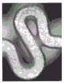
图1显示支架的完整图像(a),和沿着A-A线的横截面视图(b)。Figure 1 shows a complete image of the stent (a), and a cross-sectional view along the line A-A (b).
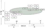
图2显示借助超声喷涂机来涂布支架的概貌。Figure 2 shows an overview of the coating of stents by means of an ultrasonic sprayer.
图3显示实施例2中的涂层的有缺陷的实例(网状并拢(adduct))。Figure 3 shows a defective example of the coating in Example 2 (adduct).
图4显示实施例2中的涂层的有缺陷的实例(不均匀的涂层)。FIG. 4 shows a defective example of the coating in Example 2 (non-uniform coating).
图5显示实施例2的结果。Figure 5 shows the results of Example 2.
图6显示实施例3的结果。Figure 6 shows the results of Example 3.
图7显示实施例4的结果。Figure 7 shows the results of Example 4.
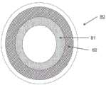
图8显示用于解释实施例5的图。FIG. 8 shows a diagram for explaining
图9显示为实施例5的结果的内膜中层(intima-media)/血管中层(vascularmedia)比。FIG. 9 shows the intima-media/vascularmedia ratio as the results of Example 5. FIG.
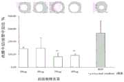
图10显示为实施例5的结果的新生内膜面积。FIG. 10 shows the neointimal area as a result of Example 5. FIG.
图11显示为实施例5的结果的内皮覆盖率。Figure 11 shows endothelial coverage as a result of Example 5.
具体实施方式Detailed ways
已知本文中使用的西洛他唑(其化学名为6-[4-(1-环己基-1H-四唑-5-基)丁氧基]-3,4-二氢喹诺酮)具有血小板凝集抑制作用,磷酸二酯酶(PDE)抑制作用,抗溃疡、降压作用和消炎作用,并且可用作抗血栓形成剂、用于改善脑循环的药物、消炎剂、抗溃疡药物、降压药物、平喘药物、磷酸二酯酶抑制剂等。西洛他唑还涵盖其药学上可接受的盐。Cilostazol (whose chemical name is 6-[4-(1-cyclohexyl-1H-tetrazol-5-yl)butoxy]-3,4-dihydroquinolone) used herein is known to have platelet Agglutination inhibitory effect, phosphodiesterase (PDE) inhibitory effect, antiulcer, antihypertensive and antiinflammatory effect, and can be used as an antithrombotic agent, a drug for improving cerebral circulation, an antiinflammatory agent, an antiulcer drug, an antihypertensive agent Drugs, antiasthmatic drugs, phosphodiesterase inhibitors, etc. Cilostazol also encompasses its pharmaceutically acceptable salts.
本文中所用的生物可吸收聚合物包括,例如包含丙交酯和/或乙交酯的聚丙交酯,其分子量为40,000至600,000。具体地,所述生物可吸收聚合物包括包含DL丙交酯、L丙交酯、乙交酯、己内酯等的聚合物,更具体地,(a)包含重量比为7:3-9:1的DL丙交酯和乙交酯的聚合物,其分子量为40,000-400,000,(b)包含DL丙交酯的聚合物,其分子量为50,000-100,000,(c)包含重量比为6:4-8:2的L丙交酯和DL丙交酯的聚合物,其分子量为300,000-600,000,(d)包含L丙交酯的聚合物,其分子量为50,000-150,000,和(e)包含重量比为6:4-8:2的L丙交酯和己内酯的聚合物,其分子量为150,000-400,000。优选地,所述生物可吸收聚合物包括下文实施例部分的表1中所列的生物可吸收聚合物,或其混合物,更优选RG858S、RG755S、LR704S、755/703或其混合物。Bioabsorbable polymers used herein include, for example, polylactides comprising lactide and/or glycolide, having a molecular weight of 40,000 to 600,000. Specifically, the bioabsorbable polymer includes a polymer comprising DL lactide, L lactide, glycolide, caprolactone, etc., more specifically, (a) comprising a weight ratio of 7:3-9 : 1 DL lactide and a polymer of glycolide having a molecular weight of 40,000-400,000, (b) a polymer comprising DL lactide having a molecular weight of 50,000-100,000, (c) comprising a weight ratio of 6: 4-8:2 polymers of L-lactide and DL-lactide having a molecular weight of 300,000-600,000, (d) a polymer comprising L-lactide having a molecular weight of 50,000-150,000, and (e) comprising A polymer of L-lactide and caprolactone in a weight ratio of 6:4-8:2 with a molecular weight of 150,000-400,000. Preferably, the bioabsorbable polymer comprises the bioabsorbable polymers listed in Table 1 of the Examples section below, or mixtures thereof, more preferably RG858S, RG755S, LR704S, 755/703 or mixtures thereof.
涂布剂3包含作为药物的西洛他唑和上述生物可吸收聚合物的混合物。所述生物可吸收聚合物需要防止包含西洛他唑的涂层(coating layer)免于除去,因为西洛他唑水溶性差,并且还需要维持高涂层强度(coating strength)。Coating agent 3 contains a mixture of cilostazol as a drug and the above-mentioned bioabsorbable polymer. The bioabsorbable polymer needs to protect the coating layer containing cilostazol from removal because cilostazol is poorly water soluble, and also needs to maintain high coating strength.
西洛他唑和聚丙交酯的按重量计的混合比优选为4:6-7:3。当该混合比在所述范围内时,期望获得良好的抑制内膜增厚的效果。并且,当该混合比在4:6-6:4内时,所述涂层强度可进一步增加。The mixing ratio by weight of cilostazol and polylactide is preferably 4:6-7:3. When the mixing ratio is within the range, it is expected to obtain a good effect of suppressing intimal thickening. And, when the mixing ratio is within 4:6-6:4, the coating strength can be further increased.
本文中使用的支架是由金属或聚合物材料制得的常规支架。在金属支架的情况下,该金属包括合适的镍、钴、铬、钛的合金和/或不锈钢,优选钴-铬合金作为主要成分。The stents used herein are conventional stents made from metallic or polymeric materials. In the case of a metallic stent, the metal comprises suitable alloys of nickel, cobalt, chromium, titanium and/or stainless steel, preferably a cobalt-chromium alloy as the main component.
本发明中用于使用西洛他唑和生物可吸收聚合物的混合物涂布支架的方法包括常规的简化喷涂法、浸渍法、电沉积、超声喷涂法等等,从涂层强度的角度优选超声喷涂法。The method for coating the stent using the mixture of cilostazol and the bioabsorbable polymer in the present invention includes conventional simplified spraying method, dipping method, electrodeposition, ultrasonic spraying method and the like, and ultrasonic wave is preferred from the viewpoint of coating strength spray method.
在下文中,在显示附图的情况下例示本发明的实施方案。In the following, embodiments of the present invention are illustrated with the accompanying drawings.
本发明的发明人已进行大量研究以便解决关于常规药物洗脱支架的问题,并已发现通过用西洛他唑和下文描述的聚合物涂布金属支架或聚合物材料支架可能制备可将西洛他唑稳定保持在支架上且强烈抑制内膜增厚的药物洗脱支架。The inventors of the present invention have conducted extensive research to address the problems associated with conventional drug-eluting stents, and have found that it is possible to prepare stents that can be treated with cilostazol by coating a metal stent or a polymer material stent with cilostazol and the polymer described below. Drug-eluting stents in which stazol remains stably on the stent and strongly inhibits intimal thickening.
图1(a)是显示本发明的药物洗脱支架的视图。图1(b)是图1(a)沿着A-A线的横截面视图。支架1具有圆柱体内腔结构,其具有较长方向轴,该轴的外周具有网状图案,其可向外扩张。将支架在未扩张状态下插入身体,并随后在治疗目标部位处在血管中扩张以留置在血管中。该扩张可用气囊导管中在血管中实现。图1显示网状图案的视图,但是本发明可包括其它网状图案。Fig. 1(a) is a view showing the drug-eluting stent of the present invention. Fig. 1(b) is a cross-sectional view taken along line A-A of Fig. 1(a). The
如图1(b)中所示,在基底构件2上用涂布剂3涂布本发明的支架1。基底构件2可以任意方法制备。例如,其可由中空不锈钢管或成型不锈钢管通过激光、放电铣削、化学刻蚀或其它方法来制备。基底构件2可由合适的镍、钴、铬、钛的合金和/或不锈钢制得。As shown in FIG. 1( b ), the
图2是显示施加涂布剂3到基底构件2上的超声喷涂机4的视图。在涂布步骤之前,基底构件2的表面用等离子体机(未显示于图2中)进行等离子体处理。在等离子体处理之后,将基底构件2与芯轴连接,该芯轴固定在超声喷涂机4中。在超声喷涂机4中,液体涂布剂经由带有注射泵的管道6运送,并随后用超声喷嘴5进行雾化和喷雾。在喷雾时,基底构件2旋转并在超声喷嘴5下线性移动以将涂布剂3堆积到基底构件2上。随后,基底构件2旋转并在氮气流下线性移动,并进一步在干燥器中在真空中干燥以制备支架1。FIG. 2 is a view showing the
本文中使用的液体涂布剂是西洛他唑和聚合物在溶剂中的溶液。本文中使用的溶剂包括具有低沸点的挥发性溶剂,其可在涂布之后容易除去。所述挥发性溶剂包括,例如,甲醇、乙醇、三氟乙醇、六氟异丙醇、异戊醇、乙酸甲酯、乙酸乙酯、丙酮、甲基乙基酮、二氯甲烷、氯仿、二氯乙烷和两种或更多种溶剂的混合物。The liquid coating agent used herein is a solution of cilostazol and polymer in a solvent. The solvent used herein includes a volatile solvent having a low boiling point, which can be easily removed after coating. Such volatile solvents include, for example, methanol, ethanol, trifluoroethanol, hexafluoroisopropanol, isoamyl alcohol, methyl acetate, ethyl acetate, acetone, methyl ethyl ketone, dichloromethane, chloroform, dichloromethane A mixture of ethyl chloride and two or more solvents.
实施例Example
下表1中所列的各聚合物用于以下各实施例。Each of the polymers listed in Table 1 below was used in each of the following examples.
表1Table 1

实施例1Example 1
钴-铬合金作为基底构件2通过超声喷雾涂布有西洛他唑和以上所列的(e)、(j)和(p)中的一种的聚合物的混合物,其中西洛他唑和所述聚合物的混合比如表2中所示变化,并且评价各涂层强度。结果显示于表2中。在该表中,符号"○"表明强度非常高,符号"Δ"表明强度高,且符号"×"表明强度低。Cobalt-chromium alloy as base member 2 is coated with a mixture of cilostazol and a polymer of one of (e), (j) and (p) listed above by ultrasonic spray, wherein cilostazol and The mixing ratio of the polymers was varied as shown in Table 2, and the strength of each coating was evaluated. The results are shown in Table 2. In this table, the symbol "○" indicates that the intensity is very high, the symbol "Δ" indicates that the intensity is high, and the symbol "x" indicates that the intensity is low.
结果表明当西洛他唑和聚合物的混合比(D/P比)为6:4或更低时,即,西洛他唑的量小于该比率时,强度高,并且特别地,当该比率为5:5时,强度足够高。然而,当D/P比小于4:6时,据推测用于药物洗脱支架的西洛他唑的药物效果可降低。因此,D/P比优选为4:6或更高。The results show that when the mixing ratio (D/P ratio) of cilostazol and the polymer is 6:4 or less, that is, when the amount of cilostazol is smaller than this ratio, the strength is high, and in particular, when the At a ratio of 5:5, the strength is high enough. However, when the D/P ratio is less than 4:6, it is speculated that the drug effect of cilostazol for drug-eluting stents may be reduced. Therefore, the D/P ratio is preferably 4:6 or higher.
表2Table 2

实施例2Example 2
钴-铬合金作为基底构件2通过超声喷雾涂布有通过混合西洛他唑和表1中所列的17种聚合物(a)至(q)中的一种制备的涂布剂。西洛他唑和各聚合物的混合比为5:5。A cobalt-chromium alloy as the base member 2 was coated with a coating agent prepared by mixing cilostazol and one of the 17 polymers (a) to (q) listed in Table 1 by ultrasonic spraying. The mixing ratio of cilostazol and each polymer was 5:5.
就涂层工程(coating work)来观察各制备的支架的外观。在外部观察中,在涂布支架中不具有显示于图3中的网状并拢或不具有显示于图4中的不均匀涂层并且表面不像橘子皮而是平整且光滑的涂布支架被评价为“良好”。结果显示于图5中。在图5中,坐标轴表示与西洛他唑混合的各聚合物的分子量,并且横坐标轴表示涂布剂的溶解速率。显示于图5中的(a)至(q)(其为表1中所列的17种聚合物)通过其位置显示两种因素的关系。The appearance of each prepared stent was observed for coating work. In external observation, a coated stent that does not have the mesh close together shown in Fig. 3 or that does not have the uneven coating shown in Fig. 4 and the surface is not like orange peel but flat and smooth in the coated stent is The evaluation was "good". The results are shown in FIG. 5 . In FIG. 5 , the axis of coordinates represents the molecular weight of each polymer mixed with cilostazol, and the axis of abscissa represents the dissolution rate of the coating agent. (a) to (q) shown in Figure 5, which are the 17 polymers listed in Table 1, show the relationship of the two factors by their positions.
作为涂层性能的评价,在图5中用椭圆圈出的区域中的聚合物显示良好的涂层趋势。图5表明涂层性能不能被溶解速率显著影响,但是其会被聚合物的分子量影响。欲用于包含西洛他唑的涂布剂中的聚合物优选是具有40,000-600,000的分子量的聚合物。特别地,观察到涂布剂中的聚合物(e)、(h)、(j)、(m)、(p)和(q)可带来良好的涂层性能。As an evaluation of coating performance, the polymer in the area circled with ellipses in Figure 5 showed good coating trends. Figure 5 shows that the coating performance cannot be significantly affected by the dissolution rate, but it can be affected by the molecular weight of the polymer. The polymer to be used in the coating agent containing cilostazol is preferably a polymer having a molecular weight of 40,000-600,000. In particular, polymers (e), (h), (j), (m), (p) and (q) in the coating agent were observed to give good coating properties.
实施例3Example 3
使用以上聚合物(e)、(h)、(j)、(m)、(p)和(q)以与实施例2类似的方式制备支架。如下文所解释的,将各制备的支架位于兔的髂血管中,并评价各支架抑制内膜增厚的效果。Scaffolds were prepared in a similar manner to Example 2 using the above polymers (e), (h), (j), (m), (p) and (q). As explained below, each prepared stent was placed in the iliac blood vessels of rabbits, and the effect of each stent in inhibiting intimal thickening was evaluated.
首先,切开兔的颈部,并取出右颈动脉,将插管器(introducer)与其连接。将气囊导管的导丝从插管器插入并在X射线透视下移动至待治疗的髂动脉的远端部分。并且随后,将血管造影导管沿着导丝插入,并在髂动脉的处理部位处进行血管造影。在处理部位处进行血管造影之后,将带有测试支架的气囊导管沿着气囊导管的导丝在X射线透视下插入处理部位。将测试支架(其中当标准直径扩张压力为9atm时支架直径为2.75mm)置于髂动脉的处理部位处(血管直径假定为2.5mm),并随后用充气装置(indeflator)扩张气囊,保持压力为14atm(过扩张,假定支架直径:3.0mm,20%过扩张),一次20秒。在确认支架扩张之后,将气囊放气,移除充气装置,并沿着气囊导管的导丝拔出气囊导管。在左右髂动脉处进行相同步骤。First, the neck of the rabbit was incised, and the right carotid artery was removed, and an introducer was attached to it. The guide wire of the balloon catheter is inserted from the introducer and moved under fluoroscopy to the distal portion of the iliac artery to be treated. And then, an angiographic catheter is inserted along the guide wire and angiography is performed at the treatment site of the iliac artery. Following angiography at the treatment site, a balloon catheter with a test stent was inserted into the treatment site under fluoroscopy along the guide wire of the balloon catheter. A test stent (where the stent diameter is 2.75 mm when the standard diameter expansion pressure is 9 atm) is placed at the treatment site of the iliac artery (the vessel diameter is assumed to be 2.5 mm), and the balloon is subsequently expanded with an inflator, maintaining the pressure at 2.5 mm. 14 atm (over-expanded, assumed stent diameter: 3.0 mm, 20% over-expanded), 20 seconds at a time. After confirmation of stent expansion, the balloon is deflated, the inflation device is removed, and the balloon catheter is withdrawn along the guidewire of the balloon catheter. The same procedure is performed at the left and right iliac arteries.
接着,沿着气囊导管的导丝移动血管造影导管靠近处理部位,并且在那里用稀释造影剂进行血管造影。在左右髂动脉处进行相同步骤,并随后拔出血管造影导管。最后,在鞘插入部位处将血管结扎,并缝合皮肤和肌肉层。采取这种方法,可将支架置于兔子的髂血管中。Next, the angiography catheter is moved along the guide wire of the balloon catheter close to the treatment site, and an angiography is performed there with diluted contrast medium. The same procedure was performed at the left and right iliac arteries, and the angiographic catheter was subsequently withdrawn. Finally, the blood vessels are ligated at the sheath insertion site, and the skin and muscle layers are sutured. Using this approach, a stent can be placed in the rabbit's iliac vessels.
通过使用在放置支架之前、放置支架不久之后(参考直径)和尸体解剖之前(放置之后28天)记录在DVD上的血管造影图片观察放置支架的部位来测试内膜增厚。并且基于放置支架不久之后的参考直径和尸体解剖之前的最窄血管直径之间的差异评价内膜增厚。Intimal thickening was tested by observing the site of stent placement using angiograms recorded on DVD before stent placement, shortly after stent placement (reference diameter) and before autopsy (28 days after placement). And intimal thickening was assessed based on the difference between the reference diameter shortly after stent placement and the diameter of the narrowest vessel before autopsy.
图6显示结果。图6中的术语“无药物”表示支架未涂布西洛他唑。其它结果基于涂布有包含比率为5:5的以上聚合物和西洛他唑的混合物的各涂布剂的支架。Figure 6 shows the results. The term "drug-free" in Figure 6 indicates that the stent is not coated with cilostazol. Additional results are based on stents coated with each coating agent comprising a mixture of the above polymer and cilostazol in a ratio of 5:5.
图6中的坐标轴表示内膜增厚,即当长度较小时,其表示抑制内膜增厚的效果较高。包括西洛他唑的(j)的结果显示内膜增厚被显著抑制。类似地,(e)和(p)的结果也显示具有西洛他唑的涂层可抑制内膜增厚。另外,在放置支架的部位处的内皮细胞的观察显示内皮细胞可很好地再生,并因此,涂布有西洛他唑的本发明的支架能够实现(1)抑制内膜增厚和(2)抑制血管内皮细胞再生的抑制两方面效果,这是使用莫司类型药物未实现的。The coordinate axis in FIG. 6 represents intimal thickening, that is, when the length is small, it represents that the effect of suppressing intimal thickening is high. The results of (j) including cilostazol showed that intimal thickening was significantly inhibited. Similarly, the results of (e) and (p) also show that coatings with cilostazol inhibit intimal thickening. In addition, the observation of endothelial cells at the site where the stent was placed shows that endothelial cells can be regenerated well, and thus, the stent of the present invention coated with cilostazol can achieve (1) inhibition of intimal thickening and (2) ) inhibition of vascular endothelial cell regeneration inhibited both effects, which was not achieved with limus-type drugs.
实施例4Example 4
使用以上聚合物(e)制备支架,其在西洛他唑(D)和聚合物(P)的混合比(D/P比)上不同。将制备的支架置于兔的髂血管中,并评价各支架抑制内膜增厚的效果。结果显示于图7中。A scaffold was prepared using the above polymer (e), which differed in the mixing ratio (D/P ratio) of cilostazol (D) and polymer (P). The prepared stents were placed in the iliac blood vessels of rabbits, and the effect of each stent on inhibiting intimal thickening was evaluated. The results are shown in FIG. 7 .
结果显示具有4:6或7:3的混合比(D/P比)的两种支架都可比仅由基底构件(BMS)组成的支架或仅涂布有聚合物的支架更多地抑制内膜增厚。The results show that both stents with a mixing ratio (D/P ratio) of 4:6 or 7:3 can inhibit the intima more than the stent consisting of only the base member (BMS) or the stent only coated with the polymer thickened.
另外,在放置支架的部位处的内皮细胞的观察显示内皮细胞可很好地再生,并因此,涂布有西洛他唑的本发明的支架能够实现(1)抑制内膜增厚和(2)抑制血管内皮细胞再生的抑制两方面效果,这是使用莫司类型药物未实现的。In addition, the observation of endothelial cells at the site where the stent was placed shows that endothelial cells can be regenerated well, and thus, the stent of the present invention coated with cilostazol can achieve (1) inhibition of intimal thickening and (2) ) inhibition of vascular endothelial cell regeneration inhibited both effects, which was not achieved with limus-type drugs.
实施例5Example 5
类似地,使用以上聚合物(e)制备支架,其中西洛他唑和聚合物(e)的混合比(D/P比)固定为5:5,但是西洛他唑的重量在300μg-600μg之间变化。将制备的支架置于猪的髂血管中,并随后在放置28天之后,评价在髂血管的放置支架的部位处(I)内膜中层/血管中层比、(II)新生内膜面积和(III)血管内皮细胞覆盖率。Similarly, scaffolds were prepared using polymer (e) above, where the mixing ratio (D/P ratio) of cilostazol and polymer (e) was fixed at 5:5, but the weight of cilostazol was in the range of 300 μg-600 μg change between. The prepared stents were placed in the iliac vessels of pigs, and then 28 days after placement, the evaluation of (I) intima-media/vessel-media ratio, (II) neointimal area and ( III) Vascular endothelial cell coverage.
如图8中所示,此处(II)新生内膜面积表示内膜中层81的横截面积,其是在放置支架的部位处的血管80的内部中最新形成的。(I)内膜中层/血管中层比表示以上新生内膜面积与血管中层82的横截面积的比率。As shown in FIG. 8, here (II) neointimal area represents the cross-sectional area of the intima-
如下进行评价:Evaluate as follows:
(a)拔出髂血管,(a) pulling out the iliac vessels,
(b)将其清洗,并随后使其脱脂,(b) cleaned and subsequently degreased,
(c)渗透树脂,并随后通过聚合该树脂使其固定,(c) infiltrating the resin and subsequently immobilizing the resin by polymerizing it,
(d)在目标部位处将其切断,和(d) severing it at the target site, and
(e)将其染色,并随后通过显微镜对其进行观察。(e) It was stained and then observed by microscope.
图9显示改变西洛他唑的重量确定的各(I)内膜中层/血管中层比。图的上部显示各血管横截面的图片,该图片在血管中放置载有各重量的西洛他唑的支架28天之后拍摄。本文中通过"BMS"表示的数据是通过使用未涂布有任何药物或聚合物的金属支架获得的结果。Figure 9 shows each (I) intima-media/vascular media ratio determined by varying the weight of cilostazol. The upper part of the figure shows a picture of a cross-section of each blood vessel, taken 28 days after placement of a stent loaded with each weight of cilostazol in the blood vessel. The data represented herein by "BMS" are results obtained using metal stents that are not coated with any drug or polymer.
根据图9,当西洛他唑的重量超过400μg时,内膜中层/血管中层比降至远低于100%,其表明在放置支架的部位处内膜中层的产生可被抑制。特别地,与仅由基底构件(BMS)组成的支架的情况(其内膜中层/血管中层比大于260%)相比较,当西洛他唑的重量为500μg或600μg时,内膜增厚可被显著抑制。According to Figure 9, when the weight of cilostazol exceeds 400 μg, the intima-media/vessel-media ratio drops well below 100%, indicating that intima-media production can be inhibited at the site of stent placement. In particular, when the weight of cilostazol was 500 μg or 600 μg, the intimal thickening could be compared with the case of stents composed of only base member (BMS), whose intima-media/vessel-media ratio was greater than 260%. was significantly inhibited.
然而,当西洛他唑的重量太大时,聚合物的量必须增加。在这样的情况下,涂布剂的总量应增大,并由此变得难以形成强且均匀的涂层。另外,图9中的结果显示内膜中层/血管中层比的降低在西洛他唑的重量为500μg-600μg时达到峰值。因此,西洛他唑的量优选大于400μg且小于700μg,且特别地,更优选大于500μg且小于600μg。However, when the weight of cilostazol is too large, the amount of polymer must be increased. In such a case, the total amount of the coating agent should be increased, and thus it becomes difficult to form a strong and uniform coating. Additionally, the results in Figure 9 show that the reduction in the intima-media/vascular media ratio peaked at cilostazol weights of 500 μg-600 μg. Therefore, the amount of cilostazol is preferably more than 400 μg and less than 700 μg, and in particular, more preferably more than 500 μg and less than 600 μg.
图10显示改变西洛他唑的重量确定的各(II)新生内膜面积。图10表明与仅由基底构件(BMS)组成的支架的情况相比较,涂布有西洛他唑的支架的任何情况均可降低新生内膜面积,并且特别地,当西洛他唑的重量大于400μg时,新生内膜面积大大降低。特别地,当西洛他唑的重量为600μg时,与仅由基底构件(BMS)组成的支架的情况相比较,新生内膜面积显著降低。Figure 10 shows each (II) neointimal area determined by varying the weight of cilostazol. Figure 10 demonstrates that any case of cilostazol-coated stents reduces neointimal area compared to the case of stents consisting of only base member (BMS), and in particular, when the weight of cilostazol When more than 400μg, the neointimal area was greatly reduced. In particular, when the weight of cilostazol was 600 μg, the neointimal area was significantly reduced compared to the case of the scaffold consisting only of the base member (BMS).
图11显示改变西洛他唑的重量确定5的各(III)血管内皮细胞覆盖率。结果表明与仅由基底构件(BMS)组成的支架的情况相比较,具有西洛他唑和聚合物的涂层可使得在任何西洛他唑重量下容易形成血管内皮。Figure 11 shows each (III) vascular endothelial cell coverage of 5 determined by varying the weight of cilostazol. The results show that a coating with cilostazol and a polymer can facilitate the formation of vascular endothelium at any cilostazol weight compared to the case of a stent consisting of a base member (BMS) alone.
如图9-图11中的结果所示,本发明可在放置支架的部位处抑制内膜增厚,并防止抑制血管内皮细胞再生。因此,西洛他唑的量优选大于400μg且小于700μg,并且特别地,更优选大于500μg且小于600μg。As shown in the results in Figures 9-11, the present invention can inhibit intimal thickening at the site where the stent is placed, and prevent inhibition of vascular endothelial cell regeneration. Therefore, the amount of cilostazol is preferably more than 400 μg and less than 700 μg, and in particular, more preferably more than 500 μg and less than 600 μg.
字母或数字说明letter or number description
1:支架1: Bracket
2:基底构件2: base member
3:涂布剂3: Coating agent
4:超声喷涂机4: Ultrasonic sprayer
5:超声喷嘴5: Ultrasonic nozzle
6:管道6: Pipes
80:血管的横截面80: Cross-section of blood vessels
81:内膜中层81: Intima-media
82:血管中层82: Vascular Media
Claims (12)
Applications Claiming Priority (3)
| Application Number | Priority Date | Filing Date | Title |
|---|---|---|---|
| JP2014219159 | 2014-10-28 | ||
| JP2014-219159 | 2014-10-28 | ||
| CN201580071286.5ACN107106309B (en) | 2014-10-28 | 2015-10-21 | drug eluting stent |
Related Parent Applications (1)
| Application Number | Title | Priority Date | Filing Date |
|---|---|---|---|
| CN201580071286.5ADivisionCN107106309B (en) | 2014-10-28 | 2015-10-21 | drug eluting stent |
Publications (1)
| Publication Number | Publication Date |
|---|---|
| CN110711056Atrue CN110711056A (en) | 2020-01-21 |
Family
ID=55857334
Family Applications (2)
| Application Number | Title | Priority Date | Filing Date |
|---|---|---|---|
| CN201910994051.3APendingCN110711056A (en) | 2014-10-28 | 2015-10-21 | drug eluting stent |
| CN201580071286.5AActiveCN107106309B (en) | 2014-10-28 | 2015-10-21 | drug eluting stent |
Family Applications After (1)
| Application Number | Title | Priority Date | Filing Date |
|---|---|---|---|
| CN201580071286.5AActiveCN107106309B (en) | 2014-10-28 | 2015-10-21 | drug eluting stent |
Country Status (7)
| Country | Link |
|---|---|
| US (1) | US11241322B2 (en) |
| EP (1) | EP3213721B1 (en) |
| JP (1) | JP6820745B2 (en) |
| KR (2) | KR102522542B1 (en) |
| CN (2) | CN110711056A (en) |
| TW (1) | TWI721956B (en) |
| WO (1) | WO2016067994A1 (en) |
Families Citing this family (2)
| Publication number | Priority date | Publication date | Assignee | Title |
|---|---|---|---|---|
| CN106730050B (en)* | 2017-02-22 | 2019-12-20 | 广州南创珠峰医疗科技有限责任公司 | Preparation method of multifunctional drug eluting coating for intravascular stent |
| TWI794616B (en)* | 2019-07-09 | 2023-03-01 | 日商大塚醫療器材股份有限公司 | Drug-eluting stent |
Citations (7)
| Publication number | Priority date | Publication date | Assignee | Title |
|---|---|---|---|---|
| CN1355005A (en)* | 2001-12-13 | 2002-06-26 | 华东理工大学 | Drug-eluting cardiovascular stent and preparation method thereof |
| CN1655738A (en)* | 2002-05-20 | 2005-08-17 | 奥勃斯医学技术股份有限公司 | Drug eluting implantable medical device |
| CN1669596A (en)* | 2004-03-16 | 2005-09-21 | 程树军 | Medicine eluted cardiovascular support |
| CN1669595A (en)* | 2004-03-16 | 2005-09-21 | 程树军 | Medicine eluted cardiovascular support |
| CN1669597A (en)* | 2004-03-16 | 2005-09-21 | 程树军 | Medicine eluted cardiovascular support |
| WO2010111238A2 (en)* | 2009-03-23 | 2010-09-30 | Micell Technologies, Inc. | Improved biodegradable polymers |
| CN101879102A (en)* | 2009-05-07 | 2010-11-10 | 微创医疗器械(上海)有限公司 | Groove carrying-type coating decomposable drug eluting stent |
Family Cites Families (37)
| Publication number | Priority date | Publication date | Assignee | Title |
|---|---|---|---|---|
| EG20321A (en)* | 1993-07-21 | 1998-10-31 | Otsuka Pharma Co Ltd | Medical material and process for producing the same |
| US5637113A (en)* | 1994-12-13 | 1997-06-10 | Advanced Cardiovascular Systems, Inc. | Polymer film for wrapping a stent structure |
| US8057816B2 (en) | 1997-09-26 | 2011-11-15 | Abbott Laboratories | Compositions and methods of administering paclitaxel with other drugs using medical devices |
| US8394398B2 (en) | 1997-09-26 | 2013-03-12 | Abbott Laboratories | Methods of administering rapamycin analogs with anti-inflammatories using medical devices |
| JP4473390B2 (en) | 2000-01-07 | 2010-06-02 | 川澄化学工業株式会社 | Stent and stent graft |
| US20060177416A1 (en)* | 2003-10-14 | 2006-08-10 | Medivas, Llc | Polymer particle delivery compositions and methods of use |
| ATE369817T1 (en) | 2000-12-22 | 2007-09-15 | Avantec Vascular Corp | DEVICE FOR DELIVERING THERAPEUTIC ACTIVE INGREDIENTS |
| US6607548B2 (en)* | 2001-05-17 | 2003-08-19 | Inion Ltd. | Resorbable polymer compositions |
| DE60217505T2 (en)* | 2001-07-26 | 2007-11-15 | Avantec Vascular Corp., Sunnyvale | Devices for delivery of therapeutic agents with a variable release profile |
| CA2476431A1 (en)* | 2002-02-15 | 2003-08-21 | Cv Therapeutics, Inc. | Coating having polymerized silane derivatives for medical devices |
| JP4245302B2 (en) | 2002-04-05 | 2009-03-25 | 川澄化学工業株式会社 | Stent |
| AU2003280437A1 (en) | 2002-06-27 | 2004-01-19 | Microport Medical (Shanghai) Co., Ltd. | Drug eluting stent |
| AU2003261100A1 (en)* | 2002-07-25 | 2004-02-16 | Avantec Vascular Corporation | Devices delivering therapeutic agents and methods regarding the same |
| DE10244847A1 (en) | 2002-09-20 | 2004-04-01 | Ulrich Prof. Dr. Speck | Medical device for drug delivery |
| US20050112273A1 (en) | 2003-05-19 | 2005-05-26 | Stenzel Eric B. | Method of improving the quality and performance of a coating on a coated medical device using a solvent to reflow the coating |
| EP1635815A4 (en) | 2003-06-03 | 2009-03-25 | Beth Israel Hospital | METHODS AND COMPOUNDS FOR THE TREATMENT OF VASCULAR STENOSIS |
| CA2555364C (en) | 2004-03-10 | 2012-10-16 | Orbus Medical Technologies, Inc. | Progenitor endothelial cell capturing with a drug eluting implantable medical device |
| CA2559747C (en) | 2004-03-19 | 2013-12-31 | Abbott Laboratories | Multiple drug delivery from a balloon and a prosthesis |
| CN101014357A (en) | 2004-07-07 | 2007-08-08 | 麦迪库瑞国际公司 | Combination therapies employing platelet aggregation drugs |
| KR100511618B1 (en)* | 2005-01-17 | 2005-08-31 | 이경범 | Multi-layer coating of drug release controllable coronary stent and method for manufacturing the same |
| US8221824B2 (en)* | 2005-02-03 | 2012-07-17 | Boston Scientific Scimed, Inc. | Deforming surface of drug eluting coating to alter drug release profile of a medical device |
| WO2006099244A1 (en) | 2005-03-11 | 2006-09-21 | Hong Kong Nitric Oxide Limited | Combination therapy for endothelial dysfunction, angina and diabetes |
| WO2007046935A2 (en) | 2005-10-14 | 2007-04-26 | Abbott Laboratories | Compositions, systems, kits, and methods of administering rapamycin analogs with paclitaxel using medical devices |
| US8784860B2 (en) | 2005-10-27 | 2014-07-22 | Cordis Corporation | Local administration of a combination of rapamycin and cilostazol for the treatment of vascular disease |
| JP4297221B2 (en) | 2006-02-15 | 2009-07-15 | 株式会社ホソカワ粉体技術研究所 | Method for producing drug-eluting stent |
| US7981150B2 (en) | 2006-11-09 | 2011-07-19 | Boston Scientific Scimed, Inc. | Endoprosthesis with coatings |
| JP5557373B2 (en)* | 2006-11-21 | 2014-07-23 | アボット ラボラトリーズ | Use of terpolymers of tetrafluoroethylene, hexafluoropropylene, and vinylidene fluoride in drug-eluting coatings |
| WO2009031295A1 (en) | 2007-09-04 | 2009-03-12 | Japan Stent Technology Co., Ltd. | Stent for controlled drug release |
| EP2191853B1 (en)* | 2007-09-28 | 2015-07-29 | Terumo Kabushiki Kaisha | In-vivo indwelling matter |
| EP2410954A4 (en)* | 2009-03-23 | 2014-03-05 | Micell Technologies Inc | PERIPHERAL ENDOPROSTHETICS WITH LAYERS |
| US20100280600A1 (en)* | 2009-04-30 | 2010-11-04 | Vipul Bhupendra Dave | Dual drug stent |
| US20110137407A1 (en) | 2009-07-09 | 2011-06-09 | Thai Minh Nguyen | Bare metal stent with drug eluting reservoirs |
| PH12012500393A1 (en)* | 2009-08-26 | 2012-10-22 | Otsuka Medical Devices Co Ltd | Medicinal device for placement into a lumen and manufacturing method thereof |
| US20120130481A1 (en)* | 2010-11-18 | 2012-05-24 | Robert Falotico | Local vascular delivery of adenosine a2a receptor agonists in combination with other agents to reduce myocardial injury |
| EP2676639B1 (en)* | 2011-02-15 | 2019-08-07 | Terumo Kabushiki Kaisha | Stent delivery system |
| CN104587534A (en)* | 2013-10-31 | 2015-05-06 | 先健科技(深圳)有限公司 | An absorbable iron-base alloy support |
| US9855371B2 (en)* | 2014-04-28 | 2018-01-02 | John James Scanlon | Bioresorbable stent |
- 2015
- 2015-10-21EPEP15856064.9Apatent/EP3213721B1/enactiveActive
- 2015-10-21WOPCT/JP2015/079693patent/WO2016067994A1/enactiveApplication Filing
- 2015-10-21JPJP2016556518Apatent/JP6820745B2/enactiveActive
- 2015-10-21CNCN201910994051.3Apatent/CN110711056A/enactivePending
- 2015-10-21TWTW104134675Apatent/TWI721956B/enactive
- 2015-10-21USUS15/522,406patent/US11241322B2/enactiveActive
- 2015-10-21KRKR1020227019635Apatent/KR102522542B1/enactiveActive
- 2015-10-21CNCN201580071286.5Apatent/CN107106309B/enactiveActive
- 2015-10-21KRKR1020177013885Apatent/KR102409251B1/enactiveActive
Patent Citations (8)
| Publication number | Priority date | Publication date | Assignee | Title |
|---|---|---|---|---|
| CN1355005A (en)* | 2001-12-13 | 2002-06-26 | 华东理工大学 | Drug-eluting cardiovascular stent and preparation method thereof |
| CN1303947C (en)* | 2001-12-13 | 2007-03-14 | 华东理工大学 | Medicine eluted cardiovascular frame and its preparing process |
| CN1655738A (en)* | 2002-05-20 | 2005-08-17 | 奥勃斯医学技术股份有限公司 | Drug eluting implantable medical device |
| CN1669596A (en)* | 2004-03-16 | 2005-09-21 | 程树军 | Medicine eluted cardiovascular support |
| CN1669595A (en)* | 2004-03-16 | 2005-09-21 | 程树军 | Medicine eluted cardiovascular support |
| CN1669597A (en)* | 2004-03-16 | 2005-09-21 | 程树军 | Medicine eluted cardiovascular support |
| WO2010111238A2 (en)* | 2009-03-23 | 2010-09-30 | Micell Technologies, Inc. | Improved biodegradable polymers |
| CN101879102A (en)* | 2009-05-07 | 2010-11-10 | 微创医疗器械(上海)有限公司 | Groove carrying-type coating decomposable drug eluting stent |
Also Published As
| Publication number | Publication date |
|---|---|
| JP6820745B2 (en) | 2021-01-27 |
| US20170319362A1 (en) | 2017-11-09 |
| KR20220088945A (en) | 2022-06-28 |
| WO2016067994A1 (en) | 2016-05-06 |
| KR20170077161A (en) | 2017-07-05 |
| CN107106309A (en) | 2017-08-29 |
| EP3213721A1 (en) | 2017-09-06 |
| US11241322B2 (en) | 2022-02-08 |
| TWI721956B (en) | 2021-03-21 |
| EP3213721B1 (en) | 2021-06-02 |
| KR102522542B1 (en) | 2023-04-14 |
| JPWO2016067994A1 (en) | 2017-09-21 |
| TW201620551A (en) | 2016-06-16 |
| CN107106309B (en) | 2019-11-08 |
| KR102409251B1 (en) | 2022-06-14 |
| HK1243309A1 (en) | 2018-07-13 |
| EP3213721A4 (en) | 2018-10-24 |
Similar Documents
| Publication | Publication Date | Title |
|---|---|---|
| JP4493655B2 (en) | Method for applying a drug polymer coating to a stent | |
| JP5636451B2 (en) | Drug sustained release stent | |
| JP4894519B2 (en) | Indwelling stent | |
| US20230414383A1 (en) | Drug-eluting stent including crystalline cilostazol | |
| WO2004064910A1 (en) | Indwelling stent | |
| CN107106309B (en) | drug eluting stent | |
| JP2015154921A (en) | drug sustained-release stent | |
| HK1243309B (en) | Drug-eluting stent | |
| WO2006027994A1 (en) | Indwelling stent | |
| JP2016165375A (en) | Manufacturing method of stent having anticorrosion coating layer | |
| JP2007301214A (en) | Coated stent | |
| JP2010166935A (en) | Stent | |
| JPWO2007132801A1 (en) | Stent |
Legal Events
| Date | Code | Title | Description |
|---|---|---|---|
| PB01 | Publication | ||
| PB01 | Publication | ||
| SE01 | Entry into force of request for substantive examination | ||
| SE01 | Entry into force of request for substantive examination | ||
| RJ01 | Rejection of invention patent application after publication | ||
| RJ01 | Rejection of invention patent application after publication | Application publication date:20200121 |