CN110709029B - Minimally invasive implantable device and mitral valve implant system - Google Patents
Minimally invasive implantable device and mitral valve implant systemDownload PDFInfo
- Publication number
- CN110709029B CN110709029BCN201880035165.9ACN201880035165ACN110709029BCN 110709029 BCN110709029 BCN 110709029BCN 201880035165 ACN201880035165 ACN 201880035165ACN 110709029 BCN110709029 BCN 110709029B
- Authority
- CN
- China
- Prior art keywords
- tissue anchor
- annuloplasty ring
- mitral valve
- tissue
- annulus
- Prior art date
- Legal status (The legal status is an assumption and is not a legal conclusion. Google has not performed a legal analysis and makes no representation as to the accuracy of the status listed.)
- Active
Links
Images


Classifications
- A—HUMAN NECESSITIES
- A61—MEDICAL OR VETERINARY SCIENCE; HYGIENE
- A61F—FILTERS IMPLANTABLE INTO BLOOD VESSELS; PROSTHESES; DEVICES PROVIDING PATENCY TO, OR PREVENTING COLLAPSING OF, TUBULAR STRUCTURES OF THE BODY, e.g. STENTS; ORTHOPAEDIC, NURSING OR CONTRACEPTIVE DEVICES; FOMENTATION; TREATMENT OR PROTECTION OF EYES OR EARS; BANDAGES, DRESSINGS OR ABSORBENT PADS; FIRST-AID KITS
- A61F2/00—Filters implantable into blood vessels; Prostheses, i.e. artificial substitutes or replacements for parts of the body; Appliances for connecting them with the body; Devices providing patency to, or preventing collapsing of, tubular structures of the body, e.g. stents
- A61F2/02—Prostheses implantable into the body
- A61F2/24—Heart valves ; Vascular valves, e.g. venous valves; Heart implants, e.g. passive devices for improving the function of the native valve or the heart muscle; Transmyocardial revascularisation [TMR] devices; Valves implantable in the body
- A61F2/2442—Annuloplasty rings or inserts for correcting the valve shape; Implants for improving the function of a native heart valve
- A61F2/2445—Annuloplasty rings in direct contact with the valve annulus
- A—HUMAN NECESSITIES
- A61—MEDICAL OR VETERINARY SCIENCE; HYGIENE
- A61F—FILTERS IMPLANTABLE INTO BLOOD VESSELS; PROSTHESES; DEVICES PROVIDING PATENCY TO, OR PREVENTING COLLAPSING OF, TUBULAR STRUCTURES OF THE BODY, e.g. STENTS; ORTHOPAEDIC, NURSING OR CONTRACEPTIVE DEVICES; FOMENTATION; TREATMENT OR PROTECTION OF EYES OR EARS; BANDAGES, DRESSINGS OR ABSORBENT PADS; FIRST-AID KITS
- A61F2/00—Filters implantable into blood vessels; Prostheses, i.e. artificial substitutes or replacements for parts of the body; Appliances for connecting them with the body; Devices providing patency to, or preventing collapsing of, tubular structures of the body, e.g. stents
- A61F2/02—Prostheses implantable into the body
- A61F2/24—Heart valves ; Vascular valves, e.g. venous valves; Heart implants, e.g. passive devices for improving the function of the native valve or the heart muscle; Transmyocardial revascularisation [TMR] devices; Valves implantable in the body
- A61F2/2442—Annuloplasty rings or inserts for correcting the valve shape; Implants for improving the function of a native heart valve
- A61F2/2466—Delivery devices therefor
- A—HUMAN NECESSITIES
- A61—MEDICAL OR VETERINARY SCIENCE; HYGIENE
- A61B—DIAGNOSIS; SURGERY; IDENTIFICATION
- A61B17/00—Surgical instruments, devices or methods
- A61B17/064—Surgical staples, i.e. penetrating the tissue
- A61B17/0643—Surgical staples, i.e. penetrating the tissue with separate closing member, e.g. for interlocking with staple
- A—HUMAN NECESSITIES
- A61—MEDICAL OR VETERINARY SCIENCE; HYGIENE
- A61B—DIAGNOSIS; SURGERY; IDENTIFICATION
- A61B17/00—Surgical instruments, devices or methods
- A61B17/064—Surgical staples, i.e. penetrating the tissue
- A61B2017/0649—Coils or spirals
- A—HUMAN NECESSITIES
- A61—MEDICAL OR VETERINARY SCIENCE; HYGIENE
- A61F—FILTERS IMPLANTABLE INTO BLOOD VESSELS; PROSTHESES; DEVICES PROVIDING PATENCY TO, OR PREVENTING COLLAPSING OF, TUBULAR STRUCTURES OF THE BODY, e.g. STENTS; ORTHOPAEDIC, NURSING OR CONTRACEPTIVE DEVICES; FOMENTATION; TREATMENT OR PROTECTION OF EYES OR EARS; BANDAGES, DRESSINGS OR ABSORBENT PADS; FIRST-AID KITS
- A61F2220/00—Fixations or connections for prostheses classified in groups A61F2/00 - A61F2/26 or A61F2/82 or A61F9/00 or A61F11/00 or subgroups thereof
- A61F2220/0008—Fixation appliances for connecting prostheses to the body
- A61F2220/0016—Fixation appliances for connecting prostheses to the body with sharp anchoring protrusions, e.g. barbs, pins, spikes
Landscapes
- Health & Medical Sciences (AREA)
- Cardiology (AREA)
- Oral & Maxillofacial Surgery (AREA)
- Transplantation (AREA)
- Engineering & Computer Science (AREA)
- Biomedical Technology (AREA)
- Heart & Thoracic Surgery (AREA)
- Vascular Medicine (AREA)
- Life Sciences & Earth Sciences (AREA)
- Animal Behavior & Ethology (AREA)
- General Health & Medical Sciences (AREA)
- Public Health (AREA)
- Veterinary Medicine (AREA)
- Prostheses (AREA)
Abstract
Description
Translated fromChinese技术领域technical field
本发明涉及一种微创植入式装置和二尖瓣植入物系统。The present invention relates to a minimally invasive implantable device and a mitral valve implant system.
背景技术Background technique
该医学技术用于检测和消除疾病以恢复患者的健康。本发明涉及心脏手术领域。在心脏手术领域中,使用器械、装置或方法检查心脏内部和/或将其用于手术介入。特别地,本发明涉及心脏瓣膜的微创重建,其中使用手术器械,其允许借助通路进入心脏以执行各种重建以及在跳动的心脏上使用本发明的装置。该装置涉及固定到组织的可植入装置,从而限制或压缩扩张的开口。本发明涉及一种瓣环成形术环,其可用于身体器官的腔中,尤其是用于心脏中,以消除二尖瓣关闭不全。This medical technology is used to detect and eliminate diseases to restore the health of patients. The present invention relates to the field of cardiac surgery. In the field of cardiac surgery, instruments, devices or methods are used to examine the interior of the heart and/or use it for surgical intervention. In particular, the present invention relates to minimally invasive reconstruction of heart valves using surgical instruments that allow access to the heart to perform various reconstructions and use of the devices of the present invention on a beating heart. The device involves an implantable device that is secured to tissue, thereby confining or compressing an expanded opening. The present invention relates to an annuloplasty ring that can be used in cavities of bodily organs, especially in the heart, to eliminate mitral regurgitation.
心脏是一个肌肉发达的中空器官,其通过有节律的收缩将血液泵入人体,从而确保了所有器官的供应。因此,心脏疾病可能导致各种功能障碍,例如心力衰竭。心力衰竭是心脏病态的无力,其不增加心房压力无法提供身体所需的血液。心力衰竭根据其病程、主要涉及的心脏部分(右侧或左侧)和机制进行划分。另一个常见的心脏病是心脏瓣膜缺损(Herzklappenfehler)。心脏瓣膜缺损是一个或多个心脏瓣膜的功能障碍。心脏瓣膜缺损会影响四个心脏瓣膜中的每一个,其中左心脏瓣膜、主动脉瓣和二尖瓣中的瓣膜明显比右心脏瓣膜受到更多影响。功能障碍可包括变窄(狭窄(Stenose))、闭锁不全(关闭不全(Insuffizienz))或两者的结合(联合缺损(kombiniertes Vitium))。The heart is a muscular, hollow organ that pumps blood through the body through rhythmic contractions, ensuring the supply of all organs. Therefore, heart disease can lead to various dysfunctions, such as heart failure. Heart failure is the inability of the heart state to provide the blood the body needs without increasing pressure in the atria. Heart failure is classified according to its course, which part of the heart is primarily involved (right or left side), and mechanism. Another common heart disease is heart valve defect (Herzklappenfehler). A heart valve defect is a malfunction of one or more heart valves. Heart valve defects affect each of the four heart valves, with the left heart valve, the aortic valve and the mitral valve being significantly more affected than the right heart valve. Dysfunction may include narrowing (Stenose), insufficiency (Insuffizienz), or a combination of the two (kombiniertes Vitium).
二尖瓣作为单向瓣膜起效。在射血期(收缩)期间,心脏二尖瓣闭锁不全或渗漏会导致含氧血液从左心室(左心室(linker Ventrikel))到左心房(左心房(linkes Atrium))按比例回流,而含氧血液的主要部分则穿过主动脉瓣被压入主动脉。二尖瓣反流可以由二尖瓣中的各种不同的机械缺陷引起。瓣膜小叶、瓣膜、将瓣膜小叶连接至乳头肌的腱索或乳头肌本身可能受损或失灵。通常,瓣膜环关闭二尖瓣的能力足以承受左心室的高压。为了避免瓣膜反流,即在正常的心脏收缩周期中血液从左心室回流到左心房,在本领域中已知各种用于二尖瓣修复的装置和方法。二尖瓣重建涉及恢复瓣膜功能并保留二尖瓣。可以使用的手术方法包括例如胸骨切开术(Sternotomie)、导管引导的和微创的瓣环成形术(Anuloplastie)。所有类型的瓣环成形术环(AnuloplastieRinge)均可作为装置使用来消除二尖瓣后叶(后叶(posterior leaflet))和前叶(前叶(anterior leaflet))之间的渗漏。The mitral valve acts as a one-way valve. During the ejection phase (systole), insufficiency or leakage of the mitral valve of the heart causes a proportional backflow of oxygenated blood from the left ventricle (linker Ventrikel) to the left atrium (linkes Atrium), while the The major portion of the oxygenated blood is then pressed into the aorta through the aortic valve. Mitral regurgitation can be caused by a variety of different mechanical defects in the mitral valve. The valve leaflets, the valve, the chordae tendineae that connect the valve leaflets to the papillary muscles, or the papillary muscles themselves may be damaged or malfunctioning. Usually, the ability of the annulus to close the mitral valve is sufficient to withstand the high pressure of the left ventricle. To avoid valvular regurgitation, the backflow of blood from the left ventricle to the left atrium during the normal systolic cycle, various devices and methods for mitral valve repair are known in the art. Mitral valve reconstruction involves restoring valve function and preserving the mitral valve. Surgical methods that can be used include, for example, sternotomy, catheter-guided, and minimally invasive annuloplastie. All types of annuloplasty rings (Anuloplastie Ringes) can be used as devices to eliminate leakage between the posterior (posterior leaflet) and anterior (anterior leaflet) leaflets of the mitral valve.
许多不同的瓣环成形术环在本领域中是已知的。例如,刚性、半刚性和柔性瓣环成形术环,以及闭合、半闭合或开放式瓣膜成形术环。瓣环成形术环的形状是不同的,可以是圆形、D形、C形或肾形。瓣环成形术环的材料也不同。但是,所有机械瓣环成形术环都有一个共同点:它们一方面由不可溶解的材料组成,因为它们必须在瓣膜小叶的瓣环上生长,另一方面,它们应保留天然二尖瓣的功能。Many different annuloplasty rings are known in the art. For example, rigid, semi-rigid and flexible annuloplasty rings, as well as closed, semi-closed or open annuloplasty rings. Annuloplasty rings come in different shapes and can be round, D-shaped, C-shaped, or kidney-shaped. The material of the annuloplasty ring also varies. However, all mechanical annuloplasty rings have one thing in common: on the one hand they are composed of insoluble material, since they must grow on the annulus of the valve leaflets, and on the other hand, they should retain the function of the native mitral valve .
例如,在美国专利号8,545,414B2中公开了这种瓣环成形术环。该瓣环成形术环包括诸如钛的不锈钢内部材料,或者由诸如硅橡胶或达克隆(DACRON)的柔性材料制成。内部材料周围覆盖有例如生物相容的织物或布的材料。在瓣膜成形术方法中,将该瓣膜成形术环植入二尖瓣环上,以消除返流。该瓣环成形术环是杆状的并且具有大的字母“D”的形状。它在相对直的部分有一个开口,由具有达克隆网格覆盖物的塑料制成。该瓣环成形术环被缝在小叶的前后瓣环上。在刚性和扁平的实施方式中这种瓣环成形术环存在缺点。另一个缺点是它只能与心脏左心房中的常规胸骨切开术一起应用。其固定的方式也是不利的。通过沿瓣环上的二尖瓣的连续的植入缝合,可以在瓣环上完成瓣环成形术环的固定。但是,前段的缝合不当可能会导致环的不希望的三角内缩短(intratrigonales Verkürzen)。Such an annuloplasty ring is disclosed, for example, in US Patent No. 8,545,414 B2. The annuloplasty ring includes a stainless steel inner material such as titanium, or is made of a flexible material such as silicone rubber or DACRON. The inner material is surrounded by a material such as a biocompatible fabric or cloth. In the valvuloplasty method, the valvuloplasty ring is implanted on the mitral valve annulus to eliminate regurgitation. The annuloplasty ring is rod-shaped and has the shape of a large letter "D". It has an opening in the relatively straight section and is made of plastic with a Dacron mesh covering. The annuloplasty ring is sewn onto the anterior and posterior annulus of the leaflets. Such annuloplasty rings have disadvantages in rigid and flat embodiments. Another disadvantage is that it can only be used with conventional sternotomy in the left atrium of the heart. The way it is fixed is also disadvantageous. Annuloplasty ring fixation can be accomplished on the annulus by continuous implant sutures along the mitral valve on the annulus. However, improper suturing of the anterior segment may lead to undesired intratrigonal shortening of the ring (intratrigonales Verkürzen).
在US 6,858,039 B2中公开了另一种用于植入二尖瓣的瓣环成形术环。与前述来自美国专利号8,545,414 B2的瓣环成形术环的刚性和扁平的实施方式相反,该实施方式是半刚性实施的。此外,该瓣环成形术环不仅在X-Y平面上具有形状变化,而且在Z方向上也具有形状变化,由此其更接近于不仅在平面内的二尖瓣环的形状。该瓣环成形术环仅需保持其后曲率以抵抗在每个节拍周期中心脏肌肉组织产生的应力。因此,它由诸如Elgiloy(钴镍合金)、钛或镍钛诺(Nitinol)(镍钛合金)之类的材料制成。闭合的、近似D形的瓣环成形术环的固定是通过缝合完成的。该环包括内部环体和缝合鞘,其允许将环体缝合到二尖瓣环中。缝合鞘具有足够的多孔性和柔韧性,以允许缝线穿过环。同样,只有通过在心脏中应用传统的胸骨切开术,才能植入这种瓣环成形术环。沿瓣环上的二尖瓣连续的植入缝合,进行瓣环成形术环的缝合。但是,前段的缝合不当可能会导致环的不希望的三角内缩短。Another annuloplasty ring for implanting a mitral valve is disclosed in US 6,858,039 B2. In contrast to the rigid and flat embodiment of the annuloplasty ring previously described from US Patent No. 8,545,414 B2, this embodiment is implemented semi-rigidly. Furthermore, the annuloplasty ring has shape changes not only in the X-Y plane, but also in the Z direction, whereby it more closely approximates the shape of the mitral annulus not only in the plane. The annuloplasty ring need only maintain its posterior curvature to resist the stress created by the cardiac musculature during each beat cycle. Therefore, it is made of materials such as Elgiloy (cobalt-nickel alloy), titanium or Nitinol (nickel-titanium alloy). Fixation of the closed, approximately D-shaped annuloplasty ring is accomplished with sutures. The ring includes an inner ring body and a suture sheath that allows the ring body to be sutured into the mitral valve annulus. The suture sheath is sufficiently porous and flexible to allow the suture to pass through the loop. Again, this annuloplasty ring can only be implanted by applying a traditional sternotomy in the heart. The annuloplasty ring is sutured with continuous implant sutures along the mitral valve on the annulus. However, improper suturing of the anterior segment may result in unwanted intra-triangular shortening of the ring.
瓣环成形术环的进一步发展可以从EP 0 624 080 B1中获知。该瓣环成形术环具有束带,这使得其周长减小。该束带能够减小瓣环成形术环的后部的尺寸。因此,EP 0 624080 B1提供了一种瓣环成形术环,该瓣膜环在通过在瓣膜环处缝合间隔的缝线而固定之后仍可以减少瓣膜功能不全。所述减少是通过张紧一根或多根拉绳来实现的,从而可以进一步减小环的周长,以校正或最小化环植入后残留的任何的瓣膜功能不全。这种瓣环成形术环的缺点是只能借助传统的胸骨切开术将其植入心脏。仅当二尖瓣可见时,才可以将该瓣环成形术环缝合,以相应地张紧并系紧束带。通常,上述的瓣环成形术环是在开放式心脏手术期间植入的,其中可以将瓣环成形术环缝合到瓣膜环上。开放式心脏手术是需要心肺机的高侵入性手术。Further development of annuloplasty rings is known from EP 0 624 080 B1. The annuloplasty ring has a band, which reduces its circumference. The band can reduce the size of the posterior portion of the annuloplasty ring. Thus, EP 0 624080 B1 provides an annuloplasty ring that can reduce valve insufficiency after fixation by suturing spaced sutures at the valve annulus. The reduction is achieved by tensioning one or more drawstrings, thereby further reducing the circumference of the ring to correct or minimize any residual valve insufficiency after ring implantation. The disadvantage of this annuloplasty ring is that it can only be implanted into the heart by means of a traditional sternotomy. Only when the mitral valve is visible, the annuloplasty ring can be sutured to tension and tighten the band accordingly. Typically, the aforementioned annuloplasty rings are implanted during open heart surgery, where the annuloplasty ring can be sutured to the valve annulus. Open heart surgery is a highly invasive procedure that requires a heart-lung machine.
因此,为了避免胸骨切开术,US 9,433,503 B2提出了一种分段式瓣环成形术环,其在其实施方式中被构造成使用例如经中隔方法或经心尖的方法通过导管递送至心脏。上述刚性和/或半刚性瓣环成形术环不适合通过导管插入心脏。所讨论的瓣环成形术环包括具有多个可移动段的外部空心构件。相邻的段在有限的角度范围内彼此一起旋转。本发明提出了用于修复心脏瓣膜的系统和方法。这是通过经皮经导管递送并将瓣环成形术环固定到心脏瓣膜来完成的。瓣环成形术环的实施方式形成为用于递送导管的长程的递送几何形状。由于该长程的实施方式,瓣环成形术环可通过导管被递送以植入在瓣膜环上。导管到心脏的递送例如是通过腹股沟通路和随后的腔静脉,例如,通过下腔静脉进入右心房,通过房间隔进入左心房,然后在该处将瓣环固定在瓣膜环上。定位由超声、荧光学和其他成像技术控制。在控制过程中,瓣环成形术环的两个自由端然后通过一个环闭合连接在一起。分段的瓣环成形术环在其段上布置有多个间隔开的锚,然后具有“D形”的几何形状。锚是弯曲的,并由气球驱动进入组织。无需额外缝合锚钉。这种瓣环成形术环由生物或生物相容性材料制成,并且在内部包含镍钛记忆合金棒。这种瓣环成形术环的缺点是通过导管植入和固定锚的复杂方法,以及瓣膜瓣环上的瓣环成形术环的尺寸和形状的不确定变化,以完全消除反流。Therefore, in order to avoid sternotomy, US 9,433,503 B2 proposes a segmented annuloplasty ring, which in its embodiments is configured for catheter delivery to the heart using eg a transseptal approach or a transapical approach . The rigid and/or semi-rigid annuloplasty rings described above are not suitable for catheterization into the heart. The annuloplasty ring in question includes an outer hollow member having a plurality of movable segments. Adjacent segments rotate with each other within a limited angular range. The present invention proposes systems and methods for repairing heart valves. This is accomplished by percutaneous transcatheter delivery and securing the annuloplasty ring to the heart valve. Embodiments of annuloplasty rings are formed into long-range delivery geometries for use with delivery catheters. Due to this long range embodiment, the annuloplasty ring can be delivered through a catheter for implantation on the valve annulus. The delivery of the catheter to the heart is, for example, through the abdominal femoral conduit and subsequent vena cava, eg, through the inferior vena cava into the right atrium, through the atrial septum into the left atrium, where the annulus is then secured to the annulus. Localization is controlled by ultrasound, fluoroscopy, and other imaging techniques. During control, the two free ends of the annuloplasty ring are then joined together by a ring closure. A segmented annuloplasty ring has a plurality of spaced anchors disposed on its segments, and then has a "D-shaped" geometry. The anchor is curved and driven into the tissue by the balloon. No additional suture anchors are required. This annuloplasty ring is made of biological or biocompatible material and contains nickel-titanium memory alloy rods inside. Disadvantages of this annuloplasty ring are the complicated method of implanting and securing the anchors through the catheter, and indeterminate variation in the size and shape of the annuloplasty ring on the valve annulus to completely eliminate regurgitation.
可以从US 8,945,210 B2中获得另一种相对弹性的瓣环成形术环作为在二尖瓣环上的植入物。该植入物通过心肌上的心肌切口插入,其中当植入物通过心房的开口插入时已经完成。植入物可释放地连接至调节工具并由此被引导至二尖瓣环。由于其柔韧性,植入物可以适应环的尺寸和形状。然后在植入物的指定位置,将其通过心脏上的开放性手术切口在瓣膜环上缝合。然后,再次将心脏上的切口闭合,其中调节工具仍保留在植入物上。一旦患者再次“停泵(off pump)”,并且正常血液流经心脏,就可以根据需要对植入物的尺寸进行进一步的调整。通过操纵在环形植入物上使用的调节工具(例如,齿条齿轮系统)进行调整。瓣环成形术环的该实施方式的缺点是它不能在跳动的心脏上植入。Another relatively elastic annuloplasty ring is available as an implant on the mitral annulus from US 8,945,210 B2. The implant is inserted through a myocardial incision in the myocardium, which is done when the implant is inserted through the opening of the atrium. The implant is releasably connected to the adjustment tool and guided therefrom to the mitral valve annulus. Due to its flexibility, the implant can adapt to the size and shape of the ring. The implant is then sutured over the annulus through an open surgical incision in the heart at the designated location of the implant. Then, the incision on the heart is closed again, with the adjustment tool still remaining on the implant. Once the patient is "off the pump" again and normal blood is flowing through the heart, further adjustments to the size of the implant can be made as needed. Adjustment is performed by manipulating an adjustment tool (eg, a rack and pinion system) used on the annular implant. A disadvantage of this embodiment of the annuloplasty ring is that it cannot be implanted on a beating heart.
为了修复心脏瓣膜,US 8,470,028 B2公开了作为植入物的装置。该植入物涉及用于修复二尖瓣返流的瓣膜。该瓣膜插入二尖瓣的瓣膜小叶之间。另一装置涉及另一种植入物,其被设计为支架。该柔性支架作为可预加载的植入物,通过可操纵的递送导管经腹股沟动脉和房间隔,二尖瓣环,经皮递送。在瓣环处,折叠的支架打开并适应它。为固定到环,支架具有诸如叉子、钩子等的固定部件。另外,环形支架可以配备有间隔开的磁体。不利的是,已经发现扩张和放置,即支架适应二尖瓣环的尺寸和形状存在问题,因此这种植入物不能在心脏手术中普及。此外,要避免通过腹股沟动脉引导的导管的缺点。For the repair of heart valves, US 8,470,028 B2 discloses a device as an implant. The implant relates to a valve for repairing mitral regurgitation. The valve is inserted between the valve leaflets of the mitral valve. Another device involves another implant, which is designed as a stent. The flexible stent acts as a preloadable implant for percutaneous delivery via a steerable delivery catheter through the inguinal artery and atrial septum, mitral valve annulus. At the annulus, the folded stent unfolds and accommodates it. For fixing to the ring, the bracket has fixing means such as forks, hooks and the like. In addition, the ring support may be equipped with spaced magnets. Disadvantageously, expansion and placement, ie, adaptation of the stent to the size and shape of the mitral valve annulus, has been found to be problematic, and such implants have not become commonplace in cardiac surgery. Furthermore, the disadvantages of catheters guided through the inguinal artery are to be avoided.
US 9,072,511 B2公开了一种瓣环成形术环或其与组织锚的固定。在标准情况下,通过导管将这种成“C形”的瓣环成形术环用于植入左心房的二尖瓣环。为了植入,有必要借助导管在左心房中将瓣环成形术环展开、定位并固定。固定元件由三个或四个螺旋状锚制成,可在其中使用各种不同的锚固。瓣环成形术环被称为植入元件,通常由三个或四个弓形段组成。该数量由瓣膜的尺寸、长段的尺寸和导管的体积确定。这些段通过铰链连接,并且可以执行定义的但受限的枢转运动。所述运动也可以通过提供的弯曲接头进行。然后,植入元件由单片材料组成。然而,基本上,这样的瓣环成形术环具有由这些段产生的刚性结构。为了避免重复,参考了刚性结构的上述缺点(在瓣环形状上的过高的弯曲刚度、无法充分适应襟翼环的形状、在缝合或锚定后在瓣环上产生各种张力等)。只有在“C形”植入元件上额外使用横梁,才能实现用于瓣环成形术环的D形结构。为了固定由三段组成的植入元件,预先在瓣膜环周围的心脏组织中插入三个或四个单独的组织锚。组织锚通过引导线固定在植入元件的节段所提供的通孔中,并在刚性植入元件和二尖瓣环的组织上产生张力。借助于固定元件将引导线固定到植入元件上。US 9,072,511 B2的分段瓣环成形术环实施方式固定到植入的螺旋形组织锚。US 9,072,511 B2 discloses an annuloplasty ring or its fixation with tissue anchors. This "C-shaped" annuloplasty ring is used to implant the mitral annulus of the left atrium through a catheter as standard. For implantation, it is necessary to deploy, position and fix the annuloplasty ring in the left atrium by means of a catheter. The fixation element is made of three or four helical anchors, in which a variety of different anchors can be used. An annuloplasty ring is called an implant element and is usually composed of three or four arcuate segments. This number is determined by the size of the valve, the size of the long section, and the volume of the catheter. The segments are connected by hinges and can perform a defined but limited pivotal movement. Said movement can also be carried out by means of the provided bending joints. The implant element then consists of a single piece of material. Basically, however, such an annuloplasty ring has a rigid structure created by these segments. In order to avoid repetition, reference is made to the above-mentioned disadvantages of rigid structures (excessive bending stiffness in the shape of the annulus, inability to adequately adapt to the shape of the flap ring, various tensions on the annulus after suturing or anchoring, etc.). The D-shaped configuration for annuloplasty rings can only be achieved with the additional use of beams on the "C-shaped" implant element. To fix the three-segment implant element, three or four separate tissue anchors are pre-inserted in the cardiac tissue surrounding the valve annulus. Tissue anchors are secured by guide wires in the through-holes provided by the segments of the implant element and create tension on the rigid implant element and tissue of the mitral valve annulus. The guide wire is fixed to the implant element by means of the fixation element. The segmented annuloplasty ring embodiment of US 9,072,511 B2 is secured to an implanted helical tissue anchor.
组织锚在导管套中前进到左心房。将要放置组织锚的位置预先用锚引导架确定并位于二尖瓣环上的圆环上。为了使锚引导架居中,将翅片插入二尖瓣的瓣膜间隙。在另一种方法中,将锚引导架的定位部分放置在二尖瓣上。此后,打开锚引导架,其臂伸出以定位组织锚。因此,植入方法包括通过锚引导架将组织锚放置在心脏左心房二尖瓣的心房中选定位置处。然后,在嵌入式组织锚处将植入元件固定到瓣膜环上。由于组织锚设置有延伸到体外的引导线,因此植入元件的各节段固定在这些自由端,穿过导管套管前进并放置在组织锚上。为此,植入元件的节段包含开口,这些开口被推过组织锚的端部。为了引导组织锚的端部,将第一圆锥形套筒推到组织锚的端部上。圆锥形的配对件在该区段中也同样是套筒或圆锥形的开口。如果配对件是第二套筒,则将其推到第一套筒上,然后两个套筒都位于两个部分的旋转接头中。在套筒上方,仍然设置有圆柱形压缩弹簧。为了将组织锚固定在植入元件的节段上,组织锚的端部具有环形凹槽。在将节段放置在节段的固定开口上方和压缩弹簧上方的组织锚上之后,定位环形凹槽。围绕压缩弹簧,仍然设置有夹紧元件,该夹紧元件也通过引导线导入。夹紧元件例如可以包括弹簧环,植入元件的一部分通过弹簧环连接到组织锚。在所有嵌入的组织锚上重复这些节段的固定过程。由于大量的用于将瓣环成形术环固定至组织锚的单个部件,因此在植入过程中产生了缺点。另一个缺点是,左心室的变形形状在关闭二尖瓣时会受到限制,而上述植入元件无法恢复其变形,从而无法实现最佳的瓣膜关闭。使用刚性和半刚性瓣环成形术环不能充分实现二尖瓣环的重塑。而且,用于植入导管引导的支撑物的刚性瓣环成形术环的方法具有如前所述的缺点。尽管导管在纵向上具有很大的纵向容量,但是在横向或径向上却几乎没有容量。导管的内腔由于通向心脏的通路而受到限制。The tissue anchor is advanced in the catheter sheath to the left atrium. The location where the tissue anchor is to be placed is pre-determined with the anchor guide and on the annulus on the mitral annulus. To center the anchor guide, the fins were inserted into the valve space of the mitral valve. In another method, the positioning portion of the anchor guide is placed on the mitral valve. Thereafter, the anchor guide is opened with its arms extended to position the tissue anchor. Accordingly, the implantation method involves placing a tissue anchor through an anchor guide at a selected location in the atrium of the left atrial mitral valve of the heart. The implant element is then secured to the annulus at the embedded tissue anchor. Since the tissue anchor is provided with guide wires extending out of the body, the segments of the implant element are secured at these free ends, advanced through the catheter cannula and placed on the tissue anchor. To this end, the segments of the implant element contain openings that are pushed through the ends of the tissue anchor. To guide the end of the tissue anchor, the first conical sleeve is pushed over the end of the tissue anchor. The conical counterpart is likewise a sleeve or a conical opening in this section. If the counterpart is the second sleeve, push it onto the first sleeve, and both sleeves are then located in the swivel of both parts. Above the sleeve, a cylindrical compression spring is still provided. To secure the tissue anchor to the segment of the implant element, the end of the tissue anchor has an annular groove. The annular groove is positioned after the segment is placed on the tissue anchor over the fixation opening of the segment and over the compression spring. Around the compression spring, a clamping element is still provided, which is also introduced via the guide wire. The clamping element may, for example, comprise a spring ring through which a portion of the implant element is connected to the tissue anchor. The fixation process for these segments was repeated on all embedded tissue anchors. Disadvantages arise during implantation due to the large number of individual components used to secure the annuloplasty ring to the tissue anchor. Another disadvantage is that the deformed shape of the left ventricle is constrained when the mitral valve is closed, and the implant element described above cannot restore its deformation, thereby preventing optimal valve closure. Remodeling of the mitral annulus cannot be adequately achieved using rigid and semi-rigid annuloplasty rings. Furthermore, the method for implanting a rigid annuloplasty ring of a catheter-guided buttress suffers from the aforementioned disadvantages. Although the catheter has a great longitudinal volume in the longitudinal direction, it has almost no volume in the transverse or radial direction. The lumen of the catheter is restricted by access to the heart.
在过去的十年中,二尖瓣的手术重建得到了发展。为了继续进行二尖瓣修复的这种变化并提供替代和附加装置以及其他手术方法的新进展,需要避免瓣环成形术环的上述缺点,尤其是其植入程序。迄今为止,患病的二尖瓣传统地通过开放的胸腔的通路进行手术,这样外科医师可以在开放的心脏上操作,参见上述现有技术和图1。如果此介入对患者的风险过高,则此介入将借助导管进行。在这里,瓣环成形术环用细管穿过血管推入心脏,参见上述现有技术和图2。胸骨切开术的两种方法都需要在胸部中心的切口,因此通过导管引导的介入(经导管技术)进入内脏的医疗方法不适用(例如经股动脉)。此外,需要使用至少是非刚性的瓣环成形术环的实施方式。此外,瓣环成形术环应允许轻松连接到跳动的心脏。瓣环成形术环的固定不应是缝合至二尖瓣环,并且应减少刚性和分开的、由节段构成的瓣环成形术环中各种技术组件的数量。Surgical reconstruction of the mitral valve has evolved over the past decade. In order to continue this change in mitral valve repair and to provide new advances in alternative and additional devices and other surgical approaches, the aforementioned disadvantages of annuloplasty rings, especially their implantation procedures, need to be avoided. To date, diseased mitral valves have traditionally been operated on through open thoracic access so that surgeons can operate on an open heart, see prior art above and Figure 1 . If the risk to the patient is too high, the intervention will be performed with the aid of a catheter. Here, the annuloplasty ring is pushed through the blood vessel into the heart with a thin tube, see prior art above and FIG. 2 . Both methods of sternotomy require an incision in the center of the chest, so medical approaches to the viscera by catheter-guided intervention (transcatheter techniques) are not suitable (eg, transfemoral). In addition, there is a need for an embodiment using an annuloplasty ring that is at least non-rigid. Additionally, the annuloplasty ring should allow easy attachment to a beating heart. The fixation of the annuloplasty ring should not be sutured to the mitral annulus and should reduce the number of various technical components in the rigid and separate, segmented annuloplasty ring.
当前,各种常规和微创手术方法用于心脏瓣膜介入。心脏瓣膜介入是在心脏瓣膜或心脏瓣膜小叶上进行导管辅助和/或手术介入,目的是恢复心脏瓣膜的功能。因此,各种技术方法和手术器械可用于功能的产生。此类技术包括心脏瓣膜的修复和更换。为了能够在心脏进行修复,存在多种通路。一种通往心脏的手术通路通过例如正中胸骨切开术的开胸术来实现,这允许进入患者胸腔。为此,必须沿长度方向切割或锯切胸骨,并使用肋骨扩张器将胸廓的两半部分彼此拉开。这样手术团队才可以清楚地看到心脏和胸腔的脉管系统。由于良好的可视化和手术区域的尺寸,可以使用多种手术器械。然而,胸腔的这种开口导致患者高度的创伤、更长的住院时间和更长的愈合过程。在此仅示出了这些已知的进入方法和因此使用的手术器械,以记录现有技术。Currently, various conventional and minimally invasive surgical approaches are used for heart valve intervention. Heart valve intervention is the catheter-assisted and/or surgical intervention on the heart valve or heart valve leaflets with the aim of restoring the function of the heart valve. Therefore, various technical methods and surgical instruments are available for the generation of functions. Such techniques include repair and replacement of heart valves. To enable repair in the heart, there are multiple pathways. One surgical access to the heart is achieved through a thoracotomy such as a median sternotomy, which allows access to the patient's chest cavity. To do this, the sternum must be cut or sawed lengthwise and the rib cage halves pulled apart from each other using a rib expander. This allows the surgical team to clearly see the vasculature of the heart and chest. Due to good visualization and the size of the surgical field, a variety of surgical instruments are available. However, this opening of the thoracic cavity results in a high degree of trauma for the patient, a longer hospital stay, and a longer healing process. Only these known access methods and the surgical instruments used therefor are shown here to document the state of the art.
对于某些心脏病或心力衰竭,可通过导管进行心脏介入。某些心脏瓣膜缺陷可以通过现代导管方法以温和的方式加以补救并且有时可以避免进行大手术。尤其是,现在借助于导管来治疗心脏左半部的心脏瓣膜中的缺陷,即在主动脉瓣和二尖瓣中的缺陷。与其他导管介入一样,塑料导管通过腹股沟或手臂中的血管推进到心脏。同样,此处所示的这种进入心脏的方法(经导管技术)仅是为了证明现有技术水平。For some heart conditions or heart failure, cardiac intervention through a catheter is available. Certain heart valve defects can be remedied in a gentle way with modern catheter methods and sometimes major surgery can be avoided. In particular, defects in the heart valves of the left half of the heart, namely in the aortic and mitral valves, are now treated with the aid of catheters. As with other catheter interventions, a plastic catheter is advanced to the heart through a blood vessel in the groin or arm. Again, this approach to the heart (transcatheter technique) shown here is only to demonstrate the state of the art.
对于多种心脏病或心力衰竭,尤其是在二尖瓣手术中,使用微创方法可进入心脏。到目前为止在二尖瓣手术中打开患者的胸廓并使用心肺机仍然是必需的。For many types of heart disease or heart failure, especially in mitral valve surgery, minimally invasive methods are used to access the heart. Until now, opening the patient's chest and using a heart-lung machine during mitral valve surgery was still required.
在消除心脏中的心脏二尖瓣关闭不全方面,微创手术的占比正在稳步增加,并且正逐渐取代其他手术方法,例如胸骨切开术和技术复杂的经导管技术。手术途径从开放式心脏手术转向了微创手术的应用。Minimally invasive procedures are steadily increasing in the removal of cardiac mitral insufficiency in the heart and are gradually replacing other surgical methods such as sternotomy and technically sophisticated transcatheter techniques. The surgical approach has shifted from open heart surgery to the application of minimally invasive surgery.
需要改变使用微创手术进行二尖瓣重建的现有技术,使得植入瓣环成形术环微创介入可以在跳动的心脏进行以消除反流。即,将开发出不再需要开放式心脏手术以用于二尖瓣的重建的设备和方法。手术途径已从开放式心脏手术转向微创手术。There is a need to change existing techniques for mitral valve reconstruction using minimally invasive procedures so that minimally invasive intervention with implanted annuloplasty rings can be performed in a beating heart to eliminate regurgitation. That is, devices and methods will be developed that no longer require open heart surgery for reconstruction of the mitral valve. The surgical approach has shifted from open heart surgery to minimally invasive surgery.
在主动脉瓣重建和二尖瓣重建之间仍要加以区分。二尖瓣重建指的是在保留二尖瓣(二尖瓣(Bikuspidalklappe))的情况下恢复瓣膜(Ventil)功能。为了成功地修复人心脏内部二尖瓣的瓣膜功能,因此必须检查二尖瓣的各个组件并确认其可能的缺陷。其中,检查是在手术前和手术期间通过诊断进行的,例如通过基于造影剂的血管造影、X光透视以及经胸和经食道超声心动图检查。只有诊断学的使用和进步,才能使微创手术、操作得以在跳动的心脏上实施。A distinction is still to be made between aortic valve reconstruction and mitral valve reconstruction. Mitral valve reconstruction refers to the restoration of valve (Ventil) function while preserving the mitral valve (Bikuspidalklappe). In order to successfully repair the valve function of the mitral valve inside the human heart, it is therefore necessary to examine the various components of the mitral valve and identify possible defects. Among them, examinations are performed diagnostically before and during surgery, such as by contrast-based angiography, fluoroscopy, and transthoracic and transesophageal echocardiography. Only the use and advancement of diagnostics will enable minimally invasive procedures, manipulations, to be performed on the beating heart.
根据现有技术,二尖瓣重建原则上如下进行:通过例如EKG、超声心动图(TEE)、吞咽回声(超声探头)、心脏导管、多普勒检查、肺功能测试进行初步检查并确定环的大小(二尖瓣直径),以确定待植入的瓣环植入物,患者的麻醉状态,在腹股沟区域约3cm的切口,接入心肺机,接入造影剂,采用侵入性的方法通过在第4'肋骨或第5'肋骨之间的右胸肌上开一个5-8cm长的切口,使心脏停止,使用内窥镜检查等其他成像方法,通过小切口打开左心房,将人造缝线放置在环上,插入环植入物,在环植入物处缝合、打结和切割缝线,关闭左心房,关闭胸腔入口并通过吞咽回声直接在手术后检查二尖瓣的功能。在心脏中植入环植入物和在瓣环上缝合时需要避免接入心肺机和使心脏停止。According to the state of the art, mitral valve reconstruction is performed in principle as follows: preliminary examination and determination of the ring's size (mitral valve diameter) to determine the annular implant to be implanted, the patient's state of anesthesia, an incision of approximately 3 cm in the groin area, access to the heart-lung machine, access to contrast media, invasively by A 5-8cm long incision is made in the right pectoralis muscle between the 4' rib or 5' rib, the heart is stopped, the left atrium is opened through a small incision and artificial sutures are placed using other imaging methods such as endoscopy On the ring, a ring implant is inserted, sutures are sutured, knotted and cut at the ring implant, the left atrium is closed, the thoracic inlet is closed and the function of the mitral valve is checked directly after surgery by swallowing echo. Implantation of ring implants in the heart and sutures on the annulus need to avoid access to the heart-lung machine and stop the heart.
为了满足微创手术对心脏瓣膜植入物、特别是在瓣环成形术环和相关的手术器械上的要求,有必要开发心脏瓣膜植入物和手术器械的新的实施方式。In order to meet the demands of minimally invasive surgery on heart valve implants, especially on annuloplasty rings and related surgical instruments, it is necessary to develop new embodiments of heart valve implants and surgical instruments.
已知所谓的在跳动的心脏上借助微创手术的瓣膜成形术环的无缝合植入。该微创手术方法与前述方法相比具有优点,即由于更短的手术时间导致的更低的花费、更小的手术切口以及患者更快的恢复。即,在经皮手术中,通过降低手术风险、减少并发症和减少躺卧时间使患者受益。但是,微创技术的使用也带来了一些特殊的挑战。瓣环成形术环必须能够通过狭窄的管子插入和固定,这可能会增加装置结构的复杂性,因为对植入式瓣环成形术环没有直接的视觉接触。因此,一方面,这样的瓣环成形术环必须是可压缩的或可挤压的,以便能够被推动穿过通路套管导入心脏。此外,进入套管中的瓣环成形术环必须易于引导且不能被阻塞。另一方面,瓣环成形术环必须在无其他帮助的情况下自扩展至其原始形状,以便容易地放置在植入于二尖瓣环上的固定部件上。此外,瓣环成形术环必须适合收缩组织(例如二尖瓣环)或身体开口(例如心房)。因此,瓣环成形术环配备有简单但有效的固定部件。也就是说,传统的心脏瓣膜手术和微创心脏手术被有利地扩展为另一种微创手术方法。因此,瓣环成形术环的引导、放置和固定以及手术器械的定位特别重要。最重要的其他标准是植入物和器械的设计,因为在无视觉接触的情况下,设计对操作的影响很大。也就是说,必须考虑多种因素以便以微创方式进行合适的二尖瓣修复手术,患者的年龄和总体健康状况、瓣膜的损伤程度、瓣膜的类型以及患者的喜好。The seamless implantation of so-called valvuloplasty rings on a beating heart by means of minimally invasive procedures is known. This minimally invasive surgical method has advantages over the previous methods, namely lower cost due to shorter operation time, smaller surgical incision and faster recovery of the patient. That is, in percutaneous surgery, patients benefit by reducing surgical risk, reducing complications, and reducing lying time. However, the use of minimally invasive techniques presents some special challenges. The annuloplasty ring must be able to be inserted and secured through the narrow tube, which may add to the complexity of the device structure since there is no direct visual contact with the implantable annuloplasty ring. Thus, in one aspect, such an annuloplasty ring must be compressible or extrudable in order to be able to be pushed through the access cannula into the heart. Furthermore, the annuloplasty ring entering the cannula must be easy to navigate and not obstructed. On the other hand, the annuloplasty ring must self-expand to its original shape without other assistance in order to be easily placed on a fixation component implanted on the mitral valve annulus. In addition, the annuloplasty ring must fit into contracting tissue (eg, the mitral valve annulus) or body opening (eg, the atrium). Thus, the annuloplasty ring is equipped with simple but effective fixation components. That is, conventional heart valve surgery and minimally invasive cardiac surgery are advantageously extended into another minimally invasive surgical approach. Therefore, the guidance, placement and fixation of the annuloplasty ring and the positioning of the surgical instruments are of particular importance. The most important other criteria are the design of implants and devices, as design can have a large impact on operation without visual contact. That said, a number of factors must be considered in order to perform an appropriate mitral valve repair procedure in a minimally invasive manner, the patient's age and general health, the degree of damage to the valve, the type of valve, and the patient's preference.
应该考虑下面列出的其他因素。基本上,二尖瓣重建已通过瓣环成形术显著改善了二尖瓣关闭不全。二尖瓣瓣环成形术的目的是提高二尖瓣功能,例如通过重建正常的二尖瓣的生理形状和功能来恢复泄漏的二尖瓣。在正常情况下,二尖瓣在整个心动周期中会经历形状和大小的明显动态变化。这些变化主要是由于周围的二尖瓣环的动态运动。在心动周期中,左心房会发生括约肌运动,并在收缩期使开口面积变窄,以利于两个小叶的结合,并在舒张期扩张以使左心房舒张充盈。这种运动通过明显的三维结构,即收缩期的特征性鞍环形状进一步增强。整个周期的变化被认为是优化瓣膜接合和最小化组织应力的关键。二尖瓣瓣环成形术的挑战是改善二尖瓣环的患病和/或变形形状,并在保持正常环动力学的同时恢复生理形态。瓣膜成形术增加了二尖瓣小叶的接合表面,因此减小了作用在二尖瓣重建段上的张力。由于瓣环成形术的作用,确保了瓣膜小叶表面和瓣膜表面之间的正常关系,以恢复生理适应。因此,瓣膜成形术是一种有效的技术,可为患者带来良好的效果。本发明的瓣环成形术环及其固定类型满足这些要求,并且还简化了在跳动的心脏上的植入。Other factors listed below should be considered. Basically, mitral valve reconstruction has significantly improved mitral regurgitation with annuloplasty. The purpose of mitral annuloplasty is to improve mitral valve function, such as restoring a leaking mitral valve by restoring the normal mitral valve physiologic shape and function. Under normal conditions, the mitral valve undergoes marked dynamic changes in shape and size throughout the cardiac cycle. These changes are primarily due to the dynamic motion of the surrounding mitral annulus. During the cardiac cycle, sphincter motion occurs in the left atrium and narrows the opening area during systole to facilitate the union of the two leaflets, and expands during diastole to dilate the left atrium. This movement is further enhanced by a distinct three-dimensional structure, the characteristic saddle ring shape during systole. Variations across the cycle are considered key to optimizing valve coaptation and minimizing tissue stress. The challenge of mitral annuloplasty is to improve the diseased and/or deformed shape of the mitral annulus and restore the physiologic shape while maintaining normal annulus dynamics. Valvuloplasty increases the coaptation surface of the mitral valve leaflets, thus reducing the tension on the reconstructed segment of the mitral valve. Due to the action of annuloplasty, a normal relationship between the valve leaflet surface and the valve surface is ensured to restore physiological adaptation. Therefore, valvuloplasty is an effective technique with good outcomes for patients. The annuloplasty ring of the present invention and its fixation type meet these requirements and also simplify implantation on a beating heart.
如今,心脏外科医师可以从各种不同的瓣环成形术环中进行选择,以恢复二尖瓣环的原始形状。在使用的瓣环成形术环的类型、大小、材料和形状的选择讨论方面仍然存在争议。瓣环成形术环的材料性质可以是柔性的、半刚性的或刚性的,形状是不完整的或完整的,平面的或鞍形的,可调节的或不可调节的。作为形状,已知“C形”、“D形”、“圆形”、“肾形”和“鞍形”的瓣环成形术环。瓣环成形术环的合适尺寸在植入前由外科医生决定。目的是重塑二尖瓣环的长度和形状,从而重塑二尖瓣或二尖瓣环的形状。例如,在瓣环成形术环中,材料可以是钛合金,而缝合环可以是硅橡胶层,或瓣环成形术环由Elgiloy层和塑料条制成,并在缝合线边缘再以硅橡胶涂覆,或瓣环成形术环的内芯由专有金属合金或聚乙烯制成或具有细胞结构设计,这能够模仿天然二尖瓣环的3D生理运动,并考虑解剖学上的鞍形。举例来说,考虑一种形状记忆合金,例如Nitional。核心经常被织物覆盖,该织物例如由编织PET制成,并涂有碳膜或由包含一个或多个不透射线的钡浸渍硅标记物的编织PTFE组成。在刚性形式的瓣环成形术环中,芯线包括刚性钛丝,其上覆盖有高度柔性的PTFE管、聚酯编织物和细PTFE管。如果瓣环成形术环仅由PTFE和聚酯缝合线组成,则这样的环是完全柔性的,并保持瓣环的运动。大多数瓣环成形术环可能包含含有钡浸渍硅的标记物,以使射线照相可视化,从而更好地定位瓣环成形术环。Today, cardiac surgeons can choose from a variety of different annuloplasty rings to restore the original shape of the mitral annulus. Controversy remains in the discussion of the choice of type, size, material, and shape of the annuloplasty ring used. The material properties of the annuloplasty ring can be flexible, semi-rigid or rigid, incomplete or complete in shape, flat or saddle, adjustable or non-adjustable. As shapes, "C-shaped", "D-shaped", "round", "kidney-shaped" and "saddle-shaped" annuloplasty rings are known. The appropriate size of the annuloplasty ring is determined by the surgeon prior to implantation. The goal is to reshape the length and shape of the mitral valve annulus, thereby reshaping the mitral valve or mitral valve annulus. For example, in an annuloplasty ring, the material can be a titanium alloy, and the sewing ring can be a layer of silicone rubber, or an annuloplasty ring is made of a layer of Elgiloy and plastic strips with a silicone rubber coating on the edges of the sutures The overlying, or inner core of the annuloplasty ring is made of a proprietary metal alloy or polyethylene or has a cellular structure designed to mimic the 3D physiological motion of the native mitral annulus, taking into account the anatomical saddle shape. For example, consider a shape memory alloy such as Nitional. The core is often covered with fabric, eg, made of woven PET and coated with a carbon film or composed of woven PTFE containing one or more radiopaque barium-impregnated silicon markers. In rigid forms of annuloplasty rings, the core wire consists of a rigid titanium wire covered with a highly flexible PTFE tube, polyester braid, and thin PTFE tube. If an annuloplasty ring consists only of PTFE and polyester sutures, such a ring is completely flexible and maintains the motion of the annulus. Most annuloplasty rings may contain markers containing barium-impregnated silicon to allow radiographic visualization for better positioning of the annuloplasty ring.
发明内容SUMMARY OF THE INVENTION
本发明的目的是提供一种二尖瓣植入物,特别是瓣环成形术环,其可以在对心脏的右胸区域和左心房进行微创手术的情况下被引入和锚固。因此,植入物只能采用可以通过套管针和/或导管传递至手术部位的尺寸。It is an object of the present invention to provide a mitral valve implant, in particular an annuloplasty ring, which can be introduced and anchored during minimally invasive procedures on the right thoracic region and left atrium of the heart. Therefore, implants can only be of a size that can be delivered to the surgical site through a trocar and/or catheter.
另一个目的是为植入物装备固定部件。多个固定部件用于将瓣环成形术环连接到多个植入的组织锚的线上。Another purpose is to equip the implant with fixation components. A plurality of fixation components are used to connect the annuloplasty ring to the wires of the plurality of implanted tissue anchors.
该目的通过根据权利要求1的装置来实现。此外,提供了根据权利要求7的二尖瓣植入物系统。实施方案在从属权利要求中给出。This object is achieved by a device according to claim 1 . Furthermore, a mitral valve implant system according to
该装置可用于在跳动的心脏上进行微创手术。将该装置插入优选为二尖瓣上的环解剖开口或其他内腔,以调节解剖开口的形状和尺寸。装置的瓣环成形术环可从初始构型转变成递送构型,然后转变成打开构型。在初始位置,瓣环成形术环具有其预选的实施方式,例如椭圆形。瓣环成形术环以其椭圆形的打开构型被拉到组织锚的线上。当将植入的组织锚的所有线拉过瓣环成形术环时,随后将其压缩到一定的尺寸,从而获得其递送构型。在压缩状态下,将瓣环成形术环插入手术器械的套管中,并在其中将其压缩引入左心房。在心房中,压缩的瓣环成形术环展开成打开构型。瓣环成形术环的打开构型对应于压缩之前的原始初始形状。在二尖瓣环的位置,扩展的瓣环成形术环以其原始初始形状滑动到植入的组织锚上,以影响解剖开口的几何形状。接下来,将瓣环成形术环固定到植入的组织锚。The device could be used to perform minimally invasive surgery on a beating heart. The device is inserted into an annular anatomical opening or other lumen, preferably on the mitral valve, to adjust the shape and size of the anatomical opening. The annuloplasty ring of the device can be converted from an initial configuration to a delivery configuration and then to an open configuration. In the initial position, the annuloplasty ring has its preselected embodiment, eg, an oval shape. The annuloplasty ring is pulled over the line of the tissue anchor in its oval open configuration. When all the wires of the implanted tissue anchor are pulled through the annuloplasty ring, it is then compressed to size, resulting in its delivery configuration. In the compressed state, the annuloplasty ring is inserted into the cannula of the surgical instrument, where it is compressed into the left atrium. In the atrium, the compressed annuloplasty ring is deployed into an open configuration. The open configuration of the annuloplasty ring corresponds to the original initial shape before compression. At the location of the mitral annulus, the expanded annuloplasty ring is slid in its original initial shape over the implanted tissue anchor to influence the geometry of the anatomical opening. Next, the annuloplasty ring is secured to the implanted tissue anchor.
本发明提供一种可植入装置,该装置可通过微创技术部署在跳动的心脏上,并可从右胸侧进入。本发明提供一种特别是用于瓣环成形术环的二尖瓣植入物,该二尖瓣植入物制造简单且成本低廉,另一方面允许在植入过程中具有简单的操作的人机工程学方法。该装置可用于二尖瓣的手术恢复和更好的功能。The present invention provides an implantable device that can be deployed on a beating heart by minimally invasive techniques and is accessible from the right thoracic side. The present invention provides a mitral valve implant, in particular for annuloplasty rings, which is simple and inexpensive to manufacture, and on the other hand allows people with simple manipulations during the implantation process Mechanical engineering methods. The device can be used for surgical recovery and better function of the mitral valve.
瓣环成形术环的不同形式和材料性质自然会对二尖瓣环产生不同的影响,因此对二尖瓣的瓣膜的功能和最终能力也会产生不同的影响。因此,本发明不限于一种实施方式或一种特殊的瓣环成形术环,而是允许适合固定到组织锚固罐的多种不同的材料性质和不同形状的瓣环成形术环。这可以通过装置,特别是瓣环成形术环来实现,其配备至少一个组织锚,优选地配备五个或更多个组织锚。要植入的组织锚的数量取决于二尖瓣环的大小或直径,以积极影响二尖瓣环的形状和大小并消除血液返流。The different forms and material properties of the annuloplasty ring will naturally have different effects on the mitral valve annulus and therefore on the function and ultimate capacity of the valve of the mitral valve. Thus, the present invention is not limited to one embodiment or one particular annuloplasty ring, but allows for a variety of different material properties and different shapes of annuloplasty rings suitable for fixation to tissue anchoring canisters. This can be achieved by a device, in particular an annuloplasty ring, equipped with at least one tissue anchor, preferably five or more tissue anchors. The number of tissue anchors to be implanted depends on the size or diameter of the mitral annulus to positively influence the shape and size of the mitral annulus and eliminate blood regurgitation.
可以提供组织锚。组织锚可以包括螺旋形的螺旋螺杆和塑料线或由其组成。这样的组织锚可以用相同的手术器械插入到二尖瓣环周围的组织中。在组织锚的塑料线的自由端仍可以设置一个针。该针将在下面说明。组织锚可以从右胸侧单独插入左心房,并植入二尖瓣环周围。有利地,设置大约八个到十个组织锚以接收并固定成瓣环成形术环。有利地,植入的组织锚的塑料线的每个自由端都位于胸腔的外部,因此在外科医生的可及范围内。每根塑料线的自由端都可以有一个标记。标记本质上可以是彩色的和/或由代码编号等组成。由该标记可知,标记的塑料线的相关组织锚定位在二尖瓣环的哪些位置处。组织锚围绕二尖瓣环的定位在图1的示例中示出。Tissue anchors can be provided. The tissue anchor may comprise or consist of a helical helical screw and plastic wire. Such tissue anchors can be inserted into the tissue surrounding the mitral annulus using the same surgical instruments. A needle can still be placed on the free end of the plastic thread of the tissue anchor. The needle will be explained below. Tissue anchors can be inserted into the left atrium alone from the right thoracic side and implanted around the mitral annulus. Advantageously, approximately eight to ten tissue anchors are provided to receive and secure the annuloplasty ring. Advantageously, each free end of the plastic wire of the implanted tissue anchor is located outside the thoracic cavity and is therefore within the reach of the surgeon. The free end of each plastic wire can have a marker. The markings can be coloured in nature and/or consist of code numbers, etc. From this marking, it is known where the associated tissue anchors of the marked plastic thread are located in the mitral valve annulus. The positioning of the tissue anchor around the mitral valve annulus is shown in the example of FIG. 1 .
图2以平面图示出了围绕二尖瓣环缝合的环形环和二尖瓣的两个小叶。二尖瓣环具有椭圆形,其近似为“D形”。前叶AL在与后叶PL的弯曲后部相对的环形区域中形成相对直的部分。由于相对直的部分的路径长度短于弯曲部分的路径长度,因此有利的是,在直的部分上布置三个组织锚,在弯曲部分上布置五个组织锚。组织锚的距离可以相同,或由于环的解剖学3-D形状,也可以具有不同的距离。例如,如果如图1所示,植入了八个组织锚,则在胸腔外还有八根线。由于在缝合线上有标记,因此可以给胸腔外的任何缝合线分配植入心脏的组织锚及其位置。线的分配及其相关的组织锚的位置有利地通过在平面图中给环提供图像结构来进行。图像结构指定组织锚要植入的位置。例如,通过简单的标号等为每个项目分配标记。第一植入的组织锚获得例如标号1,其中该组织锚被赋予用于植入的第一位置。如先前所定义的,组织锚在瓣环上的第一位置可以是弯曲部分和笔直部分之间的左通道。然后将其他组织锚顺时针植入。即,紧随第一组织锚的第二组织锚获得标号2并被植入预定位置2等。当然,组织锚也可以以不同顺序植入。如果例如第一组织锚在位置1植入,然后下一个组织锚在位置3植入,该组织锚自然具有标号3,随后的组织锚将植入在位置5,其在线上带有标号5,依此类推。也可以以不同顺序植入组织锚。重要的是要注意,当在预定位置植入组织锚时,组织锚或其线具有相应的位置指示。Figure 2 shows in plan view the annular ring sutured around the mitral valve annulus and the two leaflets of the mitral valve. The mitral valve annulus has an oval shape, which is approximately "D-shaped". The anterior leaflet AL forms a relatively straight portion in an annular region opposite the curved posterior portion of the posterior leaflet PL. Since the path length of the relatively straight portion is shorter than the path length of the curved portion, it is advantageous to deploy three tissue anchors on the straight portion and five tissue anchors on the curved portion. The distances from the tissue anchors can be the same, or they can be different distances due to the anatomical 3-D shape of the rings. For example, if eight tissue anchors are implanted as shown in Figure 1, there are eight more wires outside the chest cavity. Because of the markings on the sutures, any sutures outside the chest cavity can be assigned the tissue anchors implanted in the heart and their location. The assignment of the lines and their associated tissue anchor locations is advantageously performed by providing the rings with image structures in plan view. The image structure specifies where the tissue anchor is to be implanted. For example, assign tags to each item by simple labels, etc. The first implanted tissue anchor obtains, for example, reference numeral 1, wherein the tissue anchor is assigned a first position for implantation. As previously defined, the first location of the tissue anchor on the annulus may be the left channel between the curved portion and the straight portion. The other tissue anchors are then implanted clockwise. That is, the second tissue anchor following the first tissue anchor is given the
因此,植入方法包括将组织锚放置在心脏左心房中二尖瓣环周围的选定位置,并通过旋入到二尖瓣环上来固定组织锚,从而用嵌入的组织锚将其围绕。组织锚的放置和植入通过各种可能的成像技术来辅助。在整个微创方法中,例如,使用磁共振成像(MRI)、心内超声心动图(ICE)、经食道超声心动图(TEE)、透视检查、CT扫描、内窥镜检查、血管内超声(IVUS)和/或其他成像伴随二尖瓣手术或本发明装置的植入。Thus, the implantation method involves placing a tissue anchor at a selected location around the mitral valve annulus in the left atrium of the heart, and securing the tissue anchor by screwing onto the mitral valve annulus, thereby surrounding it with the embedded tissue anchor. Placement and implantation of tissue anchors are assisted by various possible imaging techniques. Throughout the minimally invasive approach, for example, magnetic resonance imaging (MRI), intracardiac echocardiography (ICE), transesophageal echocardiography (TEE), fluoroscopy, CT scans, endoscopy, intravascular ultrasound ( IVUS) and/or other imaging concomitant with mitral valve surgery or implantation of the device of the present invention.
因此,在各种手术器械的引导中和/或在组织锚固件的布置中,也可以精确地放置和嵌入待植入的组织锚。例如,TEE技术可以用于确定待植入的组织锚的位置。Thus, in the guidance of various surgical instruments and/or in the deployment of tissue anchors, the tissue anchors to be implanted can also be precisely placed and embedded. For example, TEE techniques can be used to determine the location of tissue anchors to be implanted.
当所有组织锚都植入二尖瓣环周围时,用适当的手术器械插入瓣环成形术环。在医疗介入之前,可以使用超声成像来确定二尖瓣环的大小。此类信息可用于选择合适尺寸的瓣环成形术环。在某些情况下,还可根据植入的组织锚的实际位置选择瓣环成形术环。When all tissue anchors are implanted around the mitral annulus, the annuloplasty ring is inserted with appropriate surgical instruments. Before medical intervention, ultrasound imaging can be used to determine the size of the mitral valve annulus. Such information can be used to select an appropriately sized annuloplasty ring. In some cases, the annuloplasty ring may also be selected based on the actual location of the implanted tissue anchor.
首先,每根在其自由端都具有针的线被引导穿过纤维环,其例如由瓣环成形术环的PET或PTFE织物制成。为了将线定位在瓣环成形术环上,必须使用与组织锚在瓣环处相同的位置。因此,如在平面图中所见,相对于对组织锚的定位而要植入的瓣环成形术环具有与二尖瓣环的图像结构相同的图像结构。为了保留先前示例中组织锚的位置,第一组织锚在第一位置,位于环的弯曲部分和笔直部分之间的左通道处。与该组织锚1相关联的线带有标号1。这意味着,组织锚1的线1必须在瓣环成形术环的相应点1处穿过。也就是说,为了能够将瓣环成形术环放置在瓣膜环上的组织锚上,必须将具有标号1和组织锚的位置1的线分配到瓣膜成形术环上的位置1并在此处穿过瓣环成形术环组织。瓣环成形术环上的位置1也对应于瓣环成形术环的弯曲部分和笔直部分之间的左通道处的第一位置。瓣环成形术环上的第一位置对应于植入的组织锚的第一位置。这同样适用于由组织锚提供的其他线,现在在相应位置将其拉过瓣环成形术环的组织。即,位于瓣环位置3处的植入的组织锚3的线3在瓣环成形术环的位置3处穿过,瓣环处的位置3与瓣环成形术环处的位置3相同。位于二尖瓣环上位置5的植入组织锚5的缝合线5在瓣环成形术环的位置5穿过,以此类推。这确保瓣环成形术环的形状也可以适合二尖瓣环的形状并固定在组织锚上。瓣环成形术环上的位置(每根线都可以穿过该位置)可以在瓣膜成形术环上预先定义位置标记。当瓣环成形术环应用于由组织锚提供的所有线时,其在线上前进至接收手术器械。First, each thread with a needle at its free end is guided through the annulus fibrosus, eg made of PET or PTFE fabric of an annuloplasty ring. To position the wire on the annuloplasty ring, the same location as the tissue anchor at the annulus must be used. Thus, as seen in plan view, the annuloplasty ring to be implanted with respect to the positioning of the tissue anchor has the same image structure as that of the mitral valve annulus. To preserve the position of the tissue anchor in the previous example, the first tissue anchor is in the first position, at the left channel between the curved and straight portions of the ring. The line associated with this tissue anchor 1 is numbered 1 . This means that the wire 1 of the tissue anchor 1 must pass at the corresponding point 1 of the annuloplasty ring. That is, in order to be able to place the annuloplasty ring on the tissue anchor on the annulus, the wire with reference number 1 and position 1 of the tissue anchor must be assigned to position 1 on the annuloplasty ring and threaded there Passing annuloplasty ring tissue. Position 1 on the annuloplasty ring also corresponds to the first position at the left channel between the curved and straight portions of the annuloplasty ring. The first position on the annuloplasty ring corresponds to the first position of the implanted tissue anchor. The same applies to the other wire provided by the tissue anchor, which is now pulled through the tissue of the annuloplasty ring in place. That is, the
瓣环成形术环可以由可用手变形的可变形材料制成。变形涉及例如压缩。椭圆形的瓣环成形术环最小。当相对直的前部在很大程度上近似于弯曲的后部并且形成两个相邻的束带时,实现了形状的几何最小值。然后将这种瓣环成形术环的直径压缩到最小几毫米。然后,直径对应于略大于两倍的瓣环成形术环的横截面。由于用于瓣环成形术环的递送构型的可用内腔,因此不必将瓣膜成形术环压缩到最小。处于压缩状态的瓣环成形术环的长度对其向心房植入部位的递送没有影响。如果将组织锚定线拉过瓣环成形术环的初始形状,则将其挤压或压缩。瓣环成形术环的这种压缩状态被称为递送构型。在递送构型中,瓣环成形术环被推入到套管中。穿过套管针的套管到达心脏的左心房。An annuloplasty ring can be made of a deformable material that can be deformed by hand. Deformation involves compression, for example. The oval annuloplasty ring is the smallest. The geometric minima of the shape is achieved when the relatively straight front largely approximates the curved rear and forms two adjacent girdle. This annuloplasty ring is then compressed to a minimum of a few millimeters in diameter. The diameter then corresponds to slightly more than twice the cross-section of the annuloplasty ring. Due to the available lumens for the delivery configuration of the annuloplasty ring, it is not necessary to compress the annuloplasty ring to a minimum. The length of the annuloplasty ring in the compressed state has no effect on its delivery to the atrial implantation site. If the tissue anchoring wire is pulled past the original shape of the annuloplasty ring, it is squeezed or compressed. This compressed state of the annuloplasty ring is referred to as the delivery configuration. In the delivery configuration, the annuloplasty ring is advanced into the cannula. The cannula is passed through the trocar to reach the left atrium of the heart.
然后,使用另一台手术器械,将瓣环成形术环从套管中推出,同时组织锚线继续保留在体外。当瓣环成形术环退出套管并进入左心房时,它便从其递送构型扩展到其原始初始形状。原始初始形状与打开的椭圆形构型相对应,其中瓣环成形术环仍由组织锚的线引导。如上所述,沿着线,瓣环成形术环此时移动到组织锚钉的端部并放在其上,将其固定到组织锚的二尖瓣环上。TEE技术还可用于制造具有瓣环成形术环的组织锚,以及将固定部件锁定在瓣环成形术环的线上。Then, using another surgical instrument, the annuloplasty ring is pushed out of the cannula while the tissue anchor remains outside the body. As the annuloplasty ring exits the cannula and enters the left atrium, it expands from its delivery configuration to its original initial shape. The original initial shape corresponds to the open oval configuration, where the annuloplasty ring is still guided by the wire of the tissue anchor. As described above, along the line, the annuloplasty ring is now moved to and placed over the end of the tissue anchor, securing it to the mitral annulus of the tissue anchor. TEE techniques can also be used to create tissue anchors with annuloplasty rings and to lock fixation components on the wires of the annuloplasty ring.
瓣环成形术环到组织锚的固定可以通过已知的方法来完成,例如缝合、打结等。可以使用简单的固定部件将瓣环成形术环固定到组织锚。有利地,这些固定部件被放置在位于人体组织锚线外侧的位置,并且前进至瓣环成形术环。如果将能够夹紧组织锚线的固定部件放置在组织锚区域中的瓣环成形术环上,则它们在固定部件的区域中被切除并且可能打结。切割的组织锚线从心房中移出,因此从心脏中移开,并且心脏壁的切口关闭。这样就使用微创手术完成了被称为“Mitra环”的本发明装置在跳动的心脏中的植入,并消除了返流。二尖瓣恢复其正常功能并防止血液从左心室回流到左心房,因为二尖瓣的正常几何形状已经恢复。二尖瓣小叶通过彼此之间更好的接触再次满足其阀门功能。这是通过将环形设计的装置植入二尖瓣环上而成功实现的。这种形式的二尖瓣手术需要微创方法,以避免胸壁切口、心肺旁路以及心肺骤停。这样的方法明显成本较低,不占用太多时间,并且患者的死亡风险低。Fixation of the annuloplasty ring to the tissue anchor can be accomplished by known methods, such as suturing, knotting, and the like. The annuloplasty ring can be secured to the tissue anchor using simple securing components. Advantageously, these fixation components are placed in a position outside the body tissue anchor line and advanced to the annuloplasty ring. If fixation members capable of gripping the tissue anchor wires are placed on the annuloplasty ring in the region of the tissue anchor, they are resected in the area of the fixation member and may become knotted. The cut tissue anchor is removed from the atrium, and therefore the heart, and the incision in the heart wall is closed. This completes the implantation of the device of the present invention, known as a "Mitra ring", in a beating heart using a minimally invasive procedure and eliminates regurgitation. The mitral valve resumes its normal function and prevents backflow of blood from the left ventricle to the left atrium because the normal geometry of the mitral valve has been restored. The mitral valve leaflets again fulfill their valve function through better contact with each other. This was successfully achieved by implanting a ring-shaped device on the mitral valve annulus. This form of mitral valve surgery requires a minimally invasive approach to avoid chest wall incisions, cardiopulmonary bypass, and cardiopulmonary arrest. Such an approach is significantly less expensive, takes less time, and has a low risk of death for the patient.
该装置具有通过二尖瓣环经皮插入和改变二尖瓣环形状的功能,并通过这种装置使用微创手术的优越方法来收缩组织或身体开口,例如二尖瓣、三尖瓣或主动脉瓣。该装置允许微创手术将具有相关的组织锚和固定装置的瓣环成形术环植入到瓣膜环的开口周围的组织中。在本说明书中,参考了心脏手术。所描述的方法和装置还可以用于组织将要收缩的其他手术中,例如,用于胃手术或肠手术等。The device has the ability to percutaneously insert and change the shape of the mitral valve annulus through the mitral valve annulus, and use a superior method of minimally invasive surgery with this device to shrink tissue or body openings such as the mitral valve, tricuspid valve or main valve aortic valve. The device allows for minimally invasive surgical implantation of an annuloplasty ring with associated tissue anchors and fixation devices into the tissue surrounding the opening of the valve annulus. In this specification, reference is made to cardiac surgery. The methods and devices described may also be used in other procedures where tissue is to be contracted, eg, gastric or bowel procedures, and the like.
植入物可包括彼此结合以形成名为“Mitra环”的二尖瓣植入物的单个元件。“Mitra环”主要由三个元件组成。第一个元件是由带有人造线的螺旋螺杆组成的螺旋形锚定元件。第二个元件是一个柔性瓣环成形术环,该环固定在多个组织锚上。第三个元件涉及将瓣环成形术环连接到组织锚的固定部件。所有三个元件在制造后可以连接到二尖瓣植入物。对于植入这种装置的方法,可以使用系统。The implant may comprise a single element that joins with each other to form a mitral valve implant known as a "Mitra ring". The "Mitra Ring" consists mainly of three elements. The first element is a helical anchoring element consisting of a helical screw with artificial thread. The second element is a flexible annuloplasty ring that is secured to tissue anchors. The third element involves a fixation component that connects the annuloplasty ring to the tissue anchor. All three elements can be attached to the mitral valve implant after fabrication. For a method of implanting such a device, a system can be used.
该系统具有二尖瓣植入物,该二尖瓣植入物适于以微创方式在患者跳动的心脏上修复二尖瓣环。它具有用于引导内管推动件II的外管推动件I,特别是具有内腔的进入套管,以及具有第一内管推动件II,特别是具有内腔的手术器械,用于引导和旋入组织锚。在植入组织锚之后,将管推动件II换成第二内管推动件III,特别是具有内腔的手术器械,以引导瓣环成形术环。在该管推动件III中,引入了第三内管推动件IV,特别是具有内腔的手术器械,用于容纳组织锚线并且用于从管推动件III推出瓣环成形术环,直到瓣环成形术环在心房中扩展。之后,将管推动件III和IV卸下,并用第四内管推动件V代替。第四内管推动件V,特别是用于引导固定部件(特别是夹紧部件)的具有内腔的手术器械,沿着组织锚线被引导,用于固定瓣环成形术环。The system has a mitral valve implant adapted to repair the mitral valve annulus in a minimally invasive manner on the beating heart of a patient. It has an outer tube pusher I for guiding the inner tube pusher II, in particular an access cannula with a lumen, and a first inner tube pusher II, in particular a surgical instrument with a lumen, for guiding and Screw in the tissue anchor. After implantation of the tissue anchor, the tube pusher II is replaced with a second inner tube pusher III, specifically a surgical instrument with a lumen to guide the annuloplasty ring. In this tube pusher III, a third inner tube pusher IV, in particular a surgical instrument with a lumen, is introduced to accommodate the tissue anchor and to push out the annuloplasty ring from the tube pusher III until the valve An annuloplasty ring expands in the atrium. Afterwards, the tube pushers III and IV are removed and replaced with a fourth inner tube pusher V. The fourth inner tube pusher V, in particular a surgical instrument with a lumen for guiding the fixation part, in particular the clamping part, is guided along the tissue anchor line for fixing the annuloplasty ring.
附图说明Description of drawings
在下文中,将参考附图更详细地解释其他实施例。展示如下:In the following, other embodiments will be explained in more detail with reference to the accompanying drawings. Shown as follows:
图1.具有从右胸侧进入心脏的通路的人的胸腔示意图;Figure 1. Schematic diagram of the thoracic cavity of a human with access to the heart from the right thoracic side;
图2.植入装置的现有技术的俯视图的示意图,特别是固定在心脏左心房中的二尖瓣环的瓣环成形术环;Figure 2. Schematic representation of a prior art top view of an implanted device, in particular an annuloplasty ring secured to the mitral annulus in the left atrium of the heart;
图3.另一种植入装置的透视图,该装置由现有技术的具有作为固定部件的组织锚的分段瓣环成形术环构成;Figure 3. A perspective view of another implant device consisting of a prior art segmented annuloplasty ring with tissue anchors as fixation components;
图4a.本发明的可植入装置的透视图,该装置由具有作为固定部件的组织锚的瓣环成形术环构成;Figure 4a. A perspective view of an implantable device of the present invention consisting of an annuloplasty ring with tissue anchors as fixation components;
图4b.图4a的一部分截面的示意图,其具有组织锚和环体;Figure 4b. Schematic representation of a section of a portion of Figure 4a with a tissue anchor and annulus;
图5.围绕二尖瓣环的固定部件的植入的示意图;Figure 5. Schematic illustration of implantation of fixation components around the mitral valve annulus;
图6.处于递送构型的瓣环成形术环的示意图;和Figure 6. Schematic of an annuloplasty ring in a delivery configuration; and
图7.在二尖瓣环上植入的装置的示意图。Figure 7. Schematic representation of the device implanted on the mitral valve annulus.
在附图中,相同或相似的元件被赋予相同的附图标记。附图中元件的尺寸和相对位置不必按比例绘制。例如,未按比例绘制各种元件的形状和角度。为了清楚和理解,其中一些元件被任意放大示出。In the drawings, identical or similar elements are given the same reference numerals. The dimensions and relative positions of elements in the drawings are not necessarily drawn to scale. For example, the shapes and angles of the various elements are not drawn to scale. Some of these elements are shown arbitrarily exaggerated for clarity and understanding.
具体实施方式Detailed ways
在图1中示意性示出的人的胸部1示出了用于微创二尖瓣手术的通往心脏3的微创通路2。心脏3的二尖瓣14上的介入(见图5)可以是微创的,即不使用心肺机。例如,可以在麻醉患者中使用混合OR方案进行二尖瓣修复。然后,在右肺萎陷的情况下,在第3'或第4'肋间空间的右胸中开出几个侧向的小开口。该介入使用微创技术(也称为锁眼手术)执行,并使用例如套管针、伤口扩张器、光学器件、心房顶牵开器和其他器械。The human chest 1 schematically shown in Figure 1 shows a minimally
如前所述,通向心脏3的通路2是通过位于第3或第4肋间6右侧5上的小胸部开口4进行的。在手术过程中,胸部开口4用伤口扩张器7保持打开状态。在胸腔1中可进行其他检查,例如内窥镜检查(未示出)。绕其纵轴旋转的心脏3位于左胸腔8中,因此心脏的右半部分更多地靠在前胸壁上,而心脏的左半部分倾向于向后指向。提供了一种可植入装置10,特别是瓣环成形术环11,参见图4,其在患者的跳动的心脏3上进行微创手术时,可以借助已知的手术器械经由右胸腔区域5引入心脏3的解剖开口9中并固定该处。As previously mentioned, the
为了能够通过手术器械和植入物进入心脏并修复二尖瓣关闭不全,特别是血液反流,必须以较小的切口(切割)打开左心房12,并插入套管针。套管针用于例如容纳一个或多个导管及在左心房12中的植入物10并且作为其的通路引导。在以下附图中采用与图1的类似的附图标记。To be able to access the heart with surgical instruments and implants and repair mitral regurgitation, especially regurgitation, the
图2以示意图和俯视图示出了在开放的心脏3上植入的装置10,特别是现有技术的瓣环成形术环11,其被缝合在心脏3的左心房12中的二尖瓣环13周围。二尖瓣环13具有前叶16和后叶17。当植入瓣环成形术环11时,二尖瓣14的小叶16、17被拉近并支撑,从而当瓣膜19关闭时它们在间隙18中汇合。因此,瓣环成形术环11消除了功能性二尖瓣反流的问题。瓣环成形术环11具有椭圆形或近似“D形”构型,其具有与弯曲的后部21相对的相对直的前部20,两个标记22.1、22.2表示前部20与后部21之间的边界。通常使用多个打结的线环23将瓣环成形术环11固定至二尖瓣环13。所示的瓣环成形术环11是通过胸部开口4在开放的心脏3植入的。FIG. 2 shows in schematic and top view a
此外,在图2中以虚线示出了组织锚15.1-15.8在二尖瓣环13上和瓣环成形术环11上的定位24.1-24.8的布置。Furthermore, the arrangement of the locations 24.1-24.8 of the tissue anchors 15.1-15.8 on the
为了清楚起见,在图2中完成了八个组织锚15.1-15.8的定位。由此可见用微创技术在二尖瓣环13上植入的八个组织锚15.1-15.8的可能位置24.1-24.8。具有瓣环成形术环11的完整的本发明的装置10,在图4b中示出,该瓣环成形术环具有六个组织锚15.1-15.6。For clarity, the positioning of the eight tissue anchors 15.1-15.8 is done in Figure 2 . This shows the possible positions 24.1-24.8 of the eight tissue anchors 15.1-15.8 implanted on the
组织锚15.1的第一位置24.1位于前部20到后部21的左边界的标记22.1处。组织锚15.3的第三位置24.3位于前部20到后部21的右边界的标记22.2处。组织锚15.2的第二位置24.2位于前叶16的二尖瓣环13的区域中的第一位置24.1和第三位置24.3之间,而其他的组织锚15.4-15.8的位置24.4-24.8设置在后叶17的二尖瓣环13的区域中。瓣环成形术环11的后部21被成形并且通常在后叶17的区域中遵循二尖瓣环13的改变的形状。组织锚15.4至15.8被植入,使得安装在其上的瓣环成形术环11符合二尖瓣环13的形状。如图2的现有技术所示,瓣环成形术环11不是直接缝合到具有打结的线环23的二尖瓣环13上,而是如图4a所示缝合到植入在二尖瓣环13上的组织锚15.1-15.8上。在下面的附图中采用了与图2类似的附图标记。The first position 24.1 of the tissue anchor 15.1 is at the marking 22.1 of the left border of the anterior 20 to
同样,图3以透视图示出了现有技术的另一种处于展开构型的植入装置10。该装置包括分段的瓣环成形术环11,该瓣膜成形术环11具有作为固定部件25的组织锚15.1-15.4。瓣环成形术环11具有近似“C”形的构型,以增强身体组织的开口或增强天然的瓣膜19。瓣膜19具有二尖瓣14的形状,参见图2。根据该实施例,瓣环成形术环11由三个段26a、26b、26c组成。在三个段26a、26b、26c之间以及在段26a、26c的自由端28、28'上,设置了组织锚15.1-15.4,总共设置了四个组织锚15.1、15.2、15.3、15.4。组织锚15.1、15.2、15.3、15.4之间的距离由弓形段26a、26b、26c的长度预定。在组织锚15.1、15.2、15.3、15.4的位置处,布置了在段26a、26b、26c中的旋转接头29.1-29.4,其具有用于组织锚15.1、15.2、15.3、15.4的锥形接收开口(未示出)。段26a、26b、26c的弓形形状的尺寸被确定为包括二尖瓣环13的一部分。作为用于二尖瓣环13上的瓣环成形术环11的固定部件25,提供了螺旋形组织锚15.1、15.2、15.3、15.4。将所示的瓣环成形术环11引入心脏3中并植入其中。在下面的附图中采用了与图3类似的附图标记。Likewise, Figure 3 shows another prior
图4a示出了本发明的可植入装置10的透视图,该装置包括具有固定部件25的瓣环成形术环11,该固定部件25具有多个组织锚15.1、15.2、15.3、15.4、15.5、15.6。组织锚15.1、15.2、15.3、15.4、15.5、15.6依次由螺旋形的螺旋螺杆30.1-30.6形成,尽管也可以使用其他固定方式。为了清楚起见,在此省略了心脏3和二尖瓣14的小叶16、17的图示。这些在图5-7中足够明显。所示的装置10的植入是使用根据图1的微创手术进行的。Figure 4a shows a perspective view of an
根据本实施例的本发明的瓣环成形术环11大致具有大体上环形或椭圆形的形状。此外,瓣环成形术环11具有用于稳定的内层43和至少一层外层42,至少一根人造组织锚线33被拉过该外层42。这种瓣环成形术环11的横截面包括圆形的环体27,其具有相对直的前部20和弓形的后部21,如图2所示。瓣环成形术环11的前部20配置有用于前叶16的二尖瓣环13的前侧31的组织锚位置24.1-24.3,而后部21配置有用于后叶17的二尖瓣环13的后侧32的组织锚位置24.4-24.6。在瓣环成形术环11中的组织锚位置24.1-24.6设有至少一个组织锚15.1-15.6的至少一根组织锚线33.1-33.6。组织锚15.1-15.6围绕二尖瓣环13布置。植入二尖瓣环13上的每个组织锚15.1-15.6配备有组织锚线33.1-33.6,以将瓣环成形术环11固定到组织锚15.1-15.6。瓣环成形术环11中的组织锚位置24.1位于与二尖瓣环13上的组织锚位置24.1相同的纵轴39上。也就是说,瓣环成形术环11上的组织锚位置24.1和二尖瓣环13上的组织锚位置24`.1是一致的,由此,组织锚15.1的组织锚线33.1由于其在二尖瓣环13上的组织锚位置24`.1可被指定用于固定瓣环成形术环11中相同的组织锚位置24.1。为了避免重复,以上示例代表其他组织锚位置24.2-24.6和24`.2-24`.6,并始终具有一对组织锚位置24.2-24`.2、24.3-24`.3等,…,一起属于并且布置在共同的纵轴39上。The
由于多个组织锚定位置24`.1-24`.6,例如二尖瓣环13上的六个位置和植入其上的组织锚15.1-15.6,可以固定瓣环成形术环11。从图2中可以看到八个组织锚位置24.1-24.8,通常将具有组织锚位置24.1-24.8的瓣环成形术环11定位并固定到植入二尖瓣环13的组织锚15.1-15.8中。The
在俯视图中,组织锚15.1在瓣环成形术环11上的第一位置24.1位于标记22.1处,该标记标识了前部20到后部21的左边界。组织锚15.3的第三位置24.3位于标记22.2处,该标记标识了前部20到后部21的右边界。组织锚15.2的第二位置24.2在瓣环成形术环11的相对笔直的前部20中位于第一位置24.1和第三位置24.3之间,而组织锚15.4至15.6的其他位置24.4至24.6布置在弯曲的后部21的区域中。瓣环成形术环11的后部21被成形,并且通常在后叶17的区域中遵循二尖瓣环13的改变的形状。组织锚15.4至15.8被植入,使得安装在其上的瓣环成形术环11支撑二尖瓣环13的形状。这同样适用于围绕二尖瓣环13布置的组织锚15.1-15.6的组织锚位置24`.1-24`.6。在俯视图中看,组织锚15.1的第一位置24`.1位于二尖瓣环13在前部20到后部21的左边界处,在此前叶16与后叶17相遇。这同样适用于第三组织锚位置24`.3,其位于前部20到后部21的右边界处,在此前叶16与后叶17相遇。组织锚15.2的第二位置24`.2在二尖瓣环13的前叶16的区域中位于第一位置24'.1和第三位置24`.3之间,而组织锚15.4至15.6的其他位置24`.4至24`.6位于二尖瓣环13的后叶17区域。In top view, the first position 24.1 of the tissue anchor 15.1 on the
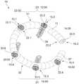
也可以借助时间指示以顺时针方向给出二尖瓣环13中的组织锚15.1-15.6的组织锚位置24`.1-24`.6及其彼此之间的距离。例如组织锚位置24'.2位于12:00并且两个组织锚位置24'.1和24'.3位于14:00和22:00,其可以界定二尖瓣环13的前部20。组织锚位置24'.4-24'.6位于16:00点、18:00点和20:00点以用于二尖瓣环13的后部21。因此,形象地说,组织锚15.1-15.6之间的距离为2h。因此可以看出,额外的组织锚15,特别是在二尖瓣环13的后部21和鞍形区域中,可以分别在例如17:00点和19:00点植入,如图2所示。形象地说,当然,其他时间间隔也是可能的,从而导致组织锚15之间的其他角距离。The tissue anchor positions 24'.1-24'.6 of the tissue anchors 15.1-15.6 in the
基于二尖瓣环13的几何形状,组织锚15.1-15.6也可以被植入二尖瓣环13上,使得瓣环成形术环11也可以模仿二尖瓣环13的不对称打开。即,瓣环成形术环11的形状可以由于许多因素而改变。图2和图4a示例性示出了许多可能的实施例中的两个。随着另外的组织锚15的植入,可以影响瓣环成形术环11的形状。而且,用于组织锚15的位置24的距离可以改变。因此,组织锚15在二尖瓣环13上的定位特别重要。由于瓣环成形术环11与植入的组织锚15一起在周围的心肌组织47上施加拉力,因此固定于植入的组织锚15的瓣环成形术环11消除了功能性二尖瓣反流的问题。原则上,如果患者在二尖瓣环13处的解剖结构不正确,则可以从一开始就使用不对称的瓣环成形术环11。尽管这里使用的瓣环成形术环11的材料允许手动变形,但是其刚度足以抵抗一旦植入并经受正常的生理应力的二尖瓣环13处的进一步变形。Based on the geometry of the
瓣环成形术环11的外层42应该足够多孔和/或柔性,以允许组织锚线33从中穿过。内层43设计成减小二尖瓣环13的周长。它必须在搏动周期中抵抗由心脏3的肌肉组织47传递的应力在后部21中保持其后弯曲。这种内层43的材料已经在说明书中举例说明。在下面的附图中采用与图4a类似的附图标记。The
图4b示出了图4a的截面X的示意图,其具有组织锚15.1和环体27的横截面,例如对于所有组织锚15.1-15.6。组织锚15.1尤其由构成组织锚15.1的远端36的螺旋形螺旋螺杆30.1组成,而在组织锚15.1的近端37处,在组织锚线33.1的自由端处布置有针34。组织锚15.1的螺旋螺杆30.1固定在承载盘38中,该承载盘在远端36的方向上从承载盘38露出。此外,用于组织锚线33.1的承载盘38保持器,其从承载板38出现在螺旋螺杆30.1的相对侧上。在另一个实施例中,组织锚线33.1固定到组织锚15.1,并且承载盘38位于线33.1和锚15.1的公共固定位置46处。承载盘38具有用于二尖瓣环13的邻接侧40和用于环体27的邻接侧41。两个邻接侧40、41包含两个接触面I 44、II 45。第一接触面I 44在组织47上用作旋入时的挡块用于组织锚15,而另一个接触面II 45则作为附接用于环体27。承载盘38的直径被设计成使得避免在心肌组织47中太深地旋入组织锚15.1。如果将所有的组织锚15.1-15.6,参见图4a,植入在二尖瓣环13周围的心肌组织47中,则接下来将瓣环成形术环11的环体27放置在组织锚15.1-15.6的承载盘38上。为了将环体27固定在组织锚15.1的承载盘38上,使用了固定部件25。组织锚15.1的至少一个组织锚线33.1至少通过固定部件25固定至瓣环成形术环11。固定部件优选地可以由夹紧部件35组成。夹紧部件35在组织锚线33.1上被推到胸部1的外侧。该过程借助于针34进行,该针被引导穿过夹紧部件35的开口。手术器械(未示出)将夹紧部件35推到环体27,并将环体27夹持在环体27和支撑板38之间。夹紧部件35被设计成使得其可以在其开口中永久地夹紧组织锚线15.1。例如,夹紧部件35也由两个部件组成,这两个部件彼此作用并在组织锚线15.1上施加夹紧作用。优选地,夹紧元件35可以由板簧组成,该板簧可以相对容易地推到心房12中的组织锚线33.1上并且在环体27的固定位置处,产生与插入方向相反的夹紧。定位在夹紧部件35与组织锚15.1的承载盘38之间的环体27具有内层43和外层42。组织锚线33.1穿过环体27的外层42,以不损坏内层43。在图6中可以看到瓣环成形术环11插入到心房12中。在所有夹紧部件35都已经放置在环体27上之后,将组织锚线33.1-33.6切断并从心脏3的心房12中移出。在下面的附图中采用与图4b类似的附图标记。Figure 4b shows a schematic view of section X of Figure 4a with a cross-section of tissue anchor 15.1 and
根据图1,图5中以示意图和基本示意图形式显示的心脏3在左胸腔8中绕其纵轴旋转,因此心脏的右半部分更多地靠在前胸壁上,而心脏的左半部分则更多地指向后方。提供了一种二尖瓣植入物,特别是瓣环成形术环11,其使用微创外科手术,借助尤其是套管针50的已知的手术器械,经由右胸腔区域5被施加到患者的跳动的心脏3上,引入心脏3的左心房12中并固定在那里。According to Fig. 1, the
因此,示出了具有左心房12和通路49的左心48,所述通路49穿过心脏组织47进入左心房12至二尖瓣14。该通路49通过所示的套管针50和各种手术器械51.1-51.5实现。下面列出了使用微创修复患者跳动的心脏3上的二尖瓣环13的各种二尖瓣植入手术器械。手术器械例如由外管推动件I 51.1组成,尤其是用于引导内管推动件II 51.2的具有内腔的进入套管。另一手术器械包括用于引导和拧紧组织锚15的具有内腔的第一内管推动件II51.2。另一手术器械包括用于引导瓣环成形术环11的具有内腔的第二内管推动件III51.3。第三内管推动件IV 51.4是一种具有内腔的手术器械,其用于容纳组织锚线33.1-33.6并用于从管推动件III 51.3推出瓣环成形术环11。第四内管推动件V 51.5也是具有腔的手术器械,用于引导固定部件25,特别是夹紧部件35,该固定部件沿着组织锚线33.1被引导以固定瓣环成形术环11。Thus, left
左心室52分为流入路径和流出路径。它通过二尖瓣14与心房12分开。二尖瓣14通过腱索(腱索(Chordae tendineae))53与乳头肌54连接,该乳头肌出现在心室壁55处,并确保二尖瓣14在其瓣膜闭合19处以及在左室52的张紧阶段(收缩)期间不会太猛烈地跳回左心房12。The
二尖瓣(二尖瓣(Mitral Valve))由四个功能组件组成。两个小叶16、17(二尖瓣小叶(Mitral Valve Leaflets))由前叶16(前叶(cupis anterior))、后叶17(后叶(cupispasterior))和小叶16、17在二尖瓣环13(二尖瓣环(Mitral Valve Annulus))中悬挂组成。二尖瓣环13由肌肉组织(在本说明书中称为二尖瓣环13)、腱索53(腱索(Chordaetendineae))组成,小叶16、17与其可移动地连接到乳头肌54(Papillary Muscles)和从心肌层47向内外翻(ausstülpen)的乳头肌54本身。为了重建每个单独的组件,可以使用不同的植入物、手术器械和/或手术方法。在当前情况下,考虑二尖瓣返流及其消除。The mitral valve (Mitral Valve) consists of four functional components. Two
为此,在左心房12二尖瓣环13周围的区域中使用组织锚15.1-15.5。由于以截面图示出了心脏3,所以这里未示出图2中示出的所有可能的植入的组织锚15.1-15.8,因为仅示出了二尖瓣环13的周围的一部分。所示的组织锚15.1-15.5代表所有植入的组织锚15.1-15.5。围绕二尖瓣环13植入的组织锚15.1-15.5以一定间隔布置。组织锚15.1-15.5彼此之间的距离可以例如在二尖瓣13的后部21的鞍形区域中,相对于其他距离变化。此外,组织锚15.1-15.5在远端36处具有锚定元件56,其中,锚定元件56由螺旋状螺旋螺杆30.1-30.5组成。螺旋螺杆30.1-30.5具有远端36和近端37,其中螺旋螺杆30.1-30.5的近端37连接到组织锚线33.1-33.5。从已知的现有技术中可以想到使用其他锚固装置来连接瓣环成形术环11。旋入的螺旋螺杆30.1-30.5位于二尖瓣环13区域中的心肌层组织47中。此外,组织锚15.1-15.5在近端37处具有固定在组织锚15.1-15.5上的承载盘38和组织锚线33.1-33.5(参见图4b)。组织锚线33.1-33.5通过套管51从胸腔1中引出以进一步使用,并且仍连接至螺旋螺杆30.1-30.5。组织锚线33.1-33.5的进一步使用可以从图6的描述中看出。在该图中采用与前面的图1-4的类似的附图标记。For this purpose, tissue anchors 15.1-15.5 are used in the area around the
图6是处于递送构型57的瓣环成形术环11的示意图。为了实现递送构型57,来自心房12的组织锚15.1-15.5的组织锚线33.1-33.5将在瓣环成形术环11上的预定位置24.1-24.5处穿过胸部1。首先,将单个组织锚线33.1-33.5(其针的自由端定位在该处)穿过瓣环成形术环11的外层42的纤维环,该环仍具有其初始形状58。为了能够通过瓣环成形术环11引导组织锚线33.1-33.5,必须预先知道哪个组织锚线33.1-33.5受到影响以及瓣膜成形术环11中的组织锚线33.1-33.5将在哪一点通过。需要知道这些是因为瓣环成形术环11具有各种部分20、21,位于前叶16上的前部20和位于后叶17上的后部21(见图2)。二尖瓣环13也具有这些部分20、21。瓣环成形术环11将以这样的方式放置在二尖瓣环13上,使得其部分20、21彼此叠置。沿着部分20、21,围绕二尖瓣环13,组织锚15.1-15.5以一定间隔布置。距离也可以是不规则的。这就提出了一个问题,即位于胸腔1外部的组织锚线33.1-33.5的哪个组织锚15.1-15.5出现,并且该组织锚15.1-15.5的哪个位置24`.1-24`.5被植入在二尖瓣环13上。为了解决这个问题,组织锚线33.1-33.5因此包含相应的标记。从标记可以清楚地看到,组织锚15.1-15.5位于二尖瓣环13上的位置24`.1-24`.5。FIG. 6 is a schematic illustration of the
为了将组织锚34定位在瓣环成形术环11上,因此必须使用组织锚15.1-15.5位于二尖瓣环13处的相同位置24.1-24.5。因此,如在俯视图中所见,相对于组织锚15.1-15.5的定位,要植入的瓣环成形术环11具有与二尖瓣环13的图像结构相同的图像结构。如果第一组织锚15.1处于第一位置24`.1,例如在二尖瓣膜瓣13的弯曲部分21和直的部分20之间的左通路处,因此该组织锚15.1相关的组织锚33.1带有标号1。也就是说,标号1在二尖瓣环13处标识了组织锚15.1的位置24`.1。但是,这也意味着组织锚15.1的组织锚线33.1必须在瓣环成形术环11中的相应点处被引导。也就是说,为了能够将瓣环成形术环11放置在二尖瓣环13上的组织锚15.1-15.5上,必须将具有标号1的组织锚线33.1和组织锚15.1的位置24`.1、瓣膜成形术环上的位置24.1对应,并在此处穿过外层42的组织引出。瓣环成形术环11上的位置24.1还对应于在瓣膜成形术环11的弯曲部分21与直的部分20之间的左通路处的第一位置24.1。瓣膜成形术环11上的第一位置24.1对应于植入的组织锚15.1的第一位置24'.1。这同样适用于由组织锚15.2-15.5提供的其他组织锚线33.2-33.5,并在相应位置24.2-24.5处将其拉过瓣环成形术环11的组织42。也就是说,植入的组织锚15.2的组织锚线33.2具有标号2,并且位于二尖瓣环13上的位置24'.2上。该组织锚线33.2在瓣环成形术环11的位置24.2处通过,其中二尖瓣环13上的位置24'.2又与瓣环成形术环11上的位置24.2相同,以此类推。In order to position the
这确保了瓣环成形术环11的形状可以被配置成适合于二尖瓣13的形状并且被固定到组织锚15.1-15.5。在瓣环成形术环11上的位置24.1-24.5处,可在每个位置拉动组织锚线33.1-33.5,例如将其拉过。已经示出瓣环成形术环11上的位置标记22.1、22.2。还可以想到的是,在瓣环成形术环11的外层42中用于组织锚线33.1-33.5的位置已经设置有用于针34的通道孔。该通道孔有助于组织锚线33.1-33.5的穿线,并避免可能损坏环体27的外层42。This ensures that the shape of the
如果将瓣环成形术环11安装在由组织锚15.1-15.5提供的所有组织锚线33.1-33.5上,则其在组织锚线33.1-33.5上前进至接收手术器械51并被压缩。在这种状态下,瓣环成形术环11此时实现其递送构型57,以插入到在套管针50中引导的套筒51中。在该阶段,组织锚线33.1-33.5用作瓣环成形术环11的引导装置。由套管针50引导的套筒51延伸到心脏3的左心房12中。利用另一种手术器械,然后将瓣环成形术环11沿着组织锚线33.1-33.5穿过套管51推入心房12。组织锚线33.1-33.5的自由端保留在身体1之外。当瓣环成形术环11完全离开套管51并进入左心房12时,其递送构型57扩展到其原始初始形状58。原始初始形状58优选地根据图2和图4a对应于打开的构型,其中瓣环成形术环11仍由组织锚15.1-15.5的组织锚线33.1-33.5引导。沿着组织锚线33.1-33.5,瓣环成形术环11此时被推到承载盘38上,该承载盘38被布置在组织锚15.1-15.5的端部上,并被放置在那里。如上所述,瓣环成形术环11此时以形状配合的方式固定到二尖瓣环13上的组织锚15.1-15.5上,并且如图4b所示。为了固定瓣环成形术环11,组织锚线33.1-33.5设有夹紧部件35,见图4b。在此图中使用了与之前图1-5类似的附图标记。If the
图7示出了植入在二尖瓣环13上的装置10的示意图,该装置10由二尖瓣植入物组成,特别是呈瓣环成形术环11形式的二尖瓣植入物,其基本上包括三个元件。第一元件是锚定元件56,其被设计为具有螺旋螺杆30、承载盘38和组织锚线33的组织锚15,并且执行瓣环成形术环11在心肌层组织47中的固定。为了清楚起见,在图7中未示出锚定元件56,但是在图4b中详细示出了锚定元件56。第二元件是作为植入物的瓣环成形术环11,其具有内层43和外层42,其中外层42容纳连接至组织锚15的组织锚线33。第三元件形成固定部件25,该固定部件25包括夹紧部件35,并且沿着组织锚线33被引导。夹紧部件35通过组织锚线33将瓣环成形术环11夹紧在其自身与载体盘38之间。最后,切断组织锚线33.1-33.8,并且将尤其是手术器械50、51从心房12移除,并且封闭通向心脏3的通路49。Figure 7 shows a schematic view of a
现在,瓣环成形术环11中的组织锚位置24.1-24.5与二尖瓣环13上的组织锚位置24`.1-24`.5位于同一纵轴39上,因此是一致的,由此在二尖瓣环13处的组织锚位置24`.1的组织锚线33.1对应于瓣环成形术环11中相同的组织锚固位置24.1。因此,瓣环成形术环11以形状配合的方式植入,以消除二尖瓣返流。在该图中采用了与来自前面图1-6类似的附图标记。The tissue anchor locations 24.1-24.5 in the
附图标记列表List of reference signs
1 胸部 31 前侧(v.13)1
2 通路 32 后侧(v.13)2-
3 心脏 33.1-33.6 组织锚线3 Heart 33.1-33.6 Tissue anchors
4 胸部开口 34 针(v.15,33)4 Chest opening 34 stitches (v.15, 33)
5 右侧(v.1) 35 夹紧部件5 Right side (v.1) 35 Clamping part
6 肋间 36 远端(v.15)6
7 伤口扩张器 37 近端(v.15)7
8 左胸腔 38 承载盘(v.15)8
9 解剖开口 39 纵向轴(v.24,24`)9
10 装置 40 邻接侧(v.13)10 Device 40 Adjacent side (v.13)
11 瓣环成形术环 41 邻接侧(v.27)11 Annuloplasty ring 41 Adjacent side (v.27)
12 心房 42 外层(v.11,27)12
13 二尖瓣环 43 内层(v.11,27)13
14 二尖瓣 44 接触面I(v.38)14 Mitral valve 44 Contact surface I (v.38)
15.1-15.6 组织锚 45 接触面II(v.38)15.1-15.6 Tissue Anchors 45 Interface II (v.38)
16 前叶(v.14) 46 固定位置16 Anterior lobe (v.14) 46 Fixed position
17 后叶(v.14) 47 组织(v.3)17 Posterior lobe (v.14) 47 Tissue (v.3)
18 间隙(v.14) 48 左心18 Interstitial (v.14) 48 Left Heart
19 瓣膜(v.14) 49 通路(至3)19 Valves (v.14) 49 Accesses (to 3)
20 前部(v.13) 50 套管针20 Front (v.13) 50 Trocar
21 后部(v.13) 51 器械21 Rear (v.13) 51 Instruments
22.1-22.2 标记(v.11) 52 左心室22.1-22.2 Labeling (v.11) 52 Left ventricle
23 线环 53 腱索23
24.1-24.8 组织锚位置(v.11) 54 乳头肌24.1-24.8 Tissue Anchor Locations (v.11) 54 Papillary Muscles
24`.1-24`.8 组织锚位置(v.13) 55 心室壁24`.1-24`.8 Tissue Anchor Locations (v.13) 55 Ventricular Wall
25 固定部件 56 锚定元件25 Fixing
25a-26c 段 57 递送构型Paragraphs 25a-26c 57 Delivery Configurations
27 环体 58 初始形状27 Ring 58 Initial shape
28 自由端28 free ends
29.1-29.4 旋转接头29.1-29.4 Swivel
30.1-30.6 螺旋螺杆。30.1-30.6 Spiral screw.
Claims (7)
Priority Applications (1)
| Application Number | Priority Date | Filing Date | Title |
|---|---|---|---|
| CN202210711436.6ACN115381599A (en) | 2017-03-28 | 2018-03-28 | Minimally Invasive Implantable Devices and Mitral Valve Implant Systems |
Applications Claiming Priority (3)
| Application Number | Priority Date | Filing Date | Title |
|---|---|---|---|
| DE102017002976.8 | 2017-03-28 | ||
| DE102017002976.8ADE102017002976B4 (en) | 2017-03-28 | 2017-03-28 | Minimally invasive implantable device for eliminating mitral valve insufficiency in the beating heart and mitral valve implant system |
| PCT/EP2018/058026WO2018178208A1 (en) | 2017-03-28 | 2018-03-28 | Device that can be implanted in a minimally invasive manner and mitral valve implant system |
Related Child Applications (1)
| Application Number | Title | Priority Date | Filing Date |
|---|---|---|---|
| CN202210711436.6ADivisionCN115381599A (en) | 2017-03-28 | 2018-03-28 | Minimally Invasive Implantable Devices and Mitral Valve Implant Systems |
Publications (2)
| Publication Number | Publication Date |
|---|---|
| CN110709029A CN110709029A (en) | 2020-01-17 |
| CN110709029Btrue CN110709029B (en) | 2022-07-12 |
Family
ID=61972497
Family Applications (2)
| Application Number | Title | Priority Date | Filing Date |
|---|---|---|---|
| CN201880035165.9AActiveCN110709029B (en) | 2017-03-28 | 2018-03-28 | Minimally invasive implantable device and mitral valve implant system |
| CN202210711436.6APendingCN115381599A (en) | 2017-03-28 | 2018-03-28 | Minimally Invasive Implantable Devices and Mitral Valve Implant Systems |
Family Applications After (1)
| Application Number | Title | Priority Date | Filing Date |
|---|---|---|---|
| CN202210711436.6APendingCN115381599A (en) | 2017-03-28 | 2018-03-28 | Minimally Invasive Implantable Devices and Mitral Valve Implant Systems |
Country Status (7)
| Country | Link |
|---|---|
| US (2) | US11529232B2 (en) |
| EP (1) | EP3600155A1 (en) |
| JP (2) | JP7570098B2 (en) |
| CN (2) | CN110709029B (en) |
| CA (1) | CA3095227A1 (en) |
| DE (1) | DE102017002976B4 (en) |
| WO (1) | WO2018178208A1 (en) |
Families Citing this family (35)
| Publication number | Priority date | Publication date | Assignee | Title |
|---|---|---|---|---|
| WO2013130641A1 (en) | 2012-02-29 | 2013-09-06 | Valcare, Inc. | Percutaneous annuloplasty system with anterior-posterior adjustment |
| US20200146854A1 (en) | 2016-05-16 | 2020-05-14 | Elixir Medical Corporation | Methods and devices for heart valve repair |
| CN108618871A (en) | 2017-03-17 | 2018-10-09 | 沃卡尔有限公司 | Bicuspid valve with multi-direction anchor portion or tricuspid valve repair system |
| WO2019195860A2 (en) | 2018-04-04 | 2019-10-10 | Vdyne, Llc | Devices and methods for anchoring transcatheter heart valve |
| US11071627B2 (en) | 2018-10-18 | 2021-07-27 | Vdyne, Inc. | Orthogonally delivered transcatheter heart valve frame for valve in valve prosthesis |
| US10595994B1 (en) | 2018-09-20 | 2020-03-24 | Vdyne, Llc | Side-delivered transcatheter heart valve replacement |
| US10321995B1 (en) | 2018-09-20 | 2019-06-18 | Vdyne, Llc | Orthogonally delivered transcatheter heart valve replacement |
| US11344413B2 (en) | 2018-09-20 | 2022-05-31 | Vdyne, Inc. | Transcatheter deliverable prosthetic heart valves and methods of delivery |
| US11278437B2 (en) | 2018-12-08 | 2022-03-22 | Vdyne, Inc. | Compression capable annular frames for side delivery of transcatheter heart valve replacement |
| US12186187B2 (en) | 2018-09-20 | 2025-01-07 | Vdyne, Inc. | Transcatheter deliverable prosthetic heart valves and methods of delivery |
| US11109969B2 (en) | 2018-10-22 | 2021-09-07 | Vdyne, Inc. | Guidewire delivery of transcatheter heart valve |
| US11253359B2 (en) | 2018-12-20 | 2022-02-22 | Vdyne, Inc. | Proximal tab for side-delivered transcatheter heart valves and methods of delivery |
| WO2020146842A1 (en) | 2019-01-10 | 2020-07-16 | Vdyne, Llc | Anchor hook for side-delivery transcatheter heart valve prosthesis |
| WO2020146889A1 (en)* | 2019-01-11 | 2020-07-16 | Mitrx, Inc. | Devices and methods for catheter-based cardiac procedures |
| US11185409B2 (en) | 2019-01-26 | 2021-11-30 | Vdyne, Inc. | Collapsible inner flow control component for side-delivered transcatheter heart valve prosthesis |
| US11273032B2 (en) | 2019-01-26 | 2022-03-15 | Vdyne, Inc. | Collapsible inner flow control component for side-deliverable transcatheter heart valve prosthesis |
| WO2020181154A2 (en) | 2019-03-05 | 2020-09-10 | Vdyne, Inc. | Tricuspid regurgitation control devices for orthogonal transcatheter heart valve prosthesis |
| US11076956B2 (en) | 2019-03-14 | 2021-08-03 | Vdyne, Inc. | Proximal, distal, and anterior anchoring tabs for side-delivered transcatheter mitral valve prosthesis |
| US11173027B2 (en) | 2019-03-14 | 2021-11-16 | Vdyne, Inc. | Side-deliverable transcatheter prosthetic valves and methods for delivering and anchoring the same |
| CA3138875A1 (en) | 2019-05-04 | 2020-11-12 | Vdyne, Inc. | Cinch device and method for deployment of a side-delivered prosthetic heart valve in a native annulus |
| EP3982881B1 (en) | 2019-06-11 | 2025-04-16 | Valcare Medical, Inc. | Annuloplasty ring with posterior leaflet for minimally invasive treatment |
| US12396853B2 (en) | 2019-06-11 | 2025-08-26 | Valcare Medical, Inc. | Systems and methods for delivery of chordae replacement system |
| EP4480458A3 (en) | 2019-08-20 | 2025-04-09 | Vdyne, Inc. | Delivery devices for side-deliverable transcatheter prosthetic valves |
| CN120531525A (en) | 2019-08-26 | 2025-08-26 | 维迪内股份有限公司 | Laterally deliverable transcatheter prosthetic valve and method for its delivery and anchoring |
| CN113116430B (en)* | 2019-12-31 | 2024-08-16 | 杭州德晋医疗科技有限公司 | Winding-preventing anchor and anchor conveyor |
| WO2021135270A1 (en)* | 2019-12-31 | 2021-07-08 | 杭州德晋医疗科技有限公司 | Integrated anchor and anchoring system |
| US11234813B2 (en) | 2020-01-17 | 2022-02-01 | Vdyne, Inc. | Ventricular stability elements for side-deliverable prosthetic heart valves and methods of delivery |
| CN113384382B (en)* | 2021-07-21 | 2024-11-19 | 华中科技大学同济医学院附属协和医院 | An artificial mitral valvuloplasty ring implantation device |
| US20230045532A1 (en)* | 2021-08-09 | 2023-02-09 | Valcare, Inc. | Systems and methods for target/article bridging engagement and anchoring |
| CN113951989B (en)* | 2021-10-13 | 2023-06-20 | 上海汇禾医疗器械有限公司 | Flexible shearing device and application method thereof |
| CN113855337B (en)* | 2021-12-07 | 2022-02-18 | 科瑞迈吉(北京)医疗科技有限公司 | Mitral valve repair implantation system |
| CN116407349A (en)* | 2021-12-31 | 2023-07-11 | 杭州德晋医疗科技有限公司 | Implant capable of accelerating endothelialization and transcatheter heart repair system |
| WO2023169376A1 (en)* | 2022-03-07 | 2023-09-14 | 宁波健世科技股份有限公司 | Forming device for repairing valve |
| CN116327445B (en)* | 2023-03-28 | 2024-06-25 | 晨兴(南通)医疗器械有限公司 | Annuloplasty device and method |
| CN118986588B (en)* | 2023-05-19 | 2025-09-12 | 宁波健世科技股份有限公司 | A precisely controlled anchoring system |
Citations (7)
| Publication number | Priority date | Publication date | Assignee | Title |
|---|---|---|---|---|
| WO2010073246A2 (en)* | 2008-12-22 | 2010-07-01 | Valtech Cardio, Ltd. | Adjustable annuloplasty devices and adjustment mechanisms therefor |
| WO2011041571A2 (en)* | 2009-10-01 | 2011-04-07 | Kardium Inc. | Medical device, kit and method for constricting tissue or a bodily orifice, for example, a mitral valve |
| WO2012019052A2 (en)* | 2010-08-04 | 2012-02-09 | Micardia Corporation | Percutaneous transcatheter repair of heart valves |
| CN102686185A (en)* | 2009-10-29 | 2012-09-19 | 瓦尔泰克卡迪欧有限公司 | Tissue anchor for annuloplasty device |
| CN102821702A (en)* | 2010-02-03 | 2012-12-12 | 爱德华兹生命科学公司 | Devices and methods for treating heart |
| WO2013088327A1 (en)* | 2011-12-12 | 2013-06-20 | David Alon | Heart valve repair device |
| CN105392449A (en)* | 2013-06-06 | 2016-03-09 | 戴维·阿隆 | Heart valve repair and replacement |
Family Cites Families (11)
| Publication number | Priority date | Publication date | Assignee | Title |
|---|---|---|---|---|
| DE69331315T2 (en) | 1992-01-27 | 2002-08-22 | Medtronic, Inc. | ANULOPLASTIC AND SEAM RINGS |
| US6858039B2 (en) | 2002-07-08 | 2005-02-22 | Edwards Lifesciences Corporation | Mitral valve annuloplasty ring having a posterior bow |
| EP1531762B1 (en) | 2002-08-29 | 2010-04-14 | St. Jude Medical, Cardiology Division, Inc. | Implantable devices for controlling the internal circumference of an anatomic orifice or lumen |
| JP5222552B2 (en) | 2004-04-30 | 2013-06-26 | ミトラル・ソリューションズ・インコーポレイテッド | Methods and devices for modulation of heart valve function |
| WO2011034628A1 (en) | 2005-02-07 | 2011-03-24 | Evalve, Inc. | Methods, systems and devices for cardiac valve repair |
| US9883943B2 (en)* | 2006-12-05 | 2018-02-06 | Valtech Cardio, Ltd. | Implantation of repair devices in the heart |
| US20080221599A1 (en)* | 2007-03-06 | 2008-09-11 | Starksen Niel F | Devices, methods, and kits for gastrointestinal procedures |
| EP2296744B1 (en)* | 2008-06-16 | 2019-07-31 | Valtech Cardio, Ltd. | Annuloplasty devices |
| US8715342B2 (en)* | 2009-05-07 | 2014-05-06 | Valtech Cardio, Ltd. | Annuloplasty ring with intra-ring anchoring |
| US9307979B1 (en)* | 2010-10-26 | 2016-04-12 | Ziptek Llc | Surgical suture system, tissue restraints, and tissue anchors |
| US9072511B2 (en) | 2011-03-25 | 2015-07-07 | Kardium Inc. | Medical kit for constricting tissue or a bodily orifice, for example, a mitral valve |
- 2017
- 2017-03-28DEDE102017002976.8Apatent/DE102017002976B4/enactiveActive
- 2018
- 2018-03-28CNCN201880035165.9Apatent/CN110709029B/enactiveActive
- 2018-03-28USUS16/498,589patent/US11529232B2/enactiveActive
- 2018-03-28WOPCT/EP2018/058026patent/WO2018178208A1/ennot_activeCeased
- 2018-03-28CNCN202210711436.6Apatent/CN115381599A/enactivePending
- 2018-03-28EPEP18717858.7Apatent/EP3600155A1/enactivePending
- 2018-03-28CACA3095227Apatent/CA3095227A1/enactivePending
- 2018-03-28JPJP2020503098Apatent/JP7570098B2/enactiveActive
- 2022
- 2022-12-15USUS18/082,035patent/US20230181319A1/enactivePending
- 2023
- 2023-02-10JPJP2023018926Apatent/JP7421248B2/enactiveActive
Patent Citations (7)
| Publication number | Priority date | Publication date | Assignee | Title |
|---|---|---|---|---|
| WO2010073246A2 (en)* | 2008-12-22 | 2010-07-01 | Valtech Cardio, Ltd. | Adjustable annuloplasty devices and adjustment mechanisms therefor |
| WO2011041571A2 (en)* | 2009-10-01 | 2011-04-07 | Kardium Inc. | Medical device, kit and method for constricting tissue or a bodily orifice, for example, a mitral valve |
| CN102686185A (en)* | 2009-10-29 | 2012-09-19 | 瓦尔泰克卡迪欧有限公司 | Tissue anchor for annuloplasty device |
| CN102821702A (en)* | 2010-02-03 | 2012-12-12 | 爱德华兹生命科学公司 | Devices and methods for treating heart |
| WO2012019052A2 (en)* | 2010-08-04 | 2012-02-09 | Micardia Corporation | Percutaneous transcatheter repair of heart valves |
| WO2013088327A1 (en)* | 2011-12-12 | 2013-06-20 | David Alon | Heart valve repair device |
| CN105392449A (en)* | 2013-06-06 | 2016-03-09 | 戴维·阿隆 | Heart valve repair and replacement |
Also Published As
| Publication number | Publication date |
|---|---|
| EP3600155A1 (en) | 2020-02-05 |
| US11529232B2 (en) | 2022-12-20 |
| DE102017002976A1 (en) | 2018-10-04 |
| CN115381599A (en) | 2022-11-25 |
| JP2020512172A (en) | 2020-04-23 |
| JP7570098B2 (en) | 2024-10-21 |
| JP2023055952A (en) | 2023-04-18 |
| JP7421248B2 (en) | 2024-01-24 |
| US20230181319A1 (en) | 2023-06-15 |
| DE102017002976B4 (en) | 2021-08-26 |
| WO2018178208A1 (en) | 2018-10-04 |
| CA3095227A1 (en) | 2018-10-04 |
| CN110709029A (en) | 2020-01-17 |
| US20210161662A1 (en) | 2021-06-03 |
Similar Documents
| Publication | Publication Date | Title |
|---|---|---|
| CN110709029B (en) | Minimally invasive implantable device and mitral valve implant system | |
| US12396720B2 (en) | Devices and methods for locating and implanting tissue anchors at mitral valve commissure | |
| US20220039951A1 (en) | Systems and methods for transcatheter treatment of valve regurgitation | |
| CN111212614B (en) | Axisymmetric adjustable device for the treatment of mitral valve regurgitation | |
| US10149759B2 (en) | Heart valve assistive prosthesis | |
| RU2491035C2 (en) | Device and method for reducing heart valve size | |
| AU2005234793B2 (en) | Implantable prosthetic valve | |
| US20120290077A1 (en) | Tissue restraining devices and methods of use | |
| CN109789019A (en) | Heart valve paired wire ring and system | |
| JP2014523282A (en) | Minimally invasive repair of heart valve leaflets | |
| CN110403735A (en) | Bicuspid valve parking device, system and method | |
| US20210030537A1 (en) | Tethering System For A Prosthetic Heart Valve | |
| US20200015970A1 (en) | Chain annuloplasty ring, delivery system and related methods | |
| CN115190786A (en) | Annuloplasty device | |
| US20250090323A1 (en) | Adjustable annuloplasty ring | |
| HK40083912A (en) | Device that can be implanted in a minimally invasive manner and mitral valve implant system |
Legal Events
| Date | Code | Title | Description |
|---|---|---|---|
| PB01 | Publication | ||
| PB01 | Publication | ||
| SE01 | Entry into force of request for substantive examination | ||
| SE01 | Entry into force of request for substantive examination | ||
| CB02 | Change of applicant information | Address after:hamburg Applicant after:Emanuel albertinin diaconi Ltd. Address before:Berlin Applicant before:Emanuel diacconi Ltd. | |
| CB02 | Change of applicant information | ||
| GR01 | Patent grant | ||
| GR01 | Patent grant |