CN110706226A - A morphological symmetry analysis method of human fingers based on 3D images - Google Patents
A morphological symmetry analysis method of human fingers based on 3D imagesDownload PDFInfo
- Publication number
- CN110706226A CN110706226ACN201910973815.0ACN201910973815ACN110706226ACN 110706226 ACN110706226 ACN 110706226ACN 201910973815 ACN201910973815 ACN 201910973815ACN 110706226 ACN110706226 ACN 110706226A
- Authority
- CN
- China
- Prior art keywords
- model
- finger
- metacarpal bone
- fingers
- images
- Prior art date
- Legal status (The legal status is an assumption and is not a legal conclusion. Google has not performed a legal analysis and makes no representation as to the accuracy of the status listed.)
- Pending
Links
Images


Classifications
- G—PHYSICS
- G06—COMPUTING OR CALCULATING; COUNTING
- G06T—IMAGE DATA PROCESSING OR GENERATION, IN GENERAL
- G06T7/00—Image analysis
- G06T7/0002—Inspection of images, e.g. flaw detection
- G06T7/0012—Biomedical image inspection
- G—PHYSICS
- G06—COMPUTING OR CALCULATING; COUNTING
- G06T—IMAGE DATA PROCESSING OR GENERATION, IN GENERAL
- G06T17/00—Three dimensional [3D] modelling, e.g. data description of 3D objects
- G—PHYSICS
- G06—COMPUTING OR CALCULATING; COUNTING
- G06T—IMAGE DATA PROCESSING OR GENERATION, IN GENERAL
- G06T5/00—Image enhancement or restoration
- G06T5/77—Retouching; Inpainting; Scratch removal
- G—PHYSICS
- G06—COMPUTING OR CALCULATING; COUNTING
- G06T—IMAGE DATA PROCESSING OR GENERATION, IN GENERAL
- G06T2207/00—Indexing scheme for image analysis or image enhancement
- G06T2207/10—Image acquisition modality
- G06T2207/10072—Tomographic images
- G06T2207/10088—Magnetic resonance imaging [MRI]
- G—PHYSICS
- G06—COMPUTING OR CALCULATING; COUNTING
- G06T—IMAGE DATA PROCESSING OR GENERATION, IN GENERAL
- G06T2207/00—Indexing scheme for image analysis or image enhancement
- G06T2207/20—Special algorithmic details
- G06T2207/20036—Morphological image processing
Landscapes
- Engineering & Computer Science (AREA)
- Physics & Mathematics (AREA)
- General Physics & Mathematics (AREA)
- Theoretical Computer Science (AREA)
- Geometry (AREA)
- Software Systems (AREA)
- Computer Graphics (AREA)
- Health & Medical Sciences (AREA)
- General Health & Medical Sciences (AREA)
- Medical Informatics (AREA)
- Nuclear Medicine, Radiotherapy & Molecular Imaging (AREA)
- Radiology & Medical Imaging (AREA)
- Quality & Reliability (AREA)
- Computer Vision & Pattern Recognition (AREA)
- Magnetic Resonance Imaging Apparatus (AREA)
Abstract
Translated fromChineseDescription
Translated fromChinese技术领域technical field
本发明涉及数字医学技术领域,特别是涉及一种基于3D影像的人体手指形态学对称性分析方法。The invention relates to the technical field of digital medicine, in particular to a 3D image-based morphological symmetry analysis method of human fingers.
背景技术Background technique
人体形态学研究过程中通常需要测量人体尺寸数据,人体尺寸数据是一项重要的基础数据资源,具有突出的时效性,标准的人体测量学测量过程中,需要采集的数据非常多,需要花费巨大的人力财力。Human body size data is usually measured in the process of anthropometric research. Human body size data is an important basic data resource with outstanding timeliness. In the standard anthropometric measurement process, a lot of data needs to be collected, and it costs a lot of money. human and financial resources.
目前,尚未定义有规范化的手指对称性分析方法,在康复医学领域中,传统的义指制作主要是通过取型、修型、成型三个步骤制成,其中的修型过程是人工操作完成,这种制作义指的方法比较粗糙,制作出来的义指不美观,不利于精确化治疗。At present, there is no standardized method for analyzing finger symmetry. In the field of rehabilitation medicine, the traditional prosthetic finger production is mainly made through three steps: taking shape, repairing shape, and molding. The repairing process is done manually. This method of making a righteous finger is relatively rough, and the produced righteous finger is not beautiful, which is not conducive to precise treatment.
因此,针对现有技术不足,提供一种基于3D影像的人体手指形态学对称性分析方法以克服现有技术不足甚为必要。Therefore, in view of the deficiencies of the prior art, it is necessary to provide a 3D image-based morphological symmetry analysis method of human fingers to overcome the deficiencies of the prior art.
发明内容SUMMARY OF THE INVENTION
本发明的目的在于避免现有技术的不足之处而提供一种基于3D影像的人体手指形态学对称性分析方法,以人体表面轮廓扫描影像数据为基础,通过三维重建软件和逆向工程软件,经过处理分析,结合统计学分析,挖掘不同数据之间的相关性,能够准确进行手指形态学对称分析。The purpose of the present invention is to avoid the deficiencies of the prior art and provide a 3D image-based human finger morphology symmetry analysis method. Processing analysis, combined with statistical analysis, mining the correlation between different data, can accurately analyze the symmetry of finger morphology.
本发明的目的通过以下技术措施实现。The object of the present invention is achieved by the following technical measures.
提供一种基于3D影像的人体手指形态学对称性分析方法,包括以下步骤:Provided is a 3D image-based morphological symmetry analysis method for human fingers, comprising the following steps:
S1,采集左右双侧手指核磁共振断层影像数据;S1, collecting MRI tomographic image data of left and right bilateral fingers;
S2,根据步骤S1得到的左右双侧手指的核磁共振断层影像数据重建双侧完整手指表面轮廓3D影像模型;S2, reconstructing a 3D image model of bilateral complete finger surface contours according to the nuclear magnetic resonance tomographic image data of the left and right bilateral fingers obtained in step S1;
S3,将得到的双侧完整手指表面轮廓3D影像模型导入逆向工程软件中进行表面优化处理和缺陷修复,得到双侧手指优化模型;S3, import the obtained 3D image model of the complete bilateral finger surface profile into reverse engineering software for surface optimization processing and defect repair, and obtain an optimized bilateral finger model;
S4,将双侧手指优化模型进行镜像对齐、拟合,得到拟合模型;S4, performing mirror alignment and fitting on the bilateral finger optimization model to obtain a fitting model;
S5,对拟合模型进行3D比较,得出3D偏差结果。S5, 3D comparison is performed on the fitted model, and a 3D deviation result is obtained.
优选的,上述步骤S1应用超导型磁共振成像仪的快速mDIXON序列,进行双侧上肢手指核磁共振断层影像数据的采集。通过让需要进行分析的人体俯卧平躺在该核磁扫描仪器上,将双手平直举过头顶,掌心向下,手指正常放松弯曲,将医用摆位沙垫放置于腕关节下方的伸肌以支持带皮下外侧,保持体位不动,进行双侧尚志手指核磁共振断层影像数据的采集。Preferably, in the above step S1, the fast mDIXON sequence of the superconducting magnetic resonance imaging apparatus is used to collect the magnetic resonance tomographic image data of the fingers of the bilateral upper limbs. By making the human body to be analyzed lie prone on the MRI scanner, raise both hands straight above the head, palms down, fingers relax and bend normally, and place the medical positioning sand pad on the extensor muscles under the wrist joint to support With the subcutaneous outer side, keep the body still, and collect the MRI data of bilateral Shangzhi's fingers.
优选的,上述步骤S2具体是:将步骤S1中采集到的左右双侧手指的核磁共振断层影像数据,以DICOM格式导入到医学三维重建软件Mimics中,设置灰度值阈值区间为95—280,通过识别掌骨及指骨的骨性结构形态作为标志,保留掌骨上沿及所有指骨部分作为目标模型,对目标模型进行初步分割、填补,保留掌心横切面至指尖段,通过三维计算得到双侧完整手指表面轮廓3D影像模型,并将双侧完整手指表面轮廓3D影像模型以STL格式文件导出。Preferably, the above step S2 is specifically: importing the MRI tomographic image data of the left and right bilateral fingers collected in step S1 into the medical three-dimensional reconstruction software Mimics in DICOM format, and setting the gray value threshold interval to 95-280, By identifying the bony structure and shape of the metacarpal and phalanx as a marker, retaining the upper edge of the metacarpal and all the phalangeal parts as the target model, the target model is preliminarily segmented and filled, and the cross-section of the palm to the fingertip is retained. The 3D image model of the finger surface contour, and the complete 3D image model of the finger surface contour on both sides is exported in the STL format file.
在对目标模型进行分割截取时,若掌骨部位截取平面处不平整,需要先对截面进行删除、补孔等操作,再进行光滑降噪,避免截面处发生变形,影响后续对3D影像的建模。When the target model is segmented and intercepted, if the intercepted plane of the metacarpal bone is not flat, it is necessary to delete the section, fill holes, etc., and then perform smooth noise reduction to avoid deformation at the section, which will affect the subsequent modeling of 3D images. .
优选的,上述定义掌骨由桡侧向尺侧依次设为第一掌骨、第二掌骨、第三掌骨、第四掌骨和第五掌骨,即拇指对应的掌骨为第一掌骨,食指对应的掌骨为第二掌骨,中指对应的掌骨为第三掌骨,无名指对应的掌骨为第四掌骨,小指对应的掌骨为第五掌骨,设第一掌骨与大多角骨连接处的中心点为点A,第二掌骨与小多角骨连接处的中心点为点B,第一掌骨的中轴线与第二掌骨的中轴线所在平面设为平面C,点A和点B连接作一直线D,在直线D上作一平面E,平面E与平面C垂直,平面E作为掌骨部位的截取平面,平面E至中指手指顶端的部分为目标模型。Preferably, the metacarpal bones are defined as the first metacarpal bone, the second metacarpal bone, the third metacarpal bone, the fourth metacarpal bone and the fifth metacarpal bone in sequence from the radial side to the ulnar side, that is, the metacarpal bone corresponding to the thumb is the first metacarpal bone, and the metacarpal bone corresponding to the index finger is The second metacarpal bone, the metacarpal bone corresponding to the middle finger is the third metacarpal bone, the metacarpal bone corresponding to the ring finger is the fourth metacarpal bone, and the metacarpal bone corresponding to the little finger is the fifth metacarpal bone. The center point of the connection between the metacarpal and the lesser polygon is point B, the plane where the central axis of the first metacarpal bone and the central axis of the second metacarpal bone are located is set as plane C, the connection between point A and point B is a straight line D, and on the straight line D is drawn. A plane E, the plane E is perpendicular to the plane C, the plane E is used as the interception plane of the metacarpal bone, and the part from the plane E to the top of the middle finger is the target model.
优选的,上述步骤S2中通过三维计算得到双侧完整手指表面轮廓3D影像模型具体是,目标模型对象通过Edit Masks工具进行分割、填补,得到左右双侧的包含五指在内的手指Mask数据,并对得到的手指Mask数据进行3D建模,得到双侧完整手指表面轮廓3D影像模型。Preferably, in the above step S2, the 3D image model of the complete finger surface contour on both sides is obtained by three-dimensional calculation. Specifically, the target model object is segmented and filled with the Edit Masks tool, and the left and right sides of the finger including the five fingers are obtained. Mask data, and 3D modeling was performed on the obtained finger Mask data, and a 3D image model of bilateral complete finger surface contour was obtained.
优选的,上述步骤S3具体是:将双侧完整手指表面轮廓3D影像模型的STL格式文件导入逆向工程软件Geomagic系列软件之一的Wrap中得到三维模型,对三维模型进行表面优化处理,将Wrap的光滑降噪程度降到最低级别,删除三维模型上的钉状物,进行三维模型的光顺处理,对三维模型的孔洞缺陷进行修复,删除内部噪声及三维模型冗余三角面片,得到双侧手指优化模型。在优化过程中,为减少主观因素对偏差分析结果的影响,在表面优化处理过程中尽量减少对模型表面的手动主动修复,光滑降噪程度选择较低的级别,先删除钉状物后进行快速光顺,再进行孔洞缺陷修复,修补孔洞选取时按小面积选取,并删除内部噪声及冗余三角面片。Preferably, the above-mentioned step S3 is specifically: importing the STL format file of the 3D image model of the complete finger surface profile on both sides into the Wrap, one of the reverse engineering software Geomagic series software, to obtain the three-dimensional model, performing surface optimization processing on the three-dimensional model, The degree of smoothing and noise reduction is reduced to the lowest level, the spikes on the 3D model are deleted, the 3D model is smoothed, the hole defects in the 3D model are repaired, the internal noise and the redundant triangular patches of the 3D model are deleted, and a double-sided model is obtained. Finger optimized model. In the optimization process, in order to reduce the influence of subjective factors on the deviation analysis results, the manual active repair of the model surface should be minimized during the surface optimization process. Smooth, and then repair the hole defect. When selecting the repaired hole, select it according to a small area, and delete the internal noise and redundant triangular patches.
优选的,上述步骤S4具体是:将步骤S3中得到的双侧手指优化模型在GeomagicWrap软件中复制一份副本,两个模型分别删除左、右一侧,得到左、右单侧手指的两个模型,取左单侧手指的模型命名为LH模型,取右单侧手指的模型命名为RH模型,在Wrap工具中对LH模型和RH模型进行镜像对齐、拟合处理得到拟合模型,将拟合模型导出为“.wrp”格式文件。Preferably, the above step S4 is specifically: make a copy of the bilateral finger optimization model obtained in step S3 in GeomagicWrap software, delete the left and right sides of the two models respectively, and obtain two left and right unilateral fingers. Model, take the model of the left unilateral finger and name it as the LH model, and take the model of the right unilateral finger and name it as the RH model. In the Wrap tool, perform mirror alignment and fitting processing on the LH model and the RH model to obtain the fitted model. The composite model is exported as a ".wrp" format file.
优选的,上述步骤S5具体是:将步骤S4中得到的“.wrp”格式文件的拟合模型导入逆向工程软件Geomagic系列软件之一的Control中,选择分析模式,对导入后的拟合模型进行3D比较,对LH模型和RH模型分别设置好参考模型和测试模型,对比得到3D偏差结果。还可以通过Control软件的计算功能进行对模型体积、面积和重心等数据的计算,进而为3D影像模型的建立提供多种数据参考。Preferably, the above-mentioned step S5 is specifically: import the fitting model of the ".wrp" format file obtained in step S4 into Control, one of the reverse engineering software Geomagic series software, select the analysis mode, and carry out the imported fitting model. For 3D comparison, the reference model and the test model are set for the LH model and the RH model respectively, and the 3D deviation results are obtained by comparison. It is also possible to calculate the model volume, area and center of gravity through the calculation function of the Control software, thereby providing a variety of data references for the establishment of 3D image models.
优选的,上述3D偏差结果为平均偏差、标准偏差、最大偏差、最小偏差中的至少一种。Preferably, the above-mentioned 3D deviation result is at least one of average deviation, standard deviation, maximum deviation, and minimum deviation.
本发明的一种基于3D影像的人体手指形态学对称性分析方法,能够准确对人体的左右手指的对称性进行分析,将断层影像数据结合计算机辅助分析软件进行分析操作,进而对对称拟合的数据进行分析,得到双侧手指拟合模型的3D偏差结果,分析结果准确。The method for analyzing the morphological symmetry of human fingers based on 3D images of the present invention can accurately analyze the symmetry of the left and right fingers of the human body. The data is analyzed, and the 3D deviation results of the bilateral finger fitting model are obtained, and the analysis results are accurate.
本发明的一种基于3D影像的人体手指形态学对称性分析方法,通过利用医用超导型磁共振成像仪MRI、三维软件Mimics和逆向三维工程Geomagic软件,从而获取左右双侧手指的核磁共振断层影像数据,并将左右双侧手指的核磁共振断层影像数据重建成双侧完整手指表面轮廓3D影像模型,对双侧完整手指表面轮廓3D影像模型进行优化、镜像对齐和拟合,最终得到左右双侧手指的拟合模型,对左右双侧手指的拟合模型进行最终的3D比较,得出左右双侧手指的3D偏差结果,运用此基于3D影像的人体手指形态学对称性分析方法对左右双侧手指进行手指对称性分析,分析结果精确。A method for analyzing the morphological symmetry of human fingers based on 3D images of the present invention obtains the nuclear magnetic resonance tomography of left and right bilateral fingers by using a medical superconducting magnetic resonance imager MRI, three-dimensional software Mimics and reverse three-dimensional engineering Geomagic software image data, and reconstruct the MRI data of the left and right bilateral fingers into a 3D image model of bilateral complete finger surface contours. The fitting model of the lateral finger, the final 3D comparison of the fitting model of the left and right bilateral fingers is carried out, and the 3D deviation result of the left and right bilateral fingers is obtained. The lateral fingers are analyzed for finger symmetry, and the analysis results are accurate.
附图说明Description of drawings
利用附图对本发明作进一步的说明,但附图中的内容不构成对本发明的任何限制。The present invention will be further described by using the accompanying drawings, but the content in the accompanying drawings does not constitute any limitation to the present invention.
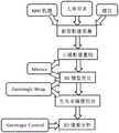
图1是本发明一种基于3D影像的人体手指形态学对称性分析方法的技术路线流程示意图。FIG. 1 is a schematic flow chart of the technical route of a 3D image-based morphological symmetry analysis method of human fingers according to the present invention.
图2是本发明一种基于3D影像的人体手指形态学对称性分析方法采集得到右手核磁断层影像示意图。FIG. 2 is a schematic diagram of a right-hand nuclear magnetic tomography image acquired by a 3D image-based morphological symmetry analysis method of human fingers according to the present invention.
图3是图1中的右手核磁断层影像在Mimics中使用渲染工具获取到二维冠状面示意图。FIG. 3 is a schematic diagram of a two-dimensional coronal plane obtained by using the rendering tool in Mimics for the right-hand MRI image in FIG. 1 .
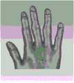
图4是图1的右手核磁断层影像三维重建后的示意图。FIG. 4 is a schematic diagram after three-dimensional reconstruction of the right-hand MRI of FIG. 1 .
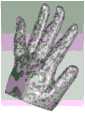
图5是本发明一种基于3D影像的人体手指形态学对称性分析方法的左手手指STL模型导入到Geomagic的Wrap中处理前的示意图。FIG. 5 is a schematic diagram of the left-hand finger STL model of a 3D image-based human finger morphological symmetry analysis method of the present invention before being imported into the Wrap of Geomagic for processing.
图6是本发明一种基于3D影像的人体手指形态学对称性分析方法的经过初步裁剪后右手三维重建示意图。6 is a schematic diagram of the three-dimensional reconstruction of the right hand after preliminary cropping of a method for analyzing the morphological symmetry of a human finger based on a 3D image according to the present invention.
图7是本发明一种基于3D影像的人体手指形态学对称性分析方法的经过对齐拟合处理后的右手的拟合模型示意图。7 is a schematic diagram of a fitting model of the right hand after alignment and fitting processing of a 3D image-based human finger morphological symmetry analysis method of the present invention.
图8是本发明实施例中对齐拟合后的左手和右手的模型示意图。FIG. 8 is a schematic diagram of a left hand and a right hand after alignment and fitting in an embodiment of the present invention.
图9是对图8进行二次裁剪的示意图。FIG. 9 is a schematic diagram of secondary cropping of FIG. 8 .
图10是经过对齐拟合裁剪后的右手模型示意图。Figure 10 is a schematic diagram of the right-hand model after alignment fitting and trimming.
图11是经过对齐拟合裁剪后的左手模型示意图。Figure 11 is a schematic diagram of the left-hand model after alignment, fitting and trimming.
图12是模型3D偏差分析的差异显示分布图。FIG. 12 is a difference display distribution diagram of model 3D deviation analysis.
图13是模型3D偏差分析挖掘到的最大偏差、最小偏差、平均偏差和RMS估计的数据图。Figure 13 is a data graph of the maximum deviation, minimum deviation, average deviation and RMS estimates mined by the 3D deviation analysis of the model.
图14是在Wrap软件中对3D模型的体积进行分析的数据图。Figure 14 is a graph of the data analyzed in the Wrap software for the volume of the 3D model.
图15是在Wrap软件中对3D模型的面积进行分析的数据图。Figure 15 is a graph of the data analyzed for the area of the 3D model in the Wrap software.
具体实施方式Detailed ways
结合以下实施例对本发明作进一步说明。The present invention will be further described with reference to the following examples.
实施例1。Example 1.
一种基于3D影像的人体手指形态学对称性分析方法,如图1至图15所示,设置有以下步骤:A method for analyzing the morphological symmetry of human fingers based on 3D images, as shown in Figure 1 to Figure 15, is provided with the following steps:
S1,采集左右双侧手指核磁共振断层影像数据;S1, collecting MRI tomographic image data of left and right bilateral fingers;
S2,根据步骤S1得到的左右双侧手指的核磁共振断层影像数据重建双侧完整手指表面轮廓3D影像模型;S2, reconstructing a 3D image model of bilateral complete finger surface contours according to the nuclear magnetic resonance tomographic image data of the left and right bilateral fingers obtained in step S1;
S3,将得到的双侧完整手指表面轮廓3D影像模型导入逆向工程软件中进行表面优化处理和缺陷修复,得到双侧手指优化模型;S3, import the obtained 3D image model of the complete bilateral finger surface profile into reverse engineering software for surface optimization processing and defect repair, and obtain an optimized bilateral finger model;
S4,将双侧手指优化模型进行镜像对齐、拟合,得到拟合模型;S4, performing mirror alignment and fitting on the bilateral finger optimization model to obtain a fitting model;
S5,对拟合模型进行3D比较,得出3D偏差结果。S5, 3D comparison is performed on the fitted model, and a 3D deviation result is obtained.
本实施例中的步骤S1应用超导型磁共振成像仪的快速mDIXON序列,进行双侧上肢手指核磁共振断层影像数据的采集。In step S1 of this embodiment, the fast mDIXON sequence of the superconducting magnetic resonance imager is used to collect the magnetic resonance tomographic image data of the fingers of the bilateral upper limbs.
本实施例中的步骤S2具体是:将步骤S1中采集到的左右双侧手指的核磁共振断层影像数据,以DICOM格式导入到医学三维重建软件Mimics中,设置灰度值阈值区间为95—280,通过识别掌骨及指骨的骨性结构形态作为标志,保留掌骨上沿及所有指骨部分作为目标模型,对目标模型进行初步分割、填补,保留掌心横切面至指尖段,通过三维计算得到双侧完整手指表面轮廓3D影像模型,并将双侧完整手指表面轮廓3D影像模型以STL格式文件导出。Step S2 in this embodiment is specifically: importing the MRI tomographic image data of the left and right bilateral fingers collected in step S1 into the medical three-dimensional reconstruction software Mimics in DICOM format, and setting the gray value threshold interval to 95-280 , by identifying the bony structure and morphology of the metacarpal and phalanx as a marker, retaining the upper edge of the metacarpal and all the phalangeal parts as the target model, preliminarily segmenting and filling the target model, retaining the palm cross section to the fingertip segment, and obtaining bilateral Complete 3D image model of finger surface contour, and export the 3D image model of complete finger surface contour on both sides in STL format file.
在对目标模型进行分割截取时,若掌骨部位截取平面处不平整,需要先对截面进行删除、补孔等操作,再进行光滑降噪,避免截面处发生变形,影响后续对3D影像的建模。When the target model is segmented and intercepted, if the intercepted plane of the metacarpal bone is not flat, it is necessary to delete the section, fill holes, etc., and then perform smooth noise reduction to avoid deformation at the section, which will affect the subsequent modeling of 3D images. .
本实施例中的步骤S2中通过三维计算得到双侧完整手指表面轮廓3D影像模型具体是,目标模型对象通过Edit Masks工具进行分割、填补,得到左右双侧的包含五指在内的手指Mask数据,并对得到的手指Mask数据进行3D建模,得到双侧完整手指表面轮廓3D影像模型。In the step S2 in this embodiment, the 3D image model of the complete finger surface contour on both sides is obtained by three-dimensional calculation. Specifically, the target model object is segmented and filled by the Edit Masks tool, and the finger mask data including the five fingers on the left and right sides are obtained, And 3D modeling was carried out on the obtained finger Mask data, and the 3D image model of bilateral complete finger surface contour was obtained.
本实施例中的步骤S3具体是:将双侧完整手指表面轮廓3D影像模型的STL格式文件导入逆向工程软件Geomagic系列软件之一的Wrap中得到三维模型,对三维模型进行表面优化处理,将Wrap的光滑降噪程度降到最低级别,删除三维模型上的钉状物,进行三维模型的光顺处理,对三维模型的孔洞缺陷进行修复,删除内部噪声及三维模型冗余三角面片,得到双侧手指优化模型。在优化过程中,为减少主观因素对偏差分析结果的影响,在表面优化处理过程中尽量减少对模型表面的手动主动修复,光滑降噪程度选择较低的级别,先删除钉状物后进行快速光顺,再进行孔洞缺陷修复,修补孔洞选取时按小面积选取,并删除内部噪声及冗余三角面片。The step S3 in this embodiment is specifically: import the STL format file of the 3D image model of the complete finger surface contour on both sides into Wrap, which is one of the reverse engineering software Geomagic series software, to obtain a three-dimensional model, perform surface optimization processing on the three-dimensional model, wrap the Wrap The smoothness and noise reduction level of the 3D model is reduced to the lowest level, the spikes on the 3D model are deleted, the 3D model is smoothed, the hole defects of the 3D model are repaired, the internal noise and the redundant triangular patches of the 3D model are deleted, and the double 3D model is obtained. Side finger optimized model. In the optimization process, in order to reduce the influence of subjective factors on the deviation analysis results, the manual active repair of the model surface should be minimized during the surface optimization process. Smooth, and then repair the hole defect. When selecting the repaired hole, select it according to a small area, and delete the internal noise and redundant triangular patches.
本实施例中的步骤S4具体是:将步骤S3中得到的双侧手指优化模型在GeomagicWrap软件中复制一份副本,两个模型分别删除左、右一侧,得到左、右单侧手指的两个模型,取左单侧手指的模型命名为LH模型,取右单侧手指的模型命名为RH模型,在Wrap工具中对LH模型和RH模型进行镜像对齐、拟合处理得到拟合模型,将拟合模型导出为“.wrp”格式文件。Step S4 in this embodiment is specifically: make a copy of the bilateral finger optimization model obtained in step S3 in the GeomagicWrap software, delete the left and right sides of the two models respectively, and obtain the left and right unilateral fingers. The model of the left unilateral finger is named as the LH model, and the model of the right unilateral finger is named as the RH model. In the Wrap tool, the LH model and the RH model are mirror-aligned and fitted to obtain the fitted model. The fitted model is exported as a ".wrp" format file.
本实施例中的步骤S5具体是:将步骤S4中得到的“.wrp”格式文件的拟合模型导入逆向工程软件Geomagic系列软件之一的Control中,选择分析模式,对导入后的拟合模型进行3D比较,对LH模型和RH模型分别设置好参考模型和测试模型,对比得到3D偏差结果。还可以通过Control软件的计算功能进行对模型体积、面积和重心等数据的计算,进而为3D影像模型的建立提供多种数据参考。Step S5 in this embodiment is specifically: import the fitting model of the ".wrp" format file obtained in step S4 into Control, one of the reverse engineering software Geomagic series software, select the analysis mode, and analyze the imported fitting model. Perform 3D comparison, set reference model and test model for LH model and RH model respectively, and compare to obtain 3D deviation results. It is also possible to calculate the model volume, area and center of gravity through the calculation function of the Control software, thereby providing a variety of data references for the establishment of 3D image models.
本实施例中的3D偏差结果为平均偏差、标准偏差、最大偏差、最小偏差中的至少一种。The 3D deviation result in this embodiment is at least one of average deviation, standard deviation, maximum deviation, and minimum deviation.
本实施例的一种基于3D影像的人体手指形态学对称性分析方法,能够准确对人体的左右手指的对称性进行分析,将断层影像数据结合计算机辅助分析软件进行分析操作,进而对对称拟合的数据进行分析,得到双侧手指拟合模型的3D偏差结果,分析结果精确。A method for analyzing the morphological symmetry of human fingers based on 3D images in this embodiment can accurately analyze the symmetry of the left and right fingers of the human body. The data were analyzed to obtain the 3D deviation results of the bilateral finger fitting model, and the analysis results were accurate.
本实施例的一种基于3D影像的人体手指形态学对称性分析方法,通过利用医用超导型磁共振成像仪MRI、三维软件Mimics和逆向三维工程Geomagic软件,从而获取左右双侧手指的核磁共振断层影像数据,并将左右双侧手指的核磁共振断层影像数据重建成双侧完整手指表面轮廓3D影像模型,对双侧完整手指表面轮廓3D影像模型进行优化、镜像对齐和拟合,最终得到左右双侧手指的拟合模型,对左右双侧手指的拟合模型进行最终的3D比较,得出左右双侧手指的3D偏差结果,运用此基于3D影像的人体手指形态学对称性分析方法对左右双侧手指进行手指对称性分析,分析结果精确。A method for analyzing the morphological symmetry of human fingers based on 3D images in this embodiment, by using a medical superconducting magnetic resonance imager MRI, three-dimensional software Mimics and reverse three-dimensional engineering Geomagic software, to obtain the nuclear magnetic resonance of the left and right bilateral fingers The tomographic image data, and the MRI tomographic image data of the left and right bilateral fingers are reconstructed into a 3D image model of the complete bilateral finger surface contour, and the 3D image model of the bilateral complete finger surface contour is optimized, mirrored and fitted, and finally the left and right sides are obtained. The fitting model of the bilateral fingers, the final 3D comparison of the fitting models of the left and right bilateral fingers is carried out, and the 3D deviation result of the left and right bilateral fingers is obtained. Finger symmetry analysis was performed on bilateral fingers, and the analysis results were accurate.
实施例2。Example 2.
一种基于三维重构的人体手指形态学对称性分析方法,其它结构与实施例1相同,不同之处在于:本实施例中对左右双侧手指断层核磁共振影像数据的扫描采集,具体如下:A method for analyzing the morphological symmetry of human fingers based on three-dimensional reconstruction, the other structures are the same as those in
应用3.0T超导型磁共振成像仪(Philips)的快速mDIXON序列,让需要进行分析的人体俯卧位平躺在该核磁扫描仪器上,将双手平直举过头顶,掌心向下,手指正常放松弯曲,将医用摆位沙垫放置于腕关节下方的伸肌支持带皮下外侧,保持体位,进行双侧上肢手指MRI断层影像数据的采集。Using the fast mDIXON sequence of a 3.0T superconducting magnetic resonance imaging instrument (Philips), let the human body to be analyzed lie down on the magnetic resonance imaging instrument in a prone position, raise both hands straight above the head, palms down, and fingers relax normally Bend, place the medical positioning sand pad on the subcutaneous outer side of the extensor retinaculum below the wrist joint, maintain the position, and collect the MRI tomographic image data of the bilateral upper extremity fingers.
本实施例中的手指表面轮廓三维模型的目标模型对象具体为:掌骨由桡侧向尺侧依次设为第一掌骨、第二掌骨、第三掌骨、第四掌骨和第五掌骨,即拇指对应的掌骨为第一掌骨,食指对应的掌骨为第二掌骨,中指对应的掌骨为第三掌骨,无名指对应的掌骨为第四掌骨,小指对应的掌骨为第五掌骨,设第一掌骨与大多角骨连接处的中心点为点A,第二掌骨与小多角骨连接处的中心点为点B,第一掌骨的中轴线与第二掌骨的中轴线所在平面设为平面C,点A和点B连接作一直线D,在直线D上作一平面E,平面E与平面C垂直,平面E作为掌骨部位的截取平面,平面E至中指手指顶端的部分为目标模型。The target model object of the three-dimensional model of the finger surface profile in this embodiment is specifically: the metacarpal bones are set as the first metacarpal bone, the second metacarpal bone, the third metacarpal bone, the fourth metacarpal bone and the fifth metacarpal bone in sequence from the radial side to the ulnar side, that is, the thumb corresponds to The metacarpal bone is the first metacarpal bone, the metacarpal bone corresponding to the index finger is the second metacarpal bone, the metacarpal bone corresponding to the middle finger is the third metacarpal bone, the metacarpal bone corresponding to the ring finger is the fourth metacarpal bone, and the metacarpal bone corresponding to the little finger is the fifth metacarpal bone. The center point of the bone junction is point A, the center point of the junction of the second metacarpal bone and the small polygonal bone is point B, the plane where the central axis of the first metacarpal bone and the central axis of the second metacarpal bone are set as plane C, point A and point B B is connected as a straight line D, and a plane E is drawn on the straight line D. The plane E is perpendicular to the plane C. The plane E is used as the interception plane of the metacarpal bone, and the part from the plane E to the top of the middle finger is the target model.
实施例的一种基于3D影像的人体手指形态学对称性分析方法,通过利用医用超导型磁共振成像仪MRI、三维软件Mimics和逆向三维工程Geomagic软件,从而获取左右双侧手指的核磁共振断层影像数据,并将左右双侧手指的核磁共振断层影像数据重建成双侧完整手指表面轮廓3D影像模型,对双侧完整手指表面轮廓3D影像模型进行优化、镜像对齐和拟合,最终得到左右双侧手指的拟合模型,对左右双侧手指的拟合模型进行最终的3D比较,得出左右双侧手指的3D偏差结果,运用此基于3D影像的人体手指形态学对称性分析方法对左右双侧手指进行手指对称性分析,分析结果精确。A method for analyzing the morphological symmetry of human fingers based on 3D images of the embodiment, by using a medical superconducting magnetic resonance imager MRI, three-dimensional software Mimics, and reverse three-dimensional engineering Geomagic software, so as to obtain the nuclear magnetic resonance tomography of the left and right bilateral fingers image data, and reconstruct the MRI data of the left and right bilateral fingers into a 3D image model of bilateral complete finger surface contours. The fitting model of the lateral finger, the final 3D comparison of the fitting model of the left and right bilateral fingers is carried out, and the 3D deviation result of the left and right bilateral fingers is obtained. The lateral fingers are analyzed for finger symmetry, and the analysis results are accurate.
最后应当说明的是,以上实施例仅用以说明本发明的技术方案而非对本发明保护范围的限制,尽管参照较佳实施例对本发明作了详细说明,本领域的普通技术人员应当理解,可以对本发明的技术方案进行修改或者等同替换,而不脱离本发明技术方案的实质和范围。Finally, it should be noted that the above embodiments are only used to illustrate the technical solutions of the present invention and not to limit the protection scope of the present invention. Although the present invention has been described in detail with reference to the preferred embodiments, those of ordinary skill in the art should The technical solutions of the present invention may be modified or equivalently replaced without departing from the spirit and scope of the technical solutions of the present invention.
Claims (9)
Priority Applications (1)
| Application Number | Priority Date | Filing Date | Title |
|---|---|---|---|
| CN201910973815.0ACN110706226A (en) | 2019-10-14 | 2019-10-14 | A morphological symmetry analysis method of human fingers based on 3D images |
Applications Claiming Priority (1)
| Application Number | Priority Date | Filing Date | Title |
|---|---|---|---|
| CN201910973815.0ACN110706226A (en) | 2019-10-14 | 2019-10-14 | A morphological symmetry analysis method of human fingers based on 3D images |
Publications (1)
| Publication Number | Publication Date |
|---|---|
| CN110706226Atrue CN110706226A (en) | 2020-01-17 |
Family
ID=69198668
Family Applications (1)
| Application Number | Title | Priority Date | Filing Date |
|---|---|---|---|
| CN201910973815.0APendingCN110706226A (en) | 2019-10-14 | 2019-10-14 | A morphological symmetry analysis method of human fingers based on 3D images |
Country Status (1)
| Country | Link |
|---|---|
| CN (1) | CN110706226A (en) |
Cited By (1)
| Publication number | Priority date | Publication date | Assignee | Title |
|---|---|---|---|---|
| CN112365499A (en)* | 2021-01-11 | 2021-02-12 | 深兰人工智能芯片研究院(江苏)有限公司 | Contour detection method, contour detection device, electronic equipment and storage medium |
Citations (4)
| Publication number | Priority date | Publication date | Assignee | Title |
|---|---|---|---|---|
| US20080021502A1 (en)* | 2004-06-21 | 2008-01-24 | The Trustees Of Columbia University In The City Of New York | Systems and methods for automatic symmetry identification and for quantification of asymmetry for analytic, diagnostic and therapeutic purposes |
| CN109255812A (en)* | 2018-08-30 | 2019-01-22 | 南方医科大学 | A kind of human body forearm morphology symmetrical analysis method based on 3D image |
| CN109389676A (en)* | 2018-10-22 | 2019-02-26 | 南方医科大学 | A kind of creation method based on Medical Imaging Technology rehabilitation brace Morphological Model |
| CN109801280A (en)* | 2019-01-22 | 2019-05-24 | 南方医科大学 | A kind of human body auricle morphology symmetrical analysis method based on 3D image |
- 2019
- 2019-10-14CNCN201910973815.0Apatent/CN110706226A/enactivePending
Patent Citations (4)
| Publication number | Priority date | Publication date | Assignee | Title |
|---|---|---|---|---|
| US20080021502A1 (en)* | 2004-06-21 | 2008-01-24 | The Trustees Of Columbia University In The City Of New York | Systems and methods for automatic symmetry identification and for quantification of asymmetry for analytic, diagnostic and therapeutic purposes |
| CN109255812A (en)* | 2018-08-30 | 2019-01-22 | 南方医科大学 | A kind of human body forearm morphology symmetrical analysis method based on 3D image |
| CN109389676A (en)* | 2018-10-22 | 2019-02-26 | 南方医科大学 | A kind of creation method based on Medical Imaging Technology rehabilitation brace Morphological Model |
| CN109801280A (en)* | 2019-01-22 | 2019-05-24 | 南方医科大学 | A kind of human body auricle morphology symmetrical analysis method based on 3D image |
Non-Patent Citations (2)
| Title |
|---|
| 方新: "《假肢学》", 中国社会出版社* |
| 许靖等: "数字化设计结合3D打印技术在拇指再造中的应用", 《中国临床解剖学杂志》* |
Cited By (1)
| Publication number | Priority date | Publication date | Assignee | Title |
|---|---|---|---|---|
| CN112365499A (en)* | 2021-01-11 | 2021-02-12 | 深兰人工智能芯片研究院(江苏)有限公司 | Contour detection method, contour detection device, electronic equipment and storage medium |
Similar Documents
| Publication | Publication Date | Title |
|---|---|---|
| Damas et al. | Forensic identification by computer-aided craniofacial superimposition: a survey | |
| WO2021068933A1 (en) | Method for automatically planning surgical path of pedicle screw on basis of deep learning network | |
| CN103310457B (en) | A kind of pulmonary parenchyma dividing method based on para-curve correction convex closure | |
| CN104462720B (en) | A kind of bone plate Fast design method of feature based | |
| CN109389676B (en) | Medical image technology-based rehabilitation brace morphological model creation method | |
| CN112006772A (en) | Method and system for establishing an intact human extracorporeal airway | |
| CN113570618A (en) | Deep learning-based weighted bone age assessment method and system | |
| CN108597017A (en) | A kind of textured bone template construction method based on measurement parameter | |
| CN105147341A (en) | Three-dimensional model reconstructing method for keeping fracture line of jaw bone | |
| CN105740533A (en) | CT grayscale-material property assignment finite element modeling method for osteotomy orthopedics | |
| CN113781637A (en) | Method for establishing upper respiratory tract-tracheal tree combined model based on three-dimensional reconstruction | |
| CN117893729A (en) | Fracture medical image analysis method based on BIFPN combined with CBAM attention mechanism | |
| CN111127488A (en) | A method for automatically constructing patient anatomy model based on statistical shape model | |
| CN116071373A (en) | Automatic U-net model tongue segmentation method based on fusion PCA | |
| CN116168097A (en) | Method, device, equipment and medium for constructing CBCT delineation model and delineation CBCT image | |
| CN110766667A (en) | Modeling method for damaged bone repair based on contour sample library matching and template guidance | |
| CN120374818A (en) | Texture mapping method and equipment for oral cavity three-dimensional model aiming at soft tissues | |
| CN110706226A (en) | A morphological symmetry analysis method of human fingers based on 3D images | |
| CN109255812A (en) | A kind of human body forearm morphology symmetrical analysis method based on 3D image | |
| CN114529553A (en) | Automatic dental digital model segmentation algorithm | |
| CN111354057B (en) | Fracture line map drawing method based on image deformation technology | |
| CN109801280A (en) | A kind of human body auricle morphology symmetrical analysis method based on 3D image | |
| CN114972484A (en) | A morphological symmetry analysis method of female breast based on 3D images | |
| CN117095443A (en) | Face wrinkle detection method | |
| CN116563270A (en) | Emphysema quantitative analysis method based on CT image random area |
Legal Events
| Date | Code | Title | Description |
|---|---|---|---|
| PB01 | Publication | ||
| PB01 | Publication | ||
| SE01 | Entry into force of request for substantive examination | ||
| SE01 | Entry into force of request for substantive examination | ||
| RJ01 | Rejection of invention patent application after publication | Application publication date:20200117 | |
| RJ01 | Rejection of invention patent application after publication |