CN110464867B - Piezoelectric composite dressing for promoting peripheral nerve repair and wound healing and loading traditional Chinese medicine exosomes and preparation method - Google Patents
Piezoelectric composite dressing for promoting peripheral nerve repair and wound healing and loading traditional Chinese medicine exosomes and preparation methodDownload PDFInfo
- Publication number
- CN110464867B CN110464867BCN201910910621.6ACN201910910621ACN110464867BCN 110464867 BCN110464867 BCN 110464867BCN 201910910621 ACN201910910621 ACN 201910910621ACN 110464867 BCN110464867 BCN 110464867B
- Authority
- CN
- China
- Prior art keywords
- exosomes
- chinese medicine
- composite dressing
- wound healing
- preparation
- Prior art date
- Legal status (The legal status is an assumption and is not a legal conclusion. Google has not performed a legal analysis and makes no representation as to the accuracy of the status listed.)
- Active
Links
- 210000001808exosomeAnatomy0.000titleclaimsabstractdescription60
- 239000002131composite materialSubstances0.000titleclaimsabstractdescription46
- 239000003814drugSubstances0.000titleclaimsabstractdescription43
- 230000008439repair processEffects0.000titleclaimsabstractdescription25
- 230000029663wound healingEffects0.000titleclaimsabstractdescription24
- 210000000578peripheral nerveAnatomy0.000titleclaimsabstractdescription21
- 238000002360preparation methodMethods0.000titleclaimsabstractdescription21
- 230000001737promoting effectEffects0.000titleabstractdescription7
- 108010010803GelatinProteins0.000claimsabstractdescription38
- 239000008273gelatinSubstances0.000claimsabstractdescription38
- 229920000159gelatinPolymers0.000claimsabstractdescription38
- 235000019322gelatineNutrition0.000claimsabstractdescription38
- 235000011852gelatine dessertsNutrition0.000claimsabstractdescription38
- 239000000499gelSubstances0.000claimsabstractdescription34
- 239000002033PVDF binderSubstances0.000claimsabstractdescription26
- 229920002981polyvinylidene fluoridePolymers0.000claimsabstractdescription26
- PCHJSUWPFVWCPO-UHFFFAOYSA-NgoldChemical compound[Au]PCHJSUWPFVWCPO-UHFFFAOYSA-N0.000claimsabstractdescription24
- 238000001085differential centrifugationMethods0.000claimsabstractdescription10
- DCUFMVPCXCSVNP-UHFFFAOYSA-Nmethacrylic anhydrideChemical compoundCC(=C)C(=O)OC(=O)C(C)=CDCUFMVPCXCSVNP-UHFFFAOYSA-N0.000claimsabstractdescription8
- 239000000243solutionSubstances0.000claimsdescription40
- 239000002953phosphate buffered salineSubstances0.000claimsdescription33
- 239000000463materialSubstances0.000claimsdescription14
- 235000011389fruit/vegetable juiceNutrition0.000claimsdescription11
- 238000004528spin coatingMethods0.000claimsdescription10
- 229910021642ultra pure waterInorganic materials0.000claimsdescription10
- 239000012498ultrapure waterSubstances0.000claimsdescription10
- 239000003504photosensitizing agentSubstances0.000claimsdescription9
- 239000006228supernatantSubstances0.000claimsdescription9
- 238000000137annealingMethods0.000claimsdescription8
- 239000002244precipitateSubstances0.000claimsdescription7
- CZMRCDWAGMRECN-UGDNZRGBSA-NSucroseChemical compoundO[C@H]1[C@H](O)[C@@H](CO)O[C@@]1(CO)O[C@@H]1[C@H](O)[C@@H](O)[C@H](O)[C@@H](CO)O1CZMRCDWAGMRECN-UGDNZRGBSA-N0.000claimsdescription6
- 229930006000SucroseNatural products0.000claimsdescription6
- AIYUHDOJVYHVIT-UHFFFAOYSA-Mcaesium chlorideChemical compound[Cl-].[Cs+]AIYUHDOJVYHVIT-UHFFFAOYSA-M0.000claimsdescription6
- LOKCTEFSRHRXRJ-UHFFFAOYSA-Idipotassium trisodium dihydrogen phosphate hydrogen phosphate dichlorideChemical compoundP(=O)(O)(O)[O-].[K+].P(=O)(O)([O-])[O-].[Na+].[Na+].[Cl-].[K+].[Cl-].[Na+]LOKCTEFSRHRXRJ-UHFFFAOYSA-I0.000claimsdescription6
- 230000010287polarizationEffects0.000claimsdescription6
- 239000005720sucroseSubstances0.000claimsdescription6
- 150000001875compoundsChemical class0.000claimsdescription4
- 238000000432density-gradient centrifugationMethods0.000claimsdescription4
- 238000001556precipitationMethods0.000claimsdescription4
- 244000124209Crocus sativusSpecies0.000claimsdescription3
- 235000015655Crocus sativusNutrition0.000claimsdescription3
- 241000405911Rehmannia glutinosaSpecies0.000claimsdescription3
- 241000304195Salvia miltiorrhizaSpecies0.000claimsdescription3
- 235000011135Salvia miltiorrhizaNutrition0.000claimsdescription3
- 235000008434ginsengNutrition0.000claimsdescription3
- 235000013974saffronNutrition0.000claimsdescription3
- 239000004248saffronSubstances0.000claimsdescription3
- 241000830535Ligustrum lucidumSpecies0.000claimsdescription2
- 244000170916Paeonia officinalisSpecies0.000claimsdescription2
- 235000006484Paeonia officinalisNutrition0.000claimsdescription2
- 239000002073nanorodSubstances0.000claimsdescription2
- 240000004371Panax ginsengSpecies0.000claims1
- 235000002789Panax ginsengNutrition0.000claims1
- 238000000034methodMethods0.000abstractdescription17
- 210000005036nerveAnatomy0.000abstractdescription16
- 230000000638stimulationEffects0.000abstractdescription12
- 206010052428WoundDiseases0.000abstractdescription6
- 208000027418Wounds and injuryDiseases0.000abstractdescription6
- 230000000694effectsEffects0.000abstractdescription6
- 229940079593drugDrugs0.000abstractdescription5
- 230000008569processEffects0.000abstractdescription4
- 230000008929regenerationEffects0.000abstractdescription3
- 238000011069regeneration methodMethods0.000abstractdescription3
- CERQOIWHTDAKMF-UHFFFAOYSA-NMethacrylic acidChemical compoundCC(=C)C(O)=OCERQOIWHTDAKMF-UHFFFAOYSA-N0.000abstract2
- 238000013329compoundingMethods0.000abstract2
- 239000000017hydrogelSubstances0.000description21
- 210000004027cellAnatomy0.000description20
- 210000002901mesenchymal stem cellAnatomy0.000description14
- 210000001185bone marrowAnatomy0.000description13
- LZZYPRNAOMGNLH-UHFFFAOYSA-MCetrimonium bromideChemical compound[Br-].CCCCCCCCCCCCCCCC[N+](C)(C)CLZZYPRNAOMGNLH-UHFFFAOYSA-M0.000description10
- 210000000130stem cellAnatomy0.000description10
- 238000000502dialysisMethods0.000description9
- 239000001963growth mediumSubstances0.000description9
- 210000003491skinAnatomy0.000description9
- 230000005684electric fieldEffects0.000description8
- 210000001519tissueAnatomy0.000description7
- 241000700159RattusSpecies0.000description6
- 230000004069differentiationEffects0.000description6
- 239000000126substanceSubstances0.000description6
- 101710134784AgnoproteinProteins0.000description5
- 230000037396body weightEffects0.000description5
- 239000010408filmSubstances0.000description5
- 230000012010growthEffects0.000description5
- 239000000178monomerSubstances0.000description5
- 239000002245particleSubstances0.000description5
- 239000000843powderSubstances0.000description5
- 230000035755proliferationEffects0.000description5
- 230000035040seed growthEffects0.000description5
- 239000010409thin filmSubstances0.000description5
- OKTJSMMVPCPJKN-UHFFFAOYSA-NCarbonChemical compound[C]OKTJSMMVPCPJKN-UHFFFAOYSA-N0.000description4
- 102000008763Neurofilament ProteinsHuman genes0.000description4
- 108010088373Neurofilament ProteinsProteins0.000description4
- 102100021669Stromal cell-derived factor 1Human genes0.000description4
- 101710088580Stromal cell-derived factor 1Proteins0.000description4
- 230000005540biological transmissionEffects0.000description4
- 238000006243chemical reactionMethods0.000description4
- 238000011160researchMethods0.000description4
- RYGMFSIKBFXOCR-UHFFFAOYSA-NCopperChemical compound[Cu]RYGMFSIKBFXOCR-UHFFFAOYSA-N0.000description3
- LFQSCWFLJHTTHZ-UHFFFAOYSA-NEthanolChemical compoundCCOLFQSCWFLJHTTHZ-UHFFFAOYSA-N0.000description3
- 102000008730NestinHuman genes0.000description3
- 108010088225NestinProteins0.000description3
- 229920004890Triton X-100Polymers0.000description3
- 239000013504Triton X-100Substances0.000description3
- 238000010521absorption reactionMethods0.000description3
- 229910052799carbonInorganic materials0.000description3
- 229910052802copperInorganic materials0.000description3
- 239000010949copperSubstances0.000description3
- 230000003511endothelial effectEffects0.000description3
- PHAFDKCRJVKSSR-UHFFFAOYSA-Nethene hydrofluorideChemical groupF.C=CPHAFDKCRJVKSSR-UHFFFAOYSA-N0.000description3
- 230000035876healingEffects0.000description3
- 239000002609mediumSubstances0.000description3
- 210000005055nestinAnatomy0.000description3
- 210000003061neural cellAnatomy0.000description3
- 230000036560skin regenerationEffects0.000description3
- 239000012192staining solutionSubstances0.000description3
- 238000005406washingMethods0.000description3
- FWBHETKCLVMNFS-UHFFFAOYSA-N4',6-Diamino-2-phenylindolChemical compoundC1=CC(C(=N)N)=CC=C1C1=CC2=CC=C(C(N)=N)C=C2N1FWBHETKCLVMNFS-UHFFFAOYSA-N0.000description2
- 241000208340AraliaceaeSpecies0.000description2
- 238000000116DAPI stainingMethods0.000description2
- 238000012404In vitro experimentMethods0.000description2
- 238000005481NMR spectroscopyMethods0.000description2
- 235000005035Panax pseudoginseng ssp. pseudoginsengNutrition0.000description2
- 235000003140Panax quinquefoliusNutrition0.000description2
- 229930040373ParaformaldehydeNatural products0.000description2
- 208000010886Peripheral nerve injuryDiseases0.000description2
- 239000000427antigenSubstances0.000description2
- 102000036639antigensHuman genes0.000description2
- 108091007433antigensProteins0.000description2
- 230000033228biological regulationEffects0.000description2
- 229910002091carbon monoxideInorganic materials0.000description2
- 230000021164cell adhesionEffects0.000description2
- XUCNUKMRBVNAPB-UHFFFAOYSA-NfluoroetheneChemical compoundFC=CXUCNUKMRBVNAPB-UHFFFAOYSA-N0.000description2
- 230000006870functionEffects0.000description2
- 125000004435hydrogen atomChemical group[H]*0.000description2
- 208000014674injuryDiseases0.000description2
- 150000002500ionsChemical class0.000description2
- 230000001537neural effectEffects0.000description2
- 210000002569neuronAnatomy0.000description2
- 210000000056organAnatomy0.000description2
- 229920002866paraformaldehydePolymers0.000description2
- 239000000546pharmaceutical excipientSubstances0.000description2
- 210000002966serumAnatomy0.000description2
- 238000010186stainingMethods0.000description2
- 230000008733traumaEffects0.000description2
- ZCYVEMRRCGMTRW-UHFFFAOYSA-N7553-56-2Chemical compound[I]ZCYVEMRRCGMTRW-UHFFFAOYSA-N0.000description1
- 206010002091AnaesthesiaDiseases0.000description1
- 208000031648Body Weight ChangesDiseases0.000description1
- 241000283707CapraSpecies0.000description1
- 239000006144Dulbecco’s modified Eagle's mediumSubstances0.000description1
- KCXVZYZYPLLWCC-UHFFFAOYSA-NEDTAChemical compoundOC(=O)CN(CC(O)=O)CCN(CC(O)=O)CC(O)=OKCXVZYZYPLLWCC-UHFFFAOYSA-N0.000description1
- 108010037362Extracellular Matrix ProteinsProteins0.000description1
- 102000010834Extracellular Matrix ProteinsHuman genes0.000description1
- XLYOFNOQVPJJNP-ZSJDYOACSA-NHeavy waterChemical compound[2H]O[2H]XLYOFNOQVPJJNP-ZSJDYOACSA-N0.000description1
- 241000282412HomoSpecies0.000description1
- UFHFLCQGNIYNRP-UHFFFAOYSA-NHydrogenChemical compound[H][H]UFHFLCQGNIYNRP-UHFFFAOYSA-N0.000description1
- 241000735234LigustrumSpecies0.000description1
- 241001465754MetazoaSpecies0.000description1
- 108010087230SincalideProteins0.000description1
- RTAQQCXQSZGOHL-UHFFFAOYSA-NTitaniumChemical compound[Ti]RTAQQCXQSZGOHL-UHFFFAOYSA-N0.000description1
- COQLPRJCUIATTQ-UHFFFAOYSA-NUranyl acetateChemical compoundO.O.O=[U]=O.CC(O)=O.CC(O)=OCOQLPRJCUIATTQ-UHFFFAOYSA-N0.000description1
- 238000002835absorbanceMethods0.000description1
- 230000009471actionEffects0.000description1
- 230000001154acute effectEffects0.000description1
- 210000001789adipocyteAnatomy0.000description1
- 230000037005anaesthesiaEffects0.000description1
- 230000033115angiogenesisEffects0.000description1
- 238000013528artificial neural networkMethods0.000description1
- 210000000467autonomic pathwayAnatomy0.000description1
- 230000003376axonal effectEffects0.000description1
- 230000009286beneficial effectEffects0.000description1
- 230000008901benefitEffects0.000description1
- 230000000903blocking effectEffects0.000description1
- 230000004579body weight changeEffects0.000description1
- 210000004271bone marrow stromal cellAnatomy0.000description1
- 239000000872bufferSubstances0.000description1
- BQRGNLJZBFXNCZ-UHFFFAOYSA-Ncalcein amChemical compoundO1C(=O)C2=CC=CC=C2C21C1=CC(CN(CC(=O)OCOC(C)=O)CC(=O)OCOC(C)=O)=C(OC(C)=O)C=C1OC1=C2C=C(CN(CC(=O)OCOC(C)=O)CC(=O)OCOC(=O)C)C(OC(C)=O)=C1BQRGNLJZBFXNCZ-UHFFFAOYSA-N0.000description1
- 239000002041carbon nanotubeSubstances0.000description1
- 229910021393carbon nanotubeInorganic materials0.000description1
- 230000015556catabolic processEffects0.000description1
- 238000010609cell counting kit-8 assayMethods0.000description1
- 230000024245cell differentiationEffects0.000description1
- 230000004663cell proliferationEffects0.000description1
- 238000002659cell therapyMethods0.000description1
- 238000005119centrifugationMethods0.000description1
- 239000000919ceramicSubstances0.000description1
- 239000003153chemical reaction reagentSubstances0.000description1
- 210000001612chondrocyteAnatomy0.000description1
- 239000011248coating agentSubstances0.000description1
- 238000000576coating methodMethods0.000description1
- 229920001940conductive polymerPolymers0.000description1
- 238000010276constructionMethods0.000description1
- 229920001577copolymerPolymers0.000description1
- 238000004132cross linkingMethods0.000description1
- 238000012258culturingMethods0.000description1
- 230000007547defectEffects0.000description1
- 230000007123defenseEffects0.000description1
- 238000006731degradation reactionMethods0.000description1
- 238000010586diagramMethods0.000description1
- 238000009826distributionMethods0.000description1
- 239000003937drug carrierSubstances0.000description1
- 238000001035dryingMethods0.000description1
- 238000004043dyeingMethods0.000description1
- 230000004821effect on boneEffects0.000description1
- 210000002889endothelial cellAnatomy0.000description1
- 230000003628erosive effectEffects0.000description1
- 230000028023exocytosisEffects0.000description1
- 210000002744extracellular matrixAnatomy0.000description1
- 210000002950fibroblastAnatomy0.000description1
- 238000009113gold standard therapyMethods0.000description1
- 239000003102growth factorSubstances0.000description1
- 210000005260human cellAnatomy0.000description1
- 229910052739hydrogenInorganic materials0.000description1
- 239000001257hydrogenSubstances0.000description1
- 238000010166immunofluorescenceMethods0.000description1
- 238000003364immunohistochemistryMethods0.000description1
- 238000002513implantationMethods0.000description1
- 238000001727in vivoMethods0.000description1
- 238000011534incubationMethods0.000description1
- 229910052740iodineInorganic materials0.000description1
- 239000011630iodineSubstances0.000description1
- 238000002955isolationMethods0.000description1
- 230000029774keratinocyte migrationEffects0.000description1
- 239000007788liquidSubstances0.000description1
- 238000004519manufacturing processMethods0.000description1
- 238000005259measurementMethods0.000description1
- 230000005012migrationEffects0.000description1
- 238000013508migrationMethods0.000description1
- 239000011259mixed solutionSubstances0.000description1
- 210000000651myofibroblastAnatomy0.000description1
- 210000004126nerve fiberAnatomy0.000description1
- 210000000653nervous systemAnatomy0.000description1
- 210000005044neurofilamentAnatomy0.000description1
- 102000039446nucleic acidsHuman genes0.000description1
- 108020004707nucleic acidsProteins0.000description1
- 150000007523nucleic acidsChemical class0.000description1
- 239000011368organic materialSubstances0.000description1
- 210000000963osteoblastAnatomy0.000description1
- 230000008447perceptionEffects0.000description1
- 239000010451perliteSubstances0.000description1
- 235000019362perliteNutrition0.000description1
- 230000007180physiological regulationEffects0.000description1
- 229920000128polypyrrolePolymers0.000description1
- 239000012460protein solutionSubstances0.000description1
- 238000000425proton nuclear magnetic resonance spectrumMethods0.000description1
- 238000011084recoveryMethods0.000description1
- 230000001172regenerating effectEffects0.000description1
- 230000004044responseEffects0.000description1
- 230000036573scar formationEffects0.000description1
- 230000037390scarringEffects0.000description1
- 239000013049sedimentSubstances0.000description1
- 230000035807sensationEffects0.000description1
- 230000001953sensory effectEffects0.000description1
- 230000037152sensory functionEffects0.000description1
- IZTQOLKUZKXIRV-YRVFCXMDSA-NsincalideChemical compoundC([C@@H](C(=O)N[C@@H](CCSC)C(=O)NCC(=O)N[C@@H](CC=1C2=CC=CC=C2NC=1)C(=O)N[C@@H](CCSC)C(=O)N[C@@H](CC(O)=O)C(=O)N[C@@H](CC=1C=CC=CC=1)C(N)=O)NC(=O)[C@@H](N)CC(O)=O)C1=CC=C(OS(O)(=O)=O)C=C1IZTQOLKUZKXIRV-YRVFCXMDSA-N0.000description1
- 210000004927skin cellAnatomy0.000description1
- 230000037380skin damageEffects0.000description1
- 238000002791soakingMethods0.000description1
- 238000001228spectrumMethods0.000description1
- 230000002269spontaneous effectEffects0.000description1
- 238000004659sterilization and disinfectionMethods0.000description1
- 238000001356surgical procedureMethods0.000description1
- 230000002195synergetic effectEffects0.000description1
- 230000009885systemic effectEffects0.000description1
- 230000001225therapeutic effectEffects0.000description1
- 238000002560therapeutic procedureMethods0.000description1
- 239000010936titaniumSubstances0.000description1
- 229910052719titaniumInorganic materials0.000description1
- 229940126680traditional chinese medicinesDrugs0.000description1
- 238000002054transplantationMethods0.000description1
- 238000002211ultraviolet spectrumMethods0.000description1
- XLYOFNOQVPJJNP-UHFFFAOYSA-NwaterSubstancesOXLYOFNOQVPJJNP-UHFFFAOYSA-N0.000description1
- 230000037314wound repairEffects0.000description1
Images


Classifications
- A—HUMAN NECESSITIES
- A61—MEDICAL OR VETERINARY SCIENCE; HYGIENE
- A61L—METHODS OR APPARATUS FOR STERILISING MATERIALS OR OBJECTS IN GENERAL; DISINFECTION, STERILISATION OR DEODORISATION OF AIR; CHEMICAL ASPECTS OF BANDAGES, DRESSINGS, ABSORBENT PADS OR SURGICAL ARTICLES; MATERIALS FOR BANDAGES, DRESSINGS, ABSORBENT PADS OR SURGICAL ARTICLES
- A61L15/00—Chemical aspects of, or use of materials for, bandages, dressings or absorbent pads
- A61L15/16—Bandages, dressings or absorbent pads for physiological fluids such as urine or blood, e.g. sanitary towels, tampons
- A61L15/18—Bandages, dressings or absorbent pads for physiological fluids such as urine or blood, e.g. sanitary towels, tampons containing inorganic materials
- A—HUMAN NECESSITIES
- A61—MEDICAL OR VETERINARY SCIENCE; HYGIENE
- A61L—METHODS OR APPARATUS FOR STERILISING MATERIALS OR OBJECTS IN GENERAL; DISINFECTION, STERILISATION OR DEODORISATION OF AIR; CHEMICAL ASPECTS OF BANDAGES, DRESSINGS, ABSORBENT PADS OR SURGICAL ARTICLES; MATERIALS FOR BANDAGES, DRESSINGS, ABSORBENT PADS OR SURGICAL ARTICLES
- A61L15/00—Chemical aspects of, or use of materials for, bandages, dressings or absorbent pads
- A61L15/16—Bandages, dressings or absorbent pads for physiological fluids such as urine or blood, e.g. sanitary towels, tampons
- A61L15/22—Bandages, dressings or absorbent pads for physiological fluids such as urine or blood, e.g. sanitary towels, tampons containing macromolecular materials
- A61L15/24—Macromolecular compounds obtained by reactions only involving carbon-to-carbon unsaturated bonds; Derivatives thereof
- A—HUMAN NECESSITIES
- A61—MEDICAL OR VETERINARY SCIENCE; HYGIENE
- A61L—METHODS OR APPARATUS FOR STERILISING MATERIALS OR OBJECTS IN GENERAL; DISINFECTION, STERILISATION OR DEODORISATION OF AIR; CHEMICAL ASPECTS OF BANDAGES, DRESSINGS, ABSORBENT PADS OR SURGICAL ARTICLES; MATERIALS FOR BANDAGES, DRESSINGS, ABSORBENT PADS OR SURGICAL ARTICLES
- A61L15/00—Chemical aspects of, or use of materials for, bandages, dressings or absorbent pads
- A61L15/16—Bandages, dressings or absorbent pads for physiological fluids such as urine or blood, e.g. sanitary towels, tampons
- A61L15/22—Bandages, dressings or absorbent pads for physiological fluids such as urine or blood, e.g. sanitary towels, tampons containing macromolecular materials
- A61L15/32—Proteins, polypeptides; Degradation products or derivatives thereof, e.g. albumin, collagen, fibrin, gelatin
- A61L15/325—Collagen
- A—HUMAN NECESSITIES
- A61—MEDICAL OR VETERINARY SCIENCE; HYGIENE
- A61L—METHODS OR APPARATUS FOR STERILISING MATERIALS OR OBJECTS IN GENERAL; DISINFECTION, STERILISATION OR DEODORISATION OF AIR; CHEMICAL ASPECTS OF BANDAGES, DRESSINGS, ABSORBENT PADS OR SURGICAL ARTICLES; MATERIALS FOR BANDAGES, DRESSINGS, ABSORBENT PADS OR SURGICAL ARTICLES
- A61L15/00—Chemical aspects of, or use of materials for, bandages, dressings or absorbent pads
- A61L15/16—Bandages, dressings or absorbent pads for physiological fluids such as urine or blood, e.g. sanitary towels, tampons
- A61L15/40—Bandages, dressings or absorbent pads for physiological fluids such as urine or blood, e.g. sanitary towels, tampons containing ingredients of undetermined constitution or reaction products thereof, e.g. plant or animal extracts
- A—HUMAN NECESSITIES
- A61—MEDICAL OR VETERINARY SCIENCE; HYGIENE
- A61L—METHODS OR APPARATUS FOR STERILISING MATERIALS OR OBJECTS IN GENERAL; DISINFECTION, STERILISATION OR DEODORISATION OF AIR; CHEMICAL ASPECTS OF BANDAGES, DRESSINGS, ABSORBENT PADS OR SURGICAL ARTICLES; MATERIALS FOR BANDAGES, DRESSINGS, ABSORBENT PADS OR SURGICAL ARTICLES
- A61L15/00—Chemical aspects of, or use of materials for, bandages, dressings or absorbent pads
- A61L15/16—Bandages, dressings or absorbent pads for physiological fluids such as urine or blood, e.g. sanitary towels, tampons
- A61L15/42—Use of materials characterised by their function or physical properties
- A61L15/44—Medicaments
- A—HUMAN NECESSITIES
- A61—MEDICAL OR VETERINARY SCIENCE; HYGIENE
- A61L—METHODS OR APPARATUS FOR STERILISING MATERIALS OR OBJECTS IN GENERAL; DISINFECTION, STERILISATION OR DEODORISATION OF AIR; CHEMICAL ASPECTS OF BANDAGES, DRESSINGS, ABSORBENT PADS OR SURGICAL ARTICLES; MATERIALS FOR BANDAGES, DRESSINGS, ABSORBENT PADS OR SURGICAL ARTICLES
- A61L2300/00—Biologically active materials used in bandages, wound dressings, absorbent pads or medical devices
- A61L2300/20—Biologically active materials used in bandages, wound dressings, absorbent pads or medical devices containing or releasing organic materials
- A61L2300/30—Compounds of undetermined constitution extracted from natural sources, e.g. Aloe Vera
- A—HUMAN NECESSITIES
- A61—MEDICAL OR VETERINARY SCIENCE; HYGIENE
- A61L—METHODS OR APPARATUS FOR STERILISING MATERIALS OR OBJECTS IN GENERAL; DISINFECTION, STERILISATION OR DEODORISATION OF AIR; CHEMICAL ASPECTS OF BANDAGES, DRESSINGS, ABSORBENT PADS OR SURGICAL ARTICLES; MATERIALS FOR BANDAGES, DRESSINGS, ABSORBENT PADS OR SURGICAL ARTICLES
- A61L2300/00—Biologically active materials used in bandages, wound dressings, absorbent pads or medical devices
- A61L2300/40—Biologically active materials used in bandages, wound dressings, absorbent pads or medical devices characterised by a specific therapeutic activity or mode of action
- A61L2300/412—Tissue-regenerating or healing or proliferative agents
- A—HUMAN NECESSITIES
- A61—MEDICAL OR VETERINARY SCIENCE; HYGIENE
- A61L—METHODS OR APPARATUS FOR STERILISING MATERIALS OR OBJECTS IN GENERAL; DISINFECTION, STERILISATION OR DEODORISATION OF AIR; CHEMICAL ASPECTS OF BANDAGES, DRESSINGS, ABSORBENT PADS OR SURGICAL ARTICLES; MATERIALS FOR BANDAGES, DRESSINGS, ABSORBENT PADS OR SURGICAL ARTICLES
- A61L2300/00—Biologically active materials used in bandages, wound dressings, absorbent pads or medical devices
- A61L2300/60—Biologically active materials used in bandages, wound dressings, absorbent pads or medical devices characterised by a special physical form
- A61L2300/602—Type of release, e.g. controlled, sustained, slow
- A—HUMAN NECESSITIES
- A61—MEDICAL OR VETERINARY SCIENCE; HYGIENE
- A61L—METHODS OR APPARATUS FOR STERILISING MATERIALS OR OBJECTS IN GENERAL; DISINFECTION, STERILISATION OR DEODORISATION OF AIR; CHEMICAL ASPECTS OF BANDAGES, DRESSINGS, ABSORBENT PADS OR SURGICAL ARTICLES; MATERIALS FOR BANDAGES, DRESSINGS, ABSORBENT PADS OR SURGICAL ARTICLES
- A61L2400/00—Materials characterised by their function or physical properties
- A61L2400/12—Nanosized materials, e.g. nanofibres, nanoparticles, nanowires, nanotubes; Nanostructured surfaces
Landscapes
- Health & Medical Sciences (AREA)
- Chemical & Material Sciences (AREA)
- Life Sciences & Earth Sciences (AREA)
- Public Health (AREA)
- Animal Behavior & Ethology (AREA)
- Veterinary Medicine (AREA)
- Hematology (AREA)
- Engineering & Computer Science (AREA)
- Materials Engineering (AREA)
- Epidemiology (AREA)
- General Health & Medical Sciences (AREA)
- Chemical Kinetics & Catalysis (AREA)
- Zoology (AREA)
- Botany (AREA)
- Inorganic Chemistry (AREA)
- Materials For Medical Uses (AREA)
- Medicinal Preparation (AREA)
Abstract
Description
Translated fromChinese技术领域technical field
本发明涉及一种促进外周神经修复和创伤愈合并负载中药外泌体的压电复合敷料及制备方法。The invention relates to a piezoelectric composite dressing which promotes peripheral nerve repair and wound healing and loads traditional Chinese medicine exosomes and a preparation method.
背景技术Background technique
皮肤是人体的第一道防线,也是人体最大的器官,具有感觉、保护等多种功能。然而在日常生活中,皮肤往往受到多种外界因素的影响,常常引起创伤,给个人与社会带来沉重的压力与负担。然而伤口愈合是一个复杂而动态的过程,包括多种组织与细胞等共同参与调节。而伤口修复过程不受控制的调节,导致创面愈合后疤痕严重、附属器官不能新生,造成修复障碍。同时,皮肤表面还分布着复杂的神经网络,主要包括感觉神经、自主神经等不同的神经系统,它们共同发挥着感知与生理调节等各种功能。严重的皮肤损伤会破坏皮肤的感受器及与其相联系的神经纤维,导致不同皮肤感觉功能的缺失。The skin is the first line of defense of the human body and the largest organ of the human body, with functions such as sensation and protection. However, in daily life, the skin is often affected by a variety of external factors, often causing trauma, bringing heavy pressure and burden to individuals and society. However, wound healing is a complex and dynamic process, involving a variety of tissues and cells involved in regulation. The uncontrolled regulation of the wound repair process results in severe scarring after wound healing and failure of the accessory organs to regenerate, resulting in repair obstacles. At the same time, complex neural networks are distributed on the surface of the skin, mainly including different nervous systems such as sensory nerves and autonomic nerves, which together play various functions such as perception and physiological regulation. Severe skin damage destroys the skin's receptors and their associated nerve fibers, resulting in the loss of various skin sensory functions.
周围神经修复是一种常见的临床问题,自发性周围神经修复几乎总是不完整的,并且功能恢复差。当周围神经损伤导致神经缺损后,尤其当存在较大的神经间隙(人体中20mm或更长)时,缝合神经残端难以进行,往往需要在神经残端之间插入某种形式的移植物用来桥接间隙并支持轴突再生。目前用于周围神经间隙修复的金标准疗法,是植入自体神经移植物,其通常是从身体的另一个部位移植功能不太重要的神经节段。然而,自体神经移植存在固有的缺点,包括供体神经的供应有限,需要进行第二次手术,供体神经和受体部位之间的不匹配和组织结构和大小的潜在差异等。Peripheral nerve repair is a common clinical problem, and spontaneous peripheral nerve repair is almost always incomplete and results in poor functional recovery. When peripheral nerve injury results in a nerve defect, especially when there is a large nerve gap (20mm or more in humans), suturing the nerve stump is difficult and often requires some form of graft to be inserted between the nerve stumps. to bridge the gap and support axonal regeneration. The current gold standard therapy for peripheral nerve gap repair is the implantation of an autologous nerve graft, which is usually a less functionally important nerve segment transplanted from another part of the body. However, autologous nerve transplantation has inherent disadvantages, including a limited supply of donor nerves, the need for a second surgery, mismatches between the donor nerve and recipient site, and potential differences in tissue structure and size.
骨髓间充质干细胞是一种多能细胞,具有分化成各种细胞类型的潜力,包括脂肪细胞,内皮细胞,成骨细胞和软骨细胞和神经细胞等。并且由于骨髓间充质干细胞来源丰富且易于分离,间充质干细胞被认为是再生医学最重要的干细胞来源之一,且也被广泛用于细胞疗法和组织工程。然而自然状态的干细胞不能分化成特定的神经谱系细胞类型,但在受到一些外在因素的影响,如化学生长因子,细胞外基质成分,以及电刺激等,骨髓间充质干细胞可以向神经细胞分化,促进周围神经修复。BMSCs are pluripotent cells with the potential to differentiate into various cell types, including adipocytes, endothelial cells, osteoblasts and chondrocytes, and nerve cells. And due to the abundant and easy isolation of bone marrow-derived mesenchymal stem cells, mesenchymal stem cells are considered to be one of the most important sources of stem cells for regenerative medicine, and are also widely used in cell therapy and tissue engineering. However, stem cells in the natural state cannot differentiate into specific neural lineage cell types, but under the influence of some external factors, such as chemical growth factors, extracellular matrix components, and electrical stimulation, bone marrow mesenchymal stem cells can differentiate into neural cells , to promote peripheral nerve repair.
人体细胞内外存在着不同种类和浓度的离子。当皮肤受伤时,伤口上不同的离子梯度会产生跨上皮电位差异,继而产生内皮电场,并保持到皮肤再生过程完成。内皮电场不仅在创伤愈合过程中调节皮肤细胞行为,还促进了皮肤的再生。现有研究发现内皮电场可刺激成纤维细胞增殖和分化为肌成纤维细胞,角质细胞迁移和血管生成。Different types and concentrations of ions exist inside and outside human cells. When the skin is injured, different ion gradients across the wound create differences in transepithelial potentials, which in turn generate endothelial electric fields, which are maintained until the skin regeneration process is complete. Endothelial electric fields not only regulate skin cell behavior during wound healing, but also promote skin regeneration. Existing studies have found that endothelial electric fields can stimulate fibroblast proliferation and differentiation into myofibroblasts, keratinocyte migration and angiogenesis.
电刺激可以促进干细胞向神经细胞分化,还可以促进创伤的愈合。因此,导电支架、导电敷料等,也逐渐为研究人员关注。其中导电水凝胶,一方面具有出色的生物相容性和一定的机械强度;一方面还可以包载药物,并在电刺激下通过侵蚀或降解等释放药物。这些特性使导电凝胶成为出色的药物载体。然而,传统的导电凝胶往往复合聚吡咯、碳纳米管等导电聚合物,往往难以降解。Electrical stimulation can promote the differentiation of stem cells into nerve cells and can also promote wound healing. Therefore, conductive stents, conductive dressings, etc. have gradually attracted the attention of researchers. Among them, conductive hydrogels, on the one hand, have excellent biocompatibility and certain mechanical strength; on the other hand, they can also encapsulate drugs and release drugs through erosion or degradation under electrical stimulation. These properties make conductive gels excellent drug carriers. However, traditional conductive gels are often compounded with conductive polymers such as polypyrrole and carbon nanotubes, which are often difficult to degrade.
导电凝胶等导电敷料在使用过程中,需要结合外部设备进行电刺激。大多数电刺激设备的搭建,常需要外部能量输入和导线连接,较为复杂。一方面,这增加治疗的步骤和治疗的要求,另一方面,也给患者带来额外的经济和心理上的负担。因此,无外接仪器的电刺激治疗,也许在临床上会得到广泛应用。Conductive dressings such as conductive gel need to be combined with external equipment for electrical stimulation during use. The construction of most electrical stimulation equipment often requires external energy input and wire connection, which is relatively complicated. On the one hand, this increases the treatment steps and treatment requirements, and on the other hand, it also brings additional economic and psychological burdens to the patients. Therefore, electrical stimulation therapy without external equipment may be widely used in clinical practice.
压电材料,在沿一定方向受到压力或拉力作用而发生变形时,内部正负电荷中心相对转移而产生极化现象,其表面上会产生电荷,继而产生电势差。压电材料,常用于传感器,发电机等领域,其中最主要的是无机压电材料,包括PZT、钙矿钛等材料制备的压电陶瓷等。然而随着研究发现,有机材料也具有这些性能。When a piezoelectric material is deformed by pressure or tension in a certain direction, the internal positive and negative charge centers are relatively transferred to generate polarization, and charges will be generated on the surface, which will then generate a potential difference. Piezoelectric materials are commonly used in sensors, generators and other fields, the most important of which are inorganic piezoelectric materials, including piezoelectric ceramics prepared from PZT, perlite titanium and other materials. However, as research has found, organic materials also have these properties.
聚偏氟乙烯(聚偏氟乙烯)及其共聚物,一方面具有极强的压电效应,堪比传统的无机压电材料,同时又是有机物,经研究证明,具有良好的生物相容性和机械性能。现有研究,主要利用其电势差对于细胞的刺激作用。然而,单纯凭借材料自身的性能往往难以达到良好的治疗效果。在临床使用中,还需要与药物的协同作用,然而在该领域方面的研究,目前仍为空白。Polyvinylidene fluoride (polyvinylidene fluoride) and its copolymers, on the one hand, have a very strong piezoelectric effect, comparable to traditional inorganic piezoelectric materials, and at the same time are organic substances. It has been proved by research that they have good biocompatibility. and mechanical properties. Existing research mainly uses the stimulation effect of its potential difference on cells. However, it is often difficult to achieve good therapeutic effects simply by relying on the properties of the material itself. In clinical use, synergy with drugs is also required, but research in this field is still blank.
发明内容SUMMARY OF THE INVENTION
本发明的目的时提供一种促进外周神经修复和创伤愈合并负载中药外泌体的压电复合敷料及制备方法,以解决:The purpose of the present invention is to provide a piezoelectric composite dressing that promotes peripheral nerve repair and wound healing and loads Chinese medicine exosomes and a preparation method to solve:
a.创伤愈合过程中皮肤再生及神经再生问题;a. Skin regeneration and nerve regeneration during wound healing;
b.电刺激需要外接仪器问题;b. Electrical stimulation requires an external instrument;
c.压电刺激与药物协同作用。c. Piezoelectric stimulation and drug synergy.
为了达到上述目的,本发明采用了下列技术方案:In order to achieve the above object, the present invention has adopted the following technical solutions:
本发明首先公开了一种促进外周神经修复和创伤愈合并负载中药外泌体的压电复合敷料的制备方法,包括以下步骤:The invention first discloses a preparation method of a piezoelectric composite dressing that promotes peripheral nerve repair and wound healing and loads traditional Chinese medicine exosomes, comprising the following steps:
A.将明胶与甲基丙烯酸酐反应制备甲基丙烯酸酯化明胶;A. Methacrylated gelatin is prepared by reacting gelatin with methacrylic anhydride;
B.通过差速离心、密度梯度离心法,制备并纯化中药外泌体;B. Prepare and purify traditional Chinese medicine exosomes by differential centrifugation and density gradient centrifugation;
C.将甲基丙烯酸酯化明胶溶于超纯水或者磷酸缓冲盐溶液中,然后将金纳米棒和制备的中药外泌体与甲基丙烯酸酯化明胶复合,并掺杂光敏剂,制备成导电预凝胶体系;D.将导电预凝胶旋涂到聚偏氟乙烯压电薄膜上,然后通过光交联,制备成复合敷料。作为本发明的优选方案,所述步骤A中,甲基丙烯酸酯化明胶的接枝率为20-90%。C. Dissolve methacrylated gelatin in ultrapure water or phosphate buffered saline solution, then compound gold nanorods and prepared traditional Chinese medicine exosomes with methacrylated gelatin, and doped with photosensitizer to prepare a Conductive pre-gel system; D. The conductive pre-gel is spin-coated onto a polyvinylidene fluoride piezoelectric film, and then cross-linked by light to prepare a composite dressing. As a preferred solution of the present invention, in the step A, the graft ratio of the methacrylated gelatin is 20-90%.
作为本发明的优选方案,所述步骤B中,外泌体的来源药材为人参、女贞子、丹参、地黄、西红花、芍药中的一种或多种。As a preferred solution of the present invention, in the step B, the source medicinal materials of the exosomes are one or more of ginseng, Ligustrum lucidum, Salvia miltiorrhiza, Rehmannia glutinosa, saffron, and peony.
作为本发明的优选方案,所述步骤B具体为:As a preferred solution of the present invention, the step B is specifically:
1)将药材洗净、然后榨汁;榨得的汁液过100-300目的筛子,除去药材残渣;将过筛的汁液进行差速离心,首先在离心力为1000-10000g下,离心10-60分钟,弃去沉淀;然后将上清在离心力为3000-30000g下,离心10-120分钟;弃去沉淀,将上清再在离心力为10000-300000g下,离心10-480分钟;1) Wash the medicinal materials, and then squeeze the juice; the squeezed juice is passed through a 100-300 mesh sieve to remove the medicinal material residue; the sieved juice is subjected to differential centrifugation, first, centrifugation for 10-60 minutes under the centrifugal force of 1000-10000g , discard the precipitation; then centrifuge the supernatant for 10-120 minutes at a centrifugal force of 3000-30000g; discard the precipitation, and centrifuge the supernatant for 10-480 minutes at a centrifugal force of 10000-300000g;
2)将步骤1)最后一步得到的沉淀,用超纯水或者磷酸缓冲盐溶液重悬,然后通过密度梯度离心纯化,其密度梯度溶液由蔗糖或CsCl制备,各梯度溶液浓度为10-90%;离心力100000-200000g,时间为10-360分钟;2) Resuspend the precipitate obtained in the last step of step 1) with ultrapure water or phosphate buffered saline solution, and then purify by density gradient centrifugation. The density gradient solution is prepared from sucrose or CsCl, and the concentration of each gradient solution is 10-90% ; Centrifugal force 100000-200000g, time 10-360 minutes;
3)将不同浓度区间的外泌体吸出,在离心力50000-300000g下离心,时间为10-360分钟,得到沉淀。用超纯水或者磷酸缓冲盐溶液重悬,得到中药外泌体。3) Aspirate the exosomes in different concentration ranges, and centrifuge them under a centrifugal force of 50,000-300,000 g for 10-360 minutes to obtain a precipitate. Resuspend with ultrapure water or phosphate buffered saline to obtain Chinese medicine exosomes.
作为本发明的优选方案,所述金纳米棒的长径比为1-5。As a preferred solution of the present invention, the aspect ratio of the gold nanorods is 1-5.
作为本发明的优选方案,所述步骤C的导电预凝胶体系中,甲基丙烯酸酯化明胶的质量百分比浓度为5%-30%;纳米棒的浓度为0.1-5mg/ml;中药外泌体的浓度为1ng-10ug/ml;光敏剂为LAP与I2959中的一种或两种。As a preferred solution of the present invention, in the conductive pre-gel system in step C, the mass percentage concentration of methacrylated gelatin is 5%-30%; the concentration of nanorods is 0.1-5mg/ml; The concentration of the body is 1ng-10ug/ml; the photosensitizer is one or both of LAP and I2959.
作为本发明的优选方案,所述聚偏氟乙烯压电薄膜的制备方法为:通过旋转涂布法涂布,然后进行退火、极化制备成聚偏氟乙烯压电薄膜;旋涂后薄膜的厚度为10-50μm,退火的温度在60-150℃之间,极化温度为50-150℃,电压为80-200V/μm,极化时间为1-8小时。As a preferred solution of the present invention, the preparation method of the polyvinylidene fluoride piezoelectric film is as follows: coating by a spin coating method, followed by annealing and polarization to prepare a polyvinylidene fluoride piezoelectric film; The thickness is 10-50μm, the annealing temperature is between 60-150℃, the polarization temperature is 50-150℃, the voltage is 80-200V/μm, and the polarization time is 1-8 hours.
作为本发明的优选方案,所述步骤D中,导电预凝胶体系中,光交联时间为10-360秒,旋涂后甲基丙烯酸酯化明胶厚度为1-5mm。As a preferred solution of the present invention, in the step D, in the conductive pre-gel system, the photo-crosslinking time is 10-360 seconds, and the thickness of the methacrylated gelatin after spin coating is 1-5 mm.
本发明还公开了所述方法制得的复合敷料。The invention also discloses the composite dressing prepared by the method.
本发明还公开了所述的复合敷料在智能响应材料、创伤愈合和神经修复领域中的应用。The invention also discloses the application of the composite dressing in the fields of intelligent response material, wound healing and nerve repair.
由于采用上述技术方案,本发明取得了以下有益效果:Owing to adopting the above-mentioned technical scheme, the present invention has achieved the following beneficial effects:
1.本发明适用范围广,不需要使用特殊的材料,降低生产成本;1. The present invention has a wide range of applications, does not need to use special materials, and reduces production costs;
2.本发明制备的导电凝胶,具有良好的导电性和机械强度,可降解并具有缓释作用,且在体外实验中,具有的细胞黏附能力和生物相容性;2. The conductive gel prepared by the present invention has good electrical conductivity and mechanical strength, is degradable and has a slow release effect, and has excellent cell adhesion and biocompatibility in in vitro experiments;
3.本发明制备的中药外泌体,来源于中药,来源范围广,制备容易,产量高,具有广大的医用以及商用前景。且在体外实验中,证实可有效的促进骨髓间充质干细胞增殖、迁移,以及促进骨髓间充质干细胞像神经类细胞进行分化;3. The traditional Chinese medicine exosomes prepared by the present invention are derived from traditional Chinese medicines, have a wide range of sources, are easy to prepare, have high yield, and have broad medical and commercial prospects. And in vitro experiments, it is confirmed that it can effectively promote the proliferation and migration of bone marrow mesenchymal stem cells, and promote the differentiation of bone marrow mesenchymal stem cells like neural cells;
4.本发明制备的复合敷料,可以通过自身产生电刺激,并包载释放外泌体。一方面,可以诱导干细胞向伤口处迁移,通过外泌体作用促进干细胞分化;一方面,聚偏氟乙烯产生的电势,通过导电凝胶的传导,作用到干细胞上,提供电刺激,协同外泌体一起促进干细胞向神经细胞分化,且促进伤口愈合。4. The composite dressing prepared by the present invention can generate electrical stimulation by itself, and encapsulate and release exosomes. On the one hand, it can induce stem cells to migrate to the wound and promote stem cell differentiation through the action of exosomes; on the other hand, the potential generated by polyvinylidene fluoride acts on the stem cells through the conduction of the conductive gel, providing electrical stimulation and synergistic exocytosis. Together, they promote the differentiation of stem cells into neural cells and promote wound healing.
附图说明Description of drawings
图1为明胶与甲基丙烯酸酯化明胶的核磁共振氢谱图;Fig. 1 is the hydrogen nuclear magnetic resonance spectrogram of gelatin and methacrylated gelatin;
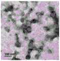
图2为复合敷料的细胞黏附结果图;Fig. 2 is the cell adhesion result graph of composite dressing;
图3为金纳米棒的紫外光谱扫描图;Fig. 3 is the ultraviolet spectrum scanning figure of gold nanorod;
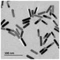
图4为金纳米棒的透射电镜结果图;Fig. 4 is the result picture of transmission electron microscope of gold nanorod;
图5为复合敷料的活死染色结果图;Fig. 5 is the living and dead dyeing result graph of composite dressing;
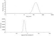
图6为外泌体的粒径电位图;Figure 6 is a particle size potential diagram of exosomes;
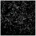
图7为外泌体的透射电镜结果图;Fig. 7 is the result of transmission electron microscope of exosome;
图8为干细胞在外泌体给药后1天的CCK-8增殖结果图;Figure 8 is a graph showing the results of CCK-8 proliferation of
图9为干细胞在外泌体给药7天后的巢蛋白(Nestin)免疫荧光结果图;Figure 9 is a graph showing the results of Nestin immunofluorescence of stem cells 7 days after exosome administration;
图10为复合敷料的大鼠伤口愈合结果图;Figure 10 is a graph of the results of wound healing in rats with composite dressings;
图11为复合敷料的大鼠体重结果图;Fig. 11 is the result chart of rat body weight of composite dressing;
图12为复合敷料的大鼠皮肤神经丝蛋白(NF-L)免疫组化结果图。Figure 12 is a graph showing the results of immunohistochemistry of rat skin neurofilament protein (NF-L) in composite dressings.
具体实施方式Detailed ways
下面结合附图,对本发明作详细的说明。The present invention will be described in detail below with reference to the accompanying drawings.
为了使本发明的目的、技术方案以及优点更加清晰明白,以下结合附图和实施例,对本发明进行进一步详细说明。此处所描述的具体实施例,仅用于解释本发明,并不用于限定本发明。In order to make the objectives, technical solutions and advantages of the present invention clearer, the present invention will be described in further detail below with reference to the accompanying drawings and embodiments. The specific embodiments described herein are only used to explain the present invention, and are not used to limit the present invention.
实施例1:一种促进外周神经修复的压电-导电凝胶并负载中药外泌体的复合敷料及其制备方法,包括以下步骤:Embodiment 1: a piezoelectric-conductive gel for promoting peripheral nerve repair and a composite dressing loaded with traditional Chinese medicine exosomes and a preparation method thereof, comprising the following steps:
1)将明胶与甲基丙烯酸酐反应,之后用8k-14kDa的透析袋中室温透析1周,然后冻干,制备成甲基丙烯酸酯化明胶;1) Reaction of gelatin with methacrylic anhydride, followed by dialysis at room temperature in a dialysis bag of 8k-14kDa for 1 week, and then freeze-dried to prepare methacrylated gelatin;
2)先将女贞,洗净,榨汁,过100目筛;再通过差速离心法,先后用1000g、12000g离心之后,取上清液用超速离心机在100000g下离心,然后将沉淀用PBS重悬。用蔗糖配备密度梯度溶液,然后在200000g下超离60分钟;再取不同组分的外泌体,在50000g下离心60分钟,制备成中药外泌体;2) First, wash the privet, squeeze the juice, and pass through a 100-mesh sieve; then through the differential centrifugation method, after centrifuging with 1000g and 12000g successively, take the supernatant and centrifuge it with an ultracentrifuge at 100000g, and then use the Resuspend in PBS. A density gradient solution was prepared with sucrose, and then ultra-centrifuged at 200,000 g for 60 minutes; then exosomes of different components were taken and centrifuged at 50,000 g for 60 minutes to prepare traditional Chinese medicine exosomes;
3)通过HAuCl4与CTAB、NaBH4制备成种子溶液,HAuCl4、CTAB、AgNO3、AA制备成生长溶液,然后通过种子生长法,于25℃,生长过夜,制备成金纳米棒;3) Prepare a seed solution by HAuCl4 , CTAB and NaBH4 , prepare a growth solution from HAuCl4 , CTAB, AgNO3 , and AA, and then grow it overnight at 25° C. by a seed growth method to prepare gold nanorods;
4)将甲基丙烯酸酯化明胶用PBS溶解,使其浓度达到5%;然后加入金纳米棒,使其浓度达到1mg/ml;再加入中药外泌体,使其最终浓度达到10μg/ml。再混入光敏剂LAP,得到水胶预凝胶体系;4) Dissolve the methacrylated gelatin with PBS to make the concentration reach 5%; then add gold nanorods to make the concentration reach 1 mg/ml; and then add traditional Chinese medicine exosomes to make the final concentration reach 10 μg/ml. Then mixed with photosensitizer LAP to obtain a hydrogel pregel system;
5)取聚偏氟乙烯粉末单体,用无水DMF溶解后,旋转涂布成薄膜,然后120℃退火,退火后,100℃,120V/μm的电场下,极化8小时,制备聚偏氟乙烯,其厚度为10μm;5) Take the polyvinylidene fluoride powder monomer, dissolve it with anhydrous DMF, spin-coat it into a thin film, and then anneal at 120°C. After annealing, polarize it for 8 hours at 100°C under an electric field of 120V/μm to prepare polyvinylidene fluoride. vinyl fluoride with a thickness of 10 μm;
6)将水凝胶预凝胶体系,旋涂到聚偏氟乙烯上,然后光交联60s,制备成复合敷料,其中水凝胶厚度为2mm;6) spin-coating the hydrogel pre-gel system on the polyvinylidene fluoride, and then photocrosslinking for 60s to prepare a composite dressing, wherein the hydrogel thickness is 2mm;
制备甲基丙烯酸酯化明胶之后,溶解于氘代水中,然后用核磁共振仪测试,获得相关材料的1H-NMR谱图,确定甲基丙烯酸酯化明胶的化学结构;如图1所示,相较于明胶,甲基丙烯酸酯化明胶的H谱中存在化学位移在5.2~5.6ppm之间的峰(黑色虚线框中所示),代表双键碳上的氢原子。并且受局部屏蔽效应和远程屏蔽效应的影响,双键碳上的两个氢原子处在不同的化学位移上.After preparing the methacrylated gelatin, it was dissolved in deuterated water, and then tested with a nuclear magnetic resonance instrument to obtain the1 H-NMR spectrum of the relevant material to determine the chemical structure of the methacrylated gelatin; as shown in Figure 1, Compared with gelatin, the H spectrum of methacrylated gelatin has peaks with chemical shifts between 5.2 and 5.6 ppm (shown in black dashed boxes), representing hydrogen atoms on double-bonded carbons. And affected by the local shielding effect and the remote shielding effect, the two hydrogen atoms on the double-bonded carbon are at different chemical shifts.
制备成复合敷料后,将复合敷料于超净台中紫外照射0.5小时。用骨髓间充质干细胞完全培养液洗去酒精并浸泡水凝胶,于37℃中平衡。将消化的骨髓间充质干细胞以1×10⁵/mL的细胞密度接种在凝胶上,置于37℃、5%CO2的孵箱内培养24小时后,吸掉培养基,PBS清洗两次,再用4%多聚甲醛浸泡固定10分钟后,用含0.1%Triton X-100的PBS洗涤2次,每次约5分钟。用含有5%BSA和0.1%Triton X-100的PBS按照1:100的比例稀释Actin-Tracker Green染色液,滴加到细胞上,室温避光孵育1h后再用PBS洗两次。加入500μl/mlDAPI溶液,室温孵育15分钟,除去DAPI溶液,PBS清洗两次后,立即置于共聚焦显微镜下观察。如图2所示,在培养24h后,细胞在水凝胶上正常黏附,细胞形态正常,成梭状伸展。After the composite dressing was prepared, the composite dressing was irradiated with ultraviolet light for 0.5 hour in an ultra-clean bench. The alcohol was washed away with bone marrow mesenchymal stem cell complete culture medium and the hydrogel was soaked, and equilibrated at 37°C. The digested bone marrow mesenchymal stem cells were seeded on the gel at a cell density of 1×10⁵ /mL, placed in a 37°C, 5% CO2 incubator for 24 hours, the medium was aspirated, and washed with PBS for two days. 2 times, soaked in 4% paraformaldehyde for 10 minutes, and then washed twice with PBS containing 0.1% Triton X-100 for about 5 minutes each time. Actin-Tracker Green staining solution was diluted 1:100 with PBS containing 5% BSA and 0.1% Triton X-100, dropped onto the cells, incubated at room temperature in the dark for 1 h, and then washed twice with PBS. Add 500 μl/ml DAPI solution, incubate for 15 minutes at room temperature, remove the DAPI solution, wash twice with PBS, and observe under a confocal microscope immediately. As shown in Figure 2, after 24 hours of culture, the cells adhered normally on the hydrogel, and the cells had a normal shape and expanded into a fusiform.
实施例2:一种促进外周神经修复的压电-导电凝胶并负载中药外泌体的复合敷料及其制备方法,包括以下步骤:Embodiment 2: a piezoelectric-conductive gel for promoting peripheral nerve repair and a composite dressing loaded with traditional Chinese medicine exosomes and a preparation method thereof, comprising the following steps:
1)将明胶与甲基丙烯酸酐反应,之后用8k-14kDa的透析袋中室温透析2周,然后冻干,制备成甲基丙烯酸酯化明胶;1) The gelatin is reacted with methacrylic anhydride, then dialyzed for 2 weeks at room temperature in a dialysis bag of 8k-14kDa, and then freeze-dried to prepare methacrylated gelatin;
2)先将丹参,洗净,榨汁,过200目筛;再通过差速离心法,先后用2000g、10000g离心之后,取上清液用超速离心机在120000g下离心,然后将沉淀用PBS重悬。用CsCl梯度溶液,然后在100000g下超离60分钟;再取不同组分的外泌体,在100000g下离心60分钟,制备成中药外泌体;2) First, wash the Salvia miltiorrhiza, squeeze the juice, and pass through a 200-mesh sieve; then through the differential centrifugation method, after successively centrifuging at 2000g and 10000g, take the supernatant and centrifuge it with an ultracentrifuge at 120,000g, and then use PBS for the sediment. Resuspended. Use CsCl gradient solution, and then ultra-centrifuge at 100,000g for 60 minutes; then take exosomes of different components and centrifuge at 100,000g for 60 minutes to prepare traditional Chinese medicine exosomes;
3)通过HAuCl4与CTAB、NaBH4制备成种子溶液,HAuCl4、CTAB、AgNO3、AA制备成生长溶液,然后通过种子生长法,于28℃,生长过夜,制备成金纳米棒;3) Prepare a seed solution by HAuCl4 , CTAB and NaBH4 , prepare a growth solution from HAuCl4 , CTAB, AgNO3 , and AA, and then grow it overnight at 28° C. by a seed growth method to prepare gold nanorods;
4)将甲基丙烯酸酯化明胶用PBS溶解,使其浓度达到10%;然后加入金纳米棒,使其浓度达到2mg/ml;再加入中药外泌体,使其最终浓度达到1μg/ml。4) Dissolve the methacrylated gelatin with PBS to make its concentration reach 10%; then add gold nanorods to make its concentration reach 2 mg/ml; and then add traditional Chinese medicine exosomes to make its final concentration reach 1 μg/ml.
再混入光敏剂LAP,得到水胶预凝胶体系;Then mixed with photosensitizer LAP to obtain a hydrogel pregel system;
5)取聚偏氟乙烯粉末单体,用无水DMF溶解后,旋转涂布成薄膜,然后100℃退火,退火后,130℃,120V/μm的电场下,极化4小时,制备聚偏氟乙烯,其厚度为20μm;5) Take the polyvinylidene fluoride powder monomer, dissolve it with anhydrous DMF, spin-coat it into a thin film, and then anneal at 100 °C. After annealing, polarize for 4 hours at 130 °C under an electric field of 120V/μm to prepare polyvinylidene fluoride. vinyl fluoride with a thickness of 20 μm;
6)将水凝胶预凝胶体系,旋涂到聚偏氟乙烯上,然后光交联30s,制备成复合敷料,其中水凝胶厚度为5mm;6) spin-coating the hydrogel pre-gel system on the polyvinylidene fluoride, and then photocrosslinking for 30s to prepare a composite dressing, wherein the hydrogel thickness is 5mm;
将制备完成的金纳米棒,先通过紫外可见光吸收光谱仪测试,确定金纳米棒在350-900nm的吸收峰;如图3所示,金纳米棒共有两个吸收峰,分别对应金纳米棒的长径和短径。The prepared gold nanorods are first tested by UV-Vis absorption spectrometer to determine the absorption peaks of gold nanorods at 350-900 nm; as shown in Figure 3, there are two absorption peaks for gold nanorods, corresponding to the length of the gold nanorods respectively. diameter and short diameter.
然后将金纳米棒溶液,滴于镀有碳膜的铜网上,自然干燥后滴在铜网上,通过透射电镜观察其形貌。如图4所示:金纳米棒呈标准的棒状结构,形貌均一,水中分散性良好,经测量,金纳米棒长径比约为4.2。Then, the gold nanorod solution was dropped on the copper mesh coated with carbon film, and after natural drying, it was dropped on the copper mesh, and its morphology was observed by transmission electron microscope. As shown in Figure 4: the gold nanorods have a standard rod-like structure, uniform morphology, and good dispersibility in water. After measurement, the aspect ratio of the gold nanorods is about 4.2.
制备成复合敷料后,用骨髓间充质干细胞完全培养液洗去酒精并浸泡水凝胶,于37℃中平衡。将消化的骨髓间充质干细胞以1×10⁵/mL的细胞密度接种在凝胶上,置于37℃、5%CO2的孵箱内培养48h后,去除培养液,PBS清洗3次。加入已用PBS分别稀释到2μM和8mΜ的Calcein AM和PI溶液,没过细胞,避光孵育30-45分钟,最后用PBS清洗三次,将残余的试剂去除,立即将待测样品放在激光共聚焦显微镜下观察。其中活细胞显示绿色,死细胞中的核酸物质被染成红色。如图5所示,培养48h后,复合敷料中细胞基本显绿色,未见红色,证明复合敷料有良好的生物相容性。After the composite dressing was prepared, the alcohol was washed away with the bone marrow mesenchymal stem cell complete culture medium, and the hydrogel was soaked, and equilibrated at 37°C. The digested bone marrow mesenchymal stem cells were seeded on the gel at a cell density of 1 × 10⁵/mL, placed in a 37°C, 5% CO2 incubator for 48 hours, then the culture medium was removed and washed with PBS three times. Add Calcein AM and PI solutions diluted with PBS to 2 μM and 8 mM, respectively, cover the cells, and incubate in the dark for 30-45 minutes. Finally, wash three times with PBS to remove the residual reagents. Immediately place the samples to be tested on the laser. Observe under a focusing microscope. Among them, live cells are shown in green, and nucleic acid substances in dead cells are stained in red. As shown in Figure 5, after 48 hours of culture, the cells in the composite dressing were basically green, and no red was seen, which proved that the composite dressing had good biocompatibility.
实施例3:一种促进外周神经修复的压电-导电凝胶并负载中药外泌体的复合敷料及其制备方法,包括以下步骤:Embodiment 3: a piezoelectric-conductive gel for promoting peripheral nerve repair and a composite dressing loaded with traditional Chinese medicine exosomes and a preparation method thereof, comprising the following steps:
1)将明胶与甲基丙烯酸酐反应,之后8k-14kDa的透析袋中室温透析1周,然后冻干,制备成甲基丙烯酸酯化明胶;1) Reaction of gelatin with methacrylic anhydride, followed by dialysis at room temperature in a dialysis bag of 8k-14kDa for 1 week, and then freeze-dried to prepare methacrylated gelatin;
2)先将地黄洗净,榨汁,过150目筛;再通过差速离心法,先后用2000g、10000g离心之后,取上清液用超速离心机在150000g下离心,然后将沉淀用PBS重悬。用蔗糖梯度溶液,然后在150000g下超离120分钟;再取不同组分的外泌体,在150000g下离心60分钟,制备成中药外泌体;2) Wash the Rehmannia glutinosa first, squeeze the juice, and pass through a 150-mesh sieve; then through the differential centrifugation method, after successively centrifuging at 2000g and 10000g, take the supernatant and centrifuge it at 150000g with an ultracentrifuge, and then resuspend the precipitate with PBS. hanging. Use sucrose gradient solution, and then ultra-centrifuge at 150,000g for 120 minutes; then take exosomes of different components and centrifuge at 150,000g for 60 minutes to prepare traditional Chinese medicine exosomes;
3)通过HAuCl4与CTAB、NaBH4制备成种子溶液,HAuCl4、CTAB、AgNO3、AA制备成生长溶液,然后通过种子生长法,于30℃,生长过夜,制备成金纳米棒3) A seed solution is prepared by HAuCl4 and CTAB and NaBH4 , and a growth solution is prepared by HAuCl4 , CTAB, AgNO3 , and AA, and then grown overnight at 30° C. by the seed growth method to prepare gold nanorods
4)将甲基丙烯酸酯化明胶用超纯水溶解使其浓度达到20%;然后加入金纳米棒,使其浓度达到2mg/ml;再加入中药外泌体,使其最终浓度达到1μg/ml。再混入光敏剂I2959,得到水胶预凝胶体系;4) Dissolve methacrylated gelatin with ultrapure water to make its concentration reach 20%; then add gold nanorods to make its concentration reach 2 mg/ml; then add Chinese medicine exosomes to make its final concentration reach 1 μg/ml . The photosensitizer I2959 is mixed again to obtain the hydrogel pregel system;
5)取聚偏氟乙烯粉末单体,用无水DMF溶解后,旋转涂布成薄膜,然后120℃退火,退火后,115℃,90V/μm的电场下,极化8h,制备聚偏氟乙烯,其厚度为20μm;5) Take the polyvinylidene fluoride powder monomer, dissolve it with anhydrous DMF, spin-coat it into a thin film, then anneal at 120°C, after annealing, polarize for 8h at 115°C under an electric field of 90V/μm to prepare polyvinylidene fluoride Ethylene, the thickness of which is 20 μm;
6)将水凝胶预凝胶体系,旋涂到聚偏氟乙烯上,然后光交联60s,制备成复合敷料,其中水凝胶厚度为3mm;6) spin-coating the hydrogel pre-gel system on the polyvinylidene fluoride, and then photocrosslinking for 60s to prepare a composite dressing, wherein the hydrogel thickness is 3mm;
将制备完成的外泌体,于马尔文粒径分析仪中,分析其粒径电位。如图6所示,外泌体的粒径和电位分布均一,其中粒径为144.1±2.762nm,PDI为0.218,表面电位为-27.4±0.451mV。The prepared exosomes were analyzed for their particle size potential in a Malvern particle size analyzer. As shown in Figure 6, the particle size and potential distribution of exosomes were uniform, in which the particle size was 144.1 ± 2.762 nm, the PDI was 0.218, and the surface potential was -27.4 ± 0.451 mV.
然后将外泌体溶液滴于镀有碳膜的铜网上,之后用醋酸双氧铀进行复染,透射电镜观察。如图7所示,外泌体呈标准饼状结构。Then, the exosome solution was dropped on the copper grid coated with carbon film, and then counterstained with uranyl acetate, and observed by transmission electron microscope. As shown in Figure 7, the exosomes have a standard pie-like structure.
取第三代的骨髓间充质干细胞,以5×10³/孔的密度种在96孔板上,培养24小时,向96孔板中加入不同浓度的外泌体。放入细胞培养箱中孵育6小时后,吸出培养液,PBS洗两次,将培养液更换为新鲜的含有10%血清的新鲜培养液并继续培养18小时后,除去培养液,加入含有CCK-8的DMEM低糖培养液100μL,放入细胞培养箱培养4小时,在酶标仪475nm处测定吸光值。计算细胞增殖百分率。如图8所示,在三种不同组别的外泌体浓度培养下,从低浓度到高浓度,外泌体均对骨髓间充质干细胞有良好的增殖作用,且增殖率逐渐上升。The third-generation bone marrow mesenchymal stem cells were seeded in a 96-well plate at a density of 5×10³ /well, cultured for 24 hours, and different concentrations of exosomes were added to the 96-well plate. After 6 hours of incubation in the cell incubator, aspirate the culture medium, wash twice with PBS, replace the culture medium with fresh culture medium containing 10% serum and continue to culture for 18 hours, remove the culture medium, and add the medium containing CCK- 100 μL of DMEM low-sugar medium of 8 was placed in a cell incubator for 4 hours, and the absorbance value was measured at 475 nm on a microplate reader. Calculate the percentage of cell proliferation. As shown in Figure 8, under the culture of three different groups of exosome concentrations, from low concentration to high concentration, exosomes have a good proliferation effect on bone marrow mesenchymal stem cells, and the proliferation rate gradually increases.
实施例4:一种促进外周神经修复的压电-导电凝胶并负载中药外泌体的复合敷料及其制备方法,包括以下步骤:Embodiment 4: a piezoelectric-conductive gel for promoting peripheral nerve repair and a composite dressing loaded with traditional Chinese medicine exosomes and a preparation method thereof, comprising the following steps:
1)将明胶与甲基丙烯酸酐反应,之后8k-14kDa的透析袋中室温透析2周,然后冻干,制备成甲基丙烯酸酯化明胶;1) Reaction of gelatin with methacrylic anhydride, followed by dialysis at room temperature in a dialysis bag of 8k-14kDa for 2 weeks, and then freeze-dried to prepare methacrylated gelatin;
2)先将人参,洗净,榨汁,过200目筛;再通过差速离心法,先后用3000g、10000g离心之后,取上清液用超速离心机在150000g下离心,然后将沉淀用PBS重悬。用蔗糖梯度溶液,然后在100000g下超离120分钟;再取不同组分的外泌体,在30000g下离心60分钟,制备成中药外泌体;2) First, wash the ginseng, squeeze the juice, and pass through a 200-mesh sieve; then through the differential centrifugation method, after centrifuging with 3000g and 10000g successively, take the supernatant and centrifuge it with an ultracentrifuge at 150000g, and then use PBS for the precipitate. Resuspended. Use sucrose gradient solution, and then ultra-centrifuge at 100,000g for 120 minutes; then take exosomes of different components, centrifuge at 30,000g for 60 minutes, and prepare traditional Chinese medicine exosomes;
3)通过HAuCl4与CTAB、NaBH4制备成种子溶液,HAuCl4、CTAB、AgNO3、AA制备成生长溶液,然后通过种子生长法,于25℃,生长过夜,制备成金纳米棒3) A seed solution is prepared by HAuCl4 and CTAB and NaBH4 , and a growth solution is prepared by HAuCl4 , CTAB, AgNO3 and AA, and then grown overnight at 25° C. by the seed growth method to prepare gold nanorods
4)将甲基丙烯酸酯化明胶用超纯水溶解使其浓度达到10%;然后加入金纳米棒,使其浓度达到3mg/ml;再加入中药外泌体,使其最终浓度达到2μg/ml。再混入光敏剂LAP,得到水胶预凝胶体系;4) Dissolve methacrylated gelatin with ultrapure water to make its concentration reach 10%; then add gold nanorods to make its concentration reach 3 mg/ml; then add traditional Chinese medicine exosomes to make its final concentration reach 2 μg/ml . Then mixed with photosensitizer LAP to obtain a hydrogel pregel system;
5)取聚偏氟乙烯粉末单体,用无水DMF溶解后,旋转涂布成薄膜,然后120℃退火,退火后,100℃,120V/μm的电场下,极化8h,制备聚偏氟乙烯,其厚度为30μm;5) Take the polyvinylidene fluoride powder monomer, dissolve it with anhydrous DMF, spin-coat it into a thin film, then anneal at 120 °C, after annealing, polarize it for 8 hours at 100 °C under an electric field of 120V/μm to prepare polyvinylidene fluoride Ethylene, the thickness of which is 30 μm;
6)将水凝胶预凝胶体系,旋涂到聚偏氟乙烯上,然后光交联120s,制备成复合敷料,其中水凝胶厚度为5mm;6) spin-coating the hydrogel pre-gel system on the polyvinylidene fluoride, and then photocrosslinking for 120s to prepare a composite dressing, wherein the hydrogel thickness is 5mm;
将复合辅料于超净台中,紫外照射0.5小时,用骨髓间充质干细胞完全培养液洗去酒精并浸泡水凝胶,于37℃中平衡。将消化的骨髓间充质干细胞以1×10⁵/mL的细胞密度接种在凝胶上,置于37℃、5%CO2的孵箱内每隔一天换一次培养液,培养至14天后,将凝胶样品取出进行巢蛋白(Nestin)染色。其具体步骤如下:用PBS浸泡清洗水凝胶样品两次,加入4%多聚甲醛覆盖住细胞,室温固定15分钟后再用PBS浸泡清洗三遍,0.1%Triton X-100透化5分钟,清洗后加入山羊血清封闭30分钟,除去液体并清洗后加入一抗置于4℃孵育过夜,PBS充分浸泡清洗五遍,加入荧光二抗,37℃避光孵育1h,PBS充分浸泡清洗三遍,加入DAPI染色液覆盖住细胞,室温染色10分钟,用PBS充分浸泡清洗三遍后加入PBS浸润,共聚焦显微镜检测观察。如图9所示,细胞经染色后,显现强烈的绿色荧光,表明在复合敷料上的干细胞,巢蛋白高度表达,骨髓间充质干细胞呈神经分化趋势。The compound excipients were placed in an ultra-clean bench, irradiated with ultraviolet light for 0.5 hours, and the alcohol was washed away with the complete culture medium of bone marrow mesenchymal stem cells, soaked in the hydrogel, and equilibrated at 37 °C. The digested bone marrow mesenchymal stem cells were seeded on the gel at a cell density of 1×10⁵ /mL, and the culture medium was changed every other day in a 37°C, 5% CO2 incubator. After culturing for 14 days, Gel samples were removed for Nestin staining. The specific steps are as follows: soak and wash the hydrogel samples with PBS twice, add 4% paraformaldehyde to cover the cells, fix for 15 minutes at room temperature, then soak and wash three times with PBS, permeabilize with 0.1% Triton X-100 for 5 minutes, After washing, add goat serum to block for 30 minutes, remove the liquid and wash, add primary antibody and incubate at 4 °C overnight, fully soak and wash in PBS for five times, add fluorescent secondary antibody, incubate at 37 °C for 1 h in the dark, and fully soak and wash in PBS three times. DAPI staining solution was added to cover the cells, stained at room temperature for 10 minutes, fully soaked and washed with PBS for three times, then infiltrated with PBS, and detected and observed by confocal microscope. As shown in Figure 9, the cells showed strong green fluorescence after staining, indicating that the stem cells on the composite dressing highly expressed nestin, and the bone marrow mesenchymal stem cells showed a tendency of neural differentiation.
实施例5:一种促进外周神经修复的压电-导电凝胶并负载中药外泌体的复合敷料及其制备方法,包括以下步骤:Embodiment 5: a piezoelectric-conductive gel for promoting peripheral nerve repair and a composite dressing loaded with traditional Chinese medicine exosomes and a preparation method thereof, comprising the following steps:
1)将明胶与甲基丙烯酸酐反应,之后8k-14kDa的透析袋中室温透析2周,然后冻干,制备成甲基丙烯酸酯化明胶;1) Reaction of gelatin with methacrylic anhydride, followed by dialysis at room temperature in a dialysis bag of 8k-14kDa for 2 weeks, and then freeze-dried to prepare methacrylated gelatin;
2)先将西红花,洗净,榨汁,过150目筛;再通过差速离心法,先后用3000g、5000g离心之后,取上清液用超速离心机在100000g下离心,然后将沉淀用超纯水重悬。用蔗糖梯度溶液,然后在100000g下超离60分钟;再取不同组分的外泌体,在10000g下离心60分钟,制备成中药外泌体;2) First, wash the saffron, squeeze the juice, and pass through a 150-mesh sieve; then through the differential centrifugation method, after successively centrifuging at 3000g and 5000g, take the supernatant and centrifuge it at 100,000g with an ultracentrifuge, and then remove the precipitate. Resuspend in ultrapure water. Use sucrose gradient solution, and then ultra-centrifuge at 100,000g for 60 minutes; then take exosomes of different components and centrifuge at 10,000g for 60 minutes to prepare traditional Chinese medicine exosomes;
3)通过HAuCl4与CTAB、NaBH4制备成种子溶液,HAuCl4、CTAB、AgNO3、AA制备成生长溶液,然后通过种子生长法,于25℃,生长过夜,制备成金纳米棒3) A seed solution is prepared by HAuCl4 and CTAB and NaBH4 , and a growth solution is prepared by HAuCl4 , CTAB, AgNO3 and AA, and then grown overnight at 25° C. by the seed growth method to prepare gold nanorods
4)将甲基丙烯酸酯化明胶用超纯水溶解使其浓度达到15%;然后加入金纳米棒,使其浓度达到2mg/ml;再加入中药外泌体,使其最终浓度达到0.1μg/ml。再混入光敏剂LAP,得到水胶预凝胶体系;4) Dissolve the methacrylated gelatin with ultrapure water to make its concentration reach 15%; then add gold nanorods to make its concentration reach 2 mg/ml; then add traditional Chinese medicine exosomes to make its final concentration reach 0.1 μg/ml ml. Then mixed with photosensitizer LAP to obtain a hydrogel pregel system;
5)取聚偏氟乙烯粉末单体,用无水DMF溶解后,旋转涂布成薄膜,然后120℃退火,退火后,60℃,120V/μm的电场下,极化8h,制备聚偏氟乙烯,其厚度为40μm;5) Take the polyvinylidene fluoride powder monomer, dissolve it with anhydrous DMF, spin-coat it into a thin film, then anneal at 120 °C, after annealing, polarize it for 8 hours at 60 °C under an electric field of 120V/μm to prepare polyvinylidene fluoride Ethylene, the thickness of which is 40 μm;
6)将水凝胶预凝胶体系,旋涂到聚偏氟乙烯上,然后光交联80s,制备成复合敷料,其中水凝胶厚度为3mm;6) spin-coating the hydrogel pre-gel system on the polyvinylidene fluoride, and then photocrosslinking for 80s to prepare a composite dressing, wherein the hydrogel thickness is 3mm;
建立急性创伤模型:将48清洁级SD雄性大鼠(体重150-200g)分成8组:空白对照组、OMV溶液组、SDF-1蛋白溶液组、OMV+SDF-1混合溶液组、Gelma凝胶组、Gelma/SDF-1凝胶组、Gelma/OMV凝胶组、Gelma/OMV/SDF-1凝胶组。每组6只,麻醉后剪去背部毛发,去除1.5cm×1.5cm的皮肤,并用碘酒进行消毒。第二天开始给药,每隔两天给药一次。空白组每只每次给予200μL PBS,三组溶液组同样每只每次给药200μL溶液,凝胶组每只每次给药1.5cm×1.5cm的凝胶片层制剂,并用无菌贴片将其固定。并注意观察创面的愈合情况和疤痕的形成情况,每3天进行一次拍照,并测量伤口面积和大鼠的体重,计算愈合率与体重变化情况。Establishment of acute trauma model: 48 clean grade SD male rats (body weight 150-200g) were divided into 8 groups: blank control group, OMV solution group, SDF-1 protein solution group, OMV+SDF-1 mixed solution group, Gelma gel group, Gelma/SDF-1 gel group, Gelma/OMV gel group, Gelma/OMV/SDF-1 gel group. There were 6 animals in each group. After anesthesia, the back hair was cut, the skin of 1.5cm×1.5cm was removed, and iodine was used for disinfection. Dosing began the next day and was administered every other day. The blank group was given 200 μL of PBS each time, the three groups of solution groups were also given 200 μL of solution each time, and the gel group was given a 1.5cm × 1.5cm gel sheet preparation each time, and a sterile patch was used. fix it. And pay attention to observe the wound healing and scar formation, take pictures every 3 days, measure the wound area and the body weight of the rats, and calculate the healing rate and body weight changes.

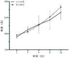
如图10和11所示,复合辅料组的体重与愈合率均较空白对照组明显增加,其中在第六天呈显著性差异。其中体重结果客观证明复合敷料对SD大鼠无有害的全身影响,而愈合率的明显增加证明复合敷料鞥有效提高伤口的愈合率。As shown in Figures 10 and 11, the body weight and healing rate of the compound excipient group were significantly increased compared with the blank control group, and there was a significant difference on the sixth day. The body weight results objectively proved that the composite dressing had no harmful systemic effects on SD rats, and the significant increase in the healing rate proved that the composite dressing could effectively improve the wound healing rate.
将组织切片置于盛满EDTA抗原修复缓冲液(PH8.0)的修复盒中进行抗原修复,滴加3%BSA均匀覆盖组织,室温封闭30分钟,轻轻甩掉封闭液,在切片上滴加PBS按一定比例配好的神经丝蛋白(NF-L)一抗,切片平放于湿盒内4℃孵育过夜。玻片置于PBS中在脱色摇床上晃动洗涤3次,每次5分钟。滴加与一抗相应种属的二抗覆盖组织,避光室温孵育50分钟。PBS充分冲洗3遍后,滴加DAPI染液,避光室温孵育10分钟。用PBS充分浸泡清洗五遍后加入PBS浸润,切片于共聚焦显微镜下观察并采集图像。如图12所示,复合敷料组皮肤组织切片中,红色荧光高度表达,且成丝状,可见大量的神经丝的分布。证明复合敷料可以在体内,有效的促进伤口处神经的分化,修复外周神经损伤。Place the tissue sections in a retrieval box filled with EDTA antigen retrieval buffer (PH8.0) for antigen retrieval, add 3% BSA dropwise to cover the tissue evenly, seal at room temperature for 30 minutes, gently shake off the blocking solution, and drop drops on the slices. The neurofilament protein (NF-L) primary antibody prepared in a certain proportion in PBS was added, and the slices were placed flat in a humid box and incubated at 4°C overnight. The slides were washed three times in PBS with shaking on a destaining shaker for 5 minutes each. Add the secondary antibody corresponding to the primary antibody dropwise to cover the tissue, and incubate at room temperature for 50 minutes in the dark. After fully washing with PBS three times, DAPI staining solution was added dropwise, and incubated at room temperature for 10 minutes in the dark. After fully soaking and washing with PBS for five times, PBS was added to infiltrate, and the sections were observed under a confocal microscope and images were collected. As shown in Figure 12, in the skin tissue section of the composite dressing group, red fluorescence was highly expressed and filamentous, and a large number of neurofilaments were distributed. It is proved that the composite dressing can effectively promote the differentiation of the nerve at the wound and repair the peripheral nerve injury in vivo.
Claims (8)
Translated fromChinesePriority Applications (1)
| Application Number | Priority Date | Filing Date | Title |
|---|---|---|---|
| CN201910910621.6ACN110464867B (en) | 2019-09-25 | 2019-09-25 | Piezoelectric composite dressing for promoting peripheral nerve repair and wound healing and loading traditional Chinese medicine exosomes and preparation method |
Applications Claiming Priority (1)
| Application Number | Priority Date | Filing Date | Title |
|---|---|---|---|
| CN201910910621.6ACN110464867B (en) | 2019-09-25 | 2019-09-25 | Piezoelectric composite dressing for promoting peripheral nerve repair and wound healing and loading traditional Chinese medicine exosomes and preparation method |
Publications (2)
| Publication Number | Publication Date |
|---|---|
| CN110464867A CN110464867A (en) | 2019-11-19 |
| CN110464867Btrue CN110464867B (en) | 2020-07-28 |
Family
ID=68516853
Family Applications (1)
| Application Number | Title | Priority Date | Filing Date |
|---|---|---|---|
| CN201910910621.6AActiveCN110464867B (en) | 2019-09-25 | 2019-09-25 | Piezoelectric composite dressing for promoting peripheral nerve repair and wound healing and loading traditional Chinese medicine exosomes and preparation method |
Country Status (1)
| Country | Link |
|---|---|
| CN (1) | CN110464867B (en) |
Families Citing this family (15)
| Publication number | Priority date | Publication date | Assignee | Title |
|---|---|---|---|---|
| CN108949677B (en)* | 2018-07-05 | 2021-11-30 | 浙江大学 | Application of rehmannia root glycoside C and salvianolic acid A in promoting proliferation of mesenchymal stem cells cultured in vitro and inhibiting replicative senescence |
| CN111001036B (en)* | 2019-12-19 | 2021-10-22 | 北京大学人民医院(北京大学第二临床医学院) | A kind of single-walled carbon nanotube composite conductive nerve sleeve and its preparation method and application |
| CN111671972B (en)* | 2020-07-30 | 2021-10-29 | 中国人民解放军空军军医大学 | Tissue engineering scaffold of compound exosome Nidogen-1 and preparation method thereof |
| CN112656836B (en)* | 2020-12-28 | 2022-02-11 | 浙江大学 | Application of transdermal peptide modified pachyrhizua angulatus exosome nano preparation in preparation of anti-skin-aging products |
| CN112870228B (en)* | 2021-01-20 | 2023-01-24 | 杭州贤石生物科技有限公司 | Multifunctional microenvironment protection exosome hydrogel and preparation method and application thereof |
| CN113274539B (en)* | 2021-04-29 | 2022-03-22 | 西安理工大学 | A kind of self-powered wound patch and preparation method thereof |
| CN113262105B (en)* | 2021-05-08 | 2023-02-28 | 南京理工大学 | Wound dressing based on piezoelectric effect and preparation method thereof |
| CN113373595B (en)* | 2021-05-19 | 2023-02-03 | 济南大学 | A kind of FeOOH/PVDF fiber support and its preparation method and application |
| CN114099786A (en)* | 2021-11-18 | 2022-03-01 | 范磊 | Preparation method of exosome-loaded electroactive hydrogel dressing |
| CN115387029B (en)* | 2022-08-23 | 2024-05-17 | 北京倍美药业有限公司 | Preparation method of self-powered adhesive bandage with stripe structure |
| CN115444974B (en)* | 2022-10-25 | 2024-01-30 | 中国地质大学(北京) | Electroactive composite patch for treating scalds and preparation method and application thereof |
| CN115607745B (en)* | 2022-11-09 | 2023-09-15 | 深圳先进技术研究院 | Exosome-programmed tissue repair materials and preparation methods |
| WO2024098285A1 (en)* | 2022-11-09 | 2024-05-16 | 深圳先进技术研究院 | Exosome program-controlled tissue repair material and preparation method therefor |
| CN115671366B (en)* | 2022-12-30 | 2023-04-18 | 深圳湾实验室 | Composite dressing for promoting wound healing, its preparation method and application |
| CN117919344A (en)* | 2023-02-16 | 2024-04-26 | 成都诺美康医疗科技有限公司 | Skin ulcer repairing and regenerating gel and preparation method thereof |
Family Cites Families (5)
| Publication number | Priority date | Publication date | Assignee | Title |
|---|---|---|---|---|
| CN1424396A (en)* | 2002-12-13 | 2003-06-18 | 中山大学 | Preparation of implanting neurons for treating nerve injury diseases |
| CN103479682B (en)* | 2012-06-14 | 2015-03-11 | 苏州恒宇生物科技有限公司 | Preparation method for plant source active component nano-scale membrane type vesicle |
| CN107802878B (en)* | 2017-09-06 | 2020-07-28 | 华南理工大学 | Modified gelatin/potassium-sodium niobate composite electroactive antibacterial biological dressing and preparation and application thereof |
| CN108187130B (en)* | 2017-09-15 | 2020-08-18 | 海宁侏罗纪生物科技有限公司 | Reagent for repairing biological injury or stopping bleeding and application thereof |
| CN107929805B (en)* | 2018-01-10 | 2021-08-10 | 四川大学 | Metal/hydrogel composite dressing for promoting wound healing and preparation method thereof |
- 2019
- 2019-09-25CNCN201910910621.6Apatent/CN110464867B/enactiveActive
Also Published As
| Publication number | Publication date |
|---|---|
| CN110464867A (en) | 2019-11-19 |
Similar Documents
| Publication | Publication Date | Title |
|---|---|---|
| CN110464867B (en) | Piezoelectric composite dressing for promoting peripheral nerve repair and wound healing and loading traditional Chinese medicine exosomes and preparation method | |
| CN110755676B (en) | A composite dressing for promoting wound healing and regeneration and loading traditional Chinese medicine exosomes and preparation method | |
| Zhang et al. | Mimosa‐Inspired Stimuli‐Responsive Curling Bioadhesive Tape Promotes Peripheral Nerve Regeneration | |
| Liu et al. | Application of dental pulp stem cells in oral maxillofacial tissue engineering | |
| Zhang et al. | A nerve graft constructed with xenogeneic acellular nerve matrix and autologous adipose-derived mesenchymal stem cells | |
| Xu et al. | Multifunctional biodegradable conductive hydrogel regulating microenvironment for stem cell therapy enhances the nerve tissue repair | |
| Aulino et al. | Muscle extracellular matrix scaffold is a multipotent environment | |
| Qin et al. | Effects of electric field‐modulated conductive hydrogel on osseoperception and osseointegration of dental implants | |
| Cai et al. | Strategy towards independent electrical stimulation from cochlear implants: Guided auditory neuron growth on topographically modified nanocrystalline diamond | |
| Zhang et al. | Injectable, electroconductive, free radical scavenging silk fibroin/black phosphorus/glycyrrhizic acid nanocomposite hydrogel for enhancing spinal cord repair | |
| Rochkind et al. | Development of a tissue-engineered composite implant for treating traumatic paraplegia in rats | |
| Gao et al. | Comparison of morphology and biocompatibility of acellular nerve scaffolds processed by different chemical methods | |
| Zhang et al. | A biodegradable piezoelectric scaffold promotes spinal cord injury nerve regeneration | |
| CN113425899A (en) | Conductive degradable multifunctional tissue engineering scaffold and preparation method thereof | |
| CN108434526B (en) | Electroactive double-layer bone-like membrane material and preparation method thereof | |
| Haneef et al. | Development of bioartificial myocardium by electrostimulation of 3D collagen scaffolds seeded with stem cells | |
| CN103505761B (en) | Preparation method and application of silk bracket, and three-phase silk ligament graft and preparation method thereof | |
| Feng et al. | Reduced graphene oxide-mediated magnetoelectric effect drives neural differentiation of mesenchymal stem cells | |
| Xu et al. | Injectable hydrogel harnessing foreskin mesenchymal stem cell-derived extracellular vesicles for treatment of chronic diabetic skin wounds | |
| CN114848919A (en) | Composite hydrogel for TBI immune regulation and tissue repair and preparation method thereof | |
| AU2011289214B2 (en) | Methods for making a tissue engineered muscle repair (TEMR) construct in vitro for implantation in vivo | |
| Yan et al. | Magnetic field-oriented conductive decellularized extracellular matrix hydrogel synergizes with electrical stimulation to promote spinal cord injury repair and electrophysiological function restoration | |
| Ji et al. | A nerve growth factor persistent delivery scaffold seeded with neurally differentiated bone marrow mesenchymal stem cells promoted the functional recovery of spinal cord injury in rats | |
| WO2012094208A1 (en) | Nanowired three dimensional tissue scaffolds | |
| CN115737907A (en) | Application of bionic electroactive implant film |
Legal Events
| Date | Code | Title | Description |
|---|---|---|---|
| PB01 | Publication | ||
| PB01 | Publication | ||
| SE01 | Entry into force of request for substantive examination | ||
| SE01 | Entry into force of request for substantive examination | ||
| GR01 | Patent grant | ||
| GR01 | Patent grant |