CN110408590B - Method for inducing differentiation of human mesenchymal stem cells into osteoblasts and application thereof - Google Patents
Method for inducing differentiation of human mesenchymal stem cells into osteoblasts and application thereofDownload PDFInfo
- Publication number
- CN110408590B CN110408590BCN201910776470.XACN201910776470ACN110408590BCN 110408590 BCN110408590 BCN 110408590BCN 201910776470 ACN201910776470 ACN 201910776470ACN 110408590 BCN110408590 BCN 110408590B
- Authority
- CN
- China
- Prior art keywords
- osteoblasts
- stem cells
- mesenchymal stem
- human mesenchymal
- bone
- Prior art date
- Legal status (The legal status is an assumption and is not a legal conclusion. Google has not performed a legal analysis and makes no representation as to the accuracy of the status listed.)
- Active
Links
- 210000002901mesenchymal stem cellAnatomy0.000titleclaimsabstractdescription46
- 210000000963osteoblastAnatomy0.000titleclaimsabstractdescription38
- 238000000034methodMethods0.000titleclaimsabstractdescription21
- 230000001939inductive effectEffects0.000titleclaimsabstractdescription12
- 230000004069differentiationEffects0.000titleabstractdescription15
- 230000006698inductionEffects0.000claimsabstractdescription17
- OYPRJOBELJOOCE-UHFFFAOYSA-NCalciumChemical compound[Ca]OYPRJOBELJOOCE-UHFFFAOYSA-N0.000claimsabstractdescription9
- 239000011575calciumSubstances0.000claimsabstractdescription9
- 229910052791calciumInorganic materials0.000claimsabstractdescription9
- 150000001875compoundsChemical class0.000claimsdescription29
- 238000002955isolationMethods0.000claimsdescription4
- 210000004027cellAnatomy0.000abstractdescription37
- 210000000988bone and boneAnatomy0.000abstractdescription16
- 230000000694effectsEffects0.000abstractdescription10
- 102000002260Alkaline PhosphataseHuman genes0.000abstractdescription9
- 108020004774Alkaline PhosphataseProteins0.000abstractdescription9
- 239000003550markerSubstances0.000abstractdescription7
- 230000002188osteogenic effectEffects0.000abstractdescription7
- 238000002054transplantationMethods0.000abstractdescription7
- 230000011164ossificationEffects0.000abstractdescription6
- 206010061728Bone lesionDiseases0.000abstractdescription5
- 238000000338in vitroMethods0.000abstractdescription5
- 108090000623proteins and genesProteins0.000abstractdescription5
- 102000004169proteins and genesHuman genes0.000abstractdescription5
- 230000005847immunogenicityEffects0.000abstractdescription4
- 201000010099diseaseDiseases0.000abstractdescription3
- 208000037265diseases, disorders, signs and symptomsDiseases0.000abstractdescription3
- 210000000130stem cellAnatomy0.000description14
- XEKOWRVHYACXOJ-UHFFFAOYSA-NEthyl acetateChemical compoundCCOC(C)=OXEKOWRVHYACXOJ-UHFFFAOYSA-N0.000description9
- 238000011282treatmentMethods0.000description8
- 208000020084Bone diseaseDiseases0.000description7
- 239000000284extractSubstances0.000description6
- 238000010186stainingMethods0.000description5
- 240000008397Ganoderma lucidumSpecies0.000description4
- 235000001637Ganoderma lucidumNutrition0.000description4
- 229910052739hydrogenInorganic materials0.000description4
- 239000001257hydrogenSubstances0.000description4
- 230000009818osteogenic differentiationEffects0.000description4
- -1small molecule compoundsChemical class0.000description4
- 238000012360testing methodMethods0.000description4
- OKKJLVBELUTLKV-UHFFFAOYSA-NMethanolChemical compoundOCOKKJLVBELUTLKV-UHFFFAOYSA-N0.000description3
- 208000001132OsteoporosisDiseases0.000description3
- 239000002771cell markerSubstances0.000description3
- 230000004663cell proliferationEffects0.000description3
- 239000003814drugSubstances0.000description3
- 238000005516engineering processMethods0.000description3
- OWPIQJAXQUDFDL-UHFFFAOYSA-Nganoderma aldehydeNatural productsOc1ccc2OC3(O)C(CCC(C(=C)C=O)C3=O)C(=O)c2c1OWPIQJAXQUDFDL-UHFFFAOYSA-N0.000description3
- 239000000411inducerSubstances0.000description3
- 230000004072osteoblast differentiationEffects0.000description3
- 239000000243solutionSubstances0.000description3
- 238000011476stem cell transplantationMethods0.000description3
- 238000002560therapeutic procedureMethods0.000description3
- OKTJSMMVPCPJKN-UHFFFAOYSA-NCarbonChemical compound[C]OKTJSMMVPCPJKN-UHFFFAOYSA-N0.000description2
- 102000004190EnzymesHuman genes0.000description2
- 108090000790EnzymesProteins0.000description2
- RHNFCIPJKSUUES-URYZRQEPSA-NGanoderal ANatural productsO=C/C(=C\CC[C@@H](C)[C@@H]1[C@]2(C)[C@](C)(C=3C([C@]4(C)[C@H](C(C)(C)C(=O)CC4)CC=3)=CC2)CC1)/CRHNFCIPJKSUUES-URYZRQEPSA-N0.000description2
- RHNFCIPJKSUUES-SPFFTVLFSA-NGanoderal AChemical compoundCC1(C)C(=O)CC[C@]2(C)C3=CC[C@]4(C)[C@@H]([C@@H](CC\C=C(/C)C=O)C)CC[C@@]4(C)C3=CC[C@H]21RHNFCIPJKSUUES-SPFFTVLFSA-N0.000description2
- BXNJHAXVSOCGBA-UHFFFAOYSA-NHarmineChemical compoundN1=CC=C2C3=CC=C(OC)C=C3NC2=C1CBXNJHAXVSOCGBA-UHFFFAOYSA-N0.000description2
- UFHFLCQGNIYNRP-UHFFFAOYSA-NHydrogenChemical compound[H][H]UFHFLCQGNIYNRP-UHFFFAOYSA-N0.000description2
- 238000003820Medium-pressure liquid chromatographyMethods0.000description2
- 238000005481NMR spectroscopyMethods0.000description2
- VYPSYNLAJGMNEJ-UHFFFAOYSA-NSilicium dioxideChemical compoundO=[Si]=OVYPSYNLAJGMNEJ-UHFFFAOYSA-N0.000description2
- RGCKGOZRHPZPFP-UHFFFAOYSA-NalizarinChemical compoundC1=CC=C2C(=O)C3=C(O)C(O)=CC=C3C(=O)C2=C1RGCKGOZRHPZPFP-UHFFFAOYSA-N0.000description2
- 238000004458analytical methodMethods0.000description2
- 230000015572biosynthetic processEffects0.000description2
- 210000001185bone marrowAnatomy0.000description2
- 230000010478bone regenerationEffects0.000description2
- 229910052799carbonInorganic materials0.000description2
- 238000004587chromatography analysisMethods0.000description2
- 230000003013cytotoxicityEffects0.000description2
- 231100000135cytotoxicityToxicity0.000description2
- 230000007547defectEffects0.000description2
- 238000001514detection methodMethods0.000description2
- UREBDLICKHMUKA-CXSFZGCWSA-NdexamethasoneChemical compoundC1CC2=CC(=O)C=C[C@]2(C)[C@]2(F)[C@@H]1[C@@H]1C[C@@H](C)[C@@](C(=O)CO)(O)[C@@]1(C)C[C@@H]2OUREBDLICKHMUKA-CXSFZGCWSA-N0.000description2
- 229960003957dexamethasoneDrugs0.000description2
- WGLUMOCWFMKWIL-UHFFFAOYSA-Ndichloromethane;methanolChemical compoundOC.ClCClWGLUMOCWFMKWIL-UHFFFAOYSA-N0.000description2
- 229940079593drugDrugs0.000description2
- 235000013399edible fruitsNutrition0.000description2
- 238000010828elutionMethods0.000description2
- 229940088598enzymeDrugs0.000description2
- SRCZQMGIVIYBBJ-UHFFFAOYSA-Nethoxyethane;ethyl acetateChemical compoundCCOCC.CCOC(C)=OSRCZQMGIVIYBBJ-UHFFFAOYSA-N0.000description2
- 238000000684flow cytometryMethods0.000description2
- 238000012760immunocytochemical stainingMethods0.000description2
- 238000004949mass spectrometryMethods0.000description2
- 210000000056organAnatomy0.000description2
- 239000003208petroleumSubstances0.000description2
- 239000013641positive controlSubstances0.000description2
- 239000000843powderSubstances0.000description2
- 238000004237preparative chromatographyMethods0.000description2
- 230000001737promoting effectEffects0.000description2
- 230000001105regulatory effectEffects0.000description2
- 239000000741silica gelSubstances0.000description2
- 229910002027silica gelInorganic materials0.000description2
- 238000010898silica gel chromatographyMethods0.000description2
- 238000010183spectrum analysisMethods0.000description2
- 210000001519tissueAnatomy0.000description2
- XLYOFNOQVPJJNP-UHFFFAOYSA-NwaterSubstancesOXLYOFNOQVPJJNP-UHFFFAOYSA-N0.000description2
- NLAWPKPYBMEWIR-SKYQDXIQSA-N(2S)-poncirinChemical compoundC1=CC(OC)=CC=C1[C@H]1OC2=CC(O[C@H]3[C@@H]([C@@H](O)[C@H](O)[C@@H](CO)O3)O[C@H]3[C@@H]([C@H](O)[C@@H](O)[C@H](C)O3)O)=CC(O)=C2C(=O)C1NLAWPKPYBMEWIR-SKYQDXIQSA-N0.000description1
- XBZYWSMVVKYHQN-MYPRUECHSA-N(4as,6as,6br,8ar,9r,10s,12ar,12br,14bs)-10-hydroxy-2,2,6a,6b,9,12a-hexamethyl-9-[(sulfooxy)methyl]-1,2,3,4,4a,5,6,6a,6b,7,8,8a,9,10,11,12,12a,12b,13,14b-icosahydropicene-4a-carboxylic acidChemical compoundC1C[C@H](O)[C@@](C)(COS(O)(=O)=O)[C@@H]2CC[C@@]3(C)[C@]4(C)CC[C@@]5(C(O)=O)CCC(C)(C)C[C@H]5C4=CC[C@@H]3[C@]21CXBZYWSMVVKYHQN-MYPRUECHSA-N0.000description1
- 23800000164413C nuclear magnetic resonance spectroscopyMethods0.000description1
- 1021000224645'-nucleotidaseHuman genes0.000description1
- QRXMUCSWCMTJGU-UHFFFAOYSA-N5-bromo-4-chloro-3-indolyl phosphateChemical compoundC1=C(Br)C(Cl)=C2C(OP(O)(=O)O)=CNC2=C1QRXMUCSWCMTJGU-UHFFFAOYSA-N0.000description1
- 102100024222B-lymphocyte antigen CD19Human genes0.000description1
- 108091003079Bovine Serum AlbuminProteins0.000description1
- 102100032912CD44 antigenHuman genes0.000description1
- 208000024172Cardiovascular diseaseDiseases0.000description1
- 102000029816CollagenaseHuman genes0.000description1
- 108060005980CollagenaseProteins0.000description1
- 241000190633CordycepsSpecies0.000description1
- 102000015775Core Binding Factor Alpha 1 SubunitHuman genes0.000description1
- 108010024682Core Binding Factor Alpha 1 SubunitProteins0.000description1
- 239000006144Dulbecco’s modified Eagle's mediumSubstances0.000description1
- 102100037241EndoglinHuman genes0.000description1
- 239000005977EthyleneSubstances0.000description1
- 241000233866FungiSpecies0.000description1
- 102000006354HLA-DR AntigensHuman genes0.000description1
- 108010058597HLA-DR AntigensProteins0.000description1
- RERZNCLIYCABFS-UHFFFAOYSA-NHarmaline hydrochlorideNatural productsC1CN=C(C)C2=C1C1=CC=C(OC)C=C1N2RERZNCLIYCABFS-UHFFFAOYSA-N0.000description1
- 102100031573Hematopoietic progenitor cell antigen CD34Human genes0.000description1
- 108090000100Hepatocyte Growth FactorProteins0.000description1
- 102000003745Hepatocyte Growth FactorHuman genes0.000description1
- 101000678236Homo sapiens 5'-nucleotidaseProteins0.000description1
- 101000980825Homo sapiens B-lymphocyte antigen CD19Proteins0.000description1
- 101000868273Homo sapiens CD44 antigenProteins0.000description1
- 101000881679Homo sapiens EndoglinProteins0.000description1
- 101000777663Homo sapiens Hematopoietic progenitor cell antigen CD34Proteins0.000description1
- 101000599951Homo sapiens Insulin-like growth factor IProteins0.000description1
- 101001046686Homo sapiens Integrin alpha-MProteins0.000description1
- 101000935043Homo sapiens Integrin beta-1Proteins0.000description1
- 101000998011Homo sapiens Keratin, type I cytoskeletal 19Proteins0.000description1
- 101000738771Homo sapiens Receptor-type tyrosine-protein phosphatase CProteins0.000description1
- 101000800116Homo sapiens Thy-1 membrane glycoproteinProteins0.000description1
- 206010020710HyperphagiaDiseases0.000description1
- 102100037852Insulin-like growth factor IHuman genes0.000description1
- 102100022338Integrin alpha-MHuman genes0.000description1
- 102100025304Integrin beta-1Human genes0.000description1
- 102100033420Keratin, type I cytoskeletal 19Human genes0.000description1
- 102000043131MHC class II familyHuman genes0.000description1
- 108091054438MHC class II familyProteins0.000description1
- 101710167839Morphogenetic proteinProteins0.000description1
- 241000906034OrthopsSpecies0.000description1
- 102000010780Platelet-Derived Growth FactorHuman genes0.000description1
- 108010038512Platelet-Derived Growth FactorProteins0.000description1
- NLAWPKPYBMEWIR-VGQRFNKBSA-NPoncirinNatural productsO([C@H]1[C@@H](O)[C@H](O)[C@H](CO)O[C@H]1Oc1cc(O)c2C(=O)C[C@@H](c3ccc(OC)cc3)Oc2c1)[C@H]1[C@H](O)[C@@H](O)[C@@H](O)[C@H](C)O1NLAWPKPYBMEWIR-VGQRFNKBSA-N0.000description1
- 102100037422Receptor-type tyrosine-protein phosphatase CHuman genes0.000description1
- 102100033523Thy-1 membrane glycoproteinHuman genes0.000description1
- 102000004887Transforming Growth Factor betaHuman genes0.000description1
- 108090001012Transforming Growth Factor betaProteins0.000description1
- 102000004142TrypsinHuman genes0.000description1
- 108090000631TrypsinProteins0.000description1
- 102000013127VimentinHuman genes0.000description1
- 108010065472VimentinProteins0.000description1
- 210000001789adipocyteAnatomy0.000description1
- 210000004504adult stem cellAnatomy0.000description1
- 150000001336alkenesChemical class0.000description1
- 210000001691amnionAnatomy0.000description1
- 230000009286beneficial effectEffects0.000description1
- 238000004113cell cultureMethods0.000description1
- 208000026106cerebrovascular diseaseDiseases0.000description1
- 230000002648chondrogenic effectEffects0.000description1
- 229960002424collagenaseDrugs0.000description1
- 210000002808connective tissueAnatomy0.000description1
- 238000013270controlled releaseMethods0.000description1
- 231100000433cytotoxicToxicity0.000description1
- 230000001472cytotoxic effectEffects0.000description1
- 238000011161developmentMethods0.000description1
- 230000018109developmental processEffects0.000description1
- 238000010586diagramMethods0.000description1
- VILAVOFMIJHSJA-UHFFFAOYSA-Ndicarbon monoxideChemical group[C]=C=OVILAVOFMIJHSJA-UHFFFAOYSA-N0.000description1
- 230000029087digestionEffects0.000description1
- 238000001035dryingMethods0.000description1
- 230000002526effect on cardiovascular systemEffects0.000description1
- 230000002255enzymatic effectEffects0.000description1
- 238000001952enzyme assayMethods0.000description1
- 210000002919epithelial cellAnatomy0.000description1
- 238000003810ethyl acetate extractionMethods0.000description1
- 238000002474experimental methodMethods0.000description1
- 238000000605extractionMethods0.000description1
- 239000012091fetal bovine serumSubstances0.000description1
- 125000002485formyl groupChemical group[H]C(*)=O0.000description1
- 239000008103glucoseSubstances0.000description1
- VJHLDRVYTQNASM-UHFFFAOYSA-NharmineNatural productsCC1=CN=CC=2NC3=CC(=CC=C3C=21)OCVJHLDRVYTQNASM-UHFFFAOYSA-N0.000description1
- 210000003958hematopoietic stem cellAnatomy0.000description1
- 238000004128high performance liquid chromatographyMethods0.000description1
- 229940088597hormoneDrugs0.000description1
- 239000005556hormoneSubstances0.000description1
- 238000002347injectionMethods0.000description1
- 239000007924injectionSubstances0.000description1
- 208000014674injuryDiseases0.000description1
- 239000000463materialSubstances0.000description1
- 238000010297mechanical methods and processMethods0.000description1
- 239000002609mediumSubstances0.000description1
- 238000011133mesenchymal stem cell therapyMethods0.000description1
- 210000003716mesodermAnatomy0.000description1
- 208000030159metabolic diseaseDiseases0.000description1
- 238000003808methanol extractionMethods0.000description1
- 125000002496methyl groupChemical group[H]C([H])([H])*0.000description1
- 238000012986modificationMethods0.000description1
- 230000004048modificationEffects0.000description1
- 210000003205muscleAnatomy0.000description1
- 229930014626natural productNatural products0.000description1
- 231100001083no cytotoxicityToxicity0.000description1
- 238000000655nuclear magnetic resonance spectrumMethods0.000description1
- 210000002826placentaAnatomy0.000description1
- 238000002360preparation methodMethods0.000description1
- 230000003449preventive effectEffects0.000description1
- 208000034133primary 1 avascular necrosis of femoral headDiseases0.000description1
- 230000005180public healthEffects0.000description1
- 208000037922refractory diseaseDiseases0.000description1
- 230000001172regenerating effectEffects0.000description1
- 229920006395saturated elastomerPolymers0.000description1
- 238000012216screeningMethods0.000description1
- 230000011664signalingEffects0.000description1
- 150000003384small moleculesChemical class0.000description1
- AVPCPPOOQICIRJ-UHFFFAOYSA-Lsodium glycerol 2-phosphateChemical compound[Na+].[Na+].OCC(CO)OP([O-])([O-])=OAVPCPPOOQICIRJ-UHFFFAOYSA-L0.000description1
- 238000001228spectrumMethods0.000description1
- 238000009168stem cell therapyMethods0.000description1
- 238000009580stem-cell therapyMethods0.000description1
- 239000000126substanceSubstances0.000description1
- 210000001258synovial membraneAnatomy0.000description1
- 150000003521tetracyclic triterpenoidsChemical class0.000description1
- ZRKFYGHZFMAOKI-QMGMOQQFSA-NtgfbetaChemical compoundC([C@H](NC(=O)[C@H](C(C)C)NC(=O)CNC(=O)[C@H](CCC(O)=O)NC(=O)[C@H](CCCNC(N)=N)NC(=O)[C@H](CC(N)=O)NC(=O)[C@H](CC(C)C)NC(=O)[C@H]([C@@H](C)O)NC(=O)[C@H](CCC(O)=O)NC(=O)[C@H]([C@@H](C)O)NC(=O)[C@H](CC(C)C)NC(=O)CNC(=O)[C@H](C)NC(=O)[C@H](CO)NC(=O)[C@H](CCC(N)=O)NC(=O)[C@@H](NC(=O)[C@H](C)NC(=O)[C@H](C)NC(=O)[C@@H](NC(=O)[C@H](CC(C)C)NC(=O)[C@@H](N)CCSC)C(C)C)[C@@H](C)CC)C(=O)N[C@@H]([C@@H](C)O)C(=O)N[C@@H](C(C)C)C(=O)N[C@@H](CC=1C=CC=CC=1)C(=O)N[C@@H](C)C(=O)N1[C@@H](CCC1)C(=O)N[C@@H]([C@@H](C)O)C(=O)N[C@@H](CC(N)=O)C(=O)N[C@@H](CCC(O)=O)C(=O)N[C@@H](C)C(=O)N[C@@H](CC=1C=CC=CC=1)C(=O)N[C@@H](CCCNC(N)=N)C(=O)N[C@@H](C)C(=O)N[C@@H](CC(C)C)C(=O)N1[C@@H](CCC1)C(=O)N1[C@@H](CCC1)C(=O)N[C@@H](CCCNC(N)=N)C(=O)N[C@@H](CCC(O)=O)C(=O)N[C@@H](CCCNC(N)=N)C(=O)N[C@@H](CO)C(=O)N[C@@H](CCCNC(N)=N)C(=O)N[C@@H](CC(C)C)C(=O)N[C@@H](CC(C)C)C(O)=O)C1=CC=C(O)C=C1ZRKFYGHZFMAOKI-QMGMOQQFSA-N0.000description1
- 230000001225therapeutic effectEffects0.000description1
- 230000008733traumaEffects0.000description1
- 239000012588trypsinSubstances0.000description1
- 210000005048vimentinAnatomy0.000description1
- 238000001262western blotMethods0.000description1
Images


Classifications
- A—HUMAN NECESSITIES
- A61—MEDICAL OR VETERINARY SCIENCE; HYGIENE
- A61K—PREPARATIONS FOR MEDICAL, DENTAL OR TOILETRY PURPOSES
- A61K35/00—Medicinal preparations containing materials or reaction products thereof with undetermined constitution
- A61K35/12—Materials from mammals; Compositions comprising non-specified tissues or cells; Compositions comprising non-embryonic stem cells; Genetically modified cells
- A61K35/32—Bones; Osteocytes; Osteoblasts; Tendons; Tenocytes; Teeth; Odontoblasts; Cartilage; Chondrocytes; Synovial membrane
- A—HUMAN NECESSITIES
- A61—MEDICAL OR VETERINARY SCIENCE; HYGIENE
- A61L—METHODS OR APPARATUS FOR STERILISING MATERIALS OR OBJECTS IN GENERAL; DISINFECTION, STERILISATION OR DEODORISATION OF AIR; CHEMICAL ASPECTS OF BANDAGES, DRESSINGS, ABSORBENT PADS OR SURGICAL ARTICLES; MATERIALS FOR BANDAGES, DRESSINGS, ABSORBENT PADS OR SURGICAL ARTICLES
- A61L27/00—Materials for grafts or prostheses or for coating grafts or prostheses
- A61L27/36—Materials for grafts or prostheses or for coating grafts or prostheses containing ingredients of undetermined constitution or reaction products thereof, e.g. transplant tissue, natural bone, extracellular matrix
- A61L27/38—Materials for grafts or prostheses or for coating grafts or prostheses containing ingredients of undetermined constitution or reaction products thereof, e.g. transplant tissue, natural bone, extracellular matrix containing added animal cells
- A61L27/3804—Materials for grafts or prostheses or for coating grafts or prostheses containing ingredients of undetermined constitution or reaction products thereof, e.g. transplant tissue, natural bone, extracellular matrix containing added animal cells characterised by specific cells or progenitors thereof, e.g. fibroblasts, connective tissue cells, kidney cells
- A61L27/3821—Bone-forming cells, e.g. osteoblasts, osteocytes, osteoprogenitor cells
- A—HUMAN NECESSITIES
- A61—MEDICAL OR VETERINARY SCIENCE; HYGIENE
- A61P—SPECIFIC THERAPEUTIC ACTIVITY OF CHEMICAL COMPOUNDS OR MEDICINAL PREPARATIONS
- A61P19/00—Drugs for skeletal disorders
- A61P19/08—Drugs for skeletal disorders for bone diseases, e.g. rachitism, Paget's disease
- C—CHEMISTRY; METALLURGY
- C07—ORGANIC CHEMISTRY
- C07J—STEROIDS
- C07J9/00—Normal steroids containing carbon, hydrogen, halogen or oxygen substituted in position 17 beta by a chain of more than two carbon atoms, e.g. cholane, cholestane, coprostane
- C—CHEMISTRY; METALLURGY
- C12—BIOCHEMISTRY; BEER; SPIRITS; WINE; VINEGAR; MICROBIOLOGY; ENZYMOLOGY; MUTATION OR GENETIC ENGINEERING
- C12N—MICROORGANISMS OR ENZYMES; COMPOSITIONS THEREOF; PROPAGATING, PRESERVING, OR MAINTAINING MICROORGANISMS; MUTATION OR GENETIC ENGINEERING; CULTURE MEDIA
- C12N5/00—Undifferentiated human, animal or plant cells, e.g. cell lines; Tissues; Cultivation or maintenance thereof; Culture media therefor
- C12N5/06—Animal cells or tissues; Human cells or tissues
- C12N5/0602—Vertebrate cells
- C12N5/0652—Cells of skeletal and connective tissues; Mesenchyme
- C12N5/0654—Osteocytes, Osteoblasts, Odontocytes; Bones, Teeth
- C—CHEMISTRY; METALLURGY
- C12—BIOCHEMISTRY; BEER; SPIRITS; WINE; VINEGAR; MICROBIOLOGY; ENZYMOLOGY; MUTATION OR GENETIC ENGINEERING
- C12N—MICROORGANISMS OR ENZYMES; COMPOSITIONS THEREOF; PROPAGATING, PRESERVING, OR MAINTAINING MICROORGANISMS; MUTATION OR GENETIC ENGINEERING; CULTURE MEDIA
- C12N2501/00—Active agents used in cell culture processes, e.g. differentation
- C12N2501/999—Small molecules not provided for elsewhere
- C—CHEMISTRY; METALLURGY
- C12—BIOCHEMISTRY; BEER; SPIRITS; WINE; VINEGAR; MICROBIOLOGY; ENZYMOLOGY; MUTATION OR GENETIC ENGINEERING
- C12N—MICROORGANISMS OR ENZYMES; COMPOSITIONS THEREOF; PROPAGATING, PRESERVING, OR MAINTAINING MICROORGANISMS; MUTATION OR GENETIC ENGINEERING; CULTURE MEDIA
- C12N2506/00—Differentiation of animal cells from one lineage to another; Differentiation of pluripotent cells
- C12N2506/13—Differentiation of animal cells from one lineage to another; Differentiation of pluripotent cells from connective tissue cells, from mesenchymal cells
- C12N2506/1346—Differentiation of animal cells from one lineage to another; Differentiation of pluripotent cells from connective tissue cells, from mesenchymal cells from mesenchymal stem cells
- C12N2506/1392—Differentiation of animal cells from one lineage to another; Differentiation of pluripotent cells from connective tissue cells, from mesenchymal cells from mesenchymal stem cells from mesenchymal stem cells from other natural sources
Landscapes
- Health & Medical Sciences (AREA)
- Life Sciences & Earth Sciences (AREA)
- Engineering & Computer Science (AREA)
- Chemical & Material Sciences (AREA)
- Biomedical Technology (AREA)
- Organic Chemistry (AREA)
- General Health & Medical Sciences (AREA)
- Cell Biology (AREA)
- Orthopedic Medicine & Surgery (AREA)
- Zoology (AREA)
- Bioinformatics & Cheminformatics (AREA)
- Rheumatology (AREA)
- Biotechnology (AREA)
- Medicinal Chemistry (AREA)
- Veterinary Medicine (AREA)
- Public Health (AREA)
- Animal Behavior & Ethology (AREA)
- Genetics & Genomics (AREA)
- Epidemiology (AREA)
- Pharmacology & Pharmacy (AREA)
- Chemical Kinetics & Catalysis (AREA)
- Physical Education & Sports Medicine (AREA)
- Wood Science & Technology (AREA)
- Immunology (AREA)
- General Chemical & Material Sciences (AREA)
- Nuclear Medicine, Radiotherapy & Molecular Imaging (AREA)
- Virology (AREA)
- Microbiology (AREA)
- Developmental Biology & Embryology (AREA)
- Biochemistry (AREA)
- General Engineering & Computer Science (AREA)
- Urology & Nephrology (AREA)
- Botany (AREA)
- Dermatology (AREA)
- Oral & Maxillofacial Surgery (AREA)
- Transplantation (AREA)
- Medicines Containing Material From Animals Or Micro-Organisms (AREA)
- Micro-Organisms Or Cultivation Processes Thereof (AREA)
- Pharmaceuticals Containing Other Organic And Inorganic Compounds (AREA)
Abstract
Translated fromChinese
Description
Translated fromChinese技术领域technical field
本发明涉及生物医药技术领域,具体来说是一种诱导人间充质干细胞向成骨细胞分化的方法与应用。The invention relates to the technical field of biomedicine, in particular to a method and application for inducing differentiation of human mesenchymal stem cells into osteoblasts.
背景技术Background technique
骨缺损、骨质疏松症、股骨头缺血性坏死等骨病发病率高,甚至超过心脑血管疾病,因骨病引起失能而严重影响患者生活质量。而且传统疗法效果甚微,是医学领域的一大难题,并给国家和社会带来沉重的负担,成为公共卫生领域的一大挑战 (Dimitriou R, etal. Bone regeneration: current concepts and future directions. BMC Med. 2011,9: 66;Wang Y,et al. Small molecules and their controlled release that inducethe osteogenic/chondrogenic commitment of stem cells. Biotechnology Advances,2015, 33(8):1626-40.)。The incidence of bone diseases such as bone defects, osteoporosis, and avascular necrosis of the femoral head is high, and even exceeds that of cardiovascular and cerebrovascular diseases. Disability caused by bone diseases seriously affects the quality of life of patients. Moreover, traditional treatments have little effect, which is a major problem in the medical field, and brings a heavy burden to the country and society, becoming a major challenge in the field of public health (Dimitriou R, etal. Bone regeneration: current concepts and future directions. BMC Med. 2011, 9: 66; Wang Y,et al . Small molecules and their controlled release that induce the osteogenic/chondrogenic commitment of stem cells. Biotechnology Advances, 2015, 33(8):1626-40.).
干细胞是一类具有自我更新及分化潜能的细胞,在一定条件下,能分化成不同类型功能细胞,而对组织器官创伤起到修复作用。近年来,以干细胞为核心的再生医学与组织工程的飞速发展,间充质干细胞在治愈骨缺损、骨质疏松症等骨损疾病方面展示了巨大潜力(Xiao N. Application of bone marrow stem cell based therapy in bone lossdiseases. Curr Pharm Des 2017; 23(41):6288-97; Phetfong J, et al.Osteoporosis: the current status of mesenchymal stem cell-based therapy. CellMol Biol Lett. 2016; 21:12.)。Stem cells are a type of cells with self-renewal and differentiation potential. Under certain conditions, they can differentiate into different types of functional cells, and play a role in repairing tissue and organ trauma. In recent years, with the rapid development of regenerative medicine and tissue engineering with stem cells as the core, mesenchymal stem cells have shown great potential in curing bone defects, osteoporosis and other bone damage diseases (Xiao N. Application of bone marrow stem cell based therapy in bone lossdiseases. Curr Pharm Des 2017; 23(41):6288-97; Phetfong J, et al. Osteoporosis: the current status of mesenchymal stem cell-based therapy. CellMol Biol Lett. 2016; 21:12.).
间充质干细胞是中胚层发育而来的一种主要成体干细胞,具有自我更新和多向分化能力的干细胞。间充质干细胞在人体中分布较广,主要存在于结缔组织和器官间质中,包括骨髓、脂肪、滑膜、骨骼(骨外膜和骨小梁)、肌肉等。作为干细胞治疗的主要种子细胞,间充质干细胞在许多国家已开展了治疗骨损等难治性疾病的临床试验研究,并取得了令人鼓舞的进展(Jin YZ and Lee JH. Mesenchymal stem cell therapy for boneregeneration. Clin Orthop Surg. 2018;10(3):271-8)。通常,在干细胞移植治疗前,需要将干细胞诱导分化为特定的细胞类型,规避干细胞直接注入宿主可能致瘤风险。因此,找到安全、高效调控干细胞定向分化的诱导剂成为干细胞领域的重要课题。Mesenchymal stem cells are the main adult stem cells developed from the mesoderm, and have the ability of self-renewal and multi-directional differentiation. Mesenchymal stem cells are widely distributed in the human body, mainly in connective tissue and organ interstitium, including bone marrow, fat, synovium, bone (periostium and trabecular bone), muscle, etc. As the main seed cells for stem cell therapy, mesenchymal stem cells have carried out clinical trials in many countries for the treatment of refractory diseases such as bone lesions, and have made encouraging progress (Jin YZ and Lee JH. Mesenchymal stem cell therapy for boneregeneration. Clin Orthop Surg. 2018;10(3):271-8). Usually, before stem cell transplantation therapy, stem cells need to be induced to differentiate into specific cell types to avoid the risk of tumorigenicity by direct injection of stem cells into the host. Therefore, finding a safe and efficient inducer for regulating the directional differentiation of stem cells has become an important topic in the field of stem cells.
在干细胞移植治疗骨病损及骨组织工程领域,提高间充质干细胞的体外成骨分化能力,是确保其用于移植治疗骨病损的关键技术。目前,在干细胞成骨分化的体系中,常加入地塞米松、转化生长因子-β、骨形态蛋白、胰岛素样生长因子1、肝细胞生长因子、血小板源生长因子、β-甘油磷酸钠等因子构成的组合诱导体系。存在诱导体系较复杂、成骨分化效率低等劣势,再有地塞米松是激素类药物,对骨组织严重副作用,严重制约其临床应用。因此,发掘高效、安全成骨分化诱导剂有重要意义。In the field of stem cell transplantation for the treatment of bone lesions and bone tissue engineering, improving the in vitro osteogenic differentiation ability of mesenchymal stem cells is the key technology to ensure their use in transplantation for the treatment of bone lesions. At present, in the osteogenic differentiation system of stem cells, factors such as dexamethasone, transforming growth factor-β, bone morphoprotein, insulin-
近些年来,许多研究发现天然小分子化合物在调控干细胞命运过程扮演了重要角色 (Lyssiotis CA,et al. Chemical control of stem cell fate and developmentalpotential. Angew.Chem. Int. Ed., 2011, 50(1): 200-242;《科技导报》2016年,第20期,25-33页)。而且,采用天然小分子化合物诱导获得的功能性细胞被认为更符合临床应用要求,譬如纯度高、受外界污染少,可控性较好,且无免疫原性。因此,在天然小分子化合物发掘干细胞成骨诱导剂既有可行性更有必要性。现有的研究提示,天然小分子化合物骆驼蓬碱(Yonezawa T, et al. Harmine promotes osteoblast differentiation throughbone morphogenetic protein signaling. Biochem Biophys Res Commun 409(2): 260-265, 2011)、枸橘苷(Yoon HY, et al. Poncirin promotes osteoblastdifferentiation but inhibits adipocyte differentiation in mesenchymal stemcells. Eur J Pharmacol 664(1-3): 54-59, 2011)等均有促进干细胞成骨分化效应。In recent years, many studies have found that natural small molecule compounds play an important role in regulating stem cell fate (Lyssiotis CA,et al . Chemical control of stem cell fate and developmental potential. Angew.Chem. Int. Ed., 2011, 50(1 ): 200-242; Science and Technology Herald, 2016, No. 20, pp. 25-33). Moreover, functional cells induced by natural small molecule compounds are considered to be more in line with the requirements of clinical applications, such as high purity, less external pollution, better controllability, and no immunogenicity. Therefore, it is both feasible and necessary to explore stem cell osteogenic inducers in natural small molecule compounds. Existing studies suggest that the natural small molecule compounds Yonezawa T, et al. Harmine promotes osteoblast differentiation throughbone morphogenetic protein signaling. Biochem Biophys Res Commun 409(2): 260-265, 2011), Yoon HY, et al. Poncirin promotes osteoblastdifferentiation but inhibits adipocyte differentiation in mesenchymal stemcells. Eur J Pharmacol 664(1-3): 54-59, 2011), etc. have the effect of promoting osteoblast differentiation of stem cells.
因此,以我国名贵中药材灵芝、虫草等真菌来源的天然小分子化合物为筛选对象,在国际上首次发现三萜化合物Ganoderal A (中文译为灵芝醛A, 简称GD-A) 能显著诱导人间充质干细胞向成骨细胞分化,具有巨大临床应用潜力。本发明所述灵芝醛A具有制备治疗骨疾病药物潜力,其亦可联合人间充质干细胞移植治疗相关骨疾病潜力。Therefore, taking the natural small molecular compounds derived from fungi such as Ganoderma lucidum, Cordyceps and other precious Chinese medicinal materials in my country as the screening objects, it was discovered for the first time in the world that the triterpenoid Ganoderal A (translated in Chinese as Ganoderma aldehyde A, referred to as GD-A) can significantly induce human hyperphagia. Stem cells differentiate into osteoblasts and have great potential for clinical application. The ganoderma aldehyde A of the present invention has the potential to prepare a drug for treating bone diseases, and it can also be combined with human mesenchymal stem cell transplantation to treat related bone diseases.
发明内容SUMMARY OF THE INVENTION
针对背景技术中所存在的问题,基于在治疗相关骨疾病中的重要科学意义及临床价值,本发明的目的一是提供一种具有安全、高效,具有诱导人间充质干细胞向成骨细胞分化的化合物,二是利用该化合物用于成骨诱导方法,三是利用成骨诱导方法所形成的成骨细胞应用于种子细胞中,用于治疗骨疾病具有重要意义,具体地说是一种诱导人间充质干细胞向成骨细胞分化的方法与应用。In view of the problems existing in the background technology, based on the important scientific significance and clinical value in the treatment of related bone diseases, the first object of the present invention is to provide a safe, efficient, and capable of inducing human mesenchymal stem cells to differentiate into osteoblasts. The compound, the second is to use the compound in an osteogenic induction method, and the third is to use the osteoblasts formed by the osteogenic induction method to be applied to seed cells, which is of great significance for the treatment of bone diseases, specifically an induced human Methods and applications of mesenchymal stem cells to differentiate into osteoblasts.
为解决上述技术问题,本发明所采用的技术方案是:一种诱导人间充质干细胞向成骨细胞分化的方法,所述方法包括以下步骤:In order to solve the above-mentioned technical problems, the technical solution adopted in the present invention is: a method for inducing human mesenchymal stem cells to differentiate into osteoblasts, the method comprising the following steps:
(1)人间充质干细胞的分离培养;(1) Isolation and culture of human mesenchymal stem cells;
(2)制备具有诱导作用的化合物;(2) preparing a compound with an inductive effect;
(3)将所制备的化合物稀释到安全剂量后,添加到人间充质干细胞中进行诱导培养,诱导培养至形成矿化钙结节后,即得到成骨细胞。(3) After the prepared compound is diluted to a safe dose, it is added to human mesenchymal stem cells for induction culture, and osteoblasts are obtained after induction and culture until mineralized calcium nodules are formed.
进一步地,本发明所述的方法,其中步骤(3)中所述化合物的分子式为C30H44O2,分子量为436,其结构式如通式(I)所示:Further, in the method of the present invention, the molecular formula of the compound described in the step (3) is C30 H44 O2 , the molecular weight is 436, and its structural formula is shown in the general formula (I):

其中所述化合物的制备方法为:取灵芝子实体经干燥后,通过超微破壁粉碎为800-1000目细粉,然后在细粉中加入10倍量的体积百分浓度为90~95%甲醇提取三次,每次提取的时间为2小时,合并三次提取液,并将收集的提取液经减压浓缩干燥后获得浸膏;对所得的浸膏先用温水使浸膏混悬后,经乙酸乙酯萃取,乙酸乙酯萃取部位采用硅胶柱层析,经过层析后,再用二氯甲烷-甲醇(1:0→0:1)梯度洗脱,获得的洗脱馏分再次通过硅胶柱层析,最后再用石油醚-乙酸乙酯(1:0→0:1)梯度洗脱,获得的洗脱馏分经中压液相色谱和反相半制备色谱分离后,即获得所述化合物。The preparation method of the compound is as follows: after drying the fruit body of Ganoderma lucidum, pulverize it into 800-1000 mesh fine powder by ultra-fine wall breaking, and then add 10 times the volume of the fine powder to a concentration of 90-95% Methanol extraction three times, each extraction time is 2 hours, the three extracts are combined, and the collected extract is concentrated and dried under reduced pressure to obtain an extract; the obtained extract is first suspended with warm water, Ethyl acetate was extracted, and the ethyl acetate extraction part was subjected to silica gel column chromatography. After chromatography, it was eluted with a gradient of dichloromethane-methanol (1:0→0:1), and the obtained eluted fraction was passed through the silica gel column again. Chromatography, and finally use petroleum ether-ethyl acetate (1:0→0:1) gradient elution, the obtained eluted fractions are separated by medium-pressure liquid chromatography and reverse-phase semi-preparative chromatography to obtain the compound .
进一步地,本发明所述的方法,其中步骤(3)中所述化合物安全剂量为0.001~10µM(微摩尔浓度),所述诱导培养的时间为5~20天。Further, according to the method of the present invention, the safe dose of the compound in step (3) is 0.001-10 µM (micromolar concentration), and the induction culture time is 5-20 days.
进一步地,本发明所述的方法,其中所述化合物安全剂量为0.01µM,所述诱导培养的时间为17天。Further, in the method of the present invention, the safe dose of the compound is 0.01 µM, and the time of the induction and culture is 17 days.
本发明还公开了利用上述方法制备的成骨细胞可应用于骨组织工程和细胞移植的种子细胞。The invention also discloses that the osteoblasts prepared by the above method can be used as seed cells for bone tissue engineering and cell transplantation.
本发明的有益效果在于:从灵芝子实体中提取的化合物,具有诱导人间充质干细胞向成骨细胞分化,可显著促进人间充质干细胞向成骨细胞分化,细胞形态变成卵圆形至多边形,并使其高表达成骨细胞标志物碱性磷酸酶,最终形成矿化钙结节,利用其成骨诱导方法所形成的成骨细胞应用于种子细胞中,用于治疗骨疾病具有重要意义,可作为骨相关疾病细胞移植和骨组织工程的种子细胞,并作为种子细胞替代成骨细胞,能够避免体外获取成骨细胞难和细胞移植后高免疫原性问题。The beneficial effects of the invention are as follows: the compounds extracted from the fruiting bodies of Ganoderma lucidum can induce the differentiation of human mesenchymal stem cells into osteoblasts, can significantly promote the differentiation of human mesenchymal stem cells into osteoblasts, and the cell shape becomes oval to polygonal , and make it highly express the osteoblast marker alkaline phosphatase, and finally form mineralized calcium nodules. The osteoblasts formed by its osteogenic induction method are used in seed cells, which are of great significance for the treatment of bone diseases. , which can be used as seed cells for bone-related disease cell transplantation and bone tissue engineering, and as seed cells to replace osteoblasts, which can avoid the difficulty of obtaining osteoblasts in vitro and the problem of high immunogenicity after cell transplantation.
附图说明Description of drawings
下面结合附图对本发明作进一步详细说明。The present invention will be further described in detail below in conjunction with the accompanying drawings.
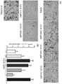
图1为人间充质干细胞鉴定结果图:其中A表示流式细胞术鉴定细胞表面分子;B表示免疫细胞化学染色法鉴定细胞特征标志物;Figure 1 shows the results of identification of human mesenchymal stem cells: A indicates the identification of cell surface molecules by flow cytometry; B indicates the identification of cell characteristic markers by immunocytochemical staining;
图2为GD-A化合物的纯度及结构鉴定图;Fig. 2 is the purity and structure identification diagram of GD-A compound;
图3为GD-A化合物的质谱分析图;Fig. 3 is the mass spectrometry analysis figure of GD-A compound;
图4为GD-A化合物的NMR氢谱图;Fig. 4 is the hydrogen NMR spectrum of GD-A compound;
图5为GD-A化合物的NMR碳谱图;Fig. 5 is the carbon NMR spectrum of GD-A compound;
图6为不同浓度的GD-A对人间充质干细胞的细胞毒性作用分析结果图;Figure 6 is a graph showing the analysis results of the cytotoxic effect of different concentrations of GD-A on human mesenchymal stem cells;
图7为GD-A诱导人间充质干细胞向成骨细胞分化过程成骨细胞标志物碱性磷酸酶的检测结果图:其中A表示成骨细胞标志物碱性磷酸酶的表达;B表示成骨细胞标志物碱性磷酸酶的酶活;Figure 7 is a graph showing the detection results of the osteoblast marker alkaline phosphatase during the differentiation of human mesenchymal stem cells into osteoblasts induced by GD-A: A represents the expression of the osteoblast marker alkaline phosphatase; B represents the osteoblast marker The enzymatic activity of the cellular marker alkaline phosphatase;
图8为GD-A诱导人间充质干细胞向成骨细胞分化过程细胞形态变化图;Fig. 8 is a graph showing changes in cell morphology during the differentiation of human mesenchymal stem cells into osteoblasts induced by GD-A;
图9为GD-A诱导人间充质干细胞形成成骨细胞标志物矿化钙结节的结果图;Figure 9 is a graph showing the results of GD-A inducing human mesenchymal stem cells to form mineralized calcium nodules, an osteoblast marker;
图10为GD-A诱导人间充质干细胞表达特异性成骨相关蛋白的结果图。Figure 10 is a graph showing the results of GD-A-induced expression of specific osteogenesis-related proteins in human mesenchymal stem cells.
具体实施方式Detailed ways
为了更充分的解释本发明的实施,使本发明的目的、技术方案及优点更加清楚明白,以下结合附图及实施例,对本发明的技术方案进行进一步详细说明。应当理解,此处所描述的具体实施例仅用以解释本发明的实施,并不用于限定本发明。In order to more fully explain the implementation of the present invention and make the purpose, technical solutions and advantages of the present invention more clearly understood, the technical solutions of the present invention are further described in detail below with reference to the accompanying drawings and embodiments. It should be understood that the specific embodiments described herein are only used to explain the implementation of the present invention, and are not used to limit the present invention.
实施例1:人间充质干细胞分离培养与鉴定Example 1: Isolation, culture and identification of human mesenchymal stem cells
利用健康孕妇足月剖宫产新鲜胎盘,采用机械法联用两酶(胰蛋白酶和II型胶原酶)消化法从羊膜中分离得到间充质干细胞。用含10%胎牛血清的低糖DMEM培养基重悬细胞,种于T25细胞培养瓶,在37℃,含5% CO2及95%以上空气饱和湿度恒温培养,待细胞融合度达80%以上进行传代培养,第2-5代细胞用于后续实验。当第2代细胞培养到融合度80%时,收集细胞采用流式细胞术鉴定细胞表面分子,该细胞高表达CD90、CD105、CD73、CD44和CD29等细胞间充质细胞表面分子,不表达CD34、CD11b、CD19、CD45和HLA-DR等造血干细胞标志以及MHC II类细胞表面分子,如图1中A所示。免疫细胞化学染色法检测结果提示,该细胞高表达间充质细胞标志物波形蛋白,不表达上皮细胞标志物角蛋白CK19,如图1中B所示。因此该细胞具有典型的间充质细胞表型。Mesenchymal stem cells were isolated from the amniotic membrane using fresh placenta delivered by healthy pregnant women at term cesarean section by mechanical method combined with two enzymes (trypsin and type II collagenase) digestion. Cells were resuspended in low-glucose DMEM medium containing 10% fetal bovine serum, seeded in T25 cell culture flasks, and incubated at 37°C with 5% CO2 and air-saturated humidity above 95%. Subculture, passages 2-5 were used for subsequent experiments. When the second passage cells were cultured to 80% confluence, the cells were collected and identified by flow cytometry. The cells highly expressed CD90, CD105, CD73, CD44 and CD29 and other mesenchymal cell surface molecules, but did not express CD34. , CD11b, CD19, CD45 and HLA-DR and other hematopoietic stem cell markers and MHC class II cell surface molecules, as shown in Figure 1A. The results of immunocytochemical staining showed that the cells highly expressed the mesenchymal cell marker vimentin, but did not express the epithelial cell marker keratin CK19, as shown in B in Figure 1. The cells thus have a typical mesenchymal phenotype.
实施例2:GD-A天然化合物的分离与鉴定Example 2: Isolation and identification of GD-A natural compounds
取干燥的灵芝子实体经超微破壁粉碎,经10倍体积的90-95%甲醇回流提取3次,每次2小时,提取液经减压浓缩干燥后获得浸膏。温水混悬后经乙酸乙酯萃取,乙酸乙酯萃取部位采用硅胶柱层析,用二氯甲烷-甲醇(1:0→0:1)梯度洗脱,获得的洗脱馏分再次硅胶柱层次,用石油醚-乙酸乙酯(1:0→0:1)梯度洗脱,该洗脱馏分经中压液相色谱和反相半制备色谱分离,获得化合物GD-A。该化合物经HPLC分析纯度为95%,如图2所示,其质谱检测结果见图3所示,其分子量为436,分子式为C30H44O2。经NMR氢谱分析如图4所示,其高场区主要结果为δ0.83(3H,s), δ1.13(3H,s),δ0.94(3H,d,J=6.0Hz),δ1.75(3H,s),δ1.13(3H,s),δ1.03(3H,s),δ0.83(3H,s)分别为18、19、21、27、28、29和30位的甲基氢信号,可判断该化合物为四环三萜类;低场区δ6.48(1H,s),δ7.28(1H,m)和δ9.40(1H,s)分别是乙烯基甲基、烯烃基、甲酰基上的氢信号;NMR碳谱分析结果,如图5所示,其低场区为3和26位δ195.4、δ216.8为羰基碳信号,δ142.7、δ139.1、δ144.6、δ155.4、δ117.1、δ120.1分别为7、8、9、11、24、25位的双键碳信号。根据这些主要的波谱学特征并结合文献结果对照,鉴定该化合物为ganoderal A(译成灵芝醛A,简称GD-A),其结构式如通式I所示。The dried Ganoderma lucidum fruit body was crushed by ultra-micro-breaking, and extracted three times by 10 times the volume of 90-95% methanol for 2 hours each time. The extract was concentrated and dried under reduced pressure to obtain the extract. After suspending in warm water, it was extracted with ethyl acetate. The ethyl acetate extracted part was subjected to silica gel column chromatography, eluted with dichloromethane-methanol (1:0→0:1) gradient, and the obtained eluted fraction was again layered on silica gel column. Use petroleum ether-ethyl acetate (1:0→0:1) gradient elution, the eluted fractions are separated by medium pressure liquid chromatography and reversed-phase semi-preparative chromatography to obtain compound GD-A. The compound has a purity of 95% by HPLC analysis, as shown in Figure 2, and its mass spectrometry detection result is shown in Figure 3, its molecular weight is 436, and its molecular formula is C30H44O2. The NMR hydrogen spectrum analysis is shown in Figure 4. The main results in the high field region are δ0.83 (3H,s), δ1.13 (3H,s), δ0.94 (3H,d,J=6.0Hz), δ1.75(3H,s), δ1.13(3H,s), δ1.03(3H,s), δ0.83(3H,s) are 18, 19, 21, 27, 28, 29 and 30 respectively The methyl hydrogen signal at the position can be judged that the compound is a tetracyclic triterpenoid; the low field region δ6.48 (1H,s), δ7.28 (1H,m) and δ9.40 (1H,s) are ethylene Hydrogen signals on methyl, alkene, and formyl; NMR carbon spectrum analysis results, as shown in Figure 5, the low field region is the 3 and 26 positions δ195.4, δ216.8 is the carbonyl carbon signal, δ142.7 , δ139.1, δ144.6, δ155.4, δ117.1, and δ120.1 are the double-bonded carbon signals at

实施例3:GD-A对人间充质干细胞的细胞毒性分析Example 3: Cytotoxicity analysis of GD-A on human mesenchymal stem cells
为了说明采用本发明所述化合物使用安全性问题,即对人间充质干细胞是否安全,进行相应测试:In order to illustrate the safety of using the compounds of the present invention, that is, whether it is safe for human mesenchymal stem cells, corresponding tests are carried out:
采用MTT法,测定不同剂量(0.001μM, 0.01μM, 0.1μM, 1.0μM, 10 μM和100 μM)GD-A,分别连续作用人间充质干细胞24小时、48小时和72小时。结果如图6所示,与对照组相比,GD-A作用24h、48h和72h后,在0.001μM-10μM剂量范围内,GD-A对人间充质干细胞无细胞毒性,甚至呈促进细胞增殖作用,尤其是作用24时呈明显促细胞增殖作用。但GD-A在100μM剂量时,三个时间点均呈抑制细胞增殖作用,有较明显的细胞毒性。Using MTT method, different doses (0.001μM, 0.01μM, 0.1μM, 1.0μM, 10 μM and 100 μM) of GD-A were determined to continuously act on human mesenchymal stem cells for 24 hours, 48 hours and 72 hours, respectively. The results are shown in Figure 6. Compared with the control group, after 24h, 48h and 72h of GD-A treatment, GD-A has no cytotoxicity to human mesenchymal stem cells in the dose range of 0.001μM-10μM, and even promotes cell proliferation. The effect, especially the effect of 24, showed a significant effect of promoting cell proliferation. However, at the dose of 100 μM, GD-A inhibited cell proliferation at all three time points, and had obvious cytotoxicity.
实施例4:GD-A诱导人间充质干细胞高表达碱性磷酸酶Example 4: GD-A induces high expression of alkaline phosphatase in human mesenchymal stem cells
为了说明采用本发明所述化合物用于诱导人间充质干细胞在成骨细胞中的情况,进行相应测试:In order to illustrate the use of the compounds of the present invention for inducing human mesenchymal stem cells in osteoblasts, the corresponding tests were carried out:
用不同安全剂量(0.001μM, 0.01μM, 0.1μM, 1.0μM和10 μM)GD-A添加到人间充质干细胞中。在加药后第5天采用BCIP/NBT染色法测定成骨细胞的标志物碱性磷酸酶。如图7中A所示,在剂量0.001μM~0.1μM范围内,有较多紫色阳性细胞,尤其是0.01μM剂量的效果最好。进一步的碱性磷酸酶酶活力测定结果提示,其表达情况与酶活的结果基本一致,如见图7中B所示,浓度为0.01μM剂量时GD-A的诱导分化效果最佳。GD-A was added to human mesenchymal stem cells with different safe doses (0.001 μM, 0.01 μM, 0.1 μM, 1.0 μM and 10 μM). Alkaline phosphatase, a marker of osteoblasts, was determined by BCIP/NBT staining on the 5th day after dosing. As shown in A in Figure 7, within the dose range of 0.001 μM to 0.1 μM, there are more purple-positive cells, especially the 0.01 μM dose has the best effect. Further alkaline phosphatase enzyme activity assay results indicated that its expression was basically consistent with the enzyme activity results. As shown in Figure 7, B, GD-A had the best induction and differentiation effect at a concentration of 0.01 μM.
实施例5:GD-A诱导人间充质干细胞生成矿化钙结节Example 5: GD-A induces the formation of mineralized calcium nodules in human mesenchymal stem cells
为了说明采用本发明所述化合物用于诱导人间充质干细胞在成骨细胞中的情况,进行相应测试:In order to illustrate the use of the compounds of the present invention for inducing human mesenchymal stem cells in osteoblasts, the corresponding tests were carried out:
用不同剂量GD-A(0.001μM,0.01μM,0.1μM,1.0μM和10μM)添加到人间充质干细胞中,在加药后第17天,采用茜素红染色,检测成骨细胞的另一标志特征形成钙化结节。如图8所示,对照组染色前的细胞仍然呈梭形,细胞没有形成不透明矿化钙结节,而阳性对照组及GD-A给药组染色前细胞已变成卵圆形到多边形,细胞汇合成鹅卵石状,形成了不透明的矿化钙结节。最后采用茜素红染色的结果如图9所示,并与类似常规成骨诱导阳性对照组,GD-A给药组形成了数量不等的红色矿化钙结节,尤其是0.01μM剂量GD-A组,有大量红色的矿化钙化结节。Different doses of GD-A (0.001 μM, 0.01 μM, 0.1 μM, 1.0 μM and 10 μM) were added to human mesenchymal stem cells. On the 17th day after dosing, alizarin red staining was used to detect another increase in osteoblasts. The hallmark feature is the formation of calcified nodules. As shown in Figure 8, the cells in the control group were still spindle-shaped before staining, and the cells did not form opaque mineralized calcium nodules, while the cells in the positive control group and GD-A administration group had become oval to polygonal before staining. The cells coalesced into cobblestones, forming opaque nodules of mineralized calcium. Finally, the results of alizarin red staining are shown in Figure 9. Similar to the positive control group of conventional osteogenic induction, the GD-A administration group formed different numbers of red mineralized calcium nodules, especially the 0.01 μM dose of GD. - Group A with numerous red mineralized calcified nodules.
实施例6:GD-A诱导人间充质干细胞表达特异性成骨相关蛋白Example 6: GD-A induces human mesenchymal stem cells to express specific osteogenesis-related proteins
为了说明采用本发明所述化合物用于诱导人间充质干细胞在成骨细胞中的情况,进行相应测试:In order to illustrate the use of the compounds of the present invention for inducing human mesenchymal stem cells in osteoblasts, the corresponding tests were carried out:
用最佳剂量0.01μM GD-A添加到人间充质干细胞中,在加药后第5天和第17天,采用Western blotting检测特异性成骨相关蛋白的表达情况。结果如图10所示。与对照组相比,GD-A类似成骨阳性诱导组,能显著促进OPN、RUNX2、OSX、ALP和CoL1α1等特异性成骨相关蛋白的表达。The optimal dose of 0.01 μM GD-A was added to human mesenchymal stem cells, and the expression of specific osteogenesis-related proteins was detected by Western blotting on the 5th and 17th days after dosing. The results are shown in Figure 10. Compared with the control group, GD-A was similar to the osteogenesis-positive induction group and could significantly promote the expression of specific osteogenesis-related proteins such as OPN, RUNX2, OSX, ALP and CoL1α1.
实施例7. GD-A诱导人间充质干细胞向成骨细胞分化的用途Example 7. Use of GD-A to induce differentiation of human mesenchymal stem cells into osteoblasts
人间充质干细胞在体外用GD-A诱导向成骨细胞分化,可作为骨组织工程和细胞移植的种子细胞的用途。对骨病损及骨代谢性疾病均有防治作用。采用本发明所述化合物GD-A用于诱导分化人间充质干细胞作为种子细胞替代成骨细胞,可避免成骨细胞体外较难获取和高免疫原性的问题。Human mesenchymal stem cells can be induced to differentiate into osteoblasts with GD-A in vitro, which can be used as seed cells for bone tissue engineering and cell transplantation. It has preventive and therapeutic effects on bone lesions and bone metabolic diseases. Using the compound GD-A of the present invention to induce differentiation of human mesenchymal stem cells as seed cells to replace osteoblasts can avoid the problems of difficulty in obtaining osteoblasts in vitro and high immunogenicity.
以上所述实施例仅表达了本发明的具体实施方式,但并不能因此而理解为对本发明专利范围的限制。应当指出的是,对于本领域的普通技术人员来说,在不脱离本发明构思的前提下,还可以做出若干变形和改进,这些都属于本发明的保护范围。The above-mentioned embodiments only represent specific embodiments of the present invention, but should not be construed as limiting the scope of the present invention. It should be pointed out that for those skilled in the art, without departing from the concept of the present invention, several modifications and improvements can be made, which all belong to the protection scope of the present invention.
Claims (3)
Translated fromChinese
Priority Applications (1)
| Application Number | Priority Date | Filing Date | Title |
|---|---|---|---|
| CN201910776470.XACN110408590B (en) | 2019-09-06 | 2019-09-06 | Method for inducing differentiation of human mesenchymal stem cells into osteoblasts and application thereof |
Applications Claiming Priority (1)
| Application Number | Priority Date | Filing Date | Title |
|---|---|---|---|
| CN201910776470.XACN110408590B (en) | 2019-09-06 | 2019-09-06 | Method for inducing differentiation of human mesenchymal stem cells into osteoblasts and application thereof |
Publications (2)
| Publication Number | Publication Date |
|---|---|
| CN110408590A CN110408590A (en) | 2019-11-05 |
| CN110408590Btrue CN110408590B (en) | 2022-10-25 |
Family
ID=68368480
Family Applications (1)
| Application Number | Title | Priority Date | Filing Date |
|---|---|---|---|
| CN201910776470.XAActiveCN110408590B (en) | 2019-09-06 | 2019-09-06 | Method for inducing differentiation of human mesenchymal stem cells into osteoblasts and application thereof |
Country Status (1)
| Country | Link |
|---|---|
| CN (1) | CN110408590B (en) |
Citations (8)
| Publication number | Priority date | Publication date | Assignee | Title |
|---|---|---|---|---|
| WO2007025306A1 (en)* | 2005-08-26 | 2007-03-01 | University Of Rochester | Transplantation of glial restricted precursor-derived astrocytes for promotion of axon growth |
| CN101371857A (en)* | 2007-08-24 | 2009-02-25 | 林华型 | Composite medicament for inhibiting tumor and preparation method thereof as well as technique field |
| CN101679951A (en)* | 2007-04-17 | 2010-03-24 | 罗切斯特大学 | Telencephalic glial-restricted cell populations and related compositions and methods |
| EP2544007A1 (en)* | 2011-07-07 | 2013-01-09 | Atlas Antibodies AB | Biomarker of renal impairment |
| AU2010359001A1 (en)* | 2010-08-10 | 2013-03-14 | Abbvie Inc. | Novel TRPV3 modulators |
| CN106497871A (en)* | 2016-11-01 | 2017-03-15 | 遵义医学院附属医院 | A kind of method for improving human amnion mesenchymal stem cell Osteoblast Differentiation efficiency and application |
| CN108181396A (en)* | 2017-12-29 | 2018-06-19 | 上海市农业科学院 | The detection method of 17 kinds of triterpenoid contents in a kind of ganoderma lucidum |
| CN110169959A (en)* | 2019-05-20 | 2019-08-27 | 中国人民解放军总医院 | Growth factor slow-release microballoon, tissue engineering bone/cartilage compound rest and preparation method |
Family Cites Families (2)
| Publication number | Priority date | Publication date | Assignee | Title |
|---|---|---|---|---|
| KR20090115142A (en)* | 2007-01-30 | 2009-11-04 | 유니버시티 오브 조지아 리서치 파운데이션, 인코포레이티드 | Early mesoderm cells, a stable population of mesendoderm cells that has utility for generation of endoderm and mesoderm lineages and multipotent migratory cells(mmc) |
| US8728730B2 (en)* | 2009-09-03 | 2014-05-20 | Genentech, Inc. | Methods for treating, diagnosing, and monitoring rheumatoid arthritis |
- 2019
- 2019-09-06CNCN201910776470.XApatent/CN110408590B/enactiveActive
Patent Citations (8)
| Publication number | Priority date | Publication date | Assignee | Title |
|---|---|---|---|---|
| WO2007025306A1 (en)* | 2005-08-26 | 2007-03-01 | University Of Rochester | Transplantation of glial restricted precursor-derived astrocytes for promotion of axon growth |
| CN101679951A (en)* | 2007-04-17 | 2010-03-24 | 罗切斯特大学 | Telencephalic glial-restricted cell populations and related compositions and methods |
| CN101371857A (en)* | 2007-08-24 | 2009-02-25 | 林华型 | Composite medicament for inhibiting tumor and preparation method thereof as well as technique field |
| AU2010359001A1 (en)* | 2010-08-10 | 2013-03-14 | Abbvie Inc. | Novel TRPV3 modulators |
| EP2544007A1 (en)* | 2011-07-07 | 2013-01-09 | Atlas Antibodies AB | Biomarker of renal impairment |
| CN106497871A (en)* | 2016-11-01 | 2017-03-15 | 遵义医学院附属医院 | A kind of method for improving human amnion mesenchymal stem cell Osteoblast Differentiation efficiency and application |
| CN108181396A (en)* | 2017-12-29 | 2018-06-19 | 上海市农业科学院 | The detection method of 17 kinds of triterpenoid contents in a kind of ganoderma lucidum |
| CN110169959A (en)* | 2019-05-20 | 2019-08-27 | 中国人民解放军总医院 | Growth factor slow-release microballoon, tissue engineering bone/cartilage compound rest and preparation method |
Non-Patent Citations (7)
| Title |
|---|
| Ganoderal A effectively induces osteogenic differention of human amniotic messenchymal stem cells via cross-talk between Wnt/β-catenin and BMP/SMAD signalig pathways;Yi-Qing Wang等;《Biomed Pharmacother》;20191230;全文* |
| Information entropy-based classification of triterpenoids and steroids from Ganoderma;Gloria Castellano等;《Phytochemistry》;20150526;全文* |
| The interplay of T-1 and T-2-relaxation on T-1 weighted MRI of hMSCs induced by GD-DOTA-peptides;Cao LM等;《Biomaterials》;20140401;第35卷(第13期);全文* |
| 含氟羟基磷灰石涂层体外细胞相容性研究;赵彬等;《中国生物医学工程学报》;20051030(第05期);全文* |
| 天然小分子化合物诱导间充质干细胞向成骨分化的研究进展;王怡晴等;《中草药》;20190527(第11期);全文* |
| 牙源性干细胞及其在再生医学中的应用进展;姜杨杨等;《医学综述》;20180620(第12期);全文* |
| 骨髓间充质干细胞经不同时间成骨细胞诱导后细胞性质比较;郭若霖等;《基础医学与临床》;20060620(第09期);全文* |
Also Published As
| Publication number | Publication date |
|---|---|
| CN110408590A (en) | 2019-11-05 |
Similar Documents
| Publication | Publication Date | Title |
|---|---|---|
| EP3189828B1 (en) | Composition for differentiation induction of adipocyte containing stem cell-derived exosome, regeneration of adipose tissue, and skin whitening or wrinkle improvement | |
| Hu et al. | Human umbilical cord mesenchymal stem cell-derived exosomes suppress dermal fibroblasts-myofibroblats transition via inhibiting the TGF-β1/Smad 2/3 signaling pathway | |
| CN105861430A (en) | Exosome, preparing method of exosome and application of exosome in preparing medicine or preparation for treating sepsis | |
| US20250249062A1 (en) | Use of dipsaci radix-derived extracellular vesicle-like nanoparticles in preparation of drug for preventing or treating orthopedic diseases | |
| CN109136174B (en) | Stem cell-derived exosome preparation for delaying senescence | |
| CN113274509B (en) | Polypeptide drug nano-targeting drug delivery system HTPP-Exo-M1-8 and preparation method and application thereof | |
| CN113122497B (en) | Engineered mitochondria and preparation method thereof | |
| CN112402413B (en) | Application of lindley eupatorium herb sesquiterpene lactone B in preparation of anti-liver cancer medicine and anti-liver cancer medicine | |
| CN118526530A (en) | Application of melastoma dodecandrum and water extract and active ingredients thereof in preparation of osteoporosis prevention and treatment medicine | |
| CN113274411A (en) | Application of genetically modified bone marrow mesenchymal stem cell-derived microvesicles in preparation of medicines for treating renal injury | |
| CN110408590B (en) | Method for inducing differentiation of human mesenchymal stem cells into osteoblasts and application thereof | |
| Yang et al. | Myosin heavy chain-derived peptide of Gadus morhua promotes proliferation and differentiation in osteoblasts and bone formation and maintains bone homeostasis in ovariectomized mice | |
| Rosenbloom et al. | Hydroxylation of proline after the release of proline-rich polypeptides from ribosomal complexes during uninhibited collagen biosynthesis | |
| CN113430165B (en) | THC-loaded stem cell drug delivery system and preparation method thereof | |
| Zhu et al. | Human Umbilical Cord Mesenchymal Stem Cells' Cultivation and Treatment of Liver Diseases | |
| Chin et al. | 2, 3, 5, 4′-tetrahydroxystilbene-2-O-β-D-glucoside-stimulated dental pulp stem cells-derived conditioned medium enhances cell activity and anti-inflammation | |
| CN110257333A (en) | A kind of neural stem cell microcapsule bubble and its application | |
| WO2022193398A1 (en) | Trivalent dimethylarsinous acid induced human keratinocyte malignant transformation cell strain, and use thereof | |
| KR102563616B1 (en) | Soluble extracellular matrix derived from stem cells and preparing method thereof | |
| CN102199081A (en) | 2-acetylaloeemodin and preparation method and application thereof | |
| CN109679892B (en) | Application of baicalin in promoting in-vitro amplification of breast stem cells and maintaining characteristics of stem cells of breast stem cells | |
| CN110330542B (en) | Compound for delaying human mesenchymal stem cell aging and preparation method and application thereof | |
| Mesbah et al. | Studying the suppressing effect of mesenchymal stem cells derived from amniotic membrane on colorectal cancer | |
| CN101273983B (en) | Use of neurocyte differentiated from stem cell induced by isobavachin | |
| CN116444598B (en) | Ganoderic acid A derivative, its synthesis method and application in treating tumor |
Legal Events
| Date | Code | Title | Description |
|---|---|---|---|
| PB01 | Publication | ||
| PB01 | Publication | ||
| SE01 | Entry into force of request for substantive examination | ||
| SE01 | Entry into force of request for substantive examination | ||
| GR01 | Patent grant | ||
| GR01 | Patent grant |