CN110292583B - Application of fullerol and composition thereof in preparation of antithrombotic drugs - Google Patents
Application of fullerol and composition thereof in preparation of antithrombotic drugsDownload PDFInfo
- Publication number
- CN110292583B CN110292583BCN201910570879.6ACN201910570879ACN110292583BCN 110292583 BCN110292583 BCN 110292583BCN 201910570879 ACN201910570879 ACN 201910570879ACN 110292583 BCN110292583 BCN 110292583B
- Authority
- CN
- China
- Prior art keywords
- fullerol
- composition
- thrombus
- inhibiting
- mesoporous silicon
- Prior art date
- Legal status (The legal status is an assumption and is not a legal conclusion. Google has not performed a legal analysis and makes no representation as to the accuracy of the status listed.)
- Expired - Fee Related
Links
Images


Classifications
- A—HUMAN NECESSITIES
- A61—MEDICAL OR VETERINARY SCIENCE; HYGIENE
- A61K—PREPARATIONS FOR MEDICAL, DENTAL OR TOILETRY PURPOSES
- A61K33/00—Medicinal preparations containing inorganic active ingredients
- A61K33/44—Elemental carbon, e.g. charcoal, carbon black
- A—HUMAN NECESSITIES
- A61—MEDICAL OR VETERINARY SCIENCE; HYGIENE
- A61K—PREPARATIONS FOR MEDICAL, DENTAL OR TOILETRY PURPOSES
- A61K47/00—Medicinal preparations characterised by the non-active ingredients used, e.g. carriers or inert additives; Targeting or modifying agents chemically bound to the active ingredient
- A61K47/50—Medicinal preparations characterised by the non-active ingredients used, e.g. carriers or inert additives; Targeting or modifying agents chemically bound to the active ingredient the non-active ingredient being chemically bound to the active ingredient, e.g. polymer-drug conjugates
- A61K47/69—Medicinal preparations characterised by the non-active ingredients used, e.g. carriers or inert additives; Targeting or modifying agents chemically bound to the active ingredient the non-active ingredient being chemically bound to the active ingredient, e.g. polymer-drug conjugates the conjugate being characterised by physical or galenical forms, e.g. emulsion, particle, inclusion complex, stent or kit
- A61K47/6921—Medicinal preparations characterised by the non-active ingredients used, e.g. carriers or inert additives; Targeting or modifying agents chemically bound to the active ingredient the non-active ingredient being chemically bound to the active ingredient, e.g. polymer-drug conjugates the conjugate being characterised by physical or galenical forms, e.g. emulsion, particle, inclusion complex, stent or kit the form being a particulate, a powder, an adsorbate, a bead or a sphere
- A61K47/6923—Medicinal preparations characterised by the non-active ingredients used, e.g. carriers or inert additives; Targeting or modifying agents chemically bound to the active ingredient the non-active ingredient being chemically bound to the active ingredient, e.g. polymer-drug conjugates the conjugate being characterised by physical or galenical forms, e.g. emulsion, particle, inclusion complex, stent or kit the form being a particulate, a powder, an adsorbate, a bead or a sphere the form being an inorganic particle, e.g. ceramic particles, silica particles, ferrite or synsorb
- A—HUMAN NECESSITIES
- A61—MEDICAL OR VETERINARY SCIENCE; HYGIENE
- A61K—PREPARATIONS FOR MEDICAL, DENTAL OR TOILETRY PURPOSES
- A61K9/00—Medicinal preparations characterised by special physical form
- A61K9/48—Preparations in capsules, e.g. of gelatin, of chocolate
- A61K9/50—Microcapsules having a gas, liquid or semi-solid filling; Solid microparticles or pellets surrounded by a distinct coating layer, e.g. coated microspheres, coated drug crystals
- A61K9/5005—Wall or coating material
- A61K9/5063—Compounds of unknown constitution, e.g. material from plants or animals
- A61K9/5068—Cell membranes or bacterial membranes enclosing drugs
- A—HUMAN NECESSITIES
- A61—MEDICAL OR VETERINARY SCIENCE; HYGIENE
- A61P—SPECIFIC THERAPEUTIC ACTIVITY OF CHEMICAL COMPOUNDS OR MEDICINAL PREPARATIONS
- A61P7/00—Drugs for disorders of the blood or the extracellular fluid
- A61P7/02—Antithrombotic agents; Anticoagulants; Platelet aggregation inhibitors
- A—HUMAN NECESSITIES
- A61—MEDICAL OR VETERINARY SCIENCE; HYGIENE
- A61P—SPECIFIC THERAPEUTIC ACTIVITY OF CHEMICAL COMPOUNDS OR MEDICINAL PREPARATIONS
- A61P9/00—Drugs for disorders of the cardiovascular system
- A61P9/10—Drugs for disorders of the cardiovascular system for treating ischaemic or atherosclerotic diseases, e.g. antianginal drugs, coronary vasodilators, drugs for myocardial infarction, retinopathy, cerebrovascula insufficiency, renal arteriosclerosis
Landscapes
- Health & Medical Sciences (AREA)
- Life Sciences & Earth Sciences (AREA)
- Engineering & Computer Science (AREA)
- Chemical & Material Sciences (AREA)
- Bioinformatics & Cheminformatics (AREA)
- Pharmacology & Pharmacy (AREA)
- Animal Behavior & Ethology (AREA)
- General Health & Medical Sciences (AREA)
- Public Health (AREA)
- Veterinary Medicine (AREA)
- Medicinal Chemistry (AREA)
- Epidemiology (AREA)
- Chemical Kinetics & Catalysis (AREA)
- Organic Chemistry (AREA)
- Inorganic Chemistry (AREA)
- Nuclear Medicine, Radiotherapy & Molecular Imaging (AREA)
- General Chemical & Material Sciences (AREA)
- Vascular Medicine (AREA)
- Heart & Thoracic Surgery (AREA)
- Cardiology (AREA)
- Urology & Nephrology (AREA)
- Biophysics (AREA)
- Cell Biology (AREA)
- Molecular Biology (AREA)
- Virology (AREA)
- Botany (AREA)
- Zoology (AREA)
- Ceramic Engineering (AREA)
- Diabetes (AREA)
- Hematology (AREA)
- Pharmaceuticals Containing Other Organic And Inorganic Compounds (AREA)
Abstract
Description
Translated fromChinese技术领域technical field
本发明涉及药物的应用技术领域,具体涉及一种富勒醇及其组合物在制备抗血栓药物中的应用。The invention relates to the technical field of drug application, in particular to the application of a fullerol and a composition thereof in the preparation of antithrombotic drugs.
背景技术Background technique
当病理学进程已经无法由止血调控机制进行调控时,大量的凝血酶将会产生,从而引发血栓的形成。血栓形成是心血管疾病产生的重要因素,例如心肌梗死和中风等动脉疾病和静脉血栓栓塞紊乱,血栓的形成导致高发病率和死亡率。除此之外,静脉血栓症是导致癌症病人死亡的主要原因之一。When the pathological process can no longer be regulated by the hemostatic regulatory mechanism, a large amount of thrombin will be produced, thereby triggering the formation of thrombus. Thrombosis is an important factor in the development of cardiovascular diseases, such as arterial diseases such as myocardial infarction and stroke, and venous thromboembolic disorders, which lead to high morbidity and mortality. In addition, venous thrombosis is one of the leading causes of death in cancer patients.
血栓的形成和多种因素有关,包括血小板、纤维蛋白、胶原、组织因子和凝血酶等。当血管壁受损或内皮细胞被破坏,胶原和组织因子暴露在流动的血液中,从而启动了血栓的形成。暴露的胶原引发了血小板的激活和聚集,同样的,组织因子的暴露导致了凝血酶的产生。凝血酶不止催化纤维蛋白原转化为纤维蛋白,同时也可以活化血小板。The formation of thrombus is related to a variety of factors, including platelets, fibrin, collagen, tissue factor and thrombin. When vessel walls are damaged or endothelial cells are destroyed, collagen and tissue factor are exposed to flowing blood, which initiates thrombus formation. Exposure of collagen triggers platelet activation and aggregation, and similarly, tissue factor exposure leads to thrombin production. Thrombin not only catalyzes the conversion of fibrinogen to fibrin, but also activates platelets.
血小板激活可以由两种独立的方式进行。研究人员从小鼠身上发现了有两种独立的方式可以分别激活血小板。一种方式为内皮下的胶原暴露启动了血小板的激活;另一种方式为血管壁或流动的血液中所包含的组织因子产生的凝血酶导致了血小板的活化。这两种方式谁占主导作用取决于血小板激活是因为受伤还是因为疾病,但无论是哪种方式占主导,最终导致的结果是相同的。Platelet activation can proceed in two independent ways. In mice, the researchers found two independent ways to activate platelets separately. One way is that collagen exposure under the endothelium initiates platelet activation; another way is that thrombin generated by tissue factor contained in the vessel wall or in flowing blood results in platelet activation. Which of the two methods dominates depends on whether platelet activation is due to injury or disease, but whichever mode dominates, the end result is the same.
尚未成熟的血栓会在后续招募未受刺激的血小板,但是并不是所有招募过来的血小板最终都会形成血栓,一部分血小板还会脱离血栓部位。简而言之,血栓的形成是一种动态的过程,在这个过程中,一些血小板会粘附在血栓部位,而另一些血小板则从血栓部位分离。血栓凝结块的组成或结构很大程度上取决于剪切力、流动性、波动和循环中血小板的数量等因素。The immature thrombus will subsequently recruit unstimulated platelets, but not all the recruited platelets will eventually form a thrombus, and some platelets will also break away from the thrombus site. In short, thrombus formation is a dynamic process in which some platelets adhere to the thrombus site while others detach from the thrombus site. The composition or structure of a thrombus clot is largely dependent on factors such as shear force, fluidity, fluctuation, and the number of circulating platelets.
急性炎症和感染、内毒素血症和败血症等会导致血液形成高凝状态。当调节机制已经无法进行调控时,急性弥散性血管内凝血继而发生,伴随着大量消耗凝血相关蛋白和血小板,最终导致出血的情况。而病人处于慢性弥散性血管内凝血状态时,相比于出血,血栓的形成情况更为严峻。血栓形成和炎症反应互相关联,并相互加强。Acute inflammation and infection, endotoxemia and sepsis can lead to hypercoagulable state of blood. When the regulatory mechanism is out of control, acute disseminated intravascular coagulation ensues, accompanied by massive depletion of coagulation-related proteins and platelets, eventually leading to a bleeding condition. In patients with chronic disseminated intravascular coagulation, thrombosis is more severe than bleeding. Thrombosis and inflammatory responses are interconnected and reinforce each other.
组织因子可以在体内循环的携带组织因子的微颗粒、单核细胞和活化的内皮细胞上进行表达。慢性弥散性血管内凝血导致血栓形成的主要原因是内源性凝血途径的破坏。在正常血液中无法检测出组织因子的活性,然而在健康的人体内存在携带组织因子的微颗粒。微颗粒携带的组织因子在被招募到血管损伤位点时会被激活。处于病态的微颗粒可以携带活化的组织因子,可能会导致血栓栓塞的发生。肿瘤细胞或炎症细胞产生的携带组织因子的微颗粒可以导致血栓的发生,携带活化的组织因子的微颗粒可以被用作血栓形成风险增加的生物标志物。癌症增加血栓风险的原因可能包括以下几点:肿瘤部位的组织因子激活了凝血途径;半胱氨酸蛋白酶激活了凝血因子X;黏性糖蛋白的产生;MET致癌基因的激活以及肿瘤衍生的携带组织因子的微颗粒。Tissue factor can be expressed on circulating tissue factor-bearing microparticles, monocytes, and activated endothelial cells. The main cause of thrombus formation in chronic disseminated intravascular coagulation is the disruption of the intrinsic coagulation pathway. Tissue factor activity cannot be detected in normal blood, however, tissue factor-carrying microparticles exist in healthy humans. Tissue factor carried by the microparticles is activated when recruited to the site of vascular injury. Sick microparticles can carry activated tissue factor, which may lead to thromboembolism. Tissue factor-carrying microparticles produced by tumor cells or inflammatory cells can lead to thrombosis, and activated tissue factor-carrying microparticles can be used as biomarkers of increased thrombosis risk. The reasons why cancer increases the risk of thrombosis may include the following: activation of the coagulation pathway by tissue factor at the tumor site; activation of coagulation factor X by caspases; production of viscous glycoprotein; activation of the MET oncogene and tumor-derived carrier Microparticles of tissue factor.
如果动脉粥样硬化没有导致血栓形成(急性冠状动脉疾病的主要致病过程),那么动脉粥样硬化将属于一种慢性病,该病与血管狭窄病变导致靶器官血流减少有关,不会导致高死亡率。冠状动脉壁上长期的动脉粥样硬化损伤具有扩散的趋势,并且该趋势与斑块是否具有梗阻能力相关。目前的假设是,斑块纤维帽的破裂将细胞外基质中的胶原蛋白暴露于血液,从而引发血栓形成,该步骤先于含脂质巨噬细胞包含的组织因子暴露,或二者同时发生。组织因子是动脉粥样化的组成部分,并且在冠状动脉血栓形成过程中扮演重要角色。在冠状动脉损伤动物模型中,组织因子抑制剂的使用有效减小了血栓的尺寸。If atherosclerosis does not lead to thrombosis (the main pathogenic process of acute coronary artery disease), then atherosclerosis is a chronic disease that is associated with reduced blood flow to target organs due to stenotic lesions and does not lead to high mortality rate. Long-term atherosclerotic lesions in the coronary walls have a tendency to spread, and this tendency correlates with whether the plaque has the ability to block. The current hypothesis is that rupture of the plaque fibrous cap exposes collagen in the extracellular matrix to the blood, thereby triggering thrombus formation, either prior to exposure to tissue factor contained in lipid-containing macrophages, or both. Tissue factor is a component of atherosclerosis and plays an important role in coronary thrombosis. In animal models of coronary injury, the use of tissue factor inhibitors effectively reduced thrombus size.
新型的药物制剂在治疗和预防血栓类疾病方面具有替代华法林、肝素以及低分子量肝素等临床常用凝血抑制剂的潜在趋势,最前沿的策略是直接抑制FXa或凝血酶。如专利申请号申请号:03129363.8公开了一种凝血酶止血栓剂,其是由凝血酶和基质组成,基质选自混合脂肪酸甘油酯类油脂性基质或聚乙二醇类亲水性基质,制备方法采用冷压法或热熔法,能起到快速止血的目的。这些新药相比于传统的凝血抑制剂提高了方便性、安全性,并具有等效或更加高效的特点。然而,这些抑制剂的靶点与肝素或华法林的靶点相同,可能会导致止血过程受阻,从而在抑制血栓形成(即同抗栓、抗血栓、抗血栓形成)时引发出血。理想的抗栓药物应该只抑制血栓却不影响止血过程。血栓形成的机制在不同情境下并不完全相同。发展用来阻止与特定疾病相关血栓形成的病理学制剂,应该考虑不同影响机制的变化。Novel pharmaceutical preparations have the potential to replace common clinical coagulation inhibitors such as warfarin, heparin and low molecular weight heparin in the treatment and prevention of thrombotic diseases. The most cutting-edge strategy is to directly inhibit FXa or thrombin. For example, Patent Application No. Application No.: 03129363.8 discloses a thrombin hemostatic agent, which is composed of thrombin and a matrix, and the matrix is selected from mixed fatty acid glyceride lipid matrix or polyethylene glycol hydrophilic matrix, preparation method The cold pressing method or the hot melting method can achieve the purpose of rapid hemostasis. Compared with traditional coagulation inhibitors, these new drugs have improved convenience and safety, and have equivalent or more efficient characteristics. However, these inhibitors, which target the same targets as heparin or warfarin, may cause blockage of the hemostatic process, which can lead to bleeding when inhibiting thrombosis (ie, antithrombotic, antithrombotic, antithrombotic). The ideal antithrombotic drug should only inhibit the thrombus without affecting the hemostasis process. The mechanisms of thrombosis are not identical in different contexts. The development of pathological agents to prevent thrombus formation associated with specific diseases should take into account variations in the different mechanisms of influence.
凝血系统作为人体中最重要的防护机制之一,是维持体内稳态,保证体内不受外界干扰的重要屏障。当把纳米材料通过静脉给药进入体内时,势必要经过血液循环系统,而研究纳米材料是否对动物体内的凝血系统产生影响则成为评价其安全性的首要任务。富勒醇纳米颗粒是具有光明前景的富勒烯纳米材料的衍生物。相比于富勒烯纳米材料,富勒醇纳米颗粒的水溶性更好,生物相容性更强,从而具有在生物体内进行广泛应用的潜力。因此,研究富勒醇对凝血系统的影响将为富勒醇的体内应用提供指导意义,同时也为发展纳米结构抗血栓药物提供重要的线索。溶栓与抗栓作为血栓类疾病的主要防治方法,其中,抗栓即从血液凝固的源头下手,利用药物抑制血栓的形成,而溶栓则是在血栓形成后,利用药物使血栓溶解,在医疗上作为多种血栓栓塞疾病的治疗。As one of the most important protective mechanisms in the human body, the coagulation system is an important barrier to maintain homeostasis and ensure that the body is not disturbed by the outside world. When nanomaterials are administered intravenously into the body, they must pass through the blood circulatory system, and the study of whether nanomaterials have an effect on the coagulation system in animals has become the primary task of evaluating their safety. Fullerol nanoparticles are promising derivatives of fullerene nanomaterials. Compared with fullerene nanomaterials, fullerol nanoparticles have better water solubility and stronger biocompatibility, so they have the potential to be widely used in vivo. Therefore, studying the effect of fullerol on the coagulation system will provide guidance for the in vivo application of fullerol, and also provide important clues for the development of nanostructured antithrombotic drugs. Thrombolysis and antithrombosis are the main prevention and treatment methods for thrombotic diseases. Among them, antithrombotic starts from the source of blood coagulation and uses drugs to inhibit the formation of thrombus, while thrombolysis is to use drugs to dissolve thrombus after thrombus formation. Medically, it is used as the treatment of various thromboembolic diseases.
目前现有技术中的溶栓(即同溶解血栓)药物在治疗血栓中已经取得了显著的成绩,但是仍然存在:半衰期短、易引起出血且对陈旧血栓溶解效果差等一系列问题。理想的溶栓药物应该具有安全、有效、给药方便、特异性强、半衰期长、能溶解陈旧血栓、复发率低、无出血副反应等特点。因此,寻找新型安全溶栓药物具有重要的意义。At present, the thrombolytic drugs in the prior art (that is, the same thrombolytic drugs) have achieved remarkable results in the treatment of thrombus, but there are still a series of problems: short half-life, easy to cause bleeding and poor dissolution effect on old thrombus. An ideal thrombolytic drug should be safe, effective, convenient for administration, strong in specificity, long in half-life, capable of dissolving old thrombus, low in recurrence rate, and free of bleeding side effects. Therefore, it is of great significance to find new safe thrombolytic drugs.
发明内容SUMMARY OF THE INVENTION
为了解决上述现有技术存在的问题,本发明提供了一种富勒醇及其组合物在制备抗血栓药物中的应用,研究发现富勒醇具有明显的溶栓、抗栓效果,通过生物膜载带富勒醇纳米药物能够增强血栓的靶向性和富集程度,起到良好的溶栓效果。In order to solve the above-mentioned problems in the prior art, the present invention provides the application of fullerol and its composition in the preparation of antithrombotic drugs. It is found that fullerol has obvious thrombolytic and antithrombotic effects. The nano-drugs loaded with fullerol can enhance the targeting and enrichment degree of thrombus, and play a good thrombolytic effect.
本发明的任务之一在于提供富勒醇C60(OH)X在制备溶解血栓和/或抑制血栓形成药物中的应用,其中,10≤X<40。One of the tasks of the present invention is to provide the application of fullerol C60 (OH)X in the preparation of thrombus-dissolving and/or thrombus-inhibiting drugs, wherein 10≦X<40.
所述药物包括富勒醇和/或介孔硅和/或细胞膜上述的富勒醇,是一个由C原子构成的纳米碳笼,其表面存在许多羟基基团,具有很好的生物亲和性;同时,由于羟基基团的存在,毒性被大大降低。The drug includes fullerol and/or mesoporous silicon and/or the above-mentioned fullerol of cell membrane, which is a nano-carbon cage composed of C atoms, and there are many hydroxyl groups on its surface, which has good biological affinity; At the same time, the toxicity is greatly reduced due to the presence of hydroxyl groups.
由于相邻羟基的重排等原因,碳笼上O的数目与H的数目会有一些差异,因此,也可以将上述通式写成C60(O)x(H)y形式。其中X≠Y,10≤X或Y<40。Due to the rearrangement of adjacent hydroxyl groups and other reasons, the number of O and H on the carbon cage will be somewhat different. Therefore, the above general formula can also be written in the form of C60(O)x (H)y . where X≠Y, 10≤X or Y<40.
本发明的另一任务在于提供一种用于抑制和/或溶解血栓的组合物,包括富勒醇、介孔硅及细胞膜,所述的富勒醇的通式为C60(OH)X,其中,10≤X<40。Another task of the present invention is to provide a composition for inhibiting and/or dissolving thrombus, comprising fullerol, mesoporous silicon and cell membrane, and the general formula of the fullerol is C60 (OH)X , Wherein, 10≤X<40.
经试验研究,申请人惊喜的发现,上述组合物能够溶解血栓和抗血栓形成。After experimental research, the applicant surprisingly found that the above composition can dissolve thrombus and resist thrombosis.
作为本发明的一个优选方案,所述的细胞膜为红细胞膜和血小板模,所述介孔硅的粒径为130-150nm。As a preferred solution of the present invention, the cell membrane is an erythrocyte membrane and a platelet mold, and the particle size of the mesoporous silicon is 130-150 nm.
作为本发明的另一个优选方案,所述的组合物还包括溶剂和/或药学上可接受的载体。As another preferred embodiment of the present invention, the composition further includes a solvent and/or a pharmaceutically acceptable carrier.
进一步的,所述的溶剂为水、生理盐水、葡萄糖溶液或磷酸盐缓冲液;所述的载体为稀释剂、赋形剂、填充剂或吸收促进剂。Further, the solvent is water, physiological saline, glucose solution or phosphate buffer; the carrier is a diluent, excipient, filler or absorption enhancer.
进一步的,其是利用介孔硅物理吸附富勒醇,通过红细胞膜、血小板膜包覆介孔硅所得。Further, it is obtained by using mesoporous silicon to physically adsorb fullerol, and coating mesoporous silicon with red blood cell membrane and platelet membrane.
本发明的任务再一任务在于提供上述用于抑制和/或溶解血栓的组合物的制备方法,其是利用介孔硅物理吸附富勒醇,通过红细胞膜、血小板膜包覆介孔硅所得。Another task of the present invention is to provide a method for preparing the above-mentioned composition for inhibiting and/or dissolving thrombus, which is obtained by using mesoporous silicon to physically adsorb fullerol, and coating mesoporous silicon with red blood cell membrane and platelet membrane.
进一步的,上述的制备方法为:首先按照1:1的比例将混介孔硅和富勒醇混合并室温搅拌一段时间;然后加入红细胞膜和血小板膜并超声振荡,得混合体系;最后将所述混合体系加入到截留分子量3500的透析袋中透析,即得。Further, the above-mentioned preparation method is as follows: first, according to the ratio of 1:1, the mixed mesoporous silicon and fullerol are mixed and stirred at room temperature for a period of time; then the red blood cell membrane and the platelet membrane are added and ultrasonically oscillated to obtain a mixed system; The mixed system was added to a dialysis bag with a molecular weight cut-off of 3500 for dialysis.
本发明的任务之四在于提供一种用于抑制和/或溶解血栓的组合物在制备抗血栓和/或溶解血栓药物中的应用。The fourth task of the present invention is to provide an application of a composition for inhibiting and/or dissolving thrombosis in the preparation of antithrombotic and/or thrombolytic drugs.
进一步的,上述的药物制备成各种剂型,这些剂型所对应的给药量以富勒醇计为0.4mg/kg/天。这一给药量是由药效实验的大鼠给药量2.5mg/kg/天换算而来。Further, the above-mentioned medicines are prepared into various dosage forms, and the dosage corresponding to these dosage forms is 0.4 mg/kg/day in terms of fullerol. This dose was converted from the dose of 2.5 mg/kg/day in rats in the efficacy experiment.
上述药物优选通过静脉注射、腹腔内注射或局部给药等方式施用于需要治疗的患者。在本发明的一个优选实施方案中,将上述抗血栓药物制成注射用溶液。The above-mentioned drugs are preferably administered to patients in need of treatment by intravenous injection, intraperitoneal injection or topical administration. In a preferred embodiment of the present invention, the above-mentioned antithrombotic drug is prepared into a solution for injection.
与目前临床广泛使用的尿激酶等溶栓药物相比,富勒醇C60(OH)X具有用量小、毒性低的优点,通过细胞膜伪装纳米载带体系,不仅降低了药物本身对血液的直接作用,而且增加了血栓部位的靶向作用和局部药物浓度,降低了药物用量,提高了血栓溶解效果和抗血栓形成效果。Compared with the currently widely used thrombolytic drugs such as urokinase, fullerol C60 (OH)X has the advantages of small dosage and low toxicity. The nano-carrier system is camouflaged through the cell membrane, which not only reduces the direct effect of the drug itself on the blood. Moreover, the targeting effect of the thrombus site and the local drug concentration are increased, the drug dosage is reduced, and the thrombolytic effect and the antithrombotic effect are improved.
与现有技术相比,本发明带来了以下有益技术效果:Compared with the prior art, the present invention brings the following beneficial technical effects:
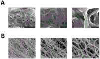
第一、聚合的纤维蛋白单体形成的网状结构是静脉血栓和混合血栓的重要组成部分,由于其特殊的支架结构,纤维蛋白聚合物将血小板、凝血因子或血细胞等包覆于内部,最终形成成熟的血栓。本实验利用富勒醇处理成熟的纤维蛋白聚合物,研究了处理组和对照组之间纤维蛋白量的变化,0.5mM和1.0mM组富勒醇处理后的纤维蛋白聚合结构明显减少,但0.1mM组的富勒醇处理后纤维蛋白聚合物溶解的比例相对较少,溶解能力较弱,而中、高浓度的富勒醇确实具有溶解纤维蛋白网状结构的能力。我们利用扫描电镜观查富勒醇处理后纤维蛋白聚合物的形态变化,对照组的纤维蛋白聚合物的结构较为粗壮,而经过富勒醇处理后的纤维蛋白结构相对于对照组来说,直径变得纤细。结果表明,富勒醇处理后的纤维蛋白结构发生了变化,富勒醇会导致纤维蛋白的聚合程度变弱。First, the network structure formed by polymerized fibrin monomers is an important part of venous thrombosis and mixed thrombus. Due to its special scaffold structure, fibrin polymer coats platelets, coagulation factors or blood cells inside, and finally Form a mature thrombus. In this experiment, the mature fibrin polymer was treated with fullerol, and the changes in the amount of fibrin between the treatment group and the control group were studied. The proportion of fibrin polymer dissolved after fullerol treatment in the mM group was relatively small, and the dissolving ability was weak, while the middle and high concentrations of fullerol did have the ability to dissolve the fibrin network structure. We used scanning electron microscopy to observe the morphological changes of fibrin polymers after fullerol treatment. The structure of fibrin polymers in the control group was relatively thick, while the fibrin structures after fullerol treatment were smaller in diameter than those in the control group. become slender. The results showed that the structure of fibrin was changed after fullerol treatment, and fullerol would lead to the weakening of the degree of polymerization of fibrin.
第二、取新鲜大鼠血浆,进行APTT时间测定,发现富勒醇显著性增加了APTT时间。Second, the fresh rat plasma was taken, and the APTT time was measured. It was found that fullerol significantly increased the APTT time.
第三、利用三氯化铁构建血栓模型,进行溶解血栓的体内实验发现。直接注射富勒醇的溶解血栓效果不明显,且富勒醇具有一定的抗血液凝固效果,因此,不适宜直接高浓度静脉注射。因此,本发明构建了基于细胞膜伪装的纳米载药体系,实现溶栓药物的靶向富集,并为降低溶栓药物的药物浓度提供基础。Third, use ferric chloride to build a thrombus model and conduct in vivo experiments to dissolve thrombus. The thrombolytic effect of direct injection of fullerol is not obvious, and fullerol has a certain anti-blood coagulation effect, so it is not suitable for direct high-concentration intravenous injection. Therefore, the present invention constructs a nano-drug loading system based on cell membrane camouflage, realizes the targeted enrichment of thrombolytic drugs, and provides a basis for reducing the drug concentration of thrombolytic drugs.
第四、本发明构建了红细胞伪装颗粒,发现红细胞载药体系具有良好的生物安全性,抗巨噬细胞吞噬效果。利用红细胞伪装载药体系载带富勒醇纳米颗粒,其血液安全性好,血液半衰期延长,并且溶栓效果显著增强。Fourth, the present invention constructs erythrocyte camouflage particles, and finds that the erythrocyte drug-carrying system has good biological safety and anti-phagocytic effect of macrophages. The red blood cell camouflage drug-carrying system is used to carry fullerol nanoparticles, which has good blood safety, prolonged blood half-life, and significantly enhanced thrombolytic effect.
附图说明Description of drawings
下面结合附图对本发明做进一步说明:The present invention will be further described below in conjunction with the accompanying drawings:
图1为本发明富勒醇体外溶解聚合的纤维蛋白结果对照图,其中:A是对照组(PBS)的纤维蛋白聚合情况,从左至右分别为放大10k,30k和70k倍后的结果,标尺分别代表5μm,1μm,500nm;B是0.5mM富勒醇处理后的纤维蛋白聚合情况,从左至右分别为放大10k,30k和70k倍后的结果,标尺与所对应的A中相同;Fig. 1 is the fibrin result comparison chart of the in vitro dissolution and polymerization of fullerol of the present invention, wherein: A is the fibrin polymerization situation of the control group (PBS), from left to right are the results after magnifying 10k, 30k and 70k times respectively, The scales represent 5 μm, 1 μm, and 500 nm, respectively; B is the fibrin polymerization after 0.5 mM fullerol treatment, and from left to right are the results after magnification of 10k, 30k and 70k times, respectively, the scale is the same as the corresponding A;
图2为扫描电镜观查富勒醇处理后纤维蛋白聚合物的形态变化图;Fig. 2 is a graph showing the morphological changes of fibrin polymers after fullerol treatment by scanning electron microscopy;
图3为富勒醇对APTT时间的影响图;Figure 3 is a graph showing the effect of fullerol on APTT time;
图4为富勒醇的体外溶栓效果图;Fig. 4 is the in vitro thrombolysis effect diagram of fullerol;
图5为富勒醇的体内溶栓效果图;Fig. 5 is the in vivo thrombolysis effect diagram of fullerol;
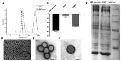
图6为合成细胞膜伪装纳米载药体系的状态示意图;图中,A、粒径分布图,B、Zeta电势结果,C、组合药物和红细胞表面蛋白的SDS-PAGE结果,D、组合药物的SEM照片,E、透射电镜结果,标尺100nm,F、透射电镜结果,标尺50nm;Figure 6 is a schematic diagram of the state of the synthetic cell membrane camouflaged nano-drug loading system; in the figure, A, particle size distribution, B, Zeta potential results, C, SDS-PAGE results of the combined drug and erythrocyte surface protein, D, SEM of the combined drug Photo, E, TEM results, scale 100nm, F, TEM results, scale 50nm;
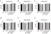
图7为纳米载药体系对血管内皮细胞的细胞存活率影响图;Figure 7 is a graph showing the effect of nano-drug loading system on the cell viability of vascular endothelial cells;
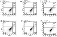
图8为纳米载药体系对血管内皮细胞的细胞凋亡影响图;Fig. 8 is a graph showing the effect of nano-drug loading system on apoptosis of vascular endothelial cells;
图9为纳米载药体系对血细胞数目影响图;;Figure 9 is a graph showing the effect of nano-drug loading system on the number of blood cells;
图10为纳米载药体系体内溶栓的HE结果图;Fig. 10 is the HE result graph of the in vivo thrombolysis of the nano-drug-loading system;
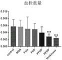
图11为纳米载药体系体内溶栓的血栓重量结果图。Fig. 11 is a graph showing the results of the thrombus weight of the nano-drug-loading system in vivo thrombolysis.
具体实施方式Detailed ways
本发明提出了一种富勒醇及其组合物在制备抗血栓和/或溶解血栓药物中的应用,为了使本发明的优点、技术方案更加清楚、明确,下面结合具体实施例对本发明做详细说明。The present invention proposes the application of a fullerol and its composition in the preparation of antithrombotic and/or thrombolytic drugs. In order to make the advantages and technical solutions of the present invention clearer and clearer, the present invention is described in detail below with reference to specific examples. illustrate.
本发明所需原料均可通过商业渠道购买获得。The raw materials required by the present invention can be purchased through commercial channels.
本发明富勒醇C60(OH)X,10≤X<40的制备方法参照现有技术,若未注明具体的方法通常参照常规操作方法。For the preparation method of the fullerol C60 (OH)X, 10≤X<40 of the present invention, reference is made to the prior art, and if the specific method is not specified, the conventional operation method is usually referred to.
实施例1:Example 1:
制备血栓抑制组合物:To prepare the thrombosis inhibitory composition:
所需组分:富勒醇、介孔硅、红细胞膜及血小板膜,富勒醇的通式为C60(OH)X,其中,10≤X<40。Required components: fullerol, mesoporous silicon, red blood cell membrane and platelet membrane, the general formula of fullerol is C60 (OH)X , where 10≦X<40.
制备方法:Preparation:
第一步、大鼠心脏取血,通过离心,分离获得红细胞和血小板,加入低渗溶液,放入液氮中,进行反复冻融5次,除去细胞内容物,获得红细胞膜和血小板膜。In the first step, blood was drawn from the rat heart, and the red blood cells and platelets were obtained by centrifugation, and then the hypotonic solution was added, and then put into liquid nitrogen, and the cells were repeatedly frozen and thawed for 5 times, and the cell contents were removed to obtain the red blood cell membrane and the platelet membrane.
第二步、合成140nm左右的介孔硅,按照1:1的比例混合介孔硅和富勒醇,室温搅拌24小时。加入红细胞膜和血小板膜,超声2分钟。将混合体系加入到截留分子量3500的透析袋中透析72小时,收集伪装纳米载药颗粒作为血栓抑制组合物。The second step is to synthesize mesoporous silicon with a thickness of about 140 nm, mix mesoporous silicon and fullerol in a ratio of 1:1, and stir at room temperature for 24 hours. Red blood cell membranes and platelet membranes were added and sonicated for 2 minutes. The mixed system was added to a dialysis bag with a molecular weight cut-off of 3500 and dialyzed for 72 hours, and the camouflaged nano-drug-loading particles were collected as the thrombosis inhibitory composition.
实施例2:Example 2:
制备血栓抑制组合物:To prepare the thrombosis inhibitory composition:
所需组分:富勒醇、介孔硅、红细胞膜、血小板膜及溶剂生理盐水,富勒醇的通式为C60(OH)X,其中,10≤X<40。Required components: fullerol, mesoporous silicon, red blood cell membrane, platelet membrane and solvent physiological saline, the general formula of fullerol is C60 (OH)X , where 10≤X<40.
制备方法:Preparation:
将实施例1中的血栓抑制组合物分散在生理盐水中即获得。It is obtained by dispersing the thrombosis inhibitory composition in Example 1 in physiological saline.
具体实验步骤:Specific experimental steps:
1.1、富勒醇体外溶解聚合的纤维蛋白1.1. Fullerol dissolves polymerized fibrin in vitro
溶解聚合的纤维蛋白是溶解血栓的重要机制。为了检测富勒醇体外溶解血栓的能力,我们进行了富勒醇对聚合纤维蛋白溶解实验。首先,将20μL 0.5U/mL的凝血酶与20mM的Tris-HCl(pH=7.4)和纤维蛋白原混匀,在37℃孵育1h,使之形成成熟的纤维蛋白聚合物。再将不同浓度的富勒醇或者组织型纤溶酶原激活物(t-PA)加入至上述混合溶液中,并继续孵育30min。反应之后,利用1/100000天平对生成的纤维蛋白凝结块进行称重,并利用奥林巴斯E520单反相机进行拍照记录,如图1所示。Dissolution of polymerized fibrin is an important mechanism of thrombolysis. To test the ability of fullerol to dissolve thrombus in vitro, we performed a polymerized fibrinolysis experiment with fullerol. First, 20 μL of 0.5 U/mL thrombin was mixed with 20 mM Tris-HCl (pH=7.4) and fibrinogen, and incubated at 37° C. for 1 h to form mature fibrin polymers. Then, different concentrations of fullerol or tissue plasminogen activator (t-PA) were added to the above mixed solution, and the incubation was continued for 30 min. After the reaction, the resulting fibrin clot was weighed with a 1/100,000 balance, and photographed and recorded with an Olympus E520 single-lens reflex camera, as shown in FIG. 1 .
1.2、扫描电镜观察纤维蛋白形态1.2. Scanning electron microscope to observe the morphology of fibrin
聚合的纤维蛋白的扫描电镜实验使用仪器为日立S-4800。首先,将20mM的Tris-HCl(pH=7.4)和100μL 5mM纤维蛋白原与20μL 0.5U/mL凝血酶混匀,37℃孵育1h,形成成熟的纤维蛋白聚合物,然后加入0.5mM的富勒醇进行处理,再孵育30min,收集处理后的纤维蛋白聚合物,利用PBS进行润洗,再利用3%的戊二醛将处理后的纤维蛋白网状结构进行固定,之后利用乙醇进行逐级脱水,在进行临界点干燥之后,将样品粘附至样品台并放置到喷金台上进行喷金,再进行接下来的扫描电镜(SEM)成像过程。扫描电镜获取图片放大倍数为10k、30k或70k,如图2所示,图中:A、对照组(PBS)的纤维蛋白聚合情况,从左至右分别为放大10k,30k和70k倍后的结果,标尺分别代表5μm,1μm,500nm;B、0.5mM富勒醇处理后的纤维蛋白聚合情况,从左至右分别为放大10k,30k和70k倍后的结果,标尺与所对应的A中相同。The SEM experiment of polymerized fibrin was performed using a Hitachi S-4800. First, 20 mM Tris-HCl (pH=7.4) and 100 μL of 5 mM fibrinogen were mixed with 20 μL of 0.5 U/mL thrombin, incubated at 37°C for 1 h to form mature fibrin polymers, and then 0.5 mM of Fuller was added. The treated fibrin polymer was collected and rinsed with PBS, and then the treated fibrin network was fixed with 3% glutaraldehyde, and then gradually dehydrated with ethanol. , after critical point drying, the sample is adhered to the sample stage and placed on the gold spray stage for gold spraying, and then the subsequent scanning electron microscope (SEM) imaging process is performed. The magnification of the image obtained by scanning electron microscope is 10k, 30k or 70k, as shown in Figure 2, in the figure: A, the fibrin polymerization of the control group (PBS), from left to right, after magnification of 10k, 30k and 70k, respectively Results, the scales represent 5 μm, 1 μm, and 500 nm, respectively; B, the fibrin polymerization after 0.5 mM fullerol treatment, from left to right are the results after magnification of 10k, 30k and 70k times, respectively, the scale and the corresponding A same.
1.3、体外APTT时间测定1.3. In vitro APTT time measurement
APTT时间的检测是用APTT检测试剂盒完成的,具体检测方法参照说明书。本实验设置了生理盐水组(ctrl)、介孔硅组(MSN)、三种富勒醇组(Fol1,FolS,FolC)、介孔硅载富勒醇组(FNP)、血小板伪装颗粒组(PFNP)、红细胞伪装颗粒组(RFNP)、血小板膜组(PG)以及红细胞膜组(RG)。药物浓度按照富勒醇的浓度100ug/ml换算而来。富勒醇对APTT时间的影响如图3所示。The detection of APTT time is completed with the APTT detection kit, and the specific detection method refers to the instructions. In this experiment, saline group (ctrl), mesoporous silicon group (MSN), three fullerol groups (Fol1, FolS, FolC), mesoporous silicon-loaded fullerol group (FNP), platelet camouflage particle group ( PFNP), erythrocyte camouflage granule group (RFNP), platelet membrane group (PG) and erythrocyte membrane group (RG). The drug concentration was converted according to the concentration of fullerol at 100ug/ml. The effect of fullerol on APTT time is shown in Figure 3.
1.4、体外溶栓实验1.4. In vitro thrombolysis experiment
大鼠眼眶取血,毛细管吸取全血至统一高度,静止凝血,制成体外混合血栓,从毛细管另一端加入相应药物,分成对照组(PBS)、尿激酶组(UK)和三组富勒醇处理组(Fol1,FolS,FolC)骨蜡封口,37℃摇床孵育,3小时后检测毛细管中血栓长度。富勒醇的体外溶栓效果如图4所示。Orbital blood was collected from rats, whole blood was drawn by capillary tube to a uniform height, coagulation was static, and mixed thrombus was made in vitro. Corresponding drugs were added from the other end of capillary tube and divided into control group (PBS), urokinase group (UK) and three groups of fullerol The treatment group (Fol1, FolS, FolC) was sealed with bone wax, incubated at 37°C on a shaker, and the length of the thrombus in the capillary was detected after 3 hours. The in vitro thrombolytic effect of fullerol is shown in Figure 4.
1.5、富勒醇体内溶栓检测1.5. Fullerol in vivo thrombolysis detection
本实验利用血栓模型来检测富勒醇的体内溶栓情况。首先,使用戊巴比妥钠利用腹腔注射的方式将160g左右的SD大鼠进行麻醉。注射后立即使用手术工具将大鼠的左侧颈动脉进行分离暴露,其余组织利用止血钳进行分离固定,为了之后的实验药品不会腐蚀其它组织,将适宜尺寸的透明保鲜膜垫在血管和组织之间。将滤纸片裁剪成5mm×5mm大小,浸入至5.5%FeCl3溶液中,使其完全浸满FeCl3溶液,再将滤纸片环形包覆在已暴露的动脉血管周围,开始计时,5min后,将滤纸片取走,在刚刚包覆的部位利用无菌生理盐水进行清洗,清洗三次后之后,通过尾静脉注射的方式注射不同浓度的富勒醇及PBS或t-PA。20min后利用止血钳将损伤部位血管两端钳住,利用锋利的手术剪刀轻轻剪下损伤部位血管,立即放入组织容器中,置于4%多聚甲醛中,4℃固定24-48h。之后,将固定好的组织进行石蜡包埋切片,再进行苏木精-伊红(HE)染色,切片结果利用多功能光学显微镜进行拍照记录,利用Image J进行照片中血栓定量的统计。富勒醇直接溶解血栓的效果不显著,见图5所示。In this experiment, a thrombus model was used to detect the thrombolysis of fullerol in vivo. First, SD rats of about 160 g were anesthetized with sodium pentobarbital by intraperitoneal injection. Immediately after injection, surgical tools were used to separate and expose the left carotid artery of the rat, and the remaining tissues were separated and fixed with hemostatic forceps. In order to prevent the subsequent experimental drugs from corroding other tissues, a suitable size of transparent plastic wrap was placed on the blood vessels and tissues. between. Cut the filter paper into a size of 5mm × 5mm, immerse it in 5.5% FeCl3 solution, make it completely immersed in the FeCl3 solution, and then wrap the filter paper around the exposed arterial blood vessels. The filter paper was removed, and the just-coated site was washed with sterile saline. After three times of washing, different concentrations of fullerol and PBS or t-PA were injected through tail vein injection. After 20 minutes, use hemostatic forceps to clamp both ends of the blood vessel at the injury site, use sharp surgical scissors to gently cut the blood vessel at the injury site, immediately put it into a tissue container, place it in 4% paraformaldehyde, and fix it at 4°C for 24-48h. After that, the fixed tissue was embedded in paraffin, and then stained with hematoxylin-eosin (HE). The effect of fullerol in directly dissolving thrombus was not significant, as shown in Figure 5.
1.6、D-二聚体的检测1.6. Detection of D-dimer
D-二聚体的检测是由ELISA试剂盒完成的,检测具体方法参照产品说明书。本实验设置三个处理组浓度梯度,分别为0.1mM富勒醇组、0.5mM富勒醇组和1.0mM富勒醇组。实验结果经由酶标仪进行分析,并利用SPSS将结果进行统计。钙离子探针分析细胞内钙含量变化:100g,3分钟,离心收集对照组和处理组的肿瘤球,用预冷的PBS洗涤三次后,用胰酶将肿瘤球消化成单细胞,用无钙的细胞外液重悬,接种在confocal小皿中,加入Fluo 4-AM染料,孵育20分钟后,用confocal荧光显微镜进行拍照,分析细胞内的荧光变化。富勒醇直接溶解血栓的效果不显著,见图5所示。The detection of D-dimer is completed by ELISA kit, and the specific method of detection refers to the product manual. In this experiment, three concentration gradients of treatment groups were set, which were 0.1mM fullerol group, 0.5mM fullerol group and 1.0mM fullerol group respectively. The experimental results were analyzed by a microplate reader, and the results were counted by SPSS. Calcium ion probe to analyze the changes of intracellular calcium content: 100 g, 3 minutes, the tumor spheres in the control group and the treatment group were collected by centrifugation, washed three times with pre-cooled PBS, and the tumor spheres were digested into single cells with trypsin and calcium The extracellular fluid was resuspended, seeded in confocal small dishes, and Fluo 4-AM dye was added. After incubation for 20 minutes, pictures were taken with a confocal fluorescence microscope to analyze the intracellular fluorescence changes. The effect of fullerol in directly dissolving thrombus was not significant, as shown in Figure 5.
1.7、细胞膜伪装纳米载药体系(血栓抑制组合物)的构建和表征1.7. Construction and characterization of cell membrane camouflaged nano-drug delivery system (thrombosis inhibitory composition)
大鼠心脏取血,通过离心,分离获得红细胞和血小板,加入低渗溶液,放入液氮中,进行反复冻融5次,除去细胞内容物,获得红细胞膜和血小板膜。Blood was collected from the rat heart, and erythrocytes and platelets were obtained by centrifugation, and then hypotonic solution was added, placed in liquid nitrogen, and frozen and thawed 5 times repeatedly to remove cell contents to obtain erythrocyte membranes and platelet membranes.
合成140nm左右的介孔硅,按照1:1的比例混合介孔硅和富勒醇,室温搅拌24小时。加入红细胞膜和血小板膜,超声2分钟。将混合体系加入到截留分子量3500的透析袋中透析72小时,收集伪装纳米载药颗粒。粒径分析仪分析水合粒径和Zeta电势。SEM电镜检测形态。透射电子显微镜(TEM)鉴定细胞膜包覆效果。SDS-PAGE检测伪装颗粒表面的蛋白与细胞膜蛋白的一致性。其具体见图6所示。To synthesize mesoporous silicon of about 140 nm, mix mesoporous silicon and fullerol in a ratio of 1:1, and stir at room temperature for 24 hours. Red blood cell membranes and platelet membranes were added and sonicated for 2 minutes. The mixed system was added to a dialysis bag with a molecular weight cut-off of 3500 and dialyzed for 72 hours to collect the camouflaged nano-drug-loaded particles. A particle size analyzer analyzes hydrated particle size and Zeta potential. Morphology was detected by SEM electron microscope. Transmission electron microscopy (TEM) was used to identify the effect of cell membrane coating. SDS-PAGE detected the consistency of the proteins on the surface of the camouflaged particles with the cell membrane proteins. The details are shown in Figure 6.
1.8、血管内皮细胞的毒性评价1.8. Toxicity evaluation of vascular endothelial cells
细胞存活率:人脐静脉血管内皮细胞(HUVEC)接种于96孔板中,加入不同的药物处理,孵育24小时,48小时和72小时,用CCK-8检测试剂盒检测细胞的存活率变化。Cell viability: Human umbilical vein endothelial cells (HUVEC) were seeded in 96-well plates, treated with different drugs, incubated for 24 hours, 48 hours and 72 hours, and the changes in cell viability were detected by CCK-8 detection kit.
细胞凋亡检测:人脐静脉血管内皮细胞(HUVEC)接种于6孔板中,加入不同的药物处理24小时后,消化成单细胞,利用Annexin-V/PI细胞凋亡检测试剂盒进行染色,通过流式分析检测细胞凋亡比例变化。Apoptosis detection: Human umbilical vein endothelial cells (HUVEC) were seeded in 6-well plates, treated with different drugs for 24 hours, digested into single cells, and stained with Annexin-V/PI apoptosis detection kit. The changes of apoptosis ratio were detected by flow analysis.
1.9、纳米载药体系的血常规检测1.9. Blood routine detection of nano-drug delivery system
将生理盐水、富勒醇、介孔硅、红细胞伪装颗粒和血小板伪装颗粒通过尾静脉注射入大鼠体内,1小时后,血常规分析仪检测血液中白细胞、红细胞、血小板以及血红蛋白的变化。Physiological saline, fullerol, mesoporous silicon, erythrocyte camouflage particles and platelet camouflage granules were injected into rats through the tail vein. After 1 hour, the blood routine analyzer detected the changes of white blood cells, red blood cells, platelets and hemoglobin in the blood.
图7示出了纳米载药体系对血管内皮细胞的细胞存活率无影响;图中,A、B、C分别为血小板膜纳米载药体系与人脐静脉内皮细胞(HUVEC)共孵育24小时、48小时、72小时的细胞活力结果;D、E、F分别为红细胞膜纳米载药体系与人脐静脉内皮细胞(HUVEC)共孵育24小时、48小时、72小时的细胞活力结果。Figure 7 shows that the nano-drug loading system has no effect on the cell viability of vascular endothelial cells; in the figure, A, B, and C are the platelet membrane nano-drug loading system incubated with human umbilical vein endothelial cells (HUVEC) for 24 hours, The cell viability results at 48 hours and 72 hours; D, E, and F are the cell viability results of the erythrocyte membrane nano-drug loading system and human umbilical vein endothelial cells (HUVEC) co-incubated for 24 hours, 48 hours, and 72 hours, respectively.
图8示出了纳米载药体系对血管内皮细胞的细胞凋亡无影响,图中:A、对照组,B、介孔硅,C、富勒醇,D、介孔硅载富勒醇,E、红细胞载药体系,F、血细胞载药体系对人脐静脉内皮细胞(HUVEC)细胞凋亡的影响。Figure 8 shows that the nano-drug loading system has no effect on the apoptosis of vascular endothelial cells, in the figure: A, control group, B, mesoporous silicon, C, fullerol, D, mesoporous silicon loaded with fullerol, E, erythrocyte drug loading system, F, the effect of blood cell drug loading system on apoptosis of human umbilical vein endothelial cells (HUVEC).
图9示出了纳米载药体系对血细胞数目无影响,图中:A、不同处理对白细胞,B、红细胞,C、血红蛋白,D、血小板的影响。Figure 9 shows that the nano-drug loading system has no effect on the number of blood cells, in the figure: A, the effects of different treatments on white blood cells, B, red blood cells, C, hemoglobin, D, platelets.
1.10、体内溶栓实验1.10. In vivo thrombolysis experiment
大鼠麻醉后,暴露一侧颈动脉,用浸泡在35%的FeCl3的宽1mm的滤纸片包裹颈动脉5分钟,除去滤纸片,并用生理盐水清洗伤口。尾静脉注射富勒醇以及伪装颗粒(其中富勒醇药物量为500ug)以及40000IU的尿激酶,1小时后,取血栓部位,称重,计算血栓重量变化。血栓并进行HE染色。After the rats were anesthetized, one side of the carotid artery was exposed, the carotid artery was wrapped with a 1 mm wide filter paper piece soaked in 35% FeCl for5 min, the filter paper piece was removed, and the wound was washed with normal saline. The tail vein was injected with fullerol and camouflage particles (the amount of fullerol was 500ug) and 40000IU of urokinase. One hour later, the thrombus site was taken and weighed to calculate the weight change of the thrombus. Thrombus and HE staining.
图10示出了纳米载药体系体内溶栓的HE结果,其从左到右依次为介孔硅、血小板载药体系、红细胞载药体系、富勒醇和对照组的HE染色效果图。Figure 10 shows the HE results of the in vivo thrombolysis of the nano-drug-loading system, which, from left to right, are the HE staining effects of mesoporous silicon, platelet-loading system, erythrocyte-drug-loading system, fullerol, and the control group.
图11示出了纳米载药体系体内溶栓的血栓重量结果。血栓大鼠静脉注射1mg富勒醇1小时后的血栓溶解效果见图4所示,图中:A、对照组(PBS)的血栓HE染色照片;B、富勒醇处理后的血栓HE染色照片;C、对照组和富勒醇处理组血栓比例统计情况;D、对照组和富勒醇处理组D-二聚体含量检测结果。富勒醇处理组D-二聚体的含量与对照组的结果相比无显著性差异。利用细胞膜包覆介孔硅载带的富勒醇纳米颗粒之后,增加了血栓部位的富勒醇浓度,实现了富勒醇的体内溶栓。溶栓效果如图10,图11所示。Figure 11 shows the thrombus weight results of in vivo thrombolysis of the nano-drug delivery system. Figure 4 shows the thrombus dissolution effect of thrombus rats after intravenous injection of 1 mg of fullerol for 1 hour. In the figure: A, HE staining photo of thrombus in the control group (PBS); B, HE staining photo of thrombus treated with fullerol ; C, the statistics of the proportion of thrombus in the control group and the fullerol treatment group; D, the detection results of the D-dimer content in the control group and the fullerol treatment group. The content of D-dimer in the fullerol-treated group was not significantly different from that in the control group. After the mesoporous silicon-loaded fullerol nanoparticles are coated with the cell membrane, the concentration of fullerol at the thrombus site is increased, and the in vivo thrombolysis of fullerol is realized. The thrombolytic effect is shown in Figure 10 and Figure 11.
本发明中未述及的部分借鉴现有技术即可实现。The parts not mentioned in the present invention can be realized by referring to the prior art.
需要说明的是,在本说明书的教导下本领域技术人员所做出的任何等同方式,或明显变型方式均应在本发明的保护范围内。It should be noted that any equivalent manner or obvious modification manner made by those skilled in the art under the teaching of this specification shall fall within the protection scope of the present invention.
Claims (10)
Priority Applications (1)
| Application Number | Priority Date | Filing Date | Title |
|---|---|---|---|
| CN201910570879.6ACN110292583B (en) | 2019-06-28 | 2019-06-28 | Application of fullerol and composition thereof in preparation of antithrombotic drugs |
Applications Claiming Priority (1)
| Application Number | Priority Date | Filing Date | Title |
|---|---|---|---|
| CN201910570879.6ACN110292583B (en) | 2019-06-28 | 2019-06-28 | Application of fullerol and composition thereof in preparation of antithrombotic drugs |
Publications (2)
| Publication Number | Publication Date |
|---|---|
| CN110292583A CN110292583A (en) | 2019-10-01 |
| CN110292583Btrue CN110292583B (en) | 2020-06-16 |
Family
ID=68029250
Family Applications (1)
| Application Number | Title | Priority Date | Filing Date |
|---|---|---|---|
| CN201910570879.6AExpired - Fee RelatedCN110292583B (en) | 2019-06-28 | 2019-06-28 | Application of fullerol and composition thereof in preparation of antithrombotic drugs |
Country Status (1)
| Country | Link |
|---|---|
| CN (1) | CN110292583B (en) |
Families Citing this family (2)
| Publication number | Priority date | Publication date | Assignee | Title |
|---|---|---|---|---|
| CN112823791A (en)* | 2019-11-19 | 2021-05-21 | 复旦大学 | Bionic nano drug delivery system for protein thrombolytic drug and application of bionic nano drug delivery system |
| CN111035766A (en)* | 2019-12-31 | 2020-04-21 | 中国科学院高能物理研究所 | Application of erythrocyte and activated platelet cell membrane as carrier in preparing thrombus treating medicine |
Citations (3)
| Publication number | Priority date | Publication date | Assignee | Title |
|---|---|---|---|---|
| CN101239026A (en)* | 2007-12-25 | 2008-08-13 | 中国科学院上海应用物理研究所 | Application of Fullerene Alcohol in Beauty and Skin Care Products |
| CN101695502A (en)* | 2005-09-19 | 2010-04-21 | 中国科学院高能物理研究所 | Lanthanum fullerenol and application in preparing medicaments for inhibiting tumor growth |
| CN104555977A (en)* | 2014-12-11 | 2015-04-29 | 河南农业大学 | Preparation method of fullerol |
Family Cites Families (1)
| Publication number | Priority date | Publication date | Assignee | Title |
|---|---|---|---|---|
| CN103482599B (en)* | 2013-09-04 | 2015-10-21 | 贵州特力达纳米碳素科技有限公司 | A kind of nano carbon sol and application thereof |
- 2019
- 2019-06-28CNCN201910570879.6Apatent/CN110292583B/ennot_activeExpired - Fee Related
Patent Citations (3)
| Publication number | Priority date | Publication date | Assignee | Title |
|---|---|---|---|---|
| CN101695502A (en)* | 2005-09-19 | 2010-04-21 | 中国科学院高能物理研究所 | Lanthanum fullerenol and application in preparing medicaments for inhibiting tumor growth |
| CN101239026A (en)* | 2007-12-25 | 2008-08-13 | 中国科学院上海应用物理研究所 | Application of Fullerene Alcohol in Beauty and Skin Care Products |
| CN104555977A (en)* | 2014-12-11 | 2015-04-29 | 河南农业大学 | Preparation method of fullerol |
Non-Patent Citations (1)
| Title |
|---|
| The reduction of oxidative stress by nanocomposite Fullerol decreasesmucositis severity and reverts leukopenia induced by Irinotecan;Raquel Duque Nascimento Arifa等;《Pharmacological Research》;20161231(第107期);文章摘要* |
Also Published As
| Publication number | Publication date |
|---|---|
| CN110292583A (en) | 2019-10-01 |
Similar Documents
| Publication | Publication Date | Title |
|---|---|---|
| Zhang et al. | Cyclic RGD functionalized liposomes encapsulating urokinase for thrombolysis | |
| Huang et al. | Nitric oxide-loaded echogenic liposomes for nitric oxide delivery and inhibition of intimal hyperplasia | |
| WO2019141267A1 (en) | Recombinant bionic nanocarrier delivery system for targeting active cd44 molecule, preparation method therefor, and uses thereof | |
| US20140227350A1 (en) | Annexin A2 and Tissue Plasminogen Activator For Treating Vascular Disease | |
| Wrobeln et al. | Albumin-derived perfluorocarbon-based artificial oxygen carriers: A physico-chemical characterization and first in vivo evaluation of biocompatibility | |
| Sezer et al. | Oxidized regenerated cellulose cross-linked gelatin microparticles for rapid and biocompatible hemostasis: A versatile cross-linking agent | |
| JP2002503254A (en) | Microspheres coated with fibrinogen | |
| Cheng et al. | Functionally integrating nanoparticles alleviate deep vein thrombosis in pregnancy and rescue intrauterine growth restriction | |
| Shekhar et al. | In vitro characterization of sonothrombolysis and echocontrast agents to treat ischemic stroke | |
| Markowicz-Piasecka et al. | Studies towards biocompatibility of PAMAM dendrimers–overall hemostasis potential and integrity of the human aortic endothelial barrier | |
| CN112823791A (en) | Bionic nano drug delivery system for protein thrombolytic drug and application of bionic nano drug delivery system | |
| CN110292583B (en) | Application of fullerol and composition thereof in preparation of antithrombotic drugs | |
| Xuan et al. | Biocompatibility and effectiveness evaluation of a new hemostatic embolization agent: Thrombin loaded alginate calcium microsphere | |
| Huang et al. | Multi-enzyme mimetic iridium nanozymes-based thrombus microenvironment-modulated nanoplatform for enhanced thrombolytic therapy | |
| Cao et al. | Thrombus-targeted nano-agents for NIR-II diagnostic fluorescence imaging-guided flap thromboembolism multi-model therapy | |
| Zhong et al. | Hydrogen sulfide‐loaded microbubbles combined with ultrasound mediate thrombolysis and simultaneously mitigate ischemia‐reperfusion injury in a rat hindlimb model | |
| CN111035766A (en) | Application of erythrocyte and activated platelet cell membrane as carrier in preparing thrombus treating medicine | |
| Huang et al. | Cellular Membrane‐Engineered Nanovesicles as a Three‐Stage Booster to Target the Lesion Core | |
| Chen et al. | Dual-targeting fucoidan-based microvesicle for arterial thrombolysis and re-occlusion inhibition | |
| Jung et al. | H2O2‐Triggered Self Immolative Prodrug Nanoassemblies as Self‐Deliverable Nanomedicines for Targeted On‐Demand Therapy of Thrombotic Disorders | |
| Luo et al. | Cytosolic perfluorocarbon delivery to platelets via albumin for antithrombotic therapy | |
| US11419892B2 (en) | Antimicrobial platelet-like particles | |
| Zhang et al. | Cyclic RGD functionalized PLGA nanoparticles loaded with noncovalent complex of indocyanine green with urokinase for synergistic thrombolysis | |
| Rana et al. | Peptide-based targeting: Novel concept for thrombosis diagnosis and treatment | |
| CN119119226A (en) | A conotoxin-derived anticoagulant polypeptide Cb-13 and its application |
Legal Events
| Date | Code | Title | Description |
|---|---|---|---|
| PB01 | Publication | ||
| PB01 | Publication | ||
| SE01 | Entry into force of request for substantive examination | ||
| SE01 | Entry into force of request for substantive examination | ||
| GR01 | Patent grant | ||
| GR01 | Patent grant | ||
| CF01 | Termination of patent right due to non-payment of annual fee | Granted publication date:20200616 | |
| CF01 | Termination of patent right due to non-payment of annual fee |