CN110267586B - Method and system for determining prevalence of cardiac phenomena - Google Patents
Method and system for determining prevalence of cardiac phenomenaDownload PDFInfo
- Publication number
- CN110267586B CN110267586BCN201880009120.4ACN201880009120ACN110267586BCN 110267586 BCN110267586 BCN 110267586BCN 201880009120 ACN201880009120 ACN 201880009120ACN 110267586 BCN110267586 BCN 110267586B
- Authority
- CN
- China
- Prior art keywords
- prevalence
- cardiac
- locations
- discrete times
- tissue
- Prior art date
- Legal status (The legal status is an assumption and is not a legal conclusion. Google has not performed a legal analysis and makes no representation as to the accuracy of the status listed.)
- Active
Links
Images


Classifications
- A—HUMAN NECESSITIES
- A61—MEDICAL OR VETERINARY SCIENCE; HYGIENE
- A61B—DIAGNOSIS; SURGERY; IDENTIFICATION
- A61B5/00—Measuring for diagnostic purposes; Identification of persons
- A61B5/24—Detecting, measuring or recording bioelectric or biomagnetic signals of the body or parts thereof
- A61B5/25—Bioelectric electrodes therefor
- A61B5/279—Bioelectric electrodes therefor specially adapted for particular uses
- A61B5/28—Bioelectric electrodes therefor specially adapted for particular uses for electrocardiography [ECG]
- A61B5/283—Invasive
- A61B5/287—Holders for multiple electrodes, e.g. electrode catheters for electrophysiological study [EPS]
- A—HUMAN NECESSITIES
- A61—MEDICAL OR VETERINARY SCIENCE; HYGIENE
- A61B—DIAGNOSIS; SURGERY; IDENTIFICATION
- A61B5/00—Measuring for diagnostic purposes; Identification of persons
- A61B5/24—Detecting, measuring or recording bioelectric or biomagnetic signals of the body or parts thereof
- A61B5/316—Modalities, i.e. specific diagnostic methods
- A61B5/318—Heart-related electrical modalities, e.g. electrocardiography [ECG]
- A61B5/339—Displays specially adapted therefor
- A—HUMAN NECESSITIES
- A61—MEDICAL OR VETERINARY SCIENCE; HYGIENE
- A61B—DIAGNOSIS; SURGERY; IDENTIFICATION
- A61B34/00—Computer-aided surgery; Manipulators or robots specially adapted for use in surgery
- A61B34/10—Computer-aided planning, simulation or modelling of surgical operations
- A—HUMAN NECESSITIES
- A61—MEDICAL OR VETERINARY SCIENCE; HYGIENE
- A61B—DIAGNOSIS; SURGERY; IDENTIFICATION
- A61B5/00—Measuring for diagnostic purposes; Identification of persons
- A61B5/06—Devices, other than using radiation, for detecting or locating foreign bodies ; Determining position of diagnostic devices within or on the body of the patient
- A61B5/061—Determining position of a probe within the body employing means separate from the probe, e.g. sensing internal probe position employing impedance electrodes on the surface of the body
- A—HUMAN NECESSITIES
- A61—MEDICAL OR VETERINARY SCIENCE; HYGIENE
- A61B—DIAGNOSIS; SURGERY; IDENTIFICATION
- A61B5/00—Measuring for diagnostic purposes; Identification of persons
- A61B5/24—Detecting, measuring or recording bioelectric or biomagnetic signals of the body or parts thereof
- A61B5/316—Modalities, i.e. specific diagnostic methods
- A61B5/318—Heart-related electrical modalities, e.g. electrocardiography [ECG]
- A61B5/346—Analysis of electrocardiograms
- A61B5/349—Detecting specific parameters of the electrocardiograph cycle
- A—HUMAN NECESSITIES
- A61—MEDICAL OR VETERINARY SCIENCE; HYGIENE
- A61B—DIAGNOSIS; SURGERY; IDENTIFICATION
- A61B5/00—Measuring for diagnostic purposes; Identification of persons
- A61B5/48—Other medical applications
- A61B5/4836—Diagnosis combined with treatment in closed-loop systems or methods
- A—HUMAN NECESSITIES
- A61—MEDICAL OR VETERINARY SCIENCE; HYGIENE
- A61B—DIAGNOSIS; SURGERY; IDENTIFICATION
- A61B5/00—Measuring for diagnostic purposes; Identification of persons
- A61B5/68—Arrangements of detecting, measuring or recording means, e.g. sensors, in relation to patient
- A61B5/6846—Arrangements of detecting, measuring or recording means, e.g. sensors, in relation to patient specially adapted to be brought in contact with an internal body part, i.e. invasive
- A61B5/6847—Arrangements of detecting, measuring or recording means, e.g. sensors, in relation to patient specially adapted to be brought in contact with an internal body part, i.e. invasive mounted on an invasive device
- A61B5/6852—Catheters
- A61B5/6857—Catheters with a distal pigtail shape
- A—HUMAN NECESSITIES
- A61—MEDICAL OR VETERINARY SCIENCE; HYGIENE
- A61B—DIAGNOSIS; SURGERY; IDENTIFICATION
- A61B5/00—Measuring for diagnostic purposes; Identification of persons
- A61B5/68—Arrangements of detecting, measuring or recording means, e.g. sensors, in relation to patient
- A61B5/6846—Arrangements of detecting, measuring or recording means, e.g. sensors, in relation to patient specially adapted to be brought in contact with an internal body part, i.e. invasive
- A61B5/6847—Arrangements of detecting, measuring or recording means, e.g. sensors, in relation to patient specially adapted to be brought in contact with an internal body part, i.e. invasive mounted on an invasive device
- A61B5/6852—Catheters
- A61B5/6858—Catheters with a distal basket, e.g. expandable basket
- A—HUMAN NECESSITIES
- A61—MEDICAL OR VETERINARY SCIENCE; HYGIENE
- A61B—DIAGNOSIS; SURGERY; IDENTIFICATION
- A61B5/00—Measuring for diagnostic purposes; Identification of persons
- A61B5/72—Signal processing specially adapted for physiological signals or for diagnostic purposes
- A61B5/7271—Specific aspects of physiological measurement analysis
- A61B5/7282—Event detection, e.g. detecting unique waveforms indicative of a medical condition
- A—HUMAN NECESSITIES
- A61—MEDICAL OR VETERINARY SCIENCE; HYGIENE
- A61B—DIAGNOSIS; SURGERY; IDENTIFICATION
- A61B18/00—Surgical instruments, devices or methods for transferring non-mechanical forms of energy to or from the body
- A61B18/04—Surgical instruments, devices or methods for transferring non-mechanical forms of energy to or from the body by heating
- A61B18/12—Surgical instruments, devices or methods for transferring non-mechanical forms of energy to or from the body by heating by passing a current through the tissue to be heated, e.g. high-frequency current
- A61B18/14—Probes or electrodes therefor
- A—HUMAN NECESSITIES
- A61—MEDICAL OR VETERINARY SCIENCE; HYGIENE
- A61B—DIAGNOSIS; SURGERY; IDENTIFICATION
- A61B18/00—Surgical instruments, devices or methods for transferring non-mechanical forms of energy to or from the body
- A61B2018/00571—Surgical instruments, devices or methods for transferring non-mechanical forms of energy to or from the body for achieving a particular surgical effect
- A61B2018/00577—Ablation
- A—HUMAN NECESSITIES
- A61—MEDICAL OR VETERINARY SCIENCE; HYGIENE
- A61B—DIAGNOSIS; SURGERY; IDENTIFICATION
- A61B34/00—Computer-aided surgery; Manipulators or robots specially adapted for use in surgery
- A61B34/10—Computer-aided planning, simulation or modelling of surgical operations
- A61B2034/101—Computer-aided simulation of surgical operations
- A61B2034/105—Modelling of the patient, e.g. for ligaments or bones
- A—HUMAN NECESSITIES
- A61—MEDICAL OR VETERINARY SCIENCE; HYGIENE
- A61B—DIAGNOSIS; SURGERY; IDENTIFICATION
- A61B90/00—Instruments, implements or accessories specially adapted for surgery or diagnosis and not covered by any of the groups A61B1/00 - A61B50/00, e.g. for luxation treatment or for protecting wound edges
- A61B90/36—Image-producing devices or illumination devices not otherwise provided for
- A61B2090/364—Correlation of different images or relation of image positions in respect to the body
- A61B2090/365—Correlation of different images or relation of image positions in respect to the body augmented reality, i.e. correlating a live optical image with another image
Landscapes
- Health & Medical Sciences (AREA)
- Life Sciences & Earth Sciences (AREA)
- Engineering & Computer Science (AREA)
- Surgery (AREA)
- Animal Behavior & Ethology (AREA)
- Veterinary Medicine (AREA)
- Public Health (AREA)
- General Health & Medical Sciences (AREA)
- Biomedical Technology (AREA)
- Heart & Thoracic Surgery (AREA)
- Medical Informatics (AREA)
- Molecular Biology (AREA)
- Pathology (AREA)
- Physics & Mathematics (AREA)
- Biophysics (AREA)
- Cardiology (AREA)
- Physiology (AREA)
- Signal Processing (AREA)
- Artificial Intelligence (AREA)
- Computer Vision & Pattern Recognition (AREA)
- Psychiatry (AREA)
- Human Computer Interaction (AREA)
- Robotics (AREA)
- Nuclear Medicine, Radiotherapy & Molecular Imaging (AREA)
- Measurement And Recording Of Electrical Phenomena And Electrical Characteristics Of The Living Body (AREA)
- Surgical Instruments (AREA)
Abstract
Description
Translated fromChinese相关申请的交叉引用CROSS-REFERENCE TO RELATED APPLICATIONS
本申请要求2017年2月10日提交的临时申请序列No.62/457,384的优先权,其通过引用整体并入。This application claims priority to Provisional Application Serial No. 62/457,384, filed February 10, 2017, which is incorporated by reference in its entirety.
技术领域technical field
本公开涉及用于基于电生理学数据检测心脏现象的系统和方法。更具体地,本公开涉及用于基于电生理学数据确定心脏现象随时间推移的患病率的计算机实现的系统和方法。The present disclosure relates to systems and methods for detecting cardiac phenomena based on electrophysiological data. More particularly, the present disclosure relates to computer-implemented systems and methods for determining the prevalence of cardiac phenomena over time based on electrophysiological data.
背景技术Background technique
众所周知,各种基于计算机的系统和计算机实现的方法可用于生成几何结构的多维表面模型,诸如例如解剖结构。更具体地,已经使用各种系统和方法来生成心脏和/或其特定部分的多维表面模型。Various computer-based systems and computer-implemented methods are well known for generating multi-dimensional surface models of geometric structures, such as, for example, anatomical structures. More specifically, various systems and methods have been used to generate multidimensional surface models of the heart and/or specific portions thereof.
人类心脏肌肉通常经历穿过其许多表面和心室的包括心内膜腔室的电流。就在每次心脏收缩之前,心脏肌肉被称为“去极化”和“再极化”,因为电流在心脏和整个身体中传播。在健康的心脏中,心脏的表面和心室将经历去极化波的有序进展。在不健康的心脏中,诸如经历房性心律失常的心脏,包括例如异位房性心动过速、心房颤动和心房扑动,去极化波的进展可能不是那么有序。由于瘢痕组织或其它障碍导致快速和均匀的去极化,心律失常可能持续存在。这些障碍可能导致去极化波重复围绕心脏的一些部分的环路。房性心律失常可以产生各种危险状况,包括不规则的心率、同步性房室收缩的丧失和血流停滞,所有这些都可能导致各种疾病甚至死亡。Human heart muscle typically experiences electrical currents across its many surfaces and the ventricles, including the endocardial chambers. Just before each heart contraction, the heart muscle is called "depolarized" and "repolarized" as electrical current travels through the heart and throughout the body. In a healthy heart, the surface and ventricles of the heart will experience an orderly progression of depolarizing waves. In unhealthy hearts, such as those experiencing atrial arrhythmias, including, for example, ectopic atrial tachycardia, atrial fibrillation, and atrial flutter, the progression of depolarization waves may not be so orderly. Arrhythmias may persist due to rapid and uniform depolarization due to scar tissue or other disorders. These obstacles can cause the depolarizing waves to repeat their loops around some parts of the heart. Atrial arrhythmias can produce a variety of dangerous conditions, including irregular heart rates, loss of synchronized atrioventricular contractions, and stasis of blood flow, all of which can lead to a variety of illnesses and even death.
医疗设备,诸如例如电生理学(EP)导管,用于各种诊断和/或治疗医疗过程中以校正这种心律失常。通常在过程中,导管例如通过患者的脉管系统操纵到患者的心脏,并且携带可以用于标测、消融、诊断和/或执行其它功能的一个或多个电极。一旦到达预定部位,治疗可包括射频(RF)消融、冷冻消融、激光、化学品、高强度聚焦超声等。消融导管将这种消融能量赋予心脏组织以在心脏组织中产生病变。该病变破坏了不期望的电通路,并且从而限制或防止了导致心律失常的杂散电信号。显而易见的是,这种治疗需要在去往治疗部位、离开治疗部位和处在治疗部位的操纵期间精确控制导管,这总是与用户技能水平的有关。Medical devices, such as, for example, electrophysiology (EP) catheters, are used in various diagnostic and/or therapeutic medical procedures to correct such arrhythmias. Typically during the procedure, a catheter is maneuvered to the patient's heart, eg, through the patient's vasculature, and carries one or more electrodes that can be used for mapping, ablation, diagnosis, and/or performing other functions. Once at the intended site, treatment may include radio frequency (RF) ablation, cryoablation, lasers, chemicals, high-intensity focused ultrasound, and the like. The ablation catheter imparts this ablation energy to the cardiac tissue to create a lesion in the cardiac tissue. The lesion disrupts undesired electrical pathways and thereby limits or prevents stray electrical signals that lead to arrhythmias. Obviously, this treatment requires precise control of the catheter during manipulation to, from, and at the treatment site, which is always related to the skill level of the user.
然而,在消融过程之前或期间,用户必须测量和诊断这些不期望的电通路和心律失常区域“发作”。用于帮助识别这些区域的电描记图是电位随时间推移变化的任何记录,其通常通过将电极直接放置在心脏组织的表面上或附近来获得。为了获取电描记图,传统技术包括记录电位变化的逐点方法。然后可以将这些电位变化标测到解剖结构的对应模型上。换句话说,这些方法能够通过围绕关注区域导航一个或多个导管并从一个点到下一个点收集电描记图和空间定位数据,并且然后相应地标测所收集的数据来创建心电图。However, before or during the ablation procedure, the user must measure and diagnose these undesired electrical pathways and "onsets" of arrhythmia areas. An electrogram used to help identify these regions is any recording of changes in electrical potential over time, usually obtained by placing electrodes directly on or near the surface of the heart tissue. To obtain electrograms, conventional techniques include point-by-point methods of recording potential changes. These potential changes can then be mapped onto corresponding models of anatomical structures. In other words, these methods can create an electrocardiogram by navigating one or more catheters around an area of interest and collecting electrogram and spatial positioning data from one point to the next, and then mapping the collected data accordingly.
期望基于电生理学(EP)数据识别心律失常的来源,特别是对于对患者执行诊断、治疗和消融手术的系统。EP数据可以来自内在节律,诸如例如窦性心律、心房扑动和心房颤动。EP数据也可以例如来自手动干预,诸如起搏和诱发性心律失常。It is desirable to identify the source of arrhythmia based on electrophysiology (EP) data, especially for systems that perform diagnostic, therapeutic, and ablation procedures on patients. EP data can come from intrinsic rhythms such as, for example, sinus rhythm, atrial flutter, and atrial fibrillation. EP data can also come from manual interventions, such as pacing and induced arrhythmias, for example.
在至少一些已知系统中,可检测到许多类型的心脏现象。然而,未确定这些心脏现象的患病率(即,这些心脏现象多久发生一次)。因此,如果针对受试者心脏上的特定位置收集数据二十次,则现有系统可以指示转子存在,而不管转子是否仅被检测到二十次中的一次,或者二十次中的十八次。因此,为了帮助临床医生,期望能够不仅检测一种或多种心脏现象的存在,而且还能检测其患病率。In at least some known systems, many types of cardiac phenomena can be detected. However, the prevalence of these cardiac phenomena (ie, how often these cardiac phenomena occur) has not been determined. Thus, if data is collected twenty times for a particular location on a subject's heart, existing systems can indicate the presence of a rotor, regardless of whether the rotor is detected only once in twenty, or eighteen out of twenty Second-rate. Therefore, to assist clinicians, it is desirable to be able to detect not only the presence, but also the prevalence, of one or more cardiac phenomena.
发明内容SUMMARY OF THE INVENTION
在一个实施例中,本公开涉及一种用于基于来自身体组织的电生理学(EP)数据来确定心脏现象的患病率的系统。通过设置在可定位在身体组织附近的至少一个医疗设备上的至少一个传感器在多个位置处测量EP数据。该系统包括电子控制单元,该电子控制单元通信地耦合到显示设备并且被配置为,对于多个位置中的每一个位置,在预定时间段期间发生的多个离散时间中的每一个离散时间,基于EP数据检测在该位置处是否发生心脏现象,基于检测确定心脏现象的患病率,并且在显示设备上显示指示所确定的心脏现象的患病率的信息。In one embodiment, the present disclosure relates to a system for determining the prevalence of cardiac phenomena based on electrophysiology (EP) data from body tissue. EP data is measured at a plurality of locations by at least one sensor disposed on at least one medical device positionable near body tissue. The system includes an electronic control unit communicatively coupled to the display device and configured to, for each of the plurality of locations, each of a plurality of discrete times occurring during a predetermined period of time, Whether a cardiac phenomenon occurs at the location is detected based on the EP data, a prevalence of the cardiac phenomenon is determined based on the detection, and information indicative of the determined prevalence of the cardiac phenomenon is displayed on a display device.
在另一个实施例中,本公开涉及一种计算机实现的方法,该方法基于来自身体组织的电生理学(EP)数据来确定心脏现象的患病率,该EP数据通过设置在可定位在身体组织附近的至少一个医疗设备上的至少一个传感器在多个位置处测量。该方法包括,对于多个位置中的每一个位置,在预定时间段期间发生的多个离散时间中的每一个离散时间,基于EP数据检测在该位置处是否发生心脏现象,基于检测确定心脏现象的患病率,并显示指示所确定的心脏现象的患病率的信息。In another embodiment, the present disclosure is directed to a computer-implemented method for determining the prevalence of cardiac phenomena based on electrophysiology (EP) data from body tissue, the EP data being positioned in a body tissue locatable At least one sensor on at least one nearby medical device measures at a plurality of locations. The method includes, for each of the plurality of locations, detecting whether a cardiac phenomenon occurs at the location based on the EP data for each of a plurality of discrete times that occur during a predetermined time period, and determining the cardiac phenomenon based on the detection and displays information indicative of the prevalence of the identified cardiac phenomenon.
在另一实施例中,本公开涉及一种用于基于来自身体组织的电生理学(EP)数据来确定心脏现象的患病率的处理装置,该EP数据通过设置在可定位在身体组织附近的至少一个医疗设备上的至少一个传感器在多个位置处测量。处理装置被配置为,对于多个位置中的每一个位置,在预定时间段期间发生的多个离散时间中的每一个离散时间,基于EP数据检测在该位置处是否发生心脏现象,基于检测确定心脏现象的患病率,并且使得指示所确定的心脏现象的患病率的信息显示在显示设备上。In another embodiment, the present disclosure relates to a processing apparatus for determining the prevalence of cardiac phenomena based on electrophysiological (EP) data from body tissue, the EP data being provided by a device positioned near the body tissue. At least one sensor on at least one medical device measures at a plurality of locations. The processing device is configured to, for each of the plurality of locations, detect, based on the EP data, whether a cardiac phenomenon has occurred at the location, and determine based on the detection, for each of a plurality of discrete times that occur during a predetermined period of time the prevalence of the cardiac phenomenon, and causing information indicative of the determined prevalence of the cardiac phenomenon to be displayed on the display device.
通过阅读以下描述和权利要求以及评审附图,本公开的前述和其它方面、特征、细节、效用和优点将变得显而易见。The foregoing and other aspects, features, details, utilities and advantages of the present disclosure will become apparent from a reading of the following description and claims, and a review of the accompanying drawings.
附图说明Description of drawings
图1是根据本教导的用于执行诊断和治疗医疗过程中的至少一个的系统的示意图和图解视图。1 is a schematic and diagrammatic view of a system for performing at least one of diagnostic and therapeutic medical procedures in accordance with the present teachings.
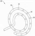
图2是以螺旋配置布置的医疗设备的一个实施例的远端的等距视图。Figure 2 is an isometric view of the distal end of one embodiment of a medical device arranged in a helical configuration.
图3是以篮子配置布置的医疗设备的另一实施例的远端的等距视图。3 is an isometric view of a distal end of another embodiment of a medical device arranged in a basket configuration.
图4A和图4B分别是以矩阵状配置布置的医疗设备的一个实施例的远端的等距视图和侧视图。4A and 4B are isometric and side views, respectively, of the distal end of one embodiment of a medical device arranged in a matrix configuration.
图5是医疗设备的一个实施例的远端的顶视图,其中医疗设备是射频(RF)消融导管。5 is a top view of a distal end of one embodiment of a medical device, wherein the medical device is a radio frequency (RF) ablation catheter.
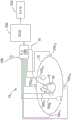
图6是可以与图1中所示的系统一起使用的可视化、导航和标测子系统的一个实施例的示意图和图解视图。FIG. 6 is a schematic and diagrammatic view of one embodiment of a visualization, navigation and mapping subsystem that may be used with the system shown in FIG. 1 .
图7是用于确定可以与图6中所示的系统一起使用的心脏现象的患病率的方法的流程图。FIG. 7 is a flowchart of a method for determining the prevalence of cardiac phenomena that may be used with the system shown in FIG. 6 .
图8是图形用户界面的一个实施例,示出了投影到三维解剖模型上的彩色图,该三维解剖模型可以使用图6中所示的系统生成。FIG. 8 is one embodiment of a graphical user interface showing a color map projected onto a three-dimensional anatomical model that may be generated using the system shown in FIG. 6 .
在整个附图的若干视图中,对应的附图标记表示对应的部件。Corresponding reference characters indicate corresponding parts throughout the several views of the drawings.
具体实施方式Detailed ways
本公开提供了用于检测和确定各种心脏现象的患病率的系统和方法。在此描述的实施例包括,在预定时间段期间发生的多个离散时间中的每一个离散时间,检测在某个位置处心脏现象是否发生。基于检测确定心脏现象的患病率(即,心脏现象多久发生一次)。此外,向用户(例如,临床医生)显示指示所确定的患病率的信息。The present disclosure provides systems and methods for detecting and determining the prevalence of various cardiac phenomena. Embodiments described herein include, at each of a plurality of discrete times occurring during a predetermined period of time, detecting whether a cardiac phenomenon occurs at a certain location. The prevalence of the cardiac phenomenon (ie, how often the cardiac phenomenon occurs) is determined based on the detection. Additionally, information indicative of the determined prevalence is displayed to a user (eg, a clinician).
现在参考附图,其中相同的附图标记用于识别各个视图中的相同组件,图1示出了用于在身体14的组织12上或针对身体14的组织12执行一个或多个诊断和/或治疗功能的系统10的一个示例性实施例。在示例性实施例中,组织12包括人体14内的心脏或心脏组织。然而,应该理解,系统10可以找到与人体和非人体内的各种其它组织有关的应用。因此,本公开并不意味着仅限于仅与心脏组织和/或人体有关的系统10的使用。Referring now to the drawings, wherein like reference numerals are used to identify like components throughout the various views, FIG. 1 illustrates a method for performing one or more diagnostics and/or diagnosis on or against
系统10可以包括医疗设备(例如,导管16)和用于内部身体结构的可视化、导航和/或标测的子系统18(下面称为“可视化、导航和标测子系统18”或“子系统18”)。
在该实施例中,医疗设备包括导管16,诸如例如电生理学导管。在其它示例性实施例中,医疗设备可以采用除导管16之外的形式,诸如例如但不限于护套或导管导引器,或除电生理学导管之外的导管。仅为了清楚和说明的目的,以下描述将限于系统10的实施例,其中医疗设备是导管(导管16)。In this embodiment, the medical device includes a
提供导管16用于检查、诊断和/或治疗诸如组织12的内部身体组织。导管16可包括电缆连接器20或接口、手柄22、具有近端26和远端28的轴24(如在此所使用的,“近端”指的是朝向手柄22附近的导管16的端部的方向,并且“远端”指的是远离手柄22的方向),以及一个或多个传感器,诸如例如并且不限于安装在轴24的远端28处或附近的导管16的轴24中或之上的多个电极30(即301,302,...,30N)。传感器可以包括例如阻抗电极。
在该实施例中,每个电极30被配置为既获取与组织12对应的电生理学(EP)数据,又产生指示其三维(3-D)位置的信号(下文中称为“定位数据”)。在另一个实施例中,导管16可包括电极30和一个或多个定位传感器(例如,除电极30或磁传感器(例如,线圈)之外的电极)的组合。在一个这种实施例中,电极30被配置为获取与组织12有关的EP数据,而定位传感器被配置为生成指示其3-D位置的定位数据,该定位数据可以用于确定每个电极30的3-D位置。在其它实施例中,导管16可进一步包括其它传统组件,诸如例如但不限于,操纵线和致动器、灌注腔和端口、压力传感器、接触传感器、温度传感器、附加电极和对应的导体或引线和/或消融元件(例如,消融电极、高强度聚焦超声消融元件等)。In this embodiment, each
连接器20为一个或多个电缆32提供机械和电连接,该电缆32例如从可视化、导航和标测子系统18延伸到一个或多个电极30或安装在导管16上的定位传感器。在其它实施例中,连接器20还可以为从系统10(诸如例如,消融系统和流体源(当导管16包括灌注导管时))中的其它组件延伸的电缆提供机械、电气和/或流体连接。连接器20设置在导管16的近端26处。
手柄22为用户提供了保持导管16的位置,并且可以进一步提供用于在身体14内转向或引导轴24的部件。例如,手柄22可以包括用于操纵一个或多个转向线延伸通过导管16到轴24的远端28以将轴24转向的部件。本领域技术人员可以理解,手柄22的结构可以变化。在其它实施例中,导管16的控制可以是自动化的,诸如通过机器人驱动或控制,或由基于磁的引导系统驱动和控制。因此,手动或自动控制的导管二者都在本公开的精神和范围内。The
轴24是细长的管状柔性构件,其被配置为在身体14内移动。轴24支撑,例如但不限于,电极30、安装在其上的其它电极或定位传感器、相关导体,以及用于信号处理或调节的可能的附加电子器件。轴24还可以允许输送、传送和/或移除流体(包括灌注流体、低温消融流体和体液)、药物和/或手术工具或器械。可以由诸如聚氨酯的传统材料制成的轴24限定一个或多个腔,该腔被配置为容纳和/或输送电导体、流体或手术工具。可以通过传统的导引器将轴24引入身体14内的血管或其它结构中。然后可以将轴24转向或引导通过身体14到达所需的位置,诸如组织12。The
轴24的远端28可以是导管16的主要部分,该导管16包含电极30或用于获取EP数据和定位数据的其它传感器。如上所述,在一个实施例中,电极30可以被配置为获取EP数据和定位数据二者。在另一个实施例中,并且如下面将更详细描述的,电极30可以被配置为获取EP数据,而一个或多个定位传感器可以被配置为获取定位数据,然后可以使用该定位数据来确定电极30的相应位置。无论定位数据是由电极30还是由定位传感器获取,远端28可以以多种配置布置,这有助于从组织12对EP数据的有效获取、测量、收集等。The
在一个实施例中,如图2中所示,远端28可以以螺旋配置布置。在该实施例中,螺旋配置可以是大致平面的并且可以包含高密度的电极30,用于对来自组织12的EP数据进行单极或双极测量。单极测量通常可以表示在每个电极处感知的电压。但是,双极测量通常可以表示任何一对电极之间的电位。并且如本领域技术人员将认识到的,可以从单极测量计算双极测量。此外,电极30可以以已知的空间构造设置在远端28中或沿着远端28设置,使得电极30之间的距离是已知的。环(诸如环52)的直径可以随不同实施例变化。在一个实施例中,最外环的直径为20毫米。在替代实施例中,螺旋配置可包含多个螺旋环。In one embodiment, as shown in FIG. 2, the
将高密度电极30放置在螺旋配置上或导管16的远端28处有许多优点。因为电极30的分布是密集的,并且由于大量可能的电极30的单极和双极比较,螺旋配置对于产生代表组织12上的电活动的高清晰度(HD)表面标测图可能是理想的。There are many advantages to placing the
在另一个实施例中,如图3中所示,远端28可以布置成篮子配置。篮子配置或具有大致圆柱形电极30的阵列的类似配置可包含高密度电极30。在一个实施例中,电极30可以是非接触电极,其通常不需要与组织12接触以测量EP数据。在另一个实施例中,电极30可包括接触和非接触电极。In another embodiment, as shown in FIG. 3, the
这种非接触电极可用于单极分析。分析单极EP数据可能是有利的,因为单极电描记图形态可以提供关于碰撞波前的更多信息(在本领域中已知的QRS复合波中存在“R”波)、短半径再入波前(存在正弦波形),以及源波前(去极化开始时电描记图上的“QS”形态)。通常,去极化波前是穿过身体14的组织12的一组电矢量。去极化波前在图案、大小、幅度、速度等方面可以变化。并且一些去极化波前可能相对有序,而其它一些可能相对甚至完全无序。This non-contact electrode can be used for unipolar analysis. Analysis of monopolar EP data may be advantageous because monopolar electrogram morphology can provide more information about the collision wavefront (the presence of "R" waves in QRS complexes known in the art), short radius reentry The wavefront (in the presence of a sinusoidal waveform), and the source wavefront (the "QS" pattern on the electrogram at the onset of depolarization). Typically, a depolarizing wavefront is a set of electrical vectors that travel through
然而,在另一实施例中,双极EP数据可提供更好的空间定位数据、更好的消偏振波方向性指示和更好的交流(AC)电噪声抑制。对于双极EP数据,一对电极30(通常称为“极”或“双极”)可以间隔开,但相对于于由身体14的其它远端部分引起的电场相对靠近在一起。由于电极30彼此靠近定位并且经历来自远处电场的类似效果,因此可以抵消来自远程电场的影响。However, in another embodiment, bipolar EP data may provide better spatial positioning data, better indication of depolarized wave directivity, and better alternating current (AC) electrical noise rejection. For bipolar EP data, a pair of electrodes 30 (commonly referred to as "poles" or "bipolars") may be spaced apart, but relatively close together relative to the electric field induced by other distal portions of the
在图4A和图4B中所示的远端28的又一个实施例中,也可以提供具有高密度电极30的矩阵状配置。图4A示出了矩阵状配置的等距视图,而图4B示出了侧视图。矩阵状配置可以具有并排布置的多个花键72,每个花键72具有安装在其上的至少一个电极30。较长的花键可以包含更多的电极30,以在整个矩阵状配置中保持一致的电极密度。In yet another embodiment of the
在图4A和图4B中所示的实施例中,矩阵状配置可以是杯形的,几乎就像具有如图4A中所示的轻微铲形。在另一个实施例(未示出)中,矩阵状配置可以是基本上平坦的或平面的,没有任何铲状特征。虽然两个实施例都可以促进来自组织12的数据测量,但是图4A中所示的矩阵状配置特别可以用于获取至少一些非接触测量。矩阵状配置的另一种可能用途是帮助诊断心包空间中的心律失常和直接心外膜消融治疗。In the embodiment shown in Figures 4A and 4B, the matrix-like configuration may be cup-shaped, almost as if to have a slight spade shape as shown in Figure 4A. In another embodiment (not shown), the matrix-like configuration may be substantially flat or planar without any spade-like features. While both embodiments may facilitate data measurements from
在一个实施例中,矩阵状配置以及远端28的其它配置可以塌缩成流线型轮廓,用于从身体14插入、操纵和移除。另外,或者可替代地,远端28可以是当不收集数据或执行过程时,至少部分地在轴24内隐藏和输送。轴24可以比远端28更流线型,并且因此可以提供更好的载体,用于将远端28输送到组织12和离开组织12。一旦到达预定部位,远端28可以从轴24展开以执行预期的过程。同样地,在执行过程之后,远端28可以至少部分地重新隐藏在轴24内,以从身体14移除。In one embodiment, the matrix-like configuration, as well as other configurations of the
其中矩阵状配置可塌缩成流线型轮廓或完全或部分可展开的一种示例性方式是允许外花键72在轴24内适度平移,同时将最内侧花键72在其远端28处的点74处锚固到轴24。此外,为了增强功能性,可以在点74附近结合接头76,用于提供柔性或用于选择性地偏转远端28,从而允许远端28更好地接近组织12。One exemplary way in which the matrix-like configuration can be collapsed into a streamlined profile or fully or partially deployable is to allow moderate translation of the
图5中示出了高密度电极导管的另一示例性实施例。在该实施例中,远端28包括消融尖端80,并且可以非常适合于增强射频(RF)消融过程。更具体地,该布置可以允许提供快速定位反馈,并且还可以在执行消融过程时使得能够对HD表面图进行更新。Another exemplary embodiment of a high density electrode catheter is shown in FIG. 5 . In this embodiment, the
继续参考图5,在其中可视化、导航和标测子系统18是基于电场的系统的示例性实施例中,远端28可包括近端环电极30A,其邻近但与一系列点或按钮电极30B间隔开。近端环形电极30A和点电极30B可用于获取EP数据和定位数据二者。在远离点电极30B的远侧间隔开,远端环形电极30C可以设置在轴24中或轴24上,使得可以在点电极30B和远端环形电极30C之间进行EP数据的双极测量。最后,远端28进一步包括用于执行消融治疗(诸如但不限于RF消融治疗)的消融电极82。With continued reference to Figure 5, in an exemplary embodiment in which the visualization, navigation and
如下面将更详细描述的,可视化、导航和标测子系统18可以以与图6中所示的电极30的位置相同的方式确定近端环形电极30A(或其几何中心)、点电极30B和远端环形电极30C(或其几何中心)的位置。基于这些位置和/或远端28的已知配置(例如,各种电极的间隔),还可以确定消融电极82的位置,并且在某些实施例中,将投影到几何解剖模型上。As will be described in more detail below, visualization, navigation and
通过结合例如如图5中所示的至少三个非共线电极,可以计算关于远端28的旋转信息(称为“取向”)。因此,可以确定导管16的消融尖端80的六个自由度(三个用于位置,并且三个用于取向)。相对于关于导管本身的坐标系,知道远端28的位置和取向允许将坐标更简单地配准到身体坐标系中。By combining at least three non-collinear electrodes, eg, as shown in FIG. 5, rotational information (referred to as "orientation") about the
在一些实施例中,可视化、导航和标测子系统18包括基于磁场的系统。例如,可视化、导航和标测子系统18可以包括基于电场和磁场的系统,诸如可从雅培实验室(AbbottLaboratories)商业获得并且通常参考题为“用于心脏中的导管导航和定位以及标测的方法和装置”的美国专利No.7,263,397示出的EnSiteTMPrecisionTM系统,其全部公开内容通过引用结合在此。在这种实施例中,远端28可包括至少一个磁场传感器-例如,磁线圈(未示出)。如果在消融电极82附近设置两个或更多个磁场传感器,则可以实现磁性和空间坐标的完全六自由度配准,而不必通过求解来自各种位置和取向的配准变换来确定正交坐标。这种配置的进一步益处可以包括高级移位检测和导出动态场缩放,因为它们可以是自包含的。In some embodiments, visualization, navigation and
在图5中所示的远端28的又一个实施例中,可以省略远端环形电极30C,并且点电极30B可以位于其位置中。结果,点电极30B将更靠近消融电极82,这将提供更靠近消融电极82的定位坐标。这反过来可以提供更准确和精确的消融电极82的位置计算。另外,就好像远端环形电极30C仍处于适当位置,来自点电极30B和近端环形电极30A的平均信号仍可用于获得双极EP数据。In yet another embodiment of the
参考图1和图6,现在将描述可视化、导航和标测子系统18。提供可视化、导航和标测子系统18用于内部身体结构和/或医疗设备的可视化、导航和/或标测。在示例性实施例中,子系统18可以以两种主要方式贡献于系统10的功能。首先,子系统18可以向系统10提供表示组织12的至少一部分的几何解剖模型。其次,子系统18可以提供一种部件,通过该部件可以确定电极30(或总地是传感器)的位置坐标(x,y,z),因为它们测量EP数据用于作为系统10的一部分执行的分析。在某些实施例中,相对于电极30固定的定位传感器(例如,基于电场或基于磁场)用于确定位置坐标。定位传感器为子系统18提供足以确定电极30的位置坐标的定位数据。在其它实施例中,可以通过使用例如由电极30测量的电压从电极30自身确定位置坐标。1 and 6, the visualization, navigation and
可视化、导航和标测子系统18可以利用基于电场的系统,诸如例如可以从雅培实验室商业获得并且如参考题为“用于心脏中的导管导航和定位以及标测的方法和装置”的美国专利No.7,263,397所示的ENSITE NAVXTM系统,其全部公开内容通过引用并入在此,或者运行NAVXTM软件的版本的ENSITE VELOCITYTM系统。The visualization, navigation, and
在其它示例性实施例中,子系统18可以使用除基于电场的系统之外的系统。例如,子系统18可以包括基于磁场的系统,诸如可从韦伯斯特生物传感(Biosense Webster)商业获得并且通常如参考如下中的一个或多个示出的CARTOTM系统:题为“体内测量”的美国专利No.6,498,944;题为“医疗诊断、治疗和成像系统”的美国专利No.6,788,967;以及题为“确定有创医疗器械的位置和取向的系统和方法”的美国专利No.6,690,963,其公开内容通过引用整体并入在此。In other exemplary embodiments,
在又一个示例性实施例中,子系统18可以包括基于磁场的系统,诸如可从MediGuide公司商业获得并且通常参考如下中的一个或多个示出的GMPS系统:题为“医疗定位系统”的美国专利No.6,233,476;题为“确定导管的位置和取向的系统”的美国专利No.7,197,354;以及题为“医学成像和导航系统”的美国专利No.7,386,339,其公开内容通过引用整体并入在此。In yet another exemplary embodiment,
在另一示例性实施例中,子系统18可以利用组合的基于电场和基于磁场的系统,如通常参考题为“混合的基于磁场和基于阻抗的位置传感”的美国专利No.7,536,218所示,其公开内容通过引用整体并入在此。在其它示例性实施例中,子系统18可以包括或与其它常用系统结合使用,诸如例如但不限于,荧光透视、计算机断层摄影(CT)和基于磁共振成像(MRI)的系统。In another exemplary embodiment,
在子系统18包括基于电场的系统的一个实施例中,并且如上所述,导管16包括多个电极30,该电极30被配置为既获取EP数据又产生指示导管位置和/或取向信息的信号(定位数据)。子系统18可以使用,例如但不限于,时分多路复用或其它类似技术,使得用EP数据间歇地测量指示电极30的位置的定位数据。因此,可以在EP数据的测量之间激活用于定位电极30的电场,并且电极30可以被配置为测量EP数据和来自子系统18的电场(尽管在不同的时间)二者。In one embodiment where
然而,在其它实施例中,其中电极30可以不配置为产生定位数据,导管16除了电极30之外还可以包括一个或多个定位传感器。在一个这种实施例中,导管16可以包括一个或多个定位电极,该定位电极被配置为生成指示定位电极的3-D位置或定位的信号。使用定位电极的位置以及导管16的已知配置(例如,定位电极和电极30之间的已知间隔),可以确定每个电极30的位置或定位。However, in other embodiments in which the
可替代地,在另一实施例中,子系统18包括基于磁场的系统,而不是包括基于电场的系统。在这种实施例中,导管16可包括一个或多个磁传感器(例如,线圈),该磁传感器被配置成检测低强度磁场的一个或多个特性。例如,可以使用检测到的特性来确定磁传感器的3-D位置或定位,然后可以将该3-D位置或定位与导管16的已知配置一起使用以确定每个电极30的位置或定位。Alternatively, in another embodiment,
仅出于清楚和说明的目的,以下将子系统18描述为包括基于电场的系统,诸如例如上文所述的ENSITE NAVXTM或VELOCITYTM系统。此外,以下描述将限于系统10的实施例,其中电极30被配置为既获取EP数据又产生定位数据。然而,鉴于以上内容将理解,本公开不意味着限于其中子系统18包括基于电场的系统或者电极30用于双重目的或功能的实施例。因此,其中子系统18不是基于电场的系统以及除了电极30之外导管16还包括定位传感器的实施例仍然在本公开的精神和范围内。For purposes of clarity and illustration only,
参考图1和图6,在该实施例中,子系统18可以包括电子控制单元(ECU)100和显示设备102。可替代地,ECU 100和显示设备102中的一个或二者可以与子系统18分开并且不同,但是电连接并且配置用于与子系统18通信。子系统18可以进一步包括多个贴片电极104,以及其它组件。除了被称为“腹部贴片”的贴片电极104B之外,设置贴片电极104以生成例如用于确定导管16的位置和取向,以及在其引导中的电信号。导管16可以通过有线或无线连接耦合到ECU 100或子系统18。Referring to FIGS. 1 and 6 , in this embodiment, the
在一个实施例中,贴片电极104正交地放置在身体14的表面上并且用于在身体14内产生轴特定的电场。例如,贴片电极104X1、104X2可以沿着第一(x)轴放置。贴片电极104Y1、104Y2可以沿第二(y)轴放置,并且贴片电极104Z1、104Z2可以沿第三(z)轴放置。这些贴片可以作为一对或偶极子。另外或可替代地,贴片可以离轴配对或串联配对,例如,104X1与104Y1配对,然后与104Y2、104Z1、104Z2配对。另外,可以在一个轴上(例如在患者下面)放置多个贴片。贴片电极104中的每一个可以耦合到多路开关106。在该实施例中,ECU 100通过适当的软件配置为向开关106提供控制信号,从而顺序地将成对的电极104耦合到信号发生器108。每对电极104的激励在身体14内和关注区域(诸如组织12)内生成电场。非激励电极104(其参考腹部贴片104B)处的电压电平被过滤并转换并提供给ECU 100用作参考值。In one embodiment, the patch electrodes 104 are placed orthogonally on the surface of the
在电极30电耦合到ECU 100的情况下,电极30被放置在电场内,当贴片电极104被激励时,贴片电极104在身体14中(例如,在心脏内)产生该电场。电极30经受取决于贴片电极104之间的相应位置和电极30相对于组织12的相应位置的电压。电极30和贴片电极104之间进行的电压测量比较可用于确定每个电极30相对于组织12的位置。因此,ECU 100被配置为确定每个电极30的位置坐标(x,y,z)。此外,靠近或抵靠组织12(例如,在心脏腔室内)的电极30的移动产生关于组织12的几何形状的信息。With the
例如,可以使用与组织12的几何形状有关的信息来生成可以在显示设备(诸如例如,显示设备102)上显示的解剖结构的模型和/或图。从电极30接收的信息还可用于在显示设备102上显示电极30和/或导管16的尖端相对于组织12的位置和取向。因此,除其它之外,ECU 100可以提供用于为显示设备102生成显示信号并且用于在显示设备102上创建图形用户界面(GUI)的部件。应当注意,在本公开将对象指为在GUI或显示设备102上显示的一些情况下,这实际上可以意味着这些对象的表示正在GUI或显示设备102上显示。For example, information related to the geometry of
还应注意,虽然在示例性实施例中,ECU 100被配置为执行上文和下文所述的一些或全部功能,但在另一示例性实施例中,ECU 100可以与子系统18分开并且不同,并且子系统18可以具有另一ECU,该另一ECU被配置为执行在此描述的一些或全部功能。在这种实施例中,该ECU可以电耦合到ECU 100并且配置为与ECU 100通信。然而,仅出于清楚和说明的目的,下面的描述将限于其中ECU 100在子系统18和系统10之间共享并且被配置为执行在此描述的功能的实施例。此外,尽管参考为“单元”,ECU 100可包括多个或甚至相当数量的组件(例如,多个单元、多个计算机等),以实现在此所述的示例性功能。然后,在一些实施例中,本公开将ECU 100视为包含处于不同位置的组件。It should also be noted that while in an exemplary embodiment,
ECU 100可以包括例如可编程微处理器或微控制器,或者可以包括专用集成电路(ASIC)。ECU 100可以包括中央处理单元(CPU)和输入/输出(I/O)接口,ECU 100可以通过该接口接收多个输入信号,包括例如由贴片电极104和定位传感器生成的信号。ECU 100还可以生成多个输出信号,包括例如用于控制显示设备102和开关106的输出信号。ECU 100可以被配置为利用适当的编程指令或代码执行各种功能,诸如在上面和下面更详细描述的那些功能。因此,在一个实施例中,ECU 100用在计算机可读存储介质上编码的一个或多个计算机程序编程,用于执行在此所述的功能。
除了上述之外,ECU 100可以进一步提供用于控制系统10的各种组件的部件,包括但不限于开关106。在操作中,ECU 100生成控制开关106的信号,从而选择性地激励贴片电极104。ECU 100接收来自导管16的反映电压电平变化的定位数据和来自非激励贴片电极104的定位数据。ECU 100使用由贴片电极104和电极30产生的原始定位数据,并使用已知或以下开发的技术校正数据以解释呼吸、心脏活动和其它伪影。然后,ECU 100可以以多种方式使用包括与每个电极30对应的位置坐标(例如,(x,y,z))的校正数据,该多种方式诸如例如但不限于,创建解剖结构的几何解剖模型或者创建导管16的表示,该表示可以叠加在由ECU 100生成或获取的组织12的地图、模型或图像上。In addition to the above,
ECU 100可以被配置为构建组织12的几何解剖模型以在显示设备102上显示。ECU100还可以被配置为生成GUI,通过该GUI,用户可以查看几何解剖模型等。ECU 100可以使用从远端28上的电极30或其它传感器或从另一个导管获取的定位数据来构建几何解剖模型。在一个实施例中,通过沿组织12的表面扫过导管16的远端28,可以从组织12的表面获取以数据点集合形式的定位数据。从该数据点集合,ECU 100可以构建几何解剖模型。构建几何解剖模型的一种方法在题为“用标测系统模拟腔室收缩的多壳构造”的美国专利申请序列No.12/347,216中描述,其全部公开内容通过引用结合在此。此外,解剖模型可包括3-D模型或2维(2-D)模型。如下面将更详细描述的,各种信息可以显示在显示设备102上,以及在其上显示的GUI中,特别是结合几何解剖模型,诸如例如EP数据、导管16和/或电极30的图像、基于EP数据的度量值、HD表面图和HD复合表面图。
为了显示由ECU 100产生的数据和图像,显示设备102可以包括一个或多个传统的计算机监视器或本领域中公知的其它显示设备。期望显示设备102使用避免混叠的硬件。为了避免混叠,显示设备102刷新的速率应该至少与ECU 100能够连续计算各种视觉辅助(诸如例如HD表面图)的频率一样快。To display data and images generated by
如上所述,布置在导管16的远端28处的多个电极30被配置为获取EP数据。可以同时收集由相应电极30收集的数据。在一个实施例中,EP数据可包括至少一个电描记图。电描记图指示在一段时间内在一个位置(例如,沿组织12的点)处测量的电压。通过在远端28上放置高密度电极30,ECU 100可以获取在相同时间段期间从组织12中的相邻位置测量的一组电描记图。远端28上的相邻电极30位置可以统称为“区域”。As described above, the plurality of
ECU 100还可以获取测量电描记图的时间、测量电描记图的位置以及电极30之间的距离。对于定时数据,ECU 100可以跟踪、维持或将定时数据与如测量的每个电极30的电压相关联。另外,每个电极30在测量电压时的3-D位置坐标可以例如如上所述通过可视化、导航和标测子系统18来确定。ECU 100可以被配置为特别是当电极30正测量EP数据时连续地获取电极30的位置坐标。因为ECU 100可以知道每个远端28构造(例如,矩阵状、螺旋状、篮子状等)的电极30的空间分布,因此ECU 100可以从电极30的位置坐标识别出远端28的哪个构造部署在患者体内。此外,ECU 100可以知道电极30之间的距离,因为电极30可以精确且策略性地布置在已知的空间构造中。因此,如果远端28没有变形,则各种分析可以使用电极30之间的已知距离,而不必从子系统18获得坐标位置以求解电极30之间的距离。The
除了已知电极30空间构造之外,由于ECU 100具有与相应电极30对应的电压、定时和位置数据,如下所述,可以执行许多比较时间和空间分析。这些分析中的一些导致产生表示来自组织12的激活模式的HD表面图,这可能部分地是因为轴24的远端28处的电极30的高密度。通过在远端28处提供高密度的电极,增强了系统10产生的HD表面图的精度和分辨率。In addition to the known
关于捕获或收集由高密度电极30测量的EP数据,在一个实施例中,ECU 100可以被编程为实时或接近实时地连续记录和分析数据。在另一个实施例中,用户可以通过用户输入设备指定时间窗口(例如,200ms、30秒、10分钟等),在此期间ECU 100可以捕获从电极30测量的数据。用户输入设备可以包括例如但非限于,鼠标、键盘、触摸屏等。应当注意,在一个实施例中,电极30可以连续地测量沿组织12的电压,并且ECU 100可以选择性地从电极30捕获或记录这种电压。在又一个实施例中,电极30根据来自ECU 100的采样率或命令测量电压。一旦轴24的远端28根据需要定位在组织12附近或沿组织12定位,用户就可以提示用于时间窗口的触发。用户可以将用于时间窗口的触发配置为对应于例如特定心脏信号或计时器的期满。为了说明,可以设置触发,使得ECU 100在心律失常发作或消失之前、期间和之后记录来自电极30的数据。捕获恰好在特定心脏信号之前发生的数据的一种可能方式是使用数据缓冲器,该数据缓冲器存储数据(其稍后可以获得)一段时间。With regard to capturing or collecting EP data measured by the
ECU 100可以被配置为识别特定心脏信号以触发时间窗口。为此,当位于组织12附近时,电极30可以恒定地测量EP数据。即使用户没有提示用于时间窗口的触发,情况也是如此。例如,ECU 100可以基于预期在组织12附近的电压范围内的连续测量来识别远端28靠近身体14内的组织12。或者,ECU 100可以例如被配置为当ECU 100接通电源时持续监视来自电极30的电压。无论如何,ECU 100可以连续地获取EP数据并连续地评估EP数据中的模式和特性。例如,基于EP数据的度量包括例如局部激活时间(LAT)、去极化幅度电压(例如,峰-峰幅度(PP))、复杂的分次电描记图(CFE)活动、主频率(DF)、快速傅里叶变换(FFT)比、激活电位、舒张电位和晚期电位。题为“用于诊断心律失常和引导导管治疗的系统和方法”的美国专利No.9,186,081公开了基于EP数据的度量的多个示例,其公开内容通过引用整体并入在此。The
在此描述的实施例中,如在此所述,ECU 100确定组织12上的一个或多个位置处的心脏现象的患病率。例如,ECU 100可以确定具有一致激活的区域、波前转动的具有一致转弯的区域,和/或具有一致快速激活的区域。具体地,ECU 100不仅确定心脏现象发生,还确定心脏现象多久发生一次。In the embodiments described herein, the
在至少一些已知系统中,心脏现象是可检测的。然而,未确定这些心脏现象的患病率(即,这些心脏现象多久发生一次)。例如,转子是使用在此描述的系统和方法可观察到的心脏现象的一个特定示例。如果针对组织12上的特定位置收集数据二十次,则至少一些已知系统指示存在转子,而不管转子是否被检测到二十次中的一次,或者二十次中的十八次。然而,转子持续时间越长,转子消融就越有可能消除心房纤颤(AF)。因此,为了帮助临床医生,期望能够不仅检测一种或多种心脏现象的存在,而且还能检测其患病率。In at least some known systems, cardiac phenomena are detectable. However, the prevalence of these cardiac phenomena (ie, how often these cardiac phenomena occur) has not been determined. For example, a rotor is one specific example of a cardiac phenomenon that can be observed using the systems and methods described herein. If data is collected twenty times for a particular location on
图7是用于确定心脏现象的患病率的方法700的一个实施例的流程图。如在此所使用的,“心脏现象”可包括使用系统10(图1中所示)可检测或可观察到的任何病症。例如,在一些实施例中,心脏现象是在预定范围内、高于预定阈值或低于预定阈值的测量电压。在其它实施例中,心脏现象是转子或驱动器(即,由在圆形路径中传播的波生成的解剖学折返)。转子可以被定义为例如激活相对(例如,大于25%)圆形的区域。在其它实施例中,心脏现象是另一种可检测病症。FIG. 7 is a flowchart of one embodiment of a
例如,基于EP数据的某些度量在本领域中是公知的,并且可以是使用在此描述的实施例检测的心脏现象。这些包括,例如,局部激活时间(LAT)、去极化幅度电压(例如,峰-峰幅度(PP))、复杂的分次电描记图(CFE)活动、主频(DF)、快速傅立叶变换(FFT)比、激活电位、舒张电位、晚期电位等。LAT度量表示静止参考电极经历去极化波前时和一个或多个巡回电极(扫过组织12或在组织12周围的电极)经历去极化波前之间的时间差。PP度量表示在去极化波期间由组织12上的特定点经历的最高峰值电压和最低谷值电压之间的变化量。CFE度量在题为“用于电生理学信息的三维标测的系统和方法”的美国专利No.8,038,625中描述,其全部公开内容通过引用并入在此。DF度量表示给定心脏信号间隔的功率谱分析中的最主要频率。For example, certain metrics based on EP data are well known in the art and may be cardiac phenomena detected using the embodiments described herein. These include, for example, local activation time (LAT), depolarization amplitude voltage (eg, peak-to-peak amplitude (PP)), complex fractionated electrogram (CFE) activity, dominant frequency (DF), fast Fourier transform (FFT) ratio, activation potential, diastolic potential, late potential, etc. The LAT metric represents the time difference between when a stationary reference electrode experiences a depolarization wavefront and one or more itinerant electrodes (electrodes swept across or around tissue 12) experience a depolarization wavefront. The PP metric represents the amount of change between the highest peak voltage and the lowest valley voltage experienced by a particular point on
方法700可以在组织12上的多个位置处或在组织12上的单个位置处进行。方法700包括在预定时间段期间发生的多个离散时间中的每一个离散时间检测702在该位置处是否发生心脏现象。如在此所使用的,位置可以指离散位置或区域。例如,可以通过相对于解剖结构的中心点的预定距离内的点(例如,左静脉附近的区域、右静脉附近的区域等)或使用任何合适的技术来定义区域。通常,获得的数据越多,区域大小越小。在该实施例中,通过使用ECU 100处理由电极30获取的数据来完成检测702。值得注意的是,上述任何医疗设备(例如,图2-5中所示的医疗设备)可用于获取数据以供ECU 100处理。
预定时间段可以是可观察到心脏现象的任何合适的时间段。例如,预定时间段可以短至30秒,多达10分钟,或任何其它合适的持续时间。当然,预定时间段越长,心脏现象的患病率的确定越稳定。采样频率(即,定义确定心脏现象是否发生的离散时间之间的间隔)也可以是任何合适的采样频率。例如,采样频率可以是1赫兹(Hz)或100赫兹。在一些实施例中,预定时间段和/或采样频率可以由用户定义或选择(例如,使用用户界面)。The predetermined period of time may be any suitable period of time during which cardiac phenomena may be observed. For example, the predetermined period of time may be as short as 30 seconds, as long as 10 minutes, or any other suitable duration. Of course, the longer the predetermined time period, the more stable the determination of the prevalence of the cardiac phenomenon. The sampling frequency (ie, the interval between discrete times that defines whether a cardiac phenomenon has occurred) can also be any suitable sampling frequency. For example, the sampling frequency may be 1 hertz (Hz) or 100 hertz. In some embodiments, the predetermined time period and/or sampling frequency may be defined or selected by the user (eg, using a user interface).
方法进一步包括基于检测702确定704心脏现象的患病率。具体地,在该实施例中,将患病率确定704为检测到心脏现象时的离散时间的数量除以离散时间的总数。因此,患病率可以表示为分数或百分比。例如,如果在包括总共十个离散时间的预定时间段内在七个离散时间观察到转子,则确定的704患病率将是7/10或70%。在另一个示例中,如果超过100个循环,转子始终在这些循环的一个区域17中,则患病率将为17%。The method further includes determining 704 the prevalence of the cardiac phenomenon based on the detecting 702. Specifically, in this embodiment, the prevalence is determined 704 as the number of discrete times when a cardiac phenomenon is detected divided by the total number of discrete times. Therefore, prevalence can be expressed as a fraction or a percentage. For example, if a rotor is observed at seven discrete times within a predetermined time period that includes a total of ten discrete times, the determined 704 prevalence will be 7/10 or 70%. In another example, if over 100 cycles, the rotor is always in one area 17 of these cycles, the prevalence will be 17%.
方法进一步包括显示706指示所确定的患病率的信息。例如,可以在显示设备102(图6中示出)上显示706信息。在一个实施例中,显示的706信息包括投影到二维或三维几何解剖模型上的彩色图。例如,具有高患病率(例如,大于75%)心脏现象的位置可以以第一颜色(例如,黑色)显示,具有中等患病率(例如,在25%和75%之间)心脏现象的位置可以以第二颜色(例如,灰色)显示,并且具有低患病率(例如,小于25%)心脏现象的位置可以以第三颜色(例如,白色)显示。可替代地,可以使用任何合适的配色方案。被认为是高、中、低患病率的范围通常取决于所观察到的特定心脏现象。此外,在一些实施例中,ECU 100可以自动估计正在观察什么特定心脏现象,并且基于该确定,相应地调节患病率范围。The method further includes displaying 706 information indicative of the determined prevalence. For example, the information may be displayed 706 on display device 102 (shown in Figure 6). In one embodiment, the displayed 706 information includes a color map projected onto a two-dimensional or three-dimensional geometric anatomical model. For example, locations with a high prevalence (eg, greater than 75%) of cardiac phenomena may be displayed in a first color (eg, black), locations with a moderate prevalence (eg, between 25% and 75%) of cardiac phenomena Locations may be displayed in a second color (eg, gray) and locations with low prevalence (eg, less than 25%) cardiac phenomena may be displayed in a third color (eg, white). Alternatively, any suitable color scheme may be used. The ranges considered high, moderate, and low prevalence generally depend on the specific cardiac phenomenon observed. Furthermore, in some embodiments, the
根据所显示的信息,临床医生可以快速且容易地确定哪些解剖区域具有关注的心脏现象的高患病率。此外,临床医生可以基于显示的706信息选择消融位置。例如,消融检测到转子具有高患病率的第一区域可能比消融检测到转子具有低患病率的第二区域消除AF更有效。图8是图形用户界面800的一个实施例,示出了投影到三维解剖模型上的彩色图。如图8中所示,具有不同心脏现象患病率的区域被不同地显示(例如,以不同的颜色)。From the displayed information, clinicians can quickly and easily determine which anatomical regions have a high prevalence of the cardiac phenomenon of interest. Additionally, the clinician can select an ablation location based on the displayed 706 information. For example, ablating a first region of the trochanter detected with a high prevalence may be more effective in eliminating AF than ablation of a second region of the trochanter detected with a low prevalence. FIG. 8 is one embodiment of a
在一些实施例中,ECU确定并显示708包括心脏现象的位置的列表(即,精确位置或区域)。例如,可以按最普遍到最不普遍的顺序对列表进行排序。例如,如果心脏现象是具有70毫秒(ms)梯度的复杂的分次心房电描记图(CFAE),则具有90%时间的70ms梯度的CFAE的第一区域将比具有17%时间的70ms梯度的CFAE的第二区域排名更高。此外,如果心脏现象是转子,则在7%的时间检测到转子的第一区域比在25%的时间检测到转子的第二区域排名更低。因此,临床医生可以快速确定心脏现象在哪个位置最普遍。In some embodiments, the ECU determines and displays 708 a list including locations of cardiac phenomena (ie, precise locations or regions). For example, a list can be sorted from most common to least common. For example, if the cardiac phenomenon is a complex fractionated atrial electrogram (CFAE) with a 70-millisecond (ms) gradient, the first region of a CFAE with a 70-ms gradient 90% of the time will CFAE's second area ranks higher. Furthermore, if the cardiac phenomenon was a trochanter, the first region where the trochanter was detected 7% of the time ranked lower than the second region where the trochanter was detected 25% of the time. Thus, clinicians can quickly determine where cardiac phenomena are most prevalent.
在一些实施例中,针对位置(即,对于精确位置或区域)确定的患病率值本身(例如,77%)被显示706作为指示所确定的患病率的信息。还可以确定并显示710与患病率值相关联的置信度分数。通常,预定时间段越长并且采样频率越高,置信度分数越大。置信度分数可以例如由ECU 100计算。In some embodiments, the determined prevalence value itself (eg, 77%) for a location (ie, for a precise location or region) is displayed 706 as information indicative of the determined prevalence. A confidence score associated with the prevalence value may also be determined and displayed 710. In general, the longer the predetermined time period and the higher the sampling frequency, the larger the confidence score. The confidence score may be calculated by the
在又一个实施例中,显示的706信息包括已经观察到心脏现象的通知。在该实施例中,当确定的704患病率大于预定阈值时,生成通知。例如,在一个示例中,如果转子患病率大于50%,则系统10可以仅生成并显示指示转子存在的通知。本领域技术人员将理解,可以使用在此描述的系统和方法来实现任何合适的阈值比较。此外,在一些实施例中,ECU 100可以使用统计分析(例如,贝叶斯型分析)和/或存储先前观察到的心脏现象的数据库和发生这些心脏现象的病症来估计心脏现象的可能性。另外,在一些实施例中,ECU 100可以同时确定多个心脏现象的患病率/可能性。In yet another embodiment, the information displayed 706 includes a notification that a cardiac phenomenon has been observed. In this embodiment, a notification is generated when the determined 704 prevalence rate is greater than a predetermined threshold. For example, in one example, the
因此,在此描述的实施例中,确定心脏现象的患病率(例如,电压值、转子、CFAE等),并且向用户显示指示所确定的患病率的信息。这改善了临床医生可用的信息质量(例如,用于确定消融位置的信息)。Thus, in the embodiments described herein, the prevalence of cardiac phenomena (eg, voltage values, rotors, CFAE, etc.) is determined, and information indicative of the determined prevalence is displayed to the user. This improves the quality of information available to clinicians (eg, information used to determine ablation locations).
应当理解,如上所述的系统10,并且特别是ECU 100,可以包括能够执行存储在相关存储器中的预编程指令的本领域已知的传统处理装置,所有这些都根据在此描述的功能执行。预期在此所述的方法(包括但不限于本发明实施例的方法步骤)将在一些实施例中被编程,其中所得到的软件存储在相关存储器中,并且如此描述,也可以构成用于执行这种方法的部件。鉴于前述的使能描述,在软件中实现本发明仅需要本领域普通技术人员的编程技能的常规应用。这种系统可以进一步具有ROM、RAM、非易失性和易失性(可修改)存储器的组合的类型,使得可以存储软件并且允许存储和处理动态产生的数据和/或信号。It should be appreciated that the
尽管以上已经以一定程度的特殊性描述了本公开的某些实施例,但是本领域技术人员可以在不脱离本公开的精神或范围的情况下对所公开的实施例进行多种改变。所有方向参考(例如,上、下、向上、向下、左、右、向左、向右、顶部、底部、上方、下方、垂直、水平、顺时针和逆时针)仅用于识别目的,以帮助读者理解本公开内容并且不产生限制,特别是关于本公开的位置、取向或用途。连接参考(例如,附接、耦合、连接等)将被广义地解释,并且可以包括元件连接和元件之间的相对移动之间的中间构件。因此,连接参考不一定推断出两个元件直接连接并且彼此处于固定关系。旨在将以上描述中包含的或附图中示出的所有内容解释为仅是说明性的而非限制性的。在不脱离所附权利要求限定的本公开的精神的情况下,可以进行细节或结构的改变。Although certain embodiments of the present disclosure have been described above with a certain degree of particularity, those skilled in the art can make various changes to the disclosed embodiments without departing from the spirit or scope of the present disclosure. All directional references (e.g., up, down, up, down, left, right, left, right, top, bottom, above, below, vertical, horizontal, clockwise, and counterclockwise) are for identification purposes only, to To assist the reader in understanding the present disclosure and not to create limitations, particularly with regard to the location, orientation, or use of the present disclosure. Connection references (eg, attached, coupled, connected, etc.) are to be construed broadly and may include intermediate members between the connection of elements and relative movement between the elements. Thus, a connection reference does not necessarily infer that two elements are directly connected and in a fixed relationship to each other. It is intended that all matter contained in the above description or shown in the accompanying drawings shall be interpreted as illustrative and not in a limiting sense. Changes in detail or structure may be made without departing from the spirit of the present disclosure as defined by the appended claims.
当介绍本公开的元件或其优选实施例时,冠词“一”、“一个”、“该”和“所述”旨在表示存在一个或多个元件。术语“包括”、“包含”和“具有”旨在包括在内并且意味着可能存在除所列元素之外的其它元素。When introducing elements of the present disclosure or the preferred embodiments thereof, the articles "a," "an," "the," and "said" are intended to mean that there are one or more of the elements. The terms "comprising", "comprising" and "having" are intended to be inclusive and mean that there may be other elements other than the listed elements.
由于可以在不脱离本公开的范围的情况下对上述结构进行各种改变,所以包含在以上描述中或在附图中示出的所有内容应当被解释为说明性的而不是限制性的。As various changes could be made in the above structure without departing from the scope of the present disclosure, it is intended that all matter contained in the above description or shown in the accompanying drawings shall be interpreted as illustrative and not in a limiting sense.
Claims (20)
Applications Claiming Priority (3)
| Application Number | Priority Date | Filing Date | Title |
|---|---|---|---|
| US201762457384P | 2017-02-10 | 2017-02-10 | |
| US62/457384 | 2017-02-10 | ||
| PCT/US2018/017605WO2018148532A1 (en) | 2017-02-10 | 2018-02-09 | Methods and systems for determining prevalence of cardiac phenomena |
Publications (2)
| Publication Number | Publication Date |
|---|---|
| CN110267586A CN110267586A (en) | 2019-09-20 |
| CN110267586Btrue CN110267586B (en) | 2022-04-12 |
Family
ID=61386908
Family Applications (1)
| Application Number | Title | Priority Date | Filing Date |
|---|---|---|---|
| CN201880009120.4AActiveCN110267586B (en) | 2017-02-10 | 2018-02-09 | Method and system for determining prevalence of cardiac phenomena |
Country Status (5)
| Country | Link |
|---|---|
| US (1) | US10588531B2 (en) |
| EP (1) | EP3551057B1 (en) |
| JP (1) | JP6806915B2 (en) |
| CN (1) | CN110267586B (en) |
| WO (1) | WO2018148532A1 (en) |
Families Citing this family (10)
| Publication number | Priority date | Publication date | Assignee | Title |
|---|---|---|---|---|
| WO2020106604A1 (en)* | 2018-11-20 | 2020-05-28 | Boston Scientific Scimed Inc | Systems for autonomous cardiac mapping |
| US11207016B2 (en)* | 2018-12-28 | 2021-12-28 | Biosense Webster (Israel) Ltd. | Mapping ECG signals using a multipole electrode assembly |
| WO2020176731A1 (en)* | 2019-02-28 | 2020-09-03 | St. Jude Medical, Cardiology Division, Inc. | Systems and methods for displaying ep maps using confidence metrics |
| US10939863B2 (en)* | 2019-05-28 | 2021-03-09 | Biosense Webster (Israel) Ltd. | Determining occurrence of focal and/or rotor arrhythmogenic activity in cardiac tissue regions |
| US11304645B2 (en)* | 2019-10-15 | 2022-04-19 | Biosense Webster (Israel) Ltd. | Local rendering based detail subset presentation |
| WO2021130683A1 (en) | 2019-12-23 | 2021-07-01 | Alimetry Limited | Electrode patch and connection system |
| US11730413B2 (en) | 2020-07-01 | 2023-08-22 | Biosense Webster (Israel) Ltd. | Analyzing multi-electrode catheter signals to determine electrophysiological (EP) wave propagation vector |
| US11478182B2 (en) | 2021-01-07 | 2022-10-25 | Biosense Webster (Israel) Ltd. | Incorporating a confidence level into an electrophysiological (EP) map |
| JP7477554B2 (en)* | 2022-03-29 | 2024-05-01 | 日本ライフライン株式会社 | Power supply device and power supply method |
| CN116919563B (en)* | 2023-09-18 | 2024-01-16 | 迈得诺医疗科技集团有限公司 | Ablation catheter, ablation device and ablation control method of ablation device |
Citations (6)
| Publication number | Priority date | Publication date | Assignee | Title |
|---|---|---|---|---|
| US20070197929A1 (en)* | 2006-01-12 | 2007-08-23 | Joshua Porath | Mapping of complex fractionated atrial electrogram |
| US20110021903A1 (en)* | 2007-05-08 | 2011-01-27 | Mediguide Ltd | Method for producing an electrophysiological map of the heart |
| WO2013040546A1 (en)* | 2011-09-16 | 2013-03-21 | University Of Pittsburgh - Of The Commonwealth | Noncontact electrophysiological measurement and imaging of the heart |
| CN103354730A (en)* | 2010-12-30 | 2013-10-16 | 圣犹达医疗用品电生理部门有限公司 | System and method for diagnosing arrhythmias and directing catheter therapies |
| US20150065836A1 (en)* | 2013-08-28 | 2015-03-05 | Boston Scientific Scimed Inc. | Estimating the prevalence of activation patterns in data segments during electrophysiology mapping |
| CN105636513A (en)* | 2013-10-31 | 2016-06-01 | 波士顿科学医学有限公司 | Medical device for high resolution mapping using localized matching |
Family Cites Families (12)
| Publication number | Priority date | Publication date | Assignee | Title |
|---|---|---|---|---|
| US6690963B2 (en) | 1995-01-24 | 2004-02-10 | Biosense, Inc. | System for determining the location and orientation of an invasive medical instrument |
| EP0973440B1 (en) | 1998-01-22 | 2006-08-02 | Biosense Webster, Inc. | Intrabody measurement |
| US7263397B2 (en) | 1998-06-30 | 2007-08-28 | St. Jude Medical, Atrial Fibrillation Division, Inc. | Method and apparatus for catheter navigation and location and mapping in the heart |
| US6301496B1 (en)* | 1998-07-24 | 2001-10-09 | Biosense, Inc. | Vector mapping of three-dimensionally reconstructed intrabody organs and method of display |
| US7386339B2 (en) | 1999-05-18 | 2008-06-10 | Mediguide Ltd. | Medical imaging and navigation system |
| US6233476B1 (en) | 1999-05-18 | 2001-05-15 | Mediguide Ltd. | Medical positioning system |
| US7197354B2 (en) | 2004-06-21 | 2007-03-27 | Mediguide Ltd. | System for determining the position and orientation of a catheter |
| US7536218B2 (en) | 2005-07-15 | 2009-05-19 | Biosense Webster, Inc. | Hybrid magnetic-based and impedance-based position sensing |
| US8038625B2 (en) | 2005-09-15 | 2011-10-18 | St. Jude Medical, Atrial Fibrillation Division, Inc. | System and method for three-dimensional mapping of electrophysiology information |
| CN104873191A (en)* | 2008-10-09 | 2015-09-02 | 加利福尼亚大学董事会 | Device and process for automatically positioning biological rhythm disorder source |
| RU2523128C2 (en)* | 2008-11-24 | 2014-07-20 | Конинклейке Филипс Электроникс Н.В. | Heart imaging apparatus |
| US9307931B2 (en) | 2008-12-31 | 2016-04-12 | St. Jude Medical, Atrial Fibrillation Division, Inc. | Multiple shell construction to emulate chamber contraction with a mapping system |
- 2018
- 2018-02-09JPJP2019543083Apatent/JP6806915B2/enactiveActive
- 2018-02-09EPEP18707804.3Apatent/EP3551057B1/enactiveActive
- 2018-02-09WOPCT/US2018/017605patent/WO2018148532A1/ennot_activeCeased
- 2018-02-09CNCN201880009120.4Apatent/CN110267586B/enactiveActive
- 2018-02-09USUS15/893,007patent/US10588531B2/enactiveActive
Patent Citations (6)
| Publication number | Priority date | Publication date | Assignee | Title |
|---|---|---|---|---|
| US20070197929A1 (en)* | 2006-01-12 | 2007-08-23 | Joshua Porath | Mapping of complex fractionated atrial electrogram |
| US20110021903A1 (en)* | 2007-05-08 | 2011-01-27 | Mediguide Ltd | Method for producing an electrophysiological map of the heart |
| CN103354730A (en)* | 2010-12-30 | 2013-10-16 | 圣犹达医疗用品电生理部门有限公司 | System and method for diagnosing arrhythmias and directing catheter therapies |
| WO2013040546A1 (en)* | 2011-09-16 | 2013-03-21 | University Of Pittsburgh - Of The Commonwealth | Noncontact electrophysiological measurement and imaging of the heart |
| US20150065836A1 (en)* | 2013-08-28 | 2015-03-05 | Boston Scientific Scimed Inc. | Estimating the prevalence of activation patterns in data segments during electrophysiology mapping |
| CN105636513A (en)* | 2013-10-31 | 2016-06-01 | 波士顿科学医学有限公司 | Medical device for high resolution mapping using localized matching |
Also Published As
| Publication number | Publication date |
|---|---|
| JP2020507395A (en) | 2020-03-12 |
| US20180228389A1 (en) | 2018-08-16 |
| WO2018148532A1 (en) | 2018-08-16 |
| US10588531B2 (en) | 2020-03-17 |
| EP3551057B1 (en) | 2021-04-28 |
| EP3551057A1 (en) | 2019-10-16 |
| CN110267586A (en) | 2019-09-20 |
| JP6806915B2 (en) | 2021-01-06 |
Similar Documents
| Publication | Publication Date | Title |
|---|---|---|
| CN110267586B (en) | Method and system for determining prevalence of cardiac phenomena | |
| US10441187B2 (en) | System and method for diagnosing arrhythmias and directing catheter therapies | |
| EP3270776B1 (en) | Systems and methods for orientation independent sensing | |
| JP6408563B2 (en) | Using the spatial arrangement of electrodes to characterize cardiac conduction status | |
| US8467863B2 (en) | Sensing apparatus for sensing an object | |
| US20110264000A1 (en) | System and method for determining tissue type and mapping tissue morphology | |
| JP6152474B2 (en) | System and method for generating an electrophysiological map | |
| US11553867B2 (en) | Systems and methods for displaying EP maps using confidence metrics | |
| CN108366825B (en) | System and method for representation and visualization of catheter applied forces and powers | |
| US11419537B2 (en) | Methods and systems for resolving catheter rendering issues | |
| US20200397329A1 (en) | Methods and systems for transmural tissue mapping | |
| US20250186126A1 (en) | Multiplane suggested orientation |
Legal Events
| Date | Code | Title | Description |
|---|---|---|---|
| PB01 | Publication | ||
| PB01 | Publication | ||
| SE01 | Entry into force of request for substantive examination | ||
| SE01 | Entry into force of request for substantive examination | ||
| GR01 | Patent grant | ||
| GR01 | Patent grant |