CN110042053B - A single-cell laser ejection substrate, method and application - Google Patents
A single-cell laser ejection substrate, method and applicationDownload PDFInfo
- Publication number
- CN110042053B CN110042053BCN201810041167.0ACN201810041167ACN110042053BCN 110042053 BCN110042053 BCN 110042053BCN 201810041167 ACN201810041167 ACN 201810041167ACN 110042053 BCN110042053 BCN 110042053B
- Authority
- CN
- China
- Prior art keywords
- cell
- cells
- ejection
- laser
- liquid
- Prior art date
- Legal status (The legal status is an assumption and is not a legal conclusion. Google has not performed a legal analysis and makes no representation as to the accuracy of the status listed.)
- Active
Links
- 239000000758substrateSubstances0.000titleclaimsabstractdescription45
- 238000000034methodMethods0.000titleclaimsabstractdescription22
- 210000004027cellAnatomy0.000claimsabstractdescription97
- 239000007791liquid phaseSubstances0.000claimsabstractdescription25
- 230000000694effectsEffects0.000claimsabstractdescription12
- 239000011247coating layerSubstances0.000claimsdescription15
- 208000005443Circulating Neoplastic CellsDiseases0.000claimsdescription13
- 125000003277amino groupChemical group0.000claimsdescription11
- 238000001069Raman spectroscopyMethods0.000claimsdescription9
- 238000005516engineering processMethods0.000claimsdescription8
- 239000010410layerSubstances0.000claimsdescription8
- 239000000463materialSubstances0.000claimsdescription7
- 239000006285cell suspensionSubstances0.000claimsdescription6
- 238000004458analytical methodMethods0.000claimsdescription5
- 241001465754MetazoaSpecies0.000claimsdescription4
- XLOMVQKBTHCTTD-UHFFFAOYSA-NZinc monoxideChemical compound[Zn]=OXLOMVQKBTHCTTD-UHFFFAOYSA-N0.000claimsdescription4
- 238000002073fluorescence micrographMethods0.000claimsdescription4
- VYPSYNLAJGMNEJ-UHFFFAOYSA-NSilicium dioxideChemical compoundO=[Si]=OVYPSYNLAJGMNEJ-UHFFFAOYSA-N0.000claimsdescription3
- WUKWITHWXAAZEY-UHFFFAOYSA-Lcalcium difluorideChemical compound[F-].[F-].[Ca+2]WUKWITHWXAAZEY-UHFFFAOYSA-L0.000claimsdescription3
- 229910001634calcium fluorideInorganic materials0.000claimsdescription3
- 239000005383fluoride glassSubstances0.000claimsdescription3
- 239000005368silicate glassSubstances0.000claimsdescription3
- 206010064571Gene mutationDiseases0.000claimsdescription2
- 210000000170cell membraneAnatomy0.000claimsdescription2
- 230000008878couplingEffects0.000claimsdescription2
- 238000010168coupling processMethods0.000claimsdescription2
- 238000005859coupling reactionMethods0.000claimsdescription2
- 238000001514detection methodMethods0.000claimsdescription2
- 238000000799fluorescence microscopyMethods0.000claimsdescription2
- 238000012268genome sequencingMethods0.000claimsdescription2
- 238000011534incubationMethods0.000claimsdescription2
- AMGQUBHHOARCQH-UHFFFAOYSA-Nindium;oxotinChemical compound[In].[Sn]=OAMGQUBHHOARCQH-UHFFFAOYSA-N0.000claimsdescription2
- 239000000644isotonic solutionSubstances0.000claimsdescription2
- 230000007935neutral effectEffects0.000claimsdescription2
- XOLBLPGZBRYERU-UHFFFAOYSA-Ntin dioxideChemical compoundO=[Sn]=OXOLBLPGZBRYERU-UHFFFAOYSA-N0.000claimsdescription2
- 229910001887tin oxideInorganic materials0.000claimsdescription2
- 239000011787zinc oxideSubstances0.000claimsdescription2
- 239000011248coating agentSubstances0.000claims1
- 238000000576coating methodMethods0.000claims1
- 238000003745diagnosisMethods0.000claims1
- 201000010099diseaseDiseases0.000claims1
- 208000037265diseases, disorders, signs and symptomsDiseases0.000claims1
- 238000001548drop coatingMethods0.000claims1
- 238000000926separation methodMethods0.000abstractdescription17
- 210000004102animal cellAnatomy0.000abstractdescription6
- 238000001035dryingMethods0.000abstractdescription3
- 238000005259measurementMethods0.000abstractdescription2
- 101001012157Homo sapiens Receptor tyrosine-protein kinase erbB-2Proteins0.000description12
- 102100030086Receptor tyrosine-protein kinase erbB-2Human genes0.000description12
- 239000000243solutionSubstances0.000description8
- 240000004808Saccharomyces cerevisiaeSpecies0.000description5
- 230000003321amplificationEffects0.000description4
- 239000013642negative controlSubstances0.000description4
- 238000003199nucleic acid amplification methodMethods0.000description4
- YXFVVABEGXRONW-UHFFFAOYSA-NTolueneChemical compoundCC1=CC=CC=C1YXFVVABEGXRONW-UHFFFAOYSA-N0.000description3
- 238000006243chemical reactionMethods0.000description3
- 238000012163sequencing techniqueMethods0.000description3
- IJGRMHOSHXDMSA-UHFFFAOYSA-NAtomic nitrogenChemical compoundN#NIJGRMHOSHXDMSA-UHFFFAOYSA-N0.000description2
- 206010006187Breast cancerDiseases0.000description2
- 208000026310Breast neoplasmDiseases0.000description2
- 239000008280bloodSubstances0.000description2
- 210000004369bloodAnatomy0.000description2
- 239000007853buffer solutionSubstances0.000description2
- 239000007788liquidSubstances0.000description2
- 239000002905metal composite materialSubstances0.000description2
- 230000000813microbial effectEffects0.000description2
- 238000001000micrographMethods0.000description2
- 239000013641positive controlSubstances0.000description2
- 108090000623proteins and genesProteins0.000description2
- 238000001179sorption measurementMethods0.000description2
- 239000012780transparent materialSubstances0.000description2
- 210000005253yeast cellAnatomy0.000description2
- 10802000446516S ribosomal RNAProteins0.000description1
- 10802000446318S ribosomal RNAProteins0.000description1
- SJECZPVISLOESU-UHFFFAOYSA-N3-trimethoxysilylpropan-1-amineChemical compoundCO[Si](OC)(OC)CCCNSJECZPVISLOESU-UHFFFAOYSA-N0.000description1
- 241000894006BacteriaSpecies0.000description1
- LFQSCWFLJHTTHZ-UHFFFAOYSA-NEthanolChemical compoundCCOLFQSCWFLJHTTHZ-UHFFFAOYSA-N0.000description1
- 241000282412HomoSpecies0.000description1
- 206010028980NeoplasmDiseases0.000description1
- 238000012408PCR amplificationMethods0.000description1
- BLRPTPMANUNPDV-UHFFFAOYSA-NSilaneChemical compound[SiH4]BLRPTPMANUNPDV-UHFFFAOYSA-N0.000description1
- FAPWRFPIFSIZLT-UHFFFAOYSA-MSodium chlorideChemical compound[Na+].[Cl-]FAPWRFPIFSIZLT-UHFFFAOYSA-M0.000description1
- CZMRCDWAGMRECN-UGDNZRGBSA-NSucroseChemical compoundO[C@H]1[C@H](O)[C@@H](CO)O[C@@]1(CO)O[C@@H]1[C@H](O)[C@@H](O)[C@H](O)[C@@H](CO)O1CZMRCDWAGMRECN-UGDNZRGBSA-N0.000description1
- 229930006000SucroseNatural products0.000description1
- NJSVDVPGINTNGX-UHFFFAOYSA-N[dimethoxy(propyl)silyl]oxymethanamineChemical compoundCCC[Si](OC)(OC)OCNNJSVDVPGINTNGX-UHFFFAOYSA-N0.000description1
- 230000000890antigenic effectEffects0.000description1
- QVGXLLKOCUKJST-UHFFFAOYSA-Natomic oxygenChemical compound[O]QVGXLLKOCUKJST-UHFFFAOYSA-N0.000description1
- 230000009286beneficial effectEffects0.000description1
- 201000011510cancerDiseases0.000description1
- 238000004113cell cultureMethods0.000description1
- 230000003833cell viabilityEffects0.000description1
- 210000002421cell wallAnatomy0.000description1
- 238000005119centrifugationMethods0.000description1
- 239000003153chemical reaction reagentSubstances0.000description1
- 229940121657clinical drugDrugs0.000description1
- 238000011109contaminationMethods0.000description1
- 238000000151depositionMethods0.000description1
- 238000013399early diagnosisMethods0.000description1
- 238000011156evaluationMethods0.000description1
- 238000001704evaporationMethods0.000description1
- 230000008020evaporationEffects0.000description1
- 238000001943fluorescence-activated cell sortingMethods0.000description1
- 239000007850fluorescent dyeSubstances0.000description1
- 238000001215fluorescent labellingMethods0.000description1
- 238000001502gel electrophoresisMethods0.000description1
- 230000002068genetic effectEffects0.000description1
- 239000001963growth mediumSubstances0.000description1
- 238000003384imaging methodMethods0.000description1
- 230000001900immune effectEffects0.000description1
- 238000002955isolationMethods0.000description1
- 238000004519manufacturing processMethods0.000description1
- 239000003550markerSubstances0.000description1
- 238000000691measurement methodMethods0.000description1
- 238000012986modificationMethods0.000description1
- 230000004048modificationEffects0.000description1
- 238000012544monitoring processMethods0.000description1
- 230000035772mutationEffects0.000description1
- 229910052757nitrogenInorganic materials0.000description1
- 238000012634optical imagingMethods0.000description1
- 239000001301oxygenSubstances0.000description1
- 229910052760oxygenInorganic materials0.000description1
- 238000004393prognosisMethods0.000description1
- 230000035755proliferationEffects0.000description1
- 102000004169proteins and genesHuman genes0.000description1
- 229910000077silaneInorganic materials0.000description1
- 239000002356single layerSubstances0.000description1
- 238000005507sprayingMethods0.000description1
- 238000004544sputter depositionMethods0.000description1
- 239000005720sucroseSubstances0.000description1
- 238000012795verificationMethods0.000description1
- 230000003313weakening effectEffects0.000description1
Images


Classifications
- C—CHEMISTRY; METALLURGY
- C12—BIOCHEMISTRY; BEER; SPIRITS; WINE; VINEGAR; MICROBIOLOGY; ENZYMOLOGY; MUTATION OR GENETIC ENGINEERING
- C12M—APPARATUS FOR ENZYMOLOGY OR MICROBIOLOGY; APPARATUS FOR CULTURING MICROORGANISMS FOR PRODUCING BIOMASS, FOR GROWING CELLS OR FOR OBTAINING FERMENTATION OR METABOLIC PRODUCTS, i.e. BIOREACTORS OR FERMENTERS
- C12M1/00—Apparatus for enzymology or microbiology
- C12M1/42—Apparatus for the treatment of microorganisms or enzymes with electrical or wave energy, e.g. magnetism, sonic waves
- C—CHEMISTRY; METALLURGY
- C12—BIOCHEMISTRY; BEER; SPIRITS; WINE; VINEGAR; MICROBIOLOGY; ENZYMOLOGY; MUTATION OR GENETIC ENGINEERING
- C12M—APPARATUS FOR ENZYMOLOGY OR MICROBIOLOGY; APPARATUS FOR CULTURING MICROORGANISMS FOR PRODUCING BIOMASS, FOR GROWING CELLS OR FOR OBTAINING FERMENTATION OR METABOLIC PRODUCTS, i.e. BIOREACTORS OR FERMENTERS
- C12M47/00—Means for after-treatment of the produced biomass or of the fermentation or metabolic products, e.g. storage of biomass
- C12M47/02—Separating microorganisms from the culture medium; Concentration of biomass
- C—CHEMISTRY; METALLURGY
- C12—BIOCHEMISTRY; BEER; SPIRITS; WINE; VINEGAR; MICROBIOLOGY; ENZYMOLOGY; MUTATION OR GENETIC ENGINEERING
- C12M—APPARATUS FOR ENZYMOLOGY OR MICROBIOLOGY; APPARATUS FOR CULTURING MICROORGANISMS FOR PRODUCING BIOMASS, FOR GROWING CELLS OR FOR OBTAINING FERMENTATION OR METABOLIC PRODUCTS, i.e. BIOREACTORS OR FERMENTERS
- C12M47/00—Means for after-treatment of the produced biomass or of the fermentation or metabolic products, e.g. storage of biomass
- C12M47/04—Cell isolation or sorting
- C—CHEMISTRY; METALLURGY
- C12—BIOCHEMISTRY; BEER; SPIRITS; WINE; VINEGAR; MICROBIOLOGY; ENZYMOLOGY; MUTATION OR GENETIC ENGINEERING
- C12N—MICROORGANISMS OR ENZYMES; COMPOSITIONS THEREOF; PROPAGATING, PRESERVING, OR MAINTAINING MICROORGANISMS; MUTATION OR GENETIC ENGINEERING; CULTURE MEDIA
- C12N1/00—Microorganisms, e.g. protozoa; Compositions thereof; Processes of propagating, maintaining or preserving microorganisms or compositions thereof; Processes of preparing or isolating a composition containing a microorganism; Culture media therefor
- C12N1/02—Separating microorganisms from their culture media
- C—CHEMISTRY; METALLURGY
- C12—BIOCHEMISTRY; BEER; SPIRITS; WINE; VINEGAR; MICROBIOLOGY; ENZYMOLOGY; MUTATION OR GENETIC ENGINEERING
- C12N—MICROORGANISMS OR ENZYMES; COMPOSITIONS THEREOF; PROPAGATING, PRESERVING, OR MAINTAINING MICROORGANISMS; MUTATION OR GENETIC ENGINEERING; CULTURE MEDIA
- C12N1/00—Microorganisms, e.g. protozoa; Compositions thereof; Processes of propagating, maintaining or preserving microorganisms or compositions thereof; Processes of preparing or isolating a composition containing a microorganism; Culture media therefor
- C12N1/14—Fungi; Culture media therefor
- C12N1/16—Yeasts; Culture media therefor
- C—CHEMISTRY; METALLURGY
- C12—BIOCHEMISTRY; BEER; SPIRITS; WINE; VINEGAR; MICROBIOLOGY; ENZYMOLOGY; MUTATION OR GENETIC ENGINEERING
- C12N—MICROORGANISMS OR ENZYMES; COMPOSITIONS THEREOF; PROPAGATING, PRESERVING, OR MAINTAINING MICROORGANISMS; MUTATION OR GENETIC ENGINEERING; CULTURE MEDIA
- C12N5/00—Undifferentiated human, animal or plant cells, e.g. cell lines; Tissues; Cultivation or maintenance thereof; Culture media therefor
- C12N5/06—Animal cells or tissues; Human cells or tissues
- C12N5/0602—Vertebrate cells
- C12N5/0693—Tumour cells; Cancer cells
- C—CHEMISTRY; METALLURGY
- C12—BIOCHEMISTRY; BEER; SPIRITS; WINE; VINEGAR; MICROBIOLOGY; ENZYMOLOGY; MUTATION OR GENETIC ENGINEERING
- C12Q—MEASURING OR TESTING PROCESSES INVOLVING ENZYMES, NUCLEIC ACIDS OR MICROORGANISMS; COMPOSITIONS OR TEST PAPERS THEREFOR; PROCESSES OF PREPARING SUCH COMPOSITIONS; CONDITION-RESPONSIVE CONTROL IN MICROBIOLOGICAL OR ENZYMOLOGICAL PROCESSES
- C12Q1/00—Measuring or testing processes involving enzymes, nucleic acids or microorganisms; Compositions therefor; Processes of preparing such compositions
- C12Q1/68—Measuring or testing processes involving enzymes, nucleic acids or microorganisms; Compositions therefor; Processes of preparing such compositions involving nucleic acids
- C12Q1/6806—Preparing nucleic acids for analysis, e.g. for polymerase chain reaction [PCR] assay
- G—PHYSICS
- G01—MEASURING; TESTING
- G01J—MEASUREMENT OF INTENSITY, VELOCITY, SPECTRAL CONTENT, POLARISATION, PHASE OR PULSE CHARACTERISTICS OF INFRARED, VISIBLE OR ULTRAVIOLET LIGHT; COLORIMETRY; RADIATION PYROMETRY
- G01J3/00—Spectrometry; Spectrophotometry; Monochromators; Measuring colours
- G01J3/28—Investigating the spectrum
- G01J3/44—Raman spectrometry; Scattering spectrometry ; Fluorescence spectrometry
- G—PHYSICS
- G01—MEASURING; TESTING
- G01N—INVESTIGATING OR ANALYSING MATERIALS BY DETERMINING THEIR CHEMICAL OR PHYSICAL PROPERTIES
- G01N21/00—Investigating or analysing materials by the use of optical means, i.e. using sub-millimetre waves, infrared, visible or ultraviolet light
- G01N21/01—Arrangements or apparatus for facilitating the optical investigation
- G—PHYSICS
- G01—MEASURING; TESTING
- G01N—INVESTIGATING OR ANALYSING MATERIALS BY DETERMINING THEIR CHEMICAL OR PHYSICAL PROPERTIES
- G01N21/00—Investigating or analysing materials by the use of optical means, i.e. using sub-millimetre waves, infrared, visible or ultraviolet light
- G01N21/62—Systems in which the material investigated is excited whereby it emits light or causes a change in wavelength of the incident light
- G01N21/63—Systems in which the material investigated is excited whereby it emits light or causes a change in wavelength of the incident light optically excited
- G01N21/65—Raman scattering
- C—CHEMISTRY; METALLURGY
- C12—BIOCHEMISTRY; BEER; SPIRITS; WINE; VINEGAR; MICROBIOLOGY; ENZYMOLOGY; MUTATION OR GENETIC ENGINEERING
- C12N—MICROORGANISMS OR ENZYMES; COMPOSITIONS THEREOF; PROPAGATING, PRESERVING, OR MAINTAINING MICROORGANISMS; MUTATION OR GENETIC ENGINEERING; CULTURE MEDIA
- C12N2509/00—Methods for the dissociation of cells, e.g. specific use of enzymes
Landscapes
- Life Sciences & Earth Sciences (AREA)
- Health & Medical Sciences (AREA)
- Chemical & Material Sciences (AREA)
- Engineering & Computer Science (AREA)
- Bioinformatics & Cheminformatics (AREA)
- Organic Chemistry (AREA)
- Wood Science & Technology (AREA)
- Zoology (AREA)
- Biotechnology (AREA)
- Genetics & Genomics (AREA)
- Biochemistry (AREA)
- General Health & Medical Sciences (AREA)
- Biomedical Technology (AREA)
- Microbiology (AREA)
- General Engineering & Computer Science (AREA)
- Physics & Mathematics (AREA)
- Medicinal Chemistry (AREA)
- Analytical Chemistry (AREA)
- Tropical Medicine & Parasitology (AREA)
- Virology (AREA)
- Sustainable Development (AREA)
- Mycology (AREA)
- General Physics & Mathematics (AREA)
- Spectroscopy & Molecular Physics (AREA)
- Immunology (AREA)
- Proteomics, Peptides & Aminoacids (AREA)
- Cell Biology (AREA)
- Botany (AREA)
- Molecular Biology (AREA)
- Pathology (AREA)
- Biophysics (AREA)
- Oncology (AREA)
- Chemical Kinetics & Catalysis (AREA)
- Nuclear Medicine, Radiotherapy & Molecular Imaging (AREA)
- Apparatus Associated With Microorganisms And Enzymes (AREA)
- Measuring Or Testing Involving Enzymes Or Micro-Organisms (AREA)
- Micro-Organisms Or Cultivation Processes Thereof (AREA)
Abstract
Description
Translated fromChinese技术领域technical field
本发明涉及单细胞分析及分选技术领域,具体涉及一种单细胞激光弹射基片及利用单细胞激光弹射基片进行液相单细胞弹射的方法。The invention relates to the technical field of single-cell analysis and sorting, in particular to a single-cell laser ejection substrate and a method for performing liquid-phase single-cell ejection using the single-cell laser ejection substrate.
背景技术Background technique
前期发展的基于脉冲激光的单细胞弹射技术作为一种能够在单细胞水平上对微生物细胞进行精确地定向分离手段,能够结合完善的细胞多模态观察和测量方法(如光学成像、拉曼光谱、荧光标记等),同时耦合下游组学测序技术,弥补了FACS及其他一些技术的不足,因此,对于单细胞功能及遗传信息的精确解析具有极大潜力。然而,该技术在使用前,往往需要将细胞悬液滴涂于弹射基片表面进行干燥固定,因此仅适用于有细胞壁的微生物细胞,如细菌、酵母、微藻等;而干燥处理往往造成细胞活性减弱或丧失,对于动物细胞甚至造成细胞破裂,因此,前期技术对细胞活性影响较大,且无法用于对动物细胞的分离获取。The single-cell ejection technology based on pulsed laser, developed in the early stage, can be used as a method for precise directional separation of microbial cells at the single-cell level, which can be combined with well-established cell multimodal observation and measurement methods (such as optical imaging, Raman spectroscopy). , fluorescent labeling, etc.), coupled with downstream omics sequencing technology, to make up for the shortcomings of FACS and some other technologies, therefore, it has great potential for the accurate analysis of single-cell function and genetic information. However, this technology often needs to drop the cell suspension on the surface of the ejection substrate for drying and fixation before use, so it is only suitable for microbial cells with cell walls, such as bacteria, yeast, microalgae, etc. The weakening or loss of activity can even cause cell rupture in animal cells. Therefore, the previous technology has a great impact on cell activity and cannot be used for the isolation and acquisition of animal cells.
发明内容SUMMARY OF THE INVENTION
针对以上技术问题,本发明提供了一种单细胞激光弹射基片和液相单细胞弹射的方法,解决了干燥处理造成细胞活性受损和动物细胞弹射分离的问题。In view of the above technical problems, the present invention provides a single-cell laser ejection substrate and a liquid-phase single-cell ejection method, which solves the problems of cell activity damage and animal cell ejection separation caused by drying.
本发明提供一种单细胞激光弹射基片,依次包含基底层和镀膜层,其中镀膜层表面经化学修饰偶联有氨基基团或特异性抗体,所述单细胞激光弹射基片用于单细胞弹射分选。The invention provides a single-cell laser ejection substrate, which sequentially includes a base layer and a coating layer, wherein the surface of the coating layer is chemically modified and coupled with amino groups or specific antibodies, and the single-cell laser ejection substrate is used for single-cell laser ejection. Ejection sorting.
所述基底层材料为光透明材料,包括硅酸盐玻璃、石英玻璃和氟化钙玻璃中的一种或多种。The base layer material is a light-transparent material, including one or more of silicate glass, quartz glass and calcium fluoride glass.
所述镀膜层材料为透明金属复合氧化物薄膜材料,包括ITO(氧化铟锡)、FTO(氟掺杂氧化锡)、AZO(铝掺杂氧化锌)中的一种或多种。The coating layer material is a transparent metal composite oxide film material, including one or more of ITO (indium tin oxide), FTO (fluorine-doped tin oxide), and AZO (aluminum-doped zinc oxide).
所述基底的厚度为0.1-2mm,所述镀膜层的厚度为10-500nm。The thickness of the substrate is 0.1-2 mm, and the thickness of the coating layer is 10-500 nm.
所述氨基基团或特异性抗体,用于结合单细胞。The amino group or specific antibody is used to bind to single cells.
所述氨基基团通过静电吸附方式结合单细胞。The amino groups bind to single cells by electrostatic adsorption.
所述特异性抗体通过免疫结合方式结合单细胞。The specific antibody binds to single cells by immunological binding.
所述特异性抗体能够与单细胞表面所具有的抗原位点特异性结合,特异性抗体种类因单细胞不同而异。The specific antibody can specifically bind to the antigenic site on the surface of the single cell, and the type of the specific antibody varies with the single cell.
特异性抗体包括单克隆抗体或多克隆抗体。Specific antibodies include monoclonal or polyclonal antibodies.
所述氨基基团或特异性抗体为单分子层。The amino group or specific antibody is a monolayer.
所述氨基基团或特异性抗体通过共价反应与镀膜层偶联。The amino group or specific antibody is coupled to the coating layer through covalent reaction.
所述单细胞为细胞膜完整且活性不丧失的人或动物来源的细胞。The single cells are cells of human or animal origin with intact cell membranes and no loss of activity.
本发明提供一种利用单细胞激光弹射基片进行液相单细胞弹射的方法,包括以下步骤:The present invention provides a method for performing liquid-phase single-cell ejection using a single-cell laser ejection substrate, comprising the following steps:
1)将细胞悬液滴涂于单细胞激光弹射基片表面并孵育;1) drop the cell suspension on the surface of the single-cell laser ejection substrate and incubate;
2)冲洗除去单细胞激光弹射基片上未被结合的细胞,并使细胞始终处于液相环境中;2) Rinse and remove the unbound cells on the single-cell laser ejection substrate, and keep the cells in the liquid phase environment all the time;
3)利用显微镜观察识别结合于单细胞激光弹射基片上的细胞,采用脉冲激光进行单细胞弹射,使单细胞脱离单细胞激光弹射基片表面;3) Use microscope observation to identify the cells bound to the single-cell laser-ejection substrate, and use pulsed laser to perform single-cell ejection, so that the single-cell is separated from the surface of the single-cell laser-ejection substrate;
4)使用接收装置接收被分离的单细胞。4) Use the receiving device to receive the isolated single cells.
所述液相环境为中性等渗溶液,优选地,为PBS溶液、盐溶液、蔗糖溶液或细胞培养液中的一种或多种。The liquid phase environment is a neutral isotonic solution, preferably, one or more of a PBS solution, a saline solution, a sucrose solution or a cell culture solution.
所述孵育的时间为10-30分钟。The incubation time is 10-30 minutes.
所述结合方式包括静电吸附或免疫结合。The binding mode includes electrostatic adsorption or immunobinding.
所述显微镜包括正置显微镜。The microscope includes an upright microscope.
所述观察识别包括收集单细胞拉曼散射信号或荧光信号,获得单细胞拉曼图谱或荧光图像,分析拉曼图谱或荧光图像识别单细胞。The observation and identification include collecting single-cell Raman scattering signals or fluorescence signals, obtaining single-cell Raman maps or fluorescence images, and analyzing the Raman maps or fluorescence images to identify single cells.
所述脉冲激光波长为激光波长为266nm-1064nm,优选地,为266nm、532nm或1064nm,更优选地,为532nm。The wavelength of the pulsed laser is 266nm-1064nm, preferably 266nm, 532nm or 1064nm, more preferably 532nm.
所述脉冲激光的脉宽为1fs-1 μs中的一种,优选地,为1ns。The pulse width of the pulsed laser is one of 1 fs-1 μs, preferably, 1 ns.
所述脉冲激光的能量范围为1μJ-100 μJ,优选地,为10μJ。The energy range of the pulsed laser is 1 μJ-100 μJ, preferably, 10 μJ.
所述激光的脉冲频率为5-16.6kHz,优选地,为11.0-14.6kHz。The pulse frequency of the laser is 5-16.6 kHz, preferably, 11.0-14.6 kHz.
所述接收装置包括离心管或微孔。The receiving device includes centrifuge tubes or microwells.
本发明提供一种单细胞激光弹射基片在循环肿瘤细胞(CTC)分析中的应用,包括以下步骤:The invention provides an application of a single-cell laser ejection substrate in circulating tumor cell (CTC) analysis, comprising the following steps:
1)CTC细胞悬液滴涂于单细胞激光弹射基片表面;1) The CTC cell suspension was drop-coated on the surface of the single-cell laser ejection substrate;
2)利用拉曼检测或荧光成像对CTC细胞进行观察识别,并采用激光弹射技术分离获取单个CTC细胞;2) Use Raman detection or fluorescence imaging to observe and identify CTC cells, and use laser ejection technology to separate and obtain single CTC cells;
3)对分离后的单个CTC细胞进行单细胞转录组或基因组测序,分析其表达谱或基因突变位点。3) Perform single-cell transcriptome or genome sequencing on the isolated single CTC cells to analyze their expression profiles or gene mutation sites.
本发明的有益效果是:能够最大程度保持分离单细胞的活性,用于进一步培养,可实现对人或动物来源的单细胞的弹射分离;同时,能够结合其他观察、测量手段对单细胞进行定位、判断,以满足对特殊细胞如CTC细胞的选择性分选。该方法的建立拓展了单细胞弹射分离技术的应用范围,将为人或动物来源的细胞尤其是特殊细胞的捕获和分选提供新的工具。The beneficial effects of the present invention are as follows: the activity of separating single cells can be maintained to the greatest extent for further culture, and the ejection separation of single cells derived from humans or animals can be realized; meanwhile, the positioning of single cells can be combined with other observation and measurement means. , Judgment to meet the selective sorting of special cells such as CTC cells. The establishment of this method expands the application scope of single-cell ejection separation technology, and will provide a new tool for the capture and sorting of human or animal-derived cells, especially special cells.
应理解,在本发明范围内,本发明的上述各技术特征和在下文(如实施例)中具体描述的各技术特征之间都可以互相组合,从而构成新的或优选的技术方案。限于篇幅,在此不再一一累述。It should be understood that, within the scope of the present invention, the above-mentioned technical features of the present invention and the technical features specifically described in the following (eg, the embodiments) can be combined with each other to form new or preferred technical solutions. Due to space limitations, it is not repeated here.
附图说明Description of drawings
为了更清楚地说明本发明实施例或现有技术中的技术方案,下面将对实施例或现有技术描述中所需要使用的附图做简单地介绍,显而易见地,下面描述中的附图仅仅是本发明的一些实施例,对于本领域普通技术人员来讲,在不付出创造性劳动的前提下,还可以根据这些附图获得其他的附图。In order to illustrate the embodiments of the present invention or the technical solutions in the prior art more clearly, the following briefly introduces the accompanying drawings used in the description of the embodiments or the prior art. Obviously, the drawings in the following description are only These are some embodiments of the present invention. For those of ordinary skill in the art, other drawings can also be obtained according to these drawings without creative efforts.
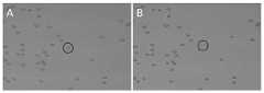
图1A和图1B分别为经过化学修饰和未经化学修饰的单细胞激光弹射基片结合单细胞的显微照片。1A and 1B are photomicrographs of chemically modified and non-chemically modified single-cell laser-ejected substrates bound to single cells, respectively.
图2A和图2B分别为结合有HeLa细胞的单细胞激光弹射基片在液相弹射分离前和液相弹射分离后的显微照片。FIG. 2A and FIG. 2B are micrographs of the single-cell laser-ejected substrate incorporating HeLa cells before and after liquid-phase ejection separation, respectively.
图3A和图3B分别为弹射分离前和弹射分离后聚焦于单细胞激光弹射基片的图像;图3C为微孔中接收到所分离的单细胞图像。Fig. 3A and Fig. 3B are images of the single-cell laser-ejection substrate before and after ejection separation, respectively; Fig. 3C is an image of the separated single cell received in the microwell.
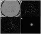
图4为酵母单细胞弹射分离后的培养结果;其中图4A、4B为液相弹射获取的单细胞酵母培养结果,图4C为干性弹射获取的单细胞酵母培养结果,其中图4A、4C为实物拍照照片,4B为采用10倍物镜获得的显微图像。Figure 4 is the culture result after yeast single-cell ejection separation; Figures 4A and 4B are the single-cell yeast culture results obtained by liquid-phase ejection, Figure 4C is the single-cell yeast culture result obtained by dry ejection, and Figures 4A and 4C are Photographs of real objects, 4B is a microscopic image obtained with a 10x objective lens.
图5为单个HeLa细胞经弹射分离后进行单细胞MDA扩增及PCR验证的结果;其中,N0为PCR反应阴性对照,N为MDA阴性对照,P为MDA阳性对照,#7为无弹射细胞的对照孔,#1-#6、#8及#9为各弹射一个细胞的样品孔。Figure 5 shows the results of single-cell MDA amplification and PCR verification of single HeLa cells separated by ejection; wherein, N0 is the negative control of PCR reaction, N is the negative control of MDA, P is the positive control of MDA, and #7 is the cell without ejection The control wells, #1-#6, #8 and #9 are sample wells where one cell was ejected each.
图6A、6B分别为HER-2抗体修饰的单细胞激光弹射基片结合MCF-7及MCF-7/HER-2细胞的亮场图像和荧光图像(10倍物镜),其中颜色较亮的细胞为HER2蛋白高表达的乳腺癌细胞,颜色较暗的细胞为普通MCF-7细胞;图6C为单个MCF-7/HER-2细胞经液相弹射分离后的对比图像(10倍物镜);图6D为微孔接收装置中所接收到的单细胞图像(40倍物镜)。Figures 6A and 6B are the bright-field images and fluorescence images (10x objective) of HER-2 antibody-modified single-cell laser-ejected substrates combined with MCF-7 and MCF-7/HER-2 cells, respectively, in which the brighter cells are breast cancer cells with high expression of HER2 protein, and the darker cells are ordinary MCF-7 cells; Figure 6C is the contrast image of a single MCF-7/HER-2 cell separated by liquid-phase ejection (10x objective); Figure 6C 6D is the single-cell image received in the microwell receiver (40x objective).
具体实施方式Detailed ways
实施例1:单细胞激光弹射基片的制作及单细胞结合Example 1: Fabrication of Single Cell Laser Ejection Substrate and Single Cell Combination
单细胞激光弹射基片依次包括基底层和镀膜层,其中镀膜层表面经化学修饰偶联有氨基基团或特异性抗体。基底层采用光透明材料,包括但不限于硅酸盐玻璃、石英玻璃、氟化钙玻璃中的一种或多种;镀膜层材料为透明金属复合氧化物薄膜材料,包括但不限于ITO、FTO、AZO中的一种或多种,采用喷涂、蒸发、溅射等沉积方式在基底层表面形成镀膜层,镀膜层经氧气等离子体处理后,将单细胞激光弹射基片迅速放入0.5%-5%的氨基为末端的硅烷偶联试剂(如3-氨基丙基-三甲氧基硅烷,APTMS)甲苯溶液中,处理0.5-2个小时,随后取出,经无水乙醇彻底清洗,氮气吹干,置80℃烘箱中干燥处理1-3个小时,使镀膜层表面偶联氨基基团。正常培养的HeLa细胞经离心清洗后重悬于pH7.0-7.4的PBS缓冲溶液中,将一定量的细胞悬液滴涂于上述经处理后的单细胞激光弹射基片表面,在37℃培养箱中孵育10-30分钟;随后,PBS溶液冲洗2-3次以除去未被结合的细胞。由于单细胞激光弹射基片表面带有氨基基团,其在PBS溶液中解离使表面带有正电荷;而细胞在PBS中表面呈负电性,由于静电作用,细胞被吸引并附着于单细胞激光弹射基片表面。The single-cell laser ejection substrate sequentially includes a base layer and a coating layer, wherein the surface of the coating layer is chemically modified and coupled with amino groups or specific antibodies. The base layer is made of optically transparent materials, including but not limited to one or more of silicate glass, quartz glass, and calcium fluoride glass; the coating layer material is a transparent metal composite oxide film material, including but not limited to ITO, FTO , one or more of AZO, a coating layer is formed on the surface of the base layer by spraying, evaporation, sputtering and other deposition methods. After the coating layer is treated by oxygen plasma, the single-cell laser ejection substrate is quickly put into 0.5%- 5% amino-terminated silane coupling reagent (such as 3-aminopropyl-trimethoxysilane, APTMS) in toluene solution, treated for 0.5-2 hours, then taken out, thoroughly washed with absolute ethanol, and dried with nitrogen , put it in an oven at 80 °C for 1-3 hours to dry, so that the surface of the coating layer is coupled with amino groups. The normal cultured HeLa cells were washed by centrifugation and resuspended in PBS buffer solution with pH 7.0-7.4. A certain amount of cell suspension was drop-coated on the surface of the treated single-cell laser ejection substrate, and cultured at 37°C. Incubate in the box for 10-30 minutes; then, rinse with PBS solution 2-3 times to remove unbound cells. Since the surface of the single-cell laser-ejected substrate has amino groups, which dissociate in PBS solution, the surface is positively charged; while the surface of cells in PBS is negatively charged, due to electrostatic effect, cells are attracted and attached to single cells Laser catapults the substrate surface.
图1A和图1B分别为经过化学修饰和未经化学修饰的单细胞激光弹射基片结合单细胞的显微照片,经过对比可知,未经化学修饰的单细胞激光弹射基片表面无法直接吸附和结合HeLa细胞。Figure 1A and Figure 1B are micrographs of chemically modified and non-chemically modified single-cell laser-ejection substrates combined with single cells, respectively. After comparison, it can be seen that the surface of the chemically-modified single-cell laser-ejection substrate cannot directly adsorb and Binds to HeLa cells.
实施例2:动物细胞的液相单细胞弹射分离及获取Example 2: Liquid-phase single-cell ejection separation and acquisition of animal cells
按照实施例1中方法将HeLa细胞结合于单细胞激光弹射基片表面,冲洗除去未被结合的多余HeLa细胞,并使HeLa细胞始终处于液相缓冲体系中以保持细胞活性;随后利用显微镜观察测量结合于单细胞激光弹射基片表面的HeLa细胞,并采用脉冲激光弹射方法选择单个HeLa细胞进行弹射分离,可获得如图2所示的结果,从图中画圈位置可看出原有HeLa细胞的位置已成为空白,说明HeLa单细胞已从单细胞激光弹射基片表面分离。The HeLa cells were bound to the surface of the single-cell laser ejection substrate according to the method in Example 1, the excess HeLa cells that were not bound were washed away, and the HeLa cells were always kept in the liquid buffer system to maintain cell viability; then observed and measured with a microscope Combined with the single-cell laser ejection of HeLa cells on the surface of the substrate, and use the pulsed laser ejection method to select a single HeLa cell for ejection separation, the results shown in Figure 2 can be obtained, and the original HeLa cells can be seen from the circled position in the figure The position has become blank, indicating that HeLa single cells have been detached from the surface of the single-cell laser-ejected substrate.
经弹射分离后的单细胞可通过微孔装置接收,微孔装置位于单细胞激光弹射基片下方,经弹射分离后的单细胞从基片表面掉落至微孔底部,图3A和图3B分别为弹射分离前和弹射分离后聚焦于单细胞激光弹射基片的图像,弹射分离后单细胞掉落至微孔底部,造成了图像的脱焦;图3C为聚焦于微孔底部的图像。The single cells separated by ejection can be received through the microwell device. The microwell device is located under the single cell laser ejection substrate. The single cells separated by ejection fall from the surface of the substrate to the bottom of the microwell, as shown in Figure 3A and Figure 3B, respectively. The images before and after ejection separation were focused on the single-cell laser ejection substrate. After ejection separation, the single cell fell to the bottom of the microwell, causing the image to be defocused; Figure 3C is the image focused on the bottom of the microwell.
实施例3:酵母单细胞液相弹射分离及培养Example 3: Yeast single cell liquid-phase ejection separation and culture
采用液相单细胞弹射方法,可最大程度地保护细胞活性,对分离获得的单细胞可进一步进行培养增殖。采用实施例1及实施例2所述的方法,对酵母细胞进行了液相单细胞弹射分离,同时采用微孔接收所分离的单细胞,并在培养液中对所获得的酵母细胞进行培养,12小时后可获得图4所示结果。采用液相弹射所获得的9个单细胞中有7个活性未受损伤,显示了不同程度增殖(图4A、4B)。而采用干性环境下的弹射分离,所获得的单细胞均未能成功培养增殖(图4C)。The single-cell ejection method in liquid phase can protect the cell activity to the greatest extent, and the isolated single cells can be further cultured and proliferated. Using the methods described in Example 1 and Example 2, liquid-phase single-cell ejection separation was performed on yeast cells, and at the same time, the separated single cells were received by micropores, and the obtained yeast cells were cultured in the culture medium, The results shown in Figure 4 were obtained after 12 hours. Seven of the nine single cells obtained using liquid-phase ejection were not damaged in activity, showing varying degrees of proliferation (Fig. 4A, 4B). However, the single cells obtained by ejection separation in a dry environment were not successfully cultured and proliferated (Fig. 4C).
实施例4:单细胞扩增测序Example 4: Single Cell Amplification Sequencing
采用液相弹射方法所分离获取的单细胞可进一步进行基因组测序。采用实施例1及实施例2所述的方法,对HeLa细胞进行了液相单细胞弹射分离,同时采用微孔接收所分离的单个HeLa细胞;随后对细胞进行裂解并采用MDA方法对其基因组DNA进行扩增,8小时后以MDA产物为模板,分别进行16S rRNA,18S rRNA及SSU rRNA的PCR扩增,对MDA产物及PCR产物进行凝胶电泳成像可获得图5所示结果。其中,N0为PCR反应阴性对照,N为MDA阴性对照,P为MDA阳性对照,#7为无弹射细胞的对照孔,#1-#6、#8及#9为各弹射一个HeLa细胞的结果。Single cells isolated by liquid ejection can be further sequenced. Using the methods described in Example 1 and Example 2, HeLa cells were isolated by liquid-phase single-cell ejection, and the isolated single HeLa cells were received by micropores; the cells were then lysed and their genomic DNA was isolated by MDA method. After 8 hours of amplification, using the MDA product as a template, PCR amplification of 16S rRNA, 18S rRNA and SSU rRNA was performed respectively, and the results shown in Figure 5 were obtained by gel electrophoresis imaging of the MDA product and the PCR product. Among them, N0 is the negative control of the PCR reaction, N is the negative control of MDA, P is the positive control of MDA, #7 is the control well without ejected cells, #1-#6, #8 and #9 are the ejection wells of one HeLa cell each. result.
图5显示,采用液相弹射技术,所获得的单细胞均可成功进行MDA扩增,经18S和SSU标记基因确认可认定MDA产物大部分来自于单细胞基因组;同时,经16S标记基因证实,采用本方法可对外源污染进行较好的控制,能够获得较为纯净的单细胞基因组。进一步的,可对单细胞MDA扩增产物进行常规测序以分析其基因组信息。Figure 5 shows that the single cells obtained can be successfully amplified by MDA using the liquid-phase ejection technique. The 18S and SSU marker genes confirm that most of the MDA products come from the single-cell genome. By adopting this method, exogenous contamination can be better controlled, and a relatively pure single-cell genome can be obtained. Further, single-cell MDA amplification products can be routinely sequenced to analyze their genomic information.
实施例5:CTC单细胞分选Example 5: CTC single cell sorting
本发明所提出的单细胞激光弹射基片与液相单细胞弹射方法,可用于癌症患者血液中CTC细胞的富集捕获和单细胞分选,以进一步进行CTC单细胞测序,对其表达谱和突变位点进行检测,从而用于早期诊断、临床用药评价及预后监测等过程。本实施例中,采用人表皮生长因子受体-2(HER2)蛋白高表达的乳腺癌细胞MCF-7/HER-2模拟血液中CTC细胞,以HER2蛋白特异性抗体对弹射基片进行修饰,对MCF-7/HER-2进行特异性捕获;随后采用液相单细胞弹射方法分离单个MCF-7/HER-2细胞做进一步分析。图6A、6B所示为经抗体修饰过的单细胞激光弹射基片对MCF-7及MCF-7/HER-2细胞的捕获效果,图6C为单个MCF-7/HER-2细胞经液相弹射分离后的显微图像,图6D为微孔接收装置中所接收到的单细胞图像。The single-cell laser ejection substrate and the liquid-phase single-cell ejection method proposed in the present invention can be used for the enrichment capture and single-cell sorting of CTC cells in the blood of cancer patients, so as to further perform CTC single-cell sequencing, its expression profile and Mutation sites can be detected for early diagnosis, clinical drug evaluation, and prognosis monitoring. In this example, breast cancer cells MCF-7/HER-2 with high expression of human epidermal growth factor receptor-2 (HER2) protein were used to simulate CTC cells in blood, and the ejection substrate was modified with HER2 protein-specific antibodies. MCF-7/HER-2 was specifically captured; single MCF-7/HER-2 cells were subsequently isolated for further analysis using a liquid-phase single-cell ejection method. Figures 6A and 6B show the capture effect of antibody-modified single-cell laser ejection substrates on MCF-7 and MCF-7/HER-2 cells, and Figure 6C shows single MCF-7/HER-2 cells after liquid phase Microscopic image after ejection separation, Figure 6D is an image of a single cell received in the microwell receiving device.
在本发明提及的所有文献都在本申请中引用作为参考,就如同每一篇文献被单独引用作为参考那样。此外应理解,在阅读了本发明的上述讲授内容之后,本领域技术人员可以对本发明作各种改动或修改,这些等价形式同样落于本申请所附权利要求书所限定的范围。All documents mentioned herein are incorporated by reference in this application as if each document were individually incorporated by reference. In addition, it should be understood that after reading the above teaching content of the present invention, those skilled in the art can make various changes or modifications to the present invention, and these equivalent forms also fall within the scope defined by the appended claims of the present application.
Claims (7)
Translated fromChinesePriority Applications (2)
| Application Number | Priority Date | Filing Date | Title |
|---|---|---|---|
| CN201810041167.0ACN110042053B (en) | 2018-01-16 | 2018-01-16 | A single-cell laser ejection substrate, method and application |
| PCT/CN2019/071791WO2019141164A1 (en) | 2018-01-16 | 2019-01-15 | Single cell laser ejection substrate, method and application |
Applications Claiming Priority (1)
| Application Number | Priority Date | Filing Date | Title |
|---|---|---|---|
| CN201810041167.0ACN110042053B (en) | 2018-01-16 | 2018-01-16 | A single-cell laser ejection substrate, method and application |
Publications (2)
| Publication Number | Publication Date |
|---|---|
| CN110042053A CN110042053A (en) | 2019-07-23 |
| CN110042053Btrue CN110042053B (en) | 2022-07-01 |
Family
ID=67273035
Family Applications (1)
| Application Number | Title | Priority Date | Filing Date |
|---|---|---|---|
| CN201810041167.0AActiveCN110042053B (en) | 2018-01-16 | 2018-01-16 | A single-cell laser ejection substrate, method and application |
Country Status (2)
| Country | Link |
|---|---|
| CN (1) | CN110042053B (en) |
| WO (1) | WO2019141164A1 (en) |
Family Cites Families (7)
| Publication number | Priority date | Publication date | Assignee | Title |
|---|---|---|---|---|
| WO1997029354A1 (en)* | 1996-02-05 | 1997-08-14 | Bayer Aktiengesellschaft | Process and device for sorting and for extraction of biological objects arranged on planar means, such as biological cells or cell organelles, histological sections, chromosome particles etc. using laser beams |
| EP1561107A1 (en)* | 2002-11-13 | 2005-08-10 | Micromet AG | Method for identifying antigen specific b cells |
| DE102006033875A1 (en)* | 2006-07-21 | 2008-01-31 | Siemens Ag | Analysis system based on porous material for highly parallel single cell detection |
| CN103353452B (en)* | 2013-07-12 | 2016-08-17 | 上海合森生物科技有限公司 | Cell carrier chip and utilize its method carrying out unicellular Rapid identification or sorting |
| GB201603088D0 (en)* | 2016-02-23 | 2016-04-06 | Isis Innovation | Cell sorting |
| CN106754439B (en)* | 2016-12-28 | 2019-12-06 | 广东工业大学 | Method for separating single cell |
| CN107490545A (en)* | 2017-07-21 | 2017-12-19 | 中国科学院青岛生物能源与过程研究所 | A kind of unicellular automation of high-flux microorganism sorts and reception system |
- 2018
- 2018-01-16CNCN201810041167.0Apatent/CN110042053B/enactiveActive
- 2019
- 2019-01-15WOPCT/CN2019/071791patent/WO2019141164A1/ennot_activeCeased
Also Published As
| Publication number | Publication date |
|---|---|
| WO2019141164A1 (en) | 2019-07-25 |
| CN110042053A (en) | 2019-07-23 |
Similar Documents
| Publication | Publication Date | Title |
|---|---|---|
| EP2593229B1 (en) | Direct clone analysis and selection method | |
| CN107589105B (en) | An all-in-one device for fast Raman measurement and laser ejection sorting of single cells | |
| CN107532333A (en) | Microscreening devices, methods and products | |
| WO2012141202A1 (en) | Cell-adhering light-controllable substrate | |
| WO2022041644A1 (en) | Erythrocyte biomimetic coating for enriching circulating tumor cells | |
| Annunziata et al. | An optimised method for intact nuclei isolation from diatoms | |
| JP2023116581A (en) | IDENTIFICATION AND ISOLATION OF HUMAN CORNEAL ENDOTHELIAL CELLS (HCECs) | |
| EP3265809B1 (en) | Method and device for detecting in real time a secreted compound and the secretion target, and uses thereof | |
| CN110042053B (en) | A single-cell laser ejection substrate, method and application | |
| CN114295828B (en) | Exosome chip liquid biopsy method | |
| Wang et al. | Functionalized microwell laser sorting method for single-cell viable microbial detection | |
| CN109557068B (en) | Integrated sorting device for single cell Raman measurement and laser microdissection | |
| CN109765381B (en) | Aptamer signal amplification-based platelet-derived growth factor PDGF-BB test strip and detection method | |
| US11692191B2 (en) | Method for separating, capturing, analyzing and retrieving cells and cell products by using microstructure | |
| Mukherjee et al. | Isolation and purification of various mammalian cells: single cell isolation | |
| EP1390467B1 (en) | Use of a miniature device for separating and isolating objects and separation method | |
| CN116809134A (en) | Microfluidic chip system and method for capturing living circulation tumor cells and comprehensively analyzing genetic behaviors | |
| Shadpour et al. | Enrichment and expansion of cells using antibody‐coated micropallet arrays | |
| CN114895040A (en) | A method of combining rapid immunofluorescence labeling and super-resolution imaging to observe nuclear proteins | |
| Spokevicius et al. | The use of Induced Somatic Sector Analysis (ISSA) for studying genes and promoters involved in wood formation and secondary stem development | |
| Hung et al. | One-to-one Fusion of Plant Protoplasts by Using Electrofusion Based on Electric Field Constriction | |
| García-Aguilar et al. | Localization of chromatin marks in Arabidopsis early embryos | |
| US20230160889A1 (en) | Screening method for biological specimens and screening separator | |
| CN103983640A (en) | Protein chip sample point quality control and tracing method | |
| Conn | Laser capture in microscopy and microdissection |
Legal Events
| Date | Code | Title | Description |
|---|---|---|---|
| PB01 | Publication | ||
| PB01 | Publication | ||
| SE01 | Entry into force of request for substantive examination | ||
| SE01 | Entry into force of request for substantive examination | ||
| GR01 | Patent grant | ||
| GR01 | Patent grant |