CN110022785B - Selecting a medical device for use in a medical procedure - Google Patents
Selecting a medical device for use in a medical procedureDownload PDFInfo
- Publication number
- CN110022785B CN110022785BCN201780068862.XACN201780068862ACN110022785BCN 110022785 BCN110022785 BCN 110022785BCN 201780068862 ACN201780068862 ACN 201780068862ACN 110022785 BCN110022785 BCN 110022785B
- Authority
- CN
- China
- Prior art keywords
- plane
- point
- heart
- laa
- centroid
- Prior art date
- Legal status (The legal status is an assumption and is not a legal conclusion. Google has not performed a legal analysis and makes no representation as to the accuracy of the status listed.)
- Active
Links
Images


Classifications
- A—HUMAN NECESSITIES
- A61—MEDICAL OR VETERINARY SCIENCE; HYGIENE
- A61B—DIAGNOSIS; SURGERY; IDENTIFICATION
- A61B34/00—Computer-aided surgery; Manipulators or robots specially adapted for use in surgery
- A61B34/10—Computer-aided planning, simulation or modelling of surgical operations
- A—HUMAN NECESSITIES
- A61—MEDICAL OR VETERINARY SCIENCE; HYGIENE
- A61B—DIAGNOSIS; SURGERY; IDENTIFICATION
- A61B17/00—Surgical instruments, devices or methods
- A61B17/12—Surgical instruments, devices or methods for ligaturing or otherwise compressing tubular parts of the body, e.g. blood vessels or umbilical cord
- A61B17/12022—Occluding by internal devices, e.g. balloons or releasable wires
- A61B17/12027—Type of occlusion
- A61B17/12031—Type of occlusion complete occlusion
- A—HUMAN NECESSITIES
- A61—MEDICAL OR VETERINARY SCIENCE; HYGIENE
- A61B—DIAGNOSIS; SURGERY; IDENTIFICATION
- A61B17/00—Surgical instruments, devices or methods
- A61B17/12—Surgical instruments, devices or methods for ligaturing or otherwise compressing tubular parts of the body, e.g. blood vessels or umbilical cord
- A61B17/12022—Occluding by internal devices, e.g. balloons or releasable wires
- A61B17/12099—Occluding by internal devices, e.g. balloons or releasable wires characterised by the location of the occluder
- A61B17/12122—Occluding by internal devices, e.g. balloons or releasable wires characterised by the location of the occluder within the heart
- A—HUMAN NECESSITIES
- A61—MEDICAL OR VETERINARY SCIENCE; HYGIENE
- A61B—DIAGNOSIS; SURGERY; IDENTIFICATION
- A61B17/00—Surgical instruments, devices or methods
- A61B17/00234—Surgical instruments, devices or methods for minimally invasive surgery
- A61B2017/00238—Type of minimally invasive operation
- A61B2017/00243—Type of minimally invasive operation cardiac
- A—HUMAN NECESSITIES
- A61—MEDICAL OR VETERINARY SCIENCE; HYGIENE
- A61B—DIAGNOSIS; SURGERY; IDENTIFICATION
- A61B17/00—Surgical instruments, devices or methods
- A61B17/00234—Surgical instruments, devices or methods for minimally invasive surgery
- A61B2017/00292—Surgical instruments, devices or methods for minimally invasive surgery mounted on or guided by flexible, e.g. catheter-like, means
- A—HUMAN NECESSITIES
- A61—MEDICAL OR VETERINARY SCIENCE; HYGIENE
- A61B—DIAGNOSIS; SURGERY; IDENTIFICATION
- A61B17/00—Surgical instruments, devices or methods
- A61B17/0057—Implements for plugging an opening in the wall of a hollow or tubular organ, e.g. for sealing a vessel puncture or closing a cardiac septal defect
- A61B2017/00575—Implements for plugging an opening in the wall of a hollow or tubular organ, e.g. for sealing a vessel puncture or closing a cardiac septal defect for closure at remote site, e.g. closing atrial septum defects
- A61B2017/00632—Occluding a cavity, i.e. closing a blind opening
- A—HUMAN NECESSITIES
- A61—MEDICAL OR VETERINARY SCIENCE; HYGIENE
- A61B—DIAGNOSIS; SURGERY; IDENTIFICATION
- A61B17/00—Surgical instruments, devices or methods
- A61B17/12—Surgical instruments, devices or methods for ligaturing or otherwise compressing tubular parts of the body, e.g. blood vessels or umbilical cord
- A61B17/12022—Occluding by internal devices, e.g. balloons or releasable wires
- A61B2017/1205—Introduction devices
- A—HUMAN NECESSITIES
- A61—MEDICAL OR VETERINARY SCIENCE; HYGIENE
- A61B—DIAGNOSIS; SURGERY; IDENTIFICATION
- A61B34/00—Computer-aided surgery; Manipulators or robots specially adapted for use in surgery
- A61B34/10—Computer-aided planning, simulation or modelling of surgical operations
- A61B2034/101—Computer-aided simulation of surgical operations
- A61B2034/105—Modelling of the patient, e.g. for ligaments or bones
- A—HUMAN NECESSITIES
- A61—MEDICAL OR VETERINARY SCIENCE; HYGIENE
- A61B—DIAGNOSIS; SURGERY; IDENTIFICATION
- A61B34/00—Computer-aided surgery; Manipulators or robots specially adapted for use in surgery
- A61B34/10—Computer-aided planning, simulation or modelling of surgical operations
- A61B2034/107—Visualisation of planned trajectories or target regions
- A—HUMAN NECESSITIES
- A61—MEDICAL OR VETERINARY SCIENCE; HYGIENE
- A61B—DIAGNOSIS; SURGERY; IDENTIFICATION
- A61B34/00—Computer-aided surgery; Manipulators or robots specially adapted for use in surgery
- A61B34/10—Computer-aided planning, simulation or modelling of surgical operations
- A61B2034/108—Computer aided selection or customisation of medical implants or cutting guides
- A—HUMAN NECESSITIES
- A61—MEDICAL OR VETERINARY SCIENCE; HYGIENE
- A61B—DIAGNOSIS; SURGERY; IDENTIFICATION
- A61B34/00—Computer-aided surgery; Manipulators or robots specially adapted for use in surgery
- A61B34/25—User interfaces for surgical systems
- A61B2034/256—User interfaces for surgical systems having a database of accessory information, e.g. including context sensitive help or scientific articles
- A—HUMAN NECESSITIES
- A61—MEDICAL OR VETERINARY SCIENCE; HYGIENE
- A61B—DIAGNOSIS; SURGERY; IDENTIFICATION
- A61B90/00—Instruments, implements or accessories specially adapted for surgery or diagnosis and not covered by any of the groups A61B1/00 - A61B50/00, e.g. for luxation treatment or for protecting wound edges
- A61B90/36—Image-producing devices or illumination devices not otherwise provided for
- A61B90/37—Surgical systems with images on a monitor during operation
- A61B2090/374—NMR or MRI
- A—HUMAN NECESSITIES
- A61—MEDICAL OR VETERINARY SCIENCE; HYGIENE
- A61B—DIAGNOSIS; SURGERY; IDENTIFICATION
- A61B90/00—Instruments, implements or accessories specially adapted for surgery or diagnosis and not covered by any of the groups A61B1/00 - A61B50/00, e.g. for luxation treatment or for protecting wound edges
- A61B90/36—Image-producing devices or illumination devices not otherwise provided for
- A61B90/37—Surgical systems with images on a monitor during operation
- A61B2090/376—Surgical systems with images on a monitor during operation using X-rays, e.g. fluoroscopy
- A61B2090/3762—Surgical systems with images on a monitor during operation using X-rays, e.g. fluoroscopy using computed tomography systems [CT]
- A—HUMAN NECESSITIES
- A61—MEDICAL OR VETERINARY SCIENCE; HYGIENE
- A61B—DIAGNOSIS; SURGERY; IDENTIFICATION
- A61B90/00—Instruments, implements or accessories specially adapted for surgery or diagnosis and not covered by any of the groups A61B1/00 - A61B50/00, e.g. for luxation treatment or for protecting wound edges
- A61B90/36—Image-producing devices or illumination devices not otherwise provided for
- A61B90/37—Surgical systems with images on a monitor during operation
- A61B2090/378—Surgical systems with images on a monitor during operation using ultrasound
- A—HUMAN NECESSITIES
- A61—MEDICAL OR VETERINARY SCIENCE; HYGIENE
- A61B—DIAGNOSIS; SURGERY; IDENTIFICATION
- A61B2576/00—Medical imaging apparatus involving image processing or analysis
Landscapes
- Health & Medical Sciences (AREA)
- Surgery (AREA)
- Life Sciences & Earth Sciences (AREA)
- Engineering & Computer Science (AREA)
- Heart & Thoracic Surgery (AREA)
- Veterinary Medicine (AREA)
- Nuclear Medicine, Radiotherapy & Molecular Imaging (AREA)
- Biomedical Technology (AREA)
- Public Health (AREA)
- Medical Informatics (AREA)
- Molecular Biology (AREA)
- Animal Behavior & Ethology (AREA)
- General Health & Medical Sciences (AREA)
- Robotics (AREA)
- Reproductive Health (AREA)
- Vascular Medicine (AREA)
- Apparatus For Radiation Diagnosis (AREA)
- Ultra Sonic Daignosis Equipment (AREA)
- Surgical Instruments (AREA)
Abstract
Description
Translated fromChinese相关申请的交叉引用Cross References to Related Applications
本申请要求2016年11月8日提交的美国临时申请第62/419,072 号的权益,其全部内容通过引用在此并入。This application claims the benefit of US Provisional Application No. 62/419,072, filed November 8, 2016, the entire contents of which are hereby incorporated by reference.
技术领域technical field
本公开一般涉及用于医疗程序的围手术期计划,并且更具体地涉及包括心脏在内的非侵入性医疗程序的围手术期计划,包括选择待在此类程序中使用的医疗装置。The present disclosure relates generally to perioperative planning for medical procedures, and more particularly to perioperative planning for non-invasive medical procedures, including cardiac, including selection of medical devices to be used in such procedures.
背景background
心脏装置的非侵入性经皮植入对医生提出了某些挑战。与手术侵入性程序诸如例如开心手术相反,执行非侵入性心脏植入程序的医生具有有限的视野并且通常限于在程序期间使用由二维(2D)成像模式 (如,超声、荧光透视等)生成的图像指导。由于医生在执行程序期间通常局限于2D成像,因此需要进行适当的围手术期计划和评价,以准确评估和确定例如某些解剖结构的尺寸以及待在程序期间使用的装置(如,导管)的一种或多种类型和/或一个或多个尺寸。Non-invasive percutaneous implantation of cardiac devices presents certain challenges to physicians. In contrast to surgically invasive procedures such as, for example, open-heart surgery, physicians performing non-invasive cardiac implant procedures have a limited field of view and are typically limited to the use of images generated by two-dimensional (2D) imaging modalities (e.g., ultrasound, fluoroscopy, etc.) during the procedure. image guidance. Since physicians are usually limited to 2D imaging during procedures, proper perioperative planning and evaluation is required to accurately assess and determine, for example, the dimensions of certain anatomical structures and the dimensions of devices (e.g., catheters) to be used during the procedure. One or more types and/or one or more sizes.
然而,与程序内指导一样,传统的围手术期计划技术通常基于采用2D成像的成像平台和模式。因此,与植入程序本身一样,由于所使用的传统2D成像的固有限制,这种程序的围手术期计划对医生提出了挑战。However, as with intra-procedural guidance, traditional perioperative planning techniques are often based on imaging platforms and modalities employing 2D imaging. Thus, like the implantation procedure itself, the perioperative planning of such a procedure presents challenges to physicians due to the inherent limitations of the traditional 2D imaging used.
因此,需要一种围手术期计划方法和系统,其能最小化和/或消除传统围手术期计划方法/技术中的一个或多个上述缺陷。Accordingly, there is a need for a perioperative planning method and system that minimizes and/or eliminates one or more of the above-mentioned deficiencies in conventional perioperative planning methods/techniques.
概要summary
根据一个实施方案,提供了选择用于执行医疗程序的医疗装置的方法。该方法包括获取与患者身体的目标解剖区相关的图像数据,使用所获取的图像数据生成所述目标解剖区的多维描绘,相对于所述多维描绘限定多个点,基于限定的多个点确定一个或多个测量值,和基于确定的测量值选择待使用的医疗装置。According to one embodiment, a method of selecting a medical device for performing a medical procedure is provided. The method includes acquiring image data related to a target anatomical region of the patient's body, generating a multidimensional depiction of the target anatomical region using the acquired image data, defining a plurality of points with respect to the multidimensional depiction, determining based on the defined plurality of points One or more measurements, and selecting a medical device to use based on the determined measurements.
根据另一实施方案,提供了存储有指令的非暂时性计算机可读存储介质。使得存储的指令当由一个或多个电子处理器执行时,导致所述一个或多个处理器实施以下方法:获取与患者身体的目标解剖区相关的图像数据;使用所获取的图像数据生成所述目标解剖区的多维描绘;相对于所述多维描绘限定多个点;基于限定的多个点确定一个或多个测量值;和基于确定的测量值选择待使用的医疗装置。According to another embodiment, a non-transitory computer-readable storage medium storing instructions is provided. causing the stored instructions, when executed by the one or more electronic processors, to cause the one or more processors to implement the method of: acquiring image data related to a target anatomical region of the patient's body; using the acquired image data to generate a A multidimensional depiction of the target anatomical region; defining a plurality of points relative to the multidimensional depiction; determining one or more measurements based on the defined plurality of points; and selecting a medical device to be used based on the determined measurements.
根据又一个实施方案,提供了选择用于医疗程序中的医疗装置的系统。该系统包括电子处理器和电连接至电子处理器并具有在其上存储的指令的电子存储装置。处理器被配置为能访问存储装置并执行存储其中的指令,使得其可操作来获取与患者身体的目标解剖区相关的图像数据,使用所获取的图像数据生成目标解剖区的多维描绘,相对于多维描绘限定多个点,基于限定的多个点确定一个或多个测量值,和基于所确定的测量值选择待使用的医疗装置。According to yet another embodiment, a system for selecting a medical device for use in a medical procedure is provided. The system includes an electronic processor and an electronic storage device electrically connected to the electronic processor and having instructions stored thereon. The processor is configured to access the memory device and execute instructions stored therein such that it is operable to acquire image data related to a target anatomical region of the patient's body, use the acquired image data to generate a multidimensional depiction of the target anatomical region, relative to The multi-dimensional mapping defines a plurality of points, determines one or more measurements based on the defined plurality of points, and selects a medical device to be used based on the determined measurements.
附图简述Brief description of the drawings
在下文中将结合附图描述本发明的一个或多个实施方案,其中类似的名称表示类似的元件,并且其中:Hereinafter, one or more embodiments of the invention will be described with reference to the accompanying drawings, wherein like designations refer to like elements, and in which:
图1是人心脏的一部分的示意的图解视图;Figure 1 is a schematic diagrammatic view of a portion of a human heart;
图2是用于执行本文所述方法的一个或多个实施方案的系统的说明性实施方案的示意性框图;Figure 2 is a schematic block diagram of an illustrative embodiment of a system for performing one or more embodiments of the methods described herein;
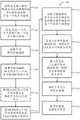
图3是可用于选择或确定待在医疗程序中使用的医疗装置的方法的说明性实施方案的流程图;3 is a flow diagram of an illustrative embodiment of a method that may be used to select or determine a medical device to be used in a medical procedure;
图4a-4c是可用于例如执行图3所示方法的一个或多个步骤的患者心脏部分的计算机断层扫描(CT)图像;4a-4c are computed tomography (CT) images of portions of a patient's heart that may be used, for example, to perform one or more steps of the method shown in FIG. 3;
图5-13是可以用于执行图3所示方法的一个或多个步骤的模型的各种描述,并且示出了可以如何实施图3中所示的方法的说明性实施方案;5-13 are various depictions of models that may be used to perform one or more steps of the method shown in FIG. 3 and show illustrative embodiments of how the method shown in FIG. 3 may be implemented;
图14-17是可以在图3示出的方法的一个或多个步骤中确定的各测量值的图解的示意图,其可用于图3示出的方法的一个或多个其他步骤中。应当理解,图中所示的任何特定测量值仅用于说明目的,并不意味着以任何方式进行限制;14-17 are schematic diagrams of graphical representations of measurements that may be determined in one or more steps of the method shown in FIG. 3 , which may be used in one or more other steps of the method shown in FIG. 3 . It should be understood that any particular measurements shown in the graphs are for illustrative purposes only and are not meant to be limiting in any way;
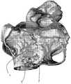
图18示出了在其中输入医疗装置的模型的解剖结构的描绘,用于执行评价例如医疗装置的恰当性的方法的一个或多个步骤。Fig. 18 shows a depiction of the anatomy of a model into which a medical device is input for performing one or more steps of a method of evaluating, for example, the adequacy of a medical device.
详述detail
本文描述的系统和方法可以帮助医生进行经皮程序的操作前计划 (还被称为“围手术期计划”),例如但不限于,涉及植入诸如人工心瓣、左心耳(LAA)封堵装置等的医疗装置的程序。通常,本文描述的系统和方法使用高级成像和建模策略来准确地评估各种目标解剖结构的位置和尺寸,并确定或选择待用于执行医疗程序(如,植入程序)的医疗装置(如,导管)的理想或最佳类型和尺寸,其对待执行程序的特定患者是特异性的。尽管该系统和方法可适用于计划和评价各种程序,但特别适用的是涉及LAA的程序,并且特别是用于封堵LAA的装置的植入。因此,下面的描述将主要涉及选择呈导管形式的用于将封堵装置递送至LAA的医疗装置。然而,应当理解,本文阐述的各种教导也可以应用于多种其他程序,即心脏相关的和其他的程序。例如,该教导可以应用于选择用于将人工二尖瓣递送至患者心脏的二尖瓣环的导管。因此,应当理解,本公开不旨在限于将本文描述的系统和方法用于任何特定类型的程序。The systems and methods described herein can assist physicians in preoperative planning (also referred to as "perioperative planning") of percutaneous procedures, such as, but not limited to, those involving implantation of devices such as artificial heart valves, left atrial appendage (LAA) devices and other medical device procedures. In general, the systems and methods described herein use advanced imaging and modeling strategies to accurately assess the location and size of various target anatomical structures and determine or select medical devices ( For example, the ideal or optimal type and size of a catheter) that is specific to the particular patient on whom the procedure is to be performed. While the system and method are applicable to planning and evaluating a variety of procedures, they are particularly applicable to procedures involving the LAA, and in particular the implantation of devices for occluding the LAA. Accordingly, the following description will primarily relate to selecting a medical device in the form of a catheter for delivering the occlusion device to the LAA. It should be understood, however, that the various teachings set forth herein may also be applied to a variety of other procedures, cardiac related and otherwise. For example, the teachings can be applied to selecting a catheter for delivering a prosthetic mitral valve to the mitral annulus of a patient's heart. Accordingly, it should be understood that the present disclosure is not intended to be limited to the use of the systems and methods described herein with any particular type of program.
出于上下文的目的,图1示出了人心脏10的一部分,包括LAA 12、左心房14、左心室16、右心室18、右心房20、心房壁22和下腔静脉 (IVC)24。LAA是口袋状,从左心房接收血液并将血液排入左心房。尽管LAA在心脏手术中没有贡献或提供重要的功能,但它是一个令人关注的位点,因为它涉及心脏血栓形成。更具体地,LAA在心脏内提供血液可以收集或汇集、凝结并形成凝块的区域。众所周知,经过患者血流的血凝块和其他栓塞可能具有有害作用。LAA中的凝块可能从LAA迁移到左心房中,穿过二尖瓣进入左心室,经过主动脉瓣进入主动脉并进入患者的血流,在那里它可能堵塞血液流动至,例如心脏、肺脏或大脑,可能导致心脏病发作、中风或其他不良事件。For purposes of context, FIG. 1 shows a portion of a
为了减轻来自LAA的凝块的迁移,可以执行程序以封闭LAA。例如,可以将封堵装置诸如例如从Boston Scientific商购获得的
图2描绘了用于确定或选择待用于将医疗装置递送至患者身体的目标解剖区的医疗装置的系统26的说明性实施方案。在一个实施方案中,医疗装置可以包括可植入装置诸如人工心瓣(如,人工二尖瓣)或封堵装置(如,LAA封堵装置),并且目标解剖区是患者心脏的至少一个区域。在说明性实施方案中,除了可能的其他组件,系统26还包括电子控制单元(ECU)28、显示装置30和一个或多个用户界面装置32。FIG. 2 depicts an illustrative embodiment of a
ECU 28可包括一个或多个电子处理器34,其具有一个或多个电输入和一个或多个电输出。电子处理器34可包括本领域已知的任何适合的电子处理器(如,微处理器、微控制器、ASIC等),其被配置为能执行电子指令。ECU 28 may include one or more
ECU 28还可以包括或电连接至和/或被配置为能访问电子存储装置36。存储装置36可以是处理器34的一部分或电连接至处理器34 和/或处理器34可访问的。电子存储装置36可以包括本领域已知的任何适合的存储装置,并且可以在其中或其上存储各种数据、信息和/ 或指令。在一个实施方案中,存储装置36具有在其中或其上存储的用于软件、固件、程序、算法、脚本、应用、数据结构(如,查找表)等中的一个或多个的信息和指令,其可以管理和/或促进本文所述的方法的全部或一部分。在至少一些实施方案中,存储装置36可包括计算机可读的存储介质(如非暂时性或非瞬态存储介质),其可包括用于存储机器或电子处理器/计算装置(如,处理器34)可读形式的信息的任何机制,包括但不限于:磁存储介质(如软盘);光存储介质(如CD-ROM);磁光存储介质;只读存储器(ROM);随机存取存储器(RAM);可删可编程存储器(如EPROM和EEPROM);闪存;或用于存储此类信息/指令的电介质或其他类型的介质。此外,可以使用光学、声学或其他形式的传播信号(如,载体波、红外信号、数字信号或其他类型的信号或介质)来传送程序指令。The ECU 28 may also include or be electrically connected to and/or be configured to have access to an
在任何情况下,在一个实施方案中,处理器34可以访问存储装置 36并执行和/或使用那个或那些指令和信息来实施或执行本文描述的功能和方法中的一些或全部。In any case, in one embodiment,
显示装置30可包括本领域已知的多种显示装置,例如但不限于液晶显示器(LCD)、阴极射线管(CRT)、等离子体或发光二极管(LED)监视器或显示器。显示装置30电连接或耦合至ECU 28和被配置为能由 ECU 28控制,使得例如由ECU 28生成、获得或获取的解剖结构的图像、模型或描绘-包括用于执行下述方法的那些-可以在其上显示,并且可以用于本文所述的目的。另外,在其中ECU 28可以被配置为能生成交互式图形用户界面(GUI)(其允许例如医生操纵显示装置上显示的图像或模型(如,旋转/移动模型、切割模型、隐藏模型的一部分、相对于模型/描绘限定点或平面等)、帮助确定测量值等)的一个实施方案中,显示装置30也可以显示此类GUI。在任何情况下,显示装置 30被配置为能接收来自ECU 28的电信号并显示由接收信号表示的内容,该内容可由例如医生查看。
一个或多个用户界面装置32可包括本领域中已知的多种适合的装置。例如但不限于,用户输入装置32可以包括触摸屏(如,LCD触摸屏)、按键、键盘、计算机鼠标或滚球和/或操纵杆中的一个或组合以引用一些可能性。在某些实现方式中,显示装置30和用户输入装置 32可以组合在一起成为单个装置,使得它们同样是一体的。无论一个或多个用户界面装置采用何种特定形式,一个或多个用户输入装置32 可以电连接或耦合(如,经由有线或无线连接)至ECU 28,并且被配置为能促进用户(如,医生)和系统26,以及特别是其ECU 28之间的通信。更具体地,一个或多个用户界面装置32可以允许医生操纵显示装置30上显示的图像或模型/描绘(如,隐藏模型/描绘的一部分,相对于模型/描绘限定点或平面,旋转/移动模型/描绘等),选择或命令确定与在显示装置30上显示的模型/描绘中表示的解剖结构有关的确定值等。The one or more
虽然上面已经描述了系统26的某些组件,但是应当理解,在一些实施方式中,系统26可以包括比上述布置中包括的更多或更少的组件。因此,本公开不旨在限于系统26的任一个或多个特定实施方式或布置。While certain components of
现在转到图3,示出了用于确定或选择待用于执行医疗程序的医疗装置的方法(方法100)的说明性实施方案。更具体地,图3示出了选择待用于执行程序的长型医疗装置(如,导管)的方法,在该程序期间医疗装置被递送并植入患者身体的特定解剖区中的目标结构内。在特定的说明性实施方案中,待植入的装置是LAA封堵装置,并因此其中目标结构(即,LAA)所位于的解剖区包括至少一部分患者心脏。出于说明的目的,下面的描述将主要关于选择用于递送和放置LAA封堵装置的导管。然而,应当理解,本文描述的方法可以用于评价其他装置的放置,其中一些装置可以在下面描述。Turning now to FIG. 3 , an illustrative embodiment of a method (method 100 ) for determining or selecting a medical device to be used to perform a medical procedure is shown. More specifically, FIG. 3 illustrates a method of selecting an elongated medical device (e.g., a catheter) to be used in performing a procedure during which the medical device is delivered and implanted within a target structure in a specific anatomical region of the patient's body . In certain illustrative embodiments, the device to be implanted is a LAA occlusion device, and thus the anatomical region in which the target structure (ie, the LAA) is located includes at least a portion of the patient's heart. For purposes of illustration, the following description will be primarily concerned with selecting a catheter for delivery and placement of the LAA occlusion device. It should be understood, however, that the methods described herein may be used to evaluate the placement of other devices, some of which may be described below.
在至少一些实施方案中,方法100的所有步骤可以由恰当或适合配置的系统执行或实施,例如但不限于单独或与来自用户(如,医生) 的输入结合的上述系统26。然而,在其他实施方案中,可以通过不同的系统执行或实施某些步骤但是不是所有步骤,使得某些步骤可以由一个系统(如,系统26)执行,并且其他步骤可以由一个或多个其他适合的系统执行。出于说明的目的,下面的描述将主要关于这样的实施方案,其中方法100由单独或与用户输入结合的系统26执行(并且方法100的一些或所有步骤的执行至少部分地通过存储在例如系统26 的存储装置36中的软件实现。然而,应当理解,本公开不限于这样的实施方案。另外,应当理解,除非另有说明,否则方法100的执行并不意在限于任何一个特定的顺序或步骤顺序,或者用于执行步骤的任一个或多个特定组件。In at least some embodiments, all steps of
在一个实施方案中,方法100包括获取与患者心脏的解剖区(其可以包括待植入医疗装置的结构的至少一部分)相关的图像数据的步骤102。例如,在其中LAA封堵装置待植入患者心脏的LAA的一个实施方案中,在步骤102中获取的图像数据可以涉及患者心脏的LAA、左心房、右心房以及下腔静脉(IVC)的至少一部分。In one embodiment,
在一个说明性实施方案中,图像数据包括计算机断层扫描(CT)图像数据,并且更具体地二维(2D)CT数据。然而,应当理解,在其他实施方案中,图像数据可包括使用除CT之外的成像模式获取的数据,例如磁共振成像(MRI)、超声心动图成像或其他适合的成像模式。因此,本公开不旨在限于任何特定类型的图像数据;然而,出于说明和清楚的目的,下面的描述将主要关于其中使用CT图像数据的实施方案。另外,在一个实施方案中,可以在患者心动周期的舒张期期间获取图像数据。然而,应理解,在其他实施方案中,图像数据可以在心动周期的收缩期期间另外或可替代地获取。在任何情况下,可以从所获取的图像数据生成或产生在步骤102中获取的图像数据所对应的解剖区的一个或多个2D图像或视图。图4a-4c示出了沿着患者心脏的不同平面拍摄的此类图像的实例,其中图4a是沿冠状平面拍摄的图像,图 4b是沿轴向平面拍摄的图像,和图4c是沿着矢状平面拍摄的图像。In an illustrative embodiment, the image data includes computed tomography (CT) image data, and more specifically two-dimensional (2D) CT data. However, it should be understood that in other embodiments the image data may include data acquired using imaging modalities other than CT, such as magnetic resonance imaging (MRI), echocardiographic imaging, or other suitable imaging modalities. Accordingly, the present disclosure is not intended to be limited to any particular type of image data; however, for purposes of illustration and clarity, the following description will primarily relate to embodiments in which CT image data is used. Additionally, in one embodiment, the image data may be acquired during the diastolic phase of the patient's cardiac cycle. However, it should be understood that in other embodiments image data may additionally or alternatively be acquired during the systolic phase of the cardiac cycle. In any event, one or more 2D images or views of the anatomical region corresponding to the image data acquired in
在步骤104中,那个或那些2D图像可以用于鉴定和限定其中示出的一个或多个解剖结构。例如,使用图4a-4c中所示的一个或多个 2D图像,可以定位/鉴定患者心脏的卵圆窝,并且限定卵圆窝的边界。如图4a-4c所示,卵圆窝的边界可以通过将一个或多个标记38放置在图像上与沿着设置在左心房和右心房之间的房隔的点对应的位置处来限定。除了限定卵圆窝之外,这些标记还表示穿过房隔的潜在插入点,用于使用方法100选择医疗装置。(因为在某些程序(如,LAA-相关的程序)期间,医疗装置必须从右心房穿过房隔至左心房。)在一个实施方案中,一旦标记38被放置在解剖区的一个视图或图像中,它就会自动出现在解剖区的其他视图/图像中,如果在那个或那些其他视图中可以看到与标记38对应的位置的话,如图4a-4c所示。In
可以通过以多种方式放置一个或多个标记38来限定解剖结构(如,卵圆窝)。例如,一个或多个标记38可以由系统26的ECU 28(如,由 ECU 28的处理器34)使用例如适合的图像处理软件/技术自动放置。在其他实施方案中,一个或多个标记38可以由用户(如,医生)放置。更具体地,2D图像可以显示在显示装置30上,并且用户可以使用系统 26的一个或多个用户界面装置32于其上放置一个或多个标记38。例如,用户可以操纵计算机鼠标以将光标移动到显示图像中的所需位置并“点击”鼠标以放置标记38。An anatomical structure (eg, fossa ovale) can be defined by placing one or
虽然上面已经提供了用于限定目标解剖结构的某些技术或实施方式,但是应当理解,可以使用用于这样做的任一种或多种适合的技术。因此,本公开不旨在限于用于这样做的任一种或多种特定技术。While certain techniques or implementations for defining target anatomy have been provided above, it should be appreciated that any one or more suitable techniques for doing so may be used. Accordingly, it is not intended that this disclosure be limited to any one or more particular techniques for doing so.
在任何情况下,步骤102和104的执行可以至少部分地由存储在例如系统26的存储装置36中的软件来促进。在一个实施方案中,该软件可以包括可从Materialise NV以商品名
在步骤102之后,并且在至少一些实施方案中,在步骤102和步骤104两者之后,方法100可以前进到获取患者身体的目标解剖区的一个或多个描绘/模型的步骤106。因此,如图5所示,在一个实施方案中,步骤106包括获取患者心脏的至少一部分的一个或多个描绘或模型40,除了其他结构之外,患者心脏的至少一部分包括LAA 12、左心房16和右心房20。在步骤106中获取的一个或多个模型或描绘 40还可以包括在步骤104中使用的标记,以限定一个或多个目标结构 (如,卵圆窝的边界)。因此,图5中所示的描绘包括放置在步骤104中的至少一些标记38,以限定患者心脏的卵圆窝。在一个实施方案中,描绘40包括一个或多个计算机生成的目标解剖区的模型,例如,一个或多个多维模型(如,一个或多个三维(3D))模型)。出于说明和清楚的目的,以下描述将关于其中所获取的描绘40包括目标解剖区的3D模型的实施方案。然而,应该理解,在其他实施方案中,可以使用不同类型的描绘(如,除了3D模型之外的计算机生成的模型)。After
在步骤106中获取3D模型的一个实施方案中,可以以多种方式获取此模型。一种方式是通过从存储装置,例如系统26的存储装置 36获得先前生成的模型。另一种方式是通过从图像数据,例如2D图像数据生成模型。在后一种情况下,图像数据可以是在步骤102中获取的相同图像数据,或者可替代地可以是作为步骤106的一部分获取的其他图像数据(如,2D CT图像数据)。在任一种情况下,可以使用本领域公知的技术生成模型,诸如例如2015年8月7日提交的美国专利公开第2016/0038246号中描述的那种或那些技术,其全部内容通过引用并入本文;并且在一个实施方案中,模型可以由例如系统26的 ECU 28并且特别是其处理器34生成。因此,应当理解,本公开不旨在限于获取步骤106中的一个或多个描绘的任一种或多种特定方式。In one embodiment where the 3D model is obtained in
无论如何在步骤106中获取一个或多个描绘/模型40,在一个实施方案中,所获取的描绘40(如,单个3D模型)可以通过适合的软件程序生成和/或复制到适合的软件程序中或由适合的软件程序使用,以用于执行下面的步骤。这种软件的一个实例是可从Materialise NV以商品名3-Matic STL商购获得的软件。如上面简要描述的,如果适用的话,放置在步骤104中以限定一个或多个解剖结构的方面(如,边界) 的一个或多个标记38的表示也可以被输入到步骤106获取的模型/描绘40中。Regardless of how one or more renderings/
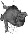
在至少一些实施方案中,在步骤106中获取的描绘40可以使得,可以选择性地隐藏代表不同解剖结构的描绘的部分,以便提供例如其他解剖结构的更好或更清晰的视图。例如,图5中所示的描绘包括LAA 12、左心房16和右心房20。然而,在例如图6所示的描绘中,隐藏右心房,以便提供LAA 12和左心房16的更好、更清晰的视图。因此,本公开不旨在限于在步骤106中获取任何特定类型的一个或多个描绘 (如,静态或动态)。In at least some embodiments, the
一旦在步骤106中获取了目标解剖区的描绘,方法100就移动到步骤108,即使用所获取的描绘40来限定与获取的描绘40中所示的一个或多个解剖结构相对应的一个或多个平面。例如,在一个实施方案中,诸如图6所示的实施方案中,步骤108包括限定含有LAA 12的心门的平面。在一个实施方案中,所限定的平面不对应于或含有本领域普通技术人员通常认为的LAA的心门(以下称为“假心门”),其是紧邻左心房的LAA的开口。相反,用于本公开目的的LAA的“心门”(以下称为“真心门”)可以包括LAA的部分或点,其具有最大的周边/周长,是LAA的传统“心门”的远端(即进一步进入LAA并远离左心房,而不是传统的心门)并且有垂直于LAA质心的平面。出于说明的目的,图 7示出了含有假心门的平面42和含有真心门的平面44(含有真心门的平面44也在图6中示出)。Once the delineation of the target anatomical region is obtained in
真心门的平面44可以以多种方式限定。一种方式是通过将LAA 12的周长追踪到其最大点。另一种方式是通过将横截面平面放置和定位在描绘40中或描绘40上的期望位置。在任一种情况下,平面44 可以由系统26的ECU 28(如,由ECU 28的处理器34)使用例如适合的图像处理软件/技术自动限定。可替代地,平面44可以由操纵系统 26的一个或多个用户界面装置32的用户(如,医师)限定。更具体地,在步骤106中获取的描绘40可以显示在显示装置30上,并且用户可以使用一个或多个用户界面装置32追踪LAA 12的周长或者将横截面平面放置在描绘40上。例如,用户可以操纵计算机鼠标以将光标移动至显示的描绘40中的期望位置并“点击”鼠标以将横截面平面放置在该位置。无论如何限定平面44,在一个实施方案中,可以在描绘40 上显示限定的平面44的表示以供用户观看,如图6和7所示。The
虽然上面已经提供了用于限定LAA 12的真心门平面44的某些技术或实施方式,但应当理解,可以使用用于这样做的任一种或多种适合的技术。因此,本公开不旨在限于用于这样做的任一种或多种特定技术。While certain techniques or implementations have been provided above for defining the
可以在步骤108中限定的另一个平面是含有患者心脏的二尖瓣环的平面;此平面在本文中被称为“二尖瓣平面”。可替代地,二尖瓣平面可以在步骤108之前或之后执行的方法100的步骤中限定,或者可以根本不限定。在限定二尖瓣平面的情况下,可以使用本领域已知的多种技术以多种方式完成它。例如,在一个实施方案中,步骤108可包括获取与患者心脏的解剖区相关的图像数据,所述解剖区包括例如患者心脏的左心室、左心房和主动脉。图像数据可以是在步骤102中获取的相同图像数据,或者包括不同的图像数据。在任一情况下,图像数据可包括CT图像数据,并且更具体地,2D CT图像数据。然而,应当理解,在其他实施方案中,图像数据可以包括使用除CT之外的适合的成像模式(例如本文其他地方所鉴定的那些成像模式中的一个或多个)获取的数据。因此,本公开不旨在限于任何特定类型的图像数据。然而,为了说明和清楚的目的,下面的描述将关于CT数据的使用。另外,在一个实施方案中,可以获取心动周期的舒张期和收缩期两者的图像数据,并且在这样的实施方案中,可以为每个时期限定二尖瓣平面。可替代地,可以获取数据并且仅针对舒张期和收缩期之一限定二尖瓣平面。Another plane that may be defined in
在一个实施方案中,可以使用从获取的CT图像数据生成的一个或多个2D图像来限定二尖瓣平面。更具体地,2D图像可以用于限定特定数量的点(如,三(3)个点),其可以用于限定二尖瓣平面。在一个实施方案中,可以使用一个或多个预定界标(如,解剖界标)来鉴定/限定平面-限定点。所使用的特定界标可以至少部分地取决于靠近二尖瓣环的结构的性质。例如,在其中结构是天然的二尖瓣的情况下,界标可以包括在天然瓣膜的二尖瓣环处的钙化和/或小叶尖端和/或插入点的区域,以引用几种可能性。在其中结构包括先前植入的装置或物体例如二尖瓣环的情况下,界标可包括此装置或其至少某些部分。最后,在结构包括先前植入的人工二尖瓣的情况下,界标可包括先前植入的瓣膜的部分,例如先前植入的瓣膜的支柱的尖端。在任何情况下,平面-限定点可以以多种方式限定或鉴定。In one embodiment, one or more 2D images generated from acquired CT image data may be used to define the mitral valve plane. More specifically, the 2D image can be used to define a certain number of points (eg, three (3) points), which can be used to define the mitral valve plane. In one embodiment, one or more predetermined landmarks (eg, anatomical landmarks) may be used to identify/define the plane-defining points. The particular landmarks used may depend, at least in part, on the nature of the structures proximal to the mitral annulus. For example, in the case where the structure is a native mitral valve, landmarks may include areas of calcification and/or leaflet tips and/or insertion points at the mitral annulus of the native valve, to name several possibilities. In cases where the structure includes a previously implanted device or object such as the mitral valve annulus, the landmark may include this device or at least some portion thereof. Finally, where the structure includes a previously implanted prosthetic mitral valve, the landmarks may include portions of the previously implanted valve, such as the tips of the struts of the previously implanted valve. In any case, plane-defining points can be defined or identified in a variety of ways.
在一个实施方案中,所述点可以由系统26的ECU 28(如,由ECU 28的处理器34)使用例如适合的图像处理软件/技术自动限定。在其他实施方案中,所述点可以由用户(如,医生)限定。更具体地,2D图像可以显示在显示装置30上,并且用户可以使用系统26的一个或多个用户界面装置32来限定平面-限定点。例如,用户可以操纵计算机鼠标以将光标移动至图像上的所需位置并“点击”鼠标来限定点。在任何情况下,一旦限定了平面-限定点,就可以将含有所有限定点的平面限定为二尖瓣平面。在至少一些实施方案中,二尖瓣平面可以通过例如曲线在2D图像上表示。虽然上面已经提供了用于限定二尖瓣平面-限定点以及因此限定二尖瓣平面本身的某些技术或实施方式,但是应当理解,可以使用用于这样做的任一种或多种适合的技术。因此,本公开不旨在限于用于这样做的任一种或多种特定技术。In one embodiment, the points may be automatically defined by the
在另一实施方案中,不是使用2D图像来限定二尖瓣平面,而是可以使用在步骤106中获取的描绘40。例如,横截面平面可以定位于描绘40中或描绘40上的所需位置处。在此类实施方案中,二尖瓣平面可以由系统26的ECU 28(如,由ECU 28的处理器34)使用例如适合的图像处理软件/技术自动限定。可替代地,平面可以由操纵系统26 的一个或多个用户界面装置32的用户(如,医师)限定。更具体地,在步骤106中获取的描绘40可以显示在显示装置30上,并且用户可操纵计算机鼠标以将光标移动至所显示的描绘40中的期望位置并“点击”鼠标以在此位置处放置横截面平面。在这种情况下,在其中可以隐藏在步骤106中获取的描绘40的一部分的一个实施方案中,可以显示先前在执行方法100的其他步骤期间隐藏的结构,以便于执行步骤 108(如,左心室、二尖瓣、二尖瓣环等)。In another embodiment, instead of using a 2D image to define the mitral valve plane, the
无论如何限定二尖瓣平面,在一个实施方案中,可以在描绘40 上显示所限定的二尖瓣平面的表示,如例如图12中所示,其中参考数字46对应于所限定的二尖瓣平面。虽然上面已经提供了用于限定二尖瓣平面46的某些技术或实施方式,但是应当理解,可以使用用于这样做的任一种或多种适合的技术。因此,本公开不旨在限于用于这样做的任一种或多种特定技术。另外,步骤108的执行可以至少部分地由存储在例如系统26的存储装置36中的软件促进。在一个实施方案中,该软件可以包括从Materialise NV以名称3Matic STL或
虽然到目前为止对步骤108的描述已经涉及对含有某些特定解剖结构或对应于某些特定解剖结构的平面的限定,但是应当理解,在其他实施方案中,可以限定另外或替代的平面。因此,本公开不必限于步骤108中的任何特定平面的限定,而是可以在步骤108中限定多种适合的平面。Although the description of
回到图3,在一个实施方案中,方法100还可以包括沿着物体坐标系(X&Y)移动在步骤108中限定的真心门平面44,直到平面44的原点处于LAA 12的真心门的中心的任选步骤110。在一个实施方案中,可以使用空腹血容量模型来执行该步骤。在一个实施方案中,步骤110 可以由系统26的ECU 28(如,由ECU 28的处理器34)使用例如适合的图像处理软件/技术自动执行。可替代地,步骤110可以由操纵系统 26的一个或多个用户界面装置32的用户(如,医生)执行。更具体地,用户可以操纵计算机鼠标以将平面44移动至在显示的描述中对应于 LAA的真心门中心的所需点或位置。Returning to FIG. 3 , in one embodiment, the
不论方法100是否包括步骤110并且参考图3和8,在一个实施方案中,方法100包括复制真心门平面44以创建复制平面48并沿着垂直于平面44的物体坐标系(如,在Z方向)移动复制平面48至描绘 40中LAA 12外侧或之外的点的步骤112。平面48在下文中可称为垂直偏移平面48或偏移真心门平面48。在一个实施方案中,LAA 12与 LAA 12外侧或之外的点之间的距离可以是几厘米的量级,但是本公开不旨在限于任何特定的距离。在一个实施方案中,步骤112可以由系统26的ECU 28(如,由ECU 28的处理器34)使用例如适合的图像处理软件/技术自动执行。可替代地,步骤112可以由操纵系统26的一个或多个用户界面装置32的用户(如,医生)执行。更具体地,用户可以操纵计算机鼠标以使平面44复制至复制平面48并且然后将复制平面48移动到所显示的描绘40中的LAA 12之外的所需点或位置,从而建立垂直偏移平面48。Regardless of whether
在一个实施方案中,方法100还可包括相对于描绘40限定多个点 (即,在描绘40中/上或附近)的步骤114,其中的两个或更多个将用于确定或者计算相对于一个或多个解剖结构的一个或多个测量值,所述一个或多个测量值继而将用于选择待用于执行医疗程序的医疗装置。In one embodiment, the
在一个实施方案,诸如图9所示的实施方案中,步骤114包括多个子步骤,包括鉴定和限定一个或多个平面的一个或多个原点。例如,在说明性实施方案中,步骤114包括鉴定和限定真心门平面44和垂直偏移平面48中的一个或两个的原点的子步骤。步骤114还可包括鉴定和限定含有卵圆窝的平面的原点(即卵圆窝的质心(还被称为卵圆窝平面的原点))和/或含有IVC的心门的平面的原点(即IVC心门的质心(还被称为IVC心门平面的原点))的子步骤,它们分别在图9中用参考数字50,52标识。在其他实施方案中,可以鉴定和限定除了上面提到的那些之外或不同于上面提到的那些的一个或多个平面的一个或多个原点。在任何情况下,步骤114的每个子步骤可以以多种方式执行。In one embodiment, such as that shown in FIG. 9,
一种方式是在目标平面/结构的原点/质心上或原点/质心处放置点或标记54,如图10所示,其中标记54a对应于卵圆窝的原点/质心并且标记54b对应于IVC心门的原点/质心。在一个实施方案中,子步骤中的一个或多个可以由系统26的ECU 28(如,由ECU 28的处理器34) 使用例如适合的图像处理软件/技术自动执行。可替代地,子步骤中的一个或多个可以由操纵系统26的一个或多个用户界面装置32的用户 (如,医生)执行。One way is to place a point or marker 54 on or at the origin/centroid of the target plane/structure, as shown in Figure 10, where
更具体地,用户可以操纵计算机鼠标以旋转或移动显示器30上显示的描绘40和/或将光标移动至对应于所需的原点/质心的所显示的描绘40中的所需位置并“点击”鼠标以在此位置处放置点或标记。因为它涉及鉴定/限定含有卵圆窝的平面的原点,在一个实施方案,诸如图10 中所示的实施方案中,对应于例如步骤104中所限定的卵圆窝的边界的标记38可以用于指导原点/质心54a的鉴定和限定。在另一个实施方案,诸如图11所示的实施方案中,厚度分析工具可用于鉴定/限定卵圆窝的原点/质心54a。更具体地,此类工具可用于鉴定卵圆窝的最薄或最窄区域。例如,工具可以生成彩色图,其中不同的颜色代表不同的厚度(如,红色是最薄的,并且绿色是最厚的)。使用该图,可以鉴定最薄点并将其视为卵圆窝的原点/质心54a,并且可以限定包括此原点/质心54a的平面。在另一个实施方案中,平面可以使用图像数据例如CT数据来限定。更具体地,使用2D CT图像,可以在图像中鉴定左心房和右心房相遇的点,并且可以限定包括此点的平面。因此,应当理解,可以使用多种技术。More specifically, the user may manipulate the computer mouse to rotate or move the displayed
因此,应当理解,本公开不旨在限于执行步骤114的任何特定方式或技术。另外,应理解,鉴于例如图5、6和8,在其中可以隐藏在步骤106中获取的描绘40的部分的一些实施方案中,之前在执行方法 100的其他步骤期间隐藏的结构可以显示以便于执行一个或多个步骤 114。Accordingly, it should be understood that this disclosure is not intended to be limited to any particular manner or technique of performing
因此,应当理解,本公开不旨在限于执行步骤114的任何特定方式或技术。另外,应理解,鉴于例如图5、6和8,在其中可以隐藏在步骤106中获取的描绘40的部分的一些实施方案中,之前在执行方法 100的其他步骤期间隐藏的结构可以显示以便于执行一个或多个步骤 114。Accordingly, it should be understood that this disclosure is not intended to be limited to any particular manner or technique of performing
在至少一些实施方案中,无论如何鉴定/限定原点/质心,都可以在描绘上显示对应于所鉴定/限定的原点/质心的标记54,以用于用户观看。In at least some embodiments, however the origin/centroid is identified/defined, a marker 54 corresponding to the identified/defined origin/centroid may be displayed on the delineation for viewing by the user.
虽然上面的描述是关于某些特定结构/平面的原点/质心的鉴定/限定,但是应当理解,在其他实施方案中,诸如例如下面描述的那些,另外或替代结构/平面的一个或多个原点/质心可以被鉴定/限定。因此,本公开不旨在限于步骤114中的任何特定结构/平面的一个或多个原点 /质心的鉴定/限定。While the description above is in relation to the identification/definition of the origin/centroid of certain particular structures/planes, it should be understood that in other embodiments, such as for example those described below, one or more origins of structures/planes are additionally or substituted / Centroids can be identified/defined. Accordingly, the present disclosure is not intended to be limited to the identification/definition of one or more origins/centroids of any particular structure/plane in
如图3所示并参考图12,在一个实施方案中,方法100还包括步骤116:复制步骤112中限定的二尖瓣平面46并沿着垂直于平面46 的物体坐标系(如,以Z方向)移动该复制平面或平面46的表示(由图 12中的参考数字56表示)至一点,使得平面46的表示56从二尖瓣平面46偏移并且与真心门平面44的原点(由参考数字57表示)对齐或持平,如图12所示。表示56包括偏移二尖瓣平面。As shown in FIG. 3 and with reference to FIG. 12 , in one embodiment, the
在至少一些实施方案中,方法100还可包括步骤118:第二次复制二尖瓣平面46并沿着垂直于平面46的物体坐标系(如,以Z方向) 移动第二复制平面或平面46的表示(由图13中的参考数字58表示)至一点,使得平面46的表示58从二尖瓣平面46和平面46的代表56偏移,并且还与卵圆窝的原点/质心54a对齐或持平,如图13所示。表示58还包括偏移二尖瓣平面。In at least some embodiments, the
无论方法100是包括步骤116,118中的一个还是两个,那些步骤中的一个或两个可以由系统26的ECU 28(如,由ECU 28的处理器34) 使用例如适合的图像处理软件/技术自动执行。可替代地,步骤116,118 中的一个或两个可以由操纵系统26的一个或多个用户界面装置32的用户(如,医生)执行。更具体地,用户可以操纵计算机鼠标以引起显示在系统26的显示器30上的描绘40上所示的二尖瓣平面46被复制一次或多次,然后移动(如,拖动)一个或多个复制的二尖瓣平面(即,表示56和/或58)至一个或多个期望的点或位置以建立一个或多个偏移二尖瓣平面。另外,应理解,在其中可以隐藏在步骤106中获取的描绘的部分的一些实施方案中,可以显示先前在执行方法100的其他步骤期间隐藏的结构,以便于执行步骤116,118。Whether
至少部分地使用步骤114中鉴定或限定的点,方法100包括确定或计算各个测量值的步骤120,例如两个或更多个限定的点之间的距离和/或由点的组合限定的角度。例如并参考图14,在其中方法100 用于确定或选择待在LAA相关程序(如,以递送和放置LAA封堵装置) 中使用的医疗装置(如,导管)的一个实施方案中,可以确定以下五(5) 个测量值中的一个或多个,并且可以使用所确定的测量值中的一个或多个来确定或选择医疗装置。Using at least in part the points identified or defined in
在图14中鉴定为测量值“1”的第一测量值是对应于IVC心门的原点/质心(即,含有IVC心门的平面的原点)的点54b与对应于卵圆窝的原点/质心(即,含有卵圆窝的平面的原点)的点54a之间的距离。The first measurement identified as measurement "1" in FIG. 14 is the
在图14中鉴定为测量值“2”的第二测量值是对应于卵圆窝的原点/ 质心的点54a和对应于真心门的平面44的原点的点57之间的距离。The second measurement, identified as measurement "2" in FIG. 14, is the distance between
在图14中鉴定为测量值“3”的第三测量值是由对应于IVC心门的原点/质心的点54b、对应于卵圆窝的原点/质心的点54a和对应于真 LAA心门的平面44的原点的点57形成的角度。The third measurement, identified as measurement "3" in FIG. The angle formed by
在图14中鉴定为测量值“4”的第四测量值是由对应于卵圆窝的原点/质心的点54a、对应于真LAA心门的平面44的原点的点57和对应于偏移LAA真心门平面48(还被称为偏移真心门平面48)的原点的点 59形成的角度。The fourth measurement, identified as measurement "4" in FIG. The angle formed by the
并且在图14中鉴定为测量值“5”的第五测量值是第一和第二偏移二尖瓣平面56,58之间的距离,第一和第二偏移二尖瓣平面56,58分别与真心门平面44和卵圆窝的原点/质心54a相交。And the fifth measurement identified as measurement "5" in FIG. 14 is the distance between the first and second offset mitral valve planes 56, 58, the first and second offset mitral valve planes 56, 58 intersects the
应当理解,虽然上面已经鉴定和讨论了某些特定的测量值,但是在其他实施方案中,除了以上所述的那些之外或者代替以上所述的那些的一个或多个测量值,可以如下所述确定和使用。It should be understood that while certain specific measurements have been identified and discussed above, in other embodiments one or more measurements in addition to or instead of those described above may be as follows identified and used as described above.
例如并参考图15,在其中方法100用于选择用于与二尖瓣相关的程序的医疗装置的一个实施方案中,方法100可以包括这样的步骤(未示出),其在步骤120之前执行并且包括复制图15和16中表示为参考数字60的含有卵圆窝的原点/质心54a的平面(即卵圆窝平面),并且沿其原点移动或偏移该复制平面或者平面60的表示(如图15和16中的参考数字62表示)直到它与二尖瓣平面46的原点的轴64相交。然后再次复制二尖瓣平面46,并且使平面46的复制平面或表示(如图15 和16中的参考数字66表示)移动或偏移至偏移卵圆窝平面62与轴64 相交的点。For example and referring to FIG. 15 , in one embodiment in which
如图15所示,在至少一些实施方案中,该步骤可以使用其中所有二尖瓣平面边缘和卵圆窝平面边缘对齐的视图来执行。在任何情况下,上述步骤可以由系统26的ECU 28(如,由ECU 28的处理器34)使用例如适合的图像处理软件/技术自动执行。可替代地,步骤可以由操纵系统26的一个或多个用户界面装置32的用户(如,医生)执行。更具体地,用户可以操纵计算机鼠标以使得显示在系统26的显示器30上的二尖瓣平面46和卵圆窝平面60被复制一次或多次,然后将一个或多个复制的偏移平面移动(如,拖动)至一个或多个期望的点或位置。另外,应理解,在其中可以隐藏在步骤106中获取的描绘的部分的一些实施方案中,可以显示先前在执行方法100的其他步骤期间隐藏的结构,以便于执行这一步。As shown in Figure 15, in at least some embodiments, this step can be performed using a view in which all mitral valve plane edges and fossa ovale plane edges are aligned. In any event, the steps described above may be performed automatically by the
在其中方法100包括上述步骤的一个实施方案中,步骤120包括使用在步骤114中鉴定的点,一个或多个步骤116,118中限定的平面和上述一个或多个交叉点以确定或计算各个测量值。例如并参考图16 和17,以下八(8)个不同测量值中的一个或多个可以被确定并用于确定选择医疗装置。In one embodiment where the
第一测量值是二尖瓣平面46的原点和偏移二尖瓣平面66的原点之间的距离(即,图16中的点“A”和“B”之间的距离)。The first measurement is the distance between the origin of
第二测量值是二尖瓣平面46的原点与偏移二尖瓣平面66和偏移卵圆窝平面62的交叉点之间的距离(即,图16中的点“A”和“C”之间的距离)。The second measurement is the distance between the origin of the
第三测量值是偏移二尖瓣平面66的原点与偏移二尖瓣平面66和偏移卵圆窝平面62的交叉点之间的距离(即,图16中的点“B”和“C”之间的距离)。The third measurement is the distance between the origin of the offset
第四测量值是由对应于以下的点形成的角度:偏移二尖瓣平面66 的原点、二尖瓣平面46的原点及偏移二尖瓣平面66和偏移卵圆窝平面62的交叉点(即图16中的角度“BAC”)。The fourth measurement is the angle formed by the points corresponding to: the origin of the offset
第五测量值是由对应于以下的点形成的角度:二尖瓣平面46的原点、偏移二尖瓣平面66的原点及偏移二尖瓣平面66和偏移卵圆窝平面62的交叉点(即图16中的角度“ABC”)。The fifth measurement is the angle formed by the points corresponding to: the origin of the
第六测量值是由对应于以下的点形成的角度:二尖瓣平面46的原点、偏移二尖瓣平面66和偏移卵圆窝平面62的交叉点以及偏移二尖瓣平面66的原点(即图16中的角度“ACB”)。The sixth measurement is the angle formed by the points corresponding to: the origin of the
第七测量值是由对应于以下的点形成的角度:IVC心门的原点/ 质心54b、卵圆窝的原点/质心54a和二尖瓣平面46的原点(即,图16 中的点“D”处的角度)。The seventh measurement is the angle formed by the points corresponding to: the origin/
并且第八测量值是由对应于以下的点形成的角度:卵圆窝的原点/ 质心54a、二尖瓣平面46的原点和偏移二尖瓣平面66的原点(即,图 16中的角度“DAB”,并在图17中显示为角度“E”)。And the eighth measurement is the angle formed by the points corresponding to: the origin/
同样,应理解,虽然上面鉴定和讨论了某些特定测量值,但是在其他实施方案中,可以确定除了上述那些之外或代替上述那些的一个或多个测量值。另外,如图14和15-17所示,在至少一些实施方案中,可以隐藏在步骤106中获取的一些或全部描绘40,以提供在步骤120 中确定的测量值的清晰视图。Also, it should be understood that while certain specific measurements are identified and discussed above, in other embodiments one or more measurements may be determined in addition to or instead of those described above. Additionally, as shown in FIGS. 14 and 15-17 , in at least some embodiments some or all of the
无论在步骤120中确定的一个或多个特定测量值如何,在一个实施方案中,步骤120可以由系统26的ECU 28(如,由ECU 28的处理器34)使用例如适合的软件/技术自动执行。可替代地,步骤120可以由ECU 28和用户输入的组合来执行。更具体地,用户(如,医生)可以操纵用户界面装置32以选择待确定的一个或多个测量值(如,选择待确定其间距离的两个点,选择限定待确定的角度的三个点等),然后ECU 28可以确定/计算一个或多个恰当的、选择的测量值。因此,本公开不旨在限于执行步骤120的任何特定方式。Regardless of the particular measurement or values determined in
如图3所示,一旦在步骤120中确定了一个或多个恰当的测量值,方法100就进入基于在步骤120中确定的那个或那些测量值选择或确定医疗装置的步骤122。在一个实施方案中,被配置为能关联在步骤 120中确定的一个或多个测量值(输入))与不同类型(如,尺寸,形状等) 的医疗装置(输出)的数据结构(如,凭经验导出的查找表(如,多维查找表),用于选择/确定恰当的医疗装置。在一个实施方案中,数据结构可以存储在系统26的存储器(如,ECU28的存储器36)中,并且ECU 28 的处理器34可以被配置为能在数据结构中查找在步骤120中确定的一个或多个测量值,从而选择或确定使用的恰当医疗装置。然而,应理解,当然可以替代地使用用于选择医疗装置的其他适合的方式/技术。As shown in FIG. 3 , once the appropriate measurement or values are determined in
除了前文之外,在至少一些实施方案中,可以评价穿过解剖结构的一个或多个插入点和/或用于执行医疗程序的一个或多个医疗装置的恰当性或适合性。在一个实施方案中,该评价可以包括方法100的一部分,并且因此,评价过程的步骤可以在方法100的一个或多个上述步骤之后(如,在步骤106-122的一个或多个之后)执行。然而,应理解,评价过程的步骤也可以独立于方法100执行,或者可以仅需要方法100的一些步骤。仅出于说明的目的,下面的描述将针对穿过房隔,特别是卵圆窝的插入点、以及包括导管(如,与植入LAA封堵装置相关的程序中使用的导管)的医疗装置来进行。然而,应理解,穿过其他解剖结构的插入点和/或除导管之外的装置的恰当性可以以下面描述的方式评价。In addition to the foregoing, in at least some embodiments, one or more points of insertion through the anatomy and/or one or more medical devices for performing a medical procedure can be evaluated for adequacy or suitability. In one embodiment, this evaluation may comprise part of
在至少一些实施方案中,评价过程包括鉴定穿过房隔的一个或多个插入点的步骤。在步骤106中获取的描绘40或以与步骤106中描述的方式类似的方式获取的不同描绘可以用于鉴定或选择一个或多个可能的插入点。可以以多种方式鉴定或选择可能的插入点,包括例如但不限于以以上在别处描述的关于标记38的鉴定的方式。图18示出了具有由标记38表示的单个插入点的描绘40;尽管应理解,当然可以鉴定出一个以上的插入点。In at least some embodiments, the evaluation process includes the step of identifying one or more insertion points through the atrial septum. The
一旦鉴定出一个或多个插入点,就可以选择一个或多个医疗装置 (如,导管)的一个或多个模型并将其输入描绘40中。在一个说明性的实施方案中,一次一个地单独评价所鉴定的插入点,其中一个或多个选定的模型被输入描绘的给定插入点中。然而,在其他实施方案中,可以同时评价多个插入点,其中为每个插入点输入一个或多个模型。Once one or more insertion points are identified, one or more models of one or more medical devices (e.g., catheters) can be selected and entered into
装置的模型的选择可以以多种方式实施。在至少一些实施方案中,可以从具有不同特征的许多不同模型中选择特定医疗装置的模型。例如,在说明性实施方案中,特定模型可以选自代表以下的一系列模型:来自不同产品的装置、具有分配的不同材料性质的装置、和/或具有不同形状、尺寸、几何形状(如,长度、直径、曲率(曲率的数量和度数)、最小/最大弯曲半径和/或其他特性)的装置。选择也可以来自用于不同程序的装置的模型。因此,应理解,一个模型(或多个模型)的选择可以基于许多因素/特征,包括但不限于上面鉴定的那些。The selection of the model of the device can be implemented in various ways. In at least some embodiments, a model of a particular medical device can be selected from a number of different models having different characteristics. For example, in an illustrative embodiment, a particular model may be selected from a collection of models representing devices from different products, devices with assigned different material properties, and/or with different shapes, sizes, geometries (e.g., length, diameter, curvature (amount and degree of curvature), minimum/maximum bend radius and/or other characteristics). Selections can also be made from models of devices used for different procedures. Thus, it should be understood that the selection of a model (or models) can be based on a number of factors/characteristics, including but not limited to those identified above.
在一个实施方案中,对其进行选择的模型可以包含在可以存储在系统26的适合组件或者系统26可访问的组件之中或之上的数字文库或数据库中。在一个说明性的实施方案中,含有模型的文库或数据库存储在系统26的ECU 28的存储装置36之中或之上。模型选择可以由系统26(即,处理器34)自动进行,或者可以由用户经由例如用户界面装置32、显示装置30或用户界面装置32和显示器30的组合手动进行。在后一种情况下,可能模型的列表可以显示在显示装置30上,并且用户可以操纵用户界面装置32(或显示装置本身)以选择一个或多个所需的模型。列表中的模型可以通过描述模型和/或由其表示的装置的语言(如,产品名称、装置名称、装置模型编号等)来识别。另外或者可替代地,可以显示模型本身的视觉描绘(如,模型/装置的缩略图)。In one embodiment, the models from which it is selected may be contained in a digital library or database that may be stored in or on a suitable component of
在一个说明性实施方案中,可以显示一个或多个用户可输入的或用户可选择的字段(如,单选按钮、下拉菜单、用于输入信息的文本框等)以允许用户提供系统26可使用的某些信息,以缩小从中进行选择的模型的范围。这可以包括例如优选装置制造商的名称、待执行的程序的名称等。响应于用户输入,系统26可以自动选择一个或多个待使用的模型,或者可以给出用户可以从中进行选择的模型列表。In one illustrative embodiment, one or more user-enterable or user-selectable fields (e.g., radio buttons, drop-down menus, text boxes for entering information, etc.) may be displayed to allow the user to provide Certain information used to narrow down the models from which to choose. This may include, for example, the name of the preferred device manufacturer, the name of the program to be executed, etc. In response to user input,
鉴于前述内容,应理解,一种或多种医疗装置的一个或多个模型的选择可以以多种方式实施,包括但当然不限于上述的那些。In view of the foregoing, it should be appreciated that the selection of one or more models of one or more medical devices may be implemented in a variety of ways, including but of course not limited to those described above.
在某些实施方案中,模型的选择导致所选择的模型被输入描绘40 中,或者至少连同描绘40一起显示。然而,在其他实施方案中,用户必须通过例如操纵可以在显示器30上或用户界面32上显示或设置的“输入”按钮来肯定地命令输入所选择的模型。在一个说明性实施方案中,将输入的模型以这样的方式自动放置并定位于描绘40内,使得它显示了如果实际用于医疗程序的执行中,将如何处置由模型表示的装置(即,它显示模型处于装置的着陆位置)。例如,在其中医疗程序涉及LAA封堵装置的植入并且模型是用于执行这种程序的导管的模型的情况下,可以放置导管的模型使得导管模型的远端尖端位于LAA心门附近(即,靠近或在其中)。然后,用户可以使用例如用户界面装置 32通过平移和/或旋转模型来微调模型的位置。系统26可以被配置为能基于描绘40内或相对于描绘40的某些预定点(包括例如经鉴定的穿过房隔的插入点)自动地定位模型。这些点可以包括在上述方法100的步骤114中限定的一些或所有点。例如,在图18中,模型68在描绘 40内的定位由步骤114中限定的多个点限定或指示,即含有IVC心门的平面的原点、含有卵圆窝的平面的原点及含有LAA的真心门的平面的原点。在其他实施方案中,用于模型的自动放置/定位中的点可以包括另外的点和/或不同的点(而非步骤114中限定的那些),所述点由用户手动限定或由系统26自动限定。In some embodiments, selection of a model results in the selected model being entered into the
在其他实施方案中,不是如上所述自动放置的模型,用户可能能够使用例如用户界面装置32将模型手动地放置在一个或多个所需位置处。例如,用户可以“点击”并“拖动”模型至描绘40内的所需位置。可替代地,用户可以使用“平移”和/或“旋转”工具将模型移动至所需位置。在此类实施方案中,用户还可以将模型重新定位至描绘40内的不同的所需位置。In other embodiments, rather than automatically placing the model as described above, a user may be able to manually place the model at one or more desired locations using, for example, the
一旦为一个或多个插入点输入了一个或多个装置模型,就可以查看或确定每个相应插入点的每个插入模型的轨迹,并且可以确定每个装置和/或一个或多个相应的插入点的恰当性。更具体地,图18示出了相对于插入点38输入描绘40中的模型68。模型68被放置成使得它位于对应于由模型68表示的装置的所需着陆点的位置处。用户然后可以查看装置相对于描绘40中所示或表示的各解剖结构将具有的轨迹,然后至少部分地基于轨迹确定由模型68表示的装置和/或插入点 38是否恰当。Once one or more device models have been entered for one or more insertion points, the trajectory of each insertion model for each corresponding insertion point can be viewed or determined, and each device and/or one or more corresponding The appropriateness of the insertion point. More specifically, FIG. 18 shows the
另外或可替代地,系统26,特别是其ECU 28可以被配置为能自动地确定装置和/或插入点的恰当性。更具体地,与由模型68表示的装置相关的信息(即,最小弯曲半径、最大直径等)可以存储在例如存储装置36或另一个适合的存储装置(是系统26的一部分或系统26可访问的)中。系统26的ECU 28的处理器34,或系统26的另一个适合的组件或其他,可以被配置为能访问装置信息并使用它来评级或确定装置和/或给定的插入点的恰当性。在例如步骤114中限定的点也可以与该信息一起使用以确定装置和/或给定插入点的恰当性。Additionally or alternatively, the
如果仅评价装置的恰当性并且确定装置是恰当的,则用户可以选择此装置用于执行程序。否则,可以针对不同的装置重复上述过程,或者如果同时评价多个装置,则用户可以选择被认为最恰当的装置。如果仅评价插入点的恰当性并且确定插入点是恰当的,则用户可以选择该插入点作为程序的目标插入点。否则,可以针对不同的插入点重复上述过程,或者如果同时评价多个插入点,则用户可以选择被认为最恰当的插入点。最后,如果正在评价装置和插入点的恰当性,并且确定给定插入点和给定装置都是恰当的,则用户可以选择此装置和插入点用于执行程序。否则,可以针对不同的装置/插入点组合重复上述过程,或者如果同时评价多个装置和/或多个插入点,则用户可以选择被视为最恰当的装置/插入点组合。If only the suitability of the device is evaluated and the device is determined to be suitable, the user can select this device for executing the program. Otherwise, the above process can be repeated for a different device, or if multiple devices are evaluated at the same time, the user can select the device deemed most appropriate. If only the appropriateness of the insertion point is evaluated and the insertion point is determined to be appropriate, the user can select the insertion point as the target insertion point of the program. Otherwise, the above process can be repeated for different insertion points, or if several insertion points are evaluated simultaneously, the user can select the insertion point deemed most appropriate. Finally, if the suitability of the device and insertion point is being evaluated, and it is determined that a given insertion point and a given device are both suitable, the user may select this device and insertion point for execution of the program. Otherwise, the above process can be repeated for different device/plug-in point combinations, or if multiple devices and/or multiple plug-in points are evaluated simultaneously, the user can select the device/plug-in point combination deemed most appropriate.
因此,应当理解,医疗装置和/或穿过解剖结构的插入点的恰当性或适合性可以以多种方式确定,包括但不限于上述那些。Accordingly, it should be understood that the appropriateness or suitability of a medical device and/or insertion point through an anatomical structure may be determined in a variety of ways, including but not limited to those described above.
应理解,前文是对本发明的一个或多个实施方案的描述。本发明不限于本文公开的一个或多个特定实施方案,而是仅由下面的权利要求限定。此外,前面描述中包含的陈述涉及特定的实施方案,并且不应被解释为对本发明的范围或权利要求中使用的术语的定义的限制,除非以上明确定义术语或短语。各种其他实施方案和对所公开的一个或多个实施方案的各种变化和修改对于本领域技术人员而言将变得显而易见。所有此类其他实施方案、变化和修改都旨在落入所附权利要求的范围内。It is to be understood that the foregoing is a description of one or more embodiments of the invention. The present invention is not to be limited by the particular embodiments or embodiments disclosed herein, but only by the claims that follow. Furthermore, statements contained in the foregoing description refer to particular embodiments and should not be construed as limitations on the scope of the invention or definitions of terms used in the claims unless a term or phrase is explicitly defined above. Various other embodiments and various changes and modifications to the disclosed embodiment or embodiments will become apparent to those skilled in the art. All such other embodiments, changes and modifications are intended to come within the scope of the appended claims.
如本说明书和权利要求中所使用,术语“如”、“例如(for example)”、“例如(forinstance)”、“诸如”和“像”,以及动词“包含”、“具有”、“包括”和其他动词形式,当与一个或多个组件或其他项目的列表结合使用时,各自都被解释为开放式的,这意味着该列表不被视为排除其他、另外组件或项目。其他术语应使用其最广泛的合理含义来解释,除非它们用于需要不同解释的上下文中。As used in this specification and claims, the terms "such as", "for example", "for instance", "such as" and "like", as well as the verbs "comprises", "has", "including ” and other verb forms, when used in conjunction with a list of one or more components or other items, are each to be construed as open-ended, meaning that the list is not deemed to exclude other, additional components or items. Other terms should be interpreted using their broadest reasonable meaning, unless they are used in a context requiring a different interpretation.
Claims (14)
Translated fromChineseApplications Claiming Priority (3)
| Application Number | Priority Date | Filing Date | Title |
|---|---|---|---|
| US201662419072P | 2016-11-08 | 2016-11-08 | |
| US62/419,072 | 2016-11-08 | ||
| PCT/US2017/060592WO2018089461A1 (en) | 2016-11-08 | 2017-11-08 | Selecting a medical device for use in a medical procedure |
Publications (2)
| Publication Number | Publication Date |
|---|---|
| CN110022785A CN110022785A (en) | 2019-07-16 |
| CN110022785Btrue CN110022785B (en) | 2023-01-03 |
Family
ID=62065224
Family Applications (1)
| Application Number | Title | Priority Date | Filing Date |
|---|---|---|---|
| CN201780068862.XAActiveCN110022785B (en) | 2016-11-08 | 2017-11-08 | Selecting a medical device for use in a medical procedure |
Country Status (7)
| Country | Link |
|---|---|
| US (2) | US10792104B2 (en) |
| EP (1) | EP3538008B1 (en) |
| JP (3) | JP7019694B2 (en) |
| CN (1) | CN110022785B (en) |
| AU (1) | AU2017359301B2 (en) |
| CA (1) | CA3041538A1 (en) |
| WO (1) | WO2018089461A1 (en) |
Families Citing this family (13)
| Publication number | Priority date | Publication date | Assignee | Title |
|---|---|---|---|---|
| CN110022785B (en)* | 2016-11-08 | 2023-01-03 | 亨利福特保健系统 | Selecting a medical device for use in a medical procedure |
| NL2021849B1 (en)* | 2018-10-22 | 2020-05-13 | Mat Nv | System and method for catheter based intervention |
| WO2020096612A1 (en)* | 2018-11-09 | 2020-05-14 | Henry Ford Health System | Evaluating blood flow obstruction through anatomical structure |
| EP4172956A1 (en)* | 2020-06-29 | 2023-05-03 | Koninklijke Philips N.V. | Generating and displaying a rendering of a left atrial appendage |
| US12097071B2 (en)* | 2020-11-03 | 2024-09-24 | Biosense Webster (Israel) Ltd. | Left atrial appendage (LAA) transseptal access point optimization |
| US20230230684A1 (en)* | 2022-01-14 | 2023-07-20 | Medtronic, Inc. | Catheter design tool and methods thereof |
| EP4463091A1 (en)* | 2022-01-14 | 2024-11-20 | Medtronic, Inc. | Catheter design tool and methods thereof |
| US12433567B2 (en) | 2022-03-16 | 2025-10-07 | Bard Access Systems, Inc. | Ultrasound imaging system |
| LU503167B1 (en)* | 2022-08-02 | 2024-02-02 | Max Planck Gesellschaft | Computer-implemented method and device configured to determine a design of a medical device comprising a rod shaped portion |
| WO2024028352A1 (en)* | 2022-08-02 | 2024-02-08 | MAX-PLANCK-Gesellschaft zur Förderung der Wissenschaften e.V. | Computer-implemented method and device configured to determine a design of a medical device comprising a rod shaped portion |
| CN120035409A (en)* | 2022-10-11 | 2025-05-23 | 皇家飞利浦有限公司 | Provide guidance on the management of blocked blood vessels |
| EP4353175A1 (en)* | 2022-10-11 | 2024-04-17 | Koninklijke Philips N.V. | Providing guidance for a treatment procedure on an occluded vessel |
| US20250017559A1 (en)* | 2023-07-12 | 2025-01-16 | Bard Access Systems, Inc. | System and Method for Suggesting Catheter Parameters |
Family Cites Families (28)
| Publication number | Priority date | Publication date | Assignee | Title |
|---|---|---|---|---|
| US20020168618A1 (en)* | 2001-03-06 | 2002-11-14 | Johns Hopkins University School Of Medicine | Simulation system for image-guided medical procedures |
| US7840393B1 (en) | 2000-10-04 | 2010-11-23 | Trivascular, Inc. | Virtual prototyping and testing for medical device development |
| US7607440B2 (en) | 2001-06-07 | 2009-10-27 | Intuitive Surgical, Inc. | Methods and apparatus for surgical planning |
| US20030023266A1 (en)* | 2001-07-19 | 2003-01-30 | Borillo Thomas E. | Individually customized atrial appendage implant device |
| JP4691017B2 (en)* | 2003-03-18 | 2011-06-01 | セント ジュード メディカル インコーポレイテッド | Body tissue remodeling method and apparatus |
| JP4904293B2 (en)* | 2005-03-03 | 2012-03-28 | セント・ジュード・メディカル・エイトリアル・フィブリレーション・ディヴィジョン・インコーポレーテッド | Method and apparatus for determining the location of the foveal fossa, creating a virtual foveal fossa and performing a transseptal puncture |
| US7877128B2 (en)* | 2005-08-02 | 2011-01-25 | Biosense Webster, Inc. | Simulation of invasive procedures |
| US8771189B2 (en)* | 2009-03-18 | 2014-07-08 | Siemens Medical Solutions Usa, Inc. | Valve assessment from medical diagnostic imaging data |
| WO2010120990A1 (en) | 2009-04-15 | 2010-10-21 | James Schroeder | Personal fit medical implants and orthopedic surgical instruments and methods for making |
| US10719986B2 (en) | 2009-12-22 | 2020-07-21 | Siemens Healthcare Gmbh | Method and system for virtual percutaneous valve implantation |
| US8494245B2 (en) | 2010-03-09 | 2013-07-23 | Siemens Aktiengesellschaft | System and method for guiding transcatheter aortic valve implantations based on interventional C-Arm CT imaging |
| WO2012063204A1 (en) | 2010-11-12 | 2012-05-18 | Koninklijke Philips Electronics N.V. | Identifying individual sub-regions of the cardiovascular system for calcium scoring |
| US9875339B2 (en) | 2011-01-27 | 2018-01-23 | Simbionix Ltd. | System and method for generating a patient-specific digital image-based model of an anatomical structure |
| US8920322B2 (en) | 2011-03-09 | 2014-12-30 | Siemens Aktiengesellschaft | Valve treatment simulation from medical diagnostic imaging data |
| US8830233B2 (en)* | 2011-04-28 | 2014-09-09 | Howmedica Osteonics Corp. | Surgical case planning platform |
| DE102011077753B4 (en) | 2011-06-17 | 2020-06-10 | Siemens Healthcare Gmbh | Device for planning a transcatheter aortic valve implantation |
| US10238483B2 (en) | 2011-09-16 | 2019-03-26 | 3Dt Holdings, Llc | Devices and methods for assisting valve function, replacing venous valves, and predicting valve treatment success |
| US8934693B2 (en) | 2011-11-23 | 2015-01-13 | Siemens Aktiengesellschaft | Method and system for intervention planning for transcatheter aortic valve implantation from 3D computed tomography data |
| US10789772B2 (en) | 2012-05-16 | 2020-09-29 | Feops Nv | Pre-operative simulation of trans-catheter valve implantation |
| US9283078B2 (en)* | 2012-09-21 | 2016-03-15 | Materialise N.V. | Patient-specific intraluminal implants |
| US9406129B2 (en)* | 2013-10-10 | 2016-08-02 | Medtronic, Inc. | Method and system for ranking instruments |
| US9050188B2 (en)* | 2013-10-23 | 2015-06-09 | Caisson Interventional, LLC | Methods and systems for heart valve therapy |
| US20150119692A1 (en) | 2013-10-25 | 2015-04-30 | Medtronic, Inc. | Non-invasive evaluation of fit and sizing of device prior to implant |
| EP4254334A3 (en)* | 2014-05-20 | 2024-01-17 | Materialise NV | System and method for valve quantification |
| US10881461B2 (en)* | 2014-08-07 | 2021-01-05 | Henry Ford Health System | Method of analyzing hollow anatomical structures for percutaneous implantation |
| US9693830B2 (en)* | 2014-12-10 | 2017-07-04 | Henry Ford Health System | Evaluating prosthetic heart valve placement |
| US20190021699A1 (en)* | 2016-01-15 | 2019-01-24 | Koninklijke Philips N.V. | Automatic probe steering to clinical views using annotations in a fused image guidance system |
| CN110022785B (en)* | 2016-11-08 | 2023-01-03 | 亨利福特保健系统 | Selecting a medical device for use in a medical procedure |
- 2017
- 2017-11-08CNCN201780068862.XApatent/CN110022785B/enactiveActive
- 2017-11-08EPEP17868612.7Apatent/EP3538008B1/enactiveActive
- 2017-11-08JPJP2019523790Apatent/JP7019694B2/enactiveActive
- 2017-11-08CACA3041538Apatent/CA3041538A1/enactivePending
- 2017-11-08USUS15/806,819patent/US10792104B2/enactiveActive
- 2017-11-08AUAU2017359301Apatent/AU2017359301B2/enactiveActive
- 2017-11-08WOPCT/US2017/060592patent/WO2018089461A1/ennot_activeCeased
- 2020
- 2020-09-04USUS17/012,138patent/US11793572B2/enactiveActive
- 2022
- 2022-02-02JPJP2022014847Apatent/JP2022078028A/enactivePending
- 2023
- 2023-12-27JPJP2023221377Apatent/JP2024029142A/enactivePending
Also Published As
| Publication number | Publication date |
|---|---|
| JP2024029142A (en) | 2024-03-05 |
| JP2022078028A (en) | 2022-05-24 |
| AU2017359301A8 (en) | 2019-07-11 |
| CA3041538A1 (en) | 2018-05-17 |
| JP7019694B2 (en) | 2022-02-15 |
| EP3538008A1 (en) | 2019-09-18 |
| EP3538008A4 (en) | 2020-06-17 |
| US20210093383A1 (en) | 2021-04-01 |
| JP2019533537A (en) | 2019-11-21 |
| US20180125581A1 (en) | 2018-05-10 |
| AU2017359301A1 (en) | 2019-05-30 |
| AU2017359301B2 (en) | 2022-10-06 |
| US11793572B2 (en) | 2023-10-24 |
| US10792104B2 (en) | 2020-10-06 |
| CN110022785A (en) | 2019-07-16 |
| EP3538008B1 (en) | 2025-05-14 |
| WO2018089461A1 (en) | 2018-05-17 |
Similar Documents
| Publication | Publication Date | Title |
|---|---|---|
| CN110022785B (en) | Selecting a medical device for use in a medical procedure | |
| US12383340B2 (en) | Image-based navigation system and method of using same | |
| US12161418B2 (en) | Method of analyzing hollow anatomical structures for percutaneous implantation | |
| US11617622B2 (en) | Evaluating prosthetic heart valve placement | |
| EP3912139B1 (en) | Method of visualising a dynamic anatomical structure | |
| JP2018500091A (en) | Medical image editing | |
| CN112041895B (en) | Transseptal puncture-guided cardiac repair | |
| CN111432718B (en) | Assessing blood flow obstruction through anatomical structures |
Legal Events
| Date | Code | Title | Description |
|---|---|---|---|
| PB01 | Publication | ||
| PB01 | Publication | ||
| SE01 | Entry into force of request for substantive examination | ||
| SE01 | Entry into force of request for substantive examination | ||
| GR01 | Patent grant | ||
| GR01 | Patent grant |