CN109903278B - Ultrasonic breast tumor morphological quantitative feature extraction method based on shape histogram - Google Patents
Ultrasonic breast tumor morphological quantitative feature extraction method based on shape histogramDownload PDFInfo
- Publication number
- CN109903278B CN109903278BCN201910138613.4ACN201910138613ACN109903278BCN 109903278 BCN109903278 BCN 109903278BCN 201910138613 ACN201910138613 ACN 201910138613ACN 109903278 BCN109903278 BCN 109903278B
- Authority
- CN
- China
- Prior art keywords
- breast tumor
- shape histogram
- maximum curvature
- shape
- ultrasonic
- Prior art date
- Legal status (The legal status is an assumption and is not a legal conclusion. Google has not performed a legal analysis and makes no representation as to the accuracy of the status listed.)
- Active
Links
- 208000026310Breast neoplasmDiseases0.000titleclaimsabstractdescription136
- 206010006187Breast cancerDiseases0.000titleclaimsabstractdescription129
- 230000000877morphologic effectEffects0.000titleclaimsabstractdescription50
- 238000000605extractionMethods0.000titleclaimsdescription11
- 206010028980NeoplasmDiseases0.000claimsabstractdescription37
- 238000000034methodMethods0.000claimsabstractdescription34
- 238000011002quantificationMethods0.000claimsabstractdescription31
- 230000003211malignant effectEffects0.000claimsabstractdescription29
- 201000011510cancerDiseases0.000claimsabstractdescription8
- 230000008569processEffects0.000claimsdescription12
- 101001056234Homo sapiens Sperm mitochondrial-associated cysteine-rich proteinProteins0.000claims1
- 102100026503Sperm mitochondrial-associated cysteine-rich proteinHuman genes0.000claims1
- 230000036210malignancyEffects0.000claims1
- 238000002604ultrasonographyMethods0.000abstractdescription38
- 238000003745diagnosisMethods0.000abstractdescription13
- 238000004195computer-aided diagnosisMethods0.000abstractdescription6
- 201000007295breast benign neoplasmDiseases0.000description20
- 238000013461designMethods0.000description4
- 238000001514detection methodMethods0.000description4
- 230000008901benefitEffects0.000description3
- 238000013139quantizationMethods0.000description3
- 230000035945sensitivityEffects0.000description3
- 238000012549trainingMethods0.000description3
- 238000004458analytical methodMethods0.000description2
- 230000008859changeEffects0.000description2
- 201000010099diseaseDiseases0.000description2
- 208000037265diseases, disorders, signs and symptomsDiseases0.000description2
- 230000004660morphological changeEffects0.000description2
- 238000011160researchMethods0.000description2
- 239000000523sampleSubstances0.000description2
- 230000011218segmentationEffects0.000description2
- 238000012360testing methodMethods0.000description2
- 206010004243Benign breast neoplasmDiseases0.000description1
- 238000013528artificial neural networkMethods0.000description1
- 230000009286beneficial effectEffects0.000description1
- 230000005540biological transmissionEffects0.000description1
- 210000000481breastAnatomy0.000description1
- 238000004364calculation methodMethods0.000description1
- 238000010276constructionMethods0.000description1
- 230000007812deficiencyEffects0.000description1
- 230000000694effectsEffects0.000description1
- 238000002474experimental methodMethods0.000description1
- 208000014674injuryDiseases0.000description1
- 238000012986modificationMethods0.000description1
- 230000004048modificationEffects0.000description1
- 238000007781pre-processingMethods0.000description1
- 230000005855radiationEffects0.000description1
- 230000009467reductionEffects0.000description1
- 230000008733traumaEffects0.000description1
- 238000012285ultrasound imagingMethods0.000description1
Images


Landscapes
- Ultra Sonic Daignosis Equipment (AREA)
Abstract
Translated fromChinese
Description
Translated fromChinese技术领域technical field
本发明涉及计算机医疗辅助诊断技术领域,具体涉及一种基于形状直方图的超声乳腺肿瘤形态量化特征提取方法。The invention relates to the technical field of computer-aided medical diagnosis, in particular to a method for extracting morphological quantitative features of ultrasonic breast tumors based on shape histograms.
背景技术Background technique
当下乳腺癌作为全球女性常患的一种恶性肿瘤疾病,具有极高的发病率和死亡率,在全世界范围内每年因乳腺癌致死的女性患者数目就高达数百万,且常年来致死率呈不断攀升之势。超声成像凭借其无辐射创伤、价格低廉、实时便携等优势,已成为乳腺肿瘤早期检测和诊断的一种重要手段。超声医生凭借经验对采集的超声乳腺肿瘤图像进行良恶性判别,这是一个主观判断过程,诊断结果受医生的经验丰富程度、知识水平高低、以及疲劳程度等条件的限制,不同医生对同一超声图像,可能诊断结果完全不同,甚至同一医生在不同时间、不同状态下对同一超声图像的诊断结果也不相同。因此,利用计算机辅助诊断(Computer Aided Diagnosis,CAD)系统对超声乳腺肿瘤图像进行良恶性判别具有极其重要的研究价值和应用前景,它可以有效减少医生的主观因素影响,提高疾病解释一致性,使诊断结果更客观、更准确。At present, breast cancer is a malignant tumor frequently suffered by women around the world, with extremely high morbidity and mortality. The number of female patients who die of breast cancer every year in the world is as high as millions, and the mortality rate is high over the years. On the rise. Ultrasound imaging has become an important method for early detection and diagnosis of breast tumors due to its advantages of no radiation trauma, low price, and real-time portability. Ultrasound doctors rely on their experience to judge the benign and malignant images of ultrasound breast tumors. This is a subjective judgment process. The diagnosis results are limited by the doctor's experience, knowledge level, and fatigue. , the diagnostic results may be completely different, and even the diagnostic results of the same ultrasound image by the same doctor at different times and in different states are different. Therefore, the use of Computer Aided Diagnosis (CAD) system to discriminate benign and malignant ultrasound breast tumor images has extremely important research value and application prospects. It can effectively reduce the influence of subjective factors of doctors, improve the consistency of disease interpretation, and make the The diagnosis results are more objective and accurate.
基于CAD的超声乳腺肿瘤良恶性检测是利用计算机对超声乳腺肿瘤图像进行特征提取和分类的过程,主要包括预处理、肿瘤目标检测与区域分割、肿瘤特征提取和肿瘤良恶性分类四个步骤。其中,乳腺肿瘤特征提取是超声乳腺肿瘤良恶性检测的一个重要环节,其描述的肿瘤特征是否准确很大程度上决定了乳腺肿瘤良恶性分类的准确率。目前,对超声乳腺肿瘤特征研究多集中在形态量化特征提取方面,例如,Chang等采用水平集算法从超声图片中分割出乳腺肿瘤区域,然后提取了似圆度、形式因子、光滑度、长宽比、凸起度及面积比率六种形态特征用于分类器训练;Huang等从ROI中提取出19个形态特征,经PCA降维后输入SVM进行分类训练;Chen等在传统肿瘤形态特征的基础上,又提出了肿瘤边缘起伏数目、小叶指数、归一化椭圆周长等五种新型形态特征,并利用多层前向神经网络训练分类器。CAD-based ultrasound breast tumor benign and malignant detection is a process of using computer to extract and classify ultrasound breast tumor images. It mainly includes four steps: preprocessing, tumor target detection and region segmentation, tumor feature extraction and tumor benign and malignant classification. Among them, breast tumor feature extraction is an important link in the detection of benign and malignant breast tumors by ultrasound, and the accuracy of the described tumor features largely determines the accuracy of the classification of benign and malignant breast tumors. At present, the research on ultrasound breast tumor features mostly focuses on the extraction of morphological quantitative features. For example, Chang et al. used the level set algorithm to segment the breast tumor region from the ultrasound image, and then extracted the roundness, form factor, smoothness, length and width. The six morphological features of ratio, convexity and area ratio are used for classifier training; Huang et al. extracted 19 morphological features from ROI, and input them into SVM for classification training after PCA dimensionality reduction; Chen et al. based on traditional tumor morphological features In addition, five new morphological features, such as the number of tumor edge undulations, lobular index, and normalized ellipse circumference, were proposed, and a multi-layer feedforward neural network was used to train the classifier.
目前,常用的一些传统形态量化特征主要有:边缘粗糙度、形状规则度、纵横比、椭圆紧致度、圆形度等等。然而,这些传统的超声乳腺肿瘤形态量化特征的构造原理多是从全局的角度描述肿瘤形状变化,例如,边缘粗糙度是计算肿瘤边缘到肿瘤质心的径向长度差;形状规则度是分析肿瘤面积与拟合椭圆面积的重合率;纵横比是计算椭圆长短轴的比值;椭圆紧致度是计算椭圆周长与肿瘤轮廓周长的比值;圆形度则是评价肿瘤形状与圆形的近似程度。可见,它们均没有考虑乳腺肿瘤的局部形态变化,提取的乳腺肿瘤良恶性特征并不准确。At present, some commonly used traditional morphological quantitative features mainly include: edge roughness, shape regularity, aspect ratio, ellipse compactness, circularity and so on. However, the construction principles of these traditional ultrasound breast tumor morphological quantification features are mostly to describe the tumor shape change from a global perspective. For example, the edge roughness is to calculate the radial length difference from the tumor edge to the tumor centroid; the shape regularity is to analyze the tumor area. The coincidence rate with the area of the fitted ellipse; the aspect ratio is the ratio of the long and short axes of the calculated ellipse; the ellipse compactness is the ratio of the circumference of the calculated ellipse to the circumference of the tumor contour; the circularity is the degree of approximation between the shape of the tumor and the circle . It can be seen that they do not consider the local morphological changes of breast tumors, and the extracted benign and malignant characteristics of breast tumors are not accurate.
因此,如何建立一种准确可靠的判断乳腺肿瘤良恶的方法,是有效减少医生的主观因素影响,有助于提高医生诊断的准确率的关键,是当前亟待解决的。Therefore, how to establish an accurate and reliable method for judging the benign and malignant breast tumors is the key to effectively reduce the influence of subjective factors of doctors and help to improve the accuracy of doctors' diagnosis, and is currently an urgent problem to be solved.
发明内容SUMMARY OF THE INVENTION
本发明的目的是克服传统的乳腺肿瘤形态量化特征提取过程中存在的问题。本发明的基于形状直方图的超声乳腺肿瘤形态量化特征提取方法,从乳腺肿瘤轮廓出发,构造乳腺肿瘤的形状直方图,再以形状直方图为基础,设计三个形态量化特征:最大曲率和、最大曲率峰值和、最大曲率标准差和,用以有效表征超声乳腺肿瘤的局部良恶性差异,可以有效减少医生的主观因素影响,有助于提高医生诊断的准确率,可适用于其他肿瘤良恶性的诊断,具有良好的应用前景。The purpose of the present invention is to overcome the problems existing in the traditional extraction process of morphological quantification features of breast tumors. The method for extracting morphological quantification features of ultrasonic breast tumors based on the shape histogram of the present invention starts from the contour of the breast tumor, constructs the shape histogram of the breast tumor, and then designs three morphological quantification features based on the shape histogram: the maximum curvature and The maximum curvature peak sum and the maximum curvature standard deviation sum are used to effectively characterize the local benign and malignant differences of ultrasound breast tumors, which can effectively reduce the influence of subjective factors of doctors, help to improve the accuracy of doctors' diagnosis, and can be applied to other benign and malignant tumors. The diagnosis has a good application prospect.
为了达到上述目的,本发明所采用的技术方案是:In order to achieve the above object, the technical scheme adopted in the present invention is:
一种基于形状直方图的超声乳腺肿瘤形态量化特征提取方法,包括以下步骤,A shape histogram-based method for extracting morphological quantification features of ultrasonic breast tumors, comprising the following steps:
步骤(A),超声乳腺肿瘤原图中分割出乳腺肿瘤区域图;Step (A), segmenting the breast tumor region map from the original ultrasound breast tumor image;
步骤(B),获取该乳腺肿瘤区域图的乳腺肿瘤边缘图;Step (B), obtaining the breast tumor edge map of the breast tumor region map;
步骤(C),通过椭圆或者圆拟合得到该乳腺肿瘤边缘图内肿瘤目标形状的椭圆曲线,并得到该超声乳腺肿瘤的形状直方图;Step (C), obtaining the elliptic curve of the tumor target shape in the breast tumor edge map by ellipse or circle fitting, and obtaining the shape histogram of the ultrasound breast tumor;
步骤(D),根据得到的超声乳腺肿瘤的形状直方图,描述出该乳腺肿瘤形态的三个量化特征:最大曲率和、最大曲率峰值和、最大曲率标准差和。In step (D), according to the obtained shape histogram of the breast tumor, three quantitative features of the breast tumor shape are described: the maximum curvature sum, the maximum curvature peak sum, and the maximum curvature standard deviation sum.
前述的基于形状直方图的超声乳腺肿瘤形态量化特征提取方法,步骤(C),通过椭圆拟合得到该乳腺肿瘤边缘图内肿瘤目标形状的椭圆曲线,并得到该超声乳腺肿瘤的形状直方图,包括以下步骤,The aforementioned method for extracting morphological quantification features of ultrasonic breast tumors based on shape histogram, step (C), obtaining an elliptic curve of the tumor target shape in the breast tumor edge map by ellipse fitting, and obtaining a shape histogram of the ultrasonic breast tumor, Include the following steps,
(C1),根据乳腺肿瘤边缘图的肿瘤边缘轮廓的像素点坐标,得到拟合的肿瘤目标形状的椭圆曲线;(C1), according to the pixel coordinates of the tumor edge contour of the breast tumor edge map, the elliptic curve of the fitted tumor target shape is obtained;
(C2),以该椭圆曲线的质心为中心点作一系列射线,该一系列射线以椭圆长轴为起始点,沿逆时针方向围绕椭圆质心旋转,相邻的射线之间的间隔角度θ的取值范围为5°≤θ≤6°;(C2), take the center of mass of the elliptic curve as the center point to make a series of rays, the series of rays take the long axis of the ellipse as the starting point, rotate around the center of mass of the ellipse in the counterclockwise direction, and the interval angle θ between adjacent rays is The value range is 5°≤θ≤6°;
(C3),该一系列射线与椭圆曲线、乳腺肿瘤边缘分别有交点,计算出每条射线与椭圆曲线的交点、每条射线与乳腺肿瘤边缘的交点之间的差值,得到该超声乳腺肿瘤的形状直方图。(C3), the series of rays have intersections with the elliptic curve and the breast tumor edge respectively, calculate the difference between the intersection of each ray and the elliptic curve, and the intersection of each ray and the breast tumor edge, and obtain the ultrasound breast tumor shape histogram.
前述的基于形状直方图的超声乳腺肿瘤形态量化特征提取方法,步骤(D),根据得到的超声乳腺肿瘤的形状直方图,描述出该乳腺肿瘤形态的最大曲率和量化特征,过程如下,In the aforementioned method for extracting quantitative features of ultrasonic breast tumor morphology based on shape histogram, step (D), according to the obtained shape histogram of breast tumor, the maximum curvature and quantitative features of the breast tumor morphology are described, and the process is as follows:
(D11),将该形状直方图中所有数值相连接,得到形状直方图的曲线;(D11), connect all the values in the shape histogram to obtain the curve of the shape histogram;
(D12),计算该形状直方图中每个区间的所有数值点的曲率,如公式(1)所示,(D12), calculate the curvature of all numerical points in each interval in the shape histogram, as shown in formula (1),

其中,hi'(j)为形状直方图中第i个区间第j个数值点在曲线上的一阶导数,h″i(j)为形状直方图中第i个区间第j个数值点在曲线上的二阶导数,Cij则为形状直方图中第i个区间第j个数值点的曲率;Among them, hi '(j) is the first derivative on the curve of the jth numerical point in the ith interval in the shape histogram, and h″i (j) is the jth numerical point in the ith interval in the shape histogram The second derivative on the curve, Cij is the curvature of the jth numerical point in the ith interval in the shape histogram;
(D13),基于该形状直方图中每个区间的所有数值点的曲率,取出每个区间的最大曲率


前述的基于形状直方图的超声乳腺肿瘤形态量化特征提取方法,步骤(D),根据得到的超声乳腺肿瘤的形状直方图,描述出该乳腺肿瘤形态的最大曲率峰值和量化特征,过程如下,The aforementioned method for extracting quantitative features of ultrasonic breast tumor morphology based on shape histogram, step (D), according to the obtained shape histogram of the ultrasonic breast tumor, describe the maximum curvature peak value and quantitative feature of the breast tumor morphology, and the process is as follows:
(D21),根据每个区间的最大曲率

其中,

前述的基于形状直方图的超声乳腺肿瘤形态量化特征提取方法,步骤(D),根据得到的超声乳腺肿瘤的形状直方图,描述出该乳腺肿瘤形态的最大曲率标准差和量化特征,过程如下,The aforementioned method for extracting quantitative features of ultrasonic breast tumor shape based on shape histogram, step (D), according to the obtained shape histogram of ultrasonic breast tumor, describe the maximum curvature standard deviation and quantitative feature of the shape of the breast tumor, and the process is as follows:
(D31),根据公式(4),计算形状直方图中每个区间的峰值标准差,(D31), according to formula (4), calculate the peak standard deviation of each interval in the shape histogram,

其中,
(D32),通过峰值标准差对曲率进行加权,得到最大曲率标准差和SMCSD,如公式(5)所示,(D32), the curvature is weighted by the peak standard deviation to obtain the maximum curvature standard deviation and SMCSD, as shown in formula (5),

其中,
前述的基于形状直方图的超声乳腺肿瘤形态量化特征提取方法,还包括步骤(E),根据步骤(D)得到的该乳腺肿瘤形态的三个量化特征,对该超声乳腺肿瘤的良恶性进行判别。The aforementioned method for extracting quantitative features of ultrasonic breast tumor morphology based on shape histogram further includes step (E), according to the three quantitative features of the breast tumor morphology obtained in step (D), to discriminate the benign and malignant of the ultrasonic breast tumor .
前述的基于形状直方图的超声乳腺肿瘤形态量化特征提取方法,步骤(E),采用SVM分类器对步骤(D)得到的该乳腺肿瘤形态的三个量化特征进行该超声乳腺肿瘤的良恶性判别。The aforementioned method for extracting quantitative features of ultrasonic breast tumor morphology based on shape histogram, step (E), using SVM classifier to perform benign and malignant discrimination of the ultrasonic breast tumor on the three quantitative features of the breast tumor morphology obtained in step (D) .
本发明的有益效果是:本发明的基于形状直方图的超声乳腺肿瘤形态量化特征提取方法,针对计算机辅助诊断系统中,传统超声乳腺肿瘤良恶性量化特征描述不准确的缺点,从乳腺肿瘤轮廓出发,构造乳腺肿瘤的形状直方图,再以形状直方图为基础,设计三个形态量化特征:最大曲率和、最大曲率峰值和、最大曲率标准差和,用以有效表征超声乳腺肿瘤的局部良恶性差异;并将本发明提出的三个形态量化特征应用于分类器中,得到的超声乳腺肿瘤的良恶性识别率相比传统形态量化特征的识别率有显著提高,具有实用价值;同时,最大曲率和、最大曲率峰值和、最大曲率标准差和也丰富了现有超声乳腺肿瘤的形态量化特征库,本发明可以有效减少医生的主观因素影响,有助于提高医生诊断的准确率,可适用于其他肿瘤良恶性的诊断,具有良好的应用前景。The beneficial effects of the present invention are as follows: the method for extracting the morphological quantification features of ultrasonic breast tumors based on the shape histogram of the present invention aims at the inaccurate description of the quantification features of benign and malignant breast tumors in the traditional ultrasonic breast tumor in the computer-aided diagnosis system. , construct the shape histogram of breast tumors, and then based on the shape histogram, three morphological quantitative features are designed: the maximum curvature sum, the maximum curvature peak sum, and the maximum curvature standard deviation sum, to effectively characterize the local benign and malignant ultrasound breast tumors. and applying the three morphological quantitative features proposed by the present invention to the classifier, the obtained ultrasonic breast tumor recognition rate of benign and malignant is significantly improved compared with the traditional morphological quantitative features, which has practical value; at the same time, the maximum curvature The sum, the maximum curvature peak sum, and the maximum curvature standard deviation sum also enrich the morphological quantification feature library of the existing ultrasonic breast tumors. The present invention can effectively reduce the influence of the doctor’s subjective factors, help improve the accuracy of the doctor’s diagnosis, and can be applied to The diagnosis of other benign and malignant tumors has good application prospects.
附图说明Description of drawings
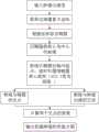
图1是本发明的基于形状直方图的超声乳腺肿瘤形态量化特征提取方法的流程图;Fig. 1 is the flow chart of the method for extracting morphological quantification features of ultrasonic breast tumor based on shape histogram of the present invention;
图2是本发明的得到该超声乳腺肿瘤的形状直方图的流程图;Fig. 2 is the flow chart of the present invention to obtain the shape histogram of this ultrasound breast tumor;
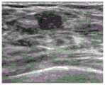
图3是良性超声乳腺肿瘤的图像;Figure 3 is an image of a benign ultrasound breast tumor;
图4是恶性超声乳腺肿瘤的图像;Figure 4 is an image of a malignant ultrasound breast tumor;
图5是良性超声乳腺肿瘤的边缘轮廓图;Fig. 5 is the edge contour map of benign ultrasound breast tumor;
图6是恶性超声乳腺肿瘤的边缘轮廓图;Fig. 6 is the edge contour map of malignant ultrasound breast tumor;
图7是良性超声乳腺肿瘤的形状直方图;Fig. 7 is the shape histogram of benign ultrasound breast tumor;
图8是恶性超声乳腺肿瘤的形状直方图;Fig. 8 is the shape histogram of malignant ultrasound breast tumor;
图9是良性超声乳腺肿瘤的形状直方图的曲线图;Figure 9 is a graph of a shape histogram of a benign ultrasound breast tumor;
图10是恶性超声乳腺肿瘤的形状直方图的曲线图;Figure 10 is a graph of a shape histogram of malignant ultrasound breast tumors;
图11是超声乳腺肿瘤的样本图。Figure 11 is a sample image of a breast tumor by ultrasound.
具体实施方式Detailed ways
下面将结合说明书附图,对本发明作进一步的说明。The present invention will be further described below with reference to the accompanying drawings.
如图1所示,本发明的基于形状直方图的超声乳腺肿瘤形态量化特征提取方法,包括以下步骤,As shown in FIG. 1 , the method for extracting morphological quantification features of ultrasonic breast tumors based on shape histogram of the present invention includes the following steps:
步骤(A),超声乳腺肿瘤原图中分割出乳腺肿瘤区域图;Step (A), segmenting the breast tumor region map from the original ultrasound breast tumor image;
步骤(B),获取该乳腺肿瘤区域图的乳腺肿瘤边缘图;Step (B), obtaining the breast tumor edge map of the breast tumor region map;
步骤(C),通过椭圆或者圆拟合得到该乳腺肿瘤边缘图内肿瘤目标形状的椭圆曲线,并得到该超声乳腺肿瘤的形状直方图,如图2所示,包括以下步骤,Step (C), obtaining the elliptic curve of the tumor target shape in the breast tumor edge map by ellipse or circle fitting, and obtaining the shape histogram of the ultrasound breast tumor, as shown in FIG. 2 , including the following steps:
(C1),根据乳腺肿瘤边缘图的肿瘤边缘轮廓的像素点坐标,得到拟合的肿瘤目标形状的椭圆曲线;(C1), according to the pixel coordinates of the tumor edge contour of the breast tumor edge map, the elliptic curve of the fitted tumor target shape is obtained;
(C2),以该椭圆曲线的质心为中心点作一系列射线,该一系列射线以椭圆长轴为起始点,沿逆时针方向围绕椭圆质心旋转,相邻的射线之间的间隔角度θ的取值范围为5°≤θ≤6°,经多次实验表明,当θ=5.7°时,效果最佳;(C2), take the center of mass of the elliptic curve as the center point to make a series of rays, the series of rays take the long axis of the ellipse as the starting point, rotate around the center of mass of the ellipse in the counterclockwise direction, and the interval angle θ between adjacent rays is The value range is 5°≤θ≤6°. After many experiments, it is shown that when θ=5.7°, the effect is the best;
(C3),该一系列射线与椭圆曲线、乳腺肿瘤边缘分别有交点,计算出每条射线与椭圆曲线的交点、每条射线与乳腺肿瘤边缘的交点之间的差值,得到该超声乳腺肿瘤的形状直方图(C3), the series of rays have intersections with the elliptic curve and the breast tumor edge respectively, calculate the difference between the intersection of each ray and the elliptic curve, and the intersection of each ray and the breast tumor edge, and obtain the ultrasound breast tumor shape histogram of
步骤(D),根据得到的超声乳腺肿瘤的形状直方图,描述出该乳腺肿瘤形态的三个量化特征:最大曲率和、最大曲率峰值和、最大曲率标准差和,In step (D), according to the obtained shape histogram of the breast tumor, three quantitative features of the breast tumor shape are described: the maximum curvature sum, the maximum curvature peak sum, and the maximum curvature standard deviation sum,
其中,描述最大曲率和的过程如下,Among them, the process of describing the maximum curvature sum is as follows,
(D11),将该形状直方图中所有数值相连接,得到形状直方图的曲线;(D11), connect all the values in the shape histogram to obtain the curve of the shape histogram;
(D12),计算该形状直方图中每个区间的所有数值点的曲率,如公式(1)所示,(D12), calculate the curvature of all numerical points in each interval in the shape histogram, as shown in formula (1),

其中,hi'(j)为形状直方图中第i个区间第j个数值点在曲线上的一阶导数,h″i(j)为形状直方图中第i个区间第j个数值点在曲线上的二阶导数,Cij则为形状直方图中第i个区间第j个数值点的曲率;Among them, hi '(j) is the first derivative on the curve of the jth numerical point in the ith interval in the shape histogram, and h″i (j) is the jth numerical point in the ith interval in the shape histogram The second derivative on the curve, Cij is the curvature of the jth numerical point in the ith interval in the shape histogram;
(D13),基于该形状直方图中每个区间的所有数值点的曲率,取出每个区间的最大曲率


描述出该乳腺肿瘤形态的最大曲率峰值和量化特征,过程如下,The maximum curvature peak and quantitative characteristics of the breast tumor morphology are described, and the process is as follows,
(D21),根据每个区间的最大曲率

其中,

描述出该乳腺肿瘤形态的最大曲率标准差和量化特征,过程如下,The maximum curvature standard deviation and quantitative characteristics of the breast tumor morphology are described, and the process is as follows,
(D31),根据公式(4),计算形状直方图中每个区间的峰值标准差,(D31), according to formula (4), calculate the peak standard deviation of each interval in the shape histogram,

其中,
(D32),通过峰值标准差对曲率进行加权,得到最大曲率标准差和SMCSD,如公式(5)所示,(D32), the curvature is weighted by the peak standard deviation to obtain the maximum curvature standard deviation and SMCSD, as shown in formula (5),

其中,
本发明的基于形状直方图的超声乳腺肿瘤形态量化特征提取方法,还包括步骤(E),根据步骤(D)得到的该乳腺肿瘤形态的三个量化特征,对该超声乳腺肿瘤的良恶性进行判别,可采用SVM分类器对步骤(D)得到的该乳腺肿瘤形态的三个量化特征进行该超声乳腺肿瘤的良恶性判别。The method for extracting morphological quantification features of ultrasonic breast tumor based on shape histogram of the present invention further comprises step (E), according to the three quantitative features of the shape of the breast tumor obtained in step (D), the benign and malignant of the ultrasonic breast tumor is analyzed. To discriminate, the SVM classifier can be used to discriminate benign and malignant of the ultrasound breast tumor on the three quantitative features of the breast tumor morphology obtained in step (D).
下面根据本发明的基于形状直方图的超声乳腺肿瘤形态量化特征提取方法,具体描述一实施过程,The following describes an implementation process in detail according to the shape histogram-based morphological quantification feature extraction method for ultrasonic breast tumors of the present invention.
(1)输入超声乳腺肿瘤图像,如图3、图4所示,其中图3为良性,图4为恶性;(1) Input ultrasound breast tumor images, as shown in Figure 3 and Figure 4, where Figure 3 is benign and Figure 4 is malignant;
(2)对超声乳腺肿瘤图像进行肿瘤目标分割,得到肿瘤边缘轮廓图,如图5及图6所示,其中图5为良性乳腺肿瘤的边缘轮廓图,图6为恶性乳腺肿瘤的边缘轮廓图;(2) Perform tumor target segmentation on the ultrasound breast tumor image to obtain a tumor edge contour map, as shown in Figures 5 and 6, wherein Figure 5 is the edge contour map of a benign breast tumor, and Figure 6 is the edge contour map of a malignant breast tumor ;
(3)依据肿瘤边缘轮廓的像素点坐标,得到拟合的肿瘤目标形状的椭圆曲线;(3) obtaining the elliptic curve of the fitted tumor target shape according to the pixel coordinates of the tumor edge contour;
(4)以椭圆的质心为中心点作一系列射线,该射线以椭圆长轴为起始点,沿逆时针方向围绕椭圆质心旋转,射线间隔角度θ=5.7°;(4) Make a series of rays with the center of mass of the ellipse as the center point. The rays take the long axis of the ellipse as the starting point and rotate around the center of mass of the ellipse in the counterclockwise direction, and the ray interval angle θ=5.7°;
(5)射线与椭圆曲线和肿瘤边缘分别有交点;(5) The ray intersects with the elliptic curve and the tumor edge respectively;
(6)计算出射线与椭圆曲线的交点以及射线与肿瘤边缘的交点之间的差值,得到乳腺肿瘤的形状直方图,如图7及图8所示,其中,图7为良性乳腺肿瘤的形状直方图,图8为恶性乳腺肿瘤的形状直方图。(6) Calculate the difference between the intersection of the ray and the elliptic curve and the intersection of the ray and the edge of the tumor, and obtain the shape histogram of the breast tumor, as shown in Figure 7 and Figure 8, wherein Figure 7 is a benign breast tumor. Shape histogram, Figure 8 is the shape histogram of malignant breast tumors.
从图7及图8中可以看出,形状直方图中数值波动情况与肿瘤形态变化是一一对应的,因此基于形状直方图设计相应的量化特征具有可行性。It can be seen from Fig. 7 and Fig. 8 that there is a one-to-one correspondence between the numerical fluctuations in the shape histogram and the changes in tumor morphology, so it is feasible to design corresponding quantitative features based on the shape histogram.
传统的形态量化特征是从全局的角度描述乳腺肿瘤的良恶性差异,针对这个不足,本发明基于形状直方图,以区间为单位设计量化特征,从局部的角度描述乳腺肿瘤的形态变化,从而更准确表征乳腺肿瘤的良恶性差异。这里,形状直方图的正值区间表示肿瘤边缘凸在椭圆外侧,负值区间表示肿瘤边缘凹在椭圆内侧,其中最大曲率和、最大曲率峰值和以及最大曲率标准差和三个形态量化特征的设计细节如下,The traditional morphological quantitative features describe the difference between benign and malignant breast tumors from a global perspective. In view of this deficiency, the present invention designs quantitative features based on the shape histogram in units of intervals, and describes the morphological changes of breast tumors from a local perspective. Accurately characterize the difference between benign and malignant breast tumors. Here, the positive value interval of the shape histogram indicates that the tumor edge is convex outside the ellipse, and the negative value interval indicates that the tumor edge is concave inside the ellipse, where the maximum curvature sum, the maximum curvature peak sum and the maximum curvature standard deviation and the design of the three morphological quantitative features Details as follow,
(1)最大曲率和(Sum of Maximum Curvature,SMC)(1) Sum of Maximum Curvature (SMC)
将形状直方图中所有数值相连接,得到形状直方图的曲线图,如图9及图10所示,其中,图9为良性乳腺肿瘤形状直方图的连接曲线,图10)为恶性乳腺肿瘤形状直方图的连接曲线;Connect all the values in the shape histogram to obtain a graph of the shape histogram, as shown in Figures 9 and 10, wherein Figure 9 is the connection curve of the shape histogram of benign breast tumors, and Figure 10) is the shape of malignant breast tumors the connection curve of the histogram;
(2)最大曲率峰值和(Sum of Maximum Curvature and Peak,SMCP)(2) Sum of Maximum Curvature and Peak (SMCP)
曲率描述了每个区间的曲线的弯曲程度,但不能体现曲线峰值的大小,因此,在计算最大曲率和时还需要考虑形状直方图中每个区间的曲峰数值的变化程度,本发明进一步用曲线峰值加权曲率,得到最大曲率峰值和;The curvature describes the degree of curvature of the curve in each interval, but cannot reflect the size of the peak value of the curve. Therefore, when calculating the maximum curvature sum, it is also necessary to consider the degree of change in the value of the peak value of each interval in the shape histogram. The present invention further uses The curve peak weights the curvature to obtain the maximum curvature peak sum;
(3)最大曲率标准差和(Sum of Maximum Curvature and Standard Deviation,SMCSD)(3) Sum of Maximum Curvature and Standard Deviation (SMCSD)
同样的,形状直方图中每个区间的峰值波动程度也体现了超声乳腺肿瘤的良恶性差异,通过用峰值标准差对曲率进行加权,得到最大曲率标准差和。Similarly, the peak fluctuation degree of each interval in the shape histogram also reflects the benign and malignant differences of ultrasound breast tumors. By weighting the curvature with the peak standard deviation, the maximum curvature standard deviation sum is obtained.
下面具体介绍,根据本发明的基于形状直方图的超声乳腺肿瘤形态量化特征提取方法,得到三个形态量化特征:最大曲率和(SMC)、最大曲率峰值和(SMCP)、最大曲率标准差和(SMCSD)进行乳腺良恶性肿瘤分析的实施过程,The following is a detailed introduction. According to the method for extracting morphological quantification features of ultrasonic breast tumors based on shape histograms of the present invention, three morphological quantification features are obtained: maximum sum of curvature (SMC), maximum sum of curvature peaks (SMCP), maximum sum of curvature standard deviation ( SMCSD) for the implementation of breast benign and malignant tumor analysis,
在实验测试中,数据源选择超声乳腺肿瘤数据来源于超声诊断仪(VINNO70,飞依诺科技有限公司,苏州),探头发射频率为5MHz~14MHz,一共采集192张图片,其中恶性肿瘤图片71张,良性肿瘤图片121张,部分样本如图11所示,其中图(a-d)为良性,图(e-h)为恶性。所有数据都获得受试者书面同意,且符合医院人体伦理认可。In the experimental test, the data source was selected as the data source of ultrasonic breast tumor data from the ultrasonic diagnostic instrument (VINNO70, Feiyinuo Technology Co., Ltd., Suzhou), and the transmission frequency of the probe was 5MHz-14MHz. A total of 192 pictures were collected, including 71 pictures of malignant tumors. , there are 121 benign tumor pictures, and some samples are shown in Figure 11, in which the pictures (a-d) are benign, and the pictures (e-h) are malignant. All data were obtained with the written consent of the subjects and accorded with the hospital human ethics approval.
(1)良恶性乳腺肿瘤识别率比较(1) Comparison of the recognition rate of benign and malignant breast tumors
采用本发明的三个形态量化特征:最大曲率和(SMC)、最大曲率峰值和(SMCP)、最大曲率标准差和(SMCSD)以及传统形态量化特征:粗糙度、规则度、纵横比、椭圆紧致度、圆形度对实际采集到的超声乳腺肿瘤图像进行特征提取,并采用SVM分类器进行良恶性判别,识别结果如表1所示。这里,选取50张恶性肿瘤图片和90张良性肿瘤图片作为训练样本,剩余图片作为测试样本,Three morphological quantification features of the present invention are employed: Maximum Sum of Curvature (SMC), Maximum Sum of Curvature Peaks (SMCP), Maximum Sum of Curvature Standard Deviation (SMCSD) and traditional morphological quantification features: roughness, regularity, aspect ratio, ellipse tightness Conformity and circularity were used to extract the features of the actual collected ultrasound breast tumor images, and the SVM classifier was used to discriminate benign and malignant. The identification results are shown in Table 1. Here, 50 images of malignant tumors and 90 images of benign tumors are selected as training samples, and the remaining images are used as test samples.
表1 本发明和传统形态量化特征在超声乳腺肿瘤数据上识别率比较Table 1 Comparison of recognition rates of the present invention and traditional morphological quantitative features on ultrasound breast tumor data
从表1中可以看出,采用本发明的三个形态量化特征:最大曲率和(SMC)、最大曲率峰值和(SMCP)、最大曲率标准差和(SMCSD)进行超声乳腺肿瘤良恶性判别时,可以达到82.69%的识别率,相比五个传统形态量化特征的识别率提升了15.38%。当传统量化特征与本发明提出的三个量化特征相结合时,识别率仍为82.69%,可见,本发明提出的最大曲率和(SMC)、最大曲率峰值和(SMCP)、最大曲率标准差和(SMCSD)在描述乳腺肿瘤良恶性差异时具有绝对优势。As can be seen from Table 1, when using the three morphological quantification features of the present invention: the maximum sum of curvature (SMC), the maximum sum of curvature peaks (SMCP), and the maximum sum of curvature standard deviation (SMCSD) to discriminate benign and malignant breast tumors by ultrasound, It can reach a recognition rate of 82.69%, which is 15.38% higher than the recognition rate of five traditional morphological quantitative features. When the traditional quantization feature is combined with the three quantization features proposed by the present invention, the recognition rate is still 82.69%. It can be seen that the maximum sum of curvature (SMC), the maximum sum of curvature peaks (SMCP), and the maximum sum of curvature standard deviations proposed by the present invention (SMCSD) has an absolute advantage in describing the difference between benign and malignant breast tumors.
(2)良恶性乳腺肿瘤形态量化特征数值比较(2) Numerical comparison of morphological quantitative features of benign and malignant breast tumors
为了更好地说明本发明的有效性,分别计算良恶性乳腺肿瘤在各种特征算子下的最大值、最小值和均值,如表2所示。In order to better illustrate the effectiveness of the present invention, the maximum value, minimum value and mean value of benign and malignant breast tumors under various characteristic operators are calculated respectively, as shown in Table 2.
表2 良恶性乳腺肿瘤的形态量化特征数值对比Table 2 Comparison of morphological quantitative characteristics of benign and malignant breast tumors

基于表2数值再进一步计算各个形态量化特征的良恶性判别距离D,计算公式(6)如下,Based on the values in Table 2, the benign and malignant discrimination distance D of each morphological quantitative feature is further calculated, and the calculation formula (6) is as follows:

其中,Maxbeni为良性乳腺肿瘤的最大值、Maxmali为恶性乳腺肿瘤的最大值,Minbeni为良性乳腺肿瘤的最小值,Minmali为恶性乳腺肿瘤的最小值,μbeni为良性乳腺肿瘤的均值,μmali为恶性乳腺肿瘤的均值。根据公式(6)整理得到表3,由表3可以看出,由最大曲率和(SMC)、最大曲率峰值和(SMCP)、最大曲率标准差和(SMCSD)计算得到的良恶性乳腺肿瘤的判别距离D明显大于传统特征,因此,进一步说明,本发明提出的三个形态量化特征可以更准确描述乳腺肿瘤的良恶性差异,Among them, Maxbeni is the maximum value of benign breast tumors, Maxmali is the maximum value of malignant breast tumors, Minbeni is the minimum value of benign breast tumors, Minmali is the minimum value of malignant breast tumors, and μbeni is the mean value of benign breast tumors ,μmali is the mean of malignant breast tumors. According to formula (6), Table 3 is obtained. It can be seen from Table 3 that the discrimination of benign and malignant breast tumors calculated by the maximum sum of curvature (SMC), the maximum sum of curvature peaks (SMCP), and the maximum sum of curvature standard deviation (SMCSD) The distance D is significantly larger than the traditional features. Therefore, it is further explained that the three morphological quantitative features proposed in the present invention can more accurately describe the difference between benign and malignant breast tumors.
表3 形态量化特征的良恶性判别距离Table 3 Benign and malignant discriminant distances of morphological quantitative features

(3)本发明提供方法的性能分析(3) Performance analysis of the method provided by the present invention
为了进一步说明本发明的有效性,这里使用5个指标评估分类器的性能,分别为准确度(Accuracy),灵敏度(Sensitivity)、特异度(Specificity)、阳性预测值(PositivePredictive Value,PPV)、阴性预测值(Negative Predictive Value,NPV),定义如下:In order to further illustrate the effectiveness of the present invention, five indicators are used to evaluate the performance of the classifier, namely Accuracy, Sensitivity, Specificity, Positive Predictive Value (PPV), Negative Negative Predictive Value (NPV), defined as follows:


其中,TP为真阳性例数,TN为真阴性例数,FP为假阳性例数,FN为假阴性例数。本发明和传统量化特征的性能参数比较如表4所示,Among them, TP is the number of true positive cases, TN is the number of true negative cases, FP is the number of false positive cases, and FN is the number of false negative cases. The performance parameters of the present invention and traditional quantization features are compared as shown in Table 4,
表4 本发明和传统方法的性能参数比较Table 4 Comparison of performance parameters between the present invention and the traditional method
由表4中可以看出,本发明较传统形态量化特征具有较高的准确度、灵敏度、特异度、阳性预测率和阴性预测率,尤其是PPV值为87.5%,NPV值为80.56%,这说明本发明在检测阳性和阴性结果时具有卓越的查准率,即被检测为阳性或阴性的患者确实患病或无病的可能性最大。As can be seen from Table 4, the present invention has higher accuracy, sensitivity, specificity, positive predictive rate and negative predictive rate than traditional morphological quantitative features, especially the PPV value is 87.5%, and the NPV value is 80.56%. It is shown that the present invention has excellent precision in detecting positive and negative results, that is, patients who are detected as positive or negative are most likely to have the disease or not.
综上所述,本发明的基于形状直方图的超声乳腺肿瘤形态量化特征提取方法,针对计算机辅助诊断系统中,传统超声乳腺肿瘤良恶性量化特征描述不准确的缺点,从乳腺肿瘤轮廓出发,构造乳腺肿瘤的形状直方图,再以形状直方图为基础,设计三个形态量化特征:最大曲率和、最大曲率峰值和、最大曲率标准差和,用以有效表征超声乳腺肿瘤的局部良恶性差异;并将本发明提出的三个形态量化特征应用于分类器中,得到的超声乳腺肿瘤的良恶性识别率相比传统形态量化特征的识别率有显著提高,具有实用价值;同时,最大曲率和、最大曲率峰值和、最大曲率标准差和也丰富了现有超声乳腺肿瘤的形态量化特征库,本发明可以有效减少医生的主观因素影响,有助于提高医生诊断的准确率,可适用于其他肿瘤良恶性的诊断,具有良好的应用前景。To sum up, the method for extracting morphological quantification features of ultrasonic breast tumors based on shape histogram of the present invention aims at the disadvantage of inaccurate description of benign and malignant quantification features of traditional ultrasonic breast tumors in the computer-aided diagnosis system. The shape histogram of breast tumors, and then based on the shape histogram, three morphological quantitative features are designed: the maximum curvature sum, the maximum curvature peak sum, and the maximum curvature standard deviation sum, which can effectively characterize the local benign and malignant differences of ultrasound breast tumors; The three morphological quantification features proposed by the present invention are applied to the classifier, and the obtained ultrasound breast tumor recognition rate of benign and malignant is significantly improved compared with the traditional morphological quantification features, which has practical value; at the same time, the maximum curvature sum, The maximum curvature peak sum and the maximum curvature standard deviation sum also enrich the morphological quantification feature library of existing ultrasonic breast tumors. The present invention can effectively reduce the influence of the doctor's subjective factors, help improve the accuracy of the doctor's diagnosis, and can be applied to other tumors The diagnosis of benign and malignant has good application prospects.
以上显示和描述了本发明的基本原理、主要特征及优点。本行业的技术人员应该了解,本发明不受上述实施例的限制,上述实施例和说明书中描述的只是说明本发明的原理,在不脱离本发明精神和范围的前提下,本发明还会有各种变化和改进,这些变化和改进都落入要求保护的本发明范围内。本发明要求保护范围由所附的权利要求书及其等效物界定。The foregoing has shown and described the basic principles, main features and advantages of the present invention. Those skilled in the art should understand that the present invention is not limited by the above-mentioned embodiments. The above-mentioned embodiments and descriptions only illustrate the principle of the present invention. Without departing from the spirit and scope of the present invention, the present invention will also have Various changes and modifications fall within the scope of the claimed invention. The claimed scope of the present invention is defined by the appended claims and their equivalents.
Claims (5)


Priority Applications (1)
| Application Number | Priority Date | Filing Date | Title |
|---|---|---|---|
| CN201910138613.4ACN109903278B (en) | 2019-02-25 | 2019-02-25 | Ultrasonic breast tumor morphological quantitative feature extraction method based on shape histogram |
Applications Claiming Priority (1)
| Application Number | Priority Date | Filing Date | Title |
|---|---|---|---|
| CN201910138613.4ACN109903278B (en) | 2019-02-25 | 2019-02-25 | Ultrasonic breast tumor morphological quantitative feature extraction method based on shape histogram |
Publications (2)
| Publication Number | Publication Date |
|---|---|
| CN109903278A CN109903278A (en) | 2019-06-18 |
| CN109903278Btrue CN109903278B (en) | 2020-10-27 |
Family
ID=66945495
Family Applications (1)
| Application Number | Title | Priority Date | Filing Date |
|---|---|---|---|
| CN201910138613.4AActiveCN109903278B (en) | 2019-02-25 | 2019-02-25 | Ultrasonic breast tumor morphological quantitative feature extraction method based on shape histogram |
Country Status (1)
| Country | Link |
|---|---|
| CN (1) | CN109903278B (en) |
Families Citing this family (1)
| Publication number | Priority date | Publication date | Assignee | Title |
|---|---|---|---|---|
| CN111311578B (en)* | 2020-02-17 | 2024-05-03 | 腾讯科技(深圳)有限公司 | Object classification method and device based on artificial intelligence, and medical imaging equipment |
Citations (2)
| Publication number | Priority date | Publication date | Assignee | Title |
|---|---|---|---|---|
| CN1518719A (en)* | 2001-04-23 | 2004-08-04 | 西门子共同研究公司 | Method and system for automatically detecting lung nodules from multi-slice high resolution computed tomography (MSHR CT) images |
| CN109242016A (en)* | 2018-08-30 | 2019-01-18 | 天津理工大学 | A kind of similitude judgment method of space curve |
Family Cites Families (3)
| Publication number | Priority date | Publication date | Assignee | Title |
|---|---|---|---|---|
| WO2000065985A2 (en)* | 1999-04-29 | 2000-11-09 | University Of South Florida | Method and system for knowledge guided hyperintensity detection and volumetric measurement |
| CN101517614A (en)* | 2006-09-22 | 2009-08-26 | 皇家飞利浦电子股份有限公司 | Advanced computer-aided diagnosis of lung nodules |
| CN103425986B (en)* | 2013-08-31 | 2016-08-10 | 西安电子科技大学 | Mammary gland tumor image characteristic extracting method based on edge neighborhood weighting |
- 2019
- 2019-02-25CNCN201910138613.4Apatent/CN109903278B/enactiveActive
Patent Citations (2)
| Publication number | Priority date | Publication date | Assignee | Title |
|---|---|---|---|---|
| CN1518719A (en)* | 2001-04-23 | 2004-08-04 | 西门子共同研究公司 | Method and system for automatically detecting lung nodules from multi-slice high resolution computed tomography (MSHR CT) images |
| CN109242016A (en)* | 2018-08-30 | 2019-01-18 | 天津理工大学 | A kind of similitude judgment method of space curve |
Also Published As
| Publication number | Publication date |
|---|---|
| CN109903278A (en) | 2019-06-18 |
Similar Documents
| Publication | Publication Date | Title |
|---|---|---|
| Liu et al. | Breast tumors recognition based on edge feature extraction using support vector machine | |
| CN101763644B (en) | Pulmonary nodule three-dimensional segmentation and feature extraction method and system thereof | |
| Ge et al. | Computer‐aided detection of lung nodules: false positive reduction using a 3D gradient field method and 3D ellipsoid fitting | |
| CN112215842B (en) | Malignant nodule edge detection image processing method based on benign thyroid template | |
| CN108648182B (en) | Breast cancer nuclear magnetic resonance image tumor region segmentation method based on molecular subtype | |
| CN103745227A (en) | Method for identifying benign and malignant lung nodules based on multi-dimensional information | |
| Yue et al. | Automatic acetowhite lesion segmentation via specular reflection removal and deep attention network | |
| Kim et al. | Deep convolutional neural network for classification of thyroid nodules on ultrasound: Comparison of the diagnostic performance with that of radiologists | |
| CN101103924A (en) | Breast cancer computer-aided diagnosis method and system based on mammography | |
| Liu et al. | Computer aided diagnosis system for breast cancer based on color Doppler flow imaging | |
| CN101714153A (en) | Visual perception based interactive mammography image searth method | |
| Wang et al. | A method of ultrasonic image recognition for thyroid papillary carcinoma based on deep convolution neural network. | |
| CN103778600A (en) | Image processing system | |
| Xing et al. | Automatic detection of A‐line in lung ultrasound images using deep learning and image processing | |
| Shen et al. | Lesion segmentation in breast ultrasound images using the optimized marked watershed method | |
| CN108288265A (en) | A kind of segmentation and sorting technique towards HCC pathological image nucleus | |
| CN113658151A (en) | Mammary gland lesion magnetic resonance image classification method and device and readable storage medium | |
| Wu et al. | An Effective Machine-Learning Based Feature Extraction/RecognitionModel for Fetal Heart Defect Detection from 2D Ultrasonic Imageries. | |
| Hsieh et al. | Stage classification in chronic kidney disease by ultrasound image | |
| CN109903278B (en) | Ultrasonic breast tumor morphological quantitative feature extraction method based on shape histogram | |
| Diao et al. | Highly sensitive computer aided diagnosis system for breast tumor based on color Doppler flow images | |
| CN103455821A (en) | Device and method for analyzing images on basis of BI-RADS (breast imaging reporting and data system) | |
| Han et al. | Automatic detection of thyroid nodule characteristics from 2D ultrasound images | |
| Zhang | Classification and diagnosis of thyroid carcinoma using reinforcement residual network with visual attention mechanisms in ultrasound images | |
| An et al. | The research of automatic classification of ultrasound thyroid nodules |
Legal Events
| Date | Code | Title | Description |
|---|---|---|---|
| PB01 | Publication | ||
| PB01 | Publication | ||
| SE01 | Entry into force of request for substantive examination | ||
| SE01 | Entry into force of request for substantive examination | ||
| GR01 | Patent grant | ||
| GR01 | Patent grant | ||
| TR01 | Transfer of patent right | ||
| TR01 | Transfer of patent right | Effective date of registration:20201230 Address after:Room 108, 7 Yingcui Road, Jiangning Development Zone, Nanjing, Jiangsu 210000 Patentee after:Nanjing tianzhixin Technology Co.,Ltd. Address before:211167 No.1, Hongjing Avenue, Jiangning Science Park, Jiangning District, Nanjing City, Jiangsu Province Patentee before:NANJING INSTITUTE OF TECHNOLOGY |