CN109893146B - A method and system for evaluating female pelvic floor dysfunction - Google Patents
A method and system for evaluating female pelvic floor dysfunctionDownload PDFInfo
- Publication number
- CN109893146B CN109893146BCN201910171389.9ACN201910171389ACN109893146BCN 109893146 BCN109893146 BCN 109893146BCN 201910171389 ACN201910171389 ACN 201910171389ACN 109893146 BCN109893146 BCN 109893146B
- Authority
- CN
- China
- Prior art keywords
- muscle
- pelvic floor
- displacement
- time
- data
- Prior art date
- Legal status (The legal status is an assumption and is not a legal conclusion. Google has not performed a legal analysis and makes no representation as to the accuracy of the status listed.)
- Expired - Fee Related
Links
Images


Classifications
- A—HUMAN NECESSITIES
- A61—MEDICAL OR VETERINARY SCIENCE; HYGIENE
- A61B—DIAGNOSIS; SURGERY; IDENTIFICATION
- A61B5/00—Measuring for diagnostic purposes; Identification of persons
- A—HUMAN NECESSITIES
- A61—MEDICAL OR VETERINARY SCIENCE; HYGIENE
- A61B—DIAGNOSIS; SURGERY; IDENTIFICATION
- A61B5/00—Measuring for diagnostic purposes; Identification of persons
- A61B5/22—Ergometry; Measuring muscular strength or the force of a muscular blow
- A—HUMAN NECESSITIES
- A61—MEDICAL OR VETERINARY SCIENCE; HYGIENE
- A61B—DIAGNOSIS; SURGERY; IDENTIFICATION
- A61B8/00—Diagnosis using ultrasonic, sonic or infrasonic waves
- G—PHYSICS
- G06—COMPUTING OR CALCULATING; COUNTING
- G06T—IMAGE DATA PROCESSING OR GENERATION, IN GENERAL
- G06T7/00—Image analysis
- G06T7/20—Analysis of motion
- G06T7/246—Analysis of motion using feature-based methods, e.g. the tracking of corners or segments
Landscapes
- Health & Medical Sciences (AREA)
- Life Sciences & Earth Sciences (AREA)
- Engineering & Computer Science (AREA)
- Physics & Mathematics (AREA)
- Molecular Biology (AREA)
- Animal Behavior & Ethology (AREA)
- Veterinary Medicine (AREA)
- Public Health (AREA)
- Biophysics (AREA)
- General Health & Medical Sciences (AREA)
- Pathology (AREA)
- Surgery (AREA)
- Biomedical Technology (AREA)
- Heart & Thoracic Surgery (AREA)
- Medical Informatics (AREA)
- Multimedia (AREA)
- Radiology & Medical Imaging (AREA)
- Theoretical Computer Science (AREA)
- Nuclear Medicine, Radiotherapy & Molecular Imaging (AREA)
- Computer Vision & Pattern Recognition (AREA)
- General Physics & Mathematics (AREA)
- Physical Education & Sports Medicine (AREA)
- Measurement Of The Respiration, Hearing Ability, Form, And Blood Characteristics Of Living Organisms (AREA)
Abstract
Translated fromChinese
Description
Translated fromChinese技术领域technical field
本发明涉及盆底肌肉运动分析技术领域,特别涉及一种女性盆底功能障碍评估方法及其系统。The invention relates to the technical field of pelvic floor muscle movement analysis, in particular to a female pelvic floor dysfunction assessment method and system.
背景技术Background technique
女性盆底功能障碍性疾病(female pelvic flour dysfunction,FPFD)是一种由于盆底支撑结构损伤从而导致盆底脏器位置和功能异常的中老年女性常见疾病,主要表现为:压力性尿失禁、盆腔器官脱垂和性功能障碍。现已成为威胁女性健康的5种常见慢性疾病之一,是一个在全球范围内日益受到重视的社会卫生问题。FPFD虽不威胁生命,但它严重影响妇女社会活动、身心健康和生活质量。流行病学调查显示,妊娠和分娩是FPFD的独立危险因素。据报道美国大约10-36%的初产妇产后发生盆底肌肉或筋膜的损伤,约10%的美国妇女由于严重的FPFD需要手术治疗,美国每年治疗单纯尿失禁的费用高达到100亿美元。据澳大利亚的统计报道,产后尿失禁的发生率约15%~40%。我国目前还缺乏确切的流行病学数据,2005年据北京地区流行病学调查,约38.5%的产后妇女患有压力性尿失禁(SUI),且随年龄的增长患病率增加。Female pelvic flour dysfunction (FPFD) is a common disease of middle-aged and elderly women with abnormal position and function of pelvic floor organs due to damage to the pelvic floor supporting structure. Pelvic organ prolapse and sexual dysfunction. It has now become one of the five common chronic diseases that threaten women's health, and is a social health problem that has received increasing attention worldwide. Although FPFD is not life-threatening, it seriously affects women's social activities, physical and mental health and quality of life. Epidemiological surveys have shown that pregnancy and childbirth are independent risk factors for FPFD. It is reported that about 10-36% of primiparous women in the United States have postpartum pelvic floor muscle or fascia injuries, and about 10% of American women require surgery due to severe FPFD. According to Australian statistics, the incidence of postpartum urinary incontinence is about 15% to 40%. There is still a lack of exact epidemiological data in my country. According to an epidemiological survey in Beijing in 2005, about 38.5% of postpartum women suffered from stress urinary incontinence (SUI), and the prevalence increased with age.
现有的吊床学说,即“阴道三个支持水平”理论,将支持阴道的筋膜、韧带、结缔组织和盆底肌肉分为上、中、下三个水平以撑托盆底,并指出肛提肌群是盆底三个支撑水平中最重要的支持结构,一旦发生松弛或损伤,盆腔器官将无法维持在正常位置,从而出现盆底脏器脱垂等盆底功能障碍性疾病。盆底功能性障碍疾病防治的前提是及时发现和准确诊断。临床诊断盆底功能性障碍疾病的主要依据是妇科检查结果,应用国际尿控协会推荐的盆腔器脱垂的定量POP-Q分度法对盆腔脏器的脱垂及其程度进行诊断。目前对产后盆底支持结构损伤的诊断尚缺乏一个简单、客观、准确的方法,病人和临床医生对FPFD的认识不足,发病隐匿,患者多数羞于就医,症状常常被病人隐瞒,仅3%的患者因FPFD的症状寻医,因而早期就诊的病人少,绝大多数患者是在出现明显临床症状,甚至影响日常生活后才就诊,最后需要通过手术治疗才能改善症状,错过了早期诊断和及时康复(通过盆底肌锻炼、生物反馈和盆底磁疗等物理治疗)的无创治疗时期。The existing hammock theory, that is, the "three levels of vaginal support" theory, divides the fascia, ligaments, connective tissue and pelvic floor muscles that support the vagina into three levels: upper, middle, and lower to support the pelvic floor, and points out that the anus supports the pelvic floor. The levator muscle group is the most important supporting structure among the three support levels of the pelvic floor. Once relaxation or damage occurs, the pelvic organs will not be able to maintain their normal positions, resulting in pelvic floor dysfunction diseases such as pelvic organ prolapse. The premise of prevention and treatment of pelvic floor dysfunction is timely detection and accurate diagnosis. The main basis for the clinical diagnosis of pelvic floor dysfunction is the results of gynecological examinations. The quantitative POP-Q method of pelvic organ prolapse recommended by the International Continence Association is used to diagnose pelvic organ prolapse and its degree. At present, there is still a lack of a simple, objective and accurate method for the diagnosis of postpartum pelvic floor supporting structure damage. Patients and clinicians have insufficient understanding of FPFD, the onset is hidden, most patients are ashamed to seek medical treatment, and their symptoms are often concealed by patients. Patients seek medical attention due to the symptoms of FPFD, so few patients seek medical treatment early. Most patients seek medical treatment after obvious clinical symptoms appear, or even affect their daily life. Finally, surgical treatment is required to improve symptoms, and early diagnosis and timely recovery are missed. A period of non-invasive treatment (through physical therapy such as pelvic floor exercises, biofeedback, and magnetic pelvic floor therapy).
2009年国家卫生部妇幼保健与社区卫生司主持启动了以关注妇女生殖健康,尤其是盆底功能障碍性疾病的一项综合防治项目,盆底康复治疗已成为国家安康工程之一。2012年国家科技部将“盆底功能障碍性疾病规范化诊疗”列为国家科技计划人口与健康领域的重大专项指南。如何加强和提高年轻母亲们的生殖健康保健意识,降低产后FPFD的发生,提高她们的生活质量和家庭幸福,是一个重大民生课题。In 2009, the Department of Maternal and Child Health and Community Health of the National Ministry of Health launched a comprehensive prevention and treatment project focusing on women's reproductive health, especially pelvic floor dysfunction. Pelvic floor rehabilitation has become one of the national health projects. In 2012, the Ministry of Science and Technology of the People's Republic of China listed "standardized diagnosis and treatment of pelvic floor dysfunction diseases" as a major special guideline in the field of population and health of the National Science and Technology Program. How to strengthen and improve the reproductive health care awareness of young mothers, reduce the incidence of postpartum FPFD, and improve their quality of life and family happiness is a major livelihood issue.
现有的盆底肌肉评估方法包括触诊、阴道内压力测试、肌电图测试和影像技术,具有各自的优势也各存限制。Existing pelvic floor muscle assessment methods include palpation, intravaginal pressure testing, electromyography, and imaging techniques, each with their own advantages and limitations.
传统的阴道触诊(Vaginal palpation)是一种目前被理疗学家广泛用来评估盆底肌肉收缩能力的方法,测试者通过指诊的方式直接感受阴道收缩的肌力大小,并结合Oxford评分系统对肌肉进行分级评分。阴道触诊方法感性、简单,常用;但是由测试者主观判断,需要诊断者具有熟练的经验,所以具有一定的测量误差。Traditional vaginal palpation (Vaginal palpation) is a method widely used by physiotherapists to evaluate pelvic floor muscle contraction ability. The tester directly feels the muscle strength of vaginal contraction through digital examination, combined with the Oxford scoring system. Muscles are graded and scored. The vaginal palpation method is perceptual, simple, and commonly used; however, it is subjectively judged by the tester and requires skilled experience of the diagnostician, so there is a certain measurement error.
阴道内压力测试是通过测量肌肉的挤压力来对肌肉进行评估,主要针对肌肉力量。这种方法容易受其他肌群力量的干扰,而且无法定位具体是哪块肌肉有问题。An intravaginal stress test assesses muscles by measuring the squeezing force of the muscles, primarily for muscle strength. This method is susceptible to interference by the strength of other muscle groups, and it is impossible to pinpoint which muscle is at fault.
肌电图测试是通过测肌肉的电位活动来对肌肉进行评估,其包括针电极和表面电极。表面电极收集的是肌肉群的整体肌电信号,无法具体定位单块肌肉;针电极可以测试单块肌肉,但是具有强烈的侵入性,临床病人耐受性比较差。The EMG test assesses muscles by measuring their electrical activity, including needle electrodes and surface electrodes. Surface electrodes collect the overall EMG signals of muscle groups and cannot specifically locate a single muscle; needle electrodes can test a single muscle, but are highly invasive and poorly tolerated by clinical patients.
随着影像技术的快速发展,超声成像技术、磁共振成像技术及X线成像技术能够对盆底肌肉的解剖结构及形态位置进行成像,通过测量盆膈裂孔面积等量化参数间接对其进行评估。通过超声测量尿道内口在肛提肌从静息状态到最大收缩状态时相对耻骨联合后下边缘的横向位移和纵向位移来间接地对被试的肛提肌的活动度进行间接的评估。With the rapid development of imaging technology, ultrasound imaging technology, magnetic resonance imaging technology and X-ray imaging technology can image the anatomical structure and morphological position of pelvic floor muscles, and indirectly evaluate them by measuring quantitative parameters such as pelvic diaphragm hiatus area. The mobility of the levator ani muscle was indirectly assessed by ultrasound measurement of the lateral and longitudinal displacement of the internal urethral orifice relative to the posterior inferior border of the pubic symphysis when the levator ani muscle moved from a resting state to a maximally contracted state.
上述对盆底肌肉力学特性的评估方法具有主观性或有创的(即带创伤),获取的评估参数并不能定量反映盆底肌自身生物力学特性,对盆底肌力的检测准确性较低。The above-mentioned evaluation methods for the mechanical properties of pelvic floor muscles are subjective or invasive (that is, with trauma). .
因而现有技术还有待改进和提高。Therefore, the existing technology still needs to be improved and improved.
发明内容SUMMARY OF THE INVENTION
鉴于上述现有技术的不足之处,本发明的目的在于提供一种女性盆底功能障碍评估方法及其系统,以解决现有盆底肌肉评估方法对盆底肌力的检测准确性较低的问题。In view of the deficiencies of the above-mentioned prior art, the object of the present invention is to provide a female pelvic floor dysfunction assessment method and system thereof, to solve the problem that the detection accuracy of the pelvic floor muscle strength by the existing pelvic floor muscle assessment method is relatively low. question.
为了达到上述目的,本发明采取了以下技术方案:In order to achieve the above object, the present invention has adopted the following technical solutions:
一种女性盆底功能障碍评估方法,其包括A method for assessing pelvic floor dysfunction in women, comprising:
步骤A、测试时实时获取盆底肌肉的肌力数据和盆底肌解剖结构图像数据;During step A, the test, acquire the muscle strength data of the pelvic floor muscles and the anatomical structure image data of the pelvic floor muscles in real time;
步骤B、对肌力数据和盆底肌解剖结构图像数据进行同步采集;Step B, synchronously collecting muscle strength data and pelvic floor muscle anatomical structure image data;
步骤C、根据盆底肌解剖结构图像数据计算出盆底肌肉上感兴趣区域的特征点随时间变化的位移曲线;Step C, according to the anatomical structure image data of the pelvic floor muscle, calculate the time-varying displacement curve of the feature point of the region of interest on the pelvic floor muscle;
步骤D、根据肌力数据计算阴道肌力随时间变化的曲线,根据位移曲线计算肌肉形变量随时间变化的位移量曲线。Step D: Calculate the curve of vaginal muscle strength with time according to the muscle strength data, and calculate the displacement curve of muscle deformation with time according to the displacement curve.
所述的女性盆底功能障碍评估方法中,在所述步骤A中,实时获取盆底肌肉的肌力数据具体包括:In the described method for evaluating female pelvic floor dysfunction, in the step A, acquiring the muscle strength data of the pelvic floor muscles in real time specifically includes:
步骤A1、通过压强检测模块获取女性盆底肌力,并将女性盆底肌力转换成相应的压强信号输出;Step A1, obtaining the female pelvic floor muscle strength through the pressure detection module, and converting the female pelvic floor muscle strength into a corresponding pressure signal output;
步骤A2、数据处理模块对所述压强信号进行信号处理、转换成波形图显示并输出肌力数据。Step A2: The data processing module performs signal processing on the pressure signal, converts it into a waveform display, and outputs muscle strength data.
所述的女性盆底功能障碍评估方法中,所述步骤B具体包括:In the described female pelvic floor dysfunction assessment method, the step B specifically includes:
图像采集卡在各个预设时间点对盆底肌解剖结构图像数据进行采集,并将采集的时间点与所采的盆底肌解剖结构图像数据一一对应标记;数据采集卡在所述各个预设时间点对肌力数据进行采集,并将采集的时间点与所采的肌力数据一一对应标记。The image acquisition card collects the image data of the anatomical structure of the pelvic floor muscles at each preset time point, and marks the collected time points and the collected image data of the anatomical structure of the pelvic floor muscles one-to-one; the data acquisition card is in each preset time point. Set time points to collect muscle strength data, and mark the collected time points with the collected muscle strength data in one-to-one correspondence.
所述的女性盆底功能障碍评估方法中,在所述步骤C中,通过运动追踪算法来建立盆底肌肉各感兴趣区域随时间变化的位移曲线。根据盆底肌解剖结构图像数据计算出盆底肌肉上感兴趣区域的特征点随时间变化的位移曲线In the method for evaluating female pelvic floor dysfunction, in the step C, a movement tracking algorithm is used to establish the displacement curve of each region of interest of the pelvic floor muscle over time. According to the anatomical structure image data of the pelvic floor muscle, the time-varying displacement curve of the feature points of the region of interest on the pelvic floor muscle is calculated
所述的女性盆底功能障碍评估方法中,所述步骤C具体包括:In the described female pelvic floor dysfunction assessment method, the step C specifically includes:
步骤C1、追踪盆底肌解剖结构图像数据中的第n帧和第n+2帧图像中的感兴趣区域的第一特征点和第二特征点的位移场;Step C1, tracking the displacement field of the first feature point and the second feature point of the region of interest in the nth frame and the n+2th frame image in the pelvic floor muscle anatomical structure image data;
步骤C2、将第n帧和第n+2帧图像进行对比,计算第一特征点和第二特征点在位移场上随时间变化的位移曲线。Step C2: Compare the images of the nth frame and the n+2th frame, and calculate the displacement curves of the first feature point and the second feature point that change with time on the displacement field.
所述的女性盆底功能障碍评估方法中,所述第一特征点为将耻骨直肠肌平均分为三段的靠近耻骨的一个均分点,第二特征点为将耻骨直肠肌平均分为三段的靠近直肠的另一个均分点。In the method for evaluating female pelvic floor dysfunction, the first feature point is an average point near the pubic bone that divides the puborectalis muscle into three segments, and the second feature point is an average point that divides the puborectalis muscle into three segments. Another equalization point of the segment near the rectum.
一种用于实现所述的女性盆底功能障碍评估方法的系统,其包括:A system for realizing the described method for evaluating female pelvic floor dysfunction, comprising:
阴道肌力采集装置,用于测试时实时获取盆底肌肉的肌力数据;The vaginal muscle strength acquisition device is used to obtain the muscle strength data of the pelvic floor muscles in real time during the test;
盆底肌肉成像装置,用于测试时实时获取盆底肌解剖结构图像数据;The pelvic floor muscle imaging device is used for real-time acquisition of pelvic floor muscle anatomical structure image data during testing;
数据采集装置,用于对肌力数据和盆底肌解剖结构图像数据进行同步采集;a data acquisition device for synchronous acquisition of muscle strength data and pelvic floor muscle anatomical structure image data;
PC机,用于根据盆底肌解剖结构图像数据计算出盆底肌肉上感兴趣区域的特征点随时间变化的位移曲线;根据肌力数据计算阴道肌力随时间变化的曲线,根据位移曲线计算肌肉形变量随时间变化的位移量曲线。The PC is used to calculate the time-varying displacement curve of the feature points of the region of interest on the pelvic floor muscles according to the anatomical structure image data of the pelvic floor muscles; calculate the time-varying curve of vaginal muscle strength according to the muscle strength data, and calculate the curve according to the displacement curve. Displacement curve of muscle deformation as a function of time.
所述的系统中,所述阴道肌力采集装置包括:In the described system, the vaginal muscle strength collection device includes:
压强检测模块,用于获取女性盆底肌力,并将女性盆底肌力转换成相应的压强信号输出;The pressure detection module is used to obtain the female pelvic floor muscle strength and convert the female pelvic floor muscle strength into the corresponding pressure signal output;
数据处理模块,用于对所述压强信号进行信号处理、转换成波形图显示并输出肌力数据。The data processing module is used to perform signal processing on the pressure signal, convert it into waveform display and output muscle strength data.
所述的系统中,所述数据采集装置包括:In the system, the data acquisition device includes:
图像采集卡,用于在各个预设时间点对盆底肌解剖结构图像数据进行采集,并将采集的时间点与所采的盆底肌解剖结构图像数据一一对应标记;The image acquisition card is used to collect the image data of the anatomical structure of the pelvic floor muscle at each preset time point, and mark the collected time point and the collected image data of the anatomical structure of the pelvic floor muscle in one-to-one correspondence;
数据采集卡,用于在所述各个预设时间点对肌力数据进行采集,并将采集的时间点与所采的肌力数据一一对应标记。The data acquisition card is used for collecting muscle strength data at each preset time point, and marking the collected time points and the collected muscle strength data in one-to-one correspondence.
相较于现有技术,本发明提供的女性盆底功能障碍评估方法及其系统,测试时实时获取盆底肌肉的肌力数据和盆底肌解剖结构图像数据;对肌力数据和盆底肌解剖结构图像数据进行同步采集;根据盆底肌解剖结构图像数据计算出盆底肌肉上感兴趣区域的特征点随时间变化的位移曲线;根据肌力数据计算阴道肌力随时间变化的曲线,根据位移曲线计算肌肉形变量随时间变化的位移量曲线。通过同时采集肌力数据和盆底肌解剖结构图像数据这两个参数并计算出与时间对应的曲线,即可对生物力学特性进行定量评价,为女性盆底功能障碍评估提供准确的判断依据。Compared with the prior art, the method and system for evaluating female pelvic floor dysfunction provided by the present invention can acquire the muscle strength data of the pelvic floor muscles and the anatomical structure image data of the pelvic floor muscles in real time during the test; The anatomical structure image data is collected synchronously; the displacement curve of the feature points of the region of interest on the pelvic floor muscle is calculated according to the pelvic floor muscle anatomical structure image data; Displacement Curve Calculates the displacement curve of the muscle deformation amount as a function of time. By collecting the two parameters of muscle strength data and pelvic floor muscle anatomical structure image data at the same time and calculating the curve corresponding to time, the biomechanical characteristics can be quantitatively evaluated, and an accurate judgment basis for the evaluation of female pelvic floor dysfunction can be provided.
附图说明Description of drawings
图1为本发明提供的女性盆底功能障碍评估方法流程图。FIG. 1 is a flowchart of a method for evaluating female pelvic floor dysfunction provided by the present invention.
图2为本发明提供的女性盆底功能障碍评估的系统的结构框图。FIG. 2 is a structural block diagram of a system for evaluating female pelvic floor dysfunction provided by the present invention.
图3为本发明提供的女性盆底功能障碍评估的系统中压强检测模块的示意图。3 is a schematic diagram of a pressure detection module in the system for evaluating female pelvic floor dysfunction provided by the present invention.
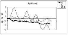
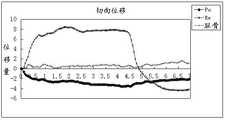
图4为本发明快缩时阴道肌力随时间变化的曲线图。Figure 4 is a graph showing the change of vaginal muscle strength with time during rapid contraction of the present invention.
图5为本发明慢缩时阴道肌力随时间变化的曲线图。Figure 5 is a graph showing the change of vaginal muscle strength with time during slow contraction of the present invention.
图6为本发明快缩时两个特征点的径向位移上肌肉形变量随时间变化的位移量曲线示意图。6 is a schematic diagram of the displacement curve of the muscle deformation amount changing with time on the radial displacement of the two characteristic points in the present invention.
图7为本发明快缩时两个特征点的切向位移上肌肉形变量随时间变化的位移量曲线示意图。FIG. 7 is a schematic diagram of the displacement curve of the tangential displacement of the two feature points of the present invention as a function of the time-varying displacement of the muscle deformation.
图8为本发明慢缩时两个特征点的径向位移上肌肉形变量随时间变化的位移量曲线示意图。FIG. 8 is a schematic diagram of the displacement curve of the muscle deformation amount changing with time on the radial displacement of the two characteristic points during the slow contraction of the present invention.
图9为本发明慢缩时两个特征点的切向位移上肌肉形变量随时间变化的位移量曲线示意图。FIG. 9 is a schematic diagram of the displacement curve of the tangential displacement of the two characteristic points in the slow contraction of the present invention as the variation of the muscle deformation with time.
具体实施方式Detailed ways
本发明提供一种女性盆底功能障碍评估方法及其系统,能对盆底肌肉收缩力量进行无创、测试时实时评估,对软组织的生物力学特性进行定量评价,为女性盆底功能障碍评估提供准确的判断依据。为使本发明的目的、技术方案及效果更加清楚、明确,以下参照附图并举实施例对本发明进一步详细说明。应当理解,此处所描述的具体实施例仅用以解释本发明,并不用于限定本发明。The invention provides a female pelvic floor dysfunction assessment method and system, which can non-invasively and real-time assess the pelvic floor muscle contraction force during testing, quantitatively assess the biomechanical properties of soft tissues, and provide accurate and accurate assessment for female pelvic floor dysfunction. basis of judgment. In order to make the objectives, technical solutions and effects of the present invention clearer and clearer, the present invention will be further described in detail below with reference to the accompanying drawings and examples. It should be understood that the specific embodiments described herein are only used to explain the present invention, but not to limit the present invention.
请同时参阅图1和图2,本发明提供的女性盆底功能障碍评估方法包括:Please refer to FIG. 1 and FIG. 2 at the same time, the female pelvic floor dysfunction assessment method provided by the present invention includes:
S100、测试时实时获取盆底肌肉的肌力数据和盆底肌解剖结构图像数据;S100, acquire the muscle strength data of the pelvic floor muscles and the image data of the anatomical structure of the pelvic floor muscles in real time during the test;
S200、对肌力数据和盆底肌解剖结构图像数据进行同步采集;S200, synchronously collecting muscle strength data and pelvic floor muscle anatomical structure image data;
S300、根据盆底肌解剖结构图像数据计算出盆底肌肉上感兴趣区域的特征点随时间变化的位移曲线;S300, according to the image data of the anatomical structure of the pelvic floor muscle, calculate the displacement curve of the feature point of the region of interest on the pelvic floor muscle as a function of time;
S400、根据肌力数据计算阴道肌力随时间变化的曲线,根据位移曲线计算肌肉形变量随时间变化的位移量曲线。S400 , calculating a curve of vaginal muscle strength changing with time according to the muscle strength data, and calculating a displacement curve of muscle deformation changing with time according to the displacement curve.
本实施例能对软组织(特指盆底肌肉)的生物力学特性进行定量评价,主要是基于两个参数,即肌肉运动时主动或被动施加的肌力,和肌肉的形变或者位移的形变量。而总结现有盆底肌的评估技术,目前尚没有一种技术可以对盆底肌肉同时获得这两个参数,从而不能对肌肉内在的生物力学属性进行定量评估。This embodiment can quantitatively evaluate the biomechanical properties of soft tissues (specifically, pelvic floor muscles), mainly based on two parameters, namely, the muscle force exerted actively or passively during muscle movement, and the deformation or displacement of the muscle. Summarizing the existing assessment techniques for pelvic floor muscles, there is no technique that can simultaneously obtain these two parameters for pelvic floor muscles, so that the intrinsic biomechanical properties of muscles cannot be quantitatively assessed.
在所述步骤S100中,通过阴道肌力采集装置1来实时获取肌力数据(即肌力的值)。如图2所示,所述阴道肌力采集装置1包括压强检测模块11和数据处理模块12,所述压强检测模块与数据处理模块连接;通过压强检测模块获取女性盆底肌力,并将女性盆底肌力转换成相应的压强信号输出,由数据处理模块对所述压强信号进行信号处理、转换成波形图显示(曲线方式实时显示压力变化)并输出肌力数据。所述数据处理模块12设置在PC(Personal Computer)机中。In the step S100 , the muscle strength data (ie, the value of muscle strength) is acquired in real time through the vaginal muscle
例如,所述压强检测模块11如图3所示,其包括气囊110、第一导管121、第二导管122、三通阀130和用于放置压强传感器的置纳盒140。所述气囊110的注入口a连通第一导管121的一端,第一导管121的另一端通过置纳盒140连通第二导管122的一端,第二导管122的另一端连通三通阀130的第二阀口2;压强传感器的数据采集部(图中未示出)置于置纳盒140内,压强传感器的数据传输部伸出置纳盒140,数据传输部的数据线301与数据处理模块连接,第一导管121、第二导管122以及压强传感器与置纳盒的连接处密封固定。For example, as shown in FIG. 3 , the
注射器插入第三阀口3向气囊110填充气体或液体后,将气囊110放入阴道内与其肌肉接触,当盆底肌肉收缩时挤压气囊110导致其内部压强变化,置纳盒140内也能获得同等的压强变化,通过压强传感器的数据采集部(相关的感应压强的器件)检测置纳盒140内的压强,从而将肌肉的挤压力转换为对气囊110的挤压力,从而间接获得盆底肌肉的肌力。压强传感器获取的压强信号通过数据线301传输给数据处理模块12进行相应处理即可获得肌力数据。上述实现方式简单,使用时不会对人体带来创口和疼痛;能实时获取肌力数据并以曲线的波形图显示,显示结果直观便于观察。After the syringe is inserted into the
在所述步骤S100中,通过盆底肌肉成像装置2来实时获取盆底肌解剖结构图像数据(即形变量的值)。所述盆底肌肉成像装置2包括但不限于超声成像在内的多种临床成像装置,本实施例以超声成像为例,则盆底肌肉成像装置2为现有的超声仪。In the step S100 , the pelvic floor muscle anatomical structure image data (ie, the value of the deformation amount) is acquired in real time through the pelvic floor
在所述步骤S200中,通过数据采集装置3来对肌力数据和盆底肌解剖结构图像数据进行同步采集。数据采集装置3设置在PC机中,包括用于在各个预设时间点对盆底肌解剖结构图像数据进行采集的图像采集卡31、和用于在各个预设时间点对肌力数据进行采集的数据采集卡32。在具体实施时,图像采集卡31和数据采集卡32可直接插到PC机4的主板上,通过主板上的接口,数据采集卡连接数据处理模块,图像采集卡连接超声仪的图像输出端口。In the step S200, the muscle strength data and the pelvic floor muscle anatomical structure image data are collected synchronously by the
当盆底肌肉做主动或者被动收缩运动时,肌力数据被气囊中的压力传感器同步连续记录、并通过数据处理模块12传输给数据采集卡32。盆底肌解剖结构图像数据被超声仪同步连续记录且并行传输给图像采集卡31。图像采集卡31和数据采集卡32的时间统一且同步,两者的预设时间点相同,即在相同的时间点采集一次肌力数据和盆底肌解剖结构图像数据,并采集的时间点与所采的盆底肌解剖结构图像数据/肌力数据一一对应标记。此时可在PC机上实时显示肌力数据和盆底肌解剖结构图像数据。When the pelvic floor muscles are actively or passively contracting, the muscle strength data is synchronously and continuously recorded by the pressure sensor in the airbag, and transmitted to the
盆底肌解剖结构图像数据是多帧图像,在PC机上快速显示时相当于播放盆底肌肉扩张收缩状态(即扩张和收缩时的形变量)的视频。由于盆底肌内监测点太多,本实施例仅对几个特征点的肌肉的形变量进行检测。The image data of the anatomical structure of the pelvic floor muscle is a multi-frame image, which is equivalent to playing a video of the expansion and contraction state of the pelvic floor muscle (that is, the deformation amount during expansion and contraction) when displayed quickly on a PC. Since there are too many monitoring points in the pelvic floor muscle, this embodiment only detects the deformation amount of the muscle at several characteristic points.
在所述步骤S300中,可采用运动追踪(motion tracking)算法(此为现有技术)来建立盆底肌肉各感兴趣区域随时间变化的位移曲线。各类基于图像或者超声射频信号的位移追踪算法均适用。以基于灰度图像的光流法为例,追踪盆底肌解剖结构图像数据中的第n帧和第n+2帧图像中的感兴趣区域(ROI,Region of interest)的第一特征点和第二特征点的第一位移场u、v(表示直角坐标,u为横向位移, v为纵向位移)。然后通过坐标转换得到第二位移场r、q(表示极坐标,r为径向位移, q为切向位移)。将第n帧和第n+2帧图像进行对比,计算第一特征点和第二特征点在两个位移场上随时间变化的位移曲线。u、v与r、q是同一个位移场在不同的坐标系下的表述;(u、v)是直角坐标,(r、q)是极坐标。同一个感兴趣点的位移值在两个坐标下可以互相换算。本实施例中第一位移场和第二位移场用于区分基于不同坐标系的位移场;也可以不用区分,位移场是全局计算,是整个ROI的所有像素点都计算了位移场,只是最后用来表征的时候,采用了第一特征点和第二特征点的位移曲线。In the step S300, a motion tracking algorithm (which is the prior art) may be used to establish the displacement curve of each region of interest of the pelvic floor muscle as a function of time. Various displacement tracking algorithms based on images or ultrasound RF signals are applicable. Taking the optical flow method based on grayscale images as an example, the first feature points and The first displacement fields u and v of the second feature point (representing Cartesian coordinates, u is the lateral displacement, and v is the longitudinal displacement). Then, the second displacement fields r and q are obtained through coordinate transformation (representing polar coordinates, r is the radial displacement, and q is the tangential displacement). The images of the nth frame and the n+2th frame are compared, and the time-varying displacement curves of the first feature point and the second feature point on the two displacement fields are calculated. u, v and r, q are expressions of the same displacement field in different coordinate systems; (u, v) are rectangular coordinates, and (r, q) are polar coordinates. The displacement values of the same point of interest can be converted to each other in the two coordinates. In this embodiment, the first displacement field and the second displacement field are used to distinguish the displacement fields based on different coordinate systems; it is also not necessary to distinguish the displacement fields. When used for characterization, the displacement curves of the first feature point and the second feature point are used.
本实施例将耻骨和直肠之间的耻骨直肠肌平均分为三段,则耻骨直肠肌上有两个均分点,第一特征点即靠近耻骨的一个均分点,第二特征点即靠近直肠的另一个均分点。从n=1开始,分别找出第1帧和第3帧图像中第一特征点的位移场(u、v和r、q),将第1帧和第3帧图像进行对比,即可得到第一特征点的位移场(u、v和r、q)从第1帧到第3帧的变化值,不同的帧即为不同的时间,从而获得第一特征点随时间变化的位移曲线。以此类推,将第2帧和第4帧对比,第3帧和第5帧对比,第4帧和第6帧对比,即可获得特征点随时间变化的位移曲线。In this example, the puborectalis muscle between the pubic bone and the rectum is divided into three segments equally, and there are two equalization points on the puborectalis muscle. The first characteristic point is an equalization point close to the pubic bone, and the second characteristic point is close to Another equinox of the rectum. Starting from n=1, find out the displacement fields (u, v and r, q) of the first feature point in the first and third frame images respectively, and compare the first and third frame images to get The change value of the displacement field (u, v and r, q) of the first feature point from the first frame to the third frame, different frames are different times, so as to obtain the displacement curve of the first feature point changing with time. By analogy, compare the 2nd frame with the 4th frame, the 3rd frame with the 5th frame, and the 4th frame with the 6th frame to obtain the displacement curve of the feature points over time.
最后在所述步骤S400中,画出阴道肌力随时间变化的曲线和肌肉形变量随时间变化的位移量曲线。这些曲线可以显示出最大肌力与肌肉最大位移的同步性,维持最大肌力时肌肉厚度变化百分比,肌肉最大位移等参数。根据曲线图上的参数可对女性盆底功能障碍进行定量评估。Finally, in the step S400, a curve of vaginal muscle force changing with time and a displacement curve of muscle deformation amount changing with time are drawn. These curves can show the synchrony of maximal muscle strength and maximal muscle displacement, the percentage change in muscle thickness while maintaining maximal muscle strength, and the maximal muscle displacement. Quantitative assessment of pelvic floor dysfunction in women is based on the parameters on the graph.
基于上述的女性盆底功能障碍评估方法,本发明还提供一种女性盆底功能障碍评估的系统,请继续参阅图2,所述系统包括:Based on the above-mentioned method for evaluating female pelvic floor dysfunction, the present invention also provides a system for evaluating female pelvic floor dysfunction, please continue to refer to FIG. 2 , the system includes:
阴道肌力采集装置1,用于测试时实时获取盆底肌肉的肌力数据;Vaginal muscle
盆底肌肉成像装置2,用于测试时实时获取盆底肌解剖结构图像数据;具体可采用超声仪。The pelvic floor
数据采集装置3,用于对肌力数据和盆底肌解剖结构图像数据进行同步采集;The
PC机4,用于根据盆底肌解剖结构图像数据计算出盆底肌肉上感兴趣区域的特征点随时间变化的位移曲线;根据肌力数据计算阴道肌力随时间变化的曲线,根据位移曲线计算肌肉形变量随时间变化的位移量曲线。The
其中,所述阴道肌力采集装置1包括:Wherein, the vaginal muscle
压强检测模块11,用于获取女性盆底肌力,并将女性盆底肌力转换成相应的压强信号输出;具体结构如图3所示。The
数据处理模块12,用于对所述压强信号进行信号处理、转换成波形图显示并输出肌力数据;其设置在PC机中。The data processing module 12 is used for performing signal processing on the pressure signal, converting it into waveform display and outputting muscle strength data; it is arranged in a PC.
所述数据采集装置3设置在PC机中,与PC机的主板插接,包括:The
图像采集卡31,用于在各个预设时间点对盆底肌解剖结构图像数据进行采集,并将采集的时间点与所采的盆底肌解剖结构图像数据一一对应标记;The
数据采集卡32,用于在所述各个预设时间点对肌力数据进行采集,并将采集的时间点与所采的肌力数据一一对应标记。The
请继续参阅图1-图3,假设患者仰卧位平躺在检查床上。慢缩是病人慢慢收缩肌肉至最大并尽量坚持5秒后放松,以评估肛提肌中维持持续张力的I类纤维(慢收缩纤维)的功能。快缩是病人按指定频率连续快速收缩放松至少3次,以评估肛提肌中维持反射及自主收缩的II类纤维(快收缩纤维)的功能。Continuing to refer to Figures 1-3, assume the patient is lying on the examination table in a supine position. Slow contraction is the patient's slow contraction of the muscle to the maximum and try to hold for 5 seconds and then relax to evaluate the function of the type I fibers (slow-twitch fibers) in the levator ani muscle that maintain continuous tension. Fast contractions are defined as the patient's rapid contraction and relaxation at least 3 times in succession at a specified frequency to assess the function of type II fibers (fast-twitch fibers) in the levator ani muscle that maintain reflexes and voluntary contractions.
医生在阴道肌力采集装置1中,将气囊充少量水,轻轻置于患者阴道内。为保证气囊感受到的阴道肌力的准确性和可重复性,医生会继续向气囊中注水,以保证气囊与阴道壁尽量贴合,并同时照顾到病人的实际感受,以便不影响后续的valsaval动作等盆底肌自主收缩动作的正常进行。记录在病人放松状态下阴道肌力的初始值。In the vaginal muscle
医生将超声仪的超声探头至于阴唇部位,调整超声探头位置和方向,以获取目标区域某块或者某几块盆底肌肉的完整超声切面图像(即盆底肌解剖结构图像数据)。The doctor places the ultrasound probe of the ultrasound system on the labia and adjusts the position and direction of the ultrasound probe to obtain a complete ultrasound slice image of a certain or several pelvic floor muscles in the target area (ie, pelvic floor muscle anatomical structure image data).
病人在医生的指导下完成指定动作,如上述的慢缩或快缩,缓慢或者持续以较快频率收缩盆底肌肉。在动作开始时,PC机启动计算机数据采集程序,数据采集卡和图像采集卡同步连续采集肌力数据和盆底肌解剖结构图像数据。待动作完成时停止数据采集。Under the guidance of the doctor, the patient completes the specified movements, such as the above-mentioned slow contraction or fast contraction, and slowly or continuously contracts the pelvic floor muscles at a relatively fast frequency. At the beginning of the action, the PC starts the computer data acquisition program, the data acquisition card and the image acquisition card synchronously and continuously acquire the muscle strength data and the pelvic floor muscle anatomical structure image data. Stop data collection when the action is completed.
PC机对盆底肌解剖结构图像数据采用运动追踪算法,以获得图像中各像素相对于初始帧随时间变化的位移曲线。画出同个时间段肌力随时间变化的曲线。The PC uses the motion tracking algorithm on the image data of the anatomical structure of the pelvic floor muscle to obtain the displacement curve of each pixel in the image with respect to the initial frame as a function of time. Draw a curve of muscle strength over time at the same time period.
由于肌力和肌肉位移是同步获得的,因此两条曲线可以合并在同一个时间轴上,用于提取多个定量评估肌肉生物力学特性的参数,比如,病人盆底肌自主收缩时能达到的最大肌力,肌力与肌肉位移的时间同步性,维持最大肌力时肌肉厚度变化百分比,肌肉最大位移等。这些参数都在临床被证明与女性盆底脱垂有显著相关性。Since muscle force and muscle displacement are obtained synchronously, the two curves can be combined on the same time axis to extract multiple parameters for quantitative assessment of muscle biomechanical properties, such as the voluntary contraction of the patient's pelvic floor muscles. Maximum muscle strength, time synchronization between muscle strength and muscle displacement, percentage change in muscle thickness when maintaining maximum muscle strength, maximum muscle displacement, etc. These parameters have been clinically shown to be significantly associated with pelvic floor prolapse in women.
获得的阴道肌力随时间变化的曲线如图4(快缩时的肌力)和图5(慢缩时的肌力)所示。图4和图5中X轴为时间t(单位s),Y轴为各时间点对应的肌力大小(单位N)。图6为快缩时两个特征点的径向位移上肌肉形变量随时间变化的位移量曲线示意图。图7为快缩时两个特征点的切向位移上肌肉形变量随时间变化的位移量曲线示意图。图8为慢缩时两个特征点的径向位移上肌肉形变量随时间变化的位移量曲线示意图。图9为慢缩时两个特征点的切向位移上肌肉形变量随时间变化的位移量曲线示意图。图6-图9中X轴为时间t(单位s),Y轴为各特征点(Pu表示靠近耻骨的第一特征点、Re表示靠近直肠的第二特征点)在相应时间点上对应的位移量。The curves of vaginal muscle strength obtained over time are shown in Figure 4 (muscle strength during fast contractions) and Figure 5 (muscle strength during slow contractions). In Figures 4 and 5, the X axis is the time t (unit s), and the Y axis is the muscle strength corresponding to each time point (unit N). FIG. 6 is a schematic diagram of the displacement curve of the muscle deformation amount changing with time on the radial displacement of the two characteristic points during fast contraction. FIG. 7 is a schematic diagram of the displacement curve of the tangential displacement of the two feature points when the muscle deformation changes with time. FIG. 8 is a schematic diagram of the displacement curve of the muscle deformation amount changing with time on the radial displacement of the two characteristic points during slow contraction. FIG. 9 is a schematic diagram of the displacement curve of the tangential displacement of the two characteristic points of the muscle deformation with time during slow contraction. In Figures 6-9, the X-axis is time t (unit s), and the Y-axis is the corresponding time point of each feature point (Pu represents the first feature point near the pubic bone, Re represents the second feature point near the rectum). displacement.
从图4-图9中可以测出如下参数:From Figure 4-Figure 9, the following parameters can be measured:
女性在医生指导下进行盆底肌收缩时的最大阴道内压力、从放松状态达到最大压力的速率、维持最大阴道内压力的时间长度、进行快速收缩和放松时能达到最大压力的次数等。Women's maximum intravaginal pressure during pelvic floor muscle contraction under the guidance of a doctor, the rate at which the maximum pressure is reached from a relaxed state, the length of time to maintain the maximum intravaginal pressure, and the number of times the maximum pressure can be reached when performing rapid contractions and relaxations.
女性盆底肌在进行指定动作时能达到的最大位移、最大厚度、维持肌肉最大厚度的时间长度、进行快速收缩放松动作时肌肉各部分纤维的同步性等。The maximum displacement, the maximum thickness, the length of time to maintain the maximum muscle thickness, and the synchronization of the fibers of each part of the muscle during rapid contraction and relaxation of the female pelvic floor muscles during specified movements.
盆底肌的生物力学特性,比如肌力与肌肉位移随时间变化的同步性、肌力与位移曲线的斜率(通常这个斜率代表组织的硬度)、维持最大肌力区间肌肉的厚度变化率等。The biomechanical properties of the pelvic floor muscles, such as the synchrony of muscle force and muscle displacement over time, the slope of the muscle force and displacement curve (usually this slope represents the stiffness of the tissue), the rate of change in muscle thickness in the maintenance of maximal muscle strength, etc.
综上所述,本发明采用经阴道方式,在盆底肌主动或者被动运动时,同步采集肌肉运动整个过程时连续变化的肌力数据和盆底肌解剖结构图像数据,经过形变追踪处理后,能够获得肌肉本身运动位移情况,形变和肌力的综合分析结果能够客观、有效反映盆底肌肉的力学特性,根据阴道肌力随时间变化的曲线和肌肉形变量随时间变化的位移量曲线上的参数对女性盆底肌肉脱垂程度的分级与临床分级标准吻合度非常高;具有无创性和客观性,实现了对盆底肌肉弹性属性的客观定量评估。To sum up, the present invention adopts the transvaginal method. When the pelvic floor muscle is actively or passively moving, the muscle strength data and the anatomical structure image data of the pelvic floor muscle that change continuously during the whole process of the muscle movement are synchronously collected. The movement and displacement of the muscle itself can be obtained, and the comprehensive analysis results of the deformation and muscle strength can objectively and effectively reflect the mechanical characteristics of the pelvic floor muscles. The parameter classification of female pelvic floor muscle prolapse is in good agreement with the clinical classification standard; it is non-invasive and objective, and realizes an objective quantitative evaluation of the elastic properties of pelvic floor muscles.
可以理解的是,对本领域普通技术人员来说,可以根据本发明的技术方案及其发明构思加以等同替换或改变,而所有这些改变或替换都应属于本发明所附的权利要求的保护范围。It can be understood that for those of ordinary skill in the art, equivalent replacements or changes can be made according to the technical solutions of the present invention and the inventive concept thereof, and all these changes or replacements should belong to the protection scope of the appended claims of the present invention.
Claims (4)
Priority Applications (2)
| Application Number | Priority Date | Filing Date | Title |
|---|---|---|---|
| CN201910171389.9ACN109893146B (en) | 2019-03-07 | 2019-03-07 | A method and system for evaluating female pelvic floor dysfunction |
| PCT/CN2019/126795WO2020177447A1 (en) | 2019-03-07 | 2019-12-20 | Method and system for evaluating female pelvic floor dysfunction |
Applications Claiming Priority (1)
| Application Number | Priority Date | Filing Date | Title |
|---|---|---|---|
| CN201910171389.9ACN109893146B (en) | 2019-03-07 | 2019-03-07 | A method and system for evaluating female pelvic floor dysfunction |
Publications (2)
| Publication Number | Publication Date |
|---|---|
| CN109893146A CN109893146A (en) | 2019-06-18 |
| CN109893146Btrue CN109893146B (en) | 2022-07-05 |
Family
ID=66946604
Family Applications (1)
| Application Number | Title | Priority Date | Filing Date |
|---|---|---|---|
| CN201910171389.9AExpired - Fee RelatedCN109893146B (en) | 2019-03-07 | 2019-03-07 | A method and system for evaluating female pelvic floor dysfunction |
Country Status (2)
| Country | Link |
|---|---|
| CN (1) | CN109893146B (en) |
| WO (1) | WO2020177447A1 (en) |
Families Citing this family (18)
| Publication number | Priority date | Publication date | Assignee | Title |
|---|---|---|---|---|
| CN109893146B (en)* | 2019-03-07 | 2022-07-05 | 深圳大学 | A method and system for evaluating female pelvic floor dysfunction |
| CN112535480B (en)* | 2019-09-20 | 2024-07-12 | 深圳市理邦精密仪器股份有限公司 | Method for identifying pelvic floor muscle state, related device, equipment and storage device |
| CN111383212B (en)* | 2020-03-06 | 2023-09-01 | 深圳度影医疗科技有限公司 | Analysis method of basin bottom ultrasonic video image |
| CN111513713B (en)* | 2020-04-30 | 2022-01-14 | 中山大学附属第一医院 | Method for evaluating body muscle cerebral cortex movement representative region and application thereof |
| CN112426322B (en)* | 2020-12-10 | 2022-05-31 | 南京麦澜德医疗科技股份有限公司 | Stretch training system for female pelvic floor muscles |
| CN112700856A (en)* | 2021-01-15 | 2021-04-23 | 南京微医智能医药科技有限公司 | Integrated application device for pelvic floor muscles and control method thereof |
| CN114343646B (en)* | 2021-04-28 | 2024-08-30 | 南京麦澜德医疗科技股份有限公司 | Multisource fusion muscle strength assessment method, training method and multisource fusion probe |
| CN113367661A (en)* | 2021-06-11 | 2021-09-10 | 河南大学淮河医院 | Detection and rehabilitation method and system for postpartum rectus abdominis separation |
| CN113520412A (en)* | 2021-07-15 | 2021-10-22 | 浙江远翔医疗科技有限公司 | A kind of electromyography intelligent detection device and operation method |
| CN113509644B (en)* | 2021-08-10 | 2023-04-25 | 浙江大学 | Multi-site electric stimulation system for pelvic floor rehabilitation and capable of adjusting parameters in real time |
| CN113974688B (en)* | 2021-09-18 | 2024-04-16 | 深圳迈瑞生物医疗电子股份有限公司 | Ultrasonic imaging method and ultrasonic imaging system |
| CN114307085A (en)* | 2022-02-09 | 2022-04-12 | 北京惠仁阳光医疗器械有限公司 | Resistance training device for pelvic floor rehabilitation |
| CN115363606A (en)* | 2022-08-26 | 2022-11-22 | 左点实业(湖北)有限公司 | A method, device and equipment for analyzing the coordination of pelvic floor muscles and abdominal muscles |
| CN115814359A (en)* | 2022-11-17 | 2023-03-21 | 首都医科大学附属北京潞河医院 | Pelvic floor muscle training guidance system and method and electronic equipment |
| CN115530881B (en)* | 2022-11-29 | 2023-03-07 | 四川大学华西第二医院 | A method and device for overall evaluation of pelvic floor function based on multimodal data fusion |
| CN116671940A (en)* | 2023-05-22 | 2023-09-01 | 广州辉博信息技术有限公司 | A screening and treatment system and storage medium for pelvic floor muscle dysfunction |
| CN119257583A (en)* | 2024-09-23 | 2025-01-07 | 河南翔宇医疗设备股份有限公司 | A pelvic floor muscle pressure assessment method and system |
| CN118942733B (en)* | 2024-10-15 | 2025-01-10 | 吉林大学 | Intelligent management system and method for pelvic floor rehabilitation of postpartum women |
Citations (1)
| Publication number | Priority date | Publication date | Assignee | Title |
|---|---|---|---|---|
| CN105962957A (en)* | 2016-05-18 | 2016-09-28 | 深圳大学 | System for evaluating female pelvic floor muscle strength |
Family Cites Families (5)
| Publication number | Priority date | Publication date | Assignee | Title |
|---|---|---|---|---|
| US8052622B2 (en)* | 2009-09-02 | 2011-11-08 | Artann Laboratories Inc | Methods for characterizing vaginal tissue elasticity |
| CN103584884B (en)* | 2013-11-07 | 2015-07-01 | 中国科学院深圳先进技术研究院 | Muscle force assessment method and device and muscle rehabilitation training tracking and assessment method and system |
| US20170065249A1 (en)* | 2015-09-08 | 2017-03-09 | Advanced Tactile Imaging Inc. | Methods and probes for vaginal tactile and ultrasound imaging |
| CN108742705A (en)* | 2018-04-10 | 2018-11-06 | 深圳大学 | An ultrasonic imaging device and method for real-time detection of muscle morphological parameters |
| CN109893146B (en)* | 2019-03-07 | 2022-07-05 | 深圳大学 | A method and system for evaluating female pelvic floor dysfunction |
- 2019
- 2019-03-07CNCN201910171389.9Apatent/CN109893146B/ennot_activeExpired - Fee Related
- 2019-12-20WOPCT/CN2019/126795patent/WO2020177447A1/ennot_activeCeased
Patent Citations (1)
| Publication number | Priority date | Publication date | Assignee | Title |
|---|---|---|---|---|
| CN105962957A (en)* | 2016-05-18 | 2016-09-28 | 深圳大学 | System for evaluating female pelvic floor muscle strength |
Non-Patent Citations (1)
| Title |
|---|
| 基于超声弹性成像的女性盆底肌肉定量评估方法;黄帅等;《中国生物医学工程学报》;20170831;第36卷(第4期);第401-409页* |
Also Published As
| Publication number | Publication date |
|---|---|
| CN109893146A (en) | 2019-06-18 |
| WO2020177447A1 (en) | 2020-09-10 |
Similar Documents
| Publication | Publication Date | Title |
|---|---|---|
| CN109893146B (en) | A method and system for evaluating female pelvic floor dysfunction | |
| Frawley et al. | An International Continence Society (ICS) report on the terminology for pelvic floor muscle assessment | |
| Hundley et al. | A comparison of perineometer to brink score for assessment of pelvic floor muscle strength | |
| US11266343B2 (en) | Treatment of fecal incontinence | |
| Bump et al. | The standardization of terminology of female pelvic organ prolapse and pelvic floor dysfunction | |
| Nasrabadi et al. | A comprehensive survey on non-invasive wearable bladder volume monitoring systems | |
| Griffiths et al. | Dynamic testing | |
| WO2024113521A1 (en) | Multi-modal data fusion method and device for pelvic floor function overall evaluation | |
| US20180146892A1 (en) | Diagnostic probe for measuring the deformation of an internal cavity | |
| Miller et al. | Test‐retest reliability of an instrumented speculum for measuring vaginal closure force | |
| Constantinou | Dynamics of female pelvic floor function using urodynamics, ultrasound and magnetic resonance imaging (MRI) | |
| CN105962957A (en) | System for evaluating female pelvic floor muscle strength | |
| Thibault-Gagnon et al. | The temporal relationship between activity of the pelvic floor muscles and motion of selected urogenital landmarks in healthy nulliparous women | |
| MacLachlan et al. | Good Urodynamic CrossMark Practice | |
| CN105249979B (en) | A kind of noninvasive multiple spot guarding measuring device | |
| CN104323761A (en) | Infrared thermal imaging technology-based vascular endothelial function detection device and detection method thereof | |
| Mattox et al. | Urodynamic effects of reducing devices in women with genital prolapse | |
| Zhang et al. | A feasibility study of three‐dimensional ultrasound imaging of the vagina under distension | |
| CN117084720A (en) | Quantitative analysis method for female pelvic floor muscles based on ultrasonic imaging technology | |
| CN215305419U (en) | Basin bottom health management system based on intelligent closestool and intelligent closestool | |
| CN116468650A (en) | Algorithm for Automatic Identification and Quantification of Endometrial Motility in Magnetic Resonance Images | |
| CN113425301A (en) | Closestool-based basin bottom health management system and management method and intelligent closestool | |
| CN113143255A (en) | Medical vagina inner diameter measurer | |
| Miller et al. | Test-retest reliability of an instrumented speculum for measuring vaginal closure force No conflict of interest reported by the author (s). Work performed at the University of Michigan School of Nursing and University of Michigan Medical Center. | |
| CN205107709U (en) | There is not multiple spot of wound guarding measuring device |
Legal Events
| Date | Code | Title | Description |
|---|---|---|---|
| PB01 | Publication | ||
| PB01 | Publication | ||
| SE01 | Entry into force of request for substantive examination | ||
| SE01 | Entry into force of request for substantive examination | ||
| GR01 | Patent grant | ||
| GR01 | Patent grant | ||
| CF01 | Termination of patent right due to non-payment of annual fee | ||
| CF01 | Termination of patent right due to non-payment of annual fee | Granted publication date:20220705 |