CN109378048B - Radiation Dose Analysis System - Google Patents
Radiation Dose Analysis SystemDownload PDFInfo
- Publication number
- CN109378048B CN109378048BCN201811485231.0ACN201811485231ACN109378048BCN 109378048 BCN109378048 BCN 109378048BCN 201811485231 ACN201811485231 ACN 201811485231ACN 109378048 BCN109378048 BCN 109378048B
- Authority
- CN
- China
- Prior art keywords
- image
- analysis
- value
- equipment
- component
- Prior art date
- Legal status (The legal status is an assumption and is not a legal conclusion. Google has not performed a legal analysis and makes no representation as to the accuracy of the status listed.)
- Expired - Fee Related
Links
- 238000004458analytical methodMethods0.000titleclaimsabstractdescription108
- 230000005855radiationEffects0.000titleclaimsabstractdescription55
- 238000001514detection methodMethods0.000claimsabstractdescription7
- 238000012360testing methodMethods0.000claimsdescription24
- 238000012545processingMethods0.000claimsdescription16
- 238000004891communicationMethods0.000claimsdescription8
- 238000009825accumulationMethods0.000claimsdescription5
- 238000004364calculation methodMethods0.000claimsdescription5
- 238000006243chemical reactionMethods0.000claimsdescription4
- 239000000470constituentSubstances0.000claimsdescription3
- 238000001914filtrationMethods0.000claimsdescription3
- 238000007405data analysisMethods0.000claims2
- 230000008713feedback mechanismEffects0.000abstractdescription3
- 206010057362UnderdoseDiseases0.000abstract1
- 238000000034methodMethods0.000description5
- 238000012986modificationMethods0.000description3
- 230000004048modificationEffects0.000description3
- 238000010586diagramMethods0.000description2
- 230000000694effectsEffects0.000description2
- 238000005516engineering processMethods0.000description2
- 239000003574free electronSubstances0.000description2
- 239000004065semiconductorSubstances0.000description2
- 238000012935AveragingMethods0.000description1
- 206010034972Photosensitivity reactionDiseases0.000description1
- 238000010521absorption reactionMethods0.000description1
- 210000000988bone and boneAnatomy0.000description1
- 239000003990capacitorSubstances0.000description1
- 238000001816coolingMethods0.000description1
- 230000007812deficiencyEffects0.000description1
- 238000003745diagnosisMethods0.000description1
- 210000003205muscleAnatomy0.000description1
- 230000003287optical effectEffects0.000description1
- 238000010827pathological analysisMethods0.000description1
- 230000035515penetrationEffects0.000description1
- 230000036211photosensitivityEffects0.000description1
- 230000009469supplementationEffects0.000description1
- 230000000472traumatic effectEffects0.000description1
- WFKWXMTUELFFGS-UHFFFAOYSA-NtungstenChemical compound[W]WFKWXMTUELFFGS-UHFFFAOYSA-N0.000description1
- 229910052721tungstenInorganic materials0.000description1
- 239000010937tungstenSubstances0.000description1
- 230000009278visceral effectEffects0.000description1
Images
Classifications
- G—PHYSICS
- G16—INFORMATION AND COMMUNICATION TECHNOLOGY [ICT] SPECIALLY ADAPTED FOR SPECIFIC APPLICATION FIELDS
- G16H—HEALTHCARE INFORMATICS, i.e. INFORMATION AND COMMUNICATION TECHNOLOGY [ICT] SPECIALLY ADAPTED FOR THE HANDLING OR PROCESSING OF MEDICAL OR HEALTHCARE DATA
- G16H20/00—ICT specially adapted for therapies or health-improving plans, e.g. for handling prescriptions, for steering therapy or for monitoring patient compliance
- G16H20/40—ICT specially adapted for therapies or health-improving plans, e.g. for handling prescriptions, for steering therapy or for monitoring patient compliance relating to mechanical, radiation or invasive therapies, e.g. surgery, laser therapy, dialysis or acupuncture
Landscapes
- Health & Medical Sciences (AREA)
- Nuclear Medicine, Radiotherapy & Molecular Imaging (AREA)
- Surgery (AREA)
- Urology & Nephrology (AREA)
- Engineering & Computer Science (AREA)
- Epidemiology (AREA)
- General Health & Medical Sciences (AREA)
- Medical Informatics (AREA)
- Primary Health Care (AREA)
- Public Health (AREA)
- Apparatus For Radiation Diagnosis (AREA)
- Analysing Materials By The Use Of Radiation (AREA)
Abstract
Translated fromChinese
Description
Translated fromChinese技术领域technical field
本发明涉及射线放射领域,尤其涉及一种放射剂量分析系统。The invention relates to the field of radiation radiation, in particular to a radiation dose analysis system.
背景技术Background technique
X线的发生程序是首先接通电源,经过降压变压器,供X线管灯丝加热,产生自由电子并云集在阴极附近。当升压变压器向X线管两极提供高压电时,阴极与阳极间的电势差陡增,处于活跃状态的自由电子,受强有力的吸引,使成束的电子,以高速由阴极向阳极行进,撞击阳极钨靶原子结构。此时发生了能量转换,其中约1%以下的能量形成了X线,其余99%以上则转换为热能。前者主要由X线管窗口发射,后者由散热设施散发。The procedure of X-ray generation is to first turn on the power supply and pass through the step-down transformer to heat the filament of the X-ray tube to generate free electrons and gather near the cathode. When the step-up transformer supplies high-voltage power to the two poles of the X-ray tube, the potential difference between the cathode and the anode increases sharply, and the free electrons in the active state are strongly attracted, so that the electrons in bundles travel from the cathode to the anode at high speed. , hitting the atomic structure of the anode tungsten target. At this point, energy conversion occurs, of which about 1% or less of the energy forms X-rays, and more than 99% of the remaining energy is converted into heat energy. The former is mainly emitted by the X-ray tube window, and the latter is emitted by the cooling facilities.
发明内容SUMMARY OF THE INVENTION
为了解决现有技术中X射线剂量释放缺乏有效反馈机制的技术问题,本发明提供了一种放射剂量分析系统。In order to solve the technical problem that the X-ray dose release lacks an effective feedback mechanism in the prior art, the present invention provides a radiation dose analysis system.
本发明至少具有以下两个重要发明点:The present invention has at least the following two important invention points:
(1)对定制图像选择的数据进行R颜色分量值的均值分析,以基于分析结果确定当前射线放射设备放射出的X射线是否剂量不足或过量;(1) Perform an average value analysis of the R color component values on the data selected by the customized image, to determine whether the dose of X-rays emitted by the current radiation equipment is insufficient or excessive based on the analysis results;
(2)采用无线通信方式获知最新的标准测试图,基于标准测试图执行对形状校准处理后的图像的形状符合度的分析,以确定是否需要补加形状校准动作。(2) The latest standard test chart is obtained by wireless communication, and the shape conformity analysis of the image after shape calibration processing is performed based on the standard test chart to determine whether a shape calibration action needs to be added.
根据本发明的一方面,提供了一种放射剂量分析系统,所述系统包括:According to an aspect of the present invention, a radiation dose analysis system is provided, the system comprising:
X光机主体,包括射线放射设备、闪烁屏、影像增强板、反光镜、镜头组和CMOS感应设备;其中,所述射线放射设备将放射出的X射线发射到所述闪烁屏上,所述影像增强板设置在所述闪烁屏的后方。The main body of the X-ray machine includes a radiation radiation device, a scintillation screen, an image intensifier plate, a reflector, a lens group and a CMOS sensing device; wherein, the radiation radiation device emits X-rays to the scintillation screen, and the The image intensifier board is arranged behind the flashing screen.
更具体地,在所述放射剂量分析系统中:所述反光镜设置在所述影像增强板的后方,用于将来自所述影像增强板的X射线投射到所述镜头组。More specifically, in the radiation dose analysis system: the reflector is arranged behind the image intensifier plate, and is used for projecting the X-rays from the image intensifier plate to the lens group.
更具体地,在所述放射剂量分析系统中:所述镜头组设置在所述反光镜的下方,所述CMOS感应设备设置在所述反光镜的下方,用于感应出射线投影图像,所述CMOS感应设备包括被动式CMOS传感器。More specifically, in the radiation dose analysis system: the lens group is arranged below the reflector, the CMOS sensing device is arranged below the reflector, and is used to sense a ray projection image, the CMOS sensing devices include passive CMOS sensors.
更具体地,在所述放射剂量分析系统中,还包括:More specifically, in the radiation dose analysis system, it also includes:
超标检测设备,与数据解析设备连接,用于在R颜色参考值大于等于第一R颜色分量阈值时,发出剂量超标信号,用于在所述R颜色参考值小于第二R颜色分量阈值时,发出剂量不足信号;在所述超标检测设备中,所述第一R颜色分量阈值是所述第二R颜色分量阈值的两倍;图像增强设备,与所述CMOS感应设备连接,用于接收所述射线投影图像,对所述射线投影图像执行基于信噪比的多次图像增强,以获得相应的多次增强图像,所述射线投影图像的信噪比越低,执行的图像增强的次数越多;形状校准设备,与所述图像增强设备连接,用于接收所述多次增强图像,对所述多次增强图像执行形状校准处理,以获得并输出相应的形状校准图像;数据选择设备,与所述形状校准设备连接,用于接收所述形状校准图像,对所述形状校准图像执行基于九宫图的图像块获取以获得九个相同大小的图像块,将所述形状校准图像内所述九个相同大小的图像块的九个形状符合度进行算术平均值计算以获得图像形状符合度,还用于对标准测试图执行基于九宫图的图像块获取以获得九个相同大小的图像块,将所述标准测试图内所述九个相同大小的图像块的九个形状符合度进行算术平均值计算以获得测试形状符合度;在所述数据选择设备中,当所述图像形状符合度超过所述测试形状符合度时,发出参数可靠命令,当所述图像形状符合度未超过所述测试形状符合度时,发出参数不可靠命令;补加校准设备,与所述数据选择设备连接,用于在接收到所述参数不可靠命令,对所述形状校准图像执行补加形状校准动作,以获得补加校准图像,还用于在接收到所述参数可靠命令,将所述形状校准图像作为补加校准图像输出;数据解析设备,与所述补加校准设备连接,用于接收所述补加校准图像,并对所述补加校准图像的各个组成像素点的各个R颜色分量值进行均值计算以获得R颜色参考值。The exceeding standard detection device is connected with the data parsing device, and is used for sending out a dose exceeding signal when the R color reference value is greater than or equal to the first R color component threshold value, for when the R color reference value is less than the second R color component threshold value, Sending a signal of insufficient dose; in the over-standard detection device, the first R color component threshold is twice the second R color component threshold; an image enhancement device is connected to the CMOS sensing device for receiving the the ray projection image, performing multiple image enhancements based on the signal-to-noise ratio on the ray-projection image to obtain a corresponding multiple-time enhanced image, the lower the signal-to-noise ratio of the ray projection image, the more times the image enhancement is performed. a shape calibration device, connected with the image enhancement device, for receiving the multiple times of enhanced images, and performing shape calibration processing on the multiple times of enhanced images to obtain and output corresponding shape calibration images; a data selection device, connected with the shape calibration device, for receiving the shape calibration image, performing image block acquisition based on the nine-pattern map on the shape calibration image to obtain nine image blocks of the same size, and incorporating the shape calibration image into the shape calibration image. The arithmetic mean of the nine shape conformity degrees of the nine image blocks of the same size is calculated to obtain the image shape conformity, and is also used to perform the nine-pattern-based image block acquisition on the standard test map to obtain nine image blocks of the same size, Perform an arithmetic mean calculation on the nine shape conformities of the nine image blocks of the same size in the standard test chart to obtain a test shape conformity; in the data selection device, when the image shape conformity exceeds When the shape conformity is tested, a parameter reliable command is issued, and when the image shape conformity does not exceed the test shape conformity, a parameter unreliable command is issued; a calibration device is added, connected with the data selection device, and used After receiving the parameter unreliable command, performing a supplementary shape calibration action on the shape calibration image to obtain a supplementary calibration image, and for receiving the parameter reliable command, using the shape calibration image as the shape calibration image. A supplementary calibration image is output; a data parsing device, connected to the supplementary calibration device, is used for receiving the supplementary calibration image, and averaging the values of each R color component of each constituent pixel of the supplementary calibration image Calculated to obtain the R color reference value.
更具体地,在所述放射剂量分析系统中,还包括:More specifically, in the radiation dose analysis system, it also includes:
WIFI收发设备,与所述数据选择设备连接,用于通过WIFI通信链路无线获知所述标准测试图,并将所述标准测试图发送给所述数据选择设备。A WIFI transceiver device, connected to the data selection device, is used for wirelessly acquiring the standard test chart through a WIFI communication link, and sending the standard test chart to the data selection device.
更具体地,在所述放射剂量分析系统中,还包括:More specifically, in the radiation dose analysis system, it also includes:
HSB解析设备,与所述CMOS感应设备连接,用于解析出所述射线投影图像中每一个像素点的色相成分值即H成分值、亮度成分值即S成分值和饱和度成分值即B成分值;成分分析设备,与所述HSB解析设备连接,用于确定单位面积图像块内各个像素点去重后的H成分值的总数,确定单位面积图像块内各个像素点去重后的S成分值的总数以及确定单位面积图像块内各个像素点去重后的B成分值的总数。The HSB analysis device is connected to the CMOS sensing device, and is used to analyze the hue component value of each pixel in the ray projection image, that is, the H component value, the luminance component value, that is, the S component value, and the saturation component value, that is, the B component. value; a component analysis device, connected with the HSB analysis device, for determining the total number of H component values after deduplication of each pixel in the image block per unit area, and determining the S component after deduplication of each pixel in the image block per unit area The total number of values and the total number of B component values after deduplication of each pixel in the unit area image block are determined.
更具体地,在所述放射剂量分析系统中,还包括:More specifically, in the radiation dose analysis system, it also includes:
总数累计设备,与所述成分分析设备连接,用于将单位面积图像块的H成分值的总数、S成分值的总数和B成分值的总数相加以获得单位面积图像块的累计值;累计值处理设备,与所述总数累计设备连接,用于将累计值最多的单位面积图像块作为目标图像块输出。A total sum accumulation device, connected with the component analysis device, for adding the total number of H component values, the total number of S component values and the total number of B component values of the image block per unit area to obtain the accumulated value of the image block per unit area; the accumulated value The processing device is connected to the total number accumulation device, and is configured to output the image block per unit area with the most accumulated value as the target image block.
更具体地,在所述放射剂量分析系统中,还包括:More specifically, in the radiation dose analysis system, it also includes:
第一分析设备,与所述累计值处理设备连接,用于对所述目标图像块执行信号的小波分析,以实现相应的小波滤波处理,并获得对应的第一分析图像;第二分析设备,与所述第一分析设备连接,用于在接收到所述第一分析图像时,对所述第一分析图像执行与所述第一分析图像当前直方图均衡等级对应的直方图均衡处理,以获得第二分析图像,并将所述第二分析图像替换所述射线投影图像发送给所述图像增强设备。a first analysis device, connected with the accumulated value processing device, for performing wavelet analysis of the signal on the target image block, so as to realize the corresponding wavelet filtering process and obtain the corresponding first analysis image; the second analysis device, be connected to the first analysis device, and configured to perform histogram equalization processing corresponding to the current histogram equalization level of the first analysis image on the first analysis image when the first analysis image is received, so as to A second analysis image is obtained and sent to the image enhancement device in place of the radiographic image.
更具体地,在所述放射剂量分析系统中:所述成分分析设备包括H值分析子设备、S值分析子设备和B值分析子设备;其中,所述第一分析图像当前直方图均衡等级越大,执行的对应的直方图均衡处理的强度越小。More specifically, in the radiation dose analysis system: the component analysis device includes an H-value analysis sub-device, an S-value analysis sub-device, and a B-value analysis sub-device; wherein, the current histogram equalization level of the first analyzed image is The larger the value, the less intense the corresponding histogram equalization process is performed.
更具体地,在所述放射剂量分析系统中,还包括:More specifically, in the radiation dose analysis system, it also includes:
PSTN通信接口,与所述第二分析设备连接,用于接收所述第二分析图像,并发送所述第二分析图像;其中,所述HSB解析设备通过对所述射线投影图像执行RGB颜色空间到HSB颜色空间的转换以获得所述射线投影图像中每一个像素点的色相成分值即H成分值、亮度成分值即S成分值和饱和度成分值即B成分值。A PSTN communication interface, connected to the second analysis device, for receiving the second analysis image and sending the second analysis image; wherein, the HSB analysis device performs RGB color space on the ray projection image by The conversion to the HSB color space is performed to obtain the hue component value, ie, the H component value, the luminance component value, ie, the S component value, and the saturation component value, ie, the B component value, of each pixel in the ray projection image.
附图说明Description of drawings
以下将结合附图对本发明的实施方案进行描述,其中:Embodiments of the present invention will be described below with reference to the accompanying drawings, wherein:
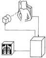
图1为根据本发明实施方案示出的放射剂量分析系统的工作场景图。FIG. 1 is a working scene diagram of a radiation dose analysis system according to an embodiment of the present invention.
具体实施方式Detailed ways
下面将参照附图对本发明的放射剂量分析系统的实施方案进行详细说明。Embodiments of the radiation dose analysis system of the present invention will be described in detail below with reference to the accompanying drawings.
X光机应用于医学诊断,主要依据X射线的穿透作用、差别吸收、感光作用和荧光作用。由于X射线穿过人体时,受到不同程度的吸收,如骨骼吸收的X射线量比肌肉吸收的量要多,那么通过人体后的X射线量就不一样,这样便携带了人体各部密度分布的信息,在荧光屏上或摄影胶片上引起的荧光作用或感光作用的强弱就有较大差别,因而在荧光屏上或摄影胶片上(经过显影、定影)将显示出不同密度的阴影。X-ray machines are used in medical diagnosis, mainly based on X-ray penetration, differential absorption, photosensitivity and fluorescence. Since X-rays are absorbed in different degrees when passing through the human body, for example, the amount of X-rays absorbed by bones is greater than that absorbed by muscles, then the amount of X-rays after passing through the human body is different, which carries the density distribution of various parts of the human body. There is a big difference in the intensity of the fluorescence effect or photosensitive effect caused by the information on the fluorescent screen or on the photographic film, so shadows of different densities will be displayed on the fluorescent screen or on the photographic film (after developing and fixing).
根据阴影浓淡的对比,结合临床表现、化验结果和病理诊断,即可判断人体某一部分是否正常。于是,X射线诊断技术便成了世界上最早应用的非刨伤性的内脏检查技术。According to the contrast of shades, combined with clinical manifestations, laboratory results and pathological diagnosis, it is possible to judge whether a certain part of the human body is normal. As a result, X-ray diagnostic technology has become the world's first non-traumatic visceral examination technology.
为了克服上述不足,本发明搭建了一种放射剂量分析系统,能够有效解决相应的技术问题。In order to overcome the above deficiencies, the present invention builds a radiation dose analysis system, which can effectively solve the corresponding technical problems.
图1为根据本发明实施方案示出的放射剂量分析系统的工作场景图,所述系统包括:1 is a working scene diagram of a radiation dose analysis system according to an embodiment of the present invention, the system includes:
X光机主体,包括射线放射设备、闪烁屏、影像增强板、反光镜、镜头组和CMOS感应设备;The main body of the X-ray machine, including radiation radiation equipment, scintillation screen, image intensifier board, mirror, lens group and CMOS sensing equipment;
其中,所述射线放射设备将放射出的X射线发射到所述闪烁屏上,所述影像增强板设置在所述闪烁屏的后方。Wherein, the radiation radiation equipment emits X-rays to the scintillation screen, and the image intensifier plate is arranged behind the scintillation screen.
接着,继续对本发明的放射剂量分析系统的具体结构进行进一步的说明。Next, the specific structure of the radiation dose analysis system of the present invention will be further described.
在所述放射剂量分析系统中:所述反光镜设置在所述影像增强板的后方,用于将来自所述影像增强板的X射线投射到所述镜头组。In the radiation dose analysis system: the reflector is arranged behind the image intensifier plate for projecting the X-rays from the image intensifier plate to the lens group.
在所述放射剂量分析系统中:所述镜头组设置在所述反光镜的下方,所述CMOS感应设备设置在所述反光镜的下方,用于感应出射线投影图像,所述CMOS感应设备包括被动式CMOS传感器。In the radiation dose analysis system: the lens group is arranged under the reflector, the CMOS sensing device is arranged under the reflector, and is used for sensing a ray projection image, and the CMOS sensing device includes Passive CMOS sensor.
在所述放射剂量分析系统中,还包括:In the radiation dose analysis system, it also includes:
超标检测设备,与数据解析设备连接,用于在R颜色参考值大于等于第一R颜色分量阈值时,发出剂量超标信号,用于在所述R颜色参考值小于第二R颜色分量阈值时,发出剂量不足信号;The exceeding standard detection device is connected with the data parsing device, and is used for sending out a dose exceeding signal when the R color reference value is greater than or equal to the first R color component threshold value, for when the R color reference value is less than the second R color component threshold value, Signal an insufficient dose;
在所述超标检测设备中,所述第一R颜色分量阈值是所述第二R颜色分量阈值的两倍;In the out-of-standard detection device, the first R color component threshold is twice the second R color component threshold;
图像增强设备,与所述CMOS感应设备连接,用于接收所述射线投影图像,对所述射线投影图像执行基于信噪比的多次图像增强,以获得相应的多次增强图像,所述射线投影图像的信噪比越低,执行的图像增强的次数越多;an image enhancement device, connected with the CMOS sensing device, for receiving the ray projection image, and performing multiple image enhancements based on the signal-to-noise ratio on the ray projection image to obtain a corresponding multiple enhancement image, the ray projection image The lower the signal-to-noise ratio of the projected image, the more image enhancements are performed;
形状校准设备,与所述图像增强设备连接,用于接收所述多次增强图像,对所述多次增强图像执行形状校准处理,以获得并输出相应的形状校准图像;a shape calibration device, connected to the image enhancement device, for receiving the multiple enhanced images, and performing shape calibration processing on the multiple enhanced images to obtain and output corresponding shape calibration images;
数据选择设备,与所述形状校准设备连接,用于接收所述形状校准图像,对所述形状校准图像执行基于九宫图的图像块获取以获得九个相同大小的图像块,将所述形状校准图像内所述九个相同大小的图像块的九个形状符合度进行算术平均值计算以获得图像形状符合度,还用于对标准测试图执行基于九宫图的图像块获取以获得九个相同大小的图像块,将所述标准测试图内所述九个相同大小的图像块的九个形状符合度进行算术平均值计算以获得测试形状符合度;a data selection device, connected to the shape calibration device, for receiving the shape calibration image, performing image block acquisition based on the nine-pattern map on the shape calibration image to obtain nine image blocks of the same size, and calibrating the shape The nine shape conformity degrees of the nine image blocks of the same size in the image are calculated by arithmetic mean to obtain the image shape conformity, and also used to perform nine-pattern-based image block acquisition on the standard test chart to obtain nine identical sizes. The image block, the nine shape conformity degrees of the nine image blocks of the same size in the standard test chart are calculated by arithmetic mean to obtain the test shape conformity;
在所述数据选择设备中,当所述图像形状符合度超过所述测试形状符合度时,发出参数可靠命令,当所述图像形状符合度未超过所述测试形状符合度时,发出参数不可靠命令;In the data selection device, when the degree of conformity of the image shape exceeds the degree of conformity of the test shape, a parameter reliable command is issued, and when the degree of conformity of the image shape does not exceed the degree of conformity of the test shape, an unreliable parameter is issued Order;
补加校准设备,与所述数据选择设备连接,用于在接收到所述参数不可靠命令,对所述形状校准图像执行补加形状校准动作,以获得补加校准图像,还用于在接收到所述参数可靠命令,将所述形状校准图像作为补加校准图像输出;A supplementary calibration device, connected to the data selection device, for performing supplementary shape calibration actions on the shape calibration image after receiving the parameter unreliable command, so as to obtain a supplementary calibration image, and for receiving a supplementary calibration image To the parameter reliable command, output the shape calibration image as a supplementary calibration image;
数据解析设备,与所述补加校准设备连接,用于接收所述补加校准图像,并对所述补加校准图像的各个组成像素点的各个R颜色分量值进行均值计算以获得R颜色参考值。A data parsing device, connected to the supplementary calibration device, for receiving the supplementary calibration image, and performing mean calculation on each R color component value of each constituent pixel of the supplementary calibration image to obtain an R color reference value.
在所述放射剂量分析系统中,还包括:In the radiation dose analysis system, it also includes:
WIFI收发设备,与所述数据选择设备连接,用于通过WIFI通信链路无线获知所述标准测试图,并将所述标准测试图发送给所述数据选择设备。A WIFI transceiver device, connected to the data selection device, is used for wirelessly acquiring the standard test chart through a WIFI communication link, and sending the standard test chart to the data selection device.
在所述放射剂量分析系统中,还包括:In the radiation dose analysis system, it also includes:
HSB解析设备,与所述CMOS感应设备连接,用于解析出所述射线投影图像中每一个像素点的色相成分值即H成分值、亮度成分值即S成分值和饱和度成分值即B成分值;The HSB analysis device is connected to the CMOS sensing device, and is used to analyze the hue component value of each pixel in the ray projection image, that is, the H component value, the luminance component value, that is, the S component value, and the saturation component value, that is, the B component. value;
成分分析设备,与所述HSB解析设备连接,用于确定单位面积图像块内各个像素点去重后的H成分值的总数,确定单位面积图像块内各个像素点去重后的S成分值的总数以及确定单位面积图像块内各个像素点去重后的B成分值的总数。A component analysis device, connected with the HSB analysis device, is used to determine the total number of H component values after deduplication of each pixel in the image block per unit area, and determine the total number of S component values after deduplication of each pixel in the image block per unit area. The total number and the total number of B component values after deduplication of each pixel in the image block of unit area are determined.
在所述放射剂量分析系统中,还包括:In the radiation dose analysis system, it also includes:
总数累计设备,与所述成分分析设备连接,用于将单位面积图像块的H成分值的总数、S成分值的总数和B成分值的总数相加以获得单位面积图像块的累计值;A total number accumulation device, connected with the component analysis device, for adding the total number of H component values, the total number of S component values and the total number of B component values of the image block per unit area to obtain the accumulated value of the image block per unit area;
累计值处理设备,与所述总数累计设备连接,用于将累计值最多的单位面积图像块作为目标图像块输出。The accumulated value processing device is connected to the total number accumulation device, and is used for outputting the unit area image block with the most accumulated value as the target image block.
在所述放射剂量分析系统中,还包括:In the radiation dose analysis system, it also includes:
第一分析设备,与所述累计值处理设备连接,用于对所述目标图像块执行信号的小波分析,以实现相应的小波滤波处理,并获得对应的第一分析图像;a first analysis device, connected to the accumulated value processing device, for performing wavelet analysis of the signal on the target image block, so as to realize the corresponding wavelet filtering process and obtain the corresponding first analysis image;
第二分析设备,与所述第一分析设备连接,用于在接收到所述第一分析图像时,对所述第一分析图像执行与所述第一分析图像当前直方图均衡等级对应的直方图均衡处理,以获得第二分析图像,并将所述第二分析图像替换所述射线投影图像发送给所述图像增强设备。A second analysis device, connected to the first analysis device, and configured to perform a histogram corresponding to the current histogram equalization level of the first analysis image on the first analysis image when the first analysis image is received Image equalization is performed to obtain a second analysis image, and the second analysis image is sent to the image enhancement device in place of the radiographic projection image.
在所述放射剂量分析系统中:所述成分分析设备包括H值分析子设备、S值分析子设备和B值分析子设备;In the radiation dose analysis system: the component analysis device includes an H-value analysis sub-device, an S-value analysis sub-device and a B-value analysis sub-device;
其中,所述第一分析图像当前直方图均衡等级越大,执行的对应的直方图均衡处理的强度越小。Wherein, the higher the current histogram equalization level of the first analysis image, the smaller the intensity of the corresponding histogram equalization processing performed.
在所述放射剂量分析系统中,还包括:In the radiation dose analysis system, it also includes:
PSTN通信接口,与所述第二分析设备连接,用于接收所述第二分析图像,并发送所述第二分析图像;A PSTN communication interface, connected to the second analysis device, for receiving the second analysis image and sending the second analysis image;
其中,所述HSB解析设备通过对所述射线投影图像执行RGB颜色空间到HSB颜色空间的转换以获得所述射线投影图像中每一个像素点的色相成分值即H成分值、亮度成分值即S成分值和饱和度成分值即B成分值。Wherein, the HSB parsing device performs the conversion from the RGB color space to the HSB color space on the ray projection image to obtain the hue component value of each pixel in the ray projection image, namely the H component value, and the luminance component value, namely S The component value and the saturation component value are the B component value.
另外,所述CMOS感应设备为被动式像素传感器。被动式像素传感器(PassivePixel Sensor,简称PPS),又叫无源式像素传感器,他由一个反向偏置的光敏二极管和一个开关管构成。光敏二极管本质上是一个由P型半导体和N型半导体组成的PN结,他可等效为一个反向偏置的二极管和一个MOS电容并联。当开关管开启时,光敏二极管与垂直的列线(Column bus)连通。位于列线末端的电荷积分放大器读出电路(Charge integratingamplifier)保持列线电压为一常数,当光敏二极管存贮的信号电荷被读出时,其电压被复位到列线电压水平,与此同时,与光信号成正比的电荷由电荷积分放大器转换为电荷输出。In addition, the CMOS sensing device is a passive pixel sensor. Passive Pixel Sensor (PPS for short), also known as passive pixel sensor, consists of a reverse-biased photodiode and a switch tube. The photodiode is essentially a PN junction composed of a P-type semiconductor and an N-type semiconductor, which can be equivalent to a reverse-biased diode and a MOS capacitor in parallel. When the switch is turned on, the photodiode is connected to the vertical column bus. The charge integrating amplifier readout circuit (Charge integrating amplifier) at the end of the column line keeps the column line voltage constant. When the signal charge stored by the photodiode is read out, its voltage is reset to the column line voltage level. At the same time, The charge proportional to the optical signal is converted into a charge output by the charge integrating amplifier.
采用本发明的放射剂量分析系统,针对现有技术中X射线剂量释放缺乏有效反馈机制的技术问题,通过对定制图像选择的数据进行R颜色分量值的均值分析,以基于分析结果确定当前射线放射设备放射出的X射线是否剂量不足或过量;同时还采用无线通信方式获知最新的标准测试图,基于标准测试图执行对形状校准处理后的图像的形状符合度的分析,以确定是否需要补加形状校准动作。By adopting the radiation dose analysis system of the present invention, aiming at the technical problem of lack of effective feedback mechanism for X-ray dose release in the prior art, the average value analysis of the R color component value is performed on the data selected by the customized image, so as to determine the current radiation radiation based on the analysis result. Whether the dose of X-rays emitted by the equipment is insufficient or excessive; at the same time, the latest standard test chart is obtained by wireless communication, and the shape conformity analysis of the image after shape calibration processing is performed based on the standard test chart to determine whether additional supplementation is required. Shape calibration action.
可以理解的是,虽然本发明已以较佳实施例披露如上,然而上述实施例并非用以限定本发明。对于任何熟悉本领域的技术人员而言,在不脱离本发明技术方案范围情况下,都可利用上述揭示的技术内容对本发明技术方案做出许多可能的变动和修饰,或修改为等同变化的等效实施例。因此,凡是未脱离本发明技术方案的内容,依据本发明的技术实质对以上实施例所做的任何简单修改、等同变化及修饰,均仍属于本发明技术方案保护的范围内。It should be understood that, although the present invention has been disclosed above with preferred embodiments, the above embodiments are not intended to limit the present invention. For any person skilled in the art, without departing from the scope of the technical solution of the present invention, many possible changes and modifications can be made to the technical solution of the present invention by using the technical content disclosed above, or modified to equivalent changes, etc. effective example. Therefore, any simple modifications, equivalent changes and modifications made to the above embodiments according to the technical essence of the present invention without departing from the content of the technical solutions of the present invention still fall within the protection scope of the technical solutions of the present invention.
Claims (7)
Priority Applications (1)
| Application Number | Priority Date | Filing Date | Title |
|---|---|---|---|
| CN201811485231.0ACN109378048B (en) | 2018-12-06 | 2018-12-06 | Radiation Dose Analysis System |
Applications Claiming Priority (1)
| Application Number | Priority Date | Filing Date | Title |
|---|---|---|---|
| CN201811485231.0ACN109378048B (en) | 2018-12-06 | 2018-12-06 | Radiation Dose Analysis System |
Publications (2)
| Publication Number | Publication Date |
|---|---|
| CN109378048A CN109378048A (en) | 2019-02-22 |
| CN109378048Btrue CN109378048B (en) | 2022-09-23 |
Family
ID=65376248
Family Applications (1)
| Application Number | Title | Priority Date | Filing Date |
|---|---|---|---|
| CN201811485231.0AExpired - Fee RelatedCN109378048B (en) | 2018-12-06 | 2018-12-06 | Radiation Dose Analysis System |
Country Status (1)
| Country | Link |
|---|---|
| CN (1) | CN109378048B (en) |
Families Citing this family (1)
| Publication number | Priority date | Publication date | Assignee | Title |
|---|---|---|---|---|
| CN111240232B (en)* | 2019-03-13 | 2020-11-13 | 盐城智享科技咨询服务有限公司 | Instant micro-control terminal for electronic equipment |
Citations (11)
| Publication number | Priority date | Publication date | Assignee | Title |
|---|---|---|---|---|
| US4591984A (en)* | 1981-08-10 | 1986-05-27 | Tokyo Shibaura Denki Kabushiki Kaisha | Radiation measuring device |
| DK154687D0 (en)* | 1986-03-27 | 1987-03-26 | Thomson Brandt Gmbh | DIGITAL TRANSMISSION SYSTEM |
| CN1469721A (en)* | 2000-10-11 | 2004-01-21 | �����ɷ� | Methods and devices for analysis of X-ray images |
| CN101011617A (en)* | 2006-12-29 | 2007-08-08 | 成都川大奇林科技有限责任公司 | Method for determining radiating field output dose accurately in conformalradiotherapy |
| CN101120871A (en)* | 2006-12-29 | 2008-02-13 | 成都川大奇林科技有限责任公司 | Precise radiotherapy planning system |
| CN102949194A (en)* | 2011-08-16 | 2013-03-06 | 富士胶片株式会社 | Radiation dose information sharing device and method |
| CN203337825U (en)* | 2013-06-20 | 2013-12-11 | 张军 | Radiological dose measuring device |
| CN204044381U (en)* | 2014-06-11 | 2014-12-24 | 苏州奥特福环境科技有限公司 | The control device of X-ray beam in a kind of green channel detection system |
| CN105389476A (en)* | 2015-12-24 | 2016-03-09 | 四川大学 | Interpolation algorithm for intensity-modulated radiation therapy plan dose data based on gradient features |
| AU2016203814A1 (en)* | 2010-12-08 | 2016-06-30 | Bayer Healthcare Llc | Generating a suitable model for estimating patient radiation dose resulting from medical imaging scans |
| JP2017006244A (en)* | 2015-06-18 | 2017-01-12 | 株式会社日立製作所 | X-ray diagnostic apparatus and operation method thereof |
Family Cites Families (2)
| Publication number | Priority date | Publication date | Assignee | Title |
|---|---|---|---|---|
| JP2011050528A (en)* | 2009-09-01 | 2011-03-17 | Fujifilm Corp | Radiography management system and method |
| WO2013096903A2 (en)* | 2011-12-22 | 2013-06-27 | The Regents Of The University Of Colorado, A Body Corproate | Methods for prediction of clinical response to radiation therapy in cancer patients |
- 2018
- 2018-12-06CNCN201811485231.0Apatent/CN109378048B/ennot_activeExpired - Fee Related
Patent Citations (11)
| Publication number | Priority date | Publication date | Assignee | Title |
|---|---|---|---|---|
| US4591984A (en)* | 1981-08-10 | 1986-05-27 | Tokyo Shibaura Denki Kabushiki Kaisha | Radiation measuring device |
| DK154687D0 (en)* | 1986-03-27 | 1987-03-26 | Thomson Brandt Gmbh | DIGITAL TRANSMISSION SYSTEM |
| CN1469721A (en)* | 2000-10-11 | 2004-01-21 | �����ɷ� | Methods and devices for analysis of X-ray images |
| CN101011617A (en)* | 2006-12-29 | 2007-08-08 | 成都川大奇林科技有限责任公司 | Method for determining radiating field output dose accurately in conformalradiotherapy |
| CN101120871A (en)* | 2006-12-29 | 2008-02-13 | 成都川大奇林科技有限责任公司 | Precise radiotherapy planning system |
| AU2016203814A1 (en)* | 2010-12-08 | 2016-06-30 | Bayer Healthcare Llc | Generating a suitable model for estimating patient radiation dose resulting from medical imaging scans |
| CN102949194A (en)* | 2011-08-16 | 2013-03-06 | 富士胶片株式会社 | Radiation dose information sharing device and method |
| CN203337825U (en)* | 2013-06-20 | 2013-12-11 | 张军 | Radiological dose measuring device |
| CN204044381U (en)* | 2014-06-11 | 2014-12-24 | 苏州奥特福环境科技有限公司 | The control device of X-ray beam in a kind of green channel detection system |
| JP2017006244A (en)* | 2015-06-18 | 2017-01-12 | 株式会社日立製作所 | X-ray diagnostic apparatus and operation method thereof |
| CN105389476A (en)* | 2015-12-24 | 2016-03-09 | 四川大学 | Interpolation algorithm for intensity-modulated radiation therapy plan dose data based on gradient features |
Non-Patent Citations (1)
| Title |
|---|
| 医用X射线辐射监测及联锁装置的设计;李宪军等;《中国医疗设备》;20101231;第25卷(第10期);全文* |
Also Published As
| Publication number | Publication date |
|---|---|
| CN109378048A (en) | 2019-02-22 |
Similar Documents
| Publication | Publication Date | Title |
|---|---|---|
| US9226716B2 (en) | Nuclear medicine imaging apparatus and radiation therapy apparatus | |
| EP3424430B1 (en) | X-ray exposure control device and x-ray image detection apparatus | |
| CN102266233B (en) | Radiographic apparatus and control method | |
| US10557806B2 (en) | CT system and CT method | |
| JP2014158579A (en) | Radiation image processing method and method, and radiographic device | |
| WO2014129443A1 (en) | Radiation image analysis device and method, and radiation imaging device | |
| JP2004521721A (en) | Method and apparatus for identifying and correcting line artifacts in solid state x-ray detectors | |
| JP6058272B2 (en) | Nuclear medicine diagnostic apparatus and control method | |
| US8520797B2 (en) | Medical imaging apparatus, control method, and computer program product | |
| US12200368B2 (en) | Radiation detection apparatus and output method | |
| CN107550505A (en) | X-ray detector, x-ray camera system and x-ray image capture method | |
| KR20140142803A (en) | Radiography imaging apparatus and method for generating an radiographic image | |
| CN109378048B (en) | Radiation Dose Analysis System | |
| US20250235172A1 (en) | Low-dose x-ray imaging system | |
| CN105829915B (en) | Improved temperature stability for digital positron emission tomography (PET) detectors | |
| US20110297835A1 (en) | Nuclear medicine imaging apparatus, control method, and computer program product | |
| JP6431307B2 (en) | Radiation imaging apparatus and radiation imaging system | |
| CN111839561B (en) | Radiation detection system and control method thereof | |
| JP2015017906A (en) | Nuclear medicine diagnosis apparatus and position estimation method | |
| US11125892B2 (en) | Radiation detection system, radiation output device, and radiation detection device | |
| US20230404515A1 (en) | Information processing device, information processing method, program, and radiographic imaging system | |
| ES3014021T3 (en) | X-ray imaging method and system thereof | |
| CN112596099A (en) | Drift template updating method and device of flat panel detector and storage medium | |
| RU148455U1 (en) | DIGITAL SCANNING MAMMOGRAPH | |
| JP2024168248A (en) | Radiation monitoring device and radiation monitoring method |
Legal Events
| Date | Code | Title | Description |
|---|---|---|---|
| PB01 | Publication | ||
| PB01 | Publication | ||
| SE01 | Entry into force of request for substantive examination | ||
| SE01 | Entry into force of request for substantive examination | ||
| CB03 | Change of inventor or designer information | Inventor after:Meng Linghong Inventor after:Huang Junyun Inventor after:Zhang Gonglin Inventor after:Zhu Yalan Inventor after:Li Xiaofen Inventor before:Zhu Yalan Inventor before:Li Xiaofen | |
| CB03 | Change of inventor or designer information | ||
| TA01 | Transfer of patent application right | Effective date of registration:20220902 Address after:People's Hospital of Zoucheng City, 5677 Chongyi Road, Zoucheng City, Jining City, Shandong Province Applicant after:Meng Linghong Applicant after:Huang Junyun Applicant after:Zhang Gonglin Address before:315400 Yangming West Road, Yuyao, Ningbo, Zhejiang 188 Applicant before:YUYAO DECHENG TECHNOLOGY CONSULTING Co.,Ltd. | |
| TA01 | Transfer of patent application right | ||
| GR01 | Patent grant | ||
| GR01 | Patent grant | ||
| CF01 | Termination of patent right due to non-payment of annual fee | Granted publication date:20220923 | |
| CF01 | Termination of patent right due to non-payment of annual fee |