CN109003279B - Fundus retina blood vessel segmentation method and system based on K-Means clustering labeling and naive Bayes model - Google Patents
Fundus retina blood vessel segmentation method and system based on K-Means clustering labeling and naive Bayes modelDownload PDFInfo
- Publication number
- CN109003279B CN109003279BCN201810733535.8ACN201810733535ACN109003279BCN 109003279 BCN109003279 BCN 109003279BCN 201810733535 ACN201810733535 ACN 201810733535ACN 109003279 BCN109003279 BCN 109003279B
- Authority
- CN
- China
- Prior art keywords
- image
- filter
- feature
- response
- pixel
- Prior art date
- Legal status (The legal status is an assumption and is not a legal conclusion. Google has not performed a legal analysis and makes no representation as to the accuracy of the status listed.)
- Expired - Fee Related
Links
Images


Classifications
- G—PHYSICS
- G06—COMPUTING OR CALCULATING; COUNTING
- G06T—IMAGE DATA PROCESSING OR GENERATION, IN GENERAL
- G06T7/00—Image analysis
- G06T7/10—Segmentation; Edge detection
- G06T7/11—Region-based segmentation
- G—PHYSICS
- G06—COMPUTING OR CALCULATING; COUNTING
- G06F—ELECTRIC DIGITAL DATA PROCESSING
- G06F18/00—Pattern recognition
- G06F18/20—Analysing
- G06F18/21—Design or setup of recognition systems or techniques; Extraction of features in feature space; Blind source separation
- G06F18/214—Generating training patterns; Bootstrap methods, e.g. bagging or boosting
- G—PHYSICS
- G06—COMPUTING OR CALCULATING; COUNTING
- G06F—ELECTRIC DIGITAL DATA PROCESSING
- G06F18/00—Pattern recognition
- G06F18/20—Analysing
- G06F18/23—Clustering techniques
- G06F18/232—Non-hierarchical techniques
- G06F18/2321—Non-hierarchical techniques using statistics or function optimisation, e.g. modelling of probability density functions
- G06F18/23213—Non-hierarchical techniques using statistics or function optimisation, e.g. modelling of probability density functions with fixed number of clusters, e.g. K-means clustering
- G—PHYSICS
- G06—COMPUTING OR CALCULATING; COUNTING
- G06F—ELECTRIC DIGITAL DATA PROCESSING
- G06F18/00—Pattern recognition
- G06F18/20—Analysing
- G06F18/24—Classification techniques
- G06F18/241—Classification techniques relating to the classification model, e.g. parametric or non-parametric approaches
- G06F18/2415—Classification techniques relating to the classification model, e.g. parametric or non-parametric approaches based on parametric or probabilistic models, e.g. based on likelihood ratio or false acceptance rate versus a false rejection rate
- G06F18/24155—Bayesian classification
- G—PHYSICS
- G06—COMPUTING OR CALCULATING; COUNTING
- G06T—IMAGE DATA PROCESSING OR GENERATION, IN GENERAL
- G06T5/00—Image enhancement or restoration
- G06T5/20—Image enhancement or restoration using local operators
- G—PHYSICS
- G06—COMPUTING OR CALCULATING; COUNTING
- G06T—IMAGE DATA PROCESSING OR GENERATION, IN GENERAL
- G06T2207/00—Indexing scheme for image analysis or image enhancement
- G06T2207/20—Special algorithmic details
- G06T2207/20024—Filtering details
- G—PHYSICS
- G06—COMPUTING OR CALCULATING; COUNTING
- G06T—IMAGE DATA PROCESSING OR GENERATION, IN GENERAL
- G06T2207/00—Indexing scheme for image analysis or image enhancement
- G06T2207/20—Special algorithmic details
- G06T2207/20081—Training; Learning
- G—PHYSICS
- G06—COMPUTING OR CALCULATING; COUNTING
- G06T—IMAGE DATA PROCESSING OR GENERATION, IN GENERAL
- G06T2207/00—Indexing scheme for image analysis or image enhancement
- G06T2207/30—Subject of image; Context of image processing
- G06T2207/30004—Biomedical image processing
- G06T2207/30041—Eye; Retina; Ophthalmic
- G—PHYSICS
- G06—COMPUTING OR CALCULATING; COUNTING
- G06T—IMAGE DATA PROCESSING OR GENERATION, IN GENERAL
- G06T2207/00—Indexing scheme for image analysis or image enhancement
- G06T2207/30—Subject of image; Context of image processing
- G06T2207/30004—Biomedical image processing
- G06T2207/30101—Blood vessel; Artery; Vein; Vascular
Landscapes
- Engineering & Computer Science (AREA)
- Theoretical Computer Science (AREA)
- Physics & Mathematics (AREA)
- Data Mining & Analysis (AREA)
- General Physics & Mathematics (AREA)
- Computer Vision & Pattern Recognition (AREA)
- Bioinformatics & Cheminformatics (AREA)
- Bioinformatics & Computational Biology (AREA)
- Artificial Intelligence (AREA)
- Evolutionary Biology (AREA)
- Evolutionary Computation (AREA)
- General Engineering & Computer Science (AREA)
- Life Sciences & Earth Sciences (AREA)
- Probability & Statistics with Applications (AREA)
- Eye Examination Apparatus (AREA)
- Image Processing (AREA)
- Image Analysis (AREA)
Abstract
Description
Translated fromChinese技术领域technical field
本发明涉及医学图像处理领域,具体而言,尤其涉及一种基于K-Means 聚类标注和朴素贝叶斯模型的眼底视网膜血管分割方法。The invention relates to the field of medical image processing, in particular, to a fundus retinal blood vessel segmentation method based on K-Means cluster labeling and naive Bayesian model.
背景技术Background technique
视网膜血管是全身微循环系统的重要组成部分,其形态结构的变化与糖尿病、高血压、冠状动脉硬化和脑血管硬化等心血管类疾病的病情严重程度密切相关。因此,视网膜图像血管分割技术的研究对于医学临床应用具有十分重要的意义。Retinal blood vessels are an important part of the systemic microcirculation system, and the changes of its morphological structure are closely related to the severity of cardiovascular diseases such as diabetes, hypertension, coronary arteriosclerosis and cerebrovascular sclerosis. Therefore, the research on retinal image blood vessel segmentation technology is of great significance for medical clinical applications.
目前的眼底视网膜血管方法主要有两大方向:基于规则的血管分割方法和基于学习的血管分割方法。基于规则的血管分割方法主要利用的是图像处理技术,根据血管特性设计一些规则来寻找血管:根据血管的分段线性和连通性设计了不同尺度下线性滤波器,通过结合不同尺度下的滤波结果和阈值化处理得到最后的血管分割结果;由于血管的横截面成倒立的高斯分布,所以二维高斯匹配滤波方法利用表征不同方向的12个高斯模板作为匹配滤波器来对图像进行滤波,然后对响应结果进行阈值化,选取响应最大的匹配滤波结果作为血管输出,最终提取出视网膜血管图像。基于 Hessian矩阵的滤波方法利用Hessian矩阵的特征值增强包括血管分叉在内的所有血管结构,并抑制非血管结构。这些图像处理方法都需要阈值化处理,而且针对不同的视网膜眼底图像需要选择不同的阈值,这并不是一个自动化的过程。The current fundus retinal blood vessel methods mainly have two main directions: rule-based blood vessel segmentation methods and learning-based blood vessel segmentation methods. The rule-based blood vessel segmentation method mainly uses image processing technology, and designs some rules according to the characteristics of blood vessels to find blood vessels: according to the piecewise linearity and connectivity of blood vessels, linear filters at different scales are designed, and by combining the filtering results at different scales and thresholding to obtain the final blood vessel segmentation result; since the cross-section of the blood vessel is an inverted Gaussian distribution, the two-dimensional Gaussian matched filtering method uses 12 Gaussian templates representing different directions as matched filters to filter the image, and then filter the image. The response result is thresholded, and the matched filtering result with the largest response is selected as the blood vessel output, and finally the retinal blood vessel image is extracted. The Hessian matrix-based filtering method utilizes the eigenvalues of the Hessian matrix to enhance all vascular structures including vascular bifurcations and suppress non-vascular structures. These image processing methods all require thresholding, and different thresholds need to be selected for different retinal fundus images, which is not an automated process.
基于学习的分割方法将血管分割问题视为像素级的分类问题,通过一组带有标签的训练数据集训练得到分类器。其中,特征主要包括像素灰度值、边界特征和Gabor小波变换等,分类器主要包括多层神经网络、贝叶斯分类器、随机森林和支持向量机。由于引入了判别性学习的强大机制,这些方法的分割性能往往优于基于规则的分割方法。基于学习的方法能够自动分割并且准确率较高,主要流程是图像数据预处理、图像特征提取和模型训练。难点在于视网膜眼底图像特征提取和像素级标签数据获取。有监督的机器学习技术需要大量的专家人工标注数据,而眼底图像中的视网膜血管呈发散的树型、以及末梢细小血管的存在使得标记任务更加困难。Learning-based segmentation methods treat the vessel segmentation problem as a pixel-level classification problem, and train a classifier through a set of labeled training datasets. Among them, the features mainly include pixel gray value, boundary features and Gabor wavelet transform, etc. The classifier mainly includes multi-layer neural network, Bayesian classifier, random forest and support vector machine. Due to the introduction of a powerful mechanism of discriminative learning, the segmentation performance of these methods tends to outperform rule-based segmentation methods. The learning-based method can automatically segment and has high accuracy. The main process is image data preprocessing, image feature extraction and model training. The difficulty lies in the feature extraction of retinal fundus images and the acquisition of pixel-level label data. Supervised machine learning technology requires a large amount of expert manual labeling data, and the retinal blood vessels in the fundus image show a divergent tree shape and the existence of small peripheral blood vessels make the labeling task more difficult.
在计算机辅助医学图像分析中,可以从医院获得大量的医学图像作为训练集,但如果要求医学专家把这些图像中的血管都标识出来,则往往是不现实的。如果只使用少量的有标记示例,那么利用它们所训练出的学习系统往往很难具有强泛化能力;另一方面,如果仅使用少量“昂贵的”有标记示例而不利用大量“廉价的”未标记示例,则是对数据资源的极大的浪费。In computer-aided medical image analysis, a large number of medical images can be obtained from hospitals as training sets, but it is often unrealistic to ask medical experts to identify the blood vessels in these images. If only a small number of labeled examples are used, it is often difficult for the learning system trained with them to have strong generalization ability; on the other hand, if only a small number of "expensive" labeled examples are used without using a large number of "cheap" Unlabeled examples are a huge waste of data resources.
发明内容SUMMARY OF THE INVENTION
根据上述提出的有监督的机器学习技术需要大量的专家人工标注数据,费时费力的技术问题,而提供一种基于K-Means聚类标注和朴素贝叶斯模型的眼底视网膜血管分割方法。本发明主要利用K-Means聚类标注的结果作为数据的标签,然后利用其再去训练分类模型进行分割,从而提高了工作效率,分割效果更好,节省了人力物力。本发明采用的技术手段如下:According to the above-mentioned supervised machine learning technology, a large amount of expert manual annotation data is required, which is time-consuming and labor-intensive technical problems, and a fundus retinal blood vessel segmentation method based on K-Means clustering annotation and naive Bayesian model is provided. The invention mainly uses the result of K-Means clustering and labeling as the label of the data, and then uses it to train the classification model for segmentation, thereby improving work efficiency, better segmentation effect, and saving manpower and material resources. The technical means adopted in the present invention are as follows:
一种基于K-Means聚类标注和朴素贝叶斯模型的眼底视网膜血管分割方法,包括如下步骤:A fundus retinal blood vessel segmentation method based on K-Means clustering annotation and naive Bayesian model, comprising the following steps:
构建训练集图像的像素级标签,具体为:对特征提取后的训练集中的每幅图像使用K-Means聚类算法将特征向量进行聚类标注;Constructing pixel-level labels of images in the training set, specifically: using the K-Means clustering algorithm to cluster and label the feature vectors for each image in the training set after feature extraction;
基于K-Means聚类标注的训练集数据训练朴素贝叶斯模型;Train a naive Bayesian model based on the training set data labeled by K-Means clustering;
用训练好的朴素贝叶斯模型分割测试集中每幅图像的血管。The blood vessels of each image in the test set are segmented using the trained Naive Bayes model.
进一步地,所述步骤构建训练集图像的像素级标签之前还具有如下步骤:Further, the steps also include the following steps before constructing the pixel-level labels of the training set images:
随机抽取数据集中的彩色眼底图像,构建训练集和测试集;Randomly extract color fundus images from the dataset to construct training and test sets;
提取彩色眼底图像的G通道的灰度图作为特征提取的对象,并对视网膜感兴趣区域进行膨胀处理;Extract the grayscale image of the G channel of the color fundus image as the object of feature extraction, and perform dilation processing on the retinal region of interest;
对膨胀处理后的所述灰度图进行特征提取,将训练集和测试集图像中的每个像素用十维的特征向量表示,具体为:Feature extraction is performed on the grayscale image after the expansion processing, and each pixel in the training set and the test set image is represented by a ten-dimensional feature vector, specifically:
通过对比度受限的自适应直方图均衡化方法增强所述灰度图的对比度,将此灰度值作为区分血管和背景的一种特征,通过基于血管特性设计的血管增强技术处理所述灰度图,The contrast of the grayscale image is enhanced by a contrast-limited adaptive histogram equalization method, and the grayscale value is used as a feature to distinguish blood vessels from the background, and the grayscale is processed by a blood vessel enhancement technology designed based on blood vessel characteristics. picture,
分别对所述灰度图进行如下滤波处理:The grayscale images are respectively filtered as follows:
通过Gabor小波变换处理,得到四维的Gabor特征;Through Gabor wavelet transform processing, four-dimensional Gabor features are obtained;
通过线性检测器对视网膜图像进行滤波处理,得到线性检测的响应结果作为一维的线性检测特征;The retinal image is filtered by a linear detector, and the response result of the linear detection is obtained as a one-dimensional linear detection feature;
通过高斯形状的滤波器对视网膜图像进行滤波处理,选取响应最大的滤波结果作为一维的高斯匹配特征;The retinal image is filtered through a Gaussian shape filter, and the filtering result with the largest response is selected as the one-dimensional Gaussian matching feature;
基于Hessian矩阵的Frangi滤波特征对视网膜图像进行滤波处理,选取最大响应作为一维的Frangi滤波特征;The retinal image is filtered based on the Frangi filter feature of the Hessian matrix, and the maximum response is selected as the one-dimensional Frangi filter feature;
基于B-COSFIRE滤波特征对视网膜图像进行滤波处理,将对称 B-COSFIRE滤波器和非对称B-COSFIRE滤波器响应结果求和得到最终的一维的B-COSFIRE滤波特征;The retinal image is filtered based on the B-COSFIRE filter feature, and the response results of the symmetric B-COSFIRE filter and the asymmetric B-COSFIRE filter are summed to obtain the final one-dimensional B-COSFIRE filter feature;
通过韦伯描述算子求解视网膜图像中任一像素点的一维的韦伯描述特征;Solve the one-dimensional Weber description feature of any pixel in the retinal image by the Weber description operator;
所述用训练好的朴素贝叶斯模型分割测试集中每幅图像的血管之后还具有如下步骤:After the trained naive Bayes model is used to segment the blood vessels of each image in the test set, there are also the following steps:
通过预设的评价指标判断血管分割方法是否有效。Whether the blood vessel segmentation method is effective is judged by the preset evaluation index.
进一步地,所述步骤随机抽取数据集中的彩色眼底图像,构建训练集和测试集中,数据集使用公开的DRIVE数据集,所述训练集和测试集的图像数目相同,且每幅图像对应2个专家手动分割的结果和1个显示眼底图像的有效信息区域的Mask图像。Further, the step randomly extracts the color fundus images in the data set, constructs the training set and the test set, the data set uses the public DRIVE data set, the training set and the test set have the same number of images, and each image corresponds to 2 Results of manual segmentation by experts and 1 Mask image showing the effective information area of the fundus image.
进一步地,通过如下方式进行Gabor小波变换处理:Further, the Gabor wavelet transform processing is performed in the following manner:
Gabor核函数如下所示:The Gabor kernel function is as follows:

其中,x'=xcosθ+ysinθ,y'=-xsinθ+ycosθ,Among them, x'=xcosθ+ysinθ, y'=-xsinθ+ycosθ,
(u0,v0)表示正弦平面波的时域频率,σx和σy表示x和y方向上的椭圆高斯的方差,其决定了滤波器作用区域的大小,即尺度,θ为旋转角度,(u0 , v0 ) represents the time domain frequency of the sine plane wave, σx and σy represent the variance of the ellipse Gaussian in the x and y directions, which determines the size of the filter’s active area, that is, the scale, θ is the rotation angle,
通过改变参数σ和θ得到一组方向和尺度不同的Gabor小波,在固定尺度下,比较所有方向上的响应,把最大响应作为该尺度下的特征图像,根据设置的尺度,把所有尺度下的结果作为Gabor特征。By changing the parameters σ and θ, a set of Gabor wavelets with different directions and scales are obtained. At a fixed scale, the responses in all directions are compared, and the maximum response is taken as the feature image at this scale. The results are taken as Gabor features.
进一步地,所述线性检测器通过在W*W的窗口下,旋转长度为l的检测线得到N个不同方向的滤波器,用滤波器对灰度图滤波处理,当检测线的方向和血管方向一致时,响应最大,比较所有角度下的响应保留最大值作为该窗口下的检测结果
进一步地,通过如下方式进行高斯形状的滤波器图像滤波处理:Further, the filter image filtering processing of the Gaussian shape is performed in the following manner:
倒立的高斯内核函数如下所示:The inverted Gaussian kernel function looks like this:

其中
进一步地,通过如下方式进行基于Hessian矩阵的Frangi滤波处理:Further, the Frangi filtering process based on the Hessian matrix is performed in the following manner:
对于二维输入图像,与高斯核函数的二阶偏导卷积得到构造每一个像素 (x,y)的Hessian矩阵,For a two-dimensional input image, the second-order partial derivative convolution with the Gaussian kernel function obtains the Hessian matrix that constructs each pixel (x, y),

通过如下函数计算Frangi滤波的结果:The result of Frangi filtering is calculated by the following function:

其中,Hessian矩阵的特征值为λ1和λ2,且|λ1|<|λ2|,Rb=λ2/λ1,
β用于调整线状和块状的区别,c表述控制整体平滑度的参数,比较每个像素点在各个尺度下的响应,选取最大响应作为Frangi滤波特征。β is used to adjust the difference between linear and block, and c represents the parameter that controls the overall smoothness, compares the response of each pixel at each scale, and selects the maximum response as the Frangi filter feature.
进一步地,B-COSFIRE滤波器由一组共线排列的高斯差分滤波器,即 DoG滤波器构成,B-COSFIRE滤波响应通过计算所有的DoG滤波响应的几何均值得到,所述DoG滤波响应通过DoG滤波器与图像的卷积得到,Further, the B-COSFIRE filter is composed of a group of Gaussian difference filters arranged collinearly, namely DoG filters, and the B-COSFIRE filter response is obtained by calculating the geometric mean of all DoG filter responses. The DoG filter response is obtained by DoG The convolution of the filter and the image is obtained,
对称结构的COSFIRE模型用于提取连续血管结构特征,DoG滤波器成对存在,它们的中心以相反方向位于B-COSFIRE滤波器中心的两侧;非对称结构的COSFIRE模型用于检测末端血管,DoG滤波器的中心位于 B-COSFIRE滤波器中心的一侧,对DoG滤波响应进行模糊操作,所述模糊操作为DoG滤波器加权阈值响应的最大值,权重是DoG滤波响应与高斯函数系数的乘积,将对称B-COSFIRE滤波器和非对称B-COSFIRE滤波响应结果作和得到最终的B-COSFIRE滤波特征。The COSFIRE model with symmetric structure is used to extract continuous vessel structure features, DoG filters exist in pairs, and their centers are located on both sides of the center of the B-COSFIRE filter in opposite directions; the COSFIRE model with asymmetric structure is used to detect terminal blood vessels, DoG The center of the filter is located on one side of the center of the B-COSFIRE filter, and a fuzzy operation is performed on the DoG filter response. The fuzzy operation is the maximum value of the DoG filter weighted threshold response, and the weight is the product of the DoG filter response and the Gaussian function coefficient, The symmetric B-COSFIRE filter and the asymmetric B-COSFIRE filter response results are summed to obtain the final B-COSFIRE filter characteristic.
进一步地,通过如下公式求解视网膜图像中任一像素点的韦伯描述特征:Further, the Weber description feature of any pixel in the retinal image is solved by the following formula:

其中,p为图像I中任一像素点,N(p)表示像素p的邻域像素集合,I(z) 表示目标像素p的邻域中N(p)的任一像素的灰度值。Among them, p is any pixel in the image I, N(p) represents the neighborhood pixel set of the pixel p, and I(z) represents the gray value of any pixel of N(p) in the neighborhood of the target pixel p.
进一步地,所述通过预设的评价指标判断血管分割方法是否有效,具体为:Further, determining whether the blood vessel segmentation method is effective by using a preset evaluation index is specifically:

其中,每幅图像的血管点通过四种不同的颜色区分,正确分割的血管点真阳性TP、分割错误的非血管点假阳性FP、错误分割的血管点真阴性 TN、正确分割的非血管点假阴性FN,Acc表示分割正确的像素点占整个图像像素点总和的比例,即准确度,Se表示分割正确的血管点占金标准血管点总和的比例,即灵敏度,Sp表示分割正确的背景点占金标准背景点总和的比例,即特异性,Ppv表示分割正确的血管点占分割出的血管点的比例,即精度预测比值。Among them, the blood vessel points of each image are distinguished by four different colors, the true positive TP of the correctly segmented blood vessel points, the false positive FP of the incorrectly segmented non-vessel points, the incorrectly segmented true negative TN of the blood vessel points, and the correctly segmented non-vessel points. False negative FN, Acc represents the proportion of correctly segmented pixels to the sum of the entire image pixel points, that is, accuracy, Se represents the proportion of correctly segmented blood vessel points to the sum of the gold standard blood vessel points, that is, sensitivity, Sp represents the correctly segmented background points The ratio of the sum of the gold standard background points, that is, the specificity, and Ppv represents the proportion of the correctly segmented blood vessel points to the segmented blood vessel points, that is, the precision prediction ratio.
本发明还提供了一种基于K-Means聚类标注和朴素贝叶斯模型的眼底视网膜血管分割系统,包括:The present invention also provides a fundus retinal blood vessel segmentation system based on K-Means clustering annotation and naive Bayesian model, including:
样本取样单元,用于从DRIVE数据集中随机抽取彩色眼底图像,构建训练集和测试集;The sample sampling unit is used to randomly extract color fundus images from the DRIVE dataset to construct training and test sets;
预处理单元,用于提取彩色眼底图像中的G通道的灰度图并对其中的视网膜感兴趣区域进行膨胀处理;a preprocessing unit, used for extracting the grayscale image of the G channel in the color fundus image and performing dilation processing on the retinal region of interest therein;
特征提取单元,用于对膨胀处理后的G通道的灰度图进行特征提取,将训练集和测试集图像中的每个像素用十维的特征向量表示,其包括:The feature extraction unit is used to perform feature extraction on the grayscale image of the G channel after the expansion processing, and each pixel in the training set and the test set image is represented by a ten-dimensional feature vector, which includes:
Gabor小波处理模块,线性检测器滤波处理模块,高斯匹配特征提取模块,Frangi滤波特征提取模块,B-COSFIRE滤波特征提取模块,韦伯描述特征提取模块;Gabor wavelet processing module, linear detector filtering processing module, Gaussian matching feature extraction module, Frangi filter feature extraction module, B-COSFIRE filter feature extraction module, Weber description feature extraction module;
聚类标注单元:用于通过K-Means聚类算法将特征向量进行聚类标注;Clustering labeling unit: used to label feature vectors by clustering by K-Means clustering algorithm;
机器学习模型训练单元:用于通过K-Means聚类标注的训练集数据训练朴素贝叶斯模型;Machine learning model training unit: used to train a naive Bayesian model with training set data labeled by K-Means clustering;
图像处理单元:用于通过分割模型对测试集中每幅图像的血管进行分割;Image processing unit: used to segment the blood vessels of each image in the test set through the segmentation model;
分割效果评判单元:用于对血管分割方法的有效性进行评判。Segmentation effect evaluation unit: used to evaluate the effectiveness of the blood vessel segmentation method.
较现有技术相比,本发明具有以下优点:Compared with the prior art, the present invention has the following advantages:
本发明通过聚类技术把聚类的结果作为有监督训练的标记,然后利用这些标记训练朴素贝叶斯分类模型进行视网膜血管分割,整个过程不需要人为参与标记,省时省力,大幅度提高了机器学习模型的处理效率,相对于聚类的分割结果,本发明在视网膜图像分割处理过程中准确率得到提高。The present invention uses the clustering technology to use the clustering results as markers for supervised training, and then uses these markers to train a naive Bayesian classification model to segment retinal blood vessels. The processing efficiency of the machine learning model is compared with the segmentation result of the clustering, and the present invention improves the accuracy of the retinal image segmentation processing process.
基于上述理由本发明可在医学图像处理领域广泛推广。Based on the above reasons, the present invention can be widely promoted in the field of medical image processing.
附图说明Description of drawings
为了更清楚地说明本发明实施例或现有技术中的技术方案,下面将对实施例或现有技术描述中所需要使用的附图做以简单地介绍,显而易见地,下面描述中的附图是本发明的一些实施例,对于本领域普通技术人员来讲,在不付出创造性劳动性的前提下,还可以根据这些附图获得其他的附图。In order to illustrate the embodiments of the present invention or the technical solutions in the prior art more clearly, the following briefly introduces the accompanying drawings that need to be used in the description of the embodiments or the prior art. Obviously, the accompanying drawings in the following description These are some embodiments of the present invention, and for those of ordinary skill in the art, other drawings can also be obtained from these drawings without any creative effort.
图1为本发明方法流程图。Fig. 1 is the flow chart of the method of the present invention.
图2为本发明装置模块图。FIG. 2 is a block diagram of the device of the present invention.
图3为本发明中原始眼底视网膜图像和Mask图像的示意图。FIG. 3 is a schematic diagram of the original fundus retinal image and the Mask image in the present invention.
图4为本发明中RGB三通道灰度图的对比度分析示意图,其中,(a) 为红色通道,(b)为绿色通道,(c)为蓝色通道。4 is a schematic diagram of contrast analysis of a grayscale image of RGB three channels in the present invention, wherein (a) is a red channel, (b) is a green channel, and (c) is a blue channel.
图5为本发明绿色通道灰度图中视网膜感兴趣区域膨胀处理示意图。FIG. 5 is a schematic diagram of the expansion processing of the retinal region of interest in the grayscale image of the green channel of the present invention.
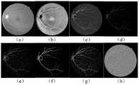
图6为本发明经过各特征提取后依次得到得特征图示意图,其中,(a) 为原始彩色图像,(b)为灰度特征,(c)为Gabor变换,(d)为线性检测 (e)为高斯匹配滤波,(f)为Frangi滤波,(g)为B-COSFIRE滤波,(h) 为韦伯描述。Figure 6 is a schematic diagram of the feature maps obtained in turn after each feature extraction in the present invention, wherein (a) is the original color image, (b) is the grayscale feature, (c) is the Gabor transform, and (d) is the linear detection (e) ) is Gaussian matched filter, (f) is Frangi filter, (g) is B-COSFIRE filter, (h) is Weber description.
图7为本发明使用K-Means聚类算法在训练集图像的聚类结果示意图。FIG. 7 is a schematic diagram of the clustering results of images in the training set using the K-Means clustering algorithm according to the present invention.
图8为本发明训练好的分割模型分割测试集的分割结果示意图。FIG. 8 is a schematic diagram of the segmentation result of the segmentation model of the present invention to segment the test set.
具体实施方式Detailed ways
为了使本技术领域的人员更好地理解本发明方案,下面将结合本发明实施例中的附图,对本发明实施例中的技术方案进行清楚、完整地描述,显然,所描述的实施例仅仅是本发明一部分的实施例,而不是全部的实施例。基于本发明中的实施例,本领域普通技术人员在没有做出创造性劳动前提下所获得的所有其他实施例,都应当属于本发明保护的范围。In order to make those skilled in the art better understand the solutions of the present invention, the technical solutions in the embodiments of the present invention will be clearly and completely described below with reference to the accompanying drawings in the embodiments of the present invention. Obviously, the described embodiments are only Embodiments are part of the present invention, but not all embodiments. Based on the embodiments of the present invention, all other embodiments obtained by persons of ordinary skill in the art without creative efforts shall fall within the protection scope of the present invention.
如图1所示,本发明提供了一种基于K-Means聚类标注和朴素贝叶斯模型的眼底视网膜血管分割方法,包括如下步骤:As shown in FIG. 1 , the present invention provides a fundus retinal blood vessel segmentation method based on K-Means clustering annotation and naive Bayes model, including the following steps:
S1、随机抽取数据集中的彩色眼底图像,本实施例使用公开的DRIVE 数据集,DRIVE是Niemeijer团队在2004年根据荷兰糖尿病视网膜病变筛查工作建立的彩色眼底图库,其图像是从453名25~90岁的不同个体拍摄得到。本实施例提取数据库中共40幅图像,其中7幅是有早期糖尿病视网膜病变的,33幅是没有糖尿病视网膜病变的眼底图,每幅图像的像素为584×565。S1. Randomly extract the color fundus images in the dataset. This embodiment uses the public DRIVE dataset. DRIVE is a color fundus gallery established by the Niemeijer team in 2004 based on the screening work for diabetic retinopathy in the Netherlands. Photographed by different individuals in their 90s. In this example, a total of 40 images are extracted from the database, of which 7 are with early diabetic retinopathy, and 33 are fundus images without diabetic retinopathy, and the pixels of each image are 584×565.
分成训练集和测试集,每个子集20幅图像,每幅图像对应2个专家手动分割的结果和1个显示眼底图像的有效信息区域的mask图像。如图3所示,是部分彩色视网膜眼底图像和对应的Mask图像。该库是衡量视网膜血管分割方法性能好坏的最常用数据库。Divided into training set and test set, each subset contains 20 images, each image corresponds to 2 results of manual segmentation by experts and 1 mask image showing the effective information area of the fundus image. As shown in Figure 3, it is a partial color retinal fundus image and the corresponding Mask image. This library is the most commonly used database to measure the performance of retinal vessel segmentation methods.
S2、分析彩色眼底图像的RGB三通道的灰度图对比度,G通道的灰度图中血管和背景的对比度最高,如图4所示,提取彩色眼底图像的G通道的灰度图作为特征提取的对象,为了减少区域轮廓边缘噪声的影响,对视网膜感兴趣区域进行如图5所示的膨胀处理。S2. Analyze the grayscale contrast of the RGB three channels of the color fundus image. The grayscale image of the G channel has the highest contrast between the blood vessels and the background. As shown in Figure 4, extract the grayscale of the G channel of the color fundus image as a feature extraction. In order to reduce the influence of noise on the edge of the region contour, the retinal region of interest is subjected to dilation processing as shown in Figure 5.
S3、通过对比度受限的自适应直方图均衡化方法增强所述灰度图的对比度,将此灰度值作为区分血管和背景的一种特征,通过基于血管特性设计的血管增强技术处理所述灰度图,强调了眼底图像中的血管结构,同时抑制非重要的部分,加强图像判读和识别。分为基于空域和基于频域两大算法,前者直接对图像灰度作计算,后者基于图像变换域对变换系数进行修正。S3. Enhance the contrast of the grayscale image through a contrast-limited adaptive histogram equalization method, use the grayscale value as a feature for distinguishing blood vessels from the background, and process the grayscale image through a blood vessel enhancement technology designed based on blood vessel characteristics The grayscale image emphasizes the vascular structure in the fundus image, while suppressing the unimportant parts, enhancing image interpretation and recognition. It is divided into two algorithms based on the space domain and based on the frequency domain. The former calculates the image gray level directly, and the latter corrects the transform coefficients based on the image transform domain.
如图6所示,分别对所述灰度图进行如下滤波处理:As shown in FIG. 6 , the following filtering processes are performed on the grayscale images respectively:
Gabor小波变换,视网膜图像中的血管曲率小,宽度渐进变化且方向是任意的,而Gabor小波变换对图像边缘敏感,具有良好的尺度选择和方向选择的特性,所以使用Gabor小波变换将图像从时域转换到频域研究。Gabor wavelet transform, the curvature of blood vessels in retinal images is small, the width changes gradually and the direction is arbitrary, while the Gabor wavelet transform is sensitive to the edge of the image and has good characteristics of scale selection and direction selection, so Gabor wavelet transform is used to transform the image from time to time. Domain conversion to frequency domain studies.
Gabor核函数是一个被复正弦函数调制的高斯窗函数:The Gabor kernel function is a Gaussian window function modulated by a complex sine function:

其中,x'=xcosθ+ysinθ,y'=-xsinθ+ycosθ,Among them, x'=xcosθ+ysinθ, y'=-xsinθ+ycosθ,
(u0,v0)表示正弦平面波的时域频率,σx和σy表示x和y方向上的椭圆高斯的方差,其决定了滤波器作用区域的大小,可见平行和抑制区条纹数量越多,通常称为尺度,θ为旋转角度。(u0 , v0 ) represents the time domain frequency of the sine plane wave, σx and σy represent the variance of the ellipse Gaussian in the x and y directions, which determines the size of the filter’s active area. It can be seen that the number of parallel and suppression area stripes increases. Many, usually called scale, and θ is the rotation angle.
改变主要参数σ和θ可以得到一组方向和尺度不同的Gabor小波。在固定尺度下,比较所有方向上的响应,把最大响应作为该尺度下的特征图像。在实验中设置尺度为[2,3,4,5],这样会得到4维的Gabor特征。A set of Gabor wavelets with different directions and scales can be obtained by changing the main parameters σ and θ. At a fixed scale, the responses in all directions are compared, and the maximum response is taken as the feature image at that scale. In the experiment, set the scale to [2, 3, 4, 5], which will get 4-dimensional Gabor features.
线性检测器是一个人工设计的滤波器,主要是基于血管的分段线性和连通性这一先验知识被提出的,由固定尺度模式发展到多尺度模式。在W*W 的窗口下,长度为l的检测线以15°在0:165内旋转得到12个不同方向的滤波器对原图像滤波处理,当检测线的方向和血管方向一致时,响应最大,比较所有角度下的响应保留最大值作为该窗口下的检测结果
高斯匹配滤波特征。视网膜血管横截面灰度分布呈倒立的高斯分布,所以可以利用高斯形状的滤波器来‘匹配’用于检测血管。倒立的高斯内核函数定义为:Gaussian matched filter feature. The gray distribution of retinal blood vessels cross-section is inverted Gaussian distribution, so a Gaussian-shaped filter can be used to 'match' for blood vessel detection. The inverted Gaussian kernel function is defined as:

其中
Frangi滤波特征。眼底的血管部分是一个管状的结构,高斯二阶导的响应值比较大;眼底的背景部分分布均匀,高斯二阶导的响应值比较小。而 Hessian矩阵是一个二阶偏导矩阵,并且对于二维图像来说,每个像素点处 Hessian矩阵特征值有两个,血管点处的特征值一大一小,血管交叉点处的特征值两个都很大,背景点处的特征值两个都很小。Frangi filter features. The blood vessel part of the fundus is a tubular structure, and the response value of the second-order Gaussian derivative is relatively large; the background part of the fundus is evenly distributed, and the response value of the second-order Gaussian derivative is relatively small. The Hessian matrix is a second-order partial derivative matrix, and for a two-dimensional image, there are two eigenvalues of the Hessian matrix at each pixel point. Both are large, and the eigenvalues at the background points are both small.
对于二维输入图像,与高斯核函数的二阶偏导卷积得到构造每一个像素 (x,y)的Hessian矩阵,For a two-dimensional input image, the second-order partial derivative convolution with the Gaussian kernel function obtains the Hessian matrix that constructs each pixel (x, y),

通过如下函数计算Frangi滤波的结果:The result of Frangi filtering is calculated by the following function:

其中,Hessian矩阵的特征值为λ1和λ2,且|λ1|<|λ2|,Rb=λ2/λ1,
β用于调整线状和块状的区别,c表述控制整体平滑度的参数,本实施例选取β为0.5,c为5,Gauss函数的尺度范围为σ∈[1,3],比较每个像素点在各个尺度下的响应,选取最大响应作为Frangi滤波特征。β is used to adjust the difference between linear and block, and c represents the parameter that controls the overall smoothness. In this embodiment, β is selected as 0.5, c is 5, and the scale range of Gauss function is σ∈[1,3]. The response of the pixel at each scale, and the maximum response is selected as the Frangi filter feature.
B-COSFIRE滤波特征。B-COSFIRE filter feature.
B-COSFIRE滤波器由一组共线排列的高斯差分滤波器,即DoG滤波器构成,B-COSFIRE滤波响应通过计算所有的DoG滤波响应的几何均值得到。 DoG滤波器表达式如下:The B-COSFIRE filter is composed of a group of Gaussian difference filters arranged in a collinear arrangement, namely the DoG filter. The B-COSFIRE filter response is obtained by calculating the geometric mean of all the DoG filter responses. The DoG filter expression is as follows:

图像的DoG滤波响应通过DoG滤波器与图像的卷积得到 cσ(x,y)=|I*DoG|。参数集
对称结构的COSFIRE模型用于提取连续血管结构特征,DoG滤波器成对存在,它们的中心以相反方向位于B-COSFIRE滤波器中心的两侧;The COSFIRE model with symmetric structure is used to extract the features of continuous vessel structure, DoG filters exist in pairs, and their centers are located on both sides of the center of the B-COSFIRE filter in opposite directions;
非对称结构的COSFIRE模型用于检测末端血管,DoG滤波器的中心位于 B-COSFIRE滤波器中心的一侧,The COSFIRE model with asymmetric structure is used to detect the terminal blood vessels, the center of the DoG filter is located on one side of the center of the B-COSFIRE filter,
为了提高各个点位置的容错性,对DoG滤波响应进行模糊操作,所述模糊操作为DoG滤波器加权阈值响应的最大值,权重是DoG滤波器响应与高斯函数Gσ'(x',y')系数的乘积,其中标准差σ'是距离ρi的线性函数:In order to improve the fault tolerance of each point position, a fuzzy operation is performed on the DoG filter response. The fuzzy operation is the maximum value of the DoG filter weighted threshold response, and the weight is the DoG filter response and the Gaussian function Gσ '(x',y' ) coefficients, where the standard deviation σ' is a linear function of the distance ρi :
σ'=σ0+αρiσ'=σ0 +αρi
σ0和ρi都是常数。然后对与φi相反方向的DoG滤波器模糊操作后进行移位运算,移位向量为(Δxi,Δyi),Δxi=-ρicosφi,Δyi=-ρisinφi。σ0 and ρi are both constants. Then the DoG filter in the opposite direction to φi is blurred and then shifted, the shift vector is (Δxi , Δyi ), Δxi =-ρi cosφi , Δyi =-ρi sinφi .
最后参数集S中每一个元组(σi,ρi,φi)对应的DoG滤波器的模糊移位响应为:The fuzzy shift response of the DoG filter corresponding to each tuple (σi , ρi , φi ) in the final parameter set S is:

所以B-COSFIRE滤波器响应为参数集S对应的所有模糊移位DoG滤波响应的加权几何均值,计算公式如下:Therefore, the B-COSFIRE filter response is the weighted geometric mean of all fuzzy-shifted DoG filter responses corresponding to the parameter set S, and the calculation formula is as follows:

其中|·|t表示对最大响应阈值化处理,
当改变φ时,DoG滤波器的排列方式会改变相当于旋转B-COSFIRE滤波器,计算图像中的每一个像素在12个方向ψ上的滤波结果,取最大值作为该像素点的B-COSFIRE特征,相关公式如下:When changing φ, the arrangement of the DoG filter will change, which is equivalent to rotating the B-COSFIRE filter, calculating the filtering results of each pixel in the image in 12 directions ψ, and taking the maximum value as the B-COSFIRE of the pixel. features, the relevant formulas are as follows:

最后将对称B-COSFIRE滤波器和非对称B-COSFIRE滤波器响应结果作和得到最终的B-COSFIRE滤波特征。Finally, the response results of the symmetric B-COSFIRE filter and the asymmetric B-COSFIRE filter are summed to obtain the final B-COSFIRE filter characteristic.
关于滤波器的参数设置如下:The parameters of the filter are set as follows:

韦伯描述算子作为一种强有力的局部纹理描述方法,对于图像I中任一像素点p,微分激励是该像素点相对于它邻域像素的灰度差值,然后求差值的总和与中心像素点的比值,然后求解反正切得到差励的梯度作为像素点p的韦伯描述特征,
其中,p为图像I中任一像素点,N(p)表示像素p的邻域像素集合,I(z) 表示目标像素p的邻域中N(p)的任一像素的灰度值。Among them, p is any pixel in the image I, N(p) represents the neighborhood pixel set of the pixel p, and I(z) represents the gray value of any pixel of N(p) in the neighborhood of the target pixel p.
S4、如图7所示,经过特征提取之后,训练集和测试集图像中的每个像素用10维的特征向量表示。对于训练集中的每幅图像,使用K-Means聚类算法将所有特征向量聚成2类-血管簇(标记为1)和背景簇(标记为0),这样训练集图像就具有了像素级标签。S4. As shown in Figure 7, after feature extraction, each pixel in the training set and the test set image is represented by a 10-dimensional feature vector. For each image in the training set, use the K-Means clustering algorithm to cluster all feature vectors into 2 classes - vessel clusters (labeled 1) and background clusters (labeled 0), so that the training set images have pixel-level labels .
S5、基于K-Means聚类标注的训练集数据训练朴素贝叶斯模型,对于有少量错误标签的数据,朴素贝叶斯模型可以近似得到真实标签数据对应的先验概率和条件概率分布。S5. The naive Bayes model is trained based on the training set data marked by K-Means clustering. For data with a small number of wrong labels, the naive Bayes model can approximate the prior probability and conditional probability distribution corresponding to the real label data.
基于2016RaMATLAB平台,K-Means聚类和朴素贝叶斯模型使用 MATLAB的统计及机器学习工具箱中的函数idx=kmeans(X,k)和Mdl= fitcnb(X,Y),X是训练数据集,k是聚类簇数,idx是通过聚类算法得到的 X的标记,Y是对应的聚类标签,Mdl是训练得到的朴素贝叶斯模型。Based on the 2016RaMATLAB platform, K-Means clustering and naive Bayesian models use the functions idx=kmeans(X,k) and Mdl=fitcnb(X,Y) in MATLAB's Statistics and Machine Learning Toolbox, where X is the training dataset , k is the number of clusters, idx is the label of X obtained by the clustering algorithm, Y is the corresponding cluster label, and Mdl is the naive Bayes model obtained by training.
S6、用训练好的朴素贝叶斯模型分割测试集中每幅图像的血管。如图8 所示,其中,绿色表示正确分割的血管点,红色表示分割错误的非血管点,蓝色是错误分割的血管点;黑色是正确分割的非血管点。S6. Use the trained Naive Bayes model to segment the blood vessels of each image in the test set. As shown in Figure 8, green represents the correctly segmented vessel points, red represents the incorrectly segmented non-vessel points, blue represents the incorrectly segmented vessel points; black represents the correctly segmented non-vessel points.
S7、为了判断血管分割方法是否有效,需要一个有效的性能评价指标。将血管分割结果与专家手动标定的金标准相比较,像素点的分割结果有一下4种情况,真阳性(truepositive,TP)表示分割正确的血管点;假阳性 (false positive,FP)表示分割错误的血管点;真阴性(true negative,TN)表示分割正确的背景点;假阴性(false negative,FN)表示分割错误的背景点。S7. In order to judge whether the blood vessel segmentation method is effective, an effective performance evaluation index is required. Comparing the blood vessel segmentation results with the gold standard manually calibrated by experts, the segmentation results of pixel points have the following four cases, true positive (TP) indicates the correct blood vessel point; false positive (FP) indicates the segmentation error blood vessel points; true negative (TN) represents the correctly segmented background point; false negative (false negative, FN) represents the incorrectly segmented background point.

Acc表示分割正确的像素点占整个图像像素点总和的比例,即准确度,Se表示分割正确的血管点占金标准血管点总和的比例,即灵敏度, Sp表示分割正确的背景点占金标准背景点总和的比例,即特异性,Ppv表示分割正确的血管点占分割出的血管点的比例,即精度预测比值。Acc represents the proportion of correctly segmented pixels to the sum of the entire image pixel points, that is, accuracy, Se represents the proportion of correctly segmented blood vessel points to the sum of the gold standard blood vessel points, that is, sensitivity, Sp represents the correctly segmented background points to the gold standard background The ratio of the sum of points, that is, the specificity, and Ppv represents the proportion of correctly segmented vessel points to the segmented vessel points, that is, the accuracy prediction ratio.
下表给出了使用K-Mean聚类算法和本文方法的性能对比,两种方法的准确率约96%,而在视网膜图像中血管比例很小,所以Se和PPv值更能代表一个方法的性能。本文方法的Se值更大表明能够分割出更多的血管点,并且80%都是正确的血管点,所以本文方法效果确实相对于 K-Means聚类有所提升。The following table shows the performance comparison between the K-Mean clustering algorithm and the method in this paper. The accuracy of the two methods is about 96%, and the proportion of blood vessels in the retinal image is very small, so the Se and PPv values are more representative of a method. performance. The larger Se value of the method in this paper indicates that more blood vessel points can be segmented, and 80% of them are correct blood vessel points, so the effect of the method in this paper is indeed improved compared to K-Means clustering.


图8Figure 8
如图2所示,本发明还提供了一种基于K-Means聚类标注和朴素贝叶斯模型的眼底视网膜血管分割系统,包括:As shown in FIG. 2 , the present invention also provides a fundus retinal blood vessel segmentation system based on K-Means clustering annotation and naive Bayes model, including:
样本取样单元,用于从DRIVE数据集中随机抽取彩色眼底图像,构建训练集和测试集;The sample sampling unit is used to randomly extract color fundus images from the DRIVE dataset to construct training and test sets;
预处理单元,用于提取彩色眼底图像中的G通道的灰度图并对其中的视网膜感兴趣区域进行膨胀处理;a preprocessing unit, used for extracting the grayscale image of the G channel in the color fundus image and performing dilation processing on the retinal region of interest therein;
特征提取单元,用于对膨胀处理后的G通道的灰度图进行特征提取,将训练集和测试集图像中的每个像素用十维的特征向量表示,其包括:The feature extraction unit is used to perform feature extraction on the grayscale image of the G channel after the expansion processing, and each pixel in the training set and the test set image is represented by a ten-dimensional feature vector, which includes:
Gabor小波处理模块,线性检测器滤波处理模块,高斯匹配特征提取模块,Frangi滤波特征提取模块,B-COSFIRE滤波特征提取模块,韦伯描述特征提取模块;Gabor wavelet processing module, linear detector filtering processing module, Gaussian matching feature extraction module, Frangi filter feature extraction module, B-COSFIRE filter feature extraction module, Weber description feature extraction module;
聚类标注单元:用于通过K-Means聚类算法将特征向量进行聚类标注;Clustering labeling unit: used to label feature vectors by clustering by K-Means clustering algorithm;
机器学习模型训练单元:用于通过K-Means聚类标注的训练集数据训练朴素贝叶斯模型;Machine learning model training unit: used to train a naive Bayesian model with training set data labeled by K-Means clustering;
图像处理单元:用于通过分割模型对测试集中每幅图像的血管进行分割;Image processing unit: used to segment the blood vessels of each image in the test set through the segmentation model;
分割效果评判单元:用于对血管分割方法的有效性进行评判。Segmentation effect evaluation unit: used to evaluate the effectiveness of the blood vessel segmentation method.
最后应说明的是:以上各实施例仅用以说明本发明的技术方案,而非对其限制;尽管参照前述各实施例对本发明进行了详细的说明,本领域的普通技术人员应当理解:其依然可以对前述各实施例所记载的技术方案进行修改,或者对其中部分或者全部技术特征进行等同替换;而这些修改或者替换,并不使相应技术方案的本质脱离本发明各实施例技术方案的范围。Finally, it should be noted that the above embodiments are only used to illustrate the technical solutions of the present invention, but not to limit them; although the present invention has been described in detail with reference to the foregoing embodiments, those of ordinary skill in the art should understand that: The technical solutions described in the foregoing embodiments can still be modified, or some or all of the technical features thereof can be equivalently replaced; and these modifications or replacements do not make the essence of the corresponding technical solutions deviate from the technical solutions of the embodiments of the present invention. scope.
Claims (8)
Translated fromChinese


Priority Applications (1)
| Application Number | Priority Date | Filing Date | Title |
|---|---|---|---|
| CN201810733535.8ACN109003279B (en) | 2018-07-06 | 2018-07-06 | Fundus retina blood vessel segmentation method and system based on K-Means clustering labeling and naive Bayes model |
Applications Claiming Priority (1)
| Application Number | Priority Date | Filing Date | Title |
|---|---|---|---|
| CN201810733535.8ACN109003279B (en) | 2018-07-06 | 2018-07-06 | Fundus retina blood vessel segmentation method and system based on K-Means clustering labeling and naive Bayes model |
Publications (2)
| Publication Number | Publication Date |
|---|---|
| CN109003279A CN109003279A (en) | 2018-12-14 |
| CN109003279Btrue CN109003279B (en) | 2022-05-13 |
Family
ID=64599062
Family Applications (1)
| Application Number | Title | Priority Date | Filing Date |
|---|---|---|---|
| CN201810733535.8AExpired - Fee RelatedCN109003279B (en) | 2018-07-06 | 2018-07-06 | Fundus retina blood vessel segmentation method and system based on K-Means clustering labeling and naive Bayes model |
Country Status (1)
| Country | Link |
|---|---|
| CN (1) | CN109003279B (en) |
Families Citing this family (28)
| Publication number | Priority date | Publication date | Assignee | Title |
|---|---|---|---|---|
| CN109829931B (en)* | 2019-01-07 | 2023-07-11 | 三峡大学 | A Retinal Vessel Segmentation Method Based on Region Growing PCNN |
| CN109978819B (en)* | 2019-01-22 | 2022-11-15 | 江南大学 | Method for segmenting retinal blood vessels based on low-scale blood vessel detection |
| CN109859146A (en)* | 2019-02-28 | 2019-06-07 | 电子科技大学 | A kind of colored eye fundus image blood vessel segmentation method based on U-net convolutional neural networks |
| CN109998681B (en)* | 2019-03-16 | 2022-02-01 | 哈尔滨理工大学 | Lumen image preprocessing method for distinguishing specular reflection area from blood vessel |
| CN110135481B (en)* | 2019-04-30 | 2023-01-24 | 佛山科学技术学院 | Crop lesion detection method and detection device |
| CN110189309B (en)* | 2019-05-21 | 2021-06-15 | 上海商汤智能科技有限公司 | Image processing method and device, electronic equipment and storage medium |
| CN110310235B (en)* | 2019-05-21 | 2021-07-27 | 北京至真互联网技术有限公司 | Fundus image processing method, device and equipment and storage medium |
| CN110490040B (en)* | 2019-05-30 | 2022-04-12 | 浙江理工大学 | Method for identifying local vascular stenosis degree in DSA coronary artery image |
| CN110648312A (en)* | 2019-09-03 | 2020-01-03 | 上海工程技术大学 | Method for identifying wool and cashmere fibers based on scale morphological characteristic analysis |
| CN111563890A (en)* | 2020-05-07 | 2020-08-21 | 浙江大学 | Fundus image blood vessel segmentation method and system based on deep forest |
| CN112396565A (en)* | 2020-11-19 | 2021-02-23 | 同济大学 | Method and system for enhancing and segmenting blood vessels of images and videos of venipuncture robot |
| CN112529879A (en)* | 2020-12-16 | 2021-03-19 | 福建农林大学 | Multi-scale sampling retinal vessel image segmentation method based on B-COSFIRE filtering processing of support vector machine |
| CN112464010B (en)* | 2020-12-17 | 2021-08-27 | 中国矿业大学(北京) | An automatic image labeling method based on Bayesian network and classifier chain |
| CN112862789B (en)* | 2021-02-10 | 2023-06-13 | 上海大学 | Interactive image segmentation method based on machine learning |
| CN113409293A (en)* | 2021-06-30 | 2021-09-17 | 上海市第一人民医院 | Pathology image automatic segmentation system based on deep learning |
| CN113450351B (en)* | 2021-08-06 | 2024-01-30 | 推想医疗科技股份有限公司 | Segmentation model training method, image segmentation method, device, equipment and medium |
| CN113658207B (en)* | 2021-08-20 | 2023-06-16 | 华南师范大学 | Retina blood vessel segmentation method and device based on guided filtering |
| CN113762400B (en)* | 2021-09-13 | 2023-10-31 | 南昌大学 | An autonomous extraction method of weld seam position based on naive Bayes classifier |
| CN113989246B (en)* | 2021-10-29 | 2023-01-24 | 南开大学 | A Segmentation Method of Transparent Blood Vessel Image Based on Blood Flow Characteristics |
| CN113989170B (en)* | 2021-10-29 | 2023-01-24 | 南开大学 | A method for identifying transparent blood vessel types based on blood flow characteristics |
| CN113947592B (en)* | 2021-11-01 | 2024-11-15 | 中山大学中山眼科中心 | A system for capturing and analyzing image data of a target |
| CN114627046B (en)* | 2021-12-30 | 2024-07-16 | 山东爱普电气设备有限公司 | Power line detection method and system based on visible light multi-filtering dimension |
| CN115546044B (en)* | 2022-05-26 | 2025-09-09 | 南京邮电大学 | Deep learning medical image denoising method based on Gabor filtering |
| CN114818839B (en)* | 2022-07-01 | 2022-09-16 | 之江实验室 | A method and device for identifying underwater acoustic signals of optical fiber sensing based on deep learning |
| CN114913986B (en)* | 2022-07-15 | 2022-10-14 | 四川大学华西医院 | Medical object clustering method and device, electronic equipment and storage medium |
| CN116739965A (en)* | 2023-02-23 | 2023-09-12 | 南京中医药大学 | Improved centerline vessel segmentation method and system based on Bayesian classification |
| CN116259418A (en)* | 2023-02-28 | 2023-06-13 | 上海市徐汇区中心医院 | Primary prevention method for screening probability of cardiovascular disease |
| CN116681715B (en)* | 2023-08-04 | 2023-10-10 | 杭州脉流科技有限公司 | Blood vessel segmentation method, device, equipment and storage medium based on pixel value change |
Citations (4)
| Publication number | Priority date | Publication date | Assignee | Title |
|---|---|---|---|---|
| CN102346851A (en)* | 2011-11-04 | 2012-02-08 | 西安电子科技大学 | Image segmentation method based on NJW (Ng-Jordan-Weiss) spectral clustering mark |
| CN103745473A (en)* | 2014-01-16 | 2014-04-23 | 南方医科大学 | Brain tissue extraction method |
| CN104463215A (en)* | 2014-12-10 | 2015-03-25 | 东北大学 | Tiny aneurysm occurrence risk prediction system based on retina image processing |
| CN106408562A (en)* | 2016-09-22 | 2017-02-15 | 华南理工大学 | Fundus image retinal vessel segmentation method and system based on deep learning |
Family Cites Families (2)
| Publication number | Priority date | Publication date | Assignee | Title |
|---|---|---|---|---|
| US20060034536A1 (en)* | 2004-06-23 | 2006-02-16 | Ogren Wayne E | Systems and methods relating to magnitude enhancement analysis suitable for high bit level displays on low bit level systems, determining the material thickness, and 3D visualization of color space dimensions |
| US8687862B2 (en)* | 2008-04-08 | 2014-04-01 | National University Of Singapore | Retinal image analysis systems and methods |
- 2018
- 2018-07-06CNCN201810733535.8Apatent/CN109003279B/ennot_activeExpired - Fee Related
Patent Citations (4)
| Publication number | Priority date | Publication date | Assignee | Title |
|---|---|---|---|---|
| CN102346851A (en)* | 2011-11-04 | 2012-02-08 | 西安电子科技大学 | Image segmentation method based on NJW (Ng-Jordan-Weiss) spectral clustering mark |
| CN103745473A (en)* | 2014-01-16 | 2014-04-23 | 南方医科大学 | Brain tissue extraction method |
| CN104463215A (en)* | 2014-12-10 | 2015-03-25 | 东北大学 | Tiny aneurysm occurrence risk prediction system based on retina image processing |
| CN106408562A (en)* | 2016-09-22 | 2017-02-15 | 华南理工大学 | Fundus image retinal vessel segmentation method and system based on deep learning |
Non-Patent Citations (4)
| Title |
|---|
| R GEETHARAMANI 等.Automatic segmentation of blood vessels from retinal fundus images through image processing and data mining techniques.《Sa¯dhana¯》.2015,第40卷(第6期),* |
| 基于 2D Gabor 小波与组合线检测算子的视网膜血管分割;吴奎 等;《科学技术与工程》;20160430;第16卷(第12期);第106页摘要、109页3.1节第1-2段* |
| 基于图像识别的纸张计数系统研究;蒋进;《中国优秀博硕士学位论文全文数据库(硕士) 工程科技Ⅰ辑》;20151015(第10期);第B024-127页正文第55页第2-3段、62页5.2.2节第1段* |
| 基于融合相位特征的视网膜血管分割算法;李媛媛 等;《计算机应用》;20180320;第1-9页* |
Also Published As
| Publication number | Publication date |
|---|---|
| CN109003279A (en) | 2018-12-14 |
Similar Documents
| Publication | Publication Date | Title |
|---|---|---|
| CN109003279B (en) | Fundus retina blood vessel segmentation method and system based on K-Means clustering labeling and naive Bayes model | |
| Rodrigues et al. | Segmentation of optic disc and blood vessels in retinal images using wavelets, mathematical morphology and Hessian-based multi-scale filtering | |
| Dharmawan et al. | A new hybrid algorithm for retinal vessels segmentation on fundus images | |
| CN109461495B (en) | Medical image recognition method, model training method and server | |
| Mendonça et al. | Ph2: A public database for the analysis of dermoscopic images | |
| Khawaja et al. | An improved retinal vessel segmentation framework using frangi filter coupled with the probabilistic patch based denoiser | |
| Zhang et al. | Robust retinal vessel segmentation via locally adaptive derivative frames in orientation scores | |
| Yin et al. | Accurate image analysis of the retina using hessian matrix and binarisation of thresholded entropy with application of texture mapping | |
| Staal et al. | Ridge-based vessel segmentation in color images of the retina | |
| Lu et al. | Automatic segmentation of scaling in 2-D psoriasis skin images | |
| Marín et al. | A new supervised method for blood vessel segmentation in retinal images by using gray-level and moment invariants-based features | |
| Lupascu et al. | FABC: retinal vessel segmentation using AdaBoost | |
| Deenan et al. | Image segmentation algorithms for Banana leaf disease diagnosis | |
| CN104036521A (en) | Novel retina eye fundus image segmenting method | |
| Liu et al. | Extracting lungs from CT images via deep convolutional neural network based segmentation and two-pass contour refinement | |
| Gao et al. | An effective retinal blood vessel segmentation by using automatic random walks based on centerline extraction | |
| David et al. | A Comprehensive Review on Partition of the Blood Vessel and Optic Disc in RetinalImages | |
| Chalakkal et al. | Improved vessel segmentation using curvelet transform and line operators | |
| Saeed | A machine learning based approach for segmenting retinal nerve images using artificial neural networks | |
| CN116758336A (en) | Medical image intelligent analysis system based on artificial intelligence | |
| CN104933723A (en) | Tongue image segmentation method based on sparse representation | |
| Farokhian et al. | Blood vessels detection and segmentation in retina using gabor filters | |
| Saroj et al. | Efficient kernel based matched filter approach for segmentation of retinal blood vessels | |
| Ali et al. | Segmenting retinal blood vessels with Gabor filter and automatic binarization | |
| Flores et al. | Segmentation of pigmented melanocytic skin lesions based on learned dictionaries and normalized graph cuts |
Legal Events
| Date | Code | Title | Description |
|---|---|---|---|
| PB01 | Publication | ||
| PB01 | Publication | ||
| SE01 | Entry into force of request for substantive examination | ||
| SE01 | Entry into force of request for substantive examination | ||
| GR01 | Patent grant | ||
| GR01 | Patent grant | ||
| CF01 | Termination of patent right due to non-payment of annual fee | Granted publication date:20220513 | |
| CF01 | Termination of patent right due to non-payment of annual fee |