CN108882854B - Virtual reality or augmented reality visualization of 3D medical images - Google Patents
Virtual reality or augmented reality visualization of 3D medical imagesDownload PDFInfo
- Publication number
- CN108882854B CN108882854BCN201780018916.1ACN201780018916ACN108882854BCN 108882854 BCN108882854 BCN 108882854BCN 201780018916 ACN201780018916 ACN 201780018916ACN 108882854 BCN108882854 BCN 108882854B
- Authority
- CN
- China
- Prior art keywords
- operator
- sensor data
- image
- visualization
- electroanatomical
- Prior art date
- Legal status (The legal status is an assumption and is not a legal conclusion. Google has not performed a legal analysis and makes no representation as to the accuracy of the status listed.)
- Active
Links
Images


Classifications
- A—HUMAN NECESSITIES
- A61—MEDICAL OR VETERINARY SCIENCE; HYGIENE
- A61B—DIAGNOSIS; SURGERY; IDENTIFICATION
- A61B34/00—Computer-aided surgery; Manipulators or robots specially adapted for use in surgery
- A61B34/20—Surgical navigation systems; Devices for tracking or guiding surgical instruments, e.g. for frameless stereotaxis
- A—HUMAN NECESSITIES
- A61—MEDICAL OR VETERINARY SCIENCE; HYGIENE
- A61B—DIAGNOSIS; SURGERY; IDENTIFICATION
- A61B5/00—Measuring for diagnostic purposes; Identification of persons
- A61B5/06—Devices, other than using radiation, for detecting or locating foreign bodies ; Determining position of diagnostic devices within or on the body of the patient
- A61B5/065—Determining position of the probe employing exclusively positioning means located on or in the probe, e.g. using position sensors arranged on the probe
- A61B5/066—Superposing sensor position on an image of the patient, e.g. obtained by ultrasound or x-ray imaging
- A—HUMAN NECESSITIES
- A61—MEDICAL OR VETERINARY SCIENCE; HYGIENE
- A61B—DIAGNOSIS; SURGERY; IDENTIFICATION
- A61B5/00—Measuring for diagnostic purposes; Identification of persons
- A61B5/24—Detecting, measuring or recording bioelectric or biomagnetic signals of the body or parts thereof
- A61B5/25—Bioelectric electrodes therefor
- A61B5/279—Bioelectric electrodes therefor specially adapted for particular uses
- A61B5/28—Bioelectric electrodes therefor specially adapted for particular uses for electrocardiography [ECG]
- A61B5/283—Invasive
- A—HUMAN NECESSITIES
- A61—MEDICAL OR VETERINARY SCIENCE; HYGIENE
- A61B—DIAGNOSIS; SURGERY; IDENTIFICATION
- A61B5/00—Measuring for diagnostic purposes; Identification of persons
- A61B5/74—Details of notification to user or communication with user or patient; User input means
- A61B5/742—Details of notification to user or communication with user or patient; User input means using visual displays
- A61B5/743—Displaying an image simultaneously with additional graphical information, e.g. symbols, charts, function plots
- A—HUMAN NECESSITIES
- A61—MEDICAL OR VETERINARY SCIENCE; HYGIENE
- A61B—DIAGNOSIS; SURGERY; IDENTIFICATION
- A61B5/00—Measuring for diagnostic purposes; Identification of persons
- A61B5/74—Details of notification to user or communication with user or patient; User input means
- A61B5/742—Details of notification to user or communication with user or patient; User input means using visual displays
- A61B5/745—Details of notification to user or communication with user or patient; User input means using visual displays using a holographic display
- A—HUMAN NECESSITIES
- A61—MEDICAL OR VETERINARY SCIENCE; HYGIENE
- A61B—DIAGNOSIS; SURGERY; IDENTIFICATION
- A61B90/00—Instruments, implements or accessories specially adapted for surgery or diagnosis and not covered by any of the groups A61B1/00 - A61B50/00, e.g. for luxation treatment or for protecting wound edges
- A61B90/36—Image-producing devices or illumination devices not otherwise provided for
- A61B90/361—Image-producing devices, e.g. surgical cameras
- A—HUMAN NECESSITIES
- A61—MEDICAL OR VETERINARY SCIENCE; HYGIENE
- A61B—DIAGNOSIS; SURGERY; IDENTIFICATION
- A61B90/00—Instruments, implements or accessories specially adapted for surgery or diagnosis and not covered by any of the groups A61B1/00 - A61B50/00, e.g. for luxation treatment or for protecting wound edges
- A61B90/36—Image-producing devices or illumination devices not otherwise provided for
- A61B90/37—Surgical systems with images on a monitor during operation
- G—PHYSICS
- G06—COMPUTING OR CALCULATING; COUNTING
- G06F—ELECTRIC DIGITAL DATA PROCESSING
- G06F3/00—Input arrangements for transferring data to be processed into a form capable of being handled by the computer; Output arrangements for transferring data from processing unit to output unit, e.g. interface arrangements
- G06F3/01—Input arrangements or combined input and output arrangements for interaction between user and computer
- G06F3/017—Gesture based interaction, e.g. based on a set of recognized hand gestures
- G—PHYSICS
- G06—COMPUTING OR CALCULATING; COUNTING
- G06T—IMAGE DATA PROCESSING OR GENERATION, IN GENERAL
- G06T15/00—3D [Three Dimensional] image rendering
- G06T15/10—Geometric effects
- G06T15/20—Perspective computation
- G06T15/205—Image-based rendering
- G—PHYSICS
- G06—COMPUTING OR CALCULATING; COUNTING
- G06T—IMAGE DATA PROCESSING OR GENERATION, IN GENERAL
- G06T17/00—Three dimensional [3D] modelling, e.g. data description of 3D objects
- G06T17/20—Finite element generation, e.g. wire-frame surface description, tesselation
- G—PHYSICS
- G06—COMPUTING OR CALCULATING; COUNTING
- G06T—IMAGE DATA PROCESSING OR GENERATION, IN GENERAL
- G06T19/00—Manipulating 3D models or images for computer graphics
- G—PHYSICS
- G06—COMPUTING OR CALCULATING; COUNTING
- G06T—IMAGE DATA PROCESSING OR GENERATION, IN GENERAL
- G06T19/00—Manipulating 3D models or images for computer graphics
- G06T19/006—Mixed reality
- A—HUMAN NECESSITIES
- A61—MEDICAL OR VETERINARY SCIENCE; HYGIENE
- A61B—DIAGNOSIS; SURGERY; IDENTIFICATION
- A61B18/00—Surgical instruments, devices or methods for transferring non-mechanical forms of energy to or from the body
- A61B18/02—Surgical instruments, devices or methods for transferring non-mechanical forms of energy to or from the body by cooling, e.g. cryogenic techniques
- A—HUMAN NECESSITIES
- A61—MEDICAL OR VETERINARY SCIENCE; HYGIENE
- A61B—DIAGNOSIS; SURGERY; IDENTIFICATION
- A61B18/00—Surgical instruments, devices or methods for transferring non-mechanical forms of energy to or from the body
- A61B18/04—Surgical instruments, devices or methods for transferring non-mechanical forms of energy to or from the body by heating
- A61B18/12—Surgical instruments, devices or methods for transferring non-mechanical forms of energy to or from the body by heating by passing a current through the tissue to be heated, e.g. high-frequency current
- A61B18/14—Probes or electrodes therefor
- A61B18/1492—Probes or electrodes therefor having a flexible, catheter-like structure, e.g. for heart ablation
- A—HUMAN NECESSITIES
- A61—MEDICAL OR VETERINARY SCIENCE; HYGIENE
- A61B—DIAGNOSIS; SURGERY; IDENTIFICATION
- A61B17/00—Surgical instruments, devices or methods
- A61B2017/00017—Electrical control of surgical instruments
- A61B2017/00207—Electrical control of surgical instruments with hand gesture control or hand gesture recognition
- A—HUMAN NECESSITIES
- A61—MEDICAL OR VETERINARY SCIENCE; HYGIENE
- A61B—DIAGNOSIS; SURGERY; IDENTIFICATION
- A61B18/00—Surgical instruments, devices or methods for transferring non-mechanical forms of energy to or from the body
- A61B2018/00315—Surgical instruments, devices or methods for transferring non-mechanical forms of energy to or from the body for treatment of particular body parts
- A61B2018/00345—Vascular system
- A61B2018/00351—Heart
- A—HUMAN NECESSITIES
- A61—MEDICAL OR VETERINARY SCIENCE; HYGIENE
- A61B—DIAGNOSIS; SURGERY; IDENTIFICATION
- A61B18/00—Surgical instruments, devices or methods for transferring non-mechanical forms of energy to or from the body
- A61B2018/00571—Surgical instruments, devices or methods for transferring non-mechanical forms of energy to or from the body for achieving a particular surgical effect
- A61B2018/00577—Ablation
- A—HUMAN NECESSITIES
- A61—MEDICAL OR VETERINARY SCIENCE; HYGIENE
- A61B—DIAGNOSIS; SURGERY; IDENTIFICATION
- A61B18/00—Surgical instruments, devices or methods for transferring non-mechanical forms of energy to or from the body
- A61B2018/00636—Sensing and controlling the application of energy
- A61B2018/00642—Sensing and controlling the application of energy with feedback, i.e. closed loop control
- A—HUMAN NECESSITIES
- A61—MEDICAL OR VETERINARY SCIENCE; HYGIENE
- A61B—DIAGNOSIS; SURGERY; IDENTIFICATION
- A61B18/00—Surgical instruments, devices or methods for transferring non-mechanical forms of energy to or from the body
- A61B2018/00636—Sensing and controlling the application of energy
- A61B2018/00773—Sensed parameters
- A61B2018/00839—Bioelectrical parameters, e.g. ECG, EEG
- A—HUMAN NECESSITIES
- A61—MEDICAL OR VETERINARY SCIENCE; HYGIENE
- A61B—DIAGNOSIS; SURGERY; IDENTIFICATION
- A61B18/00—Surgical instruments, devices or methods for transferring non-mechanical forms of energy to or from the body
- A61B18/02—Surgical instruments, devices or methods for transferring non-mechanical forms of energy to or from the body by cooling, e.g. cryogenic techniques
- A61B2018/0212—Surgical instruments, devices or methods for transferring non-mechanical forms of energy to or from the body by cooling, e.g. cryogenic techniques using an instrument inserted into a body lumen, e.g. catheter
- A—HUMAN NECESSITIES
- A61—MEDICAL OR VETERINARY SCIENCE; HYGIENE
- A61B—DIAGNOSIS; SURGERY; IDENTIFICATION
- A61B34/00—Computer-aided surgery; Manipulators or robots specially adapted for use in surgery
- A61B34/10—Computer-aided planning, simulation or modelling of surgical operations
- A61B2034/101—Computer-aided simulation of surgical operations
- A61B2034/105—Modelling of the patient, e.g. for ligaments or bones
- A—HUMAN NECESSITIES
- A61—MEDICAL OR VETERINARY SCIENCE; HYGIENE
- A61B—DIAGNOSIS; SURGERY; IDENTIFICATION
- A61B34/00—Computer-aided surgery; Manipulators or robots specially adapted for use in surgery
- A61B34/10—Computer-aided planning, simulation or modelling of surgical operations
- A61B2034/107—Visualisation of planned trajectories or target regions
- A—HUMAN NECESSITIES
- A61—MEDICAL OR VETERINARY SCIENCE; HYGIENE
- A61B—DIAGNOSIS; SURGERY; IDENTIFICATION
- A61B34/00—Computer-aided surgery; Manipulators or robots specially adapted for use in surgery
- A61B34/20—Surgical navigation systems; Devices for tracking or guiding surgical instruments, e.g. for frameless stereotaxis
- A61B2034/2046—Tracking techniques
- A61B2034/2051—Electromagnetic tracking systems
- A—HUMAN NECESSITIES
- A61—MEDICAL OR VETERINARY SCIENCE; HYGIENE
- A61B—DIAGNOSIS; SURGERY; IDENTIFICATION
- A61B34/00—Computer-aided surgery; Manipulators or robots specially adapted for use in surgery
- A61B34/20—Surgical navigation systems; Devices for tracking or guiding surgical instruments, e.g. for frameless stereotaxis
- A61B2034/2046—Tracking techniques
- A61B2034/2055—Optical tracking systems
- A—HUMAN NECESSITIES
- A61—MEDICAL OR VETERINARY SCIENCE; HYGIENE
- A61B—DIAGNOSIS; SURGERY; IDENTIFICATION
- A61B34/00—Computer-aided surgery; Manipulators or robots specially adapted for use in surgery
- A61B34/20—Surgical navigation systems; Devices for tracking or guiding surgical instruments, e.g. for frameless stereotaxis
- A61B2034/2046—Tracking techniques
- A61B2034/2063—Acoustic tracking systems, e.g. using ultrasound
- A—HUMAN NECESSITIES
- A61—MEDICAL OR VETERINARY SCIENCE; HYGIENE
- A61B—DIAGNOSIS; SURGERY; IDENTIFICATION
- A61B34/00—Computer-aided surgery; Manipulators or robots specially adapted for use in surgery
- A61B34/20—Surgical navigation systems; Devices for tracking or guiding surgical instruments, e.g. for frameless stereotaxis
- A61B2034/2046—Tracking techniques
- A61B2034/2065—Tracking using image or pattern recognition
- A—HUMAN NECESSITIES
- A61—MEDICAL OR VETERINARY SCIENCE; HYGIENE
- A61B—DIAGNOSIS; SURGERY; IDENTIFICATION
- A61B34/00—Computer-aided surgery; Manipulators or robots specially adapted for use in surgery
- A61B34/25—User interfaces for surgical systems
- A61B2034/252—User interfaces for surgical systems indicating steps of a surgical procedure
- A—HUMAN NECESSITIES
- A61—MEDICAL OR VETERINARY SCIENCE; HYGIENE
- A61B—DIAGNOSIS; SURGERY; IDENTIFICATION
- A61B90/00—Instruments, implements or accessories specially adapted for surgery or diagnosis and not covered by any of the groups A61B1/00 - A61B50/00, e.g. for luxation treatment or for protecting wound edges
- A61B90/36—Image-producing devices or illumination devices not otherwise provided for
- A61B2090/364—Correlation of different images or relation of image positions in respect to the body
- A61B2090/365—Correlation of different images or relation of image positions in respect to the body augmented reality, i.e. correlating a live optical image with another image
- A—HUMAN NECESSITIES
- A61—MEDICAL OR VETERINARY SCIENCE; HYGIENE
- A61B—DIAGNOSIS; SURGERY; IDENTIFICATION
- A61B90/00—Instruments, implements or accessories specially adapted for surgery or diagnosis and not covered by any of the groups A61B1/00 - A61B50/00, e.g. for luxation treatment or for protecting wound edges
- A61B90/36—Image-producing devices or illumination devices not otherwise provided for
- A61B2090/364—Correlation of different images or relation of image positions in respect to the body
- A61B2090/367—Correlation of different images or relation of image positions in respect to the body creating a 3D dataset from 2D images using position information
- A—HUMAN NECESSITIES
- A61—MEDICAL OR VETERINARY SCIENCE; HYGIENE
- A61B—DIAGNOSIS; SURGERY; IDENTIFICATION
- A61B90/00—Instruments, implements or accessories specially adapted for surgery or diagnosis and not covered by any of the groups A61B1/00 - A61B50/00, e.g. for luxation treatment or for protecting wound edges
- A61B90/36—Image-producing devices or illumination devices not otherwise provided for
- A61B2090/364—Correlation of different images or relation of image positions in respect to the body
- A61B2090/368—Correlation of different images or relation of image positions in respect to the body changing the image on a display according to the operator's position
- A—HUMAN NECESSITIES
- A61—MEDICAL OR VETERINARY SCIENCE; HYGIENE
- A61B—DIAGNOSIS; SURGERY; IDENTIFICATION
- A61B90/00—Instruments, implements or accessories specially adapted for surgery or diagnosis and not covered by any of the groups A61B1/00 - A61B50/00, e.g. for luxation treatment or for protecting wound edges
- A61B90/36—Image-producing devices or illumination devices not otherwise provided for
- A61B90/37—Surgical systems with images on a monitor during operation
- A61B2090/376—Surgical systems with images on a monitor during operation using X-rays, e.g. fluoroscopy
- A—HUMAN NECESSITIES
- A61—MEDICAL OR VETERINARY SCIENCE; HYGIENE
- A61B—DIAGNOSIS; SURGERY; IDENTIFICATION
- A61B90/00—Instruments, implements or accessories specially adapted for surgery or diagnosis and not covered by any of the groups A61B1/00 - A61B50/00, e.g. for luxation treatment or for protecting wound edges
- A61B90/36—Image-producing devices or illumination devices not otherwise provided for
- A61B90/37—Surgical systems with images on a monitor during operation
- A61B2090/378—Surgical systems with images on a monitor during operation using ultrasound
- A61B2090/3782—Surgical systems with images on a monitor during operation using ultrasound transmitter or receiver in catheter or minimal invasive instrument
- A—HUMAN NECESSITIES
- A61—MEDICAL OR VETERINARY SCIENCE; HYGIENE
- A61B—DIAGNOSIS; SURGERY; IDENTIFICATION
- A61B90/00—Instruments, implements or accessories specially adapted for surgery or diagnosis and not covered by any of the groups A61B1/00 - A61B50/00, e.g. for luxation treatment or for protecting wound edges
- A61B90/50—Supports for surgical instruments, e.g. articulated arms
- A61B2090/502—Headgear, e.g. helmet, spectacles
- G—PHYSICS
- G06—COMPUTING OR CALCULATING; COUNTING
- G06T—IMAGE DATA PROCESSING OR GENERATION, IN GENERAL
- G06T2210/00—Indexing scheme for image generation or computer graphics
- G06T2210/41—Medical
- G—PHYSICS
- G06—COMPUTING OR CALCULATING; COUNTING
- G06T—IMAGE DATA PROCESSING OR GENERATION, IN GENERAL
- G06T2219/00—Indexing scheme for manipulating 3D models or images for computer graphics
- G06T2219/004—Annotating, labelling
- G—PHYSICS
- G06—COMPUTING OR CALCULATING; COUNTING
- G06T—IMAGE DATA PROCESSING OR GENERATION, IN GENERAL
- G06T2219/00—Indexing scheme for manipulating 3D models or images for computer graphics
- G06T2219/024—Multi-user, collaborative environment
Landscapes
- Health & Medical Sciences (AREA)
- Engineering & Computer Science (AREA)
- Life Sciences & Earth Sciences (AREA)
- Physics & Mathematics (AREA)
- Surgery (AREA)
- Animal Behavior & Ethology (AREA)
- General Health & Medical Sciences (AREA)
- Veterinary Medicine (AREA)
- Public Health (AREA)
- Biomedical Technology (AREA)
- Heart & Thoracic Surgery (AREA)
- Medical Informatics (AREA)
- Molecular Biology (AREA)
- Theoretical Computer Science (AREA)
- Pathology (AREA)
- Nuclear Medicine, Radiotherapy & Molecular Imaging (AREA)
- General Physics & Mathematics (AREA)
- Biophysics (AREA)
- General Engineering & Computer Science (AREA)
- Computer Graphics (AREA)
- Radiology & Medical Imaging (AREA)
- Software Systems (AREA)
- Human Computer Interaction (AREA)
- Computer Hardware Design (AREA)
- Gynecology & Obstetrics (AREA)
- Geometry (AREA)
- Oral & Maxillofacial Surgery (AREA)
- Computing Systems (AREA)
- Robotics (AREA)
- Cardiology (AREA)
- Apparatus For Radiation Diagnosis (AREA)
- Measurement And Recording Of Electrical Phenomena And Electrical Characteristics Of The Living Body (AREA)
- Magnetic Resonance Imaging Apparatus (AREA)
- Processing Or Creating Images (AREA)
Abstract
Description
Translated fromChinese相关申请的交叉引用CROSS-REFERENCE TO RELATED APPLICATIONS
本申请要求于2016年3月21日提交的美国临时申请No.62/310,969的权益,其全部内容并入本文。This application claims the benefit of US Provisional Application No. 62/310,969, filed March 21, 2016, the entire contents of which are incorporated herein.
背景技术Background technique
介入外科手术的中心问题仍然是器官和血管内未暴露的解剖结构的可视化和医疗装置(诸如导管、支架、探针等)的定位。随着手术从最大暴露转向微创,对增强可视化的要求更加深远。例如用于心律失常的微创经导管消融。A central issue in interventional surgery remains the visualization of unexposed anatomical structures within organs and vessels and the positioning of medical devices such as catheters, stents, probes, and the like. As surgery moves from maximal exposure to minimally invasive, the need for enhanced visualization is even more profound. For example, minimally invasive transcatheter ablation for cardiac arrhythmias.
在健康的心脏中,有组织的电激发导致心脏收缩。当该电活动变得不规律时,心脏不再有效泵送,并且患者会出现头晕、昏厥和/或猝死。据美国疾病控制和预防中心的统计数据估计,在美国每年有超过60万受害者因心脏病猝死。不稳定的不规率的电心脏活动被称为心律失常,并且通常由心脏中的异常电连接引起。心律失常影响所有年龄段的人。In a healthy heart, organized electrical excitation causes the heart to contract. When this electrical activity becomes irregular, the heart can no longer pump efficiently, and the patient experiences dizziness, fainting, and/or sudden death. Statistics from the U.S. Centers for Disease Control and Prevention estimate that more than 600,000 victims die of sudden heart disease in the United States each year. Unstable, irregular rates of electrical heart activity are called arrhythmias, and are often caused by abnormal electrical connections in the heart. Arrhythmias affect people of all ages.
通过放置在心脏中的导管将一个或多个能量脉冲施加到心脏的所选区域可以有效地去除这些短路,称为经导管消融。可以使用经导管消融施加的能量脉冲的类型的非限制性示例包括射频能量脉冲、低能量脉冲和高频超声脉冲。作为现代心律失常疗法的主要支柱,消融手术需要将多个导管插入心脏,以记录电活动,识别心律失常原因的关键位置,并使用射频能量或冷冻疗法消融组织。目前,胸壁掩蔽心脏和电生理学实验室中的数据分离(即电信号、解剖位置等)使消融手术变得复杂,需要医生在心理上重建心脏模型。These short circuits can be effectively removed by applying one or more pulses of energy to selected areas of the heart through a catheter placed in the heart, known as transcatheter ablation. Non-limiting examples of the types of energy pulses that can be applied using transcatheter ablation include radio frequency energy pulses, low energy pulses, and high frequency ultrasound pulses. A mainstay of modern arrhythmia therapy, ablation procedures require the insertion of multiple catheters into the heart to record electrical activity, identify key locations of the cause of the arrhythmia, and ablate tissue using radiofrequency energy or cryotherapy. Currently, chest wall masking of the heart and separation of data (ie, electrical signals, anatomical location, etc.) in electrophysiology labs complicate ablation procedures, requiring physicians to mentally reconstruct the heart model.
通过开发电解剖标测系统显著地增强了这些手术,该电解剖标测系统构建了包含解剖位置和局部电信号二者的心脏内膜表面(心内膜)的逐点图。然而,这些系统受到多个二维屏幕上的关键测量的显示限制。在心理上将电记录与整体多维心脏解剖结构相关的技能仍然是心脏电生理学家和手术内协作训练中的关键挑战。因此,培训新医生非常困难,并且结果中显著的技能依赖变化是常见的。These procedures have been significantly enhanced by the development of an electroanatomical mapping system that builds a point-by-point map of the endocardial surface (endocardium) containing both anatomical locations and local electrical signals. However, these systems are limited by the display of critical measurements on multiple 2D screens. The skill to mentally relate electrical recordings to the overall multidimensional cardiac anatomy remains a key challenge in the training of cardiac electrophysiologists and intraoperative collaboration. Consequently, training new physicians is very difficult and significant skill-dependent changes in outcomes are common.
发明内容SUMMARY OF THE INVENTION
在一个方面,提供了一种VR/AR可视化系统。VR/AR可视化系统包括:VR/AR装置,其包括被配置为向操作者显示全息图像的全息显示器;以及计算装置,其可操作地耦接到VR/AR装置。计算装置包括非易失性存储器和处理器。计算装置被配置为接收对象的解剖结构的至少一个存储的3D图像,接收至少一个外科器械的至少一个实时3D位置,将至少一个外科器械的至少一个实时3D位置配准为对应于对象的解剖结构的至少一个存储的3D图像,并生成全息图像。全息图像包括在对象的解剖结构的至少一个3D图像上覆盖的至少一个外科器械的至少一个实时3D位置。In one aspect, a VR/AR visualization system is provided. The VR/AR visualization system includes: a VR/AR device including a holographic display configured to display a holographic image to an operator; and a computing device operably coupled to the VR/AR device. The computing device includes non-volatile memory and a processor. The computing device is configured to receive at least one stored 3D image of the anatomy of the subject, receive at least one real-time 3D position of the at least one surgical instrument, register the at least one real-time 3D position of the at least one surgical instrument to correspond to the subject's anatomy of at least one stored 3D image and generate a holographic image. The holographic image includes at least one real-time 3D position of at least one surgical instrument overlaid on at least one 3D image of the subject's anatomy.
在另一方面,提供了一种3D医学图像的VR/AR可视化方法。方法包括使用计算装置接收对象的解剖结构的至少一个存储的3D图像。计算装置可操作地耦接到VR/AR装置,并且VR/AR装置包括全息显示器和至少一个传感器。方法进一步包括使用计算装置接收至少一个外科器械的至少一个实时3D位置,将至少一个外科器械的至少一个实时3D位置配准为对应于对象的解剖结构的至少一个存储的3D图像,以及使用全息显示器,向操作者显示全息图像,全息图像包括在对象的解剖结构的至少一个3D图像上覆盖的至少一个外科器械的至少一个实时3D位置。In another aspect, a VR/AR visualization method of 3D medical images is provided. The method includes receiving, using a computing device, at least one stored 3D image of an anatomy of a subject. The computing device is operably coupled to the VR/AR device, and the VR/AR device includes a holographic display and at least one sensor. The method further includes receiving, using the computing device, at least one real-time 3D position of the at least one surgical instrument, registering the at least one real-time 3D position of the at least one surgical instrument to at least one stored 3D image corresponding to the anatomy of the subject, and using the holographic display , displaying a holographic image to the operator, the holographic image comprising at least one real-time 3D position of at least one surgical instrument overlaid on at least one 3D image of the subject's anatomy.
在另一方面,提供了用于向操作者提供三维医学图像的VR/AR可视化的至少一种非暂态计算机可读存储介质。计算机可读存储介质具有体现在其上的计算机可执行指令,其中,计算机可执行指令在由至少一个处理器执行时使处理器接收对象的解剖结构的至少一个存储的3D图像,接收至少一个外科器械的至少一个实时3D位置,将至少一个外科器械的至少一个实时3D位置配准为对应于对象的解剖结构的至少一个存储的3D图像,并显示全息图像。全息图像包括针对操作者在对象的解剖结构的至少一个3D图像上覆盖的至少一个外科器械的至少一个实时3D位置。In another aspect, at least one non-transitory computer-readable storage medium for providing VR/AR visualization of three-dimensional medical images to an operator is provided. The computer-readable storage medium has computer-executable instructions embodied thereon, wherein the computer-executable instructions, when executed by the at least one processor, cause the processor to receive at least one stored 3D image of the subject's anatomy, receive at least one surgical at least one real-time 3D position of the instrument, registering the at least one real-time 3D position of the at least one surgical instrument to at least one stored 3D image corresponding to the anatomy of the subject, and displaying the holographic image. The holographic image includes at least one real-time 3D position for the operator of at least one surgical instrument overlaid on at least one 3D image of the subject's anatomy.
在一个方面,本公开是用于在患者身体的部分内发生的手术的虚拟现实/增强现实(VR/AR)系统。在一个方面,本公开是用于发生在难以观察以及接近的人体解剖结构的部分内的医疗手术的VR/AR系统。在一个方面,VR/AR系统在手术期间为用户(例如,操作者)提供多维体验,除了其它手术相关数据之外还具有患者的内部人体器官/系统的针对患者的3D表示。在一个方面,可以在增强现实环境中呈现3D表示和附加信息。在其它方面,可以在VR/AR环境中提供此类信息。另外,系统能够映射和表示在手术期间使用的器械(例如导管)的实时定位。3D表示和相关数据被配置为呈现并允许与用户交互,使得用户不需要与任何其他人通信也不需要破坏无菌性。例如,用户可以向系统提供命令,而无需将输入装置与用户身体的任何部分(诸如用户的手)进行物理接触。In one aspect, the present disclosure is a virtual reality/augmented reality (VR/AR) system for procedures that take place within a portion of a patient's body. In one aspect, the present disclosure is a VR/AR system for medical procedures that take place within portions of human anatomy that are difficult to view and access. In one aspect, a VR/AR system provides a user (eg, operator) with a multi-dimensional experience during surgery, with a patient-specific 3D representation of the patient's internal human organs/systems in addition to other surgery-related data. In one aspect, the 3D representation and additional information can be presented in an augmented reality environment. In other aspects, such information can be provided in a VR/AR environment. Additionally, the system is capable of mapping and representing the real-time location of instruments (eg, catheters) used during surgery. The 3D representation and associated data are configured to present and allow interaction with the user such that the user does not need to communicate with anyone else nor destroy sterility. For example, a user can provide commands to the system without physically contacting the input device with any part of the user's body, such as the user's hand.
在示例性方面,VR/AR系统用于心脏介入手术。系统在手术发生时实时提供患者心脏的针对患者的3D模型,包括跟踪患者心脏内使用的导管定位的能力。可以以如下所述的方式通过其它感觉(例如,听觉信号)向用户提供附加信息。In an exemplary aspect, the VR/AR system is used in cardiac interventional procedures. The system provides a patient-specific 3D model of the patient's heart in real-time as the procedure occurs, including the ability to track the positioning of catheters used within the patient's heart. Additional information may be provided to the user through other senses (eg, auditory signals) in the manner described below.
在一个方面,VR/AR系统使用计算装置接收对象的解剖结构(例如,患者的解剖结构)的至少一个存储的3D图像。计算装置耦接到VR/AR装置,VR/AR装置包括全息显示器和至少一个传感器。计算装置接收至少一个外科器械的至少一个实时3D位置。将至少一个外科器械的至少一个实时3D位置配准为对应于对象的解剖结构的至少一个存储的3D图像。全息显示器向操作者显示全息图像,全息图像包括在对象的解剖结构的至少一个3D图像上覆盖的至少一个外科器械的至少一个实时3D位置。In one aspect, the VR/AR system receives at least one stored 3D image of an anatomy of a subject (eg, a patient's anatomy) using a computing device. The computing device is coupled to a VR/AR device that includes a holographic display and at least one sensor. The computing device receives at least one real-time 3D position of at least one surgical instrument. At least one real-time 3D position of at least one surgical instrument is registered to at least one stored 3D image corresponding to the anatomy of the subject. The holographic display displays to the operator a holographic image comprising at least one real-time 3D position of at least one surgical instrument overlaid on at least one 3D image of the subject's anatomy.
在另一方面,VR/AR系统通过处理由插入心血管器官内的导管生成的第一组传感器数据,以3D生成表示对象的心血管器官的电解剖可视化的第一3D图像。第一3D图像被提供给全息显示器,包括但不限于操作者佩戴的头戴式显示器(HMD),以在操作者的视场中显示电解剖可视化。从输入装置接收第二组传感器数据,其中第二组传感器数据指示操作者的身体部分与电解剖可视化交互的运动。通过处理第二组传感器数据来确定身体部分的运动路径。基于运动路径确定电解剖可视化的旋转角度。向HMD提供第二3D图像,以通过在操作者的视场中旋转心血管器官一定旋转角度来更新电解剖可视化的显示。In another aspect, the VR/AR system generates a first 3D image in 3D representing an electroanatomical visualization of the cardiovascular organ of the subject by processing the first set of sensor data generated by the catheter inserted into the cardiovascular organ. The first 3D image is provided to a holographic display, including but not limited to a head mounted display (HMD) worn by the operator, to display electroanatomical visualizations in the operator's field of view. A second set of sensor data is received from the input device, wherein the second set of sensor data is indicative of movement of the operator's body part interacting with the electroanatomical visualization. The path of movement of the body part is determined by processing the second set of sensor data. Determine the rotation angle of the electroanatomical visualization based on the motion path. A second 3D image is provided to the HMD to update the display of the electroanatomical visualization by rotating the cardiovascular organ by a rotational angle in the operator's field of view.
从以下对本发明优选实施例的详细描述中,本发明的这些和其它目的和优点将变得显而易见。前面的一般描述和以下的详细描述二者都仅是示例性和说明性的,并且旨在提供对要求保护的本发明的进一步说明。These and other objects and advantages of the present invention will become apparent from the following detailed description of the preferred embodiments of the present invention. Both the foregoing general description and the following detailed description are exemplary and explanatory only and are intended to provide further explanation of the invention as claimed.
包括附图以提供对本公开的进一步理解,并且附图被并入并构成本说明书的一部分,示出了本公开的若干实施例,并且与说明书一起用于解释本公开的原理。The accompanying drawings, which are included to provide a further understanding of the disclosure and are incorporated in and constitute a part of this specification, illustrate several embodiments of the disclosure and together with the description serve to explain the principles of the disclosure.
附图说明Description of drawings
图1是根据现有方法用于定位心脏内的导管的荧光透视图像。Figure 1 is a fluoroscopic image of a catheter used to locate a heart within the heart according to a prior art method.
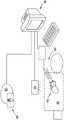
图2是根据本公开的一个方面的用于内部医疗手术的VR/AR系统的示意性表示。2 is a schematic representation of a VR/AR system for internal medical procedures in accordance with one aspect of the present disclosure.
图3是根据本公开的一个方面的电解剖图的图像。3 is an image of an electroanatomical map according to one aspect of the present disclosure.
图4示出根据本公开的一个方面的患者心脏的3D模型。4 illustrates a 3D model of a patient's heart according to one aspect of the present disclosure.
图5A是根据一个方面的提供给VR/AR系统的操作者的包括下拉菜单的图的图像。5A is an image of a diagram including a drop-down menu provided to an operator of a VR/AR system, according to one aspect.
图5B是根据一个方面的提供给VR/AR系统的操作者的包括插入图像下拉菜单的图的图像。5B is an image of a diagram provided to an operator of a VR/AR system including an insert image drop-down menu, according to one aspect.
图5C是根据一个方面的提供给VR/AR系统的操作者的包括3D心脏模型的横截面图的图的图像。5C is an image of a diagram including a cross-sectional view of a 3D heart model provided to an operator of a VR/AR system, according to one aspect.
图6是示出图2的VR/AR系统的组件的示意性表示的框图。FIG. 6 is a block diagram illustrating a schematic representation of components of the VR/AR system of FIG. 2 .
具体实施方式Detailed ways
在以下优选实施例的详细描述中,参考了附图,该附图形成其一部分,并且在附图中通过图示的方式示出了可以实现本公开的具体实施例。应当理解,在不脱离本公开的范围的情况下,可以利用其它实施例并且可以进行结构改变。In the following detailed description of the preferred embodiments, reference is made to the accompanying drawings, which form a part hereof, and in which are shown by way of illustration specific embodiments in which the present disclosure may be practiced. It is to be understood that other embodiments may be utilized and structural changes may be made without departing from the scope of the present disclosure.
如说明书和所附权利要求中所使用的,单数形式“一”、“一个”和“该”包括复数指示物,除非上下文另有明确说明。本文中范围可以表述为从“约”一个特定值,和/或到“约”另一个特定值。当表述这种范围时,另一个实施例包括从一个特定值和/或到另一个特定值。类似地,当通过使用先行词“约”将值表述为近似值时,将理解该特定值形成另一个实施例。将进一步理解,范围中每个范围的端点相对于另一个端点都是显著的,并且独立于另一个端点。As used in the specification and the appended claims, the singular forms "a," "an," and "the" include plural referents unless the context clearly dictates otherwise. Ranges can be expressed herein as from "about" one particular value, and/or to "about" another particular value. When such a range is expressed, another embodiment includes from one particular value and/or to another particular value. Similarly, when values are expressed as approximations, by use of the antecedent "about," it will be understood that the particular value forms another embodiment. It will be further understood that an endpoint of each range in a range is significant relative to, and independent of, the other endpoint.
“可选的”或“可选地”是指随后描述的事件或情况可能发生或可能不发生,并且描述包括该事件或情况发生的实例和不发生的实例。"Optional" or "optionally" means that the subsequently described event or circumstance may or may not occur, and that the description includes instances in which the event or circumstance occurs and instances in which it does not.
在本说明书的整个描述和权利要求中,词语“包括”和词语的变体,诸如“包括”和“包含”,是指“包括但不限于”,并且不旨在排除例如其它添加物、组件、整数或步骤。“示例性”是指“……的示例”,并且不旨在表达优选或理想实施例的指示。“诸如”不用于限制性意义,而是用于解释目的。Throughout the description and claims of this specification, the word "including" and variations of words, such as "including" and "comprising", mean "including but not limited to" and are not intended to exclude, for example, other additives, components , integer, or step. "Exemplary" means "an example of" and is not intended to convey an indication of a preferred or ideal embodiment. "Such as" is not used in a limiting sense, but is used for explanatory purposes.
公开了可用于执行所公开的方法和系统的组件。在此公开了这些和其它组件,并且可以理解,当公开这些组件的组合、子集、交互、集合等时,对于所有方法和系统,本文中对每个都特别考虑和描述,然而可能未明确公开每种不同的个体和集体组合的特定参考以及这些组合的排列。这适用于本申请的所有方面,包括但不限于所公开方法中的步骤。因此,如果存在可以执行的各种附加步骤,则应理解,这些附加步骤中的每一个可以采用所公开方法的任何特定实施例或实施例的组合来执行。Components are disclosed that can be used to implement the disclosed methods and systems. These and other components are disclosed herein, and it is to be understood that when combinations, subsets, interactions, collections, etc. of these components are disclosed, for all methods and systems, each is specifically considered and described herein, however may not be explicitly Specific references to each of the various individual and collective combinations and permutations of those combinations are disclosed. This applies to all aspects of this application, including but not limited to steps in the disclosed methods. Thus, if there are various additional steps that can be performed, it is understood that each of these additional steps can be performed using any particular embodiment or combination of embodiments of the disclosed methods.
如所属领域的技术人员将了解的,本公开的方面可采用完全硬件实施例、完全软件实施例或组合软件和硬件方面的实施例的形式。在一个方面,本公开可以包括被配置为执行由硬件和软件组件的组合控制的某些步骤和功能(例如,获得电解剖测量等)的物理组件的组合。此外,方法和系统的组件可以采用计算机可读非暂态存储介质上的计算机程序产品的形式,该计算机可读非暂态存储介质具有体现在存储介质中的计算机可读程序指令(例如,计算机软件)。可以利用任何合适的计算机可读存储介质,包括硬盘、CD-ROM、光存储装置、闪存装置、固态存储装置和磁存储装置。As will be appreciated by those skilled in the art, aspects of the present disclosure may take the form of an entirely hardware embodiment, an entirely software embodiment, or an embodiment combining software and hardware aspects. In one aspect, the present disclosure may include a combination of physical components configured to perform certain steps and functions (eg, obtaining electroanatomical measurements, etc.) controlled by a combination of hardware and software components. Furthermore, components of the methods and systems may take the form of a computer program product on a computer-readable non-transitory storage medium having computer-readable program instructions embodied in the storage medium (eg, a computer software). Any suitable computer-readable storage medium may be utilized, including hard disks, CD-ROMs, optical storage devices, flash memory devices, solid state storage devices, and magnetic storage devices.
此外,如下所述的本公开所使用的组件和方法可以在程序环境中执行,该程序环境可以包括通用计算机或专用装置,诸如硬件装置、控制器或手持计算机。另外,可以使用本领域中已知的各种技术来实现在此描述的组件的技术。例如,该方法可以在计算机系统上执行的软件中实施,或者利用微处理器的组合或其它专门设计的专用集成电路、可编程逻辑装置或其各种组合在硬件中实施。Furthermore, the components and methods used in the present disclosure, as described below, can be implemented in a programming environment, which can include a general purpose computer or special purpose apparatus, such as a hardware device, a controller, or a handheld computer. Additionally, the techniques of the components described herein may be implemented using various techniques known in the art. For example, the method may be implemented in software executing on a computer system, or in hardware using a combination of microprocessors or other specially designed application specific integrated circuits, programmable logic devices, or various combinations thereof.
下面参考方法、系统、设备和计算机程序产品的框图和流程图图示来描述方法和系统的一些方面。应当理解,框图和流程图图示的每个框以及框图和流程图图示中的框的组合可以分别由计算机程序指令实施。这些计算机程序指令可以加载到通用计算机、专用计算机或其它可编程数据处理设备上以产生机器,使得在计算机或其它可编程数据处理设备上执行的指令创建用于实施在流程图的框或多个框中指定的功能的部件。Some aspects of the methods and systems are described below with reference to block diagrams and flowchart illustrations of methods, systems, apparatus, and computer program products. It will be understood that each block of the block diagrams and flowchart illustrations, and combinations of blocks in the block diagrams and flowchart illustrations, respectively, can be implemented by computer program instructions. These computer program instructions can be loaded on a general purpose computer, special purpose computer or other programmable data processing apparatus to produce a machine such that the instructions executed on the computer or other programmable data processing apparatus create blocks or more for implementing the flowcharts The part of the function specified in the box.
这些计算机程序指令还可以存储在非暂态计算机可读存储器中,该计算机可读存储器可以指示计算机或其它可编程数据处理设备以特定方式工作,使得存储在计算机可读存储器中的指令产生包括用于实施流程图的框或多个框中指定的功能的计算机可读指令的制品。计算机程序指令也可以加载到计算机或其它可编程数据处理设备上,以使在计算机或其它可编程设备上执行一系列操作步骤,以产生计算机实施的过程,使得在计算机或其它可编程设备上执行的指令提供用于实施流程图的框或多个框中指定的功能的步骤。These computer program instructions can also be stored in non-transitory computer readable memory, which can instruct a computer or other programmable data processing device to operate in a particular manner such that the instructions stored in the computer readable memory generate a An article of manufacture of computer readable instructions for implementing the functions specified in the block or blocks of the flowchart. Computer program instructions can also be loaded on a computer or other programmable data processing device to cause a series of operational steps to be performed on the computer or other programmable device to produce a computer-implemented process for execution on the computer or other programmable device The instructions provide steps for implementing the functions specified in the block or blocks of the flowchart.
因此,框图和流程图图示的框支持用于执行指定功能的部件的组合,用于执行指定功能的步骤的组合,以及用于执行指定功能的程序指令部件。还将理解,框图和流程图图示的每个框以及框图和流程图图示中的框的组合可以由执行特定功能或步骤的专用基于硬件的计算机系统或专用硬件和计算机指令的组合来实施。Accordingly, blocks of the block diagrams and flowchart illustrations support combinations of means for performing the specified functions, combinations of steps for performing the specified functions, and program instruction means for performing the specified functions. It will also be understood that each block of the block diagrams and flowchart illustrations, and combinations of blocks in the block diagrams and flowchart illustrations, can be implemented by special purpose hardware-based computer systems that perform the specified functions or steps, or combinations of special purpose hardware and computer instructions .
在各个方面,本公开涉及虚拟现实和/或增强现实(VR/AR)系统,下面将详细讨论。尽管在全文中使用术语虚拟现实来描述系统,但是本领域技术人员将理解,在术语的一般使用中,虚拟现实可以包括虚拟现实(VR)和增强现实(AR)。在一些情况下,术语“VR/AR”将用于识别系统。因此,当在此使用术语“虚拟现实”或“VR/AR”时,应该理解为包括所有类型的修改现实,除非特别区分。In various aspects, the present disclosure relates to virtual reality and/or augmented reality (VR/AR) systems, discussed in detail below. Although the term virtual reality is used throughout to describe the system, those skilled in the art will understand that, in general use of the term, virtual reality may include virtual reality (VR) and augmented reality (AR). In some cases, the term "VR/AR" will be used to identify the system. Accordingly, when the term "virtual reality" or "VR/AR" is used herein, it should be understood to include all types of modified reality, unless otherwise specifically distinguished.
如在此所使用的“虚拟现实”是指显示表示计算机生成的数据的一个或多个元素和/或与表示计算机生成的数据的一个或多个元素交互的方法。通常,在虚拟现实显示器的视场内可见的所有元素都是计算机生成的元素。As used herein, "virtual reality" refers to a method of displaying and/or interacting with one or more elements representing computer-generated data. Typically, all elements visible within the field of view of a virtual reality display are computer-generated elements.
如在此所使用的,“增强现实”是指显示表示计算机生成的数据的一个或多个元素和/或与该一个或多个元素交互的方法。增强现实是虚拟现实和现实生活的混合,其中典型的增强现实显示包括在操作者可见的现实物体上覆盖的一个或多个计算机生成的元素。如在此所使用的术语“增强现实”可以进一步包括“混合现实”,术语“混合现实”是指特别包括用户或操作者与计算机生成的元素交互的能力的增强现实显示方法。As used herein, "augmented reality" refers to a method of displaying and/or interacting with one or more elements representing computer-generated data. Augmented reality is a hybrid of virtual reality and real life, where a typical augmented reality display includes one or more computer-generated elements overlaid on real objects visible to the operator. The term "augmented reality" as used herein may further include "mixed reality," which refers to an augmented reality display method that specifically includes the ability of a user or operator to interact with computer-generated elements.
如在此所使用的“全息显示”是指显示虚拟3D物体和/或与虚拟3D物体交互的方法,其中虚拟3D物体被动态更新以响应于操作者的运动,或操作者请求的虚拟3D物体的视图的修改(诸如放大/缩小、平移/旋转的虚拟3D物体的横截面视图等),来修改虚拟3D物体的操作者的视图。"Holographic display" as used herein refers to a method of displaying and/or interacting with virtual 3D objects, wherein the virtual 3D objects are dynamically updated in response to operator movements, or virtual 3D objects requested by the operator Modification of the view of the virtual 3D object (such as zooming in/out, translating/rotating the cross-sectional view of the virtual 3D object, etc.) to modify the operator's view of the virtual 3D object.
本公开涉及VR/AR可视化系统10(在此也称为VR/AR系统10或系统10),用于3D成像数据以及附加信息的可视化和操纵,附加信息包括但不限于2D成像数据、生命体征数据和对象人口统计数据,其与在难以观察/接近对象的解剖结构的部分内发生的医学诊断和治疗过程相关联。通过非限制性示例,VR/AR可视化系统10可用于心脏、胃肠系统、耳道和其它类型的解剖结构或生物系统内的手术。虽然下面描述的实施例涉及与心脏介入手术相关联的VR/AR可视化系统10,但是本领域技术人员可认识到使用其它3D定位或姿势估计模态,包括但不限于结合在此描述的VR/AR可视化系统10的基于阻抗的定位、磁定位、基于标记的定位、或3D超声,可以容易地扩展到其它器官系统和/或其它解剖区域中的诊断或介入手术。The present disclosure relates to a VR/AR visualization system 10 (also referred to herein as VR/AR system 10 or system 10 ) for visualization and manipulation of 3D imaging data and additional information including, but not limited to, 2D imaging data, vital signs Data and subject demographic data associated with medical diagnosis and treatment procedures that occur within portions of the subject's anatomy that are difficult to observe/access. By way of non-limiting example, the VR/AR visualization system 10 may be used for procedures within the heart, gastrointestinal system, ear canal, and other types of anatomical or biological systems. Although the embodiments described below relate to the VR/AR visualization system 10 in connection with cardiac interventional procedures, those skilled in the art will recognize the use of other 3D positioning or pose estimation modalities, including but not limited to the VR/AR visualization systems described herein. Impedance-based localization, magnetic localization, marker-based localization, or 3D ultrasound of AR visualization system 10 can be easily extended to diagnostic or interventional procedures in other organ systems and/or other anatomical regions.
在一个方面,本公开涉及用于与心脏诊断手术和/或心脏介入手术相关联的VR/AR可视化系统10。VR/AR系统10经由3D医学成像装置40能够采集患者20(例如,对象)的心脏15的解剖特征。另外,可以使用一个或多个电解剖标测装置50来获得心脏15的电数据,该电解剖标测装置50被配置为收集电数据以及相关器械的定位和电数据测量的位置。与一个或多个电解剖标测装置50相关联的器械的非限制性示例包括诊断导管、参考导管、消融导管、监测导管、非接触标测导管、多电极阵列导管、多极导管、多极圆形标测导管和磁传感器导管。在一个方面,可以通过相关联的电解剖标测系统分析由一个或多个电解剖标测装置50获得的数据,以确定描述心脏15的心房和心室的内表面的空间布置以及相对靠近心脏15的静脉和动脉(包括但不限于腔静脉、主动脉、肺动脉等)的空间布置的心内膜图。In one aspect, the present disclosure relates to a VR/AR visualization system 10 for use in connection with cardiac diagnostic procedures and/or cardiac interventional procedures. The VR/AR system 10 can acquire anatomical features of the
在一个方面,一个或多个电解剖标测装置50可以提供将使用3D医学成像装置40获得的患者20的心脏15的解剖特征的至少一个3D图和使用一个或多个电解剖标测装置50获得的至少一个3D心内膜表面图进行组合。在一个方面,其内定义3D心内膜表面图的3D坐标系可以配准到其内定义心脏的解剖特征的3D图的坐标系,使得在同一3D坐标系内定义解剖特征的3D图和3D心内膜表面图。In one aspect, the one or more
在一个方面,由一个或多个电解剖标测装置50产生的定义共同配准的解剖特征的3D图和3D心内膜表面图的数据可以由VR/AR可视化系统10的计算装置30接收。使用定义共同配准的3D图的数据,计算装置30生成心脏15的3D模型65,该3D模型65被配置为显示在由VR/AR装置60的全息显示器产生的全息图像内。In one aspect, data generated by one or more
在一个方面,全息图像可以仅由心脏15的3D模型65的至少一部分组成。在各种其它方面,VR/AR可视化系统10的计算装置30可以接收附加数据,该附加数据可以结合到全息图像中,以便在VR/AR装置60的全息显示器上显示给操作者。In one aspect, the holographic image may consist of only at least a portion of the
在各个方面,VR/AR系统10可以包括多个VR/AR装置60。作为非限制性示例,第一操作者可以佩戴第一VR/AR装置60,包括但不限于,第一头戴式显示器,并且第二操作者可佩戴第二VR/AR装置60,包括但不限于第二头戴式显示器。在该非限制性示例中,第一操作者和第二操作者可以一起对患者执行外科手术。第一VR/AR装置60和第二VR/AR装置60可以显示患者20的解剖结构的同一器官或部分的不同视图。例如,患者心脏15的显示视图可以基于对应的VR/AR装置60相对于患者20的位置以不同角度定位。In various aspects, VR/AR system 10 may include multiple VR/
在各种其它方面,附加数据可以由VR/AR可视化系统10的计算装置30接收并且结合到全息图像中。适合于结合到全息图像中的附加数据的非限制性示例包括:在定义3D模型65的坐标系内定义位置和/或测量值的实时3D数据;由诸如荧光镜成像装置的2D成像装置产生的实时2D数据;诸如一个或多个生命体征的实时数字测量、诸如患者人口统计数据的预定数据及其任何组合。在这些各种其它方面中,附加数据可以结合和/或覆盖在心脏15的3D模型65上,或者附加数据可以被显示为全息图像内的单独元素,如下面另外详细描述的。In various other aspects, additional data may be received by computing
在一个方面,由电解剖标测装置50获得的附加数据可以由计算装置30接收并且结合到全息图像中。在一个方面,电解剖标测装置可以进一步包括被配置为获得至少一个外科器械的至少一个实时3D位置的器械位置传感器,包括但不限于电生理学(EP)导管。在该方面,计算装置30可以接收至少一个外科器械的至少一个实时3D位置,并且生成包括在对象的解剖结构的至少一个3D图像(包括但不限于心脏15的3D模型65)上覆盖的至少一个外科器械的至少一个实时位置的全息图像。在另一方面,可以使用单独的装置(包括但不限于外科器械系统的单独的器械位置传感器)获得至少一个外科器械的至少一个实时3D位置。合适的器械位置传感器的非限制性示例包括一个或多个电解剖标测装置,除了用于获得3D心内膜表面标测数据的电解剖标测装置50之外,还包括利用超声、磁场、电场和/或任何其它现有合适的位置感测方法的位置感测装置的其它实时位置标测系统。In one aspect, additional data obtained by
在另一方面,由电解剖标测装置50获得的附加数据可包括由一个或多个电生理学(EP)导管获得的附加电生理学测量。在一个方面,数据可以包括定义映射到心脏15的3D模型65的坐标系的实时电生理学测量的一个或多个数据集的附加3D实时数据。实时电生理学测量的非限制性示例包括电压图、激活时序图和传播图。在该一个方面,由计算装置30接收的附加电生理学测量的图可以覆盖在全息图像内的心脏15的3D模型65上。在一个方面,附加电生理学测量的图可以被选择以进行显示,使其变得透明,或者从由操作者产生的一个或多个提示所定义的全息图像中去除。在另一方面,由电解剖标测装置50获得的附加数据可包括与消融导管的消融相关联的附加电生理学测量,包括但不限于在心脏15的3D模型65内的特定位置处施加的射频(RF)或低温能量的量。在该另一方面,附加电生理学测量可以以3D视觉元素的形式结合到全息图像中,诸如位于3D模型65内的消融能量的应用点处的圆圈或其它符号,和/或颜色、尺寸、数值或其它视觉元素,以指示在由消融导管实现的消融事件期间测量的消融能量或消融力的量和/或方向。In another aspect, the additional data obtained by the
在另外的方面,计算装置30可以接收定义从至少一个附加医学成像装置获得的至少一个附加2D图像的一个或多个附加数据集。在一个方面,至少一个附加2D图像可以包括3D实时图像(包括但不限于实时荧光镜图像)的2D表示。在该一个方面,至少一个附加2D图像可以以与3D模型65分开显示的2D视觉元素的形式结合到全息图像中。结合到全息图像中的非限制性的合适的2D视觉元素包括虚拟2D监测、插入图像以及3D全息图像中的2D视觉元素的任何其它已知表示。通过非限制性示例,定义实时荧光透视图像的附加数据集可以以插入图像的形式结合到全息图像中,如图5B中所示。In further aspects,
在一个方面,全息图像可以在VR/AR装置60的全息显示器上显示给操作者。响应于来自操作者的一个或多个提示,计算装置30可以根据操作者的偏好修改全息显示器上显示的全息图像。通过非限制性示例,操作者可以放大、缩小、旋转或移动如在全息显示器上显示的全息图像,以便于完成诊断和/或外科手术。在各个方面,如果VR/AR系统10包括多个VR/AR装置60,则由第一操作者佩戴的第一VR/AR装置60可以可操作地耦接到计算装置30,使得仅第一操作者可以修改如在VR/AR系统10的所有VR/AR装置60的所有全息显示器上显示的全息图像。在这些各种其它方面,计算装置30可以接收多个VR/AR装置60中的每一个装置的相对位置和取向,并生成针对每个VR/AR装置60的全息图像,该全息图像对应于每个VR/AR装置60相对于第一操作者佩戴的第一VR/AR装置60的每个位置和每个取向,该第一操作者还控制全息图像的修改。In one aspect, the holographic image may be displayed to the operator on the holographic display of the VR/
在一个非限制性示例中,计算装置30可以生成患者心脏的心电图,以显示在全息图像的插入图像中。计算装置30可以从VR/AR装置60或单独的输入装置(例如,相机或运动传感器)接收用户输入的指示(即提示),以将虚拟标记定位在心电图上而不破坏无菌性。基于用户输入,计算装置30可以确定用于在插入图像以及心电图上显示的度量。例如,虚拟标记可以是虚拟卡尺,其被配置为测量患者心跳的周期或幅度(例如,度量)。In one non-limiting example,
在另一个附加方面,计算装置30可以接收一个或多个附加字母数字数据集,包括但不限于患者人口统计数据集和/或从现有生命体征测量装置获得的对象的生命体征的实时测量。在该另一方面,一个或多个附加字母数字数据集可以以覆盖在全息图像内的透明字母数字元素的形式结合到全息图像中。In another additional aspect,
在另外的方面,计算装置30可以生成菜单或其它图像元素,该图像元素包括以非透明或透明的行或列的字母数字串、符号和/或图标的形式显示的用户可选元素。在这些附加方面,用户可选元素提供选择由计算装置30的一个或多个处理器执行以实现VR/AR系统10的操作的一个或多个指令的方式。通过非限制性示例,计算装置30可以在如图5A中所示的全息图像内生成透明菜单。In further aspects,
通过选择全息图像内的一个或多个交互式菜单元素,VR/AR装置60的操作者还可以观察3D模型65和附加信息并与之交互而不必破坏无菌性,或与在诊断或外科手术期间在场的任何人通信。如在此所公开的,VR/AR装置60可以取决于系统10的操作者的需要在真实虚拟现实环境或增强现实环境中根据需要呈现和去除信息。在一个方面,全息图像内的元素的布置可以由计算装置30保存并由VR/AR系统10检索,以供随后由同一操作者使用作为优选的元素布置。By selecting one or more interactive menu elements within the holographic image, the operator of the VR/
在各个方面,计算装置30可以随时间推移更新全息图像以考虑由于一个或多个时变因素引起的变化,包括但不限于:由操作者对用户可选择的菜单元素的选择或取消选择,接收更新的实时数据(诸如更新的生命体征数据)、附加消融事件、其它数据集(诸如与荧光镜成像相关联的实时数据集)的变化。计算装置30可以更新全息图像以包括相对于患者器官定位的器械的一部分的表示,例如,导管尖端相对于患者心脏的位置。计算装置30可以基于来自指示检测到的器械的运动的器械的传感器数据来更新全息图像。In various aspects,
在一个方面,VR/AR系统10的计算装置30被配置为接收患者的心脏解剖结构的重建的3D模型65。在此,3D医学成像装置40被配置为获取与患者心脏15相关的特定物理信息。该信息将编码心脏15的尺寸及其组成/腔和预定义的解剖学界标。这些解剖学上不同的界标用于将3D模型65配准到由电解剖标测装置50获得的心内膜表面的电解剖图。通过将电解剖图配准到其内3D解剖模型的坐标系,由电解剖标测装置50获得的映射到心内膜表面的电解剖图的其它测量也被配准到定义心脏15的3D模型65的坐标系,使得这些映射的电生理学测量能够相对于显示给操作者的全息图像内的3D模型65而被可视化。在一些方面,3D模型65和电解剖图以不同的物理单位生成。因此,为了归一化到同一坐标系,计算装置30例如基于解剖学界标将3D模型65的第一组点配准(例如,映射)到电解剖图的第二组点。In one aspect, the
在各个方面,心内膜表面的电解剖图与使用至少一个3D医学成像装置获得的3D解剖模型的配准由计算装置30和/或电解剖标测装置50执行。可以使用任何合适的现有方法来执行两个图的配准,而不进行限制。在一个方面,至少多个预定界标在3D解剖模型和3D心内膜表面图中被识别,以提供两个图的限定交叉点。合适的心脏界标的非限制性示例包括上腔静脉/右心房交界处、下腔静脉/右心房交界处、冠状窦、右心室尖部和右心室流出道。在与心脏的其它已知部分相关联的界标可以用于本公开的其它方面中的界标的同时,这些界标经由MRI和荧光透视过程易于接近和识别,并且采用网格配准,如下面更详细讨论的。在其它方面,可以使用统计或基于特征的推断来确定点的位置和数量。In various aspects, registration of the electroanatomical map of the endocardial surface with a 3D anatomical model obtained using at least one 3D medical imaging device is performed by computing
在一个方面,3D医学成像装置40可以包括能够采集与心脏的解剖结构相关的3D数据的任何装置。例如,3D医学成像装置可以包括但不限于荧光透视装置、超声心动图装置(例如,经胸廓、经食道、心内)、X射线装置、探索性内窥镜系统、MRI和CT扫描器等。3D医学成像装置40可以取决于要与VR/AR可视化系统10组合执行的诊断和/或外科手术的类型来选择。例如,超声心动图装置40提供心脏解剖结构的快速获取,因为是无创的,可以快速完成,不会使患者暴露于辐射,并且无麻醉。此外,对对象的不动性没有长期要求。在一个方面,定义重建的3D解剖模型的数据可以在结合使用VR/AR可视化系统10执行的诊断或外科手术之前由3D医学成像装置40获得和分析。In one aspect, 3D
3D医学成像装置40采集用于重建对象的心脏15的3D模型65的空间信息。在一个方面,3D医学成像装置40重建空间数据并创建对象的心脏解剖结构的3D解剖模型。可以使用本领域已知的各种技术来创建3D解剖模型。通过非限制性示例,可以收集患者心脏的多个2D视图,并且然后将其重新组合成患者解剖结构的粗略3D图像。一旦生成患者解剖结构的3D图像,电解剖标测装置50向计算装置30提供要修改和/或变换的通用高分辨率心脏网格,以匹配解剖学心脏模型和电解剖心内膜表面模型中识别的相应界标的重叠。计算装置30使用从所创建的患者心脏解剖结构的3D图像采集的数据来变换心脏网格,包括所采集的界标的空间维度(如上所述)。一旦变换,由计算装置30或电解剖标测装置50生成患者的心脏结构的全3D模型65。通过一个非限制性示例,3D医学成像装置40可以通过生成表示对应的解剖结构的3D几何形状的一系列连接的2D多边形(例如,网格)来创建3D解剖模型。通过另一个非限制性示例,装置40可以使用射线投射和/或跟踪来创建3D解剖模型。The 3D
在患者心脏结构的变换的全3D模型65已经被电解剖标测装置50创建之后,从对象处收集包括特定于消融需求的电生理学数据的电解剖数据并将其映射到变换的全3D模型65。在一个方面,电解剖数据从一个或多个电解剖标测装置50收集。在一个方面,电解剖标测装置50包括放置在患者心脏15内的电生理学(EP)导管。测量的电解剖数据可包括在患者心脏15的给定位置处发生的电活动(例如,电压数据、激活定时和传播图),该电活动可用于使用现有诊断方法确定消融需要发生的地方。After the transformed
为了执行EP研究,VR/AR系统100可以使用一个或多个电解剖标测装置50。在一个方面,可以最初使用诊断导管50,并且如果基于诊断电生理学测量而指示消融则可以使用附加消融导管50。另外,由消融导管50施加的任何能量可以由一个或多个电解剖标测装置50测量和记录。通过非限制性示例,消融导管可以被配置为在导管尖端处施加射频或低能量。在另一方面,消融导管可以被配置为感测在导管50的尖端处由导管施加的力的级别(包括方向性,即轴向或横向)。此类信息可用于模拟3D模型65的虚拟心脏中的实际病变创建,如通过使用测量的力和阻抗实时制作病变。在一个方面,从消融导管测量的力数据然后可以用于采用VR/AR装置60对操作者的听觉反馈。通过非限制性示例,VR/AR装置60可以提供包括改变音调的音高和频率的听觉反馈,其指示消融导管50推动心脏组织的力级别。To perform an EP study, the VR/AR system 100 may use one or more
EP导管50可以由操作者在整个患者心脏15中移动并收集诊断信息。诊断和消融导管50二者被配置为在其整个使用中(包括在消融期间)获得电解剖数据。除了收集电解剖数据之外,VR/AR系统100还可以采集空间相关信息,即,在患者心脏内发生电解剖数据的位置,以使得电解剖数据可以被映射到变换后的全3D模型。在一个方面,电解剖标测装置50也采集患者体内器械的定位。例如,当利用EP导管50时,采集电极和导管50的轴的远端部分的定位。在一个方面,电解剖标测装置50利用电解剖标测系统来找到电解剖数据的坐标。标测系统能够识别心脏15内的EP导管50的X、Y和Z坐标,并且然后可以将电解剖数据放置到坐标。例如,可以使用诸如ENSITETMNAVXTM导航和可视化以及CARTOTM(韦伯斯特生物传感系统)的电解剖标测系统来收集这些数据。然而,也可以使用能够提供此类信息的其它系统。The
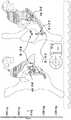
图3示出用于心脏模型的当前电解剖图的示例。该心脏模型仅限于右心房(RA)和左心房(LA)的几何形状。左侧的图像以正交视图呈现,其中右前斜位(RAO)视图在左侧,并且长轴向斜位(LAO)视图在右侧。电生理学导管的远端在该几何形状内可视化。具有四个电极的第一(第1)导管定位在正常传导部位处,位于His位置处。具有10个电极的第二导管位于冠状窦中。由于具有4个电极的第3导管前进通过三尖瓣并进入右心室,因此该第3导管仅在LAO投影中可视化。最后,具有4个电极的射频(RF)消融导管位于异常电组织的部位处。球体(以红色显示)标记RF病变所处的部位。图3的中央底部的靶心投影指示由消融导管施加到心脏组织的总力(TF)。Figure 3 shows an example of a current electroanatomical map for a heart model. This heart model is limited to the geometry of the right atrium (RA) and left atrium (LA). The images on the left are presented in an orthographic view with the right anterior oblique (RAO) view on the left and the long axial oblique (LAO) view on the right. The distal end of the electrophysiology catheter is visualized within this geometry. The first (1st) catheter with four electrodes was positioned at the normal conduction site, at the His position. A second catheter with 10 electrodes is located in the coronary sinus. The 3rd catheter with 4 electrodes was only visualized in the LAO projection as it advanced through the tricuspid valve and into the right ventricle. Finally, a radio frequency (RF) ablation catheter with 4 electrodes is located at the site of abnormal electrical tissue. The sphere (shown in red) marks where the RF lesion is located. The bullseye projection of the bottom center of Figure 3 indicates the total force (TF) applied by the ablation catheter to the cardiac tissue.
虽然以上描述了与心脏手术相关联的电解剖标测装置50和电解剖数据,但可理解的是装置和数据可与人体内发现的其它系统和器官相关联。Although the
当电解剖数据如上描述而被收集和映射时,VR/AR装置60由计算装置30提供患者心脏15的3D模型65(参见图4),包括EP导管50的放置(参见图5B)。在一个方面,VR/AR装置60可以包括真实的虚拟现实(VR)装置(即,将操作者完全沉浸在创建环境中的装置)或增强现实(AR)装置(即,操作者可以具有在虚拟空间中虚拟显示的图像或模型,但仍然能够看到真实环境并与真实环境交互)。按照这些方式,VR/AR装置60可以是可穿戴装置,包括但不限于Microsoft HOLOLENSTM(即AR装置)和OCULUS RIFTTM(即VR装置)。VR/AR装置60可以包括传感器(诸如运动传感器(例如,加速度计、陀螺仪或惯性测量单元)、音频传感器、眼睛和视线跟踪传感器),和/或电子显示器等,以及其它部件。在另一方面,VR/AR装置60可以提供包括3D模型65的投影全息显示器。VR/AR装置60可以经由无线交换协议或经由有线连接通信地耦接到HMD。在至少一些方面,AR装置的使用可能是有利的,因为其允许操作者实时看到患者并与患者交互,同时观看3D模型65并与3D模型65交互并获得其益处,有助于更安全的患者体验。When the electroanatomical data is collected and mapped as described above, the VR/
在这些方面中,全息图像内的3D模型65可以取决于EP导管的位置呈现为钝的或半透明,或者完全透明以使得能够无阻碍地观察EP导管位置。在透明视图中,操作者可以使用由VR/AR装置的至少一个传感器接收的操作者启用的提示来改变心脏壁的透明度,从而允许在诊断和/或外科手术期间使心脏中的导管容易明显的可视化。导管的各部分也可以在任何视图中表示。另外,VR/AR装置60还可以允许操作者操纵心脏的位置、取向和尺寸,以及创建要观察的切片。此外,操作者可以在不使用手的情况下在视图之间切换以及数据显示,因此操作者可以在整个过程中保持无菌性。In these aspects, the
通过非限制性示例,VR/AR系统100可以使用头部和/或眼睛跟踪技术来从操作者接收输入命令(例如,用户输入),而无需操作者使用操作者的手物理地触摸输入装置(例如,VR/AR装置60)。在一些实施例中,输入装置物理地连接到VR/AR装置60。在其它实施例中,输入装置与VR/AR装置60分离并且通信地耦接到计算装置30。例如,输入装置是Microsoft KINECTTM。输入装置可以包括成像传感器(例如,相机)、用于成像传感器的照明源、运动传感器、深度传感器以及其它组件。基于传感器数据,输入装置可以采集手势并执行操作者的姿势检测。在一个方面,操作者启用的输入可以从修改VR/AR装置60提供的现有操作者启用的输入来导出。By way of non-limiting example, VR/AR system 100 may use head and/or eye tracking techniques to receive input commands (eg, user input) from an operator without requiring the operator to physically touch the input device with the operator's hands ( For example, VR/AR device 60). In some embodiments, the input device is physically connected to the VR/
另外,也可以结合VR/AR装置60使用电解剖标测装置50来产生从收集的电解剖数据采集的为消融手术准备的计划映射。在另一方面,VR/AR装置60可以显示虚拟卡尺(例如,标尺),以允许操作者在虚拟环境中进行实时准确的测量。该特征允许操作者在各个位置处进行测量,例如,在测量电描记图时,使用毫秒,以及在心脏几何结构中,以毫米为单位进行测量。Additionally, the
可以向操作者显示或传达其它电解剖信息。例如,电解剖信息可以在心脏的3D模型65上数字地表示(例如,心脏上的颜色编码区域,以指示电活动和EP导管在施加时的力数据),可视地显示给操作者(例如,示出相关力数据和电活动的表格),或以听觉方式。在一个方面,听觉方式可用于通知操作者在消融手术期间激活EP导管时施加的力。在这种情况下,听觉响应可以与施加时的力成比例。听觉信号涉及由消融导管施加的力。施加的力越强,对于操作者音调就越频繁和高音。该听觉特征仅存在于力传感导管中。力传感导管的示例是TACTICATHTM(St Jude Medical),其向操作者提供反馈,以指示导管尖端施加到组织多少的力(例如以克为单位测量)。Other electroanatomical information may be displayed or communicated to the operator. For example, electroanatomical information can be represented digitally on the
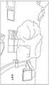
图5A、图5B和图5C示出根据本公开的一个方面的由VR/AR装置60显示的全息图像的示例性图。图5A示出3D模型65(虚拟心脏模型)的打开的主菜单。该视图中3D心脏模型65的透明度被增加,以允许操作者在一个或多个平面中快速地可视化精确的导管位置,而无需改变模型的取向。在各个方面,VR/AR系统的计算装置30可以将3D模型分割成子单元,以通过渲染3D模型65的一部分成透明和/或不可见来修改全息显示,从而便于选择3D模型65的部分。可以由计算装置30分割的3D模型65的子单元的非限制性示例包括左心房和右心房、左心室和右心室、一个或多个瓣膜,与心脏相关联的一个或多个动脉和静脉,以及任何其它相关的心脏结构。5A, 5B, and 5C illustrate exemplary diagrams of holographic images displayed by VR/
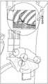
图5B示出在导管位置打开的情况下在后部投影中显示的3D模型65,其中使用切割平面特征切除了心脏的一部分,使得可以可视化心内导管位置。在该视图中,可以看到四个导管(被圈出)位于冠状窦中(在左心房与左心室分开的房室沟中;朝向屏幕的左侧),高右心房(在右上腔室,右心房/上腔静脉交界处附近)中,右心室尖部(在右下腔室,指向心脏的顶点)和正常的His传导系统(朝向心脏的中心或关键处)中。在一些方面,计算装置30基于全息显示器的操作者控制的缩放级别(例如,放大/缩小)在显示的3D模型65中包括附加组件。例如,在更大的缩放级别(即,放大的图像)下,计算装置30包括图5B中所示的四个导管中的一个或多个导管。另一方面,在更低的缩放级别(即,缩小的图像)下,例如由于所显示的3D模型65的所得的有限分辨率水平,计算装置30不包括四个导管。在图5B中的右侧,荧光透视屏幕已在虚拟环境中打开。Figure 5B shows the
图5C示出显示为在心室的筒下视图(down-the-barrel view)(即,外科医生的视图)中取向的3D模型65,其中心房和大动脉被虚拟地去除,例如横截面视图。这些图像提供了操作者可以使用VR/AR系统10看到的潜在视图的非限制性示例,并且不应该被解释为由本公开的系统10提供的唯一视图。Figure 5C shows a
在各个方面,计算装置30可以包括但不限于笔记本计算机、台式计算机、平板计算机、具有连接的显示器的服务器等。根据一个方面,如图6中所示,计算装置30可以通过各种已知方式(包括本领域已知的有线和无线连接)与3D医学成像装置40、电解剖标测装置50以及VR/AR装置60通信。在一个方面,计算装置30可以包括无线接口控制器(“W.I.”)100,该无线接口控制器100被配置为控制无线收发机102的操作,以及从其它装置接收和发送信息。无线收发机102可以在宽范围的公共频率上通信,包括但不限于频带2.40Hz和/或5GHz-5.8GHz。此外,无线收发机102采用无线接口控制器100还可以利用各种公共协议。例如,在本公开的一些实施例中,组合的无线接口控制器100和无线收发机102可以在各种现有和提出的IEEE无线协议上操作,包括但不限于IEEE 802.11b/g/n/a/ac,最大理论数据传输速率/吞吐量分别为11Mbps/54Mbps/600Mbps/54Mbps/1Gbps。在一个方面,计算装置30可包括网络适配器126,网络适配器126被配置为通过各种网络和连接与其它装置通信。In various aspects,
计算装置30可以具有一个或多个软件应用104以执行上面讨论的方法。计算装置30包括系统存储器108以及操作系统110,系统存储器108可以存储各种应用104,包括执行上述功能的应用。系统存储器108还可以包括可由各种软件应用104访问的数据112。系统存储器108可以包括随机存取存储器(RAM)或只读存储器(ROM)。存储在计算装置30上的数据112可以是任何类型的可检索数据。数据可以存储在各种各样的数据库中,包括关系数据库,包括但不限于Microsoft Access和SQL Server、MySQL、INGRES、DB2、INFORMIX、Oracle、PostgreSQL、Sybase 11、Linux数据存储部件等。
计算装置30可以包括各种其它计算机可读介质,包括存储装置114。存储装置114可以用于存储计算机代码、计算机可读指令、程序模块和用于计算装置30的其它数据112,并且存储装置114可以用于备份或可替代地运行操作系统110和/或其它应用104。存储装置114可以包括硬盘、各种磁存储装置(诸如磁带盒或磁盘)、固态闪存驱动器,或其它光学存储器、随机存取存储器等。
计算装置30可以包括系统总线118,系统总线118将计算装置30的各种组件连接到系统存储器108和存储装置114,以及彼此连接。计算装置30的其它组件可以包括一个或多个处理器或处理单元120、用户界面(UI)122,以及一个或多个输入/输出接口124。此外,计算装置30包括网络适配器126。此外,计算装置30可以包括电源128,包括但不限于电池或外部电源。此外,计算装置30可以包括显示适配器226和显示器228(例如,监视器或屏幕)。另外,输入装置(例如,键盘、鼠标、操纵杆等)可以经由输入输出接口124使用。此外,其它3D医学成像装置40、电解剖标测装置50和VR/AR装置60也可以经由输入/输出接口124与计算装置30通信。
VR/AR系统10被配置为在诊断和/或外科手术期间在介入医生(例如,操作者)前面显示虚拟的针对患者的3D模型65。通过使用揭示实时电生理学的3D模型65,将发生医生培训、患者结果和临床医生协作的改进,以及降低患者和医生二者的辐射暴露率。此外,系统10还将减少患者的医疗和经济负担,否则患者将经受由其解剖结构的不良可视化而影响的多个过程。The VR/AR system 10 is configured to display a
虽然本公开的前述书面描述使普通技术人员能够制造和使用目前被认为是其最优模式的事物,但是普通技术人员将了解并理解在此的特定实施例、方法和示例的变型、组合和等同物的存在。因此,本公开不应限制为上述实施例、方法和示例,而是本公开的范围和精神内的所有实施例和方法。在理解或完成本公开所必需的程度上,在此提及的所有出版物、专利和专利申请通过引用明确地并入本文,其程度如同每个单独地并入。While the foregoing written description of the present disclosure enables those of ordinary skill to make and use what is presently considered to be its best mode, those of ordinary skill will appreciate and appreciate modifications, combinations, and equivalents of the specific embodiments, methods, and examples herein. existence of things. Accordingly, the present disclosure should not be limited to the above-described embodiments, methods, and examples, but rather all embodiments and methods within the scope and spirit of the present disclosure. All publications, patents, and patent applications mentioned herein are expressly incorporated by reference to the extent necessary to understand or complete the disclosure, to the same extent as if each were individually incorporated.
已经如此描述了本公开的示例性实施例,本领域技术人员将理解,所公开的内容仅是示例性的,并且可以在本公开的范围内进行各种其它替换、更改和修改。因此,本公开不限于如在此所示的特定实施例。Having thus described exemplary embodiments of the present disclosure, it will be understood by those skilled in the art that what is disclosed is merely exemplary and that various other substitutions, changes and modifications may be made within the scope of the present disclosure. Therefore, the present disclosure is not limited to the specific embodiments as shown herein.
Claims (18)
Priority Applications (1)
| Application Number | Priority Date | Filing Date | Title |
|---|---|---|---|
| CN202210518876.XACN114903591A (en) | 2016-03-21 | 2017-03-20 | Virtual reality or augmented reality visualization of 3D medical images |
Applications Claiming Priority (3)
| Application Number | Priority Date | Filing Date | Title |
|---|---|---|---|
| US201662310969P | 2016-03-21 | 2016-03-21 | |
| US62/310,969 | 2016-03-21 | ||
| PCT/US2017/023221WO2017165301A1 (en) | 2016-03-21 | 2017-03-20 | Virtual reality or augmented reality visualization of 3d medical images |
Related Child Applications (1)
| Application Number | Title | Priority Date | Filing Date |
|---|---|---|---|
| CN202210518876.XADivisionCN114903591A (en) | 2016-03-21 | 2017-03-20 | Virtual reality or augmented reality visualization of 3D medical images |
Publications (2)
| Publication Number | Publication Date |
|---|---|
| CN108882854A CN108882854A (en) | 2018-11-23 |
| CN108882854Btrue CN108882854B (en) | 2022-05-24 |
Family
ID=59900807
Family Applications (2)
| Application Number | Title | Priority Date | Filing Date |
|---|---|---|---|
| CN202210518876.XAPendingCN114903591A (en) | 2016-03-21 | 2017-03-20 | Virtual reality or augmented reality visualization of 3D medical images |
| CN201780018916.1AActiveCN108882854B (en) | 2016-03-21 | 2017-03-20 | Virtual reality or augmented reality visualization of 3D medical images |
Family Applications Before (1)
| Application Number | Title | Priority Date | Filing Date |
|---|---|---|---|
| CN202210518876.XAPendingCN114903591A (en) | 2016-03-21 | 2017-03-20 | Virtual reality or augmented reality visualization of 3D medical images |
Country Status (6)
| Country | Link |
|---|---|
| US (2) | US10258426B2 (en) |
| EP (1) | EP3432780A4 (en) |
| CN (2) | CN114903591A (en) |
| AU (1) | AU2017236893A1 (en) |
| CA (1) | CA3016346A1 (en) |
| WO (1) | WO2017165301A1 (en) |
Families Citing this family (88)
| Publication number | Priority date | Publication date | Assignee | Title |
|---|---|---|---|---|
| US10013808B2 (en) | 2015-02-03 | 2018-07-03 | Globus Medical, Inc. | Surgeon head-mounted display apparatuses |
| GB2536650A (en) | 2015-03-24 | 2016-09-28 | Augmedics Ltd | Method and system for combining video-based and optic-based augmented reality in a near eye display |
| US11282191B2 (en)* | 2017-01-12 | 2022-03-22 | Navix International Limited | Flattened view for intra-lumenal navigation |
| US11842456B2 (en) | 2017-01-12 | 2023-12-12 | Navix International Limited | Flattened view for intra-lumenal navigation |
| EP3585254B1 (en) | 2017-02-24 | 2024-03-20 | Masimo Corporation | Medical device cable and method of sharing data between connected medical devices |
| US11024064B2 (en) | 2017-02-24 | 2021-06-01 | Masimo Corporation | Augmented reality system for displaying patient data |
| EP3612127A1 (en)* | 2017-04-20 | 2020-02-26 | The Cleveland Clinic Foundation | System and method for holographic image-guided percutaneous endovascular percutaneous procedures |
| JP6951114B2 (en)* | 2017-04-27 | 2021-10-20 | キヤノンメディカルシステムズ株式会社 | Medical image diagnostic equipment and magnetic resonance imaging equipment |
| CN117373636A (en) | 2017-05-08 | 2024-01-09 | 梅西莫股份有限公司 | System for pairing a medical system with a network controller using an adapter |
| US10470645B2 (en) | 2017-05-22 | 2019-11-12 | Gustav Lo | Imaging system and method |
| CN109674533B (en)* | 2017-10-18 | 2022-07-05 | 刘洋 | Operation navigation system and method based on portable color ultrasound equipment |
| WO2019091875A1 (en)* | 2017-11-07 | 2019-05-16 | Koninklijke Philips N.V. | Augmented reality triggering of devices |
| US11179200B2 (en)* | 2017-12-15 | 2021-11-23 | Medtronic, Inc. | Augmented reality solution to disrupt, transform and enhance cardiovascular surgical and/or procedural mapping navigation and diagnostics |
| US20190254753A1 (en) | 2018-02-19 | 2019-08-22 | Globus Medical, Inc. | Augmented reality navigation systems for use with robotic surgical systems and methods of their use |
| CN112424870A (en)* | 2018-04-30 | 2021-02-26 | 非凡健康私人有限公司 | Medical virtual reality and mixed reality collaboration platform |
| US11980507B2 (en) | 2018-05-02 | 2024-05-14 | Augmedics Ltd. | Registration of a fiducial marker for an augmented reality system |
| US10869727B2 (en)* | 2018-05-07 | 2020-12-22 | The Cleveland Clinic Foundation | Live 3D holographic guidance and navigation for performing interventional procedures |
| TWI741196B (en)* | 2018-06-26 | 2021-10-01 | 華宇藥品股份有限公司 | Surgical navigation method and system integrating augmented reality |
| US10777012B2 (en)* | 2018-09-27 | 2020-09-15 | Universal City Studios Llc | Display systems in an entertainment environment |
| US11555874B2 (en)* | 2018-11-12 | 2023-01-17 | Case Western Reserve University | System and method for real-time magnetic resonance imaging data visualization in three or four dimensions |
| KR102270170B1 (en)* | 2018-11-14 | 2021-06-25 | 임승준 | Surgery supporting instrument using augmented reality |
| US11766296B2 (en) | 2018-11-26 | 2023-09-26 | Augmedics Ltd. | Tracking system for image-guided surgery |
| CN109364387B (en)* | 2018-12-05 | 2020-07-03 | 上海市肺科医院 | A radiotherapy AR positioning system |
| US11350847B2 (en)* | 2018-12-13 | 2022-06-07 | Biosense Webster (Israel) Ltd. | Composite visualization of body part |
| US11406472B2 (en)* | 2018-12-13 | 2022-08-09 | DePuy Synthes Products, Inc. | Surgical instrument mounted display system |
| US11357593B2 (en) | 2019-01-10 | 2022-06-14 | Covidien Lp | Endoscopic imaging with augmented parallax |
| CN109512511A (en)* | 2019-01-14 | 2019-03-26 | 常州锦瑟医疗信息科技有限公司 | The method and apparatus of operation augmented reality positioning based on positioning guide plate |
| WO2020185556A1 (en)* | 2019-03-08 | 2020-09-17 | Musara Mubayiwa Cornelious | Adaptive interactive medical training program with virtual patients |
| CN111862333B (en)* | 2019-04-28 | 2024-05-28 | 广东虚拟现实科技有限公司 | Augmented reality-based content processing method, device, terminal equipment, and storage medium |
| WO2020220208A1 (en)* | 2019-04-29 | 2020-11-05 | Shanghai United Imaging Healthcare Co., Ltd. | Systems and methods for object positioning and image-guided surgery |
| CN119791847A (en) | 2019-05-31 | 2025-04-11 | 直观外科手术操作公司 | Composite medical imaging system and method |
| US12161512B2 (en) | 2019-05-31 | 2024-12-10 | Intuitive Surgical Operations, Inc. | Systems and methods for integrating imagery captured by different imaging modalities into composite imagery of a surgical space |
| DE102019116269A1 (en)* | 2019-06-14 | 2020-12-17 | apoQlar GmbH | Video image transmission device, method for live transmission and computer program |
| US10832486B1 (en) | 2019-07-17 | 2020-11-10 | Gustav Lo | Systems and methods for displaying augmented anatomical features |
| US11288802B2 (en) | 2019-07-17 | 2022-03-29 | Gustav Lo | Systems and methods for displaying augmented anatomical features |
| US12178666B2 (en) | 2019-07-29 | 2024-12-31 | Augmedics Ltd. | Fiducial marker |
| US11980506B2 (en) | 2019-07-29 | 2024-05-14 | Augmedics Ltd. | Fiducial marker |
| CN110292439A (en)* | 2019-07-31 | 2019-10-01 | 东北大学 | Cardiac stent operation auxiliary diagnosis and therapy system and application method based on augmented reality |
| CN110522514A (en)* | 2019-08-21 | 2019-12-03 | 昆明医科大学第二附属医院 | A positioning and tracking system for hepatobiliary surgery |
| CA3158776A1 (en) | 2019-10-25 | 2021-04-29 | SentiAR, Inc. | Electrogram annotation system |
| US11871904B2 (en)* | 2019-11-08 | 2024-01-16 | Covidien Ag | Steerable endoscope system with augmented view |
| US12133772B2 (en) | 2019-12-10 | 2024-11-05 | Globus Medical, Inc. | Augmented reality headset for navigated robotic surgery |
| US11992373B2 (en) | 2019-12-10 | 2024-05-28 | Globus Medical, Inc | Augmented reality headset with varied opacity for navigated robotic surgery |
| US12220176B2 (en) | 2019-12-10 | 2025-02-11 | Globus Medical, Inc. | Extended reality instrument interaction zone for navigated robotic |
| US11382712B2 (en) | 2019-12-22 | 2022-07-12 | Augmedics Ltd. | Mirroring in image guided surgery |
| US12002571B2 (en)* | 2019-12-30 | 2024-06-04 | Cilag Gmbh International | Dynamic surgical visualization systems |
| US11219501B2 (en)* | 2019-12-30 | 2022-01-11 | Cilag Gmbh International | Visualization systems using structured light |
| CN111150490B (en)* | 2020-01-15 | 2021-01-29 | 陈挺 | Intelligent assistant system for cardiac radio frequency ablation surgery based on AR and AI technologies |
| US11464581B2 (en) | 2020-01-28 | 2022-10-11 | Globus Medical, Inc. | Pose measurement chaining for extended reality surgical navigation in visible and near infrared spectrums |
| EP4096549A4 (en)* | 2020-02-01 | 2024-01-24 | Mediview XR, Inc. | REAL-TIME MERGED HOLOGRAPHIC VISUALIZATION AND GUIDANCE ALLOWING THE INSTALLATION OF A REPAIR OR STRUCTURAL REPLACEMENT PRODUCT OF THE HEART |
| US11382699B2 (en) | 2020-02-10 | 2022-07-12 | Globus Medical Inc. | Extended reality visualization of optical tool tracking volume for computer assisted navigation in surgery |
| US11207150B2 (en) | 2020-02-19 | 2021-12-28 | Globus Medical, Inc. | Displaying a virtual model of a planned instrument attachment to ensure correct selection of physical instrument attachment |
| US11295468B2 (en)* | 2020-03-23 | 2022-04-05 | Biosense Webster (Israel) Ltd. | Determining an enclosing wall surface of a cavity of an organ |
| IT202000007252A1 (en)* | 2020-04-06 | 2021-10-06 | Artiness S R L | Method of tracking a medical device in real time from echocardiographic images for remote holographic supervision |
| US11607277B2 (en) | 2020-04-29 | 2023-03-21 | Globus Medical, Inc. | Registration of surgical tool with reference array tracked by cameras of an extended reality headset for assisted navigation during surgery |
| US11153555B1 (en) | 2020-05-08 | 2021-10-19 | Globus Medical Inc. | Extended reality headset camera system for computer assisted navigation in surgery |
| US11510750B2 (en) | 2020-05-08 | 2022-11-29 | Globus Medical, Inc. | Leveraging two-dimensional digital imaging and communication in medicine imagery in three-dimensional extended reality applications |
| US11382700B2 (en) | 2020-05-08 | 2022-07-12 | Globus Medical Inc. | Extended reality headset tool tracking and control |
| DE102020205976A1 (en)* | 2020-05-12 | 2021-11-18 | Siemens Healthcare Gmbh | Adapting an augmented and / or artificial reality |
| CN113744385B (en)* | 2020-05-29 | 2024-12-27 | 迈铵医疗科技(上海)有限公司 | Method and device for displaying three-dimensional digital model of organ |
| US11389252B2 (en) | 2020-06-15 | 2022-07-19 | Augmedics Ltd. | Rotating marker for image guided surgery |
| US12402955B2 (en) | 2020-06-29 | 2025-09-02 | Regents Of The University Of Minnesota | Extended-reality visualization of endovascular navigation |
| CN111882941A (en)* | 2020-08-11 | 2020-11-03 | 中南大学湘雅二医院 | A virtual reality-based teaching system and method for cardiac and great vessel anatomy |
| US11737831B2 (en) | 2020-09-02 | 2023-08-29 | Globus Medical Inc. | Surgical object tracking template generation for computer assisted navigation during surgical procedure |
| US12239385B2 (en) | 2020-09-09 | 2025-03-04 | Augmedics Ltd. | Universal tool adapter |
| US20220087643A1 (en) | 2020-09-23 | 2022-03-24 | 3Dintegrated Aps | Patient bearing system, a robotic system |
| US12023106B2 (en) | 2020-10-12 | 2024-07-02 | Johnson & Johnson Surgical Vision, Inc. | Virtual reality 3D eye-inspection by combining images from position-tracked optical visualization modalities |
| US12045957B2 (en) | 2020-10-21 | 2024-07-23 | Johnson & Johnson Surgical Vision, Inc. | Visualizing an organ using multiple imaging modalities combined and displayed in virtual reality |
| US12361557B2 (en) | 2020-12-21 | 2025-07-15 | Medtronic Navigation, Inc. | Systems and methods for monitoring one or more anatomical elements |
| CN113303806A (en)* | 2021-04-24 | 2021-08-27 | 华中科技大学同济医学院附属协和医院 | Wireless holographic display electrocardiogram monitoring system and monitoring method |
| US12251167B2 (en)* | 2021-05-24 | 2025-03-18 | Biosense Webster (Israel) Ltd. | Gesture based selection of portion of catheter |
| DE102021206565A1 (en)* | 2021-06-24 | 2022-12-29 | Siemens Healthcare Gmbh | Display device for displaying a graphical representation of an augmented reality |
| US12182956B2 (en) | 2021-07-01 | 2024-12-31 | Microport Orthopedics Holdings Inc. | Systems and methods of using three-dimensional image reconstruction to aid in assessing bone or soft tissue aberrations for orthopedic surgery |
| US11896445B2 (en) | 2021-07-07 | 2024-02-13 | Augmedics Ltd. | Iliac pin and adapter |
| AU2022314050A1 (en) | 2021-07-20 | 2024-02-08 | Microport Orthopedics Holdings Inc. | Systems and methods for using photogrammetry to create patient-specific guides for orthopedic surgery |
| US12150821B2 (en) | 2021-07-29 | 2024-11-26 | Augmedics Ltd. | Rotating marker and adapter for image-guided surgery |
| US12229900B2 (en)* | 2021-08-11 | 2025-02-18 | Mediview Xr, Inc. | Augmented reality system and methods for stereoscopic projection and cross-referencing of live x-ray fluoroscopic and computed tomographic c-arm imaging during surgery |
| WO2023021448A1 (en) | 2021-08-18 | 2023-02-23 | Augmedics Ltd. | Augmented-reality surgical system using depth sensing |
| AU2022355074A1 (en) | 2021-09-30 | 2024-03-07 | Microport Orthopedics Holdings Inc. | Systems and methods of using photogrammetry for intraoperatively aligning surgical elements |
| WO2023126753A1 (en)* | 2021-12-31 | 2023-07-06 | Auris Health, Inc. | Two-dimensional image registration |
| EP4231246A1 (en)* | 2022-02-16 | 2023-08-23 | Siemens Healthcare GmbH | Technique for optical guidance during a surgical procedure |
| EP4511809A1 (en) | 2022-04-21 | 2025-02-26 | Augmedics Ltd. | Systems and methods for medical image visualization |
| US12277267B2 (en) | 2022-04-22 | 2025-04-15 | SentiAR, Inc. | Two-way communication between head-mounted display and electroanatomic system |
| IL319523A (en) | 2022-09-13 | 2025-05-01 | Augmedics Ltd | Augmented reality eyewear for image-guided medical intervention |
| CN115547129B (en)* | 2022-10-19 | 2023-10-03 | 肇庆医学高等专科学校 | AR realization system and method for three-dimensional visualization of heart |
| TWI843542B (en)* | 2023-04-27 | 2024-05-21 | 中國醫藥大學 | Left atrial appendage occluder determining method, left atrial parameter acquiring method and system thereof |
| CN116392158B (en)* | 2023-06-09 | 2023-08-22 | 北京唯迈医疗设备有限公司 | Physical model DSA control and feedback device |
| US12393317B2 (en)* | 2023-09-14 | 2025-08-19 | Immersivevision Technology Pvt. Ltd. | Simulation system for virtual human-body dissection and method for simulating virtual human-body dissection |
Citations (11)
| Publication number | Priority date | Publication date | Assignee | Title |
|---|---|---|---|---|
| US6556695B1 (en)* | 1999-02-05 | 2003-04-29 | Mayo Foundation For Medical Education And Research | Method for producing high resolution real-time images, of structure and function during medical procedures |
| CN1689518A (en)* | 2004-04-21 | 2005-11-02 | 西门子共同研究公司 | Method for augmented reality instrument placement using an image-based navigation system |
| CN101422352A (en)* | 2008-12-10 | 2009-05-06 | 华北电力大学(保定) | Interactive coronary artery virtual angioscope implementation method |
| CN103479431A (en)* | 2013-09-26 | 2014-01-01 | 中国科学院深圳先进技术研究院 | Non-intrusive minimally invasive operation navigation system |
| CN103519895A (en)* | 2013-10-18 | 2014-01-22 | 江苏艾迪尔医疗科技股份有限公司 | Orthopedic operation auxiliary guide method |
| CN103948432A (en)* | 2014-04-30 | 2014-07-30 | 深圳先进技术研究院 | Algorithm for augmented reality of three-dimensional endoscopic video and ultrasound image during operation |
| CN104470458A (en)* | 2012-07-17 | 2015-03-25 | 皇家飞利浦有限公司 | Augmented reality imaging system for surgical instrument guidance |
| CN104586505A (en)* | 2014-09-19 | 2015-05-06 | 张巍 | Navigating system and method for orthopedic operation |
| CN104887314A (en)* | 2015-04-21 | 2015-09-09 | 长春理工大学 | Virtual three-dimensional endoscope displaying method and equipment for three-dimensional endoscopic surgery navigation |
| CN105342705A (en)* | 2009-03-24 | 2016-02-24 | 伊顿株式会社 | Surgical robot system using augmented reality, and method for controlling same |
| CN105395252A (en)* | 2015-12-10 | 2016-03-16 | 哈尔滨工业大学 | Wearable 3D image navigation device for vascular interventional surgery with human-computer interaction |
Family Cites Families (90)
| Publication number | Priority date | Publication date | Assignee | Title |
|---|---|---|---|---|
| US5662108A (en) | 1992-09-23 | 1997-09-02 | Endocardial Solutions, Inc. | Electrophysiology mapping system |
| US7189208B1 (en) | 1992-09-23 | 2007-03-13 | Endocardial Solutions, Inc. | Method for measuring heart electrophysiology |
| JP3869029B2 (en)* | 1995-05-16 | 2007-01-17 | オリンパス株式会社 | Stereoscopic endoscope system |
| US6323971B1 (en) | 2000-10-23 | 2001-11-27 | Zebra Imaging, Inc. | Hologram incorporating a plane with a projected image |
| US6631016B1 (en) | 2001-07-18 | 2003-10-07 | Zebra Imaging, Inc. | Full-parallax holographic stereograms on curved substrates |
| TW200304608A (en)* | 2002-03-06 | 2003-10-01 | Z Kat Inc | System and method for using a haptic device in combination with a computer-assisted surgery system |
| US7831292B2 (en)* | 2002-03-06 | 2010-11-09 | Mako Surgical Corp. | Guidance system and method for surgical procedures with improved feedback |
| US7314446B2 (en)* | 2002-07-22 | 2008-01-01 | Ep Medsystems, Inc. | Method and apparatus for time gating of medical images |
| US7966058B2 (en) | 2003-12-31 | 2011-06-21 | General Electric Company | System and method for registering an image with a representation of a probe |
| US7774044B2 (en) | 2004-02-17 | 2010-08-10 | Siemens Medical Solutions Usa, Inc. | System and method for augmented reality navigation in a medical intervention procedure |
| US20050228281A1 (en)* | 2004-03-31 | 2005-10-13 | Nefos Thomas P | Handheld diagnostic ultrasound system with head mounted display |
| WO2005112563A2 (en) | 2004-04-13 | 2005-12-01 | The University Of Georgia Research Foundation, Inc. | Virtual surgical system and methods |
| US7604595B2 (en)* | 2004-06-22 | 2009-10-20 | General Electric Company | Method and system for performing real time navigation of ultrasound volumetric data |
| US7376903B2 (en) | 2004-06-29 | 2008-05-20 | Ge Medical Systems Information Technologies | 3D display system and method |
| US7983733B2 (en) | 2004-10-26 | 2011-07-19 | Stereotaxis, Inc. | Surgical navigation using a three-dimensional user interface |
| DE102005032755B4 (en) | 2005-07-13 | 2014-09-04 | Siemens Aktiengesellschaft | System for performing and monitoring minimally invasive procedures |
| US8038625B2 (en) | 2005-09-15 | 2011-10-18 | St. Jude Medical, Atrial Fibrillation Division, Inc. | System and method for three-dimensional mapping of electrophysiology information |
| US8050739B2 (en) | 2005-12-15 | 2011-11-01 | Koninklijke Philips Electronics N.V. | System and method for visualizing heart morphology during electrophysiology mapping and treatment |
| US8731367B2 (en)* | 2006-03-01 | 2014-05-20 | Kabushiki Kaisha Toshiba | Image processing apparatus |
| US8165659B2 (en)* | 2006-03-22 | 2012-04-24 | Garrett Sheffer | Modeling method and apparatus for use in surgical navigation |
| US7725157B2 (en) | 2006-05-16 | 2010-05-25 | General Electric Company | System and method for interventional procedures using MRI |
| US7515954B2 (en) | 2006-06-13 | 2009-04-07 | Rhythmia Medical, Inc. | Non-contact cardiac mapping, including moving catheter and multi-beat integration |
| US7729752B2 (en) | 2006-06-13 | 2010-06-01 | Rhythmia Medical, Inc. | Non-contact cardiac mapping, including resolution map |
| WO2008028149A2 (en) | 2006-09-01 | 2008-03-06 | Voyage Medical, Inc. | Electrophysiology mapping and visualization system |
| US20080190438A1 (en) | 2007-02-08 | 2008-08-14 | Doron Harlev | Impedance registration and catheter tracking |
| JP5275340B2 (en) | 2007-05-08 | 2013-08-28 | シー・アール・バード・インコーポレーテッド | Rapid 3D mapping using multi-electrode position data |
| US8989842B2 (en) | 2007-05-16 | 2015-03-24 | General Electric Company | System and method to register a tracking system with intracardiac echocardiography (ICE) imaging system |
| US8494608B2 (en) | 2008-04-18 | 2013-07-23 | Medtronic, Inc. | Method and apparatus for mapping a structure |
| KR101840405B1 (en) | 2008-07-10 | 2018-03-20 | 리얼 뷰 이미징 리미티드 | Broad viewing angle displays and user interfaces |
| US8478393B2 (en) | 2008-11-10 | 2013-07-02 | Cardioinsight Technologies, Inc. | Visualization of electrophysiology data |
| JP5424299B2 (en)* | 2008-12-16 | 2014-02-26 | 国立大学法人東北大学 | Ion implantation apparatus, ion implantation method, and semiconductor device |
| CA2754128C (en) | 2009-03-04 | 2019-01-08 | Imricor Medical Systems, Inc. | Combined field location and mri tracking |
| US9398862B2 (en) | 2009-04-23 | 2016-07-26 | Rhythmia Medical, Inc. | Multi-electrode mapping system |
| US8103338B2 (en) | 2009-05-08 | 2012-01-24 | Rhythmia Medical, Inc. | Impedance based anatomy generation |
| US8571647B2 (en) | 2009-05-08 | 2013-10-29 | Rhythmia Medical, Inc. | Impedance based anatomy generation |
| DE102009025077A1 (en)* | 2009-06-10 | 2010-12-16 | Karl Storz Gmbh & Co. Kg | System for orientation support and representation of an instrument in the interior of an examination object, in particular in the human body |
| US9439736B2 (en) | 2009-07-22 | 2016-09-13 | St. Jude Medical, Atrial Fibrillation Division, Inc. | System and method for controlling a remote medical device guidance system in three-dimensions using gestures |
| US9445742B2 (en) | 2009-09-01 | 2016-09-20 | Roman A. Slizynski | Electrical impedance techniques in tissue-mass detection and characterization |
| US8504136B1 (en) | 2009-10-06 | 2013-08-06 | University Of South Florida | See-through abdomen display for minimally invasive surgery |
| US20110082351A1 (en)* | 2009-10-07 | 2011-04-07 | Inneroptic Technology, Inc. | Representing measurement information during a medical procedure |
| US8819591B2 (en)* | 2009-10-30 | 2014-08-26 | Accuray Incorporated | Treatment planning in a virtual environment |
| US8996173B2 (en) | 2010-09-21 | 2015-03-31 | Intuitive Surgical Operations, Inc. | Method and apparatus for hand gesture control in a minimally invasive surgical system |
| JP5421828B2 (en)* | 2010-03-17 | 2014-02-19 | 富士フイルム株式会社 | Endoscope observation support system, endoscope observation support device, operation method thereof, and program |
| JP2011200533A (en)* | 2010-03-26 | 2011-10-13 | Fujifilm Corp | Ultrasonic diagnostic apparatus |
| JP5394299B2 (en)* | 2010-03-30 | 2014-01-22 | 富士フイルム株式会社 | Ultrasonic diagnostic equipment |
| US9888973B2 (en) | 2010-03-31 | 2018-02-13 | St. Jude Medical, Atrial Fibrillation Division, Inc. | Intuitive user interface control for remote catheter navigation and 3D mapping and visualization systems |
| IT1401669B1 (en) | 2010-04-07 | 2013-08-02 | Sofar Spa | ROBOTIC SURGERY SYSTEM WITH PERFECT CONTROL. |
| JP5632245B2 (en)* | 2010-09-27 | 2014-11-26 | Hoya株式会社 | Eyeglass field image display device |
| US9122053B2 (en) | 2010-10-15 | 2015-09-01 | Microsoft Technology Licensing, Llc | Realistic occlusion for a head mounted augmented reality display |
| US20130154913A1 (en) | 2010-12-16 | 2013-06-20 | Siemens Corporation | Systems and methods for a gaze and gesture interface |
| US9277872B2 (en) | 2011-01-13 | 2016-03-08 | Rhythmia Medical, Inc. | Electroanatomical mapping |
| US9123155B2 (en) | 2011-08-09 | 2015-09-01 | Covidien Lp | Apparatus and method for using augmented reality vision system in surgical procedures |
| US8644917B2 (en) | 2011-09-20 | 2014-02-04 | Albert Einstein Healthcare Network | Cardio mapping system and method for cardio mapping |
| EP2769270B1 (en)* | 2011-10-20 | 2018-09-19 | Koninklijke Philips N.V. | Holographic user interfaces for medical procedures |
| JP6028357B2 (en)* | 2012-03-22 | 2016-11-16 | ソニー株式会社 | Head mounted display and surgical system |
| US20130267838A1 (en) | 2012-04-09 | 2013-10-10 | Board Of Regents, The University Of Texas System | Augmented Reality System for Use in Medical Procedures |
| WO2013169327A1 (en)* | 2012-05-07 | 2013-11-14 | St. Jude Medical, Atrial Fibrillation Division, Inc. | Medical device navigation system stereoscopic display |
| IL221863A (en) | 2012-09-10 | 2014-01-30 | Elbit Systems Ltd | Digital system for surgical video capturing and display |
| US20140176661A1 (en) | 2012-12-21 | 2014-06-26 | G. Anthony Reina | System and method for surgical telementoring and training with virtualized telestration and haptic holograms, including metadata tagging, encapsulation and saving multi-modal streaming medical imagery together with multi-dimensional [4-d] virtual mesh and multi-sensory annotation in standard file formats used for digital imaging and communications in medicine (dicom) |
| US20140222526A1 (en) | 2013-02-07 | 2014-08-07 | Augmedix, Inc. | System and method for augmenting healthcare-provider performance |
| KR20140112207A (en) | 2013-03-13 | 2014-09-23 | 삼성전자주식회사 | Augmented reality imaging display system and surgical robot system comprising the same |
| CN107632709B (en) | 2013-03-15 | 2021-06-08 | 奇跃公司 | Display system and method |
| US10262462B2 (en) | 2014-04-18 | 2019-04-16 | Magic Leap, Inc. | Systems and methods for augmented and virtual reality |
| WO2015004670A1 (en) | 2013-07-10 | 2015-01-15 | Real View Imaging Ltd. | Three dimensional user interface |
| US10228242B2 (en) | 2013-07-12 | 2019-03-12 | Magic Leap, Inc. | Method and system for determining user input based on gesture |
| US9494800B2 (en) | 2014-01-21 | 2016-11-15 | Osterhout Group, Inc. | See-through computer display systems |
| CN106029000A (en)* | 2014-02-21 | 2016-10-12 | 阿克伦大学 | Imaging and display systems for guiding medical interventions |
| JP6264087B2 (en)* | 2014-02-21 | 2018-01-24 | ソニー株式会社 | Display control device, display device, and display control system |
| EP3122232B1 (en) | 2014-03-28 | 2020-10-21 | Intuitive Surgical Operations Inc. | Alignment of q3d models with 3d images |
| US20150324114A1 (en) | 2014-05-06 | 2015-11-12 | Conceptualiz Inc. | System and method for interactive 3d surgical planning and modelling of surgical implants |
| US20150366628A1 (en) | 2014-06-18 | 2015-12-24 | Covidien Lp | Augmented surgical reality environment system |
| CN112862775A (en) | 2014-07-25 | 2021-05-28 | 柯惠Lp公司 | Augmenting surgical reality environment |
| US9412205B2 (en) | 2014-08-25 | 2016-08-09 | Daqri, Llc | Extracting sensor data for augmented reality content |
| US9406171B2 (en) | 2014-08-25 | 2016-08-02 | Daqri, Llc | Distributed aperture visual inertia navigation |
| US9662033B2 (en) | 2014-12-31 | 2017-05-30 | Biosense Webster (Israel) Ltd. | System and method for visualizing electrophysiology data |
| US9538962B1 (en) | 2014-12-31 | 2017-01-10 | Verily Life Sciences Llc | Heads-up displays for augmented reality network in a medical environment |
| US10121063B2 (en) | 2015-01-12 | 2018-11-06 | BMT Business Meets Technology Holding AG | Wink gesture based control system |
| CN107249497B (en) | 2015-02-20 | 2021-03-16 | 柯惠Lp公司 | Operating Room and Surgical Site Awareness |
| JP2018516718A (en) | 2015-03-01 | 2018-06-28 | アリス エムディー, インコーポレイテッドARIS MD, Inc. | Morphological diagnosis of extended reality |
| US20160324580A1 (en) | 2015-03-23 | 2016-11-10 | Justin Esterberg | Systems and methods for assisted surgical navigation |
| GB2536650A (en) | 2015-03-24 | 2016-09-28 | Augmedics Ltd | Method and system for combining video-based and optic-based augmented reality in a near eye display |
| EP3274912B1 (en)* | 2015-03-26 | 2022-05-11 | Biomet Manufacturing, LLC | System for planning and performing arthroplasty procedures using motion-capture data |
| GB201510959D0 (en) | 2015-06-22 | 2015-08-05 | Ec Medica Ltd | Augmented reality imaging System, Apparatus and Method |
| US10052170B2 (en) | 2015-12-18 | 2018-08-21 | MediLux Capitol Holdings, S.A.R.L. | Mixed reality imaging system, apparatus and surgical suite |
| WO2017145158A1 (en) | 2016-02-22 | 2017-08-31 | Real View Imaging Ltd. | Zero order blocking and diverging for holographic imaging |
| US20190155033A1 (en) | 2016-02-22 | 2019-05-23 | Real View Imaging Ltd. | Holographic display |
| WO2017145155A1 (en) | 2016-02-22 | 2017-08-31 | Real View Imaging Ltd. | A method and system for displaying holographic images within a real object |
| CN111329553B (en)* | 2016-03-12 | 2021-05-04 | P·K·朗 | Devices and methods for surgery |
| US20220008141A1 (en)* | 2016-10-04 | 2022-01-13 | Petal Surgical, Inc. | Enhanced reality medical guidance systems and methods of use |
| US20180092698A1 (en)* | 2016-10-04 | 2018-04-05 | WortheeMed, Inc. | Enhanced Reality Medical Guidance Systems and Methods of Use |
- 2017
- 2017-03-20CNCN202210518876.XApatent/CN114903591A/enactivePending
- 2017-03-20CNCN201780018916.1Apatent/CN108882854B/enactiveActive
- 2017-03-20CACA3016346Apatent/CA3016346A1/enactivePending
- 2017-03-20EPEP17770914.4Apatent/EP3432780A4/enactivePending
- 2017-03-20WOPCT/US2017/023221patent/WO2017165301A1/ennot_activeCeased
- 2017-03-20AUAU2017236893Apatent/AU2017236893A1/ennot_activeAbandoned
- 2018
- 2018-03-12USUS15/918,418patent/US10258426B2/enactiveActive
- 2019
- 2019-04-16USUS16/386,156patent/US11771520B2/enactiveActive
Patent Citations (11)
| Publication number | Priority date | Publication date | Assignee | Title |
|---|---|---|---|---|
| US6556695B1 (en)* | 1999-02-05 | 2003-04-29 | Mayo Foundation For Medical Education And Research | Method for producing high resolution real-time images, of structure and function during medical procedures |
| CN1689518A (en)* | 2004-04-21 | 2005-11-02 | 西门子共同研究公司 | Method for augmented reality instrument placement using an image-based navigation system |
| CN101422352A (en)* | 2008-12-10 | 2009-05-06 | 华北电力大学(保定) | Interactive coronary artery virtual angioscope implementation method |
| CN105342705A (en)* | 2009-03-24 | 2016-02-24 | 伊顿株式会社 | Surgical robot system using augmented reality, and method for controlling same |
| CN104470458A (en)* | 2012-07-17 | 2015-03-25 | 皇家飞利浦有限公司 | Augmented reality imaging system for surgical instrument guidance |
| CN103479431A (en)* | 2013-09-26 | 2014-01-01 | 中国科学院深圳先进技术研究院 | Non-intrusive minimally invasive operation navigation system |
| CN103519895A (en)* | 2013-10-18 | 2014-01-22 | 江苏艾迪尔医疗科技股份有限公司 | Orthopedic operation auxiliary guide method |
| CN103948432A (en)* | 2014-04-30 | 2014-07-30 | 深圳先进技术研究院 | Algorithm for augmented reality of three-dimensional endoscopic video and ultrasound image during operation |
| CN104586505A (en)* | 2014-09-19 | 2015-05-06 | 张巍 | Navigating system and method for orthopedic operation |
| CN104887314A (en)* | 2015-04-21 | 2015-09-09 | 长春理工大学 | Virtual three-dimensional endoscope displaying method and equipment for three-dimensional endoscopic surgery navigation |
| CN105395252A (en)* | 2015-12-10 | 2016-03-16 | 哈尔滨工业大学 | Wearable 3D image navigation device for vascular interventional surgery with human-computer interaction |
Also Published As
| Publication number | Publication date |
|---|---|
| CN108882854A (en) | 2018-11-23 |
| EP3432780A4 (en) | 2019-10-23 |
| US10258426B2 (en) | 2019-04-16 |
| CA3016346A1 (en) | 2017-09-28 |
| US20200107904A1 (en) | 2020-04-09 |
| EP3432780A1 (en) | 2019-01-30 |
| CN114903591A (en) | 2022-08-16 |
| US20210113293A9 (en) | 2021-04-22 |
| US11771520B2 (en) | 2023-10-03 |
| AU2017236893A1 (en) | 2018-09-06 |
| WO2017165301A1 (en) | 2017-09-28 |
| US20180200018A1 (en) | 2018-07-19 |
Similar Documents
| Publication | Publication Date | Title |
|---|---|---|
| CN108882854B (en) | Virtual reality or augmented reality visualization of 3D medical images | |
| CN109199598B (en) | System and method for glassy views in real-time three-dimensional (3D) cardiac imaging | |
| CN108694743B (en) | Method of projecting two-dimensional images/photographs onto 3D reconstruction such as epicardial view of the heart | |
| CN110383345B (en) | Flattened views for lumen navigation | |
| CN104055520B (en) | Human organ motion monitoring method and operation guiding system | |
| JP6719885B2 (en) | Positioning map using intracardiac signals | |
| CN101467894B (en) | Flashlight view of anatomical structure | |
| CN102118994B (en) | Method and apparatus for mapping structures | |
| JP7460355B2 (en) | Medical User Interface | |
| JP2014509895A (en) | Diagnostic imaging system and method for providing an image display to assist in the accurate guidance of an interventional device in a vascular intervention procedure | |
| US20240293071A1 (en) | System, method and apparatus for real-time 3d cardiac mapping with multi-catheter support and cardiac-wall analytics | |
| CN115553819A (en) | Ultrasonic slice enhancement | |
| JP7486976B2 (en) | Body cavity map | |
| CN111317562A (en) | Synthetic visualization of body parts | |
| US12303203B1 (en) | Methods and system for atrial fibrillation ablation using balloon based catheters and utilizing medical images (CT or MRI in segments) based cardiac mapping and/or utilizing virtual reality (VR), and/or augmented reality (AR), and/or mixed reality (MR) for aiding in cardiac procedures | |
| HK40079359A (en) | Virtual reality or augmented reality visualization of 3d medical images | |
| CN116636927A (en) | Method and system for tracking and visualizing medical devices | |
| HK1263061B (en) | Virtual reality or augmented reality visualization of 3d medical images | |
| HK1263061A1 (en) | Virtual reality or augmented reality visualization of 3d medical images | |
| GB2576489A (en) | Flattened view for intra-lumenal navigation | |
| WO2020106664A1 (en) | System and method for volumetric display of anatomy with periodic motion |
Legal Events
| Date | Code | Title | Description |
|---|---|---|---|
| PB01 | Publication | ||
| PB01 | Publication | ||
| SE01 | Entry into force of request for substantive examination | ||
| SE01 | Entry into force of request for substantive examination | ||
| REG | Reference to a national code | Ref country code:HK Ref legal event code:DE Ref document number:1263061 Country of ref document:HK | |
| GR01 | Patent grant | ||
| GR01 | Patent grant |