CN108653747B - Degradable nano-carrier for co-delivery of gene and hydrophobic drug, and preparation method and application thereof - Google Patents
Degradable nano-carrier for co-delivery of gene and hydrophobic drug, and preparation method and application thereofDownload PDFInfo
- Publication number
- CN108653747B CN108653747BCN201710198879.9ACN201710198879ACN108653747BCN 108653747 BCN108653747 BCN 108653747BCN 201710198879 ACN201710198879 ACN 201710198879ACN 108653747 BCN108653747 BCN 108653747B
- Authority
- CN
- China
- Prior art keywords
- gene
- nanocarrier
- drug
- degradable
- cells
- Prior art date
- Legal status (The legal status is an assumption and is not a legal conclusion. Google has not performed a legal analysis and makes no representation as to the accuracy of the status listed.)
- Active
Links
Images


Classifications
- A—HUMAN NECESSITIES
- A61—MEDICAL OR VETERINARY SCIENCE; HYGIENE
- A61K—PREPARATIONS FOR MEDICAL, DENTAL OR TOILETRY PURPOSES
- A61K48/00—Medicinal preparations containing genetic material which is inserted into cells of the living body to treat genetic diseases; Gene therapy
- A61K48/0008—Medicinal preparations containing genetic material which is inserted into cells of the living body to treat genetic diseases; Gene therapy characterised by an aspect of the 'non-active' part of the composition delivered, e.g. wherein such 'non-active' part is not delivered simultaneously with the 'active' part of the composition
- A61K48/0025—Medicinal preparations containing genetic material which is inserted into cells of the living body to treat genetic diseases; Gene therapy characterised by an aspect of the 'non-active' part of the composition delivered, e.g. wherein such 'non-active' part is not delivered simultaneously with the 'active' part of the composition wherein the non-active part clearly interacts with the delivered nucleic acid
- A—HUMAN NECESSITIES
- A61—MEDICAL OR VETERINARY SCIENCE; HYGIENE
- A61K—PREPARATIONS FOR MEDICAL, DENTAL OR TOILETRY PURPOSES
- A61K31/00—Medicinal preparations containing organic active ingredients
- A61K31/70—Carbohydrates; Sugars; Derivatives thereof
- A61K31/7042—Compounds having saccharide radicals and heterocyclic rings
- A61K31/7048—Compounds having saccharide radicals and heterocyclic rings having oxygen as a ring hetero atom, e.g. leucoglucosan, hesperidin, erythromycin, nystatin, digitoxin or digoxin
- A—HUMAN NECESSITIES
- A61—MEDICAL OR VETERINARY SCIENCE; HYGIENE
- A61K—PREPARATIONS FOR MEDICAL, DENTAL OR TOILETRY PURPOSES
- A61K49/00—Preparations for testing in vivo
- A61K49/001—Preparation for luminescence or biological staining
- A61K49/0013—Luminescence
- A61K49/0017—Fluorescence in vivo
- A61K49/005—Fluorescence in vivo characterised by the carrier molecule carrying the fluorescent agent
- A61K49/0054—Macromolecular compounds, i.e. oligomers, polymers, dendrimers
- B—PERFORMING OPERATIONS; TRANSPORTING
- B82—NANOTECHNOLOGY
- B82Y—SPECIFIC USES OR APPLICATIONS OF NANOSTRUCTURES; MEASUREMENT OR ANALYSIS OF NANOSTRUCTURES; MANUFACTURE OR TREATMENT OF NANOSTRUCTURES
- B82Y5/00—Nanobiotechnology or nanomedicine, e.g. protein engineering or drug delivery
- C—CHEMISTRY; METALLURGY
- C08—ORGANIC MACROMOLECULAR COMPOUNDS; THEIR PREPARATION OR CHEMICAL WORKING-UP; COMPOSITIONS BASED THEREON
- C08G—MACROMOLECULAR COMPOUNDS OBTAINED OTHERWISE THAN BY REACTIONS ONLY INVOLVING UNSATURATED CARBON-TO-CARBON BONDS
- C08G81/00—Macromolecular compounds obtained by interreacting polymers in the absence of monomers, e.g. block polymers
Landscapes
- Health & Medical Sciences (AREA)
- Chemical & Material Sciences (AREA)
- Life Sciences & Earth Sciences (AREA)
- Engineering & Computer Science (AREA)
- Medicinal Chemistry (AREA)
- General Health & Medical Sciences (AREA)
- Veterinary Medicine (AREA)
- Molecular Biology (AREA)
- Pharmacology & Pharmacy (AREA)
- Epidemiology (AREA)
- Animal Behavior & Ethology (AREA)
- Public Health (AREA)
- Nanotechnology (AREA)
- Biotechnology (AREA)
- General Engineering & Computer Science (AREA)
- Chemical Kinetics & Catalysis (AREA)
- Biophysics (AREA)
- Genetics & Genomics (AREA)
- Medical Informatics (AREA)
- Crystallography & Structural Chemistry (AREA)
- Biochemistry (AREA)
- Bioinformatics & Cheminformatics (AREA)
- Polymers & Plastics (AREA)
- Organic Chemistry (AREA)
- Biomedical Technology (AREA)
- Medicinal Preparation (AREA)
- Medicines That Contain Protein Lipid Enzymes And Other Medicines (AREA)
- Pharmaceuticals Containing Other Organic And Inorganic Compounds (AREA)
Abstract
Description
Translated fromChinese技术领域technical field
本发明属于生物医药类纳米材料制备领域,更具体而言,本发明涉及一种基因与药物共输送的可降解纳米载体及其特定药物联用的抗肿瘤效果。The invention belongs to the field of preparation of biomedical nanomaterials, and more specifically, the invention relates to a degradable nanocarrier co-delivered with genes and drugs and the antitumor effect of specific drug combination.
背景技术Background technique
癌症是影响全球发病率和死亡率最主要原因之一。中国每年新增的癌症病患数量已经位居世界前列,且呈现年轻化趋势。目前,临床上恶性肿瘤所采取的主要治疗手段主要还是以外科手术,放射疗法和小分子化疗药物为主,前二者由于肿瘤部位的限制等一些原因,使得化疗药成为运用最为广泛的手段。但由于肿瘤长期暴露在化疗药物的环境下容易对大多数抗癌药物不敏感,产生耐药性,甚至会增加肿瘤干细胞的数量,化疗有效率仍小于15%。加上化疗药物的毒副作用明显,因此单一使用化疗药,疗效并不理想。为了克服癌细胞对化疗药物的耐受,减小用药的毒副作用,基因治疗越来越受到人们关注,治疗基因可以仅在肿瘤细胞中发挥药效而不影响正常细胞,选择性诱导肿瘤细胞的凋亡,具有潜在临床应用价值。Cancer is one of the leading causes of global morbidity and mortality. The number of new cancer patients in China every year ranks among the top in the world, and it is showing a younger trend. At present, the main treatment methods for malignant tumors in clinical practice are mainly surgery, radiotherapy and small molecule chemotherapy drugs. Due to some reasons such as the limitation of the tumor site, chemotherapy drugs have become the most widely used means. However, due to long-term exposure of tumors to chemotherapy drugs, they tend to be insensitive to most anticancer drugs, develop drug resistance, and even increase the number of tumor stem cells. The effective rate of chemotherapy is still less than 15%. In addition, the toxic and side effects of chemotherapeutic drugs are obvious, so the curative effect of single use of chemotherapeutic drugs is not ideal. In order to overcome the resistance of cancer cells to chemotherapy drugs and reduce the side effects of drugs, gene therapy has attracted more and more attention. Therapeutic genes can only exert drug effects in tumor cells without affecting normal cells, and selectively induce tumor cells Apoptosis has potential clinical application value.
但目前,如何将治疗基因靶向输送至目的病灶成为基因治疗的一大难题。而转染的效率又是另一大难题。病毒载体转染效率高,但存在大量免疫方面和致癌风险问题而无法安全被使用。非病毒载体因其安全,方便的操作得到了广泛的认同和应用。But at present, how to deliver therapeutic genes to target lesions has become a major problem in gene therapy. The efficiency of transfection is another big problem. Viral vectors have high transfection efficiency, but they cannot be used safely due to a large number of immunological and carcinogenic risk problems. Non-viral vectors have been widely recognized and applied because of their safe and convenient operation.
尽管在开发携载治疗基因的载体中做了巨大的努力,但是,非病载体携载基因的治疗仍然是基因药物领域的一个严峻的挑战。基因治疗肿瘤的障碍有以下几点:1、非病毒载体大多为正电较强的阳离子脂质体或阳离子载体,易产生团聚体,稳定性有待提高;2、载体的生物相容性和可降解性有待提高。3、其靶向细胞或组织的特异性有待被提升,也就是非病毒载体本身的低效性;4、体内循环时需要克服一些生理屏障,故而对其进行遮蔽和修饰是必要的(如PEG化的修饰);5、细胞水平或者动物水平的基因表达效率较低,表达不稳定等。因此,有效的靶向结肠癌细胞或宫颈癌细胞的唯一办法就是成功制备一种载药系统,既可以逃逸屏障而防止被网状内皮细胞和巨噬细胞清除,通过被动的增强渗透与滞留效应EPR效应和纳米载体修饰基团主动的靶向效应又可以到达肿瘤组织,杀死癌细胞。Despite great efforts in the development of vectors carrying therapeutic genes, gene therapy carried by non-disease vectors remains a serious challenge in the field of gene medicine. The obstacles to gene therapy for tumors are as follows: 1. Most of the non-viral vectors are positively charged cationic liposomes or cationic carriers, which are prone to aggregates and their stability needs to be improved; 2. The biocompatibility and reliability of the carriers Degradability needs to be improved. 3. The specificity of its targeting cells or tissues needs to be improved, that is, the low efficiency of the non-viral vector itself; 4. It is necessary to overcome some physiological barriers during in vivo circulation, so it is necessary to shield and modify it (such as
一般来说,临床应用联合两种或者以上的药物,用以克服不良的毒副作用和增强其治疗效果是一种十分有前景的战略,携载基因的纳米载体搭载化疗药物联合治疗不仅可以靶向作用部位,更可以减小剂量,达到良好疗效。故而本领域迫切需要开发一种新型的可降解纳米载体,实现基因和化学药物的共输送,从而克服单一用药的屏障。Generally speaking, it is a very promising strategy to combine two or more drugs in clinical application to overcome adverse side effects and enhance their therapeutic effect. The site of action can be reduced to achieve good curative effect. Therefore, there is an urgent need in this field to develop a new type of degradable nanocarrier to realize the co-delivery of genes and chemical drugs, so as to overcome the barrier of single drug.
TRAIL是一种肿瘤坏死因子(TNF)相关诱导配体。它可以通过死亡受体结构域DR4和DR5选择性诱导肿瘤细胞的凋亡,而正常细胞上存在的诱骗受体能使得其逃逸TRAIL带来Caspase家族级联放大产生的凋亡作用,具有潜在的临床应用价值。TRAIL is a tumor necrosis factor (TNF)-related inducing ligand. It can selectively induce the apoptosis of tumor cells through the death receptor domains DR4 and DR5, and the decoy receptors on normal cells can make it escape from TRAIL and bring about the cascade amplification of Caspase family, which has potential clinical application value.
莫能霉素(Monensin,缩写Mo)是一种天然的聚醚类离子载体抗生素,通过影响pH值或Na+,K+平衡来促使革兰氏阴性菌死亡,是一种常见用于抗球虫的治疗的兽药。莫能霉素还能使细胞1发生去极化,ROS水平增高,并造成内质网压力,抑制细胞内错误折叠的蛋白降解。也有文献报道Wnt通路的过度活跃和多种癌细胞产生有关。莫能霉素可以抑制通路的过度活跃,并且可以稳定DR5蛋白,增强TRAIL的治疗效果。Monensin (Monensin, abbreviated as Mo) is a natural polyether ionophore antibiotic that promotes the death of Gram-negative bacteria by affecting the pH value or Na+ , K+ balance. Veterinary medicine for the treatment of worms. Monensin can also depolarize
发明内容Contents of the invention
本发明的第一方面提供了一种可降解纳米载体,所述的可降解纳米载体包含具有如式I所示结构的复合物、基因和化学药物:The first aspect of the present invention provides a kind of degradable nanocarrier, described degradable nanocarrier comprises complex, gene and chemical medicine with the structure shown in formula I:
A-L-(B)n (I)AL-(B)n (I)
其中,A为β-环糊精,B为聚乙烯亚胺,L为含有二硫键的连接基团,n为正整数,且1≤n≤5;Wherein, A is β-cyclodextrin, B is polyethyleneimine, L is a linking group containing a disulfide bond, n is a positive integer, and 1≤n≤5;
并且,所述的基因通过静电作用吸附于所述的复合物,而所述的化学药物包合于β-环糊精腔内。Moreover, the gene is adsorbed to the complex through electrostatic interaction, and the chemical drug is contained in the cavity of β-cyclodextrin.
在另一优选例中,所述的纳米载体粒径为80-300nm,且表面带有正电荷。In another preferred example, the particle size of the nano-carrier is 80-300 nm, and the surface is positively charged.
在另一优选例中,所述的纳米载体呈均一球形。In another preferred example, the nano-carrier is uniformly spherical.
在另一优选例中,所述的纳米载体具有良好的水溶性。In another preferred example, the nanocarrier has good water solubility.
在另一优选例中,所述的纳米载体外表还包含负电荷分子修饰层。In another preferred example, the outer surface of the nanocarrier further includes a negatively charged molecular modification layer.
在另一优选例中,所述的负电荷分子是天然聚合物,较佳地,为10-100KDa的天然聚合物。In another preferred example, the negatively charged molecule is a natural polymer, preferably a natural polymer of 10-100 KDa.
在另一优选例中,所述的负电荷分子是糖类化合物,较佳地,为10-50KDa的糖类大分子。In another preferred example, the negatively charged molecule is a sugar compound, preferably a sugar macromolecule of 10-50KDa.
在另一优选例中,所述的负电荷分子选自下组:γ聚谷氨酸、透明质酸、甘露糖、叶酸、白蛋白、乳铁蛋白、肝素、或其组合。In another preferred embodiment, the negatively charged molecules are selected from the group consisting of γ-polyglutamic acid, hyaluronic acid, mannose, folic acid, albumin, lactoferrin, heparin, or combinations thereof.
在另一优选例中,所述的负电荷分子是经多肽或其它小分子修饰后的分子。In another preferred example, the negatively charged molecules are molecules modified by polypeptides or other small molecules.
在另一优选例中,所述的多肽为穿膜肽,较佳地,为1-3KDa的穿膜肽。In another preferred example, the polypeptide is a membrane-penetrating peptide, preferably a membrane-penetrating peptide of 1-3KDa.
在另一优选例中,所述的多肽为低分子量鱼精蛋白、TAT、HIV-1Rev、ANTP、FHV外壳蛋白及人工合成的小分子寡聚精氨酸[(R)n]和寡聚赖氨酸[(K)n]。In another preferred example, the polypeptide is low molecular weight protamine, TAT, HIV-1 Rev, ANTP, FHV coat protein and artificially synthesized small molecule oligomeric arginine [(R)n] and oligomeric lysine amino acid [(K)n].
在另一优选例中,所述的多肽为靶向肽。In another preferred example, the polypeptide is a targeting peptide.
在另一优选例中,所述的多肽为RGD、T7、FGF、或Trimer。In another preferred example, the polypeptide is RGD, T7, FGF, or Trimer.
在另一优选例中,所述的药物为疏水药物或疏水荧光染料。In another preferred example, the drug is a hydrophobic drug or a hydrophobic fluorescent dye.
在另一优选例中,所述的药物为抗肿瘤药物或协同促进基因作用的抗肿瘤药物。In another preferred example, the drug is an anti-tumor drug or an anti-tumor drug that synergistically promotes the action of genes.
在另一优选例中,所述的药物选自下组:阿霉素、紫杉醇、瑞戈非尼、吉非替尼、莫能霉素、或其组合。In another preferred embodiment, the drug is selected from the group consisting of doxorubicin, paclitaxel, regorafenib, gefitinib, monensin, or a combination thereof.
在另一优选例中,所述的疏水荧光染料为香豆素6、尼罗红、CY5、FITC、或其组合。In another preferred embodiment, the hydrophobic fluorescent dye is Coumarin 6, Nile Red, CY5, FITC, or a combination thereof.
在另一优选例中,所述的药物为莫能霉素。In another preferred example, the drug is monensin.
在另一优选例中,所述的基因为环状质粒或抑制类发夹RNA(shRNA)、沉默RNA(siRNA)、或其组合。In another preferred embodiment, the gene is a circular plasmid or suppressing hairpin RNA (shRNA), silencing RNA (siRNA), or a combination thereof.
在另一优选例中,所述的基因选自下组:增强型绿色荧光蛋白基因、红色荧光蛋白基因、P53基因、TRAIL基因、RB-1基因、或其组合。In another preferred embodiment, the gene is selected from the group consisting of enhanced green fluorescent protein gene, red fluorescent protein gene, P53 gene, TRAIL gene, RB-1 gene, or a combination thereof.
在另一优选例中,所述的基因为TRAIL基因。In another preferred example, the gene is TRAIL gene.
在另一优选例中,所述基因的携载量为5%-50%,和/或所述化学药物的携载量为1%-4%,以所述的可降解纳米载体的总重量计。In another preferred example, the carrying amount of the gene is 5%-50%, and/or the carrying amount of the chemical drug is 1%-4%, based on the total weight of the degradable nanocarrier count.
本发明的第二方面提供了一种可降解纳米载体的制备方法,包括步骤如下:A second aspect of the present invention provides a method for preparing a degradable nanocarrier, comprising the following steps:
(1)在缓冲盐溶液存在下,小分子量聚乙烯亚胺和硫代化合物进行反应,从而形成第一中间产物,即二硫键交联的大分子聚乙烯亚胺(SSPEI);(1) In the presence of a buffered saline solution, the small molecular weight polyethyleneimine reacts with the thio compound to form the first intermediate product, i.e. a disulfide bond crosslinked macromolecular polyethyleneimine (SSPEI);
(2)在缓冲盐溶液中,使用过量的三(2-羰基乙基)磷盐酸盐处理步骤(1)中得到的第一中间产物,再加入5,5'-二硫代双(2-硝基苯甲酸)(DTNB)使其与处理后的第一中间产物进行反应,从而得到第二中间产物和游离TNB;和(2) In a buffered saline solution, use an excess of tris(2-carbonylethyl)phosphorus hydrochloride to treat the first intermediate product obtained in step (1), and then add 5,5'-dithiobis(2 - nitrobenzoic acid) (DTNB) reacting it with the treated first intermediate to obtain a second intermediate and free TNB; and
(3)在缓冲盐溶液和有机溶剂存在下,第二中间产物与巯基化β-环糊精进行反应,从而形成如式I所示结构的复合物:(3) In the presence of a buffered saline solution and an organic solvent, the second intermediate product reacts with mercaptolated β-cyclodextrin to form a complex with a structure shown in formula I:
A-L-(B)n (I)AL-(B)n (I)
其中,A为β-环糊精,B为聚乙烯亚胺,L为含有二硫键的连接基团,n为正整数,且1≤n≤5。Wherein, A is β-cyclodextrin, B is polyethyleneimine, L is a linking group containing a disulfide bond, n is a positive integer, and 1≤n≤5.
在另一优选例中,所述的小分子量聚乙烯亚胺分子量为0.6-2K,优选为0.6k,0.8k或1.8k。In another preferred example, the molecular weight of the low molecular weight polyethyleneimine is 0.6-2K, preferably 0.6K, 0.8K or 1.8K.
在另一优选例中,所述的二硫代化合物选自下组:二硫代二丙酸、二硫代二丁酸、二硫双[琥珀酰亚胺基丙酸盐]、硫化丙烯、或其组合。In another preferred example, the dithio compound is selected from the group consisting of dithiodipropionic acid, dithiodibutyric acid, dithiobis[succinimidyl propionate], propylene sulfide, or a combination thereof.
在另一优选例中,所述的二硫代化合物为二硫代二丙酸。In another preferred example, the dithio compound is dithiodipropionic acid.
在另一优选例中,所述的小分子量聚乙烯亚胺与二硫代二丙酸的质量比为1:1-5。In another preferred example, the mass ratio of the low molecular weight polyethyleneimine to dithiodipropionic acid is 1:1-5.
在另一优选例中,所述步骤(1)中的盐缓冲液pH值为4-9,较佳地,为5-8.5,更佳地,为5-8。In another preferred example, the pH value of the salt buffer solution in the step (1) is 4-9, preferably 5-8.5, more preferably 5-8.
在另一优选例中,所述步骤(1)中盐缓冲液为PBS磷酸盐缓冲液、Tris缓冲液、HEPES缓冲液、MES缓冲液、或其组合。In another preferred embodiment, the salt buffer in the step (1) is PBS phosphate buffer, Tris buffer, HEPES buffer, MES buffer, or a combination thereof.
在另一优选例中,所述步骤(1)中的盐缓冲液浓度范围0.1-5mol/L,优选为0.5-3mol/L。In another preferred example, the concentration range of the salt buffer solution in the step (1) is 0.1-5 mol/L, preferably 0.5-3 mol/L.
在另一优选例中,所述步骤(1)中的活化试剂为1-(3-二甲氨基丙基)-3-乙基碳二亚胺盐酸、二环己基碳亚胺、N-羟基琥珀酰亚胺、或其组合。In another preferred example, the activation reagent in the step (1) is 1-(3-dimethylaminopropyl)-3-ethylcarbodiimide hydrochloride, dicyclohexylcarboimide, N-hydroxy succinimide, or a combination thereof.
在另一优选例中,所述步骤(2)中的盐缓冲液pH值为5-9。In another preferred example, the pH value of the salt buffer solution in the step (2) is 5-9.
在另一优选例中,所述步骤(2)中使用三(2-羰基乙基)磷盐酸盐、β-巯基乙醇、硼氢化钠、二硫苏糖醇,或硫化钠来处理步骤(1)中得到的第一中间产物。In another preference, in the step (2), use three (2-carbonyl ethyl) phosphorus hydrochloride, β-mercaptoethanol, sodium borohydride, dithiothreitol, or sodium sulfide to process the step ( The first intermediate product obtained in 1).
在另一优选例中,所述步骤(3)中的盐缓冲液为加入EDTA的0.1M磷酸钠缓冲液。In another preferred example, the salt buffer in the step (3) is 0.1M sodium phosphate buffer added with EDTA.
在另一优选例中,所述步骤(3)中的有机溶剂为二甲基亚砜(DMSO)、DMF、甲酰胺、无水乙醇、或其组合,和三乙胺溶液。In another preferred example, the organic solvent in the step (3) is dimethyl sulfoxide (DMSO), DMF, formamide, absolute ethanol, or a combination thereof, and triethylamine solution.
在另一优选例中,所述的巯基化β-环糊精与第一中间产物的摩尔比为1:10-10:1。In another preferred example, the molar ratio of the thiolated β-cyclodextrin to the first intermediate product is 1:10-10:1.
在另一优选例中,本发明第二方面所涉及的方法还包括步骤:In another preferred example, the method involved in the second aspect of the present invention also includes the steps of:
(4)使步骤(3)所得的复合物包合药物后,获得的产物再与基因发生静电吸附作用,从而形成如本发明第一方面所述的纳米载体。(4) After the compound obtained in step (3) includes the drug, the obtained product undergoes electrostatic adsorption with the gene, thereby forming the nanocarrier as described in the first aspect of the present invention.
在另一优选例中,所述步骤(4)之后还包括:对所述的纳米载体外表进行负电荷分子修饰。In another preferred example, after the step (4), it further includes: modifying the surface of the nanocarrier with negatively charged molecules.
本发明的第三方面提供了一种如本发明第一方面所述的纳米载体的用途,所述的纳米载体用于治疗肿瘤。The third aspect of the present invention provides a use of the nanocarrier according to the first aspect of the present invention, and the nanocarrier is used for treating tumors.
在另一优选例中,所述的肿瘤为皮下肿瘤或原位肿瘤,较佳地,为实体瘤,更佳地,为结肠癌耐药细胞成瘤或宫颈癌肿瘤。In another preferred example, the tumor is a subcutaneous tumor or an in situ tumor, preferably a solid tumor, more preferably a colon cancer drug-resistant cell tumor or a cervical cancer tumor.
应理解,在本发明范围内中,本发明的上述各技术特征和在下文(如实施例)中具体描述的各技术特征之间都可以互相组合,从而构成新的或优选的技术方案。限于篇幅,在此不再一一累述。It should be understood that within the scope of the present invention, the above-mentioned technical features of the present invention and the technical features specifically described in the following (such as embodiments) can be combined with each other to form new or preferred technical solutions. Due to space limitations, we will not repeat them here.
附图说明Description of drawings
图1为根据本发明实验实例制备包裹了负电荷分子材料共载基因和疏水药物的可降解纳米载体的合成示意图(A)以及抗肿瘤作用图(B)。Fig. 1 is a synthesis schematic diagram (A) and an anti-tumor effect diagram (B) of preparing a degradable nanocarrier wrapped with negatively charged molecular materials co-carrying genes and hydrophobic drugs according to an experimental example of the present invention.
图2为根据本发明实验实例制备包裹了负电荷分子材料并装载药物和基因的可降解纳米载体(rPGA/SSPEI-beta-CD-Mo/pDNA)的透射电镜图。Fig. 2 is a transmission electron microscope image of a degradable nanocarrier (rPGA/SSPEI-beta-CD-Mo/pDNA) coated with negatively charged molecular materials and loaded with drugs and genes according to an experimental example of the present invention.
图3为根据本发明制备实例制备可降解纳米载体(SSPEI-beta-CD)与基因在不同比例下的包裹情况。Fig. 3 is the encapsulation of degradable nanocarrier (SSPEI-beta-CD) and gene in different ratios according to the preparation example of the present invention.
图4为根据本发明制备实例制备搭载基因的可降解纳米载体等在DNA酶(DNAse)存在的情况对pDNA的保护情况。Fig. 4 shows the protection of pDNA in the presence of DNase (DNAse) such as degradable nanocarriers loaded with genes prepared according to the preparation example of the present invention.
图5为根据本发明制备实例制备的可降解纳米载体等在DTT存在的情况下凝胶电泳的情况。Fig. 5 is the situation of gel electrophoresis in the presence of DTT for the degradable nano-carriers prepared according to the preparation example of the present invention.
图6为根据本发明制备实例制备的包裹了负电荷分子材料和未包裹的可降解纳米载体在模拟血液环境中对BSA的吸附情况。Fig. 6 is the adsorption situation of BSA in a simulated blood environment of the negatively charged molecule material wrapped and the unwrapped degradable nanocarrier prepared according to the preparation example of the present invention.
图7为根据本发明实验实例制备的可降解纳米载体(SSPEI-beta-CD/pDNA)和双载药可降解纳米载体(γ-PGA/SSPEI-beta-CD/pDNA)的粒径图和电位图。Fig. 7 is the particle size diagram and the potential of the degradable nanocarrier (SSPEI-beta-CD/pDNA) prepared according to the experimental example of the present invention and the double drug-loaded degradable nanocarrier (γ-PGA/SSPEI-beta-CD/pDNA) picture.
图8为根据本发明制备实例制备可降解纳米载体的热重分析仪(TGA)测定其组成,并计算SSPEI与巯基化β-环糊精的比例。Fig. 8 is a thermogravimetric analyzer (TGA) to measure the composition of degradable nanocarriers prepared according to the preparation example of the present invention, and calculate the ratio of SSPEI to thiolated β-cyclodextrin.
图9为根据本发明制备实例制备载有疏水药物莫能霉素的可降解纳米载体,其中对包载莫能霉素的定量。Fig. 9 shows the preparation of degradable nanocarriers loaded with hydrophobic drug monensin according to the preparation example of the present invention, and the quantification of monensin loaded therein.
图10为根据本发明制备实例制备装载疏水药物莫能霉素的可降解纳米载体释放药物的规律曲线。Fig. 10 is a drug release regularity curve of a degradable nanocarrier loaded with a hydrophobic drug monensin prepared according to a preparation example of the present invention.
图11为根据本发明制备实例制备纳米复合物在PBS+10%FBS和含有DTT的PBS+10%FBS内的稳定性。Fig. 11 shows the stability of nanocomposites prepared according to the preparation example of the present invention in PBS+10% FBS and PBS+10% FBS containing DTT.
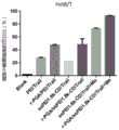
图12为根据本发明实验实例中的共载的可降解纳米载体(SSPEI-beta-CD/Mo/pDNA)和包裹了负电荷的共载可降解纳米载体纳米载体(γ-PGA/SSPEI-beta-CD/Mo/pDNA)等的细胞摄取效果图。其中,图12(A)为在Hela细胞上,PGA/SSPEI-beta-CD/Mo/pDNA被摄取后,pDNA在细胞内的分布(染料为YoYo-1,绿色);图12(B)左为在HCT8/T细胞上,γ-PGA/SSPEI-beta-CD/Mo/pDNA被摄取后,pDNA在细胞内的分布(染料为YoYo-1,绿色);右为同一细胞上,疏水药物(荧光染料为C6,绿色)在细胞内的分布。Fig. 12 is the co-loaded degradable nanocarrier (SSPEI-beta-CD/Mo/pDNA) according to the experimental example of the present invention and the co-loaded degradable nanocarrier nanocarrier (γ-PGA/SSPEI-beta - Effect diagram of cellular uptake of CD/Mo/pDNA). Among them, Figure 12 (A) is the distribution of pDNA in the cell after PGA/SSPEI-beta-CD/Mo/pDNA is taken up on Hela cells (the dye is YoYo-1, green); Figure 12 (B) left On HCT8/T cells, after γ-PGA/SSPEI-beta-CD/Mo/pDNA is taken up, the distribution of pDNA in the cells (the dye is YoYo-1, green); the right is on the same cell, the hydrophobic drug ( Fluorescent dye C6, green) intracellular distribution.
图13为根据本发明实验实例中的共载的可降解纳米载体(γ-PGA/SSPEI-beta-CD/Mo/pDNA)的细胞摄取抑制试验;其中,右图为细胞给予纳米粒前分别加入5ug,2ug,1ug,0ugγ-PGA组和空白组(不给予纳米粒)后,细胞内pDNA(YoYo-1)的流式荧光强度检测;左图为右图的定量分析。Figure 13 is the cell uptake inhibition test of the co-loaded degradable nanocarrier (γ-PGA/SSPEI-beta-CD/Mo/pDNA) according to the experimental example of the present invention; wherein, the right figure shows that the cells were added before the nanoparticles were given After 5ug, 2ug, 1ug, 0ugγ-PGA group and blank group (without administration of nanoparticles), the flow cytometric fluorescence intensity detection of pDNA (YoYo-1) in cells; the left figure is the quantitative analysis of the right figure.
图14为根据本发明实验实例中共载的可降解纳米载体和包裹了负电荷的共载可降解纳米载体对Hela和Hct8/T细胞进行转染;其中,图14(A)为不同纳米复合物中pEGFP在Hela细胞的表达;图14(B)为不同纳米复合物中pEGFP在Hct8/T细胞的表达;图14(C)为图14(A)中转染荧光强度的定量。Figure 14 is a co-loaded degradable nanocarrier according to an experimental example of the present invention and a negatively charged co-loaded degradable nanocarrier that transfects Hela and Hct8/T cells; wherein, Figure 14 (A) is a different nanocomposite The expression of pEGFP in Hela cells; Figure 14(B) is the expression of pEGFP in different nanocomplexes in Hct8/T cells; Figure 14(C) is the quantification of transfection fluorescence intensity in Figure 14(A).
图15为根据本发明实验实例中共载药物的可降解纳米载体等对肿瘤细胞的抑制作用(MTT法);其中,图15(A)为材料和空载体在Hela细胞上的毒性考察,PEI25k为毒性对照;图15(B)为材料和空载体在Hct8/T细胞上的毒性考察,PEI25k为毒性对照;图15(C)为不同纳米复合物在Hela细胞上的毒性实验。Figure 15 is the inhibitory effect (MTT method) on tumor cells of degradable nanocarriers loaded with drugs according to the experimental example of the present invention; wherein, Figure 15 (A) is the toxicity investigation of materials and empty carriers on Hela cells, and PEI25k is Toxicity control; Figure 15(B) is the toxicity test of materials and empty vectors on Hct8/T cells, PEI25k is the toxicity control; Figure 15(C) is the toxicity test of different nanocomposites on Hela cells.
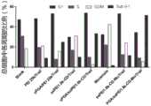
图16为根据本发明实验实例中共载药物的可降解纳米载体等对肿瘤细胞凋亡作用的测定;其中,图16(A)为不同纳米复合物在Hela细胞上的凋亡分析图;图16(B)为图16(A)的定量分析柱状图;图16(C)为不同纳米复合物在Hct8/T细胞上的凋亡分析图;图16(D)为图16(B)的定量分析柱状图。Fig. 16 is the measurement of tumor cell apoptosis by the degradable nanocarriers co-loaded with drugs according to the experimental example of the present invention; wherein, Fig. 16 (A) is the apoptosis analysis diagram of different nanocomposites on Hela cells; Fig. 16 (B) is the quantitative analysis histogram of Figure 16 (A); Figure 16 (C) is the apoptosis analysis graph of different nanocomplexes on Hct8/T cells; Figure 16 (D) is the quantitative analysis of Figure 16 (B) Analyze histograms.
图17为根据本发明实验实例中共载药物的可降解纳米载体等给予肿瘤细胞后细胞周期的测定;其中,图17(A)为不同纳米复合物处理Hct8/T细胞后,细胞周期检测;图17(B)为图17(A)中细胞周期检测的定量柱状图。Figure 17 is the determination of the cell cycle after the degradable nanocarriers loaded with drugs, etc. are given to tumor cells according to the experimental example of the present invention; wherein, Figure 17 (A) is the detection of the cell cycle after different nanocomposites are used to treat Hct8/T cells; Figure 17 17(B) is a quantitative histogram of the cell cycle assay in FIG. 17(A).
具体实施方式Detailed ways
本发明的目的在于:①提供一种集基因,化学药物输送于一体的可降解纳米载体;②开发了一种由上所述方法制备的双药共输送纳米载体。③以及这种双药共输送纳米载体在包载靶向或穿膜类型的负电荷大分子后在肿瘤治疗中的应用。不仅能减少体内非特异性互相作用,同时能把药物和基因高效地输送到肿瘤靶点部位协同发挥疗效。基于此其载药系统,本发明还优选了TRAIL基因与化学药物莫能霉素共输送联合治疗肿瘤的应用。The purpose of the present invention is to: ① provide a degradable nanocarrier integrating gene and chemical drug delivery; ② develop a double-drug co-delivery nanocarrier prepared by the above method. ③And the application of this double-drug co-delivery nanocarrier in tumor therapy after loading targeting or membrane-penetrating negatively charged macromolecules. Not only can it reduce non-specific interactions in the body, but it can also efficiently deliver drugs and genes to tumor target sites to synergistically exert curative effects. Based on the drug delivery system, the present invention also optimizes the application of co-delivery of TRAIL gene and chemical drug monensin to treat tumors.
本发明主要以可还原降解的二硫键交联的LWPEI作为主要载体材料,采用巯基化β-环糊精通过巯基交换反应进行进一步的交联得到可以同时共载基因和化学药物的纳米载体。其环糊精腔内装载疏水药物,优选为化疗药物或协同基因作用的药物,利用纳米载体所带较强正电荷静电吸附基因,优选为治疗基因质粒或者沉默RNA质粒(如siRNA),再在载药体系外层包裹修饰后的负电荷分子,提高药物靶向作用。该双载药纳米载体的粒径在100-300nm左右,首先利用外层负电荷分子主动靶向和EPR被动靶向作用到达靶组织,然后进入内体小泡,接着进入溶酶体,通过PH改变,负电荷外壳脱落,又因肿瘤细胞内高分布的谷胱甘肽酶(GSH)还原性降解交联PEI变为LWPEI,首先释放出所携带的基因药物,之后环内疏水药物缓慢释放协同发挥疗效。The present invention mainly uses reductively degradable disulfide bond cross-linked LWPEI as the main carrier material, uses mercaptolated β-cyclodextrin to carry out further cross-linking through thiol exchange reaction, and obtains a nano-carrier that can co-carry genes and chemical drugs simultaneously. The cyclodextrin cavity is loaded with hydrophobic drugs, preferably chemotherapeutic drugs or drugs that act synergistically with genes, using the strong positive charge of the nanocarrier to electrostatically adsorb genes, preferably therapeutic gene plasmids or silencing RNA plasmids (such as siRNA), and then The outer layer of the drug-loading system wraps the modified negatively charged molecules to improve the drug targeting effect. The particle size of the dual drug-loaded nanocarrier is about 100-300nm. First, it uses the active targeting of the outer negatively charged molecules and the passive targeting of the EPR to reach the target tissue, then enters the endosomal vesicle, and then enters the lysosome. Change, the negatively charged shell falls off, and because of the reductive degradation of glutathione (GSH) highly distributed in tumor cells, the cross-linked PEI becomes LWPEI, first releasing the carried gene drug, and then slowly releasing the hydrophobic drug in the ring to play a synergistic role curative effect.
本发明的可降解纳米载体的制备方法The preparation method of the degradable nanocarrier of the present invention
将相对分子质量为600或者1800的小分子聚乙烯亚胺溶解在1-10ml超纯水中形成pH较高的溶液,与二硫代二丙酸混合,比例为1:1-5。该混合液逐滴加入pH范围为4-9,优选为5-8.5的缓冲盐溶液中(所述盐溶液的浓度范围0.1-5mol/L,优选为0.5-3mol/L)混匀后,加入摩尔比为2-5倍过量EDC,NHS,反应温度范围20-50℃,优选为25-40℃,氮气保护,磁力搅拌,反应时间优选为12-48h,更优选为20-30h,形成微黄较粘稠液体。Dissolve small-molecule polyethyleneimine with a relative molecular mass of 600 or 1800 in 1-10ml of ultrapure water to form a solution with a higher pH, and mix it with dithiodipropionic acid at a ratio of 1:1-5. The mixed solution is added dropwise to a buffered saline solution with a pH range of 4-9, preferably 5-8.5 (the concentration range of the saline solution is 0.1-5mol/L, preferably 0.5-3mol/L), and after mixing, add The molar ratio is 2-5 times excess EDC, NHS, the reaction temperature range is 20-50°C, preferably 25-40°C, nitrogen protection, magnetic stirring, the reaction time is preferably 12-48h, more preferably 20-30h, forming micro Yellow viscous liquid.
将上述微黄较粘稠液体于2k-8k的透析袋内,透析溶液一开始选用30-70mM的NaCl溶液透析,随后RO水彻底透析2-5天,得到产物SSPEI,冷冻干燥成干粉低温保存。Put the above yellowish viscous liquid in a dialysis bag of 2k-8k. The dialysis solution is initially dialyzed with 30-70mM NaCl solution, and then RO water is thoroughly dialyzed for 2-5 days to obtain the product SSPEI, which is freeze-dried into a dry powder and stored at low temperature. .
将产物SSPEI溶于1-10ml pH 5-9的含EDTA的磷酸盐缓冲液中(盐溶液的浓度范围0.1-0.5mol/L),并用过量的三(2-羰基乙基)磷盐酸盐处理1h,切断产品中的二硫键,随后加入5,5'-二硫代双(2-硝基苯甲酸)(DTNB)反应,转子搅拌,反应避光,持续时间为1-3h。得到的溶液在RO水中彻底透析2天除去多余的TNB和盐类并冷冻干燥。将得到的干粉再次溶于pH在5-9的含EDTA缓冲盐溶液(盐溶液的浓度范围0.1-0.5mol/L)中,β环糊精溶于DMSO中,逐滴加入体系,再加入优选为10-100ul的三乙胺溶液进入反应体系,反应避光,反应时间优选为4-16h,得到β环糊精内嵌的聚合大分子聚乙烯亚胺-β环糊精(SSPEI-beta-CD)纳米载体溶液。得到的纳米载体SSPEI-beta-CD溶液于2k-8k透析袋内彻底透析2-5天,随后冷冻干燥后低温保存。The product SSPEI was dissolved in 1-10ml pH 5-9 containing EDTA-containing phosphate buffer (the concentration range of the salt solution was 0.1-0.5mol/L), and was added with an excess of three (2-carbonyl ethyl) phosphate hydrochloride Treat for 1 hour to cut off the disulfide bonds in the product, then add 5,5'-dithiobis(2-nitrobenzoic acid) (DTNB) to react, stir with the rotor, and keep the reaction away from light for 1-3 hours. The resulting solution was thoroughly dialyzed in RO water for 2 days to remove excess TNB and salts and freeze-dried. Dissolve the obtained dry powder again in EDTA-containing buffered saline solution with a pH of 5-9 (the concentration range of the saline solution is 0.1-0.5mol/L), dissolve β-cyclodextrin in DMSO, add the system dropwise, and then add the preferred The triethylamine solution of 10-100ul enters the reaction system, and the reaction is protected from light. The reaction time is preferably 4-16h, and the polymerized macromolecular polyethyleneimine-beta-cyclodextrin (SSPEI-beta-cyclodextrin) embedded in the beta-cyclodextrin is obtained. CD) Nanocarrier solution. The obtained nanocarrier SSPEI-beta-CD solution is thoroughly dialyzed in a 2k-8k dialysis bag for 2-5 days, then freeze-dried and stored at low temperature.
使用2-10倍摩尔比过量的疏水性药物,优选地,所述药物为自阿霉素、紫杉醇、瑞戈非尼、吉非替尼、莫能霉素中的一种。用优选为50-200ul的有机相溶解后加入纳米载体水溶液中制成药物储备液在磁子搅拌的过程中逐滴加入到反应体系中。黑暗下磁力搅拌,反应时间3-15h,优选时间为4-8h,随后离心4000-8000转,去除微量沉淀,收集上清液体,过0.45水系滤膜,随后冻干,获得载有疏水药的纳米载体干粉。A 2-10 times molar excess of the hydrophobic drug is used, preferably, the drug is one of adriamycin, paclitaxel, regorafenib, gefitinib, and monensin. Dissolve the organic phase preferably in the range of 50-200 ul and then add it to the nanocarrier aqueous solution to make a drug stock solution, and add it dropwise to the reaction system during magnetic stirring. Magnetic stirring in the dark, the reaction time is 3-15 hours, preferably 4-8 hours, then centrifuged at 4000-8000 rpm to remove trace precipitates, collect the supernatant liquid, pass through a 0.45 water filter membrane, and then freeze-dry to obtain hydrophobic drug-loaded Nano-carrier dry powder.
将上述载药的纳米载体溶于超纯水中,可降解疏水药载体与DNA的质量比例为0.5-50:1,所述的DNA优选为环状质粒和抑制类发夹RNA(shRNA),如报告基因增强型绿色荧光蛋白(EGFP)质粒,治疗型基因如P53,TRAIL或沉默RNA;将一定体积的DNA以涡旋的方式逐滴加入到等体积的载体溶液中,涡旋时间优选为10-20s,孵育时间为20-40分钟,室温反应,得到携载基因和药物的纳米载体。Dissolving the above drug-loaded nano-carrier in ultrapure water, the mass ratio of the degradable hydrophobic drug carrier to DNA is 0.5-50:1, and the DNA is preferably a circular plasmid and an inhibitory hairpin RNA (shRNA), Such as reporter gene enhanced green fluorescent protein (EGFP) plasmid, therapeutic gene such as P53, TRAIL or silencing RNA; a certain volume of DNA is added dropwise to an equal volume of carrier solution in a vortex manner, and the vortex time is preferably 10-20s, the incubation time is 20-40 minutes, and react at room temperature to obtain nanocarriers carrying genes and drugs.
上述携载基因和药物的纳米载体通过静电吸附,与包裹负电荷分子结合,负电荷分子优选为10-100KDa的天然聚合物(如γ聚谷氨酸,聚精氨酸)或者为10-100KDa的糖类大分子(如透明质酸);带负电荷的分子溶于水溶液,体积与携载基因和药物的纳米载体溶液相同,并将纳米载体溶液逐滴涡旋加入到负电荷分子水溶液中,涡旋时间优选为3-10s,更优选为5-8s,室温下静置孵育15-30分钟。上述携载基因和药物的纳米载体复合物冷冻干燥成冻干粉,长期低温保存。The above-mentioned nano-carriers carrying genes and drugs are combined with the packaged negatively charged molecules through electrostatic adsorption, and the negatively charged molecules are preferably 10-100KDa natural polymers (such as gamma polyglutamic acid, polyarginine) or 10-100KDa Sugar macromolecules (such as hyaluronic acid); negatively charged molecules are dissolved in aqueous solution, the volume is the same as the nanocarrier solution carrying genes and drugs, and the nanocarrier solution is vortexed drop by drop into the aqueous solution of negatively charged molecules , The vortex time is preferably 3-10s, more preferably 5-8s, and incubated at room temperature for 15-30 minutes. The above-mentioned nano-carrier complex carrying genes and drugs is freeze-dried into freeze-dried powder, and stored at low temperature for a long time.
多肽或其他小分子修饰负电荷分子的方法Method for modifying negatively charged molecules by peptides or other small molecules
以γ聚谷氨酸(γ-PGA)上修饰RGD环肽为实例,具体步骤如下:Taking the modified RGD cyclic peptide on γ-polyglutamic acid (γ-PGA) as an example, the specific steps are as follows:
称取50mg的γ-PGA溶解于5ml的RO水中,称取0.5mg和0.7mg的NHS和EDC,溶解后加入到γ-PGA溶液中,反应转子搅拌,室温活化1h。称取7.5mg的c(RGDfK)溶于RO水中,待溶解完全后,将c(RGDfK)逐滴加入到活化后的γ-PGA溶液内,调节PH至6.5-8.5范围内,室温搅拌5-10h,反应终止。产物通过FPLC分子筛进行纯化,检测波长为280nm,流速为1ml/min。首先将Desalting和FPLC进行连接,使用0.1M磷酸盐缓冲液(pH7.2)作为流动相平衡柱子约4-5个柱体积,即40-50ml,然后将反应产物注入到进样环中,将紫外基线调为零后以1ml/min的速度样品进入到Desalting中,约3min后通过程序设定仪器收集洗脱出来的紫外峰,等待紫外基线恢复到零后,依次进行下一针的纯化。即获得修饰了RGD的γ-PGA。Weigh 50mg of γ-PGA and dissolve it in 5ml of RO water, weigh 0.5mg and 0.7mg of NHS and EDC, dissolve and add to the γ-PGA solution, stir with the reaction rotor, and activate at room temperature for 1h. Weigh 7.5 mg of c(RGDfK) and dissolve it in RO water. After the dissolution is complete, add c(RGDfK) dropwise to the activated γ-PGA solution, adjust the pH to 6.5-8.5, and stir at room temperature for 5- 10h, the reaction was terminated. The product was purified by FPLC molecular sieve, the detection wavelength was 280nm, and the flow rate was 1ml/min. First connect Desalting and FPLC, use 0.1M phosphate buffer (pH7.2) as the mobile phase to equilibrate the column for about 4-5 column volumes, that is, 40-50ml, then inject the reaction product into the injection loop, and After the UV baseline is adjusted to zero, the sample enters the Desalting at a speed of 1ml/min. After about 3 minutes, the eluted UV peak is collected by the program setting instrument. After the UV baseline returns to zero, the purification of the next injection is carried out in sequence. That is, γ-PGA modified with RGD was obtained.
本发明的主要优点Main advantages of the invention
(1)合成的可降解纳米载体具有良好的稳定性,可以同时共载基因和疏水药物,原料价格低廉,无需专门设备制作,操作简单。(1) The synthesized degradable nano-carrier has good stability, can co-carry genes and hydrophobic drugs at the same time, the raw material is cheap, no special equipment is required for production, and the operation is simple.
(2)聚合的PEI通过可还原性降解生成LWPEI毒性低,避免了传统阳离子载体PEI25k不能降解所带来的毒性和副作用,环糊精也具有可降解性,故空载的可降解纳米载体对细胞毒性低,安全可靠。(2) Polymerized PEI is reductively degraded to generate LWPEI with low toxicity, which avoids the toxicity and side effects caused by the inability of the traditional cationic carrier PEI25k to degrade. Low cytotoxicity, safe and reliable.
(3)合成的可降解纳米载体搭载疏水药物,不仅可以提高难溶性药物的溶解度,还可以降低其引起的毒副反应。(3) The synthesized degradable nanocarriers carry hydrophobic drugs, which can not only improve the solubility of poorly soluble drugs, but also reduce the toxic and side effects caused by them.
(4)最外层包裹的负电荷分子可以选用pH响应的智能材料如γ-PGA或者是可在靶组织的特定环境中降解的透明质酸,使得可降解纳米载体所载疏水药物释放具有智能的pH响应性或酶响应性。(4) The negatively charged molecules wrapped in the outermost layer can be selected from pH-responsive smart materials such as γ-PGA or hyaluronic acid that can be degraded in the specific environment of the target tissue, making the release of hydrophobic drugs carried by degradable nanocarriers intelligent. pH-responsive or enzyme-responsive.
(5)纳米载体对所载基因具有保护作用,外层负电荷分子屏蔽血液环境中被网状内皮细胞的捕获,共载体系靶向到达肿瘤细胞后能有效被摄取,siRNA沉默目的基因表达,DNA得到有效转录,表达目的基因。(5) The nanocarrier has a protective effect on the contained genes, the outer layer of negatively charged molecules shields the capture of the reticuloendothelial cells in the blood environment, the co-carrier system can be effectively taken up after reaching the tumor cells, and the siRNA silences the expression of the target gene, The DNA is efficiently transcribed and the target gene is expressed.
(5)共载基因和疏水药物,可以使用有协同效应,能相互作用药物,降低化疗药物的剂量的同时亦可达到增效减毒的效果,能有望实现逆转肿瘤多药耐药性。(5) Co-carrying genes and hydrophobic drugs can use drugs that have synergistic effects and can interact with each other. While reducing the dose of chemotherapy drugs, it can also achieve the effect of synergistic and attenuating toxicity, and it is expected to achieve the reversal of tumor multidrug resistance.
(6)合成的可降解纳米载体不仅能作为药物和基因共输送的载体,而且还能搭载超声成像的造影剂或荧光小分子探针用于成像。(6) The synthesized degradable nanocarriers can not only serve as carriers for co-delivery of drugs and genes, but also carry contrast agents or fluorescent small molecule probes for ultrasound imaging for imaging.
(7)本发明结合实例,联合使用治疗基因TRAIL和聚醚类离子载体莫能霉素,利用莫能霉素使细胞ROS水平增高,DR5的mRNA和蛋白表达增多,协助TRAIL发挥作用,共同对抗肿瘤。(7) The present invention is combined with an example, using the therapeutic gene TRAIL and the polyether ionophore monensin in combination, using monensin to increase the level of cellular ROS, increase the expression of mRNA and protein of DR5, assist TRAIL to play a role, and jointly fight against tumor.
下面结合具体实施例,进一步阐述本发明。应理解,这些实施例仅用于说明本发明而不用于限制本发明的范围。下列实施例中未注明具体条件的实验方法,通常按照常规条件,或按照制造厂商所建议的条件。除非另外说明,否则百分比和份数按重量计算。Below in conjunction with specific embodiment, further illustrate the present invention. It should be understood that these examples are only used to illustrate the present invention and are not intended to limit the scope of the present invention. For the experimental methods without specific conditions indicated in the following examples, the conventional conditions or the conditions suggested by the manufacturer are usually followed. Percentages and parts are by weight unless otherwise indicated.
试剂和药品Reagents and Medicines
小分子枝状聚乙烯亚胺(PEI1800k)(Alter listur公司);3,3'-二硫代二丙酸(国药集团化学试剂有限公司);二硫双(琥珀酰亚胺基丙酸盐)(美国ProteoChem公司);二水磷酸二氢钠、十二水磷酸氢二钠、氯化钠、氯化钾、盐酸、氢氧化钠、乙二胺四乙酸二钠、琼脂糖(国药集团化学试剂有限公司);MES试剂(Sigma-Aldrich,美国);DTNB(Sigma-Aldrich,美国);三(2-羰基乙基)磷盐酸盐(百灵威科技有限公司):七(6-巯基-6-去氧)-β-环糊精(CAS:160661-60-9,山东滨州智源生物科技有限公司);莫能霉素标准品(Monensin,Mo)(大连美仑生物技术有限公司);c(RGDfK)(上海丽昂化学有限公司);γ-聚谷氨(γ-PGA)(Mw20kD)(南京赛泰斯有限公司)3-(4,5-二甲基噻唑-2)-2,5-二苯基四氮唑溴盐(MTT),Hoechest 33258,3500mw透析袋(Sigma-Aldrich,美国);透析袋MD24(Spectrum美国);DNA酶(上海捷瑞生物工程有限公司);1640细胞培养基(Gibco,Invitrogen,USA);澳洲血源胎牛血清(上海熠晨生物有限公司);0.25%胰蛋白酶(上海碧云天生物技术有限公司);Gel-red(BiotiumHayward,USA);二甲基亚砜、N,N-二甲基甲酰胺(国药集团化学试剂有限公司);pTRAIL,pEGFP和pDsRFP等质粒在DH5alpha大肠杆菌菌株中扩增,无内毒素质粒抽提试剂盒Mega(Qiagen,Hilden,Germany)抽提。Small molecule dendritic polyethyleneimine (PEI1800k) (Alter listur company); 3,3'-dithiodipropionic acid (Sinopharm Chemical Reagent Co., Ltd.); dithiobis(succinimidyl propionate) (U.S. ProteoChem Company); sodium dihydrogen phosphate dihydrate, disodium hydrogen phosphate dodecahydrate, sodium chloride, potassium chloride, hydrochloric acid, sodium hydroxide, disodium edetate, agarose (Sinopharm Chemical Reagent Ltd.); MES reagent (Sigma-Aldrich, USA); DTNB (Sigma-Aldrich, USA); tris(2-carbonylethyl)phosphonium hydrochloride (Bailingwei Technology Co., Ltd.): seven (6-mercapto-6- Deoxy)-β-cyclodextrin (CAS: 160661-60-9, Shandong Binzhou Zhiyuan Biotechnology Co., Ltd.); monensin standard (Monensin, Mo) (Dalian Meilun Biotechnology Co., Ltd.); c (RGDfK) (Shanghai Lyon Chemical Co., Ltd.); γ-polyglutamic acid (γ-PGA) (Mw20kD) (Nanjing Saitaisi Co., Ltd.) 5-diphenyltetrazolium bromide (MTT), Hoechest 33258, 3500mw dialysis bag (Sigma-Aldrich, USA); dialysis bag MD24 (Spectrum USA); DNase (Shanghai Jierui Bioengineering Co., Ltd.); 1640 cells Culture medium (Gibco, Invitrogen, USA); Australian blood-derived fetal bovine serum (Shanghai Yichen Biotechnology Co., Ltd.); 0.25% trypsin (Shanghai Biyuntian Biotechnology Co., Ltd.); Gel-red (Biotium Hayward, USA); sulfoxide, N, N-dimethylformamide (Sinopharm Chemical Reagent Co., Ltd.); pTRAIL, pEGFP and pDsRFP and other plasmids were amplified in DH5alpha E. coli strains, and the endotoxin-free plasmid extraction kit Mega (Qiagen, Hilden, Germany) extraction.
基因与药物共输送的可降解纳米载体Degradable nanocarriers for gene-drug co-delivery
(1)共载基因和药物的可降解纳米载体由小分子PEI与巯基β-环糊精通过二硫键交联聚合而成,环糊精疏水腔内部装载协同TRAIL基因作用的蛋白质转运抑制剂莫能霉素,通过正电性吸附逆转抗癌基因质粒TRAIL。合成示意图如图1(A)所示,抗肿瘤作用如图1(B)所示。(1) The degradable nanocarrier co-carrying genes and drugs is made of small molecule PEI and mercapto β-cyclodextrin cross-linked and polymerized through disulfide bonds, and the hydrophobic cavity of cyclodextrin is loaded with protein transport inhibitors that cooperate with the TRAIL gene Monensin, reverses the anticancer gene plasmid TRAIL by electropositive adsorption. The schematic diagram of the synthesis is shown in Figure 1(A), and the antitumor effect is shown in Figure 1(B).
(2)该共输送纳米载体粒径大小在100-300nm范围内,呈均一球形,在水溶液状态下分散良好。(2) The particle size of the co-transported nano-carrier is in the range of 100-300nm, is uniform spherical, and is well dispersed in the aqueous solution state.
(3)纳米载体通过EPR效应和外层靶向负分子双向靶向肿瘤部位,(3) The nanocarriers bidirectionally target the tumor site through the EPR effect and the outer layer targeting negative molecules,
到达肿瘤组织后,有效被肿瘤细胞摄取,进入溶酶体后在PH较低的情况下,外层负电分子γ-PGA首先改变二级结构与载体分离,接着PEI与γ-PGA共同的质子海绵效应使溶酶体涨破,胞内高浓度GSH降解载体释放出所携载基因,治疗基因TRAIL表达,莫能霉素也从环内释放,二者协同发挥相应疗效。After reaching the tumor tissue, it is effectively taken up by tumor cells. After entering the lysosome at a low pH, the outer negatively charged molecule γ-PGA first changes the secondary structure and separates from the carrier, and then PEI and γ-PGA jointly form a proton sponge The effect causes the lysosome to burst, the high-concentration GSH degradation carrier in the cell releases the carried gene, the therapeutic gene TRAIL is expressed, and monensin is also released from the ring, and the two synergistically exert the corresponding curative effect.
制备实施例1:共载药可降解纳米体系的制备Preparation Example 1: Preparation of co-loaded degradable nanosystems
1.小分子聚乙酰亚胺和环糊精交联的复合物(SSPEI-beta-CD)的制备1. Preparation of the complex (SSPEI-beta-CD) cross-linked by small molecule polyethylimide and cyclodextrin
将相对分子质量为1800的小分子聚乙烯亚胺溶解在3ml超纯水中形成PH较高的溶液,与二硫代二丙酸混合,比例为1:1-5。该混合液逐滴加入MES缓冲液(浓度范围0.1-5mol/L,优选为0.5-3mol/L)混匀后,加入摩尔比为2.5倍过量EDC,NHS,35℃反应,氮气保护,磁力搅拌,反应时间为30h,形成微黄较粘稠液体;将液体于3500k的透析袋内,透析溶液一开始选用30-70mM的NaCl溶液透析,随后RO水彻底透析2-5天,得到产物SSPEI,冷冻干燥成干粉低温保存。Dissolve small-molecule polyethyleneimine with a relative molecular mass of 1800 in 3ml ultrapure water to form a solution with a higher pH, and mix it with dithiodipropionic acid at a ratio of 1:1-5. Add the mixed solution dropwise to MES buffer solution (concentration range 0.1-5mol/L, preferably 0.5-3mol/L) and mix well, then add 2.5 times excess EDC, NHS, react at 35°C, nitrogen protection, magnetic stirring , the reaction time is 30h, and a yellowish viscous liquid is formed; the liquid is placed in a 3500k dialysis bag, and the dialysis solution is initially dialyzed with 30-70mM NaCl solution, and then RO water is thoroughly dialyzed for 2-5 days to obtain the product SSPEI. Freeze-dried into dry powder and stored at low temperature.
得到的产品SSPEI溶于4ml pH为8的含EDTA的0.1M磷酸钠缓冲液中,并与等体积的过量三(2-羰基乙基)磷盐酸盐溶液混合反应1h,切断产品中的二硫键(无需除去,不影响下一部反应),随后加入2倍摩尔比过量的5,5'-二硫代双(2-硝基苯甲酸)(DTNB)反应,转子搅拌,反应避光,反应1h。得到的溶液在RO水中彻底透析2天除去多余的TNB和盐类并冷冻干燥。将得到的干粉再次溶于pH为8的含EDTA缓冲盐溶液(盐溶液的浓度范围0.1-0.5mol/L)中,β环糊精溶于100ul DMSO中,逐滴加入体系,再加入50ul的三乙胺溶液进入反应体系,反应避光,反应时间优选为4-16h,得到β环糊精内嵌的聚合大分子聚乙烯亚胺-β环糊精(SSPEI-beta-CD)纳米载体溶液。得到的纳米载体SSPEI-beta-CD溶液于MWCO为7000透析袋内彻底透析2-5天,随后冷冻干燥后低温保存。The product SSPEI that obtains is dissolved in the 0.1M sodium phosphate buffer solution that contains EDTA that 4ml pH is 8, and with the excessive three (2-carbonyl ethyl) phosphorous hydrochloride solution mixing reaction 1h of equal volume, cut off the dioxane in the product Sulfur bond (does not need to be removed, does not affect the next reaction), then add 5,5'-dithiobis(2-nitrobenzoic acid) (DTNB) in excess of 2 times the molar ratio to react, the rotor stirs, and the reaction is protected from light , reaction 1h. The resulting solution was thoroughly dialyzed in RO water for 2 days to remove excess TNB and salts and freeze-dried. Dissolve the obtained dry powder again in an EDTA-containing buffered saline solution with a pH of 8 (the concentration range of the saline solution is 0.1-0.5mol/L), dissolve β-cyclodextrin in 100ul DMSO, add dropwise to the system, and then add 50ul of The triethylamine solution enters the reaction system, the reaction is protected from light, and the reaction time is preferably 4-16h to obtain a polymeric macromolecular polyethyleneimine-β-cyclodextrin (SSPEI-beta-CD) nanocarrier solution embedded in β-cyclodextrin . The obtained nano-carrier SSPEI-beta-CD solution was thoroughly dialyzed in a dialysis bag with a MWCO of 7000 for 2-5 days, then freeze-dried and stored at low temperature.
2.双载药纳米载体(SSPEI-beta-CD-Mo/pDNA)的制备2. Preparation of dual drug-loaded nanocarriers (SSPEI-beta-CD-Mo/pDNA)
莫能霉素3倍摩尔比过量,用200ul的甲醇溶解后在磁子搅拌的过程中逐滴加入纳米载体水溶液中制成药物储备液。黑暗下磁力搅拌,反应时间4h,随后离心5000转,去除微量沉淀,收集上清液体,过0.45水系滤膜,随后冻干,获得载有疏水药的纳米载体干粉。The molar ratio of monensin is 3 times excessive, dissolved in 200 ul of methanol, and then added dropwise to the nanocarrier aqueous solution during magnetic stirring to prepare a drug stock solution. Stir magnetically in the dark, react for 4 hours, then centrifuge at 5,000 rpm to remove trace precipitates, collect the supernatant, pass through a 0.45 water filter membrane, and then freeze-dry to obtain a nanocarrier dry powder loaded with hydrophobic drugs.
将上述载药的纳米载体溶于超纯水中配制成1mg/ml的储备液,可降解疏水药载体与质粒DNA(pDNA)的质量比例为20:1;将一定体积的基因以涡旋的方式逐滴加入到等体积的载体溶液中,涡旋时间15s,孵育30分钟,室温反应,得到携载基因和药物的纳米载体。The above drug-loaded nanocarriers were dissolved in ultrapure water to prepare a stock solution of 1 mg/ml, and the mass ratio of the degradable hydrophobic drug carrier to plasmid DNA (pDNA) was 20:1; a certain volume of genes was vortexed Add it dropwise to an equal volume of carrier solution, vortex for 15 seconds, incubate for 30 minutes, and react at room temperature to obtain nanocarriers carrying genes and drugs.
3.小分子靶向肽c(RGDfK)修饰的γ-PGA的合成3. Synthesis of small molecule targeting peptide c (RGDfK) modified γ-PGA
称取50mg的γ-PGA溶解于5ml的RO水中,称取0.5mg和0.7mg的NHS和EDC,溶解后加入到γ-PGA溶液中,反应转子搅拌,室温活化1h。称取7.5mg的c(RGDfK)溶于RO水中,待溶解完全后,将c(RGDfK)逐滴加入到活化后的γ-PGA溶液内,调节pH至6.5-8.5范围内,室温搅拌5h,反应终止。产物通过FPLC分子筛进行纯化,检测波长为210nm,流速为1ml/min。首先将Desalting和FPLC进行连接,使用0.1M磷酸盐缓冲液(pH7.2)作为流动相平衡柱子约4-5个柱体积,即40-50ml,然后将反应产物注入到进样环中,将紫外基线调为零后以1ml/min的速度样品进入到Desalting中,约3min后通过程序设定仪器收集洗脱出来的紫外峰,等待紫外基线恢复到零后,依次进行下一针的纯化。即获得有修饰了RGD的γ-PGA。冷冻干燥后4度长期保存,并配制成1mg/ml的溶液备用。Weigh 50mg of γ-PGA and dissolve it in 5ml of RO water, weigh 0.5mg and 0.7mg of NHS and EDC, dissolve and add to the γ-PGA solution, stir with the reaction rotor, and activate at room temperature for 1h. Weigh 7.5 mg of c(RGDfK) and dissolve it in RO water. After the dissolution is complete, add c(RGDfK) dropwise into the activated γ-PGA solution, adjust the pH to 6.5-8.5, and stir at room temperature for 5 hours. The reaction was terminated. The product was purified by FPLC molecular sieve, the detection wavelength was 210nm, and the flow rate was 1ml/min. First connect Desalting and FPLC, use 0.1M phosphate buffer (pH7.2) as the mobile phase to equilibrate the column for about 4-5 column volumes, that is, 40-50ml, then inject the reaction product into the injection loop, and After the UV baseline is adjusted to zero, the sample enters the Desalting at a speed of 1ml/min. After about 3 minutes, the eluted UV peak is collected by the program setting instrument. After the UV baseline returns to zero, the purification of the next injection is carried out in sequence. That is, γ-PGA modified with RGD was obtained. Store at 4°C for a long time after freeze-drying, and prepare a 1mg/ml solution for future use.
4.包裹γ-PGA的双载药纳米载体(rPGA/SSPEI-beta-CD-Mo/pDNA)的制备4. Preparation of dual drug-loaded nanocarriers (rPGA/SSPEI-beta-CD-Mo/pDNA) encapsulated with γ-PGA
上述携载基因和药物的纳米载体通过静电吸附,与包裹负电荷分子γ-PGA结合;带负电荷的分子溶于水溶液配制成0.4mg/ml的溶液,以相同体积与携载基因和药物的纳米载体溶液混合,涡旋5-8s,室温下静置孵育25分钟。将获得的包裹γ-PGA的双载药纳米载体(rPGA/SSPEI-beta-CD-Mo/pDNA)冷冻干燥成冻干粉,长期低温保存。The above-mentioned nano-carriers carrying genes and drugs are combined with the negatively charged molecule γ-PGA through electrostatic adsorption; The nanocarrier solution was mixed, vortexed for 5-8s, and incubated at room temperature for 25 minutes. The obtained double drug-loaded nanocarrier (rPGA/SSPEI-beta-CD-Mo/pDNA) wrapped with γ-PGA was lyophilized into a lyophilized powder and stored at low temperature for a long time.
测试实施例1:双载药可降解纳米载体(SSPEI-beta-CD-Mo/pDNA)和包裹γ-PGA的双载药可降解纳米载体(rPGA/SSPEI-beta-CD-Mo/pDNA)的体外表征实验Test Example 1: Double drug-loaded degradable nanocarriers (SSPEI-beta-CD-Mo/pDNA) and double-loaded drug-degradable nanocarriers (rPGA/SSPEI-beta-CD-Mo/pDNA) encapsulating γ-PGA In vitro characterization experiments
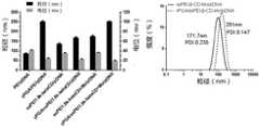
(1)粒径和电位(1) Particle size and potential
取基因质量为10ug pEGFP质粒的载莫能霉素纳米载体和包裹γ-PGA的双载药可降解纳米载体等,用纯化水稀释到终体积为1ml,使用马尔文粒径仪进行粒径和电位的测定,结果如图7所示。Take the monensin-loaded nanocarriers with a gene quality of 10ug pEGFP plasmid and the double-loaded degradable nanocarriers wrapped with γ-PGA, etc., dilute with purified water to a final volume of 1ml, and use a Malvern particle sizer to measure the particle size and The results of the potential measurement are shown in Figure 7.
结果:随着组成的增多和药物的搭载,纳米复合物的粒径逐渐增大,电位由正负电荷材料交替吸附而显现上下动荡,客观上说明纳米载体合成的成功。Results: With the increase of composition and loading of drugs, the particle size of the nanocomposite gradually increased, and the potential fluctuated up and down due to the alternate adsorption of positive and negative charged materials, which objectively indicated the success of the synthesis of nanocarriers.
(2)TEM(2)TEM
将包裹γ-PGA的双载药可降解纳米载体和双载药可降解纳米载体分别滴加到预先水化后的铜网上,2min后吸干,滴加醋酸釉负染1min后50℃加热灯照射20分钟晾干。采用透射电镜进行测定分析,rPGA/SSPEI-beta-CD-Mo/pDNA的分析结果如图2所示。Add the double drug-loaded degradable nanocarriers wrapped with γ-PGA and the double drug-loaded degradable nanocarriers respectively onto the pre-hydrated copper grid, blot dry after 2 minutes, add acetic acid glaze for 1 minute and then heat the lamp at 50°C Let dry for 20 minutes. Measurement and analysis were carried out by transmission electron microscopy, and the analysis results of rPGA/SSPEI-beta-CD-Mo/pDNA are shown in FIG. 2 .
结果:粒度仪测定的是水合粒径,TEM能看出纳米载体的实际尺寸。从图2中可以看出包裹γ-PGA的双载药可降解纳米载体为形态约为100nm左右均一的球形。Results: The particle size analyzer measures the hydrated particle size, and the actual size of the nanocarrier can be seen by TEM. It can be seen from Figure 2 that the double drug-loaded degradable nanocarrier wrapped with γ-PGA is a spherical shape with a uniform shape of about 100 nm.
(3)琼脂糖凝胶电泳阻滞实验(3) Agarose gel electrophoresis retardation experiment
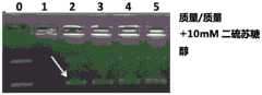
利用琼脂糖凝胶电泳实验考察不同比例纳米载体与固定质量的质粒DNA(pDNA)作用的状态。称取0.8g琼脂糖于250ml锥形瓶中,加入100ml 1×TEA缓冲液中,微波高火加热5min至琼脂糖完全溶解,制成0.8%琼脂糖凝胶溶液,待冷却至60℃左右,按1000x稀释方法加入gel-red混合均匀,倒入制胶槽中,插上电泳梳,室温静置30min至琼脂糖凝胶完全凝固后轻轻拔去电泳梳。将装有凝胶的小槽放入电泳槽中,槽中为1×TEA缓冲液。缓冲液浸没凝胶。取新鲜制备的复合物与上样loading混合均匀,每孔20ul样品跑电泳。电泳电压为80V、时间45min,电泳结束后置于凝胶成像系统进行分析,观察裸EGFP(pDNA)被复合物包裹的情况,结果如图3所示。Using agarose gel electrophoresis experiment to investigate the state of interaction between different proportions of nanocarriers and fixed mass of plasmid DNA (pDNA). Weigh 0.8g of agarose into a 250ml Erlenmeyer flask, add 100ml of 1×TEA buffer solution, heat in microwave for 5 minutes until the agarose is completely dissolved, and make a 0.8% agarose gel solution, wait to cool to about 60°C, According to the 1000x dilution method, add gel-red and mix evenly, pour it into the gel-making tank, insert the electrophoresis comb, and let it stand at room temperature for 30 minutes until the agarose gel is completely solidified, then gently unplug the electrophoresis comb. Put the small tank containing the gel into the electrophoresis tank, which is 1×TEA buffer. Buffer submerges the gel. Take the freshly prepared complex and mix it evenly with the loading sample, and run electrophoresis with 20ul samples per well. The electrophoresis voltage was 80V, and the time was 45min. After the electrophoresis, it was placed in a gel imaging system for analysis, and the condition of the naked EGFP (pDNA) being wrapped by the complex was observed. The results are shown in Figure 3.
第1道:裸EGFPLane 1: Naked EGFP
第2道:SSPEI-β-CD/pDNA 1:1Lane 2: SSPEI-β-CD/pDNA 1:1
第3道:SSPEI-β-CD/pDNA 5:1Lane 3: SSPEI-β-CD/pDNA 5:1
第4道:SSPEI-β-CD/pDNA 10:1Lane 4: SSPEI-β-CD/pDNA 10:1
第5道:SSPEI-β-CD/pDNA 50:1Lane 5: SSPEI-β-CD/pDNA 50:1
第6道:SSPEI-β-CD/pDNA 100:1Lane 6: SSPEI-β-CD/pDNA 100:1
确定了质粒和纳米载体的质量比后,为观察纳米载体可降解性,将新鲜的pDNA,γ-PGA/PEI25k/pDNA,SSPEI-beta-CD/pDNA,SSPEI-beta-CD-Mo/pDNA和γ-PGA/SSPEI-beta-CD-Mo/pDNA纳米载体与终浓度为15mM的DTT等体积混合。放置于摇床上100r,在37℃下孵育2h。随后,与上样loading混合均匀,每孔20ul样品跑电泳。电泳电压为80V、时间45min,电泳结束后置于凝胶成像系统进行分析,观察pDNA被纳米载体包裹的情况,结果如图5所示。After determining the mass ratio of plasmid and nanocarrier, in order to observe the degradability of nanocarrier, fresh pDNA, γ-PGA/PEI25k/pDNA, SSPEI-beta-CD/pDNA, SSPEI-beta-CD-Mo/pDNA and γ-PGA/SSPEI-beta-CD-Mo/pDNA nanocarriers were mixed with equal volumes of DTT at a final concentration of 15 mM. Place on a shaker at 100r and incubate at 37°C for 2h. Then, mix well with the loading sample, and run electrophoresis with 20ul samples per well. The electrophoresis voltage was 80V, and the time was 45min. After the electrophoresis, it was placed in a gel imaging system for analysis, and the situation of pDNA wrapped by nanocarriers was observed. The results are shown in Figure 5.
结果:材料与pDNA在质量比为1:1的时候就能够将pDNA包裹起来阻滞在孔中。为提高包裹pDNA的稳定性和转染效率并结合实验室早期试验,我们采用γ-PGA:纳米载体:pDNA为2:10:1作为最佳质量比,进行后续实验;为了确定小分子PEI和环糊精交联的纳米载体是谷胱甘肽响应的可降解材料,图5可以看出加入DTT之后,pDNA正在逐步被释放。Results: When the mass ratio of material and pDNA is 1:1, pDNA can be wrapped and blocked in the hole. In order to improve the stability and transfection efficiency of the packaged pDNA and combined with the early experiments in the laboratory, we used γ-PGA:nanocarrier:pDNA as the optimal mass ratio of 2:10:1 for follow-up experiments; in order to determine the small molecule PEI and The cyclodextrin-crosslinked nanocarrier is a glutathione-responsive degradable material. It can be seen from Figure 5 that after adding DTT, pDNA is gradually released.
第0道:裸EGFPLane 0: Naked EGFP
第1道:PEI25k/DNA3:1Lane 1: PEI25k/DNA3:1
第2道:rPGA/SSPEI-β-CD/DNA 2:10:1Lane 2: rPGA/SSPEI-β-CD/DNA 2:10:1
第3道:SSPEI-β-CD-Mo/DNA 2:10:1Lane 3: SSPEI-β-CD-Mo/DNA 2:10:1
第4道:rPGA/SSPEI-β-CD-Mo/DNA 2:10:1Lane 4: rPGA/SSPEI-β-CD-Mo/DNA 2:10:1
第5道:RGD-rPGA/SSPEI-β-CD-Mo/DNA 2:10:1Lane 5: RGD-rPGA/SSPEI-β-CD-Mo/DNA 2:10:1
(4)共载药纳米载体抗核酸酶的能力(4) Anti-nuclease ability of co-loaded nanocarriers
为了检测共载药纳米载体抗核酸酶的能力,将pDNA,SSPEI-beta-CD-Mo/pDNA和γ-PGA/SSPEI-beta-CD-Mo/pDNA纳米复合物与DNAase(1unit/μg of DNA)混合,对照组用等体积的水替代DNAase作用。样品置于37℃恒温摇床,80rpm,反应30min。然后,加入适量EDTA·2Na溶液至终浓度为500nmol/L,置室温下孵育15min终止消化,之后加入肝素钠溶液释放出纳米复合物中质粒DNA(100units/ug DNA)。取20ul样品与上样缓冲液混合,0.8%预制琼脂糖凝胶内上样,电泳条件:80V、45min,实验完毕后采用凝胶成像系统进行分析,观察DNA,结果如图4所示。In order to test the anti-nuclease ability of co-loaded nanocarriers, pDNA, SSPEI-beta-CD-Mo/pDNA and γ-PGA/SSPEI-beta-CD-Mo/pDNA nanocomplexes were mixed with DNAase (1 unit/μg of DNA ) were mixed, and the control group replaced DNAase with an equal volume of water. The sample was placed on a constant temperature shaker at 37°C, 80rpm, and reacted for 30min. Then, add an appropriate amount of EDTA·2Na solution to a final concentration of 500nmol/L, incubate at room temperature for 15min to terminate digestion, and then add heparin sodium solution to release the plasmid DNA (100units/ug DNA) in the nanocomposite. Take 20ul sample and mix it with loading buffer, load the sample in 0.8% prefabricated agarose gel, electrophoresis conditions: 80V, 45min, use gel imaging system to analyze after the experiment, and observe DNA, the result is shown in Figure 4.
结果:1为裸EGFP(pDNA),Result: 1 is naked EGFP(pDNA),
2泳道为SSPEI-beta-CD-Mo/pDNA,
3泳道为γ-PGA/SSPEI-beta-CD-Mo/pDNA;
可以发现,裸pDNA由于没有载体保护,很快被降解,2和3中的载体包裹pDNA,使得其得到了保护,不被DNase所破坏,解离纳米复合物后仍然较好的保持了pDNA的完整性。It can be found that the naked pDNA is quickly degraded because it is not protected by the carrier, and the carrier in 2 and 3 wraps the pDNA so that it is protected from being damaged by DNase, and the pDNA is still well maintained after dissociation of the nanocomplex. integrity.
(5)模拟血清蛋白测定载体吸附蛋白情况。(5) Determination of carrier-adsorbed protein by simulating serum protein.
1.制样。取含有20ug(0.2ug/ul)pEGFP的SSPEI-beta-CD-Mo/pDNA和γ-PGA/SSPEI-beta-CD-Mo/pDNA纳米复合物溶液与0.2mg/ml牛血清白蛋白溶液等体积混合,置于37℃的摇床中孵育1h,然后用超高速离心机室温100,000rpm离心2h,纳米复合物沉降在离心管底部。弃去上清,用滤纸吸收离心管壁上残余液体。将沉降的纳米复合物与30ul含有SDS和巯基乙醇的上样缓冲液混合均匀,置于沸水浴煮10min,使得纳米复合物上吸附的白蛋白解离和变性,置于-20℃保存备用。1. Sample preparation. Take SSPEI-beta-CD-Mo/pDNA and γ-PGA/SSPEI-beta-CD-Mo/pDNA nanocomplex solution containing 20ug (0.2ug/ul) pEGFP and 0.2mg/ml bovine serum albumin solution in equal volume Mix and incubate in a shaker at 37° C. for 1 h, then centrifuge at 100,000 rpm for 2 h at room temperature with an ultra-high-speed centrifuge, and the nanocomposites settle at the bottom of the centrifuge tube. Discard the supernatant, and absorb the residual liquid on the wall of the centrifuge tube with filter paper. Mix the sedimented nanocomposite with 30ul of loading buffer containing SDS and mercaptoethanol, place it in a boiling water bath for 10min to dissociate and denature the albumin adsorbed on the nanocomposite, and store it at -20°C for later use.
2.制胶。洗净制胶板,吹干,置胶夹中卡紧,垂直放置在制胶架上。配置5ml浓度为12%的商品化下层胶(额外添加10%过硫酸铵和TEMED),用移液器吸取加入到玻璃板之间,接着加入2ml无水乙醇压胶,室温放置30min待胶完全凝固后倒掉上层乙醇,用滤纸吸干残余液体。配置3ml商品化浓缩胶(额外添加10%过硫酸铵和TEMED),用移液器吸取加入到玻璃板之间,立即将制胶梳插入到浓缩胶中,室温放置30min,凝固后备用。2. Glue making. Clean the glue-making board, blow dry, clamp it in the glue clip, and place it vertically on the glue-making rack. Prepare 5ml of commercial primer with a concentration of 12% (additional 10% ammonium persulfate and TEMED), use a pipette to absorb it and add it between the glass plates, then add 2ml of absolute ethanol to press the glue, and place it at room temperature for 30 minutes until the glue is complete After solidification, pour off the ethanol in the upper layer, and blot the residual liquid with filter paper. Prepare 3ml of commercially available stacking gel (additionally 10% ammonium persulfate and TEMED), pipette it and add it between the glass plates, immediately insert the gel comb into the stacking gel, leave it at room temperature for 30min, and set it up for later use.
3.上样电泳。制好的12%聚丙烯酰胺凝胶置于电泳槽中,内槽加满新鲜缓冲液,轻轻拔出梳子。接着,将30μl样品上样到胶孔中进行蛋白电泳,电压为150V,电泳1h,接着SDS-PAGE凝胶放在摇床上90r,加入固定液(醋酸和异丙醇)固定15min后倒走固定液,加入考马斯亮蓝G-250染色直至显色后用脱色剂脱色,实验完毕后采用凝胶成像系统观察吸附情况。结果如图6所示。3. Loading electrophoresis. The prepared 12% polyacrylamide gel was placed in the electrophoresis tank, the inner tank was filled with fresh buffer, and the comb was gently pulled out. Next, load 30 μl of the sample into the gel well for protein electrophoresis at a voltage of 150V for 1 hour, then place the SDS-PAGE gel on a shaker for 90 r, add fixative (acetic acid and isopropanol) to fix for 15 minutes, and then reverse the fixation Add Coomassie Brilliant Blue G-250 to stain until the color develops, then decolorize with a decolorizing agent. After the experiment, use a gel imaging system to observe the adsorption situation. The result is shown in Figure 6.
结果:相比于吸附很多BSA的SSPEI-beta-CD-Mo/pDNA,包裹了的γ-PGA的SSPEI-beta-CD-Mo纳米复合物能有效减少BSA的吸附,体现其类似PEG的屏蔽效果,表现出其优越的血管内稳定性。Results: Compared with SSPEI-beta-CD-Mo/pDNA that adsorbed a lot of BSA, the SSPEI-beta-CD-Mo nanocomposite wrapped with γ-PGA can effectively reduce the adsorption of BSA, reflecting its shielding effect similar to PEG , showing its superior intravascular stability.
第1道:蛋白makerTrack 1: protein maker
第2道:SSPEI-beta-CD-Mo/pDNA,Lane 2: SSPEI-beta-CD-Mo/pDNA,
第3道:γ-PGA/SSPEI-beta-CD-Mo/pDNALane 3: γ-PGA/SSPEI-beta-CD-Mo/pDNA
(6)可降解纳米载体的热重分析。(6) Thermogravimetric analysis of degradable nanocarriers.
1.设置好控制程序。将热重分析仪设置为起始温度30℃,结束温度600℃;温度升高速度为每分钟20℃。1. Set up the control program. The thermogravimetric analyzer was set to a starting temperature of 30°C and an ending temperature of 600°C; the rate of temperature increase was 20°C per minute.
2.坩埚用酒精灯灼烧干净后,用载物台将其悬挂于挂钩上,点击闭合按钮,仪器对坩埚进行称重,调节天平到平衡位置,并点击去皮,此时仪器显示原始坩埚基本质量为0。2. After the crucible is burnt clean with an alcohol lamp, hang it on the hook with the stage, click the close button, the instrument will weigh the crucible, adjust the balance to the balance position, and click peel, and the instrument will display the original crucible Base quality is 0.
3.点击打开按钮,取下空坩堝,分别称取5-10mg全巯基β环糊精,PEI1800k,可降解纳米载体SSPEI-beta-CD放在其中,轻轻振动,使之自然堆积。然后将坩埚用载物台仍挂回天平挂钩上,点击闭合按钮。3. Click the open button, remove the empty crucible, weigh 5-10 mg of total mercapto β-cyclodextrin, PEI1800k, and place the degradable nano-carrier SSPEI-beta-CD in it, and gently vibrate to make it accumulate naturally. Then hang the crucible back on the balance hook with the stage, and click the close button.
4.等待仪器内的坩埚在天平上平衡,仪器显示的质量数值基本没有波动后,点击称重按钮,仪器记录数值,再点击开始,之后将y轴的模式调节为百分比显示。样品将按设定好的程序进行热重分析。4. Wait for the crucible in the instrument to be balanced on the balance. After the mass value displayed by the instrument has basically no fluctuations, click the weighing button, the instrument will record the value, and then click Start, and then adjust the mode of the y-axis to percentage display. The sample will be subjected to thermogravimetric analysis according to the set procedure.
5.得到的三种样品的热重数据作图进行分析,结果如图8所示。5. The obtained thermogravimetric data of the three samples were plotted and analyzed, and the results are shown in Figure 8.
结果:从反应物和产物的曲线上看,产物的特征曲线在两种反应物之间,表明新物质的生成且与两个反应物相关,两种反应物PEI1800k和和全巯基-β-CD的热重曲线在0~600度分别是一个重量为0,一个还剩总的32%,而产物剩余质量为9%左右,由此可以算出SSPEI与beta-CD的质量比约为7.2:2.8,根据相对分子质量,其摩尔比约为2:1。Results: From the curves of reactants and products, the characteristic curve of the product is between the two reactants, indicating that the formation of new substances is related to the two reactants, the two reactants PEI1800k and all-mercapto-β-CD The thermogravimetric curve at 0-600 degrees is that one weight is 0, the other is 32% of the total, and the remaining mass of the product is about 9%. From this, it can be calculated that the mass ratio of SSPEI to beta-CD is about 7.2:2.8 , according to the relative molecular mass, the molar ratio is about 2:1.
(7)共载药体系中莫能霉素的定量。(7) Quantification of monensin in the co-loading system.
香草醛和莫能霉素在酸性条件衍生下生成有色络合物。70℃水浴中显色20分钟,快速冷却,用紫外分光光度计在400-800nm波长下扫普得到吸收峰在560nm。Vanillin and monensin form colored complexes under acidic conditions. Color was developed in a water bath at 70°C for 20 minutes, cooled rapidly, and the absorption peak was obtained at 560nm by scanning with a UV spectrophotometer at a wavelength of 400-800nm.
1.做0-500ug/ml莫能霉素标曲:取莫能霉素标准品,用无水甲醇配制成1mg/ml的溶液后,与无水甲醇以不同的比例分别配制成浓度为0,50,100,200,300,400,500,600ug/ml的莫能霉素标准溶液,每个浓度平均重复三组。分别取2ml不同浓度的莫能霉素标准品溶于10ml容量瓶中,加入2ml现配的衍生试剂(3g香草醛,100ml无水甲醇,1ml浓硫酸于棕瓶内),再用无水甲醇定容至10ml,水浴锅恒温75℃,孵育20min,快速冷却至室温后,将每个浓度的莫能霉素溶液平行重复五个复孔,用多功能酶标仪测定560nm吸光度的值。标曲线性良好,R2=0.9986,结果如图9所示。1. Make 0-500ug/ml monensin standard song: take monensin standard product, prepare 1mg/ml solution with anhydrous methanol, and prepare the concentration of 0 with anhydrous methanol in different proportions. , 50, 100, 200, 300, 400, 500, 600ug/ml monensin standard solution, each concentration repeated three groups on average. Dissolve 2ml of monensin standard products of different concentrations in a 10ml volumetric flask, add 2ml of the ready-made derivatization reagent (3g of vanillin, 100ml of anhydrous methanol, 1ml of concentrated sulfuric acid in a brown bottle), and then use anhydrous methanol Set the volume to 10ml, keep the water bath at 75°C, incubate for 20min, quickly cool down to room temperature, repeat five replicate wells of each concentration of monensin solution in parallel, and measure the absorbance at 560nm with a multi-functional microplate reader. The calibration curve is good, R2=0.9986, and the results are shown in Figure 9.
2.SSPEI1.8k-CD-莫能霉素的测定:称取5mg配制成1mg/ml的溶液。在1.5mlEP管内加入100ul上述溶液,继续加入900ul甲醇,涡旋20s后进行超声。超声时间为20分钟。随后,用离心机离心10000r,10分钟,萃取材料中的莫能霉素。得到的液体在70℃水浴中显色20分钟,快速冷却,用分光光度计在400-800nm波长下扫普得到吸收峰在560nm。进行衍生化处理后紫外检测得到材料中的莫能霉素的量约为10.5-40ug/ml。载药量为1%-4%之间,符合实验需求。。2. Determination of SSPEI1.8k-CD-monensin: Weigh 5 mg to prepare a 1 mg/ml solution. Add 100ul of the above solution into a 1.5ml EP tube, continue to add 900ul of methanol, vortex for 20s, and perform ultrasonication. Sonication time is 20 minutes. Subsequently, the monensin in the material was extracted by centrifuging at 10000 r for 10 minutes in a centrifuge. The obtained liquid was developed in a 70°C water bath for 20 minutes, cooled rapidly, and scanned with a spectrophotometer at a wavelength of 400-800nm to obtain an absorption peak at 560nm. After the derivatization treatment, the amount of monensin in the material obtained by ultraviolet detection is about 10.5-40ug/ml. The drug loading is between 1% and 4%, which meets the experimental requirements. .
(8)莫能霉素的释放(8) Release of monensin
由pH敏感和GSH介导的γ-PGA/SSPEI-beta-CD-Mo/pDNA与SSPEI-beta-CD-Mo/pDNA的药物释放实验分别在pH7.4和pH5.0的0.1MPBS中进行。The drug release experiments of γ-PGA/SSPEI-beta-CD-Mo/pDNA and SSPEI-beta-CD-Mo/pDNA mediated by pH-sensitive and GSH were carried out in 0.1MPBS at pH7.4 and pH5.0, respectively.
精密称取5mg冻干后的SSPEI-beta-CD-Mo,按照上述制备实例制备两种纳米复合物。最终分别溶解在5ml,0.1M的PBS溶液里,其中PBS的pH为7.4和5.0的。Accurately weigh 5 mg of SSPEI-beta-CD-Mo after freeze-drying, and prepare two kinds of nanocomposites according to the above preparation example. Finally, they were dissolved in 5ml of 0.1M PBS solution, where the pH of PBS was 7.4 and 5.0.
将纳米载体溶液放入mwco为3500的透析袋内,接着将透析袋塞入50ml离心管内,离心管内预先含有20ml相应pH值得PBS缓冲液,DTT组分别加入为不同浓度DTT的PBS缓冲液(0mM,10mM)。Put the nano-carrier solution into a dialysis bag with a mwco of 3500, then insert the dialysis bag into a 50ml centrifuge tube, which contains 20ml of PBS buffer with corresponding pH value in advance, and add PBS buffer (0mM , 10mM).
把离心管放入震荡培养箱,为37℃恒温水浴震荡,转速为120r/min。分别在30min,1h,2h,4h,6h,8h,12h,24h,36h进行取样,同时补充1ml的对应PH和GSH的释放介质。不同组分别取1ml的释放介质用衍生法测定释放的莫能霉素的浓度。结果如图10所示DTTPut the centrifuge tube into a shaking incubator, shake in a constant temperature water bath at 37°C, and rotate at a speed of 120r/min. Samples were taken at 30min, 1h, 2h, 4h, 6h, 8h, 12h, 24h, and 36h, and 1ml of release medium corresponding to pH and GSH was supplemented at the same time. Different groups took 1ml of the release medium to measure the concentration of monensin released by the derivatization method. The result is shown in Figure 10 DTT
(9)可降解纳米复合物体外稳定性的测定(9) Determination of in vitro stability of degradable nanocomposites
新鲜制备出的SSPEI-beta-CD-Mo/pDNA纳米复合物和γ-PGA/SSPEI-beta-CD-Mo/pDNA复合物分别溶解于终浓度为10%FBS的150mM PBS中(pH7.4),对照组添加10mM的DTT放置于37℃中,在设定的时间点(0,5,10,20,40,80,160,320,640,1280min)取1ml进行粒径的测定。结果如图11所示。Freshly prepared SSPEI-beta-CD-Mo/pDNA nanocomplexes and γ-PGA/SSPEI-beta-CD-Mo/pDNA complexes were respectively dissolved in 150mM PBS with a final concentration of 10% FBS (pH7.4) , the control group was added with 10mM DTT and placed in 37°C, and 1ml was taken at set time points (0, 5, 10, 20, 40, 80, 160, 320, 640, 1280min) to measure the particle size. The result is shown in Figure 11.
结果:可以看出,在150mM PBS+10%FBS的条件下,纳米复合物稳定性良好;加入DTT后,材料开始解聚,纳米复合物变得不稳定,粒径明显增大,600min以后已无法检测,产生沉淀。Results: It can be seen that under the condition of 150mM PBS+10%FBS, the nanocomposite has good stability; after adding DTT, the material begins to depolymerize, the nanocomposite becomes unstable, and the particle size increases obviously. Undetectable, precipitated.
(10)细胞摄取实验(10) Cell uptake experiment
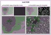
考察可降解纳米复合物在Hela细胞和HCT8/T上中的摄取情况。The uptake of degradable nanocomplexes in Hela cells and HCT8/T was investigated.
将消化好的细胞种在24孔板中,每孔加入50μl密度为2×10⁶个/ml的细胞悬液,再用含血清1640培养基补齐液体至500ul,置于细胞培养箱中培养12h,汇合程度约为70%-80%。Plant the digested cells in a 24-well plate, add 50 μl of cell suspension with a density of 2×10⁶ cells/ml to each well, fill up the liquid with serum-containing 1640 medium to 500ul, and place it in a cell culture incubator 12h, the degree of confluence is about 70%-80%.
弃去与加入纳米载体同体积的培养基,将预先用YoYo-1标记好的pDNA制备出的γ-PGA/SSPEI-beta-CD/pDNA-YoYo1或者是疏水药物用染料香豆素6(C6)代替的γ-PGA/SSPEI-beta-CD-C6/pDNA,加入到不同细胞的对应孔内,各自孔中孵育3h,然后将培养上清弃去,加YoYo-1组用台盼蓝孵育5min淬灭胞外荧光,接着用PBS洗三遍。之后加入4%多聚甲醛37℃固定10min,弃去多聚甲醛,用PBS洗三遍后,加入DAPI孵育10min,用PBS清洗三遍后,加入少量PBS浸润细胞,置于荧光显微镜下观察。如图13所示。为了定量考察两种细胞对纳米复合物的摄取情况,使用流式细胞仪进行检测。具体操作步骤同上,分别给予预先用YoYo-1标记好的pDNA制备出的新鲜纳米复合物,孵育3h后,先用台盼蓝淬灭,再用PBS清洗3遍,胰酶消化并收集细胞,1000rpm离心5min后,用PBS再洗两遍,加入500μl PBS置于流式细胞仪上检测。结果如图12所示。Discard the medium with the same volume as the nanocarrier added, and use the γ-PGA/SSPEI-beta-CD/pDNA-YoYo1 prepared in advance with YoYo-1-labeled pDNA or the hydrophobic drug dye coumarin 6 (C6 ) instead of γ-PGA/SSPEI-beta-CD-C6/pDNA, added to the corresponding wells of different cells, incubated in each well for 3h, then discarded the culture supernatant, added YoYo-1 group and incubated with trypan blue The extracellular fluorescence was quenched for 5 minutes, and then washed three times with PBS. After that, add 4% paraformaldehyde and fix at 37°C for 10 min, discard the paraformaldehyde, wash with PBS three times, add DAPI and incubate for 10 min, wash with PBS three times, add a small amount of PBS to infiltrate the cells, and observe under a fluorescent microscope. As shown in Figure 13. In order to quantitatively investigate the uptake of the nanocomplex by the two cells, flow cytometry was used for detection. The specific operation steps are the same as above. The fresh nanocomplexes prepared in advance with YoYo-1-labeled pDNA were given respectively. After incubation for 3 hours, they were first quenched with trypan blue, washed 3 times with PBS, digested with trypsin and collected cells. After centrifugation at 1000rpm for 5min, wash twice with PBS, add 500μl PBS and place on flow cytometer for detection. The result is shown in Figure 12.
摄取抑制实验:为验证γ-PGA存在的主动靶向作用介导的内吞是摄取提高的原因,利用文献报道的γ-PGA抑制剂GGsTop对细胞进行预处理1h,并设置抑制剂的不同浓度作对比,再给予新鲜制备的标记YoYo-1的γ-PGA-PEI25k/EGFP,共孵育4h后如上述步骤所示,消化细胞,利用流式细胞仪进行绿色荧光的分析。结果如图13所示。Uptake inhibition experiment: In order to verify that the endocytosis mediated by the active targeting effect of γ-PGA is the reason for the increased uptake, the cells were pretreated for 1 h with the γ-PGA inhibitor GGsTop reported in the literature, and different concentrations of the inhibitor were set For comparison, freshly prepared YoYo-1-labeled γ-PGA-PEI25k/EGFP was administered, and after co-incubating for 4 hours, the cells were digested as shown in the above steps, and the green fluorescence was analyzed by flow cytometry. The result is shown in Figure 13.
结果:在两种细胞上,纳米复合物都能被有效的摄取,基因进入溶酶体,疏水药进入细胞浆,且有γ-PGA的纳米载体组摄取略强于普通纳米载体组;细胞摄取抑制发现,随着GGsTop的增加,摄取抑制效果也逐渐增加,呈现出浓度依赖性,表现出了γ-PGA的促进摄取的作用。Results: On the two kinds of cells, the nanocomplexes can be effectively taken up, the gene enters the lysosome, and the hydrophobic drug enters the cytoplasm, and the uptake of the nanocarrier group with γ-PGA is slightly stronger than that of the ordinary nanocarrier group; the cell uptake Inhibition found that with the increase of GGsTop, the uptake inhibition effect also gradually increased, showing a concentration-dependent, showing the role of γ-PGA to promote uptake.
(11)细胞转染实验(11) Cell transfection experiment
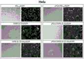
采用pEGPF作为报告基因考察纳米复合物的体外转染效率。The in vitro transfection efficiency of nanocomplexes was investigated using pEGPF as a reporter gene.
HeLa和Hct8/T(耐药)细胞的转染(定性):培养的细胞用0.25%胰蛋白酶消化后吹打均匀,细胞计数后,将细胞以1×105细胞/孔的密度均匀接种到12孔的培养板中,培养24h至细胞汇合度70-80%。转染前弃去培养基并加入新鲜培养基。从板里吸走与制备纳米复合物相同体积的培养基后,将新鲜制备的纳米复合物加到各孔(3μg pEGFP/孔)中,每组设置三个复孔。继续培养至48h。培养结束后,弃去培养液,用PBS漂洗两次。用4%多聚甲醛固定细胞10min后,加入DAPI浸没各孔并孵育10min,再用PBS洗三遍,置于荧光显微镜下观察绿色荧光的分布与强度,并拍照。Transfection (qualitative) of HeLa and Hct8/T (drug-resistant) cells: the cultured cells were digested with 0.25% trypsin and evenly pipetted. After counting the cells, the cells were evenly seeded into 12 wells at a density of 1×10⁵ cells/well In the culture plate, cultivate for 24 hours until the cell confluency is 70-80%. Discard the medium and add fresh medium before transfection. After aspirating the same volume of medium as used to prepare the nanocomplexes from the plate, add the freshly prepared nanocomplexes to each well (3 μg pEGFP/well), and set up triplicate wells for each group. Continue to cultivate to 48h. After culturing, the culture medium was discarded and rinsed twice with PBS. After the cells were fixed with 4% paraformaldehyde for 10 min, DAPI was added to submerge each well and incubated for 10 min, then washed three times with PBS, placed under a fluorescence microscope to observe the distribution and intensity of green fluorescence, and photographed.
HeLa和Hct8/T(耐药)细胞的转染(定量):采用流式细胞仪测定绿色荧光蛋在各种细胞中表达的细胞数目。将培养的细胞用胰酶消化后吹打均匀,细胞计数后,将细胞以1×105细胞/孔的密度均匀接种到12孔的培养板中,培养24h至细胞汇合度70-80%。转染前弃去培养基并加入新鲜培养基。从板里吸走与制备纳米复合物相同体积的培养基后,将新鲜制备的纳米复合物加到各孔(3μg pEGFP/孔)中,每组设置三个复孔。继续培养至48h。培养结束后,用0.25%胰蛋白酶消化细胞,1000rpm离心5min收集细胞,用冰PBS漂洗两次,最终将细胞重悬于500ul PBS溶液中,转移到流式细胞管,立即用流式细胞仪FL-1通道测定有绿色荧光的细胞数目,每个样品均收集10,000个细胞。采用Flow Jo软件分析处理实验结果。结果如图14所示。Transfection (quantification) of HeLa and Hct8/T (drug-resistant) cells: The number of cells expressing green fluorescent eggs in various cells was measured by flow cytometry. The cultured cells were digested with trypsin and blown evenly. After the cells were counted, the cells were evenly seeded into a 12-well culture plate at a density of 1×10 5 cells/well, and cultured for 24 hours until the cell confluence was 70-80%. Discard the medium and add fresh medium before transfection. After aspirating the same volume of medium as used to prepare the nanocomplexes from the plate, add the freshly prepared nanocomplexes to each well (3 μg pEGFP/well), and set up triplicate wells for each group. Continue to cultivate to 48h. After the culture, digest the cells with 0.25% trypsin, collect the cells by centrifugation at 1000rpm for 5min, rinse twice with ice PBS, and finally resuspend the cells in 500ul PBS solution, transfer to the flow cytometry tube, and immediately use the flow cytometer FL The number of cells with green fluorescence was measured in channel -1, and 10,000 cells were collected for each sample. The experimental results were analyzed and processed by FlowJo software. The result is shown in Figure 14.
结果:在Hela和Hct8/T上,各组与黄金标准PEI25k复合的纳米载体的转染效果进行对比,SSPEI-beta-CD-Mo/pDNA组已经展现出了低毒且相似的转染效果,而γ-PGA/SSPEI-beta-CD-Mo/pDNA组转染好于SSPEI-beta-CD-Mo/pDNA和PEI25k组也说明了γ-PGA展现出的效果。Results: On Hela and Hct8/T, the transfection effect of each group was compared with the gold standard PEI25k composite nanocarrier, and the SSPEI-beta-CD-Mo/pDNA group has shown low toxicity and similar transfection effect, The transfection of γ-PGA/SSPEI-beta-CD-Mo/pDNA group was better than that of SSPEI-beta-CD-Mo/pDNA and PEI25k group, which also showed the effect of γ-PGA.
测试实施例2:共载药可降解纳米载体的体外联合治疗效果Test Example 2: In Vitro Combination Therapeutic Effects of Co-loaded Degradable Nanocarriers
(1)MTT(3-(4,5-二甲基噻唑-2)-2,5-二苯基四氮唑溴蓝,商品名:噻唑蓝)法,测定可降解纳米材料的材料毒性,单载药及共载药体系的抗肿瘤作用。(1) MTT (3-(4,5-dimethylthiazole-2)-2,5-diphenyl bromide tetrazolium blue, trade name: thiazolium blue) method to determine the material toxicity of degradable nanomaterials, Antitumor effects of single-drug and co-drug-loaded systems.
空载体,单载药,共载药纳米复合物处理48h小时后细胞的存活率,从而考察载药纳米复合物的抗肿瘤能力。培养的细胞用0.25%胰蛋白酶消化后吹打均匀,细胞计数后,将HeLa细胞以5000个每孔的密度接种到96孔培养板中,每孔100ul培养液,细胞在培养箱中培养24h。空载体毒性以PEI的浓度(ug/mL)为横坐标,单载药和双载药纳米复合物以治疗基因TRAIL的浓度(ug/mL)为横坐标。考察载药纳米粒的细胞抑制率时,我们制备新鲜的γ-PGA/SSPEI-beta-CD-Mo/pDNA等纳米复合物,按100ul/孔给药,并使其终浓度为对应的药物浓度。药物浓度等比梯度稀释。继续培养48h。设置空白孔和对照孔,每组六个复孔。给药结束后在各孔中加入20μl MTT溶液(5mg/ml),并继续在二氧化碳培养中孵育4h。随后,用注射器小心吸掉培养上清液,各孔中加入150μl的DMSO(二甲基亚砜),用锡纸包裹平放在不动的摇床上37℃孵育10min,最后将96孔板放入酶标仪孵育并震荡1min以溶解甲臜晶体,测量各孔在570nm处的吸光值。使用以下公式计算细胞存活率(%):Empty carrier, drug-loaded single, and drug-loaded nanocomposites were treated for 48 hours to investigate the anti-tumor ability of the drug-loaded nanocomposites. The cultured cells were digested with 0.25% trypsin and blown evenly. After cell counting, HeLa cells were seeded into 96-well culture plates at a density of 5000 per well, with 100 ul of culture medium per well, and the cells were cultured in an incubator for 24 hours. The abscissa was the concentration of PEI (ug/mL) for the toxicity of the empty vector, and the concentration (ug/mL) of the therapeutic gene TRAIL for the single-drug-loaded and double-drug-loaded nanocomplexes. When investigating the cell inhibition rate of drug-loaded nanoparticles, we prepared fresh nanocomposites such as γ-PGA/SSPEI-beta-CD-Mo/pDNA, administered at 100ul/well, and made the final concentration the corresponding drug concentration . Drug concentration isotropic gradient dilution. Continue to cultivate for 48h. Set up blank wells and control wells, with six replicate wells in each group. After administration, 20 μl of MTT solution (5 mg/ml) was added to each well, and incubated in carbon dioxide culture for 4 h. Subsequently, carefully suck off the culture supernatant with a syringe, add 150 μl of DMSO (dimethyl sulfoxide) to each well, wrap it with tin foil and place it on a stationary shaker for 10 minutes at 37°C, and finally put the 96-well plate into Incubate with a microplate reader and shake for 1 min to dissolve the formazan crystals, and measure the absorbance of each well at 570 nm. Calculate cell viability (%) using the following formula:
Cell Viability(%)=[(AE-AB)/(AC-AB)]×100%Cell Viability(%)=[(AE-AB)/(AC-AB)]×100%
AE,AC和AB分别代表实验组的吸光值,空白组的吸光值和对照的吸光值。结果如图16所示。(A实验组-A空白组)/(A对照组-A空白组)AE, AC and AB represent the absorbance value of the experimental group, the absorbance value of the blank group and the absorbance value of the control, respectively. The result is shown in Figure 16. (A experimental group-A blank group)/(A control group-A blank group)
结果:A图为hela细胞,B图为Hct8/T上纳米材料毒性的MTT,与PEI25k相比,其展现出低毒的优越性。而载药后的纳米载体展现出了优越的细胞杀伤抑制效果。如图15所示。Results: Figure A shows hela cells, and picture B shows the MTT of nanomaterial toxicity on Hct8/T. Compared with PEI25k, it shows the advantage of low toxicity. The nanocarriers loaded with drugs showed superior cell killing inhibition effect. As shown in Figure 15.
(2)细胞凋亡测定(2) Apoptosis assay
采用流式细胞仪定量检测可降解纳米复合物诱导肿瘤细胞凋亡的能力。Flow cytometry was used to quantitatively detect the ability of degradable nanocomplexes to induce tumor cell apoptosis.
将HeLa和Hct8/T细胞分别以5×105个/孔的密度接种到6孔培养板中,培养约24h后,细胞汇合度70-80%,转染前弃去培养基并加入2ml新鲜培养基。各孔先吸出与纳米复合物溶液相同体积的培养基,再加入含有相当于4μg TRAIL的纳米复合物。除去给药组之外,同时参照说明书,用细胞凋亡阳性对照试剂处理细胞,设置早期凋亡和晚期凋亡两个阳性对照组Inoculate HeLa and Hct8/T cells into 6-well culture plates at a density of 5×10⁵ cells/well. After culturing for about 24 hours, the confluence of the cells is 70-80%. Discard the medium before transfection and add 2ml of fresh Medium. The same volume of medium as the nanocomplex solution was first aspirated from each well, and then the nanocomplex containing the equivalent of 4 μg TRAIL was added. In addition to the administration group, refer to the instructions at the same time, treat the cells with the positive control reagent for apoptosis, and set up two positive control groups for early apoptosis and late apoptosis.
细胞在培养箱中孵育48h后,小心将培养上清收集在离心管中,用0.25%胰蛋白酶消化贴壁细胞,Hela在37℃孵育3min,Hct8/T在37℃孵育3min,随后加入先前取出的培养上清液加入对应孔内轻轻吹打下贴壁细胞,加入到对应离心管中,3000rpm离心5min收集细胞,弃上清,再以同样的方法,用冷PBS漂洗三次,最终将试剂盒里的10×Annexin V-FITC结合液稀释至1×,用100ul 1×Annexin V-FITC 1×结合液轻轻重悬细胞,细胞浓度约为1×10⁷个细胞/ml。设置单染组,双染组后,样品每管中加入1μl 100μg/ml Annexin V-FITC溶液和1μl 100μg/ml碘化丙啶溶液,黑暗下室温下共孵育15min后,再加入400μl 1×AnnexinV-FITC结合液,吹打混匀。After the cells were incubated in the incubator for 48 hours, carefully collect the culture supernatant in a centrifuge tube, digest the adherent cells with 0.25% trypsin, incubate Hela at 37°C for 3 minutes, and incubate Hct8/T at 37°C for 3 minutes, then add the previously removed Add the culture supernatant to the corresponding well and gently blow down the adherent cells, add it to the corresponding centrifuge tube, centrifuge at 3000rpm for 5min to collect the cells, discard the supernatant, and rinse three times with cold PBS in the same way, and finally the kit Dilute the 10×Annexin V-FITC binding solution in it to 1×, gently resuspend the cells with
立即加入流式管,用流式细胞仪根据两个阳性样品:单染和双染调节参数和补偿使其处在正确位置,然后横轴为FL-1通道检测阳性细胞绿色荧光的分布情况,纵轴为FL-2通道测定细胞红色荧光的分布情况。每个样品均收集10,000个细胞。Immediately add the flow tube, and use the flow cytometer to adjust the parameters and compensation according to the two positive samples: single staining and double staining to make it in the correct position, and then the horizontal axis is the distribution of the green fluorescence of the positive cells detected by the FL-1 channel, The vertical axis is the distribution of cell red fluorescence measured by FL-2 channel. 10,000 cells were collected from each sample.
结果:包裹了γ-PGA的双药联用组效果十分明显,晚期凋亡比例大于普通双药联用组,证明了γ-PGA的靶向性,双药组比单药组凋亡效果也要明显也证明了联用的成功。A,B:Hela凋亡及定量分析,C,D:Hct8/T凋亡及定量分析。Results: The effect of the double-drug combination group coated with γ-PGA is very obvious, and the proportion of late apoptosis is higher than that of the ordinary double-drug combination group, which proves the targeting of γ-PGA, and the apoptosis effect of the double-drug group is also higher than that of the single-drug group. It must be obvious that it also proves the success of the joint use. A, B: Hela apoptosis and quantitative analysis, C, D: Hct8/T apoptosis and quantitative analysis.
(3)细胞周期测定(3) Cell cycle assay
采用经典的碘化丙啶染色(PI)方法进行细胞周期分析。Cell cycle analysis was performed using the classic propidium iodide staining (PI) method.
培养的细胞用0.25%胰蛋白酶消化后吹打均匀,细胞计数后,将细胞以5×10⁵细胞/孔的密度接种到6孔培养板中。培养24h至细胞汇合度70-80%左右,转染前弃去培养基并加入2ml新鲜培养基。各孔先吸出与纳米复合物溶液相同体积的培养基,再加入含有相当于4μg TRAIL的纳米复合物,细胞培养箱中共孵育48h。The cultured cells were digested with 0.25% trypsin and blown evenly. After the cells were counted, the cells were seeded into a 6-well culture plate at a density of 5×10⁵ cells/well. Cultivate for 24 hours until the cell confluency is about 70-80%, discard the medium before transfection and add 2ml of fresh medium. The medium with the same volume as the nanocomplex solution was aspirated from each well, and then the nanocomposite containing 4 μg TRAIL was added, and incubated in the cell culture box for 48 hours.
小心将培养上清液收集到2ml离心管中,用0.25%胰蛋白酶消化贴壁细胞,Hela在37℃孵育3min,Hct8/T在37℃孵育6min,分别加入之前取出的培养上清液加入对应孔轻轻吹打下贴壁细胞,加入离心管中,3000rpm离心5min收集细胞,小心弃去上清,用冰PBS漂洗三次。最后在细胞内加入75%乙醇,轻轻吹打混匀,置于4℃固定12h以上。也可长期保存在-20℃中。Carefully collect the culture supernatant into a 2ml centrifuge tube, digest the adherent cells with 0.25% trypsin, incubate Hela at 37°C for 3 minutes, and incubate Hct8/T at 37°C for 6 minutes, add the previously removed culture supernatant to the corresponding Gently blow off the adherent cells in the hole, add to the centrifuge tube, centrifuge at 3000rpm for 5min to collect the cells, carefully discard the supernatant, and rinse with ice PBS three times. Finally, 75% ethanol was added to the cells, mixed by pipetting gently, and fixed at 4°C for more than 12 hours. It can also be stored at -20°C for a long time.
3000rpm离心5min,小心弃去酒精,用冷PBS漂洗两遍。配制100×PI工作液(包含10mg/ml RNase,PI母液,用PBS稀释补齐刻度)用时稀释成1×,加入至对应细胞,轻轻吹散细胞混匀,室温下避光放置30min,随后3000rpm离心5min,弃上清液,用冰PBS漂洗两遍。最后用500μl PBS重悬细胞,随即采用流式细胞仪测定。Centrifuge at 3000rpm for 5min, carefully discard the alcohol, and rinse twice with cold PBS. Prepare 100×PI working solution (including 10mg/ml RNase, PI mother solution, dilute with PBS to make up the scale) and dilute to 1× when used, add to the corresponding cells, gently blow off the cells and mix well, place in the dark at room temperature for 30min, then Centrifuge at 3000rpm for 5min, discard the supernatant, and rinse twice with ice-cold PBS. Finally, the cells were resuspended in 500 μl PBS, and then measured by flow cytometry.
结果:A:Hct8/T的周期图。双药组明显subG1期高于任一组。Results: A: Periodogram of Hct8/T. The subG1 phase of the double-drug group was significantly higher than that of either group.
综上,本发明的修饰有γ-PGA的SSPEI-β-CD纳米载体可以有效地共载携载质粒(或者小分子干扰RNA)和疏水莫能霉素,主动靶向细胞,并穿透进入细胞内部,释放药物,有效表达基因,具有显著的肿瘤杀伤效果。对肿瘤细胞的体外的生长抑制作用都表现出了明显的作用。同时,本发明的二硫键交联可降解纳米载体也显著降低了毒性,并保留PEI25k的高转染效率。In summary, the SSPEI-β-CD nanocarrier modified with γ-PGA of the present invention can effectively co-carry plasmid (or small interfering RNA) and hydrophobic monensin, actively target cells, and penetrate into Inside the cell, it releases drugs, effectively expresses genes, and has a significant tumor-killing effect. The growth inhibitory effect on tumor cells in vitro has shown obvious effects. At the same time, the disulfide bond cross-linked degradable nanocarrier of the present invention also significantly reduces toxicity and retains the high transfection efficiency of PEI25k.
在本发明提及的所有文献都在本申请中引用作为参考,就如同每一篇文献被单独引用作为参考那样。此外应理解,在阅读了本发明的上述讲授内容之后,本领域技术人员可以对本发明作各种改动或修改,这些等价形式同样落于本申请所附权利要求书所限定的范围。All documents mentioned in this application are incorporated by reference in this application as if each were individually incorporated by reference. In addition, it should be understood that after reading the above teaching content of the present invention, those skilled in the art can make various changes or modifications to the present invention, and these equivalent forms also fall within the scope defined by the appended claims of the present application.
Claims (8)
Priority Applications (1)
| Application Number | Priority Date | Filing Date | Title |
|---|---|---|---|
| CN201710198879.9ACN108653747B (en) | 2017-03-29 | 2017-03-29 | Degradable nano-carrier for co-delivery of gene and hydrophobic drug, and preparation method and application thereof |
Applications Claiming Priority (1)
| Application Number | Priority Date | Filing Date | Title |
|---|---|---|---|
| CN201710198879.9ACN108653747B (en) | 2017-03-29 | 2017-03-29 | Degradable nano-carrier for co-delivery of gene and hydrophobic drug, and preparation method and application thereof |
Publications (2)
| Publication Number | Publication Date |
|---|---|
| CN108653747A CN108653747A (en) | 2018-10-16 |
| CN108653747Btrue CN108653747B (en) | 2023-04-07 |
Family
ID=63786828
Family Applications (1)
| Application Number | Title | Priority Date | Filing Date |
|---|---|---|---|
| CN201710198879.9AActiveCN108653747B (en) | 2017-03-29 | 2017-03-29 | Degradable nano-carrier for co-delivery of gene and hydrophobic drug, and preparation method and application thereof |
Country Status (1)
| Country | Link |
|---|---|
| CN (1) | CN108653747B (en) |
Families Citing this family (6)
| Publication number | Priority date | Publication date | Assignee | Title |
|---|---|---|---|---|
| CN110075312A (en)* | 2019-04-25 | 2019-08-02 | 中国科学院上海应用物理研究所 | A kind of method and the thus obtained compound of single stranded DNA and hydrophobic drug covalent linkage |
| CN111110866B (en)* | 2019-12-30 | 2023-04-11 | 福州大学 | Reductive polyglutamic acid/polyethyleneimine/siRNA composite nanoparticles, and preparation and application thereof |
| CN113318242B (en)* | 2020-02-28 | 2023-03-10 | 四川大学华西医院 | A tumor-targeting multifunctional non-viral gene carrier and its preparation method and application |
| CN112402394A (en)* | 2020-11-25 | 2021-02-26 | 天津医科大学第二医院 | BCG complex adsorbing nano-drug carrier by utilizing positive and negative charge characteristics and preparation method thereof |
| CN115137839B (en)* | 2021-03-30 | 2025-04-22 | 南京纳么美科技有限公司 | Ferritin cage nanocarriers for targeting and co-loading hydrophilic/hydrophobic drugs and their applications |
| CN114769609B (en)* | 2022-03-29 | 2024-04-09 | 华南理工大学 | Host-guest chemistry mediated size controllable luminescent gold nanoparticle assembly, and preparation method and application thereof |
Citations (1)
| Publication number | Priority date | Publication date | Assignee | Title |
|---|---|---|---|---|
| US6048736A (en)* | 1998-04-29 | 2000-04-11 | Kosak; Kenneth M. | Cyclodextrin polymers for carrying and releasing drugs |
- 2017
- 2017-03-29CNCN201710198879.9Apatent/CN108653747B/enactiveActive
Patent Citations (1)
| Publication number | Priority date | Publication date | Assignee | Title |
|---|---|---|---|---|
| US6048736A (en)* | 1998-04-29 | 2000-04-11 | Kosak; Kenneth M. | Cyclodextrin polymers for carrying and releasing drugs |
Also Published As
| Publication number | Publication date |
|---|---|
| CN108653747A (en) | 2018-10-16 |
Similar Documents
| Publication | Publication Date | Title |
|---|---|---|
| CN108653747B (en) | Degradable nano-carrier for co-delivery of gene and hydrophobic drug, and preparation method and application thereof | |
| Kozielski et al. | Cancer-selective nanoparticles for combinatorial siRNA delivery to primary human GBM in vitro and in vivo | |
| Wannasarit et al. | A virus‐mimicking pH‐responsive acetalated dextran‐based membrane‐active polymeric nanoparticle for intracellular delivery of antitumor therapeutics | |
| Shi et al. | Novel CD44-targeting and pH/redox-dual-stimuli-responsive core–shell nanoparticles loading triptolide combats breast cancer growth and lung metastasis | |
| Sun et al. | RETRACTED ARTICLE: siRNA-loaded poly (histidine-arginine) 6-modified chitosan nanoparticle with enhanced cell-penetrating and endosomal escape capacities for suppressing breast tumor metastasis | |
| Xu et al. | Targeting death receptors for drug-resistant cancer therapy: Codelivery of pTRAIL and monensin using dual-targeting and stimuli-responsive self-assembling nanocomposites | |
| Jiang et al. | pH‐triggered pinpointed cascading charge‐conversion and redox‐controlled gene release design: modularized fabrication for nonviral gene transfection | |
| Lin et al. | Polycation-detachable nanoparticles self-assembled from mPEG-PCL-g-SS-PDMAEMA for in vitro and in vivo siRNA delivery | |
| Sharma et al. | Vitamin B6 tethered endosomal pH responsive lipid nanoparticles for triggered intracellular release of doxorubicin | |
| Zhao et al. | Gene therapy of endometriosis introduced by polymeric micelles with glycolipid-like structure | |
| Mishra et al. | Reconstitutable charged polymeric (PLGA) 2-b-PEI micelles for gene therapeutics delivery | |
| Shukla et al. | Multifunctional hybrid nanoconstructs facilitate intracellular localization of doxorubicin and genistein to enhance apoptotic and anti-angiogenic efficacy in breast adenocarcinoma | |
| Park et al. | Selective stimulation of caveolae-mediated endocytosis by an osmotic polymannitol-based gene transporter | |
| CN108542885A (en) | Antitumor drug and preparation method thereof | |
| JP2011524446A (en) | Chitosan oligosaccharide fatty acid graft product modified with polyglycol, its preparation method and use thereof | |
| JP2017533280A (en) | Bile acid particle composition and method | |
| Garg et al. | Triphenylamine coupled chitosan with high buffering capacity and low viscosity for enhanced transfection in mammalian cells, in vitro and in vivo | |
| Li et al. | Collaborative assembly of doxorubicin and galactosyl diblock glycopolymers for targeted drug delivery of hepatocellular carcinoma | |
| Mandracchia et al. | In vitro evaluation of glycol chitosan based formulations as oral delivery systems for efflux pump inhibition | |
| Wang et al. | Functionalized O-carboxymethyl-chitosan/polyethylenimine based novel dual pH-responsive nanocarriers for controlled co-delivery of DOX and genes | |
| CN115252560B (en) | A self-assembled nanoparticle based on natural products and its preparation method and application | |
| WO2021055467A1 (en) | Orally administrable nano-medicine for viral diseases | |
| Wen et al. | Redox-responsive polymer inhibits macrophages uptake for effective intracellular gene delivery and enhanced cancer therapy | |
| Wang et al. | Tumor microenvironment dual-responsive core–shell nanoparticles with hyaluronic acid-shield for efficient co-delivery of doxorubicin and plasmid DNA | |
| Lin et al. | The development of tertiary amine cationic lipids for safe and efficient siRNA delivery |
Legal Events
| Date | Code | Title | Description |
|---|---|---|---|
| PB01 | Publication | ||
| PB01 | Publication | ||
| SE01 | Entry into force of request for substantive examination | ||
| SE01 | Entry into force of request for substantive examination | ||
| GR01 | Patent grant | ||
| GR01 | Patent grant |