CN108334730B - A Muscle Group-Based Modeling and Simulation Method of Human Hip - Google Patents
A Muscle Group-Based Modeling and Simulation Method of Human HipDownload PDFInfo
- Publication number
- CN108334730B CN108334730BCN201710753127.4ACN201710753127ACN108334730BCN 108334730 BCN108334730 BCN 108334730BCN 201710753127 ACN201710753127 ACN 201710753127ACN 108334730 BCN108334730 BCN 108334730B
- Authority
- CN
- China
- Prior art keywords
- hip
- model
- muscle
- human
- geometric
- Prior art date
- Legal status (The legal status is an assumption and is not a legal conclusion. Google has not performed a legal analysis and makes no representation as to the accuracy of the status listed.)
- Expired - Fee Related
Links
Images


Classifications
- G—PHYSICS
- G09—EDUCATION; CRYPTOGRAPHY; DISPLAY; ADVERTISING; SEALS
- G09B—EDUCATIONAL OR DEMONSTRATION APPLIANCES; APPLIANCES FOR TEACHING, OR COMMUNICATING WITH, THE BLIND, DEAF OR MUTE; MODELS; PLANETARIA; GLOBES; MAPS; DIAGRAMS
- G09B23/00—Models for scientific, medical, or mathematical purposes, e.g. full-sized devices for demonstration purposes
- G09B23/28—Models for scientific, medical, or mathematical purposes, e.g. full-sized devices for demonstration purposes for medicine
Landscapes
- Engineering & Computer Science (AREA)
- Physics & Mathematics (AREA)
- General Physics & Mathematics (AREA)
- Computational Mathematics (AREA)
- Mathematical Analysis (AREA)
- Medicinal Chemistry (AREA)
- General Health & Medical Sciences (AREA)
- Algebra (AREA)
- Health & Medical Sciences (AREA)
- Chemical & Material Sciences (AREA)
- Medical Informatics (AREA)
- Mathematical Optimization (AREA)
- Mathematical Physics (AREA)
- Pure & Applied Mathematics (AREA)
- Business, Economics & Management (AREA)
- Educational Administration (AREA)
- Educational Technology (AREA)
- Theoretical Computer Science (AREA)
- Prostheses (AREA)
Abstract
Description
Translated fromChinese技术领域technical field
本发明涉及基于肌肉群的人体髋部建模与仿真方法。The invention relates to a method for modeling and simulating a human hip based on muscle groups.
背景技术Background technique
由于人体髋部的重要性和易损伤性使得针对人体髋部的研究越来越重要。在对人体髋部的研究中比如对人体髋部的手术培训、大腿假肢接受腔的研究和汽车对人体髋部碰撞的研究等方面的传统方法是利用尸体或者模型进行试验。但由于尸体数量的有限性以及尸体使用的不可重复性导致了以上所述培训和研究的机会少、成本高、周期长。同时由于尸体和活体中的人体组织的生物力学特性存在差异导致了通过对尸体的研究得到的研究成果与实际情况不可避免的存在差异。随着软件技术和硬件技术的发展,数字医学和人体生物力学的发展进入快车道,使得虚拟手术系统和人体组织有限元分析应运而生。通过计算机仿真技术对人体力学特性进行高度接近真实的模拟分析可以有效解决以上问题。Due to the importance and vulnerability of the human hip, the research on the human hip is becoming more and more important. In the study of the human hip, such as the surgical training of the human hip, the study of the thigh prosthesis socket, and the study of the human hip collision by the automobile, the traditional method is to use the cadaver or the model to carry out the test. However, due to the limited number of cadavers and the unrepeatable use of cadavers, there are few opportunities, high costs, and long periods of training and research. At the same time, due to the differences in the biomechanical properties of human tissues in cadavers and living organisms, there is an inevitable difference between the research results obtained through the study of cadavers and the actual situation. With the development of software technology and hardware technology, the development of digital medicine and human biomechanics has entered the fast lane, which makes the virtual surgery system and the finite element analysis of human tissue emerge as the times require. The above problems can be effectively solved by the simulation analysis of human body mechanics characteristics which is highly close to the real by computer simulation technology.
人体髋部有限元模型研究中,模型主要分为只包含关节骨的仿真模型、包含骨骼和简化的肌肉模型、包含骨骼与肌肉的几何模型。有限元模拟人体髋部力学特性的方式主要包括质点-弹簧模型、各向同性线弹性模型和非线性模型。目前的人体下肢模型研究体现出一些特征,比如:依据不同的模型用途,所建立的模型的精细程度、建模方式、建模层次、所附材质都会不同。一般情况下,只涉及几何模型和模型可视化的,所建立的模型可以更加细致一些;如果是建立有限元模型,由于有限元计算对模型的要求,所建立的模型在建模过程中经常会被简化。In the study of the finite element model of the human hip, the models are mainly divided into simulation models containing only joint bones, bone and simplified muscle models, and geometric models containing bones and muscles. The methods of finite element simulation of the mechanical properties of the human hip mainly include the mass-spring model, the isotropic linear elastic model and the nonlinear model. The current research on human lower extremity models reflects some characteristics, such as: the fineness, modeling method, modeling level, and attached materials of the established models will be different according to different model uses. Under normal circumstances, if only the geometric model and model visualization are involved, the established model can be more detailed; if the finite element model is established, the established model is often used in the modeling process due to the requirements of finite element calculation for the model. simplify.
尽管已有的人体髋部或者下肢模型,已经向更加细致的表达人体解剖学结构的方向发展,但由于髋部组织解剖结构的复杂性、几何形态的不规则性、组织间接触的复杂性和有限元计算的限制,仍然存在一些需要解决的问题:一是模型能够更加细致的反应髋部的解剖学结构;二是基于复杂结构建立的人体下肢模型能够有利于实现有限元仿真计算。Although the existing human hip or lower extremity models have developed towards a more detailed expression of human anatomy, due to the complexity of the anatomical structure of the hip tissue, the irregularity of geometry, the complexity of tissue contact and the Due to the limitation of finite element calculation, there are still some problems to be solved: first, the model can reflect the anatomical structure of the hip in more detail; second, the human lower limb model established based on the complex structure can be beneficial to the realization of finite element simulation calculation.
发明内容SUMMARY OF THE INVENTION
本发明的目的是为了解决现有的人体髋部模型不能够兼顾细化的人体结构和有限元仿真计算,而提出一种基于肌肉群的人体髋部建模与仿真方法。The purpose of the present invention is to propose a muscle group-based modeling and simulation method of the human hip in order to solve the problem that the existing human hip model cannot take into account the refined human body structure and finite element simulation calculation.
一种基于肌肉群的人体髋部建模与仿真方法按以下步骤实现:A method for modeling and simulating a human hip based on muscle groups is implemented in the following steps:
步骤一、人体髋部图像数据获取;Step 1, the acquisition of human hip image data;
步骤二、在人体髋部图像数据获取的基础上,进行人体髋部几何建模,包括髋骨(皮质骨与松质骨)、股骨(皮质骨与松质骨)、关节软骨、缝匠肌、股四头肌、股二头肌、半腱肌、半膜肌、股薄肌、趾骨肌、长收肌、短收肌、大收肌、臀大肌、臀中肌、臀小肌、整个髋部的几何三维重建;Step 2: On the basis of the acquisition of human hip image data, carry out geometric modeling of human hip, including hip bone (cortical bone and cancellous bone), femur (cortical bone and cancellous bone), articular cartilage, sartorius muscle , quadriceps femoris, biceps femoris, semitendinosus, semimembranosus, gracilis, phalanges, adductor longus, adductor brevis, adductor magnus, gluteus maximus, gluteus medius, gluteus minimus, Geometric 3D reconstruction of the entire hip;
步骤三、在人体髋部几何模型基础上,建立肌肉群模型,对髋骨、股骨、关节软骨、肌肉群、整个髋部实施模型装配,建立脂肪和皮肤模型,添加肌腱;Step 3: On the basis of the geometric model of the human hip, establish a muscle group model, implement model assembly for the hip bone, femur, articular cartilage, muscle group, and the entire hip, establish fat and skin models, and add tendons;
步骤四、在人体髋部装配模型基础上,确定网格划分参数,确定组织间接触关系,实施几何模型的网格划分;Step 4: On the basis of the human hip assembly model, determine the meshing parameters, determine the contact relationship between the tissues, and implement the meshing of the geometric model;
步骤五、在人体髋部模型网格划分基础上,为人体髋部模型各部分结构附材质,确定材料参数;Step 5: On the basis of the mesh division of the human hip model, attach materials to the structures of each part of the human hip model, and determine the material parameters;
步骤六、在人体髋部模型附材质的基础上,实现人体髋部模型的力学仿真。Step 6: On the basis of the attached material of the human hip model, the mechanical simulation of the human hip model is realized.
本发明的有益效果为:The beneficial effects of the present invention are:
人体髋部组织解剖结构是极其复杂的,包括皮肤、13块肌肉、脂肪、关节软骨、股骨、髋骨。由于目前有限元分析技术的限制,模型所含的组织数量和种类越多、结构越复杂、形状越不规则使得模型间的接触设置、边界条件的设置和网格划分越复杂直接导致了有限元分析结果的不准确甚至是分析结果不收敛。因此最主要的问题是如何建立一个既能客观反映人体髋部真实解剖结构及其生物力学行为又能满足有限元分析要求的模型。The anatomy of the human hip tissue is extremely complex, including skin, 13 muscles, fat, articular cartilage, femur, and hip bone. Due to the limitations of the current finite element analysis technology, the more the number and types of tissues contained in the model, the more complex the structure, the more irregular the shape, the more complex the contact settings between models, the setting of boundary conditions and the mesh division, which directly leads to the finite element The analysis results are inaccurate or even the analysis results do not converge. Therefore, the main problem is how to establish a model that can not only objectively reflect the real anatomical structure and biomechanical behavior of the human hip, but also meet the requirements of finite element analysis.
与以往不同的是,本发明将软组织根据其生理解剖结构、功能以及各自的生物力学特性更详细的划分为皮肤、脂肪和四个肌肉群,分别建立皮肤有限元模型、脂肪有限元模型和四个肌肉群有限元模型,同时添加骨、软骨和肌腱模型,最终建立一个既能更加真实地呈现人体髋部生理解剖结构及其生物力学特性,又能满足有限元分析要求的有限元模型。该模型能够用于实现人体髋部有限元仿真,仿真一次的时间小于等于10分钟,同时通过将仿真结果与一系列大腿动态三点弯曲实验结果进行对比,证明了模型的精确性。Different from the past, the present invention divides the soft tissue into skin, fat and four muscle groups in more detail according to its physiological anatomical structure, function and respective biomechanical properties, and establishes the skin finite element model, fat finite element model and four muscle groups respectively. A finite element model of a muscle group is added, and bone, cartilage and tendon models are added, and a finite element model that can not only present the physiological anatomy of the human hip and its biomechanical properties more realistically, but also meet the requirements of finite element analysis is established. The model can be used to realize the finite element simulation of the human hip, and the simulation time is less than or equal to 10 minutes. At the same time, the accuracy of the model is proved by comparing the simulation results with a series of experimental results of dynamic three-point bending of the thigh.
发明的用途:Use of the invention:
(1)通过构建人体髋部模型,该模型能作为一个仿真试验平台,用于各种适应人体生物力学要求的康复器具的评价和优化设计。(1) By constructing a human hip model, the model can be used as a simulation test platform for the evaluation and optimal design of various rehabilitation appliances that meet the requirements of human biomechanics.
(2)通过构建人体髋部模型,该模型能作为一个仿真试验平台,模拟各种生物力学实验,给出数据信息,指导医生临床操作。(2) By constructing a human hip model, the model can be used as a simulation test platform to simulate various biomechanical experiments, provide data information, and guide doctors in clinical operations.
(3)可以利用计算机中建立的仿真模型进行多次重复试验研究,不需要真实的生物学试验,节省时间,提高效率,节省费用,避免人道主义的争议。(3) The simulation model established in the computer can be used to carry out repeated experiments and research, without the need for real biological experiments, saving time, improving efficiency, saving costs, and avoiding humanitarian disputes.
(4)利用人体髋部模型,结合残肢-假肢接受腔有限元模型,通过进行假肢接受腔对残肢的应力仿真分析,得到假肢接受腔内硅胶垫的最佳弹性模量值。(4) Using the human hip model, combined with the residual limb-prosthetic socket finite element model, the optimal elastic modulus value of the silicone pad in the prosthetic socket was obtained by performing the stress simulation analysis of the prosthetic socket on the residual limb.
(5)利用人体髋部模型,以长筒丝袜对人体下肢压力舒适性为研究目标,分析人体下肢在穿着长筒丝袜时的应力和变形量的分布状态和量化值,为丝袜的设计提供量化参考。(5) Using the human hip model, taking the pressure comfort of the stockings on the lower limbs of the human body as the research goal, analyze the distribution state and quantified value of the stress and deformation of the lower limbs of the human body when wearing stockings, and provide quantitative values for the design of stockings refer to.
发明的特色之处:Features of the invention:
(1)建立了包括髋骨、股骨、关节软骨、肌腱、皮肤、脂肪和四个肌肉群的几何模型,该模型从几何建模的角度更加细致的反应了髋部的解剖学结构。(1) A geometric model including hip bone, femur, articular cartilage, tendon, skin, fat and four muscle groups was established, which reflected the anatomical structure of the hip in more detail from the perspective of geometric modeling.
(2)髋部模型的材料性能设置包括皮质骨(分段线性材料模型)、松质骨(分段线性材料模型)、关节软骨(线弹性材料模型)、肌肉和脂肪(粘弹性材料模型)、皮肤(线弹性材料模型),提高了下肢模型材料性能设置的复杂度和精确性;(2) The material properties settings of the hip model include cortical bone (piecewise linear material model), cancellous bone (piecewise linear material model), articular cartilage (linear elastic material model), muscle and fat (viscoelastic material model) , skin (linear elastic material model), which improves the complexity and accuracy of material performance settings for lower extremity models;
(3)复杂的几何模型与生物力学模型能够在视觉和数值分析结果两个方面带来更加理想的仿真效果,同时基于肌肉群的下肢模型,能够有效降低组织间的复杂度,有利于实现有限元计算,提高仿真分析的效率。(3) The complex geometric model and biomechanical model can bring more ideal simulation effects in both visual and numerical analysis results. At the same time, the lower limb model based on muscle groups can effectively reduce the complexity between tissues, which is conducive to the realization of limited Meta-computing to improve the efficiency of simulation analysis.
(4)通过生物力学实验,验证了仿真模型的精确性,该模型可以应用于残肢-假体接受腔仿真和丝袜舒适度仿真,为康复器械设计和丝袜设计提供设计依据。(4) The accuracy of the simulation model is verified through biomechanical experiments. The model can be applied to the simulation of residual limb-prosthesis socket and the comfort of stockings, providing a design basis for the design of rehabilitation equipment and stockings.
通过对比仿真结果和实验数据可以得出:基于肌肉群的人体髋部有限元模型能精确模拟人体髋部生物力学特性,有限元仿真程序可行,且完成一次仿真时间小于等于10分钟。By comparing the simulation results and experimental data, it can be concluded that the human hip finite element model based on muscle groups can accurately simulate the biomechanical characteristics of the human hip, the finite element simulation program is feasible, and the time to complete one simulation is less than or equal to 10 minutes.
附图说明Description of drawings
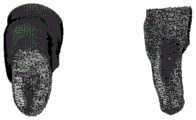
图1a为臀部肌肉群示意图;Figure 1a is a schematic diagram of the buttocks muscle group;
图1b为肌肉前群示意图;Figure 1b is a schematic diagram of the anterior muscle group;
图1c为肌肉内侧群示意图;Figure 1c is a schematic diagram of the inner muscle group;
图1d为肌肉后群示意图;Figure 1d is a schematic diagram of the posterior muscle group;
图2为人体髋部组织有限元模型剖视图;Figure 2 is a sectional view of a finite element model of human hip tissue;
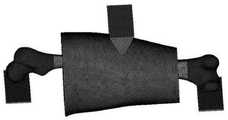
图3为实验有限元模型示意图;Figure 3 is a schematic diagram of the experimental finite element model;
图4为仿真环境下的载荷与约束;Figure 4 shows the loads and constraints in the simulation environment;
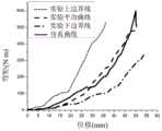
图5为力-位移曲线(远心端1/3处加载)示意图;Figure 5 is a schematic diagram of the force-displacement curve (loading at 1/3 of the distal end);
图6弯矩-位移曲线(远心端1/3处加载)示意图;Fig. 6 Schematic diagram of bending moment-displacement curve (loading at 1/3 of the distal end);
图7大腿中部弯曲实验验证力-位移曲线示意图;Fig. 7 Schematic diagram of the force-displacement curve of the mid-thigh bending experiment verification;
图8大腿中部弯曲实验验证弯矩-位移曲线示意图。Fig. 8 Schematic diagram of the bending moment-displacement curve verified by the mid-thigh bending experiment.
具体实施方式Detailed ways
具体实施方式一:本实施方式的一种基于肌肉群的人体髋部建模与仿真方法具体是按照以下步骤进行的:Embodiment 1: A muscle group-based modeling and simulation method for a human hip in this embodiment is specifically carried out according to the following steps:
步骤一、人体髋部图像数据获取;Step 1, the acquisition of human hip image data;
步骤二、在人体髋部图像数据获取的基础上,进行人体髋部几何建模,包括髋骨(皮质骨与松质骨)、股骨(皮质骨与松质骨)、关节软骨、缝匠肌、股四头肌、股二头肌、半腱肌、半膜肌、股薄肌、趾骨肌、长收肌、短收肌、大收肌、臀大肌、臀中肌、臀小肌、整个髋部的几何三维重建;Step 2: On the basis of the acquisition of human hip image data, carry out geometric modeling of human hip, including hip bone (cortical bone and cancellous bone), femur (cortical bone and cancellous bone), articular cartilage, sartorius muscle , quadriceps femoris, biceps femoris, semitendinosus, semimembranosus, gracilis, phalanges, adductor longus, adductor brevis, adductor magnus, gluteus maximus, gluteus medius, gluteus minimus, Geometric 3D reconstruction of the entire hip;
步骤三、在人体髋部几何模型基础上,建立肌肉群模型,对髋骨、股骨、关节软骨、肌肉群、整个髋部实施模型装配,建立脂肪和皮肤模型,添加肌腱;Step 3: On the basis of the geometric model of the human hip, establish a muscle group model, implement model assembly for the hip bone, femur, articular cartilage, muscle group, and the entire hip, establish fat and skin models, and add tendons;
步骤四、在人体髋部装配模型基础上,确定网格划分参数,确定组织间接触关系,实施几何模型的网格划分;Step 4: On the basis of the human hip assembly model, determine the meshing parameters, determine the contact relationship between the tissues, and implement the meshing of the geometric model;
步骤五、在人体髋部模型网格划分基础上,为人体髋部模型各部分结构附材质,确定材料参数;Step 5: On the basis of the mesh division of the human hip model, attach materials to the structures of each part of the human hip model, and determine the material parameters;
步骤六、在人体髋部模型附材质的基础上,实现人体髋部模型的力学仿真。Step 6: On the basis of the attached material of the human hip model, the mechanical simulation of the human hip model is realized.
具体实施方式二:本实施方式与具体实施方式一不同的是:所述步骤一中人体髋部图像数据获取;具体过程为:Embodiment 2: The difference between this embodiment and Embodiment 1 is: in the step 1, the image data of the human body is acquired; the specific process is:
图像数据来源于实验室成员,女,26岁,身高:170cm,体重:62kg,身体状况良好,无下肢骨折或肌肉损伤等病史。CT数据在中国人民解放军第二一一医院进行采集,MRI数据在哈尔滨医科大学附属第一医院进行采集。CT图像扫描参数:轴位连续断层扫描,电压140.0KV,电流180mA,层厚1mm,矩阵512×512,视野FOV 18cm,共采集700张图像。MRI图像扫描参数:轴位扫描,T2加权自旋回波序列,重复时间(TR)566.7ms,回波时间(TE)10.6ms,矩阵512×512,层厚3mm,视野(FOV)17cm,共采集182张图像。CT图像数据主要用于骨骼和下肢整体外形结构的建模,MRI图像数据主要用于软组织建模。The image data comes from a laboratory member, female, 26 years old, height: 170cm, weight: 62kg, in good physical condition, with no history of lower extremity fractures or muscle damage. CT data were collected at the 211th Hospital of the Chinese People's Liberation Army, and MRI data were collected at the First Affiliated Hospital of Harbin Medical University. CT image scanning parameters: axial continuous tomography, voltage 140.0KV, current 180mA, slice thickness 1mm, matrix 512×512, field of view FOV 18cm, a total of 700 images were collected. MRI image scanning parameters: axial scan, T2-weighted spin echo sequence, repetition time (TR) 566.7ms, echo time (TE) 10.6ms, matrix 512×512, slice thickness 3mm, field of view (FOV) 17cm, total acquisition 182 images. CT image data is mainly used for modeling the overall shape and structure of bones and lower limbs, and MRI image data is mainly used for soft tissue modeling.
其它步骤及参数与具体实施方式一相同。Other steps and parameters are the same as in the first embodiment.
具体实施方式三:本实施方式与具体实施方式一或二不同的是:所述步骤二中在人体髋部图像数据获取的基础上,进行人体髋部几何建模,包括髋骨(皮质骨与松质骨)、股骨(皮质骨与松质骨)、关节软骨、缝匠肌、股四头肌、股二头肌、半腱肌、半膜肌、股薄肌、趾骨肌、长收肌、短收肌、大收肌、臀大肌、臀中肌、臀小肌、整个髋部的几何三维重建;具体过程为:Embodiment 3: This embodiment differs from Embodiment 1 or 2 in that: in the second step, based on the acquisition of human hip image data, the geometric modeling of the human hip is performed, including the hip bone (cortical bone and cancellous bone), femur (cortical and cancellous), articular cartilage, sartorius, quadriceps femoris, biceps femoris, semitendinosus, semimembranosus, gracilis, phalanx, adductor longus , geometric three-dimensional reconstruction of the adductor brevis, adductor magnus, gluteus maximus, gluteus medius, gluteus minimus, and the entire hip; the specific process is:
利用图像数据生成骨组织(股骨和髋骨)和软组织的三维几何模型。首先对医学图像进行图像分割,图像分割按照阈值分割、区域增长、蒙版编辑三个步骤进行;Image data are used to generate 3D geometric models of bone tissue (femur and hip) and soft tissue. First, image segmentation is performed on medical images, and image segmentation is performed according to three steps: threshold segmentation, region growth, and mask editing;
通过选择不同的灰度值区间实现目标组织分离就是阈值分割;To achieve target tissue separation by selecting different gray value ranges is threshold segmentation;
经过初步阈值分割后的蒙板上具有相同灰度值区间的组织同时显示出来,为了将这些具有相同的灰度值区间的不同组织进行分离,我们进行进一步的详细分割去除蒙板中的噪点从而将相互不连接的单独区域区分开并生成表达之前灰度值区间中的某一组织的新蒙板,经过区域增长我们实现了相同灰度值区间内不同组织的分离;The tissues with the same gray value interval on the mask after the initial threshold segmentation are displayed simultaneously. In order to separate these different tissues with the same gray value interval, we perform further detailed segmentation to remove the noise in the mask so as to Distinguish separate areas that are not connected to each other and generate a new mask that expresses a certain tissue in the previous gray value interval. After region growth, we realize the separation of different tissues in the same gray value interval;
最后对蒙板进行修缘、擦除、补充等操作实现蒙版编辑;Finally, perform operations such as trimming, erasing, and supplementing the mask to achieve mask editing;
对蒙版编辑过的模型进行光滑处理;Smooth the mask-edited model;
对编辑过的模型进行三角面片的缩减;Reduce triangular patches on the edited model;
对模型表面的三角面片进行检测,将检测出的自相交的三角面片和尖状物删除;Detect the triangular patches on the model surface, and delete the detected self-intersecting triangular patches and sharp objects;
对模型表面的空洞进行填充;Fill the voids on the surface of the model;
提取模型的轮廓线;Extract the contour lines of the model;
完成模型的栅格构造;Complete the grid construction of the model;
对模型进行曲面拟合最终生成三维几何模型。Surface fitting is performed on the model to finally generate a 3D geometric model.
其它步骤及参数与具体实施方式一或二相同。Other steps and parameters are the same as in the first or second embodiment.
具体实施方式四:本实施方式与具体实施方式一至三之一不同的是:所述步骤三中在人体髋部几何模型基础上,建立肌肉群模型,对髋骨、股骨、关节软骨、肌肉群、整个髋部实施模型装配,建立脂肪和皮肤模型,添加肌腱;具体过程为:Embodiment 4: This embodiment is different from one of Embodiments 1 to 3 in that: in the step 3, on the basis of the geometric model of the human hip, a muscle group model is established, and the hip bone, femur, articular cartilage, muscle group are , Implement model assembly for the entire hip, build fat and skin models, and add tendons; the specific process is:
根据各个肌肉组织的功能将肌肉组织划分为四个肌肉群,分别为:肌肉前群(包括缝匠肌、股四头肌、股二头肌)如图1a所示、肌肉后群(包括半腱肌、半膜肌、股薄肌、趾骨肌)如图1b所示、肌肉内侧群(长收肌、短收肌、大收肌)如图1c所示和肌肉臀部群(臀大肌、臀中肌、臀小肌)如图1d所示。肌肉前群主要功能为屈髋伸膝,肌肉后群主要功能为伸髋屈膝,肌肉内侧群主要功能为内收外旋髋关节,肌肉臀部群主要功能为伸髋关节、外展髋关节;According to the function of each muscle tissue, the muscle tissue is divided into four muscle groups, namely: the anterior muscle group (including sartorius, quadriceps femoris, and biceps femoris) as shown in Figure 1a, the posterior muscle group (including half Tendons, semimembranosus, gracilis, phalanges) are shown in Figure 1b, the medial groups of muscles (adductor longus, brevis, adductor magnus) are shown in Figure 1c, and the muscles of the buttocks (gluteus maximus, gluteus medius, gluteus minimus) are shown in Figure 1d. The main function of the anterior muscle group is to flex the hip and extend the knee, the main function of the posterior muscle group is to extend the hip and flex the knee, the main function of the inner muscle group is to adduct and externally rotate the hip joint, and the main function of the muscle buttock group is to extend the hip joint and abduct the hip joint;
将缝匠肌、股四头肌、股二头肌的几何模型合并为肌肉前群模型,对模型表面进行优化处理;Combine the geometric models of sartorius, quadriceps, and biceps into a muscle anterior group model, and optimize the surface of the model;
将半腱肌、半膜肌、股薄肌、趾骨肌的几何模型合并为肌肉后群模型,对模型表面进行优化处理;Combine the geometric models of semitendinosus, semimembranosus, gracilis, and phalangeal muscles into a muscle posterior group model, and optimize the surface of the model;
将长收肌、短收肌、大收肌的几何模型合并为肌肉内侧群模型,对模型表面进行优化处理;Combine the geometric models of the adductor longus, brevis, and adductor magnus into a muscle medial group model, and optimize the surface of the model;
将臀大肌、臀中肌、臀小肌的几何模型合并为肌肉臀部群模型,对模型表面进行优化处理;Combine the geometric models of gluteus maximus, gluteus medius, and gluteus minimus into a muscle buttocks group model, and optimize the surface of the model;
将髋骨(皮质骨与松质骨)、股骨(皮质骨与松质骨)、关节软骨、肌肉前群、肌肉后群、肌肉内侧群、肌肉臀部群的几何模型按照生理学位置实施装配;Assemble the geometric models of hip bone (cortical bone and cancellous bone), femur (cortical bone and cancellous bone), articular cartilage, muscle anterior group, muscle posterior group, muscle medial group, and muscle hip group according to the physiological position;
将装配体与整体髋部几何模型进行求差操作,去除装配体,剩下的求差体就是皮肤和脂肪的几何模型;Perform a difference operation between the assembly and the overall hip geometric model, remove the assembly, and the remaining difference is the geometric model of skin and fat;
将整个髋部几何模型最外面1mm的厚度定义为皮肤模型;Define the thickness of the outermost 1mm of the entire hip geometric model as the skin model;
肌肉与骨骼通过肌腱连接,用线单元来模拟肌腱。Muscles and bones are connected by tendons, which are modeled with wire elements.
其它步骤及参数与具体实施方式一至三之一相同。Other steps and parameters are the same as one of the first to third embodiments.
具体实施方式五:本实施方式与具体实施方式一至四之一不同的是:所述步骤四中在人体髋部装配模型基础上,确定网格划分参数,确定组织间接触关系,实施几何模型的网格划分;具体过程为:Embodiment 5: The difference between this embodiment and one of Embodiments 1 to 4 is that: in the step 4, based on the human hip assembly model, the meshing parameters are determined, the contact relationship between tissues is determined, and the geometric model is implemented. Meshing; the specific process is:
选用四面体网格作为所有组织的网格单元形状;Select a tetrahedral mesh as the mesh element shape for all organizations;
髋骨皮质骨、股骨皮质骨和皮肤选用壳单元模拟;The cortical bone of the hip, the cortical bone of the femur and the skin are simulated by shell elements;
髋骨松质骨、股骨松质骨、肌肉群、脂肪选用体单元模拟;Hip cancellous bone, femoral cancellous bone, muscle group, fat selection body unit simulation;
组织间采用共节点接触;Common node contact is adopted between organizations;
网格划分参数:扭曲度小于29、雅克比大于0.6、翘曲率小于7、长宽比小于58、最大内角大于30度、最小内角小于150度;Meshing parameters: distortion less than 29, Jacobian greater than 0.6, warpage less than 7, aspect ratio less than 58, maximum interior angle greater than 30 degrees, and minimum interior angle less than 150 degrees;
该模型共包括14440个节点和72962个四面体单元如图2所示;The model includes a total of 14440 nodes and 72962 tetrahedral elements as shown in Figure 2;
其它步骤及参数与具体实施方式一至四之一相同。Other steps and parameters are the same as one of the first to fourth embodiments.
具体实施方式六:本实施方式与具体实施方式一至五之一不同的是:所述步骤五中在人体髋部模型网格划分基础上,为人体髋部模型各部分结构附材质,确定材料参数;具体过程为:Embodiment 6: The difference between this embodiment and one of Embodiments 1 to 5 is that: in the
将髋骨皮质骨厚度和股骨皮质骨厚度设置为2mm;Set hip cortical bone thickness and femoral cortical bone thickness to 2 mm;
髋骨和股骨的皮质骨采用分段线性材料模型,松质骨采用分段线性材料模型,关节软骨选用线弹性材料模型;The cortical bone of the hip bone and the femur adopts the piecewise linear material model, the cancellous bone adopts the piecewise linear material model, and the articular cartilage adopts the linear elastic material model;
关节软骨材料参数设置为:弹性模量10Mpa、泊松比0.4;The articular cartilage material parameters are set as: elastic modulus 10Mpa, Poisson's ratio 0.4;
股骨、髋骨的材料参数:股骨干皮质骨:密度2000kg/m3,泊松比0.3,弹性模量17.3GPa,屈服应力54.5MPa,极限应变1.6%。股骨干松质质骨:密度861.5kg/m3,泊松比0.3,弹性模量0.04GPa,屈服应力13.3MPa,极限应变13.4%。股骨两端皮质骨:密度2000kg/m3,泊松比0.3,弹性模量17.3GPa,屈服应力34.5MPa,极限应变2.8%。股骨两端松质骨:密度861.5kg/m3,泊松比0.3,弹性模量0.16GPa,屈服应力29.0MPa,极限应变20.0%。髋骨皮质骨:密度1900kg/m3,泊松比0.3,弹性模量15GPa,屈服应力50.0MPa,极限应变1.5%。髋骨松质骨:密度750kg/m3,泊松比0.3,弹性模量0.10GPa,屈服应力31.5MPa,极限应变15.5%。Material parameters of femur and hip bone: Femoral shaft cortical bone: density 2000kg/m3, Poisson's ratio 0.3, elastic modulus 17.3GPa, yield stress 54.5MPa, ultimate strain 1.6%. Femoral shaft cancellous bone: density 861.5kg/m3, Poisson's ratio 0.3, elastic modulus 0.04GPa, yield stress 13.3MPa, ultimate strain 13.4%. Cortical bone at both ends of the femur: density 2000kg/m3, Poisson's ratio 0.3, elastic modulus 17.3GPa, yield stress 34.5MPa, ultimate strain 2.8%. Cancellous bone at both ends of the femur: density 861.5kg/m3, Poisson's ratio 0.3, elastic modulus 0.16GPa, yield stress 29.0MPa, ultimate strain 20.0%. Hip cortical bone: density 1900kg/m3, Poisson's ratio 0.3, elastic modulus 15GPa, yield stress 50.0MPa, ultimate strain 1.5%. Cancellous bone of hip bone: density 750kg/m3, Poisson's ratio 0.3, elastic modulus 0.10GPa, yield stress 31.5MPa, ultimate strain 15.5%.
肌肉和脂肪设置为粘弹性材料模型,设置为体单元模拟。肌肉、脂肪材料参数为:肌肉:密度1600kg/m3,体积模量19MPa,短期剪切模量0.22MPa,长期剪切模量0.095MPa,衰减系数100s-1。脂肪:密度1000kg/m3,体积模量20MPa,短期剪切模量0.12MPa,长期剪切模量0.04MPa,衰减系数100s-1。Muscle and fat are set up as viscoelastic material models and set up as body element simulations. The parameters of muscle and fat material are: muscle: density 1600kg/m3, bulk modulus 19MPa, short-term shear modulus 0.22MPa, long-term shear modulus 0.095MPa, attenuation coefficient 100s-1 . Fat: density 1000kg/m3, bulk modulus 20MPa, short-term shear modulus 0.12MPa, long-term shear modulus 0.04MPa, attenuation coefficient 100s-1 .
皮肤选用线弹性材料模型并用壳单元模拟,皮肤的弹性模量设置为1Mpa。The skin is a linear elastic material model and simulated with shell elements, and the elastic modulus of the skin is set to 1Mpa.
其它步骤及参数与具体实施方式一至五之一相同。Other steps and parameters are the same as one of the specific embodiments one to five.
具体实施方式七:本实施方式与具体实施方式一至七之一不同的是:所述步骤六中在人体髋部模型附材质的基础上,实现人体髋部模型的力学仿真;具体过程为:Embodiment 7: The difference between this embodiment and Embodiments 1 to 7 is: in the step 6, the mechanical simulation of the human hip model is realized on the basis of the material attached to the human hip model; the specific process is:
在建立的人体髋部有限元模型的基础上,根据实验的装置和条件建立仿真模型,在仿真环境中,载荷和约束设置与生物力学实验保持一致,进行仿真,提取结果曲线。On the basis of the established human hip finite element model, a simulation model is established according to the experimental device and conditions. In the simulation environment, the load and constraint settings are consistent with the biomechanical experiment, and the simulation is performed to extract the result curve.
采用以下实施例验证本发明的有益效果:Adopt the following examples to verify the beneficial effects of the present invention:
本实施例一种基于肌肉群的人体髋部建模与仿真方法具体是按照以下步骤制备的:A muscle group-based modeling and simulation method for a human hip of the present embodiment is specifically prepared according to the following steps:
在建立的人体髋部有限元模型的基础上,根据实验的装置和条件建立弯曲实验仿真模型,如图3所示。Kerrigan等人利用12具尸体进行了12次实验,其中六次载荷位置为大腿远心端1/3处,其余六次载荷位置为大腿中部,载荷的方向均设置为L-M(大腿外侧向内侧)方向。在仿真环境中,载荷和约束设置与生物力学实验保持一致如图4所示,具体参数如表1所示;On the basis of the established human hip finite element model, a bending experiment simulation model is established according to the experimental device and conditions, as shown in Figure 3. Kerrigan et al. conducted 12 experiments using 12 cadavers, six of which were located at the distal 1/3 of the thigh, and the remaining six were located in the middle of the thigh. The direction of the load was set to L-M (outside of the thigh to the inside). direction. In the simulation environment, the load and constraint settings are consistent with the biomechanical experiments as shown in Figure 4, and the specific parameters are shown in Table 1;
当在大腿远心端1/3处加载仿真过程中,软组织首先产生变形,变形量越来越大,这个阶段股骨变形量较小,当加载到一定位移后,股骨开始变形,直到模型失效股骨骨折。力-位移曲线和弯矩-位移曲线分别如图5和图6所示;When the simulation process is loaded at the distal 1/3 of the thigh, the soft tissue first deforms, and the deformation becomes larger and larger. At this stage, the deformation of the femur is small. When the load reaches a certain displacement, the femur begins to deform until the model fails. fracture. The force-displacement curve and the bending moment-displacement curve are shown in Figure 5 and Figure 6, respectively;
大腿中部加载的仿真结果,冲击块撞击大腿中部直至模型失效,力-位移曲线和弯矩-位移曲线分别如图分别如图7和图8所示。The simulation results of loading in the middle of the thigh, the impact block hits the middle of the thigh until the model fails, the force-displacement curve and the bending moment-displacement curve are shown in Figures 7 and 8, respectively.
表1实验参数设置Table 1 Experimental parameter settings

本发明还可有其它多种实施例,在不背离本发明精神及其实质的情况下,本领域技术人员当可根据本发明作出各种相应的改变和变形,但这些相应的改变和变形都应属于本发明所附的权利要求的保护范围。The present invention can also have other various embodiments. Without departing from the spirit and essence of the present invention, those skilled in the art can make various corresponding changes and deformations according to the present invention, but these corresponding changes and deformations are all It should belong to the protection scope of the appended claims of the present invention.
Claims (4)
Translated fromChinesePriority Applications (1)
| Application Number | Priority Date | Filing Date | Title |
|---|---|---|---|
| CN201710753127.4ACN108334730B (en) | 2017-08-29 | 2017-08-29 | A Muscle Group-Based Modeling and Simulation Method of Human Hip |
Applications Claiming Priority (1)
| Application Number | Priority Date | Filing Date | Title |
|---|---|---|---|
| CN201710753127.4ACN108334730B (en) | 2017-08-29 | 2017-08-29 | A Muscle Group-Based Modeling and Simulation Method of Human Hip |
Publications (2)
| Publication Number | Publication Date |
|---|---|
| CN108334730A CN108334730A (en) | 2018-07-27 |
| CN108334730Btrue CN108334730B (en) | 2020-01-31 |
Family
ID=62922396
Family Applications (1)
| Application Number | Title | Priority Date | Filing Date |
|---|---|---|---|
| CN201710753127.4AExpired - Fee RelatedCN108334730B (en) | 2017-08-29 | 2017-08-29 | A Muscle Group-Based Modeling and Simulation Method of Human Hip |
Country Status (1)
| Country | Link |
|---|---|
| CN (1) | CN108334730B (en) |
Families Citing this family (8)
| Publication number | Priority date | Publication date | Assignee | Title |
|---|---|---|---|---|
| EP3893787B1 (en)* | 2018-12-12 | 2025-09-10 | Howmedica Osteonics Corp. | Bone density modeling and orthopedic surgical planning system |
| CN111445580B (en)* | 2018-12-28 | 2023-05-05 | 天津科技大学 | Construction method and system of six-year-old child pedestrian natural walking pose finite element model suitable for Euro NCAP |
| CN109977590A (en)* | 2019-04-09 | 2019-07-05 | 哈尔滨理工大学 | A kind of titanium implant structural optimization method based on age factor |
| CN112113836A (en)* | 2020-03-02 | 2020-12-22 | 四川大学 | Intra-articular pressure and bone non-contact strain measurement system based on 3D printing |
| CN113033050B (en)* | 2021-03-23 | 2022-09-27 | 上海工程技术大学 | Reliability evaluation method for dressing test data of smart clothing flexible pressure sensor |
| CN113822994B (en)* | 2021-11-24 | 2022-02-15 | 深圳普罗米修斯视觉技术有限公司 | Three-dimensional model construction method and device and storage medium |
| CN118614868B (en)* | 2023-04-13 | 2025-01-24 | 四川大学 | An experimental test method for estimating the bending strength and disturbance of rat femur |
| CN119964823B (en)* | 2025-04-09 | 2025-06-13 | 贵州利美康外科医院股份有限公司 | Shaping effect simulation display method and system based on 3D simulation technology |
Citations (4)
| Publication number | Priority date | Publication date | Assignee | Title |
|---|---|---|---|---|
| CN103530466A (en)* | 2013-10-21 | 2014-01-22 | 哈尔滨理工大学 | Method for optimally selecting thighbone prostheses based on material performance multi-objective optimization |
| EP2830033A1 (en)* | 2013-06-26 | 2015-01-28 | Dassault Systemes Simulia Corp. | Musculo-skeletal modeling using finite element analysis, process integration, and design optimization |
| CN106021977A (en)* | 2016-07-14 | 2016-10-12 | 哈尔滨理工大学 | Biomechanics modeling method for subcutaneous adipose tissues based on linear elasticity and superelasticity models |
| CN106991720A (en)* | 2017-06-02 | 2017-07-28 | 南方医科大学 | A personalized acetabular reconstruction plate pre-bending method based on finite element analysis |
Family Cites Families (2)
| Publication number | Priority date | Publication date | Assignee | Title |
|---|---|---|---|---|
| CN105303605B (en)* | 2015-10-26 | 2017-10-13 | 哈尔滨理工大学 | A kind of bone surgery analogue system based on force feedback |
| CN106227924B (en)* | 2016-07-14 | 2017-10-10 | 哈尔滨理工大学 | A kind of hipbone model meshes division methods |
- 2017
- 2017-08-29CNCN201710753127.4Apatent/CN108334730B/ennot_activeExpired - Fee Related
Patent Citations (4)
| Publication number | Priority date | Publication date | Assignee | Title |
|---|---|---|---|---|
| EP2830033A1 (en)* | 2013-06-26 | 2015-01-28 | Dassault Systemes Simulia Corp. | Musculo-skeletal modeling using finite element analysis, process integration, and design optimization |
| CN103530466A (en)* | 2013-10-21 | 2014-01-22 | 哈尔滨理工大学 | Method for optimally selecting thighbone prostheses based on material performance multi-objective optimization |
| CN106021977A (en)* | 2016-07-14 | 2016-10-12 | 哈尔滨理工大学 | Biomechanics modeling method for subcutaneous adipose tissues based on linear elasticity and superelasticity models |
| CN106991720A (en)* | 2017-06-02 | 2017-07-28 | 南方医科大学 | A personalized acetabular reconstruction plate pre-bending method based on finite element analysis |
Also Published As
| Publication number | Publication date |
|---|---|
| CN108334730A (en) | 2018-07-27 |
Similar Documents
| Publication | Publication Date | Title |
|---|---|---|
| CN108334730B (en) | A Muscle Group-Based Modeling and Simulation Method of Human Hip | |
| CN105997306B (en) | A kind of bone, which implants, fills the design method of perforated grill structure | |
| San Antonio et al. | Orientation of orthotropic material properties in a femur FE model: A method based on the principal stresses directions | |
| Butz et al. | Stress distributions and material properties determined in articular cartilage from MRI-based finite strains | |
| Rowe et al. | Biomechanics of juvenile tyrannosaurid mandibles and their implications for bite force: Evolutionary biology | |
| CN105678845A (en) | 3D printing customized modeling method based on finite element analysis | |
| Francis et al. | Computational modeling of human femur using CT data for finite element analysis | |
| Dao et al. | Multimodal medical imaging (CT and dynamic MRI) data and computer-graphics multi-physical model for the estimation of patient specific lumbar spine muscle forces | |
| Czyż et al. | Finite element modelling of the cervical spinal cord injury–clinical assessment | |
| Mo et al. | Coupling musculoskeletal dynamics and subject‐specific finite element analysis of femoral cortical bone failure after endoprosthetic knee replacement | |
| Shireesha et al. | Modelling and static analysis of femur bone by using different implant materials | |
| Phate et al. | Three-Dimensional Finite Element Analysis of Human Tibia Bone | |
| Zhang et al. | Heterogeneous modelling and finite element analysis of the femur | |
| Torres-Moreno et al. | Magnetic resonance imaging of residual soft tissues for computer-aided technology applications in prosthetics—a case study | |
| Geraldes | Orthotropic modelling of the skeletal system | |
| Larin et al. | Automated 3D CAE modelling of femur human bones | |
| Fu et al. | Biomechanics analysis of human proximal femur under four different standing postures based on finite element method | |
| Wang et al. | Establishment and application of lower limb finite element model based on muscle groups | |
| Zhiheng et al. | A novel finite element method based biomechanical model for HIT-robot assisted orthopedic surgery system | |
| Vulović et al. | Finite Element Analysis of Femur During Gait Cycle | |
| Scheuring | Assessing activity-specific fracture risk in femora with metastatic lesions | |
| Tudora et al. | Virtual human hip joint obtained from CT images and FEM test | |
| Zhang et al. | Computer tomography scanning and modelling of ostrich foot | |
| Zhu | Techniques for finite element modeling and remodeling of bones with applications to pig skulls | |
| Long | Modeling and analysis of knee joint impact damage in triple jump manipulators based on finite element method |
Legal Events
| Date | Code | Title | Description |
|---|---|---|---|
| PB01 | Publication | ||
| PB01 | Publication | ||
| SE01 | Entry into force of request for substantive examination | ||
| SE01 | Entry into force of request for substantive examination | ||
| GR01 | Patent grant | ||
| GR01 | Patent grant | ||
| CF01 | Termination of patent right due to non-payment of annual fee | ||
| CF01 | Termination of patent right due to non-payment of annual fee | Granted publication date:20200131 Termination date:20200829 |