CN108292530B - Chest wall estimation from optical scanning - Google Patents
Chest wall estimation from optical scanningDownload PDFInfo
- Publication number
- CN108292530B CN108292530BCN201680070819.2ACN201680070819ACN108292530BCN 108292530 BCN108292530 BCN 108292530BCN 201680070819 ACN201680070819 ACN 201680070819ACN 108292530 BCN108292530 BCN 108292530B
- Authority
- CN
- China
- Prior art keywords
- image
- outer layer
- wall
- estimate
- thickness
- Prior art date
- Legal status (The legal status is an assumption and is not a legal conclusion. Google has not performed a legal analysis and makes no representation as to the accuracy of the status listed.)
- Active
Links
Images


Classifications
- G—PHYSICS
- G06—COMPUTING OR CALCULATING; COUNTING
- G06T—IMAGE DATA PROCESSING OR GENERATION, IN GENERAL
- G06T7/00—Image analysis
- G06T7/0002—Inspection of images, e.g. flaw detection
- G06T7/0012—Biomedical image inspection
- G—PHYSICS
- G16—INFORMATION AND COMMUNICATION TECHNOLOGY [ICT] SPECIALLY ADAPTED FOR SPECIFIC APPLICATION FIELDS
- G16H—HEALTHCARE INFORMATICS, i.e. INFORMATION AND COMMUNICATION TECHNOLOGY [ICT] SPECIALLY ADAPTED FOR THE HANDLING OR PROCESSING OF MEDICAL OR HEALTHCARE DATA
- G16H50/00—ICT specially adapted for medical diagnosis, medical simulation or medical data mining; ICT specially adapted for detecting, monitoring or modelling epidemics or pandemics
- G16H50/50—ICT specially adapted for medical diagnosis, medical simulation or medical data mining; ICT specially adapted for detecting, monitoring or modelling epidemics or pandemics for simulation or modelling of medical disorders
- G—PHYSICS
- G06—COMPUTING OR CALCULATING; COUNTING
- G06T—IMAGE DATA PROCESSING OR GENERATION, IN GENERAL
- G06T7/00—Image analysis
- G06T7/50—Depth or shape recovery
- G—PHYSICS
- G06—COMPUTING OR CALCULATING; COUNTING
- G06T—IMAGE DATA PROCESSING OR GENERATION, IN GENERAL
- G06T7/00—Image analysis
- G06T7/60—Analysis of geometric attributes
- G—PHYSICS
- G16—INFORMATION AND COMMUNICATION TECHNOLOGY [ICT] SPECIALLY ADAPTED FOR SPECIFIC APPLICATION FIELDS
- G16H—HEALTHCARE INFORMATICS, i.e. INFORMATION AND COMMUNICATION TECHNOLOGY [ICT] SPECIALLY ADAPTED FOR THE HANDLING OR PROCESSING OF MEDICAL OR HEALTHCARE DATA
- G16H30/00—ICT specially adapted for the handling or processing of medical images
- G16H30/40—ICT specially adapted for the handling or processing of medical images for processing medical images, e.g. editing
- G—PHYSICS
- G06—COMPUTING OR CALCULATING; COUNTING
- G06T—IMAGE DATA PROCESSING OR GENERATION, IN GENERAL
- G06T2207/00—Indexing scheme for image analysis or image enhancement
- G06T2207/10—Image acquisition modality
- G06T2207/10028—Range image; Depth image; 3D point clouds
- G—PHYSICS
- G06—COMPUTING OR CALCULATING; COUNTING
- G06T—IMAGE DATA PROCESSING OR GENERATION, IN GENERAL
- G06T2207/00—Indexing scheme for image analysis or image enhancement
- G06T2207/30—Subject of image; Context of image processing
- G06T2207/30004—Biomedical image processing
- G06T2207/30068—Mammography; Breast
- G—PHYSICS
- G06—COMPUTING OR CALCULATING; COUNTING
- G06T—IMAGE DATA PROCESSING OR GENERATION, IN GENERAL
- G06T2207/00—Indexing scheme for image analysis or image enhancement
- G06T2207/30—Subject of image; Context of image processing
- G06T2207/30004—Biomedical image processing
- G06T2207/30088—Skin; Dermal
Landscapes
- Engineering & Computer Science (AREA)
- Health & Medical Sciences (AREA)
- Medical Informatics (AREA)
- Public Health (AREA)
- General Health & Medical Sciences (AREA)
- Physics & Mathematics (AREA)
- Primary Health Care (AREA)
- Epidemiology (AREA)
- Computer Vision & Pattern Recognition (AREA)
- General Physics & Mathematics (AREA)
- Theoretical Computer Science (AREA)
- Radiology & Medical Imaging (AREA)
- Nuclear Medicine, Radiotherapy & Molecular Imaging (AREA)
- Biomedical Technology (AREA)
- Data Mining & Analysis (AREA)
- Databases & Information Systems (AREA)
- Pathology (AREA)
- Geometry (AREA)
- Quality & Reliability (AREA)
- Apparatus For Radiation Diagnosis (AREA)
Abstract
Translated fromChinese
Description
Translated fromChinese技术领域technical field
本发明涉及图像处理系统、图像处理方法、计算机可读介质以及计算机程序单元。The present invention relates to an image processing system, an image processing method, a computer readable medium and a computer program element.
背景技术Background technique
乳腺癌是西方世界女性遭受的最常见的癌症类型。Breast cancer is the most common type of cancer suffered by women in the Western world.
患者有几种治疗选择,例如,手术,其中,乳房组织的受影响部分会被移除。在联合决策制定的背景下,已经开发出了生物力学方法来预先模拟乳房手术的结果。该模拟为患者或医学人员提供了手术结果的视觉表示以更好地理解预后。Patients have several treatment options, such as surgery, in which the affected portion of breast tissue is removed. In the context of joint decision-making, biomechanical methods have been developed to pre-simulate the outcome of breast surgery. The simulation provides the patient or medical staff with a visual representation of the surgical outcome to better understand the prognosis.
目前,生物力学组件(例如,人体躯干)的这种模拟的产生是基于针对特定患者中的任一个采集的MR图像或CT图像。换句话说,在能够计算针对给定患者的模拟之前,需要首先采集特定患者的图像。然而,这种图像的采集要么非常昂贵,要么由于X辐射情况下的辐射剂量而本身会造成健康风险。Currently, the generation of such simulations of biomechanical components (eg, human torso) is based on MR images or CT images acquired for any one of a particular patient. In other words, before a simulation for a given patient can be calculated, images of a particular patient need to be acquired first. However, the acquisition of such images is either very expensive or itself poses a health risk due to the radiation dose in the case of X-rays.
发明内容SUMMARY OF THE INVENTION
因此需要替代系统或方法来促进更安全地产生对生物力学组件的模拟。Alternative systems or methods are therefore needed to facilitate safer generation of simulations of biomechanical components.
本发明的目的通过独立权利要求的主题来解决,其中,进一步的实施例被并入从属权利要求中。应当注意,本发明的以下描述的方面等同地适用于图像处理方法、图像处理系统、计算机程序单元和计算机可读介质。The object of the invention is solved by the subject-matter of the independent claims, wherein further embodiments are incorporated in the dependent claims. It should be noted that the aspects of the invention described below apply equally to the image processing method, the image processing system, the computer program element and the computer readable medium.
根据本发明的第一方面,提供了一种图像处理系统,包括:According to a first aspect of the present invention, an image processing system is provided, comprising:
输入端口,其用于接收当前生物力学组件的外层的(输入)表面图像,所述图像是由表面成像装置沿着至少一个成像方向采集的,所述组件包括相对于所述成像方向在所述外层后面的、从后面连结到所述外层的至少一个内壁元件;An input port for receiving an (input) surface image of the outer layer of the current biomechanical assembly, the image being acquired by the surface imaging device along at least one imaging direction, the assembly including the at least one inner wall element behind said outer layer and joined from behind to said outer layer;
壁估计器,其被配置为提供针对所述内壁元件的几何数据的估计,所述估计基于按照所述表面图像的图像信息。a wall estimator configured to provide an estimate of geometrical data for the inner wall element, the estimate being based on image information in accordance with the surface image.
以合适的数据结构(例如,多边形(例如,三角形)网格模型)或以函数方式用包络函数等来提供对几何数据的估计。几何数据具体包括对壁元件的形状和/或取向和/或位置的描述。An estimate of the geometric data is provided in a suitable data structure (eg, a polygonal (eg, triangular) mesh model) or in a functional manner with an envelope function or the like. The geometrical data includes in particular a description of the shape and/or orientation and/or position of the wall elements.
外层基本上遮挡壁元件而不能进行视觉检查。所提出的方法仍然旨在在给定由表面(例如,光学)成像装置捕捉的外层的几何形状的情况下推断“下层”壁元件的几何形状。这是可能的,因为在一些生物力学组件中,可见外层的至少部分的几何形状对应于至少一个下层壁元件。“壁元件”是指位于外层后面的可能较大的壁结构的部分。换句话说,能够在不使用昂贵或有害的非表面(即,穿透式)成像仪器(例如,MRI或CT)的情况下搜集关于下层壁元件的几何形状的信息。光学成像足以满足当前的目的。更具体地,在一个非限制性实施例中,生物力学组件是人体(例如,女性)躯干。皮肤定义了阻挡位于一个或两个乳房后面的肋骨笼的部分的外层。在一个实施例中,壁元件的“形状”是指由位于一个或两个乳房后面的肋骨形成的几何包络。作为对此的细化,壁元件可以定义由肋骨笼和覆盖的胸肌形成的内边界。在其他实施例中,壁元件仅与覆盖肋骨的胸肌壁相关。所提出的系统能够应用于被皮肤遮挡的人体或动物解剖结构的其他部分。The outer layer substantially blocks the wall elements from visual inspection. The proposed method still aims to infer the geometry of the "lower layer" wall elements given the geometry of the outer layer captured by surface (eg optical) imaging means. This is possible because in some biomechanical assemblies the geometry of at least part of the visible outer layer corresponds to at least one lower wall element. "Wall element" refers to the portion of a possibly larger wall structure that lies behind the outer layer. In other words, information about the geometry of the underlying wall elements can be gathered without the use of expensive or harmful non-surface (ie, penetrating) imaging instruments (eg, MRI or CT). Optical imaging is sufficient for current purposes. More specifically, in one non-limiting embodiment, the biomechanical component is a human (eg, female) torso. The skin defines the outer layer that blocks the portion of the rib cage behind one or both breasts. In one embodiment, the "shape" of the wall element refers to the geometric envelope formed by the ribs behind one or both breasts. As a refinement of this, the wall element may define the inner boundary formed by the rib cage and the overlying pectoral muscles. In other embodiments, the wall elements are only associated with the pectoralis muscle wall covering the ribs. The proposed system can be applied to other parts of human or animal anatomy that are occluded by skin.
使用由所提出的系统提供的几何数据能够有益于生物力学模拟。在对乳房的生物力学模拟中,最好知道后边界的形状和位置两者,即,肋骨笼的乳房组织连结到的那部分的形状和位置。Using the geometric data provided by the proposed system can benefit biomechanical simulations. In a biomechanical simulation of the breast, it is desirable to know both the shape and location of the posterior boundary, ie the shape and location of the portion of the rib cage to which the breast tissue is attached.
根据一个实施例,所述外层包括凸起部分,并且所述系统还包括图像分析器,所述图像分析器被配置为将所述表面图像分析为表示所述凸起部分的(第一)部分图像,其中,所述壁估计器被配置为提供所述估计,同时忽略按照所述部分图像的图像信息。换句话说,所述第一部分图像的补集形成输入图像的其余部分(其余图像),并且估计仅基于该其余图像,第一部分图像中的图像信息被忽略。在一个实施例中,通过将几何模型(“基元”)拟合到输入表面图像的仅其余部分来获得这种壁估计。这种几何模型是一种“基元”,其整体形状类型与输入表面图像所限定的形状相对应。在女性人体躯干实施例中,凸起部分对应于乳房组织。该方法利用了这样的事实:如果忽略乳房几何形状的原样的“扭曲”效应,则皮肤轮廓的部分至少在形状上(但不一定在位置上)与肋骨笼壁形状相对应。According to one embodiment, the outer layer includes raised portions and the system further comprises an image analyzer configured to analyze the surface image into (first) representations of the raised portions a partial image, wherein the wall estimator is configured to provide the estimate while ignoring image information per the partial image. In other words, the complement of the first partial image forms the rest of the input image (the rest image) and the estimation is based only on this rest image, the image information in the first partial image being ignored. In one embodiment, this wall estimate is obtained by fitting a geometric model ("primitive") to only the remainder of the input surface image. This geometric model is a "primitive" whose overall shape type corresponds to the shape defined by the input surface image. In the female human torso embodiment, the raised portion corresponds to breast tissue. This method takes advantage of the fact that, ignoring the native "warping" effect of breast geometry, parts of the skin contour correspond at least in shape (but not necessarily in position) to the shape of the rib cage wall.
为了改善这种形状估计和/或估计躯干内部的壁元件的位置,根据一个实施例,所述系统包括厚度估计器,所述厚度估计器被配置为提供针对所述组件的或所述组件内的内层的厚度的估计。所述内层位于所述内壁与所述外层之间。所述壁估计器被配置为将所述厚度估计与按照所述表面图像的图像信息进行组合以提供所述内壁元件的所述几何数据。在女性人体躯干实施例中,凸起部分对应于乳房组织,并且内层是肋骨笼与皮肤/乳房之间的结缔组织层。厚度估计可以以平均厚度、单个控制点处的厚度或者优选地作为多个控制点处的厚度值的形式来提供,这些读数一起形成厚度分布。我们发现:对该层的厚度分布的了解允许细化通过将几何基元拟合到上述其余表面而获得的肋骨笼元件的几何数据。而且,对这种厚度分布的了解以及将这种几何信息结合到模拟中,允许建立更逼真的躯干模拟,因为已经发现该层可以在重力作用下影响乳房动力学。In order to improve this shape estimation and/or estimate the position of wall elements inside the torso, according to one embodiment, the system includes a thickness estimator configured to provide for or within the component An estimate of the thickness of the inner layer. The inner layer is located between the inner wall and the outer layer. The wall estimator is configured to combine the thickness estimate with image information from the surface image to provide the geometrical data for the inner wall element. In the female human torso embodiment, the raised portion corresponds to breast tissue, and the inner layer is the layer of connective tissue between the rib cage and the skin/breast. Thickness estimates may be provided in the form of average thickness, thickness at a single control point, or preferably as thickness values at multiple control points, these readings together forming a thickness distribution. We found that knowledge of the thickness distribution of this layer allows refinement of the geometric data of the rib cage elements obtained by fitting geometric primitives to the remaining surfaces described above. Moreover, knowledge of this thickness distribution, and incorporating this geometric information into the simulation, allows the creation of more realistic torso simulations, as this layer has been found to affect breast dynamics under gravity.
根据一个实施例,所述厚度估计器基于先前根据基础事实数据和从不同类型的这样的生物力学组件的群体获得的历史元数据而学习的函数关系模型来操作。能够使用机器学习或更传统的函数拟合技术。这允许准确且经济高效的厚度估计。According to one embodiment, the thickness estimator operates based on a functional relationship model previously learned from ground truth data and historical metadata obtained from populations of such biomechanical components of different types. Ability to use machine learning or more traditional function fitting techniques. This allows accurate and cost-effective thickness estimation.
根据一个实施例,所述厚度估计器操作为将所述当前生物力学组件的元数据应用于所学习的函数关系模型以获得针对所述当前生物力学组件的所述厚度估计。这允许将所学习的知识应用于特定患者的细节。According to one embodiment, the thickness estimator is operative to apply metadata of the current biomechanical component to the learned functional relationship model to obtain the thickness estimate for the current biomechanical component. This allows applying the learned knowledge to specific patient details.
根据一个实施例,所述系统包括被配置为生成所述组件的生物力学模拟的模拟单元。可以以图形方式来绘制模拟。According to one embodiment, the system includes a simulation unit configured to generate a biomechanical simulation of the assembly. Simulations can be drawn graphically.
根据一个实施例,所述成像装置包括深度感测相机。也设想到诸如立体成像、激光扫描器系统(有或没有飞行时间)的其他光学技术以及诸如回波定位等的非光学技术,但是由于光学系统具有更高准确性而优选光学系统。简而言之,根据一个实施例,提出了一种系统和相关方法,其使得能够在不需要MR成像或CT成像的情况下进行乳房的生物力学模拟,其纯粹基于能够在一个优选实施例中根据光学表面扫描和元数据获得的目标的表面的表示。According to one embodiment, the imaging device includes a depth-sensing camera. Other optical techniques such as stereoscopic imaging, laser scanner systems (with or without time of flight) and non-optical techniques such as echolocation are also contemplated, but optical systems are preferred due to their higher accuracy. Briefly, according to one embodiment, a system and related method are presented that enable biomechanical simulation of the breast without the need for MR imaging or CT imaging, based purely on being able to perform in a preferred embodiment A representation of the target's surface obtained from the optical surface scan and metadata.
附图说明Description of drawings
现在将参考以下附图来描述本发明的示范性实施例,在附图中:Exemplary embodiments of the present invention will now be described with reference to the following drawings, in which:
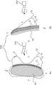
图1不一定按比例绘制,示出了生物力学组件的部件;Figure 1 is not necessarily to scale, showing components of a biomechanical assembly;
图2示出了图像处理系统的示意性框图;并且Figure 2 shows a schematic block diagram of an image processing system; and
图3示出了图像处理方法的流程图。FIG. 3 shows a flowchart of an image processing method.
具体实施方式Detailed ways
本文提出的是一种图像处理系统IPS,其基于生物力学组件的表面图像数据来估计所述组件内的内部结构的几何形状,所述组件内的所述内部结构被所述组件的外层遮挡。具体地,估计组件内的壁结构的几何形状。Presented herein is an image processing system IPS that estimates, based on surface image data of biomechanical components, the geometry of internal structures within the components that are occluded by the outer layers of the components . Specifically, the geometry of the wall structure within the assembly is estimated.
但是在更详细地解释该图像处理系统的操作之前,首先参考图1(其不一定按比例绘制)来说明生物力学组件TOR的基本部件将是有益的。But before explaining the operation of the image processing system in more detail, it will be instructive to first illustrate the basic components of the biomechanical assembly TOR with reference to Figure 1 (which is not necessarily drawn to scale).
图1A示出了诸如女性人体躯干TOR的生物力学组件的轴向视图。另一方面,图1B提供了所述躯干TOR的矢状视图(侧视图)。在图1A、图1B中,附图标记l和r分别指示左侧和右侧,而附图标记t和b分别指代躯干TOR的顶部和底部。从结构上讲,躯干TOR具有分层组成,一层被布置在另一层后面,暴露的外层OL(皮肤)遮挡躯干TOR内的层。这两个乳房形成为由凸起进入皮肤OL的下层乳房组织引起的皮肤OL的凸起部分。乳房组织连结到躯干内的肋骨笼RC的部分。在本文中具体将这个部分称为肋骨笼壁元件RC,或者简称为“壁元件”。更具体地,乳房组织连结到肋骨笼壁元件。壁元件RC是骨(肋骨)和胸肌组织的混合物。在皮肤/乳房组织与壁元件之间还有另外的中间层或内层IL。该内层IL由结缔组织形成,但也包括乳房组织和脂肪组织。被作为生物力学组件看待的女性躯干的主要部件因此包括外层(皮肤)、下层乳房组织、形成壁的肋骨笼RC,乳房组织经由位于乳房组织与肋骨笼之间的中间层与所述壁连结。Figure 1A shows an axial view of a biomechanical component such as the TOR of a female human torso. Figure IB, on the other hand, provides a sagittal view (side view) of the torso TOR. In Figures 1A, 1B, reference numerals l and r designate the left and right sides, respectively, while reference numerals t and b designate the top and bottom, respectively, of the torso TOR. Structurally, the torso TOR has a layered composition, with one layer being arranged behind the other, with the exposed outer layer OL (skin) obscuring the layers within the torso TOR. The two breasts are formed as raised portions of the skin OL caused by the bulge into the underlying breast tissue of the skin OL. The breast tissue is attached to the portion of the rib cage RC within the torso. This part is specifically referred to herein as the rib cage wall element RC, or simply "wall element". More specifically, the breast tissue is attached to the rib cage wall elements. The wall element RC is a mixture of bone (ribs) and pectoral muscle tissue. There is an additional intermediate or inner layer IL between the skin/breast tissue and the wall element. This inner IL is formed by connective tissue, but also includes breast tissue and adipose tissue. The main components of the female torso, viewed as a biomechanical component, therefore include the outer layer (skin), the underlying breast tissue, the rib cage RC forming the wall, the breast tissue being connected to the wall via an intermediate layer located between the breast tissue and the rib cage .
本申请中设想的一个目标包括由皮肤OL的光学相机DSC采集一幅或多幅表面图像,并且我们希望特别是基于该表面图像推断出躯干的其余的下层部件的几何形状,具体地,我们希望确定关于乳房经由中间层IL连结到的肋骨笼壁的几何数据。所述几何数据包括几何描述,其具体描述了肋骨笼壁的形状,特别是描述了肋骨笼壁的与乳房组织连结的那部分的形状。额外地或替代地,所述几何数据还可以包括在肋骨笼壁的一个或多个控制点处的位置和/或取向。在一个实施例中,本文设想使用关于肋骨笼壁RC的几何数据来提供经受力(特别是受重力)的躯干的生物力学模拟。One goal envisaged in this application consists of acquiring one or more surface images by the optical camera DSC of the skin OL, and we wish to infer the geometry of the rest of the underlying parts of the torso, in particular based on this surface image, in particular we wish to Geometric data about the rib cage wall to which the breast is attached via the intermediate layer IL is determined. The geometrical data includes a geometrical description which specifically describes the shape of the rib cage wall, in particular the shape of that part of the rib cage wall that is joined to the breast tissue. Additionally or alternatively, the geometric data may also include the position and/or orientation at one or more control points of the rib cage wall. In one embodiment, it is contemplated herein to use geometric data about the rib cage walls RC to provide a biomechanical simulation of the torso subjected to forces, particularly gravity.
为了实现对躯干的特别逼真的生物力学模拟,有利的是还具有可用的内层或中间层IL的几何数据。内层IL有效地包围或嵌入乳房组织的连接到肋骨笼的那部分。我们已经观察到该内层的形状和/或特别是厚度的知识引起高度逼真的生物力学模拟。此外,已经发现,内层的厚度估计能够基于从躯干群体学习的先验知识。换句话说,总之,基于皮肤OL的表面图像并且基于关于内层的平均厚度(其能够经由元数据与从其收集到表面图像的实际患者相关)的先前知识,建立针对上述部件中的每个部件的几何数据。具体地,计算针对内层和胸壁的几何数据。In order to achieve a particularly realistic biomechanical simulation of the torso, it is advantageous to also have available geometrical data of the inner or intermediate layer IL. The inner layer of IL effectively surrounds or embeds that portion of the breast tissue that connects to the rib cage. We have observed that knowledge of the shape and/or especially the thickness of this inner layer leads to highly realistic biomechanical simulations. Furthermore, it has been found that the thickness estimation of the inner layer can be based on prior knowledge learned from the torso population. In other words, in summary, based on the surface image of the skin OL and based on prior knowledge about the average thickness of the inner layer (which can be related via metadata to the actual patient from which the surface image was collected), an establishment for each of the above components is established The geometry data of the part. Specifically, geometric data for the inner layer and chest wall are calculated.
然后能够在对通常已知的相应材料(组织)的弹性特性编码的虚拟连接点处将针对各种部件(特别是胸壁RC和内层IL)的几何数据虚拟地链接在一起。然后能够将如此链接的数据结构用作针对模拟程序的输入,该模拟程序能够例如对整个躯干的图形模拟进行绘制。基于该模拟,当来自乳房的某些组织元件被移除(例如在乳房切除术或其中组织部分被移除的其他手术介入中)时,能够在重力的影响下研究躯干的整体外观。例如,在乳房中检测到癌组织的情况下,通常是对从乳房中移除的癌部位周围的组织的切除体积(例如,圆柱体)。然后外科医生或介入医师能够选择针对该切除体积的最优取向,以便为患者实现在重力作用下的最有利的视觉外观。虽然在本文中设想优选实施例,但是应当理解,人体女性躯干仅仅是生物力学组件的一个实施例。也就是说,所提出的成像处理系统也可以有益地应用于人类(或动物)解剖结构的其他部分。图1中的虚线示出了希望估计的肋骨笼壁元件的特定形状。The geometrical data for the various components, in particular the chest wall RC and inner layer IL, can then be virtually linked together at virtual connection points that encode the generally known elastic properties of the respective material (tissue). The data structure so linked can then be used as input to a simulation program that can, for example, draw a graphical simulation of the entire torso. Based on this simulation, it is possible to study the overall appearance of the torso under the influence of gravity when certain tissue elements from the breast are removed (eg, in a mastectomy or other surgical intervention in which tissue parts are removed). For example, where cancerous tissue is detected in the breast, it is typically a resected volume (eg, a cylinder) of tissue surrounding the cancerous site removed from the breast. The surgeon or interventional physician can then select the optimal orientation for this resection volume to achieve the most favorable visual appearance under gravity for the patient. While the preferred embodiment is contemplated herein, it should be understood that the human female torso is only one example of a biomechanical assembly. That is, the proposed imaging processing system can also be beneficially applied to other parts of the human (or animal) anatomy. The dashed line in Figure 1 shows the specific shape of the rib cage wall elements that it is desired to estimate.
现在参考图2,图2中示出了所提出的图像处理系统IPS的示意性框图。图像处理系统包括诸如Microsoft Kinect的深度感测相机的表面相机DSC以及能够在通用计算单元PU上被实施为软件模块的多个处理部件。在替代实施例中,图2中的IPS的模块被布置在分布式架构中并连接在合适的通信网络中。这些模块可以用硬件布置为适当编程的FPGA(现场可编程门阵列)或硬连线集成电路。Referring now to FIG. 2, a schematic block diagram of the proposed image processing system IPS is shown. The image processing system includes a surface camera DSC, such as a depth-sensing camera of Microsoft Kinect, and a number of processing components that can be implemented as software modules on a general-purpose computing unit PU. In an alternative embodiment, the modules of the IPS in Figure 2 are arranged in a distributed architecture and connected in a suitable communication network. These modules may be arranged in hardware as suitably programmed FPGAs (Field Programmable Gate Arrays) or hardwired integrated circuits.
在系统IPS的输入端口IN处接收沿着(一个或多个)成像方向d采集的(一幅或多幅)输入表面图像。所述一幅或多幅图像优选地编码空间深度信息。特别是基于所述(一幅或多幅)输入图像,壁估计器WE估计针对被皮肤OL和内部组织IL遮挡的下层肋骨笼壁的几何数据。所述几何数据具体包括形状信息,但是也可以额外地或替代地包括位置和/或取向。The input surface image(s) acquired along the imaging direction(s) d are received at the input port IN of the system IPS. The one or more images preferably encode spatial depth information. In particular based on the input image(s), the wall estimator WE estimates geometric data for the walls of the underlying rib cage occluded by the skin OL and the internal tissue IL. The geometrical data specifically includes shape information, but may additionally or alternatively include position and/or orientation.
为了实现这种估计,根据一个实施例,系统IPS包括图像分析器IA。壁估计器WE与图像分析器IA结合操作。图像分析器将表面图像分析为两个部分图像:i)一个部分图像:图像表面的表示两个乳房的凸起部分,在本文中被称为乳房图像;以及ii)第二部分图像:表面图像的表示包围乳房部分的皮肤的其余部分的非凸起部分。本文中将第二部分称为其余图像。可以假设其余图像在第一近似中更接近于下层肋骨笼壁的走向和形状。然后,壁估计器WE将躯干的基元3D模型(例如,超椭球或其他模型)拟合到仅所述其余表面图像。也就是说,在该拟合操作期间,按照输入图像的凸起表面部分的图像信息基本上被忽略。换句话说,估计器WE执行该第一躯体拟合操作,以得出在没有乳房的情况下躯干TOR可能看起来是什么样的模型。这并不是说按照乳房图像的图像信息被丢弃。相反,如将在下面更详细地解释的,乳房图像被保留并用于建立模拟。更具体地,在一个非限制性实施例中,分离的乳房图像本身被转换成单独的网格模型,针对每个乳房一个网格模型,并且随后在构建模拟时与胸壁模型相链接。应当理解,所提出的系统和方法可能不一定适用于两个乳房,可能仅适用于要进行手术的乳房。To achieve this estimation, according to one embodiment, the system IPS includes an image analyzer IA. The wall estimator WE operates in conjunction with the image analyzer IA. The image analyzer analyzes the surface image into two partial images: i) a partial image: the raised portion of the image surface representing the two breasts, referred to herein as breast images; and ii) a second partial image: the surface image of means the non-raised portion of the rest of the skin that surrounds the breast portion. The second part is referred to herein as the rest of the image. It can be assumed that the remaining images are closer to the orientation and shape of the lower rib cage walls in a first approximation. The wall estimator WE then fits a primitive 3D model of the torso (eg a hyperellipsoid or other model) to only the remaining surface images. That is, during this fitting operation, image information according to the raised surface portion of the input image is substantially ignored. In other words, the estimator WE performs this first body fitting operation to derive a model of what the torso TOR might look like without the breasts. This is not to say that the image information according to the breast image is discarded. Instead, as will be explained in more detail below, breast images are retained and used to build the simulation. More specifically, in one non-limiting embodiment, the separate breast images are themselves converted into separate mesh models, one mesh model for each breast, and then linked with the chest wall model when building the simulation. It should be understood that the proposed systems and methods may not necessarily be applicable to both breasts, and may only be applicable to the breast to be operated on.
可以被实施为网格结构的拟合基元(即,初始形状类型)能够被认为是针对壁元件RC的几何数据的第一估计,特别是针对形状的第一估计。换句话说,在这种简化的建模中,假定内层是“零”厚度。The fitting primitives, ie the initial shape types, which may be implemented as grid structures, can be considered as the first estimates for the geometrical data of the wall element RC, in particular for the shape. In other words, in this simplified modeling, the inner layer is assumed to be of "zero" thickness.
然而,为了获得优异的模拟质量,优选地,图像处理系统IPS还包括作为额外的处理部件的厚度估计器TE,其被配置为估计位于皮肤/乳房与内部肋骨笼壁RC之间的内层的厚度。组织估计器使用先验知识来提供针对例如沿着肋骨笼壁RC的走向的内层IL的平均厚度的估计。在另一实施例中,厚度估计器提供在沿着胸壁的预定控制点处的多个厚度读数。先验知识以适当的形式(表格、算法等)被保存在知识数据库DB中,并且能够通过厚度估计器TE从知识数据库DB中查询。估计该内层的厚度还允许定位内壁,具体为估计内壁位于躯干内部有多“深”。However, in order to obtain excellent simulation quality, it is preferred that the image processing system IPS further comprises as an additional processing component a thickness estimator TE configured to estimate the thickness of the inner layer located between the skin/breast and the inner rib cage wall RC thickness. The tissue estimator uses a priori knowledge to provide an estimate for the average thickness of the inner layer IL, eg, along the course of the rib cage wall RC. In another embodiment, the thickness estimator provides a plurality of thickness readings at predetermined control points along the chest wall. The prior knowledge is stored in the knowledge database DB in a suitable form (tables, algorithms, etc.) and can be queried from the knowledge database DB by the thickness estimator TE. Estimating the thickness of this inner layer also allows locating the inner wall, in particular estimating how "deep" the inner wall is inside the torso.
i)内层的几何数据描述与ii)根据拟合到其余表面的几何模型的壁的估计的几何数据然后可以被组合以实现更好的壁估计。例如,能够从几何躯干模型(排除乳房几何形状)的先前估计中减去厚度估计值,以便细化对肋骨笼壁形状和/或位置和/或取向的估计。换句话说,关于内层厚度的知识允许细化拟合的几何躯干模型,以更确切地精确定位肋骨笼壁的位置和形状(如图1中的虚线所示)。i) The geometrical data description of the inner layer and ii) the estimated geometrical data of the walls according to the geometrical model fitted to the remaining surfaces can then be combined to achieve a better estimate of the walls. For example, the thickness estimate can be subtracted from previous estimates of the geometric torso model (excluding breast geometry) in order to refine the estimate of rib cage wall shape and/or position and/or orientation. In other words, knowledge about the inner layer thickness allows refinement of the fitted geometric torso model to more precisely pinpoint the location and shape of the rib cage walls (shown in dashed lines in Figure 1).
然后将用于针对胸壁RC和/或内层的(一幅或多幅)乳房图像的几何数据(例如,网格模型)转发到模拟单元SU。具体地,能够由模拟单元SU将如此细化的壁形状和内层厚度的几何数据与针对乳房图像的网格链接在一起,以实现更好、更逼真的完整躯干模拟。给定假定躯干TOR受到的力(例如,重力)的规范,模拟单元绘制躯干动力学的图形模拟。The geometrical data (eg mesh model) for the breast image(s) for the chest wall RC and/or inner layer are then forwarded to the simulation unit SU. In particular, the geometry data of the wall shape and inner layer thickness so refined can be linked together by the simulation unit SU with the mesh for the breast image to achieve a better and more realistic simulation of the complete torso. The simulation unit draws a graphical simulation of the torso dynamics given a specification of the forces (eg, gravity) assumed to be experienced by the torso TOR.
如果需要模拟的图形表示,则能够将其显示在诸如计算机监视器MT的显示设备上。所提出的图像处理系统IPS被布置为交互式系统。换句话说,用户能够操纵几何数据,然后响应于这样的操纵来重新运行模拟,以在操纵几何数据的同时优选地以准实时的方式更新模拟。更具体地,模拟被嵌入在合适的GUI架构中,该GUI架构包括监视用户与当前显示的模拟的交互的事件处理程序。用户可以使用诸如指示器工具(鼠标、触控笔)或触摸屏交互的输入器件来改变几何数据。以这种方式,能够模拟虚拟手术介入,其中,切除乳房组织并且系统IPS通过对现在改变的躯干动力学的模拟的图形绘制来做出响应。用户因此能够找到最有利的方式来去除组织,以尽可能地保持躯干的自然动力学,从而促进患者术后更好的生活质量。If a graphical representation of the simulation is required, it can be displayed on a display device such as a computer monitor MT. The proposed image processing system IPS is arranged as an interactive system. In other words, the user can manipulate the geometric data and then rerun the simulation in response to such manipulation to update the simulation, preferably in a near real-time manner, while manipulating the geometric data. More specifically, the simulation is embedded in a suitable GUI framework that includes event handlers that monitor the user's interaction with the currently displayed simulation. The user can change the geometric data using input devices such as pointer tools (mouse, stylus) or touch screen interaction. In this way, a virtual surgical intervention can be simulated in which breast tissue is removed and the system IPS responds by graphical rendering of the simulation to the now changing torso dynamics. The user is thus able to find the most advantageous way to remove tissue in order to preserve the natural dynamics of the torso as much as possible, thereby promoting a better quality of life for the patient after surgery.
尽管如上所述的生物力学模拟的图形表示是优选实施例,但是在一些情况下纯粹数值模拟可能是足够的,并且本文也设想到这样的实施例。While graphical representations of biomechanical simulations as described above are preferred embodiments, in some cases purely numerical simulations may be sufficient, and such embodiments are also contemplated herein.
现在参考图3,图3示出了针对图2中的图像处理系统的操作所基于的图像处理方法的流程图。然而,本领域技术人员应当理解,也可以单独阅读下文对方法的描述,这些描述不必与图2中描述的架构相联系。Referring now to FIG. 3 , there is shown a flowchart for an image processing method on which the operation of the image processing system of FIG. 2 is based. However, those skilled in the art will understand that the descriptions of the methods below can also be read separately, and these descriptions are not necessarily linked to the architecture described in FIG. 2 .
在步骤S305处,接收生物力学组件(例如,人体或动物躯干)的输入表面图像。表面图像可以被构造为测得的距离的点云,这些点云一起定义包络下层的皮肤的形状或走向。表面图像是优选通过表面成像装置DSC(例如,深度感测相机)或其他光学技术(特别是非电离成像技术)沿着一个或多个成像方向采集的。在结构上,如图1所示,生物力学组件TOR被认为包括相机DSC可见的外层OT以及沿着成像方向被外层OT遮挡的内层IL和壁元件。内层位于外层与从后面(远离相机位置)连结到内层的壁元件之间。换句话说,壁元件经由内层连结到外层。At step S305, an input surface image of a biomechanical component (eg, a human or animal torso) is received. The surface image can be constructed as a point cloud of measured distances that together define the shape or orientation of the skin that envelopes the underlying layers. Surface images are preferably acquired along one or more imaging directions by a surface imaging device DSC (eg, a depth-sensing camera) or other optical techniques, particularly non-ionizing imaging techniques. Structurally, as shown in Fig. 1, the biomechanical component TOR is thought to include an outer layer of OT visible to the camera DSC and an inner layer of IL and wall elements occluded by the outer layer of OT along the imaging direction. The inner layer is located between the outer layer and the wall elements joined to the inner layer from the back (away from the camera position). In other words, the wall elements are joined to the outer layer via the inner layer.
在步骤S320处,估计描述壁元件的几何数据。At step S320 geometrical data describing the wall elements is estimated.
在一个实施例中,这种估计基于中间步骤S310a,其中,将表面图像分析为上述两个部分图像:乳房图像,其描述或表示一个或多个凸起部分,例如,女性躯干的乳房组织;以及其余图像,其表示乳房部分外部的皮肤走向。然后,将几何模型,基元(例如,超椭球或其他形状)拟合到仅其余图像。另一方面,现在在这个拟合操作中忽略乳房图像。在该实施例中,估计壁RC的几何形状的步骤仅基于按照其余图像的图像信息。换句话说,乳房图像中的凸起部分被忽略,以便获得针对壁元件的几何数据的第一估计。针对基元的形状的选择将取决于预期的感兴趣解剖结构的整体形状特征,并且如果不是所考虑的人体躯干,则可以要求除了所提及的超椭球以外的形状。In one embodiment, this estimation is based on an intermediate step S310a, wherein the surface image is analyzed into the aforementioned two partial images: a breast image, which describes or represents one or more raised portions, eg, breast tissue of a female torso; and the rest of the images, which represent the skin orientation outside the breast portion. Then, fit geometric models, primitives (eg, hyperellipsoids or other shapes) to just the rest of the image. On the other hand, breast images are now ignored in this fitting operation. In this embodiment, the step of estimating the geometry of the wall RC is based only on image information in terms of the remaining images. In other words, raised portions in the breast image are ignored in order to obtain a first estimate of the geometrical data for the wall elements. The choice of shape for the primitive will depend on the expected overall shape characteristics of the anatomy of interest, and if not the human torso under consideration, shapes other than the mentioned hyperellipsoid may be required.
为了拟合几何模型的目的而排除乳房图像信息能够以不同方式来实现。例如,在一个实施例中,使用形成容易识别的人造“界标”的人造标记。这些标记(贴纸、标签等)在采集表面图像之前被手动附着(例如通过粘附)到患者的皮肤。标记沿着周向围绕(一个或多个)乳房进行布置。然后可以通过分割操作自动检测标记印记来区分乳房图像与非乳房其余图像。任选地,在不使用人造标记的情况下,在3D重建网格中自动检测乳房部分。基于模型的方法能够用于识别属于乳房的皮肤部分。这些模型能够在群体的3D扫描上进行训练。The exclusion of breast image information for the purpose of fitting the geometric model can be accomplished in different ways. For example, in one embodiment, artificial markers that form easily identifiable artificial "landmarks" are used. These markers (stickers, labels, etc.) are manually affixed (eg, by adhering) to the patient's skin prior to capturing an image of the surface. The markers are arranged circumferentially around the breast(s). A segmentation operation can then automatically detect marker imprints to distinguish breast images from non-breast rest images. Optionally, breast parts are automatically detected in the 3D reconstruction grid without the use of artificial markers. Model-based methods can be used to identify skin parts that belong to the breast. These models can be trained on 3D scans of the population.
通过使用“片状”内插函数基于这些(自然的或人造的)界标进行适当内插,能够消除乳房图像信息。具体地,代表乳房的图像信息通过内插被替换为从其余表面图像的边缘内插的较小曲率的内插表面。更具体地,能够将曲线拟合到在输入表面图像中识别的多个标记点。然后该操作在表面图像中留下一个或多个“孔”,由曲线勾画的孔和曲线因此定义了其余表面的(一个或多个)边缘。然后,拟合基元的表面(例如,椭球模型表面)代替乳房图像部分,从而得到针对壁的形状或走向的第一几何估计。By properly interpolating based on these (natural or artificial) landmarks using a "flaky" interpolation function, breast image information can be eliminated. Specifically, the image information representing the breast is replaced by interpolation with an interpolated surface of lesser curvature interpolated from the edges of the remaining surface images. More specifically, a curve can be fitted to a plurality of marker points identified in the input surface image. This operation then leaves one or more "holes" in the surface image, the holes and curves delineated by the curves thus defining the edge(s) of the remaining surface. Then, the surface of the fitting primitive (eg, the surface of the ellipsoid model) replaces the breast image portion, resulting in a first geometric estimate of the shape or orientation of the wall.
除了将表面图像分析为两个部分图像并将几何模型拟合到其余图像的步骤S310a之外或代替所述步骤S310a,存在步骤S310b,其中,根据来自同一类型的其他解剖结构的先验知识来估计内层的厚度。然后将如此估计的厚度应用于拟合的几何模型以导出针对壁元件的模式细化估计。In addition to or instead of step S310a of analyzing the surface image into two partial images and fitting a geometric model to the remaining images, there is a step S310b in which the method is based on prior knowledge from other anatomical structures of the same type Estimate the thickness of the inner layer. The thickness thus estimated is then applied to the fitted geometric model to derive a mode refinement estimate for the wall element.
更具体地,厚度估计步骤基于先前的学习步骤,在先前的学习步骤中,历史元数据与内层厚度的特性之间的依赖关系是从基础事实数据中学习的。基础事实数据包括例如先前从女性患者群体采集的MRI图像或CT图像。历史元数据包括如下参数:例如,年龄、体重、BMI、侧向方向(从左到右)的患者躯干尺寸,以及描述群体中的个体的其他因素(或这些因素的任意项的不同组合)。元数据在本文中被称为“历史”,因为它涉及先前从群体收集的图像数据。More specifically, the thickness estimation step is based on a previous learning step in which the dependencies between historical metadata and properties of inner layer thicknesses were learned from ground truth data. Ground truth data includes, for example, MRI or CT images previously acquired from a female patient population. Historical metadata includes parameters such as age, weight, BMI, patient torso dimensions in lateral direction (left to right), and other factors (or various combinations of any of these factors) that describe individuals in the population. Metadata is referred to herein as "history" because it pertains to image data previously collected from groups.
假设诸如MRI图像的基础事实材料以对比充分地表示三种感兴趣结构,即,肋骨笼、壁和内层。然后自动或手动处理基础事实图像,以在单个控制点处或在沿着壁RC分布的多个预定义(不一定等距)的控制点处测量肋骨笼壁与内层之间的相对距离。然后将这些测量值与那些基础事实数据的元数据一起馈送到机器学习算法中,所述机器学习算法例如为随机森林(参见例如A.Cirminisi等人的“Decision Forests for Classification,Regression,Density Estimation,Manifold Learning and Semi-Supervised Learning”(第MSR-TR-2011-114号,2011年10月28日)、神经网络或用于学习元数据与组织厚度之间的函数关系(“训练模型”)其他方法。一旦在充足文库中的基础事实数据上进行了训练,就能够给模型馈送任何针对元数据的规范,并且经训练的模型将输出如下值:该值能够作为针对壁层厚度对层距离的估计。Ground truth materials such as MRI images are assumed to adequately represent the three structures of interest, ie, the rib cage, the wall, and the inner layer, in contrast. The ground truth images are then processed automatically or manually to measure the relative distance between the rib cage wall and the inner layer at a single control point or at multiple predefined (not necessarily equidistant) control points distributed along the wall RC. These measurements are then fed into a machine learning algorithm, such as a random forest (see, e.g., "Decision Forests for Classification, Regression, Density Estimation," by A. Cirminisi et al. Manifold Learning and Semi-Supervised Learning" (No. MSR-TR-2011-114, October 28, 2011), neural networks or for learning a functional relationship between metadata and tissue thickness ("training a model") other method. Once trained on ground truth data in a sufficient library, the model can be fed any specification for the metadata, and the trained model will output a value that can be used as a measure of wall thickness versus layer distance estimate.
该厚度估计操作也可以通过使用更传统的函数拟合流程来实现。在这种方法中,假设给出了一个参数化的函数描述,这个函数描述对于元数据对层厚度的依赖性仍然是未知的。例如,可以使用某种类别的函数,例如,系数是要拟合的参数的高维多项式。然后通过以下操作来“学习”针对这些参数的特定值:通过使用假定的函数描述(例如,多项式)将优化流程中的已知的历史元数据拟合到已知的厚度/距离测量值以基于某些优化标准(例如,最小二乘和等)来计算“最优”参数。一旦计算出这些参数,就能够将这些参数替换为假定的函数描述,其现在提供“规则”或“公式”来针对元数据的任何规范计算内层IL厚度/到肋骨笼壁的距离。用于揭示这种先验未知关系/依赖性的优化流程的一个范例是来自统计实验设计领域的“响应面方法论”(RSM)。参见例如G Box等人的“On the ExperimentalAttainment of Optimum Conditions”(Journal of the Royal Statistical Society,Series B(Methodological),第13卷,第1期,1951年,第1-45页)。This thickness estimation operation can also be achieved by using a more traditional function fitting procedure. In this approach, it is assumed that a parameterized functional description is given, which is still unknown for the dependence of the metadata on the layer thickness. For example, some kind of function can be used, eg, a high-dimensional polynomial whose coefficients are the parameters to be fitted. Specific values for these parameters are then "learned" by fitting known historical metadata from the optimization process to known thickness/distance measurements using an assumed functional description (eg, a polynomial) to be based on Certain optimization criteria (eg, least squares sum, etc.) are used to calculate "optimal" parameters. Once these parameters are calculated, they can be replaced with a hypothetical functional description, which now provides "rules" or "formulas" to calculate inner IL thickness/distance to rib cage wall for any specification of the metadata. An example of an optimization pipeline for uncovering such a priori unknown relationships/dependencies is "Response Surface Methodology" (RSM) from the field of statistical experimental design. See, eg, "On the Experimental Attainment of Optimum Conditions" by G Box et al. (Journal of the Royal Statistical Society, Series B (Methodological), Vol. 13, No. 1, 1951, pp. 1-45).
换句话说,能够通过传统函数拟合方法或通过机器学习根据先验基础事实数据来估计内层的厚度。先前的方法得到元数据对层的依赖性的显式形式,并且能够从单个元数据和基础事实图像集中获得。另一种选择是首先对基础事实数据集和元数据进行求平均,然后将拟合操作应用于平均数据集。In other words, the thickness of the inner layer can be estimated from a priori ground truth data by traditional function fitting methods or by machine learning. Previous methods derive an explicit form of metadata-to-layer dependencies and can be obtained from a single set of metadata and ground truth images. Another option is to first average the ground truth dataset and metadata, and then apply a fit operation to the averaged dataset.
如上所述的机器学习方法具有以下优点:能够从多个基础事实图像数据集及其相关联的历史元数据立即获知依赖性。不需要通过求平均来折叠多个数据集。在机器学习方法中,输出通常不像更传统的函数拟合方法那样提供针对依赖性的显式的函数描述或“公式”。相反,在机器学习中,“学习的”依赖性被编码在随机森林的多个决策树结构的个体和相互配置中,或者在神经网络的情况下,“学习的”依赖性被配置在神经网络的节点层的配置中。Machine learning methods as described above have the advantage of being able to learn dependencies immediately from multiple ground truth image datasets and their associated historical metadata. There is no need to fold multiple datasets by averaging. In machine learning methods, the output typically does not provide an explicit functional description or "formula" for dependencies as more traditional function fitting methods do. In contrast, in machine learning, "learned" dependencies are encoded in the individual and mutual configuration of multiple decision tree structures of random forests, or in the case of neural networks, "learned" dependencies are configured in neural networks in the configuration of the node layer.
如上所述的学习或拟合操作是在估计针对给定患者的内层IL的厚度的上述步骤之前的前驱阶段中完成的。针对厚度估计步骤所需要的仅仅是提供要为其拟定模拟以实现厚度估计的特定患者的特定元数据。换句话说,能够避免针对给定患者的特定MRI图像或CT图像,因为能够将过去已经针对其他患者采集的所有合适图像的文库用作基础事实。这个现有的基础事实图像文库能够从附接到HIS系统的图像存储系统(PACS)中收集。更具体地并且清楚的是,用于学习函数关系的基础真实图像(例如,MRI图像或CT图像)不是从要对其运行模拟的特定患者采集的图像数据(但是,当然,如果需要的话,仍然能够获得或使用这样的个人图像)。The learning or fitting operation as described above is done in a precursor stage prior to the above step of estimating the thickness of the inner layer IL for a given patient. All that is required for the thickness estimation step is to provide specific patient-specific metadata for which a simulation is to be drawn up to achieve thickness estimation. In other words, a specific MRI image or CT image for a given patient can be avoided because the library of all suitable images that have been acquired for other patients in the past can be used as ground truth. This existing ground truth image library can be collected from the Image Storage System (PACS) attached to the HIS system. More specifically and clearly, the underlying real images (e.g., MRI images or CT images) used to learn the functional relationship are not image data acquired from the specific patient on which the simulation is to be run (but, of course, still, if desired,). be able to obtain or use such personal images).
所学习的函数元数据对组织厚度的关系能够被存储在知识数据库DB中的合适的数据结构中。合适的数据结构包括表格、函数/算法描述或其他内容。对要学习其躯干几何图形的当前患者应用当前元数据能够像针对已知厚度对元数据的依赖性的数据库查询和查找操作一样简单。在其他实施例中,该查询操作涉及诸如从已知的元数据对厚度的对/元组内插入期望厚度的计算。在其他实施例中,元数据被应用以填充学习到的函数描述(其本质上是数学公式)中的“占位符”位置。然后针对所需数量的控制点执行由该函数描述规定的算术运算,以导出针对这些控制点的一个或多个厚度估计。然后将这一个或多个厚度读数应用于在步骤S310a中拟合的几何模型,从而导出针对肋骨笼壁RC的形状/位置/取向的细化的估计。在步骤S330处,将针对内层和壁元件的几何数据进行组合以形成生物力学组件的生物力学模拟。The learned function metadata to tissue thickness relationship can be stored in a suitable data structure in the knowledge database DB. Suitable data structures include tables, function/algorithm descriptions, or other content. Applying the current metadata to the current patient whose torso geometry is to be learned can be as simple as a database query and lookup operation for a known thickness dependency on the metadata. In other embodiments, the query operation involves a calculation such as interpolating the desired thickness from within a pair/tuple of known metadata versus thickness. In other embodiments, metadata is applied to populate "placeholder" positions in the learned function description (which is essentially a mathematical formula). The arithmetic operations specified by the function description are then performed for the desired number of control points to derive one or more thickness estimates for those control points. These one or more thickness readings are then applied to the geometric model fitted in step S310a to derive a refined estimate for the shape/position/orientation of the rib cage wall RC. At step S330, the geometric data for the inner layer and wall elements are combined to form a biomechanical simulation of the biomechanical assembly.
更具体地,在一个实施例中,以计算机图形网格的形式提供针对肋骨笼壁和内层估计的几何数据。在连接点处将这些网格与针对先前在步骤S310a处忽略的来自输入表面图像的一个(或两个)乳房组织部分的网格进行组合。就像网格的节点一样,也是这些网格之间的连接点对这些网格所表示的材料的弹性性质进行编码。并且由于躯干组织的材料组成和密度是已知的,因此能够使用所提供的几何数据作为边界条件而通过求解偏微分方程(PDE)组来计算在任何力(例如,重力)的影响下整个躯干结构的动力学模拟。More specifically, in one embodiment, the estimated geometric data for the rib cage walls and inner layers is provided in the form of a computer graphics grid. These meshes are combined at the connection points with meshes for one (or two) breast tissue portions from the input surface image previously ignored at step S310a. Like the nodes of a mesh, it is the connection points between these meshes that encode the elastic properties of the material that these meshes represent. And since the material composition and density of the torso tissue is known, it is possible to calculate the entire torso under the influence of any force (eg, gravity) by solving a set of partial differential equations (PDEs) using the provided geometric data as boundary conditions Dynamic simulation of structures.
在一个实施例中,在显示单元上显示模拟,或者独立地显示模拟,或者与由相机DSC记录的原始表面扫描图像并排地显示模拟。In one embodiment, the simulation is displayed on the display unit, either independently or side by side with the raw surface scan image recorded by the camera DSC.
总而言之,在一个实施例中,本文提出的是一种方法,该方法纯粹基于光学表面扫描和元数据而使得能够进行对乳房的生物力学模拟而不需要MR成像或CT成像。使用能够例如通过使用深度感相机经济有效地采集的三维表面数据,我们提出通过在第一步骤中使用拟合到躯干表面的图像数据的几何模型来估计胸壁的形状并且因此估计表面与胸壁之间的组织的形状(利用关于从该拟合中排除的实际乳房组织的图像信息)。然后元数据驱动组织厚度估计,并且在第二步骤中估计组织厚度,这能够用于细化第一步骤中获得的几何模型。In summary, in one embodiment, presented herein is a method that enables biomechanical simulation of the breast without the need for MR imaging or CT imaging, based purely on optical surface scans and metadata. Using three-dimensional surface data that can be acquired cost-effectively, eg by using a depth-sensing camera, we propose to estimate the shape of the chest wall and thus the distance between the surface and the chest wall by using, in a first step, a geometric model fitted to the image data of the torso surface. the shape of the tissue (using image information about the actual breast tissue excluded from the fit). The metadata then drives the tissue thickness estimation, and the tissue thickness is estimated in a second step, which can be used to refine the geometric model obtained in the first step.
应当注意,上述方法和系统能够应用于一个以上的内壁元件。例如,上述方法和系统可以通过分别估计肋骨笼和胸肌的覆盖壁作为两个离散的壁元件而得以细化,每个壁元件单独作为其自身的结构而被估计。这种方法甚至可以扩展到具有两个以上(覆盖)壁元件的更复杂的组件。该模拟能够捕捉到两个或更多个这种互相连接的壁元件的动力学特性,从而允许实现更高水平的真实感。It should be noted that the methods and systems described above can be applied to more than one inner wall element. For example, the methods and systems described above can be refined by estimating the covering walls of the rib cage and the pectoralis muscle, respectively, as two discrete wall elements, each individually estimated as its own structure. This approach can even be extended to more complex assemblies with more than two (covering) wall elements. The simulation is able to capture the dynamics of two or more such interconnected wall elements, allowing a higher level of realism to be achieved.
在本发明的另一示范性实施例中,提供了一种计算机程序或计算机程序单元,其特征在于,其适于在适当的系统上运行根据前述实施例中的一个所述的方法的方法步骤。In another exemplary embodiment of the present invention, a computer program or computer program element is provided, characterized in that it is adapted to execute the method steps of the method according to one of the preceding embodiments on a suitable system .
因此,计算机程序单元可以被存储在计算机单元中,所述计算机程序单元也可以是本发明的实施例的部分。该计算单元可以适于执行或引发对上述方法的步骤的执行。此外,该计算单元可以适于操作上述装置的部件。该计算单元能够适于自动操作和/或运行用户的命令。计算机程序可以被加载到数据处理器的工作存储器中。因此,可以装备数据处理器来执行本发明的方法。Thus, a computer program element may be stored in a computer unit, which may also be part of an embodiment of the present invention. The computing unit may be adapted to perform or cause the steps of the above-described method to be performed. Furthermore, the computing unit may be adapted to operate the components of the apparatus described above. The computing unit can be adapted to operate automatically and/or execute commands of the user. The computer program can be loaded into the working memory of the data processor. Accordingly, a data processor may be equipped to perform the method of the present invention.
本发明的该示范性实施例覆盖从一开始就使用本发明的计算机程序,以及借助于将现有程序更新转换为使用本发明的程序的计算机程序二者。This exemplary embodiment of the present invention covers both computer programs that use the present invention from the outset, as well as computer programs that are converted by means of an existing program update to a program that uses the present invention.
另外,计算机程序单元可以能够提供所有必要步骤以完成如上所述的方法的示范性实施例的流程。In addition, a computer program element may be able to provide all necessary steps to carry out the flow of an exemplary embodiment of the method as described above.
根据本发明的另外的示范性实施例,提出了一种计算机可读介质,例如,CD-ROM,其中,该计算机可读介质具有被存储于所述计算机可读介质上的计算机程序单元,所述计算机程序单元由前面的章节所描述。According to a further exemplary embodiment of the present invention, a computer-readable medium, such as a CD-ROM, is proposed, wherein the computer-readable medium has a computer program element stored on the computer-readable medium, the The computer program elements are described in the preceding chapters.
计算机程序可以被存储和/或被分布在合适的介质(尤其地,但不一定为非瞬态介质)上,例如,与其他硬件一起或作为其他硬件的部分供应的光学存储介质或固态介质,但是也可以以其他形式被分布,例如,经由互联网或其他有线或无线的电信系统被分布。The computer program may be stored and/or distributed on a suitable medium (in particular, but not necessarily a non-transitory medium), such as an optical storage medium or solid state medium supplied with or as part of other hardware, However, it may also be distributed in other forms, eg via the Internet or other wired or wireless telecommunication systems.
然而,计算机程序也可以被呈现在网络上,如万维网,并且能够从这样的网络被下载到数据处理器的工作存储器中。根据本发明的另外的示范性实施例,提供了用于使计算机程序单元可用于下载的介质,所述计算机程序单元被布置为执行根据本发明的先前描述的实施例中的一个所述的方法。However, the computer program can also be presented on a network, such as the World Wide Web, and can be downloaded from such a network into the working memory of a data processor. According to a further exemplary embodiment of the invention there is provided a medium for making a computer program element available for download, the computer program element being arranged to perform the method according to one of the previously described embodiments of the invention .
必须注意,本发明的实施例是参考不同主题来描述的。尤其地,一些实施例是参考方法型权利要求来描述的,而其他实施例是参考装置型权利要求来描述的。然而,除非另有说明,本领域技术人员将从以上和以下的描述中推断出,除属于一种类型的主题的特征的任意组合之外,涉及不同主题的特征之间的任意组合也被认为在本申请中被公开。然而,所有的特征都能够被组合来提供多于特征的简单加合的协同效应。It must be noted that embodiments of the present invention are described with reference to different subject matters. In particular, some embodiments have been described with reference to method type claims whereas other embodiments have been described with reference to apparatus type claims. However, unless stated otherwise, a person skilled in the art will infer from the above and the following description that any combination of features relating to different subject matters, in addition to any combination of features belonging to one type of subject matter, is also to be considered disclosed in this application. However, all features can be combined to provide synergistic effects that are more than a simple addition of features.
尽管已经在附图和前面的描述中详细图示和描述了本发明,但是这样的图示和描述应当被认为是图示性或示范性的,而非限制性的。本发明不限于所公开的实施例。本领域技术人员通过研究附图、公开内容以及权利要求,在实践请求保护的发明时能够理解并实现对所公开的实施例的其他变型。While the invention has been illustrated and described in detail in the drawings and foregoing description, such illustration and description are to be considered illustrative or exemplary and not restrictive. The invention is not limited to the disclosed embodiments. Other modifications to the disclosed embodiments can be understood and effected by those skilled in the art in practicing the claimed invention, from a study of the drawings, the disclosure, and the claims.
在权利要求中,“包括”一词不排除其他元件或步骤,并且词语“一”或“一个”不排除多个。单个处理器或其他单元可以实现在权利要求中记载的若干项的功能。尽管某些措施被记载在互不相同的从属权利要求中,但是这并不指示不能有利地使用这些措施的组合。权利要求中的任何附图标记都不应被解释为对范围的限制。In the claims, the word "comprising" does not exclude other elements or steps, and the word "a" or "an" does not exclude a plurality. A single processor or other unit may fulfill the functions of several items recited in the claims. The mere fact that certain measures are recited in mutually different dependent claims does not indicate that a combination of these measures cannot be used to advantage. Any reference signs in the claims should not be construed as limiting the scope.
Claims (14)
Translated fromChineseApplications Claiming Priority (3)
| Application Number | Priority Date | Filing Date | Title |
|---|---|---|---|
| EP15197818 | 2015-12-03 | ||
| EP15197818.6 | 2015-12-03 | ||
| PCT/EP2016/079514WO2017093446A1 (en) | 2015-12-03 | 2016-12-01 | Chest wall estimation from optical scans |
Publications (2)
| Publication Number | Publication Date |
|---|---|
| CN108292530A CN108292530A (en) | 2018-07-17 |
| CN108292530Btrue CN108292530B (en) | 2022-05-24 |
Family
ID=55070648
Family Applications (1)
| Application Number | Title | Priority Date | Filing Date |
|---|---|---|---|
| CN201680070819.2AActiveCN108292530B (en) | 2015-12-03 | 2016-12-01 | Chest wall estimation from optical scanning |
Country Status (4)
| Country | Link |
|---|---|
| US (1) | US10692208B2 (en) |
| EP (1) | EP3384412A1 (en) |
| CN (1) | CN108292530B (en) |
| WO (1) | WO2017093446A1 (en) |
Families Citing this family (1)
| Publication number | Priority date | Publication date | Assignee | Title |
|---|---|---|---|---|
| US11160528B2 (en)* | 2015-12-18 | 2021-11-02 | General Electric Company | System and method for visualization of ultrasound volumes |
Citations (3)
| Publication number | Priority date | Publication date | Assignee | Title |
|---|---|---|---|---|
| CA2298282A1 (en)* | 1999-02-19 | 2000-08-19 | The John P. Robarts Research Institute | Semi-automated segmentation method for 3-dimensional ultrasound |
| WO2011007312A1 (en)* | 2009-07-17 | 2011-01-20 | Koninklijke Philips Electronics N.V. | Multi-modality breast imaging |
| CN102918558A (en)* | 2010-01-28 | 2013-02-06 | 拉德罗吉克斯公司 | Methods and systems for analyzing, prioritizing, visualizing and reporting on medical images |
Family Cites Families (7)
| Publication number | Priority date | Publication date | Assignee | Title |
|---|---|---|---|---|
| US20080218727A1 (en) | 2006-12-22 | 2008-09-11 | Art, Advanced Research Technologies Inc. | Method and apparatus for optical image reconstruction using contour determination |
| US7720196B2 (en) | 2008-01-07 | 2010-05-18 | Accuray Incorporated | Target tracking using surface scanner and four-dimensional diagnostic imaging data |
| US8795204B2 (en)* | 2008-01-09 | 2014-08-05 | Allergan, Inc. | Anatomical recognition and dimensional analysis of breast volume to assist breast surgery |
| WO2012116746A1 (en)* | 2011-03-02 | 2012-09-07 | Mevis Medical Solutions Ag | Image processing device for finding corresponding regions in two image data sets of an object |
| CN105474219B (en)* | 2013-08-28 | 2019-10-18 | 西门子公司 | Systems and methods for estimating physiological cardiac measurements from medical images and clinical data |
| RU2678080C2 (en) | 2013-09-24 | 2019-01-22 | Конинклейке Филипс Н.В. | Method for determining surgical operation plan |
| WO2015103388A1 (en)* | 2014-01-02 | 2015-07-09 | Metritrack, Inc. | System and method for tracking completeness of co-registered medical image data |
- 2016
- 2016-12-01USUS15/778,939patent/US10692208B2/enactiveActive
- 2016-12-01EPEP16806048.1Apatent/EP3384412A1/ennot_activeWithdrawn
- 2016-12-01WOPCT/EP2016/079514patent/WO2017093446A1/ennot_activeCeased
- 2016-12-01CNCN201680070819.2Apatent/CN108292530B/enactiveActive
Patent Citations (3)
| Publication number | Priority date | Publication date | Assignee | Title |
|---|---|---|---|---|
| CA2298282A1 (en)* | 1999-02-19 | 2000-08-19 | The John P. Robarts Research Institute | Semi-automated segmentation method for 3-dimensional ultrasound |
| WO2011007312A1 (en)* | 2009-07-17 | 2011-01-20 | Koninklijke Philips Electronics N.V. | Multi-modality breast imaging |
| CN102918558A (en)* | 2010-01-28 | 2013-02-06 | 拉德罗吉克斯公司 | Methods and systems for analyzing, prioritizing, visualizing and reporting on medical images |
Also Published As
| Publication number | Publication date |
|---|---|
| CN108292530A (en) | 2018-07-17 |
| EP3384412A1 (en) | 2018-10-10 |
| US10692208B2 (en) | 2020-06-23 |
| WO2017093446A1 (en) | 2017-06-08 |
| US20180350068A1 (en) | 2018-12-06 |
Similar Documents
| Publication | Publication Date | Title |
|---|---|---|
| CN113573640B (en) | Modeling regions of interest in anatomical structures | |
| Yau et al. | Tooth model reconstruction based upon data fusion for orthodontic treatment simulation | |
| CN104462650B (en) | A kind of hypostazation heart 3D model production methods of achievable external and internal compositionses | |
| CN109584349B (en) | Method and apparatus for rendering material properties | |
| US11382694B2 (en) | Systems and methods for predictive heart valve simulation | |
| JP2020175184A (en) | System and method for reconstruction of 3d anatomical image from 2d anatomical image | |
| CN109996495B (en) | Vessel tree normalization for biophysical simulation and/or expansion simulation for trimmed portions | |
| Eiben et al. | Symmetric biomechanically guided prone-to-supine breast image registration | |
| CN107945169B (en) | A Coronary Artery Imaging Analysis Method | |
| Uccheddu et al. | 3D printing of cardiac structures from medical images: an overview of methods and interactive tools | |
| Ebert et al. | Image segmentation of post-mortem computed tomography data in forensic imaging: Methods and applications | |
| CN107004269A (en) | The segmentation based on model to anatomical structure | |
| Zhu et al. | 3D automatic MRI level set segmentation of inner ear based on statistical shape models prior | |
| CN108292530B (en) | Chest wall estimation from optical scanning | |
| CN116997937A (en) | Method for visualizing at least one area of an object in at least one interface | |
| CN114298986A (en) | Thoracic skeleton three-dimensional construction method and system based on multi-viewpoint disordered X-ray film | |
| Li et al. | Three-dimensional reconstruction of paracentesis approach in transjugular intrahepatic portosystemic shunt | |
| Jiang et al. | Patient-specific modeling of pelvic system from MRI for numerical simulation: Validation using a physical model | |
| Andersson et al. | Digital 3D Facial Reconstruction Based on Computed Tomography | |
| Galeano et al. | Alternative Tool for the Diagnosis of Diseases Through Virtual Reality | |
| Dotremont | From medical images to 3D model: processing and segmentation | |
| Cerrolaza et al. | Modeling human tissues: an efficient integrated methodology | |
| RU2684760C1 (en) | Method and system for pre-operative modeling of medical procedure | |
| CN108885905A (en) | The biomechanical model of human or animal's trunk generates | |
| Tyfa et al. | MeMoS—A software tool for extraction of anatomical structures data from 3D medical images |
Legal Events
| Date | Code | Title | Description |
|---|---|---|---|
| PB01 | Publication | ||
| PB01 | Publication | ||
| SE01 | Entry into force of request for substantive examination | ||
| SE01 | Entry into force of request for substantive examination | ||
| GR01 | Patent grant | ||
| GR01 | Patent grant |