CN108022238B - Method, computer storage medium, and system for detecting object in 3D image - Google Patents
Method, computer storage medium, and system for detecting object in 3D imageDownload PDFInfo
- Publication number
- CN108022238B CN108022238BCN201711249474.XACN201711249474ACN108022238BCN 108022238 BCN108022238 BCN 108022238BCN 201711249474 ACN201711249474 ACN 201711249474ACN 108022238 BCN108022238 BCN 108022238B
- Authority
- CN
- China
- Prior art keywords
- image
- feature maps
- size
- region
- network
- Prior art date
- Legal status (The legal status is an assumption and is not a legal conclusion. Google has not performed a legal analysis and makes no representation as to the accuracy of the status listed.)
- Active
Links
Images


Classifications
- G—PHYSICS
- G06—COMPUTING OR CALCULATING; COUNTING
- G06V—IMAGE OR VIDEO RECOGNITION OR UNDERSTANDING
- G06V10/00—Arrangements for image or video recognition or understanding
- G06V10/70—Arrangements for image or video recognition or understanding using pattern recognition or machine learning
- G06V10/82—Arrangements for image or video recognition or understanding using pattern recognition or machine learning using neural networks
- A—HUMAN NECESSITIES
- A61—MEDICAL OR VETERINARY SCIENCE; HYGIENE
- A61B—DIAGNOSIS; SURGERY; IDENTIFICATION
- A61B5/00—Measuring for diagnostic purposes; Identification of persons
- A61B5/0033—Features or image-related aspects of imaging apparatus, e.g. for MRI, optical tomography or impedance tomography apparatus; Arrangements of imaging apparatus in a room
- A61B5/0037—Performing a preliminary scan, e.g. a prescan for identifying a region of interest
- A—HUMAN NECESSITIES
- A61—MEDICAL OR VETERINARY SCIENCE; HYGIENE
- A61B—DIAGNOSIS; SURGERY; IDENTIFICATION
- A61B5/00—Measuring for diagnostic purposes; Identification of persons
- A61B5/72—Signal processing specially adapted for physiological signals or for diagnostic purposes
- A61B5/7235—Details of waveform analysis
- A61B5/7264—Classification of physiological signals or data, e.g. using neural networks, statistical classifiers, expert systems or fuzzy systems
- A61B5/7267—Classification of physiological signals or data, e.g. using neural networks, statistical classifiers, expert systems or fuzzy systems involving training the classification device
- G—PHYSICS
- G06—COMPUTING OR CALCULATING; COUNTING
- G06N—COMPUTING ARRANGEMENTS BASED ON SPECIFIC COMPUTATIONAL MODELS
- G06N20/00—Machine learning
- G—PHYSICS
- G06—COMPUTING OR CALCULATING; COUNTING
- G06N—COMPUTING ARRANGEMENTS BASED ON SPECIFIC COMPUTATIONAL MODELS
- G06N3/00—Computing arrangements based on biological models
- G06N3/02—Neural networks
- G06N3/04—Architecture, e.g. interconnection topology
- G—PHYSICS
- G06—COMPUTING OR CALCULATING; COUNTING
- G06N—COMPUTING ARRANGEMENTS BASED ON SPECIFIC COMPUTATIONAL MODELS
- G06N3/00—Computing arrangements based on biological models
- G06N3/02—Neural networks
- G06N3/04—Architecture, e.g. interconnection topology
- G06N3/045—Combinations of networks
- G06N3/0455—Auto-encoder networks; Encoder-decoder networks
- G—PHYSICS
- G06—COMPUTING OR CALCULATING; COUNTING
- G06N—COMPUTING ARRANGEMENTS BASED ON SPECIFIC COMPUTATIONAL MODELS
- G06N3/00—Computing arrangements based on biological models
- G06N3/02—Neural networks
- G06N3/04—Architecture, e.g. interconnection topology
- G06N3/0464—Convolutional networks [CNN, ConvNet]
- G—PHYSICS
- G06—COMPUTING OR CALCULATING; COUNTING
- G06N—COMPUTING ARRANGEMENTS BASED ON SPECIFIC COMPUTATIONAL MODELS
- G06N3/00—Computing arrangements based on biological models
- G06N3/02—Neural networks
- G06N3/08—Learning methods
- G—PHYSICS
- G06—COMPUTING OR CALCULATING; COUNTING
- G06N—COMPUTING ARRANGEMENTS BASED ON SPECIFIC COMPUTATIONAL MODELS
- G06N3/00—Computing arrangements based on biological models
- G06N3/02—Neural networks
- G06N3/08—Learning methods
- G06N3/0895—Weakly supervised learning, e.g. semi-supervised or self-supervised learning
- G—PHYSICS
- G06—COMPUTING OR CALCULATING; COUNTING
- G06N—COMPUTING ARRANGEMENTS BASED ON SPECIFIC COMPUTATIONAL MODELS
- G06N3/00—Computing arrangements based on biological models
- G06N3/02—Neural networks
- G06N3/08—Learning methods
- G06N3/09—Supervised learning
- G—PHYSICS
- G06—COMPUTING OR CALCULATING; COUNTING
- G06N—COMPUTING ARRANGEMENTS BASED ON SPECIFIC COMPUTATIONAL MODELS
- G06N5/00—Computing arrangements using knowledge-based models
- G06N5/04—Inference or reasoning models
- G06N5/046—Forward inferencing; Production systems
- G—PHYSICS
- G06—COMPUTING OR CALCULATING; COUNTING
- G06T—IMAGE DATA PROCESSING OR GENERATION, IN GENERAL
- G06T11/00—2D [Two Dimensional] image generation
- G06T11/20—Drawing from basic elements, e.g. lines or circles
- G—PHYSICS
- G06—COMPUTING OR CALCULATING; COUNTING
- G06T—IMAGE DATA PROCESSING OR GENERATION, IN GENERAL
- G06T7/00—Image analysis
- G06T7/0002—Inspection of images, e.g. flaw detection
- G06T7/0012—Biomedical image inspection
- G—PHYSICS
- G06—COMPUTING OR CALCULATING; COUNTING
- G06T—IMAGE DATA PROCESSING OR GENERATION, IN GENERAL
- G06T7/00—Image analysis
- G06T7/10—Segmentation; Edge detection
- G—PHYSICS
- G06—COMPUTING OR CALCULATING; COUNTING
- G06T—IMAGE DATA PROCESSING OR GENERATION, IN GENERAL
- G06T7/00—Image analysis
- G06T7/10—Segmentation; Edge detection
- G06T7/11—Region-based segmentation
- G—PHYSICS
- G06—COMPUTING OR CALCULATING; COUNTING
- G06T—IMAGE DATA PROCESSING OR GENERATION, IN GENERAL
- G06T7/00—Image analysis
- G06T7/70—Determining position or orientation of objects or cameras
- G06T7/73—Determining position or orientation of objects or cameras using feature-based methods
- G—PHYSICS
- G06—COMPUTING OR CALCULATING; COUNTING
- G06V—IMAGE OR VIDEO RECOGNITION OR UNDERSTANDING
- G06V10/00—Arrangements for image or video recognition or understanding
- G06V10/20—Image preprocessing
- G06V10/25—Determination of region of interest [ROI] or a volume of interest [VOI]
- G—PHYSICS
- G16—INFORMATION AND COMMUNICATION TECHNOLOGY [ICT] SPECIALLY ADAPTED FOR SPECIFIC APPLICATION FIELDS
- G16H—HEALTHCARE INFORMATICS, i.e. INFORMATION AND COMMUNICATION TECHNOLOGY [ICT] SPECIALLY ADAPTED FOR THE HANDLING OR PROCESSING OF MEDICAL OR HEALTHCARE DATA
- G16H30/00—ICT specially adapted for the handling or processing of medical images
- G16H30/40—ICT specially adapted for the handling or processing of medical images for processing medical images, e.g. editing
- G—PHYSICS
- G16—INFORMATION AND COMMUNICATION TECHNOLOGY [ICT] SPECIALLY ADAPTED FOR SPECIFIC APPLICATION FIELDS
- G16H—HEALTHCARE INFORMATICS, i.e. INFORMATION AND COMMUNICATION TECHNOLOGY [ICT] SPECIALLY ADAPTED FOR THE HANDLING OR PROCESSING OF MEDICAL OR HEALTHCARE DATA
- G16H50/00—ICT specially adapted for medical diagnosis, medical simulation or medical data mining; ICT specially adapted for detecting, monitoring or modelling epidemics or pandemics
- G16H50/70—ICT specially adapted for medical diagnosis, medical simulation or medical data mining; ICT specially adapted for detecting, monitoring or modelling epidemics or pandemics for mining of medical data, e.g. analysing previous cases of other patients
- G—PHYSICS
- G06—COMPUTING OR CALCULATING; COUNTING
- G06N—COMPUTING ARRANGEMENTS BASED ON SPECIFIC COMPUTATIONAL MODELS
- G06N3/00—Computing arrangements based on biological models
- G06N3/02—Neural networks
- G06N3/06—Physical realisation, i.e. hardware implementation of neural networks, neurons or parts of neurons
- G06N3/063—Physical realisation, i.e. hardware implementation of neural networks, neurons or parts of neurons using electronic means
- G—PHYSICS
- G06—COMPUTING OR CALCULATING; COUNTING
- G06T—IMAGE DATA PROCESSING OR GENERATION, IN GENERAL
- G06T2207/00—Indexing scheme for image analysis or image enhancement
- G06T2207/10—Image acquisition modality
- G06T2207/10004—Still image; Photographic image
- G06T2207/10012—Stereo images
- G—PHYSICS
- G06—COMPUTING OR CALCULATING; COUNTING
- G06T—IMAGE DATA PROCESSING OR GENERATION, IN GENERAL
- G06T2207/00—Indexing scheme for image analysis or image enhancement
- G06T2207/10—Image acquisition modality
- G06T2207/10072—Tomographic images
- G06T2207/10081—Computed x-ray tomography [CT]
- G—PHYSICS
- G06—COMPUTING OR CALCULATING; COUNTING
- G06T—IMAGE DATA PROCESSING OR GENERATION, IN GENERAL
- G06T2207/00—Indexing scheme for image analysis or image enhancement
- G06T2207/20—Special algorithmic details
- G06T2207/20081—Training; Learning
- G—PHYSICS
- G06—COMPUTING OR CALCULATING; COUNTING
- G06T—IMAGE DATA PROCESSING OR GENERATION, IN GENERAL
- G06T2207/00—Indexing scheme for image analysis or image enhancement
- G06T2207/30—Subject of image; Context of image processing
- G06T2207/30004—Biomedical image processing
- G06T2207/30061—Lung
- G06T2207/30064—Lung nodule
- G—PHYSICS
- G06—COMPUTING OR CALCULATING; COUNTING
- G06T—IMAGE DATA PROCESSING OR GENERATION, IN GENERAL
- G06T2210/00—Indexing scheme for image generation or computer graphics
- G06T2210/12—Bounding box
- G—PHYSICS
- G06—COMPUTING OR CALCULATING; COUNTING
- G06V—IMAGE OR VIDEO RECOGNITION OR UNDERSTANDING
- G06V2201/00—Indexing scheme relating to image or video recognition or understanding
- G06V2201/03—Recognition of patterns in medical or anatomical images
- G06V2201/032—Recognition of patterns in medical or anatomical images of protuberances, polyps nodules, etc.
- G—PHYSICS
- G06—COMPUTING OR CALCULATING; COUNTING
- G06V—IMAGE OR VIDEO RECOGNITION OR UNDERSTANDING
- G06V2201/00—Indexing scheme relating to image or video recognition or understanding
- G06V2201/07—Target detection
Landscapes
- Engineering & Computer Science (AREA)
- Physics & Mathematics (AREA)
- Theoretical Computer Science (AREA)
- Health & Medical Sciences (AREA)
- General Physics & Mathematics (AREA)
- General Health & Medical Sciences (AREA)
- Life Sciences & Earth Sciences (AREA)
- Artificial Intelligence (AREA)
- Evolutionary Computation (AREA)
- Data Mining & Analysis (AREA)
- Medical Informatics (AREA)
- Biomedical Technology (AREA)
- Software Systems (AREA)
- Mathematical Physics (AREA)
- Computing Systems (AREA)
- Computer Vision & Pattern Recognition (AREA)
- Biophysics (AREA)
- Molecular Biology (AREA)
- General Engineering & Computer Science (AREA)
- Computational Linguistics (AREA)
- Public Health (AREA)
- Radiology & Medical Imaging (AREA)
- Nuclear Medicine, Radiotherapy & Molecular Imaging (AREA)
- Pathology (AREA)
- Primary Health Care (AREA)
- Epidemiology (AREA)
- Databases & Information Systems (AREA)
- Multimedia (AREA)
- Heart & Thoracic Surgery (AREA)
- Veterinary Medicine (AREA)
- Animal Behavior & Ethology (AREA)
- Surgery (AREA)
- Quality & Reliability (AREA)
- Signal Processing (AREA)
- Psychiatry (AREA)
- Physiology (AREA)
- Fuzzy Systems (AREA)
- Image Analysis (AREA)
- Apparatus For Radiation Diagnosis (AREA)
Abstract
Description
Translated fromChinese技术领域technical field
本发明涉及图像处理领域,具体涉及一种对3D图像中的对象进行检测的方法、计算机存储介质和系统。The invention relates to the field of image processing, in particular to a method, a computer storage medium and a system for detecting objects in a 3D image.
背景技术Background technique
诸如容积CT扫描的成像手段会得到数量可观的2D图像,在日常肺部放射科工作中,放射科医师通常对所得到的数百幅2D图像进行筛选,以识别出对象(例如病灶、器官、组织、血管等)。为了将不同对象区分开,通常医师需要来回观察相邻的2D图像,来近乎在头脑中重建3D空间关系,或者观察矢状或冠状视图(较低分辨率)以作参考。目前使用计算机辅助方法来试图改善放射科医师的效率并减少其工作量,并引入了学习网络比如卷积神经网络来进行图像的分割,这些学习神经网络通常是2D网络。Imaging techniques such as volumetric CT scans produce a considerable number of 2D images, and in routine pulmonary radiology, radiologists typically sift through hundreds of the resulting 2D images to identify objects (eg, lesions, organs, tissue, blood vessels, etc.). To distinguish different objects, physicians often need to look back and forth between adjacent 2D images to reconstruct the 3D spatial relationship nearly in mind, or to view sagittal or coronal views (lower resolution) for reference. Computer-aided methods are currently used in an attempt to improve the efficiency and reduce the workload of radiologists, and learning networks such as convolutional neural networks, usually 2D networks, are introduced for image segmentation.
发明内容SUMMARY OF THE INVENTION
本发明人发现,放射科医师的筛选工作因为缺乏3D空间信息,受医师的专业水平和人为判断影响较大,筛选质量不稳定;并且来回观察相邻的2D图像、在头脑中重建3D空间关系的操作,是沉闷乏味、耗时长且容易出错的。对于空间想象能力较差的医师,操作难度更大,更易出错。在2D图像中,不同的对象可能呈现出类似的形状,以肺部CT扫描的2D图像为例,血管的截面是圆形或椭圆形的,肺结节是肺中的高密度肿块,两者在2D图像中看起来相似,极大地增加了错检率,也增加了医师的工作难度和强度。The inventors found that the screening work of radiologists, due to the lack of 3D spatial information, is greatly affected by the professional level and human judgment of the physicians, and the screening quality is unstable; and the adjacent 2D images are observed back and forth, and the 3D spatial relationship is reconstructed in the mind. operation is tedious, time-consuming and error-prone. For physicians with poor spatial imagination, the operation is more difficult and error-prone. In a 2D image, different objects may present similar shapes. Take a 2D image of a CT scan of the lung as an example. The cross-section of a blood vessel is either round or oval. A pulmonary nodule is a high-density mass in the lung. Both Looking similar in 2D images greatly increases the false detection rate, and also increases the difficulty and intensity of the physician's work.
本发明人还发现,例如2D卷积神经网络的2D网络的构建和训练没有考虑到3D空间信息,因此不能受益于3D空间信息对不同类型的对象进行准确区分。很多情况下,为了准确区分对象,3D空间信息是非常重要的。The inventors have also found that the construction and training of 2D networks, such as 2D convolutional neural networks, do not take into account 3D spatial information, and therefore cannot benefit from 3D spatial information to accurately distinguish different types of objects. In many cases, 3D spatial information is very important in order to accurately distinguish objects.
但是很多技术问题阻碍了3D学习网络的应用,3D学习网络相较现有的2D学习网络至少存在以下技术难题:高维度,容易产生过拟合;3D数据、特征映射及相关的运算导致计算资源消耗很大,现在GPU往往不够用;计算耗时较多,给临床运用造成了障碍。这些挑战导致了现有技术对3D卷积网络在3D图像中对象的检测上的应用讨论较少。However, many technical problems hinder the application of 3D learning networks. Compared with existing 2D learning networks, 3D learning networks have at least the following technical problems: high dimension, easy to produce overfitting; 3D data, feature mapping and related operations lead to computational resources The consumption is very large, and the GPU is often not enough now; the calculation time is more time-consuming, which has caused obstacles to clinical application. These challenges have resulted in less discussion of the application of 3D convolutional networks for object detection in 3D images in the prior art.
本发明旨在提供一种计算机实现的对3D图像中的对象进行自动检测的方法、计算机存储介质和系统,其能够以端到端的方式实现从3D图像到不同尺寸的对象在3D图像中的自动化检测。The present invention aims to provide a computer-implemented method, computer storage medium and system for automatic detection of objects in 3D images, which can realize the automation from 3D images to objects of different sizes in 3D images in an end-to-end manner detection.
根据本发明的第一方案,提供一种计算机实现的对3D图像中对象进行检测的方法,其特征在于,所述方法包括以下步骤:According to the first aspect of the present invention, a computer-implemented method for detecting objects in a 3D image is provided, wherein the method comprises the following steps:
利用处理器,使用3D学习网络基于所述3D图像得到一组特征映射,每个特征映射针对相应的对象尺寸,所述特征映射的栅格表示所述3D图像中与该栅格对应的区域内包含相应尺寸的对象的概率相关参数;Using the processor, a 3D learning network is used to obtain a set of feature maps based on the 3D image, each feature map is for a corresponding object size, and the grid of the feature map represents the area in the 3D image corresponding to the grid Contains probabilistic related parameters for objects of the corresponding size;
利用处理器,基于对应于同个区域的栅格,来确定该区域中包含的对象的尺寸;以及using the processor to determine, based on the grid corresponding to the same region, the size of the objects contained in the region; and
利用处理器,将所确定的对象的位置从特征映射的第一坐标系转换到3D图像的第二坐标系中,以得到所述第二坐标系中各个尺寸的对象的定位。Using the processor, the determined location of the object is transformed from the first coordinate system of the feature map into the second coordinate system of the 3D image to obtain the location of the object of each size in the second coordinate system.
优选地,所述3D学习网络是3D卷积网络。Preferably, the 3D learning network is a 3D convolutional network.
优选地,所述3D图像是完整的3D图像。Preferably, the 3D image is a complete 3D image.
优选地,所述3D图像是3D图像的部分。Preferably, the 3D image is part of a 3D image.
优选地,所述方法还包括:将为3D图像的各个部分得到的对象的检测结果进行拼合。Preferably, the method further comprises: stitching together the detection results of the objects obtained for each part of the 3D image.
优选地,使用3D学习网络基于所述3D图像得到一组特征映射的步骤包括:Preferably, the step of using a 3D learning network to obtain a set of feature maps based on the 3D image includes:
利用滑动窗口从所述3D图像生成图像块;generating image patches from the 3D image using a sliding window;
利用3D学习网络对所生成的图像块分别进行计算,以得到所述一组特征映射。The generated image patches are separately calculated using a 3D learning network to obtain the set of feature maps.
优选地,所述3D学习网络是3D全卷积网络。Preferably, the 3D learning network is a 3D fully convolutional network.
优选地,所述3D图像的所述部分是通过沿纵轴对所述3D图像划分所得的组块。Preferably, the portion of the 3D image is a chunk obtained by dividing the 3D image along the longitudinal axis.
优选地,所述对象包括结节,所述3D图像包括容积CT图像。Preferably, the object comprises a nodule and the 3D image comprises a volume CT image.
优选地,所利用的滑动窗口具有不同尺寸,使得所得到的特征映射是多尺度的。Preferably, the sliding windows utilized are of different sizes so that the resulting feature maps are multi-scale.
优选地,基于对应于同个区域的栅格来确定该区域中包含的对象的尺寸的步骤包括:Preferably, the step of determining the size of the objects contained in the area based on the grid corresponding to the same area comprises:
对所得到的一组特征映射进行下采样,以得到可变尺度的特征映射的组;down-sampling the resulting set of feature maps to obtain sets of variable-scale feature maps;
基于可变尺度的特征映射的组中一组或更多组特征映射对应于同个区域的数个栅格,来确定该区域中包含的对象的尺寸。The size of the objects contained in the region is determined based on one or more sets of feature maps in the set of variable-scale feature maps corresponding to several grids of the same region.
优选地,所述3D学习网络包括用于第一尺度的特征映射的第一网络和用于至少第二尺度的特征映射的至少第二网络,所述第一网络和至少第二网络顺序串联。Preferably, the 3D learning network comprises a first network for feature mapping of a first scale and at least a second network for feature mapping of at least a second scale, the first network and the at least second network being sequentially connected in series.
优选地,使用3D学习网络基于所述3D图像得到多个特征映射的步骤包括:对所述3D图像进行分割;利用分割的结果作为约束条件来执行该步骤。Preferably, the step of using a 3D learning network to obtain a plurality of feature maps based on the 3D image includes: segmenting the 3D image; and using the segmentation result as a constraint to perform this step.
根据本发明的第二方案,提供一种计算机存储介质,其上存储有计算机可执行指令,所述计算机可执行指令被处理器执行时,实现根据上述的对3D图像中对象进行检测的任何一种方法。According to the second aspect of the present invention, there is provided a computer storage medium on which computer-executable instructions are stored, and when the computer-executable instructions are executed by a processor, implement any one of the above-mentioned methods for detecting objects in a 3D image. a method.
根据本发明的第三方案,提供一种对3D图像中对象进行检测的系统,包括:According to a third aspect of the present invention, a system for detecting objects in a 3D image is provided, comprising:
存储器,其存储计算机可执行指令;memory, which stores computer-executable instructions;
处理器装置,其通信地连接到所述存储器,并被配置为执行所述计算机可执行指令,以执行如下步骤:processor means communicatively connected to the memory and configured to execute the computer-executable instructions to perform the steps of:
使用3D学习网络基于所述3D图像得到一组特征映射,每个特征Use a 3D learning network to obtain a set of feature maps based on the 3D image, each feature
映射针对相应的对象尺寸,所述特征映射的栅格表示所述3D图像中mapping for the corresponding object size, the grid of feature maps represents the 3D image in the
与该栅格对应的区域内包含相应尺寸的对象的概率相关参数;Parameters related to the probability that objects of the corresponding size are contained in the area corresponding to the grid;
基于对应于同个区域的数个栅格,来确定该区域中包含的对象的Based on several grids corresponding to the same area, determine the size of the objects contained in the area
尺寸;以及size; and
将所确定的对象的位置从特征映射的第一坐标系转换到3D图像Transform the determined position of the object from the first coordinate system of the feature map to the 3D image
的第二坐标系中,以得到所述第二坐标系中各个尺寸的对象的定位。in the second coordinate system to obtain the positioning of objects of various sizes in the second coordinate system.
优选地,所述3D学习网络是3D卷积网络。Preferably, the 3D learning network is a 3D convolutional network.
优选地,所述3D图像是完整的3D图像。Preferably, the 3D image is a complete 3D image.
优选地,所述3D图像是3D图像的部分。Preferably, the 3D image is part of a 3D image.
优选地,所述计算机可执行指令被处理器装置执行还执行以下步骤:将为3D图像的各个部分得到的对象的检测结果进行拼合。Preferably, the computer-executable instructions are executed by the processor device and further perform the step of: stitching together the detection results of the objects obtained for each part of the 3D image.
优选地,使用3D学习网络基于所述3D图像得到一组特征映射的步骤包括:Preferably, the step of using a 3D learning network to obtain a set of feature maps based on the 3D image includes:
利用滑动窗口从所述3D图像生成图像块;generating image patches from the 3D image using a sliding window;
利用3D学习网络对所生成的图像块分别进行计算,以得到所述一组特征映射。The generated image patches are separately calculated using a 3D learning network to obtain the set of feature maps.
优选地,所述3D学习网络是3D全卷积网络。Preferably, the 3D learning network is a 3D fully convolutional network.
优选地,所述3D图像的所述部分是通过沿纵轴对所述3D图像划分所得的组块。Preferably, the portion of the 3D image is a chunk obtained by dividing the 3D image along the longitudinal axis.
优选地,所述对象包括肺结节,所述3D图像包括容积CT图像。Preferably, the object comprises a lung nodule and the 3D image comprises a volume CT image.
优选地,所利用的滑动窗口具有不同尺寸,使得所得到的特征映射是多尺度的。Preferably, the sliding windows utilized are of different sizes so that the resulting feature maps are multi-scale.
优选地,使用3D学习网络基于所述3D图像得到一组特征映射的步骤包括:对所得到的一组特征映射进行下采样,以得到可变尺度的特征映射的组。Preferably, the step of using a 3D learning network to obtain a set of feature maps based on the 3D image comprises: down-sampling the obtained set of feature maps to obtain sets of variable-scale feature maps.
优选地,所述3D学习网络包括用于第一尺度的特征映射的第一网络和用于至少第二尺度的特征映射的至少第二网络,所述第一网络和至少第二网络顺序串联。Preferably, the 3D learning network comprises a first network for feature mapping of a first scale and at least a second network for feature mapping of at least a second scale, the first network and the at least second network being sequentially connected in series.
优选地,使用3D学习网络基于所述3D图像得到多个特征映射的步骤包括:对所述3D图像进行分割;利用分割的结果作为约束条件来执行该步骤。Preferably, the step of using a 3D learning network to obtain a plurality of feature maps based on the 3D image includes: segmenting the 3D image; and using the segmentation result as a constraint to perform this step.
本发明利用3D学习网络,如此能够充分考虑3D空间信息,从而有助于改善医生的效率、减少对象的漏检率和例如由于混淆不同类型的对象所导致的错检率。其使用3D学习网络,且能够解决现有技术中阻碍其用于3D图像中的对象检测的技术问题,并且其能够得到对象在3D图像中的分布位置以及所分布的对象的尺寸。The present invention utilizes a 3D learning network, so that the 3D spatial information can be fully considered, thereby helping to improve the efficiency of doctors, reducing the missed detection rate of objects and the false detection rate caused by, for example, confusing different types of objects. It uses a 3D learning network and can solve the technical problems that hinder its use in object detection in 3D images in the prior art, and it can obtain the distribution positions of objects in the 3D images and the size of the distributed objects.
附图说明Description of drawings
包含在说明书中并构成说明书的一部分的附图示出了本公开的实施例,并且与上面给出的对本公开的大致描述以及下面给出的对实施例的详细描述一起用于解释本公开的原理。The accompanying drawings, which are incorporated in and constitute a part of this specification, illustrate embodiments of the present disclosure and, together with the general description of the disclosure given above and the detailed description of the embodiments given below, serve to explain the principles of the disclosure. principle.
图1示出根据本发明第一实施例的对3D图像中的对象进行检测的方法的流程图。FIG. 1 shows a flowchart of a method for detecting an object in a 3D image according to a first embodiment of the present invention.
图2示出根据本发明第二实施例的对3D图像中的对象进行检测的单元的框图。FIG. 2 shows a block diagram of a unit for detecting objects in a 3D image according to a second embodiment of the present invention.
图3示出根据本发明第三实施例的对3D图像中的对象进行检测的方法所利用的3D卷积网络的第一网络的第一示例,所述第一网络是全连接卷积网络。FIG. 3 shows a first example of a first network of a 3D convolutional network utilized by a method for detecting objects in a 3D image according to a third embodiment of the present invention, the first network being a fully connected convolutional network.
图4示出根据本发明第四实施例的对3D图像中的对象进行检测的方法所利用的3D卷积网络的第一网络的第二示例,所述第一网络是全卷积网络。FIG. 4 shows a second example of a first network of 3D convolutional networks, which is a fully convolutional network, utilized by the method for detecting objects in 3D images according to the fourth embodiment of the present invention.
图5示出将全连接层转换为全卷积层的图示。Figure 5 shows an illustration of converting a fully connected layer to a fully convolutional layer.
图6示出根据本发明第五实施例的利用3D卷积网络对3D图像进行计算以得到多组特征映射的步骤的示例,其中,所得到的特征映射是多尺度的,所述3D卷积网络包括用于第一尺度的特征映射的第一网络和用于至少第二尺度的特征映射的至少第二网络。FIG. 6 shows an example of steps of calculating a 3D image by using a 3D convolutional network to obtain multiple sets of feature maps according to a fifth embodiment of the present invention, wherein the obtained feature maps are multi-scale, and the 3D convolutional The network includes a first network for feature mapping at a first scale and at least a second network for feature mapping at at least a second scale.
图7示出根据本发明第六实施例的对3D图像中的对象进行检测的系统的框图。FIG. 7 shows a block diagram of a system for detecting objects in a 3D image according to a sixth embodiment of the present invention.
具体实施方式Detailed ways
此处参考附图描述本公开的各种方案以及特征。首先,本文中所述的对象表示检测的生理学目标,例如肺结节、肿瘤、血管等等,3D图像则是通过容积成像手段所得到的3D图像,容积成像手段包括容积CT扫描、三维超声成像等,也包括先进行2D成像随后进行3D重建从而实现容积成像的手段等等。Various aspects and features of the present disclosure are described herein with reference to the accompanying drawings. First of all, the objects described in this paper represent the detected physiological targets, such as lung nodules, tumors, blood vessels, etc., and the 3D images are 3D images obtained by volumetric imaging methods, including volumetric CT scans, three-dimensional ultrasound imaging etc., also including means of performing 2D imaging first and then performing 3D reconstruction to realize volume imaging, etc.
为了便利理解,下文中以肺结节作为对象、容积CT扫描作为3D图像的成像手段进行例示性说明,要注意,对象和3D图像并不仅限于这些示例,实际上,在各种3D图像中形状近似的各种对象的分类和定位上,本发明的方法、计算机存储介质和系统都表现出良好的效果。For the convenience of understanding, pulmonary nodules are used as objects and volume CT scans are used as imaging means of 3D images for illustrative description below. It should be noted that the objects and 3D images are not limited to these examples. The method, computer storage medium and system of the present invention all show good effects on the classification and localization of similar objects.
图1示出根据本发明第一实施例的计算机实现的对3D图像中的对象进行检测的方法的流程图。FIG. 1 shows a flowchart of a computer-implemented method for detecting objects in a 3D image according to a first embodiment of the present invention.
如图1所示,所述方法包括,利用处理器执行步骤102、步骤103和步骤104。在步骤102中,使用3D学习网络对所述肺部的容积CT图像进行计算以得到对应于肺结节的各个尺寸的一组特征映射C1,C2,…,Cn(步骤102),尺寸包括从0到上限尺寸的多个尺寸S1,S2……Sn,例如,肺结节的该多个尺寸包括尺寸为0(也就是没有肺结节,该尺寸的肺结节称为第一类对象)、尺寸为3mm-5mm(该尺寸的肺结节称为第二类对象)、尺寸为5mm-7mm(该尺寸的肺结节称为第三类对象)、尺寸为7mm-9mm(该尺寸的肺结节称为第四类对象)、尺寸为9mm-11mm(该尺寸的肺结节称为第五类对象)以及尺寸为11mm-13mm(该尺寸的肺结节称为第六类对象),n为尺寸的类数,如上设置,则n为6。各个特征映射Cm(m=1到n的自然数)的每个栅格Gm(xf,yf,zf)(xf,yf,zf表示该栅格在相应特征映射的坐标系中的坐标)表示该栅格所对应的图像区域内包含相应尺寸为Sm的肺结节的概率相关参数,例如得分、概率值等。如图1所示,Gm(1,1,1)表示该栅格所对应的肺部的容积CT图像中的图像区域内包含尺寸为Sm的肺结节(也就是第m类肺结节)的得分。如此,对于同一个图像区域会得到该图像区域中包含各个尺寸的肺结节的得分,例如,该图像区域中包含第一尺寸的肺结节的得分为0.3,包含第二尺寸的肺结节的得分为0.6,……包含第三尺寸的肺结节的得分为0.01,可以基于这些得分来确定该图像区域中包含的肺结节的尺寸为Sd(步骤103)。接着,将所确定的Sd尺寸的肺结节在特征映射的坐标系(Xf,Yf,Zf)中的坐标转换到3D图像的原始坐标系(Xori,Yori,Zori)中,以得到所确定的尺寸Sd的肺结节在图像坐标系中的定位(xori,yori,zori)(步骤104)。3D图像由若干图像区域构成,基于各个特征映射Cm的相应栅格G(xf,yf,zf),能够得到该栅格所对应的3D图像中的图像区域中的肺结节的定位和尺寸;通过移动栅格G(xf,yf,zf)遍历整个特征映射,能够得到3D图像中的肺结节的定位和尺寸。As shown in FIG. 1 , the method includes using a processor to execute step 102 , step 103 and step 104 . In step 102, the volume CT image of the lung is calculated using a 3D learning network to obtain a set of feature maps C1 , C2 , . . . , Cn corresponding to various sizes of lung nodules (step 102 ), The size includes aplurality of sizes S1 , S2 , . The first type of object), the size is 3mm-5mm (the size of the pulmonary nodule is called the second type of object), the size is 5mm-7mm (the pulmonary nodule of this size is called the third type of object), the size is 7mm -9mm (pulmonary nodules of this size are referred to as category 4 objects), pulmonary nodules of this size are referred to as category 5 objects, and pulmonary nodules of this size are referred to as category 5 objects is the sixth class object), n is the class number of the size, set as above, then n is 6. Each grid Gm (xf , yf , zf ) of each feature map Cm (m=1 to n is a natural number) (xf , yf , zf represents the coordinates of the grid in the corresponding feature map The coordinates in the system) represent the probability-related parameters, such as scores, probability values, etc., that the image area corresponding to the grid contains the lung nodules of the corresponding size Sm . As shown in Figure 1, Gm (1,1,1) indicates that the image area in the volume CT image of the lung corresponding to the grid contains a lung nodule of size Sm (that is, the m-th type of lung nodule). section) score. In this way, for the same image area, the score of lung nodules of various sizes in the image area will be obtained. For example, the score of lung nodules of the first size in the image area is 0.3, and the score of lung nodules of the second size in the image area is 0.3. The score of the pulmonary nodule is 0.6, ... the score of the lung nodule containing the third size is 0.01, and the size of the lung nodule contained in the image region can be determined as Sd based on these scores (step 103). Next, transform the coordinates of the determined lung nodule of size Sd in the coordinate system (Xf , Yf , Zf ) of the feature map to the original coordinate system (Xori , Yori , Zori ) of the 3D image , to obtain the location (xori , yori , zori ) of the lung nodule of the determined size Sd in the image coordinate system (step 104 ). The 3D image is composed of several image areas. Based on the corresponding grid G(xf , yf , zf ) of each feature map Cm , the pulmonary nodules in the image area in the 3D image corresponding to the grid can be obtained. Location and size: By moving the grid G(xf , yf , zf ) to traverse the entire feature map, the location and size of the lung nodules in the 3D image can be obtained.
所述方法还可以包括:对上述3D学习网络进行训练的步骤(步骤101)。该训练步骤可以利用标注好的3D训练数据集进行训练,且可以离线执行。可以采用单纯的监督性学习,也可以将训练程序分为非监督性学习和监督性学习两个阶段来进行,利用非监督性学习对监督网络的权值进行初始化,以加速监督性学习的进程。The method may further include the step of training the above-mentioned 3D learning network (step 101). This training step can be trained using annotated 3D training datasets and can be performed offline. Simple supervised learning can be used, or the training program can be divided into two stages: unsupervised learning and supervised learning. Unsupervised learning is used to initialize the weights of the supervised network to accelerate the process of supervised learning. .
可以采用多种方式基于同个图像区域中包含各个尺寸的肺结节的得分来确定该图像区域中包含的肺结节的尺寸Sd,例如,该图像区域中包含第一尺寸的肺结节的得分为0.3,包含第二尺寸的肺结节的得分为0.6,……包含第三尺寸的肺结节的得分为0.01,最大得分值为0.6,则确定该图像区域包含的是第二尺寸的肺结节。The size Sd of pulmonary nodules contained in the same image region can be determined in various ways based on the scores of pulmonary nodules of various sizes contained in the same image region, for example, the image region containing pulmonary nodules of a first size The score is 0.3, the score containing the lung nodule of the second size is 0.6, ... the score containing the lung nodule of the third size is 0.01, and the maximum score value is 0.6, then it is determined that the image area contains the second size. size of pulmonary nodules.
除此之外,还有其他尺寸确定方法。例如,基于上述得分,确定该图像区域包括接近第二尺寸的肺结节,然后采用回归方法来得到肺结节的尺寸。作为示例,还可以为每个特征空间下的像素点设置若干个锚框,每个锚框对应一个具体尺寸,通过回归方式找到锚框对应的精确尺寸,然后利用非极大抑制的方法得到肺结节的最终的尺寸。In addition to this, there are other sizing methods. For example, based on the above scores, it is determined that the image area includes a lung nodule close to the second size, and then a regression method is used to obtain the size of the lung nodule. As an example, you can also set several anchor boxes for the pixels in each feature space, each anchor box corresponds to a specific size, find the exact size corresponding to the anchor box by regression, and then use the non-maximum suppression method to get the lung The final size of the nodule.
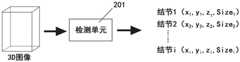
如图2所示,根据本发明第二实施例,对3D图像中的对象进行检测的检测单元201实现为执行上述步骤102-104的程序模块,该检测单元201接收3D图像,并直接输出在3D图像的坐标系中的肺结节的定位和尺寸,例如检测到i个肺结节,这些肺结节的定位和尺寸分别是:肺结节1(x1,y1,z1,Size1),肺结节2(x2,y2,z2,Size2),…肺结节i(xi,yi,zi,Sizei)。该检测单元201实现完全自动化的、端到端且准确的肺结节检测,给临床医疗带来极大的便利;其能够直接应用于3D图像并且大大降低了放射科医生的工作强度。As shown in FIG. 2, according to the second embodiment of the present invention, a
通常的3D医学图像数据量较大,3D数据、特征映射及相关的运算导致计算资源消耗很大,现在GPU往往不够用。可以将整个3D图像划分为多个部分,分别对各个部分应用上述检测单元201,以得到各个部分中肺结节的定位和尺寸,并将各个部分中肺结节的定位和尺寸进行拼合,以得到整个3D图像中肺结节的定位及尺寸。如此使得本发明的方法得以利用现代GPU内存来实现,便利了在临床上的广泛运用。Usually, the amount of 3D medical image data is large, and 3D data, feature mapping and related operations consume a lot of computing resources, and now GPUs are often not enough. The entire 3D image can be divided into a plurality of parts, and the above-mentioned
所述部分可以是3D图像的图像块,也可以是对3D图像进行划分所得到的组块。虽然可以沿着若干方向将3D图像划分为组块,优选地,沿着3D成像手段的成像设备相对于受检体的进给方向来进行划分,例如,对于容积CT扫描来说,优选沿着纵向来划分出组块,如此划分出的组块保留更多的原始信息,且这种划分操作更方便,计算成本更低。The part may be an image block of the 3D image, or may be a group block obtained by dividing the 3D image. Although the 3D image may be divided into chunks along several directions, preferably the division is along the feed direction of the imaging device of the 3D imaging means relative to the subject, eg for volumetric CT scans, preferably along The chunks are divided vertically, and the chunks thus divided retain more original information, and the division operation is more convenient and the computation cost is lower.
所述3D学习网络可以包括3D卷积神经网络、递归神经网络、各种深度学习神经网络等中的一种或多种。优选采用3D卷积神经网络,如此有效利用了GPU的快速卷积计算,能够获得可观的加速。The 3D learning network may include one or more of 3D convolutional neural networks, recurrent neural networks, various deep learning neural networks, and the like. It is preferable to use a 3D convolutional neural network, which effectively utilizes the fast convolution calculation of the GPU and can obtain considerable acceleration.
可以利用滑动窗口从3D图像生成相同尺寸的图像块,并将上述检测单元201应用于这些图像块;通过将步长设置为一个体素,通过在每个体素处执行网络前向计算,以确保不会遗漏任何肺结节的检测。下面以维度为64*64*64的图像块为例,示出应用于其的卷积神经网络的示例。Image patches of the same size can be generated from 3D images using a sliding window, and the
图3示出根据本发明第三实施例的对维度为64*64*64的图像块中的对象进行检测的方法所利用的3D卷积网络的第一示例(第一网络),所述第一网络是全连接3D卷积网络。如图3中所示,其中,用虚线框表示神经网络层,用长方体(或长方形)表示特征映射,用长方体的组表示特征映射的组,在神经网络层和长方体的组的下方,标识出了其维度。所述第一网络依序包括卷积层C1-1、卷积层C1-2、池化层1、卷积层C2-1、卷积层C2-2、池化层2、卷积层2、卷积层C3-1、卷积层C3-2、池化层3、全连接层FC1、全连接层FC2和全连接层FC3,并输出预测结果,所述预测结果包括所述图像块中的对象在10类尺寸上的得分。其中,卷积层C1-1、卷积层C1-2、卷积层C2-1、卷积层C2-2、卷积层C3-1、卷积层C3-2具有3*3*3的核,且步长均为1;如图3所示的所述图像块中的对象在10类尺寸上的得分构成相应尺寸类别的特征映射的栅格。FIG. 3 shows a first example (a first network) of a 3D convolutional network used by a method for detecting objects in an image block with dimensions of 64*64*64 according to the third embodiment of the present invention. One network is a fully connected 3D convolutional network. As shown in Figure 3, where the neural network layer is represented by a dashed box, the feature map is represented by a cuboid (or rectangle), and the group of feature maps is represented by a group of rectangles, and below the neural network layer and the group of rectangles, marked its dimensions. The first network sequentially includes a convolutional layer C1-1, a convolutional layer C1-2, a
可以在3D图像中利用滑动窗口来生成所述图像块,值得注意的是,这种基于滑动窗口的方法因搜索空间大而变得非常缓慢。在优选实施例中,可以将最后的三个全连接层转换为卷积层。The image patches can be generated using sliding windows in 3D images, and it is worth noting that this sliding window based method becomes very slow due to the large search space. In a preferred embodiment, the last three fully connected layers can be converted to convolutional layers.
图5示出将全连接层转换为全卷积层的图示。如图5所示,全连接层FC1接收2*2的特征映射作为输入,该特征映射中的元素分别乘以权重系数w00、w01、w10、w11并求和馈送到全连接层FC1的神经元。可以基于权重系数w00、w01、w10、w11生成卷积层FC1-conv,如图5所示,元素乘以权重系数并求和的运算也可以由图5所示的卷积层FC1-conv实现,由此,可以将全连接层转换为卷积层。Figure 5 shows an illustration of converting a fully connected layer to a fully convolutional layer. As shown in Figure 5, the fully connected layer FC1 receives a 2*2 feature map as input, the elements in this feature map are multiplied by the weight coefficients w00, w01, w10, w11 respectively and the sum is fed to the neurons of the fully connected layer FC1 . The convolutional layer FC1-conv can be generated based on the weight coefficients w00, w01, w10, and w11. As shown in Figure 5, the operation of multiplying the elements by the weight coefficients and summing can also be implemented by the convolutional layer FC1-conv shown in Figure 5. , whereby the fully connected layer can be converted into a convolutional layer.
通过将转换过程应用于图3所示的第一网络,就可以得到图4示出的根据本发明第四实施例的对3D图像中的对象进行检测的方法所利用的3D卷积网络的第一网络的第二示例,所述第一网络是全卷积网络。By applying the conversion process to the first network shown in FIG. 3 , the third step of the 3D convolutional network used by the method for detecting objects in a 3D image according to the fourth embodiment of the present invention shown in FIG. 4 can be obtained. A second example of a network, the first network being a fully convolutional network.
卷积的跨步如同滑动窗口的跨步,这两种操作是等效的。由于GPU上的快速卷积计算功能,通过将3D全连接神经网络转换成3D全卷积神经网络,能够获得巨大的加速,从而能够适用于对耗时有严苛要求的临床运用。Convolution strides are like sliding window strides, and the two operations are equivalent. Due to the fast convolution computing function on the GPU, by converting the 3D fully connected neural network into a 3D fully convolutional neural network, a huge speedup can be obtained, which can be suitable for clinical applications with strict time-consuming requirements.
每个栅格位置可以携带标签,该标签标识该栅格对应的图像区域中所包含的肺结节的尺寸类型,例如,标签0表示3D图像中不含有肺结节,标签1-9表示3D图像中含有肺结节,且含有的肺结节具有第一到第九类尺寸中的相应一种尺寸。每个栅格位置与3D图像的坐标系中的位置相对应。由于最大池化和卷积操作,所得到的图像尺寸相较原始图像尺寸缩减,对应位置取决于具体的3D学习网络的结构。没有边界填充的卷积操作将为最终得到的图像带来位置偏移,该偏移量可以如下计算得到:将卷积层的核尺寸-1得到差,将该差除以2然后进行向下取整运算。Each grid position can carry a label, which identifies the size and type of lung nodules contained in the image area corresponding to the grid. For example, label 0 indicates that the 3D image does not contain lung nodules, and labels 1-9 indicate 3D nodules. A lung nodule is included in the image, and the included lung nodule has a corresponding one of the first to ninth categories of dimensions. Each grid position corresponds to a position in the coordinate system of the 3D image. Due to the max pooling and convolution operations, the resulting image size is reduced compared to the original image size, and the corresponding position depends on the structure of the specific 3D learning network. The convolution operation without border padding will bring a positional offset to the final image, which can be calculated as follows: take the kernel size of the convolution layer -1 to get the difference, divide the difference by 2 and go down Rounding operation.
下面以图4所示的3D全卷积神经网络为例,说明如何将所确定的肺结节的位置(例如坐标)从特征映射的第一坐标系转换到3D图像的第二坐标系。当卷积层C1-1、卷积层C1-2、卷积层C2-1、卷积层C2-2、卷积层C3-1、卷积层C3-2都没有边界填充时,利用公式1来计算肺结节在第二坐标系中的坐标。The following takes the 3D fully convolutional neural network shown in FIG. 4 as an example to illustrate how to convert the determined position (eg, coordinates) of the lung nodule from the first coordinate system of the feature map to the second coordinate system of the 3D image. When the convolutional layer C1-1, the convolutional layer C1-2, the convolutional layer C2-1, the convolutional layer C2-2, the convolutional layer C3-1, and the convolutional layer C3-2 have no boundary padding, use the

其中,xf是特征映射的第一坐标系中的坐标,xori是3D图像的第二坐标系中的坐标,缩放因子s1=s2=s3=2,c1_1、c1_2、c2_1、c2_2、c3_1、c3_2分别是下角标所标识的卷积层的核尺寸,且均等于3,c4=8,c5=1,c6=1,
当卷积层C1-1、卷积层C1-2、卷积层C2-1、卷积层C2-2、卷积层C3-1、卷积层C3-2都具有边界填充时,利用公式2来计算肺结节在第二坐标系中的坐标。When the convolutional layer C1-1, the convolutional layer C1-2, the convolutional layer C2-1, the convolutional layer C2-2, the convolutional layer C3-1, and the convolutional layer C3-2 all have boundary padding, use the

以上采用了用于单个尺度的特征映射的基础网络进行说明,单个尺度的基础网络能够满足一些对象的检测需求。但是,有些对象需要在多个尺度上进行检测,可变尺度可为对象的检测提供适合的视野。为了进一步提高检测精度,通常可以使用金字塔输入图像。然而,对于基于卷积神经网络的方法来说,图像金字塔的计算量非常大。而对于3D深度学习,使用金字塔是不切实际的。The above uses the basic network for feature mapping of a single scale to illustrate, and the basic network of a single scale can meet the detection requirements of some objects. However, some objects need to be detected at multiple scales, and variable scales can provide a suitable field of view for object detection. To further improve detection accuracy, pyramidal input images can often be used. However, for methods based on convolutional neural networks, image pyramids are very computationally expensive. And for 3D deep learning, it is impractical to use pyramids.
为了解决这一问题,我们扩展了上文中所述的基础网络,以提供基于多尺度的特征映射的神经网络,如图6所示,输入的3D图像经过基础网络601的处理后,得到第一尺度的特征映射,其后可以设置有从第二网络到第i网络的若干网络,分别对应于第2尺度到第i尺度。尺度的数量i基于具体的任务需求,一般来说,尺寸变化范围较大的对象需要较多的尺度,通常说来,定位变化较大的对象需要较多的尺度。多尺度具有多种实现方式。例如,我们可以使用卷积过滤器或池化过滤器或者两者均使用,来将特征映射下采样到不同的尺度。再例如,在基于滑动窗口的卷积神经网络的检测方法中,特征映射的尺度可以具有相同的大小,但采用不同大小的滑动窗口。To solve this problem, we extend the basic network described above to provide a neural network based on multi-scale feature mapping, as shown in Figure 6, after the input 3D image is processed by the
在临床应用中,放射科医生需要对对象进行定量分析,但是可能存在大量的虚假警报,我们可以获得较高的招回率但精度会受影响。优选地,为了提高检测准确度,我们可以预先执行分割,利用分割的结果作为约束条件,来执行使用3D学习网络基于所述3D图像得到多个特征映射的步骤。如此可以针对对象会出现的区域进行对象检测。对于肺部中的肺结节进行检测的应用中,可以采用各种肺部分割的方法。因为肺结节永远出现在肺部内,应用肺部分割可以进一步排除虚假警报。例如,我们可以从肺部分割中产生肺凸包,然后利用凸包限制结节的检测。由于我们不需要准确的肺部边界,而仅仅利用分割结果作为结节检测的约束条件,为了将3D网络和特征映射装入GPU内存中,并加快分割程序,我们可以以较低分辨率的扫描进行肺部分割,并对结果进行上采样至原始分辨率,如此可以大大加快分割速度并节省分割所需的计算资源。优选地,可以采用3D版的U-Net(参见O.Ronneberger,P.Fischer,T.Brox,U-Net:Convolutional networks for BiomedicalImage Segmentation,MICCAI 2015)进行肺部分割。In clinical applications, radiologists need to quantitatively analyze objects, but there may be a large number of false alarms, and we can obtain high recall rates but the accuracy will be compromised. Preferably, in order to improve the detection accuracy, we can perform segmentation in advance, and use the result of the segmentation as a constraint to perform the step of using a 3D learning network to obtain multiple feature maps based on the 3D image. This allows object detection to be performed on areas where objects will appear. For the application of detection of pulmonary nodules in the lungs, various lung segmentation methods can be used. Because lung nodules are always present in the lungs, applying lung segmentation can further exclude false alarms. For example, we can generate a lung convex hull from lung segmentation and then use the convex hull to constrain the detection of nodules. Since we don't need accurate lung boundaries and just use the segmentation results as constraints for nodule detection, in order to fit the 3D network and feature maps into GPU memory and speed up the segmentation process, we can scan at lower resolutions Perform lung segmentation and upsample the results to the original resolution, which can greatly speed up the segmentation and save the computational resources required for segmentation. Preferably, the 3D version of U-Net (see O. Ronneberger, P. Fischer, T. Brox, U-Net: Convolutional networks for Biomedical Image Segmentation, MICCAI 2015) can be used for lung segmentation.
上文所描述的各种步骤、操作或功能,可以被实现或定义为软件代码或指令。这样的内容可以是直接可执行的(“对象”或“可执行”形式)、源代码或差分代码(“增量(delta)”或“补丁(patch)”代码)。可以经由具有存储在其上的代码或指令的制品或者经由操作通信接口以经由通信接口发送数据的方法来提供本文所述的实施例的软件实现。机器或计算机可读存储介质可以使得机器执行所描述的功能或操作,并且包括以机器(例如,计算装置、电子系统等等)可访问的形式存储信息的任何机构,诸如可记录/不可记录介质(例如,只读存储器(ROM)、随机存取存储器(RAM)、磁盘存储介质、光存储介质、闪存装置等)。通信接口包括与硬连线、无线、光学等介质中的任何一个交互的任何机构以与诸如存储器总线接口、处理器总线接口、因特网连接、磁盘控制器等的另一装置进行通信。通信接口可以被配置为通过提供配置参数和/或发送信号来准备通信接口以提供描述软件内容的数据信号。可以经由发送到通信接口的一个或多个命令或信号来访问通信接口。The various steps, operations or functions described above may be implemented or defined as software codes or instructions. Such content may be directly executable ("object" or "executable" form), source code or differential code ("delta" or "patch" code). Software implementations of the embodiments described herein may be provided via an article of manufacture having code or instructions stored thereon or via a method of operating a communications interface to send data via the communications interface. A machine or computer readable storage medium can cause a machine to perform the functions or operations described, and includes any mechanism for storing information in a form accessible by a machine (eg, computing device, electronic system, etc.), such as recordable/non-recordable media (eg, read only memory (ROM), random access memory (RAM), magnetic disk storage media, optical storage media, flash memory devices, etc.). A communication interface includes any mechanism that interacts with any of hardwired, wireless, optical, etc. media to communicate with another device such as a memory bus interface, processor bus interface, Internet connection, disk controller, and the like. The communication interface may be configured to prepare the communication interface by providing configuration parameters and/or sending signals to provide data signals describing the content of the software. The communication interface may be accessed via one or more commands or signals sent to the communication interface.
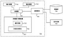
图7示出根据本发明第六实施例的对3D图像中的对象进行检测的系统的框图。如图7所示,该系统包括:存储器707,其存储计算机可执行指令;处理器703,其通信地连接到所述存储器,并被配置为执行所述计算机可执行指令,以执行如下步骤:使用3D学习网络基于所述3D图像得到一组特征映射,每个特征映射针对相应的对象尺寸,所述特征映射的栅格表示所述3D图像中与该栅格对应的区域内包含相应尺寸的对象的概率相关参数;基于对应于同个区域的栅格,来确定该区域中包含的对象的尺寸;以及将所确定的对象的位置从特征映射的第一坐标系转换到3D图像的第二坐标系中,以得到所述第二坐标系中各个尺寸的对象的定位。所存储的计算机可执行指令可以包括操作系统705和对象检测程序模块706,后者用于执行上述检测步骤。可选地,所述系统还可以包括:输入装置701,用于接收来自用户的输入;输出装置702,例如显示器,用于展现对象检测的结果;还有通信接口704。FIG. 7 shows a block diagram of a system for detecting objects in a 3D image according to a sixth embodiment of the present invention. As shown in Figure 7, the system includes: a
通信接口704可以可通信地连接到数据库708,以向其传输检测结果,可选地,可以将标有检测结果的3D图像传输到数据库708,以供其他用户调用和查看。通信接口704可以可通信地连接到数据库708,以从其接收需要进行对象检测的3D医学图像。The
通信接口704可以可通信地连接到医学成像装置709,以从其接收需要进行对象检测的3D医学图像;或者,也可以从其接收需要进行对象检测的2D医学图像的序列,并将2D医学图像的序列重建为3D医学图像,以供进行对象检测。该重建步骤可以利用上述计算机可执行指令中的程序模块来执行,该程序模块可以包含在对象检测模块706中,也可以与之彼此分离。The
上述系统可以是为了所需目的而专门构造的,或者其可以包括由存储在计算机中的计算机程序选择性地激活或重新配置的通用计算机,或者,也可以整合在医学成像装置自带的系统中。这样的计算机程序可以存储在计算机可读存储介质中,计算机可读存储介质例如是:包括软盘、光盘、CDROM以及磁光盘的任何类型的盘、只读存储器(ROM)、随机存取存储器(RAM)、EPROM、EEPROM、磁卡或光卡、或适于存储电子指令的任何类型的介质,每个介质耦合到计算机系统总线,但并不限于此。The above system may be specially constructed for the required purposes, or it may comprise a general-purpose computer selectively activated or reconfigured by a computer program stored in the computer, or it may be incorporated into a system that comes with the medical imaging device . Such a computer program may be stored in a computer-readable storage medium such as: any type of disk including floppy disk, optical disk, CDROM and magneto-optical disk, read only memory (ROM), random access memory (RAM) ), EPROM, EEPROM, magnetic or optical cards, or any type of medium suitable for storing electronic instructions, each medium coupled to a computer system bus, but not limited thereto.
除非另有说明,本文示出和描述的本发明的实施例的执行顺序或操作的执行不是必要的。也就是说,除非另有说明,操作可以以任何顺序执行,并且本发明的实施例可以包括比本文公开的操作更多或更少的操作。例如,在另一操作之前、同时或之后执行或实施特定操作被预期在本发明的方案的范围内。The order of execution or performance of the operations of the embodiments of the invention shown and described herein are not essential unless otherwise specified. That is, unless otherwise specified, the operations may be performed in any order, and embodiments of the invention may include more or fewer operations than those disclosed herein. For example, it is contemplated that a particular operation is performed or performed before, concurrently with, or after another operation is within the scope of the aspects of the present invention.
本发明的实施例可以利用计算机可执行指令来实现。计算机可执行指令可以被组织成一个或多个计算机可执行组件或模块。本发明的各方案可以利用任何数量和组织的这种组件或模块来实现。例如,本发明的各方案不限于图中所示和本文所描述的特定计算机可执行指令或特定组件或模块。本发明的其它实施例可以包括具有比本文所示出和描述的更多或更少功能的不同的计算机可执行指令或组件。Embodiments of the invention may be implemented using computer-executable instructions. Computer-executable instructions may be organized into one or more computer-executable components or modules. Aspects of the present invention may be implemented using any number and organization of such components or modules. For example, aspects of the present invention are not limited to the specific computer-executable instructions or the specific components or modules shown in the figures and described herein. Other embodiments of the invention may include different computer-executable instructions or components having more or less functionality than those shown and described herein.
已经对本发明的各方案进行了详细描述,显而易见的是,在不背离如所附权利要求中限定的本发明的方案的范围的情况下,修改和变化是可能的。由于可以在不背离本发明的方案的范围的情况下对上述结构、产品和方法进行各种改变,因此意图在于上述说明书中包含的和附图中所示的所有内容应当被解释为示例性的,而不具有限制性意义。Having described various aspects of the invention in detail, it will be evident that modifications and variations are possible without departing from the scope of the aspects of the invention as defined in the appended claims. As various changes could be made in the above structures, products and methods without departing from the scope of the present invention, it is intended that all matter contained in the foregoing specification and shown in the accompanying drawings shall be interpreted as illustrative , not in a restrictive sense.
Claims (27)
Applications Claiming Priority (2)
| Application Number | Priority Date | Filing Date | Title |
|---|---|---|---|
| US201762542890P | 2017-08-09 | 2017-08-09 | |
| US62/542,890 | 2017-08-09 |
Publications (2)
| Publication Number | Publication Date |
|---|---|
| CN108022238A CN108022238A (en) | 2018-05-11 |
| CN108022238Btrue CN108022238B (en) | 2020-07-03 |
Family
ID=62078009
Family Applications (1)
| Application Number | Title | Priority Date | Filing Date |
|---|---|---|---|
| CN201711249474.XAActiveCN108022238B (en) | 2017-08-09 | 2017-12-01 | Method, computer storage medium, and system for detecting object in 3D image |
Country Status (2)
| Country | Link |
|---|---|
| US (1) | US10867384B2 (en) |
| CN (1) | CN108022238B (en) |
Families Citing this family (66)
| Publication number | Priority date | Publication date | Assignee | Title |
|---|---|---|---|---|
| US10043101B2 (en)* | 2014-11-07 | 2018-08-07 | Adobe Systems Incorporated | Local feature representation for image recognition |
| US10607135B2 (en) | 2017-10-19 | 2020-03-31 | General Electric Company | Training an auto-encoder on a single class |
| US10460440B2 (en)* | 2017-10-24 | 2019-10-29 | General Electric Company | Deep convolutional neural network with self-transfer learning |
| US10346728B2 (en)* | 2017-10-26 | 2019-07-09 | Hitachi, Ltd. | Nodule detection with false positive reduction |
| CN108805976B (en)* | 2018-05-31 | 2022-05-13 | 武汉中观自动化科技有限公司 | Three-dimensional scanning system and method |
| US10885400B2 (en)* | 2018-07-03 | 2021-01-05 | General Electric Company | Classification based on annotation information |
| US10755147B2 (en) | 2018-07-03 | 2020-08-25 | General Electric Company | Classification and localization based on annotation information |
| US20200012884A1 (en) | 2018-07-03 | 2020-01-09 | General Electric Company | Classification based on annotation information |
| CN109063710B (en)* | 2018-08-09 | 2022-08-16 | 成都信息工程大学 | 3D CNN nasopharyngeal carcinoma segmentation method based on multi-scale feature pyramid |
| DE102018214325A1 (en)* | 2018-08-24 | 2020-02-27 | Siemens Healthcare Gmbh | Method and provision unit for the provision of a virtual tomographic stroke follow-up examination image |
| US10733727B2 (en)* | 2018-11-14 | 2020-08-04 | Qure.Ai Technologies Private Limited | Application of deep learning for medical imaging evaluation |
| CN109670532B (en)* | 2018-11-23 | 2022-12-09 | 腾讯医疗健康(深圳)有限公司 | Abnormal recognition method, device and system for biological organ tissue image |
| CN111292301A (en)* | 2018-12-07 | 2020-06-16 | 北京市商汤科技开发有限公司 | A kind of lesion detection method, device, equipment and storage medium |
| US11995854B2 (en)* | 2018-12-19 | 2024-05-28 | Nvidia Corporation | Mesh reconstruction using data-driven priors |
| JP7105918B2 (en)* | 2018-12-27 | 2022-07-25 | 富士フイルム株式会社 | AREA IDENTIFICATION APPARATUS, METHOD AND PROGRAM |
| US10325371B1 (en)* | 2019-01-22 | 2019-06-18 | StradVision, Inc. | Method and device for segmenting image to be used for surveillance using weighted convolution filters for respective grid cells by converting modes according to classes of areas to satisfy level 4 of autonomous vehicle, and testing method and testing device using the same |
| US10373317B1 (en)* | 2019-01-22 | 2019-08-06 | StradVision, Inc. | Learning method and learning device for attention-driven image segmentation by using at least one adaptive loss weight map to be used for updating HD maps required to satisfy level 4 of autonomous vehicles and testing method and testing device using the same |
| US10339424B1 (en)* | 2019-01-22 | 2019-07-02 | StradVision, Inc. | Method and device of neural network operations using a grid generator for converting modes according to classes of areas to satisfy level 4 of autonomous vehicles |
| US10387754B1 (en)* | 2019-01-23 | 2019-08-20 | StradVision, Inc. | Learning method and learning device for object detector based on CNN using 1×H convolution to be used for hardware optimization, and testing method and testing device using the same |
| US10402695B1 (en)* | 2019-01-23 | 2019-09-03 | StradVision, Inc. | Learning method and learning device for convolutional neural network using 1×H convolution for image recognition to be used for hardware optimization, and testing method and testing device using the same |
| US10410352B1 (en)* | 2019-01-25 | 2019-09-10 | StradVision, Inc. | Learning method and learning device for improving segmentation performance to be used for detecting events including pedestrian event, vehicle event, falling event and fallen event using edge loss and test method and test device using the same |
| US10402977B1 (en)* | 2019-01-25 | 2019-09-03 | StradVision, Inc. | Learning method and learning device for improving segmentation performance in road obstacle detection required to satisfy level 4 and level 5 of autonomous vehicles using laplacian pyramid network and testing method and testing device using the same |
| WO2020174770A1 (en)* | 2019-02-28 | 2020-09-03 | 富士フイルム株式会社 | Area specifying device, method, program, learning device, method, program, and identifying device |
| CN111626087A (en)* | 2019-02-28 | 2020-09-04 | 北京市商汤科技开发有限公司 | Neural network training and eye opening and closing state detection method, device and equipment |
| CN111814514A (en)* | 2019-04-11 | 2020-10-23 | 富士通株式会社 | Number identification device, method and electronic device |
| CN110032980B (en)* | 2019-04-18 | 2023-04-25 | 天津工业大学 | Organ detection and identification positioning method based on deep learning |
| CN110188607B (en)* | 2019-04-23 | 2022-10-21 | 深圳大学 | Traffic video target detection method and device based on multi-thread parallel computing |
| CN110245710B (en)* | 2019-06-18 | 2022-11-29 | 腾讯科技(深圳)有限公司 | Training method of semantic segmentation model, semantic segmentation method and device |
| CN110517221B (en)* | 2019-07-05 | 2022-05-03 | 银河水滴科技(北京)有限公司 | Gap positioning method and device based on real coordinates and storage medium |
| CN110427915B (en)* | 2019-08-14 | 2022-09-27 | 北京百度网讯科技有限公司 | Method and apparatus for outputting information |
| CN111325084A (en)* | 2019-08-29 | 2020-06-23 | 西安铱食云餐饮管理有限公司 | Dish information identification method and terminal based on YOLO neural network |
| CN112711972B (en)* | 2019-10-26 | 2024-06-14 | 海思技术有限公司 | Target detection method and device |
| CN110880035B (en)* | 2019-11-14 | 2020-12-01 | 北京推想科技有限公司 | Convolutional neural network training method and device and nodule sign identification method and device |
| US11200455B2 (en)* | 2019-11-22 | 2021-12-14 | International Business Machines Corporation | Generating training data for object detection |
| CN111144236B (en)* | 2019-12-10 | 2024-04-26 | 华南师范大学 | Cockroach mating behavior analysis method, system and storage medium |
| CN111028224B (en)* | 2019-12-12 | 2020-12-01 | 广西医准智能科技有限公司 | Data labeling method, model training device, image processing method, image processing device and storage medium |
| US10902551B1 (en)* | 2019-12-17 | 2021-01-26 | X Development Llc | True positive transplant |
| CN111179247A (en)* | 2019-12-27 | 2020-05-19 | 上海商汤智能科技有限公司 | Three-dimensional target detection method, training method of model thereof, and related device and equipment |
| CN111241969A (en)* | 2020-01-06 | 2020-06-05 | 北京三快在线科技有限公司 | Target detection method and device and corresponding model training method and device |
| CN111223113B (en)* | 2020-01-07 | 2023-04-18 | 宁波大学 | Nuclear magnetic resonance hippocampus segmentation algorithm based on dual dense context-aware network |
| CN111311655B (en)* | 2020-02-13 | 2023-07-04 | 东软医疗系统股份有限公司 | Multi-mode image registration method, device, electronic equipment and storage medium |
| CN111339861A (en)* | 2020-02-17 | 2020-06-26 | 南京工程学院 | Seat occupancy state detection method |
| CN111340768B (en)* | 2020-02-21 | 2021-03-09 | 之江实验室 | Multi-center effect compensation method based on PET/CT intelligent diagnosis system |
| WO2021173489A1 (en)* | 2020-02-28 | 2021-09-02 | Nokia Technologies Oy | Apparatus, method, and system for providing a three-dimensional texture using uv representation |
| JP7389233B2 (en) | 2020-04-09 | 2023-11-29 | 富士フイルム株式会社 | Image processing device, method and program, learning device, method and program, and derivation model |
| CN111738306B (en)* | 2020-06-01 | 2022-05-13 | 山东省人工智能研究院 | Multi-view three-dimensional model retrieval method based on block convolution neural network |
| CN111739035B (en)* | 2020-06-30 | 2022-09-30 | 腾讯科技(深圳)有限公司 | Image processing method, device and equipment based on artificial intelligence and storage medium |
| KR102532006B1 (en)* | 2020-07-24 | 2023-05-12 | 한국전자기술연구원 | Image Region Segmentation Method and System using Self-Spatial Adaptive Normalization |
| CN112101205B (en)* | 2020-09-15 | 2024-08-16 | 东软睿驰汽车技术(沈阳)有限公司 | Training method and device based on multi-task network |
| CN112184657A (en)* | 2020-09-24 | 2021-01-05 | 上海健康医学院 | Pulmonary nodule automatic detection method, device and computer system |
| JP7587815B2 (en) | 2020-10-29 | 2024-11-21 | 国立大学法人九州大学 | Medical image processing method, medical image processing device, and medical image processing program |
| CN112365507B (en)* | 2020-10-30 | 2024-02-02 | 沈阳东软智能医疗科技研究院有限公司 | CT image processing method and device, storage medium and electronic equipment |
| CN112329863B (en)* | 2020-11-09 | 2022-04-22 | 西南交通大学 | A method for identifying the state of isolation switch in a traction substation |
| WO2022128105A1 (en)* | 2020-12-17 | 2022-06-23 | Huawei Technologies Co., Ltd. | Decoding and encoding of neural-network-based bitstreams |
| CN114764916B (en)* | 2021-01-04 | 2025-05-09 | 中国移动通信有限公司研究院 | Text recognition processing method, device and related equipment |
| US11620748B2 (en)* | 2021-01-11 | 2023-04-04 | International Business Machines Corporation | Systems and methods for generating a 2D image from a 3D medical image |
| WO2022158056A1 (en)* | 2021-01-25 | 2022-07-28 | 株式会社島津製作所 | X-ray fluoroscopic imaging device |
| CN112861858B (en)* | 2021-02-19 | 2024-06-07 | 北京龙翼风科技有限公司 | Method for generating saliency truth value diagram and method for training saliency detection model |
| CN113256600B (en)* | 2021-06-10 | 2021-11-19 | 浙江华睿科技股份有限公司 | Camera dust detection method and device based on artificial intelligence and electronic equipment |
| CN113591363B (en)* | 2021-07-29 | 2023-12-12 | 云南电网有限责任公司保山供电局 | Multi-frequency ultrasonic detection-based transformer oil dielectric loss regression prediction method |
| US11521321B1 (en)* | 2021-10-07 | 2022-12-06 | Qure.Ai Technologies Private Limited | Monitoring computed tomography (CT) scan image |
| CN114118361B (en)* | 2021-10-29 | 2025-05-06 | 北京宇航系统工程研究所 | A situation assessment method based on deep learning parameter anchoring |
| CN114782402B (en)* | 2022-05-17 | 2025-03-18 | 华南师范大学 | Pulmonary nodule CT image segmentation, training method and device based on improved V-Net network |
| CN114862850B (en)* | 2022-07-06 | 2022-09-20 | 深圳科亚医疗科技有限公司 | Target detection method, device and medium for blood vessel medical image |
| CN115222930B (en)* | 2022-09-02 | 2022-11-29 | 四川蜀天信息技术有限公司 | WebGL-based 3D model arrangement and combination method |
| CN116797781B (en)* | 2023-07-12 | 2024-08-20 | 北京斯年智驾科技有限公司 | Target detection method, device, electronic device and storage medium |
Family Cites Families (22)
| Publication number | Priority date | Publication date | Assignee | Title |
|---|---|---|---|---|
| US9230320B2 (en)* | 2012-03-30 | 2016-01-05 | University Of Louisville Research Foundation, Inc. | Computer aided diagnostic system incorporating shape analysis for diagnosing malignant lung nodules |
| CA2884167C (en)* | 2012-09-13 | 2020-05-12 | The Regents Of The University Of California | System and method for automated detection of lung nodules in medical images |
| US9730643B2 (en)* | 2013-10-17 | 2017-08-15 | Siemens Healthcare Gmbh | Method and system for anatomical object detection using marginal space deep neural networks |
| US10410096B2 (en)* | 2015-07-09 | 2019-09-10 | Qualcomm Incorporated | Context-based priors for object detection in images |
| AU2016308097B2 (en)* | 2015-08-15 | 2018-08-02 | Salesforce.Com, Inc. | Three-dimensional (3D) convolution with 3D batch normalization |
| US9965719B2 (en)* | 2015-11-04 | 2018-05-08 | Nec Corporation | Subcategory-aware convolutional neural networks for object detection |
| US9881234B2 (en)* | 2015-11-25 | 2018-01-30 | Baidu Usa Llc. | Systems and methods for end-to-end object detection |
| US9760807B2 (en)* | 2016-01-08 | 2017-09-12 | Siemens Healthcare Gmbh | Deep image-to-image network learning for medical image analysis |
| US9972092B2 (en)* | 2016-03-31 | 2018-05-15 | Adobe Systems Incorporated | Utilizing deep learning for boundary-aware image segmentation |
| US10157462B2 (en)* | 2016-06-27 | 2018-12-18 | University Of Central Florida Research Foundation, Inc. | System and method for image-based quantification of white and brown adipose tissue at the whole-body, organ and body-region levels |
| CN106203327B (en)* | 2016-07-08 | 2019-04-19 | 清华大学 | Lung tumor recognition system and method based on convolutional neural network |
| US9589374B1 (en)* | 2016-08-01 | 2017-03-07 | 12 Sigma Technologies | Computer-aided diagnosis system for medical images using deep convolutional neural networks |
| US20180039853A1 (en)* | 2016-08-02 | 2018-02-08 | Mitsubishi Electric Research Laboratories, Inc. | Object Detection System and Object Detection Method |
| US9965863B2 (en)* | 2016-08-26 | 2018-05-08 | Elekta, Inc. | System and methods for image segmentation using convolutional neural network |
| US10467459B2 (en)* | 2016-09-09 | 2019-11-05 | Microsoft Technology Licensing, Llc | Object detection based on joint feature extraction |
| US10453200B2 (en)* | 2016-11-02 | 2019-10-22 | General Electric Company | Automated segmentation using deep learned priors |
| IL285121B2 (en)* | 2016-11-15 | 2023-04-01 | Magic Leap Inc | Deep learning system for cuboid detection |
| CN106780460B (en)* | 2016-12-13 | 2019-11-08 | 杭州健培科技有限公司 | A kind of Lung neoplasm automatic checkout system for chest CT images |
| US10635927B2 (en)* | 2017-03-06 | 2020-04-28 | Honda Motor Co., Ltd. | Systems for performing semantic segmentation and methods thereof |
| CN106940816B (en)* | 2017-03-22 | 2020-06-09 | 杭州健培科技有限公司 | CT image pulmonary nodule detection system based on 3D full convolution neural network |
| US10621725B2 (en)* | 2017-04-12 | 2020-04-14 | Here Global B.V. | Small object detection from a large image |
| EP3392832A1 (en)* | 2017-04-21 | 2018-10-24 | General Electric Company | Automated organ risk segmentation machine learning methods and systems |
- 2017
- 2017-12-01CNCN201711249474.XApatent/CN108022238B/enactiveActive
- 2018
- 2018-06-02USUS15/996,434patent/US10867384B2/enactiveActive
Also Published As
| Publication number | Publication date |
|---|---|
| US10867384B2 (en) | 2020-12-15 |
| CN108022238A (en) | 2018-05-11 |
| US20190050981A1 (en) | 2019-02-14 |
Similar Documents
| Publication | Publication Date | Title |
|---|---|---|
| CN108022238B (en) | Method, computer storage medium, and system for detecting object in 3D image | |
| US11288808B2 (en) | System and method for n-dimensional image segmentation using convolutional neural networks | |
| CN112529834B (en) | Spatial distribution of pathological image patterns in 3D image data | |
| US12198379B2 (en) | Systems and methods for image segmentation | |
| US8121362B2 (en) | Registration of medical images using learned-based matching functions | |
| Li et al. | Automated measurement network for accurate segmentation and parameter modification in fetal head ultrasound images | |
| Kalra | Developing fe human models from medical images | |
| CN106887000B (en) | Grid processing method and system for medical image | |
| US8319793B2 (en) | Analyzing pixel data by imprinting objects of a computer-implemented network structure into other objects | |
| US8867802B2 (en) | Automatic organ localization | |
| US10853409B2 (en) | Systems and methods for image search | |
| WO2022213654A1 (en) | Ultrasonic image segmentation method and apparatus, terminal device, and storage medium | |
| CN115018852A (en) | A method and device for detecting abdominal lymph nodes based on semi-supervised learning | |
| JP2019076699A (en) | Nodule detection with false positive reduction | |
| CN111739614A (en) | medical image enhancement | |
| CN115115567B (en) | Image processing method, device, computer equipment and medium | |
| US11908047B2 (en) | Generating synthetic x-ray images and object annotations from CT scans for augmenting x-ray abnormality assessment systems | |
| Kretschmer et al. | ADR-anatomy-driven reformation | |
| CN108597589B (en) | Model generation method, target detection method and medical imaging system | |
| Chernyshov et al. | Automated segmentation and quantification of the right ventricle in 2-D echocardiography | |
| CN108805876B (en) | Method and system for deformable registration of magnetic resonance and ultrasound images using biomechanical models | |
| CN115439650A (en) | Kidney ultrasound image segmentation method based on cross-modal transfer learning of CT images | |
| CN110992310A (en) | Method and device for determining partition where mediastinal lymph node is located | |
| Akbari et al. | BEAS-Net: A Shape-Prior-Based Deep Convolutional Neural Network for Robust Left Ventricular Segmentation in 2-D Echocardiography | |
| Bin et al. | Rapid multimodal medical image registration and fusion in 3D conformal radiotherapy treatment planning |
Legal Events
| Date | Code | Title | Description |
|---|---|---|---|
| PB01 | Publication | ||
| PB01 | Publication | ||
| SE01 | Entry into force of request for substantive examination | ||
| SE01 | Entry into force of request for substantive examination | ||
| GR01 | Patent grant | ||
| GR01 | Patent grant |