CN107109319B - Method for processing droplets containing a sample - Google Patents
Method for processing droplets containing a sampleDownload PDFInfo
- Publication number
- CN107109319B CN107109319BCN201480082752.5ACN201480082752ACN107109319BCN 107109319 BCN107109319 BCN 107109319BCN 201480082752 ACN201480082752 ACN 201480082752ACN 107109319 BCN107109319 BCN 107109319B
- Authority
- CN
- China
- Prior art keywords
- droplets
- oil
- sample
- trapping
- gelled
- Prior art date
- Legal status (The legal status is an assumption and is not a legal conclusion. Google has not performed a legal analysis and makes no representation as to the accuracy of the status listed.)
- Active
Links
Images


Classifications
- B—PERFORMING OPERATIONS; TRANSPORTING
- B01—PHYSICAL OR CHEMICAL PROCESSES OR APPARATUS IN GENERAL
- B01L—CHEMICAL OR PHYSICAL LABORATORY APPARATUS FOR GENERAL USE
- B01L3/00—Containers or dishes for laboratory use, e.g. laboratory glassware; Droppers
- B01L3/50—Containers for the purpose of retaining a material to be analysed, e.g. test tubes
- B01L3/502—Containers for the purpose of retaining a material to be analysed, e.g. test tubes with fluid transport, e.g. in multi-compartment structures
- B01L3/5027—Containers for the purpose of retaining a material to be analysed, e.g. test tubes with fluid transport, e.g. in multi-compartment structures by integrated microfluidic structures, i.e. dimensions of channels and chambers are such that surface tension forces are important, e.g. lab-on-a-chip
- B01L3/502769—Containers for the purpose of retaining a material to be analysed, e.g. test tubes with fluid transport, e.g. in multi-compartment structures by integrated microfluidic structures, i.e. dimensions of channels and chambers are such that surface tension forces are important, e.g. lab-on-a-chip characterised by multiphase flow arrangements
- B01L3/502784—Containers for the purpose of retaining a material to be analysed, e.g. test tubes with fluid transport, e.g. in multi-compartment structures by integrated microfluidic structures, i.e. dimensions of channels and chambers are such that surface tension forces are important, e.g. lab-on-a-chip characterised by multiphase flow arrangements specially adapted for droplet or plug flow, e.g. digital microfluidics
- B—PERFORMING OPERATIONS; TRANSPORTING
- B01—PHYSICAL OR CHEMICAL PROCESSES OR APPARATUS IN GENERAL
- B01L—CHEMICAL OR PHYSICAL LABORATORY APPARATUS FOR GENERAL USE
- B01L3/00—Containers or dishes for laboratory use, e.g. laboratory glassware; Droppers
- B01L3/50—Containers for the purpose of retaining a material to be analysed, e.g. test tubes
- B01L3/502—Containers for the purpose of retaining a material to be analysed, e.g. test tubes with fluid transport, e.g. in multi-compartment structures
- B01L3/5027—Containers for the purpose of retaining a material to be analysed, e.g. test tubes with fluid transport, e.g. in multi-compartment structures by integrated microfluidic structures, i.e. dimensions of channels and chambers are such that surface tension forces are important, e.g. lab-on-a-chip
- B01L3/502746—Containers for the purpose of retaining a material to be analysed, e.g. test tubes with fluid transport, e.g. in multi-compartment structures by integrated microfluidic structures, i.e. dimensions of channels and chambers are such that surface tension forces are important, e.g. lab-on-a-chip characterised by the means for controlling flow resistance, e.g. flow controllers, baffles
- B—PERFORMING OPERATIONS; TRANSPORTING
- B01—PHYSICAL OR CHEMICAL PROCESSES OR APPARATUS IN GENERAL
- B01L—CHEMICAL OR PHYSICAL LABORATORY APPARATUS FOR GENERAL USE
- B01L2200/00—Solutions for specific problems relating to chemical or physical laboratory apparatus
- B01L2200/06—Fluid handling related problems
- B01L2200/0642—Filling fluids into wells by specific techniques
- B—PERFORMING OPERATIONS; TRANSPORTING
- B01—PHYSICAL OR CHEMICAL PROCESSES OR APPARATUS IN GENERAL
- B01L—CHEMICAL OR PHYSICAL LABORATORY APPARATUS FOR GENERAL USE
- B01L2200/00—Solutions for specific problems relating to chemical or physical laboratory apparatus
- B01L2200/06—Fluid handling related problems
- B01L2200/0673—Handling of plugs of fluid surrounded by immiscible fluid
- B—PERFORMING OPERATIONS; TRANSPORTING
- B01—PHYSICAL OR CHEMICAL PROCESSES OR APPARATUS IN GENERAL
- B01L—CHEMICAL OR PHYSICAL LABORATORY APPARATUS FOR GENERAL USE
- B01L2300/00—Additional constructional details
- B01L2300/08—Geometry, shape and general structure
- B01L2300/0809—Geometry, shape and general structure rectangular shaped
- B01L2300/0816—Cards, e.g. flat sample carriers usually with flow in two horizontal directions
- B—PERFORMING OPERATIONS; TRANSPORTING
- B01—PHYSICAL OR CHEMICAL PROCESSES OR APPARATUS IN GENERAL
- B01L—CHEMICAL OR PHYSICAL LABORATORY APPARATUS FOR GENERAL USE
- B01L2300/00—Additional constructional details
- B01L2300/08—Geometry, shape and general structure
- B01L2300/0809—Geometry, shape and general structure rectangular shaped
- B01L2300/0819—Microarrays; Biochips
- B—PERFORMING OPERATIONS; TRANSPORTING
- B01—PHYSICAL OR CHEMICAL PROCESSES OR APPARATUS IN GENERAL
- B01L—CHEMICAL OR PHYSICAL LABORATORY APPARATUS FOR GENERAL USE
- B01L2300/00—Additional constructional details
- B01L2300/08—Geometry, shape and general structure
- B01L2300/0848—Specific forms of parts of containers
- B01L2300/0851—Bottom walls
- B—PERFORMING OPERATIONS; TRANSPORTING
- B01—PHYSICAL OR CHEMICAL PROCESSES OR APPARATUS IN GENERAL
- B01L—CHEMICAL OR PHYSICAL LABORATORY APPARATUS FOR GENERAL USE
- B01L2400/00—Moving or stopping fluids
- B01L2400/08—Regulating or influencing the flow resistance
- B01L2400/084—Passive control of flow resistance
- B01L2400/086—Passive control of flow resistance using baffles or other fixed flow obstructions
- B—PERFORMING OPERATIONS; TRANSPORTING
- B01—PHYSICAL OR CHEMICAL PROCESSES OR APPARATUS IN GENERAL
- B01L—CHEMICAL OR PHYSICAL LABORATORY APPARATUS FOR GENERAL USE
- B01L2400/00—Moving or stopping fluids
- B01L2400/08—Regulating or influencing the flow resistance
- B01L2400/084—Passive control of flow resistance
- B01L2400/088—Passive control of flow resistance by specific surface properties
Landscapes
- Chemical & Material Sciences (AREA)
- Health & Medical Sciences (AREA)
- Dispersion Chemistry (AREA)
- Analytical Chemistry (AREA)
- General Health & Medical Sciences (AREA)
- Hematology (AREA)
- Clinical Laboratory Science (AREA)
- Chemical Kinetics & Catalysis (AREA)
- Apparatus Associated With Microorganisms And Enzymes (AREA)
- Micro-Organisms Or Cultivation Processes Thereof (AREA)
- Sampling And Sample Adjustment (AREA)
- Colloid Chemistry (AREA)
Abstract
Description
Translated fromChinese技术领域technical field
本发明涉及用于处理水凝胶微滴中的样品、特别是生物样品的微流体方法。本发明还涉及用于进行这种方法的装置和涉及通过进行这种方法获得的样品的产品。The present invention relates to microfluidic methods for processing samples, in particular biological samples, in hydrogel droplets. The invention also relates to a device for carrying out this method and to a product relating to a sample obtained by carrying out this method.
背景技术Background technique
从Guo、Rotem、Heyman和Weitz,“Droplet microfluidics for high-throughputbiological assays”,Lab.Chip.12(2012)中已知,微流体系统中的液滴(或“微滴”)可用于包含化学反应或生物反应。在这些系统中,可以当液滴在聚焦激光器前面通过时观察液滴的荧光来测定这些液滴的含量。然而,在不将这些液滴从微流体装置中提取出来时,这些系统不能观察这些液滴的含量随时间的变化。From Guo, Rotem, Heyman and Weitz, "Droplet microfluidics for high-throughputbiological assays", Lab.Chip.12 (2012), it is known that droplets (or "droplets") in microfluidic systems can be used to contain chemical reactions or biological response. In these systems, the content of these droplets can be determined by observing their fluorescence as they pass in front of a focused laser. However, these systems cannot observe the content of these droplets over time without extracting them from the microfluidic device.
关于微滴中的个体化细胞的研究也是已知的,例如来自Joensson,H.N.和Andersson Svahn,H.,“Droplet microfluidics-a tool for single-cell analysis”,Angew.Chem.Int.Ed.Engl.51,12176-12192(2012)。事实上,这些微滴形成了明确界定的隔室,其例如使得能够分离出诸如细胞的生物样品。该文献特别教导可以通过在培养室中、在细长通道中或在静态捕集阱中积聚液滴来控制包封细胞群体的定位。该文献还教导细胞可以被包封在被油相包围的官能化水凝胶中。Studies on individualized cells in droplets are also known, eg from Joensson, H.N. and Andersson Svahn, H., "Droplet microfluidics-a tool for single-cell analysis", Angew.Chem.Int.Ed.Engl. 51, 12176-12192 (2012). In fact, these droplets form well-defined compartments which, for example, enable the separation of biological samples such as cells. The document specifically teaches that the localization of the encapsulated cell population can be controlled by accumulating droplets in a culture chamber, in an elongated channel, or in a static trap. The document also teaches that cells can be encapsulated in functionalized hydrogels surrounded by an oil phase.
然而,在这种装置中细胞的营养物质或更一般的生物感兴趣的分子的供应被证明是有限的,并且使用的方法(例如通过电融合或皮升注射)证明是复杂的。因此,这些装置对细胞行为的研究具有许多限制,特别是在时间方面。However, the supply of nutrients to cells or molecules of biological interest in general in such devices proved to be limited and the methods used (eg by electrofusion or picoliter injection) proved complex. Therefore, these devices have many limitations for the study of cellular behavior, especially in terms of time.
此外,从L.Yu,M.C.W.Chen和K.C.Chang,“Droplet-based microfluidic systemfor multicellular tumor Spheroid formation and anticancer drug testing”,LabChip(2010),已知一种处理含有多细胞球体的水凝胶微滴的方法。根据该方法,在第一微流体系统中产生包括细胞的水凝胶微珠。然后将它们回收并在浴中洗涤,然后注入包含捕集阱的第二微流体系统,使得可以固定微滴。Furthermore, from L.Yu, M.C.W.Chen and K.C.Chang, "Droplet-based microfluidic system for multicellular tumor Spheroid formation and anticancer drug testing", LabChip (2010), a method for processing hydrogel droplets containing multicellular spheroids is known method. According to the method, hydrogel microbeads comprising cells are produced in a first microfluidic system. They are then recovered and washed in a bath before being injected into a second microfluidic system containing traps so that the droplets can be immobilized.
然而,这种方法是复杂的,总共需要两个单独的微流体系统和三个装置。另外,不能连续观察样品。特别地,它不能观察到液滴的形成和其捕获之间的初始时刻。However, this method is complex, requiring a total of two separate microfluidic systems and three devices. In addition, the sample cannot be observed continuously. In particular, it cannot observe the initial moment between droplet formation and its capture.
发明内容SUMMARY OF THE INVENTION
因此,需要一种用于处理含有样品的微滴的方法,该方法更简单并且还允许对样品进行大范围的测试。还需要一种用于处理微滴的方法,该方法能够更有效地分选微滴。Therefore, there is a need for a method for processing sample-containing droplets that is simpler and also allows a wide range of samples to be tested. There is also a need for a method for processing droplets that can more efficiently sort the droplets.
为此,本发明提出了一种用于在微流体系统中处理包含样品的微滴的方法,包括以下步骤:To this end, the present invention proposes a method for processing droplets containing a sample in a microfluidic system, comprising the following steps:
i)在油中形成含有样品的水溶液的微滴,油和水溶液中的至少一者包含胶凝剂,i) forming droplets of an aqueous solution containing the sample in the oil, at least one of the oil and the aqueous solution comprising a gelling agent,
ii)通过预先布置在捕集区中的表面张力捕集阱捕集微滴,和ii) trapping the droplets by means of surface tension traps pre-arranged in the trapping zone, and
iii)将来自捕集区中的至少一部分油和至少一部分被捕集的微滴中的至少一者胶凝化。iii) gelling at least one of at least a portion of the oil from the trapping zone and at least a portion of the trapped droplets.
因此,根据本发明,为了对感兴趣的微滴(也就是说含有感兴趣的样品的微滴)进行分选,这些微滴首先被捕集在表面张力捕集阱(或毛细管捕集阱)中,然后一些微滴和/或其周围的一部分油被胶凝化。微滴和/或其周围的油的胶凝化通过提高捕集阱中捕集微滴的强度有助于分选。换言之,胶凝化步骤使得可以防止感兴趣的微滴损失。Thus, in accordance with the present invention, in order to sort droplets of interest (that is to say droplets containing a sample of interest), these droplets are first trapped in a surface tension trap (or capillary trap) , then some of the droplets and/or a portion of the oil around them are gelled. The gelation of the droplets and/or their surrounding oil facilitates sorting by increasing the strength of the trapped droplets in the trap. In other words, the gelation step makes it possible to prevent the loss of droplets of interest.
此外,这种胶凝化使得可以防止微滴融合,该融合将引起这些微滴的样品的混合。Furthermore, this gelation makes it possible to prevent the fusion of the droplets, which would cause mixing of the samples of the droplets.
表面张力捕集阱旨在表示微流体系统的捕集区,其几何形状与微滴的界面张力可以使微滴固定就位。Surface tension traps are designed to represent the trapping regions of a microfluidic system whose geometry and the interfacial tension of the droplets can hold the droplets in place.
该方法的所有步骤在单个微流体系统中进行。微流体系统旨在表示其部分是根据微细加工工艺制造的系统。这种系统具有导管,该导管的至少一个维度通常小于一毫米。All steps of the method are performed in a single microfluidic system. A microfluidic system is intended to mean a system that is, in part, fabricated according to a microfabrication process. Such systems have conduits, typically less than one millimeter in at least one dimension.
可以控制微滴的形状。对微滴形状的这种控制可以与控制微滴或其周围的一部分油胶凝化的时刻相结合,以实现各种应用,特别是在细胞操控方面。The shape of the droplets can be controlled. This control of droplet shape can be combined with controlling the moment at which the droplet or a portion of the oil around it gels for a variety of applications, particularly in cellular manipulation.
细胞旨在表示真核细胞(例如植物细胞、伞菌细胞、酵母菌细胞或哺乳动物细胞)和原核细胞(例如细菌)。对于哺乳动物细胞,区分非锚定依赖性细胞(例如血细胞系的一些细胞和高度转化的肿瘤细胞)和锚定依赖性细胞(大多数其它细胞类型),其中一些亚型会组织成球状体形式。球状体旨在表示以微组织形式组织的多细胞结构,其功能类似于衍生自器官的组织的功能。Cell is intended to mean eukaryotic cells (eg plant cells, agaric cells, yeast cells or mammalian cells) and prokaryotic cells (eg bacteria). For mammalian cells, distinguish between anchorage-independent cells (eg, some cells of the blood cell line and highly transformed tumor cells) and anchorage-dependent cells (most other cell types), some of which are organized into spheroids . Spheroids are intended to represent multicellular structures organized in the form of microtissues that function similarly to tissues derived from organs.
根据优选实施方式,根据本发明的方法单独或组合地包括以下特征的一个或多个:According to a preferred embodiment, the method according to the invention comprises, alone or in combination, one or more of the following features:
-步骤iii)为将捕集区的至少一部分油胶凝化,而不将微滴胶凝化;- step iii) of gelling at least a portion of the oil in the capture zone without gelling the droplets;
-步骤iii)为将至少一部分微滴胶凝化,而不将捕集区中的微滴周围的油胶凝化;- step iii) of gelling at least a portion of the droplets without gelling the oil surrounding the droplets in the capture zone;
-样品是一个或多个细胞(特别是细胞的球状体)、一个或多个捕集分子的珠(珠特别是由塑料制成)、或一个或多个分子中的一者;- the sample is one of one or more cells (in particular spheroids of cells), one or more beads of capture molecules (beads in particular made of plastic), or one or more molecules;
-步骤iii)在被捕集的微滴中的样品(特别是细胞样品)沉降之后、特别是在形成球状体之后进行;- step iii) after sedimentation of the sample, in particular the cell sample, in the trapped droplets, in particular after the formation of spheroids;
-步骤iii)在被捕集的微滴中的样品沉降之前进行;- step iii) is carried out before sedimentation of the sample in the trapped droplets;
-该方法还包括以下步骤:- The method also includes the following steps:
iv)用水溶液代替胶凝化的微滴周围的油;iv) replacing the oil surrounding the gelled droplets with an aqueous solution;
-代替油的水溶液含有生物化学溶液,生物化学溶液优选包含一种或多种pH缓冲液或盐缓冲液、一种或多种营养物质、一种或多种生长因子、细胞因子、一种或多种抗体、一种或多种抗原、一种或多种分子(特别是药剂分子)、一种或多种细胞、脂质、碳水化合物(特别是以单体形式或多糖形式的碳水化合物)、氨基酸和/或蛋白质中的至少一者;- instead of oil, the aqueous solution contains a biochemical solution, preferably a biochemical solution containing one or more pH buffers or salt buffers, one or more nutrients, one or more growth factors, cytokines, one or more Multiple antibodies, one or more antigens, one or more molecules (especially pharmaceutical molecules), one or more cells, lipids, carbohydrates (especially carbohydrates in monomeric or polysaccharide form) , at least one of amino acids and/or proteins;
-捕集区由包含表面张力捕集阱的微流体芯片形成;- the trapping zone is formed by a microfluidic chip containing surface tension traps;
-步骤i)为:- step i) is:
a)将含有样品和在适当的情况下含有胶凝剂的水溶液注入捕集区上游的区域,a) injecting an aqueous solution containing the sample and, where appropriate, a gelling agent into an area upstream of the trapping zone,
b)将在适当的情况下含有胶凝剂的油注入捕集区上游的区域,以将含有样品的水溶液驱向捕集区的出口,注入油以形成含有样品的微滴,然后b) injecting oil, where appropriate containing a gelling agent, into an area upstream of the trapping zone to drive the sample-containing aqueous solution towards the outlet of the trapping zone, injecting the oil to form sample-containing droplets, and then
c)将微滴移至捕集区并将微滴捕集在捕集区中;c) moving the droplets to the trapping zone and trapping the droplets in the trapping zone;
-通过进行以下操作而同时在捕集区中进行步骤i)和步骤ii):- simultaneously performing step i) and step ii) in the capture zone by doing the following:
-用含有样品和在适当的情况下含有胶凝剂的水溶液填充捕集区,然后- fill the trapping zone with an aqueous solution containing the sample and, where appropriate, a gelling agent, then
-将在适当的情况下含有胶凝剂的油注入捕集区,以将含有样品的水溶液驱向捕集区的出口,使表面张力捕集阱适于使含有样品的微滴在表面张力捕集阱处破裂;- Injecting oil, where appropriate containing a gelling agent, into the trapping zone to drive the aqueous solution containing the sample towards the outlet of the trapping zone, adapting the surface tension trap to allow sample-containing droplets to trap at the surface tension The trap is broken;
-步骤iii)为以下项中的至少一者:- step iii) is at least one of the following:
-冷却或加热微滴和/或油,- cooling or heating droplets and/or oils,
-注入含有化学胶凝剂的溶液,- injecting a solution containing a chemical gelling agent,
-将微滴和/或油暴露于引起胶凝化的光,所述光特别是UV光;- exposing the droplets and/or the oil to light causing gelation, in particular UV light;
-油含有表面活性剂,该方法优选地包括在步骤iv)之前洗涤表面活性剂的步骤;- the oil contains a surfactant, the method preferably comprises the step of washing the surfactant before step iv);
-该方法包括在步骤i)之前根据微滴的期望形状来选择表面张力捕集阱的形状的步骤;- the method comprises, prior to step i), the step of selecting the shape of the surface tension trap according to the desired shape of the droplet;
-捕集区和捕集阱被选择以:- Traps and traps are selected to:
-形成具有平坦的底部的被捕集的微滴,或- form trapped droplets with a flat bottom, or
-形成具有非平坦的、特别是弯曲的、优选凸起的底部的被捕集的微滴;- the formation of trapped droplets with a non-planar, in particular curved, preferably convex bottom;
-该方法包括在步骤iii)之后的步骤v),优选在步骤iv)之后的步骤v),该步骤v)为使步骤iii)中胶凝化的至少一些微滴去胶凝化;- the method comprises step v) after step iii), preferably step v) after step iv), of degelling at least some of the droplets gelled in step iii);
-该方法包括在步骤v)之后的步骤vi),步骤vi)为将去胶凝化的微滴和/或包含在这些去胶凝化的微滴中的样品从捕集区排出;- the method comprises step vi) following step v) of discharging the degelled droplets and/or the sample contained in these degelled droplets from the capture zone;
-该方法包括对包含在至少一部分被捕集的、胶凝化或未胶凝化的微滴中的样品施加刺激的步骤;以及- the method comprises the step of applying a stimulus to the sample contained in at least a portion of the trapped, gelled or ungelled droplets; and
-该方法包括在步骤iii)之后的步骤,该步骤iii)之后的步骤为使周围的油未胶凝化的微滴离开捕集区,以便在捕集区中仅保留那些周围的油被胶凝化的微滴。- the method comprises a step after step iii) of leaving the surrounding oil droplets that are not gelled out of the trapping zone so that only those surrounding oil are gelled in the trapping zone Coagulated droplets.
根据另一方面,本发明涉及一种用于执行如上所描述的以其所有组合的方法的装置,包括:According to another aspect, the present invention relates to an apparatus for performing the method as described above in all combinations thereof, comprising:
-形成含有样品的微滴的构件,- means for forming droplets containing the sample,
-用于在预定位置处捕集微滴的捕集区,特别是微流体芯片,以及- a trapping area, in particular a microfluidic chip, for trapping droplets at predetermined locations, and
-用于胶凝化至少一部分被捕集的微滴和/或至少一部分油的构件。- Means for gelling at least a portion of the trapped droplets and/or at least a portion of the oil.
该胶凝化构件可以包括用于将化学试剂注入捕集区的元件。The gelling member may include elements for injecting chemical agents into the capture zone.
该装置还可以包括用于使至少一些胶凝化的水凝胶微滴和/或一部分胶凝化的油去胶凝化的构件。The device may also include means for degelling at least some of the gelled hydrogel droplets and/or a portion of the gelled oil.
本发明还涉及具有胶凝化的微滴的产品,其包括用于捕集微滴的区域、特别是微流体芯片,以及各自包含样品并且被捕集在捕集区中的胶凝化的微滴,该胶凝化的微滴优选被冷冻保存。The present invention also relates to a product having gelled droplets comprising a region for trapping the droplets, in particular a microfluidic chip, and gelled microdroplets each containing a sample and trapped in the trapping region droplets, the gelled droplets are preferably cryopreserved.
为此,并且考虑到储存和/或分配该微滴产物,生物化学溶液可以含有冷冻保护剂(DMSO、甘油、海藻糖等)以使样品能够冷冻保存。To this end, and in view of storing and/or distributing the droplet product, the biochemical solution may contain cryoprotectants (DMSO, glycerol, trehalose, etc.) to enable the sample to be cryopreserved.
胶凝化的微滴也可以被浸入流体中,优选浸入水溶液中或浸入油中,流体和微滴优选被冷冻保存。The gelled droplets can also be immersed in a fluid, preferably an aqueous solution or immersed in an oil, the fluid and droplets preferably being cryopreserved.
本发明还涉及具有微滴的产品,包括捕集区、特别是微流体芯片,以及各自包含样品并且被捕集在捕集区中的微滴,微滴浸入胶凝化的油中,微滴和胶凝化的油优选被冷冻保存。The invention also relates to a product having microdroplets, including a capture zone, in particular a microfluidic chip, and droplets each containing a sample and captured in the capture zone, the droplets immersed in a gelled oil, the droplets and gelled oils are preferably cryopreserved.
样品可以是哺乳动物细胞(优选除人类细胞外的来自哺乳动物的细胞)、细菌细胞、酵母细胞或在生物过程中使用的其它细胞、分子、或在表面捕集分子的珠。The sample can be mammalian cells (preferably cells from mammals other than human cells), bacterial cells, yeast cells or other cells used in biological processes, molecules, or beads that capture molecules on the surface.
附图说明Description of drawings
根据附图,阅读本发明的示例性实施方式的以下描述将更好地理解本发明,其中:The invention will be better understood on reading the following description of exemplary embodiments of the invention in light of the accompanying drawings, wherein:
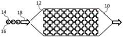
-图1示意性地示出微流体芯片;- Figure 1 schematically shows a microfluidic chip;
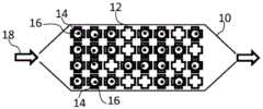
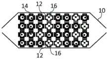
-图2示意性地示出图1的微流体芯片,其中一些捕集阱被含有样品的水凝胶微滴占据;- Fig. 2 schematically shows the microfluidic chip of Fig. 1 with some of the traps occupied by sample-containing hydrogel droplets;
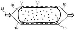
-图3示意性地示出含有水凝胶和待测试样品的混合物的图1的微流体芯片;- Figure 3 schematically shows the microfluidic chip of Figure 1 containing a mixture of hydrogel and sample to be tested;
-图4至图6示意性地示出了用于将捕集在微流体芯片中的水凝胶微滴中包含的一部分样品从该微流体芯片排出的装置;- Figures 4 to 6 schematically show a device for expelling a portion of the sample contained in a hydrogel droplet trapped in a microfluidic chip from the microfluidic chip;
-图7至图12示意性地示出表面张力捕集阱的几何形状和它们使得可以获得的微滴形状的示例;以及- Figures 7 to 12 schematically show examples of surface tension trap geometries and the droplet shapes they make possible; and
-图13至图15示意性地示出水凝胶微滴中样品沉降的示例。- Figures 13 to 15 schematically illustrate examples of sample settling in hydrogel droplets.
具体实施方式Detailed ways
本发明涉及一种处理包含待测试样品的水凝胶微滴的方法。The present invention relates to a method of processing hydrogel droplets containing a sample to be tested.
下文将更特别地涉及细胞形式的样品,但是当然可以使用其它类型的样品。The following will refer more particularly to samples in the form of cells, but of course other types of samples can be used.
该方法基本上包括三个步骤,全部在单个微流体系统中进行,所述三个步骤为:The method basically consists of three steps, all carried out in a single microfluidic system, which are:
-在油中形成含有一个或多个细胞的液体水溶液的微滴,所述油和/或所述水溶液包含胶凝剂,- forming droplets of a liquid aqueous solution containing one or more cells in an oil, said oil and/or said aqueous solution comprising a gelling agent,
-通过预先布置在捕集区中的表面张力捕集阱捕集液体微滴,以及- trapping liquid droplets by means of surface tension traps pre-arranged in the trapping zone, and
-将油和至少一部分被捕集的微滴中的至少一者胶凝化。- gelling at least one of the oil and at least a portion of the trapped droplets.
下文将更特别涉及其中水溶液是水凝胶溶液、油不包含胶凝剂的情况,以及其中上述最后步骤为将至少一部分被捕集的微滴胶凝化,而油本身未被胶凝化的情况。在这种情况下,在上述三个步骤之后,可以通过进行不同的步骤来继续该方法,特别是取决于希望进行的测试。The following will relate more particularly to the case where the aqueous solution is a hydrogel solution and the oil does not contain a gelling agent, and where the last step described above is to gel at least a portion of the trapped droplets, while the oil itself is not gelled. Happening. In this case, after the above three steps, the method can be continued by performing different steps, in particular depending on the desired test.
该方法可以特别地通过用水溶液代替胶凝化微滴周围的油而不将微滴从表面张力捕集阱移除的步骤来继续进行。水溶液可以含有具有营养物质、生长因子、抗体、药剂分子和pH缓冲液和/或盐缓冲液中的至少一者的生物化学溶液。The method may in particular be continued by replacing the oil surrounding the gelled droplets with an aqueous solution without removing the droplets from the surface tension trap. Aqueous solutions may contain biochemical solutions with at least one of nutrients, growth factors, antibodies, pharmaceutical molecules, and pH buffers and/or salt buffers.
根据另一方面,该方法使得可以控制在微流体通道和/或表面张力捕集阱中的水凝胶珠的三维形状,其主要应用是将细胞包封在这些微滴中。因此,根据微滴的形状和每个微滴的细胞浓度,细胞包封在水凝胶中使得在例如向其输入生物化学溶液或对其施加物理刺激(例如热或光)时,能够进行其培养或分析。According to another aspect, the method makes it possible to control the three-dimensional shape of hydrogel beads in microfluidic channels and/or surface tension traps, the main application of which is the encapsulation of cells in these droplets. Thus, depending on the shape of the droplets and the concentration of cells in each droplet, the cells are encapsulated in the hydrogel so that they can undergo their transformation when, for example, biochemical solutions are injected into them or physical stimuli such as heat or light are applied to them. Cultivate or analyze.
凝胶旨在表示主要由液体组成并含有分子或颗粒的介质,分子或颗粒可被组织以赋予凝胶固体外观,例如在其稳定状态下不存在流动。该溶液可以以液态处理,然后可以通过化学方法或物理方法“胶凝化”。在某些情况下,胶凝化可以是可逆的。当液体是水时,涉及水凝胶。Gel is intended to denote a medium consisting primarily of a liquid and containing molecules or particles that can be organized to give the gel a solid appearance, such as the absence of flow in its steady state. The solution can be processed in a liquid state and then can be "gelled" by chemical or physical methods. In some cases, gelation can be reversible. When the liquid is water, hydrogels are involved.
如上所述,所提出的微流体方法包括在油中形成含有生物细胞的水凝胶微滴的第一步骤。As mentioned above, the proposed microfluidic method includes a first step of forming hydrogel microdroplets containing biological cells in oil.
在这种情况下,微滴(或微珠)的直径为微米级,特别是直径在10微米和1000微米之间。In this case, the diameter of the droplets (or beads) is in the order of micrometers, in particular between 10 and 1000 micrometers in diameter.
水凝胶是例如包含胶凝剂的水溶液。用户根据应用选择胶凝剂。可以物理胶凝化的胶凝剂的实例是琼脂糖,其在室温下为液体,并且其在低温下胶凝化。可以化学胶凝化的胶凝剂例如是藻酸盐,其在溶液中为液体,并且当供应钙离子Ca2+时胶凝化。A hydrogel is, for example, an aqueous solution containing a gelling agent. The user selects the gelling agent according to the application. An example of a gelling agent that can be physically gelled is agarose, which is liquid at room temperature and which gels at low temperatures. A gelling agent that can be chemically gelled is, for example, alginate, which is liquid in solution and gels when supplied with calcium ions Ca2+ .
在生物学水平上,水凝胶的生物化学性质和生物力学性质可以使锚定敏感细胞与由此形成的基质建立特异性的相互作用。这些相互作用对于锚定依赖性哺乳动物细胞的存活是必需的,并且在调节其表型中起作用。基质的性质例如可以使得能够观察细胞迁移或蛋白水解(由细胞消化基质)。使用琼脂糖、藻酸盐和PEG-DA(聚乙二醇二丙烯酸酯)、也使用明胶、I型胶原或

先验地在形成微滴之前,细胞与水凝胶混合。然而,在形成微滴之前,水凝胶和细胞可以在微流体装置中直接混合。A priori, cells are mixed with the hydrogel prior to formation of droplets. However, the hydrogel and cells can be mixed directly in the microfluidic device before the droplets are formed.
已经提出了许多方法在流动相(诸如油)中形成这种微滴。例如,可以提及以下示例方法:A number of methods have been proposed to form such droplets in mobile phases such as oils. For example, the following example methods can be mentioned:
-称为“流动聚焦”的方法,例如在S.L.Anna,N.Bontoux和H.A.Stone,“Formationof dispersions using‘Flow-Focusing’in microchannels”,Appl.Phys.Lett.82,364(2003)中所描述的,其内容通过引用并入本文,- a method called "Flow-Focusing", eg as described in S.L.Anna, N.Bontoux and H.A.Stone, "Formation of dispersions using 'Flow-Focusing' in microchannels", Appl.Phys.Lett.82, 364 (2003), The contents of which are incorporated herein by reference,
-“T连接”方法,例如在“Dynamic pattern formation in a vesicle-generatingmicrofluidic device”由T.Thorsen,R.W.Roberts,F.H.Arnold et S.R.Quake,Phys.Rev.Lett.86,4163–4166(2001)中所描述的,其内容通过引用并入本文,或者- "T-connection" method, eg as described in "Dynamic pattern formation in a vesicle-generating microfluidic device" by T. Thorsen, R.W.Roberts, F.H.Arnold et S.R.Quake, Phys.Rev.Lett.86, 4163-4166 (2001) described, the contents of which are incorporated herein by reference, or
-“约束梯度”方法,例如在申请FR-A-2 958 186中所描述的,其内容通过引用并入本文。- "Constrained gradient" method, eg as described in application FR-A-2 958 186, the content of which is incorporated herein by reference.
这些方法使得可以形成具有基本相等尺寸的微滴。These methods make it possible to form droplets of substantially equal size.
在形成这些微滴之后,微滴通过微通道从其形成的区域输送到捕集区,该微通道由油流和/或由斜坡或轨道承载。已经观察到,这种输送有助于在微滴中形成球状体。然后由布置在捕集区中、特别是在微流体芯片中的表面张力捕集阱将微滴捕集。捕集区(或微流体芯片10)通过疏水表面处理进行处理,并填充含有表面活性剂的油。使用表面活性剂能够使微滴稳定及再现微滴的形成。表面活性剂还可以在从生产装置输送到捕集区的捕集阱期间如果发生接触则防止微滴的聚结。After these droplets are formed, the droplets are transported from the area where they are formed to the capture zone through microchannels carried by the oil flow and/or by ramps or rails. It has been observed that this transport facilitates the formation of spheroids in the droplets. The droplets are then trapped by surface tension traps arranged in the trapping zone, in particular in the microfluidic chip. The trapping area (or microfluidic chip 10) is treated with a hydrophobic surface treatment and filled with a surfactant-containing oil. The use of surfactants can stabilize and reproduce droplet formation. Surfactants can also prevent coalescence of droplets if contact occurs during transport from the production device to the trap of the capture zone.
如图1所示的微流体芯片10由可能数平方厘米的培养室组成,包含以表格或矩阵形式组织的许多表面张力捕集阱。表面张力捕集阱12可以具有各种形状。例如,在圆柱形捕集阱的情况下,根据所需的应用,它们的直径可以从几十微米到几百微米。对于单个细胞或在微滴中个体化的细胞的包封,捕集阱的直径可以是例如50微米,其对应于每平方厘米大约5000个捕集阱的密度。对于大的细胞聚集体或球状体的研究,该直径可以达到250微米,则其对应于每平方厘米大约250个捕集阱的捕集阱密度。The
如图1所示,在微流体芯片10外部形成的包括生物细胞16的微滴14例如通过箭头18所示的油流被运送到微流体芯片10中,使得这些微滴中的一些被捕集在表面张力捕集阱12中。As shown in FIG. 1,
然而,作为变型,这里提出在油中形成含有生物细胞的水凝胶微滴,而不精确控制在油中含有生物细胞的水凝胶的流动。这是因为只有那些具有合适尺寸的微滴随后被捕集在捕集区中,使得捕集区被实际上具有很大的尺寸均匀性、形状均匀性和生物细胞浓度均匀性的微滴占据。However, as a variant, here it is proposed to form hydrogel droplets containing biological cells in oil without precise control of the flow of the hydrogel containing biological cells in oil. This is because only those droplets of suitable size are subsequently trapped in the trapping zone, so that the trapping zone is occupied by droplets with practically great uniformity in size, shape and concentration of biological cells.
根据图3所示的另一变型,捕集区,特别是微流体芯片10,包含含有生物细胞16的水凝胶溶液20。然后将油注入捕集区(注射由箭头18示意性地表示),其驱动含有生物细胞16的水凝胶溶液20朝向捕集区的出口。然后通过将水凝胶捕集在微流体芯片的这些捕集阱中直到获得与图2所示的构造基本相同的构造,微滴直接在表面张力捕集阱12中形成。因此,通过在表面张力捕集阱上含有生物细胞的水凝胶溶液的自发分离(或破裂)来形成微滴。在这种情况下,也不需要对流量的精确控制,甚至可以用手推动注射器,而不必使用复杂的仪器。在这种情况下,优选的是深的表面张力捕集阱,以便在表面张力捕集阱处能够使微滴破裂(也就是说微滴的形成)。这样的捕集阱将随后描述。According to another variant shown in FIG. 3 , the capture zone, in particular the
这里应当注意,捕集阱可以具有非常不同的形状,特别是根据所需的应用,也就是说特别是根据被捕集的微滴的所需形状,捕集阱可以具有非常不同的形状。形成捕集阱的空腔也可以没有优先地位于捕集区、特别是微流体芯片的上壁、下壁或一个侧壁上。It should be noted here that the traps can have very different shapes, in particular according to the desired application, that is to say in particular according to the desired shape of the trapped droplets. The cavity forming the trap can also not be preferentially located on the upper wall, the lower wall or one of the side walls of the trapping region, in particular the microfluidic chip.
图7至图12示出可以设想用于微流体芯片10的表面张力捕集阱12的形状以及可以通过这些表面张力捕集阱12获得的微滴14的形状。FIGS. 7 to 12 illustrate the shapes of surface tension traps 12 that can be envisaged for
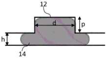
特别地,捕集阱12的形状使得可以根据形成有捕集阱12的微流体通道的几何参数和捕集的微滴的体积来控制捕集的微滴的形状。图7示意性地示出了确定微滴的轮廓所要考虑的参数,即限制在含有捕集阱12的通道中的微滴的半径R、该通道的高度h(其小于该通道中的微滴的半径R)、以及捕集阱12的直径d和深度p。In particular, the shape of the
当捕集阱12是圆柱形的并且具有大于通道高度h两倍的直径d时,如图8至图10所示,则微滴14尽可能多地装入捕集阱12中。根据捕集阱12和微滴14的相对体积,微滴14可以具有或不具有半球状的圆顶,并且可以具有或不具有由通道的壁限制的平坦部分。因此,在图8中,微滴14的体积大于捕集阱12的体积。在这种情况下,微滴14几乎完全填充捕集阱12,并且具有抵着通道的壁和捕集阱的壁扁平化的形状。在图9中,微滴14的体积略小于捕集阱12的体积,因此微滴14具有两个半球状的圆顶,并且仅轻微接触通道的壁。最后,如图10所示,如果微滴14的体积明显小于捕集阱12的体积,则微滴14(或甚至几个微滴14)完全容纳在捕集阱12内。When the
另一方面,在图11的情况下,捕集阱的直径d小于通道高度h的一半。因此,微滴14基本上仍被限制在通道中,并且在捕集阱12中仅具有小的半球状的圆顶。On the other hand, in the case of FIG. 11 , the diameter d of the trap is less than half the height h of the channel. Thus, the
最后,在图12的情况下,捕集阱12是圆锥形的并且直径d为通道高度h的两倍。微滴14因此紧密地装入捕集阱12的壁的形状中,以便在捕集阱12中形成半球状的圆顶。Finally, in the case of Figure 12, the
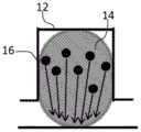
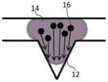
此外,如图13所示,如果捕集在捕集阱12中的微滴14具有平坦的底部,则细胞16在统计学上均匀地沉降和沉淀其自身于微滴14的底部。于是可以观察到单个的细胞并且不聚集。另一方面,如图14和图15所示,如果捕集在捕集阱12中的微滴14具有非平坦的底部、特别是凸起的底部,则细胞16在其沉降期间与微滴12的界面接触,并且必须沿着这个界面滑动。细胞16因此集中在微滴14的底部,并且可以任选地在一些锚定依赖性细胞的情况下聚集并形成球状体。Furthermore, as shown in FIG. 13 , if the
这里提出的微流体方法包括在捕集这些微滴之后对微滴胶凝化的步骤。The microfluidic method presented here includes the step of gelling the droplets after trapping these droplets.
该步骤可以以各种方式进行,特别是根据所用的水凝胶胶凝剂。因此,根据第一实施例,水凝胶含有琼脂糖,优选是琼脂糖。然后通过冷却微流体芯片将微滴胶凝化。当水凝胶含有藻酸盐或优选地为藻酸盐时,可以在其中浸入微滴的油中提供钙离子Ca2+,或者甚至将钙质颗粒与藻酸盐预混合并使其中浸入微滴的油在二氧化碳中饱和。藻酸盐因此被酸化并释放钙离子。当然,由于可以使用其它胶凝剂,因此可以使用其它胶凝化方法。This step can be carried out in various ways, especially depending on the hydrogel gelling agent used. Thus, according to a first embodiment, the hydrogel contains agarose, preferably agarose. The droplets are then gelled by cooling the microfluidic chip. When the hydrogel contains alginate or preferably alginate, calcium ions Ca2+ can be provided in the oil in which the microdroplets are immersed, or even the calcareous particles can be premixed with alginate and immersed in the microdroplets The dripping oil is saturated in carbon dioxide. The alginate is thus acidified and calcium ions are released. Of course, as other gelling agents can be used, other methods of gelling can be used.
此外,根据所寻求的应用,该胶凝化步骤可以在该处理方法期间的不同时刻进行。特别地,可以在捕集后立即进行胶凝化,以将细胞适当固定在微滴中并防止它们沉降。然后可以彼此独立地观察细胞。可替选地,在细胞沉降以形成球状体之后进行胶凝化。这使得可以观察到已经形成球状体的细胞的行为。根据另一替代方案,微滴仅在用于处理液体介质中的细胞的操作之后胶凝化,例如选择性提取某些细胞-非胶凝化微滴中的细胞。这对于例如细菌或红细胞和白细胞的细胞(其是锚定非依赖的)可是有用的。Furthermore, depending on the application sought, the gelling step can be carried out at different times during the processing method. In particular, gelation can be performed immediately after capture to properly immobilize cells in the droplets and prevent them from settling. The cells can then be observed independently of each other. Alternatively, gelation is performed after the cells have settled to form spheroids. This makes it possible to observe the behavior of cells that have formed spheroids. According to another alternative, the droplets are only gelled after operations used to treat cells in the liquid medium, eg selective extraction of certain cells - cells in non-gelled droplets. This may be useful for cells such as bacteria or red and white blood cells, which are anchorage-independent.
胶凝化后,例如可以用含有特别是包含生物化学成分(例如营养物质、生长因子、抗体、药物或药剂分子)的生物化学溶液的水溶液来代替其中浸渍有微滴的油。这些生物化学成分通过凝胶扩散并到达细胞。因此,可以研究独立的或以球状体的形式的细胞对这些刺激的反应。因此,在将细胞包封在微滴期间,水凝胶使得可以将细胞保持在精确的位置,同时允许细胞通过水相输入并且预先划分生物样品。After gelation, the oil in which the droplets are impregnated can be replaced, for example, by an aqueous solution containing a biochemical solution comprising, in particular, biochemical constituents such as nutrients, growth factors, antibodies, drugs or pharmaceutical molecules. These biochemical components diffuse through the gel and reach the cells. Thus, cell responses to these stimuli can be studied either independently or in the form of spheroids. Thus, during encapsulation of cells in droplets, the hydrogel makes it possible to hold the cells in precise locations, while allowing the cells to enter through the aqueous phase and pre-divide the biological sample.
为了成功地进行该操作,优选地迫使表面活性剂从微滴的界面出来。这是因为表面活性剂在微滴界面处形成的壳会非常有效地阻止被注入以代替油的水相填充微流体芯片,从而保持微滴在其各自的捕集阱中胶凝化。由于聚结被表面活性剂的存在所抵抗,故水相界面到达捕集阱产生施加到胶凝化微滴的力,如果构成胶凝化微滴的水凝胶是足够可压缩的,则胶凝化微滴会被迫使离开捕集阱。这就是优选通过降低界面处的表面活性剂的浓度来促进聚结的原因。为此,在注入水相之前,使微流体芯片注满油,该油与之前使用的油不同,不含有表面活性剂。微流体芯片的油中的表面活性剂的浓度降低,这使得可以将界面处的表面活性剂吸附的平衡转向解吸附。对于高浓度的表面活性剂,例如几个重量百分数的数量级,优选使微流体芯片注满相当于微流体芯片体积的50倍的量的油。该比例取决于表面活性剂的性质及其对两相的亲和力。In order to do this successfully, the surfactant is preferably forced out of the interface of the droplets. This is because the shell formed by the surfactant at the droplet interface is very effective in preventing the aqueous phase injected to replace the oil from filling the microfluidic chip, keeping the droplets gelled in their respective traps. Since coalescence is resisted by the presence of surfactants, the arrival of the water-phase interface to the trap creates a force that is applied to the gelled droplets, if the hydrogel that makes up the gelled droplets is sufficiently compressible, the gel Condensed droplets are forced out of the trap. This is why coalescence is preferably promoted by reducing the concentration of surfactant at the interface. For this purpose, the microfluidic chip is filled with oil, which, unlike the oil used before, does not contain surfactants, before injection into the aqueous phase. The concentration of surfactant in the oil of the microfluidic chip is reduced, which makes it possible to shift the equilibrium of surfactant adsorption at the interface towards desorption. For high concentrations of surfactant, eg on the order of several weight percent, it is preferable to fill the microfluidic chip with an amount of oil equivalent to 50 times the volume of the microfluidic chip. This ratio depends on the nature of the surfactant and its affinity for both phases.
捕集阱的形状也可以进行优化,以确保胶凝化微滴保持在捕集阱中的适当位置。因此,在足够深的圆柱形捕集阱的情况下,如果通道的高度大于捕集阱的半径,则进入捕集阱的微滴将是最少的,导致低的捕集效率。随后,外部流动的速度有一个限制,超过该速度,迫使微滴离开捕集阱。相反,当通道的高度小于捕集阱的半径时,只要微滴足够大,微滴就能明显地渗透到捕集阱的空腔中,从而产生高的捕集效率。无论外部流的速率如何,微滴保持在适当的位置。在第一种情况下,微滴的形状非常接近其通道中的形状,而在第二种情况下,其在局部呈现捕集阱的形状。The shape of the trap can also be optimized to ensure that the gelled droplets remain in place in the trap. Therefore, in the case of a sufficiently deep cylindrical trap, if the height of the channel is greater than the radius of the trap, the droplets entering the trap will be minimal, resulting in low trapping efficiency. Subsequently, there is a limit to the velocity of the external flow, beyond which the droplets are forced out of the trap. Conversely, when the height of the channel is smaller than the radius of the trap, as long as the droplet is large enough, the droplet can significantly penetrate into the cavity of the trap, resulting in high trapping efficiency. The droplets remain in place regardless of the rate of the external flow. In the first case, the shape of the droplet is very close to the shape in its channel, while in the second case it locally assumes the shape of a trap.
当选择的水凝胶的胶凝剂为可逆的时候,可以将微滴去胶凝化,然后从微流体芯片释放出其内容物,如图4至图6所示。在这些图4至图6的情况下,微滴14例如是胶凝化的琼脂糖微滴。这些琼脂糖微滴14通过局部加热(通过闪电栓21所示的加热),特别是通过红外激光或电极,逐个去胶凝化。热使琼脂糖液化。当微滴14周围的相是水性时,去胶凝化琼脂糖的内容物16与水相混合。然后可以使用水相的流22携带该内容物,任选地以便回收该内容物。因此,也可以通过微流体芯片10消除被认为不感兴趣的细胞。再次,优选地选择捕集阱的形状和尺寸以使得能够提取细胞。例如,在细胞必须保持存活的情况下,捕集阱的尺寸足够大以致加热水凝胶不诱导细胞死亡机制。When the gelling agent of the selected hydrogel is reversible, the droplets can be degelled and their contents released from the microfluidic chip, as shown in Figures 4-6. In the case of these Figures 4 to 6, the
可替选地,当微滴周围的相是油性时,可以施加油流以将液体微滴从捕集阱移出。在这种情况下,捕集阱的形状和强度优选为仅允许提取所选择的微滴而不是其它微滴的尺寸。该尺寸特别取决于水相和油之间的表面张力的值,也取决于凝胶微滴的刚性及其在捕集阱内的形状。Alternatively, when the phase surrounding the droplets is oily, a flow of oil can be applied to remove the liquid droplets from the trap. In this case, the shape and strength of the trap are preferably of a size that allows only selected droplets and not others to be extracted. This size depends in particular on the value of the surface tension between the water phase and the oil, but also on the rigidity of the gel droplets and their shape within the trap.
另一种替代方案是保持微滴为液体,用于处理悬浮液中的细胞,例如细菌。然后仅进行胶凝化以选择性提取微滴。在这种情况下,可以在提取前胶凝化所有微滴并且应用上述方案,或者另一方面仅胶凝化那些期望保留在捕集阱中的微滴。Another alternative is to keep the droplets as a liquid for treating cells in suspension, such as bacteria. Only gelation is then performed to selectively extract the droplets. In this case, it is possible to gel all droplets before extraction and apply the protocol described above, or on the other hand to gel only those droplets that are expected to remain in the trap.
经由轻微的修改,所提供的方法能够检测非常多样化的生物应用。在每种情况下,当然也可以通过调节微流体芯片中的通道的高度以及捕集阱的几何形状来修改该装置。With slight modifications, the provided method is capable of detecting a very diverse biological application. In each case, it is of course also possible to modify the device by adjusting the height of the channels in the microfluidic chip and the geometry of the traps.
因此,例如快速胶凝化低浓度的细胞使得可以使在每个微滴中的几个单细胞个体化,同时试图限制它们的直接相互作用。这些细胞例如可以是细菌、酵母或哺乳动物细胞。Thus, eg, rapid gelation of low concentrations of cells makes it possible to individualize several single cells in each droplet, while trying to limit their direct interactions. These cells can be, for example, bacterial, yeast or mammalian cells.
可替选地,快速胶凝化高浓度的细胞使得仍然可以获得仍被个体化但彼此接近的大量细胞。例如可以通过旁分泌分泌物检查细胞(可能地在共培养中)之间的相互作用。Alternatively, rapid gelation of high concentrations of cells allows still obtaining large numbers of cells that are still individualized but in close proximity to each other. Interactions between cells (possibly in co-culture) can be examined, for example, by paracrine secretions.
然而,在胶凝化之前,细胞可以在液相中长时间保持包封。然后,低浓度的细胞使得可以例如在悬浮液中研究锚定依赖性细胞,例如淋巴细胞。能够使细胞沉降的高浓度的细胞和捕集的微滴的形状使得可以将能够将其自身重组为球状体的细胞聚集在一起。However, cells can remain encapsulated in the liquid phase for extended periods of time before gelling. The low concentration of cells then makes it possible to study anchorage-dependent cells, such as lymphocytes, eg in suspension. The high concentration of cells capable of allowing the cells to settle and the shape of the trapped droplets make it possible to cluster together cells capable of reorganizing themselves into spheroids.
该方法还可以以受控的方式直接在芯片中形成球状体。所产生的微滴的体积可以由用于在微流体芯片上游形成微滴的装置来控制。优选地调节该体积,使得一旦被捕集,则微滴的直径等于捕集阱的直径并且微滴具有球形形状。为此,捕集阱的深度优选至少等于其直径。捕集的微滴的直径与捕集阱的直径一致的事实使得可以确保高的捕集效率。球形形状促进球状体的形成。对于该特定应用,微滴优选含有悬浮在包含培养基和水凝胶或由培养基和水凝胶构成的水相中的细胞。一旦捕集阱填充了这样的微滴,则外部的油的流动停止,这停止在微滴中的再循环并促进细胞的沉降。然后,捕集阱中的微滴的球形形状使得细胞在微滴的最低点处集中,直到它们接触。然后,在有利于细胞的存活和适当的代谢功能的条件下,特别是在温度方面,在几个小时到几天的持续时间内,将芯片静置,使得能够将集中在被捕集的微滴底部的细胞重组为球状体。The method can also form spheroids directly in the chip in a controlled manner. The volume of droplets produced can be controlled by the device used to form the droplets upstream of the microfluidic chip. The volume is preferably adjusted so that, once trapped, the diameter of the droplet is equal to the diameter of the trap and the droplet has a spherical shape. To this end, the depth of the trap is preferably at least equal to its diameter. The fact that the diameter of the trapped droplets corresponds to the diameter of the trap makes it possible to ensure a high trapping efficiency. The spherical shape promotes the formation of spheroids. For this particular application, the droplets preferably contain cells suspended in an aqueous phase comprising or consisting of medium and hydrogel. Once the trap is filled with such droplets, the flow of oil from the outside stops, which stops the recirculation in the droplets and promotes the settling of the cells. The spherical shape of the droplets in the trap then causes cells to concentrate at the lowest point of the droplets until they touch. The chip is then left at rest under conditions favourable for the survival of the cells and for proper metabolic function, especially in terms of temperature, for a duration of several hours to several days, enabling the concentration of The cells at the bottom of the drop reorganized into spheroids.
球状体形成所需的持续时间会特别取决于所使用的细胞类型和取决于水凝胶的组成。对于在培养基中稀释的1重量%的琼脂糖溶液中的H4IIEC3大鼠肝细胞,观察到该持续时间小于24小时。当然,在球状体形成期间,水凝胶保持液体。The duration required for spheroid formation will depend in particular on the cell type used and on the composition of the hydrogel. This duration was observed to be less than 24 hours for H4IIEC3 rat hepatocytes in 1 wt% agarose solution diluted in culture medium. Of course, the hydrogel remains liquid during spheroid formation.
这种方法使得可以快速形成大量的具有非常均匀尺寸的球状体。事实上,球状体的尺寸由每个微滴中包封的细胞数量确定,并因此由注入到微流体芯片中的细胞溶液的浓度来确定。只要细胞在注入时充分个体化,则每个微滴的细胞数量的分布、并因此形成的球状体的尺寸的分布就非常均匀。在本发明人进行的实验中,在孵育24小时之后,平均98%的捕集阱填充有一个含有良好重组的球状体的液体琼脂糖的微滴。This method allows for the rapid formation of large numbers of spheroids of very uniform size. In fact, the size of the spheroids is determined by the number of cells encapsulated in each droplet, and thus by the concentration of the cell solution injected into the microfluidic chip. As long as the cells are sufficiently individualized at the time of injection, the distribution of the number of cells per droplet, and thus the size of the spheroids formed, is very uniform. In experiments performed by the inventors, after 24 hours of incubation, on average 98% of the traps were filled with one droplet of liquid agarose containing well reconstituted spheroids.
在微流体芯片中获得的球状体可以保持培养若干天。例如,包封在琼脂糖中的H4IIEC3细胞的球状体可以在芯片中培养一周,而不显著改变其活力并且同时保持高功能性(在该实例中,强烈和连续的白蛋白分泌)。The spheroids obtained in the microfluidic chip can be kept in culture for several days. For example, spheroids of H4IIEC3 cells encapsulated in agarose can be cultured in a chip for a week without significantly changing their viability and at the same time maintaining high functionality (in this example, intense and continuous secretion of albumin).
这里提出的方法和通过进行该方法获得的微流控芯片构成了用于筛选药剂的优良工具。例如,可以形成癌细胞的球状体并观察根据暴露于被测分子其活力随时间是否降低。通过向芯片添加能够在室内设置浓度梯度的装置,或者通过设置平行芯片,可以在同一系统中测试整个浓度范围。形成这些球状体的方法的高效率也使得可以从非常有限的样品开始产生大量的这些球状体。因此,仅用100 000个细胞可以形成直径约70μm的500个球状体。The method presented here and the microfluidic chip obtained by carrying out the method constitute an excellent tool for screening pharmaceutical agents. For example, spheroids of cancer cells can be formed and observed whether their viability decreases over time upon exposure to the tested molecule. By adding devices to the chip that can set up concentration gradients within the chamber, or by setting up parallel chips, the entire concentration range can be tested in the same system. The high efficiency of the method of forming these spheroids also makes it possible to generate large numbers of these spheroids starting from a very limited sample. Thus, 500 spheroids with a diameter of about 70 μm can be formed with only 100 000 cells.
构成球状体的细胞也可以是不同的类型,以便接近共培养的主题。这些细胞类型可以在注入芯片中之前均匀地混合在溶液中,或者在几个连续的水凝胶层中按照某种结构组织排列或者仅通过在输入在外部水相中之后粘附水凝胶而排列。例如,可以将成纤维细胞和上皮细胞联合以形成皮肤模型并测试化妆品的毒性,将神经元和星形胶质细胞联合以模拟脑部,或者将如在血管壁中的内皮细胞和平滑肌细胞联合。The cells that make up the spheroids can also be of different types in order to approach the subject of co-culture. These cell types can be homogeneously mixed in solution prior to injection into the chip, or arranged in some structural organization in several successive hydrogel layers or simply by adhering to the hydrogel after infusion into the external aqueous phase arrangement. For example, fibroblasts and epithelial cells can be combined to form skin models and test cosmetics for toxicity, neurons and astrocytes to simulate the brain, or endothelial cells and smooth muscle cells, such as in blood vessel walls.
由于这里提出的方法可以实现对培养细胞微环境的非常高度的控制,故它还是研究干细胞分化的优良工具。事实上,包封的细胞可以经受全范围的分化因子的浓度,并且潜在地同时经受全范围的基质的刚性,同时调节例如水凝胶浓度。类似地,该方法可用于观察胚胎随时间的发育,与来自外部培养基的物理化学因子的相互作用。Since the method presented here enables a very high degree of control over the cultured cell microenvironment, it is also an excellent tool for studying stem cell differentiation. In fact, encapsulated cells can withstand a full range of differentiation factor concentrations, and potentially a full range of matrix stiffness simultaneously, while modulating, for example, hydrogel concentrations. Similarly, this method can be used to observe embryonic development over time, interacting with physicochemical factors from external media.
在来自患者的原代细胞的情况下,该方法使得可以基于细胞对某些标记的响应来进行医学诊断。在这种情况下,细胞以非常低的损失程度被捕获。然后可以使细胞进行已知的用于诊断某些疾病的测试,例如癌症活检的表征。例如,可以例如通过原位聚合酶链反应(PCR)或通过FISH方法来测试包封细胞的基因组中突变的存在。也可以通过标记方法,例如通过提供用于免疫标记的抗体或用于原位免疫酶法、ELISA(酶联免疫吸附试验)的抗体,检测特异性蛋白的表达。In the case of primary cells from patients, this method enables medical diagnosis based on the response of cells to certain markers. In this case, cells are captured with very low loss. The cells can then be subjected to tests known to diagnose certain diseases, such as the characterization of cancer biopsies. For example, the genome of encapsulated cells can be tested for the presence of mutations in the genome, eg, by in situ polymerase chain reaction (PCR) or by FISH methods. The expression of specific proteins can also be detected by labeling methods, eg by providing antibodies for immunolabeling or for in situ immunoenzymatic methods, ELISA (enzyme-linked immunosorbent assay).
上述方法提供了借助微滴在芯片中被捕集后的胶凝化,在微流体芯片中进行细胞分析和培养的所有步骤的可能性。这使得可以使用比在多孔板或培养皿中进行的测试少得多的量的试剂。这也使得可以在不同的刺激之后监测随时间的细胞响应。The method described above offers the possibility to perform all steps of cell analysis and culture in a microfluidic chip by means of gelation after the droplets are captured in the chip. This makes it possible to use much smaller quantities of reagents than tests performed in multi-well plates or petri dishes. This also makes it possible to monitor cellular responses over time following different stimuli.
上述方法可以容易地在包括以下的装置中进行:The above method can easily be carried out in an apparatus comprising:
-形成含有细胞的水凝胶微滴的构件,- building blocks for the formation of cell-containing hydrogel droplets,
-捕集区,特别是微流体芯片,用于在预定位置处捕集水凝胶微滴,以及- a trapping zone, in particular a microfluidic chip, for trapping hydrogel droplets at predetermined locations, and
-用于胶凝化至少一部分的被捕集的微滴的构件。- Means for gelling at least a portion of the trapped droplets.
该胶凝化构件包括例如用于将化学试剂注入捕集区中的元件和/或用于温度调节(例如用于冷却微流体芯片)的元件。The gelling member includes, for example, elements for injecting chemicals into the trapping zone and/or elements for temperature regulation (eg, for cooling the microfluidic chip).
该装置还可以包括用于使至少一些胶凝化的水凝胶微滴去胶凝化的构件,例如激光器。The device may also include means, such as a laser, for degelling at least some of the gelled hydrogel droplets.
上述方法也使得可以生产具有胶凝化微滴的产品,其包括用于捕集微滴的区域,特别是微流体芯片,以及各自包括一个或多个捕集在捕集区中的细胞的胶凝化微滴,胶凝化微滴优选被冷冻保存。细胞可以以簇或球状体的形式聚集。胶凝化微滴可以浸入流体中,优选浸入水溶液中或浸入油中,流体和微滴优选被冷冻保存。这种冷冻保存使得特别能够在稳定条件下长时间保持细胞,以便运输或存储它们用于随后的分析。The above method also makes it possible to produce products with gelled droplets comprising regions for trapping the droplets, in particular microfluidic chips, and gels each comprising one or more cells trapped in the trapping region The gelled droplets are preferably cryopreserved. Cells can aggregate in clusters or spheroids. The gelled droplets may be immersed in a fluid, preferably an aqueous solution or immersed in an oil, the fluid and droplets preferably being cryopreserved. This cryopreservation makes it particularly possible to maintain cells under stable conditions for long periods of time in order to transport or store them for subsequent analysis.
包封在微滴中的生物细胞可以是细菌细胞、酵母细胞、真核细胞、哺乳动物细胞、优选除人类细胞外的哺乳动物细胞,更优选大鼠细胞或来自其它哺乳动物的细胞,或从其天然环境分离的人类细胞。The biological cells encapsulated in the droplets may be bacterial cells, yeast cells, eukaryotic cells, mammalian cells, preferably mammalian cells other than human cells, more preferably rat cells or cells from other mammals, or from Human cells isolated from their natural environment.
当然,本发明不限于仅上述示例,在所附权利要求的范围内,对于本领域技术人员来讲可存在许多变型。Of course, the invention is not limited to the above-mentioned examples only, and many variants are possible for a person skilled in the art within the scope of the appended claims.
除了细胞之外,使用的样品还可以特别地是通过将它们与分子偶联而官能化的分子或塑料珠。In addition to cells, the samples used can in particular also be molecules or plastic beads functionalized by coupling them to molecules.
此外,捕集的微滴可以与由水溶液流提供的其它微滴融合。In addition, the captured droplets can fuse with other droplets provided by the flow of the aqueous solution.
构成微滴的水溶液也可含有生物化学溶液,生物化学溶液优选包含脂质(脂肪酸等)、碳水化合物(以单体形式或多糖形式等)、氨基酸和蛋白质(生长因子、细胞因子、抗体、抗原等)以及盐缓冲液和/或pH缓冲液中的至少一者。The aqueous solution making up the droplets may also contain biochemical solutions, preferably containing lipids (fatty acids, etc.), carbohydrates (in monomeric or polysaccharide form, etc.), amino acids and proteins (growth factors, cytokines, antibodies, antigens, etc.) etc.) and at least one of a salt buffer and/or a pH buffer.
最后,根据一个变型,微滴周围的油(或油相)可以含有氟油(FC40型)或者可光交联的水不混溶溶液(Norland Optical Adhesive型),其一旦聚合就可以使油胶凝化,从而物理性和选择性地分离微滴。因此,可以更可靠地相对于彼此使微滴区室化。这使得可以防止两个微滴融合,防止引起它们含有的样品的混合。这也使得可以持久地储存样品,由于通过胶凝化的油使微滴区室化,在微滴周围形成固体隔室,故微滴蒸发的风险尤其极大地降低。Finally, according to a variant, the oil (or oil phase) surrounding the droplets may contain fluorine oil (type FC40) or a photocrosslinkable water-immiscible solution (type Norland Optical Adhesive), which, once polymerized, can make the oil gel Coagulation, thereby physically and selectively separating droplets. Thus, the droplets can be more reliably compartmentalized with respect to each other. This makes it possible to prevent the fusion of the two droplets, causing mixing of the samples they contain. This also makes it possible to store the sample permanently, especially the risk of droplet evaporation is greatly reduced due to the compartmentalization of the droplet by the gelled oil, forming a solid compartment around the droplet.
一旦一部分油已被胶凝化,就可以驱动周围的油未胶凝化的微滴从捕集区排出。为此,可以在捕集区中使用油或另一种流体的流动,该流动足够强以携带微滴。因此,可以仅保留周围的油在捕集区中胶凝化的那些微滴。Once a portion of the oil has been gelled, surrounding oil droplets that are not gelled can be driven out of the trapping zone. For this purpose, a flow of oil or another fluid can be used in the capture zone, which flow is strong enough to carry the droplets. Thus, only those droplets where the surrounding oil has gelled in the trapping zone can be retained.
在此应当注意,即使在油被胶凝化的情况下,微滴也可以被胶凝化。此外,该方法当然可以包括使胶凝化的油去胶凝化的后续步骤,在这种情况下一部分油被胶凝化。It should be noted here that even where the oil is gelled, the droplets can be gelled. Furthermore, the method may of course comprise a subsequent step of degelling the gelled oil, in which case a portion of the oil is gelled.
Claims (30)
Applications Claiming Priority (1)
| Application Number | Priority Date | Filing Date | Title |
|---|---|---|---|
| PCT/FR2014/052655WO2016059302A1 (en) | 2014-10-17 | 2014-10-17 | Method for handling microdrops which include samples |
Publications (2)
| Publication Number | Publication Date |
|---|---|
| CN107109319A CN107109319A (en) | 2017-08-29 |
| CN107109319Btrue CN107109319B (en) | 2020-11-27 |
Family
ID=51897384
Family Applications (1)
| Application Number | Title | Priority Date | Filing Date |
|---|---|---|---|
| CN201480082752.5AActiveCN107109319B (en) | 2014-10-17 | 2014-10-17 | Method for processing droplets containing a sample |
Country Status (6)
| Country | Link |
|---|---|
| US (1) | US10710077B2 (en) |
| EP (1) | EP3206791B1 (en) |
| JP (1) | JP2017537772A (en) |
| CN (1) | CN107109319B (en) |
| ES (1) | ES2856733T3 (en) |
| WO (1) | WO2016059302A1 (en) |
Families Citing this family (20)
| Publication number | Priority date | Publication date | Assignee | Title |
|---|---|---|---|---|
| CN107109319B (en)* | 2014-10-17 | 2020-11-27 | 巴黎综合理工学院 | Method for processing droplets containing a sample |
| KR102334118B1 (en) | 2015-04-22 | 2021-12-01 | 버클리 라잇츠, 인크. | Freezing and archiving cells on a microfluidic device |
| US11352661B2 (en) | 2016-07-12 | 2022-06-07 | Northeastern University | Single cell fluorescence in situ hybridization in microfluidic droplets |
| FR3056927B1 (en)* | 2016-09-30 | 2021-07-09 | Ecole Polytech | MICROFLUIDIC PROCESS FOR HANDLING MICRO-DROPS |
| US11131673B2 (en) | 2017-04-27 | 2021-09-28 | Northeastern University | Live single-cell bioassay in microdroplets |
| WO2019010587A1 (en) | 2017-07-14 | 2019-01-17 | The Governing Council Of The University Of Toronto | Microfluidic platform for the rapid production of organoids/spheroids for compound screening |
| JP2019062832A (en)* | 2017-10-02 | 2019-04-25 | 国立大学法人京都大学 | Device for producing spheroid, method for producing and recovering spheroid |
| CN112119151B (en)* | 2018-03-02 | 2025-08-29 | 量子细胞公司 | Methods, compositions and devices for isolating and analyzing regions of interest from tissues for expression analysis |
| DE102018204624A1 (en)* | 2018-03-27 | 2019-10-02 | Robert Bosch Gmbh | Method and microfluidic device for aliquoting a sample liquid using a sealing liquid, method for manufacturing a microfluidic device and microfluidic system |
| WO2020094827A1 (en) | 2018-11-09 | 2020-05-14 | Eth Zurich | Microfluidic droplet concentrator |
| SG11202012938YA (en) | 2019-02-04 | 2021-01-28 | Illumina Inc | Microfluidic droplet generators |
| FR3098128B1 (en)* | 2019-07-05 | 2023-11-17 | Commissariat Energie Atomique | Microfluidic device comprising a microdrop having a sol-gel matrix. |
| CN111019805B (en)* | 2019-12-20 | 2023-01-24 | 南通大学 | Microfluidic chip device for fixing single cell and performing medical analysis in situ and application thereof |
| CN110982882B (en)* | 2019-12-20 | 2023-06-20 | 南通大学 | Microfluidic chip for single cell immobilization-isolation and in-situ nucleic acid amplification and application thereof |
| CN112774748B (en)* | 2021-01-22 | 2023-02-17 | 中国科学院上海微系统与信息技术研究所 | Micropit-anchored droplet array chip, droplet generation method and application |
| CN115138402A (en)* | 2021-03-31 | 2022-10-04 | 中国科学院深圳先进技术研究院 | Micro-fluidic chip capable of setting chemical concentration gradient and preparation method and application thereof |
| US20240318134A1 (en)* | 2021-07-22 | 2024-09-26 | Institut Pasteur | In vitro generation of organized 3d cell structures including head-trunk embryo-like structures, using epigenetic remodeling factors-microfluidic platform suitable for their generation |
| EP4219685A1 (en)* | 2022-01-31 | 2023-08-02 | Institut Pasteur | In vitro generation of organized 3d cell structures including head-trunk embryo-like structures, using epigenetic remodeling factors - microfluidic platform suitable for their generation |
| CN113617403A (en)* | 2021-08-05 | 2021-11-09 | 上海交通大学 | A Novel Microfluidic Chip for Single Cell Western Blotting |
| WO2024245402A1 (en)* | 2023-06-01 | 2024-12-05 | The Hong Kong University Of Science And Technology | Massively parallel sequential coalescence of microdroplets for multi-step reactions |
Citations (4)
| Publication number | Priority date | Publication date | Assignee | Title |
|---|---|---|---|---|
| WO2008131035A2 (en)* | 2007-04-16 | 2008-10-30 | Cellpoint Diagnotics, Inc. | Methods for diagnosing, prognosing, or theranosing a condition using rare cells |
| WO2010080978A2 (en)* | 2009-01-08 | 2010-07-15 | The General Hospital Corporation | Pre-depletion of leukocytes in whole blood samples prior to the capture of whole blood sample components |
| WO2012016136A3 (en)* | 2010-07-30 | 2012-05-18 | The General Hospital Corporation | Microscale and nanoscale structures for manipulating particles |
| CN103084225A (en)* | 2011-10-27 | 2013-05-08 | 中国科学院大连化学物理研究所 | High throughput microgel fixing method and special micro-fluidic chip thereof |
Family Cites Families (16)
| Publication number | Priority date | Publication date | Assignee | Title |
|---|---|---|---|---|
| CA2290731A1 (en)* | 1999-11-26 | 2001-05-26 | D. Jed Harrison | Apparatus and method for trapping bead based reagents within microfluidic analysis system |
| US6432290B1 (en)* | 1999-11-26 | 2002-08-13 | The Governors Of The University Of Alberta | Apparatus and method for trapping bead based reagents within microfluidic analysis systems |
| US20030049320A1 (en) | 2000-12-18 | 2003-03-13 | Wockhardt Limited | Novel in-situ forming controlled release microcarrier delivery system |
| JP2007075094A (en) | 2005-09-15 | 2007-03-29 | Minoru Seki | Structure and method for preparing gel in flow path |
| US8637317B2 (en)* | 2006-04-18 | 2014-01-28 | Advanced Liquid Logic, Inc. | Method of washing beads |
| US20140193807A1 (en)* | 2006-04-18 | 2014-07-10 | Advanced Liquid Logic, Inc. | Bead manipulation techniques |
| JP4911592B2 (en) | 2006-11-01 | 2012-04-04 | 財団法人生産技術研究奨励会 | Emulsion production method and production apparatus thereof |
| JP2008245612A (en)* | 2007-03-30 | 2008-10-16 | Hitachi Ltd | Sample preparation method and apparatus |
| US9409177B2 (en)* | 2008-03-21 | 2016-08-09 | Lawrence Livermore National Security, Llc | Chip-based device for parallel sorting, amplification, detection, and identification of nucleic acid subsequences |
| FR2958186A1 (en) | 2010-03-30 | 2011-10-07 | Ecole Polytech | DEVICE FOR FORMING DROPS IN A MICROFLUID CIRCUIT. |
| JP5700419B2 (en)* | 2011-02-21 | 2015-04-15 | 国立大学法人 千葉大学 | Method for producing hydrogel substrate and method for forming cell clumps |
| PL398979A1 (en)* | 2012-04-25 | 2013-10-28 | Scope Fluidics Spólka Z Ograniczona Odpowiedzialnoscia | A microfluidic device and a microfluidic system comprising one or more microfluidic devices |
| CA2881783A1 (en)* | 2012-08-13 | 2014-02-20 | The Regents Of The University Of California | Methods and systems for detecting biological components |
| US20180250686A2 (en)* | 2013-08-30 | 2018-09-06 | University Of Washington Through Its Center For Commercialization | Apparatus and method for manipulation of discrete polarizable objects and phases |
| EP3160654A4 (en)* | 2014-06-27 | 2017-11-15 | The Regents of The University of California | Pcr-activated sorting (pas) |
| CN107109319B (en)* | 2014-10-17 | 2020-11-27 | 巴黎综合理工学院 | Method for processing droplets containing a sample |
- 2014
- 2014-10-17CNCN201480082752.5Apatent/CN107109319B/enactiveActive
- 2014-10-17JPJP2017520412Apatent/JP2017537772A/enactivePending
- 2014-10-17EPEP14796828.3Apatent/EP3206791B1/enactiveActive
- 2014-10-17ESES14796828Tpatent/ES2856733T3/enactiveActive
- 2014-10-17USUS15/519,574patent/US10710077B2/enactiveActive
- 2014-10-17WOPCT/FR2014/052655patent/WO2016059302A1/enactiveApplication Filing
Patent Citations (4)
| Publication number | Priority date | Publication date | Assignee | Title |
|---|---|---|---|---|
| WO2008131035A2 (en)* | 2007-04-16 | 2008-10-30 | Cellpoint Diagnotics, Inc. | Methods for diagnosing, prognosing, or theranosing a condition using rare cells |
| WO2010080978A2 (en)* | 2009-01-08 | 2010-07-15 | The General Hospital Corporation | Pre-depletion of leukocytes in whole blood samples prior to the capture of whole blood sample components |
| WO2012016136A3 (en)* | 2010-07-30 | 2012-05-18 | The General Hospital Corporation | Microscale and nanoscale structures for manipulating particles |
| CN103084225A (en)* | 2011-10-27 | 2013-05-08 | 中国科学院大连化学物理研究所 | High throughput microgel fixing method and special micro-fluidic chip thereof |
Non-Patent Citations (1)
| Title |
|---|
| Microfluidic droplet trapping array as nanoliter reactors for gas–liquid chemical reaction;Qingquan Zhang et al;《Electrophoresis》;20091231;第3181-3188页* |
Also Published As
| Publication number | Publication date |
|---|---|
| US20170252744A1 (en) | 2017-09-07 |
| US10710077B2 (en) | 2020-07-14 |
| CN107109319A (en) | 2017-08-29 |
| WO2016059302A1 (en) | 2016-04-21 |
| JP2017537772A (en) | 2017-12-21 |
| EP3206791A1 (en) | 2017-08-23 |
| EP3206791B1 (en) | 2020-12-09 |
| ES2856733T3 (en) | 2021-09-28 |
Similar Documents
| Publication | Publication Date | Title |
|---|---|---|
| CN107109319B (en) | Method for processing droplets containing a sample | |
| CN110004111B (en) | A kind of preparation method of organoid spheroid | |
| Moraes et al. | Aqueous two-phase printing of cell-containing contractile collagen microgels | |
| JP2023052848A (en) | In situ-generated microfluidic isolation structures, kits thereof, and methods of use thereof | |
| JP7150707B2 (en) | A microfluidic method for processing microdrops | |
| AU2021332279B2 (en) | Methods and apparatuses for purification of gel droplets supporting biological tissue | |
| US20190365803A1 (en) | Nanostraws methods and apparatuses | |
| JP7135049B2 (en) | Methods for handling microdroplets containing samples | |
| US12036326B2 (en) | Method of encapsulating cells using a microfluidic encapsulation device | |
| JP7413644B2 (en) | Microfluidic devices for culturing cells including biowalls, bead beds, and biointerfaces, and methods for modeling biointerfaces of microfluidic devices | |
| Carreras et al. | A multifunctional microfluidic platform for generation, trapping and release of droplets in a double laminar flow | |
| KR20240029728A (en) | Micro-organic spheres for use in personalized medicine and drug development | |
| JP7712957B2 (en) | Micro Droplet Plate | |
| He et al. | Recent development of cell analysis on microfludics | |
| Hayaei Tehrani et al. | Droplet microfluidic devices for organized stem cell differentiation into germ cells: capabilities and challenges | |
| Panchal | Tracking egress of doubly encapsulated cells | |
| Sönmez et al. | Mechanostimulation of Multicellular Organisms Through a High-Throughput Microfluidic Compression System | |
| WO2025181490A1 (en) | Improvements in or relating to a dispensing device | |
| 박도현 | Capillarity Guided Patterning Based High-throughput 3D Immune Cell Cytotoxicity Assay | |
| Huang | Microfluidic Generation and Manipulation of Hydrogel Microcapsules for Biomimetic 3D Tissue Culture and Cell Cryopreservation | |
| Rehman | Laser ablation assisted size-based sorting of pure water droplets inside a microfluidic chip and designing a microfluidic chip for studying sprouting angiogenesis | |
| Aijian | Digital Microfluidic Lab-on-a-Chip Platform for the Culture and Analysis of Three-Dimensional Multicellular Spheroids | |
| JPWO2021220173A5 (en) |
Legal Events
| Date | Code | Title | Description |
|---|---|---|---|
| PB01 | Publication | ||
| PB01 | Publication | ||
| SE01 | Entry into force of request for substantive examination | ||
| SE01 | Entry into force of request for substantive examination | ||
| GR01 | Patent grant | ||
| GR01 | Patent grant |