CN106461540B - System and method for automated imaging of chromophore-labeled samples - Google Patents
System and method for automated imaging of chromophore-labeled samplesDownload PDFInfo
- Publication number
- CN106461540B CN106461540BCN201580031476.4ACN201580031476ACN106461540BCN 106461540 BCN106461540 BCN 106461540BCN 201580031476 ACN201580031476 ACN 201580031476ACN 106461540 BCN106461540 BCN 106461540B
- Authority
- CN
- China
- Prior art keywords
- light
- induced
- chromophore
- assembly
- imaging
- Prior art date
- Legal status (The legal status is an assumption and is not a legal conclusion. Google has not performed a legal analysis and makes no representation as to the accuracy of the status listed.)
- Active
Links
Images


Classifications
- G—PHYSICS
- G01—MEASURING; TESTING
- G01N—INVESTIGATING OR ANALYSING MATERIALS BY DETERMINING THEIR CHEMICAL OR PHYSICAL PROPERTIES
- G01N21/00—Investigating or analysing materials by the use of optical means, i.e. using sub-millimetre waves, infrared, visible or ultraviolet light
- G01N21/17—Systems in which incident light is modified in accordance with the properties of the material investigated
- G01N21/25—Colour; Spectral properties, i.e. comparison of effect of material on the light at two or more different wavelengths or wavelength bands
- G01N21/251—Colorimeters; Construction thereof
- G01N21/253—Colorimeters; Construction thereof for batch operation, i.e. multisample apparatus
- G—PHYSICS
- G01—MEASURING; TESTING
- G01N—INVESTIGATING OR ANALYSING MATERIALS BY DETERMINING THEIR CHEMICAL OR PHYSICAL PROPERTIES
- G01N33/00—Investigating or analysing materials by specific methods not covered by groups G01N1/00 - G01N31/00
- G01N33/48—Biological material, e.g. blood, urine; Haemocytometers
- G01N33/483—Physical analysis of biological material
- G01N33/487—Physical analysis of biological material of liquid biological material
- G—PHYSICS
- G01—MEASURING; TESTING
- G01N—INVESTIGATING OR ANALYSING MATERIALS BY DETERMINING THEIR CHEMICAL OR PHYSICAL PROPERTIES
- G01N21/00—Investigating or analysing materials by the use of optical means, i.e. using sub-millimetre waves, infrared, visible or ultraviolet light
- G01N21/17—Systems in which incident light is modified in accordance with the properties of the material investigated
- G01N21/25—Colour; Spectral properties, i.e. comparison of effect of material on the light at two or more different wavelengths or wavelength bands
- G01N21/31—Investigating relative effect of material at wavelengths characteristic of specific elements or molecules, e.g. atomic absorption spectrometry
- G—PHYSICS
- G01—MEASURING; TESTING
- G01N—INVESTIGATING OR ANALYSING MATERIALS BY DETERMINING THEIR CHEMICAL OR PHYSICAL PROPERTIES
- G01N21/00—Investigating or analysing materials by the use of optical means, i.e. using sub-millimetre waves, infrared, visible or ultraviolet light
- G01N21/62—Systems in which the material investigated is excited whereby it emits light or causes a change in wavelength of the incident light
- G01N21/63—Systems in which the material investigated is excited whereby it emits light or causes a change in wavelength of the incident light optically excited
- G01N21/64—Fluorescence; Phosphorescence
- G01N21/6428—Measuring fluorescence of fluorescent products of reactions or of fluorochrome labelled reactive substances, e.g. measuring quenching effects, using measuring "optrodes"
- G—PHYSICS
- G01—MEASURING; TESTING
- G01N—INVESTIGATING OR ANALYSING MATERIALS BY DETERMINING THEIR CHEMICAL OR PHYSICAL PROPERTIES
- G01N21/00—Investigating or analysing materials by the use of optical means, i.e. using sub-millimetre waves, infrared, visible or ultraviolet light
- G01N21/62—Systems in which the material investigated is excited whereby it emits light or causes a change in wavelength of the incident light
- G01N21/63—Systems in which the material investigated is excited whereby it emits light or causes a change in wavelength of the incident light optically excited
- G01N21/64—Fluorescence; Phosphorescence
- G01N21/645—Specially adapted constructive features of fluorimeters
- G01N21/6452—Individual samples arranged in a regular 2D-array, e.g. multiwell plates
- G—PHYSICS
- G01—MEASURING; TESTING
- G01N—INVESTIGATING OR ANALYSING MATERIALS BY DETERMINING THEIR CHEMICAL OR PHYSICAL PROPERTIES
- G01N21/00—Investigating or analysing materials by the use of optical means, i.e. using sub-millimetre waves, infrared, visible or ultraviolet light
- G01N21/62—Systems in which the material investigated is excited whereby it emits light or causes a change in wavelength of the incident light
- G01N21/63—Systems in which the material investigated is excited whereby it emits light or causes a change in wavelength of the incident light optically excited
- G01N21/64—Fluorescence; Phosphorescence
- G01N21/645—Specially adapted constructive features of fluorimeters
- G01N21/6456—Spatial resolved fluorescence measurements; Imaging
- G01N21/6458—Fluorescence microscopy
- G—PHYSICS
- G01—MEASURING; TESTING
- G01N—INVESTIGATING OR ANALYSING MATERIALS BY DETERMINING THEIR CHEMICAL OR PHYSICAL PROPERTIES
- G01N21/00—Investigating or analysing materials by the use of optical means, i.e. using sub-millimetre waves, infrared, visible or ultraviolet light
- G01N21/62—Systems in which the material investigated is excited whereby it emits light or causes a change in wavelength of the incident light
- G01N21/63—Systems in which the material investigated is excited whereby it emits light or causes a change in wavelength of the incident light optically excited
- G01N21/64—Fluorescence; Phosphorescence
- G01N21/6486—Measuring fluorescence of biological material, e.g. DNA, RNA, cells
- G—PHYSICS
- G01—MEASURING; TESTING
- G01N—INVESTIGATING OR ANALYSING MATERIALS BY DETERMINING THEIR CHEMICAL OR PHYSICAL PROPERTIES
- G01N35/00—Automatic analysis not limited to methods or materials provided for in any single one of groups G01N1/00 - G01N33/00; Handling materials therefor
- G01N35/00029—Automatic analysis not limited to methods or materials provided for in any single one of groups G01N1/00 - G01N33/00; Handling materials therefor provided with flat sample substrates, e.g. slides
- G—PHYSICS
- G02—OPTICS
- G02B—OPTICAL ELEMENTS, SYSTEMS OR APPARATUS
- G02B21/00—Microscopes
- G02B21/0004—Microscopes specially adapted for specific applications
- G02B21/0008—Microscopes having a simple construction, e.g. portable microscopes
- G—PHYSICS
- G02—OPTICS
- G02B—OPTICAL ELEMENTS, SYSTEMS OR APPARATUS
- G02B21/00—Microscopes
- G02B21/16—Microscopes adapted for ultraviolet illumination ; Fluorescence microscopes
- G—PHYSICS
- G02—OPTICS
- G02B—OPTICAL ELEMENTS, SYSTEMS OR APPARATUS
- G02B21/00—Microscopes
- G02B21/36—Microscopes arranged for photographic purposes or projection purposes or digital imaging or video purposes including associated control and data processing arrangements
- G02B21/362—Mechanical details, e.g. mountings for the camera or image sensor, housings
- G—PHYSICS
- G02—OPTICS
- G02B—OPTICAL ELEMENTS, SYSTEMS OR APPARATUS
- G02B21/00—Microscopes
- G02B21/36—Microscopes arranged for photographic purposes or projection purposes or digital imaging or video purposes including associated control and data processing arrangements
- G02B21/365—Control or image processing arrangements for digital or video microscopes
- G02B21/367—Control or image processing arrangements for digital or video microscopes providing an output produced by processing a plurality of individual source images, e.g. image tiling, montage, composite images, depth sectioning, image comparison
- G—PHYSICS
- G02—OPTICS
- G02B—OPTICAL ELEMENTS, SYSTEMS OR APPARATUS
- G02B5/00—Optical elements other than lenses
- G02B5/02—Diffusing elements; Afocal elements
- G02B5/0273—Diffusing elements; Afocal elements characterized by the use
- G02B5/0278—Diffusing elements; Afocal elements characterized by the use used in transmission
- G—PHYSICS
- G02—OPTICS
- G02B—OPTICAL ELEMENTS, SYSTEMS OR APPARATUS
- G02B5/00—Optical elements other than lenses
- G02B5/02—Diffusing elements; Afocal elements
- G02B5/0273—Diffusing elements; Afocal elements characterized by the use
- G02B5/0294—Diffusing elements; Afocal elements characterized by the use adapted to provide an additional optical effect, e.g. anti-reflection or filter
- F—MECHANICAL ENGINEERING; LIGHTING; HEATING; WEAPONS; BLASTING
- F21—LIGHTING
- F21Y—INDEXING SCHEME ASSOCIATED WITH SUBCLASSES F21K, F21L, F21S and F21V, RELATING TO THE FORM OR THE KIND OF THE LIGHT SOURCES OR OF THE COLOUR OF THE LIGHT EMITTED
- F21Y2105/00—Planar light sources
- F21Y2105/10—Planar light sources comprising a two-dimensional array of point-like light-generating elements
- F21Y2105/14—Planar light sources comprising a two-dimensional array of point-like light-generating elements characterised by the overall shape of the two-dimensional array
- G—PHYSICS
- G01—MEASURING; TESTING
- G01N—INVESTIGATING OR ANALYSING MATERIALS BY DETERMINING THEIR CHEMICAL OR PHYSICAL PROPERTIES
- G01N21/00—Investigating or analysing materials by the use of optical means, i.e. using sub-millimetre waves, infrared, visible or ultraviolet light
- G01N21/62—Systems in which the material investigated is excited whereby it emits light or causes a change in wavelength of the incident light
- G01N21/63—Systems in which the material investigated is excited whereby it emits light or causes a change in wavelength of the incident light optically excited
- G01N21/64—Fluorescence; Phosphorescence
- G01N21/6428—Measuring fluorescence of fluorescent products of reactions or of fluorochrome labelled reactive substances, e.g. measuring quenching effects, using measuring "optrodes"
- G01N2021/6439—Measuring fluorescence of fluorescent products of reactions or of fluorochrome labelled reactive substances, e.g. measuring quenching effects, using measuring "optrodes" with indicators, stains, dyes, tags, labels, marks
- G—PHYSICS
- G01—MEASURING; TESTING
- G01N—INVESTIGATING OR ANALYSING MATERIALS BY DETERMINING THEIR CHEMICAL OR PHYSICAL PROPERTIES
- G01N21/00—Investigating or analysing materials by the use of optical means, i.e. using sub-millimetre waves, infrared, visible or ultraviolet light
- G01N21/62—Systems in which the material investigated is excited whereby it emits light or causes a change in wavelength of the incident light
- G01N21/63—Systems in which the material investigated is excited whereby it emits light or causes a change in wavelength of the incident light optically excited
- G01N21/64—Fluorescence; Phosphorescence
- G01N2021/6491—Measuring fluorescence and transmission; Correcting inner filter effect
- G—PHYSICS
- G01—MEASURING; TESTING
- G01N—INVESTIGATING OR ANALYSING MATERIALS BY DETERMINING THEIR CHEMICAL OR PHYSICAL PROPERTIES
- G01N35/00—Automatic analysis not limited to methods or materials provided for in any single one of groups G01N1/00 - G01N33/00; Handling materials therefor
- G01N35/00029—Automatic analysis not limited to methods or materials provided for in any single one of groups G01N1/00 - G01N33/00; Handling materials therefor provided with flat sample substrates, e.g. slides
- G01N2035/00099—Characterised by type of test elements
- G01N2035/00158—Elements containing microarrays, i.e. "biochip"
- G—PHYSICS
- G01—MEASURING; TESTING
- G01N—INVESTIGATING OR ANALYSING MATERIALS BY DETERMINING THEIR CHEMICAL OR PHYSICAL PROPERTIES
- G01N21/00—Investigating or analysing materials by the use of optical means, i.e. using sub-millimetre waves, infrared, visible or ultraviolet light
- G01N21/17—Systems in which incident light is modified in accordance with the properties of the material investigated
- G01N21/25—Colour; Spectral properties, i.e. comparison of effect of material on the light at two or more different wavelengths or wavelength bands
- G01N21/255—Details, e.g. use of specially adapted sources, lighting or optical systems
- G—PHYSICS
- G01—MEASURING; TESTING
- G01N—INVESTIGATING OR ANALYSING MATERIALS BY DETERMINING THEIR CHEMICAL OR PHYSICAL PROPERTIES
- G01N2201/00—Features of devices classified in G01N21/00
- G01N2201/06—Illumination; Optics
- G01N2201/062—LED's
- G01N2201/0627—Use of several LED's for spectral resolution
- G—PHYSICS
- G01—MEASURING; TESTING
- G01N—INVESTIGATING OR ANALYSING MATERIALS BY DETERMINING THEIR CHEMICAL OR PHYSICAL PROPERTIES
- G01N2201/00—Features of devices classified in G01N21/00
- G01N2201/06—Illumination; Optics
- G01N2201/063—Illuminating optical parts
- G01N2201/0631—Homogeneising elements
- G—PHYSICS
- G01—MEASURING; TESTING
- G01N—INVESTIGATING OR ANALYSING MATERIALS BY DETERMINING THEIR CHEMICAL OR PHYSICAL PROPERTIES
- G01N2201/00—Features of devices classified in G01N21/00
- G01N2201/06—Illumination; Optics
- G01N2201/064—Stray light conditioning
Landscapes
- Physics & Mathematics (AREA)
- Health & Medical Sciences (AREA)
- Chemical & Material Sciences (AREA)
- Life Sciences & Earth Sciences (AREA)
- General Physics & Mathematics (AREA)
- Analytical Chemistry (AREA)
- Immunology (AREA)
- General Health & Medical Sciences (AREA)
- Pathology (AREA)
- Biochemistry (AREA)
- Engineering & Computer Science (AREA)
- Optics & Photonics (AREA)
- Nuclear Medicine, Radiotherapy & Molecular Imaging (AREA)
- Biomedical Technology (AREA)
- Spectroscopy & Molecular Physics (AREA)
- Molecular Biology (AREA)
- Multimedia (AREA)
- Urology & Nephrology (AREA)
- Hematology (AREA)
- Food Science & Technology (AREA)
- Medicinal Chemistry (AREA)
- Biophysics (AREA)
- Chemical Kinetics & Catalysis (AREA)
- Computer Vision & Pattern Recognition (AREA)
- Investigating, Analyzing Materials By Fluorescence Or Luminescence (AREA)
Abstract
Description
Translated fromChinese技术领域technical field
本申请案涉及使用自动成像系统进行高含量筛分和分析,统称为高含量成像。更具体来说,本申请案涉及使用具有多路复用成像能力的系统的高含量成像,包括不同发色团标记的样本的成像。This application relates to high-content screening and analysis using automated imaging systems, collectively referred to as high-content imaging. More specifically, the present application relates to high-content imaging using systems with multiplexed imaging capabilities, including imaging of samples labeled with different chromophores.
背景技术Background technique
最近十年显微检查方面的一个不断发展的趋势是生物样本的自动成像。自动显微检查不是手动观察样本,而是包括对样本场的计算机控制的自动选择和数字成像,从而能实现对大量样本的高产量成像,而且无需最终用户的输入。A growing trend in microscopy in the last decade is the automated imaging of biological samples. Instead of manually viewing samples, automated microscopy involves computer-controlled automatic selection of sample fields and digital imaging, enabling high-throughput imaging of large numbers of samples without end-user input.
自动成像当使用对采集到的图像的自动定量分析而应用于荧光标记细胞时,通常被称为HCI(High-Content Imaging,高含量成像)。具体来说,HCI是基于细胞的筛分方法,其得出关于细胞组分和过程的时间空间动力学的详细信息,并且在使用基于细胞的筛分来识别和验证候选药物时起到重要作用。HCI提供的信息能通过提供深入的生物信息而缓解药物开发过程中的瓶颈。根据需要的生物信息,与这种方法相关联的测定使用固定或活体细胞。Automated Imaging When applied to fluorescently labeled cells using automated quantitative analysis of acquired images, it is often referred to as HCI (High-Content Imaging). Specifically, HCI is a cell-based screening method that derives detailed information about the temporal and spatial dynamics of cellular components and processes, and plays an important role in the use of cell-based screening to identify and validate drug candidates . The information provided by HCI can alleviate bottlenecks in the drug development process by providing in-depth biological information. Assays associated with this method use either fixed or living cells, depending on the biological information required.
作为实例HCI操作方法,将所关注的组织或细胞装载到显微镜载片的一个区段上,或者装载到标准试样片中的孔阵列中(也被称作微型滴定或微孔片)。接着将载片或试样片的片段置于成像系统内的一级上,从而使得载片或试样片可以随着该级在与系统的配置光路正交的两个方向上移动。该系统通常包括用于研究靶向细胞的显微镜。因此,可以将载片的各个凹孔或片段中的任一个定位成与显微镜对准,以便通过显微镜物镜成像。As an example HCI procedure, a tissue or cell of interest is loaded onto a section of a microscope slide, or into a well array in a standard coupon (also referred to as a microtiter or microwell slide). The slide or coupon segment is then placed on a stage within the imaging system such that the slide or coupon can move with the stage in two directions orthogonal to the system's configured optical path. The system typically includes a microscope for studying targeted cells. Thus, any of the individual wells or segments of the slide can be positioned in alignment with the microscope for imaging through the microscope objective.
在典型扫描期间,通过经过配置的电机移动该级,直到载片的凹孔或片段中的一个与物镜对准为止,并且载片的该凹孔或片段内的细胞中的一或多个通过物镜成像。关于凹孔,整个凹孔可以同时成像,或者凹孔内的各种场可以分别成像。During a typical scan, the stage is moved by a configured motor until one of the wells or segments of the slide is aligned with the objective and one or more of the cells within the wells or segments of the slide pass through Objective lens imaging. With regard to the dimple, the entire dimple can be imaged simultaneously, or the various fields within the dimple can be imaged separately.
当成像完成时,接着通过电机移动该级,直到载片的凹孔或片段中的另一个与物镜对准为止,并且,类似于以上论述,载片的新对准的微孔或片段内的细胞中的一或多个通过物镜成像。这个移动和成像一直继续到例如载片的所有凹孔或限定的片段已经通过物镜成像为止。接着对获得的图像执行计算机化的分析以确定关于细胞的信息。对于使用相同机器的不同的HCI扫描,可以一天多次执行这种类型的扫描。When imaging is complete, the stage is then moved by a motor until the other of the wells or segments of the slide is aligned with the objective, and, similar to the discussion above, the newly aligned wells or segments of the slide are One or more of the cells are imaged through the objective lens. This movement and imaging continues until, for example, all wells or defined segments of the slide have been imaged by the objective. Computerized analysis is then performed on the acquired images to determine information about the cells. This type of scan can be performed multiple times a day for different HCI scans using the same machine.
HCI主要应用于用下面的荧光探针标记的细胞:例如朝向特定细胞靶点的荧光配体和免疫荧光探针,荧光环境或细胞状态传感器,或细胞内源性表达的荧光蛋白质嵌合体。为了实现最优信噪比检测,荧光通常需要使用成像系统的落射荧光几何形状,其中荧光信号重描与照明光相同的路径,并且这两者通过例如双色镜、衍射光栅之类的波长鉴别器或例如AOTF或LCTF之类的固态鉴别器彼此分隔开。HCI的附加的益处是它的多路复用的多光谱能力,其中可以检测到多个荧光探针,每个荧光探针以不同的色彩发射荧光信号,并且能够使用白光亮场成像组合采集到的图像,白光亮场成像是大多数HCI平台上可用的一种选项。HCI is primarily applied to cells labeled with fluorescent probes such as fluorescent ligands and immunofluorescent probes directed toward specific cellular targets, fluorescent environmental or cellular state sensors, or fluorescent protein chimeras that are endogenously expressed in cells. To achieve optimal signal-to-noise detection, fluorescence often requires the use of an imaging system's epi-fluorescence geometry, where the fluorescence signal retraces the same path as the illuminating light, and the two pass through a wavelength discriminator such as a dichroic mirror, diffraction grating, etc. Or solid state discriminators such as AOTF or LCTF are separated from each other. An added benefit of HCI is its multiplexed multispectral capability, where multiple fluorescent probes can be detected, each emitting a fluorescent signal in a different color, and can be acquired using a combination of white-light bright-field imaging For images, white light brightfield imaging is an option available on most HCI platforms.
亮场显微检查使用透射光几何形状,其中样本的照明和到达检测器的发光信号都跨越相同的波长。然而,应用于生物样本时使用亮场显微检查的缺点是它的对比度不佳。通常,通过例如相差和DIC(差分干扰对比)显微检查之类的方法可以增强对比度,其中去往检测器的样本照明和发光信号仍然覆盖相同的波长。Bright-field microscopy uses a transmitted light geometry in which both the illumination of the sample and the luminescence signal reaching the detector span the same wavelength. However, the disadvantage of using bright-field microscopy when applied to biological samples is its poor contrast. Often, contrast can be enhanced by methods such as phase contrast and DIC (Differential Interference Contrast) microscopy, where the sample illumination and luminescence signals to the detector still cover the same wavelengths.
改善样本对比度并且区分特定的细胞或组织结构与透射光成像的一种替代方法是用不同的发色团污染样本。发色团吸收特定波长的光,并且差分吸收(即,减法混色)和透射光的透射能增强样本的对比度。另外,根据各种发色团对于细胞和组织中的结构的不同区域的不同亲和力,通过这些发色团对于光的差分吸收,可以检测到这些不同细胞或组织区域。使用发色团以高对比度(例如使用H&E(苏木精和曙红)染色)来差分检测不同细胞或组织区域是在生命科学研究以及临床诊断应用两方面对于显微检查的常规并且传统的使用。An alternative to improving sample contrast and distinguishing specific cellular or tissue structures from transmitted light imaging is to contaminate the sample with different chromophores. Chromophores absorb light at specific wavelengths, and differential absorption (ie, subtractive color mixing) and transmission of transmitted light can enhance the contrast of the sample. In addition, based on the different affinities of various chromophores for different regions of structures in cells and tissues, these different regions of cells or tissues can be detected by differential absorption of light by these chromophores. Differential detection of different cells or tissue regions using chromophores with high contrast (eg using H&E (hematoxylin and eosin) staining) is a routine and traditional use for microscopy in both life science research and clinical diagnostic applications .
还应指出,用于数字病理分析的自动组织扫描仪能够给荧光和发色团吸收成像,但是这种方法受到使用多个检测器来俘获原色或使用彩色相机来俘获发色团的差分彩色吸收的限制。此外,添加彩色相机或多个单色相机可能成本较高。此外,分析在彩色相机上采集到的图像需要这样的图像处理算法,其首先将借助于参考图像实现的不同色彩分开。It should also be noted that automated tissue scanners for digital pathology analysis are capable of imaging both fluorescence and chromophore absorption, but this approach suffers from differential color absorption using multiple detectors to capture primary colors or using a color camera to capture chromophore absorption. limits. Also, adding a color camera or multiple monochrome cameras can be costly. Furthermore, analyzing the images acquired on a color camera requires image processing algorithms that first separate the different colors realized by means of the reference image.
因而需要提供其它有成本效益的自动系统和方法以改善样本对比度并且区分与特定的细胞或组织结构,其与自动成像系统的仅仅荧光标记细胞类型的成像互补。There is thus a need to provide other cost-effective automated systems and methods to improve sample contrast and differentiate with specific cells or tissue structures that are complementary to the imaging of only fluorescently labeled cell types by automated imaging systems.
因此,本文中的实施例提供一种用于对用荧光探针标记和/或还用发色团标记的细胞和/或组织样本的高含量成像(HCI)的系统和方法。由于HCI是一种多路复用方法,其中多个不同荧光色彩以及白光亮场成像可以组合起来,所以本文中的实施例通过包含仅仅使用单个检测器执行不同发色团的多路复用成像的能力而扩展了现有系统的能力。Accordingly, embodiments herein provide a system and method for high content imaging (HCI) of cell and/or tissue samples labeled with fluorescent probes and/or also labeled with chromophores. Since HCI is a multiplexed method in which multiple different fluorescent colors as well as white light brightfield imaging can be combined, the embodiments herein perform multiplexed imaging of different chromophores by including only a single detector. capabilities that extend the capabilities of existing systems.
发明内容SUMMARY OF THE INVENTION
本文所揭示的各种实施例涉及一种高含量成像(HCI)系统,其用于用荧光探针标记的还可能用发色团标记的细胞和/或组织样本的HCI。此外,HCI能够多路复用不同的成像模式,这意味着可使用如本文中详述的三种操作模式的任何组合,而以前只有亮场白光图像能与荧光组合。具体来说,虽然以前只有亮场白光图像能与荧光组合,但是本文中所揭示的实施例增强了HCI还能够检测发色团的不同差分彩色吸收的能力。Various embodiments disclosed herein relate to a high content imaging (HCI) system for HCI of cells and/or tissue samples labeled with fluorescent probes and possibly also labeled with chromophores. Furthermore, HCI is capable of multiplexing different imaging modalities, which means that any combination of the three modes of operation as detailed herein can be used, whereas previously only bright-field white light images could be combined with fluorescence. Specifically, while previously only bright-field white light images could be combined with fluorescence, the embodiments disclosed herein enhance the ability of HCI to also detect different differential color absorption of chromophores.
第一方面提供一种用于分析细胞的自动方法,其包括:提供位置阵列,其含有具有一或多个多个荧光报告体分子和/或一或多个发色团染色分子的细胞,其中包括所述位置中的每一个中的所述多个细胞作为子组多个细胞以提供多个场;提供光源,其被配置成选择性地将一或多个波长的辐射引导到位置阵列中的任何位置中的多个场;扫描一或多个期望位置内的多个场中的每一个;用单个单色相机为每个场提供多个图像,其中所述多个图像中的每一个包括细胞上或细胞内的诱发荧光信号和/或诱发发色团吸收信号,其中每个图像包括选自下面各项的至少一个光学模态:一或多个激励波长的辐射,选定滤光波长的辐射,和期望成像曝光周期;成像的同时比较所述多个图像中的每一个以便将来自多个图像中的每一个的诱发荧光信号和/或发色团吸收信号转换成数字数据;利用数字数据自动测量来自细胞上或细胞内的荧光报告器分子的荧光信号的强度和/或分布,和/或自动测量细胞上或细胞内的发色团诱发吸收信号的强度和/或分布;以及能够组合和显示来自在单色相机上俘获的各种通道的图像,使得这些图像作为高对比度彩色图像显示在计算机屏幕上。A first aspect provides an automated method for analyzing cells, comprising: providing an array of locations containing cells having one or more fluorescent reporter molecules and/or one or more chromophore staining molecules, wherein including the plurality of cells in each of the locations as a subset of the plurality of cells to provide a plurality of fields; providing a light source configured to selectively direct one or more wavelengths of radiation into the array of locations multiple fields in any location of the system; scan each of the multiple fields within one or more desired locations; provide multiple images for each field with a single monochrome camera, wherein each of the multiple images comprising evoked fluorescence signals and/or evoked chromophore absorption signals on or within cells, wherein each image comprises at least one optical modality selected from the group consisting of: radiation of one or more excitation wavelengths, selected filters a wavelength of radiation, and a desired imaging exposure period; imaging while comparing each of the plurality of images to convert the evoked fluorescence signal and/or chromophore absorption signal from each of the plurality of images into digital data; using digital data to automatically measure the intensity and/or distribution of fluorescence signals from fluorescent reporter molecules on or within cells, and/or automatically measure the intensity and/or distribution of chromophore-induced absorption signals on or within cells; As well as being able to combine and display images from various channels captured on a monochrome camera so that these images are displayed on a computer screen as high-contrast color images.
这种增加的能力能够实现先前不可能通过HCI平台实现的应用,例如(但不限于):This added capability enables applications that were previously not possible through the HCI platform, such as (but not limited to):
发色团染色组织的自动成像和色彩分布的定量分离和分析;Automated imaging of chromophore-stained tissues and quantitative separation and analysis of color distribution;
发色团染色组织上的免疫荧光-使得能够检测组织中的特定的分子靶点-全部都用自动方式对于一或多个样本执行;以及Immunofluorescence on chromophore-stained tissue - enabling detection of specific molecular targets in tissue - all performed in an automated fashion for one or more samples; and
光学模态扩展至多7λ,λ是特定激励光(比如(例如)发光二极管(LED)或滤光白光),特定成像光(通常从由微孔接收的光滤光)和/或特定曝光周期等。The optical modality extends up to 7λ, where λ is the specific excitation light (such as (for example) light emitting diodes (LEDs) or filtered white light), the specific imaging light (usually filtered from the light received by the microwell) and/or the specific exposure period, etc. .
附图说明Description of drawings
现在将参看附图论述各种实施例。应了解,这些图式仅仅描绘典型的实施例,因而不被视为限制本申请案的范围。还应了解,图式未必按比例绘制并且出于清楚起见可以仅仅绘制一些元件。Various embodiments will now be discussed with reference to the accompanying drawings. It should be appreciated that these drawings depict only typical embodiments and are therefore not to be considered limiting of the scope of the application. It will also be appreciated that the drawings are not necessarily to scale and that only some elements may be drawn for clarity.
图式中相同的参考标号表示相同的元件。此外,元件的多个例子可以各自包含附加到元件数字的不同的字母。举例来说,特定元件“20”的两个例子可标注为“20a”和“20b”。在这种情况下,可使用不带附加字母的元件标记(例如“20”)以总体上指代元件的每个例子;而当参照元件的特定的例子时,元件标记将包括附加字母(例如“20a”)。The same reference numerals in the figures denote the same elements. Additionally, multiple instances of an element may each contain a different letter appended to the element number. For example, two instances of a particular element "20" may be labeled "20a" and "20b." In this case, the element designation without the additional letter (eg, "20") may be used to refer to each instance of the element generally; whereas when referring to a specific instance of the element, the element designation would include the additional letter (eg, "20") "20a").
图1示出并入有本文中所揭示的或预想的特征的系统的实例实施例。FIG. 1 illustrates an example embodiment of a system incorporating features disclosed or contemplated herein.
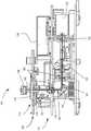
图2是根据一个实施例的细胞成像装置的截面侧视图。2 is a cross-sectional side view of a cell imaging device according to one embodiment.
图3是图2的细胞成像装置的截面俯视图,图中示出了通过装置的激励光和发射光流。3 is a cross-sectional top view of the cell imaging device of FIG. 2 showing excitation and emission light flux through the device.
图4是根据一个实施例的传输光组合件的截面侧视图。4 is a cross-sectional side view of a transmissive light assembly according to one embodiment.
图5是图4中所示的传输光组合件的分解图。FIG. 5 is an exploded view of the transmissive light assembly shown in FIG. 4 .
图6是图4和图5中所示的光组合件的顶部平面图。FIG. 6 is a top plan view of the light assembly shown in FIGS. 4 and 5 .
图7A-图7D是描绘光组合件的替代实施例的顶部平面图。7A-7D are top plan views depicting alternative embodiments of light assemblies.
图8示出了根据一个实施例的相对于选定透光LED利用的双色镜和发光滤光器的透射率对波长比较曲线图。8 shows a plot of transmittance versus wavelength for dichroic mirrors and emissive filters utilized with respect to selected light-transmitting LEDs, according to one embodiment.
图9示出了使用四种不同波长用10倍物镜俘获的H&E染色组织差分吸收图像。Figure 9 shows differential absorption images of H&E stained tissue captured with a 10x objective using four different wavelengths.
图10示出根据一个实施例的使用透射光能力的发色团染色组织的不同放大图像。Figure 10 shows different magnification images of stained tissue using a chromophore of transmitted light capability, according to one embodiment.
图11示出了用于生物染色的常见发色团的吸收带。Figure 11 shows absorption bands of common chromophores used for biological staining.
具体实施方式Detailed ways
如说明书中所使用的,以单数形式出现的词语涵盖其复数的对应部分,而以复数形式出现的词语涵盖其单数的对应部分,除非暗含地或明确地另有理解或陈述。此外,应理解,对于本文中描述的任何给定组件或实施例,针对所述组件列出的任何可能候选或替代方案通常可以个别地使用或彼此结合使用,除非隐含地或明确地另有理解或陈述。另外,将理解,除非另外隐含地或明确地理解或陈述,否则此类候选或替代方案的任何列表仅是说明性的,而不是限制性的。另外,除非另外指明,否则本说明书和权利要求书中所用的表示成分的量、组分、反应条件等等的数字应理解为均由术语“约”修饰。As used in this specification, words appearing in the singular encompass their plural counterparts and words appearing in the plural encompass their singular counterparts, unless implicitly or explicitly understood or stated otherwise. Furthermore, it should be understood that for any given component or embodiment described herein, any of the possible candidates or alternatives listed for that component can generally be used individually or in conjunction with each other, unless implicitly or explicitly otherwise understand or state. Additionally, it is to be understood that any listing of such candidates or alternatives is illustrative only, and not restrictive, unless otherwise implicitly or explicitly understood or stated. In addition, unless otherwise indicated, numbers used in the specification and claims representing amounts of ingredients, components, reaction conditions, etc., should be understood to be all modified by the term "about."
因此,除非相反地指出,否则本说明书和随附权利要求书中所阐述的数值参数是可以取决于寻求通过本文提出的主题获得的所希望特性而改变的近似值。最低限度地,并且不试图限制等效物原则对权利要求书范围的应用,每一个数值参数都应至少根据所报告的有效数字的数量并且通过应用一般四舍五入技术来解释。尽管阐述本文中提出的主题的广泛范围的数值范围和参数为近似值,但具体实例中所阐述的数值是尽可能精确报告的。然而,任何数值固有地含有某些由其相应测试测量值中所发现的标准差必然造成的误差。Accordingly, unless indicated to the contrary, the numerical parameters set forth in this specification and attached claims are approximations that may vary depending upon the desired properties sought to be obtained by the subject matter presented herein. Minimally, and without attempting to limit the application of the doctrine of equivalents to the scope of the claims, each numerical parameter should at least be construed in light of the number of reported significant digits and by applying ordinary rounding techniques. Notwithstanding that the numerical ranges and parameters setting forth the broad scope of the subject matter presented herein are approximations, the numerical values set forth in the specific examples are reported as precisely as possible. Any numerical value, however, inherently contains certain errors necessarily resulting from the standard deviation found in their respective testing measurements.
此外,如在说明书和所附权利要求书中使用,例如“顶部”、“底部”、“左”、“右”、“上”、“下”、“上部”、“下部”、“近”、“远”等等的方向术语在本文中仅用以指示相对方向且并不希望限制本发明或权利要求的范围。Also, as used in the specification and the appended claims, such as "top", "bottom", "left", "right", "upper", "lower", "upper", "lower", "near" Directional terms such as , "far," etc. are used herein to denote relative directions only and are not intended to limit the scope of the invention or the claims.
如背景技术部分中简要论述的,关于HCI一般强调优化用于检测荧光信号的技术,因为由于不利的原因,发色团染色生物样本的成像之前还不是并入HCI中的方法。然而,虽然荧光细胞和/或组织的HCI是经常使用的一种方法,但是本文所揭示的用自动方式添加发色团染色样本的HCI的新颖能力是期望添加到HCI中的一个特征。举例来说,当用亮场白光照明时,发色团的HCI提供的结构的对比度大于样本未被标记的情况,因而可以提供有待添加到HCI能力的另外的重要细胞和/或组织信息。As briefly discussed in the Background section, there is a general emphasis on optimizing techniques for detection of fluorescent signals with respect to HCI, since imaging of chromophore-stained biological samples has not previously been a method incorporated into HCI for unfavorable reasons. However, while HCl of fluorescent cells and/or tissues is a frequently used method, the novel ability to add chromophore-stained HCl to a sample disclosed herein is a feature that would be desirable to add to HCl. For example, when illuminated with brightfield white light, the HCl of the chromophore provides a structure with greater contrast than if the sample were unlabeled, and thus can provide additional important cellular and/or tissue information to be added to the capabilities of the HCl.
因而本文中提出的高含量成像(HCI)系统涉及可以扩展常规HCI系统的能力的系统。本文中所揭示的HCI系统的实施例不仅能够给用荧光团标记的生物样本成像和/或使用白光亮场照明给样本成像,这些HCI系统还可以用自动多路复用方式给用一或多个发色团标记的生物样本成像。这可以通过如下方式实现:用特定波长的透射光照明用发色团标记的样本和/或使用不同波长以落射荧光(epifluorescent)模式照明样本中的荧光团,并且自动检测表示荧光信息或样本对彩色光的差分吸收的图像。The high content imaging (HCI) system proposed herein thus relates to a system that can extend the capabilities of conventional HCI systems. Embodiments of the HCI systems disclosed herein are not only capable of imaging fluorophore-labeled biological samples and/or imaging samples using white light brightfield illumination, these HCI systems can also be automatically multiplexed with one or more Imaging of chromophore-labeled biological samples. This can be achieved by illuminating a sample labeled with a chromophore with a specific wavelength of transmitted light and/or using a different wavelength to illuminate the fluorophore in the sample in epifluorescent mode, and automatically detecting the pair representing fluorescence information or sample pairs Image of differential absorption of colored light.
关于透射光照明设置,本文所揭示的配置与被设计成用于自动荧光成像(即高含量筛分(HCS))的落射荧光显微检查平台组合构建。通过落射荧光设置的光学器件(例如双色镜、发光过滤器等)传输透射光波长,使其能够到达单个检测器或图像记录器,例如单个单色相机(CCD)。Regarding the transmitted light illumination setup, the configurations disclosed herein were constructed in combination with an epifluorescence microscopy platform designed for automated fluorescence imaging, ie high content sieving (HCS). Transmitted light wavelengths are transmitted by optics in an epi-fluorescence setup (eg, dichroic mirrors, luminescence filters, etc.), enabling it to reach a single detector or image recorder, such as a single monochromatic camera (CCD).
因此,整体设计不仅用于可见光谱(即从近紫外到近红外)上的荧光成像,而是提供检测还吸收可见光谱的跨段上的光的发色团的附加能力。另外,所揭示的设计和方法提供了一种相对低成本的方式来升级荧光显微镜使其还能够检测发色团,而不需要对于不同波长使用彩色相机或多个检测器。Thus, the overall design is not only useful for fluorescence imaging across the visible spectrum (ie, from the near-ultraviolet to the near-infrared), but provides the additional capability of detecting chromophores that also absorb light across spans of the visible spectrum. Additionally, the disclosed designs and methods provide a relatively low-cost way to upgrade a fluorescence microscope to also detect chromophores without the need for color cameras or multiple detectors for different wavelengths.
本文中所揭示的或预想的实施例可包括或利用专用或通用计算机,包含计算机硬件,比如例如一或多个处理器,如下文更详细地论述。实施例还可包含用于承载或存储计算机可执行指令和/或数据结构的物理和其它计算机可读媒体。这样的计算机可读媒体可以是可以通过通用或专用计算机系统存取的任何可用媒体。存储计算机可执行指令的计算机可读媒体是物理存储媒体。承载计算机可执行指令的计算机可读媒体是传输媒体。因此,借助于实例而非限制,实施例可以包括至少两种明显地不同类别的计算机可读媒体:计算机存储媒体和传输媒体。Embodiments disclosed or envisioned herein may include or utilize special purpose or general purpose computers, including computer hardware, such as, for example, one or more processors, as discussed in more detail below. Embodiments may also include physical and other computer-readable media for carrying or storing computer-executable instructions and/or data structures. Such computer-readable media can be any available media that can be accessed by a general purpose or special purpose computer system. Computer-readable media that store computer-executable instructions are physical storage media. Computer-readable media that carry computer-executable instructions are transmission media. Thus, by way of example and not limitation, embodiments may include at least two distinct classes of computer-readable media: computer storage media and transmission media.
计算机存储媒体包括RAM、ROM、EEPROM、CD-ROM或其它光盘存储器、磁盘存储器或其它磁性存储器装置,或者任何其它可以用于存储计算机可执行指令或数据结构的形式的期望的程序代码装置并且可以通过通用或专用计算机存取的媒体。Computer storage media includes RAM, ROM, EEPROM, CD-ROM, or other optical, magnetic, or other magnetic memory devices, or any other desired program code device in the form of computer-executable instructions or data structures that can be used to store and can Media accessed by a general-purpose or special-purpose computer.
“网络”被定义为一或多个数据链路,其使得能够在计算机系统和/或模块和/或其它电子装置之间传送电子数据。当经由网络或另一通信连接(固线式、无线式的或固线式的与无线式的组合)向计算机传送或提供信息时,计算机将连接正确地视为传输媒体。传输媒体可以包括可以用于承载数据或计算机可执行指令或数据结构形式的期望程序代码装置并且可以通过通用或专用计算机存取的网络和/或数据链路。上述各项的组合也应包含在计算机可读媒体的范围内。A "network" is defined as one or more data links that enable the transfer of electronic data between computer systems and/or modules and/or other electronic devices. When transferring or providing information to a computer via a network or another communication connection (either wired, wireless, or a combination of wired and wireless), the computer properly views the connection as a transmission medium. Transmission media may include network and/or data links which may be used to carry data or desired program code means in the form of computer-executable instructions or data structures and which may be accessed by a general purpose or special purpose computer. Combinations of the above should also be included within the scope of computer-readable media.
此外,当到达各种计算机系统组件时,计算机可执行指令或数据结构形式的程序代码装置可以从传输媒体自动传送到计算机存储媒体(或反过来)。举例来说,经由网络或数据链路接收到的计算机可执行指令或数据结构可以在网络接口模块(例如“NIC”)内的RAM中缓冲,然后最终传送到计算机系统RAM和/或计算机系统处的较低易失性的计算机存储媒体。因此,应理解,计算机存储媒体可以包括在也(或者甚至主要)利用传输媒体的计算机系统组件中。Furthermore, program code means in the form of computer-executable instructions or data structures can be automatically transferred from transmission media to computer storage media (or vice versa) when reaching various computer system components. For example, computer-executable instructions or data structures received over a network or data link may be buffered in RAM within a network interface module (eg, "NIC") and then ultimately transferred to the computer system RAM and/or computer system of less volatile computer storage media. Thus, it should be understood that computer storage media can be included in computer system components that also (or even primarily) utilize transmission media.
计算机可执行指令包括例如使通用计算机、专用计算机或专用处理装置执行某一功能或功能群组的指令和数据。计算机可执行指令可以例如是二进位、中间格式指令,例如汇编语言,或甚至源代码。尽管已经以特定地针对结构特征和/或方法动作的语言来描述主题,但应理解,所附权利要求书中所界定的主题未必限于上文所描述的特征或动作。而是,作为实施权利要求的实例形式揭示了所描述的特征和动作。Computer-executable instructions include, for example, instructions and data that cause a general purpose computer, special purpose computer, or special purpose processing device to perform a certain function or group of functions. Computer-executable instructions may be, for example, binary, intermediate format instructions, such as assembly language, or even source code. Although the subject matter has been described in language specific to structural features and/or methodological acts, it is to be understood that the subject matter defined in the appended claims is not necessarily limited to the features or acts described above. Rather, the described features and acts are disclosed as example forms of implementing the claims.
所属领域的技术人员将理解,可以在具有许多类型的计算机系统配置的网络计算环境中实践实施例,这些计算机系统配置包含个人计算机、桌上型计算机、膝上型计算机、消息处理器、手持式装置、多处理器系统、基于微处理器的或可编程消费型电子装置、网络PC、微型计算机、大型主机计算机、平板电脑、移动电话PDA、寻呼机、路由器、交换机等等。可以在分布式系统环境中实践实施例,其中本地和远程计算机系统都执行任务,该本地和远程计算机系统通过网络(通过固线式数据链路、无线数据链路或通过固线式链路与无线数据链路的组合)链接。在分布式系统环境中,程序模块可以位于本地存储装置和远程存储器存储装置中。用于一个实体的程序模块可以位于另一实体数据中心中或者在“云端中”,或者在另一实体数据中心中或者在“云端中”运行。在本说明书和以下权利要求中,计算机系统还定义为包括成像系统(例如图1中的成像系统102)。Those skilled in the art will understand that embodiments may be practiced in network computing environments with many types of computer system configurations, including personal computers, desktop computers, laptop computers, message processors, handheld devices, multiprocessor systems, microprocessor-based or programmable consumer electronics, network PCs, microcomputers, mainframe computers, tablet computers, mobile phone PDAs, pagers, routers, switches, and the like. Embodiments may be practiced in a distributed system environment, where both local and remote computer systems perform tasks that communicate with each other through a network (via a wired data link, a wireless data link, or via a wired link). A combination of wireless data links) links. In a distributed system environment, program modules may be located in both local storage devices and remote memory storage devices. A program module for one entity may reside in another entity's data center or "in the cloud" or run in another entity's data center or "in the cloud." In this specification and the following claims, a computer system is also defined to include an imaging system (eg,
图1示出并入有本文中所揭示的或预想的特征的示范性系统100。在系统的中心是定量高含量细胞成像系统102,其中对生物细胞进行扫描和分析。示例性细胞成像系统102包含(但不限于)成像装置104和计算装置106。FIG. 1 illustrates an
成像装置104包括安装在显微镜组合件110上的级壳108。级壳108被配置成收容定位含有细胞的试样片(比如(例如)96孔片)所必需的组件,使得显微镜组合件110可以给细胞成像以允许执行细胞的高含量筛分,这是所属领域的技术人员已知的。成像装置104配合计算装置106可以执行从成像获得的数据的分析和存储。
计算装置106可以用作所述系统的控制器并且用于自身或者配合成像装置104执行成像装置104获得的数据的分析和/或存储。计算装置106可以包括如上定义的通用或专用计算机或服务器等等或任何其它计算机化装置。如本领域中已知,计算装置106可以直接或通过网络与成像装置104通信。在一些实施例中,计算装置106并入到成像装置104中。
系统100还可以包括使用者显示装置112以显示结果和/或系统配置。成像装置104和/或计算装置106可以直接或间接地与使用者显示装置112通信。The
总体上布置在成像装置104中的光学配置在相机上产生细胞的放大图像以便记录细胞样本的高分辨率图像。具体来说,本文中论述的配置提供一种不仅能够实现所属领域的技术人员已知的“宽场”显微检查而且能够实现光学分段能力的系统。这可以包括(例如)在一系列细胞上扫描的聚焦照明点或照明行的标准共焦显微检查。这些能力可以与成像算法结合,比如(例如)最近邻点去模糊(下文将论述),其有助于提供通过相机记录的期望图像。An optical arrangement generally arranged in the
在一个实施例中,作为软件应用程序执行本文所述的方法步骤中的一或多个。然而,实施例不限于这种情况,并且方法步骤也可以在固件、硬件或固件、硬件和/或软件的组合中执行。此外,方法的步骤可以完全或部分地存在于成像装置104、计算装置106和/或其它计算装置上。In one embodiment, one or more of the method steps described herein are performed as a software application. However, embodiments are not limited in this case and method steps may also be performed in firmware, hardware or a combination of firmware, hardware and/or software. Furthermore, the steps of the method may reside in whole or in part on
用于该系统的装置的操作环境可包括或利用具有一或多个微处理器和系统存储器的处理系统。根据计算机编程领域的技术人员的实践,除非另有规定,下文参照处理系统执行的操作或指令的动作和符号表示描述实施例。这些动作和操作或指令被称作“计算机执行的”、“CPU执行的”或“处理器执行的”。The operating environment of the apparatus for the system may include or utilize a processing system having one or more microprocessors and system memory. Unless otherwise specified, embodiments are described below with reference to actions and symbolic representations of operations or instructions performed by a processing system, according to the practice of those skilled in the art of computer programming. These acts and operations or instructions are referred to as "computer-executed," "CPU-executed," or "processor-executed."
图2是成像装置104的示例性实施例。图2显示了内部平台设计的总体截面侧视图。总的来说,成像装置104集成定位含有生物细胞的HCS样本板116所必需的组件,从而使得显微镜组合件110可以执行生物细胞的高含量筛分。FIG. 2 is an exemplary embodiment of
级壳108包括级组合件114,其用便于与构成显微镜组合件110的组件光学和机械协作的方式安装。级组合件114一般包括上面能定位HCS样本板116的级,以及用于选择性地移动级以便观察的级定位机构,如本领域中已知。
在描绘的实施例中,显微镜组合件110容置倒置式显微镜,其可以用于从标本下方对试样样本板116上的标本执行筛分。显微镜包括物镜组合件118,如本领域中已知,其包括多个物镜以获得标本的放大视图。每个物镜可以对应于不同的放大水平。在一个实施例中,包括至少三个标准物镜。如果需要的话,还可以包括另外的物镜。实例标准物镜可以包括10x/0.4NA、20x/0.45NA和40x/0.6NA光学规范。实例另外物镜可以包括2x/0.08NA、4x/0.16NA和20x/0.7NA光学规范。还可以使用其它放大水平和物镜类型。In the depicted embodiment,
显微镜还包括焦点驱动机构120,其机械耦合到显微镜物镜组合件118。物镜组合件118可以经由焦点驱动机构120相对于级组合件114上下移动,以便将显微镜物镜组合件118的任何物镜对准和聚焦在试样样本板116内安置的生物细胞上。焦点驱动机构120可以是自动聚焦机构,但这不是必需的。焦点驱动机构120可以配置有步进式电机和螺丝/螺母组合,其降低消隙以提供(例如)低至0.006-μm/微步的分辨率以支持成像装置104中配置的显微镜物镜。The microscope also includes a
作为用于说明利用三个物镜时成像装置104的物镜工作的实例实施例,物镜组合件118可以用定制方式配置以提供三个位置,其使得能够询问样本板116内组织的细胞。焦点驱动机构120可以用自动方式快速并且可靠地在物镜之间切换。此布置的物镜通常但不是必须定位成隔开60度,这样可以使得初级物镜能够聚焦于样本板116上,另外两个物镜不会干扰成像装置104内的该级、样本板116或其它组件。As an example embodiment to illustrate the objective operation of
为了改变物镜,焦点驱动机构120可以下降到级组合件114下方,旋转到下一个物镜位置,然后将物镜上推到适当的聚焦高度。为了提供增强的系统安全,可使用机械限位开关来使转台返回原位,同时可使用一或多个光学TTL开关来确认物镜的位置已正确切换。此外,可以用旋转转台上的经过精确机器加工的机械止动件将每个光学位置保持在位。To change the objective, the
虽然本文中的论述是针对使用倒置式显微镜配置,但是应了解可以替代地使用非倒置式显微镜配置从细胞上方执行筛分。此外,虽然本文中论述的显微镜组合件110是定制的,但是在需要时可以并入其它常规显微镜配置,比如例如德国哥廷根(Goettingin)的卡尔蔡司微成像有限公司(Carl Zeiss MicroImaging,Inc.)制造的Axiovert 200M。在一些实施例中,如下文更详细论述的,根本不需要显微镜。Although the discussion herein is directed to using an inverted microscope configuration, it should be understood that a non-inverted microscope configuration can alternatively be used to perform sieving from above the cells. Furthermore, although the
显微镜组合件104还包括各种已知组件,用于产生和记录通过物镜获得的标本的图像。这些组件可包含(但不限于):
图像记录器122,比如(例如)单色CCD或CMOS相机,An
荧光团激励源124,比如(例如)包括多个发光二极管(LED)的光引擎,a
滤光器,其过滤所述激励和发射光,比如(例如)多位置双色滤光轮128和多位置发光滤光轮130,以及Filters that filter the excitation and emission light, such as, for example, a multi-position
光引导装置,其引导激励和发射光通过显微镜组合件,比如(例如)特兰透镜132、折叠镜134(例如90度折叠镜)和一或多个导光管。A light guide that directs excitation and emission light through the microscope assembly, such as, for example, a
以上组件中的一或多个通常受到计算装置106的控制以容许自动成像。总体上布置在成像装置104中的所述光学配置能在图像记录器122上产生细胞的放大图像,从而使得可以记录细胞样本的高分辨率图像。具体来说,本文中论述的配置提供一种不仅能够实现所属领域的技术人员已知的“宽场”显微检查而且能够实现光学分段能力的系统。One or more of the above components are typically controlled by computing
在一个实施例中,特兰透镜132是被设计成用于在使用具有期望发光波长的任何配置物镜时增强成像装置104在从蓝色到NIR的可见光谱的全范围上的性能的近红外(NIR)增强透镜(例如Olympus Triplet),下文将予以论述。In one embodiment, the
对于荧光分析,荧光团激励源124产生激励光,其照明细胞并且使得细胞诱发荧光团发光。举例来说,荧光团激励源124可以是多LED光引擎,其与双色滤光轮128和发光滤光轮130提供的配置激励滤光器协作工作,这两个滤光轮都可以受到计算机驱动以选择期望的滤光器。For fluorescence analysis, the
作为一种通用的操作方法,可以自动或手动地引导荧光团激励源124以提供从紫色(例如380nm)到近红外(例如至少700nm)的范围的多个光带宽,并且设计成激励荧光团,比如(例如)蓝绿色荧光蛋白质(CFP)和远红外(即,近红外)荧光团。适当激励滤光器(例如经由计算机106驱动的发光滤光轮130选择的)的实例LED带宽可包含(但不限于)紫色(380-410nm LED和386/23nm激励滤光片)、蓝色(420-455nm LED和438/24nm激励滤光片)、蓝绿色(460-490nm LED和485/20nm激励滤光片)、绿色(535-600nm LED和549/15nm激励滤光片)、绿色(535-600nm LED和560/25nm激励滤光片)、红色(620-750nm LED和650/13nm激励滤光片)和近红外(700-IR nm LED和740/13nm激励滤光片)。当期望改善红色和猩红染料的亮度时,上文所列的两个绿色/激励滤光片组合可以任选地经由例如机械鳍板提供。当然,也可以使用其它LED带宽。As a general method of operation, the
使用系统100,可以执行细胞的荧光分析。为了执行分析,级组合件114首先将样本板116移动到微孔的期望特定片段或载片的特定片段处于给定光路中的位置。Using
图3是另外示出显微镜组合件110内的实例组件的落射荧光路径的俯视图描绘(与图2的侧视图相反)。如图2和图3中所描绘,荧光团激励源124发出通过光纤递送系统中继(如图3中的大方向箭头136所示)的期望的系统波长带宽,这取决于相对于样本板116中的荧光标记细胞的应用。照明转接器138使用各种光学器件导引激励波长带宽以便另外沿着激励光路140受到引导(如图2和图3中通过包括斜杠的箭头指示),直到被安置在多位置双色滤光轮128中的期望双色组件142(图3中所示)接收到为止。双色组件142经过设计并且自动经过软件选择,以用于荧光团激励光源124提供的特定波长带宽。双色组件142将激励光引导到90度折叠镜134。此后,如图2所示,激励光沿着激励光路140继续向上,通过特兰透镜132和物镜组合件118到达安置在样本板支座116中的细胞,这是所属领域的技术人员已知的。FIG. 3 is a top view depiction (as opposed to the side view of FIG. 2 ) of the epi-fluorescence path additionally showing the example components within
激励光在安置在样本板支座116中的细胞中诱发荧光。诱发荧光沿着落射荧光返回路径144(图2和图3中通过暗箭头示出)通过物镜组合件118和特兰透镜132经由落射荧光布置从细胞传回,直到被90度折叠镜134接收到为止。如图3中具体所示,折叠镜134将诱发的荧光引导回到双色组件142,这允许诱发荧光沿着通过(例如)额外光学组件的落射荧光返回路径144继续返回。诱发的荧光接着通过布置在发光滤光轮130中的配置滤光器滤光,并且经由图像记录器122俘获滤光的诱发荧光并且记录为图像。The excitation light induces fluorescence in cells disposed in the
如图2所示,为了还能够进行亮场和发色团成像,显微镜组合件104进一步包括位于样本板116上方的传输光组合件126。为了执行发色团分析,使用各种波长带宽的光照明细胞以确定细胞内的发色团的差分吸收测量值。由于传输光组合件126的作用,本文中相对于细胞成像系统102所揭示的发射模式能力以新颖的方式用于差分成像,以便用多路复用的方式来视觉显示标准发色团和组织学染色。通过用光源(例如LED)照明图2中所示的透射光几何形状的样本来实现发色团的成像和视觉显示,光源是通过具有不同彩色波长(例如蓝色、绿色、琥珀色和红色)的传输光组合件126提供的,并且使用图像记录器122检测发色团对于光的差分吸收。As shown in FIG. 2 , in order to also enable bright field and chromophore imaging, the
传输光组合件126还可以提供透射白光以用于亮场成像。白光用于给(例如安置在样本板116中的)微孔或载片片段中的荧光成像的细胞照“背光”。一些常规高含量系统已经能够在同一装置中执行荧光和亮场成像,但是都不能还提供发色团分析。Transmitting
由于系统100的独特并且新颖的设计,现在可以在同一高含量系统中用荧光和亮场分析来执行发色团分析,以提供高含量细胞分析的三步方法。也就是说,在高含量细胞分析期间,可以在系统100中多路复用所有三种模式(荧光、亮场和发色团)。Due to the unique and novel design of
图4和图5分别描绘传输光组合件126的截面图和分解图。传输光组合件126可以包括支座200,其具有多个散热片202用于冷却组合件。如果需要的话,传输光组合件126可以并入有主动冷却机构,例如风扇或液体冷却系统,以替代或补充散热片202。支座200还可以包括凹进区域204,其受到圆形侧壁206限界。光组合件208固定在凹进区域204内。4 and 5 depict a cross-sectional view and an exploded view, respectively, of the transmissive
转向图6,光组合件208包括主体210,它是平坦的圆盘状装置,具有外围边缘212。多个凹口214围绕外围边缘212定位以在安装于支座200上期间帮助定位光组合件208。光组合件208进一步包括多个光源,例如LED 216(216a-216f),其在主体210上定位成特定图案,以允许传输光组合件126生成用于亮场成像和发色团成像的光。Turning to FIG. 6 , the
如图6中所示,LED 216围绕主体210布置成总体上圆形的图案。环绕的LED间隔开,从而使得邻近LED之间总体上距离相同,但是这并不是必需的。LED两个一组地配对,使得一个光组的两个LED在通电时发出相同色彩或波长。举例来说,图6描绘了三个不同的光组-LED 216a和216d形成第一光组,LED 216b和216e形成第二光组,LED 216c和216f形成第三光组。在描绘的实施例中,每个光组设计成发出三种相异的波长或颜色色彩-红色、绿色或蓝色中的一种。此外,每个光组中的LED布置在圆的彼此相对的侧面上。在一个实施例中,每个光组中的LED围绕共同的中心点彼此在直径上相对。As shown in FIG. 6 , the LEDs 216 are arranged in a generally circular pattern around the
每当期望产生任一种色彩或波长时,对应的光组中的两个LED都可以被通电以从圆的相对侧面发出彩色光。因此,光在LED之间的中心点处重叠,以提供特定色彩的良好混合。此外,因为光组全部具有相同的中心点,所以在同一时间从两个或更多个光组发射的光产生在透射光波束中间具有总体上更好的彩色混合物的透射光。使彩色光波束穿过光漫射器还有助于使光的色彩平衡并且获得均匀的光分布,如下文更详细论述。因此,单独的或任何组合形式的平衡的红、绿或蓝光可以由传输光组合件126产生,并且沿着传输光路146向下照射到样本板116上。因为对于不同发色团使用不同色彩的光,所以这样使得发色团分析成为可能。Whenever any color or wavelength is desired to be produced, both LEDs in the corresponding light set can be energized to emit colored light from opposite sides of the circle. Thus, the light overlaps at a central point between the LEDs to provide a good mix of specific colors. Furthermore, because the light groups all have the same center point, light emitted from two or more light groups at the same time produces transmitted light with an overall better color mixture among the transmitted light beams. Passing the colored light beams through the light diffuser also helps to balance the color of the light and achieve a uniform light distribution, as discussed in more detail below. Thus, balanced red, green, or blue light, alone or in any combination, may be generated by the transmission
如果需要的话,还可以结合LED 216使用其它光源。举例来说,在描绘的实施例中,任选的LED 218也置于主体210上。LED 218总体上置于主体210的中心,但这不是必需的。LED 218设计成在通电时发出琥珀色光以便能够成像并且分析吸收对应于琥珀色光(比如(例如)taladium blue)的光波长的发色团。还可以使用发出其它色彩的LED。Other light sources may also be used in conjunction with LEDs 216 if desired. For example, in the depicted embodiment,
除了能够执行发色团分析之外,以上布局还可以允许执行亮场分析。具体来说,当红、绿和蓝光组同时通电时,产生白光,其可以用于亮场分析。LED布局连同下文论述的光漫射器产生白光,其在整个光束上具有良好的色平衡和均匀的光分布。因此,传输光组合件126可以产生亮场分析和发色团分析所必需的光。In addition to being able to perform chromophore analysis, the above arrangement may also allow performing bright field analysis. Specifically, when the red, green, and blue light groups are energized at the same time, white light is produced, which can be used for bright field analysis. The LED layout along with the light diffuser discussed below produces white light with good color balance and uniform light distribution across the entire beam. Thus, the transmission
可以用于传输光组合件126的实例LED(Luxeon)包括:琥珀色(590nm)-77lm(LXML-PL01-0040)、绿色(530nm)-125lm(LXML-PM01-0070)、橙红色(617nm)-90lm(LXML-PH01-0050)、皇室蓝色(447.5nm)-890mW(LXML-PR02-0800)、绿色(530nm)-125lm(LXML-PM01-0070)、橙红色(617nm)-90lm(LXML-PH01-0050)、皇室蓝色(447.5nm)-890mW(LXML-PR02-0800)。Example LEDs (Luxeon) that can be used to transmit
LED只是能用于传输光组合件126的一种光源。如果需要的话,可以替代地使用其它波长的光源作为光源代替LED以生成所要的波长。LEDs are just one type of light source that can be used to transmit
图7A-图7D示出了光组合件250的替代实施例。类似于光组合件208,光组合件250包括多个LED 216,其配对成光组,从而使得每个光组中的光源彼此定位在圆的相对侧面上。然而,不是在单个主体上安装所有LED,而是光组中的每一个安装在单独的环状主体或环252(252a-252c)上。因此,如图7A至图7C中所示,红、蓝和绿色LED分别安装在环252a、252b和252c上。当然,任何环上可以安装任何彩色光组。7A-7D illustrate an alternate embodiment of a light assembly 250. Similar to
环252的尺寸设计成使得它们可以围绕光学中心点254一个套在另一个里配合在一起,如图7D中所示。因此,每个环252可以独立于其它环围绕中心点254旋转以改变安装在该环上的LED的位置。如图7D中所示,不同光组(即,相同色彩的LED)定位成离光学中心点254有不同距离。然而,对于每个光组,该光组的LED置于光学中心点254的相对侧面上,相同距离处。因此,每个环252可以按需要围绕光学中心点254旋转,并且该光组的LED将在光学中心的相对侧面上保持相同距离。The rings 252 are sized so that they fit together one within the other around the optical center point 254, as shown in Figure 7D. Thus, each ring 252 can be rotated about the center point 254 independently of the other rings to change the position of the LEDs mounted on that ring. As shown in Figure 7D, the different light groups (ie, LEDs of the same color) are positioned at different distances from the optical center point 254. However, for each light group, the LEDs of that light group are placed on opposite sides of the optical center point 254, at the same distance. Thus, each ring 252 can be rotated about the optical center point 254 as desired, and the LEDs of that light group will remain the same distance on opposite sides of the optical center.
如果使用者希望对于不同应用使用不同彩色光组,则对于每个光组使用单独的环252可能是有益的。在这种情况下,可以随时保留所有大小的环的对应于不同光组的环。然后,可以按需要混合和匹配这些环。以此方式,对于每种应用,只需要调换对应于期望波长的环,而不是更换整个光组合件。If the user wishes to use different colored light sets for different applications, it may be beneficial to use a separate ring 252 for each light set. In this case, rings of all sizes of rings corresponding to different light groups can be kept at all times. These rings can then be mixed and matched as desired. In this way, instead of replacing the entire optical assembly, only the rings corresponding to the desired wavelengths need to be exchanged for each application.
返回到图4和图5,传输光组合件126还包括透镜管220,其具有在第一端224与第二端226之间延伸的环绕侧壁222。环绕侧壁222的第一端224固定到凹进区域204的圆形侧壁206以便环绕光组合件208。在一个实施例中,透镜管的直径为两英寸。其它直径也是有可能的。第一护环228和第二护环230固定在透镜管220的第二端226,光漫射器232和抗反射(AR)窗口234安置于护环之间的透镜管220内。端盖236可移除地固定到透镜管的第二端226以在不使用时保护传输光组合件126的组件。Returning to FIGS. 4 and 5 , the transmitting
光漫射器232是混合元件,其在光穿过元件时,将光混合在一起并且使其变得均质。这样能缓解使用不同光源时可能产生的问题(例如阴影和不均匀光分布)。光漫射器可以由任何半透明的物体制成,比如(例如)毛玻璃、灰玻璃、特氟隆、蛋白化玻璃等等。将要使用的光漫射器的类型可以取决于损失量对比光的均匀分布的期望的取舍。举例来说,毛玻璃漫射器可以提供低散射损失,全息漫射器可以提高从多种光源的传输效率,UV全息漫射器可以提供紫外辐射范围内的增加的性能,而蛋白石漫射玻璃可以产生光的接近朗伯分布,但是会导致更高水平的散射损失。还可以使用磨砂漫射器。在一个实施例中,光漫射器232是蛋白化光漫射器,从而使得强度从几乎所有角度都是均匀的。这个均匀的强度结合LED放置的均匀性,允许均匀的照明。可使用的蛋白化光漫射器的一个实例是EdmundOptics制造的蛋白石漫射玻璃漫射器No.46-106。The
AR窗口234另外有助于通过减小反射而提供均匀的光分布。其允许光在一个方向通过而不在另一个方向通过,从而减少高强度光的反射。反射降低会通过消除杂散光而改善成像系统中的对比度。AR窗口234布置在透镜管220中,从而使得从光组合件208发射的光可以从中通过,但是来自另一方向的光无法通过。因此,AR窗口234防止在荧光成像期间发生的荧光发射向上穿过透镜管220进入LED 216、218中,这样可能导致LED的自发荧光。LED的自发荧光可能导致二级激励光向下传输到荧光团中,这样可能会扭曲荧光分析。
在一个实施例中,AR窗口234包括设计成反射与光组合件208的光源相关联的波长的光(比如(例如)对于LED 216和218是450-650nm)的涂层。涂层定位于AR窗口234的表面上,从而使得LED 216和218在那些波长下发射的光可以穿过涂层,但是来自其它方向的接触涂层的那些波长的光被反射,从而基本上阻挡光穿过涂层。可使用的AR窗口的一个实例是Edmund Optics制造的AR涂布塑料窗口No.46-106。In one embodiment,
为了提供用于执行亮场和发色团成像的期望传输光,将LED 216和/或LED 218的适当光组通电。每个通电的LED发射对应于LED色彩的波长的光。来自通电LED的光被引导通过透镜管220以便穿过光漫射器232和AR窗口234。光沿着传输光路146从透镜管220出来,成为传输光。由于光组合件208以及光漫射器的布局,经由透镜管220从传输光组合件126出来的传输光在LED发射的色彩之间具有良好的色平衡,并且在整个光束上具有均匀的光分布,如上文所论述。To provide the desired transmitted light for performing bright field and chromophore imaging, LEDs 216 and/or appropriate light sets of
传输光组合件126置于样本板116上方,使得传输光组合件126产生的照明光向下照射到样本板116上。如果需要的话,传输光组合件126可以配置在样本板116上方的吊杆(未图示)上,从而使得当使用者接取样本板或物镜时,传输光组合件126能摆动开来。The transmitted
为了进行亮场分析,可以将所有LED 216通电以提供白光,以便向荧光成像的微孔或载片片段(例如安置在样本板116中)照“背光”。为了进行发色团分析,可以将适当的LED216和/或218通电以提供期望的波长带宽的光以执行发色团的差分吸收测量。也就是说,当仅仅一个或两个LED 216通电(尤其是如果样本板116下方的光源未通电)时,可以在特定波长或波长组合下选择性地测量场中的样本的差分吸收特征。For bright field analysis, all LEDs 216 can be energized to provide white light to "backlight" the fluorescently imaged microwell or slide segment (eg, positioned in sample plate 116). For chromophore analysis, the appropriate LEDs 216 and/or 218 may be energized to provide light of the desired wavelength bandwidth to perform differential absorption measurements of the chromophore. That is, when only one or both of the LEDs 216 are energized (especially if the light source below the
在使用期间,传输光路146与上文所论述的落射荧光返回路径144对准,以便向荧光成像微孔或载片片段(例如安置在样本板116中)提供照明光。当传输光照射到细胞上时,光至少部分地被细胞内的发色团吸收。穿过细胞的光(即,未被发色团吸收的光)用于确定关于发色团的信息,并且在本文中定义为发色团吸收特征。由于传输光路146与落射荧光返回路径144的对准,发色团吸收特征沿着同一落射荧光返回路径144以便最终被图像记录器122成像。During use, the
因为路径包括再次穿过双色滤光轮128和多位置发光滤光轮130,所以可以具体选择对应于相应各个LED 216和218的四个透射光波长,从而使得它们透射过安置在双色滤光轮128内的多频段双色镜,(例如)5频带双色镜,另外还有多频段发射滤光器,(例如)多位置发光滤光轮130内的5频带组件。类似地,可以选择特定的双色滤光器和发射滤光器以与透射光的波长对应。Because the path includes again passing through the
举例来说,要让LED发射的光能被图像记录器122俘获,发光的波长应当能够穿过光路中的各种滤光器中的至少一个。为了执行这一点,LED的波长应当处于滤光器中的至少一个的传输带通波长内;否则LED光将简单地被滤除并且不到达图像记录器122。因此,应当谨慎选择将用于传输光组合件126的LED波长。For example, for the light emitted by the LEDs to be captured by the
图8示出在一个实施例中将要使用的用于选择特定传输LED的装置。图8尤其示出了根据一个实施例的选定双色发射滤光器的传输带宽特性对比波长,该图是结合用于各种可能传输LED的发光波长绘示的。元件符号300表示发射体滤光器带通特性,元件符号302表示双色镜传输带通特性,元件符号304(304a-304g)指代上文所列的各种Luxeon LED的传输波长。Figure 8 shows the means to be used in one embodiment for selecting a specific transmission LED. Figure 8 shows, inter alia, transmission bandwidth characteristics versus wavelengths for selected dichroic emission filters, plotted in conjunction with emission wavelengths for various possible transmission LEDs.
在所描绘的曲线图中,传输波长304a、304d、304e和304f在对应于发射体滤光器和双色镜两者的传输带通波长内,而传输波长304b、304c和304g不是。因此,当成像装置104中使用特定发射体滤光器和双色器件时,只有对应于波长304a、304d、304e和304f的LED应当被考虑用于传输光组合件126。根据这一点,元件符号310(皇室蓝色447.5nm)、312(绿色530nm)、314(琥珀色590nm)和316(橙红色617nm)(分别对应于波长304a、304d、304e和304f)指代一个实施例中使用的实例兼容传输光LED波长特性。In the depicted graph,
图9示出当使用对应于上文所论述的兼容传输光LED的LED以提供差分吸收图像时,在用作图像记录器122的CCD相机上俘获的图像。具体地说,图9a至图9d分别示出了H&E染色组织差分吸收图像320、322、324和326,其用10倍物镜分别使用四种不同波长蓝色310、绿色312、红色316和琥珀色314俘获。图9e是红色、绿色及蓝色的组合彩色图像328(本文中用黑色和白色示出),其已通过计算装置106软件同化,以便提供各个俘获到的图像否则无法示出的组织细节。Figure 9 shows an image captured on a CCD camera used as
如上文所论述,实施例可以具有几个不同的显微镜物镜,这些显微镜物镜之间能够自动切换。这样可以实现例如用低放大率物镜检测较大区域然后用较高放大率将所关注的特定区域重新成像的能力。As discussed above, embodiments may have several different microscope objectives that can be automatically switched between. This enables, for example, the ability to inspect a larger area with a low magnification objective and then re-image a specific area of interest with a higher magnification.
举例来说,图10示出了使用透射光的相同发色团染色组织的不同放大图像。具体地说,图10a至图10d分别示出使用自动成像能力在2倍、4倍、10倍和20倍放大率水平下的H&E染色组织差分吸收图像330、332、334和336。For example, Figure 10 shows different magnification images of the same chromophore stained tissue using transmitted light. Specifically, Figures 10a-10d show H&E stained tissue differential absorption images 330, 332, 334 and 336 at 2X, 4X, 10X and 20X magnification levels, respectively, using automated imaging capabilities.
因此,除了荧光标记样本之外,这种能力现在还可以自动应用于发色团标记样本或含有不同模式的组合的样本。在一个实施例中,可以通过在放大率较低的图像中自动检测所关注的特定物体或染色图案而触发对较高放大率的切换。Therefore, in addition to fluorescently labeled samples, this capability can now be automatically applied to chromophore-labeled samples or samples containing combinations of different modalities. In one embodiment, switching to higher magnification may be triggered by automatic detection of specific objects or staining patterns of interest in lower magnification images.
因此,图9和图10示出在组合检测荧光和发色团两者的能力时,各种实施例使两种成像模式中的能力最大化。Thus, Figures 9 and 10 show that when combining the ability to detect both fluorescence and chromophore, various embodiments maximize the ability in both imaging modalities.
对于荧光,这意味着能够激励荧光团并且检测其在整个可见光谱((例如)从近紫外到近红外的范围内)上发射的荧光。For fluorescence, this means being able to excite the fluorophore and detect its fluorescence emission over the entire visible spectrum (eg, in the range from the near-ultraviolet to the near-infrared).
对于发色团,这意味着能够检测可见光谱的范围上的发色团吸收所引起的吸收率标志。For chromophores, this means being able to detect the absorbance signature caused by the absorption of the chromophore over the range of the visible spectrum.
在一个实施例中,为了在荧光中实现这个目标,荧光团激励源124包括LED光源,其跨越整个可见光谱在7个不同波长带中发光。使用计算机控制的多位置双色滤光轮128以包括双色镜,并且使用计算机控制的多位置发光滤光轮130(例如6位置滤光轮)包括发射滤光器,其与双色滤光轮无关。发光滤光轮130就置于图像记录器122前面,图像记录器122是低光照水平CCD相机。多位置双色滤光轮128和发光滤光轮130都包括单频带镜或滤光器,其专用于透射特定的波长带,或者多频带镜或滤光器,其可以同时透射多个频带。这可以包括5频带双色镜和发射滤光器对,其可以同时透射5个或更多个波长带。此外,由于独立的计算机控制和激励LED、双色镜和发射滤光器的选择能力,还可以使用非常规荧光成像模式,例如荧光共振能量传递法(FRET)、比率式荧光成像或使用具有长斯托克斯位移(例如量子点)的荧光探针的成像(Byers等人,2007)。In one embodiment, to achieve this goal in fluorescence,
图11示出了用于细胞的生物染色的35种常见发色团的吸收带(从OlympusMicroscopy resource Center重新创造)。图11上叠加着对应于用于传输光组合件的一个实施例的四个LED的四个波长(通过元件符号340、342、344和346表示)。波长340、342、344和346分别对应于448nm(皇室蓝色)、530nm(绿色)、590nm(琥珀色)和617nm(橙红色)。如图11中所描绘,所列的发色团中的每一个的吸收频带包括波长340、342、344和346中的至少一个。因此,四个LED波长中的至少一个通过可见光谱上的所列常见发色团中的每一个差分吸收。因此,可以利用所列发色团中的每一个来差分吸收安置在样本支架116内的细胞的成像。这样能够实现成像系统100可使用的多种多样的发色团选项。Figure 11 shows absorption bands for 35 common chromophores used for biological staining of cells (recreated from Olympus Microscopy resource Center). Figure 11 is superimposed on four wavelengths (indicated by
鉴于这一点,添加传输光组合件126到落射荧光平台能够实现场成像的许多不同选项。举例来说,细胞成像系统102可以容许仅仅荧光的测量、仅仅吸收的测量或组合荧光/吸收测量,其中从顶部和底部并行地照明样本的场。作为增加的一个益处,LED源还可以用作白光发射体以用于亮场分析。因此,LED源不仅相对于典型的宽频带源有成本效益,而且提供相对较小并且非常容易添加到现有荧光显微镜的整体模块。In view of this, adding the transmission
因为多光学成像能力,所以可以用任何成像模式自动将高含量板片的多个凹孔或微型凹孔的多个场成像。举例来说,每个场可以安置于光路140或透射光路径146内并且成像,如图2所示。这样的微型凹孔可以在给定时间内通过成像装置104成像,级组合件114以较小增量移动样本板116(例如微孔板片或显微镜载片),从而使得一个接一个的场可以连续置于光路140或146中并且成像。举例来说,在典型实施例中,对应于微孔和成像的底表面的1/300的场可以从微孔中心的四个场开始,然后是中心四个场周围的十二个场,然后是十二个场周围的48个场,等等……直到成像装置104使用落射荧光或透射光或组合已经提供了足够的图像以俘获限定数目的“有效”细胞为止。Because of the multi-optical imaging capability, multiple wells of a high content plate or multiple fields of micro-wells can be automatically imaged in any imaging mode. For example, each field may be positioned within
因此,使用例如图1中示出的系统之类的HCI系统,仅仅使用发色团成像就可以执行高含量成像。举例来说,在高含量细胞分析的一种方法中,板片可以置于成像系统的级组合件上,板片具有多个凹孔,每个凹孔中定位有带有发色团染色分子的生物细胞。接着,对于多个凹孔中的每个凹孔,级组合件可以移动,使得凹孔变成与光学路径对准,传输光可以指向凹孔内的生物细胞,以诱发生物细胞沿着光学路径发出发色团吸收特征,并且可以记录从凹孔内的生物细胞发射的诱发发色团吸收特征的图像。最后,可以分析针对每个凹孔记录的发色团吸收特征。Thus, using an HCI system such as the system shown in Figure 1, high content imaging can be performed using only chromophore imaging. For example, in one method of high-content cell analysis, a plate can be placed on a stage assembly of an imaging system, the plate has a plurality of wells, each well positioned with a chromophore-staining molecule biological cells. Then, for each well of the plurality of wells, the stage assembly can be moved such that the wells become aligned with the optical path and the transmitted light can be directed towards biological cells within the wells to induce the biological cells to follow the optical path A chromophore absorption feature is emitted, and images of the evoked chromophore absorption feature emitted from biological cells within the well can be recorded. Finally, the chromophore absorption characteristics recorded for each well can be analyzed.
替代地,可以使用单个设备执行并入有荧光成像和发色团成像的成像。可以在不同的时间执行成像。举例来说,在一种细胞分析方法中,可以定位含有包括荧光报告体分子和发色团染色分子的生物细胞的容器,从而使得容器变成与光学路径对准。可以用激励光激励生物细胞以诱发生物细胞沿着光学路径发出荧光,并且可以记录诱发荧光的图像。传输光可以指向生物细胞以诱发生物细胞沿着光学路径发出发色团吸收特征并且还可以记录诱发发色团吸收特征的图像。最后,可以使用来自两个记录图像的数据来分析生物细胞。Alternatively, imaging incorporating fluorescence imaging and chromophore imaging can be performed using a single device. Imaging can be performed at different times. For example, in one cellular analysis method, a container containing biological cells including fluorescent reporter molecules and chromophore staining molecules can be positioned such that the container becomes aligned with an optical pathway. The biological cells can be excited with excitation light to induce the biological cells to fluoresce along the optical path, and images of the induced fluorescence can be recorded. The transmitted light can be directed at the biological cells to induce the biological cells to emit chromophore absorption features along the optical path and can also record images of the induced chromophore absorption features. Finally, biological cells can be analyzed using the data from the two recorded images.
替代地,可以并行执行成像。举例来说,在一种细胞分析方法中,可以定位含有包括荧光报告体分子和发色团染色分子的生物细胞的容器,从而使得容器变成与光学路径对准。可以用激励光激励生物细胞,并且传输光可以指向生物细胞以诱发生物细胞沿着光学路径并行发出荧光和发色团吸收特征,并且可以记录并行诱发的荧光和发色团吸收特征的图像。最后,可以使用来自图像的数据分析生物细胞。Alternatively, imaging can be performed in parallel. For example, in one cellular analysis method, a container containing biological cells including fluorescent reporter molecules and chromophore staining molecules can be positioned such that the container becomes aligned with an optical pathway. The biological cell can be excited with excitation light, and the transmitted light can be directed at the biological cell to induce the biological cell to emit fluorescence and chromophore absorption features in parallel along the optical path, and images of the parallel induced fluorescence and chromophore absorption features can be recorded. Finally, biological cells can be analyzed using the data from the images.
在一些实施例中,可以对于成像和分析使用“通道”。也就是说,可以针对每个场获得多个图像,每个图像对应于不同的通道。通道(即,光学模态)通常由特定激励光(LED或滤光白光)、特定成像光(通常由从微孔接收的光滤光)、特定曝光周期等限定。每个图像可以实际上保存为黑色和白色信号,还保存关于通道的信息,例如激励信号、曝光周期等等。还可以保存其它图像信息。因此,如果(例如)将100个场成像,每个场使用三个不同通道,则可以保存300个图像并且连同关于每个通道的信息一起存储。可使用记录图像中的一或多个来分析每个场。举例来说,在以上实例中,可以使用对应于场的通道的三个不同通道中的任何数目的通道来分析每个场。In some embodiments, "channels" may be used for imaging and analysis. That is, multiple images can be obtained for each field, each image corresponding to a different channel. The channel (ie, the optical modality) is typically defined by a specific excitation light (LED or filtered white light), a specific imaging light (usually filtered by the light received from the microwell), a specific exposure period, and the like. Each image can actually be saved as a black and white signal, but also information about the channel such as excitation signal, exposure period, etc. Other image information can also be saved. Thus, if, for example, 100 fields are imaged, each using three different channels, 300 images can be saved and stored along with information about each channel. Each field can be analyzed using one or more of the recorded images. For example, in the above example, each field may be analyzed using any number of three different channels corresponding to the channels of the field.
为了克服在常见HCI仪器中使用的单个图像记录器122(例如单色CCD或CMOS相机)的局限性,本文中引入常规HCI平台未提供的新的多模态光学能力。具体来说,不是使用不同检测器每个检测单独的初级波长,而是可使用单个单色检测器采集多个图像,每个图像对应于单独的波长。举例来说,传输光组合件126可以在不同时间用不同的相异的波长照射样本,从而使得用于荧光的图像记录器122可以采集对应于波长中的每一个的图像,如上文相对于图9a-图9d所论述。To overcome the limitations of a single image recorder 122 (eg, a monochromatic CCD or CMOS camera) used in common HCI instruments, new multimodal optical capabilities are introduced herein that are not provided by conventional HCI platforms. Specifically, instead of using different detectors each to detect a separate primary wavelength, a single monochromatic detector can be used to acquire multiple images, each image corresponding to a separate wavelength. For example, the transmitted
结果是这样的系统布置和方法:它用新颖的方式可以使用单个检测器俘获通过波长选择区分的多个图像,该区分是经由选定激励带通滤光器或选定滤光器配合组装在传输光组合件126中的多个窄带源(例如四个LED)的阵列。举例来说,在一种细胞分析方法中,含有具有发色团染色分子的生物细胞的容器可以置于成像系统的级组合件上,从而使得容器变成与光学路径对准。不同波长的传输光可以接着一次一个地指向生物细胞以诱发生物细胞沿着光学路径发出不同的发色团吸收标志。可以接着分别记录对应于不同传输波长的诱发发色团吸收标志中的每一个。可以接着由计算机化装置基于记录图像产生合成图像。The result is a system arrangement and method that in a novel way can use a single detector to capture multiple images differentiated by wavelength selection via selected excitation bandpass filters or selected filter combinations assembled on the An array of multiple narrowband sources (eg, four LEDs) in
在一个实施例中,可以使用液晶可调谐滤光器(LCTF)取代发光滤光轮130以提供波长选择。这样的滤光器的操作类似于具有几十或几百个滤光器的滤光轮,但是益处是没有移动部分。这样的可调谐滤光器发射窄带光,其峰位置可以用(例如)到大约1纳米精度的高精度并且在(例如)毫秒范围内的短时间框内电子地调谐成几乎任何波长。本文中并入这样的滤光器作为替代实施例以提供例如可见光(例如420-720nm)的期望的宽光谱范围。In one embodiment, a liquid crystal tunable filter (LCTF) may be used in place of the
在一个实施例中,可使用成像“通道”以便于自动成像,其中实时分析图像以确定是否需要另外的图像以及应当俘获场的哪个部分。因此,当成像在进行时,细胞成像系统102可以比较图像并且产生数据。虽然成像软件可以复叠一或多个场的图像以用高对比度实现多彩色图像,但是这种操作模式通常不对整个场执行。In one embodiment, an imaging "channel" may be used to facilitate automated imaging, where images are analyzed in real-time to determine if additional images are needed and which portion of the field should be captured. Thus,
实情为,可以采集第一图像(例如,使用赫斯特染色在波长1下得到细胞核荧光图案)并且使用第一图像限定场中的每个细胞核的位置、尺寸、形状等。可以接着使用数学方法限定例如环状形状的细胞质(围绕每个核)的预期区域(因此,细胞核中的许多像素)。可以接着将每个这样的环状形状以数学方式叠加在通道2和通道3中的相同场的图像上。可以从通道2和3导出数据,从而得出关于荧光染料染色的组分中有多少是活性的并且准备好受到激励并且分别在通道2和3中成像,其具有环状形状,其中推断每个细胞将要在和/或在通道1中实际上测量到的核区中。系统可以经编程以在多个通道中对相同场的图像进行多种复杂分析,这都是在系统拍摄其它场的额外图像并且对照来自前面各轮的存储图像的时候。Instead, a first image can be acquired (eg, using Hoechst staining to obtain a nuclear fluorescence pattern at wavelength 1) and used to define the location, size, shape, etc. of each nucleus in the field. Mathematical methods can then be used to define expected regions of the cytoplasm (around each nucleus), eg, a ring shape (thus, many pixels in the nucleus). Each such annular shape can then be mathematically superimposed on the images of the same field in channel 2 and channel 3. Data can be derived from channels 2 and 3 as to how many of the fluorochrome-stained components are active and ready to be excited and imaged in channels 2 and 3, respectively, which have a ring shape, where it is inferred that each The cells will be in and/or in the nuclear region that is actually measured in channel 1. The system can be programmed to perform multiple complex analyses of images of the same field in multiple channels, all while the system takes additional images of other fields and contrasts stored images from previous rounds.
正是在这种情境中,本文中的系统可以执行“自动”分析,在一次实验中对于凹孔中的多个场、板片中的多个凹孔和多个板片重复。一旦已经限定了所有参数,仪器就可以基本上无人值守地运行和分析。It is in this context that the system herein can perform an "automatic" analysis, repeating for multiple fields in a well, multiple wells in a plate, and multiple plates in a single experiment. Once all parameters have been defined, the instrument can be run and analyzed essentially unattended.
举例来说,在一种自动细胞分析方法中,具有位置阵列的容器可以置于成像系统上,该位置含有具有一或多个荧光报告体分子和一或多个发色团染色分子的多个细胞。位置中的每一个中的多个细胞可以作为一子组多个细胞被容纳,以使得多个场和成像系统可以具有第一和第二光源,其被配置成将一或多个辐射波长选择性地引导到任何位置阵列中的多个场。第一光源可以被配置成产生激励光,其诱发生物细胞发出荧光信号,并且第二光源可以被配置成产生传输光,其诱发生物细胞发出发色团吸收特征。For example, in an automated cellular analysis method, a container with an array of locations that contain a plurality of molecules having one or more fluorescent reporter molecules and one or more chromophore staining molecules can be placed on an imaging system. cell. The plurality of cells in each of the locations can be accommodated as a subset of the plurality of cells such that the plurality of fields and imaging systems can have first and second light sources configured to select one or more radiation wavelengths Directly to multiple fields in the array at any location. The first light source can be configured to generate excitation light that induces the biological cell to emit a fluorescent signal, and the second light source can be configured to generate transmission light that induces the biological cell to emit a chromophore absorption feature.
通过用图像记录器记录每个场的多个图像,可以将一或多个期望位置内的场中的每一个成像。图像中的每一个可以包括细胞上或细胞内通过第一光源诱发的荧光信号或通过第二光源诱发的发色团吸收特征,并且每个图像可以包括选自下面各项的至少一种光学模态:一或多个辐射激励波长、选定滤光辐射波长和期望的成像曝光周期。Each of the fields within one or more desired locations can be imaged by recording multiple images of each field with the image recorder. Each of the images can include a fluorescence signal on or within a cell induced by a first light source or a chromophore absorption feature induced by a second light source, and each image can include at least one optical mode selected from: state: one or more excitation wavelengths of radiation, selected wavelengths of filtered radiation, and desired imaging exposure period.
可以比较记录图像中的每一个以便将来自多个图像中的每一个的诱发荧光信号和发色团吸收标志转换成数字数据;Each of the recorded images can be compared to convert the evoked fluorescent signal and the chromophore absorption signature from each of the plurality of images into digital data;
可以利用数字数据自动确定来自细胞上或细胞内的荧光报告体分子的诱发荧光信号的强度和/或分布,并且自动确定细胞上或细胞内的诱发发色团吸收标志的强度和/或分布。变化可以指示细胞上或细胞内的荧光报告体分子或发色团的分布、环境或活动的变化。Digital data can be used to automatically determine the intensity and/or distribution of evoked fluorescent signals from fluorescent reporter molecules on or within cells, and the intensity and/or distribution of evoked chromophore uptake markers on or within cells. Changes can be indicative of changes in the distribution, environment or activity of fluorescent reporter molecules or chromophores on or within cells.
系统100还包括运行该系统的软件。这样的软件可以驻留在计算装置106上以控制所有必需的仪器硬件,例如(但不限于)级组合件114、物镜组合件118、焦点驱动机构120、双色滤光轮128和发光滤光轮130。软件还可以选择性地控制激励光源124和传输光组合件126将要提供的波长并且确定何时在图像记录器122上俘获图像。计算装置106还可以使得能够分析俘获到的图像、在监视器112上显示结果和管理还可以在集成式数据库中提供的数据。
此外,在分析使用共聚焦显微镜俘获到的图像时,计算装置106可以实施任何数目的图像处理算法以按需要处理俘获到的图像信息。作为一实例,此算法可以包括光学分段结合最近邻点去模糊,如上文简要介绍的。本文中配置的此例程通常应用于尤其在z轴中分立分布的细胞荧光结构。具体来说,如果操作员需要的话,包括性计算装置106算法可以向图像记录器122俘获的三维图像堆叠的每个二维平面逐个平面地应用操作。举例来说,通过模糊邻近平面(z+1和z-1,使用数字模糊滤光器)然后从z平面减去模糊平面,借此最近邻点算法对z平面操作。这种技术还可以用于使用者可选的多个邻近平面。此外,还可以通过向三维堆叠(即,光学分段)中的每个平面应用该算法来处理该堆叠。在此算法操作中,可以从每个平面去除模糊估计值。Furthermore, in analyzing images captured using a confocal microscope,
如上所述,在一些实施例中,可以从系统中省去物镜或甚至整个显微镜。借助于实例而非限制,传输光组合件还可以用于:As mentioned above, in some embodiments, the objective lens or even the entire microscope may be omitted from the system. By way of example and not limitation, transmitting light assemblies can also be used to:
并入有微透镜阵列而不是物镜的系统,Systems incorporating microlens arrays instead of objectives,
其中细胞放置在平面扫描器上并且使用闭路电视透镜成像的系统,以及A system in which cells are placed on a flat-screen scanner and imaged using a CCTV lens, and
其中细胞直接放置在检测器上的系统。A system in which cells are placed directly on the detector.
这些系统可以并入有例如CCD或CMOS传感器阵列或芯片之类的检测器,其以高分辨率给更大区域成像。因为这一点,视场(FOV)可能比使用标准物镜可以获得的视场大很多,这意味着生物细胞的成像区域的尺寸可以更大。举例来说,在大型FOV系统中,成像区域的尺寸的长度可以是几厘米,而不是使用标准物镜的大约一毫米的长度。These systems may incorporate detectors such as CCD or CMOS sensor arrays or chips that image larger areas with high resolution. Because of this, the field of view (FOV) can be much larger than what can be achieved with standard objectives, which means that the size of the imaged area of biological cells can be larger. For example, in large FOV systems, the size of the imaging area can be a few centimeters in length, rather than about a millimeter in length using standard objectives.
因此,物镜和/或许多显微镜组合件的其它部分可以省去。举例来说,在一个实施例中,细胞可以直接放置在并入有大型FOV检测器的图像记录器上,从而实际上消除了大多(甚至所有)显微镜组合件。还有一个结果是,例如样本板或载片之类的生物容器可能不需要移动那么多就能给所有细胞成像。举例来说,在一个实施例中,无需移动容器就可以给多个凹孔成像。在一些实施例中,无需移动容器就可以给整个容器内的所有细胞成像。Therefore, the objective lens and/or many other parts of the microscope assembly can be omitted. For example, in one embodiment, cells can be placed directly on an image recorder incorporating a large FOV detector, virtually eliminating most (or even all) microscope assemblies. There is also the consequence that biological containers such as sample plates or slides may not need to move as much to image all the cells. For example, in one embodiment, multiple wells can be imaged without moving the container. In some embodiments, all cells within the entire vessel can be imaged without moving the vessel.
与显微镜设置无关(或者省去显微镜设置),系统可以仍然并入有传输光组合件,其定位成将传输光引导到生物细胞以从细胞诱发发色团吸收特征。此系统还可以并入有荧光团激励源,其将激励光引导到生物细胞以从细胞诱发荧光团发光。Regardless of (or omitting) the microscope setup, the system can still incorporate a transmitted light assembly positioned to direct the transmitted light to biological cells to induce chromophore absorption features from the cells. This system can also incorporate a fluorophore excitation source that directs excitation light to biological cells to induce fluorophore luminescence from the cells.
应理解,本文中关于各种实施例描述的特征可以用任何期望的组合来混合和匹配。此外,本文中所揭示的或预想的概念可以用其它特定的形式来具体实施。所描述的实施例应视为在所有方面均仅为说明性而非限制性的。因此,本发明的范围由所附权利要求书而不是由前述描述指示。在权利要求书等效物的含义和范围内的所有变化均涵盖在权利要求书的范围内。It should be understood that the features described herein with respect to the various embodiments may be mixed and matched in any desired combination. Furthermore, concepts disclosed or contemplated herein may be embodied in other specific forms. The described embodiments are to be considered in all respects only as illustrative and not restrictive. Accordingly, the scope of the invention is indicated by the appended claims rather than by the foregoing description. All changes that come within the meaning and range of equivalency of the claims are intended to be within the scope of the claims.
Claims (80)
Applications Claiming Priority (3)
| Application Number | Priority Date | Filing Date | Title |
|---|---|---|---|
| US201461992008P | 2014-05-12 | 2014-05-12 | |
| US61/992,008 | 2014-05-12 | ||
| PCT/US2015/030210WO2015175433A1 (en) | 2014-05-12 | 2015-05-11 | Automated imaging of chromophore labeled samples |
Publications (2)
| Publication Number | Publication Date |
|---|---|
| CN106461540A CN106461540A (en) | 2017-02-22 |
| CN106461540Btrue CN106461540B (en) | 2020-02-14 |
Family
ID=53269726
Family Applications (2)
| Application Number | Title | Priority Date | Filing Date |
|---|---|---|---|
| CN201580031476.4AActiveCN106461540B (en) | 2014-05-12 | 2015-05-11 | System and method for automated imaging of chromophore-labeled samples |
| CN201520490296.XUExpired - LifetimeCN205139012U (en) | 2014-05-12 | 2015-05-12 | System for carry out automatic high intension cell formation of image |
Family Applications After (1)
| Application Number | Title | Priority Date | Filing Date |
|---|---|---|---|
| CN201520490296.XUExpired - LifetimeCN205139012U (en) | 2014-05-12 | 2015-05-12 | System for carry out automatic high intension cell formation of image |
Country Status (4)
| Country | Link |
|---|---|
| US (3) | US9683939B2 (en) |
| EP (2) | EP3943915A3 (en) |
| CN (2) | CN106461540B (en) |
| WO (1) | WO2015175433A1 (en) |
Families Citing this family (32)
| Publication number | Priority date | Publication date | Assignee | Title |
|---|---|---|---|---|
| JP6455829B2 (en)* | 2013-04-01 | 2019-01-23 | キヤノン株式会社 | Image processing apparatus, image processing method, and program |
| WO2017132171A1 (en)* | 2016-01-28 | 2017-08-03 | Siemens Healthcare Diagnostics Inc. | Methods and apparatus for characterizing a specimen container and specimen |
| US10948703B2 (en)* | 2016-03-01 | 2021-03-16 | Molecular Devices, Llc | Imaging system and method with scattering to reduce source auto-fluorescence and improve uniformity |
| CN107666355A (en)* | 2016-07-27 | 2018-02-06 | 傅南匡 | Quantum resonance energy transmission device and transmission method thereof |
| EP3507768B1 (en)* | 2016-08-31 | 2020-09-30 | Molecular Devices, LLC | System and method for template-based image analysis |
| DE102016116377A1 (en)* | 2016-09-01 | 2018-03-01 | Rheinisch-Westfälisch Technische Hochschule (RWTH) Aachen | Method and device for detecting process parameters in liquid cultures |
| US10983325B2 (en)* | 2016-12-12 | 2021-04-20 | Molecular Devices, Llc | Trans-illumination imaging with an array of light sources |
| CN107064481B (en)* | 2017-03-16 | 2019-07-23 | 国家烟草质量监督检验中心 | A kind of liquid-vapor interface exposure system combines the method for high intension technology quantitative detection carbon monoxide cause DNA Damage |
| EP3486705A1 (en)* | 2017-11-16 | 2019-05-22 | Koninklijke Philips N.V. | A multi-mode imager |
| TWI660218B (en)* | 2018-02-22 | 2019-05-21 | 致茂電子股份有限公司 | Automatic fluorescence detection system |
| WO2019200116A1 (en)* | 2018-04-12 | 2019-10-17 | Life Technologies Corporation | Apparatuses, systems and methods for generating color video with a monochrome sensor |
| JP7489326B2 (en)* | 2018-05-25 | 2024-05-23 | ファイヴ プライム セラピューティクス インク | Improved Cytometry for Tissue Characterization and Screening |
| AU2019389908B2 (en) | 2018-11-29 | 2025-09-11 | La Trobe University | Method of identifying a structure |
| CN113454439B (en)* | 2018-11-29 | 2025-03-07 | 乐卓博大学 | Automated methods for structure identification |
| US12332463B2 (en) | 2018-11-29 | 2025-06-17 | La Trobe University | Microscopy method and system |
| US11294162B2 (en) | 2019-02-07 | 2022-04-05 | Nanotronics Imaging, Inc. | Fluorescence microscopy inspection systems, apparatus and methods with darkfield channel |
| US20240126063A1 (en)* | 2019-02-07 | 2024-04-18 | Nanotronics Imaging, Inc. | Fluorescence microscopy inspection systems, apparatus and methods |
| CN109709025B (en)* | 2019-02-12 | 2021-07-16 | 军事科学院系统工程研究院卫勤保障技术研究所 | Multimode imaging optical system |
| CN110162649B (en)* | 2019-05-24 | 2021-06-18 | 北京百度网讯科技有限公司 | Sample data acquisition method, acquisition system, server and computer readable medium |
| US11307094B2 (en)* | 2019-05-28 | 2022-04-19 | The Regents Of The University Of California | System and method for hyperspectral imaging in highly scattering media by the spectral phasor approach using two filters |
| WO2021067726A1 (en)* | 2019-10-03 | 2021-04-08 | The Regents Of The University Of California | Fluorescence imitating brightfield imaging |
| AU2021232064A1 (en)* | 2020-03-05 | 2022-09-29 | Canopy Biosciences, Llc | Automated fluorescence imaging and single cell segmentation |
| US11320375B2 (en)* | 2020-06-05 | 2022-05-03 | Yan Wang | Multicolor fluorescence reader with dual excitation channels |
| EP3926385B1 (en)* | 2020-06-16 | 2024-05-15 | Carl Zeiss Microscopy GmbH | Digital microscope and microscopic set |
| KR20220000160A (en)* | 2020-06-25 | 2022-01-03 | 삼성전자주식회사 | Apparatus and method for analyzing substance of object |
| US12422660B2 (en) | 2020-12-31 | 2025-09-23 | Quiver Holdings Inc. | Microscope for imaging samples within wells of multi-well plates hold by stage and beam homogenizer for homogenizing beam |
| US20220203372A1 (en)* | 2020-12-31 | 2022-06-30 | Q-State Biosciences, Inc. | Optical plate controller and reader |
| CA3219059A1 (en)* | 2021-05-04 | 2022-11-10 | Pacific Biosciences Of California, Inc. | Arrays of integrated analytical devices with reduced-scale unit cell |
| US12105023B2 (en) | 2021-06-30 | 2024-10-01 | Quelltx, Inc. | Multi-well plate readers and methods of their use |
| CN114813706B (en)* | 2022-06-29 | 2022-12-13 | 国科大杭州高等研究院 | Blood cell hyperspectral optical tweezers capture energy resonance transfer analyzer |
| CN115948243A (en)* | 2022-09-30 | 2023-04-11 | 河北医科大学第二医院 | A special low-oxygen workstation for high-content |
| WO2025144684A1 (en)* | 2023-12-28 | 2025-07-03 | Nanotronics Imaging, Inc. | Fluorescence microscopy inspection systems, apparatus and methods |
Family Cites Families (26)
| Publication number | Priority date | Publication date | Assignee | Title |
|---|---|---|---|---|
| GB9223259D0 (en)* | 1992-11-06 | 1992-12-23 | Quatro Biosystems Ltd | Method and apparatus for image analysis |
| US6982431B2 (en) | 1998-08-31 | 2006-01-03 | Molecular Devices Corporation | Sample analysis systems |
| US7219016B2 (en)* | 2001-04-20 | 2007-05-15 | Yale University | Systems and methods for automated analysis of cells and tissues |
| JP3889992B2 (en)* | 2002-05-17 | 2007-03-07 | 株式会社ミツトヨ | Ring lighting device |
| US7698068B2 (en)* | 2004-06-17 | 2010-04-13 | Cadent Ltd. | Method for providing data associated with the intraoral cavity |
| FR2873815B1 (en)* | 2004-08-02 | 2006-11-24 | Inodiag Sa | READING DEVICE FOR BLADES CARRYING MICRO DEPOSITS BIOLOGICAL REACTION CARRIERS |
| US7443508B1 (en) | 2005-05-18 | 2008-10-28 | Vie Group, Llc | Spectrophotometric scanner |
| US20070153847A1 (en)* | 2005-12-21 | 2007-07-05 | Nuvonyx, Inc | Manufacturing and the design of assemblies for high power laser diode array modules |
| US20080026483A1 (en)* | 2006-06-14 | 2008-01-31 | Oldenburg Kevin R | Thermal-cycling devices and methods of using the same |
| US7646482B2 (en) | 2007-05-31 | 2010-01-12 | Genetix Limited | Methods and apparatus for optical analysis of samples in biological sample containers |
| US8849380B2 (en)* | 2007-11-26 | 2014-09-30 | Canfield Scientific Inc. | Multi-spectral tissue imaging |
| US8947510B2 (en)* | 2008-06-20 | 2015-02-03 | Visiongate, Inc. | Functional imaging of cells with optical projection tomography |
| FR2936252B1 (en)* | 2008-09-23 | 2010-11-05 | Millipore Corp | DEVICE FOR MICROBIOLOGICAL ANALYSIS. |
| US11865534B2 (en)* | 2008-09-24 | 2024-01-09 | First Light Diagnostics, Inc. | Imaging analyzer for testing analytes |
| US7894079B1 (en) | 2009-11-09 | 2011-02-22 | Mitutoyo Corporation | Linear displacement sensor using a position sensitive photodetector |
| EP2519853A2 (en)* | 2009-12-31 | 2012-11-07 | Abbott Point Of Care, Inc. | Method and apparatus for fast focus imaging biologic specimens |
| EP2359745A1 (en)* | 2010-02-12 | 2011-08-24 | Helmholtz Zentrum München Deutsches Forschungszentrum für Gesundheit und Umwelt (GmbH) | Method and device for multi-spectral photonic imaging |
| GB2492268B (en)* | 2010-02-18 | 2019-02-06 | Bima Ltd | Immobilised-bead multiplex assay |
| AT510631B1 (en)* | 2010-10-20 | 2013-01-15 | Scan Messtechnik Ges M B H | SPECTROMETER |
| US9494783B2 (en)* | 2010-11-30 | 2016-11-15 | Etaluma Inc. | Compact, high-resolution fluorescence and brightfield microscope and methods of use |
| CN103620378B (en)* | 2011-04-21 | 2017-06-09 | 麻省理工学院 | Highly compact optical flow cell with multiple optical junctions, flexibly configurable optical sensing components and systems for in situ real-time spectral measurements |
| GB2495537B (en)* | 2011-10-14 | 2017-02-15 | Solentim Ltd | Method of and apparatus for analysis of a sample of biological tissue cells |
| US9066021B2 (en)* | 2012-10-18 | 2015-06-23 | Ortho-Clinical Diagnostics, Inc. | Full resolution color imaging of an object |
| CN105051531B (en)* | 2012-12-06 | 2018-05-15 | 艾博特健康公司 | Imaging biological fluids using predetermined distributions |
| US8882310B2 (en)* | 2012-12-10 | 2014-11-11 | Microsoft Corporation | Laser die light source module with low inductance |
| EP2932425B1 (en)* | 2012-12-14 | 2025-01-29 | The J. David Gladstone Institutes, a testamentary trust established under the Will of J. David Gladstone | Automated robotic microscopy systems |
- 2015
- 2015-05-11CNCN201580031476.4Apatent/CN106461540B/enactiveActive
- 2015-05-11USUS14/709,239patent/US9683939B2/enactiveActive
- 2015-05-11EPEP21158334.9Apatent/EP3943915A3/enactivePending
- 2015-05-11EPEP15725460.8Apatent/EP3143381B1/enactiveActive
- 2015-05-11WOPCT/US2015/030210patent/WO2015175433A1/enactiveApplication Filing
- 2015-05-12CNCN201520490296.XUpatent/CN205139012U/ennot_activeExpired - Lifetime
- 2017
- 2017-05-15USUS15/594,978patent/US10234445B2/enactiveActive
- 2019
- 2019-01-28USUS16/259,072patent/US10393725B2/enactiveActive
Also Published As
| Publication number | Publication date |
|---|---|
| US20170248574A1 (en) | 2017-08-31 |
| US9683939B2 (en) | 2017-06-20 |
| CN106461540A (en) | 2017-02-22 |
| US20190154652A1 (en) | 2019-05-23 |
| WO2015175433A1 (en) | 2015-11-19 |
| EP3943915A3 (en) | 2022-05-25 |
| US10393725B2 (en) | 2019-08-27 |
| EP3143381B1 (en) | 2021-02-24 |
| US20150323462A1 (en) | 2015-11-12 |
| EP3943915A2 (en) | 2022-01-26 |
| EP3143381A1 (en) | 2017-03-22 |
| US10234445B2 (en) | 2019-03-19 |
| CN205139012U (en) | 2016-04-06 |
Similar Documents
| Publication | Publication Date | Title |
|---|---|---|
| CN106461540B (en) | System and method for automated imaging of chromophore-labeled samples | |
| US9581797B2 (en) | High-throughput hyperspectral imaging with superior resolution and optical sectioning | |
| US8884211B2 (en) | Confocal imaging methods and apparatus | |
| US7817273B2 (en) | Two-dimensional spectral imaging system | |
| US7663750B2 (en) | Two-dimensional spectral imaging system | |
| US10133048B2 (en) | Laser optical coupling for nanoparticles detection | |
| US20100302631A1 (en) | Microscope | |
| US20100151474A1 (en) | Multifunctional Device For Diagnostics and Method For Testing Biological Objects | |
| US8633432B2 (en) | Reflective focusing and transmissive projection device | |
| US20240003810A1 (en) | Universal multi-detection system for microplates with confocal imaging | |
| CN105637349A (en) | Series absorbance glass and interference filters | |
| US20240310617A1 (en) | Method and apparatus for mitigating specular highlights in imaging of biological tissue | |
| JP2025517174A (en) | ARRAY OF LIGHT SOURCES, FOURIER TYCHOGRAPHY IMAGING SYSTEM, AND METHOD FOR PERFORMING FOURIER TYCHOGRAPHY - Patent application | |
| IL314970A (en) | A device for measuring natural autofluorescence of a biological sample and a method for using it | |
| CN217304909U (en) | Portable biological detection system | |
| Glasenapp et al. | Biochip reader with dynamic holographic excitation and hyperspectral fluorescence detection | |
| US11067785B2 (en) | Method for generating fluorescent photograph using fluorescence microscope | |
| RU2679605C2 (en) | Biological microchips fluorimetric analyzer |
Legal Events
| Date | Code | Title | Description |
|---|---|---|---|
| C06 | Publication | ||
| PB01 | Publication | ||
| SE01 | Entry into force of request for substantive examination | ||
| SE01 | Entry into force of request for substantive examination | ||
| CB02 | Change of applicant information | ||
| CB02 | Change of applicant information | Address after:American California Applicant after:CELLOMICS, INC. Address before:American Pennsylvania Applicant before:CELLOMICS, INC. | |
| GR01 | Patent grant | ||
| GR01 | Patent grant |