CN1060112A - Human body C3b/C4b acceptor (CR1) - Google Patents
Human body C3b/C4b acceptor (CR1)Download PDFInfo
- Publication number
- CN1060112A CN1060112ACN90109580ACN90109580ACN1060112ACN 1060112 ACN1060112 ACN 1060112ACN 90109580 ACN90109580 ACN 90109580ACN 90109580 ACN90109580 ACN 90109580ACN 1060112 ACN1060112 ACN 1060112A
- Authority
- CN
- China
- Prior art keywords
- protein
- molecule
- cell
- sequence
- patient
- Prior art date
- Legal status (The legal status is an assumption and is not a legal conclusion. Google has not performed a legal analysis and makes no representation as to the accuracy of the status listed.)
- Pending
Links
- 0CC(C1)C(*)(*)[C@](C2)(**2C(*)*C(*)CC*)C1N=OChemical compoundCC(C1)C(*)(*)[C@](C2)(**2C(*)*C(*)CC*)C1N=O0.000description3
Images


Classifications
- C—CHEMISTRY; METALLURGY
- C07—ORGANIC CHEMISTRY
- C07K—PEPTIDES
- C07K14/00—Peptides having more than 20 amino acids; Gastrins; Somatostatins; Melanotropins; Derivatives thereof
- C07K14/195—Peptides having more than 20 amino acids; Gastrins; Somatostatins; Melanotropins; Derivatives thereof from bacteria
- C07K14/315—Peptides having more than 20 amino acids; Gastrins; Somatostatins; Melanotropins; Derivatives thereof from bacteria from Streptococcus (G), e.g. Enterococci
- C07K14/3153—Streptokinase
- A—HUMAN NECESSITIES
- A61—MEDICAL OR VETERINARY SCIENCE; HYGIENE
- A61P—SPECIFIC THERAPEUTIC ACTIVITY OF CHEMICAL COMPOUNDS OR MEDICINAL PREPARATIONS
- A61P29/00—Non-central analgesic, antipyretic or antiinflammatory agents, e.g. antirheumatic agents; Non-steroidal antiinflammatory drugs [NSAID]
- A—HUMAN NECESSITIES
- A61—MEDICAL OR VETERINARY SCIENCE; HYGIENE
- A61P—SPECIFIC THERAPEUTIC ACTIVITY OF CHEMICAL COMPOUNDS OR MEDICINAL PREPARATIONS
- A61P37/00—Drugs for immunological or allergic disorders
- A61P37/02—Immunomodulators
- A61P37/06—Immunosuppressants, e.g. drugs for graft rejection
- A—HUMAN NECESSITIES
- A61—MEDICAL OR VETERINARY SCIENCE; HYGIENE
- A61P—SPECIFIC THERAPEUTIC ACTIVITY OF CHEMICAL COMPOUNDS OR MEDICINAL PREPARATIONS
- A61P7/00—Drugs for disorders of the blood or the extracellular fluid
- A61P7/02—Antithrombotic agents; Anticoagulants; Platelet aggregation inhibitors
- A—HUMAN NECESSITIES
- A61—MEDICAL OR VETERINARY SCIENCE; HYGIENE
- A61P—SPECIFIC THERAPEUTIC ACTIVITY OF CHEMICAL COMPOUNDS OR MEDICINAL PREPARATIONS
- A61P9/00—Drugs for disorders of the cardiovascular system
- A—HUMAN NECESSITIES
- A61—MEDICAL OR VETERINARY SCIENCE; HYGIENE
- A61P—SPECIFIC THERAPEUTIC ACTIVITY OF CHEMICAL COMPOUNDS OR MEDICINAL PREPARATIONS
- A61P9/00—Drugs for disorders of the cardiovascular system
- A61P9/10—Drugs for disorders of the cardiovascular system for treating ischaemic or atherosclerotic diseases, e.g. antianginal drugs, coronary vasodilators, drugs for myocardial infarction, retinopathy, cerebrovascula insufficiency, renal arteriosclerosis
- C—CHEMISTRY; METALLURGY
- C07—ORGANIC CHEMISTRY
- C07K—PEPTIDES
- C07K14/00—Peptides having more than 20 amino acids; Gastrins; Somatostatins; Melanotropins; Derivatives thereof
- C07K14/435—Peptides having more than 20 amino acids; Gastrins; Somatostatins; Melanotropins; Derivatives thereof from animals; from humans
- C07K14/705—Receptors; Cell surface antigens; Cell surface determinants
- C07K14/70596—Molecules with a "CD"-designation not provided for elsewhere
- C—CHEMISTRY; METALLURGY
- C07—ORGANIC CHEMISTRY
- C07K—PEPTIDES
- C07K16/00—Immunoglobulins [IGs], e.g. monoclonal or polyclonal antibodies
- C07K16/18—Immunoglobulins [IGs], e.g. monoclonal or polyclonal antibodies against material from animals or humans
- C07K16/28—Immunoglobulins [IGs], e.g. monoclonal or polyclonal antibodies against material from animals or humans against receptors, cell surface antigens or cell surface determinants
- C07K16/2896—Immunoglobulins [IGs], e.g. monoclonal or polyclonal antibodies against material from animals or humans against receptors, cell surface antigens or cell surface determinants against molecules with a "CD"-designation, not provided for elsewhere
- C—CHEMISTRY; METALLURGY
- C12—BIOCHEMISTRY; BEER; SPIRITS; WINE; VINEGAR; MICROBIOLOGY; ENZYMOLOGY; MUTATION OR GENETIC ENGINEERING
- C12N—MICROORGANISMS OR ENZYMES; COMPOSITIONS THEREOF; PROPAGATING, PRESERVING, OR MAINTAINING MICROORGANISMS; MUTATION OR GENETIC ENGINEERING; CULTURE MEDIA
- C12N9/00—Enzymes; Proenzymes; Compositions thereof; Processes for preparing, activating, inhibiting, separating or purifying enzymes
- C12N9/14—Hydrolases (3)
- C12N9/48—Hydrolases (3) acting on peptide bonds (3.4)
- C12N9/50—Proteinases, e.g. Endopeptidases (3.4.21-3.4.25)
- C12N9/64—Proteinases, e.g. Endopeptidases (3.4.21-3.4.25) derived from animal tissue
- C12N9/6421—Proteinases, e.g. Endopeptidases (3.4.21-3.4.25) derived from animal tissue from mammals
- C12N9/6424—Serine endopeptidases (3.4.21)
- C12N9/6456—Plasminogen activators
- C12N9/6462—Plasminogen activators u-Plasminogen activator (3.4.21.73), i.e. urokinase
- C—CHEMISTRY; METALLURGY
- C12—BIOCHEMISTRY; BEER; SPIRITS; WINE; VINEGAR; MICROBIOLOGY; ENZYMOLOGY; MUTATION OR GENETIC ENGINEERING
- C12Y—ENZYMES
- C12Y304/00—Hydrolases acting on peptide bonds, i.e. peptidases (3.4)
- C12Y304/21—Serine endopeptidases (3.4.21)
- C12Y304/21073—Serine endopeptidases (3.4.21) u-Plasminogen activator (3.4.21.73), i.e. urokinase
- A—HUMAN NECESSITIES
- A61—MEDICAL OR VETERINARY SCIENCE; HYGIENE
- A61K—PREPARATIONS FOR MEDICAL, DENTAL OR TOILETRY PURPOSES
- A61K38/00—Medicinal preparations containing peptides
Landscapes
- Health & Medical Sciences (AREA)
- Chemical & Material Sciences (AREA)
- Organic Chemistry (AREA)
- Life Sciences & Earth Sciences (AREA)
- General Health & Medical Sciences (AREA)
- Engineering & Computer Science (AREA)
- Medicinal Chemistry (AREA)
- Bioinformatics & Cheminformatics (AREA)
- Genetics & Genomics (AREA)
- Biochemistry (AREA)
- Immunology (AREA)
- Zoology (AREA)
- Molecular Biology (AREA)
- Biophysics (AREA)
- Wood Science & Technology (AREA)
- Proteomics, Peptides & Aminoacids (AREA)
- Animal Behavior & Ethology (AREA)
- Public Health (AREA)
- Veterinary Medicine (AREA)
- Pharmacology & Pharmacy (AREA)
- Nuclear Medicine, Radiotherapy & Molecular Imaging (AREA)
- General Chemical & Material Sciences (AREA)
- Chemical Kinetics & Catalysis (AREA)
- General Engineering & Computer Science (AREA)
- Gastroenterology & Hepatology (AREA)
- Biomedical Technology (AREA)
- Biotechnology (AREA)
- Toxicology (AREA)
- Cell Biology (AREA)
- Heart & Thoracic Surgery (AREA)
- Cardiology (AREA)
- Microbiology (AREA)
- Transplantation (AREA)
- Vascular Medicine (AREA)
- Pain & Pain Management (AREA)
- Rheumatology (AREA)
- Diabetes (AREA)
- Hematology (AREA)
- Urology & Nephrology (AREA)
- Peptides Or Proteins (AREA)
Abstract
Description
Translated fromChinese1.简介1 Introduction
本发明涉及C3b/C4b受体(CR1)基因及其编码的蛋白质。本发明还涉及CR1的核酸顺序及其含有70个核苷酸的片段和它们编码的含有24个氨基酸的肽或蛋白质。本发明还提供了CR1蛋白质及其片段的表达。CR1核酸和蛋白质已被用于诊断或治疗涉及补体活性的疾病和各种炎症以及免疫性疾病。The present invention relates to C3b/C4b receptor (CR1) gene and its coded protein. The present invention also relates to the nucleic acid sequence of CR1 and its fragments comprising 70 nucleotides and their coded peptides or proteins comprising 24 amino acids. The present invention also provides the expression of CR1 protein and fragments thereof. CR1 nucleic acids and proteins have been used in the diagnosis or treatment of diseases involving complement activity and various inflammatory and immune diseases.
2.发明背景2. Background of the invention
2.1补体系统2.1 Complement system
补体系统是一组蛋白质,它们构成了人体正常血清中球蛋白的10%(Hood,L.E.等人,1984,《Immunology》,第2版,TheBenjamin/Cummings出版公司,Menlo Park,加州,第339页)。补体(C)在介导免疫和过敏反应中起着重要的作用(Rapp,H.J.和Borsos,T.1970,Molecular Basis of Complement Action,Appleton-Century-Crofts(Meredith),NewYork)。补体组分的激活导致了包括介导与补体依赖性疾病有关的炎症的趋化肽在内的一组因子产生。补体级联放大的连续活化可通过包含抗原-抗体复合体的经典型途径产生,或可通过包括识别某种细胞壁多糖的旁路途径产生。通过的活的补体蛋白质而介导的活性包括靶细胞的裂解,趋化性,调理作用,血管和其它平滑肌细胞的刺激,和诸如乳房细胞失粒的功能性紊乱,增加的小血管渗透性,白细胞的定向迁移和B淋巴细胞和巨噬细胞的活化(Eisen,H.N,1974,Immunology,Harper & Row出版公司,Hagerstown,Maryland,第512页)。The complement system is a group of proteins that make up 10% of the globulins in normal human serum (Hood, L.E. et al., 1984, Immunology, 2nd ed., The Benjamin/Cummings Publishing Company, Menlo Park, CA, p. 339 ). Complement (C) plays an important role in mediating immune and allergic responses (Rapp, H.J. and Borsos, T. 1970, Molecular Basis of Complement Action, Appleton-Century-Crofts (Meredith), NewYork). Activation of complement components results in the production of a panel of factors including chemotactic peptides that mediate inflammation associated with complement-dependent diseases. Continuous activation of the complement cascade can occur through a classical pathway involving antigen-antibody complexes, or through an alternative pathway involving recognition of certain cell wall polysaccharides. Activities mediated by live complement proteins include target cell lysis, chemotaxis, opsonization, stimulation of blood vessels and other smooth muscle cells, and functional disturbances such as mammary cell degranulation, increased small vessel permeability, Directed migration of leukocytes and activation of B lymphocytes and macrophages (Eisen, H.N, 1974, Immunology, Harper & Row Publishing Company, Hagerstown, Maryland, p. 512).
在蛋白质水解级联放大步骤中,生物活性肽片段,过敏毒素C3a,C4a和C5a(可参考WHO Scientific Group,1977,WHOTech.Rep.Ser 606:5,此处列为参考)从第3(C3),第4(C4)和第5(C5)天然补体组分(Hugli,T.E.1981,CRC Crit.Rev.Immunol.1:321;Bult,H和Herman,A.G,1983,Agents Actions 13:405)中释放出来。In the proteolytic cascade amplification step, bioactive peptide fragments, anaphylatoxins C3a, C4a and C5a (refer to WHO Scientific Group, 1977, WHOTech.Rep.Ser 606: 5, listed here for reference) from section 3 (C3 ), the 4th (C4) and 5th (C5) natural complement components (Hugli, T.E. 1981, CRC Crit. Rev. Immunol. 1: 321; Bult, H and Herman, A.G, 1983, Agents Actions 13: 405) released in.
2.2 C3b/C4b补体受体(CR1)2.2 C3b/C4b complement receptor (CR1)
人体C3b/C4b受体(称为CR1)存在于红细胞,单核细胞/巨噬细胞,粒细胞,B细胞,部分T细胞,脾卵泡树突状细胞和肾小球足状突细胞中(Fearon,D.T.1980,J.Exp.Med.152:20;Wilson,J.G.等人,1983,J.Immunol.131:684;Reynes.M.等人,1985,J.Im-munol.135:2687;Gelfand,M.C.等人,1976,N,Engl.J.Med.295:10;Kazatchkine,M.D.等人,1982,Clin.Immunol.Im-munopathol.27:170)。CR1特异性地与C3b,C4b和iC3b结合。在血浆中已发现了一种具有配体结合活性且与膜相关CR1具有相同分子量的可溶形式的受体(Yoon,SH.和Fearon,D.T.1985,J.Im-munol,134:3332)。CR1与已与免疫复合体和其它补体激活因子共价连接的C3b和C4b结合,它们相互作用的结果取决于载有该受体的细胞类型(Fearon,D.T.和Wong.W.W.1983,Ann,Rev,Im-munol 1:243),红细胞CR1与免疫复合体结合以转移至肝中(Corna-coff J.B.等人,1983,J.Clin,Invest 71:236;Medof M.E.等人,1982,J.Exp,Med,156:1739)。而嗜中性粒细胞和单核细胞的CR1则通过外膜纹孔的吸附性胞吞作用(Fearon D.T.等人,1981,J.Exp.Med.153:1615;Abrahamson D.R.和Fearon D.T.1983,Lab,Invest.48:162)或通过用佛波醇酯,趋化肽或存在于细胞外基质中的蛋白质,如纤维结合素(fibronectin)和海带氨酸(Laminin)激活受体后的吞噬作用(Newman.S.L.等人,1980,J,Immunol,125,2236;Wright S.D和Silverstein S.C 1982,J.Exp.Med.156:149; Wright S.D.等人,1983,J.Exp Med.158:1338)使连接的复合物内在化。CR1的磷酸化可能对获得吞噬活性起作用(Changelian,P.S.和Fearon.D.T.1986,J.Exp.Med.163:101)。尽管用抗CR1抗体处理这些细胞增强了它们对于亚最适剂量的美州商陆植物的有丝分裂素应答,CR1对于B淋巴细胞的作用则未完全确知(Daha,M.R.等人,1983,Immunobiol.164:227(Abstr.))。卵泡树突状细胞上的CR1有促进抗原递呈的作用(Klaus,G.G.B.等人,1980,Im-munol,Rev,53:3)。The human C3b/C4b receptor (known as CR1) is present in erythrocytes, monocytes/macrophages, granulocytes, B cells, some T cells, splenic follicular dendritic cells, and glomerular podocytes (Fearon , D.T.1980, J.Exp.Med.152:20; Wilson, J.G. et al., 1983, J.Immunol.131:684; Reynes.M. et al., 1985, J.Im-munol.135:2687; Gelfand , M.C. et al., 1976, N, Engl. J. Med. 295: 10; Kazatchkine, M. D. et al., 1982, Clin. Immunol. Im-munopathol. 27: 170). CR1 specifically binds to C3b, C4b and iC3b. A soluble form of the receptor having ligand binding activity and having the same molecular weight as membrane-associated CR1 has been found in plasma (Yoon, SH. and Fearon, D.T. 1985, J. Immunol, 134:3332). CR1 binds C3b and C4b to which immune complexes and other complement activating factors have been covalently linked, and the outcome of their interaction depends on the cell type bearing the receptor (Fearon, D.T. and Wong. W.W. 1983, Ann, Rev. Im-munol 1:243), erythrocyte CR1 binds to immune complexes to transfer to the liver (Corna-coff J.B. et al., 1983, J.Clin, Invest 71:236; Medof M.E. et al., 1982, J.Exp, Med, 156:1739). On the other hand, CR1 of neutrophils and monocytes is endocytized by adsorptive endocytosis through adventitial pits (Fearon D.T. et al., 1981, J. Exp. Med. 153:1615; Abrahamson D.R. and Fearon D.T. 1983, Lab , Invest.48:162) or by phagocytosis after receptor activation with phorbol esters, chemotactic peptides, or proteins present in the extracellular matrix, such as fibronectin and laminin ( Newman.S.L. et al., 1980, J, Immunol, 125, 2236; Wright S.D and Silverstein S.C 1982, J.Exp.Med.156:149; Wright S.D. et al., 1983, J.Exp Med.158:1338) Ligated complexes are internalized. Phosphorylation of CR1 may contribute to the acquisition of phagocytic activity (Changelian, P.S. and Fearon. D.T. 1986, J. Exp. Med. 163: 101). Although treatment of these cells with anti-CR1 antibodies enhanced their mitogen responses to suboptimal doses of pokeweed plants, the role of CR1 on B lymphocytes is not fully understood (Daha, M.R. et al., 1983, Immunobiol. 164:227 (Abstr.)). CR1 on follicular dendritic cells promotes antigen presentation (Klaus, G.G.B. et al., 1980, Im-munol, Rev, 53:3).
CR1还可以抑制C3/C5转化酶的经典和旁路的途径,以及可作为用因子Ⅰ裂解C3b和C4b的辅助因子,这表明除了作为受体外,CR1还具有补体调节作用(Fearon,D.T.1979.Proc Natl.Acad.Sci.U.S.A.76:5867;Iida,K.和Nussenzweig,V,1981,J.Exp,Med.153:1138)。在补体激活的旁路途径中,双分子复合体C3b,Bb是C3激活酶(转化酶)。CR1(和H因子,在较高浓度下)可以结合到C3b上并可促进C3b,Bb的解离。此外,C3b,CR1(和C3b,H)的形成使得C3b对于由因子Ⅰ引起的不可逆的蛋白水解失活作用敏感,而导致了形成失活的C3b(iC3b)。在补体激活的经典途径中,复合体C4b,2a是C3转化酶。CR1(和C4结合蛋白质,C4bp,在较高浓度下)可以结合到C4b上,也可促使C4b,2a的解离。这种结合使得C4b对于由因子Ⅰ引起的不可逆蛋白水解失活作用敏感而分裂为C4c和C4d(失活的补体蛋白质)。CR1 can also inhibit the classical and alternative pathways of C3/C5 convertase, and can act as a cofactor for the cleavage of C3b and C4b by factor I, which indicates that in addition to being a receptor, CR1 also has a role in complement regulation (Fearon, D.T. 1979 . Proc Natl. Acad. Sci. U.S.A. 76:5867; Iida, K. and Nussenzweig, V, 1981, J. Exp, Med. 153: 1138). In the alternative pathway of complement activation, the bimolecular complex C3b, Bb is the C3 activating enzyme (invertase). CR1 (and factor H, at higher concentrations) can bind to C3b and can promote the dissociation of C3b, Bb. Furthermore, the formation of C3b,CR1 (and C3b,H) sensitizes C3b to irreversible proteolytic inactivation by factor I, leading to the formation of inactive C3b (iC3b). In the classical pathway of complement activation, complex C4b, 2a is the C3 convertase. CR1 (and the C4-binding protein, C4bp, at higher concentrations) can bind to C4b and also promote the dissociation of C4b, 2a. This association sensitizes C4b to irreversible proteolytic inactivation by factor I to split into C4c and C4d (inactive complement proteins).
CR1是一种由单个多肽链组成的糖蛋白。业已发现了四种异型CR1,其分子量为增加约40,000-50,000道尔顿而各异。两种最常见的形式,F和S异型体,又称之为A和B异型体,它们的分子量分别为250,000和290,000道尔顿(Dykman,T.R.等人,1983,Proc.Natl.Acad,Sci.U.S.A.80:1698;Wong,W.W等人,1983,J,Clin.Invest,72:685),两种较少见形式的分子量则为210,000和> 290,000道尔顿(Dykman,T.R.等人,1984,J.Exp.Med.159:691;Dykman.T.R.等人,1985,J.Immunol.134:1987)。这些差异很清楚地表明了变异发生于CR1的多肽链,而不是糖基化状态时,因为通过用胞内糖苷酶F处理纯化的受体蛋白质不能消除这些差异(Wong.W.W.等人,1983,J.Clin,Invest,72:685),在衣霉素存在下,当受体的异型体被生物合成时,可观察到这种差异(Lublin,D.M.等人,1986.J/Biol.Chem.261:5736)。四种CR1异型体均具有C3b结合活性(Dykman T.R.等人,1983,Proc,Natl,Acad.Sci,USA,80:1698;Wong W.W.等人,1983.J.Clin.Invest.72:625;Dykman T.R等人,1984,J.Exp.Med.159:691;Dykman T.R等人,1985,J.Immunol,134:1787)。CR1 is a glycoprotein consisting of a single polypeptide chain. Four isoforms of CR1 have been identified, varying in molecular weight by approximately 40,000-50,000 Daltons. The two most common forms, the F and S isoforms, also known as the A and B isoforms, have molecular weights of 250,000 and 290,000 Daltons, respectively (Dykman, T.R. et al., 1983, Proc. Natl Acad, Sci.U.S.A.80:1698; Wong, W.W et al., 1983, J, Clin.Invest, 72:685), and two less common forms with molecular weights of 210,000 and >290,000 Daltons (Dykman, T.R. et al., 1984, J. Exp. Med. 159:691; Dykman. T.R. et al., 1985, J. Immunol. 134:1987). These differences clearly indicate that the variation occurs in the polypeptide chain of CR1 rather than in the glycosylation state, since these differences could not be eliminated by treatment of the purified receptor protein with intracellular glycosidase F (Wong.W.W. et al., 1983, J. Clin, Invest, 72:685), this difference was observed when isoforms of the receptor were biosynthesized in the presence of tunicamycin (Lublin, D.M. et al., 1986.J/Biol.Chem. 261:5736). All four CR1 isoforms have C3b binding activity (Dykman T.R. et al., 1983, Proc, Natl, Acad. Sci, USA, 80:1698; Wong W.W. et al., 1983.J.Clin.Invest.72:625; Dykman T.R et al., 1984, J. Exp. Med. 159:691; Dykman T.R et al., 1985, J. Immunol, 134:1787).
在很严格的条件下,CR1 cDNA的两种不重迭的限制性片段显示出交杂交(Wong W.W.等人,1985.Proc.Natl.Acad,Sci.U.S.A.82:7711)。两种cDNA探针也与基因组DNA的多个限制性片段杂交,其中大部分片段与两种探针是共同的(id)。CR1中存在重复编码顺序可通过顺序比较而证实(Klickstein,L.B等人,1985,Comple-ment 2:44(Abstr))。此外,通过将设有编码顺序的基因组探针与几个基因组限制性片段交叉杂交证实CR1基因具有重复的插入顺序(Wong W.W.等人,1986.J.Exp.Med.164:1531)。此外,与来自具有F异型体的个体的DNA相比较,来自具有较大的S异型体的个体的DNA有一个与这个基因组探针杂交的附加的限制性片段,表明基因组顺序的重复与较高分子量的CR1等位基因(id)有关。Under very stringent conditions, two non-overlapping restriction fragments of the CR1 cDNA were shown to hybridize (Wong W.W. et al., 1985. Proc. Natl. Acad, Sci. U.S.A. 82:7711). Both cDNA probes also hybridized to multiple restriction fragments of genomic DNA, most of which were common (id) to both probes. The presence of repetitive coding sequences in CR1 was confirmed by sequence comparison (Klickstein, L.B et al., 1985, Complement 2:44 (Abstr)). In addition, the CR1 gene was confirmed to have a repetitive insertion sequence by cross-hybridizing a genomic probe with a coding sequence to several genomic restriction fragments (Wong W.W. et al., 1986. J. Exp. Med. 164:1531). Furthermore, DNA from individuals with the larger S allotype had an additional restriction fragment that hybridized to this genomic probe compared to DNA from individuals with the F allotype, indicating duplications of genomic sequences associated with higher The molecular weight is related to the CR1 allele (id).
业已表明,CR1与补体受体2型(CR2)具有同源性(Weis J.J.等人,1986,Proc,Natl,Acad,Sci,U.S.A.83:5639-5643)。CR1 has been shown to share homology to complement receptor type 2 (CR2) (Weis J.J. et al., 1986, Proc, Natl, Acad, Sci, U.S.A. 83:5639-5643).
2.3人体疾病中CR1的异常2.3 Abnormality of CR1 in human diseases
来自几个地区的研究者报道了患有全身性红斑狼疮(SLE)的病人的红细胞中CR1的表达的减少,包括日本(Miyakawa等人, 1981,Lancet2:493-497;Minota等人,1984.Arthr,Rheum.27:1329-1335),美国(Iida等人,1982,T.Exp.Med.155:1427-1438i Wil-son等人,1982,N,Engl.J.Med,307:981-926)和欧洲(Walport等人,初.Clin.Exp,Immunol,59:547;Jouvin等人,1986,Complement 3:88-96;Holme等人,1986,Clin,Exp,Immunol.63:41-48)。将病人作为一组来看,其体内每个细胞中受体的平均数是正常人的50-60%。一份早期报道中指出红细胞中CR1的数目与疾病的活性呈反比的变化,在SLE最严重的表现期,CR1的数目最少,而在同一病人中,处于缓解期时,则观察到较多的CR1(Iida等人,1982,J.Exp,Med,155:1427-1438)。研究还发现,CR1的数目与免疫复合体的血清水平,C3d的血清水平和结合红细胞的C3dg的量呈负相关,这可能反映了补体激活免疫复合体的摄入和作为“单纯的旁观者”在红细胞上的沉积(Ross等人,1985,J,Immunol,135:2005-2014;Holme等人,1986,Clin,ExpImmunol.63:41-48;Walport等人,1985,Clin,Exp,Immunol.59:547)。我们发现红细胞上缺乏CR1的SLE的病人对于CR1具有自身抗体(Wilson等人,1985,J.Clin,In-vest,76:182-190)。抗CR1的抗体效价的减少与病人的临床情况的改善和受体异常的部分逆转相应。抗CR1抗体在其它两个SLE病人身上也已检测到了(Cook等人,1986,Clin,Immunol,Immunopathol,38:135-138)。最近,通过观察到输入的红细胞中受体迅速丧失而证实了在活动期SLE和溶血性贫血病人中,红细胞CR1的后天性丧失(Walport等人,1987,Clin Exp.Immuniol,69:501-507)。Investigators from several regions have reported reduced expression of CR1 in erythrocytes of patients with systemic lupus erythematosus (SLE), including Japan (Miyakawa et al., 1981, Lancet 2:493-497; Minota et al., 1984. Arthr, Rheum.27:1329-1335), USA (Iida et al., 1982, T.Exp.Med.155:1427-1438i Wil-son et al., 1982, N, Engl.J.Med, 307:981- 926) and Europe (Walport et al., Early. Clin. Exp, Immunol, 59:547; Jouvin et al., 1986, Complement 3:88-96; Holme et al., 1986, Clin, Exp, Immunol.63:41- 48). Looking at patients as a group, the average number of receptors per cell in their bodies is 50-60% of normal. An early report indicated that the number of CR1 in red blood cells varies inversely with the activity of the disease, with the lowest number of CR1 in the most severe manifestation of SLE, while in the same patient, more was observed in the remission phase. CR1 (Iida et al., 1982, J. Exp, Med, 155:1427-1438). The study also found that the number of CR1 was inversely correlated with serum levels of immune complexes, serum levels of C3d, and the amount of C3dg bound to erythrocytes, which may reflect the uptake of complement-activating immune complexes and their role as "mere bystanders" Deposition on erythrocytes (Ross et al., 1985, J, Immunol, 135:2005-2014; Holme et al., 1986, Clin, ExpImmunol.63:41-48; Walport et al., 1985, Clin, Exp, Immunol. 59:547). We found that SLE patients lacking CR1 on red blood cells had autoantibodies to CR1 (Wilson et al., 1985, J. Clin, Invest, 76:182-190). The reduction in antibody titers against CR1 corresponds to an improvement in the patient's clinical condition and partial reversal of the receptor abnormality. Anti-CR1 antibodies have also been detected in two other SLE patients (Cook et al., 1986, Clin, Immunol, Immunopathol, 38:135-138). Recently, acquired loss of erythrocyte CR1 was demonstrated in patients with active SLE and hemolytic anemia by observing rapid loss of the receptor in transfused erythrocytes (Walport et al., 1987, Clin Exp. Immunol, 69:501-507 ).
感染人体免疫缺乏病毒(HIV)病人(Tausk,F.A.等人,1986.T.Clin,Invest,78:977-982)和麻风病(lepromatus leprosy)(Tausk,F.A.等人,1985 J.Invest Dermat 85:58s-61s)的病人中已观察到了相应的CR1从红细胞上丧失。Human immunodeficiency virus (HIV) infected patients (Tausk, F.A. et al., 1986. T. Clin, Invest, 78: 977-982) and leprosy (lepromatus leprosy) (Tausk, F.A. et al., 1985 J.Invest Dermat 85 : 58s-61s) have been observed in patients with corresponding loss of CR1 from erythrocytes.
在SLE中补体受体表达的异常不局限于红细胞CR1,SLE病人 的嗜中性粒细胞的全部细胞CR1和B淋巴细胞的胞质膜CR1也已显示出发生相应缺少(Wilson等人,1986,Arthr,Rheum,29:739-747)。Abnormal expression of complement receptors in SLE is not limited to erythrocyte CR1, a corresponding absence of total cellular CR1 in neutrophils and plasma membrane CR1 in B lymphocytes has also been shown in SLE patients (Wilson et al., 1986, Arthr, Rheum, 29:739-747).
在患有Ⅳ型SLE肾炎的病人中,所有可测得的CR1抗原从足状突细胞中消失,而在较不严重的SLE肾炎和非SLE型增生性肾炎,包括膜增生性肾小球性肾炎Ⅰ和Ⅱ型病人中,肾小球足状突细胞中CR1的表达与正常的并无不同(Kazatchkine等人,1982,J,Clin,Invest 69:900-912;Emancipator等人,1983,Clin Immunol.Immunopathol 27:170-175)。但是,患有Ⅳ型SLE肾炎的病人的红细胞CR1的数目并不比患有其它类型的肾狼疮或无肾炎的SLE病人少。(Jouvin等人,1986,Complement 3:88-96)。在体内补体活化可促进嗜中性粒细胞的胞质膜上CR1的表达(Lee.J.等人,1984,Clin Exp.Immunol.56:205-214;MooreF.D.Jr等人,1986,N.Engl,J.Med,314:948-953)。In patients with type IV SLE nephritis, all measurable CR1 antigen was lost from podocytes, whereas in less severe SLE nephritis and non-SLE proliferative nephritis, including membranoproliferative glomerular In patients with nephritis type Ⅰ and Ⅱ, the expression of CR1 in glomerular podocytes is not different from normal (Kazatchkine et al., 1982, J, Clin, Invest 69:900-912; Emancipator et al., 1983, Clin Immunol. Immunopathol 27:170-175). However, patients with type IV SLE nephritis had no less erythrocyte CR1 than patients with other types of renal lupus or SLE without nephritis. (Jouvin et al., 1986, Complement 3:88-96). Complement activation in vivo can promote the expression of CR1 on the plasma membrane of neutrophils (Lee.J. et al., 1984, Clin Exp. Immunol.56:205-214; MooreF.D.Jr et al., 1986, N. Engl, J. Med, 314:948-953).
补体活化也与涉及炎症的疾病状态有关。节段性回肠炎的肠炎症的特征是单核和多形核白细胞的淋巴组织样的浸润。最近发现在节段性回肠炎病人的空肠液中补体C4的浓度与正常对照相化增加了(Ahrenstedt et al.,1990,New Engl.J.Med.322:1345-9)。其它影响炎症中的补体系统的疾病状态包括热损伤/灼伤(Gelfand et al.,1982,J.Clin.Invest.70:1170;Demling et al.,1989,Surgery 106:52-9),血液渗析(Deppisch et al.,1990,Kidney Inst,37:696-706;Kojima et al.,1989,Nippon Jenzo Gakkai Shi 31:91-7),及在体外循环中的泵后综合症(Chenoweth et al.,1981,Complement In-flamm,3:152-165;Chenoweth et al.,1986,Complement 3:152-165;Salama et al.,1988,N.Engl.J.Med,318:408-14)。补体和白细胞据报道均影响成人呼吸窘迫综合症的发病机制(Zilow et al.,1990,Clin.Exp.Immunol.79:151-57;Langlois et al.,1989,Heart Lung 18:71-84)。补体系统的活化据提示涉及脓毒症中致命并发症的发展,并引起自身免疫疾病的动物模型中的组织损伤,如免疫复合物诱导的脉管炎(Cochrane,1984,Springer Seminar Im-munopathol.7:263),肾小球性肾炎(Couser et al.,1985,Kidney Inst.29:879),溶血性贫血(Schreiber & Frank,1972,J.Uin,Invest 51:575),重症肌无力(Lennon et al.,1978,J.Exp.Med.147:973;Biesecker & Gomez,1989,J.Immunol.142:2654),第Ⅱ型胶原诱导的关节炎(Watson & Townes,1985,J.Exp.Med.162:1878),及实验性变态性神经炎(Feasby et al.,1987.Brain Res.419:97)。在超急性同种移植和超急性异种移值排斥中也涉及补体系统(Knechtle et al.,1985,J.Heart Transplant 4(5):541;Guttman,1974,Transplatation 17:383;Adachi et al.,1987,Trans,Proc.19(1):1145)。用重组IL-2进行免疾治疗时补体的活化从IL-2治疗中观察到将引起严重毒作用和副作用(Thijs et al.,1990,J.Immunol.144:2419)。Complement activation is also associated with disease states involving inflammation. Bowel inflammation in Crohn's disease is characterized by a lymphoid infiltration of mononuclear and polymorphonuclear leukocytes. It was recently found that the concentration of complement C4 in the jejunal fluid of Crohn's patients was significantly increased compared with that of normal controls (Ahrenstedt et al., 1990, New Engl.J.Med.322:1345-9). Other disease states affecting the complement system in inflammation include thermal injury/burn (Gelfand et al., 1982, J. Clin. Invest. 70:1170; Demling et al., 1989, Surgery 106:52-9), hemodialysis (Deppisch et al., 1990, Kidney Inst, 37:696-706; Kojima et al., 1989, Nippon Jenzo Gakkai Shi 31:91-7), and post-pump syndrome in cardiopulmonary bypass (Chenoweth et al. , 1981, Complement In-flamm, 3: 152-165; Chenoweth et al., 1986, Complement 3: 152-165; Salama et al., 1988, N. Engl. J. Med, 318: 408-14). Both complement and leukocytes have been reported to affect the pathogenesis of adult respiratory distress syndrome (Zilow et al., 1990, Clin. Exp. Immunol. 79:151-57; Langlois et al., 1989, Heart Lung 18:71-84) . Activation of the complement system has been implicated in the development of fatal complications in sepsis and causes tissue damage in animal models of autoimmune diseases, such as immune complex-induced vasculitis (Cochrane, 1984, Springer Seminar Im-munopathol. 7:263), glomerulonephritis (Couser et al., 1985, Kidney Inst.29:879), hemolytic anemia (Schreiber & Frank, 1972, J.Uin, Invest 51:575), myasthenia gravis ( Lennon et al., 1978, J.Exp.Med.147:973; Biesecker & Gomez, 1989, J.Immunol.142:2654), type II collagen-induced arthritis (Watson & Townes, 1985, J.Exp Med.162:1878), and experimental allergic neuritis (Feasby et al., 1987. Brain Res.419:97). The complement system is also involved in hyperacute allograft and hyperacute xenograft rejection (Knechtle et al., 1985, J. Heart Transplant 4(5):541; Guttman, 1974, Transplatation 17:383; Adachi et al. , 1987, Trans, Proc. 19(1):1145). Activation of complement during immunotherapy with recombinant IL-2 has been observed to cause severe toxicity and side effects from IL-2 therapy (Thijs et al., 1990, J. Immunol. 144:2419).
补体在涉及免疫复合物的疾病中可能也起作用。免疫复合物在许多病理状态下可被发现,这些状态包括但不局限于自身免疫病如类风湿性关节炎或SLE,血液的恶性疾病如爱滋病(Tayler et al.,1983,Arthritis Rheum.26:736-44;Inada et al.,1986,AIDS Re-search 2:235-247),以及涉及自身抗体和/或补体活化的疾病(Ross et al.,J.Immunol.135:2005-14)。Inada等人报道红细胞CR1在去除自身免疫性病人中循环免疫复合物上起作用,从而可能抑制体内组织内免疫复合物的素质(disposition)(Inada et al.,1989,Ann,Rheum.Dis 4:287)。在ARC和爱滋病人中,CR1活性的减少已与临床上疾病状态相关(Inada et al.,1986,AIDS Res.2:235)。Complement may also play a role in diseases involving immune complexes. Immune complexes can be found in many pathological conditions, including but not limited to autoimmune diseases such as rheumatoid arthritis or SLE, hematologic malignancies such as AIDS (Tayler et al., 1983, Arthritis Rheum.26: 736-44; Inada et al., 1986, AIDS Re-search 2:235-247), and diseases involving autoantibodies and/or complement activation (Ross et al., J. Immunol. 135:2005-14). Inada et al. reported that erythrocyte CR1 plays a role in removing circulating immune complexes in autoimmune patients, which may inhibit the disposition of immune complexes in tissues in vivo (Inada et al., 1989, Ann, Rheum. Dis 4: 287). In ARC and AIDS patients, reduced CR1 activity has been associated with clinical disease state (Inada et al., 1986, AIDS Res. 2:235).
3.本发明的概要3. Outline of the present invention
本发明涉及C3b/C4b受体(CR1)基因和它编码的蛋白质。本发明还涉及CR1核酸顺序和含有70个核苷酸的片段和它们编码的含 有24个氨基酸的肽或蛋白质。此外,本发明提供了CR1蛋白质及其片段的表达。本发明的基因和蛋白质可用于诊断和治疗涉及补体活性的疾病和各种免疫系统或炎症疾病。The present invention relates to the C3b/C4b receptor (CR1) gene and its encoded protein. The invention also relates to CR1 nucleic acid sequences and fragments comprising 70 nucleotides and their encoded peptides or proteins comprising 24 amino acids. In addition, the present invention provides expression of CR1 protein and fragments thereof. The genes and proteins of the present invention can be used in the diagnosis and treatment of diseases involving complement activity and various immune system or inflammatory diseases.
在下面的实例章节所详细描述的本发明的特定实施例中,将描述一个全长CR1cDNA及其片段的克隆、核苷酸顺序和推断的氨基酸顺序。还将描述CR1蛋白质及其片段的表达,并可得到CR1蛋白质及其含有C3b和/或C4b结合位点并显示因子Ⅰ辅助因子活性的片段的表达。In specific examples of the invention described in detail in the Examples section below, the cloning, nucleotide sequence and deduced amino acid sequence of a full-length CR1 cDNA and fragments thereof will be described. Expression of CR1 protein and fragments thereof will also be described, and expression of CR1 protein and fragments thereof containing C3b and/or C4b binding sites and exhibiting Factor I cofactor activity will be obtained.
下面的实例还描述了可溶性的CR1分子的制备和纯化,这些分子对于治疗炎症反应和减小心肌梗塞范围以及预防肿块损伤的是有用的。The following examples also describe the preparation and purification of soluble CR1 molecules useful for treating inflammatory responses and reducing the size of myocardial infarction and preventing mass damage.
3.1定义3.1 Definition
Ad2MLP=腺病毒2主要的后期启动子Ad2MLP =
C=补体C = Complement
C3(ma)=甲胺处理过的C3C3(ma) = C3 treated with methylamine
C4bp=C2结合蛋白质C4bp = C2 binding protein
CMV=细胞肥大病毒CMV = cytomegalovirus
CR1=补体受体,1型,C3b/C4b受体CR1 = complement receptor,
CR2=补体受体,2型CR2 = Complement receptor,
DCFDA=二氯荧光黄二乙酸酯(dichlorofluorescin diacetate)DCFDA = dichlorofluorescin diacetate
HPLC=高压液相色谱HPLC = High Pressure Liquid Chromatography
iC3b=失活C3biC3b = inactive C3b
LHR=长同源重复LHR = long homologous repeat
mAb=单克隆抗体mAb = monoclonal antibody
PAGE=聚丙烯酰胺凝胶电泳PAGE = polyacrylamide gel electrophoresis
RPAR=反向被动Arthrus反应RPAR = Reverse Passive Arthrus Response
SCR=短一致重复SCR = short consistent repeat
SCR1=可溶性CR1分子SCR1 = soluble CR1 molecule
4.图表说明4. Diagram Explanation
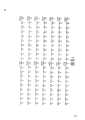
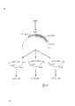
图1完整的CR1编码区的核苷酸和氨基酸顺序。该顺序始于在克隆λT109.1的八聚体EcoR1接头后的第一个核苷酸。该顺序的1531号核苷酸是图3中描绘的顺序中的1号核苷酸的第1个5’核苷酸。推断的氨基酸顺序,显示在与mRNA相应的链的下面。推断的由28-147号核苷酸编码的推断的信号顺序在括号内。Figure 1 Nucleotide and amino acid sequence of the complete CR1 coding region. This sequence starts at the first nucleotide after the octameric EcoR1 linker of clone λT109.1. Nucleotide number 1531 of this sequence is the first 5' nucleotide of
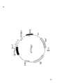
图2.人体CR1 cDNA的5.5kb的限制酶图谱。黑线表示cDNA,限制酶位点是H.Hind Ⅲ;B,BamHⅠ;R,EcoRⅠ;P,PstⅠ;A,ApaⅠ;S,SacⅠ;G,BglⅡ;K,KpnⅠ。获得该顺序的cDNA克隆显示于图的下面。箭头表示采用双脱氧核苷酸链终止方法分析顺序的方向和长度。根据限制酶图谱和重迭顺序同一性而确定cDNA克隆的方向。Figure 2. Restriction enzyme map of the 5.5kb human CR1 cDNA. The black line indicates cDNA, and the restriction enzyme sites are H. Hind III; B, BamHI; R, EcoRI; P, PstI; A, ApaI; S, SacI; G, BglII; K, KpnI. The cDNA clone from which this sequence was obtained is shown below the figure. Arrows indicate the orientation and length of sequences analyzed using the dideoxynucleotide chain termination method. Orientation of cDNA clones was determined based on restriction enzyme maps and overlapping sequence identities.
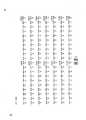
图3,人体CR1 cDNA的5.5kb的核苷酸顺序。图中显示了与mRNA相应的链,碱基1(相应于图1中的碱基1532)是大多数5’克隆中EcoRⅠ接头后的第一个碱基。终止密码子用下划线表示。在核苷酸147和148(箭头)之间发现一段110-bp顺序,用方框表示,我们认为,它表示插入顺序部分。Figure 3, the 5.5 kb nucleotide sequence of human CR1 cDNA. The strand corresponding to the mRNA is shown, with base 1 (corresponding to base 1532 in Figure 1) being the first base after the EcoRI linker in most 5' clones. Stop codons are underlined. A 110-bp sequence was found between
图4,人体CR1 cDNA的5.5kb的核苷酸顺序的点阵分析。如果90bp中至少有40bp匹配,则标绘一个斑点。沿正方形对角线划分的黑线表示与顺序本身相重合。与相重合的线相平行的另外两条黑线1.35和2.7kb代表两者一前一后,引导每一个为1.35kb的长同源重复(LHRs)。在两个LHRs之间的六条较浅的破折号线对应于~2kb的短一致重复。短一致重复(SCRs)沿长同源重复延伸出0.4kb。Fig. 4, dot matrix analysis of the 5.5 kb nucleotide sequence of human CR1 cDNA. A spot was plotted if at least 40 bp out of 90 bp matched. A black line divided along the diagonal of the square indicates coincidence with the order itself. The other two black lines 1.35 and 2.7 kb parallel to the coincident line represent the two in tandem leading to long homologous repeats (LHRs) of 1.35 kb each. The six lighter dashed lines between the two LHRs correspond to ~2 kb short consensus repeats. Short consensus repeats (SCRs) extend 0.4 kb along the long homologous repeats.
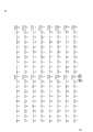
图5.人体CR1的推断的氨基酸顺序。每一个残基用一字母码表示(Lehninger A.L,1975,Biochemistry,第2版,Worth印刷公司,New York,P.72)。将长同源重复中的残基排成一行以表明它们的同源性。LHR-B中的所有残基均被表示了,只有当残基不同于 LHR-B中的时,才给出LHB-C和LHB-D的残基。亲水性排列于蛋白质的COOH末端下以说明假定的转移膜区。紧靠在疏水顺序后的四个带阳性电荷的残基的范围用上划线表示,而与表皮生长因子受体中的蛋白质激酶C磷酸化位点67%同源的六个氨基酸顺序用下划线表示,CR1蛋白质的图谱示于上述的顺序的上面。(TM)跨膜区,(Cyt)胞质区,(3’UT)非翻译顺序。Figure 5. Deduced amino acid sequence of human CR1. Each residue is represented by a one-letter code (Lehninger A.L, 1975, Biochemistry, 2nd edition, Worth Printing Co., New York, p. 72). Residues in long homologous repeats are aligned to indicate their homology. All residues in LHR-B are represented, and the residues of LHB-C and LHB-D are given only when the residues are different from those in LHR-B. A hydrophilic array is placed under the COOH terminus of the protein to account for the putative transmembrane region. The range of four positively charged residues immediately following the hydrophobic sequence is overlined, while the six amino acid sequences that are 67% homologous to the protein kinase C phosphorylation site in the epidermal growth factor receptor are underlined , the map of the CR1 protein is shown above the above sequence. (TM) transmembrane region, (Cyt) cytoplasmic region, (3'UT) untranslated sequence.
图6.(A)CR1的SCRs的排列。重复是从NH末端到COOH末端的1-23号,其中已增加空间以尽可能增大排列。一个残基如果在至少一半SCRs中存在,则被视为保守或保守置换。水平箭头表示一个SCR,它也是从CR1基因组克隆2.38来的顺序且由单一的外显子编码。(B)限制酶图谱,测序方法和基因组克隆入2.38的部分顺序。限制位点是:(B)BamHI,(S)SacI,(E)EcoRV,(K)KpnI,(P)PstI。水平箭头表示测序的方向和长度,垂直的箭头表示外显子-内含子分界线。Figure 6. (A) Arrangement of the SCRs of CR1. The repeats are numbered 1-23 from the NH terminus to the COOH terminus, where space has been added to maximize alignment. A residue is considered conservative or a conservative substitution if it is present in at least half of the SCRs. Horizontal arrows indicate an SCR, which is also a sequence from CR1 genomic clone 2.38 and is encoded by a single exon. (B) Restriction enzyme map, sequencing method and partial sequence of the genome cloned into 2.38. Restriction sites are: (B) BamHI, (S) SacI, (E) EcoRV, (K) KpnI, (P) PstI. Horizontal arrows indicate the direction and length of sequencing, and vertical arrows indicate the exon-intron boundary.
图7,已知具有该结构的蛋白质的SCRs的一致顺序的排列。在其中增加空间以尽可能增大排列。残基被视为保守如图5所示,除了只具有1或2个SCRs的那些蛋白质,其中如果残基在至少一半其它蛋白质中存在,则残基为保守的,破折号相应于非保守的位置。CR2和C2b的下划线部分表示对于这些蛋白质来说,在尚未有过公开发表的该区域的顺序资料。方框表示不变异的半胱氨酸。顺序右边的数字表示用于产生一致顺序的SCRs的数目。用于表示确定一致顺序的顺序资料的蛋白质缩写和参考是:(CR1)补体受体1型,(H)因子H(Kristensen,T.等人,1986,J.Immunol.136:3407),(C46p)C4结合蛋白质(Chung,L.P.等人,1985,Biochem.J.230:133),(CR2)补体受体2型(Weis J.J.等人,1986,Proc Natl.Acad.Sci,U.S.A83:5639),(Ba)因子B的蛋白质水解片段(Morley B.J.和Campbell R.D.1984.EMBOJ.3:153),(C2b)C2的蛋白水解 片段(Gagnon,J.1984,Philos,Trans,R.Soc,Lond.B.Biol.Sci.306:301),(Clr)Cl的r亚基(Leytus S.P.等人,1986,Biochemistry 25:4855),(ⅩⅢb)因子ⅩⅢ的b亚基(Ichinose A,等人,1986,Bio-chemistry 25:4633),(β2GP1)β2糖蛋白Ⅰ(Lozier J.等人,1984.Proc.Natl.Acad.Sci.U.S.A.81:3640),(Hap)结合珠蛋白(Kurosky,A.等人,1980,Proc.Natl.Acad,Sci,U.S.A.77:3388),(IL-2-R)白细胞介素(interleukin)-2受体(LeonardW,J.等人,1985,Science 230:633)。星号表示不完全顺序是可以得到的。Figure 7. Alignment of the consensus order of the SCRs of proteins known to have this structure. Add space in it to make the arrangement as large as possible. Residues considered conserved are shown in Figure 5, except for those proteins with only 1 or 2 SCRs, where a residue is conserved if it is present in at least half of the other proteins, with dashes corresponding to non-conserved positions . The underlined portion of CR2 and C2b indicates that there is no published sequence information for this region for these proteins. Boxes indicate cysteines that do not vary. The number to the right of the sequence indicates the number of SCRs used to generate the consistent sequence. The protein abbreviations and references used to denote the sequence data for determining the consensus sequence are: (CR1)
图8,人体CR1的推断的结构图。COOH末端胞质区是位于类脂双层膜的右边。30SCRs被线性地排列在质膜的细胞外一边,括号表示LHRs。插入图是放大的单个SCR,说明三环结构。Figure 8. Deduced structure diagram of human CR1. The COOH terminal cytoplasmic domain is located on the right side of the lipid bilayer membrane. 30SCRs are arranged linearly on the extracellular side of the plasma membrane, and brackets indicate LHRs. Inset is a magnified view of a single SCR, illustrating the tricyclic structure.
图9.编码人体CR1的质粒pBSABCD插入的限制酶图谱。在表示含有编码顺序的区的方框中显示有来自八个cDNA克隆的九个片段,它们被连接后形成CR1结构。括号分别指出了LHR-A,-B,-C,和-D的位置。方框下面的线表示新分离的5’cDNA克隆的位置。限制酶位点是:A,ApaⅠ,B,BamHⅠ;G,BglⅡ,H,HindⅢ;K,Kp-nⅠ;M,BspMⅡ;P,PstⅠ;R,EcoRⅠ;和S,SacⅠ。Figure 9. Restriction enzyme map of the insert of plasmid pBSABCD encoding human CR1. Nine fragments from eight cDNA clones, which were ligated to form the CR1 construct, are shown in the box indicating the region containing the coding sequence. Brackets indicate the positions of LHR-A, -B, -C, and -D, respectively. The line under the box indicates the position of the newly isolated 5' cDNA clone. The restriction enzyme sites are: A, ApaI, B, BamHI; G, BglII, H, HindIII; K, Kp-nI; M, BspMII; P, PstI; R, EcoRI;
图10,编码LHR-A的七个SCRs的5’cDNA克隆的推断的氨基酸顺序,以及LHR-B,-C和-D相应的SCRs的这个顺序的排列。在每一个SCR中保守的四个胱氨酸用下划线表示。只有当残基与LHR-A中的不同时,LHR-B,-C和-D的残基才表示出来。Figure 10. The deduced amino acid sequence of the 5' cDNA clone encoding the seven SCRs of LHR-A, and the alignment of this sequence for the corresponding SCRs of LHR-B, -C and -D. The four cystines conserved in each SCR are underlined. Residues for LHR-B, -C and -D are indicated only if the residues differ from those in LHR-A.
图11表达质粒piABCD和pMTABCD的限制酶图谱。Pm MT和Pcmv分别代表鼠类金属硫因(metallothionein)和细胞肥大病毒最近的早期启动子。Figure 11 Restriction enzyme maps of expression plasmids piABCD and pMTABCD. Pm MT and Pcmv represent the closest early promoters of murine metallothionein and cytomegalovirus, respectively.
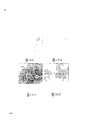
图12,分别用piABCD(a和b组)和CDM8载体(c和d组)单独转染COS细胞,然后用YZ1单克隆抗-CR1抗体和荧光素标记的山羊抗鼠F(ab’)2间接染色的相差显微镜(a和c组)和免疫荧光显微镜 (b和d组)的分析。Figure 12, COS cells were transfected with piABCD (groups a and b) and CDM8 vectors (groups c and d), respectively, and then YZ1 monoclonal anti-CR1 antibody and fluorescein-labeled goat anti-mouse F(ab')2 Analysis of indirect staining by phase-contrast microscopy (panels a and c) and immunofluorescence microscopy (panels b and d).
图13,采用表达重组CR1的COS细胞的结合C3b和C4b的分析。用piABCD(a和c组)或CDM8载体(b和d组)单独转染的COS细胞与EAC4b(lim),3b(a和b组)或EAC4b(c和d组)一起孵育,通过相差显微镜检查玫瑰花结的形成。Figure 13. Analysis of binding C3b and C4b using COS cells expressing recombinant CR1. COS cells transfected with piABCD (groups a and c) or CDM8 vectors (groups b and d) alone were incubated with EAC4b (lim), 3b (groups a and b) or EAC4b (groups c and d) by phase contrast microscopy Check for rosette formation.
图14,由转染的COS细胞表达的重组CR1的SDS-PAGE分析。分别用CDM8载体单独转染(通道1和4)和piABCD转染(通道2和5)的COS细胞,和来自于具有CR1的F和S异型的个体的红细胞(通道3和6)的表面用125Ⅰ标记。用去垢剂处理的细胞溶解产物,用琼脂糖UPC10(通道1-3)和琼脂糖-YZ1(通道4-6)连续地进行免疫吸附,在非还原条件下用SDS-PAGE和放射自显影分析洗脱物。Figure 14. SDS-PAGE analysis of recombinant CR1 expressed by transfected COS cells. COS cells transfected with CDM8 vector alone (
图15,在免疫固相化重组CR1的存在下,用因子Ⅰ裂解125Ⅰ-C3(ma)。在因子H(通道1),与CDM8载体单独(通道)转染的COS细胞的溶解产物预孵育的琼脂糖-UPC10(通道2),与PiABCD转染的COS细胞的溶解产物预孵育的琼脂糖-UPC10(通道3),用CDM8转染的COS细胞的溶解产物预孵育的琼脂糖-YZ1(通道4),和与piABCD转染的COS细胞的溶解产物预孵育的6μl(通道5),12μl(通道6)和25μl(通道7)的琼脂糖-YZ1存在下,用因子Ⅰ处理125Ⅰ-C3(ma)的重复样品。还在无因子Ⅰ的条件下,用25μl已经与piABCD转染的COS细胞(通道8)的溶解产物预孵育过的琼脂糖-YZ1处理125Ⅰ-标记的C3(ma)的样品。还原后,采用SDS-PAGE和放射自显影分析125Ⅰ-C3(ma)。Figure 15. Cleavage of125I -C3(ma) with Factor I in the presence of immunoimmobilized recombinant CR1. In Factor H (lane 1), agarose-UPC10 (lane 2) pre-incubated with lysates of COS cells transfected with CDM8 vector alone (lane), agarose pre-incubated with lysates of PiABCD-transfected COS cells - UPC10 (lane 3), agarose-YZ1 pre-incubated with lysate of CDM8-transfected COS cells (lane 4), and 6 μl pre-incubated with lysate of piABCD-transfected COS cells (lane 5), 12 μl Duplicate samples of125 I-C3 (ma) were treated with Factor I in the presence of (lane 6) and 25 µl (lane 7) of agarose-YZ1. Also in the absence of Factor I, samples of125 I-labeled C3(ma) were treated with 25 µl of agarose-YZ1 which had been preincubated with lysates of piABCD-transfected COS cells (lane 8). After reduction,125 I-C3 (ma) was analyzed by SDS-PAGE and autoradiography.
图16,编码CR1缺失型突变体的cDNA结构。在全长piABCD结构(其上显示了用于制备缺失型突变体的限制酶位点)上面,用括号表示编码四个LHRs的cDNA片段的位置。在每一个突变体中所保留的cDNA限制片段均由实线表示。限制酶位点是:A.ApaⅠ;B. BamⅠ;E,BstEⅡ和P.PstⅠ。Fig. 16, cDNA structure encoding CR1 deletion mutant. Above the full-length piABCD construct (on which restriction enzyme sites for making deletion mutants are shown), the positions of the cDNA fragments encoding the four LHRs are indicated in brackets. The cDNA restriction fragments retained in each mutant are indicated by solid lines. The restriction enzyme sites are: A. ApaI; B. BamI; E, BstEII and P.PstI.
图17,CR1重组缺失型突变株与野生型CR1的F和S异型的比较。分别用琼脂糖-UPC10抗果聚糖抗体(通道1-6),琼脂糖-YZ-1抗CR1单克隆抗体(通道7-11)和兔抗CR1抗体和琼脂糖-蛋白质A(通道12)免疫沉淀125Ⅰ表面标记红细胞(通道1和7)及分别用CDM8载体单独(通道2和8),piABCD(通道3和9),piBCD(通道4和10),piCD(通道5和11)和piD(通道6和12)转染的COS细胞去垢剂处理的溶解物。在还原条件下,将洗脱物进行SDS-PAGE和放射自显影分析。Figure 17. Comparison of F and S isotypes of CR1 recombination deletion mutants and wild-type CR1. Separately with Sepharose-UPC10 anti-fructan antibody (lanes 1-6), Sepharose-YZ-1 anti-CR1 monoclonal antibody (lanes 7-11) and rabbit anti-CR1 antibody and Sepharose-Protein A (lanes 12) Immunoprecipitation of125I surface-labeled red blood cells (
图18,在表达全长和CR1缺失型突变体的COS细胞的存在下,用因子Ⅰ裂解125Ⅰ-C3(ma)。分别用CDM8载体单独(通道1和7),piABCD(通道2和8),piAD(通道3和9),piBD(通道4和10),piCD(通道5和1),和piD(通道6和12)转染的COS细胞,在因子Ⅰ缺乏(通道1-6)或存在(通道7-12)的情况下,与125Ⅰ-C3(ma)的重复样品一起孵育。并分别用因子H和因子Ⅰ(通道13)和单独用因子Ⅰ(通道14)与125Ⅰ-C3(ma)样品一起孵育。还原后,用SDS-PAGE和放射自显影分析125Ⅰ-C3(ma)。Figure 18. Cleavage of125I -C3(ma) with Factor I in the presence of COS cells expressing full-length and CR1 deletion mutants. with CDM8 vector alone (
图19,描述包括CR1的每一个LHR的SCRs的类型,和决定C3b和D4b受体的专一性的预计位点的图解模型。它们的次级结合专一性用圆括号表示。Figure 19. A schematic model depicting the types of SCRs for each LHR including CR1, and the predicted sites that determine the specificity of C3b and D4b receptors. Their secondary binding specificities are indicated in parentheses.
图20,保留在可溶性CR1 DNA结构中的DNA区域的示意图。全长CR1 cDNA的区域由图上面的方框表示。Figure 20. Schematic representation of DNA regions retained in soluble CR1 DNA structures. The region of the full-length CR1 cDNA is indicated by the box above the figure.
图21,pTCS系列表达载体中主要成份的示意图。Figure 21, a schematic diagram of the main components of the pTCS series expression vectors.
图22,表达载体pTCSgpt的图。多聚腺苷化位点来自于鼠Ig卡巴粒顺序(NBRF Nucleic database accession # Kams,bp1306-1714),Ad2MLP和三分区来自于Ad2顺序(NBRFNucleic database accession # Gdad2,bp 5791-6069);SV40早1期启动子来自于 SV40基因组(NBRF Nucleic Database accession # GSV40W)。gpt基因,氨苄青霉素基因和细菌复制起点来自于载体pSV2gpt(ATCC Accession No.37145)。Figure 22. Diagram of the expression vector pTCSgpt. The polyadenylation site was derived from the mouse Ig kappa granule sequence (NBRF Nucleic database accession # Kams, bp1306-1714), Ad2MLP and tripartite from the Ad2 sequence (NBRF Nucleic database accession # Gdad2, bp 5791-6069); SV40 early The
图23.抗体亲和纯化的sCR1的4-20% SDS-PAGE。非还原(通道1,2,3)和还原(通道4,5,6)的条件下。通道1,3:分子量标记,通道3,5:细胞培养上清原材料;通道4,6:通过抗体亲和层析法纯化的sCR1。Figure 23. 4-20% SDS-PAGE of antibody affinity purified sCR1. Non-reducing (
图24.阳离子交换HPLC洗脱图。洗脱蛋白在280nm下测定吸收值(y轴)来监测洗脱的蛋白质。流过的(0-100分钟)和洗脱的sCR1(150-165分钟)的吸收部分均被切下。X轴表示洗脱时间(以分表示)。Figure 24. Cation exchange HPLC elution profile. Eluted protein Absorbance (y-axis) was measured at 280 nm to monitor eluted protein. Absorbed fractions of both flow-through (0-100 min) and eluted sCR1 (150-165 min) were excised. The X-axis represents the elution time (expressed in minutes).
图25.阳离子和阴离子交换HPLC纯化的sCR1的4-20%梯度SDS-PAGE。SDS-聚丙烯酰胺凝胶在非还原条件下走电泳。通道1,一部分生物反应物的上清液;通道2,经阳离子HPLC初始缓冲液透析的一部分生物反应物的上清液;通道3,从阳离子交换HPLC柱上洗脱的sCR1峰的一部分;通道4,从阳离子交换HPLC层析柱得到的再经阴离子HPLC的初始缓冲液透析的一部分sCR1峰;通道5和6,从阴离子HPLC上洗脱的sCR1的两个不同分部的一部分。Figure 25. 4-20% gradient SDS-PAGE of cation and anion exchange HPLC purified sCRl. SDS-polyacrylamide gel electrophoresis under non-reducing conditions.
图26.在人体嗜中性粒细胞中C5a所引起氧突增。随着C5a引起氧突增后,DCFDA被氧化并发出很亮的荧光。用流动血球计数法测定荧光强度,荧光强度对应于X轴,细胞数对应于y轴。a组:细胞的图和入口(gate);b组:加入C5a后0分钟;c组;1分钟;d组:2分钟;e组:3分钟;f组:4分钟;g组;20分钟。该DCFDA分析显示C5a很敏感。Figure 26. C5a-induced oxygen burst in human neutrophils. Following the oxygen burst induced by C5a, DCFDA is oxidized and emits bright fluorescence. Fluorescence intensity was measured by flow cytometry, with fluorescence intensity corresponding to the x-axis and cell number to the y-axis. Group a: cell graph and gate; group b: 0 minutes after adding C5a; group c: 1 minute; group d: 2 minutes; group e: 3 minutes; group f: 4 minutes; group g: 20 minutes . This DCFDA analysis showed that C5a is sensitive.
图27,在sCR1存在下,人体补体的激活显示还原的C5a活性(DCFDA分析)。a组,未刺激的细胞,b组,显示出高度荧光的无sCR1的对照物1;c组,在sCR1存在下DCFDA分析显示出荧光强度降低75%。y轴是细胞数,x轴是荧光强度。Figure 27. Activation of human complement in the presence of sCR1 shows reduced C5a activity (DCFDA analysis). Panel a, unstimulated cells, panel b,
图28,通过sCR1抑制在人血清中通过经典途径产生C5a和C3a。用抗体亲和纯化或HPLC纯化的sCR1观察到相似的图。Figure 28. Production of C5a and C3a by the classical pathway in human serum by sCRl inhibition. A similar profile was observed with antibody affinity purified or HPLC purified sCR1.
图29,通过重组sCR1抑制补体介导的溶血作用。用亲和纯化或HPLC纯化的sCR1观察到相似的图。Figure 29. Inhibition of complement-mediated hemolysis by recombinant sCR1. A similar profile was observed with affinity purified or HPLC purified sCR1.
图30,在sCR1处理(左)和未处理(右)过的鼠中RPAR的粗形态。(a)两个大鼠均接受卵白蛋白静脉注射,然后用sCR1(左边的鼠)或PBS(右边的鼠)与抗卵白蛋白,纯的(左边的位置);抗卵白蛋白,1/2稀释(中间位置)或兔IgG(右边位置)的混合物进行皮下注射。注射进行二次;顶端和底部呈现相同的结果。接受sCR1的鼠几乎无肉眼可见的变化,而未处理的鼠则显示了RPAR的全部症状。(b)由(a)得到的皮肤活体组织的皮肤表面。从未处理的鼠(右)中得到的活体组织明显地显示出肉眼可见的损害,而从sCR1处理过的鼠(左)中得到的活体组织则显示了正常的形态。Figure 30. Crude morphology of RPAR in sCR1-treated (left) and untreated (right) mice. (a) Both rats received IV injections of ovalbumin, followed by sCR1 (left mouse) or PBS (right mouse) with anti-ovalbumin, pure (left position); anti-ovalbumin, 1/2 dilution (middle position) or a mixture of rabbit IgG (right position) were injected subcutaneously. Injections were performed twice; top and bottom presented the same results. Mice receiving sCR1 showed few macroscopic changes, whereas untreated mice showed full symptoms of RPAR. (b) Skin surface of the skin biopsy obtained from (a). Biopsies from untreated mice (right) clearly showed macroscopic lesions, whereas biopsies from sCR1-treated mice (left) showed normal morphology.
图31.从sCR1处理过的(a)和未处理的(b)鼠中得到的皮肤活体组织的光学显微镜检查。(a)观察到血管周围聚集的多形核细胞和单核细胞,但是,未看到嗜中性粒细胞的大量渗入和红细胞的外渗。(b)可见到多形核细胞的大量渗入和红细胞的外渗。Figure 31. Light microscopy of skin biopsies obtained from sCR1-treated (a) and untreated (b) mice. (a) Perivascular accumulation of polymorphonuclear cells and monocytes was observed, however, massive infiltration of neutrophils and extravasation of red blood cells were not seen. (b) Massive infiltration of polymorphonuclear cells and extravasation of red blood cells is seen.
图32鼠和猴的血液中注入的sCR1的廓清率,表现出双相,α和β廓清率相。Figure 32. Blood clearance of infused sCR1 in rats and monkeys showing biphasic, alpha and beta clearance phases.
图33CR1 cDNA和内含子探针与表达F或F′异型的个体的DNA的EcoRV酶切物杂交的Southern Blots放射自显影图。λDNA的HindⅢ片段的位置在左面以千碱基表示,F′特异性片段的位置用单箭头表示。Figure 33 Southern Blots autoradiography of CR1 cDNA and intron probes hybridized with EcoRV digests of DNA from individuals expressing F or F' allotypes. The position of the HindIII fragment of lambda DNA is indicated in kilobases on the left, and the position of the F'-specific fragment is indicated by a single arrow.
图34CR1的F等位基因的EcoRⅤ限制图。空白框代表外显子的位置,点画框代表内含子探针杂交位置。LHR-B和-C上的方括号表示两个可能的缺失区域。Ⅴ代表EcoRⅤ位点。Figure 34 EcoRV restriction map of the F allele of CR1. Blank boxes represent the positions of exons, and dotted boxes represent hybridization positions of intron probes. Square brackets on LHR-B and -C indicate two possible deleted regions. Ⅴ represents the EcoRⅤ site.
图35重组rCR1不同型的cDNA插入子。所表示的限制性位点 为:A,ApaLⅠ;B,BamHⅠ;C,SacⅠ;H,HindⅢ;L,BglⅠ,PstⅠ;R,EcoRⅠ;及S,SmaⅠ。在上面的简图表示CR1蛋白,有同一顺序的SCR以同样的方式插入。Figure 35 cDNA inserts of recombinant rCR1 variants. Restriction sites indicated are: A, ApaLI; B, BamHI; C, SacI; H, HindIII; L, BglI, PstI; R, EcoRI; and S, SmaI. In the diagram above the CR1 protein is shown, with the same sequence of SCRs inserted in the same way.
图36通过在YZ-1-琼脂糖上吸收作用纯化重组sCR1在非还原条件下的Coomassie蓝染色的SDS-PAGE。每栏含10微克从COS细胞的培养上清液中纯化得的重组sCR1,这些细胞分别由pasecABB-CO(栏1),pasecABCD(栏2)或pasecACD(栏3)转染。Mr标志的位置在右边用KD表示。Figure 36 Coomassie blue stained SDS-PAGE of purification of recombinant sCR1 by absorption on YZ-1-agarose under non-reducing conditions. Each column contained 10 µg of recombinant sCR1 purified from the culture supernatant of COS cells transfected with pasecABB-CO (column 1), pasecABCD (column 2) or pasecACD (column 3). The position of the Mr logo is indicated by KD on the right.
图37重组sCR1的辅助因子活性。在从由pasecABBCD,pase-cABCD,或pasecACD转染的COS细胞得到的重组sCR1增加量的存在下,测定C3b的α’链的切割。Figure 37 Cofactor activity of recombinant sCR1. Cleavage of the α' chain of C3b was assayed in the presence of increasing amounts of recombinant sCR1 obtained from COS cells transfected with pasecABBCD, pase-cABCD, or pasecACD.
图38通过重组sCR1抑制红细胞摄取125Ⅰ-C3b二聚体。在从由pasecABBCD,pasecABCD,或pasecACD转染的COS细胞得到的C3b二聚体,C3b单体和重组sCR1的增加浓度下测定红细胞结合配位体。Figure 38 Inhibition of125I -C3b dimer uptake by erythrocytes by recombinant sCR1. Erythrocyte-bound ligands were assayed at increasing concentrations of C3b dimer, C3b monomer, and recombinant sCR1 obtained from COS cells transfected with pasecABBCD, pasecABCD, or pasecACD.
图39通过由编码CR1变异体的不同质粒转染的COS细胞纯化而来的重组sCR1抑制旁路(A)或经典(B)C3转化酶。Figure 39 Inhibition of alternative (A) or classical (B) C3 convertase by recombinant sCR1 purified from COS cells transfected with different plasmids encoding CR1 variants.
图40通过由编码CR1变异体的不同质粒转染的COS细胞纯化而来的重组sCR1抑制旁路(A)或经典(B)C5转化酶。Figure 40. Inhibition of alternative (A) or classical (B) C5 convertase by recombinant sCR1 purified from COS cells transfected with different plasmids encoding CR1 variants.
5.发明的详细描述5. Detailed description of the invention
本发明涉及C3b/C4b受体(CR1)基因和它编码的蛋白质。本发明还涉及CR1核酸顺序及其含有70个核苷酸的片段和它们编码的包含24个氨基酸的肽或蛋白质。此外,本发明还提供了CR1蛋白质及其片段的表达。这种CR1顺序和蛋白质在炎症或免疫系统疾病,和涉及补体活性疾病的诊断和治疗中是有价值的。The present invention relates to the C3b/C4b receptor (CR1) gene and its encoded protein. The invention also relates to CR1 nucleic acid sequences and fragments thereof comprising 70 nucleotides and their encoded peptides or proteins comprising 24 amino acids. In addition, the present invention also provides the expression of CR1 protein and its fragments. Such CR1 sequences and proteins are of value in the diagnosis and treatment of diseases of the inflammatory or immune system, and diseases involving complement activity.
在一个特例中,本发明涉及可溶性CR1分子和它们的表达,纯化和应用。在这里所采用的术语“可溶性CR1分子”意指与天然的CR1蛋白质相反,部分CR1蛋白质不能以膜蛋白质的形式在细胞表 面表达。尤其是,大量缺少跨膜区的CR1分子是可溶性CR1分子。在一个较佳实例中,可溶性CR1分子被表达它们的细胞所分泌。In a particular embodiment, the invention relates to soluble CR1 molecules and their expression, purification and use. The term "soluble CR1 molecule" as used herein means that, contrary to the native CR1 protein, a part of the CR1 protein cannot be expressed as a membrane protein on the cell surface. In particular, a large number of CR1 molecules lacking the transmembrane region are soluble CR1 molecules. In a preferred embodiment, soluble CR1 molecules are secreted by cells expressing them.
在下面将要详细描述的本发明的特例中,描述了全长CR1 cD-NA以及其片段的克隆和全部核苷酸顺序和推断的氨基酸顺序,和编码CR1产物的表达。具有C3b和/或C4b的结合位点,且抑制因子Ⅰ辅助因子活性的CR1以及其片段的表达也将描述。本发明通过可溶的,平截的CR1分子的制备和纯化而进一步加以描述。在一个特例中,这类分子被证明对于减轻炎症,减小心肌梗塞范围和防止肿块损伤的治疗是有用的。In a specific embodiment of the invention described in detail below, the cloning and full nucleotide and deduced amino acid sequences of the full-length CR1 cD-NA and fragments thereof, and expression of the encoded CR1 product are described. Expression of CR1 and fragments thereof that have binding sites for C3b and/or C4b and that inhibit Factor I cofactor activity will also be described. The invention is further described by the preparation and purification of soluble, truncated CR1 molecules. In one particular case, such molecules proved to be useful for treatments that reduce inflammation, reduce the size of myocardial infarcts, and prevent mass damage.
5.1CR1基因的分离5.1 Isolation of CR1 gene
CR1基因的完整编码顺序及其推断的氨基酸顺序列于图1。The complete coding sequence of the CR1 gene and its deduced amino acid sequence are listed in Fig. 1 .
任何人体细胞都有可能作为CR1基因的分子克隆的核酸源。CR1基因的分离包括编码显示CR1相关结构或性能(例如,结合C3b或C4b或免疫复合体,调节吞噬作用,免疫激活作用或增殖,以及补体的调节)的蛋白质的那些DNA顺序的分离。DNA可以通过该领域已知的标准方法从克隆的DNA(如,DNA“库”)中获得,可采用化学合成,cDNA克隆,或基因组DNA克隆或它的片段的克隆;和从所需要的人体细胞中提纯(如,可参考Maniatis等人,1982,Molecular Cloning,实验室手册,Gold Spring Harbor实验室,Cold Spring Harbor,New York;Glover D.M.1985,DNA克隆:Apractical Approach,MRL Press有限公司,Oxford,U.K.Vol.Ⅰ,Ⅱ.)。可作为CR1基因cDNA克隆的核酸源的细胞包括(但不局限于此)单核细胞/巨噬细胞,粒细胞,B细胞,T细胞,脾卵泡树状突细胞和肾小球足状突细胞。来自于基因组DNA的克隆除编码区外还可能包含调节和内含子DNA区;来自于cDNA的克隆则仅包含外显子顺序。不管来源如何,CR1基因均应分子克隆进入用于基因增殖的合适的载体中。Any human cell may be used as a nucleic acid source for molecular cloning of the CR1 gene. Isolation of the CR1 gene includes isolation of those DNA sequences encoding proteins exhibiting CR1-related structures or properties (eg, binding to C3b or C4b or immune complexes, regulation of phagocytosis, immune activation or proliferation, and regulation of complement). DNA can be obtained from cloned DNA (e.g., a DNA "library") by standard methods known in the art, by chemical synthesis, cDNA cloning, or genomic DNA cloning or cloning of fragments thereof; and from desired human Purification from cells (see, for example, Maniatis et al., 1982, Molecular Cloning, Laboratory Manual, Gold Spring Harbor Laboratories, Cold Spring Harbor, New York; Glover D.M. 1985, DNA Cloning: A Practical Approach, MRL Press Ltd, Oxford , U.K. Vol.Ⅰ,Ⅱ.). Cells that can serve as nucleic acid sources for CR1 gene cDNA clones include, but are not limited to, monocytes/macrophages, granulocytes, B cells, T cells, splenic follicular dendritic cells, and glomerular podocytes . Clones derived from genomic DNA may contain regulatory and intronic DNA regions in addition to coding regions; clones derived from cDNA contain only exonic sequences. Regardless of the source, the CR1 gene should be molecularly cloned into a suitable vector for gene propagation.
在对来自于基因组DNA的基因进行分子克隆时,产生DNA片段,它们中的一些将编码所需要的CR1基因。采用不同的限制酶,DNA可在特定的位点切断。或者,在锰存在下,我们可使用DNA酶分解DNA,或DNA可通过诸如声处理方法而被物理地剪切。然后可采用包括(但不局限于此)琼脂糖和聚丙烯酰胺凝胶电泳和柱层析在内的常规方法根据大小将线状的DNA片段分离。During molecular cloning of genes from genomic DNA, DNA fragments are generated, some of which will encode the desired CR1 gene. DNA can be cut at specific sites using different restriction enzymes. Alternatively, in the presence of manganese, we can use DNase to break down the DNA, or the DNA can be physically sheared by methods such as sonication. The linear DNA fragments can then be separated by size using conventional methods including, but not limited to, agarose and polyacrylamide gel electrophoresis and column chromatography.
一旦产生了DNA片段,则可用许多方法来鉴定包含CR1基因的特定DNA片段。例如,如果可以得到CR1基因或它的特定RNA,或它们的片段,并可被纯化和标记,则所产生的DNA片段就可通过与标记探针的核酸杂交进行筛选(Benton W和Davis R,1977,Sci-ence 196:180;Grunstein M和Hogness D,1975,Proc.Natl.Acad,Sci,U.S.A.72:3961)。与探针大量同源的那些DNA片段将能杂交。如果纯化的CR1专一探针得不到,则富含CR1的核酸部分可以作为探针而用于初步选择方法。作为一个实例来说,可以使用已去除了成纤维细胞表达的信息的表现B细胞cDNA的探针。也可通过限制酶酶切并根据已知的限制酶图谱(如果它是可以得到的)与预期的片段进行片段大小比较来鉴定合适的片段。在初步选择后,根据基因的性能,或它的表达产物的物理,化学或免疫性能(如后面要描述的),可以进行进一步的选择。Once the DNA fragments have been generated, a number of methods can be used to identify the specific DNA fragments comprising the CR1 gene. For example, if the CR1 gene or its specific RNA, or fragments thereof, are available and can be purified and labeled, the resulting DNA fragments can be screened by nucleic acid hybridization with labeled probes (Benton W and Davis R, 1977, Science 196: 180; Grunstein M and Hogness D, 1975, Proc. Natl. Acad, Sci, U.S.A. 72: 3961). Those DNA fragments with substantial homology to the probe will hybridize. If purified CR1-specific probes are not available, CR1-enriched nucleic acid fractions can be used as probes for primary selection methods. As an example, probes expressing B cell cDNA from which fibroblast expression information has been removed can be used. Suitable fragments can also be identified by restriction enzyme digestion and size comparison with the expected fragments based on a known restriction map (if it is available). After the initial selection, further selection may be performed on the basis of the properties of the gene, or the physical, chemical or immunological properties of its expression product (as will be described later).
CR1基因也可以通过核酸杂交然后体外翻译进行mRNA选择而被鉴定。在这个过程中,片段被用于通过杂交来分离互补的mR-NAs。这些DNA顺序可代表可以得到的,纯化的CR1DNA,或已被富集CR1顺序的DNA。The CR1 gene can also be identified by nucleic acid hybridization followed by in vitro translation for mRNA selection. In this process, fragments are used to isolate complementary mRNAs by hybridization. These DNA sequences may represent available, purified CR1 DNA, or DNA that has been enriched for CR1 sequences.
分离的mRNAs的体外翻译产物的免疫沉淀分析或功能鉴定(如,C3b或C4b的结合,或吞噬作用或免疫激活的促进,或补体调节等)识别了mRNA和含有CR1顺序的互补DNA片段。此外,通过从细胞中分离出来的多核糖体对专一地抗CR1的固相抗体的吸附, 可以选择出特定的mRNAs。用选出的mRNA(来自于被吸附的多核糖体)作为模板可以合成放射标记的CR1cDNA。然后可用放射标记的mRNA或cDNA作为探针将CR1DNA片段从基因组其它DNA片段中识别出来。Immunoprecipitation analysis or functional characterization of in vitro translation products of isolated mRNAs (e.g., binding of C3b or C4b, or promotion of phagocytosis or immune activation, or complement regulation, etc.) identified mRNAs and complementary DNA fragments containing CR1 sequences. In addition, specific mRNAs can be selected by the adsorption of polysomes isolated from cells to a solid-phase antibody specific for CR1. Radiolabeled CR1 cDNA can be synthesized using selected mRNA (from adsorbed polysomes) as a template. Radiolabeled mRNA or cDNA can then be used as a probe to discriminate the CR1 DNA fragment from other DNA fragments in the genome.
分离CR1基因组DNA的其它方法包括(但不局限于此)从已知的顺序中化学地合成基因顺序本身或制备编码CR1基因的mR-NA的cDNA。例如,如上面所描述的,用于CR1基因的cDNA克隆的RNA可以从包括(但不限于)单核细胞/巨噬细胞,粒细胞,B细胞,T细胞,树状突细胞和足状突细胞在内的细胞中分离出来。在一个较佳具体实施例中,扁桃体(tonsilar)细胞可以作为用于cDNA克隆的mRNA的来源(见后面)。在本发明范围内的其它方法也是可行的。Other methods of isolating CR1 genomic DNA include, but are not limited to, chemically synthesizing the gene sequence itself from a known sequence or preparing cDNA encoding the mRNA-NA of the CR1 gene. For example, as described above, RNA for cDNA cloning of the CR1 gene can be obtained from including (but not limited to) monocytes/macrophages, granulocytes, B cells, T cells, dendritic cells, and podocytes. Cells are isolated from cells within. In a preferred embodiment, tonsilar cells can be used as the source of mRNA for cDNA cloning (see below). Other methods are also possible within the scope of the present invention.
然后,可将经确定和分离的基因插入合适的克隆载体中。该领域已知的很多载体宿主系统均可采用。可能的载体包括(但不局限于)质粒或经修饰的病毒,但是载体系统必须与所采用的宿主细胞相容。这类载体包括(但不局限于)噬菌体,如λ衍生物;或质粒,如pBR322或pUC质粒或CDM8质粒(Seed B.1987,Nature 329:840-842)或衍生物。重组子可以通过转化,转染,感染,电刺激(elec-troporation)等而导入宿主细胞。The identified and isolated gene can then be inserted into a suitable cloning vector. Many vector host systems known in the art can be used. Possible vectors include, but are not limited to, plasmids or modified viruses, but the vector system must be compatible with the host cell employed. Such vectors include (but are not limited to) phages, such as lambda derivatives; or plasmids, such as pBR322 or pUC plasmids or CDM8 plasmids (Seed B. 1987, Nature 329: 840-842) or derivatives. Recombinants can be introduced into host cells by transformation, transfection, infection, electrical stimulation (elec-troporation) and the like.
在另一个方法中,在以“鸟枪”法插入合适的克隆载体后,可以确定和分离CR1基因。在插入克隆载体前,可通过诸如大小分部分离的方法而富集CR1基因。In another approach, the CR1 gene can be identified and isolated following "shotgun" insertion into an appropriate cloning vector. The CR1 gene can be enriched by methods such as size fractionation prior to insertion into the cloning vector.
CR1基因被插入可以用于转化、转染或感染合适的宿主细胞的克隆载体中,这样,可产生很多基因顺序的拷贝。在一个特例中,克隆载体可以是CDM8载体,它可用于在哺乳动物宿主细胞中获得表达。插入至克隆载体内可以通过将DNA片段连接到具有互补的粘性末端的克隆载体中而完成。但是,如果在克隆载体中不存在可用于 切割DNA的互补限制酶位点,则DNA分子的末端可进行酶修饰。换句话说,任何所需要的位点均可通过将核苷酸顺序(接头)连接到DNA末端而产生;这些连接接头可以包含能编码限制性核酸内切酶识别顺序的特定的化学合成的寡核苷酸。在另一个方法中,切割的载体和CR1基因可以用同聚加“尾”加以修饰。The CR1 gene is inserted into a cloning vector that can be used to transform, transfect, or infect a suitable host cell such that many copies of the gene sequence are produced. In a specific example, the cloning vector can be a CDM8 vector, which can be used to obtain expression in mammalian host cells. Insertion into a cloning vector can be accomplished by ligating the DNA fragments into the cloning vector with complementary cohesive ends. However, if there are no complementary restriction enzyme sites available to cleave the DNA in the cloning vector, the ends of the DNA molecule can be enzymatically modified. In other words, any desired site can be created by ligating nucleotide sequences (linkers) to the ends of the DNA; these ligation linkers can contain specific chemically synthesized oligos that encode recognition sequences for restriction endonucleases. Nucleotides. In another approach, the cleaved vector and CR1 gene can be modified with homopolymerization and "tailing".
根据DNA本身的性能,或者,根据它所编码的蛋白质的物理免疫学的或功能的性能,可以用许多不同的方法来进行克隆的CR1基因的鉴定。例如,DNA本身可以通过将噬菌斑或菌落与标记的探针进行核酸杂交而检测(Benton W和Davis R.1977,Science 196:180;Grunstein M和Hongness D,1975,Proc.Natl.Acad,Sci,U.S.A.72:3961)。或者,CR1基因的存在可以通过根据它的表达产物的性能的测定而检测。例如,可以将能产生这样的蛋白质,如与已知的CR1具有相近或相同的电泳迁移,等电聚焦现象,蛋白质水解消化图谱,C3b和/或C4b和/或免疫复合体结合活性,补体调节活性,对吞噬作用或免疫制激的影响,或抗原性的cDNA克隆或能杂交选择合适的mRNAs的DNA克隆选出来。采用一种CR1抗体,用ELISA(酶连免疫吸附分析法)的方法,CR1蛋白质就可通过将标记的抗体与推测的合成CR1的克隆相结合而得以鉴定。Identification of the cloned CR1 gene can be performed in a number of different ways, based on the properties of the DNA itself, or, alternatively, the physioimmunological or functional properties of the protein it encodes. For example, the DNA itself can be detected by nucleic acid hybridization of plaques or colonies to labeled probes (Benton W and Davis R.1977, Science 196:180; Grunstein M and Hongness D, 1975, Proc. Natl. Acad, Sci, U.S.A. 72:3961). Alternatively, the presence of the CR1 gene can be detected by assays based on the properties of its expression product. For example, proteins that have similar or identical electrophoretic shifts to known CR1, isoelectric focusing, proteolytic digestion profiles, C3b and/or C4b and/or immune complex binding activity, complement regulation, etc. Activity, effects on phagocytosis or immune suppression, or antigenic cDNA clones or DNA clones capable of hybridizing to select appropriate mRNAs were selected. Using a CR1 antibody, the CR1 protein can be identified by ELISA (enzyme-linked immunosorbent assay) by combining the labeled antibody with a putative CR1-synthesizing clone.
在一个特例中,用插入了分离的CR1基因,cDNA,或合成的DNA顺序的重组DNA分子进行的宿主细胞的转化能够产生多个基因拷贝。因此,通过使转化子生长,从转化子中分离重组DNA分子和,当需要时,从分离的重组DNA中重新得到所插入的基因,就可以获得大量的基因。In a specific example, transformation of a host cell with a recombinant DNA molecule into which an isolated CR1 gene, cDNA, or synthetic DNA sequence has been inserted produces multiple copies of the gene. Thus, large quantities of genes can be obtained by growing transformants, isolating recombinant DNA molecules from the transformants and, when desired, retrieving the inserted gene from the isolated recombinant DNA.
在一个特例中,在CDM8载体中的CR1cDNA克隆可以被转染入COS(猴肾)细胞,以在细胞肥大病毒启动子的控制下进行大量的表达(见下面第8章节)。In a special case, CR1 cDNA clones in the CDM8 vector can be transfected into COS (monkey kidney) cells for high expression under the control of the cytomegalovirus promoter (see
如果最终的目的是将基因插入病毒表达载体,如牛痘病毒或腺 病毒,将插入CR1基因的重组DNA分子进行修饰,以使该基因侧面与病毒顺序相接,这些病毒顺序允许在被该病毒感染的细胞内发生遗传重组从而使CR1基因能插入到病毒的基因组内。If the ultimate goal is to insert the gene into a viral expression vector, such as vaccinia virus or adenovirus, the recombinant DNA molecule into which the CR1 gene is inserted is modified so that the gene is flanked by viral sequences that permit Genetic recombination occurs in the cells of the virus so that the CR1 gene can be inserted into the virus genome.
当含CR1DNA的克隆鉴定,生长和收集后,它的DNA插入的特性如下面5.4.1中所描述的。When a clone containing CR1 DNA was identified, grown and harvested, its DNA insert was characterized as described in 5.4.1 below.
当CR1基因的遗传结构已知时,就有可能操作该结构,使其最佳地用于本发明。例如,启动子DNA可以与编码CR1的顺序的5’相连接,此外,还可替代天然启动子以增加蛋白质的表达。能表达CR1缺失突变体的表达载体也可制备,以得到CR1顺序的确定的片段的表达(见下面8.3章节)。在一个特例中,可以构建能编码可呈现所需要的C3b和/或C4b结合活性(见下面9章节)的CR1蛋白质的片段(例如,结合C4b的LHR-A,或结合C3b的LH■C)的缺失突变体。在另一个实例中,编码带有缺失跨膜区的CR1分子的表达载体可被用于制备可溶性的CR1蛋白质。在本发明的范围内,很多操作都是可能的。When the genetic structure of the CR1 gene is known, it is possible to manipulate this structure for optimal use in the present invention. For example, promoter DNA can be ligated 5' to the sequence encoding CR1, and alternatively, the native promoter can be substituted to increase protein expression. Expression vectors capable of expressing CR1 deletion mutants can also be prepared to allow expression of defined fragments of the CR1 sequence (see Section 8.3 below). In a specific example, a fragment encoding a CR1 protein that exhibits the desired C3b and/or C4b-binding activity (see
5.2克隆的CR1基因的表达5.2 Expression of cloned CR1 gene
编码CR1蛋白质(图1)或它的一部分的核苷酸顺序可以被插入合适的表达载体,即含有插入的蛋白编码顺序的转录和翻译所必需的成份的载体。必要的转录和翻译信号也可由天然CR1基因和/或它的侧面区所提供。各种宿主-载体系统可用于表达蛋白编码顺序。它们包括(但不局限于)被病毒(如牛痘病毒,腺病毒等)感染的哺乳动物细胞系统;被病毒(如杆状病毒)感染的昆虫细胞系统;微生物,如包含酵母载体的酵母(菌);或用噬菌体DNA,质粒DNA或cosmidDNA转化的细菌。这些载体的表达成份的强度和特性不同。根据所采用的宿主-载体系统,可以使用任何一种合适的转录和转译成份。例如,当在哺乳动物细胞系统中克隆时,可以采用自哺乳动物细胞的基因组或在这些细胞中生长的病毒(如腺病毒,猴 病毒40,细胞肥大病毒)分离得的启动子。也可以使用由重组DNA或合成技术所制备的启动子以转录插入的顺序。The nucleotide sequence encoding the CR1 protein (Fig. 1) or a portion thereof may be inserted into a suitable expression vector, i.e., a vector containing the elements necessary for the transcription and translation of the inserted protein coding sequence. Necessary transcriptional and translational signals may also be provided by the native CR1 gene and/or its flanking regions. A variety of host-vector systems are available for expression of protein coding sequences. They include (but are not limited to) mammalian cell systems infected by viruses (such as vaccinia virus, adenovirus, etc.); insect cell systems infected by viruses (such as baculovirus); microorganisms such as yeast (bacteria) containing yeast vectors; ); or bacteria transformed with phage DNA, plasmid DNA or cosmidDNA. The expression components of these vectors vary in strength and character. Depending on the host-vector system employed, any suitable transcription and translation components may be used. For example, when cloning in mammalian cell systems, promoters isolated from the genomes of mammalian cells or viruses (e.g., adenovirus,
为了有效地翻译插入的蛋白编码顺序,专一起始信号也是需要的。这些信号包括ATG起始密码子和邻近的顺序。在包括其固有的起始密码子和邻近的顺序的完整CR1基因被插入合适的表达载体内的情况下,不需要附加的翻译控制信号。但是,在只有部分CR1的编码顺序被插入的情况下,一定要提供包括ATG起始密码子在内的外源翻译控制信号。此外,起始密码子必须与蛋白编码顺序的读码同相,以保证整个插入部分的翻译。这种外原翻译控制信号和起始密码子可以是各种来源的,天然或合成的均可。A specific initiation signal is also required for efficient translation of inserted protein coding sequences. These signals include the ATG initiation codon and adjacent sequences. In cases where the entire CR1 gene, including its native initiation codon and adjacent sequences, is inserted into a suitable expression vector, no additional translational control signals are required. However, in cases where only part of the coding sequence for CR1 is inserted, it is important to provide exogenous translational control signals including the ATG initiation codon. In addition, the initiation codon must be in phase with the reading frame of the protein coding sequence to ensure translation of the entire insert. Such exogenous translational control signals and initiation codons can be of various origins, either natural or synthetic.
上述用于将DNA片段插入载体的任何方法均可用于构建包含由合适的转录/翻译控制信号和蛋白质编码顺序所组成的嵌合基因的表达载体。这些方法可以包括体外重组DNA和合成技术和体内重组(遗传重组)。Any of the methods described above for inserting DNA fragments into vectors can be used to construct expression vectors comprising chimeric genes consisting of appropriate transcriptional/translational control signals and protein coding sequences. These methods can include in vitro recombinant DNA and synthetic techniques and in vivo recombination (genetic recombination).
上述用于将DNA片段插入载体的任何方法均可用于构建包含由合适的转录/翻译控制信号和蛋白质编码顺序所组成的嵌合基因的表达载体。这些方法可以包括体外重组DNA和合成技术和体内重组(遗传重组)。Any of the methods described above for inserting DNA fragments into vectors can be used to construct expression vectors comprising chimeric genes consisting of appropriate transcriptional/translational control signals and protein coding sequences. These methods can include in vitro recombinant DNA and synthetic techniques and in vivo recombination (genetic recombination).
在一个特例中,可溶性CR1分子可以被表达。这种可溶性分子可以通过采用重组DNA技术,删除编码CR1跨膜区的DNA顺序而制备(见下面11-14章节)。如在下面所演示的,表达可溶性CR1分子的能力不受任何一种CR1核酸顺序的遗传修饰的限制,只要删除编码CR1跨膜区的六部分的核酸顺序,就可获得可溶性的CR1结构。In a specific example, soluble CR1 molecules can be expressed. Such soluble molecules can be prepared by deleting the DNA sequence encoding the transmembrane domain of CR1 using recombinant DNA techniques (see Sections 11-14 below). As demonstrated below, the ability to express soluble CR1 molecules is not limited by genetic modification of any one CR1 nucleic acid sequence, and soluble CR1 constructs can be obtained by deleting the six-part nucleic acid sequence encoding the CR1 transmembrane region.
含有CR1基因插入部分的表达载体可以通过三种普通的方法来鉴别:(a)DNA-DNA杂交,(b)“标记”基因的功能的存在或缺乏,和(c)插入的顺序的表达。在第一种方法中,被插入于表达载体中的 外源基因的存在可以通过DNA-DNA杂交,采用含有与插入的CR1基因同源的顺序的探针,来加以检测。在第二种方法中,重组载体/宿主系统可以根据由外源基因插入载体而引起的一定的“标记”基因的功能(例如,胸腺嘧啶核苷激酶活性,耐抗生素性,转化表型,在杆状细菌中闭合体的形成等等)的存在或缺乏而加以鉴别和选择。例如,如果CR1基因被插入于载体的标记基因顺序中,包含CR1插入部分的重组子可以通过标记基因的功能的缺乏而鉴别。在第三种方法中,可以通过测定由重组子所表达的外源基因产物而鉴定重组表达载体。这种测定方法可以根据基因产物的物理,免疫或功能的特性而进行。Expression vectors containing inserted portions of the CR1 gene can be identified by three general methods: (a) DNA-DNA hybridization, (b) presence or absence of function of the "marker" gene, and (c) expression of the inserted sequence. In the first method, the presence of a foreign gene inserted into an expression vector can be detected by DNA-DNA hybridization using a probe containing a sequence homologous to the inserted CR1 gene. In the second approach, recombinant vector/host systems can be based on certain "marker" gene functions (e.g., thymidine kinase activity, antibiotic resistance, transformed phenotype, to identify and select for the presence or absence of occlusion formation in rod-shaped bacteria, etc.). For example, if the CR1 gene is inserted within the marker gene sequence of the vector, recombinants containing the CR1 insertion can be identified by the lack of function of the marker gene. In a third approach, recombinant expression vectors can be identified by assaying for the exogenous gene product expressed by the recombinant. Such assays can be based on physical, immunological or functional properties of the gene product.
一旦当一个特定的重组DNA分子被鉴别和分离,则可用该领域中已知的几个方法使它增殖。当一个合适的宿主系统和生长条件被建立,则重组表达载体可以大量地繁殖和制备。Once a particular recombinant DNA molecule has been identified and isolated, it can be propagated by several methods known in the art. When a suitable host system and growth conditions are established, recombinant expression vectors can be propagated and produced in large quantities.
在本发明的实例中详细描述的一个特例中,携带CR1cDNA插入部分的CDM8载体可被转染入COS细胞,在其中CR1cDNA插入部分被表达以产生CR1蛋白质。在下面章节的实例中详细描述的一个特例中,携带相应于部分CR1编码区的CR1cDNA插入部分的CDM8载体可被转染入COS细胞,在其中CR1或片段被表达。在下面将要描述的另一实例中,通过采用表达载体,如在11.3.1章节中所描述的pTCS载体,平截的,可溶性CR1分子可以在哺乳动物细胞中表达。如前面所述的,可以采用的表达载体包括(但不局限于)下列载体或它们的衍生物:人体或动物病毒,如牛痘病素或腺病毒;昆虫病毒,如杆状病毒;酵母载体;噬菌体载体(如λ-噬菌体载体)和质粒和cosmid DNA载体,前面例举的只是少数。In a specific example described in detail in the Examples of the present invention, a CDM8 vector carrying a CR1 cDNA insert can be transfected into COS cells where the CR1 cDNA insert is expressed to produce CR1 protein. In a specific example described in detail in the Examples section below, a CDM8 vector carrying a CR1 cDNA insert corresponding to part of the CR1 coding region can be transfected into COS cells in which CR1 or fragments are expressed. In another example described below, truncated, soluble CR1 molecules can be expressed in mammalian cells by using an expression vector, such as the pTCS vector described in Section 11.3.1. As mentioned above, the expression vectors that can be used include (but are not limited to) the following vectors or their derivatives: human or animal viruses, such as vaccinia or adenovirus; insect viruses, such as baculovirus; yeast vectors; Phage vectors (such as λ-bacteriophage vectors) and plasmid and cosmid DNA vectors, the preceding examples are just a few.
此外,可以选择一个宿主细胞系,它能调节插入顺序的表达,或按所需要的特定形式修饰和加工嵌合基因产物。在一定的诱导物存在下,一定的启动子的表达可以增强;因此,遗传工程的CR1蛋 白质的表达可以控制。而且,不同的宿主细胞对于蛋白质的翻译和翻译后加工和修饰具有特定和专一的机制。可以选择合适的细胞系或宿主系统以保证所表达的异源蛋白质的所需要的修饰和加工。例如,在一个实施例中,在细菌系统中进行表达可用于产生一种具有图1中所推断的氨基酸顺序的未糖基化的CR1蛋白质。而在酵母中表达则得到一种糖基化的产物。在另一个实施例中,为了保证异源CR1蛋白质的“天然”糖基化作用,可以采用哺乳动物的COS细胞。此外,不同的载体/宿主表达系统可能影响加工反应,如产生不同程度的蛋白质裂解。在本发明的范围内,可以得到CR1蛋白质的多种不同的加工产物。In addition, a host cell line can be selected that regulates the expression of the inserted sequence, or that modifies and processes the chimeric gene product in the specific manner desired. In the presence of certain inducers, the expression of certain promoters can be enhanced; thus, the expression of the genetically engineered CR1 protein can be controlled. Moreover, different host cells have specific and specialized mechanisms for translational and post-translational processing and modification of proteins. Appropriate cell lines or host systems can be chosen to ensure the desired modification and processing of the expressed heterologous protein. For example, in one embodiment, expression in a bacterial system can be used to produce an aglycosylated CR1 protein having the deduced amino acid sequence in FIG. 1 . Expression in yeast yields a glycosylated product. In another embodiment, mammalian COS cells may be used in order to ensure "native" glycosylation of the heterologous CR1 protein. In addition, different vector/host expression systems may affect processing reactions, such as producing different degrees of protein cleavage. Within the scope of the present invention, a variety of different processed products of the CR1 protein can be obtained.
在本发明的一个较佳实施例中,可溶性CR1分子的大量制备可以如下面12.1等章节中所描述的那样进行。In a preferred embodiment of the present invention, the mass production of soluble CR1 molecules can be carried out as described in Section 12.1 etc. below.
5.3.表达的基因产品的鉴别和纯化5.3. Identification and purification of expressed gene products
一旦表达CR1基因的重组子被鉴别了,就应该分析基因产物。这可以通过产物的物理,免疫或功能的特性的测定而完成。Once recombinants expressing the CR1 gene have been identified, the gene product should be analyzed. This can be done by measuring the physical, immunological or functional properties of the product.
CR1蛋白质可通过包括色谱法(如离子交换,亲和层析,和大小分离柱层析,高压液相层析),离心,差异溶解性,或通过纯化蛋白质的基它常规技术而被分离和纯化。The CR1 protein can be isolated by other conventional techniques including chromatography (eg, ion exchange, affinity, and size separation column chromatography, high pressure liquid chromatography), centrifugation, differential solubility, or by other conventional techniques for protein purification and purification.
在下面的实例中详细描述的本发明的一个较佳实例,大量的可溶性CR1可以通过包括HPLC在内的方法而纯化(见12.2等章节)。如下所述,纯化的CR1的大量制备可以通过采用一种表达系统来进行,它产生可溶性CR1作为初始物质,这就不需要对膜结合CR1用去垢剂以使之溶解。在生物反应培养物中牛胎血清浓度的减小和/或在这些培养物中采用选择性培养基就消除了在后面的纯化中从含有可溶性CR1初始物质中除去高浓度的外源蛋白质的需要。在这个较佳实例中,阳离子HPLC或阳离子HPLC,然后阴离子交换HPLC相结合的方法可以用于纯化。这样,仅以一或二个步骤,就可 高产率地获得基本纯化的可溶性CR1。In a preferred embodiment of the invention described in detail in the Examples below, large quantities of soluble CR1 can be purified by methods including HPLC (see Section 12.2 et al.). As described below, large-scale preparations of purified CR1 can be performed by using an expression system that yields soluble CR1 as starting material, which eliminates the need for detergents to solubilize membrane-bound CR1. The reduction of FBS concentrations in bioreaction cultures and/or the use of selective media in these cultures eliminates the need for subsequent purification to remove high concentrations of exogenous protein from starting material containing soluble CR1 . In this preferred embodiment, cationic HPLC or a combination of cationic HPLC followed by anion exchange HPLC can be used for purification. Thus, substantially purified soluble CR1 can be obtained in high yield in only one or two steps.
或者,一旦由重组子所产生的CR1蛋白质被鉴定,则蛋白质的氨基酸顺序就可以从包含于该重组子中的嵌合基因的核苷酸顺序来推断。结果,蛋白质可以通过该领域已知的常规化学方法来合成(例如,可参考Hunkapiller M.等人,1984,Nature 310:105-111)。Alternatively, once the CR1 protein produced by the recombinant is identified, the amino acid sequence of the protein can be deduced from the nucleotide sequence of the chimeric gene contained in the recombinant. As a result, proteins can be synthesized by conventional chemical methods known in the art (see, for example, Hunkapiller M. et al., 1984, Nature 310:105-111).
在本发明的一个特例中,这种CR1蛋白质(不管是由重组DNA技术还是用化学合成方法制得的)包括(但不局限于)包含初级氨基酸顺序,图1中描绘了全部或部分氨基酸顺序包括在该顺序内的由功能相同的残基进行取代而产生同义变化(Silent Change)的改变顺序。例如,在该顺序中的一个或多个氨基酸顺序可以被一个具有相似极性的起同样功能的氨基酸所取代而产生同义置换。非保守的置换也可产生功能相同的蛋白质。In a particular embodiment of the invention, the CR1 protein (whether produced by recombinant DNA techniques or by chemical synthesis) includes (but is not limited to) comprising a primary amino acid sequence, all or part of which is depicted in Figure 1 Included in the sequence is an altered sequence that produces a synonymous change (Silent Change) by substitution of functionally identical residues. For example, one or more amino acids in the sequence may be substituted by an amino acid of similar polarity that serves the same function to produce a synonymous substitution. Non-conservative substitutions can also result in functionally equivalent proteins.
在一个实施例中,CR1顺序内的氨基酸的置换可以从该氨基酸所属的组的其他组份中选择。例如,非极性(疏水的)氨基酸包括丙酸,亮氨酸,异亮氨酸,缬氨酸,脯氨酸,苯丙氨酸,色氨酸和蛋氨酸。极性中性氨基酸包括甘氨酸,丝氨酸,苏氨酸,半胱氨酸,酪氨酸,天冬酰胺和谷氨酰胺。带正电荷(碱性)的氨基酸包括精氨酸,赖氨酸和组氨酸。带负电荷(酸性)的氨基酸包括天冬氨酸和谷氨酸。在翻译中或翻译后,进行各种不同的修饰,如糖基化,蛋白质水解等的CR1蛋白质也包括在本发明的范围内。In one embodiment, amino acid substitutions within the CR1 sequence may be selected from other members of the group to which the amino acid belongs. For example, nonpolar (hydrophobic) amino acids include propionate, leucine, isoleucine, valine, proline, phenylalanine, tryptophan, and methionine. Polar neutral amino acids include glycine, serine, threonine, cysteine, tyrosine, asparagine, and glutamine. Positively charged (basic) amino acids include arginine, lysine, and histidine. Negatively charged (acidic) amino acids include aspartic acid and glutamic acid. CR1 proteins subjected to various modifications during or after translation, such as glycosylation, proteolysis, etc., are also included within the scope of the present invention.
在下面要详细描述的本发明的一个实例中,显示了由转染的细胞表达的克隆重组CR1,它不能用SDS-PAGE方法与红细胞的F-异型体区别开来(图14),它可介导载有C4b或C3b的羊红细胞的结合,和能够再产生CR1的配体专一性(图13),并显示出对于切割C3(ma)的α多肽的因子Ⅰ辅助因子活性(图15)。In an example of the invention described in detail below, it is shown that clonal recombinant CR1 expressed by transfected cells cannot be distinguished by SDS-PAGE from the F-isoform of erythrocytes (Figure 14), which can be Mediates binding of C4b- or C3b-loaded sheep erythrocytes with ligand specificity capable of regenerating CR1 (Fig. 13) and exhibits Factor I cofactor activity for alpha polypeptides cleaving C3(ma) (Fig. 15 ).
5.4.CR1基因和蛋白质的结构5.4. Structure of the CR1 gene and protein
CR1基因和蛋白质的结构可以通过该领域已知的多种方法来分 析,包括(但不局限于)下面描述的那些。The structure of the CR1 gene and protein can be analyzed by various methods known in the art, including (but not limited to) those described below.
5.4.1.遗传分析5.4.1. Genetic analysis
与CR1基因相应的克隆的DNA或cDNA可以用包括(但不局限于)Southern杂交(Southern E.M.1975,J.Mol.Biol.98:503-517),Northern杂交(可参考Freeman等人,1983,Proc.Natl.A-cad.Sci.U.S.A.80:4094-4098),核酸限制内切酶图谱(Maniatis T.1982,Molecular Cloning,实验室手册,Cold Spring Harbor实验室,Cold Spring Harbor,New York),和DNA顺序分析在内的方法来分析。Southern和Northern杂交的杂交条件的严格程度可进行控制以保证与所用的特定CR1探针具有所需的核酸顺序的检测。例如,在不很严格的条件下,与含有编码LHR-B和LHR-C的CR1基因顺序的探针杂交可用于检测CR2核酸顺序。The cloned DNA or cDNA corresponding to the CR1 gene can be used including (but not limited to) Southern hybridization (Southern E.M.1975, J.Mol.Biol.98:503-517), Northern hybridization (referring to Freeman et al., 1983, Proc.Natl.A-cad.Sci.U.S.A.80:4094-4098), Nucleic Acid Restriction Enzyme Mapping (Maniatis T.1982, Molecular Cloning, Laboratory Manual, Cold Spring Harbor Laboratories, Cold Spring Harbor, New York) , and DNA sequence analysis methods to analyze. The stringency of the hybridization conditions for Southern and Northern hybridization can be controlled to ensure detection of the desired nucleic acid sequence with the particular CR1 probe used. For example, under less stringent conditions, hybridization to a probe containing the CR1 gene sequence encoding LHR-B and LHR-C can be used to detect the CR2 nucleic acid sequence.
限制性核酸内切酶图谱可用于粗略地确定CR1基因的遗传结构。在一个特例中,用限制酶切割可以得到如下面的图2中所示的限制酶图谱,由限制性核酸性内切酶切割所得到的限制酶图谱可以通过DNA顺序分析而得以证实。A restriction endonuclease map can be used to roughly determine the genetic structure of the CR1 gene. In a specific example, cleavage with restriction enzymes results in a restriction enzyme map as shown in Figure 2 below, which can be confirmed by DNA sequence analysis.
DNA顺序分析可以通过该领域已知的任何技术来进行,包括(但不局限于)Maxam和Gilbert方法(1980,Meth.Enzymol.65:499-560),Sanger双脱氧方法(Sanger,F.等人,1977,Proc.Natl.Acael.Sci.U.S.A.74:5463)或使用自动DNA测序仪(如,Ap-plied Biosystems,Foster City,CA.)。CR1基因的cDNA顺序包括在后面的图1中所标绘的和在第6,第7章节所描述的顺序。DNA sequence analysis can be performed by any technique known in the art, including (but not limited to) the method of Maxam and Gilbert (1980, Meth. Enzymol. 65:499-560), the Sanger dideoxy method (Sanger, F. et al. People, 1977, Proc.Natl.Acael.Sci.U.S.A.74:5463) or using an automated DNA sequencer (eg, Ap-plied Biosystems, Foster City, CA.). The cDNA sequences of the CR1 gene include those plotted in Figure 1 below and described in
5.4.2.蛋白质分析5.4.2. Protein analysis
CR1蛋白质的氨基酸顺序可以通过从DNA顺序推断,或者直接测蛋白质的顺序,如采用自动氨基酸顺序仪。一个典型的CR1蛋白质的氨基酸顺序主要包括在图1中和下面的第6章节中详细描述的顺序。如下面所要描述的,所有F异型体CR1的编码顺序均已克 隆,在切去41个氨基酸的信号肽后,成熟受体包含1998个氨基酸,其中包括形成30SCRs的1930残基的细胞外结构区(其中28个SCRs被组入LHR-A,-B,-C和-D(图10)),25个氨基酸的跨单层膜区和43个氨基酸的相对较短的胞质区。The amino acid sequence of the CR1 protein can be deduced from the DNA sequence, or the sequence of the protein can be directly measured, such as using an automatic amino acid sequencer. The amino acid sequence of a typical CR1 protein mainly includes the sequence described in detail in Figure 1 and in
在包含多个SCRs的C3/C4结合蛋白中,CR1是唯一具有几组组入LHRs的SCRs的。比较CR1的四个LHRs显示,每一个均由四种类型的SCRs组成,即a、b、c和d型(图19)。例如,LHR-A的SCR-1和SCR-2的顺序与LHR-B,-C和-D的前二个SCRs分别只有62%,62%和57%相同。但是,SCR-3至SCR-7与LHR-B上相应的SCRs2仅在一个位置上不同,SCR-3和-4与LHR-C上相应的SCRs仅在三个位置上不同(图10)。因此,LHR-A的“a”型SCRs中的一些也存在于LHR-B和-C中。与LHR-A上相应的SCRs不同的LHR-B的前二个SCRs与LHR-C上相应的SCRs有99%相同,因此,LHR-B和-C在这些位置上共有“b”型SCRs。LHR-C上的SCR-5,-6,-7只有77%与在这些位置上的LHR-A和-B的“a”型SCRs相同,被认为是“c”型SCRs。LHR-D的1-4个SCRs是相对独特的,是“d”型,而SCRs5-7有约93%与在LHR-C中发现的“c”型相同。Among the C3/C4-binding proteins comprising multiple SCRs, CR1 is the only one with several sets of SCRs incorporated into LHRs. Comparing the four LHRs of CR1 revealed that each is composed of four types of SCRs, namely types a, b, c, and d (Fig. 19). For example, the order of SCR-1 and SCR-2 of LHR-A was only 62%, 62% and 57% identical to the first two SCRs of LHR-B, -C and -D, respectively. However, SCR-3 to SCR-7 differ from the
CR1蛋白质顺序可进一步通过亲水性分析(Hopp T.和Woods K.1981,Proc.Natl.Acad.Sci.U.A,78:3824)来定性。亲水性的图可用于鉴别CR1蛋白质的疏水区的亲水区,以及编码这些区域的基因顺序的相应区域。图5描绘了CR1蛋白质的COOH末端的亲水性图。The CR1 protein sequence can be further characterized by hydrophilicity analysis (Hopp T. and Woods K. 1981, Proc. Natl. Acad. Sci. U.A, 78:3824). A map of hydrophilicity can be used to identify hydrophilic regions of the hydrophobic regions of the CR1 protein, and corresponding regions of the gene sequence encoding these regions. Figure 5 depicts a hydrophilicity map of the COOH terminus of the CR1 protein.
为了预测CR1特异性二级结构区,可以进行二级结构分析(Chou P.和Fasman G.,1974,Biochemistry 13:222)。To predict CR1-specific secondary structure regions, secondary structure analysis can be performed (Chou P. and Fasman G., 1974, Biochemistry 13:222).
可以采用其它方法进行结构分析。它们包括(但不局限于)X-射线结晶学(Engstom A.1974,Biochem.Exp.Biol.11:7-13)和计算机模型(Fletterick R.和Zoller M.ceds.),1986,Computer Graphics and Molecular Modeling,in Current Communications in Molecular Biolo-gy,Cold Spring Harbor实验室,Cold Spring Harbor,New York)。Other methods may be used for structural analysis. They include (but are not limited to) X-ray crystallography (Engstom A. 1974, Biochem. Exp. Biol. 11: 7-13) and computer modeling (Fletterick R. and Zoller M.ceds.), 1986, Computer Graphics and Molecular Modeling, in Current Communications in Molecular Biolo-gy, Cold Spring Harbor Laboratory, Cold Spring Harbor, New York).
5.5.与CR1有关的衍生物,类似物和肽5.5. Derivatives, analogues and peptides related to CR1
与CR1有关的衍生物,类似物和肽的制备和使用也是可以展望的,关在本发明的范围内。这类具有所需要的免疫原性或抗原性的衍生物、类似物或肽可以被用于诸如免疫分析,免疫接种,治疗等中。这类分子保留或者抑制了一个所需要的CR1性能,例如,C3b或C4b的结合,补体活性的调节,或促进免疫刺激或吞噬作用等,它们可分别用作这些特性的诱导物,或抑制剂。The preparation and use of derivatives, analogs and peptides related to CR1 are also envisioned and within the scope of the present invention. Such derivatives, analogs or peptides having the desired immunogenicity or antigenicity can be used, for example, in immunoassays, immunizations, treatments and the like. Such molecules retain or inhibit a desired CR1 property, e.g., C3b or C4b binding, modulation of complement activity, or promotion of immunostimulation or phagocytosis, etc., and they can be used as inducers or inhibitors of these properties, respectively .
本发明的CR1相关衍生物,类似物和肽可以通过该领域已知的多种方法来制备。制备它们的操纵可在基因或蛋白质水平上进行。例如,克隆CR1基因可以通过该领域已知的多种方法(Maniatis T.1982,Molecular Cloning,实验室手册,Cold Spring Harbor实验室,Cold Spring Harbor,New York)加以改进得到。可以作核酸限制内切酶(一种或多种)将CR1顺序在合适的位点酶切,如果需要的话,再进一步用酶进行修饰,分离,并在体外连接(见下面第8章节)。在制备编码CR1相关衍生物,类似物或肽的基因时,必须十分仔细以保证经修饰的基因在编码所需要的CR1专一活性的基因区内,保留在与CR1相同的翻译阅读框架中,不被翻译终止信号打断。在一个特定的实例中,可生产出编码融合蛋白的核苷酸顺序,其组成包括部分CR1顺序和非CR1顺序。CR1-related derivatives, analogs and peptides of the invention can be prepared by a variety of methods known in the art. Manipulation to make them can be performed at the gene or protein level. For example, cloning of the CR1 gene can be obtained by modification of various methods known in the art (Maniatis T. 1982, Molecular Cloning, Laboratory Manual, Cold Spring Harbor Laboratories, Cold Spring Harbor, New York). The CR1 sequence can be cleaved at the appropriate site by restriction endonuclease(s), modified with further enzymes if desired, isolated, and ligated in vitro (see
此外,CR1基因可以在体外或体内突变,以产生和/或破坏翻译,起始,和/或终止顺序,或在编码区内产生变异和/或新的限制性核酸内切酶位点或破坏先前所存在的位点,促进在体外的进一步修饰。该领域已知的任何突变技术均可使用,包括(但不局限于)在体外直接点突变(Hutchinson C.等人,1978,J.Biol.Chem.253:6551),TAB接头的使用(Pharmacia),等等。In addition, the CR1 gene can be mutated in vitro or in vivo to create and/or disrupt translation, initiation, and/or termination sequences, or to create variations and/or new restriction endonuclease sites or disruptions within the coding region Pre-existing sites facilitate further modification in vitro. Any mutagenesis technique known in the art may be used, including (but not limited to) direct point mutagenesis in vitro (Hutchinson C. et al., 1978, J. Biol. Chem. 253:6551), use of TAB linkers (Pharmacia ),etc.
CR1顺序的操纵也可以在蛋白质水平上进行。可采用已知的技术来进行几种化学修饰中的任一种,包括(但不局限于)通过溴化氰,胰蛋白酶,胰凝乳蛋白酶(糜蛋白酶),木瓜蛋白酶,V8蛋白酶,NaBH的特异性化学裂解;乙酰化,甲酰化,氧化,还原;在衣霉素的存在下,代谢合成;等等。Manipulation of the CR1 sequence can also be performed at the protein level. Any of several chemical modifications can be performed using known techniques, including (but not limited to) by cyanogen bromide, trypsin, chymotrypsin (chymotrypsin), papain, V8 protease, NaBH Specific chemical cleavage; acetylation, formylation, oxidation, reduction; in the presence of tunicamycin, metabolic synthesis; etc.
此外,CR1相关类似物和肽可以用化学方法合成。例如,与可介导所需要的活性(例如,C4b和/或C3b结合,免疫刺激,补体调节等)的一部分CR1相应的肽可以采用肽合成仪(例如,Applied-Biosystems Model 380A)来合成。In addition, CR1-related analogs and peptides can be synthesized chemically. For example, peptides corresponding to a portion of CR1 that mediate the desired activity (e.g., C4b and/or C3b binding, immune stimulation, complement regulation, etc.) can be synthesized using a peptide synthesizer (e.g., Applied-Biosystems Model 380A).
可通过重组DNA步骤对CR1核苷酸顺序进行特异性修饰,这样可得到编码具有重复LHR-B顺序的蛋白质顺序。这种效价修饰改变C3b结合程度。The CR1 nucleotide sequence can be specifically modified by recombinant DNA procedures such that a protein sequence encoding a repeating LHR-B sequence can be obtained. This potency modification alters the extent of C3b binding.
5.6.CR1的用途5.6. Purpose of CR1
5.6.1测试和诊断5.6.1 Testing and Diagnostics
CR1蛋白质、类似物、衍生物及其亚序列(subsequence)、及抗-CR1抗体可应用在测试及诊断中。本发明的具有所需的CR1性质或功能的分子可用于测试这类性质或功能。例如表现出以游离的或复合物形式连接C3b及/或C4b的CR1蛋白质或其片段可用于测定某一样品,例如病人体液中的该物质的量。CR1 proteins, analogs, derivatives and subsequences thereof, and anti-CR1 antibodies can be used in testing and diagnosis. Molecules of the invention having desired CR1 properties or functions can be used to test for such properties or functions. For example, a CR1 protein or fragment thereof which exhibits association with C3b and/or C4b in free or complex form can be used to determine the amount of the substance in a sample, eg a patient's body fluid.
在一个特定的实例中,具有结合C3b(例如参见下述之表Ⅱ、第9章)ic3b或C4b(例如参见表Ⅱ)的在细胞表面表达的全长CR1或CR1缺失突变体(例如在以下的第8章所述者)可用于在同一个样品中分别测定C3b、ic3b、或C4b的水平。在另一个实施例中,用重组DNA技术构建的缺失一个跨膜序列的一个CR1蛋白或其片段被分泌并被采用。In a specific example, full-length CR1 or CR1 deletion mutants expressed on the cell surface (e.g., in Those described in Chapter 8) can be used to separately measure the levels of C3b, ic3b, or C4b in the same sample. In another embodiment, a CR1 protein or fragment thereof lacking a transmembrane sequence constructed using recombinant DNA techniques is secreted and employed.
在一个特殊的实例中,这类对C3b及/或C4b的测定可以用作对补体活性的标志,从而可用于对炎症及免疫系统疾病的诊断。这 类疾病包括(但不限于)因烧伤引起的组织损伤或心肌梗塞引起的创伤成年人的呼吸窘迫综合征(shock lung),自身免疫性疾病,诸如风湿性关节炎、全身性红斑狼疮,以及其他的涉及不希望的或不正常的补体活性的各种疾病或不适(参见文献:e.g.,Miescher,P.A.and Muller-Eberhard,H.J.,eds.,1976,Text Book ofImmunopathology,2d Ed.,Vols.Ⅰ and Ⅱ,Grune andStratton,NewYork;Sandberg,A.L.,1981,in Cellular FunctionsinImmunity and Inflammation,Oppenheim,J.J.et al.,eds.,Elsevier/North Holland,New York,P.373;Conrow,R.B.et al.,1980,J.Med.Chem.23:242;Regal,J.F.and Pickering,R.H.,1983,Int.J.Immunopharmacol.5:71;Jacobs H.S.,1980,Arch.Pathol.Lab.Med.104:617)。In a specific example, such detection of C3b and/or C4b can be used as a marker for complement activity, and thus can be used for the diagnosis of inflammation and immune system diseases. Such diseases include (but are not limited to) respiratory distress syndrome (shock lung) in trauma adults due to tissue damage from burns or myocardial infarction, autoimmune diseases such as rheumatoid arthritis, systemic lupus erythematosus, and Other various diseases or disorders involving unwanted or abnormal complement activity (see literature: e.g., Miescher, P.A. and Muller-Eberhard, H.J., eds., 1976, Text Book of Immunopathology, 2d Ed., Vols. I and Ⅱ, Grune and Stratton, NewYork; Sandberg, A.L., 1981, in Cellular Functions in Immunity and Inflammation, Oppenheim, J.J.et al., eds., Elsevier/North Holland, New York, P.373; Conrow, R.B.et al., 1980 , J. Med. Chem. 23: 242; Regal, J. F. and Pickering, R. H., 1983, Int. J. Immunopharmacol. 5: 71; Jacobs H. S., 1980, Arch. Pathol. Lab. Med. 104: 617).
CR1蛋白及其片断含有一个表位,可用于(但不限于)免疫测定等测试中。可以采用的免疫测定包括(但不限于)竞争性和将竞争性测定系统,采用的技术诸如放射免疫测定、ELISA(酶联免疫附试验)、“夹心式”免疫测定、沉淀素反应、凝胶扩散沉淀素反应、免疫扩散试验、凝集试验、补体结合试验、免疫放射量测定、荧光免疫测定、蛋白A免疫测定及免疫电泳测定等,以上列举的是部分。The CR1 protein and its fragments contain an epitope that can be used in, but not limited to, tests such as immunoassays. Immunoassays that may be used include (but are not limited to) competitive and competitive assay systems using techniques such as radioimmunoassay, ELISA (enzyme-linked immunoassay), "sandwich" immunoassay, precipitin reaction, gel Diffusion precipitin reaction, immunodiffusion test, agglutination test, complement fixation test, immunoradiometric assay, fluorescent immunoassay, protein A immunoassay and immunoelectrophoresis assay, etc., are listed above.
CR1基因及相关的核酸序列及亚序列可以用于杂交测试法。这类杂交测试法可以用于监测与CR1表达相关的炎症或免疫应答,用于诊断某些与CR1表达的变化相关的疾病状态,用于确定某个病人的CR1异型,用于检测CR1基因及相关基因(例如,CR2)的存在及/或表达。The CR1 gene and related nucleic acid sequences and subsequences can be used in hybridization assays. This type of hybridization assay can be used to monitor inflammatory or immune responses associated with CR1 expression, to diagnose certain disease states associated with changes in CR1 expression, to determine CR1 isotypes in a patient, to detect CR1 gene and Presence and/or expression of related genes (eg, CR2).
用于进行本发明的测试的成套工具也有供应。Kits for carrying out the tests of the invention are also available.
5.6.2治疗5.6.2 Treatment
CR1蛋白及其片段、衍生物及类似物可在调节CR1介导的功能上有治疗作用。这类功能包括(但不限于)以游离或复合物的形式结合C3b及/或C4b、促进吞噬作用、补体的调节、免疫刺激等等。有效 剂量的本发明的CR1蛋白及相关分子对于与这些功能相关的许多疾病或不适,诸如免疫或炎症疾病(例如,如以上第5.6.1章节所述)具有治疗价值。例如,那些显示了所需活性的全长的CR1或其片段和相关分子可用于补体抑制的治疗中,通过其作为因子Ⅰ辅助因子的能力,促进补体成分C3b或C4b的不可逆失活(参见:Fearon,D.T.,1979,Proc.Natl.Acad.Sci.U.S.A.76:5867;Iida,K.andNussenzweig,V.,1981,J.Exp.Med.153:1138),及/或其通过抑制旁路或经典的C3或C5转化酶的能力。CR1 protein and its fragments, derivatives and analogs can be therapeutically useful in modulating CR1-mediated functions. Such functions include (but are not limited to) binding of C3b and/or C4b in free or complex form, promotion of phagocytosis, regulation of complement, immune stimulation, and the like. Effective doses of the CR1 proteins and related molecules of the invention have therapeutic value for many diseases or disorders related to these functions, such as immune or inflammatory diseases (eg, as described in Section 5.6.1 above). For example, full-length CR1 or fragments thereof and related molecules that exhibit the desired activity can be used in the treatment of complement inhibition by promoting the irreversible inactivation of complement components C3b or C4b through their ability to act as factor I cofactors (see: Fearon, D.T., 1979, Proc.Natl.Acad.Sci.U.S.A.76:5867; Iida, K.andNussenzweig, V., 1981, J.Exp.Med.153:1138), and/or by inhibiting bypass or Classical C3 or C5 convertase capacity.
本发明的一个特定的实例中,可以构建一个表达载体,它编码缺乏跨膜区域的一个CR1分子(例如,使C-末端缺失,到为大多数C-末端SCR所编码的精氨酸为止),从而产生一种,在游离或复合物形式下保持了连接C3b及/或C4b能力的可溶性CR1片段。在一个特别的实例中,这样的一种CR1蛋白可以不再显示出因子Ⅰ辅助因子的活性。这种可溶性的CR1产物可施于患者体内,以便使可溶性的CR1有效地与天然的细胞表面CR1竞争地结合C3b及/或C4b,从而阻断细胞表面CR1因子Ⅰ辅助因子活性,并提高补体活性。In a specific embodiment of the invention, an expression vector can be constructed that encodes a CR1 molecule lacking the transmembrane region (e.g., with a C-terminal deletion up to the arginine encoded by most C-terminal SCRs) , thereby producing a soluble CR1 fragment that retains the ability to bind C3b and/or C4b in free or complex form. In a particular example, such a CR1 protein may no longer exhibit Factor I cofactor activity. This soluble CR1 product can be administered to patients so that soluble CR1 can effectively compete with native cell surface CR1 for binding to C3b and/or C4b, thereby blocking cell surface CR1 factor I cofactor activity and increasing complement activity .
当C3b被共价地结合到颗粒及可溶性的免疫复合体后,经过蛋白水解加工C3b失活形成ic3b及C3dg,具有两种生物学结果:阻止补体系统通过放大途径的过多的激活,以及形成可以结合非CR1的受体的配位体。由于ic3b片段不能连接B因子,因而转变成这种状态后可阻断通过另一放大途径的额外的补体激活。然而,ic3b可以与CR1及CR3结合,两种补体受体介导骨髓单核细胞的吞噬作用。因而,C3b转化成为ic3b的初级生物学结果是在不干扰由CR1-及CR3介导的对C3包被的(C3-coated)复合物的清除的情况下,中止补体的激活。相反,ic3b向C3dg的另外的转化所产生的一个片段仅与CR2作用而不与CR1及CR3作用。这种情形限制了依赖补体的载 有C3dg的复合物与表达CR2的那类细胞的连结,这类细胞包括B淋巴细胞、滤泡树状突细胞,也许还包括真皮的上皮细胞、并且减少或排除了与吞噬作用型细胞的作用。这种改变的细胞联合形式的生物学结果,应该将涉及免疫应答传入期的细胞而非那些涉及清除及降解颗粒及复合物的细胞作为载有C3dg复合体的靶子。因而,CR1在治疗上不仅可用以影响清除过程,而且可用以将参与抗原递呈及抗体形成的载有CR2的细胞类型作为复合物的靶子。When C3b is covalently bound to particles and soluble immune complexes, C3b is inactivated by proteolytic processing to form ic3b and C3dg, which have two biological consequences: preventing excessive activation of the complement system through the amplification pathway, and forming Ligands that can bind receptors other than CR1. Since the ic3b fragment cannot bind factor B, transition to this state blocks additional complement activation through another amplification pathway. However, ic3b can bind to CR1 and CR3, two complement receptors that mediate phagocytosis of myelomonocytic cells. Thus, the primary biological consequence of conversion of C3b to ic3b is to abort complement activation without interfering with CR1- and CR3-mediated clearance of C3-coated complexes. In contrast, additional conversion of ic3b to C3dg generates a fragment that interacts only with CR2 and not CR1 and CR3. This situation limits complement-dependent association of C3dg-loaded complexes with CR2-expressing cell types including B lymphocytes, follicular dendritic cells, and perhaps dermal epithelial cells, and reduces or Interaction with phagocytic cells was excluded. The biological consequences of this altered form of cellular association should target cells involved in the afferent phase of the immune response rather than those involved in the clearance and degradation of particles and complexes loaded with C3dg complexes. Thus, CR1 can be used therapeutically not only to affect clearance processes, but also to target complexes on CR2-bearing cell types involved in antigen presentation and antibody formation.
在另一个实例中,能与C3b或C4b结合、及/或保留抑制旁路的或经典的C3或C5转化酶能力或保留因子Ⅰ辅助因子活性的CR1蛋白或片段可以用以促进补体的失活。在该实例中,该CR1蛋白或片段在医治那些涉及不希望的或不合适的补体活性的疾病(例如,成年呼吸窘迫症(Shock lung),因烧伤引起的组织损伤或心脏局部缺血的病情,自身免疫性疾病,类症情况等等)方面具有价值。In another example, a CR1 protein or fragment that binds to C3b or C4b and/or retains the ability to inhibit alternative or classical C3 or C5 convertase or retains Factor I cofactor activity can be used to promote complement inactivation . In this example, the CR1 protein or fragment is useful in the treatment of diseases involving unwanted or inappropriate complement activity (e.g., adult respiratory distress (Shock lung), tissue damage due to burns or cardiac ischemic conditions) , autoimmune diseases, symptomatic conditions, etc.) are of value.
在下述举例的11-14章中细述的特定的实施例,一种可溶性CR1分子被表达,它保留了所需的功能活性,例如在体外具有对经典的补体介导的血细胞胞溶,经典的C5a产生,经典的C3a的产生,或中性粒细胞的氧突增(Oxidative burst)等的抑制能力。在一个特殊的实施例中,该种可溶性的CR1分子可用于减轻炎症及其损害,或减小心肌梗塞的区域大小或预防肿块(reperfusion)损伤等等。这种体内治疗试验中有用的CR1分子可用各种现有技术中的模型系统进行测定,包括(但不限于)反相被动阿图斯氏反应(见第14.1章节)及大鼠心肌梗塞模型(见第14.3章节)。In specific examples detailed in Exemplary Chapters 11-14 below, a soluble CR1 molecule is expressed that retains the desired functional activity, such as classical complement-mediated lysis of blood cells in vitro, classical The production of C5a, the production of classic C3a, or the inhibitory ability of neutrophils’ oxygen burst (Oxidative burst). In a specific embodiment, the soluble CR1 molecule can be used to reduce inflammation and its damage, or reduce the size of myocardial infarction or prevent reperfusion damage, etc. CR1 molecules useful for such in vivo therapeutic assays can be assayed using various state-of-the-art model systems including, but not limited to, the reversed-phase passive Artus reaction (see Section 14.1) and the rat model of myocardial infarction ( See Section 14.3).
在本发明的另一个实施例中,一个显示能抑制所需的CR1性质或功能的CR1的片段或其类似物或衍生物可用以预防或治疗与这些功能有关的疾病或不适。In another embodiment of the present invention, a fragment of CR1 or an analog or derivative thereof which exhibits the ability to inhibit a desired CR1 property or function may be used to prevent or treat a disease or disorder associated with such function.
已知各种传递系统可用于递送CR1及相关分子,例如,用脂质体微分子包囊法或微胶囊法、在基因治疗中用造血干细胞后代来表 达等等。其它的引入的方法包括(但不限于)皮肤内、肌肉内、腹膜内、静脉内、皮下、鼻内及口服等等施用途径。Various delivery systems are known for the delivery of CR1 and related molecules, for example, liposomal micromolecular encapsulation or microencapsulation, expression in hematopoietic stem cell progeny in gene therapy, and the like. Other methods of introduction include, but are not limited to, intradermal, intramuscular, intraperitoneal, intravenous, subcutaneous, intranasal, and oral routes of administration, among others.
本发明还提供了药理学组合物。这些组合物包括治疗上有效量的CR1蛋白,或其类似物,衍生物,或片段,及药理学上可接受的载体。这种载体包括但不局限于盐水、缓冲盐溶液,右旋糖及水。The present invention also provides pharmacological compositions. These compositions include a therapeutically effective amount of CR1 protein, or an analogue, derivative, or fragment thereof, and a pharmacologically acceptable carrier. Such carriers include, but are not limited to, saline, buffered saline, dextrose and water.
5.6.3联合疗法5.6.3 Combination therapy
本发明的另一方面提供了治疗血栓形成的方法,尤其是人类及动物中的急性心肌梗塞,这个方法包括给需要的人或动物服用本发明的有效量的可溶性CR1蛋白质,及有效量的溶血栓剂。Another aspect of the present invention provides a method for treating thrombosis, especially acute myocardial infarction in humans and animals, the method comprising administering an effective amount of the soluble CR1 protein of the present invention and an effective amount of hemolytic suppositories.
本发明还提出了将可溶性CR1蛋白质和溶血栓剂制成药物,治疗人类及动物中的血栓形成疾病。The invention also proposes to make soluble CR1 protein and thrombolytic agent into medicine to treat thrombosis diseases in humans and animals.
在上述方法中,化合物可以任何方便的方式给药,如输注或药用注射,可以依次或一并给药。当依次给予本发明的可溶性CR1蛋白质和溶血栓剂时,可在接受溶血栓剂前或后使用可溶性CR1蛋白。当同时给予可溶性CR1蛋白质和溶血栓剂时,较好的方法是以包含两种药剂的药理学组合物的形式给药。因此,在本发明的另一方面,提供了一种药理学组合物,它包含一种可溶性CR1蛋白质和一种溶血栓剂,在一种药理学上可行的载体上。In the above methods, the compounds can be administered in any convenient manner, such as infusion or pharmaceutical injection, and can be administered sequentially or in combination. When the soluble CR1 protein of the present invention and the thrombolytic agent are administered sequentially, the soluble CR1 protein can be administered before or after receiving the thrombolytic agent. When the soluble CR1 protein and the thrombolytic agent are administered simultaneously, the preferred method is to administer it as a pharmacological composition comprising both agents. Accordingly, in another aspect of the present invention, there is provided a pharmacological composition comprising a soluble CR1 protein and a thrombolytic agent in a pharmacologically acceptable carrier.
在一个较佳实施例中,组合物可用常规方法加工成适合于人类静脉内给药的形式。In a preferred embodiment, the composition can be processed into a form suitable for intravenous administration to humans by conventional methods.
典型的静脉内给药的组合物为在无菌等渗缓冲液中的溶液。如必要的话,组合物还可包括一种增溶剂和一种局部麻醉剂,如利多卡因,以缓解注射处的疼痛。一般来说,各组分可以单位剂量形式分别或混合提供,例如,以干燥冻干粉末或无水浓缩物形式在密封的容器如安瓿或袋中,上面以活性单位形式标明活性剂的量。如组合物以输注方式给药,可将它在含无菌药学级“注射用水”或盐水的输注瓶中配药。当组合物是以注射方式给药时,需提供一安瓿元菌 注射用水或盐水,以便在给药前先混合组分。Typical compositions for intravenous administration are solutions in sterile isotonic buffer. If necessary, the composition may also include a solubilizer and a local anesthetic, such as lidocaine, to relieve pain at the site of injection. Generally, the components are presented separately or in combination in unit dosage form, for example, as a dry lyophilized powder or water-free concentrate in a hermetically sealed container such as an ampoule or sachet indicating the quantity of active agent in active units. If the composition is to be administered by infusion, it may be dispensed in an infusion bottle containing sterile pharmaceutical grade "Water for Injection" or saline. When the composition is administered by injection, an ampoule of sterile water for injection or saline is provided for mixing the components before administration.
包含一个或更多个基中有一个或更多个药理学组合物组分的容器的药物包也在本发明的范围内。Pharmaceutical packs comprising one or more containers having one or more components of the pharmacological composition therein are also within the scope of the present invention.
使用的材料量,以及溶血栓剂对CR1蛋白质的比率,将根据血栓栓塞严重的程度及血块的位置和大小来决定。所用的精确量及给药形式需考虑到病的性质,由主治医生根据情况决定。但是,通常情况下,治疗血栓的病人一般接受每标准剂量的溶血栓剂0.5至50毫克补体抑制剂(可溶性CR1组分)。The amount of material used, and the ratio of thrombolytic agent to CR1 protein, will depend on the severity of the thromboembolism and the location and size of the clot. The exact amount and form of administration to be used should be determined by the attending physician according to the circumstances, taking into account the nature of the disease. Typically, however, patients treated for thrombosis generally receive 0.5 to 50 mg of complement inhibitor (soluble CR1 fraction) per standard dose of thrombolytic agent.
用于上述联合疗法的特定的溶血栓剂为溶(血)纤维蛋白酶,包括(血)纤维蛋白溶酶原激活剂。Specific thrombolytic agents for use in the above combination therapy are (blood) plasminases, including (blood) plasminogen activators.
名词(血)纤维蛋白溶酶原激活剂包括但不局于链激酶,人体组织血纤维蛋白溶酶原激活剂(t-PA)及尿激酶(u-PA)(单链或双链形式)。这些酶可从天然来源或从组织中或通过重组DNA方法得到,在DNA重组技术中,异种宿主有机体如细菌、酵母、真菌或哺乳动物类细胞编码特定酶的基因,这一术语还包括:noun (blood) plasminogen activator including but not limited to streptokinase, human tissue plasminogen activator (t-PA) and urokinase (u-PA) (single-chain or double-chain form) . These enzymes may be obtained from natural sources or from tissues or by recombinant DNA methods. In recombinant DNA technology, genes encoding specific enzymes in cells of a heterologous host organism such as bacteria, yeast, fungi or mammalian types, this term also includes:
(a)在EP(欧洲专利公开文本)-A-0155387和EP-A-0297882中公开的蛋白质,这二篇文献公开了具血纤维蛋白溶解活性的杂交蛋白质,该蛋白质包括二链蛋白酶中的一条链与另一个不同的二链蛋白酶的一条链连结,在杂交蛋白质中至少有一条链来自有血纤维蛋白溶解活性的蛋白酶,这样杂交蛋白质具有对血纤维蛋白溶解活性重要的催化位点,它可被一个可移动的阻遏基团阻遏;(a) Proteins disclosed in EP (European Patent Publication)-A-0155387 and EP-A-0297882 which disclose hybrid proteins with fibrinolytic activity comprising One chain is linked to another chain of a different two-chain protease, at least one chain in the hybrid protein is derived from a protease with fibrinolytic activity, such that the hybrid protein has a catalytic site important for fibrinolytic activity, it Can be repressed by a mobile repressor group;
(b)EP-A-0152736公开的蛋白质结合物,如与可逆性阻遏的的血纤维蛋白溶酶连接的尿激酶;(b) protein conjugates disclosed in EP-A-0152736, such as urokinase linked to reversibly repressed plasmin;
(c)由EP-A-155388公开的血纤维蛋白溶解酶的衍生物,其中酶上的与血纤维蛋白溶解活性有关的催化位点被一种人体蛋白所阻遏,该蛋白通过可逆性连接基团而附着在其上,例如尿激酶可逆地与人类血纤维蛋白溶酶活性中心连接;(c) Derivatives of fibrinolytic enzymes disclosed in EP-A-155388, wherein the catalytic site on the enzyme associated with fibrinolytic activity is blocked by a human protein via a reversible linker Groups attached to it, such as urokinase reversibly linked to the active center of human plasmin;
(d)EP-A-0183503公开的结合物,包括一种血纤维蛋白溶解酶与一种水溶性多聚物通过一种可逆的连接基团连接;及(d) the conjugates disclosed in EP-A-0183503, comprising a fibrinolytic enzyme linked to a water-soluble polymer via a reversible linking group; and
(e)基因工程得到衍生物,包括如EP-A-0201153、EP-A-0207589、WO(PCT出版物)-8604351、EP-0041766、EP-0213794、EP-0199574、EP-A-020334、EP-A0241208、EP-A-0241209、EP-A-0241210、EP-A-0233013、EP-A-290228、EP-A-292326、EP-A-0213794、EP-A-0231883、WO-8704722、EP-A-0242836、EP-A-0234051、EP-A-0253582、EP-A-0253241、WO-8604351、EP-A-0236040、EP-A-0200451、EP-A-0238304、EP-A-0225286、DE(西德出版物)-3537176、EP-A-0236289、WO-8601538、EP-0227462,AU(澳大利亚出版物)-08661804、WO-8703906和EP-0199574公开的自然产生血纤维蛋白溶酶原激活剂的突变蛋白质,如des(cys51-asp87)t-PA。(e) Derivatives obtained by genetic engineering, including such as EP-A-0201153, EP-A-0207589, WO (PCT publication)-8604351, EP-0041766, EP-0213794, EP-0199574, EP-A-020334, EP-A0241208, EP-A-0241209, EP-A-0241210, EP-A-0233013, EP-A-290228, EP-A-292326, EP-A-0213794, EP-A-0231883, WO-8704722, EP-A-0242836, EP-A-0234051, EP-A-0253582, EP-A-0253241, WO-8604351, EP-A-0236040, EP-A-0200451, EP-A-0238304, EP-A- Naturally occurring fibrinolytic agents disclosed in 0225286, DE (West German publication)-3537176, EP-A-0236289, WO-8601538, EP-0227462, AU (Australian publication)-08661804, WO-8703906 and EP-0199574 Mutant proteins of zymogen activators such as des(cys51 -asp87 ) t-PA.
在本发明的一个特定方面,血纤维蛋白溶酶原激活剂为一个杂交分子,如EP-A-0297882所述,它包括血纤维蛋白溶酶原的5个区域通过一氨基酸顺序与t-PA或u-PA以B链连结,该B链上依次包括在残基275和276之间的t-PA的切割位点,及t-PA的264半胱氨酸残基,或者在残基158至159之间的u-PA的切割位点,及uPA的148半胱氨酸残基。In a specific aspect of the invention, the plasminogen activator is a hybrid molecule, as described in EP-A-0297882, which comprises 5 domains of plasminogen by an amino acid sequence and t-PA Or u-PA is linked by a B chain, which in turn includes the cleavage site of t-PA between residues 275 and 276, and the 264 cysteine residue of t-PA, or at residue 158 The cleavage site of u-PA between 159 and 148 cysteine residues of uPA.
这些杂交分子的例子包括含有一和二链变异体的血纤维蛋白溶酶原1-544/t-PA262-527,lys78和glu1变异体,及其混合物;Examples of such hybrid molecules include plasminogen 1-544/t-PA262-527 containing one- and two-chain variants, lys78 and glu1 variants, and mixtures thereof;
包括一和二链变异体的血纤维蛋白溶酶原1-544/t-PA262-257(arg275→gln),lys78和glu1变异体,及其混合物;Plasminogen 1-544/t-PA262-257 (arg275 → gln), including one- and two-chain variants, lys78 and glu1 variants, and mixtures thereof;
包括一和二链变异体的血纤维蛋白溶酶原1-514/t-PA262-257,lys78和glu1变异体,及其混合物;Plasminogen 1-514/t-PA262-257 including one- and two-chain variants, lys78 and glu1 variants, and mixtures thereof;
包括一和二链变异体的t-PA1-50/t-PA 88-91/pro-gly-ser/血纤维蛋白溶酶原84-544/t-PA 262-527,gly-3,ser1和val4变异 体,及其混合物;t-PA1-50/t-PA 88-91/pro-gly-ser/plasminogen 84-544/t-PA 262-527, gly-3 , ser1 including one- and two-chain variants and val4 variants, and mixtures thereof;
包括一和二链变异体的t-PA1-91/pro-gly-ser/血纤维蛋白溶酶原84-544/t-PA 262-527,gly-3,ser1和val4变异体,及其混合物;或t-PA1-91/pro-gly-ser/plasminogen 84-544/t-PA 262-527 including one- and two-chain variants, gly-3 , ser1 and val4 variants, and mixtures thereof; or
包括一和二链变异体的血纤维蛋白溶酶原1-546/u-PA 137-411,lys78和glu1变异体,及其混合物。Plasminogen 1-546/u-PA 137-411 including one- and two-chain variants, lys78 and glu1 variants, and mixtures thereof.
在一较佳实施例中,在联合疗法中用的溶血栓剂是一种被可逆阻遏的体内血纤维蛋白溶解酶,具有在美国专利号4,285,932中Smith给的含意,即一种体内血纤维蛋白溶解酶,其中对血纤维蛋白溶解活性重要的催化位点被一基团所阻遏,该基团可通过水解除去,水解率为水解的恒定准一级速率在10-6/秒至10-3/秒之间,在等渗含水介质中进行,pH为7.4,温度为37℃。In a preferred embodiment, the thrombolytic agent used in combination therapy is a reversibly suppressed in vivo fibrinolytic enzyme, having the meaning given by Smith in U.S. Patent No. 4,285,932, i.e. an in vivo Fibrinolytic enzymes in which the catalytic site important for fibrinolytic activity is blocked by a group that can be removed by hydrolysis at a constant quasi-first order rate of hydrolysis at 10-6 /sec to 10-3 /sec in an isotonic aqueous medium with a pH of 7.4 and a temperature of 37°C.
当血纤维蛋白溶解酶为血纤维蛋白溶酶原激活剂,包括t-PA或尿激酶的丝氨酸蛋白酶区时,可移动的阻遏基团的一个例子是2-氨基苯甲酰基,其中3或4位被一个卤原子取代,也可任意地进一步被一个或多个弱吸电子或供电子基团取代,其中衍生物水解的恒定准一级速率在6.0×10⁻⁵/秒至4.0×10-4/秒之间,它是在一种缓冲液系统中测定的,该系统含0.05M磷酸钠,0.1M氯化钠,0.01%v/v的去垢剂包括聚氧乙烯脱水山梨糖醇含油酸基化合物,其分子量约为1300,该缓冲液pH为7.4,温度为37℃。When the plasminase is a plasminogen activator, including t-PA or the serine protease domain of urokinase, an example of a mobile repressor group is 2-aminobenzoyl, where 3 or 4 The position is substituted by a halogen atom, and can be optionally further substituted by one or more weakly electron-withdrawing or electron-donating groups, wherein the constant quasi-first-order rate of hydrolysis of the derivative is between 6.0×10⁻⁵ /sec and 4.0×10- Between4 /sec, it is determined in a buffer system containing 0.05M sodium phosphate, 0.1M sodium chloride, 0.01% v/v detergent including polyoxyethylene sorbitan oil containing The acid-based compound has a molecular weight of about 1300, the pH of the buffer solution is 7.4, and the temperature is 37°C.
较佳的是,可逆阻遏的体内血纤维蛋白溶解酶是一个链激酶和血纤维蛋白溶酶原的二元复合物,更佳的是无内部键裂解的对甲氧苯甲酰链激酶/血纤维蛋白溶解酶原复合物,如美国专利号4,808,405所述,由Beecham Group Inc.以商标EMINASE销售(一般名称为anistreplase,以后称作APSAC,即甲氧苯甲酰化人类血纤维蛋白溶酶原-链激酶-激活剂复合物,参见如J.P.Monk and R.C.Heel,1987,Drugs 34:25-49)。Preferably, the reversibly repressed in vivo plasminase is a binary complex of streptokinase and plasminogen, more preferably methoxybenzoyl streptokinase/plasminogen without internal bond cleavage Plasminogen complex, as described in U.S. Patent No. 4,808,405, sold under the trademark EMINASE by Beecham Group Inc. (commonly known as anistreplase, hereafter APSAC, Methoxybenzoylated Human Blood Fiber Lysinogen-streptokinase-activator complex, see e.g. J.P.Monk and R.C.Heel, 1987, Drugs 34:25-49).
在一较佳实施例中,在联合疗法中用的可溶性CR1组分由核苷酸载体编码,该载体从下述组中选出,它包括pBSCR1c,pBSCR1s,pBM-CR1c,pBSCR1c/pTCSgpt及pBSCR1s/pTCSgpt,以及尤其是从上述的pBSCR1c/pTCSgpt制备的(见第12章)。In a preferred embodiment, the soluble CR1 component used in combination therapy is encoded by a nucleotide vector selected from the group consisting of pBSCR1c, pBSCR1s, pBM-CR1c, pBSCR1c/pTCSgpt and pBSCR1s /pTCSgpt, and especially prepared from pBSCR1c/pTCSgpt described above (see Chapter 12).
在联合疗法中所用的特定溶血栓剂(有给药剂量和方法例子)如下:Specific thrombolytic agents used in combination therapy (with examples of doses and methods of administration) are as follows:
链激酶 1.0-3.0兆单位 在30分钟至3小时中Streptokinase 1.0-3.0 Munits in 30 minutes to 3 hours
APSAC 30单位 注射2-5分钟
t-PA(野生型) 50-150毫克 输注6小时t-PA (wild type) 50-150 mg infusion over 6 hours
二链尿激酶 40-100毫克 输注6小时2-chain urokinase 40-100 mg infusion over 6 hours
(3-12兆单位)(3-12 mega units)
单链尿激酶 30-100毫克 输注5小时Single-chain urokinase 30-100 mg infusion over 5 hours
杂交的血纤维蛋白 20-100毫克 注射或输注Hybridized fibrin 20-100 mg injection or infusion
溶酶原激活剂和酰基衍生物Lysinogen activators and acyl derivatives
(如EP-A-0155387)(eg EP-A-0155387)
血纤维蛋白 10-100毫克 注射或输注Fibrin 10-100 mg injection or infusion
溶酶原激活剂的突变蛋白(如Mutant proteins of lysosinogen activators (such as
EP-A-0207589)EP-A-0207589)
6.例子:人类C3b/C4b受体(CR1)的克隆及测序6. Example: Cloning and sequencing of human C3b/C4b receptor (CR1)
在此详述的例子中,我们就5.5千碱基对(kb)的CR1编码区的克隆及核苷酸顺序进行了描述(Klickstein,L.B.,et al.,1987,J.Exp.Med.165:1095-1112)。In the example detailed here, we describe the cloning and nucleotide sequence of the 5.5 kilobase pair (kb) CR1 coding region (Klickstein, L.B., et al., 1987, J. Exp. Med. 165 : 1095-1112).
10个全长5.5kb的重叠的CR1 cDNA克隆从扁桃体库中分离出,并对其整个或部分进行测序。已经鉴定了一个自该克隆5’端开始的、向下游延伸4.7kb到中止密码子处的长的单独的开放阅读框架。它代表了预计为6kb的CR1F异型的编码序列的80%。还确定了三个450个氨基酸的串联的长同源重复(LHRs)。胰蛋白酶肽顺序分析为CR1F异型存在第四个LHR提供了证据。各LHR之间的氨基酸的同一性范围为自第一及第三个重复之间的70%,到第一个重复及第二个重复的氨基末端的250个氨基酸之间的99%。每个LHR包括七个60-70氨基酸的短一致重复(SCR),类似于其它C3/C4结合蛋白,诸如补体受体2型、因子B及H、C4结合蛋白及C2的SCRs。有两个另外的SCRs将LHRs与25个氨基酸的单一的跨膜区域相连接:因而,CR1的F异型可能含有至少30个SCRs,其中的个23已被测序。预计每个SCR形成一个三环结构,四个保守的半胱氨酸形成二硫桥。作为一种半坚固结构,该线状排列的30个SCR可自质膜伸出1,140埃,并且可以促进CR1与位于免疫复合物及微生物细胞壁的空隙中的C3b及C4b的作用。43个残基的羧基末端胞质区域含有一个六个氨基酸的序列,它与被蛋白激酶C磷酸化的表皮生长因子受体序列是同源的。Ten overlapping CR1 cDNA clones with a full length of 5.5 kb were isolated from tonsillar pools and sequenced in whole or in part. A long single open reading frame starting at the 5' end of the clone and extending 4.7 kb downstream to the stop codon has been identified. It represents 80% of the coding sequence of the predicted 6kb CR1F allotype. Three tandem long homologous repeats (LHRs) of 450 amino acids were also identified. Tryptic peptide sequence analysis provides evidence for the presence of a fourth LHR for the CR1F allotype. The amino acid identity between the LHRs ranged from 70% between the first and third repeats to 99% between the 250 amino acids at the amino termini of the first and second repeats. Each LHR includes seven short consensus repeats (SCRs) of 60-70 amino acids, similar to the SCRs of other C3/C4-binding proteins, such as
6.1.材料及方法6.1. Materials and methods
6.1.1.CR1胰蛋白酶肽的分离及测序6.1.1.Isolation and sequencing of CR1 tryptic peptide
CR1是用连续的Matrex.红A及YZ-1单克隆抗体亲和层析法从洗过的人类红细胞上提纯的(参见:Wong,W.W.,et al.,1985,J.Immunol.Methods 82:303)。胰蛋白酶肽是用所述的连续梯度及常液反相HPLC制备并提纯的(参见:Wong,W.W.,et al.,1985,Proc.Natl.Acad.Sci.U.S.A.82:7711)。胰蛋白酶肽的分析是用一个470A蛋白质测序仪(Applied Biosystems,Inc.,Foster city,CA)进行的,而对每个降解循环的分析是通过采用一个120 PTH氨基酸分析仪(Applied Biosystems,Inc.)进行的。CR1 was purified from washed human erythrocytes using continuous Matrex. Red A and YZ-1 monoclonal antibody affinity chromatography (see: Wong, W.W., et al., 1985, J. Immunol. Methods 82: 303). Tryptic peptides were prepared and purified by sequential gradient and normotropic reversed-phase HPLC as described (cf. Wong, W.W., et al., 1985, Proc. Natl. Acad. Sci. U.S.A. 82:7711). Analysis of tryptic peptides was performed using a 470A protein sequencer (Applied Biosystems, Inc., Foster city, CA), while analysis of each degradation cycle was performed using a 120 PTH amino acid analyzer (Applied Biosystems, Inc. )ongoing.
6.1.2.cDNA克隆及基因组克隆的分离6.1.2. Isolation of cDNA clone and genome clone
如上所述,用人类扁桃体poly(A)+RNA在λgtll中构建了一个cDNA库(参见::Wong,W.W.,et al.,1985,Proc.Natl.Acad.Sci.U.S.A.82:7711)。用RNA印迹杂交法,使扁桃体(tonsil)供体对于 CR1的F等位基因为同型接合的(id)。在琼脂糖凝胶上进行cDNA选择,使之在克隆之前包括2至7kb的组份。该库的起始容量为每100ng cDNA有4.5×10⁶重组子,该库在大肠杆菌Y1088株中扩增。该库的筛选(Maniatis,T.,et.,al.,1982,Molecular Clonig,A Labora-tory Manual,Cold Spring Harbor Laboratory,Cold Spring Harbor,New York)采用CR1探针、CR1-1(ATCC保藏号第57330号(含CR1-1质粒的大肠杆菌)、第57331号(纯化的CR1-1DNA)及CR1-2(参见:Wong,W.W.,et al.,1985,Proc.Natl.Acad.Sci.U.S.A.82:7711),其已通过缺口转移进行放射标记达比活性为2-8×10⁸cpm/μg。杂交过程是在50%甲酰胺(使用5×SSC(1×SSC:15mM柠檬酸钠,150mM氯化钠)),在43℃下进行的,而将滤纸在60℃下于0.2×SSC中洗涤,这些条件不能用于检测CR2 cDNA克隆(参见:Weis,J.J.,et al.,1986,Proc.Natl.Acad.Sci.U.S.A.83:5639)。在进行限制酶图谱及DNA序列分析之前先对阳性克隆进行两次噬菌斑纯化。A cDNA library was constructed in λgtll using human tonsil poly(A)+ RNA as described above (cf: Wong, WW, et al., 1985, Proc. Natl. Acad. Sci. USA82:7711). Tonsil donors were made homozygous (id) for the F allele of CR1 by Northern blot hybridization. cDNA selection was performed on agarose gels to include a 2 to 7 kb fraction prior to cloning. The initial capacity of the library was 4.5×10⁶ recombinants per 100ng cDNA, and the library was amplified in Escherichia coli Y1088 strain. Screening of this library (Maniatis, T., et., al., 1982, Molecular Clonig, A Laboratory-tory Manual, Cold Spring Harbor Laboratory, Cold Spring Harbor, New York) used CR1 probe, CR1-1 (ATCC deposit No. 57330 (Escherichia coli containing CR1-1 plasmid), No. 57331 (purified CR1-1 DNA) and CR1-2 (see: Wong, WW, et al., 1985, Proc.Natl.Acad.Sci. USA82:7711), which has been radiolabeled by nick transfer with a specific activity of 2-8×10⁸ cpm/μg. The hybridization process was performed in 50% formamide (using 5×SSC (1×SSC: 15 mM sodium citrate, 150mM NaCl)) at 43°C, while the filter paper was washed in 0.2×SSC at 60°C, these conditions cannot be used to detect CR2 cDNA clones (see: Weis, JJ, et al., 1986, Proc.Natl.Acad.Sci.USA83:5639).The positive clones were subjected to two plaque purifications before restriction enzyme mapping and DNA sequence analysis.
用由Sau3 AI部分酶切人类白细胞DNA所产生的15-20kb片段在EMBL-3中组建一个基因组库。最初的容量为1.2×10⁶,并将该库在大肠杆菌P2392株中进行扩增。该库同样用cDNA探针CR1-1及CR1-2进行筛选(参见:Wong,W.W.,et al.,1985,Proc.Natl.Acad.Sci.U.S.A.82:7711)。A genomic library was constructed in EMBL-3 using 15-20 kb fragments generated by partial digestion of human leukocyte DNA with Sau3 AI. The initial capacity was 1.2 x10⁶ and the library was amplified in E. coli strain P2392. This library was also screened with cDNA probes CR1-1 and CR1-2 (see: Wong, WW, et al., 1985, Proc. Natl. Acad. Sci. USA 82:7711).
6.1.3DNA顺序分析6.1.3 DNA sequence analysis
cDNA克隆的限制性片段被亚克隆入M13mp18或M13mp19并用双脱氧核苷酸链终止法(参见:Sanger F.,et al.,1977 Proc.Natl.Acad.Sci.U.S.A.74:5463)进行测序。通过首先用核酸外切酶Ⅲ产生预定的缺失突变子的方法,全部地或部分地对一些克隆进行测序(参见:Henikoff,S.,1984,Gene 28:351)。每个区域在两条链上都进行测序,在大多数情况下,每个区域在由两个独立分离的cD- NA克隆(图2)所组建的M13亚克隆上进行测序。在对序列数据进行分析时,采用了威斯康星大学遗传学计算机组设备。Restriction fragments of the cDNA clones were subcloned into M13mp18 or M13mp19 and sequenced using the dideoxynucleotide chain termination method (see: Sanger F., et al., 1977 Proc. Natl. Acad. Sci. U.S.A. 74:5463). Some clones were sequenced in whole or in part by first generating predetermined deletion mutants with exonuclease III (see: Henikoff, S., 1984, Gene 28:351). Each region was sequenced on both strands, and in most cases each region was sequenced on the M13 subclone constructed from two independently isolated cDNA clones (Fig. 2). Analysis of the sequence data was performed using University of Wisconsin Genetics Computer Group facilities.
6.2结果6.2 Results
6.2.1 CR1基因的核苷酸序列6.2.1 Nucleotide sequence of CR1 gene
用从CR1 cDNA克隆、λT8.3中得的CR1-1及CR1-2探针对经大小选择的扁桃体cDNA库进行筛选(Wong,W.W.,et al.,1985,Proc.Natl.Acad.Sci.U.S.A.82:7711)。从1.5×10⁶个重组体中确定出15个阳性噬菌体,而其中的13个为不同的克隆。对其中10个进行了限制酶图谱分析,并用双脱氧核苷酸链终止法进行全部的或部分的测序。cDNA克隆根据其重叠序列的一致性(图2)而排列成直线,并且已发现其长为5.5kb(图3)。已经确定自该cDNA克隆的5’端起一直延伸至下游的4.7kb处的一个终止密码子为单一的长的开放阅读框架。在该库中CR1的编码顺序预计为6kb,依据为该非糖基化的受体的估计分子量为220,000(参见:Wong,W.W.,et al.,1983,J.Clin Invest.72:685)。因而这些克隆占了所估计的编码序列的约80%。A size-selected tonsil cDNA library was screened with CR1-1 and CR1-2 probes from CR1 cDNA clone, lambda T8.3 (Wong, WW, et al., 1985, Proc. Natl. Acad. Sci. USA82:7711). 15 positive phages were identified from 1.5×10⁶ recombinants, and 13 of them were different clones. Ten of them were analyzed by restriction enzyme mapping and sequenced in whole or in part by the dideoxynucleotide chain termination method. The cDNA clones are aligned according to the identity of their overlapping sequences (Figure 2) and have been found to be 5.5 kb long (Figure 3). A stop codon at 4.7 kb downstream from the 5' end of the cDNA clone was determined to be a single long open reading frame. The coding sequence for CR1 in this library is predicted to be 6 kb, based on the estimated molecular mass of the aglycosylated receptor of 220,000 (see: Wong, WW, et al., 1983, J. Clin Invest. 72:685 ). These clones thus accounted for approximately 80% of the estimated coding sequence.
克隆T49.1及T55.1在其5’端含有编码序列,表明附加的5’编码及非编码序列仍待确定。在3’区域,重叠克隆T8.2、T43.1及T87.1含有由各个克隆中的相同序列所编码的跨膜及细胞质区。延伸到最远3’端的克隆T8.2含有一个无poly(A)序列的807个碱基对的不翻译序列,与克隆T6.1及T55.1中所发现的序列比较,克隆T8.3在1,406-1,497之间缺失91bp的核苷酸,而克隆T40.1在1,498-1,507之间缺失9-bp的核苷酸。这些缺失出现在具有与5’拼接位点同源序列的区域,可能表示这是mRNA中的拼接错误。克隆T49.1及T55.1在开放阅读框架的147及148核苷酸之间含有一个110bp的插入片段(图3)。该序列据判断为内含子的一部分,因为它不与扁桃体poly(A)+RNA印迹杂交,它含有一个5’拼 接位点(参见:Breathnach,R.,et al.,1978,Proc.Natl.Acad.Sci.U.S.A.75:4853)(图3),它在CR1基因组克隆中位于cDNA序列的侧翼,而且,它还移动了读码。克隆T9.4在3’端含有0.88kb的插入顺序,该插入顺序不与扁桃体poly(A)+RNA印迹杂交。Clones T49.1 and T55.1 contain coding sequences at their 5' ends, indicating that additional 5' coding and non-coding sequences remain to be determined. In the 3' region, overlapping clones T8.2, T43.1 and T87.1 contained transmembrane and cytoplasmic regions encoded by identical sequences in each clone. Clone T8.2 extending to the furthest 3' end contains an untranslated sequence of 807 base pairs without a poly(A) sequence. Compared with the sequence found in clones T6.1 and T55.1, clone T8.3 A 91-bp nucleotide was deleted between 1,406-1,497, while a 9-bp nucleotide was deleted between 1,498-1,507 in clone T40.1. These deletions occur in regions with homology to the 5' splice site, possibly indicating splicing errors in the mRNA. Clones T49.1 and T55.1 contained a 110 bp insert between
6.2.2 CR1的核苷酸及氨基酸顺序分析6.2.2 Nucleotide and amino acid sequence analysis of CR1
CR1的核苷酸顺序(图3)的点阵(Dot)分析揭示出有两种类型的内在同源性(图4)。第一类型的内在同源性如粗黑的实线所示,在1.35kb中存在三个正向的高度同源的串联重复。这些核苷酸顺序编码CR1的长同源重复(LHRs)。第二类型的重复由平行的虚线所示,指明具有较低同源性的区域。每隔190-210个核苷酸这些序列总要出现,并且编码CR1的短一致重复(SCRs)。Dot analysis of the nucleotide sequence of CR1 (Fig. 3) revealed two types of intrinsic homology (Fig. 4). The first type of intrinsic homology is shown by the thick solid black line, with three forward highly homologous tandem repeats in 1.35 kb. These nucleotide sequences encode the long homologous repeats (LHRs) of CR1. Repeats of the second type are shown by parallel dashed lines, indicating regions of lower homology. These sequences always occur every 190-210 nucleotides and encode short consensus repeats (SCRs) of CR1.
从cDNA顺序推断的氨基酸序列如图5所示,这三个LHR分别称为LHR-B、LHR-C及LHR-D,它们在图中被排成直线以示其同源性。LHR-B自残基1延伸至残基438,LHR-C相应于残基439-891,而LHR-D自残基892延伸到残基1,341。LHR-C的残基451-69499%相同于LHR-B的残基1-244,然而与LHR-D相应残基只有61%相同。相反,LHR-C的残基695-891 91%相同于LHR-D的残基1,148-1,341,而与LHR-B相应区域只有76%相同。由此看来,LHR-C为一个杂合体,包括的序列与LHR-B的前半部,及LHR-D的后半部非常同源。The amino acid sequence deduced from the cDNA sequence is shown in Fig. 5, and the three LHRs are respectively called LHR-B, LHR-C and LHR-D, and they are arranged in a straight line in the figure to show their homology. LHR-B extends from
LHRs后接两个不重复的SCR,即为一个25残基的疏水片段及一个与该SCR无序列同源性的43个氨基酸COOH末端区域(图5)。The LHRs are followed by two non-repeating SCRs, a 25-residue hydrophobic segment and a 43-amino acid COOH-terminal region with no sequence homology to the SCR (Fig. 5).
CR1编码序列的5’的1.3kb的片段为第四个LHR,即LHR-A(参见以上的图1及以下的第七章)。该结果由红细胞CR1的胰蛋白酶水解肽的分析来支持。十个胰蛋白酶水解肽具有与来自于cDNA克隆的氨基酸序列相同的序列(表Ⅰ)A 1.3 kb fragment 5' of the CR1 coding sequence is the fourth LHR, LHR-A (see Figure 1 above and
表ⅠTable I
发现于衍生的氨基酸序列*中的CR1CR1 found in the derived amino acid sequence*
胰蛋白酶水解肽Tryptic Peptides
肽号码 氨基酸序列 在衍生序列中残基数Peptide Number Amino Acid Sequence Number of Residues in Derived Sequence
66 VDFVCDEGFQLKGS-A 330-34566 VDFVCDEGFQLKGS-A 330-345
28 GAASL-QG-WSPEAP 732-749,1,185-1,20228 GAASL-QG-WSPEAP 732-749,1,185-1,202
49 -IFC-NP-AIL 805-826,1,258-1,27949-IFC-NP-AIL 805-826,1,258-1,279
35 CQALNKWEPELPSCSR 228-243,678-69335 CQALNKWEPELPSCSR 228-243, 678-693
41c DKDNFSPGQEVFYSCEPGYDLR 260-28141c DKDNFSPGQEVFYSCEPGYDLR 260-281
34b AV-YTCDPHPDRGTSFDLIGESTIR 393-41734b AV-YTCDPHPDRGTSFDLIGESTIR 393-417
44d VCQPPPEILHG 694-704,1,147-1,15744d VCQPPPEILHG 694-704,1,147-1,157
54d VFELVGEPSIYCTSNDDQVGIWSGPAPQ 152-179,602-62954d VFELVGEPSIYCTSNDDQVGIWSGPAPQ 152-179, 602-629
57b YECRPEYYGRPFS 19-31,469-48157b YECRPEYYGRPFS 19-31, 469-481
39b LIGHSSAECILSGNAA 85-10039b LIGHSSAECILSGNAA 85-100
*发现于衍生的氨基酸序列中的来自人类红细胞CR1的胰蛋白酶水解肽。*Tryptic peptide from human erythrocyte CR1 found in the derived amino acid sequence.
列于右手一栏内的数字范围表明该肽位于衍生氨基酸序列中的位子。在肽66、28及49中的每个破折线表明在那一轮中已确定多个残基。而在肽34b中的破折线表明在那一轮中已确定无残基。The number ranges listed in the right-hand column indicate where the peptide is located in the derived amino acid sequence. Each dashed line in peptides 66, 28 and 49 indicates that multiple residues have been determined in that round. Whereas the dashed line in peptide 34b indicates that no residues have been determined in that round.
每个LHR包括七个60-70氨基酸SCR,它们是C3及C4结合蛋白(C4bp)族的特征(图6A)。通过在线性排列的序列(图6A)中引入空间,可以观察到CR1的23个SCRs中最大的同源性。概括起来在每个重复中平均65个残基中共有29个保守。有六个残基存在于所有的SCRs中:四个半胱氨酸位于相似的相关位置上,这一点表明它们各自涉及关键的二硫键,及第二个半胱氨酸后面的色氨酸及 第二个甘氨酸。(图6A)。采用周氏(chou)及范斯曼(Fasman)的规则(参见:chou,P.Y.and Fasman,G.D.,1974,Biochemistry 13:222)对那些恒定的半胱氨酸之间顺序的二级结构的分析,作出的预言为,它具有较高的形成β-折叠的几率而具有较低的形成α-螺旋的几率。对两种CR1基因组克隆,即2.38(图6B)及2.46克隆的序列分析表明,SCR-14(图6A)是由一个单一的外显子编码的,而SCR-23的COOH的末端相应于一个外显子的末端。因而,CR1的SCRs可以被不相连的外显子编码,正如在B因子的SCRs(参见:Campbell,R.D.and Bentley,D.R.,1985,Immunol.Rev.87:19)及在IL-2-R的SCRs(参见:Leonard,W.J.,et,al.,1985,Science 230:633)所表现的那样。Each LHR includes seven 60-70 amino acid SCRs that are characteristic of the C3 and C4-binding protein (C4bp) family (Fig. 6A). By introducing spaces in the linearly aligned sequences (Fig. 6A), the greatest homology among the 23 SCRs of CR1 could be observed. In summary, a total of 29 were conserved out of an average of 65 residues in each repeat. Six residues are present in all SCRs: four cysteines in similarly related positions, suggesting that each is involved in a critical disulfide bond, and tryptophan following the second cysteine and the second glycine. (Fig. 6A). Analysis of the secondary structure of those constant cysteine sequences using the rules of chou and Fasman (cf. chou, P.Y. and Fasman, G.D., 1974, Biochemistry 13:222) , making predictions that it has a higher probability of forming β-sheets and a lower probability of forming α-helices. Sequence analysis of two CR1 genomic clones, 2.38 (Fig. 6B) and 2.46, revealed that SCR-14 (Fig. 6A) is encoded by a single exon, whereas the end of the COOH of SCR-23 corresponds to a end of the exon. Thus, the SCRs of CR1 can be encoded by disjoint exons, as in the SCRs of factor B (see: Campbell, R.D. and Bentley, D.R., 1985, Immunol. Rev. 87:19) and in the IL-2-R SCRs (see: Leonard, W.J., et, al., 1985, Science 230:633) as shown.
将CR1SCRs的相同序列与具有该特征性结构(图7)的超家族(Superfamily)的其它成员的SCRs相比较。这类成员不仅包括具有C3/C4结合功能的蛋白,如CR2(参见:Weis,J.J.,et al.,1986,Proc.Natl.Acad.Sci.U.S.A.83:5639)、C4bp(参见:Chung,L.P.,et al.,1985,Biochem.J.230:133)、H因子(参见:Kristensen,T.,et al.,1986,J.Immunol.136:3407)、B因子(参见:Morley,B.J.and Campbell,R.D.,1984,EMBO J.3:153;Mole,J.E.,et al.,1984,J.Biol.Chem.259:3407)及C2(参见:Bentley.D.R.and Porter,R.R.,1984,Proc.Natl.Acad.Sci.U.S.A.81:1212;Gagnon,J.,1984,Philos.Trans.R.Soc.Lond.B.Biol.Sci.306:301),而且还包括那些还不知其有该功能的蛋白,诸如:白细胞介素(interleukin)2受体(参见:Leonard,W.J.,et.,al.,1985,Science 230:633)、β2-糖蛋白Ⅰ(参见:Lozier,J.,J.et al.,1984,Proc.Natl.Acad.Sci.U.S.A.81:3640)、Clr、(参见:Leytus,S.P.,et al.,1986,Biochemistry 25:4855)、触珠蛋白α链(参见:Kurosky,A.,et al.,1980,Proc.Natl.A-cad.Sci.U.S.A.77:3388),以及ⅩⅢb因子(参见:Ichinose,A.,et al.,1986,Biochemistry 25:4633)。除了触珠蛋白缺乏第三个半胱氨酸外,半胱氨酸残基在所有蛋白的SCRs中都不变化。色氨酸同样也保持不变,但β2-糖蛋白Ⅰ中的第五个SCR及ⅩⅢb因子中的两个重复为例外。其它虽保守但并不在每一个SCR中都存在的残基趋向于聚集在半胱氨酸周围。在B因子及C2中仅有一个游离的巯基(参见:Christie,D.L.and Gagnon,J.,1982,Biochem.J.201:555;Parkes,C.,et al.,1983,Biochem.J.213:201)而在β-糖蛋白Ⅰ的SCRs中第一个半胱氨酸与第三个形成二硫键,而第二个与第四个形成二硫键(参见:Lozier,J.,et al.,1984,Proc.Natl.Acad.Sci.U.S.A.81:3640)。The same sequences of CR1 SCRs were compared with SCRs of other members of the Superfamily with this characteristic structure (Fig. 7). Such members include not only proteins with C3/C4 binding functions, such as CR2 (see: Weis, JJ, et al., 1986, Proc. Natl. Acad. Sci. USA83: 5639), C4bp (see: Chung, LP, et al., 1985, Biochem.J.230:133), H factor (see: Kristensen, T., et al., 1986, J.Immunol.136:3407), B factor (see: Morley, BJand Campbell, RD, 1984, EMBO J.3:153; Mole, JE, et al., 1984, J.Biol.Chem.259:3407) and C2 (see: Bentley.DRand Porter, RR, 1984, Proc.Natl.Acad .Sci.USA81:1212; Gagnon, J., 1984, Philos.Trans.R.Soc.Lond.B.Biol.Sci.306:301), but also those proteins that are not known to have this function, such as: Interleukin (interleukin) 2 receptor (see: Leonard, WJ, et., al., 1985, Science 230:633), β2 -glycoprotein Ⅰ (see: Lozier, J., J. et al., 1984, Proc.Natl.Acad.Sci.USA81:3640), Clr, (see: Leytus, SP, et al., 1986, Biochemistry 25:4855), haptoglobin alpha chain (see: Kurosky, A., et al., 1980, Proc. Natl. A-cad. Sci. USA77: 3388), and factor XIIIb (see: Ichinose, A., et al., 1986, Biochemistry 25: 4633). Cysteine residues are unchanged in the SCRs of all proteins except haptoglobin, which lacks a third cysteine. Tryptophan also remains unchanged, with the exception of the fifth SCR inβ2 -glycoprotein I and the two repeats in factor XIIIb. Other residues, although conserved but not present in every SCR, tend to cluster around cysteines. There is only one free sulfhydryl group in factor B and C2 (see: Christie, DL and Gagnon, J., 1982, Biochem.J.201:555; Parkes, C., et al., 1983, Biochem.J.213: 201) In the SCRs of β-glycoprotein I, the first cysteine forms a disulfide bond with the third, while the second forms a disulfide bond with the fourth (see: Lozier, J., et al ., 1984, Proc. Natl. Acad. Sci. USA81: 3640).
在衍生的CR1的氨基酸序列中,存在17个N-连接寡糖的潜在位点,而且它们均位于细胞外区域(图6A)。在存在与不存在衣霉素的情况下合成的CR1在分子量上的差异(参见:Lublin,D.M.,et al.,1986,J.Biol.Chem.261:5736)及对氨基葡萄糖含量的分析(参见:Sim,R.B.,1985,Biochem.J.232:883)表明仅有6-8N连接复合寡糖存在,说明潜在位点并未全部用上。例如,肽41C中已确定了衍生的氨基酸序列(图5)的第263残基位上为天冬酰胺(表Ⅰ),表明在该位点不存在糖基化作用。相反,在肽34b上未经确定的氨基酸也许在第395残基上对应于一个糖基化的天冬酰胺。In the derived amino acid sequence of CR1, there were 17 potential sites for N-linked oligosaccharides, and they were all located in the extracellular region (Fig. 6A). Differences in molecular weight of CR1 synthesized in the presence and absence of tunicamycin (see: Lublin, D.M., et al., 1986, J.Biol.Chem.261:5736) and analysis of glucosamine content ( See: Sim, R.B., 1985, Biochem. J. 232:883) showed that only 6-8N linked complex oligosaccharides were present, indicating that not all potential sites were used. For example, in peptide 41C an asparagine was identified at residue 263 of the derived amino acid sequence (Fig. 5) (Table I), indicating the absence of glycosylation at this position. In contrast, the unidentified amino acid on peptide 34b probably corresponds to a glycosylated asparagine at residue 395.
在5.5kb cDNA上的已确定的唯一的不重复的CR1序列位于COOH末端区域。对于该区域的二级结构的分析确定了有一个单一的25个残基的推定的跨膜片段,它具有很强的疏水性,从而具有很高的形成α-螺旋的潜力(图5)。该序列之后紧跟着四个带正电的残基,这是许多膜蛋白的特征。推测的CR1的细胞质区域包括43个残基,含有一个六个氨基酸的序列,即VHPRTL,该序列与作为在表皮生长因子(EGF)受体及erb B致癌基因产物的蛋白激酶C的磷酸化作用位点的VRKRTL有同源性(参见:Hunter,T.,et al.,1984, Nature 311:480;Davis,R.J.and Czech,M.P.,1985,Proc.Natl.A-cad.Sci.U.S.A.82:1974)。扁桃体CR1的细胞质区域中不存在酪氨酸残基。The unique non-repetitive CR1 sequence identified on the 5.5 kb cDNA is located in the COOH terminal region. Analysis of the secondary structure of this region identified a single 25-residue putative transmembrane fragment that is strongly hydrophobic and thus has a high potential for α-helix formation (Fig. 5). This sequence is followed by four positively charged residues, which are characteristic of many membrane proteins. The putative cytoplasmic domain of CR1 consists of 43 residues and contains a six amino acid sequence, VHPRTL, that interacts with phosphorylation of protein kinase C, a product of the epidermal growth factor (EGF) receptor and erb B oncogene The VRKRTL at the site has homology (see: Hunter, T., et al., 1984, Nature 311: 480; Davis, R.J. and Czech, M.P., 1985, Proc.Natl.A-cad.Sci.U.S.A.82: 1974). Tyrosine residues are absent in the cytoplasmic region of tonsillar CR1.
6.3讨论6.3 Discussion
通过对重叠cDNA克隆进行测序,已经获得CR1的F异型初级结构的约80%。在这个分析中发现的最为不寻常的CR1结构特点为,存在450个氨基酸的正向串联LHRs,预计有四个出现在长约2,000残基的多肽链CR1的F异型中(参见:Wong,W.W.,et al.,1983 J.Clin.Invest.72:685;Sim,R.B.,1985,Biochem.J.232:883)。这些LHRs中的三个已被克隆并测序,而其中的第四个存在的依据已由胰蛋白酶肽的分析而提供。每个LHR含有七个SCRs,它们是其它C3/C4连接蛋白的基本结构成分。在每个SCR中都有四个保守的半胱氨酸,第1与第3及第2与第4个半胱氨酸之间可能形成二硫键(参见:Lozier,J.,et al.,1984,Proc.Natl.Acad.Sci.U.S.A.81:3640),还有,保守的氨基酸诸如脯氨酸、甘氨酸及天冬酰胺常常出现于β-折叠中(参见:Rose,G.D.,et al.,1985,Adv.Protein Chem.37:1),所有这些情况导致这样一个推断,即SCR是借二硫键维持而形成的一个三环结构(图8)。对半胱氨酸的这一作用的推测,通过以下的发现得到支持,即经胰蛋白酶温和处理的CR1(参见:Sim,R.B.,1985,Biochem.J.232:883)及H因子(参见:Sim,R.B.and Di Scipio,R.G.,1982,Biochem.J.205:285)当用SDS-聚丙烯酰胺凝胶电泳(PAGE)在非还原条件下进行分析时发现它们作为一个完整的分子进行迁移,而当还原之后则转化成多个胰蛋白酶片段。About 80% of the primary structure of the F alloform of CR1 has been obtained by sequencing overlapping cDNA clones. The most unusual structural feature of CR1 found in this analysis is the presence of forward tandem LHRs of 450 amino acids, four of which are predicted to occur in the F isoform of CR1 in the approximately 2,000-residue long polypeptide chain (see: Wong , W.W., et al., 1983 J.Clin.Invest.72:685; Sim, R.B., 1985, Biochem.J.232:883). Three of these LHRs have been cloned and sequenced, while evidence for the existence of a fourth of these has been provided by analysis of tryptic peptides. Each LHR contains seven SCRs, which are the basic structural components of other C3/C4 junction proteins. There are four conserved cysteines in each SCR, and disulfide bonds may form between the 1st and 3rd and 2nd and 4th cysteines (see: Lozier, J., et al. , 1984, Proc.Natl.Acad.Sci.U.S.A.81:3640), also, conserved amino acids such as proline, glycine and asparagine often occur in the β-sheet (see: Rose, G.D., et al. , 1985, Adv.Protein Chem.37:1), all these circumstances lead to such an inference, that is, SCR is a tricyclic structure maintained by disulfide bonds (Figure 8). The speculation of this role for cysteine is supported by the findings of mild trypsin-treated CR1 (see: Sim, R.B., 1985, Biochem. J. 232:883) and factor H (see: Sim, R.B. and Di Scipio, R.G., 1982, Biochem. J. 205: 285) were found to migrate as a complete molecule when analyzed by SDS-polyacrylamide gel electrophoresis (PAGE) under non-reducing conditions, When reduced, it is converted into multiple trypsin fragments.
预计串联的重复的SCRs系列将形成一个延长的结构(图8),与对H因子及人类C4bp的各个亚基的预计一样(参见:Sim.R.B、and Di Scipio,R.G.,1982 Biochem.J.205:285;Whaley,K.and Ruddy, S.,1976 J.Exp.Med.144:1147;Dahlback,B.,et al.,1983,Proc.Natl.Acad.Sci.U.S.A.80:3461)。对于C4bp亚基进行的电子显微镜的研究表明其大小为300×30埃(参见:Dahlback,B.et al.,1983,Proc.Natl.Acad.Sci.U.S.A.80:3461)。因为每个亚基是由8个SCRs组成的(参见:Chung,L.P.,et al.,1985,Biochem.J.230:133),一个单独的SCR的大小计为38×30埃。假设CR1的各个SCR有相近的大小并且F异型具有30个SCR,受体可以自细胞膜向外伸展至1,140埃的距离。与对CR1结构的预言相一致的是早先的发现,即结合于中性粒细胞上的CR1的铁蛋白标记抗体常常与质膜的外层小叶相距500埃(参见:Abrahamson,D.R.andFearon,D.T.,1983,Lab.Invest.48:162)。CR1的这种延伸的结构可以促进载有受体的细胞与共价地结合于相对地不易接近的免疫复合物及微生物细胞表面上的位点的C3b发生作用。Series of tandem repeated SCRs are expected to form an elongated structure (Fig. 8), as predicted for factor H and individual subunits of human C4bp (see: Sim. R.B, and Di Scipio, R.G., 1982 Biochem.J. 205: 285; Whaley, K. and Ruddy, S., 1976 J. Exp. Med. 144: 1147; Dahlback, B., et al., 1983, Proc. Natl. Acad. Sci. U.S.A. 80: 3461). Electron microscopy studies of the C4bp subunit show a size of 300 x 30 Angstroms (cf. Dahlback, B. et al., 1983, Proc. Natl. Acad. Sci. U.S.A. 80:3461). Since each subunit is composed of 8 SCRs (see: Chung, L.P., et al., 1985, Biochem. J. 230:133), the size of a single SCR is calculated as 38 × 30 Angstroms. Assuming that the individual SCRs of CR1 are of similar size and that the F allotype has 30 SCRs, the receptor can extend outward from the cell membrane to a distance of 1,140 Angstroms. Consistent with the prediction of the structure of CR1 is the earlier finding that ferritin-tagged antibodies bound to CR1 on neutrophils are often located 500 angstroms from the outer leaflet of the plasma membrane (see: Abrahamson, D.R. and Fearon, D.T., 1983, Lab. Invest. 48: 162). This extended structure of CR1 can facilitate the interaction of receptor-bearing cells with C3b covalently bound to relatively inaccessible immune complexes and sites on the cell surface of microorganisms.
SCR是主要的也许是唯一的CR1的细胞质外的成分的发现为受体与H因子及C4bp的紧密关系提供了结构方面的依据,H因子和C4bp这两个原生质蛋白是由大量的SCR或主要由SCR组成的(参见:Chung,L.P.,et al.,1985,Biochem.J.230:133,Kristensen,T.,et al.,1986,J.Immunol.136:3407)。CR1最初是作为一个具有类H因子活性的红细胞膜蛋白用去垢剂溶解法分离的(参见:Fearon,D.T.,1979,Proc.Natl.Acad.Sci.U.S.A.76:5867),随后发现当其位于原生质膜上时具有H因子及C4bp的调节功能(参见:Lida,K.and Nussenzweig V.,1981,J.Exp.Med.153:1138)。通过对CR1、H因子、及C4bp的结构多态性的遗传分析,表明编码这三种蛋白的基因是相连的(参见:deCordoba,R.,et al.,1985,J.Exp.Med.161:1189),最近,通过原位(in situ)杂交作用及体细胞杂合体的分析已经显示出该连接组及结构上相关的受体CR2的位置位于1号染色体长臂q32带(参见:Weis,J.H.,et al.,1987,J.Immunol. 138:312)。在本研究之前,这些蛋白结构上关系的依据仅是这些蛋白质的氨基酸组成明显相似(参见:Wong,W.W.,et al.,1985,J.Immunol.Methods 82:303)。因而,本发明在CR1中至少有23个SCR的发现为受体与具有类似功能的蛋白,H因子及C4bp之间的结构关系(参见:Chung,L.P.,et al.,1985,Biochem.J.230:133;Kristensen,T.,et al.,1986,J.Immunol.136:3407)、和为受体与B因子及C2的Ba及C2b片段之间的关系提供了直接而正式的证据,B因子及C2分别为与C3b和C4b形成酶复合物的成分(参见:Morley,B.J.andCampbell,R.D.,1984,EMBOJ.3:153;Mole,J.E.,et al.,1984,J.Biol.Chem.259:3407Bentley,D.R.andPorter,R.R.,1984,Proc.Natl.Acad.Sci.U.S.A.81:1212;Gagnon,J.,1984,Philos,Trans.R.Soc.Lond.BBiol.Sci.306:301)。然而,SCR也同样可见于几个非补体蛋白中(参见:Campbell,R.D.,andBently,D.R.,1985,Immunol.Rev.87:19;Lozier,J.,et al.,1984,Proc.Natl.Acad.Sci.U.S.A.81:3640;Leytus,S.P.,et al.,1986,Biochemistry 25:4855;Kurosky,A.,et al.,1980,Proc.Natl.Acad.Sci.U.S.A.77:3388;Ichinose,A.,et al.,1986,Biochemistry 25:4633)(图7),这表明它并不一定代表一个C3/C4结合结构。The discovery that SCR is the major and perhaps only extracytoplasmic component of CR1 provides structural evidence for the close relationship of the receptor to factor H and C4bp, two protoplasmic proteins that are composed of a large number of SCRs or major Composed of SCRs (see: Chung, L.P., et al., 1985, Biochem. J. 230: 133, Kristensen, T., et al., 1986, J. Immunol. 136: 3407). CR1 was originally isolated as an erythrocyte membrane protein with H-like activity by detergent solubilization method (see: Fearon, D.T., 1979, Proc. Natl. Acad. Sci. U.S.A. 76: 5867), and was subsequently found On the plasma membrane, it has the regulatory function of H factor and C4bp (see: Lida, K. and Nussenzweig V., 1981, J. Exp. Med. 153: 1138). Through the genetic analysis of the structural polymorphisms of CR1, H factor, and C4bp, it was shown that the genes encoding these three proteins are linked (see: deCordoba, R., et al., 1985, J.Exp.Med.161 : 1189), and recently, analysis of in situ hybridization and somatic heterozygosity has shown that the connectome and the structurally related receptor CR2 are located at band q32 on the long arm of chromosome 1 (see: Weis , J.H., et al., 1987, J. Immunol. 138:312). Prior to this study, the structural relationship of these proteins was based only on the apparent similarity of the amino acid composition of these proteins (see: Wong, W.W., et al., 1985, J. Immunol. Methods 82:303). Therefore, the discovery of at least 23 SCRs in CR1 of the present invention is the structural relationship between the receptor and proteins with similar functions, H factor and C4bp (see: Chung, L.P., et al., 1985, Biochem.J. 230:133; Kristensen, T., et al., 1986, J.Immunol.136:3407), and provided direct and formal evidence for the relationship between receptors and B factors and the Ba and C2b fragments of C2, Factor B and C2 are components that form enzyme complexes with C3b and C4b respectively (see: Morley, B.J.andCampbell, R.D., 1984, EMBOJ.3: 153; Mole, J.E., et al., 1984, J.Biol.Chem. 259:3407 Bentley, D.R. and Porter, R.R., 1984, Proc. Natl. Acad. Sci. U.S.A. 81: 1212; Gagnon, J., 1984, Philos, Trans. R. Soc. Lond. BBiol. Sci. 306: 301). However, SCR is also found in several non-complement proteins (see: Campbell, R.D., and Bently, D.R., 1985, Immunol. Rev. 87:19; Lozier, J., et al., 1984, Proc. Natl. Acad .Sci.U.S.A.81:3640;Leytus,S.P.,et al.,1986,Biochemistry 25:4855;Kurosky,A.,et al.,1980,Proc.Natl.Acad.Sci.U.S.A.77:3388;Ichinose,A ., et al., 1986, Biochemistry 25:4633) (Fig. 7), which suggests that it does not necessarily represent a C3/C4 binding structure.
在那些具有SCR的蛋白中,CR1在将该种基本结构及遗传单位组织到较高的有序的LHR的结构单位方面是独特的。对于与S异型表达相关的基因组DNA的14.5kb BamHI片段的分析表明,在CR1中至少有一个重复基因组单位是一个含有编码至少五个SCR的外显子及与其邻侧相接的内含子的延伸了的DNA片段(参见:Wong,W.W.,et al.,1986,J.Exp.Med.164:1531)。这类研究还表明与F等位基因中存在的数目相比,S等位基因含有这类基因组单位的额外拷贝。这一观察连同胰蛋白酶肽图谱的研究(参见:Nickells,M.W.,et al.,1986 Mol.Immunol.23:661)及LHR代表了一个40- 50KD的肽这一发现,使我们可以预言,相对于估计在F异型(分子量为250,000道尔顿)中有四个LHR,而在S异型(290KD)中具有一个附加的LHR。Among those proteins with SCRs, CR1 is unique in organizing this basic structural and genetic unit into higher ordered structural units of the LHR. Analysis of the 14.5 kb BamHI fragment of genomic DNA associated with S allotype expression revealed that at least one repetitive genomic unit in CR1 is an exon encoding at least five SCRs flanked by introns Extended DNA fragments (see: Wong, W.W., et al., 1986, J. Exp. Med. 164:1531). Such studies also indicate that the S allele contains extra copies of such genomic units compared to the number present in the F allele. This observation, together with studies of the tryptic peptide map (see: Nickells, M.W., et al., 1986 Mol. Immunol. 23:661) and the finding that LHR represents a 40-50KD peptide, allows us to predict that relatively It is estimated that there are four LHRs in the F alloform (250,000 Daltons) and one additional LHR in the S alloform (290KD).
除了提供重复方面的依据之外,LHRs的序列还表明CR1基因内存在着转变。LHR-B与LHR-D在全长范围内有67%的同源性,而LHR-C与LHR-B在NH末端四个SCR中有99%的同源性,与LHR-D在COOH末端三个SCR中有91%的同源性。在这个杂合LHR起源时这样的结构不可能通过相同亲代等位基因之间的一次单一的重组而引起,而是杂合的LHR可能通过基因转变而产生(参见:Atchison,M.and Adesnik,M.,1986,Proc.Natl.Acad.Sci.U.S.A.83:2300),即一个LHR-C前体中的序列被存在于LHB-B或LHR-D中的序列取代了。三个LHRs中近乎完全相同的和精确的线性排列的同源顺序(图5)同样可能采用一个涉及基因转变的机制来维系。对编码该LHRs的CR1基因的那些片段的插入顺序之间的同源性程度进行分析,可以确定是基因转变还是基于功能性限制的选择严格地限制了顺序的差异。In addition to providing evidence of duplications, the sequences of the LHRs also indicated transitions within the CR1 gene. LHR-B has 67% homology with LHR-D over the entire length, while LHR-C has 99% homology with LHR-B in the four SCRs at the NH end, and LHR-D at the COOH end There is 91% homology among the three SCRs. Such a structure could not have arisen by a single recombination between the same parental alleles at the time of origin of this heterozygous LHR, but instead a heterozygous LHR could have arisen by gene conversion (cf.: Atchison, M. and Adesnik, M., 1986, Proc. Natl. Acad. Sci. U.S.A. 83: 2300), that is, a sequence in the precursor of LHR-C is replaced by a sequence present in LHB-B or LHR-D. The near-identical and precise linear alignment of homologous sequences among the three LHRs (Fig. 5) is also likely to be maintained by a mechanism involving gene conversion. Analysis of the degree of homology between the intervening sequences of those segments of the CR1 gene encoding the LHRs could determine whether gene conversion or selection based on functional constraints strictly limited sequence divergence.
尽管原先的研究表明CR1为单价的(参见:Wilson,J.G.,et al.,1982,N.Engl.J.Med.307:981),而每一个LHR可能代表一个单独的C3b/C4b结合区域,从而使受体成为多价的,并且适应于与载有多个C3b及C4b分子的复合物结合。或者,各种LHR能够分别地连结C3b及C4b,(见下文,第9章)从而为在CR1中的H因子及C4bp结合活性提供结构基础。结果,如假设的结构模型(图8)所示,CR1的LHR可以代表使CR1自质膜向外延伸的结构区域,以及正如H因子中所发现的,NH2末端区域的SCR结合C3b及C4b(参见:Sim,R.B.and Di Scipio,R.G.,1982,Biochem.J.205:285;Alsenz,J.,et al.,1984,Biochem.J.224:389)。Although previous studies have shown that CR1 is monovalent (see: Wilson, JG, et al., 1982, N. Engl. J. Med. 307: 981), and each LHR may represent a separate C3b/C4b binding domain, The receptor is thus made multivalent and adapted to bind complexes loaded with multiple C3b and C4b molecules. Alternatively, various LHRs can bind C3b and C4b, respectively, (see below, Chapter 9) thus providing the structural basis for factor H and C4bp binding activities in CR1. As a result, as suggested by the hypothetical structural model (Fig. 8), the LHR of CR1 may represent the structural region that allows CR1 to extend outward from the plasma membrane, and the SCR in the NH2- terminal region binds C3b and C4b as found in factor H (See: Sim, RB and Di Scipio, RG, 1982, Biochem. J. 205:285; Alsenz, J., et al., 1984, Biochem. J. 224:389).
用佛波醇酯激活蛋白质激酶C诱导了嗜中性粒细胞、单核细 胞、嗜酸粒细胞(参见:Changelian,P.S.and Fearon,D.T.,1986,J.Exp.Med.163:101)中的CR1的磷酸化作用,在43个氨基酸的CR1细胞质区域具有一个与在表皮生长因子受体中被蛋白激酶C磷酸化的位点同源的序列(参见:Hunger,T.,et al.,1984,Nature 311:480;Davis,R.J.and Czech,M.P.,1985,Proc.Natl.Acad.Sci.U.S.A.82:1974)。然而,存在于扁桃体库中三个独立克隆中的这个细胞质顺序,最有可能是在蛋白激酶C激活后未被磷酸化的B细胞CR1(参见:Changelian,P.S.and Fearon,D.T.,1986,J.Exp.Med.163:101)。Activation of protein kinase C with phorbol esters induces neutrophils, monocytes, eosinophils (see: Changelian, P.S. and Fearon, D.T., 1986, J.Exp.Med.163:101) Phosphorylation of CR1, the 43 amino acid CR1 cytoplasmic domain has a sequence homologous to the site phosphorylated by protein kinase C in the epidermal growth factor receptor (see: Hunger, T., et al., 1984 , Nature 311: 480; Davis, R.J. and Czech, M.P., 1985, Proc. Natl. Acad. Sci. U.S.A. 82: 1974). However, this cytoplasmic sequence, present in three independent clones from the tonsillar pool, was most likely B-cell CR1 that was not phosphorylated after protein kinase C activation (cf.: Changelian, P.S. and Fearon, D.T., 1986, J. Exp. Med. 163:101).
7.例子:含有第四个长同源重复的CR1 5’DNA序列7. Example: CR1 5' DNA sequence containing a fourth long homologous repeat
对于CR1的部分cDNA顺序的分析显示了一种结构,在该结构中有三个450个氨基酸的LHR,即LHR-B、LHR-C、LHR-D,它们各自含有七个具有C3/C4结合蛋白特征的65个氨基酸的一致短重复(SCR)(参见上文第6章节)。在此所述的例子中,我们将通过对5’cDNA克隆测序来对第四个氨基末端LHR,即LHR-A的克隆核苷酸序列进行描述(参见:Klickstein,L.B.,et al.,1987,Complement 4:180)。对LHR-A的分析显示,在五个3’SCR中它与LHR-B的同源性有99%,而在两个5’SCR中仅有61%的同源性。Analysis of the partial cDNA sequence of CR1 revealed a structure in which there are three 450 amino acid LHRs, LHR-B, LHR-C, LHR-D, each containing seven C3/C4-binding proteins A characteristic 65 amino acid short consensus repeat (SCR) (see
7.1材料及方法7.1 Materials and methods
7.1.1 cDNA库的构建7.1.1 Construction of cDNA library
一个选择性预备的cDNA库,λHH,是用自DMSO诱导(induced)细胞中纯化的3μg poly(A)+RNA构建的(有下列修饰)(参见:Chirgwin,J.M.et al.,1979,Biochemstry 18:5290;Aviv,H.and Leder,P.,1972,Proc.Natl.Acad.Sci.U.S.A.69:1408;Ausubel,F.M.,et al.,1987,Current Protocols in Molecular Biology,John Wiley & Sons,NewYork)。一个LK35.1,35-mer寡核苷酸,即5’-TGAAGTCATC ACAGGATTTC ACTTCACATG TGGGG-3’ 用以代替寡(dT)12-18,并且在第二链合成中加入40μCiαP-dCTP。三分之一的cDNA被克隆进入λgt11,并且如上文的第6.1.2所述自人类扁桃体poly(A)+RNA中构建了一个cDNA库。获得了750,000个独立的重组子。An alternative prep cDNA library, λHH, was constructed using 3 μg poly(A)+ RNA purified from DMSO-induced cells (with the following modifications) (see: Chirgwin, JM et al., 1979, Biochemstry 18: 5290; Aviv, H. and Leder, P., 1972, Proc. Natl. Acad. Sci. USA69: 1408; Ausubel, FM, et al., 1987, Current Protocols in Molecular Biology, John Wiley & Sons, New York). A LK35.1, 35-mer oligonucleotide, 5'-TGAAGTCATC ACAGGATTTC ACTTCACATG TGGGG-3' was used in place of oligo(dT) 12-18, and 40 μCi αP-dCTP was added in the second strand synthesis. One-third of the cDNA was cloned into λgt11 and a cDNA library was constructed from human tonsillar poly(A)+ RNA as described in section 6.1.2 above. 750,000 independent recombinants were obtained.
7.1.2克隆的分离、探针及DNA顺序分析7.1.2 Isolation of clones, probes and DNA sequence analysis
用于筛选cDNA库的探针为CR1-1(参见:Wong,W.W.,et al.,1985,Proc.Natl.Acad.Sci.U.S.A.82:7711)(ATCC accession no.57331)、CR-2(Wong,W.W.,et al.,supra)、CR1-4(参见:Wong,W.W.et al.,1986,J.Exp.Med.164:1531)、及CR1-18,及一个252bp Sau 3AI片段,它来自cDNA克隆λH3.1的相应于图1中核苷酸101-352的0.5kb EcoRI片段。在高度严格的条件下,CR1-18仅与编码LHR-A的NH2末端SCR或者信号肽的cDNA克隆杂交。在将片段亚克隆入M13mp18及M13mp19之后(参见:Yanisch-Perron,C.et al.,1985,Gene 28:351),通过双脱氧核苷酸技术(参见:Sanger,F.,et al.,1977,Proc.Natl.Acad.Sci.U.S.A.74:5463)对cDNA克隆插入部分进行测序。The probes used to screen cDNA libraries are CR1-1 (see: Wong, WW, et al., 1985, Proc. Natl. Acad. Sci. USA82: 7711) (ATCC accession no.57331), CR-2 (Wong , WW, et al., supra), CR1-4 (see: Wong, WW et al., 1986, J.Exp.Med.164:1531), and CR1-18, and a 252bp Sau 3AI fragment, which is derived from cDNA The 0.5 kb EcoRI fragment corresponding to nucleotides 101-352 in Fig. 1 of lambda H3.1 was cloned. Under highly stringent conditions, CR1-18 hybridized only to cDNA clones encoding the NH2- terminal SCR or signal peptide of LHR-A. After subcloning the fragments into M13mp18 and M13mp19 (see: Yanisch-Perron, C. et al., 1985, Gene 28:351), by dideoxynucleotide technique (see: Sanger, F., et al., 1977, Proc. Natl. Acad. Sci. USA 74: 5463) for sequencing the cDNA clone insert.
7.2结果7.2 Results
一个特别预备的λgt11 cDNA库,即λHH含有7.5×10⁵个重组子,是从DMSO诱导的HL-60细胞的poly(A)+RNA合成的cD-NA。这些细胞仅仅表达那些预计有四个LHR(参见:Lapata,M.A.,et al.,1984,Nucl.Acids Res.12:5707)的CR1F异型(参见:Lublin,D.M.,et al.,1986,J.Biol.Chem.261:5736)。引物,LK35.1,为一个反意35-mer,相应于图3所示的CR1部分的cDNA顺序的896-930核苷酸部分。这个寡核苷酸显示在反转录条件下可与LHR-B、LHR-C及LHR-D杂交。在用CR1-1及CR1-4的混合CR1 cDNA探针进行筛选的未扩增的含有3.8×10⁵个重组噬菌体的平板上确定了250个阳性克隆。挑出38个阳性克隆并进行噬菌斑提纯。来自这些 克隆的经EcoRI酶切的DNA的Southern印迹是用23-mer寡核苷酸,KS23.1,即5’-CTGAGCGTAC CCAAAGGGAC AAG 3’,也就是相应于图3的部分CR1 cDNA序列的核苷酸763-785来进行筛选。该探针在高度严格的条件下,仅在编码LHR-B的顺序的一个位点进行杂交,而不与LHR-C或LHR-D的编码序列杂交。克隆λH7.1(图9)的插入部分含有三个EcoRI片段,1.0kb、0.9kb及0.4kb,其中两较大的片段与KS23.1杂交,这一点表明该克隆含有编码LHR-A的3’的七分之五及LHR-B的全部顺序。这一发现证实了LHR-A是与LHR-B是高度同源的。克隆λH3.1(图9)含有一个单独的KS23.1-阳性的1.0kb EcoRI片段,及一个在高度严格的条件下与CR1-4有弱杂交的5’0.5kb的片段。据认为该克隆含有构成完全LHR-A的额外的5’序列(5’Sequence completingLHR-A),包括SCRs-1、SCRs-2及0.1kb的上游顺序。所有与CR1-1杂交的剩余的36个克隆,均不能用探针CR1-18检测到,该探针为不与编码LHR-B、-C或-D的序列杂交的来自克隆λH3.1的0.5kb EcoRI片段的252bp的Sau 3AI片段。A specially prepared λgt11 cDNA library, λHH containing 7.5 x10⁵ recombinants, was cD-NA synthesized from poly(A)+ RNA of HL-60 cells induced by DMSO. These cells express only those CR1F isoforms (see: Lublin, DM, et al., 1986, J. Biol. Chem. 261:5736). The primer, LK35.1, is an antisense 35-mer corresponding to nucleotides 896-930 of the cDNA sequence of the CR1 portion shown in FIG. 3 . This oligonucleotide was shown to hybridize to LHR-B, LHR-C and LHR-D under reverse transcription conditions. 250 positive clones were identified on the unamplified plate containing 3.8×10⁵ recombinant phages screened with the mixed CR1 cDNA probes of CR1-1 and CR1-4. Pick out 38 positive clones and perform plaque purification. Southern blots of EcoRI-digested DNA from these clones were performed with a 23-mer oligonucleotide, KS23.1, that is, 5'-CTGAGCGTAC CCAAAGGGAC AAG 3', that is, the nucleus corresponding to the partial CR1 cDNA sequence in Figure 3. Nucleotides 763-785 were used for screening. Under highly stringent conditions, the probe hybridizes only at one position of the sequence encoding LHR-B, but not with the coding sequence of LHR-C or LHR-D. The insert of clone λH7.1 (Fig. 9) contained three EcoRI fragments, 1.0kb, 0.9kb and 0.4kb, of which two larger fragments hybridized with KS23.1, which indicated that the clone contained 3 fragments encoding LHR-A. ' of five sevenths and the full order of LHR-B. This finding confirmed that LHR-A is highly homologous to LHR-B. Clone λH3.1 (Fig. 9) contained a single KS23.1-positive 1.0 kb EcoRI fragment and a 5'0.5 kb fragment that hybridized weakly to CR1-4 under highly stringent conditions. It is believed that the clone contains an additional 5' sequence that constitutes a complete LHR-A (5'Sequence completingLHR-A), including SCRs-1, SCRs-2 and an upstream sequence of 0.1 kb. All of the remaining 36 clones that hybridized to CR1-1 could not be detected with probe CR1-18, which was from clone λH3.1 that did not hybridize to sequences encoding LHR-B, -C, or -D. The 252bp Sau 3AI fragment of the 0.5kb EcoRI fragment.
λH3.1的DNA序列分析揭示了开放阅读框架持续至cDNA的5’末端,这表明该克隆不延续至翻译起始位点。因而,用探针CR1-18重新筛选cDNA库,即λHH及λS2T,并分别找到了一个克隆λH10.3及λT109.1。这些与CR1-18杂交的克隆的EcoRI片段和来自克隆λH3.1及λH7.1的插入部分一样被进行测序,该组成的序列于图1中,在图1中数码为1531号之后的核苷酸为图3中的核苷酸#1。来自HL-60的cDNA克隆的重叠序列与扁桃体库是相同的。DNA sequence analysis of λH3.1 revealed that the open reading frame continued to the 5' end of the cDNA, suggesting that the clone did not continue to the translation initiation site. Therefore, the cDNA libraries, namely λHH and λS2T, were rescreened with probe CR1-18, and a clone λH10.3 and λT109.1 were found, respectively. The EcoRI fragments of these clones that hybridized to CR1-18 were sequenced as were the inserts from clones λH3.1 and λH7.1. The sequence of this composition is shown in Figure 1, where the number is the nucleotide after number 1531 Acid is
紧靠LHR-A的上游处的克隆λH10.3及λT109.1含有相同的编码41个氨基酸,包括一个与ATG相配的被认为是真核生物翻译起始位点的(图10)NNA/GNNATGG一致顺序(参见:Kozak,M., 1986,Cell 44:283)的推定的疏水前导序列(参见:VonHeijne,G.,1986,Nucl.Acids Res.14:4683)。位于所选的ATG上游六个密码子处,且在一个框架内终止密码子下游处的第二个ATG与该一致序列的匹配很差。CR1的该前导序列的头三个氨基酸为MGA,与所报导的CR2的一样。这两个克隆的序列偏离ATG的上游,而来自克隆λ10.3的序列据信代表了一个插入序列的一部分,这已在上文的第6章节中对CR1 cDNA的其它克隆进行了叙述。The clones λH10.3 and λT109.1 immediately upstream of LHR-A contain the same code for 41 amino acids, including a NNA/GNNATGG that matches ATG and is considered to be the translation initiation site of eukaryotes (Figure 10) A putative hydrophobic leader sequence (see: Von Heijne, G., 1986, Nucl. Acids Res. 14: 4683) of consensus sequence (see: Kozak, M., 1986, Cell 44:283). A second ATG located six codons upstream of the selected ATG and one in frame downstream of the stop codon matched poorly to the consensus sequence. The first three amino acids of the leader sequence of CR1 are MGA, as reported for CR2. The sequences of these two clones deviated upstream of the ATG, while the sequence from clone λ10.3 was believed to represent part of an insertion sequence as described for other clones of the CR1 cDNA in
预计信号肽的酶切(参见:Von Heijne,G.,1986,Nucl.Acids Res.14:4683)将发生于gly-46及gly-47之间,这一点表明,被阻断(blocked)的CR1 NH-末端(参见:Wong.,W.W.,et al.,1985,J.Immunol.Methods 82:303;Holeis,V.M.,et al.,1986,Complement 3:63)可能是由于吡咯烷酮酰胺的存在引起的。这些克隆中LHR-ANH2-末端的头两个SCR与LHR-B的相应区域仅有61%的相同,而LHR-A的SCRs3-7与LHR-B的相应SCRs(图10)有99%的相同。将LHR-A与LHR-C相比较显示,仅仅是各自的第三及第四个SCRs才有较高的同源性(99%相同)。LHR-A及LHR-D仅有68%整体同一性,在每个LHR的第六个SCR之间的最高同一性为81%。因而,CR1 5’cDNA序列的完全测定表明,F异型由2039个氨基酸组成,包括41个氨基酸信号肽、四个各有七个SCRs的LHRs、两个额外的COOH-末端SCRs、一个25残基的跨膜区及一个43个氨基酸的细胞质区域。有25个潜在的N-连接的糖基化位点。It is expected that the cleavage of the signal peptide (see: Von Heijne, G., 1986, Nucl.Acids Res. 14: 4683) will occur between gly-46 and gly-47, which indicates that the blocked (blocked) CR1 NH-terminus (see: Wong., WW, et al., 1985, J. Immunol. Methods 82:303; Holeis, VM, et al., 1986, Complement 3:63) may be due to the presence of pyrrolidone amide of. The first two SCRs at the2 -terminus of LHR-ANH in these clones were only 61% identical to the corresponding region of LHR-B, whereas SCRs 3-7 of LHR-A were 99% identical to the corresponding SCRs of LHR-B (Fig. 10) of the same. Comparing LHR-A with LHR-C revealed that only the third and fourth SCRs of each have high homology (99% identity). LHR-A and LHR-D have only 68% overall identity, with a highest identity of 81% between the sixth SCR of each LHR. Thus, complete determination of the CR1 5' cDNA sequence revealed that the F alloform consists of 2039 amino acids, including a 41 amino acid signal peptide, four LHRs with seven SCRs each, two additional COOH-terminal SCRs, a 25-residue The transmembrane domain and a 43 amino acid cytoplasmic domain. There are 25 potential N-linked glycosylation sites.
7.3讨论7.3 Discussion
CR1的F异型的信号肽及NH2-末端的初级结构,已通过对5’cDNA克隆的分离及测序而推断出来。CR1序列的高度重复的特性,使之成为制备及鉴定编码受体该区域的cDNA克隆的方法发展的关键。以一个已知在反转录条件与LHR-B、-C及-D杂交的35-mer寡核苷酸为引物,制备一个cDNA库。该引物可能也会与被预测与 LHR-B具有高度的同源性的LHR-A发生杂交(参见:上文第6章节)。合适的cDNA克隆是通过采用另一个在严格条件下仅与LHR-B杂交的寡核苷酸,KS23.1,从而使找到5’cDNA克隆的几率提高的方法来检出的。发现两个克隆差不多包括了CR1的所有的残余顺序,其中一个Sau3AI片段,CR1-18,具有十分独特的顺序可用于鉴定其它5’克隆(图9,10)。The signal peptide of the F isoform of CR1 and the primary structure of the NH2 -terminus have been deduced through the isolation and sequencing of the 5' cDNA clone. The highly repetitive nature of the CR1 sequence has made it central to the development of methods for preparing and characterizing cDNA clones encoding this region of the receptor. A cDNA library was prepared using as primer a 35-mer oligonucleotide known to hybridize to LHR-B, -C and -D under reverse transcription conditions. This primer may also hybridize to LHR-A, which is predicted to have a high degree of homology to LHR-B (see:
一个来自LHR-A5’区域的250bp的探针,CR1-18,在严格的条件下不仅与7.9kb及9.2kb的CR1转录产物杂交,而且与人类扁桃体RNA的2kb的转录产物杂交。这种交叉杂交mRNA未在采用来自其它的LHRs的CR1 cDNA探针时发现,也未在来自二甲基亚砜诱导的HL-60细胞及HSB-2T类淋巴母细胞的northern印迹中发现。因而,CR1含有与两种附加的B细胞蛋白同源的序列,其一种是这种新确定的mRNA编码的,以及CR2。A 250bp probe from the 5' region of LHR-A, CR1-18, hybridized under stringent conditions not only to the 7.9kb and 9.2kb CR1 transcripts, but also to the 2kb transcript of human tonsillar RNA. This cross-hybridizing mRNA was not detected using CR1 cDNA probes from other LHRs, nor in northern blots from DMSO-induced HL-60 cells and HSB-2T lymphoblastoid cells. Thus, CR1 contains sequences homologous to two additional B cell proteins, one of which is encoded by this newly identified mRNA, and CR2.
8.例子:人类CR1重组子的表达8. Example: Expression of Human CR1 Recombinant
如上文所述,人类CR1 cDNA克隆已被分离,其长为7.0kb,包含一个编码2039氨基酸(图1)的开放阅读框架。假设的受体的前体形式包括一个41个氨基酸的信号肽、四个450个氨基酸的各含七个短一致重复(SCRs)的长同源重复(LHRS)、两个65个氨基酸的COOH末端SCRs、一个25个氨基酸的跨膜区域,以及一个43个氨基酸的细胞质区域。因而,该CR1F异型含有30个SCRs。NH-末端LHR(LHR-A)(见上文的第7章节)在头两个SCR中与LHR-B的相应区域有61%的同源性,而在COOH-末端五个SCRs中有99%的同源性。八个CR1 cDNA克隆的限制性片段,拼接而形成一个全长的6.9kb的结构,并且置于小鼠金属硫因启动子的下游或巨大细胞病毒启动子的下游,随后转染入L(小鼠)细胞或者COS(猴)细胞中。重组细胞表面CR1用间接放射免疫测试法及免疫荧光法来检测。在仅用亲代载体(CR1-)转染的细胞上未检测出抗原。用抗-CR1 单克隆抗体对转染的、表面用125Ⅰ标记的COS(猴)细胞进行免疫沉淀测试,以及用非还原的、十二烷基磺酸盐(SDS)聚丙烯酰胺凝胶电泳加以分析,得出一种与人类红细胞的F异型共迁移的190,000道尔顿的区带,对于正确分子量的重组CR1抗原的表达(参见:Klick-stein,L.B.,et al.,1988,FASEBJ.2:A1833)为该cDNA含有人类CR1完整的编码序列提供了证据。As described above, a human CR1 cDNA clone has been isolated that is 7.0 kb long and contains an open reading frame encoding 2039 amino acids (Fig. 1). The preform of the putative receptor consists of a 41 amino acid signal peptide, four 450 amino acid long homologous repeats (LHRS) each containing seven short consensus repeats (SCRs), two 65 amino acid COOH termini SCRs, a 25 amino acid transmembrane domain, and a 43 amino acid cytoplasmic domain. Thus, this CR1F allotype contains 30 SCRs. The NH-terminal LHR (LHR-A) (see
8.1含有完整的CR1编码序列的质粒pBSABCD的构建8.1 Construction of plasmid pBSABCD containing complete CR1 coding sequence
我们在此描述一个编码全长(SCRs1-30)CR1蛋白的质粒载体pBSABCD的构建。We describe here the construction of a plasmid vector pBSABCD encoding the full-length (SCRs1-30) CR1 protein.
将来自cDNA克隆λT8.2的2.3kb插入部分(参见:Klickstein,L.B.,et al.,1987,J,Exp.Med,165:105;see Section 6,supra)作为一个EcoRI片段亚克隆至pUC18中,以使5’末端靠近质粒多聚接头中的Hind Ⅲ位点。这质粒称为p188.2。p188.2用ApaI及Hind Ⅲ酶切,对较大的4.7kb的含有CR1序列自SCR26至3’非翻译区及载体序列的片段进行胶提纯。The 2.3 kb insert from cDNA clone λT8.2 (see: Klickstein, L.B., et al., 1987, J, Exp. Med, 165:105; see
来自cDNA克隆λT50.1(参见:Klickstein,L.B.,et al,1987,J.Exp.Med.165:1095 see Section 6,supra)的插入部分作为一个EcoRI片段亚克隆入M13mp18。该噬菌体称为18R50.1。来自该克隆的复制型DNA用Hind Ⅲ及ApaI进行酶切,分离其中含有CR1 SCRs18-25的1.45kb的片段,并连接至来自p188.2的4.7kb的片段上,再将该连接产物转化入大肠杆菌DH5α中。该质粒称为p8.250.1。The insert from cDNA clone λT50.1 (see: Klickstein, L.B., et al, 1987, J. Exp. Med. 165:1095 see
将来自cDNA克隆λT8.3(参见:Wong,W.W.,et al.,1985,Proc.Natl.Acad.Sci.U.S.A22:7711)的0.75kb及0.93kb的EcoRI片段亚克隆入质粒pBR327。这些亚克隆分别称为pCR1-1及pCR1-2,它们分别含有11-14SCRs及17-21SCRs。EcoRI插入部分被各自从中提纯出来。用SamI酶切0.75kb pCR1-1片段,随后将其连接到用EcoRI及SmaI切开的pUC18DNA上去。分离出一种具有0.5kb的相 应于SCRs12-14的插入部分的亚克隆p181-1.1。用Hind Ⅲ酶切0.93kb pCR1-2的片段,并连接到用EcoRI及Hind Ⅲ切开的pUC19上,并且分离出一种含有0.27kb的包括SCR17的插入部分的亚克隆p191-2.1。The 0.75 kb and 0.93 kb EcoRI fragments from cDNA clone λT8.3 (see: Wong, W.W., et al., 1985, Proc. Natl. Acad. Sci. U.S. A22:7711) were subcloned into plasmid pBR327. These subclones were called pCR1-1 and pCR1-2, and they contained 11-14 SCRs and 17-21 SCRs, respectively. The EcoRI inserts were individually purified therefrom. The 0.75 kb pCR1-1 fragment was digested with SamI and then ligated into pUC18DNA cut with EcoRI and SmaI. A subclone p181-1.1 was isolated with an insert of 0.5 kb corresponding to SCRs 12-14. The 0.93kb pCR1-2 fragment was digested with Hind III and ligated to pUC19 cut with EcoRI and Hind III, and a subclone p191-2.1 containing a 0.27 kb insert including SCR17 was isolated.
用EcoRI酶切cDNA克隆λT6.1(参见上文第6章节、Klickstein,L.B.,et al.,1987,J.Exp.Med.165:1095;Wong,W,W,et al.,1987,J.Exp.Med.164:1531)并将相应于CR1 SCR15及16的0.37kb的片段亚克隆入pBR322。该克隆称为pCR1-4。用EcoRI及ScaI酶切克隆p181-1.1,并且分离出1.4kb的片段。用EcoRI及ScaI酶切克隆p191-2.1(参见:Klickstein,L.B.,et al.,1987,J.Exp.Med.,165:1095;see Section 6,supra),分离2.0kb的片段,并连结到来自p181-1.1的1.4kb片段上,随后将混合物转化入大肠杆菌DH5α中。获得的质粒称为p1-11-2。质粒p1-11-2用EcoRI进行酶切,通过连接插入来自pCR1-4的0.37kb的插入片段。将获得的质粒用于转化大肠杆菌DH5α。Digest the cDNA clone λT6.1 with EcoRI (see
选择一个含有0.39kb的BamHI-Hind Ⅲ片段的亚克隆。该质粒称为p142,含有CR1 SCRs12-17。将来自p8.250.1的3.5kb的EcoRI-Hind Ⅲ插入片段转移到pGEM3b中。该质粒称为pG8.250.1。提纯来自p142的1.2kb Hind Ⅲ片段,并且连接到已用Hind Ⅲ切开的pG8.250.1上。选择一个含有2.4kb的PstI-ApaI插入部分的亚克隆,随后选择正确的方向。该质粒称为pCD,含有自SCR12直至3’端的CR1序列。A subclone containing the 0.39 kb BamHI-Hind III fragment was selected. This plasmid, called p142, contains CR1 SCRs12-17. The 3.5 kb EcoRI-Hind III insert from p8.250.1 was transferred into pGEM3b. This plasmid was called pG8.250.1. The 1.2 kb Hind III fragment from p142 was purified and ligated into Hind III cut pG8.250.1. A subclone containing the 2.4 kb PstI-ApaI insert was selected, followed by selection of the correct orientation. This plasmid, called pCD, contains the CR1 sequence from SCR12 to the 3' end.
用PstI切断cDNA克隆λ5’7.1(参见:Klickstein,L.B.,et al,Sept.1987,Complement 4:180;see Section 7,supra),随后分离出相应于SCRs6-12的1.35kb的片段,再连接到PstI酶切的pCD上。转化该混合物,随后选择一个含有1.35kb及1.1kb Hind Ⅲ片段的亚克隆。该克隆称为pBCD。Cut the cDNA clone λ5'7.1 with PstI (see: Klickstein, L.B., et al, Sept. 1987, Complement 4: 180; see
用EcoRI酶切cDNA克隆λ5’3.1(参见:Klickstein,L.B.,et al,1987,Complement 4:180;see Section 7,supra)并将其连接到EcoRI酶切的pUC18上。分离出一个含有1.0kb相应于SCRs3-7的插入部分(该插入部分用凝胶提纯)的亚克隆p3.11-1。用EcoRI酶切cD-NA克隆λ5’10.3(参见:Klickstein,L.B.,et al,1987,Complement 4:180;see Section 7,supra),并将含有SCRs1及2的0.63kb插入部分亚克隆入pUC18。该克隆称为p10.3.5。质粒p10.3.5用EcoRI进行部分酶切,并分离出一个相应于线性质粒的3.4kb的片段,随后,连接到来自p3.11-1的1kb的片段上。在正确的插入位点及方向上含有一个1.3kb的PstI片段的亚克隆pLA被挑出。The cDNA clone λ5'3.1 (see: Klickstein, L.B., et al, 1987, Complement 4:180; see
用EcoRI酶切cDNA克隆λT109.4(参见:Klickstein,L.B.,et al,1987,Complement 4:180;see Section 7,supra),并亚克隆入pUC18。选择一个含有相应于5’非翻译区直至前导序列及SCRs1及2的0.55kb的EcoRI片段的亚克隆。用PstI及BspM Ⅱ酶切质粒p109.4,并且分离出一个含有载体、前导序列及SCR1的3.0kb片段。将该片段连接到含有SCRs2-5的来自pLA的0.81kbPstI-BspM Ⅱ片段上。该新的质粒称为pNLA。质粒pNLA先用EcoRI部分酶切再用PstI完全酶切,将一个含有自前导序列直至SCR5的CR1序列的1.1kb的EcoRI-PstI片段分离出来,并连到pBluescript KS+上(Stratagene,SanDiego,CA),以在cDNA的5’端上放置一个XhoI位点。该质粒称为pXLA。The cDNA clone λT109.4 was digested with EcoRI (see: Klickstein, L.B., et al, 1987, Complement 4:180; see
用EcoRV酶切质粒pBCD,而后用PstI部分酶切,然后分离出一个含有自SCR6直至3’非翻译区的CR1序列的6.0kb的PstI-EcoRV片段,接着将其连接到用PstI及SmaI酶切过的pXLA上。所获的含有完整的CR1 cDNA编码序列的细菌表达质粒称为pB-SABCD。Plasmid pBCD was digested with EcoRV, and then partially digested with PstI, and then a 6.0kb PstI-EcoRV fragment containing the CR1 sequence from SCR6 to the 3' untranslated region was isolated, and then ligated into the pBCD digested with PstI and SmaI. over pXLA. The obtained bacterial expression plasmid containing the complete CR1 cDNA coding sequence was called pB-SABCD.
8.2.质粒piABCD,一种含有全部CP1编码顺序的哺乳动物表 达载体的构建和鉴定。8.2. Construction and characterization of plasmid piABCD, a mammalian expression vector containing the entire CP1 coding sequence.
用XhoI和NotI酶切pBSABCD质粒,在业已用这些限制性内切酶酶切的表达载体CDM8(Seed,B.,1987,Nature 329:840-842)的4.4kb片段中的CMV启动子的下游连接插入部分。所得的结构称之piABCD(图11)。另外,在业已用这些限制性内切酶酶切的表达载体pMT.neol中的金属硫因启动子的下游连接6.9Kb XhoI-NotI片段。所得的结构称之pMTABCD(图11)。The pBSABCD plasmid was digested with XhoI and NotI, downstream of the CMV promoter in the 4.4 kb fragment of the expression vector CDM8 (Seed, B., 1987, Nature 329:840-842) that had been digested with these restriction enzymes Connect the insert part. The resulting structure is called piABCD (Fig. 11). In addition, a 6.9 Kb XhoI-NotI fragment was ligated downstream of the metallothionein promoter in the expression vector pMT.neol which had been digested with these restriction enzymes. The resulting structure is called pMTABCD (Fig. 11).
通过用C1、C2和125I-C3先后处理EAC4b(lim)(Diamedix),随后在37℃下、在含有EDTA 40mM的明胶凡罗那缓冲盐溶液中孵育60分钟,制备用兔的抗体(EA)及少量的C4b[EAC4b(lim)]和每个细胞[EAc C4b(lim),3b]12,000cpm的125I-C3b致敏的羊红细胞。另外,把经甲胺处理的C3[C3(ma)]共价连接到用3-(2-吡啶基二硫代)丙炔酸N-羟基丁二酰亚胺酯(Sigma)处理的羊E(红细胞)上(Lambris,J.D.,et al.,1983,J.Immunol.Methods 65:277)。用纯化的C4制备EAC4b(Hammer,C.H.,et al.,1981,J.Biol.Chem.256:3995)。Antibody (EA ) and a small amount of C4b [EAC4b (lim)] and125 I-C3b sensitized sheep erythrocytes at 12,000 cpm per cell [EAc C4b (lim), 3b]. Alternatively, methylamine-treated C3[C3(ma)] was covalently attached to sheep E (Red blood cells) (Lambris, JD, et al., 1983, J. Immunol. Methods 65:277). EAC4b was prepared from purified C4 (Hammer, CH, et al., 1981, J. Biol. Chem. 256:3995).
采用DEAE(二乙胺乙基)-葡聚糖法,把piABCD和pMTABCD转染入COS(猴)细胞中。在经转染的细胞表面上,用抗CR1单克降抗体YZ-1免疫荧光法;和由125I标记细胞的免疫沉淀法及非还原性SDS-PAGE,(它显示一种具有与由F异型纯合的供体的人的红细胞免疫沉淀的CR1的相同泳动速度的蛋白质)(Wong,W.W.,et al.,1983,J.Clin.Invest.72:685);以及通过用C3b包被的羊红细胞形成玫瑰花结(Fearon,D.T.,1980,J.Exp.Med.152:20)来检测重组子CR1。天然的和重组CR1蛋白质的相同的电泳泳动速度证实CR1F异型含有SCRs1-30。piABCD and pMTABCD were transfected into COS (monkey) cells by DEAE (diethylaminoethyl)-dextran method. Immunofluorescence with anti-CR1 monoclonal antibody YZ-1 on the surface of transfected cells; and immunoprecipitation of cells labeled with125 I and non-reducing SDS-PAGE, (it showed a Human erythrocytes from heterozygous donors immunoprecipitated proteins with the same migration speed as CR1) (Wong, WW, et al., 1983, J. Clin. Invest. 72:685); and by coating with C3b The recombinant CR1 was detected by rosette formation of goat erythrocytes (Fearon, DT, 1980, J. Exp. Med. 152:20). Identical electrophoretic mobility of native and recombinant CR1 proteins confirmed that the CR1F allotype contains SCRs1-30.
此外,采用DEAE-葡聚糖法(Ausubel,F.M.,et al.,1987,Current Protocols in Molecular Biology,Seidman,J.G.and Struhl,K., eds.,John Wiley & Sons,New York;Seed,B.,1987,Nature 329:840),用双份0,2或4μg piABCD或者pMTABCD和2μg pXGH5,一种指示生长激素表达的信息质粒(Selden,R.F.,et al.,1986,Mol.Cell.Biol.6:3173)共转染鼠类L细胞。两天后收集该细胞且通过与YZ1单克隆抗CR1抗体的结合检测CR1的表达。重组体质粒DNA和CR1抗原的表达之间存在一种剂量应答关系(表Ⅱ)。In addition, the DEAE-dextran method (Ausubel, F.M., et al., 1987, Current Protocols in Molecular Biology, Seidman, J.G. and Struhl, K., eds., John Wiley & Sons, New York; Seed, B. , 1987, Nature 329:840), with duplicates of 0, 2 or 4 μg piABCD or pMTABCD and 2 μg pXGH5, an informative plasmid indicative of growth hormone expression (Selden, R.F., et al., 1986, Mol. Cell. Biol. 6:3173) co-transfected murine L cells. The cells were harvested two days later and CR1 expression was detected by binding to YZ1 monoclonal anti-CR1 antibody. There was a dose-response relationship between expression of recombinant plasmid DNA and CR1 antigen (Table II).
表ⅡTable II
重组CR1和人类生长激素在recombinant CR1 and human growth hormone in
在共转染的L细胞中的剂量应答Dose response in co-transfected L cells
测定板 pXGH5 pMTABCD5 pIABCD YZ1抗CR1 生长激素Assay plate pXGH5 pMTABCD5 pIABCD YZ1 anti-CR1 growth hormone
编号 (μg) (μg) (μg) mAB RIA* (ng/ml)No. (μg) (μg) (μg) mAB RIA* (ng/ml)
(cpm)(cpm)
1 2 0 0 1444 1201 2 0 0 1444 120
2 2 0 2 6058 1302 2 0 2 6058 130
3 2 0 2 6531 1403 2 0 2 6531 140
4 2 0 4 10620 1804 2 0 4 10620 180
5 2 0 4 9898 805 2 0 4 9898 80
6 2 2 0 3111 1806 2 2 0 3111 180
7 2 2 0 2747 1607 2 2 0 2747 160
*放射免疫测定(RIA):*Radioimmunoassay (RIA):
在0℃,在含有1%牛血清白蛋白和0.02%叠氮化钠的0.1ml磷酸盐缓冲液中将3×10⁵的转染细胞的重复样品与3μg/ml YZ-1抗CR1IgG1一起孵育60分钟(Changelian,P.S.,et al.,1985,J.Immunol.134:1851)。细胞经洗涤且重新悬浮在含有1-2μci/ml125I-F(ab’)山羊抗小鼠IgG或者125I-蛋白A的0.1ml缓冲液中。在0℃下1-2小时之后,洗涤细胞且测定125I。Incubate 3 x10 replicates of transfected cells with 3 μg/ml YZ-1 anti-CR1 IgG1 in 0.1 ml phosphate buffer containing 1% bovine serum albumin and 0.02% sodium azide at 0 °C. 60 minutes (Changelian, PS, et al., 1985, J. Immunol. 134:1851). Cells were washed and resuspended in 0.1 ml buffer containing 1-2 μci/ml125 IF(ab') goat anti-mouse IgG or125 I-protein A. After 1-2 hours at 0°C, the cells were washed andthe125I assayed.
质粒piABCD指导的CR1抗原的表达几乎比pMTABCD的多三倍。除了测定板5之外,培养基中的生长激素浓度的变化小于两倍。 补充的实验显示,piABCD在COS细胞中的CR1抗原的瞬时表达较在L细胞中多三倍。Plasmid piABCD directed expression of the CR1 antigen almost threefold more than pMTABCD. With the exception of
当由用YZ1抗CR1 mAB染色的细胞的间接免疫荧光法测定时,CR1抗原在转染的COS细胞的表面上呈束状(图12)。重组CR1在COS细胞上的这种分布类似野生型CR1在人类白细胞上的分布(Fearon et al.,1981,J.Exp.Med.153:1615)。CR1 antigen was fascicular on the surface of transfected COS cells as determined by indirect immunofluorescence of cells stained with YZ1 anti-CR1 mAb (Figure 12). This distribution of recombinant CR1 on COS cells resembles that of wild-type CR1 on human leukocytes (Fearon et al., 1981, J. Exp. Med. 153:1615).
重组CR1的分子量通过piABCD转染的COS细胞的表面碘化、用Sepharose-YZ1的细胞裂解物的免疫沉淀、SDS-PAGE以及放射自显影术确定。重组CR1的分子量不小于190,000,相等于F异型的分子量而小于红细胞CR1的S异型的分子量(图14)。The molecular weight of recombinant CR1 was determined by surface iodination of piABCD-transfected COS cells, immunoprecipitation of cell lysates with Sepharose-YZ1, SDS-PAGE and autoradiography. The molecular weight of recombinant CR1 is not less than 190,000, which is equal to that of the F isoform but smaller than that of the S isoform of erythrocyte CR1 (Figure 14).
通过在经转染的COS细胞和EAC4b或EAC4b(lim),3b之间形成玫瑰花结来测定重组CR1的C3b结合功能和C4b结合功能。在31次单独转染中,5%-50%用质粒piABCD转染的COS细胞结合5个或5个以上EAC4b或EAC4b(lim),3b.(图13)。表达CR1的COS细胞并不同EAC4b(lim),3bi形成玫瑰花结,虽然这种中间体与表达CR2的RajiB类淋巴母细胞形成玫瑰花结。The C3b-binding function and C4b-binding function of recombinant CR1 were determined by forming rosettes between transfected COS cells and EAC4b or EAC4b(lim), 3b. In 31 individual transfections, 5%-50% of COS cells transfected with plasmid piABCD bound 5 or more EAC4b or EAC4b(lim), 3b. (Fig. 13). COS cells expressing CR1 do not rosette with EAC4b(lim),3bi, although this intermediate forms rosettes with CR2-expressing RajiB lymphoblastoid cells.
8.3.CR1片段的表达8.3. Expression of CR1 Fragment
如下所述,构建编码部分CR1编码顺序的表达载体(缺失突变型体),当它们转化入COS细胞时能表达它们各自的CR1插入部分。CR1片段被表达为细胞表面蛋白质。As described below, expression vectors (deletion mutants) encoding portions of the CR1 coding sequence were constructed to express their respective CR1 inserts when transformed into COS cells. The CR1 fragment is expressed as a cell surface protein.
8.3.1缺失突变型piBCD、piABD、piACD、piAD、piBD、piCD和piD的构建8.3.1 Construction of deletion mutants piBCD, piABD, piACD, piAD, piBD, piCD and piD
这些缺失突变型的构建是利用在4个CR1长同源重复(LHRs)的每一个接近氨基未端的同源位置中存在一个单BsmI位点,而在CR1 cDNA的其他位置和Bluescript载体中无BsmI位点(Stratagene,San Diego,CA)进行的。These deletion mutants were constructed by taking advantage of the presence of a single BsmI site in each of the four CR1 long homologous repeats (LHRs) at homologous positions near the amino terminus, and the absence of BsmI at other positions in the CR1 cDNA and in the Bluescript vector site (Stratagene, San Diego, CA).
用50单位限制性内切酶BsmI部分酶切10微克质粒pB- SABCD45分钟,并且用琼脂糖凝胶电泳分离酶切产物。纯化分别对应于缺少一个、二个或三个LHRs的亲本质粒线型片段的8.55kb、7.20kb和5.85kb的DNA片段。把这三种片段中的每一片段自身连接起来,连接产物分别用于转化感受态的E.Coli DH5α使之抗氨苄青霉素。10 micrograms of plasmid pB-SABCD were partially digested with 50 units of restriction endonuclease BsmI for 45 minutes, and the digested products were separated by agarose gel electrophoresis. DNA fragments of 8.55 kb, 7.20 kb and 5.85 kb corresponding to linear fragments of the parental plasmid lacking one, two or three LHRs were purified, respectively. Each of these three fragments was ligated to itself, and the ligated products were used to transform competent E.coli DH5α to make it resistant to ampicillin.
8.55kb片段是由于pBSABCD在二个邻近的BsmI位点上酶切而产生的,故而在连接之后有三种可能的质粒产物,pBCD、pACD或pABD,其中大写字母代表质粒中仍然存在的LHRs。这些是可通过用SmaI做的限制酶图谱区别的。从12个克隆制备DNA,用SmaI酶切和琼脂糖凝胶电泳分离。5个克隆具有二个SmaI片段,2.5kb和6.1kb对应于LHR-A的编码顺序的缺失,因此代表pBCD。3个克隆具有8.5kb单个线型片段,对应于pACD。4个克隆具有1.2kb和7.4kb两个SmaI片段,预计为LHR-C的编码顺序的缺失,形成pAB-D。这三种结构中的每一种的5.6kb插入部分在用XhoI和NotI双酶切之后经凝胶纯化,连接到在用相同限制性内切酶酶切之后业已经凝胶纯化的表达载体CDM8上。用连接混合物转化E.Coli DK/p3且由各自的5个克隆制备DNA。在用SacI酶切的每种情况下都表明,在表达载体中存在缺失的CR1 cDNA插入部分,这显示了4.20kb和5.75kb的两个所预期的片段。这些质粒称之piBCD、piACD和piABD。The 8.55kb fragment is produced by the digestion of pBSABCD at two adjacent BsmI sites, so there are three possible plasmid products after ligation, pBCD, pACD or pABD, where the uppercase letters represent the LHRs that still exist in the plasmid. These are distinguishable by restriction enzyme mapping with SmaI. DNA was prepared from 12 clones, digested with SmaI and separated by agarose gel electrophoresis. Five clones had two SmaI fragments, 2.5 kb and 6.1 kb corresponding to the deletion of the coding sequence for LHR-A, thus representing pBCD. Three clones had a single linear fragment of 8.5 kb, corresponding to pACD. Four clones had two SmaI fragments of 1.2kb and 7.4kb, which were predicted to be the deletion of the coding sequence of LHR-C, forming pAB-D. The 5.6 kb insert of each of the three constructs was gel purified after double digestion with XhoI and NotI and ligated into the expression vector CDM8 which had been gel purified after digestion with the same restriction enzymes superior. E. coli DK/p3 was transformed with the ligation mixture and DNA was prepared from 5 clones each. In each case digestion with SacI showed the presence of a missing CR1 cDNA insert in the expression vector, showing the two expected fragments of 4.20 kb and 5.75 kb. These plasmids are called piBCD, piACD and piABD.
pBSABCD的部分酶切产生的7.20kb片段是BsmI在三个邻近的位点上酶切的结果,或者对该大片段而言,在两个位点间有一个未被酶切的位点,这样,在转化之后可得到两种可能的产物:pAD和pCD。这些可用XhoI和PstI双酶切来区别,在pAD情况下得到1.0kb和6.2kb两个片段,而对pCD则得到一个7.2kb线型片段。自这些质粒的每一质粒中来的4.2kb插入部分在用XhoI和NotI双酶切之后经凝胶纯化,再亚克隆入上述的CDM8。由PstI和Bg1Ⅱ双酶切 表明,在表达载体中有缺失的CR1 cDNA。克隆piAD具有2.4kb和6.2kb两片段,而piCD具有一个8.6kb片段。The 7.20 kb fragment produced by partial digestion of pBSABCD is the result of BsmI digestion at three adjacent sites, or for this large fragment, an uncut site between two sites, such that , two possible products are available after transformation: pAD and pCD. These can be distinguished by double digestion with XhoI and PstI. In the case of pAD, two fragments of 1.0kb and 6.2kb are obtained, while for pCD, a linear fragment of 7.2kb is obtained. The 4.2 kb insert from each of these plasmids was gel purified after double digestion with XhoI and NotI, and subcloned into CDM8 as described above. The double digestion by PstI and Bg1II showed that there was a missing CR1 cDNA in the expression vector. Clone piAD has two fragments of 2.4kb and 6.2kb, while piCD has an 8.6kb fragment.
自pBSABCD的BsmI酶切中来的5.85kb片段,代表一种完全酶切的产物,而单克隆pD是在转化E.ColiDH5α之后得到的。这可用Hind Ⅲ和Bg1Ⅱ双酶切,得到所预期的3.7kb和2.2kb两片段来证实。该克隆的2.9kb插入部分在用XhoI和NotI双酶切之后经凝胶纯化后连接到上述的表达载体上。所得piD克隆的Hind Ⅲ酶切得到了预期的7.3kb片段,XhoI和Bg1Ⅱ双酶切得到2.2kb和5.1kb两片段,SacI酶切产生预期的1.5kb和5.8kb两片段。The 5.85kb fragment from the BsmI digestion of pBSABCD represents a complete digestion product, while the monoclonal pD was obtained after transformation of E. ColiDH5α. This can be confirmed by Hind Ⅲ and Bg1 Ⅱ double enzyme digestion to obtain the expected 3.7kb and 2.2kb fragments. The 2.9kb insert of the clone was digested with XhoI and NotI, gel purified, and ligated to the above expression vector. The Hind III digestion of the obtained piD clone obtained the expected 7.3kb fragment, the XhoI and Bg1II double digestion yielded two fragments of 2.2kb and 5.1kb, and the SacI digestion yielded the expected two fragments of 1.5kb and 5.8kb.
由pBCD的BsmI部分酶切制备质粒pBD。凝胶纯化相应于在两邻近BsmI位点酶切产生的7.2kb线型片段,经如上所述的自身连接,再转化E.coli DH5α为抗氨苄青霉素。通过在SmaI酶切时存在1.2kb和6.0kb两片段来识别pBD。4.2kb插入部分在用XhoI和NotI双酶切之后经纯化且转移到上述的CDM8。通过在Hind Ⅲ酶切后看到预期的0.8kb和7.8kb两片段证实了克隆piBD。Plasmid pBD was prepared by partial digestion of pBCD with BsmI. Gel-purification corresponding to the 7.2 kb linear fragment generated by digestion at two adjacent BsmI sites, which was self-ligated as described above, and then transformed E. coli DH5α to ampicillin resistance. pBD was identified by the presence of two fragments of 1.2kb and 6.0kb during SmaI digestion. The 4.2 kb insert was purified after double digestion with XhoI and NotI and transferred to CDM8 as described above. The cloning of piBD was confirmed by seeing the expected two fragments of 0.8kb and 7.8kb after HindⅢ digestion.
用125I分别对瞬时表达piABCD、piBCD、piCD和piD的COS细胞作表面标记,再用抗CR1抗体作免疫沉淀。在还原性SDS-PAGE上,piABCD结构的产物与CR1的F异型共迁移,而缺失突变型表明约为45,000道尔顿逐渐减少,分别表示一个、二个和三个LHRs的缺失(图17)。COS cells transiently expressing piABCD, piBCD, piCD and piD were labeled with125 I, and immunoprecipitated with anti-CR1 antibody. On reducing SDS-PAGE, the products of the piABCD structure heterologously co-migrate with the F-type of CR1, whereas the deletion mutants showed a gradual decrease at approximately 45,000 Daltons, representing the deletion of one, two and three LHRs, respectively (Fig. 17).
8.3.2.缺失突变型piP1、piE1、piE2、piE-2、piU1、piU-2和piA/D的构建8.3.2. Construction of deletion mutants piP1, piE1, piE2, piE-2, piU1, piU-2 and piA/D
用BstEⅡ完全酶切质粒piABCD,1.35kb(一种二联体)和8.6kb的两个片段经凝胶纯化、混合以及连接,并且使E.coli DK1/p3转化成抗氨苄青霉素和四环素。通过与CR1 cDNA探针CR1-4杂交筛选菌落(参见上述8.1节),挑选强阳性克隆并且通过用SmaI酶切进一步筛选。piE1通过存在2.7kb和7.3kb两个片段来识别,piE2 由10.0kb一个线型片段来识别。piE-2被认为是一种含有一个8.6kbSmaI片段的弱CR1-4阳性克隆。Plasmid piABCD was fully digested with BstEII, the two fragments of 1.35 kb (a doublet) and 8.6 kb were gel purified, mixed and ligated, and E. coli DK1/p3 was transformed into resistance to ampicillin and tetracycline. Colonies were screened by hybridization with the CR1 cDNA probe CR1-4 (see section 8.1 above), strong positive clones were picked and further screened by digestion with SmaI. piE1 is identified by the presence of two fragments of 2.7kb and 7.3kb, and piE2 is identified by a linear fragment of 10.0kb. piE-2 was considered to be a weak CR1-4 positive clone containing an 8.6 kb SmaI fragment.
通过用PstI完全酶切piABCD以及凝胶纯化10.0kb大片段得到质粒piP1。连接该片段且用连接混合物转化E.coli DK1/p3。所得到的质粒piP1含有一个10.0kb SmaI片段。Plasmid piP1 was obtained by digesting piABCD completely with PstI and gel purifying the 10.0kb large fragment. The fragments were ligated and E. coli DK1/p3 was transformed with the ligation mixture. The resulting plasmid piP1 contained a 10.0 kb SmaI fragment.
通过首先用质粒pXLA转化dcm菌株GM271/P3,再分离DNA来制备质粒piU1和piU-2。这种DNA用StuI和NotI双酶切,凝胶纯化3.3kb片段。质粒pBSABCD用NsiI部分酶切,产生的4个碱基对的3’凸出通过用E.coli DNA聚合酶Ⅰ的Klenow片段处理来去除。然后,用Not完全酶切IDNA,凝胶纯化5.4kb和4.0kb片段。把这些片段连接到来自pXLA的3.3kb StuI-NotI片段上,再用连接混合物转化E.coli DH5α成抗氨苄青霉素。通过与CR1 cDNA探针CR1-4杂交筛选菌落,然后用Hind Ⅲ限制性酶切进一步验证阳性克隆,对于pU1来说得到0.8kb、1.3kb和6.5kb三个片段,对于pU-2得到0.8kb和6.5kb二个片段。通过这些质粒的DNA测序证实StuI钝化的NsiI剪接是处于框架内。在XhoI和NotI双酶切之后,凝胶纯化分别为5.6kb和4.2kb的pU1和pU-2的插入部分,再连接在如上所述的表达载体CDM8上。经用XhoI+PstI限制性酶切证实了克隆piU1和piU-2的结构,对于piU1,得到1.2kb和8.8kb两个预期的片段,对于piU-2,得到8.7kb-线型片段。Plasmids piU1 and piU-2 were prepared by first transforming the dcm strain GM271/P3 with plasmid pXLA and then isolating the DNA. This DNA was double digested with StuI and NotI, and the 3.3kb fragment was gel purified. Plasmid pBSABCD was partially digested with NsiI and the resulting 4 base pair 3' overhang was removed by treatment with the Klenow fragment of E. coli DNA polymerase I. Then, the IDNA was completely digested with Not, and the 5.4kb and 4.0kb fragments were gel purified. These fragments were ligated to the 3.3 kb StuI-NotI fragment from pXLA, and the ligation mixture was used to transform E. coli DH5α into ampicillin resistance. Colonies were screened by hybridization with CR1 cDNA probe CR1-4, and then the positive clones were further verified by restriction enzyme digestion with Hind III. Three fragments of 0.8kb, 1.3kb and 6.5kb were obtained for pU1, and 0.8kb for pU-2 and two fragments of 6.5kb. The StuI-inactivated NsiI splicing was confirmed to be in frame by DNA sequencing of these plasmids. After double digestion with XhoI and NotI, the 5.6 kb and 4.2 kb inserts of pU1 and pU-2 were gel purified, and then ligated to the expression vector CDM8 as described above. The structures of clones piU1 and piU-2 were confirmed by restriction digestion with XhoI+PstI. For piU1, two expected fragments of 1.2kb and 8.8kb were obtained, and for piU-2, an 8.7kb-linear fragment was obtained.
通过首先用PstI完全酶切piABCD来制备质粒piA/D。然后,用ApaI部分地酶切PstI酶切产物,再用E.coli DNA聚合酶I的Klenow片段去除了3’凸出。随后,用琼脂糖凝胶电泳使DNA分开,分离7.5kb片段,连接并转化E.coli DK1/P3转化成抗氨苄青霉素和四环素。用KpnI+SacI双酶切证实了该结构,即得到0.8kb、1.5kb、1.7kb和3.3kb四个预期的片段。Plasmid piA/D was prepared by first completely digesting piABCD with PstI. Then, the PstI digested product was partially digested with ApaI, and the 3' overhang was removed with the Klenow fragment of E. coli DNA polymerase I. Subsequently, the DNA was separated by agarose gel electrophoresis and the 7.5 kb fragment was isolated, ligated and transformed into E. coli DK1/P3 resistant to ampicillin and tetracycline. The structure was confirmed by double digestion with KpnI+SacI, and four expected fragments of 0.8kb, 1.5kb, 1.7kb and 3.3kb were obtained.
9.实例:C3b和C4b结合区域的鉴定9. Example: Identification of C3b and C4b binding regions
9.1.测定和结果9.1. Measurements and results
将含有由它们名称的大写字母表示的LHR(s)的质粒piABCD、piAD、piCD和piD转化入COS细胞,在测定中,它们用于评定它们的编码的CR1片段结合C3b或C4b的能力。结合测定这样进行;即观测由通过在其细胞表面上表达一个全长CR1分子或者一个CR1缺失突变型(瞬时表达)的COS细胞结合C3b或C4b包被的红细胞而引起的红细胞玫瑰花结。在20℃下,用在0.02ml中有2-6×10⁸/ml的载有C3或C4的红细胞与1-4×106/ml的转染细胞一起孵育60分钟。用显微镜评定形成玫瑰花结的转染细胞的百分数,将一个至少附着有五个红细胞的转染细胞计为一个玫瑰花结。其结果示于表Ⅲ。Plasmids piABCD, piAD, piCD and piD containing the LHR(s) indicated by the capital letters of their names were transformed into COS cells in assays where they were used to assess the ability of their encoded CR1 fragments to bind C3b or C4b. Binding assays were performed by observing erythrocyte rosettes caused by binding of C3b- or C4b-coated erythrocytes by COS cells expressing either a full-length CR1 molecule or a CR1 deletion mutant (transient expression) on their cell surface. Incubate 2-6
表ⅢTable III
表达CR1的重组形式的COS细胞转染子和载有C3(ma)或C4(ma)的羊红细胞之间玫瑰花结的形成Rosette formation between COS cell transfectants expressing recombinant forms of CR1 and sheep erythrocytes loaded with C3(ma) or C4(ma)
形成玫瑰花结的转染子%% of transfectants forming rosettes
具有抗CR1的荧光转染子%% of fluorescent transfectants with anti-CR1
COS细胞COS cells
转染子 EC3(ma)* EC4(ma)#Transfectants EC3(ma)* EC4(ma)#
piABCD 109(3)π62(2)piABCD 109(3)π 62(2)
piAD 8(3) 107(2)piAD 8(3) 107(2)
piBD 107(3) 12(2)piBD 107(3) 12(2)
piCD 127(3) 32(2)piCD 127(3) 32(2)
piD 0(3) 0(2)piD 0(3) 0(2)
piA/D 11(2) 83(2)piA/D 11(2) 83(2)
piE-2 1(1) 102(1)piE-2 1(1) 102(1)
*每个红细胞中的C3(ma)数目,在采用这种中间体的三次试验中 分别为60,000、350,000和900,000。*The number of C3(ma) per erythrocyte was 60,000, 350,000 and 900,000 in three experiments with this intermediate.
#每个红细胞中的C4(ma)数目,在采用这种中间体的二次试验中分别为160,000和140,000。# Number of C4(ma) per erythrocyte, 160,000 and 140,000 in secondary experiments with this intermediate.
π试验次数。πNumber of trials.
在三次单独试验的每一次中,根据用YZ1单克隆抗CR1抗体或者兔抗CR1抗血清免疫荧光评定表明(表Ⅲ),与EC3(ma)形成玫瑰花结的表达全长piABCD结构的COS细胞的比例与具有可检测的重组受体的百分率相似。反之,表达piD的细胞不形成玫瑰花结,这表明C3结合位点一定存在于或者要求有LHR-A、-B或-C的存在。通过论证表达piBD或piCD结构的细胞与EC3(ma)形成玫瑰花结,说明一个位点同时存在于LHR-B和LHR-C中。表达piAD、piA/D或piE-2的细胞并不具有相等的C3结合功能。由于piE-2结构与piCDM不同仅在具有LHR-A的SCR-1和-2而不是LHR-C的开头二个SCRs,故LHR-C中C3结合位点的功能必须要求这些NH2末端的SCRS。COS cells expressing the full-length piABCD construct roseted with EC3(ma) as assessed by immunofluorescence with YZ1 monoclonal anti-CR1 antibody or rabbit anti-CR1 antiserum (Table III) in each of three separate experiments The proportion of is similar to the percentage with detectable recombinant receptor. In contrast, cells expressing piD did not form rosettes, suggesting that the C3 binding site must be present in or requires the presence of LHR-A, -B or -C. By demonstrating that cells expressing piBD or piCD constructs rosette with EC3(ma), a locus is present in both LHR-B and LHR-C. Cells expressing piAD, piA/D or piE-2 do not have equivalent C3 binding function. Since the structure of piE-2 differs from piCDM only in having SCR-1 and -2 of LHR-A rather than the first two SCRs of LHR-C, the function of the C3-binding site in LHR-C must require the presence of these NH2- terminal SCRS.
与EC4(ma)形成玫瑰花结的表达全长piABCD重组子的COS细胞的比例小于与EC3(ma)呈玫瑰花结的百分率,也许这反映了每个红细胞中C4(ma)较少(表Ⅲ)或者每个受体中C4结合位点较少。具有全部或部分LHR-A的缺失突变型piAD、piA/D和piE-2结构与EC4(ma)的结合优于缺失突变型piBD和piCD;piD缺少这种功能。因此,CR1的C4结合位点主要存在于LHR-A中,虽然二级位点可以存在于LHR-B和-C中。piE-2结构相对于piCD具有较高呈玫瑰花结能力,说明LHR-A的SCR-1和-2与C4结合位点有关。The proportion of COS cells expressing the full-length piABCD recombinant that roseted with EC4(ma) was smaller than that with EC3(ma), perhaps reflecting less C4(ma) per erythrocyte (Table III) or fewer C4 binding sites per receptor. Deletion mutants piAD, piA/D, and piE-2 constructs with all or part of LHR-A bound EC4(ma) better than deletion mutants piBD and piCD; piD lacked this function. Thus, the C4-binding site of CR1 is mainly found in LHR-A, although secondary sites can be found in LHR-B and -C. The piE-2 structure has a higher rosette ability relative to piCD, indicating that SCR-1 and -2 of LHR-A are associated with the C4 binding site.
放射性免疫测定指出表达piABCD、piAD、piBD和piCD结构的COS细胞与YZ1单克隆抗CR1抗体的结合是显著的(表Ⅳ)。用由LHR-A的五个NH2末端SCRs和LHR-D的三个COOH末端SCRs组成的piD或piA/D转染的细胞并不与YZ1抗CR1抗体结合,虽然这 些结构的产物与多克隆抗CR1抗血清结合(表Ⅳ)。因此,YZ1表位在LHR-A、-B和-C中重复出现,但不存在于LHR-A的NH2末端SCRs中,也不存在于或者在LHRD中是不可接近的。Radioimmunoassays indicated that binding of COS cells expressing piABCD, piAD, piBD and piCD constructs to the YZ1 monoclonal anti-CR1 antibody was significant (Table IV). Cells transfected with piD or piA/D consisting of the five NH2- terminal SCRs of LHR-A and the three COOH-terminal SCRs of LHR-D did not bind the YZ1 anti-CR1 antibody, although the products of these constructs were associated with polyclonal Anti-CR1 antiserum binding (Table IV). Thus, the YZ1 epitope recurs in LHR-A, -B, and -C, but is not present in the NH2- terminal SCRs of LHR-A, nor is it present or is inaccessible in LHRD.
表ⅣTable IV
单克隆和多克隆抗CR1抗体对表达Expression of monoclonal and polyclonal anti-CR1 antibody pairs
CR1重组形式的COS细胞转染子的结合Binding of COS cell transfectants in the recombinant form of CR1
COS细胞 结合的YZ1 结合的兔的COS cell-bound YZ1 bound rabbit
转染子 单克隆抗体* 多克隆抗体*Transfectants Monoclonal Antibody* Polyclonal Antibody*
piABCD 2362 12277piABCD 2362 12277
piAD 2879 19891piAD 2879 19891
piBD 3646 21922piBD 3646 21922
piCD 2189 19926piCD 2189 19926
piA/D 410 23052piA/D 410 23052
piD 404 16386piD 404 16386
CDM8 428 4886CDM8 428 4886
*在0℃下,用3μg/ml YZ1 Ig G1抗CR1 mAb(Changelian,P.S.,et al.,1985,J.Immunol.134:1851)或者用90μg/ml兔的IgG抗CR1抗体与3×105转染细胞的重复样品在含有1%牛血清白蛋白和0.02%叠氮化钠的0.1ml磷酸盐缓冲液中孵育60分钟。细胞经洗涤且重新悬浮在含有1-2μci/ml125I-F(ab’)山羊抗小鼠IgG或者125I-蛋白A的0.1ml缓冲液中。在0℃下1-2小时之后,洗涤细胞且测定125I。所示的数值为二次测定的平均值,每3×105COS细胞中的cpm。*At 0°C, use 3 μg/ml YZ1 IgG G1 anti-CR1 mAb (Changelian, PS, et al., 1985, J. Immunol.134:1851) or 90 μg/ml rabbit IgG anti-CR1 antibody with 3×10⁵ replicate samples of transfected cells were incubated for 60 min in 0.1 ml phosphate buffered saline containing 1% bovine serum albumin and 0.02% sodium azide. Cells were washed and resuspended in 0.1 ml buffer containing 1-2 μci/ml125 IF(ab') goat anti-mouse IgG or125 I-protein A. After 1-2 hours at 0°C, the cells were washed andthe125I assayed. Values shown are averages of duplicate determinations, cpm per 3 x10⁵ COS cells.
9.2.讨论9.2. Discussion
在每个LHR中第一个SCR的编码顺序中间找到的保守的BsmI位点使得精密地相应于LHRs的界面的一系列缺失突变型的构建有 可能,并保持了开放性阅读框架和保留了形成推测的二硫键所必需的四个半胱氨酸的合适的位置(图16)。这些缺失突变型的C3(ma)和C4(ma)结合功能的比较不仅识别了具有这些特异性的LHRs,而且识别了那些对确定配位体特异性致关重要的SCRs。故而,受体的piAD、piA/D和piE-2形式(但不是piD形式)在介导转染的COS细胞和EC4(ma)之间的玫瑰花结形成方面的能力表明,LHR-A的NH2末端的二个SCRs包含一个与这种补体蛋白质相互作用的位点(表Ⅲ)。这个位点对C4(ma)仅相对专一,因为表达piAD和piA/D的转染子还能够结合EC3(ma)(表Ⅲ)。由玫瑰花结测定和因子I-辅因子对C3(ma)的酶切作用证明的由piBD和piCD结构编码的受体的C3(ma)结合功能(表Ⅲ;图18)指出在这些LHRs的开头二个SCRs中存在着对C3(ma)的专一性位点。这些位点也能够与C4(ma)相互作用(表Ⅲ)。因此,在LHR-A、-B和-C中具有优先的但是重叠的C4和C3结合活性。The conserved BsmI site found in the middle of the coding sequence of the first SCR in each LHR enabled the construction of a series of deletion mutants that correspond precisely to the interface of the LHRs, maintaining the open reading frame and preserving the formation of Proper positions of the four cysteines necessary for the putative disulfide bonds (Figure 16). Comparison of the C3(ma) and C4(ma) binding functions of these deletion mutants identified not only LHRs with these specificities but also those SCRs that are critical for determining ligand specificity. Thus, the ability of the piAD, piA/D and piE-2 forms of the receptor (but not the piD form) to mediate rosette formation between transfected COS cells and EC4(ma) suggests that LHR-A The two SCRs at the end ofNH2 contain a site for interaction with this complement protein (Table III). This site was relatively specific only for C4(ma), since transfectants expressing piAD and piA/D were also able to bind EC3(ma) (Table III). The C3(ma)-binding function of the receptors encoded by the piBD and piCD structures, as evidenced by rosetting assays and C3(ma) cleavage by Factor I-cofactors (Table III; Fig. There are sites specific for C3(ma) in the first two SCRs. These sites were also able to interact with C4(ma) (Table III). Thus, there is preferential but overlapping C4 and C3 binding activity in LHR-A, -B and -C.
另外,表达piBD和piCD结构的COS细胞结合EC4(ma)的能力可能是编码NH2末端36氨基酸的核苷酸从LHR-A的SCR-1经过BsmI片段的连接转到LHR-B和-C形成的。然而,这些36氨基酸单独并不使piD产物具有与C4形成玫瑰花结的功能。我们不可能排除LHR-D在这些反应中的辅助作用,因为这种LHR存在于所有用于测定功能的结构中。在CR1中发现三个性质不同的配位体识别位点,对C3b为二个和对C4b为一个(图19),这表明,每个受体分子能够有效地结合带有多价C4b和C3b分子的复合体,尽管它们对单价配位体具有较低的亲和性(Arnaout,M.A.,et al.,1983,Im-munology 48:229)。这种发现还为可溶性C4b不能阻止在带有C3b的红细胞和人类B类淋巴母细胞株系之间形成玫瑰花结提供一种解释(Gaither,T.A.,et al.,1983,J.Immunol.131:899)。CR1特别适合的可能的配位体可能是分子复合体:C4b/C3b和C3b/C3b,它 们分别在经典和旁路的途径的激活过程中产生。因为四个LHRs中的三个中有不同的结合位点,所以其LHRs数目不同的CR1结构异型可能具有由配位体结合位点的数目变化而引起的重要的功能差异。虽然,在体外研究中尚未报导过F,S和F’(分别为A、B和C)异型的不同结合活性,但是推测仅有三个LHRs的较小的F’异型可能具有一种削弱的清除免疫复合体的能力。据报道,F’异型可能与全身性红斑狼疮有关(Van Dyne,S.,et al.,1987,Clin.Exp.Immunol.68:570)。In addition, the ability of COS cells expressing piBD and piCD structures to bind EC4 (ma) may be due to the transfer of nucleotides encoding the 36 amino acids at the end of NH2 from SCR-1 of LHR-A to LHR-B and -C through the connection of the BsmI fragment Forming. However, these 36 amino acids alone do not render the piD product functional for rosetting with C4. It is not possible for us to rule out a secondary role for LHR-D in these responses, since this LHR is present in all structures used to assay function. Three distinct ligand recognition sites were found in CR1, two for C3b and one for C4b (Fig. 19), suggesting that each receptor molecule is capable of efficiently binding complexes of molecules, despite their lower affinity for monovalent ligands (Arnaout, MA, et al., 1983, Im-munology 48:229). This finding also provides an explanation for the inability of soluble C4b to prevent rosetting between C3b-bearing erythrocytes and human B-type lymphoblastoid cell lines (Gaither, TA, et al., 1983, J. Immunol. 131 :899). Possible ligands for which CR1 is particularly suitable may be the molecular complexes: C4b/C3b and C3b/C3b, which are produced during the activation of the classical and alternative pathways, respectively. Because of the distinct binding sites in three of the four LHRs, structural isoforms of CR1 that differ in the number of LHRs may have important functional differences arising from variations in the number of ligand binding sites. Although, different binding activities of the F, S and F' (A, B and C, respectively) isoforms have not been reported in in vitro studies, it is speculated that the smaller F' isoforms with only three LHRs may have an impaired clearance capacity of the immune complex. It has been reported that F'isotype may be related to systemic lupus erythematosus (Van Dyne, S., et al., 1987, Clin.Exp.Immunol.68:570).
10.实例:因子I-辅因子活性的证明10. Example: Demonstration of Factor I-Cofactor Activity
有人证明,在细胞表面和可溶形式中的重组CR1蛋白质及其特定的片段具有C3b因子I-辅因子活性。Recombinant CR1 protein and specific fragments thereof have been demonstrated to have C3b factor I-cofactor activity in cell surface and soluble forms.
用改进的一种公开发表的方法进行因子I-辅因子活性的测定(Fearon,D.T.,1979,Proc.Natl.Acad.Sci.U.S.A.76:5867)。Factor I-cofactor activity was determined by a modification of a published method (Fearon, D.T., 1979, Proc. Natl. Acad. Sci. U.S.A. 76:5867).
为测定可溶CR1和片段的因子I-辅因子的活性,用Nonidet P-40使细胞表面CR1蛋白质和片段溶解,再用偶联在琼脂糖珠上的抗CR1单克隆抗体YZ-1免疫沉淀裂解液。接着用SepharoseUPC10抗果聚糖和Sepharose-YZ-1免疫沉淀1×10⁶的转染COS细胞的去垢剂处理的裂解液。然后,通过用0.5μg125I-C3(ma)和200ng因子I,在37℃下与洗涤过的珠在0.05mlPBS,0.5%NP-40中孵育60分钟,测定免疫沉淀物的因子I-辅因子活性。孵育之后,用SDS-聚丙烯酰胺凝胶电泳和放射自显影术分析含有放射性标记的C3(ma)的上清液。放射自显影照片上的,由因子I蛋白分解功能产生的C3(ma)的α链的低分子量形式的出现表明了因子I-辅因子的活性。To measure factor I-cofactor activity of soluble CR1 and fragments, cell surface CR1 protein and fragments were solubilized with Nonidet P-40 and immunoprecipitated with anti-CR1 monoclonal antibody YZ-1 coupled to agarose beads lysate. Detergent-treated lysates of 1 ×10⁶ transfected COS cells were then immunoprecipitated with SepharoseUPC10 anti-fructan and Sepharose-YZ-1. The immunoprecipitates were then assayed for Factor I-cofactor I by incubating the washed beads in 0.05 ml PBS, 0.5% NP-40 with 0.5 μg125 I-C3 (ma) and 200 ng Factor I for 60 min at 37°C. factor activity. After incubation, supernatants containing radiolabeled C3(ma) were analyzed by SDS-polyacrylamide gel electrophoresis and autoradiography. The presence of a low molecular weight form of the alpha chain of C3(ma) produced by the proteolytic function of Factor I on autoradiograms indicates Factor I-cofactor activity.
为了测定细胞表面CR1和片段的因子I-辅因子活性,用0.5μg125I-C3(ma)和0.2μg因子I孵育带有CR1表达载体(如上所述的piABCD、piAD、piBD、piCD或piD)的转染COS细胞(Fearon,D.T.,1977,J.Immunol.119:1248)且按上述分析。To determine the factor I-cofactor activity of cell surface CR1 and fragments, incubate with 0.5 μg125 I-C3 (ma) and 0.2 μg factor I with CR1 expression vectors (piABCD, piAD, piBD, piCD or piD as described above). ) transfected COS cells (Fearon, DT, 1977, J. Immunol. 119:1248) and analyzed as above.
细胞表面重组CR1的因子I-辅因子活性示于图15。因子I仅仅在有免疫制动的重组CR1或者因子H的情况下才使C3(ma)的α链分解成分子量为76,000和46,000的片段(图15)。自凝胶中切去相应于放射自显影照片上的带的区域,测定125I以确定分解的α链的数量。在因子H存在下,91%的α链被分解,而在重组CR1的数量逐渐增加的情况下,则分解分别为26%、41%和55%。虽然,只用CDH8载体转染的COS细胞具有一些内源的因子I-辅因子活性,但是用piABCD、piBD和piCD转染COS细胞,这种功能会增大(图18)。在用piAD或piD转染COS细胞的情况下,未见125I-C3(ma)的分解增大。因此,在这些结构中,只有给COS细胞提供了结合C3能力的缺失突变型piBD和piCD具有分解C3的因子I辅因子活性。Factor I-cofactor activity of cell surface recombinant CR1 is shown in FIG. 15 . Factor I dissociated the α-chain of C3(ma) into fragments of molecular weight 76,000 and 46,000 only in the presence of immune-braked recombinant CR1 or factor H (Fig. 15). Regions corresponding to the bands on the autoradiogram were excised from the gel and125 I was measured to determine the amount of dissociated alpha chains. In the presence of Factor H, 91% of the α-chain was disassembled, whereas in the presence of increasing amounts of recombinant CR1, the disaggregation was 26%, 41% and 55%, respectively. Although COS cells transfected with only the CDH8 vector had some endogenous Factor I-cofactor activity, this function was augmented by transfection of COS cells with piABCD, piBD, and piCD (Fig. 18). In the case of COS cells transfected with piAD or piD, no increased breakdown of125 I-C3(ma) was seen. Thus, among these constructs, only the deletion mutants piBD and piCD, which confer the ability to bind C3 to COS cells, have factor I cofactor activity to decompose C3.
用CR1及其片段的细胞表面和溶解形式测定因子-辅因子活性的结果示于表Ⅴ。The results of factor-cofactor activity assays using cell surface and soluble forms of CR1 and its fragments are shown in Table V.
表ⅤTable V
CR1及其片段的细胞表面和溶解形式及CR1片段的因子I-辅因子活性Cell surface and soluble forms of CR1 and its fragments and factor I-cofactor activity of CR1 fragments
因子I-辅因子活性bFactor I-cofactor activityb
质粒a细胞表面 溶解的Plasmida cell surface lysed
piABCD + +piABCD + +
piAD - -piAD - -
piBD + NDcpiBD + NDc
piCD + +piCD + +
piD - NDdpiD - NDd
a编码测定的CR1蛋白质或片段,为表达转染入COS细胞。a Encoding the assayed CR1 protein or fragment, transfected into COS cells for expression.
b(+)表示辅因子活性在只用CDM8载体转染时所观测的内源水平以上的增量。b(+) indicates an increase in cofactor activity above the endogenous level observed when transfected with CDM8 vector only.
c未测定c not determined
d未测定,由于在LHR-D中缺少用抗CR1单克隆抗体YZ1识别的表位。d Not determined due to lack of epitope recognized with anti-CR1 mAb YZ1 in LHR-D.
如表Ⅴ所示,piABCD(编码-全长CR1蛋白质)、piBD(编码LHR-B和-D)或者piCD(编码LHR-C和-D)表达形成一种具有C3b因子I-辅因子活性的产物。这样,表Ⅴ的数据提供了CR1蛋白质或其一个片段可以促使补体失活的证据。As shown in Table V, expression of piABCD (encoding -full-length CR1 protein), piBD (encoding LHR-B and -D) or piCD (encoding LHR-C and -D) forms a protein with C3b factor I-cofactor activity. product. Thus, the data in Table V provide evidence that the CR1 protein or a fragment thereof can induce complement inactivation.
11.实例:可溶性CR1重组子的表达11. Example: Expression of Soluble CR1 Recombinant
用重组DNA方法修饰CR1 cDNA,以便形成CR1或CR1片段的可溶性形式(sCR1)。在能从细胞中分泌表达蛋白质的哺乳动物系统中表达sCR1结构。与膜结合形式的CR1蛋白质不同,形成大量可溶性的多肽,不必为了获得溶液中的多肽而使它们溶解。The CR1 cDNA is modified by recombinant DNA methods to form a soluble form of CR1 or a fragment of CR1 (sCR1). The sCR1 construct was expressed in a mammalian system capable of secreting the expressed protein from cells. Unlike the membrane-bound form of the CR1 protein, large amounts of soluble polypeptides are formed and it is not necessary to dissolve them in order to obtain the polypeptides in solution.
11.1材料和方法11.1 Materials and methods
11.1.1.酶切11.1.1. Digestion
根据制备者的建议(New England Biolabs,Inc,Beverley,MA)进行所有的限制性内切酶酶切,接头连接以及T4 DNA连接酶和E.Coli DNA聚合酶反应。用Morrison,D.A.,1979.Meth,Enzymol 68:326-331中的方法,使E.Coli DH1或DH5α成感受态细胞。按照Maniatis,T,.et al.,1982.Molecular Cloning,A Laboratory Manual,Cold Spring Harbor Laboratory,Cold Spring Harbor,New York的方法,用DNA转化感受态细菌细胞。用碱性裂解或者用沸腾法(上述的Maniatis,T.,et,al)以纯化质粒。All restriction enzyme digestions, adapter ligation, and T4 DNA ligase and E. Coli DNA polymerase reactions were performed according to the manufacturer's recommendations (New England Biolabs, Inc, Beverley, MA). E. coli DH1 or DH5α were made competent by the method of Morrison, D.A., 1979. Meth, Enzymol 68:326-331. According to the method of Maniatis, T,. et al., 1982. Molecular Cloning, A Laboratory Manual, Cold Spring Harbor Laboratory, Cold Spring Harbor, New York, transform competent bacterial cells with DNA. Plasmids were purified by alkaline lysis or by boiling (Maniatis, T., et al., supra).
11.1.2.DNA片段分离11.1.2. DNA Fragment Separation
如下所示,用琼脂糖凝胶(BioRad,Richmond,CA)纯化DNA片段。用刀片从凝胶中切除合适的DNA带,并且把琼脂糖块置于一片石蜡膜(parafilm)上,切成较小的片再转移到一片新的石蜡膜上。捣 碎琼脂糖片,把琼脂糖转移到1.5ml试管中。加入等体积苯酚(超纯级,BRL,Gaithersburg,MD),使混合物振荡,然后在-70℃下冷冻10分钟,再离心10分钟。水相用苯酚/氯仿(1∶1)提取两次,再用氯仿提取两次。尔后,用乙醇沉淀DNA,洗涤沉淀物,经真空干燥,重新悬浮在10mM盐酸三羟甲基氨基甲烷缓冲液(Tris-Hcl),pH7.0,1mM EDTA中。DNA fragments were purified with agarose gel (BioRad, Richmond, CA) as follows. The appropriate DNA band was excised from the gel with a razor blade, and the agarose block was placed on a piece of parafilm, cut into smaller pieces and transferred to a new piece of parafilm. Crush the agarose pieces and transfer the agarose to a 1.5ml test tube. An equal volume of phenol (ultrapure grade, BRL, Gaithersburg, MD) was added and the mixture was shaken, then frozen at −70 °C for 10 min and centrifuged for 10 min. The aqueous phase was extracted twice with phenol/chloroform (1:1) and twice with chloroform. Thereafter, the DNA was precipitated with ethanol, the precipitate was washed, dried in vacuo, and resuspended in 10 mM Tris-HCl buffer (Tris-HCl), pH 7.0, in 1 mM EDTA.
如下所述,由低熔点胶琼脂糖中分离DNA片段(FMC,Corp.,Rockland,ME)。将琼脂糖凝胶中切出来的合适的DNA带置于1.5ml试管中,在65℃下熔融15分钟。用含有0.1%十二烷基硫酸钠(SDS,超纯级,BRL,Garthersburg,MD)的苯酚提取液化凝胶。水相再用苯酚-SDS提取一次,用氯仿提取两次。然后,在2.0M乙酸铵中乙醇沉淀DNA,经干燥并重新悬浮在水中。DNA fragments were isolated from low melting gel agarose (FMC, Corp., Rockland, ME) as described below. The appropriate DNA band excised from the agarose gel was placed in a 1.5 ml tube and melted at 65°C for 15 minutes. The liquefied gel was extracted with phenol containing 0.1% sodium dodecyl sulfate (SDS, ultrapure grade, BRL, Garthersburg, MD). The aqueous phase was extracted once more with phenol-SDS and twice with chloroform. DNA was then ethanol precipitated in 2.0M ammonium acetate, dried and resuspended in water.
11.1.3.转染入哺乳动物的细胞中11.1.3. Transfection into mammalian cells
通过CaPO4沉淀和Graham和Van der Eb的甘油刺激方法(1973,Virology 52:456-467),把DNAs转染入哺乳动物细胞中。在用DNA磷酸钙制剂培养4-6小时之后,通过以吸气法去除生长培养基和1分钟内加入5ml20%甘油DMEM培养基,使DUX B11CHO细胞受甘油刺激。然后,在完全的αMEM中洗涤两次且在该培养基中培养48小时。DNAs were transfected into mammalian cells byCaPO4 precipitation and the glycerol stimulation method of Graham and Van der Eb (1973, Virology 52:456-467). After 4-6 hours of incubation with the DNA calcium phosphate preparation, DUX B11 CHO cells were stimulated with glycerol by removing the growth medium by aspiration and adding 5 ml of 20% glycerol DMEM medium within 1 minute. Then, they were washed twice in complete αMEM and incubated in this medium for 48 hours.
11.1.4.CHO转染细胞培养11.1.4. CHO transfection cell culture
使DUX B11 CHO细胞转染子在DHFR(二氢叶酸还原酶)选择性培养基中生长,该培养基由没有核苷的αMEM培养(Gibco)组成。补充有10%经透析的胎牛血清(Gibco)和4mML-谷氨酰胺。通过提高氨甲蝶呤(Sigma,#A-6770,Amethopterin)的浓度使细胞生长进行扩增(Kaufman,R.J.et al.,1985,Molec.Cell Biol.5:1750-1759)。DUX B11 CHO cell transfectants were grown in DHFR (dihydrofolate reductase) selective medium consisting of αMEM culture (Gibco) without nucleosides. Supplemented with 10% dialyzed fetal bovine serum (Gibco) and 4mM L-glutamine. Cells were grown for expansion by increasing concentrations of methotrexate (Sigma, #A-6770, Amethopterin) (Kaufman, R.J. et al., 1985, Molec. Cell Biol. 5:1750-1759).
11.1.5.可溶的CR1浓度水平测定的酶连接免疫吸附剂试验 (ELISA)11.1.5. Enzyme-Linked Immunosorbent Assay (ELISA) for Determination of Soluble CR1 Concentration Levels
11.1.5.1 CR1标准11.1.5.1 CR1 standard
把无血红蛋白的红细胞(RBC)血影细胞去污剂裂解液用作ELISA(enzyme-liked immunosorbent assay)中的CR1标准。血影细胞按上所述制备(Wong,WW,and Fearon D.T.1987.Meth,Enzymol 150:579-585)。简言之,由红十学会获得死亡后的全血。红细胞用PBS洗涤三次,然后裂解在6份体积的低渗的裂解缓冲液(10mM Tris pH8,0.1mM PMSF(苯基甲基磺酰氟),0.1mM TPCK(甲苯基亚磺酰氨基氯甲基酮),aprotonin,2mM EDTA)中。血影细胞用裂解缓冲液洗涤几次,用红细胞计数器计数、分装并在70℃下冷冻待用。对于CR1 ELISA来说,在溶解性缓冲液(10mM Tris pH8,50mM KCl,0.2% NP4O,0.3% DOC,6.2mM PMSF,0.2mM碘乙酰胺,aprotonin,0.1mM TPCK,2mMEDTA,0.3%NaN3)中把血影细胞稀释到1.6×10⁸血影细胞/ml,接着稀释到2.5×106血影细胞/ml,用作ELISA的标准。标绘出490nm下的吸收度的曲线,任何未知的样品参照该曲线可得到相当血影细胞/ml。Hemoglobin-free red blood cell (RBC) ghost cell detergent lysate was used as the CR1 standard in the ELISA (enzyme-liked immunosorbent assay). Ghost cells were prepared as described above (Wong, WW, and Fearon DT 1987. Meth, Enzymol 150:579-585). Briefly, post-mortem whole blood was obtained by the Red Cross. RBCs were washed three times with PBS and then lysed in 6 volumes of hypotonic lysis buffer (10
11.1.5.2.CR1 ELISA11.1.5.2. CR1 ELISA
用PBS中0.4μg/ml浓度的抗CR1单克隆抗体(克隆J3 D3,AMAC IOT17)(Cook,J.,et al.,1985,Molec.Immunol.22:531-538)以100μl/井包被Immulon-Ⅱ试验板且在4℃培养过夜。然后丢弃抗体溶液,再通过添加封阻缓冲液(PBS中有1.0%BSA),以300μl/井封阻试验板,在37℃下培养2小时。封阻之后。立即使用试验板或者在4℃贮存待用。Coat with 100 μl/well of anti-CR1 monoclonal antibody (clone J3 D3, AMAC IOT17) (Cook, J., et al., 1985, Molec. Immunol. 22:531-538) at a concentration of 0.4 μg/ml in PBS Immulon-II assay plates were incubated overnight at 4°C. The antibody solution was then discarded, and the assay plate was blocked again by adding blocking buffer (1.0% BSA in PBS) at 300 μl/well and incubated at 37°C for 2 hours. After blocking. Assay plates were used immediately or stored at 4°C until use.
用含有0.05%Tween-20的PBS洗涤试验板三次。以100μl/井添加样品且在37℃下培养2小时(平行作两份)如需要,用溶解缓冲液稀释样品。在每块试验板上包含标准的RBC血影细胞。在样品培养之后,洗涤试验板三次,加100μl/井的以50%FCS,50%封阻 缓冲液按1∶8000稀释的辣根过氧化物酶(HRP:horseradish peroxi-dase)(wilson,M.B.and Nakane,P.K.,1978,Immunofluorescence and Related Staining Techniques,NorthHollandBiomedical press,pp.215-224)和单克隆抗体YZ1(Changelian,p.s.,et al.,1985,J,Im-munol.184:1851-1858)的结合物。在37℃下培养2小时之后,再用含有0.05%Tween-20的PBS洗涤试验板三次。每井加入100μl0.2%浓度底物邻苯二胺(OPD),底物缓冲液为0.36%柠檬酸H2O,1.74%Na2HPO4·7H2O,0.1%乙基汞硫代水杨酸钠,0.4%H2O,pH6.3。在室温下20分钟后用50μl/井2NHSO4停止反应。记录490nm下的吸收度。The test plate was washed three times with PBS containing 0.05% Tween-20. Add samples at 100 μl/well and incubate at 37°C for 2 hours (in duplicate). If necessary, dilute samples with lysis buffer. Standard RBC ghost cells were included on each assay plate. After the samples were incubated, the test plate was washed three times, and 100 μl/well of horseradish peroxidase (HRP: horseradish peroxi-dase) (Wilson, MBand) diluted 1:8000 with 50% FCS and 50% blocking buffer was added. Nakane, PK, 1978, Immunofluorescence and Related Staining Techniques, NorthHolland Biomedical press, pp.215-224) and monoclonal antibody YZ1 (Changelian, ps, et al., 1985, J, Im-munol.184:1851-1858) conjugates. After incubation at 37°C for 2 hours, the test plate was washed three times with PBS containing 0.05% Tween-20. Add 100 μl of 0.2% concentration substrate o-phenylenediamine (OPD ) to each well, the substrate buffer is 0.36% citric acidH2O , 1.74%Na2HPO47H2O , 0.1% ethylmercury thiowater Sodium sylate, 0.4% H2 O, pH 6.3. Stop the reaction with 50 μl/well2NHSO4 after 20 min at room temperature. Absorbance at 490 nm was recorded.
11.2.CR1编码顺序的基因修饰11.2. Genetic modification of the CR1 coding sequence
CR1 cDNA由大约6,951核苷酸碱基对组成(如上图1,6,7节)。不然cDNA的转译终止信号是位于6145碱基对。蛋白质是一种膜结合受体分子,由暴露在细胞膜外表面上的四个长同源重复(LHRs)组成的加上一种约为25氨基酸的跨膜区,紧接为一延伸到细胞质中的羧基末端区,这种细胞质结构区由43氨基酸组成。我们所采用的生产可溶性CR1分子(SCR1)的方法是去除把蛋白质固定在细胞膜上的跨膜区,然后把切断的结构表达为分泌出来的多肽。The CR1 cDNA consists of approximately 6,951 nucleotide base pairs (see Figures 1, 6, and 7 above). Otherwise the translation termination signal of the cDNA is located at 6145 bp. The protein is a membrane-bound receptor molecule consisting of four long homologous repeats (LHRs) exposed on the outer surface of the cell membrane plus a transmembrane region of about 25 amino acids, followed by an extension into the cytoplasm The carboxy-terminal region of this cytoplasmic domain consists of 43 amino acids. Our approach to producing soluble CR1 molecules (SCR1) was to remove the transmembrane region that anchors the protein to the cell membrane and express the cleaved construct as a secreted polypeptide.
11.2.1.pBSCR1c的结构11.2.1. Structure of pBSCR1c
质粒pBSABCD(上述实例8)含有核苷酸1-6860中的CR1cDNA,缺少3’到6860核苷酸处的EcoRV位点的未翻译顺序。CR1cDNA在碱基对5914处具有唯一的BalI限制性的核酸内切酶识别位点,离跨膜结构区起始点有29碱基对。首先用BalI酶切pB-SABCD,以形成一个具有平头的线型分子,然后用TDNA连接酶把它连接到由两股具有以下顺序的38核苷酸互补链组成的合成的寡核苷酸上:Plasmid pBSABCD (Example 8 above) contains the CR1 cDNA from nucleotides 1-6860 and lacks the untranslated sequence of the EcoRV site at nucleotides 3' to 6860. CR1cDNA has a unique BalI-restricted endonuclease recognition site at base pair 5914, which is 29 base pairs away from the start of the transmembrane domain. First digest pB-SABCD with BalI to form a linear molecule with a blunt head, then use TDNA ligase to ligate it to a synthetic oligonucleotide consisting of two complementary strands of 38 nucleotides with the following sequence :
5’:CCAAATGTACCTCTCGTGCACATGATGCTtaaCTCGAG5': CCAAATGTACCTCTCGTGCACATGATGCTtaaCTCGAG
3’:GGTTTACATGGAGAGACGTGTACTACGAATTGACCTC3': GGTTTACATGGAGAGACGTGTACTACGAATTGACCTC
所得的分子具有一个恢复的BalI位点和一个经过改变的顺序,它再现了天然CR1顺序一直到并且包含在跨膜区起始点的丙氨酸残基。此外,紧跟在丙氨酸之后引入一转译终止信号(在下面情况和以上划线部分),接着为一个XhoI限制性位点,以促进形成亚克隆改变的cDNA。The resulting molecule has a restored BalI site and an altered sequence that reproduces the native CR1 sequence up to and including the alanine residue at the start of the transmembrane region. In addition, a translation termination signal was introduced immediately after the alanine (in the case below and underlined above), followed by an XhoI restriction site to facilitate subcloning of altered cDNA.
这个质粒(称之pBSCR1c)的XhoI酶切,是切割在cDNA上的加了寡核苷酸的XhoI位点和在CR1 cDNA的5’末端上pBSKS+多克隆部位中的XhoI位点,去除了cDNA插入部分(称之SCR1c)。pB-SCR1c含有下列C末端顺序:The XhoI digestion of this plasmid (called pBSCR1c) cuts the XhoI site with oligonucleotide added on the cDNA and the XhoI site in the pBSKS+ polycloning site at the 5' end of the CR1 cDNA, removing the cDNA Insert part (called SCR1c). pB-SCR1c contains the following C-terminal sequence:
碱基No.5911:CTGGCCAAATGTACCTCTCGTGCACATGATGCTTAACTCGAGBase No.5911: CTGGCCAAATGTACCTCTCGTGCACATGATGCTTAACTCGAG
氨基酸:L A K C T S R A H D A END XhoIAmino acid: L A K C T S R A H D A END XhoI
sitesite
11.2.2.pBSCR1s的结构11.2.2. Structure of pBSCR1s
如下所示,形成了缺少跨膜区的第二个sCR1结构。用SacI酶切pBSABCD,该酶在CR1 cDNA中5485核苷酸碱基对处唯一的SacI位点和宿主质粒的多克隆部位中的SacI位点处被切割,该位点位于CR1 cDNA的3’末端上。这一酶切导致在cDNA的3’末端切除DNA顺序中的1375核苷酸。然后电泳去除这种片段。用T4DNA聚合酶修平所得到的含有残余SCR1 cDNA的质粒的暴露末端,再进行平末端连接。该连接中还包括Pharmacia的广谱转译终止子(catalog#27-4890-01),Pharmacia,Inc.,piscataway,NJ),一在全部三个阅读框架中均含有转译终止信号的自补寡聚体。连接时,插入的寡聚体为SCR1 cDNA提供了一个新的转译终止信号。As shown below, a second sCR1 structure lacking the transmembrane region was formed. Digest pBSABCD with SacI, which is cut at the only SacI site at 5485 nucleotide base pairs in the CR1 cDNA and at the SacI site in the multicloning site of the host plasmid, which is located 3' of the CR1 cDNA on the end. This cleavage resulted in excising 1375 nucleotides of the DNA sequence at the 3' end of the cDNA. This fragment is then removed by electrophoresis. The exposed end of the resulting plasmid containing residual SCR1 cDNA was blunted withT4 DNA polymerase, followed by blunt end ligation. Also included in the linkage is Pharmacia's broad-spectrum translational terminator (catalog #27-4890-01, Pharmacia, Inc., piscataway, NJ), a self-complementary oligomeric protein containing translational termination signals in all three reading frames body. When ligated, the inserted oligomer provides a new translation termination signal to the SCR1 cDNA.
11.2.3.pBM-CR1c的结构11.2.3. Structure of pBM-CR1c
pBMT3 X是一种真核表达载体(Krystal,M.,et al.,1986,Proc.Natl.Acad Sci USA 83:2709-2713),它含有人类金属硫因- 1A基因,它使细胞具有抵抗重金属诸如镉含量增加的能力。该载体还含有小鼠金属硫因-1基因,它含有一个位于Mt-1蛋白质的起始密码子前的构建的XhoI位点。XhoI位点作为在小鼠Mt-I启动子控制下表达基因的插入部位。pBMT3 X is a eukaryotic expression vector (Krystal, M., et al., 1986, Proc. Natl. Acad Sci USA 83: 2709-2713), which contains the human metallothionein-1A gene, which makes cells resistant to The ability to increase levels of heavy metals such as cadmium. This vector also contains the mouse metallothionein-1 gene, which contains a constructed XhoI site preceding the start codon of the Mt-1 protein. The XhoI site serves as the insertion site for genes expressed under the control of the mouse Mt-I promoter.
用XhoI从pBSCR1上c切下SCR1c插入部分(约为5.9kb),然后连接到载体pBMT3X的唯一的XhoI位点上。用限制性酶切确定pBMT3X中SCR1插入部分的正确定向(Maniatis,T.,et al.,1982.Molecular Cloning,A Laboratory Manual,Cold Spring Harbor Laboratory,Cold Spring Harbor,New York)。所得质粒称之pBM-CR1c。The SCR1c insert (approximately 5.9 kb) was excised from pBSCR1 with XhoI, and then ligated into the unique XhoI site of the vector pBMT3X. Correct orientation of the SCR1 insert in pBMT3X was confirmed by restriction digestion (Maniatis, T., et al., 1982. Molecular Cloning, A Laboratory Manual, Cold Spring Harbor Laboratory, Cold Spring Harbor, New York). The resulting plasmid was designated pBM-CR1c.
11.2.4.缺失型突变体pT-CR1c1、pT-CR1c2、pT-CR1c3、pT-CR1c4和pT-CR1c5的构建11.2.4. Construction of deletion mutants pT-CR1c1, pT-CR1c2, pT-CR1c3, pT-CR1c4 and pT-CR1c5
SCR1 cDNA缺失特定的部分可以构建各种不同的缺失型突变体(图20)。每种缺失型突变体均缺少全长度cDNA的跨膜区,所以突变体的表达会产生可溶性多肽。Deletion of specific parts of the SCR1 cDNA allows the construction of various deletion mutants (Fig. 20). Each deletion mutant lacks the transmembrane region of the full-length cDNA, so expression of the mutants produces a soluble polypeptide.
11.2.4.1.pT-CR1c111.2.4.1.pT-CR1c1
用SmaI酶切pBSCR1c,获得大小分别为2.5kb和7.3kb的两个片段。它们用琼脂糖凝胶电泳分离,再纯化7.3kb片段,然后自身连接。使E.coli DH52细胞成感受态(Morrison,D,A.,1979.Meth.En-zymol,68:326-331),然后用连接混合物转化。所得质粒称之pBL-CR1c1。这种结构去除了CR1c插入部分的38%LHR-B、100%LHR-C及51%LHR-D。此外,在连接处2335/4894bp上重新形成SmaI位点,保持了正确的转译框架。用XhoI酶切pT-CR1C1,把CR1插入部分与pBlueseript载体分离。然后,将分离的CR1片段插入到表达载体pTCSgpt上唯一的XhoI位点中,形成质粒pT-CR1c1。Digest pBSCR1c with SmaI to obtain two fragments of 2.5 kb and 7.3 kb in size. They were separated by agarose gel electrophoresis, and the 7.3 kb fragment was purified and then self-ligated. E. coli DH52 cells were made competent (Morrison, D, A., 1979. Meth. Enzymol, 68:326-331) and transformed with the ligation mixture. The resulting plasmid was designated pBL-CR1c1. This construct removes 38% LHR-B, 100% LHR-C and 51% LHR-D of the CR1c insertion. In addition, the reformation of the SmaI site at junction 2335/4894bp maintained the correct translation frame. pT-CR1C1 was digested with XhoI, and the CR1 insert was separated from the pBlueseript vector. Then, the isolated CR1 fragment was inserted into the unique XhoI site on the expression vector pTCSgpt to form plasmid pT-CR1c1.
11.2.4.2.pT-CR1C211.2.4.2.pT-CR1C2
用ClaI和BalI酶切pBSCR1C,得到大小为3.96kb和5.9kb的两个片段,这两个片段用琼脂糖凝胶纯化。用ClaI和BalI酶切质粒 pBR322,纯化2.9kbpBR322片段且连接到pBSCR1c中的5.9kb片段上。用连接混合物转化E.coli DH5α细胞,所得的质粒称之pBR8.8。用XbaI酶切该质粒,形成大小为7.45kb和1.35kb的两个片段。由琼脂糖凝胶纯化7.45kb片段,然后再自身连接。所得的质粒pBR7.45用ClaI和BalI酶切,把分离得的含有SCR1 cDNA的4.5kb片段连接到pBSCR1c中的3.96kb片段上,得到质粒pBL-CR1c2。这种结构去除了SCR1插入部分中90%LHR-B,在连接处1637/2987bp重新形成XbaI位点,且保持了正确的阅读框架。用XhoI酶切pBL-CR1c2,使SCR1插入部分与pBluescript载体分离。然后,将分离的SCR1片段插入到表达载体上pTCSgpt上唯一的XhoI位点中,形成质粒pT-CR1c2。pBSCR1C was digested with ClaI and BalI to obtain two fragments of 3.96 kb and 5.9 kb in size, which were purified by agarose gel. Plasmid pBR322 was digested with ClaI and BalI, the 2.9 kb pBR322 fragment was purified and ligated to the 5.9 kb fragment in pBSCR1c. E.coli DH5α cells were transformed with the ligation mixture, and the resulting plasmid was called pBR8.8. This plasmid was digested with Xbal to form two fragments of 7.45 kb and 1.35 kb in size. The 7.45 kb fragment was purified from agarose gel and then self-ligated. The resulting plasmid pBR7.45 was digested with ClaI and BalI, and the isolated 4.5kb fragment containing SCR1 cDNA was connected to the 3.96kb fragment in pBSCR1c to obtain plasmid pBL-CR1c2. This structure removes 90% of LHR-B in the SCR1 insert, re-forms the XbaI site at the junction of 1637/2987bp, and maintains the correct reading frame. Digest pBL-CR1c2 with XhoI to separate the SCR1 insert from the pBluescript vector. Then, the isolated SCR1 fragment was inserted into the unique XhoI site of pTCSgpt on the expression vector to form plasmid pT-CR1c2.
11.2.4.3.pT-CR1c311.2.4.3.pT-CR1c3
用NsiI酶切pBSCR1c,得到大小为1.09kb、1,35kb和7.46kb的三个片段。由琼脂糖凝胶纯化7.46kb片段,再自身重连接,由此形成质粒pBL-CR1c3。这种结构去除了SCR1插入部分中77%LHR-A和100%CHR-B。在连接处463/2907bp重新形成NsiI位点,同时保持了正确的阅读框架。用XhoI酶切pBL-CR1c3,使SCR1插入部分与pBluescript载体分离。然后,将分离的SCR1片段插入到表达载体pTCSgpt中唯一的XhoI位点上,形成质粒pT-CR1c3。pBSCR1c was digested with NsiI to obtain three fragments of 1.09 kb, 1.35 kb and 7.46 kb in size. The 7.46 kb fragment was purified from agarose gel and religated with itself, thereby forming plasmid pBL-CR1c3. This construct removes 77% of LHR-A and 100% of CHR-B in the SCR1 insertion. The NsiI site was reformed at the junction 463/2907bp while maintaining the correct reading frame. Digest pBL-CR1c3 with XhoI to separate the SCR1 insert from the pBluescript vector. Then, the isolated SCR1 fragment was inserted into the unique XhoI site in the expression vector pTCSgpt to form plasmid pT-CR1c3.
11.2.4.4.pT-CR1c411.2.4.4.pT-CR1c4
用PstI酶切pBSCR1c。在把CR1 cDNA连接到该载体pBluescript上的过程中,去除了pBluescript的多聚接头区中的PstI位点(上述的实例8.1)。用凝胶电泳分离所得的大小为1.35kb和8.5kb的片段,纯化8.5kb片段,再自身连接,形成质粒pBL-CR1c4。这种结构去除了SCR1插入部分的31%LHR-A和69%LHR-B。在连接处1074/2424bp重新形成PstI位点,由此保持了正确的阅读框架。用XhoI酶切pBL-CR1c4,使SCR1插入部分与pBlueseript载体分离。然后,把 分离的SCR1片段插入到表达载体pTCSgpt中唯一的XhoI位点上,形成质粒pT-CR1c4。pBSCR1c was digested with PstI. During ligation of the CR1 cDNA into the vector pBluescript, the PstI site in the polylinker region of pBluescript was removed (Example 8.1 above). The resulting 1.35kb and 8.5kb fragments were separated by gel electrophoresis, the 8.5kb fragment was purified, and then self-ligated to form plasmid pBL-CR1c4. This construct removes 31% of LHR-A and 69% of LHR-B at the SCR1 insertion. A PstI site was reformed at the junction 1074/2424bp, thereby maintaining the correct reading frame. Digest pBL-CR1c4 with XhoI to separate the SCR1 insert from the pBlueseript vector. Then, the isolated SCR1 fragment was inserted into the unique XhoI site in the expression vector pTCSgpt to form plasmid pT-CR1c4.
11.2.4.5.pT-CR1c511.2.4.5.pT-CR1c5
用SmaI酶切pBL-CR1c1,由此在唯一的SmaI位点上使质粒线性化。使该质粒去磷酸化,再连接到含有一无义密码子的经磷酸化的NheI接头上(New England Biolabs,Beverley,MA。)这类接头含有一个在所有三个可能的阅读框架中的均有的转译终止密码子,并且还包含-NheI限制位点,这便于确认SCR1 cDNA中存在无义接头。所得的质粒称之pBL-CR1C5,它保留了SCR1 cDNA的LHR-A和62%LHR-B。用XhoI酶切pBL-CR1C5,使SCR1插入部分与pBluescript载体分离。然后,把分离出来的SCR1片段插入到表达载体pTCSgpt上唯一XhoI位点中,形成质粒pT-CR1c5。pBL-CR1c1 was digested with SmaI, thereby linearizing the plasmid at the unique SmaI site. This plasmid was dephosphorylated and ligated to a phosphorylated NheI adapter (New England Biolabs, Beverley, MA.) containing a nonsense codon. Some translation stop codons, and also contain -NheI restriction site, which facilitates the confirmation of nonsense linker in SCR1 cDNA. The resulting plasmid, called pBL-CR1C5, retained the LHR-A and 62% LHR-B of the SCR1 cDNA. Digest pBL-CR1C5 with XhoI to separate the SCR1 insert from the pBluescript vector. Then, the isolated SCR1 fragment was inserted into the unique XhoI site on the expression vector pTCSgpt to form plasmid pT-CR1c5.
11.3.可溶性CR1的表达11.3. Expression of Soluble CR1
如本文所述,可以从细胞中以高产量分泌的CR1的可溶形式的表达(ⅰ)不限于用于缺失或切断的CR1 cDNA中一个精确的位点,(ⅱ)也不限于应用一种特殊的表达载体(参见上述)。可以两个不同的表达系统来说明形成分泌SCR1的能力。As described herein, expression of a soluble form of CR1 that can be secreted in high yield from cells is (i) not limited to a precise site in the CR1 cDNA for deletion or truncation, (ii) nor is it limited to the use of a Special expression vectors (see above). The ability to form secreted SCR1 can be demonstrated with two different expression systems.
11.3.1.表达载体的pTCS系列的构建11.3.1. Construction of pTCS series of expression vectors
所用的pTCS系列表达载体由三个质粒组成,每个均具有一插入cDNA的唯一XhoI克隆位点(图21)。由一组串联启动子驱动插入的cDNA的转录。SV40早期启动子位于腺病毒的主要晚期启动子(AD2 MLP)的上游。腺病毒的三联前导顺序处于cDNA开头和AD2MLP之间。经转录的mRNAs由位于XhoI cDNA克隆位点下游的鼠类免疫球蛋白卡巴(Igk)顺序提供的多腺苷化信号终止。通过插入pSV 2gpt,pSV 2dhfr或pSV 2neo中的相应的标记,分别提供了可选择的标记黄嘌呤-鸟嘌呤磷酸核糖转移酶(gpt)、二氢叶酸还原酶(dhfr)或者新霉素抗性(neor)。这些质粒还是细菌复制起点和抗氨苄 青霉素的β-内酰胺酶基因的来源。一般来说,对载体的选择取决于什么样的选择标记或组合标记对选择重组子是较好的。The pTCS series expression vectors used consisted of three plasmids, each having a unique XhoI cloning site for cDNA insertion (Fig. 21). Transcription of the inserted cDNA is driven by a set of tandem promoters. The SV40 early promoter is located upstream of the adenovirus major late promoter (AD2 MLP). The triplet leader sequence of the adenovirus is between the beginning of the cDNA and the AD2MLP. Transcribed mRNAs are terminated by a polyadenylation signal provided by the murine immunoglobulin kappa (Igk) sequence located downstream of the XhoI cDNA cloning site. Selectable markers xanthine-guanine phosphoribosyltransferase (gpt), dihydrofolate reductase (dhfr) or neomycin resistance are provided by insertion of the corresponding markers in pSV 2gpt, pSV 2dhfr or pSV 2neo, respectively (neor). These plasmids are also the source of the bacterial origin of replication and the ampicillin-resistant β-lactamase gene. In general, the choice of vector will depend on what selectable marker or combination of markers are good for selecting recombinants.
人们已经知道腺病毒2(Ad2)、SV40、pSV 2cat(Gorman,c,1985,DNA Cloning,Volume Ⅱ,A Practical Approach,ed.D.M.Glover,IRL Press,pp.143-190)以及鼠类免疫球蛋白卡巴的全部DNA顺序。这些顺序在基因库(GenBank)
由中间质粒pEAXgpt和pMLEgpt构建的载体pTCSgpt、pTCS-neo和pTSdhfr如下:The vectors pTCSgpt, pTCS-neo and pTSdhfr constructed from the intermediate plasmids pEAXgpt and pMLEgpt are as follows:
11.3.1.1.pEAXgpt的构建11.3.1.1. Construction of pEAXgpt
步骤1从M13mp9/MLP中得到Ad2MLP DNA片段(Concino,M.F.,et al.,1983,J.Biol.Chem.258:8493-8496)。这个质粒含有腺病毒2的核苷酸5778(XhoI位点)到6231(Hind Ⅲ位点)的顺序,其中包括核苷酸6069上的PvuⅡ限制位点和核苷酸5791上的SacⅡ位点(参见NBRL核酸数据库,保藏号#Gdad2)。把XhoI到HindⅢ片段克隆到M13mp)的HindⅢ和SalI位点中,产生质粒M13mp9/MLP。
用EcoRI和Hind Ⅲ酶切质粒M13mp9/MLP,分离出较小的含MLP片段。再用EcoRI和Hind Ⅲ酶切pUC质粒(Pharmacia,Inc.,Piscataway,NJ),然后,把该质粒中较大的片段连接到EcoRI到HindⅢ的MLP片段上。结果形成一个具有pUC质粒骨架的含MLP的新质粒。用SamI酶切该质粒,连接到SalI接头上,再重新环化。然后,用PvuⅡ酶切这个新质粒,质粒的PvuⅡ位点位于腺病毒2插入顺序中#6069位置上。把所得的线型片段连接到XhoI接头上,再重新环化。尔后,用XhoI和SalI酶切该质粒,分离出含有MLPDNA的较小的片段(片段#1)。The plasmid M13mp9/MLP was digested with EcoRI and Hind III, and the smaller MLP-containing fragment was isolated. The pUC plasmid (Pharmacia, Inc., Piscataway, NJ) was digested with EcoRI and HindⅢ, and then the larger fragment of the plasmid was ligated to the MLP fragment from EcoRI to HindⅢ. The result is a new MLP-containing plasmid with the pUC plasmid backbone. The plasmid was digested with SalI, ligated to the SalI linker, and recircularized. Then, this new plasmid was digested with PvuII, and the PvuII site of the plasmid was located at position #6069 in the
步骤2用PvuⅡ酶切质粒pSV 2gpt(American Type Culture Col-lection(ATCC)保藏号37145),连接到SalI接头上,再用SalI酶切。最终产物为线型pSV2gpt片段(片段#2)。它可作为gpt基因的来源。
步骤3用HaeⅢ和AvaⅡ酶切鼠类免疫球蛋白Igk片段(Hieter,P.A.,et al;1980.Cell 22:197-207),分离含有多腺苷化顺序的片段。在可由NBRF核酸数据库(入藏号#Kcms)中得到的鼠类Ig卡巴顺序中,Ig终止密码子在1296位上,紧接的是1306上的AvaⅡ位点、1484上的AATAAA多腺苷化位点以及1714上的HacⅢ位点。用E.coli DNA聚合酶修平该片段的突出末端,然后把片段连接到XhoI接头上,再用XhoI酶切。这个片段(片段#3)用作多腺苷化位点的主源。Step 3: Digest the murine immunoglobulin Igk fragment (Hieter, P.A., et al; 1980. Cell 22: 197-207) with HaeIII and AvaII to isolate the fragment containing the polyadenylation sequence. In the murine Ig kappa sequence available from the NBRF Nucleic Acid Database (Accession #Kcms), the Ig stop codon is at position 1296, followed by the AvaII site at 1306, and the AATAAA polyadenylation at 1484 site and the HacⅢ site on 1714. The protruding end of the fragment was smoothed with E.coli DNA polymerase, and then the fragment was ligated to the XhoI adapter, and then digested with XhoI. This fragment (fragment #3) was used as the master source of polyadenylation sites.
步骤4片段1、2和3与T4 DNA连接酶一起连接,形成一环状质粒。用限制性内切酶分析证实该质粒中的片段的正确定向。XhoI cDNA克隆位点的下游是鼠类卡巴多腺苷化位点,该位点的更下游是SV40启动子和gpt基因。XhoI位点的上游是MLP启动子,该启动子的更上游是细菌复制起点和氨苄青霉素基因。然后,用SalI酶切该质粒,用E.coli DNA聚合酶修平突出末端。使所得的补平末端片段连接到EcoRI接头上,用T4DNA连接酶重新环化。这个最终质粒称之pEAXgpt。
11.3.1.2.pMLEgpt的构建11.3.1.2. Construction of pMLEgpt
步骤1质粒pMLPCAT(Lee,R.F.,et al.,1988.Virology,165:51-56)是一个具有pML载体骨架的表达质粒,它含有腺病毒2MLP和三联前导顺序,5’到CAT基因。用XhoI和SacⅡ酶切pMLPCAT;XhoI的酶切位点在CAT基因和三联前导顺序的L3区之间,SacⅡ的酶切位点在除MLP的5’以外的腺病毒DNA中的#5791位上。由此AD2 MLP和三联前导顺序位于这个小的XhoI到SacⅡ的片 段上(片段#4)。The
步骤2用XhoI和SacⅡ酶切质粒pEAXgpt,除去含有MLP的较小片段。分离较大的片段(片段#5)。使均具有SacⅡ和XhoⅠ末端的片段4和5连接,形成质粒pMLEgpt。
11.3.1.3.pTCSgpt的构建11.3.1.3. Construction of pTCSgpt
步骤1用SacⅡ酶切pMLEgpt,用T4 DNA聚合酶修平末端,得到一补平末端的片段(片段#6)。这个SacⅡ位点是位于腺毒2顺序中MLP三联前导顺序5’的核苷酸5791上。
步骤2用Hind Ⅲ和Pvu Ⅱ酶切pSV 2dhfr(ATCC保藏号37146)。用E.coli DNA聚合酶的Klenow片段修平含有SV40早期启动子的较小的342核苷酸片段(片段#7)的末端。用T4DNA连接酶连接片段6和7。限制性酶的分析证实了这些片段定向正确,在XhoI cDNA克隆位点的上游有两个前后排列的启动子,每个启动子能启动在相同方向上的RNA合成。这一质粒称之pTCSgpt(图22)。
11.3.1.4 pTCSdhfr的构建11.3.1.4 Construction of pTCSdhfr
步骤1 Hind Ⅲ和PvuⅡ酶切pSV 2dhfr,然后用琼脂糖凝胶纯化较大的片段(片段#8)。舍弃较小的含有SV40早期启动子的片段。
步骤2用EcoRI酶切pTCSgpt,然后用E.coli DNA聚合酶的Klenow片段填充,形成补平末端。再用Hind Ⅲ酶切这种线型片段,分离含有SV40启动子、MLP、三联前导顺序物、XhoI cDNA克隆位点、鼠类Igλ顺序以及第二SV40启动子的pTCS转录单元的片段(大约1600核苷酸)(片段#9)。该片段具有一平末端和-HindⅢ突末端。连接片段8和9形成质粒pTCSdhrf。
11.3.1.5.pTCSneo的构建11.3.1.5. Construction of pTCSneo
步骤1用Hind Ⅲ和BamHI酶切pSV 2neo(ATCC保藏号37149),再分离较大的片段(片段#10)。该片段含有质粒骨架和neo基因。
步骤2用Hind Ⅲ和BamHI酶切pTCSdhfr,酶切产物经琼脂糖 凝胶电泳之后,分离出pTCS转录单元(片段#11)。连接片段10和11形成质粒pTCSneo。
11.3.2.含有可溶性CR1编码顺序的哺乳动物表达载体质粒pBSCRlc、pBSCRls和pBM-CR1c表达和鉴定11.3.2. Expression and Identification of Mammalian Expression Vector Plasmids pBSCRlc, pBSCRls and pBM-CR1c Containing Soluble CR1 Coding Sequence
11.3.2.1在CR1 cDNA中的不同位置上切断的CR1结构的表达11.3.2.1 Expression of CR1 constructs cut at different positions in the CR1 cDNA
构建质粒pBSCRlc和pBSCRls,它们保存了除跨膜区和胞质区之外的大部分cDNA编码区(上述的11.1.节)(图20)。pBSCRls比pBSCRlc短,因为它缺失了pBSCR1c中所含有的LHR-D和SCRS29及30部分。将这些质粒的SCR1部分插入到pTCSgpt中,接着如上所述进行转染和表达。Plasmids pBSCRlc and pBSCRls were constructed, which preserve most of the cDNA coding region (Section 11.1. above) except for the transmembrane and cytoplasmic regions (Fig. 20). pBSCRls is shorter than pBSCRlc because it lacks the LHR-D and SCRS29 and 30 portions contained in pBSCRlc. The SCR1 portion of these plasmids was inserted into pTCSgpt, followed by transfection and expression as described above.
pBSCRlc/pTCS gpt的构建:用XhoI酶切pBSCRlc,得到5.9kb的插入部分,SCRlc。将SCRlc插入到pTCSgpt的XhoI cDNA克隆位点中,产生pBSCRlc/pTCSgpt。Construction of pBSCRlc/pTCS gpt: Digest pBSCRlc with XhoI to obtain a 5.9kb insert, SCRlc. SCRlc was inserted into the XhoI cDNA cloning site of pTCSgpt, resulting in pBSCRlc/pTCSgpt.
pBSCRls/pTCSgpt的构建:用XhoI和PvuI酶切pBSCRls得到SCRls插入部分。用T4DNA聚合酶修平插入部分的末端。用琼脂糖凝胶纯化该插入部分。用XhoI酶切载体pTCSgpt,用E.coli DNA聚合酶Ⅰ填平XhoI突末端。接着,把SCRls插入部分连接到末端补平的载体上,形成pBSCRls/pTCSgpt。Construction of pBSCRls/pTCSgpt: Digest pBSCRls with XhoI and PvuI to obtain the SCRls insert. The ends of the insert were blunted with T4 DNA polymerase. The insert was purified using agarose gel. The vector pTCSgpt was digested with XhoI, and the end of the XhoI protrusion was filled in with E.coli DNA polymerase I. Next, the SCRls insert was ligated to a blunt-ended vector to form pBSCRls/pTCSgpt.
用FspI酶切质粒pBSCRlc/pTCS gpt和pBSCRls/pPTCSgpt,与质粒pSV 2dhfr经磷酸钙共沉淀,把所得的线型DNA’s转染到dhfr基因为突变体的中国仓鼠卵巢(ChineseHamsterOvary)细胞中(CHODUX B11细胞)。让它们在DHFR选择性培养基上生长而选择转染子。用ELISA鉴定转染子克隆的培养上清中分泌的SCR1。鉴定50个pBSCRlc/pTCSgpt重组子的培养上清,并且通过在把它们提高的氨甲喋呤浓度中培养的扩增方法获得阳性重组子。此外,通过每管共同培养8个pPBSCRlc/pTCSgpt转染子并且使它们经相同的扩增方 法,制备数管转染子。扩增的结果示于表Ⅵ。Plasmids pBSCRlc/pTCS gpt and pBSCRls/pPTCSgpt were digested with FspI, co-precipitated with plasmid pSV 2dhfr by calcium phosphate, and the resulting linear DNA's were transfected into Chinese Hamster Ovary (Chinese Hamster Ovary) cells with dhfr gene as a mutant (CHODUX B11 cell). Transfectants were selected by growing them on DHFR selective medium. Secreted SCR1 in culture supernatants of transfectant clones was identified by ELISA. Culture supernatants of 50 pBSCRlc/pTCSgpt recombinants were identified, and positive recombinants were obtained by the amplification method by culturing them in elevated methotrexate concentrations. In addition, several tubes of transfectants were prepared by co-cultivating 8 pPBSCRlc/pTCSgpt transfectants per tube and subjecting them to the same amplification method. The results of the amplification are shown in Table VI.
表ⅥTable VI
pBSCRlc/pTCSgpt的表达Expression of pBSCRlc/pTCSgpt
分泌的可溶性CR1(μg/ml)Secreted soluble CR1 (μg/ml)
CLINE OMTX 20nMMTX 50nMMTX 100nMMTX 500nMMTXCLINE OMTX 20nMMTX 50nMMTX 100nMMTX 500nMMTX
2* 0.7 3.4 11 10.92* 0.7 3.4 11 10.9
4 0.04 0.14 0.04 0.1
6 0.046 0.04
9 0.029 0.02
10 0.210 0.2
11 0.1211 0.12
12 0.1412 0.14
13 0.0713 0.07
14 0.214 0.2
15 0.45 1.1 7.3 9.015 0.45 1.1 7.3 9.0
21 0.0721 0.07
30 0.27 <0.02 <0.0230 0.27 <0.02 <0.02
35*+ 0.82 6.3 8.4 10.9 10.935*+ 0.82 6.3 8.4 10.9 10.9
40 0.0540 0.05
41 0.0541 0.05
50 0.1250 0.12
52 0.1252 0.12
POOLPOOL
A 0.02A 0.02
B 0.04B 0.04
C 0.23C 0.23
D <0.02 <0.02D <0.02 <0.02
E 0.27 1.1E 0.27 1.1
F 3.6 5.8 9.1F 3.6 5.8 9.1
G 0.27G 0.27
H 0.04H 0.04
*选择克隆2和35以大规模生产SCR1。*
+通过有限稀释将克隆35进行亚克隆,测定每种亚克隆形成的可溶性CR1。+Clone 35 was subcloned by limiting dilution and the soluble CR1 formed by each subclone was determined.
pBSCRc/pTCSgpt-克隆35.6的产量最高,pBSCRc/pTCSgpt-clone 35.6 had the highest yield,
为17.7μg/ml sCR1。It was 17.7 μg/ml sCR1.
MTX:氨甲喋呤(methotrexate)MTX: methotrexate (methotrexate)
用ELISA鉴定pBSCRls/pTCSgpt的12个重组子形成的可溶性CR1。所有12个鉴定物都显示分泌可检测水平的SCR。最好的生产者产生的SCR1的水平可与最好的pBSCRlc/pTCSgpt转染子产生的相比。Soluble CR1 formed by 12 recombinants of pBSCRls/pTCSgpt was identified by ELISA. All 12 identifiers were shown to secrete detectable levels of SCR. The best producers produced levels of SCR1 comparable to those produced by the best pBSCRlc/pTCSgpt transfectants.
pBSCRlc/pTCSgpt和PBSCRls/pTCSgpt重组子产生具有相似水平的可溶性CR1。这表明,产生可溶性CR1多肽的能力并不取决于CR1 cDNA中的一个确切的切断点。The pBSCRlc/pTCSgpt and PBSCRls/pTCSgpt recombinants produced similar levels of soluble CR1. This suggests that the ability to produce soluble CR1 polypeptide does not depend on an exact cut-off point in the CR1 cDNA.
在超过500nM氨甲喋呤的条件下扩增细胞系克隆35.6的最初努力是失败的。因此,在表Ⅵ中所示的栏内寻找替代的细胞系。与克隆35比较,候选者是在表达的基础上加以选择的。在表达开始达到平稳时期的氨甲喋呤水平,对细胞系进行亚克隆,并在上清液中检测sCR1的浓度。在此基础上选择出抗50nM氨甲喋呤的细胞系15。它被亚克隆,得细胞系15.19,使表达增加到35.6水平。然后,在含有不同浓度氨甲喋呤的培养基上培养15.19,只有2500nM氨甲喋呤可得到提高的sCR1表达水平,比35.6增加分泌63%。这细胞系命名为15.192500,然后通过限制稀释进行亚克隆,得到细胞系15.192500.07,15.192500.10,及15.192500.65。这些细胞系依次比35.6每毫升多产生143%,119%及103%的sCR1。Initial efforts to expand the cell line clone 35.6 above 500 nM methotrexate were unsuccessful. Therefore, alternative cell lines were sought in the columns shown in Table VI. Candidates were selected on the basis of expression compared to clone 35. At methotrexate levels where expression begins to plateau, cell lines were subcloned and sCR1 concentrations were assayed in the supernatants. On this basis, a cell line 15 resistant to 50 nM methotrexate was selected. It was subcloned into cell line 15.19, which increased expression to the level of 35.6. Then, when 15.19 was cultured on medium containing different concentrations of methotrexate, only 2500nM methotrexate could increase the expression level of sCR1, which increased secretion by 63% compared with 35.6. This cell line was designated 15.192500, and was then subcloned by limiting dilution to yield cell lines 15.192500.07, 15.192500.10, and 15.192500.65. These cell lines sequentially produced 143%, 119% and 103% more sCR1 than 35.6 per ml.
11.3.2.2在两种不同的表达系统中sCRlc的表达11.3.2.2 Expression of sCRlc in two different expression systems
将切断的CR1 cDNA插入部分,sCRlc插入至表达载体pTCS gpt中,并如上所述进行表达。它还被插入至表达载体pBMT3X中,如上面的11.2.3章节中所描述,产生pBM-CR1c。这两种表达载体都具 有非常强的启动子。在两种系统中测试可溶性CR1的表达以确定是否有一个系统可以获得较好产量的分泌多肽。The excised CR1 cDNA insert, sCRlc, was inserted into the expression vector pTCS gpt and expressed as described above. It was also inserted into the expression vector pBMT3X as described in Section 11.2.3 above, resulting in pBM-CR1c. Both expression vectors have very strong promoters. Expression of soluble CR1 was tested in both systems to determine if one system gave better yields of secreted polypeptide.
采用磷酸钙方法(Graham,F.L.和Van derEb,A.J.1973,Virol-ogy 52:456-467),用pBM-CRlc转染C127I鼠细胞(ATCC保藏号.CRL 1616,Rockville,Maryland)。经甘油刺激后,用含有10%胎牛血清和2mML-谷氨酰胺的D-MEM培养基恢复细胞,在37℃下培养48小时。然后,用胰酶消化细胞,分成1∶5和1∶10的比例加入完全DMEM培养基+10μm氯化镉中,10天内出现耐镉菌落。采用克隆圆筒将10个菌落取出,将每个菌落都转移到含有完全D-MEM培养基的陪替氏培养皿中,在37℃,5%CO下培养直至细胞融合。然后,对每一个培养皿中的细胞进行胰酶消化,并分装入三个60mm的培养皿中用于制备冰冻细胞原种,RNA抽提物,并用于采用ELISA,法测试细胞培养基中是否存在分泌sCRlc。C127I mouse cells (ATCC Accession No. CRL 1616, Rockville, Maryland) were transfected with pBM-CRlc using the calcium phosphate method (Graham, F.L. and Van der Eb, A.J. 1973, Virol-ogy 52:456-467). After glycerol stimulation, cells were recovered with D-MEM medium containing 10% fetal bovine serum and 2 mM L-glutamine, and incubated at 37°C for 48 hours. Then, the cells were digested with trypsin, divided into 1:5 and 1:10 ratios and added to complete DMEM medium + 10 μm cadmium chloride, and cadmium-resistant colonies appeared within 10 days. 10 colonies were removed using a cloning cylinder, and each colony was transferred to a Petri dish containing complete D-MEM medium, and cultured at 37 °C, 5% CO until the cells were confluent. Then, the cells in each dish were trypsinized and divided into three 60mm dishes for the preparation of frozen cell stocks, RNA extracts, and for testing in cell culture medium by ELISA The presence or absence of secreted sCRlc.
当从每一个融合的陪替氏培养皿中移出细胞培养基并进行ELISA分析时,发现被测试的所有pBM-CRlc克隆对生产可溶性CR1均为阳性。将来自pBM-CRlc重组子的分泌sCR1的量与来自pBSCRlc/pTCSgpt重组子的那些进行比较。结果表明高产量地生产分泌sCR1多肽的能力并不依赖于仅使用某些启动子或表达系统。When the cell culture medium was removed from each confluent petri dish and analyzed by ELISA, all pBM-CRlc clones tested were found to be positive for the production of soluble CR1. The amount of secreted sCR1 from the pBM-CRlc recombinant was compared to those from the pBSCRlc/pTCSgpt recombinant. The results indicate that the ability to produce high yields of secreted sCR1 polypeptide is not dependent on the use of only certain promoters or expression systems.
11.3.3含有可溶性CR1编码顺序的哺乳动物表达载体,pT-CRlc系列,质粒pT-CR1c1,pT-CR1c2,pT-CR1c3,pT-CR1c4和pT-CR1c5的表达和测试。11.3.3 Expression and testing of mammalian expression vectors containing soluble CR1 coding sequence, pT-CR1c series, plasmids pT-CR1c1, pT-CR1c2, pT-CR1c3, pT-CR1c4 and pT-CR1c5.
pT-CR1c系列缺失突变体缺失跨膜和胞质区,和pBSCR1c和pBSCR1s的结构一样。此外,缺失突变体还包括相当大量的CR1 cD-NA的各种LHR区的缺失(见图20)。将缺失突变体在CHODUXB11细胞中表达,并测定其产生的可溶性CR1多肽的量。The deletion mutants of the pT-CR1c series lack the transmembrane and cytoplasmic regions, and have the same structure as pBSCR1c and pBSCR1s. In addition, the deletion mutants also included a considerable deletion of various LHR regions of the CR1 cDNA (see Figure 20). The deletion mutants were expressed in CHODUXB11 cells and the amount of soluble CR1 polypeptide produced was determined.
对于每一种缺失结构,选出40种不同管的克隆进行ELISA分析以确定是否产生了可溶性CR1多肽。通过ELISA或通过在细胞培养 基中检查其功能活性的存在,发现所有5种pT-CR1c结构中四种在细胞培养基内分泌sCR1。用5种pT-CR1c结构中的3种转染的细胞的上清液中所产生的sCR1,通过溶血分析测定认为是功能性的(见表Ⅶ和下面的13.2章节)。For each deletion construct, clones from 40 different tubes were selected for ELISA analysis to determine whether soluble CR1 polypeptide was produced. Four of all five pT-CR1c constructs were found to secrete sCR1 in the cell culture medium, either by ELISA or by examining the presence of its functional activity in the cell culture medium. sCR1 produced in supernatants of cells transfected with 3 of the 5 pT-CR1c constructs was considered functional as determined by hemolytic assay (see Table VII and Section 13.2 below).
表ⅦTable VII
功能性sCR1片段的制备*Preparation of functional sCR1 fragments*
结构 ELISA 溶血分析Structural ELISA Hemolytic Assay
pT-CR1c1 - +pT-CR1c1 - +
pT-CR1c2 + +pT-CR1c2++
pT-CR1c3 - -pT-CR1c3 - -
pT-CR1c4 + 未测定pT-CR1c4 + not determined
pT-CR1c5 - +pT-CR1c5 - +
*用于ELISA测试或溶血分析的上清液是从生长在T75烧瓶或24井培养板中培养物中获得。因为在这些条件下,不同量的可溶CR1都可以累积于培养上清中,所显示的结果是定性的。(+)表示由所指定的分析法检测,产生了功能性的sCR1。*Supernatants for ELISA tests or hemolysis assays were obtained from cultures grown in T75 flasks or 24-well culture plates. Since under these conditions varying amounts of soluble CR1 could accumulate in the culture supernatant, the results shown are qualitative. (+) indicates production of functional sCR1 as detected by the indicated assay.
缺失突变体也能够产生可溶性CR1的事实进一步显示了表达sCR1的能力不依赖于CR1 cDNA的一种确切的遗传修饰。象删除跨膜区后,所有的结构均能制备可溶性多肽。The fact that the deletion mutants were also able to produce soluble CR1 further shows that the ability to express sCR1 is not dependent on an exact genetic modification of the CR1 cDNA. All constructs produced soluble polypeptides, such as deletion of the transmembrane region.
12实例:可溶性的CR1的制备和纯化12 Example: Preparation and Purification of Soluble CR1
大量的sCR1在空心纤维生物反应器系统中制备。所获得的sCR1的量与接种的重组子克隆的相应产量成正比。为了获得最佳的纯化效果,选用血清培养基,在缺乏大量的外源加入的胎牛血清多肽的情况下,仍可获得高生产水平的sCR1。Large quantities of sCR1 were produced in a hollow fiber bioreactor system. The amount of sCR1 obtained is directly proportional to the corresponding yield of the inoculated recombinant clone. In order to obtain the best purification effect, the serum medium is selected, and a high production level of sCR1 can still be obtained in the absence of a large amount of exogenously added fetal calf serum polypeptide.
12.1可溶性CR1的大规模制备12.1 Large-scale preparation of soluble CR1
与Ⅳ-L型空心纤维生物反应器(30KD分子量中止)相配的细 胞-PharmTM细胞培养系统Ⅰ(CD Medical Inc,MiamiLakes,FL)在无菌条件下装配。将两个克隆(pBSCR1c/pTCSgpt的克隆2和克隆35)扩展至8个T-225烧瓶中。当融合时,用胰酶消化细胞,洗涤和制丸,并将其重悬浮于培养基中。约5×108个克隆2的细胞和10×108个克隆35的细胞接种到两个分开的空心纤维生物反应器中。采用一个20升的料液藏器,其中盛有α-MEM+10%胎牛血清,8mM L-谷氨酰胺,100μg/ml青霉素链霉素和适当浓度的氨甲蝶呤(对于克隆2为50nM,而对于克隆35为500nM)。预先混合的气体(含5%CO2的空气)通过氧气发生器向贮藏器中的培养基鼓泡以维持pH。调整培养基的重复循环,替换和气体流速以获得最大的产量。将接种的部分在1000rpm下离心10分钟,通过0.22μM孔径的过滤而收集样品,纯化前保持于4℃。收集的体积和频率从25ml,培养初期为每星期3次起逐渐增加,至40ml,2-3个月后每星期5次。通过CR1 ELISA来测试所产生的sCR1。接种后第一个月,克隆2和克隆35的产量分别为66μg/天和1060μg/天。产量随着培养株的形成而增加。A Cell-Pharm™ Cell Culture System I (CD Medical Inc, Miami Lakes, FL) compatible with a Type IV-L hollow fiber bioreactor (30 kD molecular weight cutoff) was assembled under sterile conditions. Two clones (
12.1.1在无血清培养基中sCR1的制备12.1.1 Preparation of sCR1 in serum-free medium
对两种商业上可购得的无血清培养基支持细胞生长和sCR1产生的能力进行测试。pBSCR1c/pTCS gpt克隆35的T75烧瓶中的融合物被分装入2个T75烧瓶中。一个瓶子用补充有10%胎牛血清,L-谷氨酰胺,抗生素和500nM氨甲喋呤的α-MEM培养。另一个瓶则在α-MEM+L-谷氨酰胺,抗生素,500nM氨甲蝶呤+HB CHO生长添加剂(Hana Biologics,Inc.Alameda,CA)中培养并逐步减少胎牛血清,从5%,1%,0.5%至无牛胎血清。比较两个瓶中细胞的生长和sCR1生产水平。在无血清的培养基中生长的细胞不会达到融合。sCR1生产产量列于表Ⅷ中。在每一种情况下,当细胞在10%的胎牛血清中生长时,sCR1的生产水平是最高的。比较发现,在14天时,无血清培养基中的产量是1.4×10¹⁰个血影细胞/ml,而在补充10% 胎牛血清的培养基中则为4.2×10¹⁰个血影细胞/ml。Two commercially available serum-free media were tested for their ability to support cell growth and sCR1 production. The fusion from the T75 flask of pBSCR1c/pTCS gpt clone 35 was split into 2 T75 flasks. One bottle was incubated with α-MEM supplemented with 10% fetal bovine serum, L-glutamine, antibiotics and 500 nM methotrexate. The other bottle was incubated in α-MEM + L-glutamine, antibiotics, 500 nM methotrexate + HB CHO growth supplement (Hana Biologics, Inc. Alameda, CA) and gradually reduced fetal bovine serum from 5%, 1%, 0.5% to FBS-free. The growth and sCR1 production levels of the cells in the two flasks were compared. Cells grown in serum-free media will not reach confluence. sCR1 production yields are listed in Table VIII. In each case, sCR1 production levels were highest when the cells were grown in 10% fetal bovine serum. Comparison found that at 14 days, the yield was 1.4×10¹⁰ ghost cells/ml in serum-free medium and 4.2×10¹⁰ ghost cells/ml in medium supplemented with 10% fetal bovine serum .
表ⅧTable Ⅷ
在补充CHO生长补充剂的无血清培养基和含有10%胎牛血清的培养基中In serum-free medium supplemented with CHO growth supplement and in medium containing 10% fetal bovine serum
sCR1的产生*Production of sCR1*
第4天 第7天 第11天 第14天
瓶1
CHO生长CHO growth
补充剂+ 5%FCS 1%FCS 0.5%FCS 0%FCSSupplement + 5
2.6 2.4 2.95 1.42.6 2.4 2.95 1.4
瓶2
10%FCS 4.8 3.85 4.3 4.210%FCS 4.8 3.85 4.3 4.2
*以1010个血影细胞/ml表示。* Expressed as 1010 ghost cells/ml.
采用第二种来源的无血清培养基(CHO-1,Ventrex Laboratories,Inc.Protland,ME)测试重组子中细胞的生长和sCR1的产生。因为不需要在该培养基中逐步去掉细胞生长的血清,所以,细胞在无血清的培养基中直接解冻和培养。该培养基由DME-12碱和生长添加剂组成。将相等数量的细胞解冻并接种入24-井培养板中各个井孔中。细胞附着后,除去培养基,将含有10%胎牛血清的培养基或无血清的培养基加入至适当的井孔中。每一种情况都进行二次。与先前所测试的无血清培养基的情况不同,CHO-1,Ventrex Laboratories培养基可产生与含有牛胎血清的培养基相似水平的细胞生长。Cell growth and sCR1 production in recombinants were tested using a second source of serum-free medium (CHO-1, Ventrex Laboratories, Inc. Protland, ME). Cells were thawed and cultured directly in serum-free medium because there was no need to gradually remove serum for cell growth in this medium. The medium consists of DME-12 base and growth supplements. Equal numbers of cells were thawed and seeded into individual wells of a 24-well culture plate. After the cells have attached, the medium is removed and medium containing 10% fetal bovine serum or serum-free medium is added to the appropriate wells. Each case was performed twice. Unlike the case of previously tested serum-free media, CHO-1, Ventrex Laboratories medium produced similar levels of cell growth as media containing fetal bovine serum.
12.1.2.结论12.1.2. Conclusion
上述结果表明产生sCR1 CHO细胞可以维持在一定的无血清的培养基中。这就减少了大规模生产所花费的培养基的成本。另一个优点是简化了从细胞培养上清液中纯化sCR1,因为不需要除去胎牛 血清蛋白质。The above results indicated that sCR1-producing CHO cells could be maintained in a certain serum-free medium. This reduces the cost of media for large-scale production. Another advantage is the simplified purification of sCR1 from cell culture supernatants, since no removal of fetal bovine serum proteins is required.
12.2可溶性的CR1的纯化12.2 Purification of Soluble CR1
随着专一性抗CR1抗体的出现,用简化的二步法来代替制备纯化的CR1所需的多步层析法就成为可能了。这样也增加了CR1蛋白质的产量,每5.9×10¹³个红细胞可获得约1-5mgCR1(Wong,W.W等人,1985,J.Immunol.Methods 82:303-313)。但是,因为所报导的纯化是与膜结合形式的CR1的纯化,所以总是需要用去垢剂溶解含CR1的物质。With the advent of specific anti-CR1 antibodies, it became possible to replace the multi-step chromatography required to prepare purified CR1 with a simplified two-step procedure. This also increases the production of CR1 protein to about 1-5 mg CR1 per 5.9 x10¹³ erythrocytes (Wong, WW et al., 1985, J. Immunol. Methods 82:303-313). However, since the reported purification is of the membrane-bound form of CR1, it was always necessary to solubilize the CR1-containing material with a detergent.
用重组转染子所制得的可溶性CR1不必要用去垢剂溶解再纯化;它已经是可以溶解的。尽管可溶性CR1可以通过抗CR1抗体层析法纯化(见下面),但该过程本身不适宜于大量制备。扩大产量的程度受到可以得到的用于制备抗体纯化柱的抗体基质的抗CR1抗体的量的限制。此外,对于CR1具有高结合亲和力的抗体,如YZ-1意味着相当苛刻的条件,如必须达到pH12.2以从抗体基质中去除结合的sCR1产物(Wong,W.W.等人,1985,J.Immunol.Meth-ods 82:303-313)。Soluble CR1 produced with recombinant transfectants does not have to be solubilized with detergent and repurified; it is already soluble. Although soluble CR1 can be purified by anti-CR1 antibody chromatography (see below), this process is not inherently amenable to large-scale preparations. The extent to which the yield can be scaled up is limited by the amount of anti-CR1 antibody available to prepare the antibody matrix for the antibody purification column. Furthermore, antibodies with high binding affinity for CR1, such as YZ-1, imply rather harsh conditions, such as having to reach pH 12.2 to remove the bound sCR1 product from the antibody matrix (Wong, W.W. et al., 1985, J. Immunol .Meth-ods 82:303-313).
为了具备纯化很大量可溶性CR1的能力,形成了包括HPLC柱在内的纯化方法。这些HPLC柱可以容易地扩大以生产相当大量的纯化的可溶性CR1。此外,它们不需要在苛刻的条件下洗脱和复原sCR1。In order to have the ability to purify very large amounts of soluble CR1, purification methods involving HPLC columns were developed. These HPLC columns can be easily scaled up to produce considerable quantities of purified soluble CR1. Furthermore, they do not require harsh conditions to elute and restore sCR1.
12.2.1.抗体亲和柱纯化12.2.1. Antibody affinity column purification
12.2.1.1.方法12.2.1.1. Method
为了进行sCR1的抗体亲和纯化,根据制造商的说明,将100mg单克隆抗体YZ-1以共价偶联到7mg Affi Gel-10(BioRad,Richmond,CA)上。在瓶中用固定的YZ-1孵育来自于细胞培养物的含有CR1的上清液,在4℃振荡过夜。将物质倾注入玻璃柱中,用10mM Hep-es,0.1M Nacl,pH7彻底冲洗,用20mM磷酸钠,0.7M Na Cl, pH12洗脱sCR1(Yoon.S.H和Fearon D.T.1985,J.Immunol.134:3332-3338)。用Biorad蛋白质分析仪(BioRad,Richmond,CA)来检验洗脱的级分中蛋白质的存在。将含有蛋白质的样品马上收集起来并在0.1MHepes,pH7(2×11)中,于4℃透析过夜。然后在PBS中透析样品。通过CR1 ELISA分析存在的sCR1。For antibody affinity purification of sCR1, 100 mg of monoclonal antibody YZ-1 was covalently coupled to 7 mg of Affi Gel-10 (BioRad, Richmond, CA) according to the manufacturer's instructions. CR1-containing supernatants from cell cultures were incubated with immobilized YZ-1 in flasks overnight at 4°C with shaking. Pour the material into a glass column, wash thoroughly with 10mM Hep-es, 0.1M Nacl, pH7, and elute sCR1 with 20mM sodium phosphate, 0.7M NaCl, pH12 (Yoon.S.H and Fearon D.T.1985, J.Immunol.134 : 3332-3338). Eluted fractions were checked for the presence of protein with a Biorad protein analyzer (BioRad, Richmond, CA). Protein-containing samples were collected immediately and dialyzed overnight at 4°C against 0.1 M Hepes, pH 7 (2 x 11). Samples were then dialyzed in PBS. The presence of sCR1 was analyzed by CR1 ELISA.
12.2.1.2.结果12.2.1.2. Results
将由转染子pBSCR1c/pTCSgpt克隆2所制得的含有sCR1的细胞培养上清液加到抗CR1抗体亲和层析柱上,收集峰值sCR1级分。将一组这种纯化的物质在4-20%的SDS-PAGE凝胶上走电泳(DAIICHI;Inc.,聚丙烯酰胺凝胶,modified procedure of Laemmli,U.K.,1970,Nature.227:680-685)。在还原条件下,可溶的CR1的表观分子量是约224,000道尔顿(图24)。该纯化的CR1分子具有抑制补体介导的溶血作用,同样也抑制C5a和C3a产生,这也表明它是具有活性的(下面第13章节)。The cell culture supernatant containing sCR1 prepared from the transfectant pBSCR1c/
12.2.2.采用HPLC的CR1纯化12.2.2. Purification of CR1 by HPLC
12.2.2.1.方法12.2.2.1. Method
12.2.2.1.1.原料12.2.2.1.1. Raw materials
当培养物在生物反应器中刚刚形成时,sCR1的生产水平低于培养物已生长了几个月的时候。通常,在细胞在生物反应器中达到融合和产生最大产量的sCR1以前,有一个几个星期的周期。含有少量sCR1的细胞培养上清液在纯化前可通过硫酸铵沉淀或超滤法而浓缩。用60~80%饱和的硫酸铵分级分离上清液,所沉淀的sCR1呈现基本相当的产量。将沉淀物以最小的体积溶解并在用于阳离子交换HPLC的初始缓冲液中透析。或者,可以超滤浓缩CHO细胞培养上清液并透析到用于阳离子交换层析的初始缓冲液中。When the culture was just established in the bioreactor, the production level of sCR1 was lower than when the culture had been grown for several months. Typically, there is a period of several weeks before the cells reach confluence in the bioreactor and produce maximal production of sCR1. Cell culture supernatants containing small amounts of sCR1 can be concentrated by ammonium sulfate precipitation or ultrafiltration prior to purification. The supernatant was fractionated with 60-80% saturated ammonium sulfate, and the precipitated sCR1 showed a substantially equivalent yield. The precipitate was dissolved in minimal volume and dialyzed against the initial buffer for cation exchange HPLC. Alternatively, CHO cell culture supernatants can be concentrated by ultrafiltration and dialyzed into the initial buffer for cation exchange chromatography.
因为生物反应器中产生了较高浓度的可溶性CR1,从这些培养物中得到的CHO细胞培养上清液可直接透析至用于阳离子交换层 析的初始缓冲液中。Because of the higher concentrations of soluble CR1 produced in the bioreactor, CHO cell culture supernatants from these cultures could be dialyzed directly into the initial buffer for cation exchange chromatography.
12.2.2.1.2.阳离子交换HPLC过程12.2.2.1.2. Cation-exchange HPLC procedure
将样品透析至初始缓冲液中(0.02M磷酸钠,0.06N氯化钠,pH7.0),然后通过一个0.2μm过滤器过滤以除去所有的粒状物质。然后,将样品加到阳离子交换高压液相色谱柱上(10cm×10mm,Hyd ropore-SCX HPLC柱,Rainin)。洗涤该色谱柱,并用0.02M磷酸盐,0.5N Na Cl pH7.0形成的氯化钠梯度洗脱。约在0.06N和0.25N NaCl之间将SCR1洗脱下来。通过280nm处吸收和ELISA监测洗脱。Samples were dialyzed into initial buffer (0.02M sodium phosphate, 0.06N sodium chloride, pH 7.0) and filtered through a 0.2 μm filter to remove all particulate matter. Then, the sample was applied to a cation-exchange high-pressure liquid chromatography column (10 cm × 10 mm, Hydropore-SCX HPLC column, Rainin). The column was washed and eluted with a NaCl gradient of 0.02M phosphate, 0.5N NaCl pH 7.0. SCR1 was eluted between approximately 0.06N and 0.25N NaCl. Elution was monitored by absorbance at 280 nm and ELISA.
12.2.2.1.3.阴离子交换HPLC过程12.2.2.1.3. Anion-exchange HPLC procedure
如果需要的话,可以由阴离子HPLC对阳离子HPLC纯化的sCR1进一步纯化。将阳离子HPLC的峰值级分透析至阴离子HPLC的初始缓冲液中。上样后用0.01M磷酸盐,pH7.5洗涤(Hydropore-AX,Rainin)。采用0.01M磷酸盐,0.5N Na Cl,pH7.5形成的(NaCl)梯度洗脱该柱(采用一系列的步骤)。约在0.0N和0.3NNa-Cl之间将sCR1洗脱下来。如前面对于阳离子交换HPLC一样监测洗脱情况。所给出的阳离子和阴离子HPLC柱缓冲液的浓度和pH只是一些例子。其它浓度的缓冲液,盐的条件或pH条件也可以采用。Cationic HPLC purified sCRl can be further purified by anionic HPLC if desired. The cationic HPLC peak fraction was dialyzed into the anionic HPLC initial buffer. After loading the samples were washed with 0.01 M phosphate, pH 7.5 (Hydropore-AX, Rainin). The column was eluted (in a series of steps) with a (NaCl) gradient of 0.01M phosphate, 0.5N NaCl, pH 7.5. sCR1 was eluted between approximately 0.0N and 0.3NNa-Cl. Elution was monitored as before for cation exchange HPLC. The concentrations and pH of the cationic and anionic HPLC column buffers given are only examples. Other concentrations of buffers, salt conditions or pH conditions can also be used.
12.2.2.1.4.Western印迹法分析12.2.2.1.4. Western blot analysis
Western印迹法采用Towbin H.等人,1979,Proc.Natl.Acad.Sci.USA,76:4350-4354的修改方法而进行。简而言之,将纯化的sCR1在4-20%SDS-PAGE上凝胶上电泳,再将其转移至硝酸纤维膜上,用抗CR1抗体(鼠mAbYZ-1或J3D3)专一探查,并用与碱性磷酸酯酶连接的山羊抗鼠抗体检测。Western blotting was performed using a modification of Towbin H. et al., 1979, Proc. Natl. Acad. Sci. USA, 76: 4350-4354. Briefly, purified sCR1 was electrophoresed on a 4-20% SDS-PAGE gel, transferred to a nitrocellulose membrane, probed specifically with an anti-CR1 antibody (mouse mAbYZ-1 or J3D3), and analyzed with Detection with goat anti-mouse antibody conjugated to alkaline phosphatase.
12.2.2.2.结果12.2.2.2. Results
就一个典型循环而言,将50-100ml取自生物反应器培养物的上清液透析至初始缓冲液中,并加到10cm×10mm阳离子交换 HPLC上。通过在280nm处吸光值和ELISA法确定峰值级分,并收集。通过280nm处吸光值(ε值1%)在280nm处=10,如对于CR1c氨基酸成分估计)而测定收集物中蛋白质的浓度。从100ml扩大培养的上清液中纯化得到几十毫克产品。For a typical cycle, 50-100 ml of the supernatant from the bioreactor culture was dialyzed into the initial buffer and applied to a 10 cm x 10 mm cation exchange HPLC. Peak fractions were determined by absorbance at 280 nm and ELISA and collected. The concentration of protein in the pool was determined by the absorbance at 280 nm (
在一个实例中,当采用阳离子HPLC纯化时,通过在280nm处的吸光值的测定100ml来自转染子pBSCR1c/pTCSgpt克隆2的培养物上清液产生22mg纯化的sCR1,当采用阳离子HPLC纯化时,通过在280nm处的吸光率而测定(图24)。当用CR1 ELISA检验时,计算得到产率为202%,另外13%是在流过的或柱的洗涤级分中。产率大于100%可能反映了ELISA分析中的基质的影响。In one example, 100 ml of culture supernatant from transfectant pBSCR1c/
给定培养物上清液从生物反应器中抽出的速率,在该水平的氨甲蝶呤扩增下,每个生物反应器每周可生产约100mg纯化的可溶性CR1。该水平的生产能力可以增加的一些方法包括在接种到生物反应器前,用氨甲蝶呤将初始反应物扩增到最大,增加任何一次生产中生物反应器的数量,和采用较大容量的HPLC柱。Given the rate at which the culture supernatant is withdrawn from the bioreactor, at this level of methotrexate expansion, approximately 100 mg of purified soluble CR1 can be produced per bioreactor per week. Some ways in which this level of production capacity can be increased include maximizing the initial reactant with methotrexate prior to inoculation into the bioreactor, increasing the number of bioreactors in any one run, and using larger capacity bioreactors. HPLC column.
12.2.2.3.纯化的可溶CR1的特性12.2.2.3. Characterization of purified soluble CR1
从阳离子HPLC得到的含有sCR1的峰值级分在阴离子HPLC中进一步纯化(图24)。通过SDS-PAGE对于在不同的步骤中的sCR1物质的纯度进行测试(图25)。在这些高负载凝胶上可看到的较小的带代表sCR1片段,如通过采用抗CR1单克隆抗体,YZ1或J3D3的Western印迹法所测定的。在大多数制备中,未观察到sCR1片段色带。The sCR1-containing peak fraction from cationic HPLC was further purified in anionic HPLC (Figure 24). The purity of the sCR1 material at different steps was tested by SDS-PAGE (Figure 25). The smaller bands visible on these highly loaded gels represent sCR1 fragments as determined by Western blotting with anti-CR1 monoclonal antibodies, YZ1 or J3D3. In most preparations, no sCR1 fragment bands were observed.
纯化的sCR1的功能活性通过浓度为0.25μg/ml的纯化的sCR1可以抑制50%经典的补体介导的溶血作用的能力而测试。在5μg/ml时,纯化的可溶性CR1也能够抑制50%经典的补体C5a的产生,而在13μg/ml时,则可抑制50%C3a的产生(见下面第13章节)。The functional activity of purified sCR1 was tested by the ability of purified sCR1 at a concentration of 0.25 μg/ml to inhibit 50% of classical complement-mediated hemolysis. Purified soluble CR1 also inhibited the production of classical complement C5a by 50% at 5 μg/ml and C3a by 50% at 13 μg/ml (see Section 13 below).
12.2.2.4.结论12.2.2.4. Conclusion
如上所述,我们建立了一种纯化可溶性CR1的改进的方法,所述的可溶性CR1可以容易地扩大以生产大量治疗应用所需的sCR1。该方法的基本要点包括已经溶解的起始原料,这样,就不需要用去垢剂来溶解与膜结合的CR1。在生物反应器培养基中胎牛血清浓度的减少和/或在培养中采用另一种培养基排除了在随后的纯化过程中从含有sCR1的原料中去除高浓度外源蛋白质的需要。此外,用HPLC方法进行纯化提供了大规模纯化的方法。阳离子HPLC或先阳离子HPLC,然后阴离子交换HPLC结合的方法均可用于纯化。采用这种方法,只以1或2个步骤就可以高产率地获得基本纯的可溶的sCR1。As described above, we have established an improved method for the purification of soluble CR1 which can be readily scaled up to produce large quantities of sCR1 required for therapeutic applications. The essential point of this method is to include the starting material already dissolved, so that no detergent is needed to dissolve the membrane-bound CR1. The reduction of the FBS concentration in the bioreactor medium and/or the use of another medium in the culture obviated the need to remove high concentrations of exogenous proteins from the sCR1-containing feedstock during subsequent purification. In addition, purification by HPLC method provides a method for large-scale purification. Either cationic HPLC or a combination of cationic HPLC followed by anion exchange HPLC can be used for purification. Using this method, substantially pure soluble sCR1 can be obtained in high yield in only 1 or 2 steps.
13.实例:可溶性CR1体外活性的实验13. Example: In vitro activity experiment of soluble CR1
13.1.抑制嗜中性粒细胞的氧化突增。13.1. Inhibition of Oxidative Burst in Neutrophils.
在心肌梗塞中遭受损害的组织的重输注损伤模型中,活化的补体组分诱导嗜中性粒细胞的粘连和活化,活化的嗜中性粒细胞经历氧化突增形成剧毒氧基,这些和其它一些潜在的毒素在嗜中性粒细胞脱粒时释放出来,破坏周围的组织。可溶性CR1可通过阻止在嗜中性细胞活化中产生C3a和C5a以及补体组分而减小被破坏的组织的区域。In a reinfusion injury model of tissue damaged in myocardial infarction, activated complement components induce neutrophil adhesion and activation, and activated neutrophils undergo an oxidative surge to form highly toxic oxygen species, which and other potential toxins are released when neutrophils degranulate, damaging surrounding tissue. Soluble CR1 can reduce the area of damaged tissue by preventing the production of C3a and C5a and complement components in neutrophil activation.
为了检验可溶性CR1阻止在体外补体激活中产生C5a的能力,采用一种生物分析法,它可以对在C5a诱导的氧化突增过程中由嗜中性细胞所产生的氧基进行定量(Bass,D.A.等人,1983,J.Immunol.130:1910-1917)。该分析中采用二氯二乙酸荧光素酯(DCFDA),一种可进入细胞中并残留下来的脂溶性分子,氧化时可发出强烈的荧光。To examine the ability of soluble CR1 to prevent the generation of C5a during complement activation in vitro, a bioassay that quantifies the generation of oxygen by neutrophils during a C5a-induced oxidative surge (Bass, D.A. et al., 1983, J. Immunol. 130:1910-1917). The assay uses dichlorodiacetate fluorescein (DCFDA), a fat-soluble molecule that enters cells where it remains and that fluoresces strongly when oxidized.
13.1.1.材料和方法13.1.1. Materials and methods
13.1.1.1.材料13.1.1.1. Materials
采用新鲜的全血,人体补体来源(Beth Israel Hospital,Boston, MA),干燥的Baker's酵母,具有0.1%明胶和5mM葡萄糖的PBS,100mMEDTA,在HBSS中的10mM DCFDA,(Kodak),红血细胞(RBC)溶解缓冲液(Ortho Diagnostics),纯化的C5a(Sigma Chemical CO.St.Louis,MO),和可溶性CR1。Fresh whole blood, human complement sources (Beth Israel Hospital, Boston, MA), dried Baker's yeast, PBS with 0.1% gelatin and 5 mM glucose, 100 mM EDTA, 10 mM DCFDA in HBSS, (Kodak), red blood cells ( RBC) lysis buffer (Ortho Diagnostics), purified C5a (Sigma Chemical CO. St. Louis, MO), and soluble CR1.
13.1.1.2.嗜中性粒细胞的制备13.1.1.2. Preparation of neutrophils
采用Bass(1983,J.Immunol.130:1910-1917)所描述的方法来制备嗜中性粒细胞。将2.0ml全血在PBS-明胶-葡萄糖中洗涤3次,再悬浮于5ml在HBSS中的10μM的DCFDA+5ml PBS-明胶-葡萄糖中,于37℃下孵育15分钟,然后离心分离细胞并重新悬浮于2.0mlPBS-明胶-葡萄糖+5mMEDTA中。Neutrophils were prepared as described by Bass (1983, J. Immunol. 130:1910-1917). Wash 2.0ml of whole blood three times in PBS-gelatin-glucose, resuspend in 5ml of 10μM DCFDA+5ml PBS-gelatin-glucose in HBSS, incubate at 37°C for 15 minutes, then centrifuge to separate cells and re- Suspended in 2.0ml PBS-gelatin-glucose + 5mM EDTA.
13.1.1.3酵母颗粒的制备13.1.1.3 Preparation of yeast granules
将干燥的Baker's酵母重悬浮于H2O中,洗涤2次并沸腾30分钟。将颗粒再在H2O中洗涤2次,并以0.5g/ml重悬浮于水中,(Simpson P.J.等人,上述的)。Dried Baker's yeast was resuspended inH2O , washed 2 times and boiled for 30 minutes. The particles were washed 2 more times inH2O and resuspended in water at 0.5 g/ml, (Simpson PJ et al., supra).
13.1.1.4.用纯化的C5a来活化嗜中性粒细胞13.1.1.4. Activation of neutrophils with purified C5a
用RBC溶解缓冲液处理100μl载有DCFDA的细胞,在PBS-明胶-葡萄糖-EDTA中洗涤1次,并重悬浮于1.0mlPBS-明胶-葡萄糖中。将50μl200ng/ml的纯化的C5a或对照物在37℃加入0.5ml靶细胞中,在不同的时间间隔下,用流式细胞光度计进行分析。100 μl of DCFDA-loaded cells were treated with RBC lysis buffer, washed once in PBS-gelatin-glucose-EDTA, and resuspended in 1.0 ml PBS-gelatin-glucose. 50 μl of 200 ng/ml purified C5a or control was added to 0.5 ml of target cells at 37° C., and analyzed by flow cytometry at different time intervals.
13.1.1.5.用人血清或血浆中的纯化C5a激活嗜中性粒细胞13.1.1.5. Activation of neutrophils with purified C5a from human serum or plasma
用50μl在人血清或肝素化的血浆中(100ng/ml)以1∶1稀释的C5a或对照物在37℃下与100μl载有DCFDA的细胞一起孵育30分钟。RBC被溶解出来,用流式细胞光度计分析嗜中性粒细胞。100 μl of DCFDA-loaded cells were incubated with 50 μl of C5a or control diluted 1:1 in human serum or heparinized plasma (100 ng/ml) for 30 minutes at 37°C. RBCs were lysed, and neutrophils were analyzed by flow cytometry.
13.1.1.6.用酵母颗粒活化的人血清或血浆活化嗜中性粒细胞13.1.1.6. Activation of neutrophils with human serum or plasma activated by yeast particles
在37℃下,25μl酵母颗粒与425μl新鲜冰冻的血清和血浆+50μl sCR1或对照物一起孵育30分钟。然后,将激活的补体或对照样品离心分离以除去酵母颗粒。每一个这种样品都在PBS-明胶-葡萄糖- EDTA中进行10次2倍的稀释。将50μl每一种稀释后的对照和活化的血清和血浆加入50μl载有DCFDA的靶细胞中,在37℃下孵育30分钟。然后,溶解出RBC's,用流式细胞光度计分析嗜中性粒细胞。25 μl yeast pellets were incubated with 425 μl fresh-frozen serum and plasma + 50 μl sCR1 or control for 30 min at 37°C. Activated complement or control samples were then centrifuged to remove yeast particles. Each of these samples was subjected to ten 2-fold dilutions in PBS-gelatin-dextrose-EDTA. 50 μl of each diluted control and activated serum and plasma were added to 50 μl of DCFDA-loaded target cells and incubated at 37°C for 30 min. Then, the RBC's were lysed and the neutrophils were analyzed by flow cytometry.
13.1.2.结果13.1.2. Results
13.1.2.1.可用DCFDA测定的C5a诱导的人体嗜中性粒细胞中的氧化突增13.1.2.1. C5a-induced oxidative surge in human neutrophils as measured by DCFDA
图26显示了用纯化的C5a刺激后,人体嗜中性粒细胞的荧光强度迅速增加。在加入C5a(最终浓度为20ng/ml)后4分钟内,嗜中性粒细胞比载有DCFDA的对照嗜中性粒细胞亮10倍。到20分钟时,嗜中性细胞的亮度则是对照的亮度的20倍。这个分析似乎是C5a的灵敏检测器。Figure 26 shows a rapid increase in fluorescence intensity of human neutrophils upon stimulation with purified C5a. Within 4 min after addition of C5a (
13.1.2.2.人体血清可阻断纯化的C5a对于嗜中性粒细胞的氧突增作用13.1.2.2. Human serum can block the oxygen surge effect of purified C5a on neutrophils
在与用人血清稀释的纯化C5a一起孵育的,并载有DCFDA的嗜中性粒细胞中没有观察到荧光强度的增加。这个效应可能是由于在血凝过程中从血小板中释放出来的血小板衍生生长因子(PDGF)作用的结果。业已发现,低水平的PDGF可以抑制C5a诱导的嗜中性粒细胞的活化(Wilson E.等人,1987,Proc Natl.Acad.Sci.USA 84:2213-2217)。No increase in fluorescence intensity was observed in DCFDA-loaded neutrophils incubated with purified C5a diluted in human serum. This effect may be due to the action of platelet-derived growth factor (PDGF), which is released from platelets during coagulation. It has been found that low levels of PDGF can inhibit C5a-induced activation of neutrophils (Wilson E. et al., 1987, Proc Natl. Acad. Sci. USA 84:2213-2217).
13.1.2.3.肝素化的血浆不阻滞C5a对嗜中性粒细胞的作用13.1.2.3. Heparinized plasma does not block the effect of C5a on neutrophils
在肝素化的血浆中1∶1稀释的C5a诱导载有DCFDA的嗜中性粒细胞的氧突增。尽管不如在缓冲液中由C5a引起的作用剧烈,在用该嗜中性粒细胞培养30分钟后,荧光强度增加了10倍。减弱的信号可能是由于在静脉放血或血浆分离中释放的PDGF所引起。更温和及迅速地从血液细胞组分中分离出血浆可使PDGF的释放最小并可得到较好的C5a功能。C5a diluted 1:1 in heparinized plasma induces an oxygen burst in DCFDA-loaded neutrophils. Although not as dramatic as the effect caused by C5a in buffer, there was a 10-fold increase in fluorescence intensity after 30 min incubation with the neutrophils. The reduced signal may be due to PDGF released during phlebotomy or plasma fractionation. Gentle and rapid separation of plasma from blood cellular components minimizes PDGF release and results in better C5a function.
13.1.2.4.存在于补体活化过程中的sCR1阻止C5a的产生在可溶性CR1的存在下,酵母多糖引起人类补体的活化,当用DCFDA测试时发现减少了C5a的活性。如图27所示,在sCR1存在下被活化的人类血浆的1∶16稀释液所产生的中性粒细胞中的荧光密度的增加比不存在sCR1时被活化的血浆的1∶16稀释液少70%。这意味着sCR1对C5a产生抑制作用。对DCFDA测定法及血浆的收集作进一步的改进,可以获得一个测定可溶性CR1活性的更高效和更为敏感的方法。13.1.2.4. sCR1 present during complement activation prevents C5a production In the presence of soluble CR1, zymosan caused human complement activation and was found to reduce C5a activity when tested with DCFDA. As shown in Figure 27, a 1:16 dilution of human plasma activated in the presence of sCR1 produced a smaller increase in fluorescence density in neutrophils than a 1:16 dilution of plasma activated in the absence of sCR1 70%. This means that sCR1 exerts an inhibitory effect on C5a. Further improvements to the DCFDA assay and plasma collection could lead to a more efficient and sensitive method for measuring soluble CR1 activity.
13.2.对补体介导的溶血作用的抑制13.2. Inhibition of complement-mediated hemolysis
13.2.1方法13.2.1 Method
对补体抑制能力的测试是通过测定对补体介导的红细胞溶胞(溶血作用)的抑制来进行的。对溶血作用的抑制是可溶性CR1溶液的一个功能。将sCR1样品在0.1M Hepes缓冲剂(0.15NNa Cl,pH7.4)中稀释以进行测定,在V形底的微量滴定板上的每个井中加入50μl,一般同一样品重复3次。将作为补体来源的人类血清用Hepes缓冲液稀释成1∶125,在每个井加入50μl。接着,采用商业上可获得的具有抗绵羊抗体的绵羊红细胞(Diamedix Cat.No.789-002),每井中加入100μl,以起始补体的溶血作用途径。滴定板在37℃下孵育60分钟,随后,在500xg下离心10分钟。移取上清液并置于一个平底的微量滴定板上。溶血作用的程度是通过样品在410nm处的吸收值来测定的。将仅含人类血清的红细胞样品的吸光值As减去仅含红细胞的样品的吸收值Ao,得到最大吸收值(相应于最大的溶血作用)Amax。即:Amax=As-Ao。既含有人类血清又含有sCR1的红细胞样品的吸收值与仅含有sCR1的细胞样品的吸收值之间的差值被称为A样品。抑制率IH用比值(Amax-A样品/Amax)表示,而IH50为IH=1/2时的sCR1的浓度。在监测层析的分部时,未将无血清的对照包括在内,而抗-补体活性是以样品在410nm处吸收值的降低而作定性监测的。The ability to inhibit complement is tested by measuring the inhibition of complement-mediated lysis of red blood cells (hemolysis). Inhibition of hemolysis is a function of the soluble CR1 solution. The sCR1 sample was diluted in 0.1M Hepes buffer (0.15NNaCl, pH 7.4) for assay, 50 μl was added to each well of a V-bottom microtiter plate, and the same sample was usually repeated 3 times. Human serum as a source of complement was diluted 1:125 with Hepes buffer, and 50 μl was added to each well. Next, using commercially available sheep erythrocytes (Diamedix Cat. No. 789-002) with anti-sheep antibody, 100 µl per well was added to initiate the hemolytic pathway of complement. Plates were incubated at 37°C for 60 minutes, followed by centrifugation at 500xg for 10 minutes. The supernatant was removed and plated on a flat-bottomed microtiter plate. The degree of hemolysis was determined by the absorbance of the samples at 410 nm. The maximum absorbance (corresponding to maximum hemolysis) Amax was obtained by subtracting the absorbance Ao of the sample containing only red blood cells from the absorbance As of the red blood cell sample containing only human serum. Namely: Amax=As-Ao. The difference between the absorbance value of the erythrocyte sample containing both human serum and sCR1 and the cell sample containing only sCR1 was referred to as the A sample. The inhibition rate IH is expressed by the ratio (Amax-A sample/Amax), andIH50 is the concentration of sCR1 when IH=1/2. No serum controls were included in monitoring the chromatographic fractions, and anti-complement activity was qualitatively monitored as a decrease in the absorbance of the samples at 410 nm.
上述的溶血测试也用于评价人类重组sCR1通过其他种的动物(诸如:豚鼠及大鼠)的补体而对绵羊红细胞胞溶进行抑制的能力。对于各种物种,可用新鲜冷冻的血清或新鲜冷冻干燥的血清或血浆作为补体来源。有时血清可以购得(Sigma Chemical Company,St.Louis,Mo)。The hemolysis assay described above was also used to evaluate the ability of human recombinant sCR1 to inhibit sheep erythrocyte lysis by complement from other species of animals such as guinea pigs and rats. For various species, fresh frozen serum or fresh freeze-dried serum or plasma can be used as a source of complement. Serum is sometimes commercially available (Sigma Chemical Company, St. Louis, Mo).
首先对血清溶解活化的红细胞的能力进行滴定,选样至少达到最大红细胞解的80%的最大程度的稀释液,以评价加入的人类sCR1的作用。随后如上所述,进行测试,并在较佳的稀释度时用动物血清替代人血清。Serum was first titrated for its ability to lyse activated erythrocytes, and maximal dilutions that achieved at least 80% of maximal erythrocyte lysis were selected to evaluate the effect of added human sCR1. Subsequently, tests were performed as described above, and animal sera were substituted for human sera at preferred dilutions.
13.2.2.结果13.2.2. Results
如图28所述,纯化的sCR1在浓度为0.12μg/ml时,抑制了50%的经典的补体-介导的胞溶。将抗体亲和纯化的sCR1抑制血细胞胞溶的能力与使用未纯化的材料(含有细胞培养物上清液的sCR1)的情况作比较。纯化的sCR1的活力与未纯化的sCR1的活力相比,两者在溶血测试中在1.6×108血影细胞/ml时均有50%抑制作用。这就表明,纯化步骤未使最终sCR1产物的功能性活力明显地下降。As shown in Figure 28, purified sCR1 inhibited 50% of classical complement-mediated cytolysis at a concentration of 0.12 μg/ml. The ability of antibody affinity purified sCR1 to inhibit hemocyte lysis was compared with unpurified material (sCR1 containing cell culture supernatant). The viability of purified sCR1 was compared with that of unpurified sCR1, both of which had a 50% inhibitory effect in the hemolysis test at 1.6×10⁸ ghost cells/ml. This indicates that the functional activity of the final sCR1 product was not significantly reduced by the purification step.
为了确定纯化的sCR1是否能冷冻保藏,将一份贮于-70℃达一周。当用CR1 ELISA测定及在280nm下测定吸收值时,经冷冻的sCR1的浓度与未经冷冻者相同,对血细胞溶解抑制的测定表明冷冻过的sCR1的活力也与未经冷冻者相同。To determine whether purified sCR1 could be stored frozen, an aliquot was stored at -70°C for one week. When measured by CR1 ELISA and absorbance at 280nm, the concentration of frozen sCR1 was the same as that of unfrozen sCR1, and the determination of inhibition of hemolysis showed that the viability of frozen sCR1 was also the same as that of unfrozen sCR1.
表Ⅸ罗列了人类重组sCR1对于来源于多种物种的补体所介导的血细胞胞溶的抑制能力。Table IX lists the ability of human recombinant sCR1 to inhibit hemocyte lysis mediated by complement from various species.
表ⅨTable IX
采用来源于多种动物血清的补体时,对于致敏的绵羊RBC的血细胞溶解作用Hemolysis of sensitized sheep RBC with complement derived from various animal sera
动物 采用血清的 sCR1的 抑制作用IH** IH**Inhibition of sCR1 using serum in animals IH** IH**
最终浓度 抑制作用 (血影细胞/ml) (血影细胞/ml)Final concentration Inhibitory effect (ghost cells/ml) (ghost cells/ml)
豚鼠* 1∶500 是 66%(2.6×10⁹) 1.0×10⁹Guinea pig* 1:500 is 66% (2.6×10⁹ ) 1.0×10⁹
人 1∶500 是 94%(2.5×10⁹) 2.0×10⁸Human 1:500 is 94% (2.5×10⁹ ) 2.0×10⁸
人 1∶312 是 94%(1.2×10⁹) 1.0×10⁷Human 1: 312 is 94% (1.2×10⁹ ) 1.0×10⁷
大鼠 1∶200 是 85%(2.6×10⁹) 2.4×10⁸Rat 1:200 is 85% (2.6×10⁹ ) 2.4×108
大鼠* 1∶200 是 77%(3.8×10⁹) 1.0×109Rat* 1:200 is 77% (3.8×10⁹ ) 1.0×10⁹
狗 1∶50 否Dog 1:50 No
兔* 1∶20 否Rabbit* 1:20 No
小鼠* 1∶5 否Mice* 1:5 No
*购得的冷冻干燥的血清(Sigma Chemical Co.,St.L ouis,Mo.)*Freeze-dried serum was purchased (Sigma Chemical Co., St.Louis, Mo.)
**如上文所述(第13.2章节)**As stated above (Section 13.2)
无论是豚鼠或是大鼠的补体都显示出被人类sCR1所抑制。对于其他的物种没有明显的抑制作用可能反映了(a)在测试系统中采用兔抗体及绵羊红细胞不合适,或者(b)在这个系统中血细胞胞溶需要高浓度的血清。Complement in both guinea pigs and rats has been shown to be inhibited by human sCR1. The lack of apparent inhibition for other species may reflect (a) the inappropriate use of rabbit antibodies and sheep erythrocytes in the test system, or (b) the high concentrations of serum required for hemocyte lysis in this system.
13.3.C3a及C5a产生的抑制作用13.3. Inhibition of C3a and C5a Production
13.3.1.方法13.3.1. Methods
对补体的抑制能力也可通过对C3a和C5a产生的特异性抑制作用的测试来测定。在所有的实施例中,采用同一个瓶中的人类血清作为补体的来源并分成若干份置于-70℃冷冻。人类IgG(免疫球蛋白G)被热凝集后,分成若干份置于-70℃下冷冻。在每一个实验中,每一份血清用要测定的不同浓度的sCR1于37℃进行平衡。加入凝集的人类IgG引发补体作用途径。每次都包括不含IgG的对照样品。经 过15分钟固定的反应时间(这是由先前进行的反应时间的研究所确定的,这段时间内C5a或C3a的产生近乎完全,即大于90%)后,用改良的方法,使用购得的放射免疫测定(RIA)盒(C5aRIA,Amersham Cat NO.RPA.520;C3aRIA,Amersham Cat.NO.RPA.518)用放射免疫测定法对释放的补体肽(C5a或C3a)的浓度水平进行测定。The ability to inhibit complement can also be determined by testing for specific inhibition of C3a and C5a production. In all examples, the same vial of human serum was used as the source of complement and was frozen at -70°C in several aliquots. Human IgG (Immunoglobulin G) is thermally agglutinated, divided into several portions and frozen at -70°C. In each experiment, each serum was equilibrated at 37°C with the different concentrations of sCR1 to be assayed. Addition of aggregated human IgG initiates the complement pathway. A control sample without IgG was included each time. After a fixed reaction time of 15 minutes (determined from previous studies of reaction times during which C5a or C3a production was nearly complete, i.e. greater than 90%), a modified method was used using commercially available Radioimmunoassay (RIA) cassettes (C5aRIA, Amersham Cat. NO.RPA.520; C3aRIA, Amersham Cat.NO.RPA.518) measure the concentration levels of released complement peptides (C5a or C3a) by radioimmunoassay.
因为采用的是竞争性的免疫测定法,补体肽(C5a及C3a)浓度和所得计数呈反比,样品中结合的计数(counts bound(CB)被定作总计数(每分钟计数,cpm),是在小丸中测定的。Because a competitive immunoassay is used, the concentration of complement peptides (C5a and C3a) is inversely proportional to the counts obtained, and the bound counts in the sample (counts bound (CB)) are defined as total counts (counts per minute, cpm), which are Measured in pellets.
图29中的y轴代表抑制率。抑制率等于某一“样品”的结合计数(CB),减去“没有SCR1样品”的CB,再除以“无免疫球蛋白对照”的(CB)减去“无SCR1样品”的CB。The y-axis in Figure 29 represents the inhibition rate. Inhibition is equal to the binding count (CB) of a "sample", minus the CB of the "no SCR1 sample", divided by the (CB) of the "no Ig control" minus the CB of the "no SCR1 sample".
抑制率= ([(样品的CB)-(没有sCR1的CB)])/([(无IgG的CB)-(无sCR1的CB)])Inhibition rate = ([(CB of sample)-(CB without sCR1)])/([(CB without IgG)-(CB without sCR1)])
13.3.2.结果13.3.2. Results
纯化的sCR1的活性是通过测试其在一个活化的人类血清样品中抑制C5a及C3a产生能力来测定的。The activity of purified sCR1 was determined by testing its ability to inhibit the production of C5a and C3a in an activated human serum sample.
如图29所示,在测试条件下,纯化的sCR1可最大程度抑制100%的C5a的产生,60%的C3a的产生。对C5a的产生,当sCR1的浓度为5μg/ml时,而对C3a的产生,当sCR1的浓度为15-20μg/ml时,可观察到有50%的抑制作用。这一结果表明重组sCR1对于C5转化酶的抑制比对C3转化酶的抑制为更有效。As shown in Figure 29, under the test conditions, the purified sCR1 can inhibit the production of C5a by 100% and the production of C3a by 60% to the greatest extent. For the production of C5a, when the concentration of sCR1 was 5 μg/ml, and for the production of C3a, when the concentration of sCR1 was 15-20 μg/ml, 50% inhibition was observed. This result indicates that recombinant sCR1 inhibits C5 convertase more effectively than C3 convertase.
14.例子:可溶性CR1在体内治疗活性中的功能的演示14. Example: Demonstration of the function of soluble CR1 in therapeutic activity in vivo
14.1.在反相被动阿图斯氏反应中进行可溶性CR1的体内功能的演示14.1. Demonstration of the In Vivo Function of Soluble CR1 in a Reverse-Phase Passive Artus Reaction
阿图斯(Arthus)氏反应是一个经典的免疫学诱导炎症反应,通过局部地注射抗原,随后在循环中与抗体反应。主要的生物反应的特征为,免疫复合物沉积、补体结合、多形态核(PMN)白细胞浸润、溶菌酶的释放、作用于血管的胺的释放,及局部的组织损伤(参见 Uriuhura,T.and Movat,H.Z.,1966,Exp.Mol.Pathol.5:539-558;Cochrane,C.G.,1968,Adv.Immunol.9:97-162)。作为经改良的直接的阿图斯氏反应,反相被动阿图斯反应(RPAR)已在确定抗炎症剂方面用作模型(参见:Pflum,L.R.and Graeme,M.L.,1979,Agents and Actions 9:184-189)。在RPAR中,局部注射抗体,而抗原存在于循环中。The Arthus reaction is a classical immunologically induced inflammatory response by local injection of antigen followed by reaction with antibodies in the circulation. The main biological response is characterized by immune complex deposition, complement fixation, polymorphic nuclei (PMN) leukocyte infiltration, release of lysozyme, release of vasoactive amines, and localized tissue damage (see Uriuhura, T. and Movat, H.Z., 1966, Exp. Mol. Pathol. 5:539-558; Cochrane, C.G., 1968, Adv. Immunol. 9:97-162). As a modified direct Artus response, the reversed passive Artus response (RPAR) has been used as a model in the identification of anti-inflammatory agents (see: Pflum, L.R. and Graeme, M.L., 1979, Agents and Actions 9: 184-189). In RPAR, the antibody is injected locally while the antigen is present in the circulation.
当在大鼠RPAR模型中测定时,可溶性sCR1可以阻止局部炎症反应。该可溶性CR1在体内的这一功能的作用机制可能是通过抑制补体途径的酶而介导的抑制。Soluble sCR1 prevents local inflammatory responses when measured in a rat RPAR model. The mechanism of action of this function of soluble CR1 in vivo may be inhibition mediated by inhibition of complement pathway enzymes.
14.1.1.材料及方法14.1.1. Materials and methods
对体重在100-125克范围内的五周龄的Sprague Dawley大鼠(CD株)(Charles River Laboratories,Wilmington,MA)腹膜内注射0.1至0.3ml的三溴乙醇使之麻醉。该溶液为用1克三溴乙醇溶于15mlAmel乙醇中而形成的原液经1∶2稀释而成的。剃去大鼠背部的毛,接着,使鼠尾发热,先用热水后用发热的灯泡。采用一个1ml的注射管,将浓度为5mg/ml的溶于0.15M磷酸缓冲盐的卵白蛋白0.35ml(Calbiochem Corp.,San Diego,CA)在距尾端大约1至2英寸处静脉注射注入鼠尾静脉。五分钟之后,对大鼠皮肤注射0.08ml的浓度为20mg/ml的兔1g,它是抗-卵白蛋白抗体,抗体效价为4mg/ml(Organon TeknikaCorp.,Cappel Division,West Chester,PA)或注射0.08ml 20mg/ml的兔IgG(Sigma Chemical Co.,St.Louis,Mo),或注射PBS。每次注射均同样地施于两只大鼠,注射区域用记号笔划出。随后在1、4及18小时对大鼠进行监视。24小时之后,将大鼠埋于干冰中3分钟而杀死。将注射位点处的皮肤样品割下,相同的两个样品中的一个置于10%福尔马林固定以供石蜡包埋,而另一个则冷冻以作冰冻切片。制成的组织切片用苏木素及曙红染色。Five-week-old Sprague Dawley rats (CD strain) (Charles River Laboratories, Wilmington, MA) weighing in the range of 100-125 grams were anesthetized with an intraperitoneal injection of 0.1 to 0.3 ml of tribromoethanol. The solution was obtained by diluting 1 gram of tribromoethanol in 15 ml of Amel ethanol by diluting it 1:2. The backs of the rats were shaved, and then the tails were warmed, first with hot water and then with a hot bulb. Using a 1 ml syringe, 0.35 ml of ovalbumin at a concentration of 5 mg/ml in 0.15 M phosphate buffered saline (Calbiochem Corp., San Diego, CA) was injected intravenously into the mouse approximately 1 to 2 inches from the tail. tail vein. Five minutes later, 0.08 ml of 20 mg/ml rabbit 1 g anti-ovalbumin antibody with a titer of 4 mg/ml (Organon Teknika Corp., Cappel Division, West Chester, PA) or Inject 0.08ml of 20mg/ml rabbit IgG (Sigma Chemical Co., St.Louis, Mo), or inject PBS. Each injection was similarly administered to two rats, and the injection area was marked out with a marker pen. Rats were subsequently monitored at 1, 4 and 18 hours. After 24 hours, the rats were killed by burying them in dry ice for 3 minutes. Skin samples at the injection site were excised and one of the same two samples was fixed in 10% formalin for paraffin embedding and the other was frozen for cryosectioning. The prepared tissue sections were stained with hematoxylin and eosin.
14.1.2.结果14.1.2. Results
皮肤内注射抗卵白蛋白抗体3至5小时之后,才开始可见有微弱的RPAR反应(例如:水肿及红斑)。反应强度逐渐地加剧,直到24小时之后,反应区域直径达到3-5mm(见图30b)。仅注射非免疫兔IgG或PBS的大鼠皮肤上无反应。Weak RPAR reactions (eg, edema and erythema) are not visible until 3 to 5 hours after intradermal injection of anti-ovalbumin antibody. The intensity of the reaction gradually increased until after 24 hours, the diameter of the reaction zone reached 3-5 mm (see Figure 30b). Rats injected only with non-immune rabbit IgG or PBS had no skin reaction.
在显微镜下对取自受损部位的组织切片进行观察,发现在真皮上,特别是在血管周围有许多急性炎症细胞(见图31b)。这都是典型的血管炎及血管周炎所具有的。被观察的组织显现了典型的炎症情况,即在血管外有广泛的PMN浸润、结缔组织中存在红细胞、及胶原纤维的疏松等。Observation of tissue sections taken from the damaged site under a microscope revealed many acute inflammatory cells in the dermis, especially around blood vessels (see Figure 31b). This is typical of vasculitis and perivascular inflammation. The observed tissue showed typical inflammation, that is, extensive PMN infiltration outside the blood vessel, red blood cells in the connective tissue, and loose collagen fibers.
14.1.3.皮内施用可溶性CR1的效果14.1.3. Effect of Intradermal Administration of Soluble CR1
将40μl的0.75mg/ml sCR1与等体积的抗-卵白蛋白或正常兔IgG或PBS混合而制成纯化的sCR1混合物。将sCR1:抗-卵白蛋白混合物或sCR1:兔IgG混合物、或sCR1:PBS混合物通过皮内注射入已经静脉注射了卵白蛋白的大鼠内。仅在注射了sCR1加上抗-卵白蛋白抗体的注射部位出现了可见的损伤(见图30a)。正如所料,在注射了sCR1:兔IgG或sCR1:PBS的部位未发生损伤。当用显微镜观察sCR1:抗-卵白蛋白注射位点四周的组织切片时,沿血管四周可见PMN簇及单核细胞簇,但是却没有广泛的PMN浸润或红细胞的外渗(见图31a)。这些数据表明,施用可溶性CR1可引起对内皮细胞损伤及炎症反应的抑制作用。A purified sCR1 mixture was prepared by mixing 40 μl of 0.75 mg/ml sCR1 with an equal volume of anti-ovalbumin or normal rabbit IgG or PBS. The sCR1 :anti-ovalbumin mixture or sCR1 :rabbit IgG mixture, or sCR1 :PBS mixture was injected intradermally into rats that had received ovalbumin intravenously. Visible damage occurred only at the injection site where sCR1 plus anti-ovalbumin antibody was injected (see Figure 30a). As expected, no injury occurred at sites injected with sCR1:rabbit IgG or sCR1:PBS. When tissue sections around the sCR1:anti-ovalbumin injection site were observed under a microscope, clusters of PMNs and monocytes were seen along the periphery of the vessels, but there was no extensive PMN infiltration or extravasation of erythrocytes (see Figure 31a). These data suggest that administration of soluble CR1 results in inhibition of endothelial cell injury and inflammatory responses.
为了测定在上述的卵白蛋白大鼠模型中,阻止RPAR所必需的sCR1的最低有效数量,分别地将0.75mg/ml SCR1原液十倍地系列稀释(即原液、1/10、1/100、1/1000及1/10,000)进行测定。对每个sCR1稀释液与等体积的抗-卵白蛋白抗体的原液或二分之一稀释液混合。每个注射部位的注射总量为80μl。sCR1抑制RPAR的能力是剂量依赖性的,每个注射位点施用300毫微克时,可明显地观察到水肿的减少(见表Ⅹ)。In order to determine the minimum effective amount of sCR1 necessary to prevent RPAR in the ovalbumin rat model described above, the 0.75 mg/ml SCR1 stock solution was serially diluted ten-fold (i.e. stock solution, 1/10, 1/100, 1 /1000 and 1/10,000) for measurement. An equal volume of stock or one-half dilution of anti-ovalbumin antibody was mixed for each dilution of sCR1. The total volume injected per injection site was 80 μl. The ability of sCR1 to inhibit RPAR was dose-dependent and a significant reduction in edema was observed at 300 ng per injection site (see Table X).
表ⅩTable X
sCR1的剂量对RPAR抑制作用的影响Effect of dose of sCR1 on RPAR inhibition
sCR1(微克/位点) RPAR残留的程度sCR1 (µg/site) Extent of RPAR residues
30 +/-30 +/-
3 +/-3 +/-
0.3 +/-0.3 +/-
0.03 ++0.03 ++
0.003 ++++0.003 ++++
0 ++++0 ++++
14.2.体内施用sCR1的药物动力学14.2. Pharmacokinetics of sCR1 administered in vivo
如下对sCR1的体内的生物半衰期进行测定。对相似周龄(六周)及体重(110-125克)的大鼠,通过静脉注射含250μg sCR1的注射液0.35ml。分别于注射后2分钟、5分钟、10分钟、60分钟、及24小时,将大鼠杀死,并用静脉腔穿刺取出血液。在1800rpm下离心10分钟,从每只鼠获得1-2ml血清,用CR1 ELISA对每一样品中的sCR1的量进行测定。将1μg/ml纯化sCR1的二倍稀释液加入对照大鼠血清或无血红蛋白的血影细胞(1.6×108个血影细胞/毫升)的去垢剂胞溶物中作为CR1的标准物。结果示于表Ⅺ。The in vivo biological half-life of sCR1 was determined as follows. Rats of similar age (six weeks) and body weight (110-125 grams) were injected intravenously with 0.35 ml of injection solution containing 250 μg of sCR1. At 2 minutes, 5 minutes, 10 minutes, 60 minutes, and 24 hours after the injection, the rats were killed, and the blood was taken by vena cava puncture. After centrifugation at 1800 rpm for 10 minutes, 1-2 ml of serum was obtained from each mouse, and the amount of sCR1 in each sample was determined by CR1 ELISA. Two-fold dilutions of purified sCR1 at 1 μg/ml were added to control rat serum or to detergent lysates of hemoglobin-free ghost cells (1.6 x10⁸ ghost cells/ml) as CR1 standards. The results are shown in Table XI.
表ⅪTable Ⅺ
随时间变化的注射入sCR1的血清浓度变化的药物动力学数据Pharmacokinetic Data of Injected Serum Concentrations of sCR1 Over Time
静脉注射后的时间 sCR1浓度(μg/ml)Time after iv injection sCR1 concentration (μg/ml)
对照 0.01Control 0.01
2分钟 0.172 minutes 0.17
5分钟 0.805 minutes 0.80
10分钟 1.0110 minutes 1.01
60分钟 0.3860 minutes 0.38
24小时 0.4924 hours 0.49
这些数据表明,sCR1在静脉注射后24小时还可被测出。在24小时,sCR1在血清中的水平为注射后10分钟所观察到的峰值的一半。These data indicate that sCR1 is detectable up to 24 hours after intravenous injection. At 24 hours, levels of sCR1 in serum were half of the peak observed 10 minutes after injection.
在Sprague Dawley大鼠和Cynomolgus猴(表Ⅻ)中进行了附加的研究以进一步描述体内给药sCR1的药物动力学特征。每个动物接受一次动脉内注射,或者单独注射125I标记sCR1(对大鼠为14-28million cpm,对猴为126-153million cpm),或者注射未标记(约1mg/kg)和125I标记sCR1的混合物。在2-431研究中,在猴中还测试了1mg/kg或10mg/kg剂量未标记sCR1。在表Ⅻ中指明的试样时间的几个时间点上从每个动物收集血。在大鼠和猴血中sCR1的清除为二相图。第一相(α)具短的半衰期,用分钟测量。此半衰期依赖于剂量,就如猴子研究2-431中,1mg/kg剂量给出t1/2α为9.13分,10mg/kg剂量给出t1/2α为29分。第二相(β)表示出较长的半衰期,用小时表示(见表Ⅻ和图22)。猴中实验结果表明,单次静脉内注射给药,剂量为1.0和10mg/kg,在猴中没有观察到毒性作用的临床迹象。Additional studies were performed in Sprague Dawley rats and Cynomolgus monkeys (Table XII) to further characterize the pharmacokinetics of sCR1 administered in vivo. Each animal received one intraarterial injection of either125 I-labeled sCR1 alone (14-28 million cpm for rats, 126-153 million cpm for monkeys) or unlabeled (approximately 1 mg/kg) and125 I-labeled sCR1 mixture. In the 2-431 study, 1 mg/kg or 10 mg/kg doses of unlabeled sCR1 were also tested in monkeys. Blood was collected from each animal at several time points at the sampling times indicated in Table XII. Clearance of sCR1 in rat and monkey blood is a biphasic diagram. The first phase (α) has a short half-life, measured in minutes. This half-life is dose dependent, as in monkey study 2-431, the 1 mg/kg dose gave a t1/2α of 9.13 points and the 10 mg/kg dose gave a t1/2α of 29 points. The second phase (β) exhibited a longer half-life, expressed in hours (see Table XII and Figure 22). The results of experiments in monkeys showed that no clinical signs of toxic effects were observed in monkeys after single intravenous injection at doses of 1.0 and 10 mg/kg.
表ⅫTable Ⅻ
TP10-HD的药物动力学研究Pharmacokinetic study of TP10-HD
2-378 2-592 2-709 2-403 2-4312-378 2-592 2-709 2-403 2-431
种类 大鼠 大鼠 大鼠 猴 猴Species Rat Rat Rat Monkey Monkey
数目 12 4 4 2 2
TP10-HD 只有热的 热+冷 热+冷 热+冷 只有冷的TP10-HD hot only hot+cold hot+cold hot+cold cold only
试样时间 5分钟- 1- 1- 1分钟- 1分钟-
72小时 30分钟 60分钟 24小时 24小时72
半衰期 t1/2α=307 t1/2α=3.9 t1/2α=4.3 t1/2α=6- t1/2α=9.13Half-life t1/2α = 307 t1/2α = 3.9 t1/2α = 4.3 t1/2α = 6- t1/2α = 9.13
小时 分钟 分钟 11分钟 分钟
(1mg/kg)(1mg/kg)
t1/2α=4- t1/2α=29.0t1/2α = 4 - t1/2α = 29.0
4.3小时 分钟4.3 hours minutes
(10mg/kg)(10mg/kg)
半衰期由y=mx+b测定,其中y=t1/2,时的值,m=斜率,The half-life is determined by y=mx+b, where y=t1/2, the value when m=slope,
b=外推的t。b = extrapolated t.
14.3.sCR1减小大鼠重新输注(REPERFUSED)14.3. Reinfusion in sCR1 reduced rats (REPERFUSED)
梗塞心肌的梗塞区的大小The size of the infarct area of the infarcted myocardium
如上所述的在体外试验中具有抑制补体途径C3/C5转化酶活性的能力的sCR1也可在体内用于大鼠心肌梗塞模型,可以减少受重新输注损伤的程度。sCR1, which has the ability to inhibit the C3/C5 convertase activity of the complement pathway in vitro as described above, can also be used in rat myocardial infarction models in vivo, and can reduce the degree of injury by re-infusion.
大鼠的心肌梗塞可以通过结扎冠状血管而引起。如在心肌梗塞发生后头几个小时后进行测定,发现重新输注(reperfusion)可减小梗塞区大小,改善左心室的功能,因而减少死亡率(参见:Braun-wald,E.and Kloner,R.A.,1985,J.Clin.Invest.76:1713-1719)。然而,重新输注至已严重局部缺血但并非不可逆的损伤的心肌,其本身可能产生或扩大损伤。重新输注(reperfusion)引起损伤的机理包括,由于氧自由基及细胞钙过载而引起损伤等。白细胞单独地或与微血管内皮细胞一起作用可能对可这种损伤起作用。这一过程可能涉及补体的激活(参见:Rossen,R.D.,et al.,1985,Cir.Res.57:119-130;Crawford,M.H.,et al.,1988,Circulation 78:1449-1458)。Myocardial infarction in rats can be induced by ligation of coronary vessels. When measured after the first few hours of myocardial infarction, reperfusion was found to reduce infarct size, improve left ventricular function, and thus reduce mortality (see: Braun-wald, E. and Kloner, R.A. , 1985, J. Clin. Invest. 76:1713-1719). However, reinfusion into severely ischemic but not irreversibly damaged myocardium may itself produce or amplify the injury. The mechanism of injury caused by reperfusion includes damage caused by oxygen free radicals and cellular calcium overload. Leukocytes alone or in combination with microvascular endothelial cells may contribute to this injury. This process may involve the activation of complement (see: Rossen, R.D., et al., 1985, Cir. Res. 57:119-130; Crawford, M.H., et al., 1988, Circulation 78:1449-1458).
14.3.1 sCR1抑制大鼠中补体激活及心肌重新输注损伤(my-ocardial reperfusion injury)14.3.1 sCR1 inhibits complement activation and myocardial reinfusion injury (my-ocardial reperfusion injury) in rats
给瞬间心肌缺血随后重新输注的大鼠服用sCR1。局部缺血,但不是不可逆转地损伤,重新输注的心肌的损伤机制涉及白细胞依赖性炎症反应(Romson et al.,1983,Circulation 67:1016;Martin et al.,1988,Circ.Res.63:483;Kraemer & Mullane,1989,J.Pharma-col.Exp Therap.251:620;Litt et al.,1989,Circulation 80:1816;Ambrosio et al.,1989,Circulation 80:1846),该反应也许需要补体激活(Maroko et al.,1978,J.Clin.Invest.61:661;Crawfoud et al.,1988,Circulation 78:1449)。随机将动物分组,并给动物静脉内注射单独的磷酸缓冲盐水(n=29)或含1毫克sCR1的磷酸缓冲盐水(n=31),紧接着缝结使左冠状动脉闭塞。35分钟后,拆去缝线,闭合胸腔,将动物放回到笼子中去,7天后,杀死动物,并测量心肌梗塞程度(Weisman et al.,1988,Circulation 78:186);7天的间隔可以可靠地评估梗塞区大小及分析sCR1对梗塞愈合可能有的副作用。在甲氧氟烷(methoxyflurane)麻醉下,将从大鼠中切除下的心脏主动脉插上套管,首先向冠状动脉输注Krebs Henseleit溶液,随后输注30mMKC1以舒张停搏的心脏。通过冠状动脉内输注及浸入10%缓冲的福尔马林固定后,通过计数正常的全部梗塞的和透壁性(transmurally)梗塞的左心室心肌,以组织学上分析心脏的横切片。所有切片的分数区域的总值可用来计算心肌梗塞大小和透壁性坏死对总坏死的比例。对动物的所有操作和护理均按照学院准则进行。仅给予缓冲液组中的大鼠的存活率(29个中24个)和给sCR1处理组中大鼠的存活率(31个中25个)是相似的,在冠状动脉结扎后立即死亡在对照大鼠中总共只有一只大鼠。在每组中22个大鼠的冠状动脉结扎被认为是成功的,它们都符合以下准则:直接心电图改变与局部缺血一致,前左心室壁青紫;死后有心肌坏死的组织学上证据。此结构在所有大鼠中除了二个用sCR1处理的动物外都成功地发行。对所有存活者,包括没实行重新输注的两个大鼠的分析证明用sCR1处理减少了心肌梗塞大小,在对照大鼠中左心室梗塞大小平均数为16±2%,在sCR1组中为9±2%(P<0.01)。在sCR1处理组中透壁性梗塞频率(25中6个)也比对照大鼠(24个中12个)低(P<0.04)。sCR1 was administered to rats with transient myocardial ischemia followed by reinfusion. Ischemic, but not irreversibly injurious, reinfused myocardium involves a leukocyte-dependent inflammatory response (Romson et al., 1983, Circulation 67:1016; Martin et al., 1988, Circ.Res.63 : 483; Kraemer & Mullane, 1989, J.Pharma-col.Exp Therap.251: 620; Litt et al., 1989, Circulation 80: 1816; Ambrosio et al., 1989, Circulation 80: 1846), the reaction may Complement activation is required (Maroko et al., 1978, J. Clin. Invest. 61:661; Crawfoud et al., 1988, Circulation 78:1449). Animals were randomized into groups and received intravenous injections of phosphate-buffered saline alone (n = 29) or phosphate-buffered saline containing 1 mg of sCR1 (n = 31), followed by suture occlusion of the left coronary artery. After 35 minutes, the sutures were removed, the thorax was closed, and the animal was put back into the cage. After 7 days, the animal was killed and the degree of myocardial infarction was measured (Weisman et al., 1988, Circulation 78: 186); The interval allows reliable assessment of infarct size and analysis of possible side effects of sCR1 on infarct healing. Under methoxyflurane (methoxyflurane) anesthesia, the resected cardiac aorta from rats was cannulated, and Krebs Henseleit solution was infused into the coronary arteries first, followed by infusion of 30 mM KC1 to relax the asymptomatic heart. Transverse sections of hearts were analyzed histologically by counting normal total infarcted and transmurally infarcted left ventricular myocardium after fixation by intracoronary infusion and immersion in 10% buffered formalin. The sum of the fractional areas of all slices was used to calculate the infarct size and the ratio of transmural necrosis to total necrosis. All handling and care of animals was performed in accordance with the Faculty guidelines. Survival of rats in the buffer-only group (24 of 29) and rats in the sCR1-treated group (25 of 31) was similar, with death immediately after coronary artery ligation compared with the control There was only one rat in total among the rats. Coronary artery ligation was considered successful in 22 rats in each group that met the following criteria: direct ECG changes consistent with ischemia, cyanosis of the anterior left ventricular wall; postmortem histological evidence of myocardial necrosis. This construct was successfully released in all rats except two animals treated with sCR1. Analysis of all survivors, including the two rats that did not undergo re-infusion, demonstrated that treatment with sCR1 reduced myocardial infarct size, with a mean of 16±2% left ventricular infarct size in the control rats and 2% in the sCR1 group. 9±2% (P<0.01). The frequency of transmural infarcts was also lower in the sCR1-treated group (6 of 25) than in control rats (12 of 24) (P<0.04).
用sCR1处理所有大鼠心脏四部分的心肌梗塞片段厚度总和为7.8±0.4mm,与未处理动物的7.3±0.4mm没有显著的区别,但比这两组大鼠心脏的遥远未梗塞室间隔的要小(sCR1大鼠,9.3±0.2mm,未处理大鼠,9.4±0.2mm)。这两组的心室内腔大小也没有区别(sCR1大鼠,64.9±3.6mm3,未处理大鼠,68.9±2.6mm3)。因此,sCR1抑制心肌梗塞大小,但不干扰愈合,其形式是引起心室扩张和左心室壁变薄,如在梗塞后一周对心脏所观察到的。The sum of the thickness of the myocardial infarcted segments in the four parts of the hearts treated with sCR1 was 7.8 ± 0.4 mm, which was not significantly different from the 7.3 ± 0.4 mm in untreated animals, but smaller than that of the distant non-infarcted interventricular septum in the hearts of the two groups of rats. Be small (sCR1 rats, 9.3 ± 0.2 mm, untreated rats, 9.4 ± 0.2 mm). There was also no difference in ventricular chamber size between the two groups (sCR1 rats, 64.9±3.6 mm3 , untreated rats, 68.9±2.6 mm3 ). Thus, sCR1 inhibits myocardial infarct size, but does not interfere with healing, in the form of causing ventricular dilatation and left ventricular wall thinning, as observed for hearts one week after infarction.
为测定sCR1抑制组织损伤是否与局部心肌缺血造成抑制补体激活有关,对另一组缓冲液处理(n=7)和sCR1处理大鼠(n=8)也用了同样的缺血-重新输注方案。对心脏的评估有:通过氮蓝四唑(NBT)染色在(Lillie,R.D,1965,Histopathologic Technic and Practi-cal Histochemistry,Mc GrawHill,New York ed:3:378)以从存活心肌中描绘出不可逆损伤的区域,还可通过免疫过氧化物酶染色(DeLeUis,in Basic Techniques of Immunohistochemistry,DeLellis,Ed.,Masson,New York,1981),用鼠单克隆抗体对大鼠C5b-9膜攻击复合物(Schulze et al.,1989,Kidney Int,35:60)。如前所述用鼠过氧化物酶-抗过氧化物酶(DAP)系统进行免疫染色。这方法所有步骤进行前都需先在0.05M Tris缓冲盐水中洗涤3次,每次10分钟。切片在丙酮中固定,用0.5%H2O2-甲醇溶液处理5分钟,4%热-未激活山羊血清处理1小时;它们然后顺次在室温与2μg/ml初级鼠单克隆抗体孵育18小时,与亲和纯化F(ab’)2山羊抗鼠抗体抗血清(Organon-Tecknika,West Chester,PA)孵育60分钟。与鼠PAP孵育60分钟。切片然后在3,3’-二氨基联苯胺四氢化氯中展开,用Gill’s苏木精3复染色。在对照大鼠(N=7)NBT阴性梗塞区域中,C5b-9复合物主要出现在毛细管和小静脉内皮周围,而不是在心肌纤维中。相反,在接受SCR的大鼠(n=8)中,NBT阴性区域在大小上减少许多,在这些区域中经检测没有或有很少C5b-9复合物,如所示代表 性切片所例证。To determine whether sCR1-inhibited tissue damage is related to the inhibition of complement activation by ischemia, another group of buffer-treated (n=7) and sCR1-treated rats (n=8) was also administered the same ischemia-reinfusion Note program. Hearts were assessed by nitroblue tetrazolium (NBT) staining in (Lillie, RD, 1965, Histopathologic Technic and Practi-cal Histochemistry, Mc GrawHill, New York ed: 3:378) to delineate irreversible myocardium from viable myocardium. Damaged areas can also be detected by immunoperoxidase staining (DeLeUis, in Basic Techniques of Immunohistochemistry, DeLellis, Ed., Masson, New York, 1981), using a mouse monoclonal antibody against the rat C5b-9 membrane attack complex (Schulze et al., 1989, Kidney Int., 35:60). Immunostaining was performed with the murine peroxidase-antiperoxidase (DAP) system as previously described. All steps of this method need to be washed 3 times in 0.05M Tris buffered saline, each time for 10 minutes. Sections were fixed in acetone, treated with 0.5%H2O2 -methanol solution for 5 min, and 4% heat-unactivated goat serum for 1 h; they were then sequentially incubated with 2 μg/ml primary mouse monoclonal antibody for 18 h at room temperature , and incubated with affinity-purified F(ab')2 goat anti-mouse antibody antiserum (Organon-Tecknika, West Chester, PA) for 60 min. Incubate with mouse PAP for 60 minutes. Sections were then developed in 3,3'-diaminobenzidine tetrahydrochloride and counterstained with Gill's
在这些系列切片中的白细胞数量(Ambrosio et al.,1989,Circu-lation 80:1846)显示在对照大鼠的梗塞区域,每平方毫米中有195±28个白细胞(150高能力区;n=3),它们几乎全部在毛细管和小静脉中。接受sCR1的大鼠心脏的相应切片中每平方毫米只有83±3白细胞(n=4;P=0.006),从而表明补体激活的抑制与此型炎症细胞的聚集减少有关。The number of leukocytes in these serial sections (Ambrosio et al., 1989, Circu-lation 80:1846) showed 195 ± 28 leukocytes per mm2 in the infarcted area of control rats (150 high capacity areas; n = 3), they are almost entirely in capillaries and venules. Corresponding sections from rat hearts receiving sCR1 had only 83 ± 3 leukocytes per square mm (n = 4; P = 0.006), suggesting that inhibition of complement activation was associated with reduced accumulation of this type of inflammatory cell.
C5b-9复合物沿着在内皮表面上的位点提示这些细胞可能是在缺血心肌回流损伤致病中补体激活的初级位点,与冠状动脉闭塞后24小时后补体蛋白在梗塞心肌上更扩散地分布相反(McManus et al.,1983,Lab,Invest,48:436)。当后者可能简单地反映坏死组织激活补体的功能时,前者可能表明缺血性紧张内皮需要有补体激活功能。内皮细胞对补体激活将是有力的刺激使中性细胞早期定位到缺血心肌上,C5a激活血管内白细胞及引起它们很快地调节起包括CR1和CR3的细胞受体(Fearon & Collins,1983,J.Immunol.130:370;Arnaout et al.,1984,J.Clin Invest.74:1291)。后者受体表现出促进嗜中性细胞接附到带有CR3,iC3b配位体的补体激活内皮细胞上(Marks et al.,1989,Nature 339,314)。因此,sCR1抑制补体激活也许可以解释明显粘附于内皮细胞的嗜中性白细胞数目的减少。Sites of the C5b-9 complex along the endothelial surface suggest that these cells may be the primary site of complement activation in the pathogenesis of ischemic myocardial return injury, with complement proteins more abundant in infarcted myocardium after 24 h after coronary occlusion. The opposite is true for diffuse distribution (McManus et al., 1983, Lab, Invest, 48:436). While the latter may simply reflect a complement-activating function of necrotic tissue, the former may indicate that ischemic stressed endothelium requires a complement-activating function. Complement activation by endothelial cells will be a powerful stimulus for early localization of neutrophils to ischemic myocardium, and C5a activates intravascular leukocytes and causes them to quickly regulate cell receptors including CR1 and CR3 (Fearon & Collins, 1983, J. Immunol. 130: 370; Arnaout et al., 1984, J. Clin Invest. 74: 1291). The latter receptor has been shown to facilitate the attachment of neutrophils to complement-activated endothelial cells bearing the CR3, iC3b ligand (Marks et al., 1989, Nature 339, 314). Therefore, inhibition of complement activation by sCR1 may explain the reduced number of neutrophils that are apparently adherent to endothelial cells.
溶血栓剂(thrombolytic agent)重新输注到缺血心肌减少了梗塞区大小,改进左心室功能,并减少了死亡率,如果该死亡是在冠状动脉闭塞后几小时内发生的(Guerci et al.,1987,N,Engl.J.Med.317:1613;ISIS-2 Collaborative Group,1988,Lancet ii:49;Van de Wenf & Arnold,1988,Br.Med.J.279:1374)。但是,也许不可能完全达到重新输注的潜在优点,因为回流到严重缺血但不是不可逆损伤的心肌可能减少坏死(Becker & Ambrosio,1987,Prog,Cardiovasc,Dis,30:23;Braunwald & Kloner,1985,J.Clin.Invest.76:713)。认 为与坏死因果相关的两个事件是嗜中性细胞血管内堆积和微血管内皮细胞损伤(VanBenthuysen et al.,1987,J.Clin.Invest.79:265),这两个都可能是补体激活的结果。现在发现的sCR1的心肌保护作用支持补体的中心作用的可能性,延伸了以前补体被眼镜蛇毒因子排除的研究,并且提供了一种方法以提高溶血栓治疗法的不伤害组织潜力。Reinfusion of a thrombolytic agent into ischemic myocardium reduces infarct size, improves left ventricular function, and reduces mortality if death occurs within hours of coronary occlusion (Guerci et al., 1987, N, Engl. J. Med. 317: 1613; ISIS-2 Collaborative Group, 1988, Lancet ii: 49; Van de Wenf & Arnold, 1988, Br. Med. J. 279: 1374). However, it may not be possible to fully realize the potential advantages of reinfusion, since reflow to severely ischemic but not irreversibly damaged myocardium may reduce necrosis (Becker & Ambrosio, 1987, Prog, Cardiovasc, Dis, 30:23; Braunwald & Kloner, 1985, J. Clin. Invest. 76:713). Two events thought to be causally related to necrosis are intravascular accumulation of neutrophils and injury to microvascular endothelial cells (VanBenthuysen et al., 1987, J. Clin. Invest. 79:265), both of which may be complement-activating result. The now-discovered cardioprotective role of sCR1 supports the possibility of a central role for complement, extends previous studies of complement depletion by cobra venom factors, and provides a means to increase the tissue-sparing potential of thrombolytic therapy.
14.3.2.结论14.3.2. Conclusion
结果表明,sCR1处理对于减轻体内重新输注(reperfusion)损伤是有效的,具有可以改善心肌梗塞的病症的效果。甚至可以在一定程度上改善重新输注的(reperfusion)损伤,被挽救的心肌的绝对数量可以增加,而且可以延长重新输注(reperfusion)在临床上是用用的时间。用sCR1治疗应该是伴随于对急性梗塞时所作的溶血栓处理,如下所述,或囊冠状血管成形术治疗中的有用的辅助疗法。The results showed that sCR1 treatment was effective in alleviating reinfusion (reperfusion) injury in vivo, and had the effect of improving symptoms of myocardial infarction. Even reperfusion injury can be ameliorated to some extent, the absolute number of myocardium salvaged can be increased, and the clinically useful time for reperfusion can be prolonged. Treatment with sCR1 should be concomitant with thrombolytic management of acute infarction, as described below, or useful adjuvant therapy in capsular coronary angioplasty.
15.实例:可溶性补体受体1(sCR-1)与对甲氧苯酰化人类血纤维蛋白溶酶原-链激酶-激活剂复合物(APSAC)的共同制剂15. Example: Co-formulation of Soluble Complement Receptor 1 (sCR-1) with p-Methoxylated Human Plasminogen-Streptokinase-Activator Complex (APSAC)
将如12.2节中所述制备的纯化sCR-1(0.93毫克在无菌Dulbec-co磷酸缓冲盐水中,1.0毫升)加入在无菌水(4.0毫升)中复苏的一小瓶APSAC中。APSAC制剂具以下成分:Add purified sCR-1 (0.93 mg in sterile Dulbec-co's phosphate-buffered saline, 1.0 mL), prepared as described in section 12.2, to a vial of APSAC resuscitated in sterile water (4.0 mL). The APSAC formulation has the following ingredients:
APSAC 30单位
D-甘露醇 100毫克D-Mannitol 100mg
人类血清白蛋白E.P. 30毫克Human Serum Albumin E.P. 30 mg
对氨基苯基p’茴香酸盐·HCl 0.15毫克p-Aminophenyl p’anisate·HCl 0.15 mg
L-赖氨酸·HCl 35毫克L-Lysine·HCl 35 mg
6-氨基己酸 1.4毫克6-aminocaproic acid 1.4 mg
(所有数字都经过一般分析方差)(all figures subjected to general analysis of variance)
溶液充分混和,用固体CO2使小瓶在-78℃冷冻。产物在2-3毫巴/-60℃(压缩温度)真空冷冻干燥24小时,小瓶重新塞住,在-70 ℃保存。复苏时,将小瓶溶于0.1M Hepes,0.15M NaCl pH7.4(1.0毫升),并置于冰上。测试用的稀释液也用此Hepes缓冲液制备。作为对照,一瓶单独的APSAC(同一批)用同样方式复苏,一种新鲜解冻的sCR-1样品(同一批)也作为对照进行了测试。根据13.2.1节中所述的方法,测定样品对补体介导的溶血的抑制作用,结果示于下面的表ⅩⅢ。The solution was mixed well and the vial was frozen at -78 °C with solidCO2 . The product was lyophilized under vacuum at 2-3 mbar/-60°C (compression temperature) for 24 hours, the vials were re-stoppered and stored at -70°C. Upon resuscitation, the vial was dissolved in 0.1M Hepes, 0.15M NaCl pH 7.4 (1.0 mL) and placed on ice. Dilutions for testing were also prepared with this Hepes buffer. As a control, a separate vial of APSAC (same batch) was resuscitated in the same manner, and a freshly thawed sCR-1 sample (same batch) was also tested as a control. The samples were assayed for inhibition of complement-mediated hemolysis according to the method described in Section 13.2.1 and the results are shown in Table XIII below.
表ⅩⅢTable XIII
SCR-1/APSAC,APSAC和sCR-1抗溶血活性的比较Comparison of SCR-1/APSAC, APSAC and sCR-1 Antihemolytic Activities
对照溶血的抑制百分数% inhibition of control hemolysis
稀释液 最终sCR1浓度 sCR-1/APSAC sCR-1 APSACDiluent Final sCR1 Concentration sCR-1/APSAC sCR-1 APSAC
(ng/ml)(ng/ml)
1:500 465 97.5(1.1) 96.8(1.2) 01:500 465 97.5 (1.1) 96.8 (1.2) 0
1:2500 93 90.9(0.7) 90.6(1.3) 01: 2500 93 90.9 (0.7) 90.6 (1.3) 0
1:5000 46.5 81.5(2.3) 82.6 ND1:5000 46.5 81.5 (2.3) 82.6 ND
1:12500 18.6 54.6(2.4) 51.4(2.6) 01:12500 18.6 54.6 (2.4) 51.4 (2.6) 0
1:50000 4.65 11.0(2.4) 13.2(0.6) 01: 50000 4.65 11.0 (2.4) 13.2 (0.6) 0
*在括号内的数字是四次重复测定的标准误差*Numbers in parentheses are standard errors of four replicates
表Ⅻ中的弯曲排列的数字给出50%抑制溶血的sCR-1的浓度值,对sCR-1/APSAC为15.7±3.0ng/ml,对单独SCR-1为16.0±2.9ng/ml。这些数字没有差异,结果表明与一个APSAC药物剂型的共同制剂并不影响sCR-1的活性。The curved numbers in Table XII give the concentration of sCR-1 that inhibited hemolysis by 50%, 15.7±3.0 ng/ml for sCR-1/APSAC and 16.0±2.9 ng/ml for SCR-1 alone. These numbers did not differ, suggesting that co-formulation with an APSAC dosage form did not affect sCR-1 activity.
16.实施例:人类CR1的F’异型的分子学解释:丢失一个C3b结合位点与改变的功能有关16. Example: Molecular interpretation of the F' isoform of human CR1: loss of a C3b binding site is associated with altered function
人类CR1是由编码分开的C3b或C4b结合位点的衔接长同源重复(LHR)片段组成。不等交换的同源重组被假设成为引起CR1等位基因中LHR总数不同的遗传机制。F异型具有四个LHR,从5’到3’,称作LHR-A,-B,-C,-D。在LHR-A中的位点优先结合C4b,而在LHR-B和-C中的那些位点优先结合C3b。先前的研究已揭示存在着与LHR-B相似顺序的第五个LHR,及在具更高分子量的S异型中的第三个C3b结合位点。在现今研究中,与较低分子量的F’异型表达有关的一个18kb EcoR V片段与一个独特型cDNA及对LHR-C有特异性的内含子探针杂交。LHR-B和一个C3b结合位点的缺失被假设成此F’特异性片段出现的机制。F’异型与SLE相关联的分子机制的研究是通过对含一个、二个或三个C3b结合位点的可溶性sCR1作功能上的比较进行的。虽然这三个变异体在作为辅助因子用于切割单体C3b(monomeric C3b)的能力上不表现有显著的区别,但它们与两聚体配位体的相对结合能力相差有100倍以上。另外,只具有一个C3b结合位点的变异体在抑制旁路途径C3和C5转化酶上有效性至少少10倍。这些观察结果表明F’异型在其能力上可能已受到破坏,这些能力包括结合受调理素作用的免疫复合物的能力,抑制旁路途径C3和C5转化酶形成的能力,也许还有介导其它CR1依赖性细胞应答的能力。它们还证明增加C3b结合位点的价数可能会促进CR1分子的某些功能。Human CR1 is composed of contiguous long homologous repeat (LHR) segments encoding separate C3b or C4b binding sites. Homologous recombination with unequal crossing over is hypothesized to be the genetic mechanism responsible for the difference in the total number of LHRs among the CR1 alleles. The F allotype has four LHRs, from 5' to 3', called LHR-A, -B, -C, -D. Sites in LHR-A preferentially bind C4b, while those in LHR-B and -C preferentially bind C3b. Previous studies have revealed the presence of a fifth LHR of similar order to LHR-B, and a third C3b-binding site in the higher molecular weight S isoform. In the present study, an 18 kb EcoR V fragment associated with expression of the lower molecular weight F' allotype was hybridized to an idiotype cDNA and an intronic probe specific for LHR-C. Deletion of LHR-B and a C3b binding site was hypothesized as the mechanism for the emergence of this F'-specific fragment. The molecular mechanism by which the F' allotype is associated with SLE was investigated by functionally comparing soluble sCR1 containing one, two, or three C3b-binding sites. Although the three variants did not appear to differ significantly in their ability to act as cofactors for cleaving monomeric C3b (monomeric C3b), their relative binding abilities to the dimeric ligand differed by more than 100-fold. In addition, variants with only one C3b binding site were at least 10-fold less effective at inhibiting alternative pathway C3 and C5 convertases. These observations suggest that the F' allotype may have been disrupted in its ability to bind opsonized immune complexes, inhibit the formation of alternative pathway C3 and C5 convertases, and perhaps mediate other Capacity for CR1-dependent cellular responses. They also demonstrate that increasing the valency of the C3b binding site may facilitate certain functions of the CR1 molecule.
16.1简介16.1 Introduction
本发明描述了四种CR1异型体,它们在大小上有30到50KD增加量的区别。与每个异型有关的转录本在增加量上也有约1.4Kb的不同,从而表明它们初级顺序中LHR数目不同(Wong et al.,1983,J.Clin.Invest.72:685;Dykman et al.,1983,Proc.Natl.Acad.Sci.U.S.A.80:1698;Dykman et al,1984,J.Exp.Med.159:691;Dyk-man et al,1985,J.Immunol.134:1787;Wong et al.,1986,J.Exp.Med.164:1531;Holers et al.,1987,Proc.Natl.Acad.Sci.U.S.A.84:2459)。在约250KD的F(或A)异型中,有四个LHR,依次从5’到3’叫作LHR-A,-B,-C,-D(实施例6到7,以上;Wong et al.,1989,J. Exp.Med.169:847)。在LHR-A中的头二个SCR决定其与C4b结合的能力,而在LHR-B和-C中的相应单位决定其与C3b有更高的亲和性(实施例9,以上)。用基因组噬菌体克隆的限制性图谱分析编码约290KD较大S(或B)异型的基因,发现存在着第五个LHR,它是LHR-B的5’半部分和LHR-A的3’半部分的嵌合体,并且预测它含第3个C3b结合位点(Wong et al.,1989,J.Exp.Med.169:847)。在SLE病人中及多重狼疮家族病人中发现约210KD的CR1最小F’(或C)异型具较高发生率(Dykman et al.,1984,J.Exp.Med 159:691;Van Dyne et al.,1987,Clin.Exp.Immunol.68:570),此异型可能是由于缺失一个LHR而形成的,其有效结合包有补体片段的免疫复合体的能力可能受到损害。以下的数据通过用对LHR专一性的内含子探针分析表达此异型的个体中存在的CR1的EcoRV限制性片段长度多态性(RFLP)而解释此F’等位基因的分子基础。为提供F’异型与SLE之间明显联系的解释,具有1个,2个或3个C3b识别位点的可溶性sCR1变异体分别对应于F’,F,S异型的预测结构,比较它们结合二聚配体,作为切割C3b的辅因子,及抑制旁路途径和经典途径的C3和C5转化酶的能力。The present invention describes four CR1 isoforms that differ in size by 30 to 50 KD increments. The transcripts related to each isotype also differ in the increase of about 1.4Kb, which indicates that the number of LHRs in their primary sequence is different (Wong et al., 1983, J.Clin.Invest.72:685; Dykman et al. , 1983, Proc.Natl.Acad.Sci.U.S.A.80:1698; Dykman et al, 1984, J.Exp.Med.159:691; Dykman et al, 1985, J.Immunol.134:1787; Wong et al. al., 1986, J.Exp.Med.164:1531; Holers et al., 1987, Proc.Natl.Acad.Sci.U.S.A.84:2459). In the F (or A) allotype of about 250KD, there are four LHRs, which are called LHR-A, -B, -C, -D in sequence from 5' to 3' (Examples 6 to 7, above; Wong et al ., 1989, J. Exp. Med. 169:847). The first two SCRs in LHR-A determine its ability to bind C4b, while the corresponding units in LHR-B and -C determine its higher affinity for C3b (Example 9, supra). Analysis of the gene encoding the larger S (or B) isoform of approximately 290KD by restriction mapping of genomic phage clones revealed the presence of a fifth LHR, which is the 5' half of LHR-B and the 3' half of LHR-A chimera, and it is predicted to contain the third C3b binding site (Wong et al., 1989, J.Exp.Med.169:847). In SLE patients and multiple lupus family patients, it was found that the CR1 minimal F' (or C) isotype of about 210KD has a higher incidence (Dykman et al., 1984, J.Exp.Med 159:691; Van Dyne et al. , 1987, Clin.Exp.Immunol.68:570), this isotype may be formed due to the loss of one LHR, and its ability to effectively bind immune complexes containing complement fragments may be impaired. The data below illustrate the molecular basis of this F' allele by analyzing the EcoRV restriction fragment length polymorphism (RFLP) of CR1 present in individuals expressing this allotype using an intronic probe specific for LHR. To provide an explanation for the apparent link between the F' allotype and SLE, soluble sCR1 variants with 1, 2, or 3 C3b recognition sites corresponded to the predicted structures of the F', F, and S isotypes, respectively, and their binding to the two Polyligand, as a cofactor for cleaving C3b, and its ability to inhibit the C3 and C5 convertases of the alternative and classical pathways.
16.2材料和方法16.2 Materials and methods
DNA的分析DNA analysis
从外周血白细胞制备基因组DNA,用EcoRV酶切,电泳并如前所述用Southern blot分析(Wong et al.,1986,J.Exp.Med.164:1531)。CR1 cDNA探针1-1与所有LHR的SCR-4到-7杂交,而探针1-4专一性地与LHR-B和-C的SCR-1和-2杂交(Klickstein et al,1988,J.Exp.Med.168:1699;Wong et al.,1989,J.Exp.Med.169:847)。非编码探针PE与在编码LHR-B和-C的SCR-2的两个外显子之间的内含子杂交。PX与在编码LHR-A和-B的SCR-6的两个外显子之间的内含子杂交,HE与在LHR-A和-B的SCR-7的5’的内含子杂交 (Wong et al.,1989,J.Exp.Med.169:847)(图34)。通过使跨越整个编码顺序的cDNA探针与F等位基因为纯合子的个体得到的基因库杂交而筛选CR1克隆。如前所述,对含F等位基因的重迭噬菌体克隆作限制性图谱(Wong et al.,1989,J.Exp.Med.169:847)。Genomic DNA was prepared from peripheral blood leukocytes, digested with EcoRV, electrophoresed and analyzed by Southern blot as previously described (Wong et al., 1986, J. Exp. Med. 164:1531). CR1 cDNA probe 1-1 hybridizes to SCR-4 to -7 of all LHRs, while probe 1-4 hybridizes specifically to SCR-1 and -2 of LHR-B and -C (Klickstein et al, 1988 , J. Exp. Med. 168: 1699; Wong et al., 1989, J. Exp. Med. 169: 847). The non-coding probe PE hybridizes to the intron between the two exons of SCR-2 encoding LHR-B and -C. PX hybridizes with the intron between the two exons encoding SCR-6 of LHR-A and -B, and HE hybridizes with the intron 5' of SCR-7 of LHR-A and -B ( Wong et al., 1989, J. Exp. Med. 169:847) (Fig. 34). CR1 clones were screened by hybridization of cDNA probes spanning the entire coding sequence to gene pools obtained from individuals homozygous for the F allele. Overlapping phage clones containing the F allele were restriction mapped as previously described (Wong et al., 1989, J. Exp. Med. 169:847).
表达载体的构建和可溶性SCR1的纯化Construction of expression vector and purification of soluble SCR1
从含整个CR1的F异型的编码顺序的质粒pBSABCD中分离得PstⅠ片段,该片段从LHR-A的SCR-5延伸到LHR-B的相应位置(实施例8.1,以上)。将它插入已用PstⅠ部分消化而线性化的piABCD中(实施例8.2,以上),结果产生了编码顺序与LHR-B一致的第五个LHR。通过限制性酶切图谱选择出具有框架内插入的一个叫piAB-BCD的克隆。编码整个细胞外区的片段用XhoⅠ和ApaLⅠ消化剪切,并用DNA多聚酶Klenow片段处理。此钝末端片段与联结子5’-TGAGCTAGCTCA-3’相接,用NheⅠ消化,并插入表达载体AprM8的XbaⅠ位点处,该载体是CDM8的衍生物(Seed,1987,Nature 329:840)。此质粒命名为pasecABBCD,在第37个SCR后插入了终止密码子,并缺乏编码跨膜和胞质区的顺序。质粒pasecABCD是用相同的方法将F异型的四个LHR从pBSABCD转入AprM8而做成的。第三个质粒只含3个LHR并称作pasecACD,是通过在框架内丢失从LHR-A的SCR-5延伸到LHR-8相应位点处的PstⅠ片段而形成的。用30到60微克的上述质粒的每一种质粒转染2×10⁷COS-1细胞(美国典型培养物保藏中心,洛克维尔,MD),转染是在400微克/毫升DEAE-葡聚糖,及100μM氯喹存在下,在含高葡萄糖(DMEM)(Hazelton,Lenexa,KS)和10%Nuserum(Collaborative Research,Bed-ford,MA)的Dulbecco’s改良Eagle培养基中在37℃进行4小时。在移去转染培养基后,细胞于室温和10%DMSO在无二价阳离子的HBSS中震摇3分钟(Ausubel et al.,1987,in Current Protocols in Molecular Biology,John Wiley & Sons and Greene Asso.,New York), 洗涤并培养在DMEM和10%FCS中。在10天内每隔48小时收集培养上清液,离心去除细胞残片,在-70℃冷冻。在解冻上清液时,PMSF和叠氮化钠依次加入至最终浓度为5mM和0.2%。如前所述在mAbYZ-1-琼脂糖上通过亲和层析纯化sCR1,但洗涤缓冲液中不含去垢剂(Wong et al.,1985,J.Immunol.Meth.82:303;Example 12.2.1,supra)。纯化了的蛋白质在1,000体积的PBS中透析二次,以小的等分物在-70℃冰冻。用BSA作为标准通过Micro BCA试剂盒(Pierce,Rockford,IL)测定出这方法按常规产生150-200微克sCR1。通过SDS-PAGE在含5%-15%丙烯酰胺线性梯度的凝胶上分析此蛋白质。A PstI fragment was isolated from plasmid pBSABCD containing the coding sequence for the entire F allotype of CR1, which extends from SCR-5 of LHR-A to the corresponding position of LHR-B (Example 8.1, supra). This was inserted into piABCD that had been linearized by partial digestion with PstI (Example 8.2, above), resulting in a fifth LHR with a coding sequence identical to that of LHR-B. A clone called piAB-BCD with an in-frame insertion was selected by restriction mapping. The fragment encoding the entire extracellular region was digested with XhoI and ApaLI and treated with DNA polymerase Klenow fragment. This blunt-ended fragment was joined to the linker 5'-TGAGCTAGCTCA-3', digested with NheI, and inserted into the XbaI site of the expression vector Apr M8, which is a derivative of CDM8 (Seed, 1987, Nature 329:840 ). This plasmid, named pasecABBCD, has a stop codon inserted after the 37th SCR and lacks sequences encoding the transmembrane and cytoplasmic regions. Plasmid pasecABCD was made by transferring the four LHRs of F allotype from pBSABCD into Apr M8 by the same method. A third plasmid containing only three LHRs and called pasecACD was formed by in-frame deletion of the PstI fragment extending from SCR-5 of LHR-A to the corresponding site in LHR-8. 2 x10⁷ COS-1 cells (American Type Culture Collection, Rockville, MD) were transfected with 30 to 60 μg of each of the above plasmids at 400 μg/ml DEAE-dextran , and 100 μM chloroquine in Dulbecco's modified Eagle's medium containing high glucose (DMEM) (Hazelton, Lenexa, KS) and 10% Nuserum (Collaborative Research, Bed-ford, MA) at 37°C for 4 hours. After removing the transfection medium, the cells were shaken for 3 minutes at room temperature with 10% DMSO in divalent cation-free HBSS (Ausubel et al., 1987, in Current Protocols in Molecular Biology, John Wiley & Sons and Greene Asso ., New York), washed and cultured in DMEM and 10% FCS. The culture supernatant was collected every 48 hours for 10 days, centrifuged to remove cell debris, and frozen at -70°C. While thawing the supernatant, PMSF and sodium azide were added sequentially to a final concentration of 5 mM and 0.2%. sCR1 was purified by affinity chromatography on mAbYZ-1-Sepharose as previously described, but without detergent in the wash buffer (Wong et al., 1985, J. Immunol. Meth. 82:303; Example 12.2 .1, supra). Purified proteins were dialyzed twice against 1,000 volumes of PBS and frozen at -70°C in small aliquots. This method routinely yielded 150-200 micrograms of sCR1 as determined by the Micro BCA kit (Pierce, Rockford, IL) using BSA as a standard. The protein was analyzed by SDS-PAGE on a gel containing a linear gradient of 5%-15% acrylamide.
sCR1的辅助因子活性Cofactor activity of sCR1
纯化的人类C3(Tack & Prahl,1976,生物化学15:4512)用0.5%的TPCK-胰蛋白酶(Sigma,St.Louis,Mo)在37℃处理5分钟,加入四倍摩尔过量的大豆胰蛋白酶抑制剂停止该反应。用125Ⅰ标记C3b,至用Iodogen(Pierce,Rockford,IL)测得比活性为5×10⁵cpm/μg。通过使200ng C3b,100ng因子I(Fearon,1977,J.Immunol.119:1248)与不同量的sCR1在20微升PBS在37℃孵育1小时以估计sCR1的辅助因子活性(Wong et al.,1985,J.Immunol.Meth.82:303;实施例11,以上)。使样品在等体积含0.1M二硫苏糖醇的SDS-PAGE样品缓冲液中沸腾而停止反应。电泳和放射自显影后,切下对应于α’链位点处的干胶区域,用Beckman伽马计数器测量放射活性量(Wong et al.,1985,J.Immunol.Meth.82:303)。在没有CR1存在的情况下与α’链有关的计数做为100%对照。Purified human C3 (Tack & Prahl, 1976, Biochemistry 15: 4512) was treated with 0.5% TPCK-trypsin (Sigma, St. Louis, Mo) for 5 min at 37°C, and a four-fold molar excess of soybean trypsin was added Inhibitors stop this reaction. C3b was labeled with125 I to a specific activity of 5 x 10⁵ cpm/µg as measured by lodogen (Pierce, Rockford, IL). The cofactor activity of sCR1 was estimated by incubating 200 ng C3b, 100 ng factor I (Fearon, 1977, J. Immunol. 119:1248) with various amounts of sCR1 in 20 microliters of PBS for 1 hour at 37°C (Wong et al., 1985, J. Immunol. Meth. 82:303; Example 11, supra). The samples were stopped by boiling in an equal volume of SDS-PAGE sample buffer containing 0.1 M dithiothreitol. After electrophoresis and autoradiography, the region of the dry gel corresponding to the α' chain site was excised, and the amount of radioactivity was measured with a Beckman gamma counter (Wong et al., 1985, J. Immunol. Meth. 82: 303). Counts associated with the α' chain in the absence of CR1 served as a 100% control.
sCR1与二聚体C3b结合的能力Ability of sCR1 to bind dimeric C3b
用二甲基软木酰亚胺盐(dimethyl suberimidate)(Sigma,St.Louis,Mo)(Wilson et al.,1982,New Engl.J.Med.307:981)或1,6-双马来酰亚氨基己烷(1,6-bismaleimidohexane)(Pierce,Rockford,IL)(Weisman et al.,1990,科学249:146)交联C3b,通过在含4.5%到30%线性梯度蔗糖的PBS中沉淀以选择二聚体(Wilson et al.,1982,New Engl.J.Med.307:981)。两个方法中的任一个都产生与红细胞CR1结合的二聚体,联合常数(Ka)在1到3×10⁸M-1之间。300ng125I-C3b二聚体(4×10⁶cpm/μg)与2×108红细胞在200微升含0.1%BSA的HBSS中孵育,在增加量的未标记单体或二聚体C3b,或不同溶解型的sCR1的存在或不存在下(Weisman et al.,1990,249:146)。在冰上置放1小时后,通过邻苯二甲酸二丁酯离心红细胞将结合细胞的配体与未结合材料分离开(Wilson et al.,1982,New En-gl.J.Med.307:981)。在过量兔IgG抗CR1存在下,二聚体C3b结合数作为非比活性背景,在无任何抑制剂存在下,结合的比活性计数作为100%对照。在所有结合实验中,用从一个正常个体来的红细胞。这些细胞对F异型是同源的,具有相对较高量的CR-1(约800YZ-1mAb结合位点)。Use dimethyl suberimidate (Sigma, St. Louis, Mo) (Wilson et al., 1982, New Engl. J. Med. 307:981) or 1,6-bismaleoyl Iminohexane (1,6-bismaleimidohexane) (Pierce, Rockford, IL) (Weisman et al., 1990, Science 249:146) cross-linked C3b by precipitation in PBS containing a linear gradient of 4.5% to 30% sucrose to select dimers (Wilson et al., 1982, New Engl. J. Med. 307:981). Either method produces dimers bound to erythrocyte CR1 with association constants (Ka) between 1 and 3 x10⁸ M-1 . 300ng125 I-C3b dimer (4×10⁶ cpm/μg) was incubated with 2×10⁸ red blood cells in 200 μl of HBSS containing 0.1% BSA, in increasing amounts of unlabeled monomeric or dimer C3b, Or in the presence or absence of different soluble forms of sCR1 (Weisman et al., 1990, 249:146). After 1 hour on ice, cell-bound ligands were separated from unbound material by dibutyl phthalate centrifugation of erythrocytes (Wilson et al., 1982, New En-gl. J. Med. 307: 981). In the presence of excess rabbit IgG anti-CR1, the number of dimer C3b binding was used as the non-specific activity background, and in the absence of any inhibitor, the bound specific activity count was used as the 100% control. In all binding experiments, erythrocytes from a normal individual were used. These cells are homologous to the F allotype and have relatively high amounts of CR-1 (approximately 800 YZ-1 mAb binding sites).
对旁路途径和经典途径的转化酶抑制作用Invertase inhibition of alternative and classical pathways
为评估旁路途径的活化作用,使25%人血清与5×10⁶酵母聚糖颗粒(Dr.Joyce Czop的礼物,Harvard Medical School,Boston,MA)在具有2mM MgCl2和8mM EGTA的佛紧那缓冲盐溶液中在增加量的sCR1存在或不存在下孵育。为评估经典途径的活化,用60μg/ml热聚集兔IgG替代酵母聚糖,反应在含0.5mM MgCl2和0.15mM CaCl2的佛罗那缓冲盐溶液中进行(Weisman et al.,1990,科学249:146)。在37℃孵育40分钟后,加入10mM EDTA中止反应,用放射免疫分析试剂盒(Amersham,Chicago,IL)测定C3a和C5a切割数。To assess the activation of the alternative pathway, 25% human serum was mixed with 5 x10⁶ zymosan particles (a gift from Dr. Joyce Czop, Harvard Medical School, Boston, MA) in Fossil with 2mMMgCl2 and 8mM EGTA. That buffered saline was incubated in the presence or absence of increasing amounts of sCR1. To assess activation of the classical pathway, zymosan was replaced with 60 μg/ml heat-aggregated rabbit IgG, and the reaction was performed in Verona buffered saline containing 0.5 mMMgCl2 and 0.15 mMCaCl2 (Weisman et al., 1990, Science 249:146). After incubation at 37°C for 40 min, the reaction was stopped by adding 10 mM EDTA, and the number of C3a and C5a cleavages was determined with a radioimmunoassay kit (Amersham, Chicago, IL).
16.3结果16.3 Results
CR1的F’等位基因的结构Structure of the F' allele of CR1
我们在前面已提到过,当用EcoRV消化表达约210KD CR1 F’异型的个体的DNA时,用CR1 cDNA探针1-1进行Southern blot时发现一个附加的18Kb片段(Wong et al.,1986,J.Exp.Med.164:1531)(图33)。此探针原来是从LHR-B的SCR-3到SCR-7衍生而来的,但此顺序具有足够的同源性,能与其它LHR的第四到第七个SCR杂交(实施例6到7,以上;Klickstein et al.,1987,J.Exp.Med.165:1095;Klickstein et al.,1988,J.Exp.Med.168:1699;Wong et al.,1989,J.Exp.Med.169:847)。为了分配每个片段到一个LHR中,跨越整个F等位基因的重迭基因组克隆用EcoRV作图谱(图34)。9.4Kb和22Kb的片段依次对应于那些预料来自LHR-A和-D的片段,通过使9.4Kb而不是22Kb片段与内含子探针PX和HE杂交而确证了这点(图33)。因为LHR-B中没有EcoRV位点,所以在印迹的顶端与所有探针杂交的最大片段代表跨越LHR-B和大部分LHR-C的约32Kb片段(图33和34)。跨越先前所述类似CR1假基因的基因组克隆的限制性酶切图谱(Wong et al.,1989,J.Exp.Med.169:847)表明20Kb和15Kb的片段是从此区域衍生出来的(图33)。As we mentioned earlier, when EcoRV was used to digest the DNA of an individual expressing about 210KD CR1 F' allotype, an additional 18Kb fragment was found in Southern blot with CR1 cDNA probe 1-1 (Wong et al., 1986 , J. Exp. Med. 164:1531) (Fig. 33). This probe was originally derived from SCR-3 to SCR-7 of LHR-B, but the sequence has sufficient homology to hybridize to the fourth to seventh SCRs of other LHRs (Examples 6 to 7). 7, above; Klickstein et al., 1987, J.Exp.Med.165: 1095; Klickstein et al., 1988, J.Exp.Med.168: 1699; Wong et al., 1989, J.Exp.Med .169:847). To assign each segment to an LHR, overlapping genomic clones spanning the entire F allele were mapped with EcoRV (Fig. 34). The 9.4Kb and 22Kb fragments corresponded in turn to those predicted from LHR-A and -D, which was confirmed by hybridizing the 9.4Kb but not the 22Kb fragment with intron probes PX and HE (Figure 33). Because there are no EcoRV sites in LHR-B, the largest fragment that hybridized to all probes at the top of the blot represented an approximately 32Kb fragment spanning LHR-B and most of LHR-C (Figures 33 and 34). A restriction map spanning a genomic clone of the previously described CR1-like pseudogene (Wong et al., 1989, J. Exp. Med. 169:847) indicated that fragments of 20Kb and 15Kb were derived from this region (Fig. 33 ).
与F’异型表达有关的18Kb EcoRV片段与cDNA探针1-4及内含子探针PE杂交,表明它含有LHR-B或-C的5’一半部分。此片段不与内含子探针PX和HE杂交,表明它缺失LHR-B的3’一半部分(图33)。从约32Kb EcoRV片段去除LHR-B或相似长度的另一片段将产生18Kb的片段,它从LHR-A 3’大部分EcoRV位点延伸到LHR-C 5’大部分EcoRV位点(图34)。预测此片段仅与探针1-4,PE和1-1杂交,这与图33中的发现是一致的。因为任何包含LHR-B的PX和HE位点的约15Kb的缺失必然包括含在LHR-B或-C中的C3b结合位点的至少一个外显子,所得的3个LHR的等位基因将只编码一个C3b结合位点。The 18Kb EcoRV fragment associated with F' heterogeneous expression hybridized with cDNA probes 1-4 and intron probe PE, indicating that it contained the 5' half of LHR-B or -C. This fragment did not hybridize to the intronic probes PX and HE, indicating that it is missing the 3' half of LHR-B (Figure 33). Removal of LHR-B or another fragment of similar length from the approximately 32Kb EcoRV fragment will yield an 18Kb fragment that extends from the LHR-A 3' most EcoRV site to the LHR-C 5' most EcoRV site (Figure 34) . This fragment was predicted to hybridize only to probes 1-4, PE and 1-1, consistent with the findings in Figure 33. Because any ~15Kb deletion that includes the PX and HE sites of LHR-B necessarily includes at least one exon containing the C3b binding site in LHR-B or -C, the resulting alleles of the 3 LHRs will Only one C3b binding site is encoded.
质粒构建和可溶性sCR1的纯化Plasmid construction and purification of soluble sCR1
没有直接比较CR1异型对受调理素作用的免疫复合体或多聚配位体的亲和性,因为还没有对F’异型是纯合子的个体进行鉴定。完整CR1 cDNA顺序的有效性及基因组分析的结果使我们可以生产具有预测为不同多态变异体的结构的sCR1。为估计每个异型的个体CR1分子与C3b二聚体二价相互作用的能力,将终止密码子插在3’大部分SCR末端和跨膜区开始部分制得可溶性CR1的cDNA构建体。这方法消除了二聚体C3b和分离的,邻近的膜结合CR1分子相互作用的可能性。另外,用可溶性CR1可避免干扰酶测定,因为维持膜衍生的制剂的可溶性所需要的去垢剂可能干扰酶测定。用于插入或缺失一个LHR策略是利用保守限制性位点以保存阅读框架。被插入或缺失的PstⅠ片段编码LHR-A的SCR-5,-6,-7和LHR-B的SCR-1,-2,-3和-4(图35)。因为第3个到第7个SCR的氨基酸顺序在LHR-A和-B中是一致的(图35)(实施例6和7,以上),这些步骤将导致相当于LHR-B顺序的插入或缺失。质粒pasecACD,pasecABCD和pasec ABBCD(图35)将依次编码含1个,2个或3个C3b结合位点的蛋白。There was no direct comparison of the affinity of the CR1 isotype for opsonized immune complexes or polyligands because individuals homozygous for the F' allotype have not been identified. The availability of the complete CR1 cDNA sequence and the results of the genomic analysis allowed us to generate sCR1 with structures predicted to be different polymorphic variants. To assess the ability of individual CR1 molecules of each isotype to interact divalently with C3b dimers, a soluble CR1 cDNA construct was generated by inserting a stop codon at the end of most of the 3' SCR and at the beginning of the transmembrane region. This approach eliminates the possibility of interactions between dimeric C3b and separate, adjacent membrane-bound CR1 molecules. In addition, the use of soluble CR1 avoids interference with enzyme assays, since detergents required to maintain the solubility of membrane-derived formulations may interfere with enzyme assays. One LHR strategy for insertions or deletions is to utilize conserved restriction sites to preserve the reading frame. The inserted or deleted PstI fragments encode SCR-5, -6, -7 of LHR-A and SCR-1, -2, -3 and -4 of LHR-B (Fig. 35). Because the amino acid sequence of the 3rd to 7th SCRs is identical in LHR-A and -B (Figure 35) (Examples 6 and 7, above), these steps will result in an insertion of the equivalent LHR-B sequence or missing. Plasmids pasecACD, pasecABCD and pasecABBCD (Fig. 35) will sequentially encode proteins with 1, 2 or 3 C3b binding sites.
在YZ-1-琼脂糖上层析可从用CR1质粒转染的COS细胞的培养上清液中分离得可溶性重组CR1。每个sCR1蛋白大于95%纯度,三型在SDS-PAGE上在非还原情况下呈现约30kd的增加的Mr区别,同天然发生异型所观察到的相似(图36)。仅用载体AprM8转染的COS细胞用35S-半胱氨酸生物合成标记,在YZ-1琼脂糖上不表现CR1类似蛋白质的吸收。另外,可溶性sCR1与从125I标记红细胞分离的CR1的Mr相似,与每个分子仅缺失70氨基酸一致。在含从pase-cABBCD-或pasecABCD-转染细胞中分离的sCR1的栏中,观察到具有与较小型相似的Mr的少量蛋白质(图36,栏2和3)。这些可能代表在转染的COS细胞内同时产生的同源重组的产品。Soluble recombinant CR1 was isolated from the culture supernatant of COS cells transfected with the CR1 plasmid by chromatography on YZ-1-agarose. Each sCR1 protein was greater than 95% pure, and the three forms displayed an increased Mr distinction of about 30 kd on SDS-PAGE in the non-reducing condition, similar to that observed for the naturally occurring isoforms (Figure 36). COS cells transfected with vector Apr M8 alone, labeled for35 S-cysteine biosynthesis, did not show uptake of CR1-like proteins on YZ-1 agarose. Additionally, soluble sCR1 had a similar Mr to CR1 isolatedfrom125I -labeled erythrocytes, consistent with a deletion of only 70 amino acids per molecule. In the column containing sCR1 isolated from pase-cABBCD- or pasecABCD-transfected cells, a small amount of protein with a similar Mr to the smaller form was observed (Fig. 36,
sCR1的辅助因子活性Cofactor activity of sCR1
不同型sCR1的功能完整性在辅因子检测中进行测量,其中放射标记C3b被转化成iC3b和C3dg片段。50%因子-Ⅰ-介导的C3bα’链的切割所需的sCR1量对pasec ABBCD衍生的蛋白质为3.5nM,对pasec ACD衍生的蛋白质为8nM,对三型只有很小的区别(图37)。另外,加入10nM到20nM所有sCR1型可见C3b转化为C3dg。所以可溶性重组CR1,不管其C3b结合位点数,保留了结合C3b的天然分子的能力,并作为因子-Ⅰ切割的辅因子。The functional integrity of the different forms of sCR1 was measured in a cofactor assay in which radiolabeled C3b was converted to iC3b and C3dg fragments. The amount of sCR1 required for 50% Factor-I-mediated cleavage of the C3bα' chain was 3.5 nM for the pasec ABBCD-derived protein and 8 nM for the pasec ACD-derived protein, with only a small difference for the three types (Fig. 37) . Additionally, conversion of C3b to C3dg was seen with addition of 10 nM to 20 nM of all sCR1 types. So soluble recombinant CR1, regardless of its number of C3b-binding sites, retains the ability to bind the native molecule of C3b and acts as a cofactor for Factor-I cleavage.
sCR1结合二聚体C3b的能力Ability of sCR1 to bind dimeric C3b
为评估在结合C3b包覆靶时具不同LHR-B数的作用,用sCR1变异体抑制红细胞CR1摄取125I-C3b二聚体。50%抑制所需的浓度反应了与配位体或细胞结合受体的相对结合。在图38所示的实验中,对于125I-C3b二聚体和红细胞CR1之间的相互作用的50%抑制作用,需要10nM未标记的C3b二聚体。受体与单体C3b的相互作用要弱得多,需要1μM,或多于100倍的此配位体来达到相似的抑制作用(图38)。此单体相互作用的低结合力表示在这些情况下不利于通过二聚C3b配位体的两个分离的可溶性CR1分子的交联而且有效的竞争需要占据二个分子内结合位点。此两价相互作用通过50%抑制依次需要10nM和100nM pasec ABCD-和pasec ACD-衍生的sCR1而确定。有趣的是,具有3个C3b结合位点的pasec ABBCD衍生的sCR1只需要1nM以达到相似的作用(图38)。因此具有一个,二个或三个结合位点的可溶性CR1型在结合二聚C3b上有区别,而结合此配位体的单体时没区别,该配位体在因子Ⅰ切割中被用作底物(图37)。To assess the effect of different LHR-B numbers on binding C3b-coated targets,125 I-C3b dimer uptake by erythrocyte CR1 was inhibited with sCR1 variants. The concentration required for 50% inhibition reflects the relative binding to the ligand or cell-bound receptor. In the experiment shown in Figure 38, 10 nM of unlabeled C3b dimer was required for 50% inhibition of the interactionbetween125I -C3b dimer and erythrocyte CR1. The receptor interacts much weaker with monomeric C3b, requiring 1 μM, or 100-fold more, of this ligand to achieve similar inhibition (Figure 38). The low binding force of this monomeric interaction means that crosslinking of two separate soluble CR1 molecules by dimeric C3b ligands is not favored in these cases and efficient competition requires occupation of two intramolecular binding sites. This bivalent interaction was determined by sequentially requiring 10 nM and 100 nM pasec ACD- and pasec ACD-derived sCR1 for 50% inhibition. Interestingly, only 1 nM was required for pasec ABBCD-derived sCR1 with 3 C3b binding sites to achieve a similar effect (Fig. 38). Soluble CR1 forms with one, two or three binding sites thus differentiate in binding dimeric C3b, but not in binding monomers of this ligand, which is used in Factor I cleavage. Substrate (Figure 37).
抑制旁路途径和经典途径转化酶Inhibits alternative pathway and classical pathway convertases
可溶性CR1变异体阻断旁路和经典途径转化酶的能力是通过测量用酵母聚糖或聚集IgG孵育人血清时释放的C3a和C5a而加以比较。旁路途径C3和C5转化酶活性抑制达到50%只需要1到2nM pasec ABBCD或pasec ABCD衍生的sCR1,而达到相似的作用需要多于30倍的pasec ACD sCR1(图39A和40A)。这与SCR1变异体结合C5转化酶中C3b同聚体上有区别的预测是一致的(Kinoshita et al.,1988,J.Immunol.141:3895)。对C3转化酶可见同样的作用,这表明与备解素稳定C3b Bb复合物有关的C3b多重分子的存在。相反,观察到sCR1变异体抑制经典途径C3和C5转化酶的能力上没有很大的区别(图39B和40B),这表明它们与C4bC2a复合物中C4b分子或C5转化酶的C4b/C3b异聚体相似地结合(Takata et al.,1987,J.Exp.Med.165:1494)。另外,除了pasec ACD衍生的分子外,抑制经典途径的酶相对于旁路途径需多于2到10倍的重组CR1,这也许反映了sCR1对含C4b的转化酶结合较低。The ability of soluble CR1 variants to block alternative and classical pathway convertases was compared by measuring C3a and C5a released when human serum was incubated with zymosan or aggregated IgG. Only 1 to 2 nM pasec ABBCD or pasec ABCD-derived sCR1 were required to achieve 50% inhibition of alternative pathway C3 and C5 convertase activity, whereas 30-fold more pasec ACD sCR1 was required to achieve a similar effect (Figs. 39A and 40A). This is consistent with the prediction that SCR1 variants bind differently to the C3b homomer in the C5 convertase (Kinoshita et al., 1988, J. Immunol. 141:3895). The same effect was seen for C3 convertase, suggesting the presence of multiple molecules of C3b involved in properdin stabilization of the C3bBb complex. In contrast, no major differences in the ability of sCR1 variants to inhibit classical pathway C3 and C5 convertases were observed (Figures 39B and 40B), suggesting that they heteromerize with C4b molecules in the C4bC2a complex or with C4b/C3b of C5 convertases bodies similarly bind (Takata et al., 1987, J. Exp. Med. 165:1494). In addition, with the exception of pasec ACD-derived molecules, inhibition of classical pathway enzymes requires 2 to 10-fold more recombinant CR1 relative to the alternative pathway, perhaps reflecting the lower binding of sCR1 to C4b-containing convertases.
16.4讨论16.4 Discussion
由与每个异型有关的转录本中约1.3kb区别预测人类CR1的每个多态变异体由不同数目的LHR编码(Wong et al.,1986,J.Exp,Med,164:1531;Helers et al.,1987,Proc.Natl.Acad.Sci.USA84:2459)。在CR1基因的不同LHR的编码区域同源性及相应非编码区域的同源性之间的平行使我们可以根据基因组克隆的限制性图谱预测编码顺序。因此,预测具有类似于LHR-B的5’半部分及类似于LHR-A的3’半部分的S等位基因中的第5个LHR编码S异型中对C3b的第3个结合位点(Wong et al.,1989,J.Exp.Med 169:847)。因为EcoRV是十九个中唯一的一个可揭示与F’异型有关的RFLP的限制性内切酶(Wong et al.,1986,J.Exp.Med.164:1531),此等位基因中的一个缺失明显地涉及一高度同源性区域。在此研究中,与表达F’异型个体有关的18Kb EcoRV片段(Wong et al.,1986,J.Exp Med 164:1531)与对LHR-C呈专一性的外显子及内含子衍生的探针的组合杂交。此形式仅与缺失LHR-B或一个同等的区域而丢失一个C3b结合位点一致。我们不能肯定这18Kb片段的出现将伴随 有从LHR-B和-C衍生的约30Kb片段在Southern blot上的消失,因为没有可用来比较的F’等位基因为纯合子的个体。虽然通过任意剪切可能产生此异型的较小转录本(Wong et al.,1986.J.Exp.Med.164:1531;Holers et al.,1987 Proc.Natl.Acad.Sci.USA 84:2459),但是,这些机制不能解释所观察到的EcoR Ⅴ RFLP。Each polymorphic variant of human CR1 is predicted to be encoded by a different number of LHRs from an approximately 1.3 kb difference in transcripts associated with each allotype (Wong et al., 1986, J. Exp, Med, 164:1531; Helers et al. al., 1987, Proc. Natl. Acad. Sci. USA84:2459). The parallelism between the homology of the coding regions of the different LHRs of the CR1 gene and the homology of the corresponding non-coding regions allowed us to predict the coding order from the restriction maps of the genomic clones. Thus, the fifth LHR in the S allele predicted to have a 5' half similar to LHR-B and a 3' half similar to LHR-A encodes the third binding site for C3b in the S allotype ( Wong et al., 1989, J. Exp. Med 169:847). Because EcoRV is the only restriction enzyme among the nineteen that can reveal RFLP associated with the F' allotype (Wong et al., 1986, J. Exp. Med. 164: 1531), in this allele A deletion apparently involves a region of high homology. In this study, the 18Kb EcoRV fragment (Wong et al., 1986, J. Exp Med 164:1531) associated with individuals expressing the F' allotype was associated with LHR-C-specific exon and intron derivatives. hybridization of probe combinations. This pattern is consistent only with the loss of a C3b binding site due to deletion of LHR-B or an equivalent region. We cannot be sure that the appearance of this 18Kb fragment will be accompanied by the disappearance of the approximately 30Kb fragment derived from LHR-B and -C on the Southern blot, since there were no individuals homozygous for the F' allele for comparison. Although smaller transcripts of this isotype may be produced by arbitrary splicing (Wong et al., 1986. J. Exp. Med. 164: 1531; Holers et al., 1987 Proc. Natl. Acad. Sci. USA 84: 2459 ), however, these mechanisms cannot explain the observed EcoR V RFLP.
F’异型与SLE的相关联是与这变异体可能消除了结合受调理素作用的免疫复合物能力的提议一致(DyKman et al.,1984,J.Exp.Med.159:691;Van Dyne et al.,1987,Clin.Exp.Immunol 68:570)。我们比较了LHR-B数目不同的sCR1的功能性能力。在最近的研究中可溶性CR1有效竞争C3b二聚体摄取表明可能发生串联的结合位点的占据(Weisman et al.,1990,科学249:146),因此可以直接测定每个受体分子与此二价配位体的结合。我们的方法在插入终止密码子的位点处及所用的可溶性CR1上均不同,该CR1是从用编码含1个,2个或不含LHR-B的CR1分子的质粒充分转染的COS细胞的培养上清液中纯化的(图35)。50%抑制放射标记二聚C3b同红细胞结合所需的单体或二聚C3b和pasec ABCD衍生的重组CR1的量与较早的一个报道中的那些一致(Weisman et al.,1990,科学249:146)。而且,纯化的sCR1与二聚C3b的结合随着增加一个C3b结合位点而增加10倍,从而导致从pasecACD或pasecABBCD衍生的sCR1分子中间相差100倍的区别(图38)。与以前观察到的在LHR-A中的C3b结合位点保留了对C3b的低的亲和力一致(Klickstein et al.,1988,J.Exp Med 168:1699),仅有LHR-A和-C的pasec ACD衍生的sCR1在阻断摄取放射标记二聚体上比C3b单体更有效(图38)。所观察到的pasec ABBCD衍生的sCR1相对于pasec ABCD衍生的分子更高的结合相似地提示有两对结合位点的接合(图38),并且确定了以前的假设,即这样的LHR的串联排列有利于多价相互作用(Klickstein et al.,1988,J.Exp.Med.168:1699;Wong et al.,1989, J.Exp.Med.169:847)。因为对单体C3b的CR1结合相对低(图38),在此(图37)及其研究中(Seya et al.,1985,J.Immunol.135:2661),不同sCR1型在辅助因子能力上缺少明显的区别表明所用的这条件不利于单聚配位体同时占据多于一个的活性位点。The association of the F' allotype with SLE is consistent with the proposal that this variant may abrogate the ability to bind opsonized immune complexes (DyKman et al., 1984, J. Exp. Med. 159:691; Van Dyne et al. al., 1987, Clin. Exp. Immunol 68:570). We compared the functional capacity of sCR1 with different numbers of LHR-B. The efficient competition of soluble CR1 for C3b dimer uptake in recent studies suggests that tandem binding site occupancy may occur (Weisman et al., 1990, Science 249:146), thus allowing direct determination of the relationship between each receptor molecule and the two Binding of valence ligands. Our approach differs both at the site of insertion of the stop codon and in the soluble CR1 used from COS cells well transfected with plasmids encoding CR1 molecules containing 1, 2 or no LHR-B Purified from the culture supernatant (Figure 35). The amounts of monomeric or dimeric C3b and pasec ABCD-derived recombinant CR1 required for 50% inhibition of radiolabeled dimeric C3b binding to erythrocytes were consistent with those reported in an earlier report (Weisman et al., 1990, Science 249: 146). Furthermore, the binding of purified sCR1 to dimeric C3b increased 10-fold with the addition of a C3b-binding site, resulting in a 100-fold difference between sCR1 molecules derived from pasecACD or pasecABBCD (Fig. 38). Consistent with the previous observation that the C3b binding site in LHR-A retains a low affinity for C3b (Klickstein et al., 1988, J. Exp Med 168:1699), only LHR-A and -C Pasec ACD-derived sCR1 was more effective at blocking uptake of radiolabeled dimers than C3b monomers (Fig. 38). The observed higher binding of pasec ABBCD-derived sCR1 relative to pasec ABCD-derived molecules similarly suggested the joining of two pairs of binding sites (Fig. 38) and confirmed the previous hypothesis that such a tandem arrangement of LHRs Facilitates multivalent interactions (Klickstein et al., 1988, J. Exp. Med. 168: 1699; Wong et al., 1989, J. Exp. Med. 169: 847). Because CR1 binding to monomeric C3b is relatively low (Fig. 38), here (Fig. 37) and in the study (Seya et al., 1985, J. Immunol. 135:2661), different sCR1 types differ in cofactor capacity The lack of a clear distinction suggests that the conditions used were not favorable for monomeric ligands to occupy more than one active site simultaneously.
可溶性CR1变异体在阻断旁路途径C3和C5转化酶上的有效性的不同与它们对C3b同二聚体的差别亲和力一致(Kinoshita et al.,1988.J.Immunol.141:3895)。实际上,在此检测中,具有2个或更多个C3b结合位点的sCR1变异体比只有一个位点的那些至少有效性多30倍(图39A和40A)。但是,pasec ABBCD衍生的sCR1不比从pasec ABCD衍生的更有效(图39A和40A),虽然其结合在液相试验中更高(图38)。这样,在此试验中它的全部功能可能受活性表面转化酶位点的局部分布的限制。因为所有研究的三个变异体保留了一个C4b位点和至少一个邻近的C3b位点,它们的结构与它们抑制经典途径转化酶的类似能力一致(图39B和40B)(Takata et al.,1987,J.Exp.Med.165:1494)。我们的发现与另一个实验的那些之间不同,这可用以前所用的是更不稳定的液相转化酶和F(或A)及F’(或C)异型的混合物,而不是纯化型的来解释(Seya et al.,1985,J.Im-munol,135:2661)。另一个解释是那个实验中用的F’变异体具有不同的LHR组合物。Differences in the effectiveness of soluble CR1 variants in blocking alternative pathway C3 and C5 convertases are consistent with their differential affinity for C3b homodimers (Kinoshita et al., 1988. J. Immunol. 141:3895). Indeed, sCR1 variants with 2 or more C3b-binding sites were at least 30-fold more effective than those with only one site in this assay (Figures 39A and 40A). However, sCR1 derived from pasec ABBCD was not more potent than that derived from pasec ABCD (Figures 39A and 40A), although its binding was higher in the liquid phase assay (Figure 38). Thus, its full function in this assay may be limited by the local distribution of active surface convertase sites. Because all three variants studied retained a C4b site and at least one adjacent C3b site, their structures were consistent with their similar ability to inhibit classical pathway convertases (Figures 39B and 40B) (Takata et al., 1987 , J. Exp. Med. 165:1494). Our findings differ from those of another experiment, which can be explained by the fact that the more unstable liquid-phase invertase and mixtures of the F (or A) and F' (or C) isoforms were used previously, rather than in the purified form. Explanation (Seya et al., 1985, J. Im-munol, 135:2661). Another explanation is that the F' variants used in that experiment had different LHR compositions.
虽然由CR1摄取免疫复合物的有效性是通过此受体在红细胞上簇聚而提高(Paccaud et al.,1988,J.Immunol.141:3889;Chevalier & Kazatchkine,1989,J.Immunol.142:2031),具有较少CR1数目的个体可能具有更少簇的此受体,及每簇更少受体分子(Paccaud et al.,1988,J.Immunol.141:3839)。这样在SLE病人中F’异型及低数量的CR1存在可能导致总数更少的C3b和C4b结合位点,以及在循环中清除免疫复合物时降低的效率(Dykman et al.,1984,J.Exp.Med.159:691;Van Dyne et al.,1987,Clin.Exp.ImmunoI.68: 570;Wilson et al.,1987,Immunol.Res.6:192;Miyakawa et al.,1981,Lancet ii:493;Schifferli et al.,1988,J.Immunol.140:899)。所观察到的sCR1对经典途径转化酶较低的亲和力(图39和40)表明沉积在可溶性免疫复合物上的C4b和C3b的相对量对CR1依赖性摄取和加工可能是关键的。在某些情况下,中性白细胞中预簇集CR1的缺乏(Paccaud et al.,1990,Eur.J.Immunol 20:283)提示诱导受体聚集的机制对激发有核细胞中的生物反应是重要的(Daha et al.,1984,J.Immunol.132:1197);Bacle et al.,1990,J.Immunol.144:147)。更短的CR1异型可能进一步减少这种作用的有效性,并且导致损害在组织炎性部位处受体介导的细胞应答。Although the effectiveness of uptake of immune complexes by CR1 is enhanced by the clustering of this receptor on erythrocytes (Paccaud et al., 1988, J.Immunol.141:3889; Chevalier & Kazatchkine, 1989, J.Immunol.142: 2031), individuals with fewer CR1 numbers may have fewer clusters of this receptor, and fewer receptor molecules per cluster (Paccaud et al., 1988, J. Immunol. 141:3839). Thus the F' allotype and the presence of low amounts of CR1 in SLE patients may result in a smaller total number of C3b and C4b binding sites and reduced efficiency in clearing immune complexes from the circulation (Dykman et al., 1984, J. Exp .Med.159: 691; Van Dyne et al., 1987, Clin. Exp. ImmunoI.68: 570; Wilson et al., 1987, Immunol.Res.6: 192; Miyakawa et al., 1981, Lancet ii: 493; Schifferli et al., 1988, J. Immunol. 140:899). The observed lower affinity of sCR1 for classical pathway convertases (Figures 39 and 40) suggests that the relative amounts of C4b and C3b deposited on soluble immune complexes may be critical for CR1-dependent uptake and processing. In some cases, the absence of preclustered CR1 in neutrophils (Paccaud et al., 1990, Eur. J. Immunol 20:283) suggests that the mechanism for inducing receptor clustering is essential for eliciting biological responses in nucleated cells. important (Daha et al., 1984, J. Immunol. 132:1197); Bacle et al., 1990, J. Immunol. 144:147). Shorter CR1 isotypes may further reduce the effectiveness of this effect and lead to impairment of receptor-mediated cellular responses at sites of tissue inflammation.
17.微生物保藏17. Preservation of microorganisms
带有质粒piABCD且编码全长CR1蛋白质的E.coli菌株DK1/P3(称之pCR1-piABCD)已于1988年3月31日保藏在美国伊利诺斯州、皮奥里亚、农业研究培养物收集处(NRRL)。保藏号为B-18355。E. coli strain DK1/P3 carrying the plasmid piABCD and encoding the full-length CR1 protein (referred to as pCR1-piABCD) was deposited on March 31, 1988 at Agricultural Research Cultures, Peoria, Illinois, USA Collection point (NRRL). The deposit number is B-18355.
带有编码可溶性CR1分子的质粒pBSCR1c/PTCSgpt克隆35.6的中国仓鼠卵巢细胞系DUXB11已于1989年3月23日由美国、马里兰州,罗克维莱的美国典型培养物保藏中心(ATCC)保藏,保藏号为CRL10052。The Chinese hamster ovary cell line DUXB11 carrying the plasmid pBSCR1c/PTCSgpt clone 35.6 encoding the soluble CR1 molecule was deposited by the American Type Culture Collection (ATCC) in Rockville, Maryland, USA on March 23, 1989, with the accession number for CRL10052.
本发明并不限于由保藏的微生物所规定的范围,因为具体保藏的微生物为本发明的一方面的一个实例,而功能上等同的任何微生物均在本发明的范围之内。当然,除了本文中所说明和叙述的之外,自上述的说明书和附图得出的本发明的各种改进对于本技术领域中的技术人员来说将是显而易见的。应当认为,这样一些改进均包括在以下的权利要求书的范围内。The present invention is not limited in scope by the deposited microorganisms, since the particular deposited microorganism is an example of one aspect of the present invention, and any microorganisms that are functionally equivalent are within the scope of the present invention. Of course, various modifications of the invention in addition to those illustrated and described herein will become apparent to those skilled in the art from the foregoing specification and drawings. It should be considered that such modifications are within the scope of the following claims.
还应指出,文中所给核苷酸的所有碱基对大小均是近似的且用于叙述的目的。It should also be noted that all base pair sizes for nucleotides given herein are approximate and used for descriptive purposes.
这里引用了各种参考资料,本申请结合参考资料已全文公开。Various references are cited herein, and this application is incorporated by reference in its entirety.
Claims (129)
Translated fromChineseApplications Claiming Priority (3)
| Application Number | Priority Date | Filing Date | Title |
|---|---|---|---|
| US41274589A | 1989-09-26 | 1989-09-26 | |
| US07/588,128 | 1990-09-24 | ||
| US07/588,128US5256642A (en) | 1988-04-01 | 1990-09-24 | Compositions of soluble complement receptor 1 (CR1) and a thrombolytic agent, and the methods of use thereof |
Publications (1)
| Publication Number | Publication Date |
|---|---|
| CN1060112Atrue CN1060112A (en) | 1992-04-08 |
Family
ID=27021901
Family Applications (1)
| Application Number | Title | Priority Date | Filing Date |
|---|---|---|---|
| CN90109580APendingCN1060112A (en) | 1989-09-26 | 1990-11-30 | Human body C3b/C4b acceptor (CR1) |
Country Status (15)
| Country | Link |
|---|---|
| US (3) | US5256642A (en) |
| EP (1) | EP0502892B1 (en) |
| JP (1) | JP3097753B2 (en) |
| CN (1) | CN1060112A (en) |
| AT (1) | ATE256181T1 (en) |
| AU (1) | AU656312B2 (en) |
| CA (1) | CA2067744C (en) |
| DE (1) | DE69034122D1 (en) |
| FI (1) | FI921291L (en) |
| GR (1) | GR900100716A (en) |
| IE (1) | IE903447A1 (en) |
| IL (1) | IL95806A (en) |
| NZ (1) | NZ235445A (en) |
| PT (1) | PT95437B (en) |
| WO (1) | WO1991005047A1 (en) |
Cited By (4)
| Publication number | Priority date | Publication date | Assignee | Title |
|---|---|---|---|---|
| CN1314811C (en)* | 2005-07-25 | 2007-05-09 | 中国人民解放军第四军医大学 | Recombinant chimeric molecule Cb1-Grb7(SH2)-RING eukaryotic expression plasmid and application of anti-tumor gene medicine |
| CN102413836A (en)* | 2009-03-13 | 2012-04-11 | 剑桥企业有限公司 | Treatment of disorders associated with an overactive complement system |
| CN102702339A (en)* | 2011-05-24 | 2012-10-03 | 华南师范大学 | Complement C3 gene, carrier, recombination strain and protein of Epinephelus coioids and application thereof |
| CN103349777B (en)* | 2004-10-12 | 2016-03-09 | 健泰科生物技术公司 | For the CRIg polypeptide of prevention and therapy complement-associated disorders |
Families Citing this family (72)
| Publication number | Priority date | Publication date | Assignee | Title |
|---|---|---|---|---|
| US6458360B1 (en) | 1990-04-25 | 2002-10-01 | The Johns Hopkins University | Soluble complement regulatory molecules |
| US6395888B1 (en)* | 1996-02-01 | 2002-05-28 | Gilead Sciences, Inc. | High affinity nucleic acid ligands of complement system proteins |
| JPH06503344A (en)* | 1990-12-06 | 1994-04-14 | ティーセル サイエンシズ,インコーポレーテッド | Synergistic compositions of soluble complement receptors and compounds that inhibit complement and/or suppress immune activity |
| EP0512733A2 (en) | 1991-05-03 | 1992-11-11 | Washington University | Modified complement system regulator |
| US6756211B1 (en)* | 1992-05-11 | 2004-06-29 | Corvas International, Inc. | Neutropohil inhibitors |
| US5456909A (en)* | 1992-08-07 | 1995-10-10 | T Cell Sciences, Inc. | Glycoform fractions of recombinant soluble complement receptor 1 (sCR1) having extended half-lives in vivo |
| TW262386B (en)* | 1992-09-30 | 1995-11-11 | Senju Pharma Co | |
| GB9301289D0 (en)* | 1993-01-22 | 1993-03-17 | Smithkline Beecham Plc | Novel composition |
| JP3689422B2 (en) | 1993-02-12 | 2005-08-31 | アーヴァント イミュノセラピューティクス インコーポレイテッド | Intrapulmonary administration of sCR1 and other complement inhibitory proteins |
| US5520912A (en)* | 1993-07-02 | 1996-05-28 | Immuno Aktiengesellschaft | Prevention and treatment of ischemic events and reperfusion injury resulting therefrom using lys-plasminogen |
| US5840858A (en)* | 1993-07-09 | 1998-11-24 | T Cell Sciences, Inc. | Protein purification using immobilized metal affinity chromatography for complement receptor proteins |
| US5861272A (en)* | 1995-06-02 | 1999-01-19 | Human Genome Sciences, Inc. | C5A receptor |
| FI951778A7 (en)* | 1995-04-12 | 1996-10-13 | Oy Aboatech Ab | Method for diagnosing allergy |
| GB9604518D0 (en)* | 1996-03-02 | 1996-05-01 | Smithkline Beecham Plc | Novel compounds |
| CA2248772C (en) | 1996-03-13 | 2007-06-12 | John D. Lambris | Novel peptides which inhibit complement activation |
| BR9708387A (en) | 1996-03-28 | 2000-01-04 | Genitrix Llc | Process for vaccination of a mammal to a selected antigen, host and pathogenic cells, vaccine composition, nucleic acid, and engineered opsonin. |
| TW555765B (en) | 1996-07-09 | 2003-10-01 | Amgen Inc | Low molecular weight soluble tumor necrosis factor type-I and type-II proteins |
| GB9614871D0 (en)* | 1996-07-15 | 1996-09-04 | Smithkline Beecham Plc | Compounds |
| US6319890B1 (en)* | 1996-12-30 | 2001-11-20 | Manfred P. Dierich | Inhibition of binding of complement Factor H |
| US6221621B1 (en) | 1997-03-06 | 2001-04-24 | Bard Diagnostic Sciences, Inc. | Methods of screening for colorectal cancers in which a complement Factor I or related protein is associated |
| EP0972200A1 (en)* | 1997-03-06 | 2000-01-19 | Bion Diagnostic Sciences, Inc. | Screening and treatment using complement regulator or receptor proteins |
| WO1998040743A1 (en)* | 1997-03-10 | 1998-09-17 | Beth Israel Deaconess Medical Center | Immune complexes and methods of detection and treatment thereof |
| WO1999037149A1 (en) | 1998-01-27 | 1999-07-29 | Brigham & Women's Hospital | Methods of treating cytotoxic damage |
| US7112327B2 (en)* | 1998-02-20 | 2006-09-26 | Tanox, Inc. | Inhibition of complement activation |
| US6956107B2 (en)* | 1998-02-20 | 2005-10-18 | Tanox, Inc. | Inhibitors of complement activation |
| EP3056516A1 (en)* | 1998-02-20 | 2016-08-17 | Genentech, Inc. | Inhibitors of complement activation |
| US6297024B1 (en) | 1998-10-15 | 2001-10-02 | Cell Activation, Inc. | Methods for assessing complement activation |
| US6660843B1 (en) | 1998-10-23 | 2003-12-09 | Amgen Inc. | Modified peptides as therapeutic agents |
| US6235494B1 (en) | 1999-02-08 | 2001-05-22 | The Scripps Research Institute | Substrates for assessing mannan-binding protein-associated serine protease activity and methods using the substrates |
| US20020102581A1 (en)* | 1999-02-19 | 2002-08-01 | Hageman Gregory S. | Diagnostics and therapeutics for ocular disorders |
| GB9905503D0 (en) | 1999-03-10 | 1999-05-05 | Adprotech Plc | Novel compound formulations and methods of delivery |
| US7011952B2 (en) | 2000-02-22 | 2006-03-14 | University Of Iowa Research Foundation | Diagnostics and therapeutics for macular degeneration-related disorders |
| US20030118983A1 (en)* | 2000-03-09 | 2003-06-26 | Cassidy Richard A. | Rapid screening procedure for inflammation mediators |
| DK2026073T3 (en) | 2000-04-29 | 2016-07-25 | Univ Iowa Res Found | Diagnosis and treatment of macular degeneration-related disorders |
| GB0016811D0 (en)* | 2000-07-07 | 2000-08-30 | Adprotech Ltd | Lipid rafts |
| US20030050692A1 (en)* | 2000-12-22 | 2003-03-13 | Avantec Vascular Corporation | Delivery of therapeutic capable agents |
| US20020082679A1 (en)* | 2000-12-22 | 2002-06-27 | Avantec Vascular Corporation | Delivery or therapeutic capable agents |
| US6820011B2 (en) | 2001-04-11 | 2004-11-16 | The Regents Of The University Of Colorado | Three-dimensional structure of complement receptor type 2 and uses thereof |
| BR0311419A (en)* | 2002-05-28 | 2005-04-05 | Dimensional Pharm Inc | Thiophene amidines, their compositions and methods for treating complement-mediated diseases and conditions |
| JP4614222B2 (en) | 2002-09-20 | 2011-01-19 | ザ・トラスティーズ・オブ・ザ・ユニバーシティ・オブ・ペンシルバニア | Compstatin analogs with improved activity |
| JP4915980B2 (en) | 2002-11-15 | 2012-04-11 | エムユーエスシー ファウンデーション フォー リサーチ デベロップメント | Complement receptor 2 targeted complement regulator |
| US8124097B2 (en)* | 2004-01-21 | 2012-02-28 | Case Western Reserve University | Hybrid and chimeric polypeptides that regulate activation of complement |
| US7803931B2 (en) | 2004-02-12 | 2010-09-28 | Archemix Corp. | Aptamer therapeutics useful in the treatment of complement-related disorders |
| US20080075728A1 (en)* | 2004-07-20 | 2008-03-27 | Walter Newman | Combination Therapies Of Hmgb And Complement Inhibitors Against Inflammation |
| EP1738763A1 (en) | 2005-06-30 | 2007-01-03 | AVANT Immunotherapeutics, Inc. | Use of complement inhibitory proteins to treat spinal cord injury |
| CN107929731A (en)* | 2005-11-04 | 2018-04-20 | 健泰科生物技术公司 | Utilize complement pathway inhibitors to treat ocular diseases |
| ES2677947T3 (en) | 2005-11-28 | 2018-08-07 | The Trustees Of The University Of Pennsylvania | Powerful compstatin analogues |
| US20070196367A1 (en)* | 2006-02-22 | 2007-08-23 | Valentin Dinu | Methods of preventing and treating Alzheimer's disease, age related macular degeneration and other diseases involving extra-cellular debris through the inhibition of the complement system |
| EP4159220A1 (en)* | 2006-03-08 | 2023-04-05 | Archemix LLC | Complement binding aptamers and anti-c5 agents useful in the treatment of ocular disorders |
| DE102006019296A1 (en)* | 2006-04-26 | 2007-10-31 | Robert Bosch Gmbh | Ignition coil for ignition plug in internal combustion engine, has upper and lower strips with reduced breadths in corner areas of inner magnetic core within primary and secondary coil bodies surrounding core |
| SI2044111T1 (en) | 2006-06-21 | 2015-02-27 | Musc Foundation For Research Development | Targeting complement factor h for treatment of diseases |
| NZ576195A (en) | 2006-10-10 | 2012-04-27 | Az Univ Amsterdam | Complement inhibition for improved nerve regeneration |
| RU2474589C9 (en) | 2006-11-02 | 2019-08-15 | Дженентек, Инк. | Humanised anti-factor d antibodies and use thereof |
| US20080188528A1 (en)* | 2006-12-22 | 2008-08-07 | Biediger Ronald J | Modulators of C3a receptor and methods of use thereof |
| CR20170001A (en) | 2008-04-28 | 2017-08-10 | Genentech Inc | ANTI FACTOR D HUMANIZED ANTIBODIES |
| CN102245765B (en) | 2008-10-21 | 2015-01-07 | 通用医院公司 | cell transplantation |
| EP2453906A4 (en) | 2009-07-02 | 2014-01-15 | Musc Found For Res Dev | METHODS FOR STIMULATING LIVER REGENERATION |
| EP2496259B1 (en) | 2009-11-05 | 2017-02-22 | Alexion Pharmaceuticals, Inc. | Treatment of paroxysmal nocturnal hemoglobinuria |
| TR201904224T4 (en) | 2010-05-14 | 2019-05-21 | Univ Colorado Regents | Enhanced complement receptor 2 (CR2) targeting groups. |
| BR112012033119A2 (en) | 2010-06-22 | 2016-10-25 | Univ Colorado Regents | antibodies to complement component c3d fragment 3. |
| WO2012019103A2 (en) | 2010-08-06 | 2012-02-09 | The General Hospital Corporation D/B/A | System and apparatus for cell treatment |
| CN103987725B (en) | 2011-09-07 | 2019-04-16 | 宾夕法尼亚州大学理事会 | Compstatin analogs with improved pharmacokinetic properties |
| WO2013082458A1 (en) | 2011-12-02 | 2013-06-06 | The Regents Of The University Of California | Reperfusion protection solution and uses thereof |
| WO2014028865A1 (en) | 2012-08-17 | 2014-02-20 | The Regents Of The University Of Colorado, A Body Corporate | Compositions and methods for detecting complement activation |
| US10413620B2 (en) | 2012-08-17 | 2019-09-17 | The Regents Of The University Of Colorado, A Body Corporate | Light-emitting versions of the monoclonal antibody to C3D (MAB 3D29) for imaging |
| KR20160042438A (en) | 2013-08-12 | 2016-04-19 | 제넨테크, 인크. | Compositions and method for treating complement-associated conditions |
| MA39934A (en) | 2014-05-01 | 2017-03-08 | Hoffmann La Roche | Anti-factor d antibody variants and uses thereof |
| TW201730211A (en) | 2015-10-30 | 2017-09-01 | 建南德克公司 | Anti-Factor D antibodies and conjugates |
| CN108472382A (en) | 2015-10-30 | 2018-08-31 | 豪夫迈·罗氏有限公司 | Anti- factor D antibody variants conjugate and application thereof |
| AU2019247467B2 (en) | 2018-04-06 | 2023-01-19 | The Trustees Of The University Of Pennsylvania | Compstatin analogs with increased solubility and improved pharmacokinetic properties |
| SI3793586T1 (en)* | 2018-05-16 | 2024-07-31 | Csl Limited | VARIANTS OF THE SOLUBLE COMPLEMENT TYPE 1 RECEPTOR AND THEIR USES |
| WO2024154122A1 (en) | 2023-01-18 | 2024-07-25 | Gilboa Therapeutics LTD | Immune cells expressing a complement receptor and uses thereof |
Family Cites Families (12)
| Publication number | Priority date | Publication date | Assignee | Title |
|---|---|---|---|---|
| US5212071A (en)* | 1988-04-01 | 1993-05-18 | The Johns Hopkins University | Nucleic acids encoding a human C3b/C4b receptor (CR1) |
| NZ191320A (en)* | 1978-09-07 | 1982-09-14 | Beecham Group Ltd | In vivo fibrinolytic enzyme having active site blocked by hydrolytically removable group pharmaceutical compositions |
| EP0028489B1 (en)* | 1979-11-05 | 1983-10-05 | Beecham Group Plc | Enzyme derivatives, and their preparation |
| US4642284A (en)* | 1983-06-13 | 1987-02-10 | Scripps Clinic And Research Foundation | Method and system for detection of complement pathway activation |
| US4713332A (en)* | 1984-01-13 | 1987-12-15 | The Ontario Cancer Institute | T cell specific CDNA clone |
| US4672044A (en)* | 1984-08-24 | 1987-06-09 | Scripps Clinic & Research Foundation | Murine monoclonal antibody combining site to human C3b receptor (CR1) |
| US4761371A (en)* | 1985-02-12 | 1988-08-02 | Genentech, Inc. | Insulin receptor |
| US4886743A (en)* | 1985-04-24 | 1989-12-12 | California Institute Of Technology | Diagnostic reagents based on unique sequences within the variable region of the T cell receptor and uses thereof |
| JPH0742235B2 (en)* | 1985-11-08 | 1995-05-10 | 三共株式会社 | Prophylactic / therapeutic agent for autoimmune diseases |
| IL89790A (en)* | 1988-04-01 | 2002-05-23 | Johns Hopking University | Nucleic acid sequences that encode and cells that produce cr1 protein and methods of production and purification |
| AT402153B (en)* | 1989-06-26 | 1997-02-25 | Immuno Ag | PROTEIN-S CONTAINING PHARMACEUTICAL PREPARATION |
| IL94522A (en)* | 1990-05-28 | 1994-02-27 | Joseph Eldor | Device for combined spinal and epidural anesthesia |
- 1990
- 1990-09-24USUS07/588,128patent/US5256642A/ennot_activeExpired - Lifetime
- 1990-09-25FIFI921291Apatent/FI921291L/enunknown
- 1990-09-25AUAU67550/90Apatent/AU656312B2/ennot_activeCeased
- 1990-09-25CACA002067744Apatent/CA2067744C/ennot_activeExpired - Fee Related
- 1990-09-25ATAT90917286Tpatent/ATE256181T1/ennot_activeIP Right Cessation
- 1990-09-25DEDE69034122Tpatent/DE69034122D1/ennot_activeExpired - Lifetime
- 1990-09-25WOPCT/US1990/005454patent/WO1991005047A1/enactiveIP Right Grant
- 1990-09-25NZNZ235445Apatent/NZ235445A/enunknown
- 1990-09-25EPEP90917286Apatent/EP0502892B1/ennot_activeExpired - Lifetime
- 1990-09-25JPJP03500358Apatent/JP3097753B2/ennot_activeExpired - Fee Related
- 1990-09-25IEIE344790Apatent/IE903447A1/ennot_activeApplication Discontinuation
- 1990-09-26PTPT95437Apatent/PT95437B/enactiveIP Right Grant
- 1990-09-26GRGR900100716Apatent/GR900100716A/ennot_activeIP Right Cessation
- 1990-09-26ILIL95806Apatent/IL95806A/ennot_activeIP Right Cessation
- 1990-11-30CNCN90109580Apatent/CN1060112A/enactivePending
- 1993
- 1993-10-19USUS08/138,825patent/US5472939A/ennot_activeExpired - Lifetime
- 1995
- 1995-06-06USUS08/472,038patent/US5856297A/ennot_activeExpired - Lifetime
Cited By (4)
| Publication number | Priority date | Publication date | Assignee | Title |
|---|---|---|---|---|
| CN103349777B (en)* | 2004-10-12 | 2016-03-09 | 健泰科生物技术公司 | For the CRIg polypeptide of prevention and therapy complement-associated disorders |
| CN1314811C (en)* | 2005-07-25 | 2007-05-09 | 中国人民解放军第四军医大学 | Recombinant chimeric molecule Cb1-Grb7(SH2)-RING eukaryotic expression plasmid and application of anti-tumor gene medicine |
| CN102413836A (en)* | 2009-03-13 | 2012-04-11 | 剑桥企业有限公司 | Treatment of disorders associated with an overactive complement system |
| CN102702339A (en)* | 2011-05-24 | 2012-10-03 | 华南师范大学 | Complement C3 gene, carrier, recombination strain and protein of Epinephelus coioids and application thereof |
Also Published As
| Publication number | Publication date |
|---|---|
| EP0502892A1 (en) | 1992-09-16 |
| US5856297A (en) | 1999-01-05 |
| FI921291A0 (en) | 1992-03-25 |
| US5472939A (en) | 1995-12-05 |
| JP3097753B2 (en) | 2000-10-10 |
| IL95806A (en) | 1998-04-05 |
| CA2067744C (en) | 2001-02-06 |
| GR900100716A (en) | 1992-01-20 |
| NZ235445A (en) | 1993-02-25 |
| IE903447A1 (en) | 1991-04-10 |
| IL95806A0 (en) | 1991-06-30 |
| AU656312B2 (en) | 1995-02-02 |
| ATE256181T1 (en) | 2003-12-15 |
| CA2067744A1 (en) | 1991-03-27 |
| EP0502892B1 (en) | 2003-12-10 |
| AU6755090A (en) | 1991-04-28 |
| WO1991005047A1 (en) | 1991-04-18 |
| PT95437B (en) | 1998-12-31 |
| JPH05504053A (en) | 1993-07-01 |
| FI921291A7 (en) | 1992-03-25 |
| EP0502892A4 (en) | 1992-11-19 |
| US5256642A (en) | 1993-10-26 |
| PT95437A (en) | 1991-08-14 |
| FI921291L (en) | 1992-03-25 |
| DE69034122D1 (en) | 2004-01-22 |
Similar Documents
| Publication | Publication Date | Title |
|---|---|---|
| CN1053924C (en) | Human C3B/C4B receptor (CR1) | |
| CN1060112A (en) | Human body C3b/C4b acceptor (CR1) | |
| US6316604B1 (en) | Human C3b/C4b receptor (CR1) | |
| US5981481A (en) | Human C3b/C4b receptor (CR1) | |
| CN1032019C (en) | Methods for the biosynthesis of minactivin | |
| CN1305527A (en) | angiogenesis-related molecules | |
| CN1154729C (en) | Staphylokinase derivs. | |
| CN1179976C (en) | Production method and host cell for producing blood coagulation factor VIII | |
| CN1244595C (en) | A kind of tumor suppressor protein and its application | |
| CN1194012C (en) | Sperm generation-related protein, its coding sequence and use | |
| CN1076356C (en) | protein with thrombopoietin activity | |
| CN1875104A (en) | Anti-angiogenic peptides from the N-terminus of endostatin | |
| CN1244375C (en) | Human liver regeneration related protein and its application | |
| CN1289524C (en) | Human telomerase active inhibitor protein and use thereof | |
| KR0143570B1 (en) | Human C3b / C4b Receptor (CR1) Proteins, Methods for Purification thereof and Uses thereof | |
| AU647371C (en) | The human C3b/C4b receptor (CR1) | |
| CN1710069A (en) | Adenylosuccinate synthase-like molecule and its coding sequence and use | |
| CN1631898A (en) | Mitochondrial transport protein molecule derived from human bone marrow stromal cells and its coding sequence and use | |
| NO310078B1 (en) | Nucleic acid sequence encoding CR1 protein, expression vector and recombinant cell comprising said sequence and a method of producing said protein | |
| HK1004410B (en) | Protein having tpo activity | |
| HK1004410A1 (en) | Protein having tpo activity | |
| CN1721528A (en) | Novel human complement C1r-like serine protease analog, its coding sequence and use |
Legal Events
| Date | Code | Title | Description |
|---|---|---|---|
| C06 | Publication | ||
| PB01 | Publication | ||
| C10 | Entry into substantive examination | ||
| SE01 | Entry into force of request for substantive examination | ||
| C01 | Deemed withdrawal of patent application (patent law 1993) | ||
| WD01 | Invention patent application deemed withdrawn after publication |