CN105833358B - A kind of intracranial drug-eluting stent system and preparation method thereof - Google Patents
A kind of intracranial drug-eluting stent system and preparation method thereofDownload PDFInfo
- Publication number
- CN105833358B CN105833358BCN201610283341.3ACN201610283341ACN105833358BCN 105833358 BCN105833358 BCN 105833358BCN 201610283341 ACN201610283341 ACN 201610283341ACN 105833358 BCN105833358 BCN 105833358B
- Authority
- CN
- China
- Prior art keywords
- stent
- drug
- intracranial
- drugs
- coating
- Prior art date
- Legal status (The legal status is an assumption and is not a legal conclusion. Google has not performed a legal analysis and makes no representation as to the accuracy of the status listed.)
- Active
Links
Images


Classifications
- A—HUMAN NECESSITIES
- A61—MEDICAL OR VETERINARY SCIENCE; HYGIENE
- A61L—METHODS OR APPARATUS FOR STERILISING MATERIALS OR OBJECTS IN GENERAL; DISINFECTION, STERILISATION OR DEODORISATION OF AIR; CHEMICAL ASPECTS OF BANDAGES, DRESSINGS, ABSORBENT PADS OR SURGICAL ARTICLES; MATERIALS FOR BANDAGES, DRESSINGS, ABSORBENT PADS OR SURGICAL ARTICLES
- A61L31/00—Materials for other surgical articles, e.g. stents, stent-grafts, shunts, surgical drapes, guide wires, materials for adhesion prevention, occluding devices, surgical gloves, tissue fixation devices
- A61L31/02—Inorganic materials
- A61L31/022—Metals or alloys
- A—HUMAN NECESSITIES
- A61—MEDICAL OR VETERINARY SCIENCE; HYGIENE
- A61L—METHODS OR APPARATUS FOR STERILISING MATERIALS OR OBJECTS IN GENERAL; DISINFECTION, STERILISATION OR DEODORISATION OF AIR; CHEMICAL ASPECTS OF BANDAGES, DRESSINGS, ABSORBENT PADS OR SURGICAL ARTICLES; MATERIALS FOR BANDAGES, DRESSINGS, ABSORBENT PADS OR SURGICAL ARTICLES
- A61L31/00—Materials for other surgical articles, e.g. stents, stent-grafts, shunts, surgical drapes, guide wires, materials for adhesion prevention, occluding devices, surgical gloves, tissue fixation devices
- A61L31/08—Materials for coatings
- A61L31/10—Macromolecular materials
- A—HUMAN NECESSITIES
- A61—MEDICAL OR VETERINARY SCIENCE; HYGIENE
- A61L—METHODS OR APPARATUS FOR STERILISING MATERIALS OR OBJECTS IN GENERAL; DISINFECTION, STERILISATION OR DEODORISATION OF AIR; CHEMICAL ASPECTS OF BANDAGES, DRESSINGS, ABSORBENT PADS OR SURGICAL ARTICLES; MATERIALS FOR BANDAGES, DRESSINGS, ABSORBENT PADS OR SURGICAL ARTICLES
- A61L31/00—Materials for other surgical articles, e.g. stents, stent-grafts, shunts, surgical drapes, guide wires, materials for adhesion prevention, occluding devices, surgical gloves, tissue fixation devices
- A61L31/14—Materials characterised by their function or physical properties, e.g. injectable or lubricating compositions, shape-memory materials, surface modified materials
- A61L31/16—Biologically active materials, e.g. therapeutic substances
- A—HUMAN NECESSITIES
- A61—MEDICAL OR VETERINARY SCIENCE; HYGIENE
- A61L—METHODS OR APPARATUS FOR STERILISING MATERIALS OR OBJECTS IN GENERAL; DISINFECTION, STERILISATION OR DEODORISATION OF AIR; CHEMICAL ASPECTS OF BANDAGES, DRESSINGS, ABSORBENT PADS OR SURGICAL ARTICLES; MATERIALS FOR BANDAGES, DRESSINGS, ABSORBENT PADS OR SURGICAL ARTICLES
- A61L2300/00—Biologically active materials used in bandages, wound dressings, absorbent pads or medical devices
- A61L2300/20—Biologically active materials used in bandages, wound dressings, absorbent pads or medical devices containing or releasing organic materials
- A61L2300/216—Biologically active materials used in bandages, wound dressings, absorbent pads or medical devices containing or releasing organic materials with other specific functional groups, e.g. aldehydes, ketones, phenols, quaternary phosphonium groups
- A—HUMAN NECESSITIES
- A61—MEDICAL OR VETERINARY SCIENCE; HYGIENE
- A61L—METHODS OR APPARATUS FOR STERILISING MATERIALS OR OBJECTS IN GENERAL; DISINFECTION, STERILISATION OR DEODORISATION OF AIR; CHEMICAL ASPECTS OF BANDAGES, DRESSINGS, ABSORBENT PADS OR SURGICAL ARTICLES; MATERIALS FOR BANDAGES, DRESSINGS, ABSORBENT PADS OR SURGICAL ARTICLES
- A61L2300/00—Biologically active materials used in bandages, wound dressings, absorbent pads or medical devices
- A61L2300/40—Biologically active materials used in bandages, wound dressings, absorbent pads or medical devices characterised by a specific therapeutic activity or mode of action
- A61L2300/416—Anti-neoplastic or anti-proliferative or anti-restenosis or anti-angiogenic agents, e.g. paclitaxel, sirolimus
- A—HUMAN NECESSITIES
- A61—MEDICAL OR VETERINARY SCIENCE; HYGIENE
- A61L—METHODS OR APPARATUS FOR STERILISING MATERIALS OR OBJECTS IN GENERAL; DISINFECTION, STERILISATION OR DEODORISATION OF AIR; CHEMICAL ASPECTS OF BANDAGES, DRESSINGS, ABSORBENT PADS OR SURGICAL ARTICLES; MATERIALS FOR BANDAGES, DRESSINGS, ABSORBENT PADS OR SURGICAL ARTICLES
- A61L2420/00—Materials or methods for coatings medical devices
- A61L2420/06—Coatings containing a mixture of two or more compounds
- A—HUMAN NECESSITIES
- A61—MEDICAL OR VETERINARY SCIENCE; HYGIENE
- A61L—METHODS OR APPARATUS FOR STERILISING MATERIALS OR OBJECTS IN GENERAL; DISINFECTION, STERILISATION OR DEODORISATION OF AIR; CHEMICAL ASPECTS OF BANDAGES, DRESSINGS, ABSORBENT PADS OR SURGICAL ARTICLES; MATERIALS FOR BANDAGES, DRESSINGS, ABSORBENT PADS OR SURGICAL ARTICLES
- A61L2420/00—Materials or methods for coatings medical devices
- A61L2420/08—Coatings comprising two or more layers
Landscapes
- Health & Medical Sciences (AREA)
- Life Sciences & Earth Sciences (AREA)
- Animal Behavior & Ethology (AREA)
- General Health & Medical Sciences (AREA)
- Surgery (AREA)
- Vascular Medicine (AREA)
- Epidemiology (AREA)
- Veterinary Medicine (AREA)
- Public Health (AREA)
- Heart & Thoracic Surgery (AREA)
- Chemical & Material Sciences (AREA)
- Inorganic Chemistry (AREA)
- Engineering & Computer Science (AREA)
- Biomedical Technology (AREA)
- Medicinal Chemistry (AREA)
- Molecular Biology (AREA)
- Materials For Medical Uses (AREA)
Abstract
Translated fromChinese
Description
Translated fromChinese技术领域technical field
本发明涉及用于治疗颅内动脉狭窄的一种专用药物洗脱支架系统。具体而言,本发明涉及一种用于治疗颅内动脉粥样硬化性狭窄疾病的颅内药物洗脱支架,通过支架扩张病变血管,改善颅内动脉血流灌注,支架所载药物可防止血管内膜过度增生,降低支架内再狭窄的概率,同时支架可以快速在动脉血管内愈合,从而保障病人长期安全性和有效性。The invention relates to a special drug-eluting stent system for treating intracranial artery stenosis. Specifically, the present invention relates to an intracranial drug-eluting stent for treating intracranial atherosclerotic stenosis. The stent expands diseased blood vessels and improves blood perfusion in intracranial arteries. The drugs contained in the stent can prevent blood vessels Excessive intimal hyperplasia reduces the probability of in-stent restenosis, and the stent can quickly heal in the arterial blood vessel, thus ensuring the long-term safety and efficacy of the patient.
背景技术Background technique
卒中是严重的全球健康问题,它是继癌症和心梗之后的世界第三大死因,成人残疾的3%是卒中造成。粥样硬化性颅内动脉狭窄是缺血性卒中发生的重要原因。WASID(华法林-阿司匹林治疗症状性颅内动脉狭窄研究)研究显示经阿司匹林和标准血管风险治疗后,颅内血管狭窄患者在发生卒中或TIA(短暂脑缺血发作)后病变血管复发缺血性卒中的风险仍很高,特别是狭窄程度较高(狭窄≥70%,≤99%)的患者卒中复发率为22.5%。而病变血管的复发卒中会造成近一半的患者残疾。Stroke is a serious global health problem, it is the third leading cause of death in the world after cancer and myocardial infarction, and 3% of adult disability is caused by stroke. Atherosclerotic intracranial artery stenosis is an important cause of ischemic stroke. The WASID (Warfarin-Aspirin Study in Symptomatic Intracranial Arterial Stenosis) study showed that after aspirin and standard vascular risk therapy, patients with intracranial stenosis had recurrent ischemia after stroke or TIA (transient ischemic attack). The risk of stroke remains high, especially in patients with a higher degree of stenosis (≥70% stenosis, ≤99%) with a recurrence rate of 22.5%. Recurrent stroke in diseased vessels causes disability in nearly half of patients.
颅内动脉支架狭窄的治疗主要包括药物治疗、介入手术治疗和外科治疗。内科药物治疗局限性较高,对于高度狭窄病人改善有限;外科手术治疗由于其高病死率,高致残率及高技术要求,已严重限制了其临床应用。介入手术治疗包括了单纯的球囊扩张术与血管内支架置入术。介入治疗方法因为其创伤小,恢复快,其应用逐步得到普及。颅内支架置入术缺点之一为支架内再狭窄,支架再狭窄率可高达30-50%。The treatment of intracranial artery stent stenosis mainly includes drug therapy, interventional surgery and surgery. Medical drug treatment has high limitations and limited improvement in patients with high stenosis; surgical treatment has severely limited its clinical application due to its high mortality, high disability rate and high technical requirements. Interventional surgery includes simple balloon dilation and endovascular stenting. Interventional therapy is gradually popularized because of its small trauma and quick recovery. One of the disadvantages of intracranial stenting is in-stent restenosis, and the rate of in-stent restenosis can be as high as 30-50%.
支架再狭窄的原因在于血管经球囊或支架扩张后造成血管损伤,从而刺激血管平滑肌细胞(Vascular smooth muscle cells,VSMC)过度增生,最终导致血管的再狭窄。图1展示了支架扩张后血管的连锁反应过程。The reason of stent restenosis is that the blood vessel is damaged by balloon or stent expansion, which stimulates the excessive proliferation of vascular smooth muscle cells (VSMC), and finally leads to the restenosis of the blood vessel. Figure 1 shows the chain reaction process of blood vessels after stent expansion.
在正常的颅内动脉血管中,血管内部被“活性”内皮细胞(Endothelial cells,EC)包裹,“活性”的内皮细胞会释放出一系列的VSMC增长因子或抑制因子,从而调节血管内膜结构的稳定性。在成熟的内皮血管内,内皮可以有效保持VSMC稳态。但当病理学结构改变时,例如血管经过球囊或支架扩张撕裂时,这种平衡态将被打破,导致内皮VSMC过度增生。In normal intracranial arterial blood vessels, the inside of the blood vessel is wrapped by "active" endothelial cells (EC), and the "active" endothelial cells release a series of VSMC growth factors or inhibitory factors, thereby regulating the intimal structure of the blood vessel. stability. In mature endothelial vessels, the endothelium can effectively maintain VSMC homeostasis. However, when the pathological structure changes, such as when the blood vessel is torn through balloon or stent dilation, this equilibrium will be disrupted, resulting in excessive proliferation of endothelial VSMCs.
具有功能性的内皮层在体内多项重要活动中起到调节作用,如血管弹性保持、炎症反应过程调节、抗血栓发生等。具有功能性的内皮层不但能够防止颅内血管内血栓的发生,还能够维持VSMC的增生或抑制的平衡态,从而保持血管的长期开通。The functional endothelial layer plays a regulatory role in many important activities in the body, such as maintaining vascular elasticity, regulating the process of inflammatory response, and preventing thrombosis. A functional endothelial layer can not only prevent the occurrence of intracranial thrombus, but also maintain the balance of VSMC proliferation or inhibition, thereby maintaining the long-term patency of blood vessels.
当颅内动脉血管在接受介入球囊或支架治疗时,原有狭窄部位由于扩张受力产生内皮撕裂,从而引发一系列的局部反应。血管修复,即实现内皮功能性愈合,是提供器械在颅内血管腔内长期安全性和有效性的基础。此处所描述的血管修复不仅意味着植入的支架表面被内皮细胞覆盖,而且重新覆盖的内皮细胞还需要具有良好的功能性。重新覆盖的活性内皮细胞需能够像健康的内皮一样能够调节血管内部VSMC稳态、防止血栓发生等,从而实现病变血管的功能性愈合。When intracranial arteries are treated with interventional balloons or stents, the original stenosis will cause endothelial tear due to expansion and stress, which will trigger a series of local reactions. Vascular repair, ie, achieving functional endothelial healing, is the basis for providing long-term safety and efficacy of devices within the intracranial vascular lumen. The vascular repair described here not only means that the implanted scaffold surface is covered by endothelial cells, but the re-covered endothelial cells also need to be functional. The re-covered active endothelial cells need to be able to regulate the homeostasis of VSMCs inside the blood vessels, prevent the occurrence of thrombosis, etc., like healthy endothelium, so as to achieve functional healing of diseased blood vessels.
自颅内介入术发明以来,介入医生尝试使用单纯球囊扩张、金属裸支架治疗、应用冠脉药物支架治疗等技术方案,以上方案均具有相当的局限性。单纯球囊扩张存在颅内血管内皮撕裂、血管扩张后短期回弹致再次狭窄等问题;应用金属裸支架(Bare MetalStent,BMS)治疗,如前所述,这种治疗方案虽能解决血管短期回弹问题但再狭窄率过高;应用冠脉药物洗脱支架也有部分尝试,但冠脉药物洗脱支架应用后存在由于支架内皮愈合不良导致的支架血栓以及支架晚期管腔丢失问题,前者是由于支架内皮覆盖率不足导致血栓凝结,后者由于支架内皮未能够实现功能性修复,即使支架表面实现内皮覆盖,但倘若支架内皮功能性未恢复,内皮结构无法维持稳态,VSMC依然会继续增生,从而导致支架内晚期管腔丢失的发生。Since the invention of intracranial intervention, interventional doctors have tried technical solutions such as simple balloon dilation, bare metal stent treatment, and coronary drug stent treatment, all of which have considerable limitations. Balloon dilation alone has problems such as intracranial vascular endothelial tear and short-term rebound after vascular dilatation to cause restenosis. Bare Metal Stent (BMS) is used for treatment. As mentioned above, this treatment plan can solve the short-term vascular problem Rebound problem but the restenosis rate is too high; there are also some attempts to apply coronary drug-eluting stents, but after the application of coronary drug-eluting stents, there are problems of stent thrombosis and late stent lumen loss caused by poor stent endothelial healing. Insufficient stent endothelial coverage leads to thrombus coagulation. In the latter, because the stent endothelium fails to achieve functional repair, even if the stent surface achieves endothelial coverage, if the stent endothelial function does not recover and the endothelial structure cannot maintain a steady state, VSMCs will continue to proliferate. , leading to the occurrence of late in-stent lumen loss.
为解决上述困扰和限制颅内介入支架术应用和发展的问题,颅内介入医生必须能够认识并理解支架术后内皮功能修复的过程及其重要性。基于这个认知,理想的颅内药物支架设计需要满足以下条件:In order to solve the above problems and limit the application and development of intracranial interventional stenting, intracranial interventional physicians must be able to recognize and understand the process and importance of endothelial function repair after stenting. Based on this knowledge, an ideal intracranial drug stent design needs to meet the following conditions:
1、支架在植入后的特定时间内能够通过释放药物(抗肿瘤药物或细胞因子受体阻断信号传导药物)抑制VSMC的短期过度增殖;1. The stent can inhibit the short-term hyperproliferation of VSMCs by releasing drugs (anti-tumor drugs or cytokine receptor blocking signaling drugs) within a specific time after implantation;
2、支架能够促进活性内皮细胞的增殖,使其能够完整覆盖支架表面。支架上所覆盖内皮细胞需具有功能活性,如能够形成一层连续、紧密连接的内皮层,这些活性内皮细胞能够维持血管内皮层的结构稳定,阻止支架内晚期管腔丢失的发生。2. The scaffold can promote the proliferation of active endothelial cells, so that it can completely cover the surface of the scaffold. The endothelial cells covered on the stent must have functional activity, such as being able to form a continuous and tightly connected endothelial layer. These active endothelial cells can maintain the structural stability of the vascular endothelial layer and prevent the occurrence of late lumen loss in the stent.
现有的冠脉支架药物大多在支架表面上涂覆一层或多层的含有药物的聚合物涂层,聚合物涂层可控制药物的释放速度。早期的药物支架涂层多为生物稳定性涂层(即不可降解药物涂层),涂层永久留置在支架表面上。即使相应涂层成分不会引起局部炎症反应或其他生物相容性问题,但不可降解药物涂层存在无法完全释放药物的问题,即部分药物会残留在涂层基质内,甚至某些药物涂层支架产品仅释放少量药物,大部分药物会长期残留在涂层内。而现阶段应用的活性药物已经被证实不仅能够抑制VSMC增殖,同时能够抑制EC增殖和覆盖,甚至部分药物抑制EC的效用更高。Most of the existing coronary stent drugs are coated with one or more layers of drug-containing polymer coatings on the stent surface, and the polymer coating can control the release rate of the drug. Most of the early drug stent coatings were biostable coatings (ie, non-degradable drug coatings), which were permanently indwelled on the stent surface. Even if the corresponding coating components do not cause local inflammatory reactions or other biocompatibility issues, non-degradable drug coatings have the problem of not being able to fully release the drug, that is, part of the drug will remain in the coating matrix, and even some drug coatings Stent products release only a small amount of drug, and most of the drug remains in the coating for a long time. The active drugs currently used have been confirmed to not only inhibit the proliferation of VSMCs, but also inhibit the proliferation and coverage of ECs, and even some drugs are more effective in inhibiting ECs.
图2展示了不同药物抑制VSMC及EC的IC50数据,对于雷帕霉素药物来说,其抑制VSMC的IC50所需浓度(4.1x10⁻⁹M)甚至高于抑制EC的所需浓度水平(7.1x10⁻¹⁰M)。这意味着,对于雷帕霉素药物,其达到抑制50%VSMC增殖的剂量要远远高于其抑制50%EC的剂量水平。换言之,在这样的剂量水平下,该药物能够抑制远远高于50%EC增殖的效果。Figure 2 shows the IC50 data of different drugs inhibiting VSMC and EC. For rapamycin, the required concentration of IC50 for inhibiting VSMC (4.1x10⁻⁹ M) is even higher than the required concentration level for inhibiting EC (7.1x10⁻¹⁰M ). This means that, for the rapamycin drug, the dose that achieves 50% inhibition of VSMC proliferation is much higher than the dose level at which it inhibits 50% of EC. In other words, at such dose levels, the drug was able to inhibit EC proliferation by well above 50%.
现有的冠脉药物支架相比于金属裸支架虽然可将1年内支架再狭窄率从25%降至5%,但随着药物支架的应用,其依然面临两方面的挑战:支架植入后的晚期血栓形成,以及新生内膜不断增生导致的晚期再狭窄(晚期追赶)。Compared with bare metal stents, the existing coronary drug stents can reduce the stent restenosis rate from 25% to 5% within 1 year, but with the application of drug stents, it still faces two challenges: after stent implantation of late thrombosis, and late restenosis (late catch-up) due to continuous neointimal hyperplasia.
大量报道表明,中断双抗血小板治疗(DAPT)后,药物洗脱支架存在支架内血栓问题。因此,介入医生不得不将DAPT治疗逐渐延长至3个月、6个月、9个月、2个月,甚至终生服药。Anthony等人(Al-Dehneh A,Virk H,Alkhouri Y,Hamdan A,Bikkina M.Drug-elutingstent thrombosis 1,659 days after stent deployment:Case report and literaturereview.Texas Heart Institute journal/from the Texas Heart Institute ofSt.Luke's Episcopal Hospital,Texas Children's Hospital.2010;37:343-346)报道了一个特殊病例,一个病人在植入了第一代药物支架1,659天后发生了与支架内血栓相关的临床事件。起因是该病人在30天前中断了DAPT治疗。这个临床案例表明,即使经过如此长的时间,该病人也未能实现完整的血管修复(良好内皮功能恢复)。延长DAPT治疗在颅内血管中具有更大的局限性,对于颅内动脉缺血病人,临床应用DAPT治疗时间通常不能超过三个月,因此应用现有冠脉药物支架颅内介入的医生需要面临如何平衡降低狭窄和延长DAPT治疗的问题。Byrne等人(Byrne RA,Iijima R,Mehilli J,Pinieck S,Bruskina O,SchomigA,Kastrati A.Durability of antirestenotic efficacy in drug-eluting stentswith and without permanent polymer.JACC.Cardiovascular interventions.2009;2:291-299)报道了病人植入第一代西罗莫司洗脱支架(Cypher)和第一代紫杉醇洗脱支架(Taxus)后6-8个月和2年的血管造影随访结果。通过比较两个时间点的随访数据,这两种支架均被证实存在明显的晚期管腔丢失显著增加的现象。两种支架“延迟的管腔丢失”分别为0.17±0.50mm和0.13±0.50mm。同时,晚期追赶的现象在第二代依维莫司洗脱支架(XIENCEV)上也有报道,其6个月支架内晚期管腔丢失和2年血管造影随访分别为0.17±0.32和0.33±0.37mm。Numerous reports have demonstrated stent thrombosis in drug-eluting stents after interruption of dual antiplatelet therapy (DAPT). Therefore, interventional physicians have to gradually extend DAPT treatment to 3 months, 6 months, 9 months, 2 months, or even life-long medication. Anthony et al (Al-Dehneh A, Virk H, Alkhouri Y, Hamdan A, Bikkina M. Drug-elutingstent thrombosis 1,659 days after stent deployment: Case report and literature review. Texas Heart Institute journal/from the Texas Heart Institute of St. Luke's Episcopal Hospital, Texas Children's Hospital. 2010;37:343-346) reported a special case of a patient who developed a clinical event related to stent thrombosis 1,659 days after implantation of a first-generation drug stent. The cause was that the patient had discontinued
以上两方面的问题均同支架植入后EC是否功能性愈合相关。如前所述,活性的EC不仅能够覆盖支架表面,防止异物引入的支架内血栓问题;同时内皮功能性恢复后能够调节VSMC增殖,维持内皮结构稳态,防止出现晚期再狭窄问题。J.Sun等人(Sun J,Kang X,LiT.Vascular restoration:Is there a window of opportunity Medicalhypotheses.2015;85:972-975)的研究表明,在支架应用后2-3个月为血管内皮修复的关键时间窗口,在这一时间内是否能够达到有效程度的活性EC覆盖是决定能否实现支架术后血管内皮修复的关键,也是避免药物支架应用后远期安全问题的关键。The above two issues are related to whether the EC is functionally healed after stent implantation. As mentioned above, active ECs can not only cover the stent surface and prevent stent thrombosis caused by foreign bodies; at the same time, after endothelial function is restored, it can regulate VSMC proliferation, maintain endothelial structure homeostasis, and prevent late-stage restenosis. The study by J. Sun et al. (Sun J, Kang X, LiT. Vascular restoration: Is there a window of opportunity Medicalhypotheses. 2015; 85: 972-975) showed that 2-3 months after stent application for vascular endothelial repair The key time window of vascular endothelial cells is to achieve an effective degree of active EC coverage during this time. It is the key to determine whether the vascular endothelial repair after stenting can be achieved, and it is also the key to avoid long-term safety problems after the application of drug stents.
综上所述,现阶段颅内动脉狭窄治疗面临着器械选择上的巨大困扰,针对颅内动脉狭窄问题,需要一种新的支架设计理念以解决现有问题。在支架设计上需要考虑以下几个方面:In summary, the current treatment of intracranial artery stenosis is faced with a huge problem in the selection of devices. For the problem of intracranial artery stenosis, a new stent design concept is needed to solve the existing problems. The following aspects need to be considered in the bracket design:
1、支架药物释放:在特定的时间内,药物能够实现完全、可控释放,在早期有效抑制VSMC过度增殖;1. Stent drug release: within a specific time, the drug can achieve complete and controllable release, effectively inhibiting the excessive proliferation of VSMCs in the early stage;
2、支架表面必须提供能够由于BMS的“环境”,来促进支架在特定时间内(术后2-3个月)的再内皮化。2. The stent surface must provide an "environment" that can promote the re-endothelialization of the stent within a specific period of time (2-3 months after surgery).
发明内容SUMMARY OF THE INVENTION
为解决上述颅内支架术问题,本发明提供了一种颅内药物洗脱支架及其制备方法,该发明相比于现有技术能够实现以下收益:In order to solve the above-mentioned problem of intracranial stenting, the present invention provides an intracranial drug-eluting stent and a preparation method thereof. Compared with the prior art, the invention can achieve the following benefits:
1、降低支架内再狭窄发生率;1. Reduce the incidence of in-stent restenosis;
2、实现血管功能修复,降低支架晚期血栓和再狭窄追赶问题,提高颅内狭窄病人的远期安全性。2. To achieve vascular function repair, reduce late stent thrombosis and restenosis catch-up problems, and improve the long-term safety of patients with intracranial stenosis.
因此,本发明提供一种颅内药物洗脱支架,其特征在于由金属支架和覆盖在金属支架表面上的涂层结构构成,所述涂层包括一层或多层支架基底涂层和药物涂层,所述药物涂层含有生物可降解的药物载体和抑制VSMC过度增殖的药物。Therefore, the present invention provides an intracranial drug-eluting stent, which is characterized in that it is composed of a metal stent and a coating structure covering the surface of the metal stent, and the coating includes one or more layers of stent base coating and drug coating layer, the drug coating contains a biodegradable drug carrier and a drug that inhibits VSMC hyperproliferation.
在本发明一个优选的实施方案中,根据本发明的颅内药物洗脱支架,其中所述生物可降解的药物载体可选自聚羟基烷酸、聚酯酰胺、聚乙二醇、聚乳酸、聚乙醇酸、聚乳酸聚乙醇酸共聚物中的一种或多种,优选聚乳酸和/或聚乳酸聚乙醇酸共聚物。In a preferred embodiment of the present invention, according to the intracranial drug-eluting stent of the present invention, the biodegradable drug carrier can be selected from polyhydroxyalkanoic acid, polyesteramide, polyethylene glycol, polylactic acid, One or more of polyglycolic acid and polylactic acid-polyglycolic acid copolymer, preferably polylactic acid and/or polylactic acid-polyglycolic acid copolymer.
在本发明另一个优选的实施方案中,根据本发明的颅内药物洗脱支架,其中所述药物选自有机合成药物、DNA复合药物、RNA复合药物、和蛋白药物中的一种或多种。In another preferred embodiment of the present invention, the intracranial drug-eluting stent according to the present invention, wherein the drug is selected from one or more of organic synthetic drugs, DNA complex drugs, RNA complex drugs, and protein drugs .
在本发明另一个优选的实施方案中,根据本发明的颅内药物洗脱支架,其中所述药物抑制VSMC过度增殖的药物可以为抗增生药物、抗凝血药物、抗血栓药物、抗肿瘤药物、抗炎症药物、和基因治疗药物中的一种或多种,也可以在抑制VSMC过度增殖的药物中另外添加抗增生药物、抗凝血药物、抗血栓药物、抗肿瘤药物、抗炎症药物、和基因治疗药物中的一种或多种。In another preferred embodiment of the present invention, according to the intracranial drug-eluting stent of the present invention, the drug for inhibiting the excessive proliferation of VSMCs can be an anti-proliferative drug, an anti-coagulant drug, an anti-thrombotic drug, an anti-tumor drug , one or more of anti-inflammatory drugs, and gene therapy drugs, and anti-proliferative drugs, anti-coagulant drugs, anti-thrombotic drugs, anti-tumor drugs, anti-inflammatory drugs, and one or more of gene therapy drugs.
在本发明另一个优选的实施方案中,根据本发明的颅内药物洗脱支架,其所述药物选自雷帕霉素、甲基化雷帕霉素、依维莫司、佐他莫司、和紫杉醇中的一种或多种。In another preferred embodiment of the present invention, according to the intracranial drug-eluting stent of the present invention, the drug is selected from rapamycin, methylated rapamycin, everolimus, zotarolimus , and one or more of paclitaxel.
在本发明一个优选的实施方案中,根据本发明的颅内药物洗脱支架,其中所述支架基底涂层含有聚合物,所述聚合物中的单体成分可以选自甲基丙烯酸正丁酯、甲基丙烯酸羟乙酯、甲基丙烯酸甲酯、和甲基丙烯酸十二烷基酯中的一种或多种。In a preferred embodiment of the present invention, the intracranial drug-eluting stent according to the present invention, wherein the stent base coating contains a polymer, and the monomer component in the polymer can be selected from n-butyl methacrylate , one or more of hydroxyethyl methacrylate, methyl methacrylate, and dodecyl methacrylate.
在本发明另一个优选的实施方案中,根据本发明的颅内药物洗脱支架,其中所述支架基底涂层表面光滑且各向均匀,且不包含能够阻碍内皮覆盖愈合的障碍物。In another preferred embodiment of the present invention, the intracranial drug-eluting stent according to the present invention, wherein the surface of the stent base coating is smooth and homogeneous in all directions, and does not contain obstacles that can hinder the healing of the endothelial covering.
在本发明另一个优选的实施方案中,根据本发明的颅内药物洗脱支架,其中所述支架基底涂层的厚度为80-200nm。In another preferred embodiment of the present invention, the intracranial drug-eluting stent according to the present invention, wherein the thickness of the stent base coating is 80-200 nm.
本发明所述的颅内药物洗脱支架跟现有其他药物支架不同,其能够实现在30天内药物的完整、可控释放,并在植入后2-3个月内实现内皮的完整覆盖。The intracranial drug-eluting stent of the present invention is different from other existing drug stents in that it can achieve complete and controllable drug release within 30 days, and complete endothelium coverage within 2-3 months after implantation.
在本发明的一个实施方案中,通过支架涂层内药物的有效缓释,VSMC的过度增殖可被抑制到一个合适的水平。优选的是,在支架植入后20-30天内,支架所载药物能够完整、可控释放,药物剂量水平可达到有效抑制VSMC的功效。In one embodiment of the present invention, the hyperproliferation of VSMCs can be inhibited to a suitable level by the effective sustained release of the drug within the stent coating. Preferably, within 20-30 days after stent implantation, the drug contained in the stent can be released completely and controllably, and the drug dosage level can achieve the effect of effectively inhibiting VSMC.
在本发明的另一个实施方案中,支架涂覆一层或多层具有生物相容性的可降解药物涂层,该涂层能够在20-30天内实现药物完全、可控释放。所述药物涂层可完全或部分的覆盖支架表面。In another embodiment of the present invention, the stent is coated with one or more biocompatible and degradable drug coatings that can achieve complete and controlled drug release within 20-30 days. The drug coating may completely or partially cover the stent surface.
本发明所述的颅内药物洗脱支架可包含一层或多层药物释放载体,该药物载体可在体内60天内完全降解。The intracranial drug-eluting stent of the present invention may comprise one or more layers of drug-releasing carriers, which can be completely degraded in vivo within 60 days.
为达到支架术后2-3个月的有效再内皮化,支架表面应平滑完整,无不利于EC增殖覆盖的障碍物。支架表面可涂覆一层或多层基底涂层,改善支架表面生物相容性,促进活性EC覆盖。在本发明的一个实施方案中,支架表面涂覆了一层完整基底涂层,该涂层同支架间紧密相连,且各向均匀。In order to achieve effective re-endothelialization 2-3 months after stenting, the stent surface should be smooth and complete, and there should be no obstacles that are not conducive to EC proliferation and coverage. The surface of the stent can be coated with one or more base coatings to improve the biocompatibility of the stent surface and promote the coverage of active ECs. In one embodiment of the present invention, the surface of the stent is coated with a complete base coating, which is closely connected with the stent and uniform in all directions.
本发明另一方面提供一种根据本发明的颅内药物洗脱支架的制备方法,其包括以下步骤:Another aspect of the present invention provides a method for preparing an intracranial drug-eluting stent according to the present invention, comprising the following steps:
1)支架基底涂层的制备1) Preparation of stent base coating
将聚合物单体成分溶解于溶剂中,加入电解质,搅拌,制得支架基底涂层溶液;Dissolving the polymer monomer component in the solvent, adding the electrolyte, stirring, to prepare the stent base coating solution;
将金属支架置入如上得到的支架基底涂层溶液中,通过电化学接枝或化学接枝方法完成支架基底涂层的聚合,干燥,得到支架基底涂层;Putting the metal stent into the stent base coating solution obtained above, completing the polymerization of the stent base coating by electrochemical grafting or chemical grafting, and drying to obtain the stent base coating;
2)药物涂层的制备2) Preparation of drug coating
将药物和生物可降解的药物载体溶解于溶剂中,制得药物涂层溶液;Dissolving the drug and the biodegradable drug carrier in a solvent to prepare a drug coating solution;
3)药物洗脱支架的制备3) Preparation of drug-eluting stents
将步骤2)制得的药物涂层溶液喷涂于步骤3)制得的支架基底涂层上,获得药物洗脱支架。The drug coating solution prepared in step 2) is sprayed on the stent base coating prepared in step 3) to obtain a drug-eluting stent.
以下结合附图和实施例详细说明本发明,但应理解这些附图和实施例仅是为了阐明本发明,而不以任何形式限制本发明的范围。The present invention will be described in detail below with reference to the accompanying drawings and embodiments, but it should be understood that these drawings and embodiments are only for illustrating the present invention, rather than limiting the scope of the present invention in any form.
附图说明Description of drawings
图1是支架扩张后血管的反应过程。Figure 1 shows the reaction process of blood vessels after stent expansion.
图2是不同药物抑制VSMC和EC的IC50数据比对,其中图2A为他克莫司抑制VSMC和EC的IC50值,图2B为雷帕霉素抑制VSMC和EC的IC50值,图2C为紫杉醇抑制VSMC和EC的IC50值。Figure 2 is a comparison of IC50 data of different drugs inhibiting VSMC and EC, in which Figure 2A is the IC50 value of tacrolimus inhibiting VSMC and EC, Figure 2B is the IC50 value of rapamycin inhibiting VSMC and EC, Figure 2C is Paclitaxel IC50 values for VSMC and EC inhibition.
图3是本发明颅内药物洗脱支架的体内药物释放速率图。Figure 3 is a graph of the in vivo drug release rate of the intracranial drug eluting stent of the present invention.
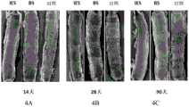
图4是不同支架植入后,对于内皮覆盖情况的内皮扫描电镜检查照片,其中图4A为植入后14天的内皮覆盖情况,图4B为植入后28天的内皮覆盖情况,图4C为植入后90天的内皮覆盖情况。Figure 4 is the scanning electron microscopy pictures of endothelial coverage after implantation of different stents, in which Figure 4A is the endothelial coverage at 14 days after implantation, Figure 4B is the endothelial coverage at 28 days after implantation, and Figure 4C is Endothelial coverage at 90 days post-implantation.
图5是不同支架植入后CD-31/PECAM-1染色结果图。Figure 5 is a graph showing the results of CD-31/PECAM-1 staining after implantation of different scaffolds.
具体实施方式Detailed ways
下面的详细描述是本发明的最佳实施方式,该描述不应被视为具有限制意义,其仅仅是为了说明本发明的一般原理。The following detailed description is of the best mode for carrying out the invention and should not be considered in a limiting sense, but merely for illustrating the general principles of the invention.
实施例1颅内药物洗脱支架的制备Example 1 Preparation of intracranial drug-eluting stent
1)支架基底涂层制备1) Preparation of stent base coating
使用二甲基甲酰胺作为溶剂溶解甲基丙烯酸正丁酯单体,加入硝酸钠作为电解质增加溶剂导电性,搅拌30分钟。Use dimethylformamide as a solvent to dissolve n-butyl methacrylate monomer, add sodium nitrate as an electrolyte to increase the conductivity of the solvent, and stir for 30 minutes.
表1.溶液制备配比Table 1. Solution preparation ratios

将金属支架(支架材料为316L不锈钢(德国Euroflex公司))进行清洗和烘干后,置入装有上述溶液的反应容器中,通过向溶液中施加电流、电压扫描完成支架基底涂层聚合。After cleaning and drying the metal stent (the stent material is 316L stainless steel (Euroflex, Germany)), it was placed in the reaction vessel containing the above solution, and the polymerization of the stent base coating was completed by applying current and voltage to the solution.
电化学条件:电压:20V;反应时间:120分钟;氮气压力:2个大气压。Electrochemical conditions: voltage: 20V; reaction time: 120 minutes; nitrogen pressure: 2 atmospheres.
将反应后支架放置真空箱内干燥。The post-reaction scaffolds were placed in a vacuum oven to dry.
2)药物涂层制备2) Preparation of drug coating
将聚乳酸聚乙醇酸颗粒(PLGA,50/50,Mn=90,000~120,000,美国Durect公司)溶解在氯仿溶液中,加入雷帕霉素药物(华北制药公司),搅拌120分钟溶解,制得药物涂层溶液。Dissolve polylactic acid polyglycolic acid particles (PLGA, 50/50, Mn=90,000~120,000, American Durect company) in chloroform solution, add rapamycin drug (North China Pharmaceutical Co., Ltd.), stir for 120 minutes to dissolve, and prepare the drug coating solution.
表2.溶液制备配比Table 2. Solution preparation ratios
将上述溶液放置于喷涂机内,将步骤1)制得的基底涂层支架固定于喷嘴下的支撑轴上,旋转支撑轴使支架各向均喷覆药物涂层溶液,支架外壁涂层厚度将大于支架内壁或侧壁涂层厚度,保证在血管内植入后外层药物可以更多被组织吸收。The above-mentioned solution is placed in a spraying machine, and the base coating stent prepared in step 1) is fixed on the support shaft under the nozzle, and the support shaft is rotated to make the stent spray the drug coating solution in all directions, and the coating thickness of the outer wall of the stent will be The thickness of the coating on the inner wall or side wall of the stent is greater than that of the stent, which ensures that the outer layer of drugs can be absorbed by the tissue more after implantation in the blood vessel.
喷涂条件如下:The spraying conditions are as follows:
溶液体积:600ml;Solution volume: 600ml;
喷涂压力:500psi;Spraying pressure: 500psi;
旋转速度:2000转/分钟;Rotation speed: 2000 rpm;
喷涂圈数:10圈;Number of spraying circles: 10 circles;
溶液进液速度:50微升/秒。Solution feed rate: 50 μl/sec.
实施例2药物释放动力学测试Example 2 Drug release kinetics test
在兔(新西兰白化兔)髂股动脉中植入实施例1制得的药物洗脱支架,在不同时间点测试兔血液中雷帕霉素药物浓度、血管内支架剩余药物浓度、支架植入动脉组织药物浓度。The drug-eluting stent prepared in Example 1 was implanted in the iliofemoral artery of a rabbit (New Zealand albino rabbit), and the drug concentration of rapamycin in the blood of the rabbit, the residual drug concentration of the intravascular stent, and the stent implanted in the artery were tested at different time points. Tissue Drug Concentrations.
兔子在麻醉后进行支架植入,穿入5F的导管鞘后注入肝素(1000IU/ml,150IU/Kg)。经导管鞘向血管内插入5F球囊导管到达髂动脉,经10-14标准大气压力扩张,之后向血管内送入试验支架样品,按照球囊尺寸与血管尺寸1:1.3比值进行扩张,保持压力30秒后撤出球囊输送系统,支架留置在体内。The rabbits were implanted with stents after anesthesia, inserted into a 5F catheter sheath and injected with heparin (1000 IU/ml, 150 IU/Kg). A 5F balloon catheter was inserted into the blood vessel through the catheter sheath to reach the iliac artery, expanded by 10-14 standard atmospheric pressure, and then the test stent sample was sent into the blood vessel, and the expansion was carried out according to the ratio of the balloon size to the blood vessel size 1:1.3, and the pressure was maintained. After 30 seconds, the balloon delivery system was withdrawn, and the stent was left in the body.
血药浓度的测定:在不同时间点收集动物全血样本,使用高效液相色谱-质谱(美国安捷伦公司)进行药物浓度分析。Determination of blood drug concentration: animal whole blood samples were collected at different time points, and the drug concentration was analyzed by high performance liquid chromatography-mass spectrometry (Agilent, USA).
组织及支架药物浓度的测定:在不同时间点将动物行安乐死,行组织检测的样品需将支架与血管小心分离,分离后的组织和支架样品使用液氮冷冻并在-80℃下保存,使用高效液相色谱-质谱(美国安捷伦公司)进行药物浓度分析。Determination of drug concentrations in tissues and scaffolds: Animals were euthanized at different time points, and the scaffolds and blood vessels were carefully separated from the samples for tissue testing. The separated tissues and scaffold samples were frozen in liquid nitrogen and stored at -80°C. High performance liquid chromatography-mass spectrometry (Agilent, USA) was used for drug concentration analysis.
测定的全身血药浓度见下表3,支架植入动脉组织的药物浓度见下表4,支架上剩余的药物浓度见下表5。The measured systemic blood drug concentration is shown in Table 3 below, the drug concentration of stent implanted in arterial tissue is shown in Table 4 below, and the remaining drug concentration on the stent is shown in Table 5 below.
表3全身血药浓度(ng/ml)Table 3 Systemic blood drug concentration (ng/ml)
表4支架植入动脉组织的药物浓度(ng/mg)Table 4 Drug concentration (ng/mg) of stent implanted in arterial tissue
表5支架上残留药物(mg/支架)Table 5 Residual drugs on stents (mg/stent)
结论in conclusion
将本发明的药物洗脱支架植入兔髂股动脉后,全身血药浓度在植入后1小时左右达到最高值,随后在72小时内逐渐降低。After the drug-eluting stent of the present invention is implanted into the rabbit iliofemoral artery, the systemic blood drug concentration reaches the highest value about 1 hour after implantation, and then gradually decreases within 72 hours.
动脉组织的药物浓度最高值出现在植入后第14天左右,3-8天为急速释放期,8-14天释放平缓,14-28天为缓慢释放期,在第28天时达到药物的全部释放,支架体内药物释放速率如图3所示。The highest value of drug concentration in arterial tissue appeared around the 14th day after implantation, 3-8 days was the rapid release period, 8-14 days was the slow release period, 14-28 days was the slow release period, and the drug reached the 28th day. All released, the drug release rate in the stent body is shown in Figure 3.
可见,全身血药浓度在安全的限度范围内,该试验表明支架在体内具有安全、可控的药物释放行为。It can be seen that the systemic blood drug concentration is within the safe limit, and this test shows that the stent has a safe and controllable drug release behavior in vivo.
实施例3颅内药物洗脱支架涂层降解及内皮修复试验Example 3 Intracranial drug-eluting stent coating degradation and endothelial repair test
在兔(新西兰白化兔)髂股动脉模型中植入金属裸支架(赛诺医疗)(BMS)、本发明实施例1制得的药物洗脱支架(IES)、不含药物涂层的基底涂层支架(BS)(其制备方法与实施例1相同,只是未喷涂药物涂层)以及另一种市售药物涂层支架(Cypher支架,美国Cordis公司)(对照),比较不同支架内皮覆盖程度及内皮活性。内皮活性通过CD-31/PECAM-1染色试验确定,成熟的内皮细胞经染色后将呈现绿色荧光信号。In the iliofemoral artery model of rabbits (New Zealand albino rabbits), bare metal stents (Sino Medical) (BMS), drug-eluting stents (IES) prepared in Example 1 of the present invention, and basecoat without drug coating were implanted. Layer stent (BS) (the preparation method is the same as that of Example 1, except that the drug coating is not sprayed) and another commercially available drug-coated stent (Cypher stent, Cordis, USA) (control), and the endothelial coverage of different stents is compared. and endothelial activity. Endothelial activity was determined by CD-31/PECAM-1 staining assay. Mature endothelial cells will show green fluorescent signal after staining.
实验结果表明,在植入90天后IES和BS均实现了内皮完整覆盖,与BMS支架相当,IES支架已无药物涂层残留,而对照支架(对照)钢筋80%以上均未覆盖。IES支架及BS支架内皮成熟均已大于75%,且内皮细胞间结构紧密,表示内皮已实现功能性恢复。不同时间点内皮扫描电镜检查照片及CD-31/PECAM-1染色结果见图4和图5。The experimental results showed that both IES and BS achieved complete endothelial coverage after 90 days of implantation, which was comparable to the BMS stent. The IES stent had no drug coating residue, while the control stent (control) had more than 80% of the steel bars not covered. The endothelium of IES scaffold and BS scaffold was more than 75% mature, and the structure between endothelial cells was tight, indicating that the endothelium had achieved functional recovery. Figure 4 and Figure 5 for endothelial scanning electron microscopy pictures and CD-31/PECAM-1 staining results at different time points.
以上详细描述了本发明的具体实施例,但可以理解,在不脱离本发明的精神下可以对其作出修改。本发明的权利要求旨在覆盖这些修改,以保证其落入本发明的真实范围和精神内。Specific embodiments of the present invention have been described above in detail, but it will be appreciated that modifications may be made thereto without departing from the spirit of the invention. The appended claims are intended to cover such modifications as fall within the true scope and spirit of this invention.
Claims (4)
Translated fromChinesePriority Applications (1)
| Application Number | Priority Date | Filing Date | Title |
|---|---|---|---|
| CN201610283341.3ACN105833358B (en) | 2016-04-28 | 2016-04-28 | A kind of intracranial drug-eluting stent system and preparation method thereof |
Applications Claiming Priority (1)
| Application Number | Priority Date | Filing Date | Title |
|---|---|---|---|
| CN201610283341.3ACN105833358B (en) | 2016-04-28 | 2016-04-28 | A kind of intracranial drug-eluting stent system and preparation method thereof |
Publications (2)
| Publication Number | Publication Date |
|---|---|
| CN105833358A CN105833358A (en) | 2016-08-10 |
| CN105833358Btrue CN105833358B (en) | 2020-12-04 |
Family
ID=56590533
Family Applications (1)
| Application Number | Title | Priority Date | Filing Date |
|---|---|---|---|
| CN201610283341.3AActiveCN105833358B (en) | 2016-04-28 | 2016-04-28 | A kind of intracranial drug-eluting stent system and preparation method thereof |
Country Status (1)
| Country | Link |
|---|---|
| CN (1) | CN105833358B (en) |
Families Citing this family (8)
| Publication number | Priority date | Publication date | Assignee | Title |
|---|---|---|---|---|
| JP6955553B2 (en)* | 2016-12-22 | 2021-10-27 | シノ メディカル サイエンシス テクノロジー,インク | Drug-eluting stents and their use to enable recovery of functional endothelial cell layers |
| CN112741720A (en)* | 2021-01-21 | 2021-05-04 | 北京久事神康医疗科技有限公司 | Recoverable intracranial drug eluting stent system |
| CN113101024A (en)* | 2021-04-08 | 2021-07-13 | 哈尔滨医科大学 | Pulmonary Drug Eluting Stents and Stent Kits |
| CN115671407B (en)* | 2022-10-25 | 2024-01-30 | 赛诺神畅医疗科技有限公司 | Self-expanding drug eluting stent system and preparation method thereof |
| CN115487410A (en)* | 2022-11-18 | 2022-12-20 | 山东瑞安泰医疗技术有限公司 | Preparation method of drug eluting stent for preventing hyperplasia and thrombus |
| CN116712620B (en)* | 2023-04-27 | 2023-12-12 | 雅伦生物科技(北京)有限公司 | Drug coating, drug-eluting stent containing same and preparation method thereof |
| CN116832228B (en)* | 2023-07-10 | 2024-05-03 | 上海心玮医疗科技股份有限公司 | Drug eluting stent coating and preparation method thereof, and drug eluting stent |
| CN119770753A (en)* | 2024-12-18 | 2025-04-08 | 上海心玮医疗科技股份有限公司 | Vascular stent and preparation method thereof |
Family Cites Families (4)
| Publication number | Priority date | Publication date | Assignee | Title |
|---|---|---|---|---|
| US5464650A (en)* | 1993-04-26 | 1995-11-07 | Medtronic, Inc. | Intravascular stent and method |
| CN101264351B (en)* | 2008-04-07 | 2011-08-24 | 易生科技(北京)有限公司 | Composite coating cardiovascular medicaments elution stent and preparation thereof |
| CN102091355A (en)* | 2011-01-11 | 2011-06-15 | 南京大学 | Compound coating coronary medicament eluting stent and preparation method thereof |
| CN103536971A (en)* | 2012-07-12 | 2014-01-29 | 赛诺医疗科学技术有限公司 | Drug eluting medical appliance capable of controllably releasing drugs and preparation method thereof |
- 2016
- 2016-04-28CNCN201610283341.3Apatent/CN105833358B/enactiveActive
Also Published As
| Publication number | Publication date |
|---|---|
| CN105833358A (en) | 2016-08-10 |
Similar Documents
| Publication | Publication Date | Title |
|---|---|---|
| CN105833358B (en) | A kind of intracranial drug-eluting stent system and preparation method thereof | |
| Khan et al. | Drug eluting stents: developments and current status | |
| CN101940802B (en) | Sustained drug-releasing stent | |
| Ma et al. | Paclitaxel/sirolimus combination coated drug-eluting stent: in vitro and in vivo drug release studies | |
| JP5329435B2 (en) | Coronary stent with asymmetric drug release controlled coating | |
| US20080085293A1 (en) | Drug eluting stent and therapeutic methods using c-Jun N-terminal kinase inhibitor | |
| CN106806948B (en) | Use of dual PI3K/mTOR inhibitors | |
| JP2004222953A (en) | Indwelling stent | |
| CN112263360A (en) | In vivo drug eluting stent and preparation method thereof | |
| CN101239216A (en) | Novel sacculus dilating catheter | |
| JP2015154925A (en) | Stent excellent in corrosion resistance | |
| WO2018059207A1 (en) | New use of amlexanox | |
| KR20150137566A (en) | Consecutive Drug releaseing stent for restenosis and inflammatory regulation and manufacturing method thereof | |
| Betala et al. | Drug-coated percutaneous balloon catheters | |
| TW202116317A (en) | Drug-eluting stent | |
| CN101496813A (en) | Anti-tissue proliferation (vascular restenosis) compositions and methods of use | |
| JP2016500686A (en) | Method for manufacturing an implantable medical device comprising a macrocyclic triene active agent and an antioxidant | |
| CN108338989A (en) | The compound anti-restenosis drugs and its controlled release system of coronary artery bracket for eluting medicament | |
| JP2015154921A (en) | drug sustained-release stent | |
| JP2016163619A (en) | Magnesium degradation rate control using anticorrosion effect | |
| TUTTLE et al. | A retrospective look at paclitaxel use in the coronary arteries | |
| Zimarino et al. | Drug-eluting balloons for percutaneous coronary interventions | |
| Poerner et al. | Drug-coated stents | |
| JP7170647B2 (en) | Implantable device with increased drug delivery area | |
| WO2025172962A1 (en) | Everolimus - eluting cobalt chromium coronary stent with abluminal biodegradable polymers |
Legal Events
| Date | Code | Title | Description |
|---|---|---|---|
| C06 | Publication | ||
| PB01 | Publication | ||
| SE01 | Entry into force of request for substantive examination | ||
| SE01 | Entry into force of request for substantive examination | ||
| CB02 | Change of applicant information | ||
| CB02 | Change of applicant information | Address after:300457 the 2 layer of B area, TEDA biological medicine research and development building 5, Fourth Avenue, Tianjin development zone. Applicant after:SINO MEDICAL SCIENCES TECHNOLOGY Inc. Address before:300457 Tianjin Binhai New Area Development Zone Fourth Avenue 5, TEDA Biomedical Research and development building B 2 floor. Applicant before:Sino Medical Sciences Technology Inc. | |
| CB03 | Change of inventor or designer information | Inventor after:Miao Zhongrong Inventor after:Sun Jianhua Inventor after:Kang Xiaoran Inventor after:Cao Yishun Inventor after:Li Tianzhu Inventor after:Wu Xiangfen Inventor before:Sun Jianhua Inventor before:Kang Xiaoran Inventor before:Cao Yishun Inventor before:Li Tianzhu Inventor before:Wu Xiangfen | |
| CB03 | Change of inventor or designer information | ||
| GR01 | Patent grant | ||
| GR01 | Patent grant | ||
| TR01 | Transfer of patent right | ||
| TR01 | Transfer of patent right | Effective date of registration:20210402 Address after:Room 501, Shilu Jinling Plaza, 88 Nanxijiang Road, Yuexi, Wuzhong District, Suzhou City, Jiangsu Province, 215104 Patentee after:Sano Shenchang Medical Technology Co.,Ltd. Address before:300457 TEDA TEDA biological medicine research and development building No. 2, No. fourth, 5 Avenue, Tianjin Development Zone, B Patentee before:SINO MEDICAL SCIENCES TECHNOLOGY Inc. | |
| CP03 | Change of name, title or address | ||
| CP03 | Change of name, title or address | Address after:215124 Building 3, Shicheng Science and Technology Park, No. 1566 Yinzhong South Road, Wuzhong Economic Development Zone, Suzhou City, Jiangsu Province Patentee after:Sano Shenchang Medical Technology Co.,Ltd. Country or region after:China Address before:Room 501, Shilu Jinling Plaza, 88 Nanxijiang Road, Yuexi, Wuzhong District, Suzhou City, Jiangsu Province, 215104 Patentee before:Sano Shenchang Medical Technology Co.,Ltd. Country or region before:China |