CN104545880B - Patient-specific pre-shaped cardiac catheter - Google Patents
Patient-specific pre-shaped cardiac catheterDownload PDFInfo
- Publication number
- CN104545880B CN104545880BCN201410532259.0ACN201410532259ACN104545880BCN 104545880 BCN104545880 BCN 104545880BCN 201410532259 ACN201410532259 ACN 201410532259ACN 104545880 BCN104545880 BCN 104545880B
- Authority
- CN
- China
- Prior art keywords
- distal tip
- organ
- medical probe
- physical model
- electrodes
- Prior art date
- Legal status (The legal status is an assumption and is not a legal conclusion. Google has not performed a legal analysis and makes no representation as to the accuracy of the status listed.)
- Expired - Fee Related
Links
- 230000000747cardiac effectEffects0.000titleclaimsdescription11
- 238000000034methodMethods0.000claimsabstractdescription44
- 239000000523sampleSubstances0.000claimsabstractdescription40
- 210000000056organAnatomy0.000claimsabstractdescription31
- 238000002679ablationMethods0.000claimsdescription19
- 239000012781shape memory materialSubstances0.000claimsdescription13
- 238000003780insertionMethods0.000claimsdescription5
- 230000037431insertionEffects0.000claimsdescription5
- 230000001766physiological effectEffects0.000claimsdescription5
- 238000001816coolingMethods0.000claims1
- 238000010438heat treatmentMethods0.000claims1
- 210000005246left atriumAnatomy0.000description13
- 238000013507mappingMethods0.000description10
- 238000010586diagramMethods0.000description6
- 230000007831electrophysiologyEffects0.000description5
- 238000002001electrophysiologyMethods0.000description5
- 229910001285shape-memory alloyInorganic materials0.000description5
- 210000005242cardiac chamberAnatomy0.000description4
- 239000000463materialSubstances0.000description4
- 238000005259measurementMethods0.000description4
- 238000007493shaping processMethods0.000description4
- 238000013153catheter ablationMethods0.000description3
- 238000002591computed tomographyMethods0.000description3
- 210000005245right atriumAnatomy0.000description3
- IAYPIBMASNFSPL-UHFFFAOYSA-NEthylene oxideChemical compoundC1CO1IAYPIBMASNFSPL-UHFFFAOYSA-N0.000description2
- 229940079593drugDrugs0.000description2
- 239000003814drugSubstances0.000description2
- 230000006870functionEffects0.000description2
- 238000002595magnetic resonance imagingMethods0.000description2
- 210000003492pulmonary veinAnatomy0.000description2
- 230000001954sterilising effectEffects0.000description2
- 238000004659sterilization and disinfectionMethods0.000description2
- 238000001356surgical procedureMethods0.000description2
- 230000002792vascularEffects0.000description2
- 229910000990Ni alloyInorganic materials0.000description1
- HZEWFHLRYVTOIW-UHFFFAOYSA-N[Ti].[Ni]Chemical compound[Ti].[Ni]HZEWFHLRYVTOIW-UHFFFAOYSA-N0.000description1
- 229910045601alloyInorganic materials0.000description1
- 239000000956alloySubstances0.000description1
- 238000013459approachMethods0.000description1
- 206010003119arrhythmiaDiseases0.000description1
- 230000006793arrhythmiaEffects0.000description1
- 238000010009beatingMethods0.000description1
- -1copper-aluminum-nickelChemical compound0.000description1
- 210000003748coronary sinusAnatomy0.000description1
- 229910003460diamondInorganic materials0.000description1
- 239000010432diamondSubstances0.000description1
- 210000002837heart atriumAnatomy0.000description1
- 208000019622heart diseaseDiseases0.000description1
- 238000003384imaging methodMethods0.000description1
- 238000002847impedance measurementMethods0.000description1
- 230000003902lesionEffects0.000description1
- 210000004115mitral valveAnatomy0.000description1
- 239000000203mixtureSubstances0.000description1
- 238000012986modificationMethods0.000description1
- 230000004048modificationEffects0.000description1
- 230000003287optical effectEffects0.000description1
- 229920000642polymerPolymers0.000description1
- 238000003825pressingMethods0.000description1
- 230000004044responseEffects0.000description1
- 230000001225therapeutic effectEffects0.000description1
- 238000002604ultrasonographyMethods0.000description1
- 210000005166vasculatureAnatomy0.000description1
Images


Classifications
- A—HUMAN NECESSITIES
- A61—MEDICAL OR VETERINARY SCIENCE; HYGIENE
- A61B—DIAGNOSIS; SURGERY; IDENTIFICATION
- A61B18/00—Surgical instruments, devices or methods for transferring non-mechanical forms of energy to or from the body
- A61B18/04—Surgical instruments, devices or methods for transferring non-mechanical forms of energy to or from the body by heating
- A61B18/12—Surgical instruments, devices or methods for transferring non-mechanical forms of energy to or from the body by heating by passing a current through the tissue to be heated, e.g. high-frequency current
- A61B18/14—Probes or electrodes therefor
- A61B18/1492—Probes or electrodes therefor having a flexible, catheter-like structure, e.g. for heart ablation
- A—HUMAN NECESSITIES
- A61—MEDICAL OR VETERINARY SCIENCE; HYGIENE
- A61B—DIAGNOSIS; SURGERY; IDENTIFICATION
- A61B17/00—Surgical instruments, devices or methods
- A61B2017/00831—Material properties
- A61B2017/00867—Material properties shape memory effect
- A—HUMAN NECESSITIES
- A61—MEDICAL OR VETERINARY SCIENCE; HYGIENE
- A61B—DIAGNOSIS; SURGERY; IDENTIFICATION
- A61B18/00—Surgical instruments, devices or methods for transferring non-mechanical forms of energy to or from the body
- A61B2018/00053—Mechanical features of the instrument of device
- A61B2018/0016—Energy applicators arranged in a two- or three dimensional array
- A—HUMAN NECESSITIES
- A61—MEDICAL OR VETERINARY SCIENCE; HYGIENE
- A61B—DIAGNOSIS; SURGERY; IDENTIFICATION
- A61B18/00—Surgical instruments, devices or methods for transferring non-mechanical forms of energy to or from the body
- A61B2018/00053—Mechanical features of the instrument of device
- A61B2018/00214—Expandable means emitting energy, e.g. by elements carried thereon
- A61B2018/0022—Balloons
- A—HUMAN NECESSITIES
- A61—MEDICAL OR VETERINARY SCIENCE; HYGIENE
- A61B—DIAGNOSIS; SURGERY; IDENTIFICATION
- A61B18/00—Surgical instruments, devices or methods for transferring non-mechanical forms of energy to or from the body
- A61B2018/00053—Mechanical features of the instrument of device
- A61B2018/00214—Expandable means emitting energy, e.g. by elements carried thereon
- A61B2018/00267—Expandable means emitting energy, e.g. by elements carried thereon having a basket shaped structure
- A—HUMAN NECESSITIES
- A61—MEDICAL OR VETERINARY SCIENCE; HYGIENE
- A61B—DIAGNOSIS; SURGERY; IDENTIFICATION
- A61B18/00—Surgical instruments, devices or methods for transferring non-mechanical forms of energy to or from the body
- A61B2018/00571—Surgical instruments, devices or methods for transferring non-mechanical forms of energy to or from the body for achieving a particular surgical effect
- A61B2018/00577—Ablation
- A—HUMAN NECESSITIES
- A61—MEDICAL OR VETERINARY SCIENCE; HYGIENE
- A61B—DIAGNOSIS; SURGERY; IDENTIFICATION
- A61B18/00—Surgical instruments, devices or methods for transferring non-mechanical forms of energy to or from the body
- A61B18/04—Surgical instruments, devices or methods for transferring non-mechanical forms of energy to or from the body by heating
- A61B18/12—Surgical instruments, devices or methods for transferring non-mechanical forms of energy to or from the body by heating by passing a current through the tissue to be heated, e.g. high-frequency current
- A61B18/14—Probes or electrodes therefor
- A61B2018/1405—Electrodes having a specific shape
- A61B2018/1435—Spiral
- A—HUMAN NECESSITIES
- A61—MEDICAL OR VETERINARY SCIENCE; HYGIENE
- A61B—DIAGNOSIS; SURGERY; IDENTIFICATION
- A61B18/00—Surgical instruments, devices or methods for transferring non-mechanical forms of energy to or from the body
- A61B18/04—Surgical instruments, devices or methods for transferring non-mechanical forms of energy to or from the body by heating
- A61B18/12—Surgical instruments, devices or methods for transferring non-mechanical forms of energy to or from the body by heating by passing a current through the tissue to be heated, e.g. high-frequency current
- A61B18/14—Probes or electrodes therefor
- A61B2018/1467—Probes or electrodes therefor using more than two electrodes on a single probe
- A—HUMAN NECESSITIES
- A61—MEDICAL OR VETERINARY SCIENCE; HYGIENE
- A61B—DIAGNOSIS; SURGERY; IDENTIFICATION
- A61B34/00—Computer-aided surgery; Manipulators or robots specially adapted for use in surgery
- A61B34/10—Computer-aided planning, simulation or modelling of surgical operations
- A61B2034/101—Computer-aided simulation of surgical operations
- A61B2034/105—Modelling of the patient, e.g. for ligaments or bones
- Y—GENERAL TAGGING OF NEW TECHNOLOGICAL DEVELOPMENTS; GENERAL TAGGING OF CROSS-SECTIONAL TECHNOLOGIES SPANNING OVER SEVERAL SECTIONS OF THE IPC; TECHNICAL SUBJECTS COVERED BY FORMER USPC CROSS-REFERENCE ART COLLECTIONS [XRACs] AND DIGESTS
- Y10—TECHNICAL SUBJECTS COVERED BY FORMER USPC
- Y10T—TECHNICAL SUBJECTS COVERED BY FORMER US CLASSIFICATION
- Y10T29/00—Metal working
- Y10T29/49—Method of mechanical manufacture
- Y10T29/49002—Electrical device making
- Y10T29/49117—Conductor or circuit manufacturing
Landscapes
- Health & Medical Sciences (AREA)
- Life Sciences & Earth Sciences (AREA)
- Surgery (AREA)
- Engineering & Computer Science (AREA)
- Plasma & Fusion (AREA)
- Medical Informatics (AREA)
- Otolaryngology (AREA)
- Physics & Mathematics (AREA)
- Cardiology (AREA)
- Biomedical Technology (AREA)
- Heart & Thoracic Surgery (AREA)
- Nuclear Medicine, Radiotherapy & Molecular Imaging (AREA)
- Molecular Biology (AREA)
- Animal Behavior & Ethology (AREA)
- General Health & Medical Sciences (AREA)
- Public Health (AREA)
- Veterinary Medicine (AREA)
- Surgical Instruments (AREA)
- Measurement And Recording Of Electrical Phenomena And Electrical Characteristics Of The Living Body (AREA)
Abstract
Translated fromChinese
Description
Translated fromChinese技术领域technical field
本发明整体涉及医疗装置,具体地讲,涉及预成形的医疗探头。The present invention relates generally to medical devices and, in particular, to preformed medical probes.
背景技术Background technique
侵入式探头用于多种医学过程中,例如心脏电生理(EP)标测和消融。一些侵入式探头具有预成形为所需形式的末端。例如,公开内容以引用方式并入本文的美国专利5,617,854描述了用于心脏回路的一部分的标测和选择性消融的预成形的心脏导管。该导管包括用于围绕冠状窦口定位的预成形的第一弯曲部分和用于将第一弯曲部保持在其所需位置的第二弯曲部分。将包括导丝和预成形的导管的导管组件插入到靠近心房的位置。当从导管内撤出导丝时,导管在目标位置处呈现其预成形的形式。作为另外一种选择,可经由导管护套来将具有或不具有导丝的导管组件引入到目标消融位点。导管包括位于导管的至少一部分上的间隔开的电极的阵列。Invasive probes are used in a variety of medical procedures, such as cardiac electrophysiology (EP) mapping and ablation. Some invasive probes have tips pre-shaped into the desired form. For example, US Patent 5,617,854, the disclosure of which is incorporated herein by reference, describes a preformed cardiac catheter for mapping and selective ablation of a portion of a cardiac circuit. The catheter includes a pre-shaped first curved portion for positioning around the coronary sinus ostium and a second curved portion for maintaining the first curved portion in its desired position. A catheter assembly including a guide wire and a preformed catheter is inserted proximal to the atrium. When the guidewire is withdrawn from the catheter, the catheter assumes its pre-shaped form at the target location. Alternatively, a catheter assembly with or without a guidewire can be introduced to the target ablation site via a catheter sheath. The catheter includes an array of spaced electrodes on at least a portion of the catheter.
公开内容以引用方式并入本文的美国专利申请公开2007/0049924描述了一种用于设置消融灶的消融导管。导管包括可从导管套筒滑出的消融元件,并且具有环状区段,所述环状区段在所述元件滑出时自伸展成自动或手动施加的预定形状,所述预定形状对应于需要消融的组织区域的实际形状。US Patent Application Publication 2007/0049924, the disclosure of which is incorporated herein by reference, describes an ablation catheter for setting an ablation lesion. The catheter includes an ablation element that is slidable from the catheter sleeve and has an annular segment that self-expands as the element is slid out into an automatically or manually applied predetermined shape corresponding to The actual shape of the tissue area to be ablated.
发明内容SUMMARY OF THE INVENTION
本文所述的本发明的实施例提供了一种方法,所述方法包括接收患者的器官的一个或多个图像。基于图像来制作器官的至少目标区域的物理模型。医疗探头的远侧末端被预成形以贴合对应于目标区域的物理模型的表面。Embodiments of the invention described herein provide a method comprising receiving one or more images of an organ of a patient. A physical model of at least the target region of the organ is made based on the image. The distal tip of the medical probe is pre-shaped to conform to the surface of the physical model corresponding to the target area.
在一些实施例中,所述方法包括将具有预成形的远侧末端的医疗探头插入患者的器官中,以及利用探头来对器官的目标区域施用医学过程。在一些实施例中,器官包括心脏并且探头包括心脏导管。In some embodiments, the method includes inserting a medical probe having a pre-shaped distal tip into an organ of a patient, and utilizing the probe to administer a medical procedure to a target region of the organ. In some embodiments, the organ includes a heart and the probe includes a cardiac catheter.
在本发明所公开的实施例中,远侧末端包括形状记忆材料,所述形状记忆材料可通过将预定义的操作条件施加到远侧末端而被配置成形,并且预成形所述远侧末端包括当使远侧末端经受预定义的操作条件时将远侧末端贴合到物理模型的表面。In disclosed embodiments, the distal tip includes a shape memory material that can be configured to be shaped by applying predefined operating conditions to the distal tip, and the pre-forming of the distal tip includes The distal tip is conformed to the surface of the physical model when the distal tip is subjected to predefined operating conditions.
在一些实施例中,远侧末端包括多个电极并且当放置在器官中时可伸展以呈现三维形状,并且预成形所述远侧末端包括使多个电极与目标区域进行同时物理接触。在示例性实施例中,远侧末端包括其上设置有电极的螺旋件。在另一实施例中,远侧末端包括其上设置有电极的可伸展的和可塌缩的篮状件。In some embodiments, the distal tip includes a plurality of electrodes and is stretchable to assume a three-dimensional shape when placed in the organ, and preforming the distal tip includes bringing the plurality of electrodes into simultaneous physical contact with the target area. In an exemplary embodiment, the distal tip includes a helical member having electrodes disposed thereon. In another embodiment, the distal tip includes an expandable and collapsible basket having electrodes disposed thereon.
在另一实施例中,制作物理模型包括利用三维(3D)打印机来打印模型。在另一个实施例中,医疗探头的远侧末端可利用充气式球囊来伸展以适形于器官的目标区域。In another embodiment, making the physical model includes printing the model with a three-dimensional (3D) printer. In another embodiment, the distal tip of the medical probe may be stretched to conform to the target area of the organ using an inflatable balloon.
根据本发明的一个实施例,另外提供了包括细长管和远侧末端的医疗探头。细长管被配置成插入患者的器官中。远侧末端连接到细长管并且在插入之前进行预成形以通过使远侧末端贴合到器官的至少部分的物理模型的表面来适形于器官的目标区域。According to one embodiment of the present invention, there is additionally provided a medical probe including an elongated tube and a distal tip. The elongated tube is configured to be inserted into the patient's organ. The distal tip is connected to the elongated tube and pre-shaped prior to insertion to conform to the target area of the organ by conforming the distal tip to the surface of a physical model of at least part of the organ.
结合附图,通过以下对实施例的详细说明,将更全面地理解本发明,其中:The present invention will be more fully understood through the following detailed description of the embodiments in conjunction with the accompanying drawings, wherein:
附图说明Description of drawings
图1为根据本发明的实施例的示意性地示出用于心脏消融的系统的框图;1 is a block diagram schematically illustrating a system for cardiac ablation according to an embodiment of the present invention;
图2A-2C为根据本发明的实施例的预成形导管的方法的示意图;2A-2C are schematic diagrams of a method of preforming a catheter according to an embodiment of the present invention;
图3为根据本发明的实施例的示意性地示出用于预成形和使用心脏导管的方法的流程图;并且3 is a flow diagram schematically illustrating a method for preforming and using a cardiac catheter according to an embodiment of the present invention; and
图4为根据本发明的可供选择的实施例的利用物理模型预成形的导管的示意图。4 is a schematic illustration of a catheter preformed using a physical model according to an alternative embodiment of the present invention.
具体实施方式Detailed ways
综述Overview
在一些医学过程(例如,心脏消融和EP标测)中,将医疗探头的远侧末端上的一个或多个电极设置成与器官中的目标区域的表面接触。与器官表面的正确接触对手术的成功至关重要。在一些类型的导管中,末端包括应同时接触表面上的多个点的多个电极。In some medical procedures (eg, cardiac ablation and EP mapping), one or more electrodes on the distal tip of the medical probe are placed in contact with the surface of the target region in the organ. Proper contact with organ surfaces is critical to the success of the surgery. In some types of catheters, the tip includes multiple electrodes that should simultaneously contact multiple points on the surface.
然而,在实施过程中,器官表面的三维(3-D)形状因患者而异。例如,心脏腔室并且具体地讲左心房(LA)表现出显著的患者间的形状差异。因此,导管末端的任何单一形状将不可避免地成为折衷方案,这样可降低手术的质量。However, in practice, the three-dimensional (3-D) shape of the organ surface varies from patient to patient. For example, the chambers of the heart, and in particular the left atrium (LA), exhibit significant inter-patient shape variability. Therefore, any single shape of the catheter tip will inevitably be a compromise, which can reduce the quality of the procedure.
本文所述的本发明的实施例提供了用于制作和利用医疗探头的改善的技术。在这种技术中,预成形医疗探头的末端,以匹配特定患者的器官表面的目标区域的实际3-D形状。然后利用具有个性化的预成形末端的探头来执行医学过程。因此,探头电极和器官表面之间的接触质量得到显著增强。本文所述的实施例主要涉及利用心脏导管在心脏中执行的手术,但本发明所公开的技术可与多种其他类型的医疗探头一起使用以用于多种其他器官。Embodiments of the invention described herein provide improved techniques for making and utilizing medical probes. In this technique, the tip of the medical probe is pre-shaped to match the actual 3-D shape of the target area on the surface of a particular patient's organ. The medical procedure is then performed using the probe with the personalized pre-shaped tip. As a result, the quality of contact between the probe electrodes and the organ surface is significantly enhanced. The embodiments described herein relate primarily to procedures performed in the heart using cardiac catheters, but the disclosed techniques may be used with various other types of medical probes for various other organs.
在一些实施例中,利用所考虑的特定患者的心脏腔室的至少部分的物理模型来预成形导管末端。基于预采集的患者心脏的三维(3-D)图像(例如,CT或MRI图像)来制作物理模型。可利用例如3D打印机来制得模型。In some embodiments, the catheter tip is preformed with a physical model of at least a portion of the heart chamber of the particular patient under consideration. A physical model is made based on pre-acquired three-dimensional (3-D) images (eg, CT or MRI images) of the patient's heart. Models can be made using, for example, a 3D printer.
本发明所公开的技术可与多种类型的导管末端一起使用,例如利用球囊膨胀的可塌缩篮状件末端、或者螺旋件末端。在一些实施例中,导管末端包括可形成为所需形状并且随后固定成此形状的柔性形状记忆材料。可例如通过使末端经受一定的温度范围来实现成形。为了匹配特定表面形状,将导管末端按压或者换句话讲压贴到物理模型的适当区域,然后固定其形状。所得的导管通常为消毒的,然后可用于执行医学过程,例如标测或消融。The disclosed techniques can be used with various types of catheter tips, such as balloon-expandable collapsible basket tips, or helical tips. In some embodiments, the catheter tip includes a flexible shape memory material that can be formed into a desired shape and then secured into that shape. Shaping can be accomplished, for example, by subjecting the tip to a range of temperatures. To match a specific surface shape, the catheter tip is pressed or otherwise pressed to the appropriate area of the physical model, and its shape is then fixed. The resulting catheter is typically sterile and can then be used to perform medical procedures, such as mapping or ablation.
当使用本发明所公开的预成形技术时,导管末端紧密地匹配患者心脏内表面的所需区域的实际3-D形状。因此,导管末端上的多个电极可实现与心脏表面的高质量同时接触。因此,当利用这些技术时,可显著地改善诸如EP标测和消融之类的手术的质量。When using the pre-shaping techniques disclosed herein, the catheter tip closely matches the actual 3-D shape of the desired region of the patient's inner surface of the heart. Thus, multiple electrodes on the catheter tip can achieve high-quality simultaneous contact with the surface of the heart. Thus, the quality of procedures such as EP mapping and ablation can be significantly improved when these techniques are utilized.
对系统的描述description of the system
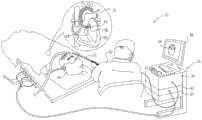
图1为根据本发明的实施例的示意性地示出用于心脏消融的系统20的框图。系统20包括探头22(在预设例子中为心脏导管)和控制台24。在本文所述的实施例中,以举例的方式假定探头22可用于患者28的心脏26中的组织消融,以便治疗心律失常。作为另外一种选择或除此之外,探头22可用于其他治疗和/或诊断目的,例如用于标测心脏中或另一身体器官中的电势。1 is a block diagram schematically illustrating a
控制台24包括处理器42,所述处理器42通常为通用计算机,所述通用计算机具有合适的前端和接口电路以用于从探头22接收信号并控制本文所述系统20的其他部件。处理器42可以软件形式进行编程以执行系统所使用的功能,并且处理器将用于软件的数据存储在存储器50中。例如,可经网络将软件以电子形式下载到控制台24,或者可将软件保存在非暂态有形介质诸如光学、磁或电子存储器介质上。作为另外一种选择,可通过专用或可编程数字硬件组件执行处理器42的一些或全部功能。The
操作者30(通常是医师)插入探头22穿过患者28的血管系统,使得探头22的远端38进入心脏26的腔室。系统20通常利用磁性位置感测来确定心脏26内部的远端的位置坐标。在这种情况下,控制台24包括驱动电路34,所述驱动电路34驱动放置在患者28外部的已知位置处(例如,患者躯干下面)的磁场发生器36。An operator 30 (usually a physician) inserts the
探头远端内的磁场传感器40响应于得自线图的磁场来产生电位置信号,从而允许处理器42确定腔室内的远端32的方位,即,位置以及通常还有取向。磁场传感器(即,位置传感器)通常包括一个或多个线圈,通常为相互正交的三个线圈。A magnetic field sensor 40 within the distal end of the probe generates an electrical position signal in response to the magnetic field derived from the line map, allowing the
这种位置感测方法在例如由Biosense Webster Inc.(Diamond Bar,Calif.)生产的CARTOTM系统中实施,并且详细地描述于美国专利No.5,391,199、No.6,690,963、No.6,484,118、No.6,239,724、No.6,618,612和No.6,332,089、PCT专利申请WO 96/05768、以及美国专利申请公开2002/0065455 A1、2003/0120150 A1和2004/0068178 A1中,上述专利的公开内容全部以引用方式并入本文。This method of position sensing is implemented, for example, in the CARTO™ system produced by Biosense Webster Inc. (Diamond Bar, Calif.) and described in detail in US Pat. Nos. 5,391,199, 6,690,963, 6,484,118, 6,239,724 , Nos. 6,618,612 and 6,332,089, PCT Patent Application WO 96/05768, and US Patent Application Publications 2002/0065455 A1, 2003/0120150 A1 and 2004/0068178 A1, the disclosures of which are incorporated herein by reference in their entirety .
尽管在本例子中,系统20被假定为利用基于磁的传感器来测量远端38的位置,但本发明的实施例可利用其他位置跟踪技术,例如基于阻抗测量的跟踪系统。基于阻抗的位置跟踪技术在例如美国专利5,983,126、6,456,864和5,944,022中有所描述,这些专利的公开内容也以引用方式并入本文。可使用本领域中的普通技术人员已知的其他位置跟踪技术来确定远端32的位置。Although in this example,
为了消融心脏26的组织,操作者30操纵探头22,使得远端38处于腔室的内表面上(或靠近腔室的内表面)的多个位置处。在每个位置处,耦接到远端的电极测量特定的生理属性(例如,局部表面电势)。处理器42使从传感器40的位置信号导出的位置测量结果与电势测量结果相关联。因此,所述系统采集多个标测点,其中每个标测点包括内部腔室表面上的坐标以及在此坐标处的相应生理属性测量结果。To ablate tissue of the
处理器42使用标测点的坐标来构造所考虑的心腔的模拟表面。处理器42随后将标测点的电势测量结果与模拟表面结合在一起以制得叠加在模拟表面上的电势的标测图。处理器42在显示器46上向操作者30显示标测图的图像44。
患者专用导管预成形Patient-specific catheter preforming
在一些实施例中,导管22的远侧末端包括应同时接触心脏26中的目标区域的内表面的多个电极。为了实现高质量同时接触,导管末端的3-D形状(以及因此多个电极的位点)应紧密地匹配目标区域的3-D形状。In some embodiments, the distal tip of the
然而,心脏表面形状因患者不同而显著地变化。为了克服这些变化,在一些实施例中,在手术之前预成形导管末端,以匹配所考虑的特定患者的目标区域的实际3-D形状。利用心脏的至少部分的物理3-D模型来执行预成形。However, the shape of the heart surface varies significantly from patient to patient. To overcome these variations, in some embodiments, the catheter tip is pre-shaped prior to surgery to match the actual 3-D shape of the target area for the particular patient under consideration. The preshaping is performed using a physical 3-D model of at least a portion of the heart.
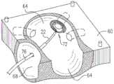
图2A为根据本发明的实施例的患者28的心脏26的部分的物理模型60的示意图。在本例子中,模型60表示心脏26的左心房(LA)的部分64和右心房(RA)部分68。使用此模型来预成形导管22以用于执行LA中的消融手术。2A is a schematic diagram of a
通常,基于心脏26或其部分的预采集图像来制作模型60。用于制作模型60的图像可包括2D或3-D图像,并且可包括例如计算机断层扫描(CT)图像、磁共振成像(MRI)图像、超声图像和/或者任何其他合适模态或模态组合的图像。在一些实施例中,通过利用导管位置跟踪系统(例如,上述CARTO系统)标测心脏内表面来获得用于制作模型的3-D心脏形状图像。Typically,
可利用任何合适的材料(例如,合适的塑料或聚合物)来制作模型60。在一些实施例中,利用3-D打印机来打印模型60。在典型的方法中,通过合适的处理器来处理预采集的图像以便计算心脏表面的3-D形状。然后基于此3-D形状例如利用3-D打印机或其他合适的装置来制得物理模型60。
模型60旨在重建医学过程的至少目标区域中的心脏内表面的3-D物理形状。其他表面和心脏特征结构可从模型中略去。在本例子中,模型60重构LA的内表面的部分64(包括四个肺静脉的部分)加上RA的部分68。
可用作模型60的示例性物理模型在Marjert等人的“A Beating Heart Model 3DPrinted from Specific Patient Data,”Proceedings of the Annual Conference ofthe IEEE Engineering in Medicine and Biology Society(2007年,第4472-4475页)中有所描述,该参考文献的内容以引用方式并入本文。物理心脏模型的其他例子在“Structural Heart Disease Modeling Project,”University of Colorado,School ofMedicine,Interventional Cardiology3-D Lab(2011年11月)中有所描述,该内容以引用方式并入本文。作为另外一种选择,模型60可具有任何其他合适的形状和/或材料组合物,并且其可以任何其他合适的方式来制作。An exemplary physical model that can be used as
图2B为根据本发明的实施例的利用物理模型60预成形导管22的方法的示意图。在本例子中,导管22包括具有螺旋形状的远侧末端72。多个电极(图中未示出)沿着螺旋件进行设置,以用于标测和/或消融围绕肺静脉中的一个的孔口的多个点。(其中导管末端包括可塌缩“篮状件”的可供选择的实施例在下文中进行进一步的描述)。2B is a schematic diagram of a method of preforming
末端72由柔性形状记忆材料构成或者至少包括柔性形状记忆材料(例如,形状记忆合金(SMA))。通过施加预定义的操作条件,可将形状记忆材料设定成所需形状,并且随后永久性地固定成这种形状。例如,通过使SMA在一定温度范围下成形来将SMA(例如,铜-铝-镍合金或镍-钛(NiTi)合金)固定成所需形状。在此温度范围之外,材料为柔性的,但在无约束时将返回到预定义的形状。The
根据所使用的材料,温度范围可在例如40至95℃的范围中。一般来讲,末端72可包括利用任何合适的方式成形的任何合适的形状记忆材料。Depending on the materials used, the temperature range may be, for example, in the range of 40 to 95°C. In general,
如在图中可见,经由护套76插入导管22。通常,经由血管系统插入护套76,直至到达消融位点附近。然后,经由护套插入导管22,直至末端72从护套伸展。As can be seen in the figures, the
当使用柔性形状记忆材料时,护套76使得末端72在插入期间呈现线性形状。当到达消融位点附近时,末端72从护套伸出并且因形状记忆材料的“记忆”特性而重新呈现其螺旋形状。When a flexible shape memory material is used, the
作为使用护套的另外一种选择,可通过导丝来将导管22引导到消融位点。经由血管系统插入此导丝,直至到达消融位点附近。然后,经由导丝插入导管22,直至到达所需位点。在此具体实施中,导丝使得末端72保持为线性,直至到达消融位点。在消融位点处,所述末端延伸超出导丝的末端并且重新呈现其预定义的螺旋形状。As an alternative to using a sheath, the
上述实施例涉及螺旋形导管末端。然而,作为另外一种选择,末端72可具有任何其他合适的形状或构型。一个可供选择的例子为“篮状件”末端,其包括其上设置有电极的SMA元件的可伸展网。任何此类末端均可用于本文所公开的技术中。The above-described embodiments relate to helical catheter tips. Alternatively, however,
在如图2B所示的典型预成形方法中,紧贴物理模型60中的所需区域来放置末端72。在计划的消融手术期间,末端72的放置模拟所需的末端位置。将末端区域(可能包括模型60的一些或全部)加热到所需的温度范围以用于使导管末端的形状记忆材料成形。然后冷却末端72,同时使其压贴在LA的部分64中的所需区域上。因此,末端72呈现患者LA的内表面的3-D形状。末端72在从模型中移出时将保持这种形状。In a typical preforming method as shown in FIG. 2B , the
图2C为根据本发明的实施例的插入患者28的心脏26内之后的导管22的示意图。图2C示出了护套76,其中导管22的末端72从护套中伸展并且呈现在模型60中固定的3-D形状。由于模型60紧密地匹配心脏26的LA的实际内部3-D形状,因此末端72中的电极能够与LA表面上的多个相应点同时进行高质量地接触。2C is a schematic illustration of the
图3为根据本发明的实施例的示意性地示出用于预成形和使用心脏导管的方法的流程图。所述方法开始于在成像步骤80处采集待治疗的患者28的心脏腔室的一个或多个图像。在模型制作步骤84处,基于所采集的图像来制作3-D模型,例如,图2A的模型60。3 is a flow chart schematically illustrating a method for preforming and using a cardiac catheter, according to an embodiment of the present invention. The method begins at imaging step 80 by acquiring one or more images of the heart chambers of the patient 28 to be treated. At a model making step 84, a 3-D model, eg,
在成形步骤88处,预成形导管22的远侧末端72以匹配心脏腔室的所需区域的3-D形状。当使末端经受用于成形形状记忆材料的温度时通过将末端72按压到模型60中的所需区域来执行预成形。然后,在消毒步骤92中,例如利用环氧乙烷(EtO)消毒来对导管22消毒。此时导管就可供使用。在手术步骤96处,操作者30利用预成形的导管末端来对患者28执行医学过程(例如,EP标测或消融)。At the shaping step 88, the
图4为根据本发明的可供选择的实施例的利用物理模型预成形的导管的示意图。在此实施例中,导管22具有包括柔性臂的可塌缩网的“篮状件”末端100。多个电极安装在臂上。4 is a schematic illustration of a catheter preformed using a physical model according to an alternative embodiment of the present invention. In this embodiment, the
在患者身体的标测手术期间,将导管22以其在护套76内的塌缩状态插入LA中。通常经由卵圆窝插入导管22,并且利用另一导管将球囊经由二尖瓣插入LA中。将球囊插入网内并且进行膨胀。因此,末端100的网伸展并且适形于LA的内表面,并且电极与该表面进行高质量的接触。图4示出了预成形这种类型的导管末端的方法。如在图中可见,末端100膨胀并且压贴模型60的内表面。During a mapping procedure of the patient's body, the
应当理解,上述实施例仅以举例的方式进行引用,并且本发明并不限于上面具体示出和描述的内容。相反,本发明的范围包括上文所述各种特征的组合与子组合,以及本领域的技术人员在阅读上述说明时可能想到且未在现有技术中公开的变型和修改形式。以引用方式并入本专利申请的文献将视为本专利申请的整体部分,但除了在这些并入的文献中以与本说明书中明确或隐含地给出的定义相冲突的方式定义的任何术语,而只应考虑本说明书中的定义。It should be understood that the above-described embodiments are cited by way of example only, and the present invention is not limited to what is specifically shown and described above. Rather, the scope of the invention includes combinations and sub-combinations of the various features described above, as well as variations and modifications that may occur to those skilled in the art upon reading the above description and not disclosed in the prior art. Documents incorporated by reference into this patent application are considered an integral part of this patent application, except for any definitions in these incorporated documents that conflict with definitions expressly or implicitly given in this specification. terms, and only the definitions in this specification should be considered.
Claims (15)
Translated fromChineseApplications Claiming Priority (2)
| Application Number | Priority Date | Filing Date | Title |
|---|---|---|---|
| US14/051491 | 2013-10-11 | ||
| US14/051,491US10687889B2 (en) | 2013-10-11 | 2013-10-11 | Patient-specific pre-shaped cardiac catheter |
Publications (2)
| Publication Number | Publication Date |
|---|---|
| CN104545880A CN104545880A (en) | 2015-04-29 |
| CN104545880Btrue CN104545880B (en) | 2020-01-14 |
Family
ID=51753006
Family Applications (1)
| Application Number | Title | Priority Date | Filing Date |
|---|---|---|---|
| CN201410532259.0AExpired - Fee RelatedCN104545880B (en) | 2013-10-11 | 2014-10-10 | Patient-specific pre-shaped cardiac catheter |
Country Status (7)
| Country | Link |
|---|---|
| US (1) | US10687889B2 (en) |
| EP (1) | EP2859861B1 (en) |
| JP (1) | JP6453027B2 (en) |
| CN (1) | CN104545880B (en) |
| AU (1) | AU2014240285B2 (en) |
| CA (1) | CA2865336A1 (en) |
| IL (1) | IL234664B (en) |
Families Citing this family (36)
| Publication number | Priority date | Publication date | Assignee | Title |
|---|---|---|---|---|
| JP6441679B2 (en) | 2011-12-09 | 2018-12-19 | メタベンション インコーポレイテッド | Therapeutic neuromodulation of the liver system |
| AU2014274903B2 (en) | 2013-06-05 | 2019-03-07 | Medtronic Ireland Manufacturing Unlimited Company | Modulation of targeted nerve fibers |
| CA2969129A1 (en) | 2014-12-03 | 2016-06-09 | Metavention, Inc. | Systems and methods for modulating nerves or other tissue |
| EP3397149A4 (en) | 2015-12-30 | 2019-08-14 | Schuler Scientific Solutions, LLC | CARTOGRAPHY AND TREATMENT OF TISSUE |
| CN108472076B (en)* | 2016-01-07 | 2022-05-31 | 伯尔尼大学 | Method and system for pose-controlled ablation |
| CN108882957B (en)* | 2016-02-10 | 2022-09-16 | 埃米尔·贝尔森 | Personalized atrial fibrillation ablation |
| US10524859B2 (en) | 2016-06-07 | 2020-01-07 | Metavention, Inc. | Therapeutic tissue modulation devices and methods |
| US10905329B2 (en) | 2016-06-09 | 2021-02-02 | Biosense Webster (Israel) Ltd. | Multi-function conducting elements for a catheter |
| US11304644B2 (en)* | 2017-03-07 | 2022-04-19 | Biosense Webster (Israel) Ltd. | 3-D electrophysiology heart simulation system and related methods |
| US12029545B2 (en) | 2017-05-30 | 2024-07-09 | Biosense Webster (Israel) Ltd. | Catheter splines as location sensors |
| US10765475B2 (en)* | 2017-10-31 | 2020-09-08 | Biosense Webster (Israel) Ltd. | All-in-one spiral catheter |
| US10918310B2 (en)* | 2018-01-03 | 2021-02-16 | Biosense Webster (Israel) Ltd. | Fast anatomical mapping (FAM) using volume filling |
| US20190314083A1 (en) | 2018-04-11 | 2019-10-17 | Biosense Webster (Israel) Ltd. | Flexible Multi-Arm Catheter with Diametrically Opposed Sensing Electrodes |
| US11045628B2 (en) | 2018-12-11 | 2021-06-29 | Biosense Webster (Israel) Ltd. | Balloon catheter with high articulation |
| US11207016B2 (en) | 2018-12-28 | 2021-12-28 | Biosense Webster (Israel) Ltd. | Mapping ECG signals using a multipole electrode assembly |
| US11850051B2 (en) | 2019-04-30 | 2023-12-26 | Biosense Webster (Israel) Ltd. | Mapping grid with high density electrode array |
| US11712172B2 (en) | 2019-07-18 | 2023-08-01 | Biosense Webster (Israel) Ltd. | Visual guidance for positioning a distal end of a medical probe |
| US11950930B2 (en) | 2019-12-12 | 2024-04-09 | Biosense Webster (Israel) Ltd. | Multi-dimensional acquisition of bipolar signals from a catheter |
| WO2021126980A1 (en)* | 2019-12-16 | 2021-06-24 | Affera, Inc. | Pulmonary vein isolation catheters and associated devices, systems, and methods |
| US11517218B2 (en) | 2019-12-20 | 2022-12-06 | Biosense Webster (Israel) Ltd. | Selective graphical presentation of electrophysiological parameters |
| US12232874B2 (en) | 2020-05-29 | 2025-02-25 | Biosense Webster (Israel) Ltd. | Electrode apparatus for diagnosis of arrhythmias |
| US11987017B2 (en) | 2020-06-08 | 2024-05-21 | Biosense Webster (Israel) Ltd. | Features to assist in assembly and testing of devices |
| US12048479B2 (en) | 2020-09-10 | 2024-07-30 | Biosense Webster (Israel) Ltd. | Surface mounted electrode catheter |
| US11950841B2 (en) | 2020-09-22 | 2024-04-09 | Biosense Webster (Israel) Ltd. | Basket catheter having insulated ablation electrodes and diagnostic electrodes |
| US11950840B2 (en) | 2020-09-22 | 2024-04-09 | Biosense Webster (Israel) Ltd. | Basket catheter having insulated ablation electrodes |
| US12082875B2 (en) | 2020-09-24 | 2024-09-10 | Biosense Webster (Israel) Ltd | Balloon catheter having a coil for sensing tissue temperature and position of the balloon |
| US11974803B2 (en) | 2020-10-12 | 2024-05-07 | Biosense Webster (Israel) Ltd. | Basket catheter with balloon |
| US12201786B2 (en) | 2020-12-17 | 2025-01-21 | Biosense Webster (Israel) Ltd. | Measurement of distal end dimension of catheters using magnetic fields |
| US11918383B2 (en) | 2020-12-21 | 2024-03-05 | Biosense Webster (Israel) Ltd. | Visualizing performance of catheter electrodes |
| US12186508B2 (en)* | 2021-04-14 | 2025-01-07 | The Board Of Trustees Of The University Of Arkansas | Patient specific medical balloon forming machine and system |
| KR102596496B1 (en)* | 2021-04-19 | 2023-11-01 | 재단법인 아산사회복지재단 | Operating guide and manufacturing method thereof |
| US12064170B2 (en) | 2021-05-13 | 2024-08-20 | Biosense Webster (Israel) Ltd. | Distal assembly for catheter with lumens running along spines |
| US12364426B2 (en) | 2021-08-12 | 2025-07-22 | Biosense Webster (Israel) Ltd. | Electro-anatomical mapping and annotation presented in electrophysiological procedures |
| US12004804B2 (en) | 2021-09-09 | 2024-06-11 | Biosense Webster (Israel) Ltd. | Basket catheter with mushroom shape distal tip |
| US12011280B2 (en) | 2021-10-04 | 2024-06-18 | Biosense Webster (Israel) Ltd. | Electrophysiological mapping in the presence of injury current |
| US12419683B2 (en) | 2021-12-22 | 2025-09-23 | Biosense Webster (Israel) Ltd. | Irreversible electroporation with shorted electrodes |
Citations (3)
| Publication number | Priority date | Publication date | Assignee | Title |
|---|---|---|---|---|
| US4591341A (en)* | 1984-10-03 | 1986-05-27 | Andrews Lawrence F | Orthodontic positioner and method of manufacturing same |
| US5549661A (en)* | 1993-10-15 | 1996-08-27 | Ep Technologies, Inc. | Systems and methods for creating complex lesion patterns in body tissue |
| CN101309651A (en)* | 2005-06-20 | 2008-11-19 | 消融前沿公司 | Ablation catheter |
Family Cites Families (24)
| Publication number | Priority date | Publication date | Assignee | Title |
|---|---|---|---|---|
| US5391199A (en) | 1993-07-20 | 1995-02-21 | Biosense, Inc. | Apparatus and method for treating cardiac arrhythmias |
| US6216043B1 (en)* | 1994-03-04 | 2001-04-10 | Ep Technologies, Inc. | Asymmetric multiple electrode support structures |
| US5617854A (en) | 1994-06-22 | 1997-04-08 | Munsif; Anand | Shaped catheter device and method |
| AU1693095A (en) | 1994-08-19 | 1996-03-14 | Biosense, Inc. | Medical diagnosis, treatment and imaging systems |
| US5876336A (en) | 1994-10-11 | 1999-03-02 | Ep Technologies, Inc. | Systems and methods for guiding movable electrode elements within multiple-electrode structure |
| US6690963B2 (en) | 1995-01-24 | 2004-02-10 | Biosense, Inc. | System for determining the location and orientation of an invasive medical instrument |
| US5697377A (en) | 1995-11-22 | 1997-12-16 | Medtronic, Inc. | Catheter mapping system and method |
| AU709081B2 (en) | 1996-02-15 | 1999-08-19 | Biosense, Inc. | Medical procedures and apparatus using intrabody probes |
| US6618612B1 (en) | 1996-02-15 | 2003-09-09 | Biosense, Inc. | Independently positionable transducers for location system |
| US5944022A (en) | 1997-04-28 | 1999-08-31 | American Cardiac Ablation Co. Inc. | Catheter positioning system |
| US6239724B1 (en) | 1997-12-30 | 2001-05-29 | Remon Medical Technologies, Ltd. | System and method for telemetrically providing intrabody spatial position |
| US6484118B1 (en) | 2000-07-20 | 2002-11-19 | Biosense, Inc. | Electromagnetic position single axis system |
| ES2265498T3 (en)* | 2001-05-21 | 2007-02-16 | Medtronic, Inc. | MALEABLE LONG MEDICAL DEVICE. |
| US20030023266A1 (en)* | 2001-07-19 | 2003-01-30 | Borillo Thomas E. | Individually customized atrial appendage implant device |
| US7729742B2 (en) | 2001-12-21 | 2010-06-01 | Biosense, Inc. | Wireless position sensor |
| US20040068178A1 (en) | 2002-09-17 | 2004-04-08 | Assaf Govari | High-gradient recursive locating system |
| DE102005041601B4 (en)* | 2005-09-01 | 2010-07-08 | Siemens Ag | Ablation catheter for setting a lesion and method for making an ablation catheter |
| US8932348B2 (en) | 2006-05-18 | 2015-01-13 | Edwards Lifesciences Corporation | Device and method for improving heart valve function |
| US8224416B2 (en)* | 2007-05-09 | 2012-07-17 | St. Jude Medical, Atrial Fibrillation Division, Inc. | Basket catheter having multiple electrodes |
| US8235988B2 (en)* | 2008-01-24 | 2012-08-07 | Coherex Medical, Inc. | Systems and methods for reduction of atrial fibrillation |
| US9067333B2 (en) | 2009-04-03 | 2015-06-30 | Scientia Vascular, Llc | Micro-fabricated guidewire devices having elastomeric fill compositions |
| JP6013186B2 (en)* | 2009-11-13 | 2016-10-25 | セント ジュード メディカル インコーポレイテッド | Staggered arrangement of shochu elements |
| US9308041B2 (en)* | 2010-12-22 | 2016-04-12 | Biosense Webster (Israel) Ltd. | Lasso catheter with rotating ultrasound transducer |
| EP2744453A4 (en) | 2011-08-15 | 2015-10-28 | Conformis Inc | Revision systems, tools and methods for revising joint arthroplasty implants |
- 2013
- 2013-10-11USUS14/051,491patent/US10687889B2/ennot_activeExpired - Fee Related
- 2014
- 2014-09-15ILIL234664Apatent/IL234664B/enactiveIP Right Grant
- 2014-09-26CACA 2865336patent/CA2865336A1/ennot_activeAbandoned
- 2014-10-03AUAU2014240285Apatent/AU2014240285B2/ennot_activeCeased
- 2014-10-10EPEP14188445.2Apatent/EP2859861B1/ennot_activeNot-in-force
- 2014-10-10JPJP2014208677Apatent/JP6453027B2/ennot_activeExpired - Fee Related
- 2014-10-10CNCN201410532259.0Apatent/CN104545880B/ennot_activeExpired - Fee Related
Patent Citations (3)
| Publication number | Priority date | Publication date | Assignee | Title |
|---|---|---|---|---|
| US4591341A (en)* | 1984-10-03 | 1986-05-27 | Andrews Lawrence F | Orthodontic positioner and method of manufacturing same |
| US5549661A (en)* | 1993-10-15 | 1996-08-27 | Ep Technologies, Inc. | Systems and methods for creating complex lesion patterns in body tissue |
| CN101309651A (en)* | 2005-06-20 | 2008-11-19 | 消融前沿公司 | Ablation catheter |
Also Published As
| Publication number | Publication date |
|---|---|
| US20150105770A1 (en) | 2015-04-16 |
| JP6453027B2 (en) | 2019-01-16 |
| US10687889B2 (en) | 2020-06-23 |
| AU2014240285B2 (en) | 2019-05-30 |
| AU2014240285A1 (en) | 2015-04-30 |
| CN104545880A (en) | 2015-04-29 |
| EP2859861B1 (en) | 2016-08-03 |
| JP2015073907A (en) | 2015-04-20 |
| CA2865336A1 (en) | 2015-04-11 |
| IL234664B (en) | 2018-01-31 |
| EP2859861A1 (en) | 2015-04-15 |
Similar Documents
| Publication | Publication Date | Title |
|---|---|---|
| CN104545880B (en) | Patient-specific pre-shaped cardiac catheter | |
| CN210962272U (en) | Enhanced large-diameter balloon catheter | |
| CN111356412B (en) | Catheter handle | |
| CN111683581B (en) | Deflectable medical probe | |
| CN111683616B (en) | Balloon catheter with internal distal end | |
| JP6903399B2 (en) | Basket catheter with long-range electrode | |
| AU2015200935B2 (en) | Multi-arm catheter with signal transmission over braid wires | |
| CN104644162B (en) | Flexible multi-arm diagnostic catheter | |
| CN107874828A (en) | Use the basket catheter for conforming to organ of strain removing element | |
| CN110013309A (en) | Graphical user interface for displaying estimated proximity of cardiac catheter to esophagus | |
| US10478263B2 (en) | Displacement control wire device and method | |
| CN110013310A (en) | Estimating the proximity of the cardiac catheter to the esophagus | |
| EP3673850A1 (en) | Balloon catheter with distal end having a recessed shape | |
| US20210186603A1 (en) | Lasso Catheter with Balloon |
Legal Events
| Date | Code | Title | Description |
|---|---|---|---|
| C06 | Publication | ||
| PB01 | Publication | ||
| C10 | Entry into substantive examination | ||
| SE01 | Entry into force of request for substantive examination | ||
| GR01 | Patent grant | ||
| GR01 | Patent grant | ||
| CF01 | Termination of patent right due to non-payment of annual fee | ||
| CF01 | Termination of patent right due to non-payment of annual fee | Granted publication date:20200114 |