CN103814045A - photosensitive chimeric GPCR protein - Google Patents
photosensitive chimeric GPCR proteinDownload PDFInfo
- Publication number
- CN103814045A CN103814045ACN201280030926.4ACN201280030926ACN103814045ACN 103814045 ACN103814045 ACN 103814045ACN 201280030926 ACN201280030926 ACN 201280030926ACN 103814045 ACN103814045 ACN 103814045A
- Authority
- CN
- China
- Prior art keywords
- mglur6
- gpcr
- protein
- chimeric
- light
- Prior art date
- Legal status (The legal status is an assumption and is not a legal conclusion. Google has not performed a legal analysis and makes no representation as to the accuracy of the status listed.)
- Granted
Links
Images


Classifications
- C—CHEMISTRY; METALLURGY
- C07—ORGANIC CHEMISTRY
- C07K—PEPTIDES
- C07K14/00—Peptides having more than 20 amino acids; Gastrins; Somatostatins; Melanotropins; Derivatives thereof
- C07K14/435—Peptides having more than 20 amino acids; Gastrins; Somatostatins; Melanotropins; Derivatives thereof from animals; from humans
- C07K14/705—Receptors; Cell surface antigens; Cell surface determinants
- A—HUMAN NECESSITIES
- A61—MEDICAL OR VETERINARY SCIENCE; HYGIENE
- A61K—PREPARATIONS FOR MEDICAL, DENTAL OR TOILETRY PURPOSES
- A61K38/00—Medicinal preparations containing peptides
- A61K38/16—Peptides having more than 20 amino acids; Gastrins; Somatostatins; Melanotropins; Derivatives thereof
- A—HUMAN NECESSITIES
- A01—AGRICULTURE; FORESTRY; ANIMAL HUSBANDRY; HUNTING; TRAPPING; FISHING
- A01K—ANIMAL HUSBANDRY; AVICULTURE; APICULTURE; PISCICULTURE; FISHING; REARING OR BREEDING ANIMALS, NOT OTHERWISE PROVIDED FOR; NEW BREEDS OF ANIMALS
- A01K67/00—Rearing or breeding animals, not otherwise provided for; New or modified breeds of animals
- A01K67/027—New or modified breeds of vertebrates
- A01K67/0275—Genetically modified vertebrates, e.g. transgenic
- A01K67/0278—Knock-in vertebrates, e.g. humanised vertebrates
- A—HUMAN NECESSITIES
- A61—MEDICAL OR VETERINARY SCIENCE; HYGIENE
- A61P—SPECIFIC THERAPEUTIC ACTIVITY OF CHEMICAL COMPOUNDS OR MEDICINAL PREPARATIONS
- A61P27/00—Drugs for disorders of the senses
- A61P27/02—Ophthalmic agents
- A—HUMAN NECESSITIES
- A61—MEDICAL OR VETERINARY SCIENCE; HYGIENE
- A61P—SPECIFIC THERAPEUTIC ACTIVITY OF CHEMICAL COMPOUNDS OR MEDICINAL PREPARATIONS
- A61P43/00—Drugs for specific purposes, not provided for in groups A61P1/00-A61P41/00
- A—HUMAN NECESSITIES
- A61—MEDICAL OR VETERINARY SCIENCE; HYGIENE
- A61P—SPECIFIC THERAPEUTIC ACTIVITY OF CHEMICAL COMPOUNDS OR MEDICINAL PREPARATIONS
- A61P9/00—Drugs for disorders of the cardiovascular system
- A61P9/10—Drugs for disorders of the cardiovascular system for treating ischaemic or atherosclerotic diseases, e.g. antianginal drugs, coronary vasodilators, drugs for myocardial infarction, retinopathy, cerebrovascula insufficiency, renal arteriosclerosis
- C—CHEMISTRY; METALLURGY
- C07—ORGANIC CHEMISTRY
- C07K—PEPTIDES
- C07K14/00—Peptides having more than 20 amino acids; Gastrins; Somatostatins; Melanotropins; Derivatives thereof
- C07K14/435—Peptides having more than 20 amino acids; Gastrins; Somatostatins; Melanotropins; Derivatives thereof from animals; from humans
- C07K14/705—Receptors; Cell surface antigens; Cell surface determinants
- C07K14/72—Receptors; Cell surface antigens; Cell surface determinants for hormones
- C07K14/723—G protein coupled receptor, e.g. TSHR-thyrotropin-receptor, LH/hCG receptor, FSH receptor
- A—HUMAN NECESSITIES
- A01—AGRICULTURE; FORESTRY; ANIMAL HUSBANDRY; HUNTING; TRAPPING; FISHING
- A01K—ANIMAL HUSBANDRY; AVICULTURE; APICULTURE; PISCICULTURE; FISHING; REARING OR BREEDING ANIMALS, NOT OTHERWISE PROVIDED FOR; NEW BREEDS OF ANIMALS
- A01K2207/00—Modified animals
- A01K2207/15—Humanized animals
- A—HUMAN NECESSITIES
- A01—AGRICULTURE; FORESTRY; ANIMAL HUSBANDRY; HUNTING; TRAPPING; FISHING
- A01K—ANIMAL HUSBANDRY; AVICULTURE; APICULTURE; PISCICULTURE; FISHING; REARING OR BREEDING ANIMALS, NOT OTHERWISE PROVIDED FOR; NEW BREEDS OF ANIMALS
- A01K2217/00—Genetically modified animals
- A01K2217/07—Animals genetically altered by homologous recombination
- A01K2217/072—Animals genetically altered by homologous recombination maintaining or altering function, i.e. knock in
- A—HUMAN NECESSITIES
- A01—AGRICULTURE; FORESTRY; ANIMAL HUSBANDRY; HUNTING; TRAPPING; FISHING
- A01K—ANIMAL HUSBANDRY; AVICULTURE; APICULTURE; PISCICULTURE; FISHING; REARING OR BREEDING ANIMALS, NOT OTHERWISE PROVIDED FOR; NEW BREEDS OF ANIMALS
- A01K2227/00—Animals characterised by species
- A01K2227/10—Mammal
- A01K2227/105—Murine
- A—HUMAN NECESSITIES
- A01—AGRICULTURE; FORESTRY; ANIMAL HUSBANDRY; HUNTING; TRAPPING; FISHING
- A01K—ANIMAL HUSBANDRY; AVICULTURE; APICULTURE; PISCICULTURE; FISHING; REARING OR BREEDING ANIMALS, NOT OTHERWISE PROVIDED FOR; NEW BREEDS OF ANIMALS
- A01K2227/00—Animals characterised by species
- A01K2227/40—Fish
- A—HUMAN NECESSITIES
- A61—MEDICAL OR VETERINARY SCIENCE; HYGIENE
- A61K—PREPARATIONS FOR MEDICAL, DENTAL OR TOILETRY PURPOSES
- A61K38/00—Medicinal preparations containing peptides
- C—CHEMISTRY; METALLURGY
- C07—ORGANIC CHEMISTRY
- C07K—PEPTIDES
- C07K2319/00—Fusion polypeptide
- C—CHEMISTRY; METALLURGY
- C07—ORGANIC CHEMISTRY
- C07K—PEPTIDES
- C07K2319/00—Fusion polypeptide
- C07K2319/01—Fusion polypeptide containing a localisation/targetting motif
- C07K2319/03—Fusion polypeptide containing a localisation/targetting motif containing a transmembrane segment
Landscapes
- Health & Medical Sciences (AREA)
- Life Sciences & Earth Sciences (AREA)
- Chemical & Material Sciences (AREA)
- Organic Chemistry (AREA)
- General Health & Medical Sciences (AREA)
- Medicinal Chemistry (AREA)
- Gastroenterology & Hepatology (AREA)
- Proteomics, Peptides & Aminoacids (AREA)
- Immunology (AREA)
- Zoology (AREA)
- Engineering & Computer Science (AREA)
- Molecular Biology (AREA)
- Genetics & Genomics (AREA)
- Biophysics (AREA)
- Biochemistry (AREA)
- Toxicology (AREA)
- Cell Biology (AREA)
- Animal Behavior & Ethology (AREA)
- Bioinformatics & Cheminformatics (AREA)
- Veterinary Medicine (AREA)
- Pharmacology & Pharmacy (AREA)
- Public Health (AREA)
- Endocrinology (AREA)
- Chemical Kinetics & Catalysis (AREA)
- General Chemical & Material Sciences (AREA)
- Nuclear Medicine, Radiotherapy & Molecular Imaging (AREA)
- Epidemiology (AREA)
- Environmental Sciences (AREA)
- Animal Husbandry (AREA)
- Cardiology (AREA)
- Heart & Thoracic Surgery (AREA)
- Ophthalmology & Optometry (AREA)
- Biotechnology (AREA)
- Vascular Medicine (AREA)
- Biodiversity & Conservation Biology (AREA)
- Urology & Nephrology (AREA)
- Peptides Or Proteins (AREA)
- Medicines That Contain Protein Lipid Enzymes And Other Medicines (AREA)
- Micro-Organisms Or Cultivation Processes Thereof (AREA)
- Medicines Containing Material From Animals Or Micro-Organisms (AREA)
Abstract
Description
Translated fromChinese发明领域field of invention
本发明在于用于治疗患有视力丧失的人或动物患者的医学治疗学和医学疗法领域,并且涉及用于用感光嵌合GPCR蛋白改善视力、特别用于治疗由视网膜光感受器变性引起的视力丧失的治疗法以及用于所述改善的药物的制备。The present invention is in the field of medical therapeutics and medical therapy for the treatment of human or animal patients suffering from vision loss, and relates to the use of photosensitive chimeric GPCR proteins for improving vision, in particular for the treatment of vision loss caused by degeneration of retinal photoreceptors Therapeutic method and preparation of medicines for said improvement.
发明背景Background of the invention
视网膜光感受器变性的主要原因包括色素性视网膜炎(RP)、年龄相关性黄斑变性(ARMD)、糖尿病性视网膜病和其它疾病。大约3000人中有一人,即全球有300万人患有色素性视网膜炎(RP),一种导致光感受器变性和最终失明的遗传病况。光感受器变性的速率(rate)和严重程度是可变的,高度取决于突变本身。超过50个基因可受到影响(Hartong等,Lancet 368:1795-1809; 2006)。迄今,对于RP患者几乎没有可用的治疗。正在进行中的专注于神经保护剂(例如睫状神经营养因子)或基因添加疗法(gene addition therapy) (引入“非突变的”基因)的试验,迄今为止仅显示有限的成功,这些试验的目的在于将获得性或遗传性基因缺陷纠正成为天然的功能基因。考虑到成体视网膜在光感受器丧失后没有产生新光感受器的能力,因此基因添加疗法只有光感受器丧失少时才是有用的,并且主要减缓或稳定早期病况。Major causes of retinal photoreceptor degeneration include retinitis pigmentosa (RP), age-related macular degeneration (ARMD), diabetic retinopathy, and others. About 1 in 3,000 people, or 3 million people worldwide, suffer from retinitis pigmentosa (RP), a genetic condition that causes photoreceptor degeneration and eventual blindness. The rate and severity of photoreceptor degeneration is variable and highly dependent on the mutation itself. More than 50 genes can be affected (Hartong et al., Lancet 368:1795-1809; 2006). To date, few treatments are available for patients with RP. Ongoing studies focus on neuroprotective agents (such as ciliary neurotrophic factor) or gene addition therapy (geneAddition therapy) (introduction of "non-mutated" genes), aimed at correcting acquired or inherited genetic defects into naturally functioning genes, has so far shown only limited success. Given that the adult retina does not have the ability to generate new photoreceptors following photoreceptor loss, gene addition therapy would only be useful if photoreceptor loss is small and primarily slows or stabilizes early-stage disease.
应用于最近实验性研究的备选方法是通过感光蛋白质的转基因表达赋予其余光感受器感光性或使内视网膜神经元感光性继续保持。An alternative approach applied in recent experimental studies is to confer photosensitivity to the remaining photoreceptors or to perpetuate the photosensitivity of inner retinal neurons by transgenic expression of photoreceptor proteins.
在US 2009/0088399和US 2010/0015095中,提出了将光门控海藻离子通道通道视紫红质-2 (light-gated algal ion-channel channelrhodopsin-2,ChR2)引入患有光感受器细胞变性的患者的内视网膜。这就赋予了天然光不敏感的内视网膜细胞(例如双极细胞或无长突细胞)感光性并且能够检测视觉信息,所述视觉信息随后传送到大脑而无需接收来自光感受器的输入。InUS 2009/0088399 andUS 2010/0015095 , it was proposed to introduce light-gated algal ion-channel channelrhodopsin-2 (ChR2) into patients suffering from photoreceptor cell degeneration the inner retina. This confers photosensitivity on naturally light-insensitive inner retinal cells (such as bipolar cells or amacrine cells) and the ability to detect visual information, which is then transmitted to the brain without receiving input from photoreceptors.
类似地,在US 2005/0208022和US 2009/0208462中,提出了将一种感光蛋白(photoreceptive protein)例如视蛋白(包括黑视蛋白(melanopsin))或细胞色素引入内视网膜神经元中,包括患有光感受器变性的患者的无长突细胞、水平细胞和双极细胞。Similarly, inUS 2005/0208022 andUS 2009/0208462 , it is proposed to introduce a photoreceptive protein such as opsin (including melanopsin) or cytochrome into inner retinal neurons, including the Amacrine, horizontal, and bipolar cells in a patient with photoreceptor degeneration.
在内视网膜神经元中表达ChR2的方法具有可观的前景,目前在非人灵长类动物(Fradot M等,Human Gene Therapy 22(5),587-593; 2011)和分离的人视网膜(Ivanova E等,Opthalmol Vis Sci 51(10),5288-5296,2010)中进行试验,这为不久的将来的临床试验增加了希望。A promising approach to express ChR2 in inner retinal neurons is currently being tested in nonhuman primates (FradotM et al, Human Gene Therapy 22(5), 587-593; 2011) and isolated human retina (IvanovaE et al., Opthalmol Vis Sci 51 (10), 5288-5296, 2010) conducted trials, which increased hope for clinical trials in the near future.
近年来,使用重组腺伴随病毒(rAAV)的视网膜基因置换疗法已获成功,并且已达到最后的临床试验。具体地讲,Bainbridge及同事使用rAAV置换缺陷型视网膜色素上皮特异性65-kDa蛋白质基因(RPE65)。RPE65蛋白的缺乏使得光感受器不能够对光起反应,因为需要其进行生色团再循环,即全反式视黄醛向11-顺式视黄醛转化(Bainbridge JWB等,N Engl J Med 358(21),2231-2239;2008)。因此,基因疗法是一种通过将合适的基因引入视网膜神经元来纠正视觉缺陷的有前景的治疗方法。In recent years, retinal gene replacement therapy using recombinant adeno-associated virus (rAAV) has been successful and has reached final clinical trials. Specifically, Bainbridge and colleagues used rAAV to replace the defective retinal pigment epithelium-specific 65-kDa protein gene (RPE65). Lack of RPE65 protein renders photoreceptors incapable of responding to light because it is required for chromophore recycling, conversion of all-trans retinal to 11-cis retinal (Bainbridge JWB et al., NEngl J Med 358(21), 2231-2239; 2008). Therefore, gene therapy is a promising therapeutic approach to correct visual defects by introducing suitable genes into retinal neurons.
然而,目前可利用的可用于基因疗法以补偿光感受器细胞丧失的光可激活蛋白(light-activatable)仍有许多实质性缺点:1)外来的无脊椎动物或藻类蛋白质(例如ChR2)的人工表达可引发患者中的不可预测的免疫反应。2) ChR2对于钙具有相对高的通透性,这在长时间内将会是有毒的。3) ChR2反应在自然光强度下本来就弱,因为每个俘获的光子只能激活单个蛋白质。4)虽然黑视蛋白能够通过门控高通量酶促反应的活性来放大光信号,但是在内视网膜神经元中无法充分获得这些酶的配偶体。因此,神经节细胞和ON-双极细胞中黑视蛋白的表达不引起足以恢复自然光强度下的功能视觉的光信号放大。5)此外,在表达外源蛋白质时,缺乏通过更新(turnover)和调节的改变而自然控制蛋白质活性的调节机制。However, currently available light-activatable proteins that can be used in gene therapy to compensate for the loss of photoreceptor cells still have many substantial disadvantages: 1) Artificial expression of foreign invertebrate or algal proteins (such as ChR2) May elicit unpredictable immune responses in patients. 2) ChR2 is relatively highly permeable to calcium, which can be toxic over long periods of time. 3) ChR2 responses are inherently weak at natural light intensities because each captured photon can only activate a single protein. 4) Although melanopsin is able to amplify light signals by gating the activity of high-throughput enzymatic reactions, the partners of these enzymes are not sufficiently available in inner retinal neurons. Thus, expression of melanopsin in ganglion cells and ON-bipolar cells did not cause light signal amplification sufficient to restore functional vision at natural light intensities. 5) Furthermore, when exogenous proteins are expressed, there is a lack of regulatory mechanisms that naturally control protein activity through turnover and regulatory changes.
本发明的目的是提供感光嵌合蛋白,所述感光嵌合蛋白当在内视网膜神经元中表达时,将克服这些缺点。也就是说,本发明的目的是提供用于改善和恢复视力(特别是视网膜光感受器变性患者的视力)的优良感光蛋白。与通过现有技术水平所给出的蛋白质可获得的感光性相比,这种嵌合蛋白将改善或恢复感光性至较高程度。本发明的其它目的包括编码嵌合感光蛋白的遗传信息和在活的细胞和生物体中表达这种嵌合蛋白的方法。本发明的更多目的包括在体内的内视网膜细胞中表达编码嵌合感光蛋白的遗传信息用于治疗性治疗及包含感光蛋白或编码所述嵌合蛋白的遗传信息的生物医学制品。It is an object of the present invention to provide photosensitive chimeric proteins which, when expressed in inner retinal neurons, will overcome these disadvantages. That is, the object of the present invention is to provide an excellent photosensitive protein for improving and restoring vision, especially vision of patients with retinal photoreceptor degeneration. Such a chimeric protein will improve or restore photosensitivity to a higher degree than is achievable by proteins given by the state of the art. Other objects of the invention include genetic information encoding chimeric photoreceptor proteins and methods of expressing such chimeric proteins in living cells and organisms. Further objects of the present invention include expression of genetic information encoding chimeric photoreceptor proteins in inner retinal cells in vivo for therapeutic treatment and biomedical articles comprising photoreceptor proteins or genetic information encoding said chimeric proteins.
发明概述Summary of the invention
该技术问题通过包含来自G蛋白偶联受体(GPCR)蛋白超家族的至少两个成员的结构域的感光嵌合蛋白得到解决,所述结构域融合在一起产生感光GPCR嵌合体,其能够将光信号与代谢型谷氨酸受体6 (mGluR6)的信号转导级联偶联。This technical problem is solved by a light-sensitive chimeric protein comprising domains from at least two members of the G protein-coupled receptor (GPCR) protein superfamily, which are fused together to generate a light-sensitive GPCR chimera capable of The light signal is coupled to the signal transduction cascade of the metabotropic glutamate receptor 6 (mGluR6).
G蛋白偶联受体(GPCR)蛋白超家族成员是将信号从细胞表面传送至胞内效应物的跨膜蛋白受体。它们具有一定结构,其通常包含7个跨膜结构域(TM1-TM7)、3个胞外环(EL1-EL3)、3个胞内环(IL1-IL3)、1个胞外N端结构域(NT)和1个胞内C端(CT)结构域。GPCR蛋白超家族包括称为感光色素的感光受体蛋白,例如视蛋白,例如视紫红质和黑视蛋白。GPCR超家族还包括配体门控性代谢型受体,例如mGluR6。代谢型G蛋白偶联受体通过由特异性G蛋白介导的信号转导级联与膜中的离子通道间接连接以实现信号放大。也就是说,活化G蛋白调节酶(例如腺苷酸环化酶)的活性,所述酶快速产生大量产物(例如cAMP),产物继而可激活细胞膜中的大量离子通道。与这种代谢型GPCR形成对比,离子型受体与膜中的离子通道直接连接。因此,离子型受体如通道视紫红质不能够像代谢型受体一样放大信号。Members of the G protein-coupled receptor (GPCR) protein superfamily are transmembrane protein receptors that transmit signals from the cell surface to intracellular effectors. They have a certain structure, which usually contains 7 transmembrane domains (TM1-TM7), 3 extracellular loops (EL1-EL3), 3 intracellular loops (IL1-IL3), 1 extracellular N-terminal domain (NT) and an intracellular C-terminal (CT) domain. The GPCR protein superfamily includes photoreceptor proteins called photopigments, such as opsins, such as rhodopsin and melanopsin. The GPCR superfamily also includes ligand-gated metabotropic receptors such as mGluR6. Metabotropic G protein-coupled receptors are indirectly connected to ion channels in the membrane through signal transduction cascades mediated by specific G proteins to achieve signal amplification. That is, activation of G proteins regulates the activity of enzymes such as adenylyl cyclase, which rapidly produce large quantities of products such as cAMP, which in turn activate a large number of ion channels in the cell membrane. In contrast to such metabotropic GPCRs, ionotropic receptors are directly linked to ion channels in the membrane. Therefore, ionotropic receptors such as channelrhodopsins are not able to amplify the signal as metabotropic receptors.
本发明的一个方面涉及嵌合GPCR蛋白,其包含来源于至少两个GPCR家族成员的结构域:One aspect of the invention pertains to chimeric GPCR proteins comprising domains derived from at least two GPCR family members:
至少两个GPCR家族成员的第一个促成介导嵌合感光GPCR蛋白的感光性的结构域。所述第一成员属于感光GPCR蛋白家族,亦称感光色素,并且在一些实施方案中,这种感光GPCR蛋白是黑视蛋白(melanopsin),特别是人黑视蛋白。The first of at least two GPCR family members contributes to a domain that mediates light sensitivity of a chimeric light-sensitive GPCR protein. The first member belongs to the family of photosensitive GPCR proteins, also known as photopigments, and in some embodiments, this photosensitive GPCR protein is melanopsin, particularly human melanopsin.
至少两个GPCR家族成员的第二个,即mGluR6,促成使光信号与mGluR6的胞内信号转导级联偶联的结构域。The second of at least two GPCR family members, mGluR6, contributes to the domain that couples the light signal to the intracellular signal transduction cascade of mGluR6.
mGluR6是内视网膜中ON-双极细胞的细胞膜的天然组分。对于本发明的治疗方面,这些ON-双极细胞是感光嵌合GPCR蛋白可在其中表达的靶细胞。在生理学上,天然ON-双极细胞mGluR6在胞外结合谷氨酸时激活其胞内信号级联。因此,ON双极细胞天然含有介导mGluR6信号转导级联的特异性胞内组分。mGluR6 is a native component of the cell membrane of ON-bipolar cells in the inner retina. For the therapeutic aspects of the invention, these ON-bipolar cells are target cells in which light-sensitive chimeric GPCR proteins can be expressed. Physiologically, native ON-bipolar cell mGluR6 activates its intracellular signaling cascade upon extracellular binding of glutamate. Thus, ON bipolar cells naturally contain specific intracellular components that mediate the mGluR6 signaling cascade.
在生理光信号转导途径中,光激活的健康视杆和视锥光感受器细胞响应光强度的降低,其中从其突触末端释放的谷氨酸水平增加,谷氨酸然后与ON-双极细胞上的mGluR6结合,这继而通过mGluR6的特异性G蛋白偶联的胞内信号转导级联引起光信号放大。与这种天然途径类似,在光激活时,失明视网膜的ON-双极细胞中表达的嵌合感光GPCR蛋白将光信号传送至依旧存在(Križaj D等,Vision Res. 50:2460-65,2010)的mGluR6受体的胞内信号级联。In the physiological light signaling pathway, light-activated healthy rod and cone photoreceptor cells respond to a decrease in light intensity with increased levels of glutamate released from their synaptic terminals, which then associate with ON-bipolar Binding of mGluR6 on the cell, which in turn leads to light signal amplification through a specific G protein-coupled intracellular signaling cascade of mGluR6. Similar to this natural pathway, upon light activation, chimeric light-sensitive GPCR proteins expressed in ON-bipolar cells of the blind retina convey light signals to the still present (Križaj D et al., Vision Res. 50:2460-65, 2010 ) intracellular signaling cascade of the mGluR6 receptor.
引人注目的是,ON-双极细胞当补充嵌合感光GPCR蛋白时,通过嵌合感光GPCR蛋白直接感知光信号,绕过因光感受器的光刺激而随之发生的间接谷氨酸信号。因此,嵌合感光GPCR蛋白能够将光激活与mGluR6信号级联直接偶联。换句话说,光激活不依赖于任何功能性视杆或视锥光感受器细胞。此外,由一个光子引起的信号的生理放大通过mGluR6的信号转导级联而被保持。Strikingly, ON-bipolar cells, when supplemented with chimeric photosensitive GPCR proteins, directly sense light signals through chimeric photosensitive GPCR proteins, bypassing the indirect glutamate signaling that ensues from photostimulation of photoreceptors. Thus, chimeric photosensitive GPCR proteins are able to directly couple light activation to the mGluR6 signaling cascade. In other words, light activation does not depend on any functional rod or cone photoreceptor cells. Furthermore, the physiological amplification of the signal induced by one photon is maintained through the signal transduction cascade of mGluR6.
在本专利申请的情况下,术语“结构域”是指GPCR蛋白家族成员的胞内环和胞外环、N端和C端和跨膜区。术语“来源于……结构域”,例如来源于mGluR6的结构域或来源于视蛋白的结构域包括任何结构域,其中生理学相关对应部分具有与GPCR家族成员的生理学对应物中的这样的结构域的序列相同的氨基酸序列或类似的氨基酸序列。一般来说,类似的氨基酸序列或类似的结构域显示至少60%同源性,优选至少80%同源性,最优选至少90%同源性。类似的结构域还特别包括包含相关保守氨基酸的结构域、不依赖于剩余序列的部分是偏离天然对应物还是从天然对应物中缺失或者在天然GPCR家族成员中不存在的嵌合蛋白中是否存在其它序列。In the context of this patent application, the term "domain" refers to the intracellular and extracellular loops, the N-terminal and C-terminal and transmembrane regions of members of the GPCR protein family. The term "domain derived from ...", such as a mGluR6-derived domain or an opsin-derived domain, includes any domain in which a physiologically relevant counterpart has such a domain as in the physiological counterpart of a member of the GPCR family The same amino acid sequence or similar amino acid sequence. Generally, similar amino acid sequences or similar domains show at least 60% homology, preferably at least 80% homology, most preferably at least 90% homology. Analogous domains also specifically include domains comprising related conserved amino acids, independent of whether portions of the remaining sequence deviate from or are absent from the natural counterpart or are present in chimeric proteins not present in natural GPCR family members other sequences.
在一些实施方案中,嵌合蛋白包含来源于双稳态感光色素(例如黑视蛋白而不是例如视紫红质)的光可激活的胞外结构域。双稳态感光色素的优势在于它们在通过光的恢复而非通过外部细胞酶进行漂白(bleaching)后循环。恢复率极快,并且甚至在高光强度下都将保持高感光性。用双稳态感光色素,光漂白和漂白恢复在高光强度下同等增加,而用非双稳态的视紫红质,在照明期间失去其感光性,因为越来越多的视紫红质被漂白。在非双稳态感光色素(例如视紫红质)中,光漂白可导致在最坏的情况下短期失明。当非双稳态感光色素(例如视紫红质)在外源细胞类型中表达时,恢复率甚至会更慢,因为附近不一定可获得恢复酶。在健康视网膜中,这些酶位于视网膜色素上皮中。In some embodiments, the chimeric protein comprises a light-activatable extracellular domain derived from a bistable photopigment (eg, melanopsin rather than, eg, rhodopsin). The advantage of bistable photopigments is that they cycle after bleaching by recovery of light rather than by external cellular enzymes. The recovery rate is extremely fast and will maintain high photosensitivity even at high light intensities. With bistable photopigments, photobleaching and bleach recovery are equally increased at high light intensities, whereas with non-bistable rhodopsins, their photosensitivity is lost during illumination as more and more rhodopsins are bleached. In non-bistable photopigments such as rhodopsins, photobleaching can lead to short-term blindness in the worst case. Recovery rates are even slower when non-bistable photopigments, such as rhodopsin, are expressed in exogenous cell types because recovery enzymes are not necessarily readily available nearby. In a healthy retina, these enzymes are located in the retinal pigment epithelium.
因此,来源于双稳态感光色素的嵌合GPCR的第一成员的结构域的选择使得光漂白后的嵌合GPCR恢复不依赖于漂白恢复酶的可用性。在一些实施方案中,双稳态光感受器蛋白的光可激活结构域选自视蛋白家族,最优选为黑视蛋白,并且如果用于人患者,则为人黑视蛋白以避免免疫反应。Thus, selection of domains derived from the first member of the bistable photopigment's chimeric GPCR enables recovery of the chimeric GPCR after photobleaching independent of the availability of bleach recovery enzymes. In some embodiments, the photoactivatable domain of the bistable photoreceptor protein is selected from the family of opsins, most preferably melanopsin, and if used in a human patient, human melanopsin to avoid an immune response.
在嵌合GPCR蛋白的一些实施方案中,第一GPCR成员至少促成含有形成席夫碱(使生色团与GPCR共价连接)的氨基酸残基的结构域,所述残基在TM3中为黑视蛋白酪氨酸149(Y149),在TM7中为赖氨酸321 (K321),或者促成所有结构域,其来源于在生理学对应物中形成生色团结合袋(binding pocket)的结构域。生色团结合袋是指光色素的结合部位,其吸收光子例如黑视蛋白中的11-顺式视黄醛(Hermann等,Neuroscience letters,第376卷,第76-80页,2004)。In some embodiments of the chimeric GPCR protein, at least the first GPCR member contributes to a domain containing the amino acid residues that form Schiff bases (covalently link the chromophore to the GPCR), which residues are black in TM3 Opsin tyrosine149 (Y149), lysine321 (K321) in TM7, or contributes to all domains derived from domains that form the chromophore binding pocket in their physiological counterparts. A chromophore binding pocket refers to the binding site of a photopigment that absorbs photons such as 11-cis-retinal in melanopsin (Hermann et al., Neuroscience letters, vol. 376, pp. 76-80, 2004).
在一些其它的实施方案中,嵌合GPCR蛋白包含第一GPCR成员的全部胞外结构域,其为N端和3个胞外环(EL1、EL2、EL3)及另外来自第一GPCR成员的所有7个跨膜结构域(TM1-TM7)。In some other embodiments, the chimeric GPCR protein comprises the entire extracellular domain of the first GPCR member, which is the N-terminus and the three extracellular loops (EL1, EL2, EL3) and additionally all of the extracellular domains from the first GPCR member. Seven transmembrane domains (TM1-TM7).
在这些实施方案的任一个中,嵌合GPCR蛋白的胞内结构域中的至少一个,即胞内环IL1、IL2、IL3和/或C端的至少一个来源于第二GPCR,第二GPCR是mGluR6。在一些实施方案中,来源于mGluR6的至少一个胞内结构域为IL3,或者为IL3和另外至少一个其它胞内结构域,例如IL3和IL2或IL3和IL2和C端或其它组合。In any of these embodiments, at least one of the intracellular domains of the chimeric GPCR protein, i.e. at least one of the intracellular loops IL1, IL2, IL3 and/or the C-terminus, is derived from a second GPCR, the second GPCR being mGluR6 . In some embodiments, at least one intracellular domain derived from mGluR6 is IL3, or IL3 and additionally at least one other intracellular domain, such as IL3 and IL2 or IL3 and IL2 and the C-terminus or other combinations.
本发明的功能性嵌合GPCR蛋白是感光的,并且能够使光激活与mGluR6信号转导级联偶联。取决于选择哪个感光色素作为嵌合蛋白的第一GPCR成员,使用该感光色素的一些或全部跨膜结构域和胞外结构域。形成生色团袋所需要的结构域是赋予嵌合蛋白光可激活性所必需的,根据现有知识,所述结构域是例如黑视蛋白中的TM3-TM7和通道视紫红质中的TM2-TM7。使光激活与mGluR6信号转导级联偶联所必需的结构域必须能够与mGluR6途径特异性的G蛋白Gα(o)结合。IL3似乎对与GPCR信号级联的G蛋白的特异性结合特别相关。一般而言,在其中IL1和IL2和C端结构域的一些或全部不是来源于mGluR6的实施方案中,其它胞内环和C端提高G蛋白结合的特异性。The functional chimeric GPCR proteins of the invention are light sensitive and are capable of coupling light activation to the mGluR6 signaling cascade. Depending on which photopigment is chosen as the first GPCR member of the chimeric protein, some or all of the transmembrane and extracellular domains of that photopigment are used. The domains required to form the chromophore pocket are necessary to confer photoactivatability on chimeric proteins, based on current knowledge, such as TM3-TM7 in melanopsin and TM2 in channelrhodopsin -TM7. The domains necessary to couple light activation to the mGluR6 signaling cascade must be able to bind the mGluR6 pathway-specific G protein Gα(o). IL3 appears to be particularly relevant for the specific binding of G proteins to GPCR signaling cascades. In general, in embodiments in which some or all of the IL1 and IL2 and C-terminal domains are not derived from mGluR6, the additional intracellular loops and the C-terminus increase the specificity of G protein binding.
在一些实施方案中,嵌合GPCR蛋白包含这样的结构域,其来源于不是第一成员并且也不是第二成员的另一种双稳态GPCR蛋白(或者基于双稳态GPCR的视蛋白嵌合体)。In some embodiments, the chimeric GPCR protein comprises a domain derived from another bistable GPCR protein that is neither the first member nor the second member (or a bistable GPCR-based opsin chimera ).
在一些实施方案中,为了使潜在的免疫原性反应最小化并使与待用于人的医学疗法的mGluR6生理偶联最优化,感光结构域来源于人GPCR,例如人黑视蛋白、人视紫红质、人视锥视蛋白以及嵌合人视蛋白。In some embodiments, the photosensitive domain is derived from a human GPCR, e.g., human melanopsin, human opsin, in order to minimize potential immunogenic responses and optimize physiological coupling to mGluR6 to be used in human medical therapy. Rhodopsin, human cone opsin, and chimeric human opsin.
按照本领域已知技术,通过使编码GPCR成员的结构域的遗传信息与所需的感光性官能团融合,并且使光激活与mGluR6的信号转导级联偶联,来构建感光嵌合GPCR蛋白。所需结构域的鉴定和任何特定结构域的N末端和C末端的合适切割和连接部位的确定主要根据:1)基因序列/保守残基的比对,和2)应用本领域可获得的标准软件进行感光GPCR家族成员和mGluR6的二级结构和三级结构的计算机建模。这种方法在各个结构域长度的确切定义中具有固有变化性,并且当提及结构域时,这种变化性包括在本发明的范围内。此外,在结构域之间的各个融合位点上,一般存在将结构域剪接在一起产生功能性蛋白质的多种可能性。而且显然,非功能所需的氨基酸序列的部分的缺失、保守氨基酸取代(例如疏水氨基酸与疏水氨基酸或亲水氨基酸与亲水氨基酸交换)和核苷酸取代也在本发明的范围内。因此,大量的序列变体(特别是在嵌合GPCR蛋白相邻结构域之间融合位点的区域中)落入本发明的范围内,只要它们产生功能性嵌合GPCR蛋白。在其中所有跨膜和胞外结构域来源于第一GPCR成员且至少一个或全部胞内结构域被来源于mGluR6的相应结构域置换的实施方案中,用于交换IL1、IL2、IL3和C端的所有可行的切割和连接位点均在本发明的范围内。Light-sensitive chimeric GPCR proteins are constructed by fusing genetic information encoding domains of GPCR members to desired light-sensitive functional groups and coupling light activation to the signal transduction cascade of mGluR6, according to techniques known in the art. Identification of desired domains and determination of appropriate cleavage and ligation sites for the N- and C-termini of any particular domain is primarily based on: 1) alignment of gene sequences/conserved residues, and 2) application of standards available in the art The software performs computer modeling of the secondary and tertiary structures of photosensitive GPCR family members and mGluR6. This approach has inherent variability in the exact definition of the length of each domain, and such variability is encompassed within the scope of the invention when referring to domains. Furthermore, at each fusion site between domains, there are generally multiple possibilities for splicing the domains together to produce a functional protein. It is also clear that deletions, conservative amino acid substitutions (eg hydrophobic amino acids for hydrophobic amino acids or hydrophilic amino acids for hydrophilic amino acids) and nucleotide substitutions of parts of the amino acid sequence which are not required for function are also within the scope of the invention. Accordingly, a large number of sequence variants, especially in the region of fusion sites between adjacent domains of a chimeric GPCR protein, fall within the scope of the present invention so long as they result in a functional chimeric GPCR protein. In embodiments in which all transmembrane and extracellular domains are derived from the first GPCR member and at least one or all intracellular domains are replaced by corresponding domains derived from mGluR6, the IL1, IL2, IL3 and C-terminus used to exchange All possible cleavage and ligation sites are within the scope of the invention.
本发明的其它方面涉及能够使光激活与mGluR6的信号转导级联偶联的光可激活的嵌合GPCR蛋白的遗传信息、包含所述遗传信息的载体(包括病毒载体,例如rAAV)、包含所述遗传信息的转基因动物(例如小鼠和斑马鱼)和包含所述遗传信息或表达能够使光激活与mGluR6的信号转导级联偶联的光可激活的嵌合GPCR蛋白的细胞培养物细胞,特别包括神经元细胞系、内视网膜神经元细胞系和双极细胞系,特别是ON-双极细胞。Other aspects of the invention relate to genetic information for a light-activatable chimeric GPCR protein capable of coupling light activation to the signal transduction cascade of mGluR6, vectors (including viral vectors such as rAAV) comprising said genetic information, comprising Transgenic animals (such as mice and zebrafish) of the genetic information and cell cultures containing the genetic information or expressing a light-activatable chimeric GPCR protein capable of coupling light activation to the signal transduction cascade of mGluR6 Cells, particularly include neuronal cell lines, inner retinal neuronal cell lines and bipolar cell lines, especially ON-bipolar cells.
本发明的又一个方面涉及将用于表达能够使光激活与mGluR6的信号转导级联偶联的光可激活的嵌合GPCR蛋白的遗传信息引入眼、优选引入ON-双极细胞的方法。本发明的再一个方面涉及将用于表达能够使光激活与mGluR6的信号转导级联偶联的光可激活的嵌合GPCR蛋白的遗传信息引入细胞培养物细胞、特别引入神经细胞系(包括视网膜细胞系,内视网膜细胞系和双极细胞系)的方法。Yet another aspect of the invention relates to a method of introducing genetic information into the eye, preferably ON-bipolar cells, for the expression of a light-activatable chimeric GPCR protein capable of coupling light activation to the signal transduction cascade of mGluR6. Yet another aspect of the invention relates to the introduction into cell culture cells, in particular neural cell lines (including retinal cell lines, inner retinal cell lines and bipolar cell lines) methods.
本发明的又一个方面涉及将能够使光激活与mGluR6的信号转导级联偶联的感光嵌合GPCR蛋白引入眼、特别引入玻璃体或视网膜下间隙以靶向视网膜细胞(包括视杆和视锥光感受器细胞两者的ON-双极细胞)以用于在医学疗法中改善视力的基因治疗方法。所述基因治疗方法包括但不限于电穿孔、病毒转导和基于化学法的转染。所述医学疗法特别包括部分或完全失明的治疗,例如色素性视网膜炎(RP)和黄斑变性(ARMD)以及其它形式的光感受器变性的治疗。Yet another aspect of the invention relates to the introduction of light-sensitive chimeric GPCR proteins capable of coupling light activation to the signal transduction cascade of mGluR6 into the eye, particularly the vitreous or subretinal space, to target retinal cells, including rods and cones. ON-bipolar cells of both photoreceptor cells) for gene therapy approaches to improve vision in medical therapy. Such gene therapy methods include, but are not limited to, electroporation, viral transduction, and chemical-based transfection. Said medical therapy includes in particular the treatment of partial or total blindness, such as the treatment of retinitis pigmentosa (RP) and macular degeneration (ARMD) and other forms of photoreceptor degeneration.
本发明的再一个方面涉及能够使光激活与mGluR6的信号转导级联偶联的感光嵌合GPCR蛋白或编码所述嵌合蛋白的遗传信息和包含所述蛋白质或所述遗传信息本身或者载体或细胞内的所述蛋白质或所述遗传信息的组合物用于医学疗法的目的、特别用于改善视力、用于部分或完全失明的治疗、用于色素性视网膜炎(RP)和黄斑变性(ARMD)以及其它形式的光感受器变性的治疗。Yet another aspect of the invention relates to a photosensitive chimeric GPCR protein capable of coupling light activation to the signal transduction cascade of mGluR6 or genetic information encoding said chimeric protein and a carrier comprising said protein or said genetic information itself or or the composition of said protein or said genetic information in a cell for the purpose of medical therapy, in particular for the improvement of vision, for the treatment of partial or complete blindness, for retinitis pigmentosa (RP) and macular degeneration ( ARMD) and other forms of photoreceptor degeneration.
在生理学上,视网膜内核层上的ON-双极细胞的代谢型谷氨酸受体在响应视网膜光感受器细胞活性时被神经递质谷氨酸激活。当光感受器被光刺激时,释放到ON-双极细胞的谷氨酸的浓度改变。感光嵌合GPCR蛋白是天然mGluR6蛋白质的变体,其被光直接激活,而天然mGluR6蛋白质在光感受器细胞被光改变刺激后通过谷氨酸被间接激活。因此,可通过在其ON-双极细胞中表达嵌合光可激活蛋白,来治疗患有光感受器变性的患者,所述嵌合光可激活蛋白包含能够使光激活与mGluR6的信号转导级联偶联的mGluR6的胞内结构域。Physiologically, the metabotropic glutamate receptors of ON-bipolar cells on the retinal inner nuclear layer are activated by the neurotransmitter glutamate in response to retinal photoreceptor cell activity. When photoreceptors are stimulated by light, the concentration of glutamate released into ON-bipolar cells changes. The photosensitive chimeric GPCR protein is a variant of the native mGluR6 protein that is directly activated by light, whereas the native mGluR6 protein is indirectly activated through glutamate after photoreceptor cells are stimulated by light changes. Thus, patients with photoreceptor degeneration can be treated by expressing in their ON-bipolar cells a chimeric light-activatable protein comprising a signal transduction stage capable of photoactivation and mGluR6 The intracellular domain of coupled mGluR6.
在感光嵌合GPCR蛋白的一些实施方案中,黑视蛋白的至少一个或全部胞内组分或另一种双稳态感光色素被mGluR6的胞内组分取代,产生包含黑视蛋白的光感受器结构域的嵌合蛋白,其能够驱动内视网膜神经元中、特别是ON-双极细胞中已有的胞内mGluR6信号转导级联。In some embodiments of the photosensitive chimeric GPCR protein, at least one or all of the intracellular component of melanopsin or another bistable photopigment is replaced by the intracellular component of mGluR6, resulting in a photoreceptor comprising melanopsin A chimeric protein of a domain capable of driving the intracellular mGluR6 signaling cascade already present in inner retinal neurons, particularly in ON-bipolar cells.
由于嵌合光可激活性mGluR6-黑视蛋白蛋白质在ON-双极细胞中的人工表达,因此弱的光信号通过操纵被天然mGluR6调节的生理上预先存在的快速酶促反应而被放大。此外,当使用天然光感受器蛋白(例如人黑视蛋白)的胞外结构域时,这类嵌合蛋白将逃逸免疫反应,因为免疫系统可及的唯一部分将与天然人黑视蛋白的胞外结构域相同。Due to the artificial expression of the chimeric light-activatable mGluR6-melanopsin protein in ON-bipolar cells, weak light signals are amplified by manipulating physiologically pre-existing fast enzymatic reactions regulated by native mGluR6. Furthermore, when using the extracellular domain of native photoreceptor proteins such as human melanopsin, such chimeric proteins will escape the immune response because the only part accessible to the immune system will be associated with the extracellular domain of native human melanopsin. domains are the same.
使用mGluR6作为第一GPCR成员的优势是mGluR6仅在视网膜中的ON-双极细胞中表达。因此,转基因表达的嵌合mGluR6-黑视蛋白将仅与ON双极细胞中的mGluR6信号转导级联有效偶联。而且,嵌合蛋白的降解和调节(例如抑制蛋白结合)可通过预先存在的mGluR6途径而发生,允许蛋白质活性的充分自我控制。The advantage of using mGluR6 as the first GPCR member is that mGluR6 is only expressed in ON-bipolar cells in the retina. Thus, transgenically expressed chimeric mGluR6-melanopsin will only efficiently couple to the mGluR6 signaling cascade in ON bipolar cells. Furthermore, degradation and regulation (eg, inhibition of protein binding) of chimeric proteins can occur through the pre-existing mGluR6 pathway, allowing for substantial self-control of protein activity.
嵌合感光mGluR6-黑视蛋白蛋白质在ON双极细胞中的表达还有另一个特殊作用以恢复视力,其不同于其它视力恢复方法:视觉对比实际上将被反转;暗将表现为明,明将表现为暗。也就是说,被光强度增强而天然激活的神经回路将被光强度的减弱激活,反之亦然。实际上相对于下文概述的现有技术,这可具有关键优势:Expression of the chimeric light-sensitive mGluR6-melanopsin protein in ON bipolar cells has another specific role to restore vision that differs from other methods of vision restoration: visual contrast will actually be reversed; darkness will appear as light, Light will appear dark. That is, neural circuits that are naturally activated by increased light intensity will be activated by decreased light intensity, and vice versa. In fact, this can have key advantages over the prior art outlined below:
在暗中,光感受器释放其相对高水平的神经递质(谷氨酸),并且亮度增强时释放较少的递质。ON-双极细胞通过mGluR6受体接受递质输入,当被激活(在暗中)时,使双极细胞超极化,反之亦然。如果没有光感受器,就没有谷氨酸,ON-双极细胞被去极化,而且存活的内视网膜实际上呈“极强光”适应方式。实际上,ON双极细胞极慢的变性可能由这种持续的去极化所致。色素性视网膜炎患者意识不到其视网膜的光适应,因为视网膜输出仅表示光强度的空间和时间改变信。也就是说,如果未察觉强度的改变,则视网膜将不会有效地将信号送往大脑,尽管视网膜仍处于充分光适应状态。In darkness, photoreceptors release their relatively high levels of a neurotransmitter (glutamate), and less of it when brightness increases. ON-bipolar cells receive transmitter input through the mGluR6 receptor, which when activated (in the dark) hyperpolarizes bipolar cells and vice versa. If there are no photoreceptors, there is no glutamate, the ON-bipolar cells are depolarized, and the surviving inner retina actually adapts in a 'extreme light' fashion. Indeed, the very slow degeneration of ON bipolar cells may result from this sustained depolarization. Patients with retinitis pigmentosa are unaware of their retina's light adaptation because retinal output only represents spatial and temporal changes in light intensity. That is, if the change in intensity is not perceived, the retina will not be effectively sending signals to the brain, although the retina is still fully light-adapted.
对于改善光感受器细胞部分或完全丧失的患者的视力,重要的是考虑视网膜处于充分光适应状态。这意味着ON-双极细胞被永久地相对去极化。在ON-双极细胞中表达的通道视紫红质-2只能将进一步使这些细胞去极化,因此有光(light-ON)状态和无光(light-OFF)状态之间的信号差异相对小。相比之下,表达本发明的嵌合感光mGluR6-GPCR蛋白的ON-双极细胞被光超极化。显然,这增加了信号差异,因此提高输出,并因而提高了感光性。For improving vision in patients with partial or complete loss of photoreceptor cells, it is important to consider that the retina is in a state of adequate light adaptation. This means that ON-bipolar cells are permanently relatively depolarized. Channelrhodopsin-2 expressed in ON-bipolar cells will only further depolarize these cells, so the difference in signal between the light-ON and light-OFF states is relatively small. Small. In contrast, ON-bipolar cells expressing the chimeric photosensitive mGluR6-GPCR protein of the present invention are hyperpolarized by light. Obviously, this increases the signal difference, thus improving the output and thus the photosensitivity.
附图简述Brief description of the drawings
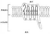
图1:显示跨越感光嵌合GPRC蛋白的实施方案的细胞膜的结构域和方向的示意图,所述感光嵌合GPRC蛋白具有黑视蛋白的N端(NT)、跨膜结构域(TM1-TM7)和胞外环1-3 (EL1-EL3)及mGluR6的胞内环1-3 (IL1-IL3)和C端(CT)。Figure1: Schematic showing the domains and orientation spanning the cell membrane of an embodiment of a photosensitive chimeric GPRC protein with an N-terminal (NT), transmembrane domain (TM1-TM7) of melanopsin and extracellular loop 1-3 (EL1-EL3) and intracellular loop 1-3 (IL1-IL3) and C-terminal (CT) of mGluR6.
图2:实施例1:用小鼠mGluR6-黑视蛋白(MGluR6的IL2(DRIY)、IL3(I)和CT,具有Seq. No.7/8的示例性实施方案D) (具有在HEK293 (GIRK)细胞中测量的最大电流的本发明的优选序列)转染的HEK293(GIRK)细胞的完整细胞电流响应2: Example 1: Using mouse mGluR6-melanopsin( IL2 (DRIY), IL3 (I) and CT of MGluR6, with Seq. No.7/8 Exemplary Embodiment D) (with HEK293 ( Complete Cell Current Response of HEK293 (GIRK) Cells Transfected by Preferred Sequences of the Invention for Maximum Current Measured in GIRK) Cells
图3:实施例1:向外K+电流Figure3: Example 1: Outward K+ current
图4:实施例2:使用rAAV2衣壳突变体载体进行的小鼠ON-双极细胞的成功和特异的mGluR6-黑视蛋白转导Figure4: Example 2: Successful and specific mGluR6-melanopsin transduction of mouse ON-bipolar cells using rAAV2 capsid mutant vectors
图5:在使用rAAV2载体将mGluR6-黑视蛋白导入视网膜ON双极细胞后1个月,自8周龄rd1小鼠视网膜(无光感受器细胞的视网膜)的视网膜神经节细胞中记录的光反应Figure5: Light responses recorded from retinal ganglion cells of 8-week-old rd1 mouse retina (photoreceptor-free retina) 1 month after mGluR6-melanopsin was introduced into retinal ON bipolar cells using rAAV2 vector
图6:用兔抗Rab1A抗体的免疫标记显示失明rd1小鼠的暗适应视网膜呈光适应去极化状态。Figure6: Immunolabeling with rabbit anti-Rab1A antibody shows light-adapted depolarization in the dark-adapted retina of blind rd1 mice.
一些实施方案的详述DETAILED DESCRIPTION OF SOME EMBODIMENTS
所需结构域的鉴定和任何特定结构域的N末端和C末端处的合适切割和连接部位的确定主要根据:1)基因序列/保守残基的比对,和2)应用例如CLC Protein Workbench、I-TASSER、MODELLER、QUARK或SWISS-Model (Kiefer F等,Nucleic Acids Res 37,D387-D392,2009),进行感光GPCR家族成员和mGluR6的二级结构和三级结构的计算机建模。Identification of desired domains and determination of suitable cleavage and ligation sites at the N-terminal and C-terminal ends of any particular domain is primarily based on: 1) alignment of gene sequences/conserved residues, and 2) applications such as CLC Protein Workbench, I-TASSER, MODELLER, QUARK or SWISS-Model (Kiefer F et al., NucleicAcids Res 37, D387-D392, 2009), for computer modeling of the secondary and tertiary structures of photosensitive GPCR family members and mGluR6.
在感光嵌合GPCR蛋白的一些实施方案中,第一GPCR成员是黑视蛋白,特别是人或小鼠黑视蛋白,第二GPCR成员是人或小鼠mGluR6。嵌合感光GPCR蛋白的这些实施方案简称为mGluR6-黑视蛋白。In some embodiments of the light-sensitive chimeric GPCR protein, the first GPCR member is melanopsin, particularly human or mouse melanopsin, and the second GPCR member is human or mouse mGluR6. These embodiments of the chimeric photosensitive GPCR protein are referred to simply as mGluR6-melanopsin.
下文中更详细地描述了构建感光mGluR6-黑视蛋白的数个实施方案。本发明的范围不限于这些具体的实施方案。在一些实施方案中,IL2、IL3和CT来源于mGluR6,而嵌合体的其余部分来源于黑视蛋白。在一些其它的实施方案中,所有3个胞内环IL1-IL3和CT来源于mGluR6,所有跨膜和胞外结构域来源于黑视蛋白。Several embodiments for the construction of light-sensitive mGluR6-melanopsin are described in more detail below. The scope of the present invention is not limited to these specific embodiments. In some embodiments, IL2, IL3, and CT are derived from mGluR6, while the remainder of the chimera is derived from melanopsin. In some other embodiments, all three intracellular loops IL1-IL3 and CT are derived from mGluR6 and all transmembrane and extracellular domains are derived from melanopsin.
图1图解地显示了跨越一个实施方案的细胞膜的结构域和方向,该实施方案具有黑视蛋白的N端(NT)、跨膜结构域(TM1-TM7)和胞外环1-3 (EL1-EL3)和mGluR6的胞内环1-3 (IL1-IL3)和C端(CT)。7个剪接位点用字母a-g标示。基本上,黑视蛋白的胞内环被mGluR6的胞内环交换的所有可行的切割和连接位点均在本发明的范围内。Figure1 schematically shows the domains and orientation spanning the cell membrane of an embodiment with the N-terminus (NT), transmembrane domain (TM1-TM7) and extracellular loops 1-3 (EL1 -EL3) and the intracellular loop 1-3 (IL1-IL3) and C-terminus (CT) of mGluR6. The seven splice sites are marked with the letters ag. Essentially all feasible cleavage and ligation sites where the intracellular loop of melanopsin is exchanged for the intracellular loop of mGluR6 are within the scope of the present invention.
表1公开了构建mGluR6-黑视蛋白实施方案的特别成功的多个剪接位点,其根据序列比对和3D-建模来选择,并发现是功能上有活性的。对于功能性mGluR6-黑视蛋白嵌合体的构建,存在可变剪接选择的各种组合,并且所述组合在本发明的范围内。Table1 discloses particularly successful multiple splice sites for constructing mGluR6-melanopsin embodiments, which were selected based on sequence alignment and 3D-modelling, and found to be functionally active. For the construction of functional mGluR6-melanopsin chimeras, various combinations of alternative splicing options exist and are within the scope of the present invention.
人mGluR6-黑视蛋白的经测定的功能性剪接-连接位点:Determined functional splice-junction sites for human mGluR6-melanopsin:

对于嵌合mGluR6-黑视蛋白的实施方案,其中黑视蛋白的IL2、IL3和C端结构域用mGluR6的相应结构域交换,并且需要根据图1编号为c-g的位点处跨膜结构域和胞内环之间的基因剪接和连接。表1中标示的剪接位点d、e、f、g产生环置换,按照实施例1的方法测定这是有功能的。对于剪接位点e,所测定的2种剪接形式是有功能的,列于表中作为形式I和II。虽然特别推荐根据表1的剪接位点d、e、f、g,但产生能够使光激活与mGluR6的信号转导级联偶联的感光mGluR6-黑视蛋白的任何剪接形式均在本发明的范围内。For the embodiment of chimeric mGluR6-melanopsin, wherein the IL2, IL3 and C-terminal domains of melanopsin are exchanged with the corresponding domains of mGluR6, and the transmembrane domain and Gene splicing and joining between intracellular loops. The splice sites d, e, f, g indicated in Table 1 produced loop displacements which were determined to be functional as in Example 1. For splice site e, the two splice forms determined to be functional are listed in the table as forms I and II. Although splice sites d, e, f, g according to Table 1 are particularly recommended, any splice form that produces a light-sensitive mGluR6-melanopsin capable of coupling light activation to the signal transduction cascade of mGluR6 is within the scope of the invention. within range.
对于TM3和IL2间的位点c处的剪接和连接,当比较黑视蛋白和mGluR6的氨基酸序列时,有若干可用选择。具有合理的氨基酸序列和3D结构同源性的任何剪接形式均在本发明的范围内。似乎重要的是保持TM3和IL2间的DRY位点,这是GPCR蛋白中最保守的氨基酸序列。值得注意的是,DRY位点的其它功能性变体包括DRIY、NRIY或NRY。在根据实施例1的测定中,所有的这些变体产生功能性mGluR6-黑视蛋白嵌合体。For splicing and ligation at site c between TM3 and IL2, there are several options available when comparing the amino acid sequences of melanopsin and mGluR6. Any spliced form with reasonable amino acid sequence and 3D structural homology is within the scope of the invention. It seems important to maintain the DRY site between TM3 and IL2, which is the most conserved amino acid sequence among GPCR proteins. Of note, other functional variants of the DRY site include DRIY, NRIY or NRY. In the assay according to Example 1, all of these variants produced functional mGluR6-melanopsin chimeras.
对于其中另外用mGluR6的IL1交换黑视蛋白的IL1的嵌合mGluR6-黑视蛋白的实施方案,按照图1编号为a和b的位点处的跨膜结构域TM1和TM2及胞内环IL1之间还需要额外的基因剪接和连接。与位点c-g相比,剪接和连接位点a和b区域中有关保守氨基酸的百分比和大部分有关3D结构预测的黑视蛋白和mGluR6的序列间的同源性较低,这就扩大了最佳剪接和连接位点的选择。用包含来源于mGluR6的IL1的实施方案的初步实验产生功能性嵌合体,预期IL1的最佳交换可增加嵌合蛋白的特异性G蛋白偶联。考虑其保守氨基酸序列,用mGluR6的IL1交换黑视蛋白的IL1的所有可行的切割和连接位点均在本发明的范围内。For the embodiment of chimeric mGluR6-melanopsin in which IL1 of melanopsin is additionally exchanged for IL1 of mGluR6, the transmembrane domains TM1 and TM2 and the intracellular loop IL1 at the sites numbered a and b according to Figure 1 Additional gene splicing and ligation are also required. Compared with sites c-g, the percentage of conserved amino acids in the regions of splicing and junction sites a and b and most of the homology between the sequences of melanopsin and mGluR6 related to the 3D structure prediction are lower, which expands the maximum Selection of optimal splicing and joining sites. Preliminary experiments with embodiments comprising IL1 derived from mGluR6 generated functional chimeras, and optimal exchange of IL1 is expected to increase specific G protein coupling of the chimeric protein. All feasible cleavage and ligation sites for exchanging IL1 of melanopsin with IL1 of mGluR6 are within the scope of the present invention, taking into account its conserved amino acid sequence.
对于mGluR6-黑视蛋白嵌合蛋白的下列示例性实施方案A-E,整个DNA基因和氨基酸序列用对应于各种结构域(例如胞内(IL)和胞外(EL)环、N端和C端结构域(NT、CT)和跨膜结构域(TM))的编码序列的标示列出。For the following exemplary embodiments A-E of the mGluR6-melanopsin chimeric protein, the entire DNA gene and amino acid sequences are represented in terms corresponding to the various structural domains (e.g., intracellular (IL) and extracellular (EL) loops, N-terminal and C-terminal The coding sequences of the domains (NT, CT) and the transmembrane domain (TM)) are listed by designation.
AA:具有来源于: has derived frommGluR6mGluR6的ofIL2(DRIY)IL2 (DRIY)、,IL3IL3剪接形式splice formII和andCTCT的人peoplemGluR6-mGluR6-黑视蛋白实施方案Melanopsin embodiment
Seq. No. 1:DNA序列Seq. No. 1: DNA sequence
编码嵌合体的DNA序列(使用人基因)。下划线区域编码mGluR6胞内结构域(IL2、IL3 (剪接形式I)和CT)。DNA sequences encoding chimeras (using human genes). Underlined regions encode mGluR6 intracellular domains (IL2, IL3 (splice form I) and CT).

Seq. No. 2:氨基酸序列Seq. No. 2: Amino acid sequence
嵌合肽序列(使用人基因)。下划线区域编码mGluR6胞内结构域(IL2、IL3 (剪接形式I)和CT)。黑体字的AA形成EL,加框的残基Y和K参与生色团结合。Chimeric peptide sequences (using human genes). The underlined region encodes the intracellular domain of mGluR6 (IL2 , IL3 (splice form I) and CT). AA in bold forms EL and boxed residues Y and K participate in chromophore binding.

BB:具有来源于: has derived frommGluR6mGluR6的ofIL2(DRIY)IL2 (DRIY)、,IL3IL3剪接形式splice formIIII和andCTCT的人peoplemGluR6-mGluR6-黑视蛋白实施方案Melanopsin embodiment
Seq. No. 3:DNA序列Seq. No. 3: DNA sequence
编码嵌合体的DNA序列(使用人基因)。下划线区域编码mGluR6胞内结构域(IL2、IL3 (剪接形式II)和CT)。DNA sequences encoding chimeras (using human genes). Underlined regions encode mGluR6 intracellular domains (IL2, IL3 (splice form II) and CT).
Seq. No. 4:氨基酸序列Seq. No. 4: Amino acid sequence
嵌合肽序列(使用人基因)。下划线区域编码mGluR6胞内结构域(IL2

CC:具有来源于: has derived frommGluR6mGluR6的ofIL1IL1、,IL2(DRIY)IL2 (DRIY)、,IL3IL3剪接形式splice formII和andCTCT的人peoplemGluR6-mGluR6-黑视蛋白实施方案Melanopsin embodiment
Seq. No. 5:DNA序列Seq. No. 5: DNA sequence
编码嵌合体的DNA序列(使用人基因)。下划线区域编码mGluR6胞内结构域(IL1、IL2、IL3 (剪接形式I)和CT)。DNA sequences encoding chimeras (using human genes). Underlined regions encode mGluR6 intracellular domains (IL1, IL2, IL3 (splice form I) and CT).

Seq. No. 6:氨基酸序列Seq. No. 6: Amino acid sequence
嵌合肽序列(使用人基因)。下划线区域编码mGluR6胞内结构域(IL1、IL2、IL3 (剪接形式I)和CT)。黑体字的AA形成EL,加框的Y和K残基参与生色团结合。Chimeric peptide sequences (using human genes). The underlined region encodes the intracellular domain of mGluR6 (IL1, IL2 , IL3 (splice form I) and CT). The AA in bold forms the EL, and the boxed Y and K residues are involved in chromophore binding.

DD.:具有来源于: has derived frommGluR6mGluR6的ofIL2(DRIY)IL2 (DRIY)、,IL3IL3剪接形式splice formII和andCTCT的小鼠the mousemGluR6-mGluR6-黑视蛋白melanopsin((按照实施方案According to the implementation planA)A)
Seq. No. 7:DNA序列Seq. No. 7: DNA sequence
编码嵌合体的DNA序列(使用小鼠基因)。下划线区域编码mGluR6胞内结构域(IL2、IL3 (剪接形式I)和CT)。DNA sequences encoding chimeras (using mouse genes). Underlined regions encode mGluR6 intracellular domains (IL2, IL3 (splice form I) and CT).

Seq. No. 8:氨基酸序列Seq. No. 8: Amino acid sequence
嵌合肽序列(使用小鼠基因)。下划线区域编码mGluR6胞内结构域(IL2

EE.:具有来源于: has derived frommGluR6mGluR6的ofIL1IL1、,IL2(DRIY)IL2 (DRIY)、,IL3IL3剪接形式splice formII和andCTCT的小鼠the mousemGluR6-mGluR6-黑视蛋白melanopsin((按照实施方案According to the implementation planC)c)
Seq. No. 9:DNA序列Seq. No. 9: DNA sequence
编码嵌合体的DNA序列(使用小鼠基因)。下划线区域编码mGluR6胞内结构域(IL1、IL2、IL3 (剪接形式I)和CT)。DNA sequences encoding chimeras (using mouse genes). Underlined regions encode mGluR6 intracellular domains (IL1, IL2, IL3 (splice form I) and CT).

Seq. No. 10:氨基酸序列Seq. No. 10: Amino acid sequence
嵌合肽序列(使用小鼠基因)。下划线区域编码mGluR6胞内结构域(IL1、IL2
通过示例性mGluR6嵌合GPCR蛋白,特别通过示例性mGluR6-黑视蛋白嵌合蛋白,表明光激活与mGluR6的信号转导级联的实施例:Examples of light activation of the signal transduction cascade with mGluR6 are shown by exemplary mGluR6 chimeric GPCR proteins, in particular by exemplary mGluR6-melanopsin chimeric proteins:
在这些的第一实验中,在培养的稳定表达GIRK钾通道的人胚肾细胞(HEK293细胞)中进行了mGluR6-黑视蛋白嵌合体的功能分析:In the first experiments of these, functional analysis of the mGluR6-melanopsin chimera was performed in cultured human embryonic kidney cells (HEK293 cells) stably expressing the GIRK potassium channel:
该实验在HEK293细胞中测定了示例性实施方案D的嵌合体(Seq. No. 7/8)的光激活与GIRK通道的功能性偶联,一种已知的功能性激活的mGluR6的能力,并且需要感光mGluR6-黑视蛋白的实施方案在培养的稳定表达GIRK钾通道的人胚肾细胞(HEK293细胞) (HEK293-GIRK细胞)中表达。This experiment determined the ability of photoactivation of the chimera of exemplary embodiment D (Seq. No. 7/8) to be functionally coupled to the GIRK channel, a known functionally activated mGluR6, in HEK293 cells, And embodiments requiring photosensitive mGluR6-melanopsin to be expressed in cultured human embryonic kidney cells (HEK293 cells) stably expressing GIRK potassium channels (HEK293-GIRK cells).
在HEK293-GIRK细胞中,mGluR6在胞内通过G蛋白与异聚的Kir3.1/3.2钾通道(GIRK通道)偶联。因此,可通过GIRK通道的激活,产生电生理学实验中可测量的K+-电流,来间接显示mGluR6-黑视蛋白嵌合体的成功光激活,如图2和图3中所示。In HEK293-GIRK cells, mGluR6 is intracellularly coupled to heteromeric Kir3.1/3.2 potassium channels (GIRK channels) via G proteins. Thus, successful photoactivation of the mGluR6-melanopsin chimera can be shown indirectly through activation of the GIRK channel, generating a K+ -current measurable in electrophysiological experiments, as shown in FIGS. 2 and 3 .
图2显示从用示例性实施方案D的嵌合体(Seq. No. 7/8)转染的HEK293-GIRK细胞记录的对-150和+60 mV之间的1-s电压递增(voltage ramp)的完整细胞电流响应。当mGluR6-黑视蛋白嵌合体被蓝(473 nm)光激活(暗灰色迹线)时,GIRK通道被激活。在不存在光(无mGluR6-黑视蛋白激活,浅灰色三角形)和存在光(有mGluR6-黑视蛋白激活,暗灰色圆形)时测定电流。差异表示为粗黑线,并表示GIRK-通道的电流-电压关系。Figure2 shows the 1-s voltage ramp between -150 and +60 mV recorded from HEK293-GIRK cells transfected with the chimera of exemplary embodiment D (Seq. No. 7/8) complete cell current response. GIRK channels are activated when the mGluR6-melanopsin chimera is activated by blue (473 nm) light (dark gray trace). Currents were measured in the absence of light (no mGluR6-melanopsin activation, light gray triangles) and in the presence of light (mGluR6-melanopsin activation, dark gray circles). Differences are indicated as thick black lines and indicate the current-voltage relationship of GIRK-channels.
图3显示用示例性实施方案D的相同实施方案的mGluR6-黑视蛋白嵌合体(Seq. No. 7/8)转染的HEK293-GIRK细胞的完整细胞膜片钳实验结果。在473 nm照明期间,通过GIRK通道的向外K+-电流变得可见为超极化电流。Figure3 shows the results of a whole cell patch clamp experiment on HEK293-GIRK cells transfected with the mGluR6-melanopsin chimera (Seq. No. 7/8) of the same embodiment as Exemplary Embodiment D. During 473 nm illumination, outward K+ -current through GIRK channels becomes visible as hyperpolarizing current.
图2和图3中所示的用示例性实施方案D的mGluR6-黑视蛋白嵌合体(Seq. No. 7/8)进行的结果表明:The results shown in Figures 2 and 3 with the mGluR6-melanopsin chimera of Exemplary Embodiment D (Seq. No. 7/8) show that:
— 嵌合体的胞外黑视蛋白部分被蓝光激活,并且在蓝光关闭时关闭。— The extracellular melanopsin portion of the chimera is activated by blue light and turns off when blue light is turned off.
— 嵌合体的胞内mGluR6部分通过G蛋白与GIRK钾通道成功偶联,使得在光刺激期间测得向外K+-电流,这显示了GIRK通道特有的动力学。— The intracellular mGluR6 moiety of the chimera was successfully coupled to the GIRK potassium channel via the G protein, allowing an outward K+ -current to be measured during photostimulation, revealing kinetics specific to GIRK channels.
因此得出结论,mGluR6-黑视蛋白嵌合体是有功能的。It was therefore concluded that the mGluR6-melanopsin chimera is functional.
本领域已知的基因治疗方法可应用于能够使光激活与mGluR6的信号转导级联偶联的感光GPCR嵌合蛋白的表达。下面描述了两种具体方法rAAV转导和电穿孔,但本发明不限于这些具体的示例性方法:Gene therapy methods known in the art can be applied to the expression of a light-sensitive GPCR chimeric protein capable of coupling light activation to the signal transduction cascade of mGluR6. Two specific methods of rAAV transduction and electroporation are described below, but the invention is not limited to these specific exemplary methods:
rAAV转导是本领域已知应用方法的第一个实例:首先小心地用皮下注射针头刺穿巩膜,然后将约1微升的rAAV (相当于约1010基因组拷贝)经视网膜下(Pang JJ等, Invest Ophthalmol Vis Sci. 49(10):4278-83,2008)或玻璃体内(更安全,可能更有效—Park等, Gene Ther 16(7):916-926,2009)注射到眼中。在约4周后,嵌合体表达,并可进行电生理学/形态学实验。rAAV transduction is the first example of an applied method known in the art: first the sclera is carefully pierced with a hypodermic needle, and then about 1 microliter of rAAV (corresponding to about10 genome copies) is injected subretinally (Pang JJ et al., Invest Ophthalmol Vis Sci. 49(10):4278-83, 2008) or intravitreally (safer and possibly more effective—Park et al., Gene Ther 16(7):916-926, 2009) into the eye. After approximately 4 weeks, the chimera is expressed and electrophysiology/morphology experiments can be performed.
用于基因递送的rAAV穿梭载体(shuttle)具有多个基因治疗优势:The rAAV shuttle vector (shuttle) for gene delivery has several gene therapy advantages:
a) rAAV2是目前用于基因疗法的最成功的载体,它们显示最小的免疫原性(Buch PK等, Gene Ther 15:849-857,2008)。a) rAAV2 is currently the most successful vector for gene therapy and they show minimal immunogenicity (Buch PK et al., Gene Ther15:849-857, 2008).
b)存在具有不同细胞特异性的若干血清型。衣壳苯丙氨酸(F)向酪氨酸(F)突变的血清型8 {rAAV2/8 (Y733F)}和血清型2 {rAAV2/2 (Y252,272,444,500,704,730F)}目前是最有前景的rAAV穿梭载体以转导内视网膜细胞(Pang JJ等, Gen Ther 19(2):234-242,2011;Petrs-Silva H等, Mol Ther 19(2):293-301,2011)。b) There are several serotypes with different cell specificities. Capsid phenylalanine (F) to tyrosine (F) mutated serotype 8 {rAAV2/8 (Y733F)} and serotype 2 {rAAV2/2 (Y252,272,444,500,704,730F)} are currently the most promising rAAV shuttle vectors to transduce inner retinal cells (Pang JJ et al, Gen Ther 19(2):234-242, 2011; Petrs-Silva H et al, Mol Ther 19(2):293-301, 2011).
c) rAAV递送导致长期的DNA表达(数年或甚至永久)—单次rAAV治疗是充分的,无必要再施用。c) rAAV delivery results in long-term DNA expression (years or even permanently) - a single rAAV treatment is sufficient and re-administration is not necessary.
d) 可通过例如以下方面,实现DNA-定位于ON-双极细胞:d) DNA-localization to ON-bipolar cells can be achieved, for example, by:
I) rAAV血清型(rAAV2/8和rAAV2/2,对于内视网膜细胞目前最有前景),I) rAAV serotypes (rAAV2/8 and rAAV2/2, currently most promising for inner retinal cells),
II)特异性ON-双极细胞表面蛋白(即夜盲蛋白、mGluR6、TRPM12)的rAAV受体靶向,II) rAAV receptor targeting of specific ON-bipolar cell surface proteins (i.e. night blindness, mGluR6, TRPM12),
III)通常使用ON-双极细胞特异性启动子或增强子/启动子序列(mGluR6和mGluR6/sv40启动子,或者启动子/增强子序列来源于Gγ13、夜盲蛋白或TRPM12的启动子/增强子序列),III) Typically ON-bipolar cell-specific promoters or enhancer/promoter sequences are used (mGluR6 and mGluR6/sv40 promoters, or promoter/enhancer sequences derived from those of Gγ13, nyctalin or TRPM12 sequence),
IV) mGluR6特异性G蛋白Gα(o)只存在于ON-双极细胞中,因此只有ON-双极细胞可有效地使mGluR6与其酶促级联有效偶联。IV) The mGluR6-specific G protein Gα(o) is only present in ON-bipolar cells, so only ON-bipolar cells can effectively couple mGluR6 to its enzymatic cascade.
电穿孔是本领域已知应用方法的第二个实例:将在ON-双极细胞特异性启动子控制下的编码嵌合蛋白的DNA溶于无菌盐水溶液,并经视网膜下注射。注射后接着使用视网膜后面的一个电极和视网膜前面的一个电极施加跨视网膜电压脉冲。电压阶跃的极性在神经节细胞侧为正极,在光感受器侧为负极。电压脉冲起临时使细胞膜透化的作用,与此同时将带负电荷的DNA拉向正极并进入视网膜细胞中(Lagali PS等, Nat Neurosci. 11(6):667-75,2008,Matsuda T和Cepko CL,PNAS 101(1):16-22,2004)。Electroporation is a second example of an applied method known in the art: DNA encoding a chimeric protein under the control of an ON-bipolar cell-specific promoter is dissolved in sterile saline solution and injected subretinally. The injection is followed by the application of transretinal voltage pulses using one electrode behind the retina and one electrode in front of the retina. The polarity of the voltage step is positive on the ganglion cell side and negative on the photoreceptor side. The voltage pulse acts to temporarily permeabilize the cell membrane while at the same time pulling the negatively charged DNA towards the positive pole and into the retinal cells (Lagali PS et al., Nat Neurosci. 11(6):667-75, 2008, Matsuda T and Cepko CL, PNAS101(1):16-22, 2004).
下列实施例证明,在小鼠ON-双极细胞中,编码能够使光激活与信号转导级联特异性偶联的示例性感光GPCR嵌合蛋白的DNA的rAAV转导和表达,特别是编码示例性mGluR6-黑视蛋白嵌合蛋白的DNA的转导和表达:The following examples demonstrate, in mouse ON-bipolar cells, the rAAV transduction and expression of DNA encoding an exemplary light-sensitive GPCR chimeric protein capable of specifically coupling light activation to a signal transduction cascade, specifically encoding Transduction and expression of DNA for an exemplary mGluR6-melanopsin chimeric protein:
在第一系列的实验中,测定了使用酪氨酸-衣壳突变的重组腺伴随病毒rAAV2/8(Y733F)和rAAV2/2(Y252,272,444,500,704,730F)是否将示例性实施方案D的mGluR6-黑视蛋白嵌合基因(Seq. No. 8)递送到小鼠视网膜的ON-双极细胞中。In a first series of experiments, it was determined whether the mGluR6-black Opsin chimeric gene (Seq. No. 8) delivered to ON-bipolar cells in mouse retina.
该实验还测定了使用mGluR6增强子sv40基础启动子元件(Kim DS等,J Neurosci 28(31):7748-64,2008)是否实现mGluR6-黑视蛋白(嵌合体实施方案D)的特异性ON-双极细胞表达。This experiment also determined whether specific ON of mGluR6-melanopsin (chimera embodiment D) was achieved using the mGluR6 enhancer sv40 basal promoter element (Kim DS et al., J Neurosci 28(31):7748-64, 2008). - Bipolar cell expression.
结果见图4,证明使用rAAV2/2衣壳突变体载体在视网膜下或玻璃体内给予后6周,小鼠视杆和视锥ON-双极细胞的成功和特异性mGluR6-黑视蛋白转导,详述如下:The results,shown in Figure4 , demonstrate successful and specific mGluR6-melanopsin transduction of mouse rod and cone ON-bipolar cells using rAAV2/2 capsid mutant vectors 6 weeks after subretinal or intravitreal administration , as detailed below:
使用含有6个衣壳苯丙氨酸(F)向酪氨酸(F)突变的rAAV2/2载体(Y252,272,444,500,704,730F;Petrs-Silva H等,Mol Ther 19(2):293-301,2011),用PRmGluR6/sv40-“mGluR6-黑视蛋白”-IRES-TurboFP635转导的小鼠视网膜的切片。在解剖分析前6周,视网膜下注射病毒。转基因(mGluR6-黑视蛋白)和报道分子(TurboFP635)的表达受mGluR6增强子sv40基础启动子元件(Kim DS等,J Neurosci 28(31):7748-64,2008)驱动。在第一个图片中,用DAPI的核染色显示视网膜外核层(ONL)、内核层(INL)和神经节细胞层(GCL)。第二个图片中,所有视杆ON双极细胞使用PKC α抗体标记。最后一个图片(rAAV)显示TurboFP635报道基因,因此表明用PRmGluR6/sv40-“mGluR6-黑视蛋白”-IRES-TurboFP635构建体的成功转导。rAAV2/2 vectors containing six capsid phenylalanine (F) to tyrosine (F) mutations (Y252,272,444,500,704,730F; Petrs-SilvaH et al., Mol Ther 19(2):293-301, 2011), Sections of mouse retina transduced with PRmGluR6/sv40-"mGluR6-melanopsin"-IRES-TurboFP635. Virus was injected subretinally 6 weeks prior to dissection analysis. Expression of the transgene (mGluR6-melanopsin) and reporter (TurboFP635) is regulated by the mGluR6 enhancer sv40 basal promoter element (Kim DS et al., J Neurosci28(31):7748-64, 2008) Drive. In the first image, nuclear staining with DAPI shows the retinal outer nuclear layer (ONL), inner nuclear layer (INL) and ganglion cell layer (GCL). In the second image, all rod ON bipolar cells were labeled with PKCα antibody. The last picture (rAAV) shows the TurboFP635 reporter gene, thus indicating successful transduction with the PRmGluR6/sv40-"mGluR6-melanopsin"-IRES-TurboFP635 construct.
5个视杆ON双极细胞显示报道分子标记(实心箭头),而4个INL内标记的其它细胞可能指示视锥ON双极细胞(空心箭头)。这是原理性证明(proof-of-principle):光可激活蛋白mGluR6-黑视蛋白可使用rAAV载体导入,并且可在靶细胞(ON双极细胞)中特异性表达,所述rAAV载体允许用于人眼的临床基因治疗性治疗(Jacobson S等,Arch Ophthalmol298v1-16,2011)。比例条表示10 μm。Five rod ON bipolar cells show reporter labeling (solid arrows), while other cells labeled within the 4 INL may indicate cone ON bipolar cells (open arrows). This is a proof-of-principle: the light-activatable protein mGluR6-melanopsin can be introduced and expressed specifically in target cells (ON bipolar cells) using rAAV vectors that allow the use of Clinical gene therapy therapy for the human eye (JacobsonS et al., Arch Ophthalmol 298v1-16, 2011). Scale bar represents 10 μm.
本领域已知的电生理学方法可用来测定在失明rd1 (Pde6brd1) FVB/N小鼠的视网膜ON-双极细胞中表达的mGluR6-黑视蛋白的适当功能。Electrophysiological methods known in the art can be used to determine the proper function of mGluR6-melanopsin expressed in retinal ON-bipolar cells of blind rd1 (Pde6brd1 ) FVB/N mice.
因此,在第二系列的实验中,小鼠视网膜中mGluR6-黑视蛋白嵌合体的离体功能分析显示,导入到失明rd1小鼠(无光感受器)视网膜的ON-双极细胞中的mGluR6-黑视蛋白使视网膜具有感光性。Thus, in a second series of experiments, ex vivo functional analysis of mGluR6-melanopsin chimeras in the mouse retina revealed that mGluR6- Melanopsin makes the retina light sensitive.
图5显示失明rd1小鼠视网膜整铺片中不同类型的神经节细胞的光反应的3个实例,所述失明rd1小鼠已用含mGluR6-黑视蛋白(嵌合体实施方案D Seq. No. 7)基因的rAAV治疗。Figure5 shows 3 examples of light responses of different types of ganglion cells in retinal whole mounts of blind rd1 mice treated with mGluR6-melanopsin (chimera embodiment D Seq. No. 7) Genetic rAAV therapy.
具体地说,在使用rAAV载体将mGluR6-黑视蛋白导入视网膜ON双极细胞后1个月,从9周龄rd1小鼠视网膜(无光感受器细胞的视网膜)的视网膜神经节细胞记录光反应,详述如下:Specifically, light responses were recorded from retinal ganglion cells of 9-week-old rd1 mouse retina (retina without photoreceptor cells) 1 month after mGluR6-melanopsin was introduced into retinal ON bipolar cells using rAAV vector, The details are as follows:
显示了来自3种细胞类型的胞外反应,瞬时ON细胞(A)、瞬时OFF细胞(B)和持续ON细胞(C)。每条迹线(D-F)邻近的光栅图表明,对相同光刺激的光反应是可再现的。在胞外迹线下灰色表明的持续时间内,将465-nm光投射到视网膜整铺片上。Extracellular responses from 3 cell types are shown, transient ON cells (A), transient OFF cells (B) and persistent ON cells (C). The raster plot adjacent to each trace (D–F) demonstrates that the photoresponse to the same photostimulus is reproducible. 465-nm light was projected onto retinal whole mounts for the duration indicated in gray under the extracellular trace.
并且注意到,持续反应(B)不太可能是黑视蛋白神经节细胞的反应,已知黑视蛋白神经节细胞在不存在光感受器输入时,具有显著较慢的峰值开始(>2.5秒;Schmidt TM等,Neurophysiol 100(1):371-84,2008)。Also note that the sustained response (B) is unlikely to be that of a melanopsin ganglion cell, which is known to have a significantly slower onset of peak in the absence of photoreceptor input (>2.5 sec; Schmidt TM et al., Neurophysiol100(1):371-84, 2008).
因此,图5中所示结果证明,在ON-双极细胞中表达的mGluR6-黑视蛋白能够恢复失明视网膜的感光性。Thus, the results shown in Figure 5 demonstrate that mGluR6-melanopsin expressed in ON-bipolar cells is able to restore photosensitivity in blind retinas.
总之,图4和5表明:In summary, Figures 4 and 5 show that:
- rAAV能够将mGluR6-黑视蛋白基因递送至ON-双极细胞,所述rAAV可允许用于人眼的临床基因治疗性治疗(Jacobson S等,Arch Ophthalmol298v1-16,2011)。- rAAV capable of delivering the mGluR6-melanopsin gene to ON-bipolar cells may allow for clinical gene therapy treatment of the human eye (JacobsonS et al., Arch Ophthalmol 298v1-16, 2011).
- rAAV血清型,可使用rAAV衣壳突变和细胞特异性启动子/增强子元件以特异性靶向ON-双极细胞用于mGluR6-黑视蛋白表达。- rAAV serotypes, rAAV capsid mutations and cell-specific promoter/enhancer elements can be used to specifically target ON-bipolar cells for mGluR6-melanopsin expression.
- 表达的mGluR6-黑视蛋白是有功能的,并使失明视网膜具有感光性。-Expressed mGluR6-melanopsin is functional and renders the blind retina photosensitive.
因此得出结论,mGluR6-黑视蛋白在其靶细胞(视网膜的双极细胞)中是有功能的。It was therefore concluded that mGluR6-melanopsin is functional in its target cells, the bipolar cells of the retina.
ON-双极细胞的最佳光感受器应给出大的差异光反应。在光刺激时,mGluR6-黑视蛋白使ON-双极细胞超极化,这与去极化的通道视紫红质相反。由于失明rd1小鼠中的ON双极细胞已处于光适应(去极化)状态,mGluR6-黑视蛋白光激活产生大的差异光反应。Optimal photoreceptors for ON-bipolar cells should give large differential light responses. Upon photostimulation, mGluR6-melanopsin hyperpolarizes ON-bipolar cells, in contrast to depolarized channelrhodopsin. Since ON bipolar cells in blind rd1 mice are already in a light-adapted (depolarized) state, mGluR6-melanopsin photoactivation produces large differential light responses.
图6显示暗适应的失明rd1 (Pde6brd1) FVB/N小鼠的视网膜处于光适应状态。Figure6 shows that the retinas of dark-adapted blind rd1 (Pde6brd1 ) FVB/N mice are in a light-adapted state.
图片A – D显示遍及失明和野生型小鼠的小鼠视网膜的切片,所述切片用兔抗Rab1A抗体免疫标记以显示失明rd1小鼠的暗适应视网膜实际上处于“光适应” (去极化)状态,这相当于野生型视网膜的“光适应”状态。抗Rab1A抗体标记内视网膜的ON双极细胞(内核层(INL),神经节细胞层(GCL)的末端),在健康视网膜中,其表达水平取决于环境光强度(Huang W等,J Vis Neurosci 26(5-6):443-452,2009)。如所预料的,抗Rab1A免疫标记(黑色结构)仅见于光适应(B)中,在暗适应(A)野生型(BL6)小鼠视网膜中则无。然而,抗Rab1A表达水平在暗适应(C)和光适应(D) rd1视网膜中相同,找不到含有光感受器的外核层(ONL),而抗Rab1A表达水平与光适应健康BL6视网膜类似。Panels A - D show sections throughout the mouse retina of blind and wild-type mice that were immunolabeled with rabbit anti-Rab1A antibody to show that the dark-adapted retina of blind rd1 mice is actually "light-adapted" (depolarized). ) state, which corresponds to the "photoadapted" state of wild-type retinas. Anti-Rab1A antibody labels ON bipolar cells of the inner retina (inner inner layer (INL), end of ganglion cell layer (GCL)), and in healthy retina, the expression level depends on the ambient light intensity (Huang W et al., J VisNeurosci 26(5-6):443-452, 2009). As expected, anti-Rab1A immunolabeling (black structures) was only seen in light-adapted (B) but not dark-adapted (A) wild-type (BL6) mouse retinas. However, anti-Rab1A expression levels were the same in dark-adapted (C) and light-adapted (D) rd1 retinas, and the outer nuclear layer (ONL) containing photoreceptors could not be found, whereas anti-Rab1A expression levels were similar to light-adapted healthy BL6 retinas.
因此得出结论,失明小鼠的rd1视网膜永久处于光适应(去极化)状态。因此,最佳光感受器应在光刺激时使ON双极细胞超极化以保证大的差异光信号,并且mGluR6-黑视蛋白也是如此。所有图的成像曝光时间相同。It was thus concluded that the rd1 retinas of blind mice were permanently light-adapted (depolarized). Thus, optimal photoreceptors should hyperpolarize ON bipolar cells upon photostimulation to guarantee a large differential light signal, and so does mGluR6-melanopsin. Imaging exposure times were the same for all panels.


Claims (22)
Priority Applications (1)
| Application Number | Priority Date | Filing Date | Title |
|---|---|---|---|
| CN201710294286.2ACN107383184A (en) | 2011-06-24 | 2012-06-22 | photosensitive chimeric GPCR protein |
Applications Claiming Priority (4)
| Application Number | Priority Date | Filing Date | Title |
|---|---|---|---|
| US201161500863P | 2011-06-24 | 2011-06-24 | |
| US61/500,863 | 2011-06-24 | ||
| US61/500863 | 2011-06-24 | ||
| PCT/CH2012/000138WO2012174674A1 (en) | 2011-06-24 | 2012-06-22 | Light-sensitive chimeric gpcr protein |
Related Child Applications (1)
| Application Number | Title | Priority Date | Filing Date |
|---|---|---|---|
| CN201710294286.2ADivisionCN107383184A (en) | 2011-06-24 | 2012-06-22 | photosensitive chimeric GPCR protein |
Publications (2)
| Publication Number | Publication Date |
|---|---|
| CN103814045Atrue CN103814045A (en) | 2014-05-21 |
| CN103814045B CN103814045B (en) | 2017-05-31 |
Family
ID=46456289
Family Applications (2)
| Application Number | Title | Priority Date | Filing Date |
|---|---|---|---|
| CN201280030926.4AActiveCN103814045B (en) | 2011-06-24 | 2012-06-22 | photosensitive chimeric GPCR protein |
| CN201710294286.2APendingCN107383184A (en) | 2011-06-24 | 2012-06-22 | photosensitive chimeric GPCR protein |
Family Applications After (1)
| Application Number | Title | Priority Date | Filing Date |
|---|---|---|---|
| CN201710294286.2APendingCN107383184A (en) | 2011-06-24 | 2012-06-22 | photosensitive chimeric GPCR protein |
Country Status (19)
| Country | Link |
|---|---|
| US (1) | US10112984B2 (en) |
| EP (2) | EP2723767B8 (en) |
| JP (1) | JP6240067B2 (en) |
| KR (1) | KR102008538B1 (en) |
| CN (2) | CN103814045B (en) |
| AU (1) | AU2012272505B2 (en) |
| BR (2) | BR112013033028A8 (en) |
| CA (1) | CA2840458C (en) |
| DK (1) | DK3029063T3 (en) |
| ES (1) | ES2766398T3 (en) |
| HR (1) | HRP20200004T1 (en) |
| HU (1) | HUE047074T2 (en) |
| LT (1) | LT3029063T (en) |
| PL (2) | PL3029063T3 (en) |
| PT (1) | PT3029063T (en) |
| RU (1) | RU2637367C2 (en) |
| SI (1) | SI3029063T1 (en) |
| WO (1) | WO2012174674A1 (en) |
| ZA (1) | ZA201308824B (en) |
Cited By (4)
| Publication number | Priority date | Publication date | Assignee | Title |
|---|---|---|---|---|
| CN110267972A (en)* | 2016-11-06 | 2019-09-20 | 纳秒示波器科技有限公司 | Optical modulation by multi-featured opsins for vision restoration and other applications |
| CN111527384A (en)* | 2018-01-02 | 2020-08-11 | 昕诺飞控股有限公司 | photomelanosin activity indicator |
| CN115038458A (en)* | 2019-11-29 | 2022-09-09 | 伯尔尼大学 | Chimeric opsin GPCR proteins |
| WO2024212723A1 (en)* | 2023-04-11 | 2024-10-17 | 苏州星明优健生物技术有限公司 | New chimeric opsin gpcr protein for treating retinal neurodegenerative diseases |
Families Citing this family (10)
| Publication number | Priority date | Publication date | Assignee | Title |
|---|---|---|---|---|
| US20140087463A1 (en)* | 2012-09-25 | 2014-03-27 | The Washington University | Optical control of cell signaling |
| EP3007730A1 (en)* | 2013-06-11 | 2016-04-20 | Friedrich Miescher Institute for Biomedical Research | Retinal on bipolar cells-specific artificial promoter |
| GB201403260D0 (en) | 2014-02-25 | 2014-04-09 | Univ Manchester | Treatment of retinal degeneration using gene therapy |
| EP3318285A1 (en)* | 2016-11-08 | 2018-05-09 | Oxford University Innovation Limited | Treatment of eye disease |
| WO2019094904A1 (en) | 2017-11-13 | 2019-05-16 | The Regents Of The University Of California | Compositions and methods for enhancing visual function |
| CN119569849A (en)* | 2019-04-29 | 2025-03-07 | 康福治疗有限公司 | Screening methods and assays for transmembrane proteins, particularly GPCRs |
| WO2021140205A1 (en)* | 2020-01-10 | 2021-07-15 | Confo Therapeutics N.V. | Methods for generating antibodies and antibody fragments and libraries comprising same |
| CN114762723A (en)* | 2020-12-31 | 2022-07-19 | 吴伯骥 | Application of HrpZpst protein in pharmacy for recognizing and activating multiple types of receptors and/or membrane proteins and signal paths thereof |
| CN112675293A (en)* | 2020-12-31 | 2021-04-20 | 吴伯骥 | Application of HrpNECb protein in pharmacy for recognizing and activating multiple types of receptors and/or membrane proteins and signal paths thereof |
| IL317394A (en)* | 2022-06-07 | 2025-02-01 | Adverum Biotechnologies Inc | Melanopsin variants for vision restoration |
Citations (2)
| Publication number | Priority date | Publication date | Assignee | Title |
|---|---|---|---|---|
| WO2006059081A2 (en)* | 2004-11-30 | 2006-06-08 | Imperial Innovations Limited | Methods of treatment using melanopsin |
| EP1891976A1 (en)* | 2006-08-23 | 2008-02-27 | Novartis Forschungsstiftung, Zweigniederlassung Friedrich Miescher Institute for Biomedical Research | Use of light sensitive genes |
Family Cites Families (7)
| Publication number | Priority date | Publication date | Assignee | Title |
|---|---|---|---|---|
| JPH10507934A (en)* | 1995-07-26 | 1998-08-04 | エヌピーエス・ファーマシウティカルズ・インコーポレイテッド | Chimeric receptors and methods for identifying compounds exhibiting activity at metabotropic glutamate receptors, and the use of such compounds in the treatment of neurological disorders and diseases |
| WO2004009022A2 (en) | 2002-07-18 | 2004-01-29 | The General Hospital Corp. | Method for augmenting vision in persons suffering from photoreceptor cell degeneration |
| EP2019588A4 (en) | 2006-05-04 | 2010-11-24 | Univ Wayne State | RECOVERY OF VISUAL REACTIONS BY IN VIVO ADMINISTRATION OF RHODOPSIN NUCLEIC ACIDS |
| US8178496B2 (en)* | 2007-12-21 | 2012-05-15 | The Regents Of The University Of California | Photoreactive regulator of glutamate receptor function and methods of use thereof |
| AU2009274482A1 (en)* | 2008-05-20 | 2010-01-28 | Eos Neuroscience, Inc. | Vectors for delivery of light-sensitive proteins and methods of use |
| RU2374245C1 (en)* | 2008-08-22 | 2009-11-27 | Андрей Александрович Иващенко | Ligand with wide range of simultaneous receptor activity, pharmaceutical composition, method of preparing said composition and medicinal agent |
| NZ602416A (en)* | 2008-11-14 | 2014-08-29 | Univ Leland Stanford Junior | Optically-based stimulation of target cells and modifications thereto |
- 2012
- 2012-06-22CNCN201280030926.4Apatent/CN103814045B/enactiveActive
- 2012-06-22CNCN201710294286.2Apatent/CN107383184A/enactivePending
- 2012-06-22KRKR1020147000648Apatent/KR102008538B1/enactiveActive
- 2012-06-22PLPL15201713Tpatent/PL3029063T3/enunknown
- 2012-06-22CACA2840458Apatent/CA2840458C/enactiveActive
- 2012-06-22BRBR112013033028Apatent/BR112013033028A8/ennot_activeApplication Discontinuation
- 2012-06-22ESES15201713Tpatent/ES2766398T3/enactiveActive
- 2012-06-22BRBR122022007044-2Apatent/BR122022007044B1/enactiveIP Right Grant
- 2012-06-22LTLTEP15201713.3Tpatent/LT3029063T/enunknown
- 2012-06-22PTPT152017133Tpatent/PT3029063T/enunknown
- 2012-06-22SISI201231720Tpatent/SI3029063T1/enunknown
- 2012-06-22USUS14/128,155patent/US10112984B2/enactiveActive
- 2012-06-22JPJP2014516148Apatent/JP6240067B2/enactiveActive
- 2012-06-22RURU2014101708Apatent/RU2637367C2/enactive
- 2012-06-22WOPCT/CH2012/000138patent/WO2012174674A1/enactiveApplication Filing
- 2012-06-22EPEP12731288.2Apatent/EP2723767B8/enactiveActive
- 2012-06-22DKDK15201713.3Tpatent/DK3029063T3/enactive
- 2012-06-22HUHUE15201713Apatent/HUE047074T2/enunknown
- 2012-06-22EPEP15201713.3Apatent/EP3029063B1/enactiveActive
- 2012-06-22AUAU2012272505Apatent/AU2012272505B2/enactiveActive
- 2012-06-22PLPL12731288Tpatent/PL2723767T3/enunknown
- 2013
- 2013-11-22ZAZA2013/08824Apatent/ZA201308824B/enunknown
- 2020
- 2020-01-03HRHRP20200004TTpatent/HRP20200004T1/enunknown
Patent Citations (2)
| Publication number | Priority date | Publication date | Assignee | Title |
|---|---|---|---|---|
| WO2006059081A2 (en)* | 2004-11-30 | 2006-06-08 | Imperial Innovations Limited | Methods of treatment using melanopsin |
| EP1891976A1 (en)* | 2006-08-23 | 2008-02-27 | Novartis Forschungsstiftung, Zweigniederlassung Friedrich Miescher Institute for Biomedical Research | Use of light sensitive genes |
Non-Patent Citations (1)
| Title |
|---|
| TAKAHIRO YAMASHITA ET AL.: ""The Second Cytoplasmic Loop of Metabotropic Glutamate Receptor Functions at the Third Loop Position of Rhodopsin"", 《J. BIOCHEM.》* |
Cited By (6)
| Publication number | Priority date | Publication date | Assignee | Title |
|---|---|---|---|---|
| CN110267972A (en)* | 2016-11-06 | 2019-09-20 | 纳秒示波器科技有限公司 | Optical modulation by multi-featured opsins for vision restoration and other applications |
| CN111527384A (en)* | 2018-01-02 | 2020-08-11 | 昕诺飞控股有限公司 | photomelanosin activity indicator |
| CN111527384B (en)* | 2018-01-02 | 2023-08-29 | 昕诺飞控股有限公司 | Photoblacking visual element activity indicator |
| CN115038458A (en)* | 2019-11-29 | 2022-09-09 | 伯尔尼大学 | Chimeric opsin GPCR proteins |
| WO2024212723A1 (en)* | 2023-04-11 | 2024-10-17 | 苏州星明优健生物技术有限公司 | New chimeric opsin gpcr protein for treating retinal neurodegenerative diseases |
| CN118791624A (en)* | 2023-04-11 | 2024-10-18 | 苏州星明优健生物技术有限公司 | Novel GPCR chimeric opsins for the treatment of retinal neurodegenerative diseases |
Also Published As
| Publication number | Publication date |
|---|---|
| EP3029063A1 (en) | 2016-06-08 |
| RU2014101708A (en) | 2015-07-27 |
| KR102008538B1 (en) | 2019-08-08 |
| BR122022007044A2 (en) | 2017-01-31 |
| US20140171376A1 (en) | 2014-06-19 |
| SI3029063T1 (en) | 2020-03-31 |
| ES2766398T3 (en) | 2020-06-12 |
| ZA201308824B (en) | 2015-02-25 |
| PL3029063T3 (en) | 2020-05-18 |
| WO2012174674A1 (en) | 2012-12-27 |
| PT3029063T (en) | 2020-01-22 |
| BR112013033028A2 (en) | 2017-01-31 |
| KR20140039281A (en) | 2014-04-01 |
| CA2840458C (en) | 2020-09-22 |
| PL2723767T3 (en) | 2016-06-30 |
| LT3029063T (en) | 2020-02-10 |
| EP2723767B1 (en) | 2016-01-06 |
| AU2012272505A1 (en) | 2014-01-16 |
| DK3029063T3 (en) | 2020-01-20 |
| CN107383184A (en) | 2017-11-24 |
| EP2723767A1 (en) | 2014-04-30 |
| HRP20200004T1 (en) | 2020-05-15 |
| BR122022007044A8 (en) | 2022-11-01 |
| CN103814045B (en) | 2017-05-31 |
| EP2723767B8 (en) | 2016-03-02 |
| US10112984B2 (en) | 2018-10-30 |
| JP6240067B2 (en) | 2017-11-29 |
| HUE047074T2 (en) | 2020-04-28 |
| BR112013033028A8 (en) | 2022-11-01 |
| CA2840458A1 (en) | 2012-12-27 |
| BR122022007044B1 (en) | 2023-11-14 |
| JP2014524903A (en) | 2014-09-25 |
| EP3029063B1 (en) | 2019-10-16 |
| RU2637367C2 (en) | 2017-12-04 |
| AU2012272505B2 (en) | 2017-03-02 |
Similar Documents
| Publication | Publication Date | Title |
|---|---|---|
| CN103814045B (en) | photosensitive chimeric GPCR protein | |
| US12343408B2 (en) | AAV-mediated subcellular targeting of heterologous rhodopsins in retinal ganglion cells | |
| US8470790B2 (en) | Restoration of visual responses by in vivo delivery of rhodopsin nucleic acids | |
| JP2021038240A (en) | Methods to Enhance Ocular Delivery of Therapeutic Compounds | |
| US20240174743A1 (en) | Chimeric opsin gpcr proteins | |
| WO2024212723A1 (en) | New chimeric opsin gpcr protein for treating retinal neurodegenerative diseases | |
| WO2024188064A1 (en) | Use of new-type photosensitive ion channel protein chrmine in retinal neurodegenerative diseases |
Legal Events
| Date | Code | Title | Description |
|---|---|---|---|
| C06 | Publication | ||
| PB01 | Publication | ||
| C10 | Entry into substantive examination | ||
| SE01 | Entry into force of request for substantive examination | ||
| GR01 | Patent grant | ||
| GR01 | Patent grant | ||
| CP03 | Change of name, title or address | Address after:Bern Patentee after:Aktos Medical Co.,Ltd. Address before:Swiss Kenitz Patentee before:Haagstreit Medtech AG | |
| CP03 | Change of name, title or address | ||
| TR01 | Transfer of patent right | Effective date of registration:20220928 Address after:Basel, SUI Patentee after:NOVARTIS AG Address before:Bern Switzerland Patentee before:Aktos Medical Co.,Ltd. | |
| TR01 | Transfer of patent right |