CN103747802A - Methods for increasing the efficacy of FOLR1 cancer therapy - Google Patents
Methods for increasing the efficacy of FOLR1 cancer therapyDownload PDFInfo
- Publication number
- CN103747802A CN103747802ACN201280015187.1ACN201280015187ACN103747802ACN 103747802 ACN103747802 ACN 103747802ACN 201280015187 ACN201280015187 ACN 201280015187ACN 103747802 ACN103747802 ACN 103747802A
- Authority
- CN
- China
- Prior art keywords
- folr1
- antibody
- sample
- cancer
- staining
- Prior art date
- Legal status (The legal status is an assumption and is not a legal conclusion. Google has not performed a legal analysis and makes no representation as to the accuracy of the status listed.)
- Granted
Links
Images


Classifications
- A—HUMAN NECESSITIES
- A61—MEDICAL OR VETERINARY SCIENCE; HYGIENE
- A61K—PREPARATIONS FOR MEDICAL, DENTAL OR TOILETRY PURPOSES
- A61K47/00—Medicinal preparations characterised by the non-active ingredients used, e.g. carriers or inert additives; Targeting or modifying agents chemically bound to the active ingredient
- A61K47/50—Medicinal preparations characterised by the non-active ingredients used, e.g. carriers or inert additives; Targeting or modifying agents chemically bound to the active ingredient the non-active ingredient being chemically bound to the active ingredient, e.g. polymer-drug conjugates
- A61K47/51—Medicinal preparations characterised by the non-active ingredients used, e.g. carriers or inert additives; Targeting or modifying agents chemically bound to the active ingredient the non-active ingredient being chemically bound to the active ingredient, e.g. polymer-drug conjugates the non-active ingredient being a modifying agent
- A61K47/68—Medicinal preparations characterised by the non-active ingredients used, e.g. carriers or inert additives; Targeting or modifying agents chemically bound to the active ingredient the non-active ingredient being chemically bound to the active ingredient, e.g. polymer-drug conjugates the non-active ingredient being a modifying agent the modifying agent being an antibody, an immunoglobulin or a fragment thereof, e.g. an Fc-fragment
- A61K47/6835—Medicinal preparations characterised by the non-active ingredients used, e.g. carriers or inert additives; Targeting or modifying agents chemically bound to the active ingredient the non-active ingredient being chemically bound to the active ingredient, e.g. polymer-drug conjugates the non-active ingredient being a modifying agent the modifying agent being an antibody, an immunoglobulin or a fragment thereof, e.g. an Fc-fragment the modifying agent being an antibody or an immunoglobulin bearing at least one antigen-binding site
- A61K47/6851—Medicinal preparations characterised by the non-active ingredients used, e.g. carriers or inert additives; Targeting or modifying agents chemically bound to the active ingredient the non-active ingredient being chemically bound to the active ingredient, e.g. polymer-drug conjugates the non-active ingredient being a modifying agent the modifying agent being an antibody, an immunoglobulin or a fragment thereof, e.g. an Fc-fragment the modifying agent being an antibody or an immunoglobulin bearing at least one antigen-binding site the antibody targeting a determinant of a tumour cell
- A—HUMAN NECESSITIES
- A61—MEDICAL OR VETERINARY SCIENCE; HYGIENE
- A61K—PREPARATIONS FOR MEDICAL, DENTAL OR TOILETRY PURPOSES
- A61K47/00—Medicinal preparations characterised by the non-active ingredients used, e.g. carriers or inert additives; Targeting or modifying agents chemically bound to the active ingredient
- A61K47/50—Medicinal preparations characterised by the non-active ingredients used, e.g. carriers or inert additives; Targeting or modifying agents chemically bound to the active ingredient the non-active ingredient being chemically bound to the active ingredient, e.g. polymer-drug conjugates
- A61K47/51—Medicinal preparations characterised by the non-active ingredients used, e.g. carriers or inert additives; Targeting or modifying agents chemically bound to the active ingredient the non-active ingredient being chemically bound to the active ingredient, e.g. polymer-drug conjugates the non-active ingredient being a modifying agent
- A61K47/68—Medicinal preparations characterised by the non-active ingredients used, e.g. carriers or inert additives; Targeting or modifying agents chemically bound to the active ingredient the non-active ingredient being chemically bound to the active ingredient, e.g. polymer-drug conjugates the non-active ingredient being a modifying agent the modifying agent being an antibody, an immunoglobulin or a fragment thereof, e.g. an Fc-fragment
- A61K47/6801—Drug-antibody or immunoglobulin conjugates defined by the pharmacologically or therapeutically active agent
- A61K47/6803—Drugs conjugated to an antibody or immunoglobulin, e.g. cisplatin-antibody conjugates
- A61K47/68033—Drugs conjugated to an antibody or immunoglobulin, e.g. cisplatin-antibody conjugates the drug being a maytansine
- A—HUMAN NECESSITIES
- A61—MEDICAL OR VETERINARY SCIENCE; HYGIENE
- A61K—PREPARATIONS FOR MEDICAL, DENTAL OR TOILETRY PURPOSES
- A61K47/00—Medicinal preparations characterised by the non-active ingredients used, e.g. carriers or inert additives; Targeting or modifying agents chemically bound to the active ingredient
- A61K47/50—Medicinal preparations characterised by the non-active ingredients used, e.g. carriers or inert additives; Targeting or modifying agents chemically bound to the active ingredient the non-active ingredient being chemically bound to the active ingredient, e.g. polymer-drug conjugates
- A61K47/51—Medicinal preparations characterised by the non-active ingredients used, e.g. carriers or inert additives; Targeting or modifying agents chemically bound to the active ingredient the non-active ingredient being chemically bound to the active ingredient, e.g. polymer-drug conjugates the non-active ingredient being a modifying agent
- A61K47/68—Medicinal preparations characterised by the non-active ingredients used, e.g. carriers or inert additives; Targeting or modifying agents chemically bound to the active ingredient the non-active ingredient being chemically bound to the active ingredient, e.g. polymer-drug conjugates the non-active ingredient being a modifying agent the modifying agent being an antibody, an immunoglobulin or a fragment thereof, e.g. an Fc-fragment
- A61K47/6801—Drug-antibody or immunoglobulin conjugates defined by the pharmacologically or therapeutically active agent
- A61K47/6803—Drugs conjugated to an antibody or immunoglobulin, e.g. cisplatin-antibody conjugates
- A61K47/6807—Drugs conjugated to an antibody or immunoglobulin, e.g. cisplatin-antibody conjugates the drug or compound being a sugar, nucleoside, nucleotide, nucleic acid, e.g. RNA antisense
- A61K47/6809—Antibiotics, e.g. antitumor antibiotics anthracyclins, adriamycin, doxorubicin or daunomycin
- A—HUMAN NECESSITIES
- A61—MEDICAL OR VETERINARY SCIENCE; HYGIENE
- A61K—PREPARATIONS FOR MEDICAL, DENTAL OR TOILETRY PURPOSES
- A61K47/00—Medicinal preparations characterised by the non-active ingredients used, e.g. carriers or inert additives; Targeting or modifying agents chemically bound to the active ingredient
- A61K47/50—Medicinal preparations characterised by the non-active ingredients used, e.g. carriers or inert additives; Targeting or modifying agents chemically bound to the active ingredient the non-active ingredient being chemically bound to the active ingredient, e.g. polymer-drug conjugates
- A61K47/51—Medicinal preparations characterised by the non-active ingredients used, e.g. carriers or inert additives; Targeting or modifying agents chemically bound to the active ingredient the non-active ingredient being chemically bound to the active ingredient, e.g. polymer-drug conjugates the non-active ingredient being a modifying agent
- A61K47/68—Medicinal preparations characterised by the non-active ingredients used, e.g. carriers or inert additives; Targeting or modifying agents chemically bound to the active ingredient the non-active ingredient being chemically bound to the active ingredient, e.g. polymer-drug conjugates the non-active ingredient being a modifying agent the modifying agent being an antibody, an immunoglobulin or a fragment thereof, e.g. an Fc-fragment
- A61K47/6801—Drug-antibody or immunoglobulin conjugates defined by the pharmacologically or therapeutically active agent
- A61K47/6803—Drugs conjugated to an antibody or immunoglobulin, e.g. cisplatin-antibody conjugates
- A61K47/6811—Drugs conjugated to an antibody or immunoglobulin, e.g. cisplatin-antibody conjugates the drug being a protein or peptide, e.g. transferrin or bleomycin
- A61K47/6817—Toxins
- A—HUMAN NECESSITIES
- A61—MEDICAL OR VETERINARY SCIENCE; HYGIENE
- A61K—PREPARATIONS FOR MEDICAL, DENTAL OR TOILETRY PURPOSES
- A61K47/00—Medicinal preparations characterised by the non-active ingredients used, e.g. carriers or inert additives; Targeting or modifying agents chemically bound to the active ingredient
- A61K47/50—Medicinal preparations characterised by the non-active ingredients used, e.g. carriers or inert additives; Targeting or modifying agents chemically bound to the active ingredient the non-active ingredient being chemically bound to the active ingredient, e.g. polymer-drug conjugates
- A61K47/51—Medicinal preparations characterised by the non-active ingredients used, e.g. carriers or inert additives; Targeting or modifying agents chemically bound to the active ingredient the non-active ingredient being chemically bound to the active ingredient, e.g. polymer-drug conjugates the non-active ingredient being a modifying agent
- A61K47/68—Medicinal preparations characterised by the non-active ingredients used, e.g. carriers or inert additives; Targeting or modifying agents chemically bound to the active ingredient the non-active ingredient being chemically bound to the active ingredient, e.g. polymer-drug conjugates the non-active ingredient being a modifying agent the modifying agent being an antibody, an immunoglobulin or a fragment thereof, e.g. an Fc-fragment
- A61K47/6835—Medicinal preparations characterised by the non-active ingredients used, e.g. carriers or inert additives; Targeting or modifying agents chemically bound to the active ingredient the non-active ingredient being chemically bound to the active ingredient, e.g. polymer-drug conjugates the non-active ingredient being a modifying agent the modifying agent being an antibody, an immunoglobulin or a fragment thereof, e.g. an Fc-fragment the modifying agent being an antibody or an immunoglobulin bearing at least one antigen-binding site
- A61K47/6849—Medicinal preparations characterised by the non-active ingredients used, e.g. carriers or inert additives; Targeting or modifying agents chemically bound to the active ingredient the non-active ingredient being chemically bound to the active ingredient, e.g. polymer-drug conjugates the non-active ingredient being a modifying agent the modifying agent being an antibody, an immunoglobulin or a fragment thereof, e.g. an Fc-fragment the modifying agent being an antibody or an immunoglobulin bearing at least one antigen-binding site the antibody targeting a receptor, a cell surface antigen or a cell surface determinant
- A—HUMAN NECESSITIES
- A61—MEDICAL OR VETERINARY SCIENCE; HYGIENE
- A61K—PREPARATIONS FOR MEDICAL, DENTAL OR TOILETRY PURPOSES
- A61K47/00—Medicinal preparations characterised by the non-active ingredients used, e.g. carriers or inert additives; Targeting or modifying agents chemically bound to the active ingredient
- A61K47/50—Medicinal preparations characterised by the non-active ingredients used, e.g. carriers or inert additives; Targeting or modifying agents chemically bound to the active ingredient the non-active ingredient being chemically bound to the active ingredient, e.g. polymer-drug conjugates
- A61K47/51—Medicinal preparations characterised by the non-active ingredients used, e.g. carriers or inert additives; Targeting or modifying agents chemically bound to the active ingredient the non-active ingredient being chemically bound to the active ingredient, e.g. polymer-drug conjugates the non-active ingredient being a modifying agent
- A61K47/68—Medicinal preparations characterised by the non-active ingredients used, e.g. carriers or inert additives; Targeting or modifying agents chemically bound to the active ingredient the non-active ingredient being chemically bound to the active ingredient, e.g. polymer-drug conjugates the non-active ingredient being a modifying agent the modifying agent being an antibody, an immunoglobulin or a fragment thereof, e.g. an Fc-fragment
- A61K47/6835—Medicinal preparations characterised by the non-active ingredients used, e.g. carriers or inert additives; Targeting or modifying agents chemically bound to the active ingredient the non-active ingredient being chemically bound to the active ingredient, e.g. polymer-drug conjugates the non-active ingredient being a modifying agent the modifying agent being an antibody, an immunoglobulin or a fragment thereof, e.g. an Fc-fragment the modifying agent being an antibody or an immunoglobulin bearing at least one antigen-binding site
- A61K47/6851—Medicinal preparations characterised by the non-active ingredients used, e.g. carriers or inert additives; Targeting or modifying agents chemically bound to the active ingredient the non-active ingredient being chemically bound to the active ingredient, e.g. polymer-drug conjugates the non-active ingredient being a modifying agent the modifying agent being an antibody, an immunoglobulin or a fragment thereof, e.g. an Fc-fragment the modifying agent being an antibody or an immunoglobulin bearing at least one antigen-binding site the antibody targeting a determinant of a tumour cell
- A61K47/6869—Medicinal preparations characterised by the non-active ingredients used, e.g. carriers or inert additives; Targeting or modifying agents chemically bound to the active ingredient the non-active ingredient being chemically bound to the active ingredient, e.g. polymer-drug conjugates the non-active ingredient being a modifying agent the modifying agent being an antibody, an immunoglobulin or a fragment thereof, e.g. an Fc-fragment the modifying agent being an antibody or an immunoglobulin bearing at least one antigen-binding site the antibody targeting a determinant of a tumour cell the tumour determinant being from a cell of the reproductive system: ovaria, uterus, testes, prostate
- A—HUMAN NECESSITIES
- A61—MEDICAL OR VETERINARY SCIENCE; HYGIENE
- A61P—SPECIFIC THERAPEUTIC ACTIVITY OF CHEMICAL COMPOUNDS OR MEDICINAL PREPARATIONS
- A61P35/00—Antineoplastic agents
- A—HUMAN NECESSITIES
- A61—MEDICAL OR VETERINARY SCIENCE; HYGIENE
- A61P—SPECIFIC THERAPEUTIC ACTIVITY OF CHEMICAL COMPOUNDS OR MEDICINAL PREPARATIONS
- A61P43/00—Drugs for specific purposes, not provided for in groups A61P1/00-A61P41/00
- C—CHEMISTRY; METALLURGY
- C07—ORGANIC CHEMISTRY
- C07K—PEPTIDES
- C07K16/00—Immunoglobulins [IGs], e.g. monoclonal or polyclonal antibodies
- C07K16/18—Immunoglobulins [IGs], e.g. monoclonal or polyclonal antibodies against material from animals or humans
- C07K16/28—Immunoglobulins [IGs], e.g. monoclonal or polyclonal antibodies against material from animals or humans against receptors, cell surface antigens or cell surface determinants
- G—PHYSICS
- G01—MEASURING; TESTING
- G01N—INVESTIGATING OR ANALYSING MATERIALS BY DETERMINING THEIR CHEMICAL OR PHYSICAL PROPERTIES
- G01N33/00—Investigating or analysing materials by specific methods not covered by groups G01N1/00 - G01N31/00
- G01N33/48—Biological material, e.g. blood, urine; Haemocytometers
- G01N33/50—Chemical analysis of biological material, e.g. blood, urine; Testing involving biospecific ligand binding methods; Immunological testing
- G01N33/53—Immunoassay; Biospecific binding assay; Materials therefor
- G01N33/574—Immunoassay; Biospecific binding assay; Materials therefor for cancer
- G01N33/57484—Immunoassay; Biospecific binding assay; Materials therefor for cancer involving compounds serving as markers for tumor, cancer, neoplasia, e.g. cellular determinants, receptors, heat shock/stress proteins, A-protein, oligosaccharides, metabolites
- G01N33/57492—Immunoassay; Biospecific binding assay; Materials therefor for cancer involving compounds serving as markers for tumor, cancer, neoplasia, e.g. cellular determinants, receptors, heat shock/stress proteins, A-protein, oligosaccharides, metabolites involving compounds localized on the membrane of tumor or cancer cells
- A—HUMAN NECESSITIES
- A61—MEDICAL OR VETERINARY SCIENCE; HYGIENE
- A61K—PREPARATIONS FOR MEDICAL, DENTAL OR TOILETRY PURPOSES
- A61K39/00—Medicinal preparations containing antigens or antibodies
- A61K2039/505—Medicinal preparations containing antigens or antibodies comprising antibodies
- A—HUMAN NECESSITIES
- A61—MEDICAL OR VETERINARY SCIENCE; HYGIENE
- A61K—PREPARATIONS FOR MEDICAL, DENTAL OR TOILETRY PURPOSES
- A61K39/00—Medicinal preparations containing antigens or antibodies
- A61K39/395—Antibodies; Immunoglobulins; Immune serum, e.g. antilymphocytic serum
- G—PHYSICS
- G01—MEASURING; TESTING
- G01N—INVESTIGATING OR ANALYSING MATERIALS BY DETERMINING THEIR CHEMICAL OR PHYSICAL PROPERTIES
- G01N2800/00—Detection or diagnosis of diseases
- G01N2800/52—Predicting or monitoring the response to treatment, e.g. for selection of therapy based on assay results in personalised medicine; Prognosis
- G—PHYSICS
- G01—MEASURING; TESTING
- G01N—INVESTIGATING OR ANALYSING MATERIALS BY DETERMINING THEIR CHEMICAL OR PHYSICAL PROPERTIES
- G01N33/00—Investigating or analysing materials by specific methods not covered by groups G01N1/00 - G01N31/00
- G01N33/48—Biological material, e.g. blood, urine; Haemocytometers
- G01N33/50—Chemical analysis of biological material, e.g. blood, urine; Testing involving biospecific ligand binding methods; Immunological testing
- G01N33/53—Immunoassay; Biospecific binding assay; Materials therefor
- G01N33/577—Immunoassay; Biospecific binding assay; Materials therefor involving monoclonal antibodies binding reaction mechanisms characterised by the use of monoclonal antibodies; monoclonal antibodies per se are classified with their corresponding antigens
Landscapes
- Health & Medical Sciences (AREA)
- Life Sciences & Earth Sciences (AREA)
- Engineering & Computer Science (AREA)
- Chemical & Material Sciences (AREA)
- Immunology (AREA)
- Bioinformatics & Cheminformatics (AREA)
- Medicinal Chemistry (AREA)
- General Health & Medical Sciences (AREA)
- Cell Biology (AREA)
- Pharmacology & Pharmacy (AREA)
- Animal Behavior & Ethology (AREA)
- Public Health (AREA)
- Veterinary Medicine (AREA)
- Molecular Biology (AREA)
- Epidemiology (AREA)
- Biochemistry (AREA)
- Organic Chemistry (AREA)
- Hematology (AREA)
- Biomedical Technology (AREA)
- Urology & Nephrology (AREA)
- Oncology (AREA)
- Microbiology (AREA)
- Chemical Kinetics & Catalysis (AREA)
- Analytical Chemistry (AREA)
- Pathology (AREA)
- General Physics & Mathematics (AREA)
- Biotechnology (AREA)
- Food Science & Technology (AREA)
- Physics & Mathematics (AREA)
- General Chemical & Material Sciences (AREA)
- Nuclear Medicine, Radiotherapy & Molecular Imaging (AREA)
- Hospice & Palliative Care (AREA)
- Reproductive Health (AREA)
- Biophysics (AREA)
- Genetics & Genomics (AREA)
- Proteomics, Peptides & Aminoacids (AREA)
- Toxicology (AREA)
- Mycology (AREA)
- Investigating Or Analysing Biological Materials (AREA)
- Peptides Or Proteins (AREA)
Abstract
Description
Translated fromChinese相关申请的交叉引用Cross References to Related Applications
本申请要求2011年4月1日提交的美国临时申请号61/471,007的权益,所述专利申请以引用方式并入本文。This application claims the benefit of US Provisional Application No. 61/471,007, filed April 1, 2011, which is incorporated herein by reference.
发明背景Background of the invention
发明领域field of invention
本发明领域总体上涉及增加治疗其特征在于人叶酸受体1(FOLR1)过度表达的癌症的功效。更确切地说,本发明涉及对容易患有或者诊断患有癌症的患者的更有效治疗,其中如通过用FOLR1拮抗剂(例如FOLR1免疫缀合物)进行基因表达测定所确定的,肿瘤细胞过度表达FOLR1。The field of the invention relates generally to increasing the efficacy of the treatment of cancers characterized by overexpression of human folate receptor 1 (FOLR1). More specifically, the present invention relates to more effective treatment of patients predisposed to or diagnosed with cancer in which tumor cells are overexpressed as determined by gene expression assays with FOLR1 antagonists (e.g., FOLR1 immunoconjugates). Expresses FOLR1.
背景技术Background technique
癌症是发达国家中死亡的主导原因之一,仅在美国每年就有超过一百万人诊断患有癌症并且有500,000例死亡。总的来说,估计超过三分之一的人会在他们的一生中出现某种形式的癌症。癌症有200多种不同类型,其中四种——乳腺癌、肺癌、结肠直肠癌和前列腺癌——占所有新发病例的一半以上(Jemal等,2003,Cancer J.Clin.53:5-26)。Cancer is one of the leading causes of death in the developed world, with more than one million people diagnosed with cancer and 500,000 deaths each year in the United States alone. Overall, it is estimated that more than one in three people will develop some form of cancer during their lifetime. There are more than 200 different types of cancer, four of which—breast, lung, colorectal, and prostate—account for more than half of all new cases (Jemal et al., 2003, Cancer J. Clin. 53:5-26 ).
叶酸受体1(FOLR1)也被称为叶酸受体-α或叶酸结合蛋白,是在细胞质膜上表达的N-糖基化蛋白。FOLR1具有对叶酸和若干还原的叶酸衍生物的高亲和力。FOLR1介导了生理叶酸即5-甲基四氢叶酸向细胞内部的递送。Folate receptor 1 (FOLR1), also known as folate receptor-α or folate-binding protein, is an N-glycosylated protein expressed on the plasma membrane of cells. FOLR1 has high affinity for folate and several reduced folate derivatives. FOLR1 mediates the intracellular delivery of physiological folic acid, 5-methyltetrahydrofolate.
FOLR1在绝大多数的卵巢癌以及在很多子宫癌、子宫内膜癌、胰腺癌、肾癌、肺癌和乳腺癌中过度表达,而在正常组织上的FOLR1表达仅限于肾近端小管、肺的肺泡肺细胞、膀胱、睾丸、脉络丛以及甲状腺中的上皮细胞顶端膜上(Weitman SD等,Cancer Res52:3396-3401(1992);Antony AC,Annu Rev Nutr16:501-521(1996);KalliKR等,Gynecol Oncol108:619-626(2008))。FOLR1的这种表达模式使其成为FOLR1引导的癌症治疗的理想靶标。FOLR1 is overexpressed in the vast majority of ovarian cancers and in many uterine, endometrial, pancreatic, renal, lung, and breast cancers, while FOLR1 expression on normal tissues is limited to renal proximal tubules, lung On the apical membrane of alveolar pneumocytes, bladder, testis, choroid plexus, and epithelial cells in the thyroid (Weitman SD et al., Cancer Res52:3396-3401 (1992); Antony AC, Annu Rev Nutr16:501-521 (1996); KalliKR et al , Gynecol Oncol 108:619-626 (2008)). This expression pattern of FOLR1 makes it an ideal target for FOLR1-guided cancer therapy.
因为卵巢癌在晚期之前通常是无症状的,所以它经常是在晚期被诊断出来,并且用目前可用的程序(通常是手术减积之后的化学治疗药物)治疗时具有不良预后(von Gruenigen V等,Cancer112:2221-2227(2008);Ayhan A等,Am J Obstet Gynecol196:81e81-86(2007);Harry VN等,Obstet Gynecol Surv64:548-560(2009))。因此,对用于卵巢癌的更有效治疗剂存在明显的尚未满足的医疗需求。Because ovarian cancer is usually asymptomatic until advanced, it is often diagnosed at an advanced stage and has a poor prognosis when treated with currently available procedures (usually chemotherapy followed by surgical debulking) (von Gruenigen V et al , Cancer 112:2221-2227 (2008); Ayhan A et al, Am J Obstet Gynecol 196:81e81-86 (2007); Harry VN et al, Obstet Gynecol Surv64:548-560 (2009)). Thus, there is a clear unmet medical need for more effective therapeutics for ovarian cancer.
发明概述Summary of the invention
本发明基于在肿瘤组织中的FOLR1表达的动态范围的发现以及以下发现:具有增加的FOLR1表达水平的肿瘤对用抗FOLR1抗体或抗FOLR1免疫缀合物进行的治疗更具反应性。本发明有利地允许治疗对以下治疗具有更大的反应可能性的患者:所述治疗通过对发现具有增加的FOLR1表达水平的患者施用治疗剂即抗FOLR1抗体或抗FOLR1免疫缀合物来进行。The present invention is based on the discovery of the dynamic range of FOLR1 expression in tumor tissues and the discovery that tumors with increased FOLR1 expression levels are more responsive to treatment with anti-FOLR1 antibodies or anti-FOLR1 immunoconjugates. The present invention advantageously allows the treatment of patients who have a greater likelihood of responding to treatment by administering a therapeutic agent, ie, an anti-FOLR1 antibody or an anti-FOLR1 immunoconjugate, to patients found to have increased FOLR1 expression levels.
本发明提供一种用于确认倾向于对叶酸受体1(FOLR1)靶向性抗癌治疗剂有良好反应的受试者的方法,所述方法包括检测来自所述受试者的组织样本中的FOLR1表达。The present invention provides a method for identifying a subject prone to respond well to a folate receptor 1 (FOLR1)-targeted anticancer therapeutic, the method comprising detecting FOLR1 expression.
本发明还提供一种用于增加癌症治疗效力的可能性的方法,所述方法包括对受试者施用治疗有效剂量的FOLR1靶向性抗癌治疗剂,其中已经发现来自所述受试者的组织样本中的FOLR1表达有所增加。The present invention also provides a method for increasing the likelihood of efficacy of a cancer treatment, the method comprising administering to a subject a therapeutically effective dose of a FOLR1-targeted anti-cancer therapeutic, wherein it has been found that FOLR1 expression was increased in tissue samples.
本发明还提供一种用于预测低剂量癌症治疗的效力的方法,所述方法包括对受试者施用治疗有效剂量的FOLR1靶向性抗癌治疗剂,其中已发现所述受试者在样本中具有增加的FOLR1表达。The present invention also provides a method for predicting the efficacy of low-dose cancer therapy comprising administering to a subject a therapeutically effective dose of a FOLR1-targeted anti-cancer therapeutic, wherein the subject has been found to have with increased FOLR1 expression.
在一个实施方案中,所述方法是针对卵巢癌、非小细胞肺腺癌(包括细支气管肺泡癌)、肾癌和子宫内膜癌。In one embodiment, the method is for ovarian cancer, non-small cell lung adenocarcinoma (including bronchioloalveolar carcinoma), renal cancer, and endometrial cancer.
在一个实施方案中,FOLR1表达的程度和均匀度通过免疫组织化学法(IHC)、流式细胞术或核酸杂交来检测。在另一个实施方案中,FOLR1表达的水平通过免疫组织化学法来检测。IHC的非限制性实例包括区分不同水平的FOLR1的IHC方法和校准的IHC方法,如本文所述的那些。FOLR1表达可以使用适当的评分系统来评分,所述评分系统包括但不限于本文所述的评分方法。例如,FOLR1表达可以使用包括染色强度为0、1、2、3和3+范围的校准的IHC方法来评分,其中0是最低水平的染色强度并且3+是最高水平的染色强度。或者或此外,FOLR1表达可以使用包括染色均匀度的校准的IHC方法来评分,所述染色均匀度的范围为从局部(<25%的细胞被染色)、至异质(25-75%的细胞被染色)、至均质(>75%的细胞被染色),其中局部染色是最不均匀的染色并且均质是最均匀的染色。In one embodiment, the extent and uniformity of FOLR1 expression is detected by immunohistochemistry (IHC), flow cytometry, or nucleic acid hybridization. In another embodiment, the level of FOLR1 expression is detected by immunohistochemistry. Non-limiting examples of IHC include IHC methods that distinguish between different levels of FOLR1 and calibrated IHC methods, such as those described herein. FOLR1 expression can be scored using an appropriate scoring system, including but not limited to the scoring methods described herein. For example, FOLR1 expression can be scored using a calibrated IHC method that includes a range of staining intensities of 0, 1, 2, 3, and 3+, where 0 is the lowest level of staining intensity and 3+ is the highest level of staining intensity. Alternatively or additionally, FOLR1 expression can be scored using calibrated IHC methods that include uniformity of staining ranging from focal (<25% of cells stained), to heterogeneous (25-75% of cells stained). stained), to homogeneous (>75% of cells stained), with focal staining being the least homogeneous staining and homogeneous being the most homogeneous staining.
在另一个实施方案中,测量在样本(例如,肿瘤组织样本)中的FOLR1表达并将其与一种或多种参考样本进行比较,并且来自受试者肿瘤、异种移植肿瘤或细胞系的组织样本中的FOLR1表达相对于所述一种或多种参考样本具有与表达程度和均匀度相关的FOLR1特异分数。在不同实例中,具有1、2、3或3+水平的FOLR1染色强度和均质染色模式的组织样本或细胞被认为具有增加的FOLR1表达;具有3水平的FOLR1染色强度和异质或局部染色模式的组织样本或细胞被认为具有增加的FOLR1表达。在另一个实施方案中,测量样本中的FOLR1表达并将其与一种或多种参考样本进行比较以确认可比的染色水平。在一个实施方案中,参考样本具有预先分配的IHC分数和/或预先确定的每个细胞的抗原(或ABC)数目,并且样本组织的抗原或ABC数目可以基于所述比较来测定。In another embodiment, FOLR1 expression is measured in a sample (e.g., a tumor tissue sample) and compared to one or more reference samples, and tissue from a subject's tumor, xenograft tumor, or cell line FOLR1 expression in the sample has a FOLR1 specificity score related to the degree and uniformity of expression relative to the one or more reference samples. In various examples, a tissue sample or cell having 1, 2, 3, or 3+ levels of FOLR1 staining intensity and a homogeneous staining pattern is considered to have increased FOLR1 expression; having a 3 level of FOLR1 staining intensity and heterogeneous or focal staining Patterns of tissue samples or cells are considered to have increased FOLR1 expression. In another embodiment, FOLR1 expression in a sample is measured and compared to one or more reference samples to confirm comparable staining levels. In one embodiment, the reference sample has a pre-assigned IHC score and/or a predetermined number of antigens (or ABCs) per cell, and the number of antigens or ABCs of the sample tissue can be determined based on said comparison.
在一个实施方案中,测量样本(例如,肿瘤组织样本)中的FOLR1表达并将其与一种或多种对照样本进行比较,并且来自受试者肿瘤、异种移植肿瘤或细胞系的组织样本中的FOLR1表达相对于所述一种或多种对照样本具有与表达程度和均匀度相关的FOLR1特异分数。在一个实施方案中,将样本中的FOLR1表达与未展现或展现较低可检测的FOLR1表达的阴性对照样本进行比较。在另一个实施方案中,将样本中的FOLR1表达与具有增加的FOLR1表达(水平1、2、3或3+)的阳性对照样本进行比较。在一些实施方案中,所述对照样本包括但不限于Namalwa、SW2、SW620、T47D、IGROV-1、300.19FR1、HeLa、或KB细胞。在具体实施方案中,所述对照样本包括来自用叶酸受体转染的细胞的细胞或细胞团块(例如,300.19FR1)。In one embodiment, FOLR1 expression is measured in a sample (e.g., a tumor tissue sample) and compared to one or more control samples, and in a tissue sample from a subject's tumor, xenograft tumor, or cell line The FOLR1 expression relative to the one or more control samples has a FOLR1 specificity score that correlates to the degree and uniformity of expression. In one embodiment, FOLR1 expression in a sample is compared to a negative control sample that exhibits no or less detectable FOLR1 expression. In another embodiment, FOLR1 expression in a sample is compared to a positive control sample having increased FOLR1 expression (
在一个实施方案中,FOLR1靶向性抗癌治疗剂是FOLR1免疫缀合物。在一个实施方案中,所述免疫缀合物包含抗FOLR1抗体、接头以及细胞毒素。In one embodiment, the FOLR1-targeting anti-cancer therapeutic is a FOLR1 immunoconjugate. In one embodiment, the immunoconjugate comprises an anti-FOLR1 antibody, a linker and a cytotoxin.
在另一个实施方案中,所述抗FOLR1抗体是huMOV19。在另一个实施方案中,所述接头选自由以下组成的组:可裂解的接头、不可裂解的接头、亲水性接头和基于二羧酸的接头。在另一个实施方案中,所述接头选自由以下组成的组:4-(2-吡啶基二硫代)戊酸N-琥珀酰亚胺酯(SPP)或4-(2-吡啶基二硫代)-2-磺基戊酸N-琥珀酰亚胺酯(磺基-SPP);4-(2-吡啶基二硫代)丁酸N-琥珀酰亚胺酯(SPDB)或4-(2-吡啶基二硫代)-2-磺基丁酸N-琥珀酰亚胺酯(磺基-SPDB);4-(马来酰亚胺基甲基)环己烷羧酸N-琥珀酰亚胺酯(SMCC);4-(马来酰亚胺基甲基)环己烷羧酸N-磺基琥珀酰亚胺酯(磺基SMCC);4-(碘乙酰基)-氨基苯甲酸N-琥珀酰亚胺酯(SIAB);以及N-琥珀酰亚胺基-[(N-马来酰亚胺基丙酰胺基)-四乙二醇]酯(NHS-PEG4-马来酰亚胺)。在另一个实施方案中,所述接头是4-(2-吡啶基二硫代)-2-磺基丁酸N-琥珀酰亚胺酯(磺基-SPDB)。在另一个实施方案中,所述细胞毒性剂选自由以下组成的组:美登木素(maytansinoid)、美登木素类似物、苯二氮
本发明还针对一种用于测量受试者中的FOLR1表达的试剂盒,其包括FOLR1检测试剂和使用说明书。在一个实施方案中,所述FOLR1检测试剂包含FOLR1结合肽、蛋白质或分子探针(即核酸)。在另一个实施方案中,所述FOLR1检测试剂是抗FOLR1抗体。在另一个实施方案中,所述试剂盒进一步包括结合抗FOLR1抗体的二抗。在一个实施方案中,所述抗体以0.5至7.5μg/ml的浓度、理想地0.9至3.8+/-0.5μg/ml的浓度包括在内。在不同实施方案中,所述抗体包括以1.0+/-0.5μg/ml、1.5+/-0.5μg/ml、1.9+/-0.5μg/ml、2.5+/-0.5μg/ml、3.0+/-0.5μg/ml、3.5+/-0.5μg/ml、3.8+/-0.5μg/ml或高达4.2μg/ml的浓度包括在内。在另一个实施方案中,所述抗体以浓溶液包括在内,具有稀释说明以实现0.9至3.8+/-0.5μg/ml的最终浓度。在另一个实施方案中,所述试剂盒进一步包括选自由以下组成的组的检测试剂:酶、荧光团、放射性标记以及发光团。在另一个实施方案中,所述检测试剂选自由以下组成的组:生物素、地高辛(digoxigenin)、荧光素、氚以及罗丹明(rhodamine)。The present invention is also directed to a kit for measuring FOLR1 expression in a subject, comprising a FOLR1 detection reagent and instructions for use. In one embodiment, the FOLR1 detection reagent comprises a FOLR1 binding peptide, protein or molecular probe (ie nucleic acid). In another embodiment, the FOLR1 detection reagent is an anti-FOLR1 antibody. In another embodiment, the kit further comprises a secondary antibody that binds an anti-FOLR1 antibody. In one embodiment, the antibody is included at a concentration of 0.5 to 7.5 μg/ml, ideally at a concentration of 0.9 to 3.8 +/- 0.5 μg/ml. In various embodiments, the antibody comprises 1.0+/-0.5 μg/ml, 1.5+/-0.5 μg/ml, 1.9+/-0.5 μg/ml, 2.5+/-0.5 μg/ml, 3.0+/- Concentrations of -0.5 μg/ml, 3.5+/-0.5 μg/ml, 3.8+/-0.5 μg/ml or up to 4.2 μg/ml are included. In another embodiment, the antibody is included as a concentrated solution with dilution instructions to achieve a final concentration of 0.9 to 3.8 +/- 0.5 μg/ml. In another embodiment, the kit further comprises a detection reagent selected from the group consisting of an enzyme, a fluorophore, a radiolabel, and a luminophore. In another embodiment, the detection reagent is selected from the group consisting of: biotin, digoxigenin, fluorescein, tritium, and rhodamine.
所述试剂盒还包括用于FOLR1表达的检测和评分的说明书。试剂盒还可以包括对照或参考样本。对照或参考样本的非限制性实例包括组织样本、细胞团块或细胞。对照或参考样本可以来源于组织培养细胞系(正常的或肿瘤的)、正常组织(正常对照)或肿瘤组织(阳性对照)样本。示例性细胞系包括SW620、T47D、IGROV-1、HELA、KB、JEG-3以及用表达FOLR1的表达载体稳定或瞬时地转染的细胞系(例如,300.19FR1)。本文描述了可在FOLR1表达的检测方法中用作正常的参考组织的示例性组织并且所述示例性组织包括正常的肺、唾液腺和胰腺。The kit also includes instructions for detection and scoring of FOLR1 expression. Kits can also include control or reference samples. Non-limiting examples of control or reference samples include tissue samples, cell masses or cells. A control or reference sample can be derived from a tissue culture cell line (normal or neoplastic), normal tissue (normal control) or tumor tissue (positive control) sample. Exemplary cell lines include SW620, T47D, IGROV-1, HELA, KB, JEG-3, and cell lines stably or transiently transfected with an expression vector expressing FOLR1 (eg, 300.19FR1). Exemplary tissues that can be used as normal reference tissues in methods for detecting FOLR1 expression are described herein and include normal lung, salivary gland, and pancreas.
本发明还针对一种用于确认癌症可能对抗FOLR1抗体或抗FOLR1免疫缀合物有反应的方法,其包括:(a)使包含来自所述癌症的细胞的生物样本与结合细胞表面上FOLR1蛋白的药剂接触;(b)检测结合在(a)的所述生物样本的细胞表面上的FOLR1蛋白的所述药剂的结合;(c)将分数分配给步骤(b)的所述结合,其中所述分数基于与一种或多种参考样本的比较来分配;以及(d)将步骤(c)中的所述分数与参考组织或细胞的分数进行比较,其中所述癌症FOLR1水平的分数大于正常或低FOLR1表达的参考样本的分数、或者所述癌症FOLR1水平的分数等于或大于高FOLR1表达的参考样本的分数,则确认所述癌症可能对抗FOLR1抗体或抗FOLR1免疫缀合物有反应。在某些实施方案中,所述癌症是卵巢癌或肺癌。The present invention is also directed to a method for confirming that a cancer is likely to be responsive to an anti-FOLR1 antibody or an anti-FOLR1 immunoconjugate comprising: (a) binding a biological sample comprising cells from said cancer to FOLR1 protein on the cell surface (b) detecting binding of said agent bound to FOLR1 protein on the cell surface of said biological sample of (a); (c) assigning a fraction to said binding of step (b), wherein said said score is assigned based on a comparison with one or more reference samples; and (d) comparing said score in step (c) to a score of a reference tissue or cell, wherein said score of cancer FOLR1 level is greater than normal or a fraction of a reference sample with low FOLR1 expression, or a fraction of the cancer with FOLR1 levels equal to or greater than the fraction of a reference sample with high FOLR1 expression, confirms that the cancer is likely to respond to an anti-FOLR1 antibody or an anti-FOLR1 immunoconjugate. In certain embodiments, the cancer is ovarian or lung cancer.
本发明还针对一种确认肿瘤对用抗FOLR1抗体或抗FOLR1免疫缀合物进行的治疗敏感的方法,所述方法包括:(a)测量从所述肿瘤获得的肿瘤组织样本中FOLR1表达的水平,其中所述测量包括使用相对于一种或多种参考样本中的染色强度或染色均匀度来区分表达FOLR1的癌症样本中的染色强度或染色均匀度的检测方法;(b)确定所述肿瘤组织样本的FOLR1染色强度的分数;以及(c)将在步骤(b)中确定的FOLR1染色强度分数与通过测量至少一种参考样本中FOLR1蛋白表达所确定的相对值进行比较,其中所述至少一种参考样本是对用抗FOLR1抗体或抗FOLR1免疫缀合物进行的治疗不敏感的组织、细胞、或细胞团块样本,并且其中步骤(b)中确定的所述样本的FOLR1染色强度分数高于所述相对值,则确认所述肿瘤对用抗FOLR1抗体或抗FOLR1免疫缀合物进行的治疗敏感。在某些实施方案中,所述检测方法手动执行或使用自动化系统来执行。在一个实施方案中,所述检测方法是IHC。在另一个实施方案中,所述IHC是可以区分不同水平的FOLR1表达的校准的IHC。The invention is also directed to a method of confirming that a tumor is sensitive to treatment with an anti-FOLR1 antibody or an anti-FOLR1 immunoconjugate, said method comprising: (a) measuring the level of FOLR1 expression in a tumor tissue sample obtained from said tumor , wherein said measuring comprises using an assay to differentiate staining intensity or staining uniformity in cancer samples expressing FOLR1 relative to staining intensity or staining uniformity in one or more reference samples; (b) determining said tumor a score of FOLR1 staining intensity for the tissue sample; and (c) comparing the FOLR1 staining intensity score determined in step (b) with a relative value determined by measuring FOLR1 protein expression in at least one reference sample, wherein at least A reference sample is a sample of tissue, cells, or cell aggregates that is insensitive to treatment with an anti-FOLR1 antibody or an anti-FOLR1 immunoconjugate, and wherein the FOLR1 staining intensity score of said sample determined in step (b) Above the relative value, the tumor is confirmed to be sensitive to treatment with anti-FOLR1 antibody or anti-FOLR1 immunoconjugate. In certain embodiments, the detection method is performed manually or using an automated system. In one embodiment, the detection method is IHC. In another embodiment, the IHC is a calibrated IHC that can distinguish between different levels of FOLR1 expression.
本发明还针对一种用抗FOLR1抗体或抗FOLR1免疫缀合物来为患有肺癌或卵巢癌的受试者优化治疗方案的方法,所述方法包括:(a)使来自所述受试者的所述样本与特异性地结合细胞表面FOLR1的抗体接触;(b)使用检测方法来测量(a)中的所述抗体对所述样本中的所述细胞表面FOLR1的结合并且将染色分数分配给所述样本,所述检测方法可以相对于一种或多种参考样本中的染色强度或染色均匀度来区分表达FOLR1的癌症样本中的染色强度或染色均匀度;以及(c)当步骤(b)中的分数小于或等于正常或低FOLR1表达的参考样本的分数时施用高剂量的抗FOLR1免疫缀合物、或者当所述分数大于正常或低FOLR1表达的参考样本的分数时施用低剂量的抗FOLR1免疫缀合物。The present invention is also directed to a method of using an anti-FOLR1 antibody or an anti-FOLR1 immunoconjugate to optimize a treatment regimen for a subject with lung or ovarian cancer, the method comprising: (a) administering The sample is contacted with an antibody that specifically binds cell surface FOLR1; (b) using a detection method to measure binding of the antibody in (a) to the cell surface FOLR1 in the sample and assigning a staining fraction to The sample, the detection method can distinguish the staining intensity or staining uniformity in the cancer sample expressing FOLR1 relative to the staining intensity or staining uniformity in one or more reference samples; and (c) when step (b ) is less than or equal to the fraction of a reference sample with normal or low FOLR1 expression, or a low dose of anti-FOLR1 immunoconjugate is administered when the fraction is greater than the fraction of a reference sample with normal or low FOLR1 expression Anti-FOLR1 immunoconjugate.
本发明还针对一种检测来自受试者的肿瘤组织样本中癌细胞上的细胞表面FOLR1表达的方法,所述方法包括:(a)获得肿瘤组织样本,其中所述癌症样本是福尔马林固定石蜡包埋的;(b)使所述样本与特异性地结合细胞表面FOLR1的抗体接触;(c)使用检测方法测量(b)中的所述抗体对所述肿瘤组织样本中的所述细胞表面FOLR1的结合,所述检测方法可以相对于一种或多种参考样本中的染色强度或染色均匀度来区分表达FOLR1的癌症样本中的染色强度或染色均匀度;以及(d)在将所述肿瘤组织样本中的细胞表面FOLR1染色强度或染色均匀度的水平与一种或多种参考样本进行比较之后,将FOLR1表达分数分配给所述FOLR1。The present invention is also directed to a method of detecting cell surface FOLR1 expression on cancer cells in a tumor tissue sample from a subject, the method comprising: (a) obtaining a tumor tissue sample, wherein the cancer sample is formalin fixing paraffin-embedded; (b) contacting said sample with an antibody that specifically binds to cell surface FOLR1; (c) using a detection method to measure the effect of said antibody in (b) on said tumor tissue sample binding of FOLR1 on the cell surface, the detection method can distinguish the intensity of staining or the uniformity of staining in a cancer sample expressing FOLR1 relative to the intensity of staining or the uniformity of staining in one or more reference samples; and (d) in the A FOLR1 expression score is assigned to the FOLR1 after the level of cell surface FOLR1 staining intensity or staining uniformity in the tumor tissue sample is compared to one or more reference samples.
本发明还针对一种确认患有肺癌或卵巢癌的受试者可能对低剂量抗FOLR1抗体或抗FOLR1免疫缀合物治疗方案有反应的方法,所述方法包括:(a)使包含来自所述卵巢癌或肺癌的细胞的生物样本与结合细胞表面FOLR1蛋白的药剂接触;(b)检测所述药剂对(a)的所述生物样本的结合;(c)将分数分配给步骤(b)的所述结合,其中所述分数基于与一种或多种参考样本的比较来分配;以及(d)将步骤(c)中的所述分数与参考组织或细胞的分数进行比较,其中所述卵巢癌或肺癌FOLR1水平的分数大于正常或低FOLR1表达的参考样本的分数、或者所述卵巢癌或肺癌FOLR1水平的分数等于或大于高FOLR1表达的参考样本的分数,则确认所述卵巢癌或肺癌可能对低剂量抗FOLR1抗体或抗FOLR1免疫缀合物有反应。在某些实施方案中,所述方法进一步包括对所述受试者施用治疗有效量的人源化抗FOLR1抗体或抗FOLR1免疫缀合物。The present invention is also directed to a method of identifying that a subject with lung or ovarian cancer is likely to respond to a low-dose anti-FOLR1 antibody or anti-FOLR1 immunoconjugate regimen, comprising: (a) making contacting a biological sample of cells of ovarian cancer or lung cancer with an agent that binds a cell surface FOLR1 protein; (b) detecting binding of said agent to said biological sample of (a); (c) assigning a fraction to step (b) wherein said score is assigned based on comparison with one or more reference samples; and (d) comparing said score in step (c) to a score of a reference tissue or cell, wherein said Ovarian cancer or lung cancer with a fraction of FOLR1 levels greater than the fraction of a reference sample with normal or low FOLR1 expression, or a fraction of the ovarian or lung cancer FOLR1 level equal to or greater than the fraction of a reference sample with high FOLR1 expression, confirms the ovarian cancer or Lung cancer may respond to low doses of anti-FOLR1 antibodies or anti-FOLR1 immunoconjugates. In certain embodiments, the method further comprises administering to the subject a therapeutically effective amount of a humanized anti-FOLR1 antibody or anti-FOLR1 immunoconjugate.
附图简述Brief description of the drawings
图1.手动染色方法:抗FOLR1抗体检测转染细胞中的FOLR1表达。将300.19细胞用编码人FOLR1的多核苷酸转染。使用鼠抗体BN3.2检测FOLR1蛋白表达。Smith AE等,Hybridoma(Larchmt).2007年10月;26(5):281-8。Figure 1. Manual staining method: Anti-FOLR1 antibody detects FOLR1 expression in transfected cells. 300.19 cells were transfected with a polynucleotide encoding human FOLR1. FOLR1 protein expression was detected using murine antibody BN3.2. Smith AE et al, Hybridoma (Larchmt). 2007 Oct;26(5):281-8.
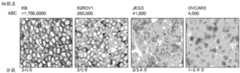
图2.手动染色方法:抗FOLR1抗体可以区分不同水平的FOLR1表达。将抗体BN3.2用于检测不同异种移植细胞中的FOLR1表达。BN3.2抗体的检测限度大约是每个细胞结合4000个抗体(ABC)。Figure 2. Manual staining method: Anti-FOLR1 antibody can distinguish different levels of FOLR1 expression. Antibody BN3.2 was used to detect FOLR1 expression in different xenografted cells. The limit of detection of the BN3.2 antibody is approximately 4000 antibody bound per cell (ABC).
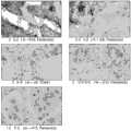
图3.手动染色方法:抗FOLR1抗体可以区分组织样本中不同水平的FOLR1表达。将BN3.2用于检测卵巢肿瘤(A)以及非小细胞肺癌肿瘤(B)二者中的FOLR1表达。Figure 3. Manual staining method: Anti-FOLR1 antibody can distinguish different levels of FOLR1 expression in tissue samples. BN3.2 was used to detect FOLR1 expression in both ovarian tumors (A) and non-small cell lung cancer tumors (B).
图4.手动染色方法:在卵巢肿瘤和NSCLC肿瘤中的均匀的FOLR1表达。FOLR1表达在很多测试的卵巢癌以及肺腺癌和细支气管肺泡癌中较高。大部分卵巢癌样本在浆液细胞或子宫内膜样细胞中具有最高的染色强度。在NSCLC肿瘤中,在细支气管肺泡癌和乳头状腺癌中发现最高的ABC值。Figure 4. Manual staining method: uniform FOLR1 expression in ovarian and NSCLC tumors. FOLR1 expression was higher in many ovarian cancers tested, as well as lung adenocarcinoma and bronchioloalveolar carcinoma. Most ovarian cancer samples had the highest staining intensity in serous cells or endometrioid cells. Among NSCLC tumors, the highest ABC values were found in bronchioloalveolar carcinoma and papillary adenocarcinoma.
图5.手动染色方法:FOLR1表达大体上局限在NSCLC细胞的膜上。高分辨率显微术显示大部分FOLR1染色局限于NSCLC肿瘤中的膜上。Figure 5. Manual staining method: FOLR1 expression is largely restricted to the membrane of NSCLC cells. High-resolution microscopy revealed that most FOLR1 staining was confined to the membrane in NSCLC tumors.
图6.手动染色方法:FOLR1表达大体上局限在卵巢癌细胞的膜上。高分辨率显微术显示大部分FOLR1染色局限于卵巢肿瘤中的膜上。Figure 6. Manual staining method: FOLR1 expression is largely restricted to the membrane of ovarian cancer cells. High-resolution microscopy revealed that most FOLR1 staining was confined to the membrane in ovarian tumors.
图7.在KB异种移植模型中huMov19靶向的缀合物的体内功效。使用所建立的皮下植入到SCID小鼠中的KB细胞的异种移植模型来测试与非FOLR1靶向性huC242-SPDB-DM4(D)相比的FOLR1靶向性可裂解缀合物huMov19-SPDB-DM4(B)、以及与非靶向性huC242-PEG4Mal-DM4(E)相比的不可裂解缀合物huMov19-PEG4-Mal-DM4(C)。由huMov19靶向FOLR1导致平均肿瘤体积的显著减小。Figure 7. In vivo efficacy of huMovl9-targeted conjugates in a KB xenograft model. An established xenograft model of KB cells implanted subcutaneously in SCID mice was used to test the FOLR1-targeting cleavable conjugate huMov19-SPDB compared to the non-FOLR1-targeting huC242-SPDB-DM4 (D) - DM4 (B), and the non-cleavable conjugate huMov19-PEG4-Mal-DM4 (C) compared to non-targeting huC242-PEG4Mal-DM4 (E). Targeting of FOLR1 by huMov19 resulted in a significant reduction in mean tumor volume.
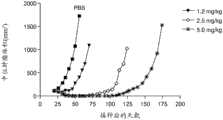
图8.在OVCAR-3人卵巢癌异种移植物中IMGN853处理的剂量-反应抗肿瘤活性。通过单次静脉内注射1.2mg/kg、2.5mg/kg或5.0mg/kg的IMGN853来处理小鼠。对照组的动物接受PBS的单次静脉内注射。Figure 8. Dose-response antitumor activity of IMGN853 treatment in OVCAR-3 human ovarian cancer xenografts. Mice were treated with a single intravenous injection of 1.2 mg/kg, 2.5 mg/kg or 5.0 mg/kg of IMGN853. Animals in the control group received a single intravenous injection of PBS.
图9.在IGROV-1人卵巢癌异种移植物中IMGN853处理的剂量-反应抗肿瘤活性。通过单次静脉内注射1.2mg/kg、2.5mg/kg或5.0mg/kg的IMGN853来处理小鼠。对照组的动物接受PBS的单次静脉内注射。Figure 9. Dose-response antitumor activity of IMGN853 treatment in IGROV-1 human ovarian cancer xenografts. Mice were treated with a single intravenous injection of 1.2 mg/kg, 2.5 mg/kg or 5.0 mg/kg of IMGN853. Animals in the control group received a single intravenous injection of PBS.
图10.在OV-90人卵巢癌异种移植物中IMGN853处理的剂量-反应抗肿瘤活性。通过单次静脉内注射1.2mg/kg、2.5mg/kg或5.0mg/kg的IMGN853来处理小鼠。对照组的动物接受PBS的单次静脉内注射。Figure 10. Dose-response antitumor activity of IMGN853 treatment in OV-90 human ovarian cancer xenografts. Mice were treated with a single intravenous injection of 1.2 mg/kg, 2.5 mg/kg or 5.0 mg/kg of IMGN853. Animals in the control group received a single intravenous injection of PBS.
图11.在SKOV-3人卵巢癌异种移植物中IMGN853处理的剂量-反应抗肿瘤活性。通过单次静脉内注射1.2mg/kg、2.5mg/kg或5.0mg/kg的IMGN853来处理小鼠。对照组的动物接受PBS的单次静脉内注射。Figure 11. Dose-response antitumor activity of IMGN853 treatment in SKOV-3 human ovarian cancer xenografts. Mice were treated with a single intravenous injection of 1.2 mg/kg, 2.5 mg/kg or 5.0 mg/kg of IMGN853. Animals in the control group received a single intravenous injection of PBS.
图12.在KB人宫颈腺癌异种移植物中IMGN853处理的剂量-反应抗肿瘤活性。通过单次静脉内注射1.0mg/kg、2.5mg/kg或5.0mg/kg的IMGN853来处理小鼠。对照组的动物接受PBS的单次静脉内注射。Figure 12. Dose-response antitumor activity of IMGN853 treatment in KB human cervical adenocarcinoma xenografts. Mice were treated with a single intravenous injection of 1.0 mg/kg, 2.5 mg/kg or 5.0 mg/kg of IMGN853. Animals in the control group received a single intravenous injection of PBS.
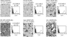
图13.自动化染色方法:代表性照片和直方图描绘了通过IHC和流式细胞术测得的细胞系中的FOLR1表达。SW620、T47D、Igrov-1、300.19/FR1、HeLa以及KB细胞均进行FOLR1染色强度和均匀度的评分。SW630和IGROV-1细胞被评分为1-3异质、T47D被评分为1-2异质、HeLa被评分为2-3异质,而300.19/FR1和KB被评分为3均质。Figure 13. Automated staining method: representative photographs and histograms depicting FOLR1 expression in cell lines as measured by IHC and flow cytometry. SW620, T47D, Igrov-1, 300.19/FR1, HeLa and KB cells were all scored for FOLR1 staining intensity and uniformity. SW630 and IGROV-1 cells were scored 1-3 heterogeneous, T47D was scored 1-2 heterogeneous, HeLa was scored 2-3 heterogeneous, while 300.19/FR1 and KB were scored 3 homogeneous.
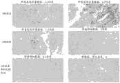
图14.自动化染色方法:在浆液性卵巢癌中的代表性FOLR1染色。通过IHC,来自浆液性卵巢癌的组织切片显示了展现3均质、2-3均质、2均质以及2异质染色的染色模式。Figure 14. Automated staining method: representative FOLR1 staining in serous ovarian carcinoma. By IHC, tissue sections from serous ovarian carcinoma showed staining patterns exhibiting 3 homogeneous, 2-3 homogeneous, 2 homogeneous, and 2 heterogeneous staining.
图15.自动化染色方法:在子宫内膜样卵巢癌中的代表性FOLR1染色。通过IHC,来自子宫内膜样癌的组织切片显示了展现3均质、2-3均质、3局部以及1-2异质染色的染色模式。Figure 15. Automated staining method: representative FOLR1 staining in endometrioid ovarian carcinoma. By IHC, tissue sections from endometrioid carcinoma showed staining patterns exhibiting 3 homogeneous, 2-3 homogeneous, 3 focal, and 1-2 heterogeneous staining.
图16.自动化染色方法:在腺癌亚型(细支气管肺泡癌除外)的NSCLC中的代表性FOLR1染色。通过IHC,来自非小细胞肺癌、腺癌亚型的组织切片显示了展现3均质、2-3均质、2异质、2均质以及1-2异质染色的染色模式。Figure 16. Automated staining method: representative FOLR1 staining in NSCLC of adenocarcinoma subtypes (except bronchioloalveolar carcinoma). By IHC, tissue sections from non-small cell lung cancer, adenocarcinoma subtypes showed staining patterns exhibiting 3 homogeneous, 2-3 homogeneous, 2 heterogeneous, 2 homogeneous and 1-2 heterogeneous staining.
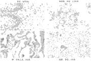
图17.自动化染色方法:在子宫内膜腺癌中的代表性FOLR1染色。通过IHC,来自子宫内膜腺癌的组织切片显示了展现3异质、2异质以及1异质染色的染色模式。Figure 17. Automated staining method: representative FOLR1 staining in endometrial adenocarcinoma. By IHC, tissue sections from endometrial adenocarcinoma showed a staining pattern exhibiting 3 heterogeneous, 2 heterogeneous and 1 heterogeneous staining.
图18.自动化染色方法:在肾透明细胞癌中的代表性FOLR1染色。通过IHC,来自肾细胞癌的组织切片显示了展现2均质、2异质以及1异质染色的染色模式。Figure 18. Automated staining method: representative FOLR1 staining in renal clear cell carcinoma. By IHC, tissue sections from renal cell carcinoma showed a staining pattern exhibiting 2 homogeneous, 2 heterogeneous and 1 heterogeneous staining.
图19.IMGN853在体外的细胞毒性活性。分析了五种FOLR1阳性细胞系(KB、IGROV-1、JEG-3、SKOV-3和OVCAR-3)和两种FOLR1阴性细胞系(Namalwa和SW2)对IMGN853的细胞毒性作用的敏感性。使细胞持续5天暴露于IMGN853(实线)或暴露于IMGN853加上0.5μM非缀合huMov19(M9346A)(虚线),并且通过基于WST-8的测定来确定细胞存活率。示出了代表性数据。绘制了存活细胞的百分比与IMGN853浓度的以10为底的对数的关系图。Figure 19. Cytotoxic activity of IMGN853 in vitro. The sensitivity of five FOLR1-positive cell lines (KB, IGROV-1, JEG-3, SKOV-3, and OVCAR-3) and two FOLR1-negative cell lines (Namalwa and SW2) to the cytotoxic effects of IMGN853 was analyzed. Cells were exposed to IMGN853 (solid line) or to IMGN853 plus 0.5 μM unconjugated huMov19(M9346A) (dashed line) for 5 days and cell viability was determined by WST-8 based assay. Representative data are shown. The percentage of surviving cells is plotted against the base 10 logarithm of the IMGN853 concentration.
图20.FOLR1阳性细胞系对IMGN853的敏感性与FOLR1表达水平的关系。分析了IMGN853针对具有广泛范围的FOLR1表达的FOLR1阳性细胞系的效力和特异性。将细胞系与IMGN853一起孵育,并且KB、Igrov-1和Jeg-3对IMGN853特别敏感,而非缀合的huMov19(M9346A)显示缀合物的降低的活性。Skov-3和Ovcar-3对IMGN853不敏感并且非缀合的huMov19(M9346A)不改变缀合物的活性。Figure 20. The relationship between the sensitivity of FOLR1-positive cell lines to IMGN853 and the expression level of FOLR1. The potency and specificity of IMGN853 against FOLR1 positive cell lines with a broad range of FOLR1 expression was analyzed. Cell lines were incubated with IMGN853, and KB, Igrov-1 and Jeg-3 were particularly sensitive to IMGN853, while non-conjugated huMov19 (M9346A) showed reduced activity of the conjugate. Skov-3 and Ovcar-3 were insensitive to IMGN853 and non-conjugated huMov19(M9346A) did not alter the activity of the conjugate.
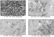
图21.自动化染色方法:对于FOLR1的染色的卵巢癌异种移植功效模型。通过IHC,来自卵巢癌异种移植物的组织切片显示了展现1-3异质(Ovcar3)、1-3均质(Igrov1)、1-2异质(Ov90)以及阴性(SKOV3)的染色模式。Figure 21. Automated staining method: Ovarian cancer xenograft efficacy model for staining of FOLR1. By IHC, tissue sections from ovarian cancer xenografts showed staining patterns exhibiting 1-3 heterogeneous (Ovcar3), 1-3 homogeneous (Igrov1), 1-2 heterogeneous (Ov90), and negative (SKOV3).
图22.自动化染色方法:小鼠异种移植模型。示出了NSCLC(A)、子宫内膜癌(B)和宫颈癌(C)细胞系的异种移植物中的FOLR1染色模式。NSCLC样本展现2-3均质或2均质染色,子宫内膜癌展现2异质/3局部染色,并且宫颈癌展现3均质染色。Figure 22. Automated staining method: mouse xenograft model. FOLR1 staining patterns in xenografts of NSCLC (A), endometrial cancer (B) and cervical cancer (C) cell lines are shown. NSCLC samples exhibited 2-3 homogeneous or 2 homogeneous staining, endometrial carcinoma exhibited 2 heterogeneous/3 focal staining, and cervical carcinoma exhibited 3 homogeneous staining.
图23.测定对照组织的自动化染色指南。如通过自动化IHC所测定,示出了阴性(食管0)和阳性对照样本(唾液腺1-2异质、肺2均质、胰腺3均质)的染色模式。Figure 23. Automated staining guide for assaying control tissue. Staining patterns are shown for negative (esophagus 0) and positive control samples (saliva 1-2 heterogeneous,
图24.肿瘤组织的自动化染色指南。如通过自动化IHC所测定,在对照组织上示出了3级、2级和1级染色的代表性染色模式。Figure 24. Automated staining guide for tumor tissue. Representative staining patterns of
图25.肿瘤组织的自动化染色指南。如通过自动化IHC所测定,在对照组织上示出了3级、2级、和1级/阴性染色的代表性染色模式。Figure 25. Automated staining guide for tumor tissue. Representative staining patterns of
发明详述Detailed description of the invention
本发明提供用于增加治疗其特征在于FOLR1过度表达的癌症的功效或者增加所述癌症对所述治疗的反应可能性的方法。本发明基于相对于正常组织的在肿瘤组织中FOLR1表达的动态范围的发现以及以下发现:具有增加水平的FOLR1表达的肿瘤对用抗FOLR1抗体或抗FOLR1免疫缀合物进行的治疗更具反应性。我们还发现了在自动化方法和手动方法之间动态范围的敏感性和检测的差异。进一步提供包括适用于实践本发明方法的一种或多种试剂的试剂盒。The present invention provides methods for increasing the efficacy of, or increasing the likelihood of, a cancer being responsive to such treatment, a cancer characterized by overexpression of FOLR1. The present invention is based on the discovery of the dynamic range of FOLR1 expression in tumor tissue relative to normal tissue and the discovery that tumors with increased levels of FOLR1 expression are more responsive to treatment with anti-FOLR1 antibodies or anti-FOLR1 immunoconjugates . We also found differences in dynamic range sensitivity and detection between automated and manual methods. Kits comprising one or more reagents suitable for use in practicing the methods of the invention are further provided.
I.定义I. Definition
为了有助于本发明的理解,在下文中定义了一些术语和短语。In order to facilitate the understanding of the present invention, some terms and phrases are defined below.
如本文使用的术语“人叶酸受体1”或“FOLR1”除另外指明外,是指任何天然的人FOLR1。术语“FOLR1”涵盖了“全长的”未加工的FOLR1以及由细胞内的加工所引起的任何形式的FOLR1。所述术语还涵盖天然存在的FOLR1变体,例如剪接变体、等位基因变体以及同工型。本文所述的FOLR1多肽可以从多种来源如人组织类型或另一种来源中分离,或者通过重组或合成的方法来制备。FOLR1序列的实例包括但不限于NCBI参考号P15328、NP_001092242.1、AAX29268.1、AAX37119.1、NP_057937.1以及NP_057936.1,以及在SEQ ID NO:1和2中所示的那些。As used herein, the term "
术语FOLR1的“增加的表达”是指含有升高的FOLR1表达水平的样本。在一个实例中,FOLR1表达通过IHC来测量并且通过与显示限定分数的对照(例如校准的对照)进行比较来给出染色强度分数或染色均匀度分数(例如如果强度与3级的校准的对照可比则将3分的强度分数给予测试样本、或者如果强度与2级的校准的对照可比则将2分的强度给予测试样本)。例如,通过免疫组织化学法测得的1、2、3或3+或更大的分数指示增加的FOLR1表达。异质或均质的染色均匀度也指示增加的FOLR1表达。染色强度和染色均匀度分数可以单独或组合(例如,2均质、2异质、3均质、3异质等)使用。在另一个实例中,FOLR1表达的增加可以通过检测相对于对照值(例如,在来自未患有癌症或患有FOLR1值未升高的癌症的受试者组织或细胞中的表达水平)至少2倍、至少3倍或至少5倍)的增加来测定。The term "increased expression" of FOLR1 refers to a sample containing an elevated expression level of FOLR1. In one example, FOLR1 expression is measured by IHC and a staining intensity score or staining uniformity score is given by comparison with a control showing a defined score (eg, a calibrated control) (eg, if the intensity is comparable to a calibrated control at level 3) An intensity score of 3 was then assigned to the test sample, or an intensity of 2 was given to the test sample if the intensity was comparable to the calibrated control at level 2). For example, a score of 1, 2, 3, or 3+ or greater as measured by immunohistochemistry indicates increased FOLR1 expression. Heterogeneous or homogeneous staining uniformity is also indicative of increased FOLR1 expression. Stain intensity and stain evenness scores can be used alone or in combination (eg, 2 homogeneous, 2 heterogeneous, 3 homogeneous, 3 heterogeneous, etc.). In another example, the increase in FOLR1 expression can be measured by at least 2 relative to a control value (e.g., the expression level in a tissue or cell from a subject that does not have cancer or has a cancer that does not have elevated FOLR1 values). fold, at least 3-fold, or at least 5-fold).
“参考样本”可以用于关联并比较以本发明的方法从测试样本中获得的结果。参考样本可为细胞(例如,细胞系、细胞团块)或组织。在“参考样本”中的FOLR1水平可为FOLR1的绝对量或相对量、一个范围的量、最小和/或最大量、平均量和/或中位量。本发明的诊断方法涉及测试样本中的FOLR1表达水平与“参考值”之间的比较。在一些实施方案中,所述参考值是参考样本中的FOLR1表达水平。参考值可为预先确定的值并且还可以从与测试样本同时进行测试的参考样本(例如对照生物样本)来测定。参考值可为单一截止值,如中位值或平均值或一个范围的值,如置信区间。可以为个体的不同亚组建立参考值,所述个体如易患癌症的个体、患有早期或晚期癌症的个体、男性和/或女性个体、或者经受癌症治疗的个体。本文描述了正常参考样本或值和阳性参考样本或值的实例。A "reference sample" can be used to correlate and compare results obtained from test samples with the methods of the present invention. A reference sample can be a cell (eg, cell line, cell aggregate) or tissue. The level of FOLR1 in a "reference sample" can be an absolute or relative amount of FOLR1, a range of amounts, a minimum and/or maximum amount, an average amount and/or a median amount. The diagnostic methods of the present invention involve a comparison between the expression level of FOLR1 in a test sample and a "reference value". In some embodiments, the reference value is the expression level of FOLR1 in a reference sample. The reference value can be a predetermined value and can also be determined from a reference sample (eg, a control biological sample) that is tested at the same time as the test sample. The reference value can be a single cut-off value, such as a median or mean, or a range of values, such as a confidence interval. Reference values can be established for different subgroups of individuals, such as individuals predisposed to cancer, individuals with early or advanced cancer, male and/or female individuals, or individuals undergoing cancer treatment. Examples of normal reference samples or values and positive reference samples or values are described herein.
在一些实施方案中,所述参考样本是来自健康组织、特别是不受癌症影响的相应组织的样本。这些类型的参考样本被称为阴性对照样本。在其它实施方案中,所述参考样本是来自表达FOLR1的肿瘤组织的样本。这些类型的参考样本被称为阳性对照样本。阳性对照样本还可以用作与FOLR1表达水平相关的染色强度的均匀度(异质对均质)和/或程度(1、2、3、3+)的比较指标。阳性对照比较样本还可以被称为展现染色强度或均匀度动态范围的校准的参考样本。如实施例1至9所示,无FOLR1表达的参考样本包括人食管组织;低FOLR1参考包括唾液腺(特别是闰管)和肺(特别是呼吸上皮)组织;并且高FOLR1表达的组织包括胰腺(特别是胰管细胞)。对于细胞系,低表达者包括但不限于OVCAR3和T47D,中等表达者包括但不限于SW620、IGROV-1、JEG3,并且高表达者包括但不限于KB和IGROV1。特别理想的阳性高FOLR1参考是用叶酸受体1稳定或瞬时转染的细胞系(例如,300.19/FR1)。用于特定癌症的适当阳性和阴性参考的FOLR1水平可以通过测量一个或多个适当受试者中的FOLR1水平来确定,并且可以为受试者的特定人群来定制这些参考水平(例如,参考水平可为年龄相匹配的,以使得可以在来自一定年龄的受试者的样本中的FOLR1水平与一定年龄组中关于特殊疾病状态、表型或其缺乏的参考水平之间作出比较)。可以为用于测量生物样本中的FOLR1水平的特定技术(例如,免疫测定法等)定制这些参考水平,其中FOLR1水平可以基于所使用的所述特定技术而不同。In some embodiments, the reference sample is a sample from a healthy tissue, particularly a corresponding tissue not affected by cancer. These types of reference samples are called negative control samples. In other embodiments, the reference sample is a sample from tumor tissue expressing FOLR1. These types of reference samples are called positive control samples. Positive control samples can also be used as a comparative indicator of the uniformity (heterogeneous vs. homogeneous) and/or degree (1, 2, 3, 3+) of staining intensity relative to FOLR1 expression levels. A positive control comparison sample can also be referred to as a calibrated reference sample that exhibits a dynamic range of staining intensity or uniformity. As shown in Examples 1 to 9, reference samples with no FOLR1 expression include human esophageal tissue; low FOLR1 references include salivary gland (particularly intercalated duct) and lung (particularly respiratory epithelium) tissues; and high FOLR1 expressing tissues include pancreas ( especially pancreatic duct cells). For cell lines, low expressors include but not limited to OVCAR3 and T47D, medium expressors include but not limited to SW620, IGROV-1, JEG3, and high expressers include but not limited to KB and IGROV1. A particularly desirable positive high FOLR1 reference is a cell line stably or transiently transfected with folate receptor 1 (eg, 300.19/FR1). Appropriate positive and negative reference FOLR1 levels for a particular cancer can be determined by measuring FOLR1 levels in one or more appropriate subjects, and these reference levels can be tailored for specific populations of subjects (e.g., reference levels can be age-matched so that comparisons can be made between FOLR1 levels in samples from subjects of a certain age and reference levels in a certain age group for a particular disease state, phenotype or lack thereof). These reference levels can be tailored to the particular technique used to measure FOLR1 levels in a biological sample (eg, immunoassay, etc.), where FOLR1 levels can vary based on the particular technique used.
在本文中术语“一抗”是指特异性地结合组织样本中的靶蛋白抗原的抗体。一抗通常是免疫组织化学(IHC)程序中使用的第一种抗体。在一个实施方案中,所述一抗是在IHC程序中使用的唯一抗体。本文中的术语“二抗”是指特异性地结合一抗从而在一抗与后续试剂(如果有的话)之间形成桥的抗体。所述二抗通常是免疫组织化学程序中使用的第二种抗体。The term "primary antibody" herein refers to an antibody that specifically binds a target protein antigen in a tissue sample. The primary antibody is usually the first antibody used in an immunohistochemistry (IHC) procedure. In one embodiment, the primary antibody is the only antibody used in the IHC procedure. The term "secondary antibody" herein refers to an antibody that specifically binds a primary antibody to form a bridge between the primary antibody and subsequent reagents, if any. The secondary antibody is typically the second antibody used in an immunohistochemical procedure.
在具体实施方案中,本发明的“样本”或“生物样本”来源于生物,例如来自真核生物。在优选实施方案中,所述样本是人样本,但是动物样本也可以用于本发明的实践中。用于在本发明中使用的样本的非限制性来源例如包括实体组织、活检抽吸物、腹水、流体浸出物、血液、血浆、血清、脊髓液、淋巴液、皮肤的外部切片、呼吸道、肠道和泌尿生殖道、泪液、唾液、乳汁、肿瘤、器官、细胞培养物和/或细胞培养物成分。本发明特别适用于癌症样本,所述癌症样本大体上包含实体组织样本或其中可用材料量很少的其它体液如腹水。所述方法可以用于检测FOLR1的表达方面或样本的状态,包括但不限于:比较细胞或组织的不同类型、比较不同发育阶段、以及检测或确定疾病或异常的存在和/或类型。In a particular embodiment, a "sample" or "biological sample" of the invention is derived from an organism, such as from a eukaryote. In a preferred embodiment, the sample is a human sample, but animal samples may also be used in the practice of the invention. Non-limiting sources of samples for use in the present invention include, for example, solid tissue, biopsy aspirate, ascites, fluid extract, blood, plasma, serum, spinal fluid, lymph fluid, external sections of skin, respiratory tract, intestinal tract and genitourinary tract, tears, saliva, breast milk, tumors, organs, cell cultures and/or cell culture components. The present invention is particularly applicable to cancer samples that generally comprise solid tissue samples or other bodily fluids such as ascitic fluid in which there is little material available. The methods can be used to detect aspects of FOLR1 expression or the state of a sample, including but not limited to: comparing different types of cells or tissues, comparing different developmental stages, and detecting or determining the presence and/or type of disease or abnormality.
出于本文的目的,组织样本的“切片”是指组织样本的单一部分或一片,例如从组织样本切下的组织薄片或细胞。应了解,根据本发明可以取得组织样本的多个切片并对其进行分析。在一些情况下,所选择的组织部分或切片包括同源细胞群。在其它情况下,所选择的部分包括组织的一个区域,例如作为非限制性实例的管腔。例如,所选择的部分可以像一个细胞或两个细胞那样小,或者可以代表成千上万个细胞。在大多数情况下,细胞的收集是重要的,并且虽然已经描述了本发明用于在细胞组分的检测中使用,但所述方法还可以用于检测生物的非细胞组分(例如作为非限制性实例的在血液中的可溶性组分)。For purposes herein, a "section" of a tissue sample refers to a single portion or piece of a tissue sample, such as a thin slice of tissue or cells cut from a tissue sample. It will be appreciated that multiple sections of a tissue sample may be taken and analyzed in accordance with the present invention. In some cases, the selected tissue portion or section includes a homogeneous population of cells. In other cases, the selected portion includes a region of tissue, such as a lumen, as a non-limiting example. For example, the selected portion can be as small as one cell or two cells, or can represent thousands of cells. In most cases, the collection of cells is important, and while the invention has been described for use in the detection of cellular components, the method can also be used to detect non-cellular components of organisms (e.g. as non-cellular Soluble components in blood for a limiting example).
通过“关联(correlate)”或“关联(correlating)”意指以任何方式对第一次分析的性能和/或结果与第二次分析的性能和/或结果进行比较。例如,可以将第一次分析的结果用于执行第二次分析、和/或可以将第一次分析的结果用于确定是否应该进行第二次分析、和/或可以将第一次分析的结果与第二次分析的结果进行比较。在一个实施方案中,增加的FOLR1表达与FOLR1靶向性抗癌治疗的增加的效力可能性相关联。By "correlate" or "correlating" is meant any way of comparing the performance and/or results of a first analysis with those of a second analysis. For example, the results of the first analysis can be used to perform a second analysis, and/or the results of the first analysis can be used to determine whether a second analysis should be performed, and/or the results of the first analysis can be used to The results are compared with those of the second analysis. In one embodiment, increased FOLR1 expression correlates with increased likelihood of efficacy of a FOLR1-targeted anticancer therapy.
术语“抗体”意指通过在免疫球蛋白分子的可变区内的至少一个抗原识别位点来识别并特异性地结合靶标(如蛋白质、多肽、肽、碳水化合物、多核苷酸、脂质或前述的组合)的所述免疫球蛋白分子。如本文使用的术语“抗体”涵盖了完整多克隆抗体、完整单克隆抗体、抗体片段(如Fab、Fab'、F(ab')2和Fv片段)、单链Fv(scFv)突变体、多特异性抗体(如由至少两个完整抗体产生的双特异性抗体)、嵌合抗体、人源化抗体、人抗体、包含抗体的抗原决定部分的融合蛋白,以及包含抗原识别位点的任何其它修饰的免疫球蛋白分子,只要所述抗体显示所需的生物活性。抗体可为以下五种主要类别的免疫球蛋白中的任一种:IgA、IgD、IgE、IgG以及IgM(基于其重链恒定区的鉴定而分别被称为α、δ、ε、γ和μ),或其亚类(同种型)(例如,IgG1、IgG2、IgG3、IgG4、IgA1和IgA2)。不同类别的免疫球蛋白具有不同的且众所周知的亚单位结构和三维构型。抗体可为裸的或者与其它分子如毒素、放射性同位素等缀合。The term "antibody" means a target (such as a protein, polypeptide, peptide, carbohydrate, polynucleotide, lipid or the aforementioned immunoglobulin molecules). The term "antibody" as used herein encompasses whole polyclonal antibodies, whole monoclonal antibodies, antibody fragments (such as Fab, Fab', F(ab')2 and Fv fragments), single chain Fv (scFv) mutants, polyclonal Specific antibodies (such as bispecific antibodies produced from at least two intact antibodies), chimeric antibodies, humanized antibodies, human antibodies, fusion proteins comprising epitopes of antibodies, and any other antibody comprising an antigen recognition site Modified immunoglobulin molecules, so long as the antibody exhibits the desired biological activity. Antibodies can be any of the five major classes of immunoglobulins: IgA, IgD, IgE, IgG, and IgM (referred to as alpha, delta, epsilon, gamma, and mu, respectively, based on the identification of their heavy chain constant regions ), or a subclass (isotype) thereof (eg, IgG1, IgG2, IgG3, IgG4, IgA1 and IgA2). Different classes of immunoglobulins have distinct and well-known subunit structures and three-dimensional configurations. Antibodies can be naked or conjugated to other molecules such as toxins, radioisotopes, and the like.
“封闭性”抗体或“拮抗剂”抗体是抑制或减小其结合的抗原如FOLR1的生物活性的抗体。在某个实施方案中,封闭性抗体或拮抗剂抗体大致上或完全抑制抗原的生物活性。理想地,生物活性降低10%、20%、30%、50%、70%、80%、90%、95%或甚至100%。A "blocking" antibody or "antagonist" antibody is an antibody that inhibits or reduces the biological activity of an antigen to which it binds, such as FOLR1. In a certain embodiment, the blocking antibody or antagonist antibody substantially or completely inhibits the biological activity of the antigen. Ideally, the biological activity is reduced by 10%, 20%, 30%, 50%, 70%, 80%, 90%, 95%, or even 100%.
术语“抗FOLR1抗体”或“结合FOLR1的抗体”是指能够以足够的亲和力结合FOLR1的抗体,所述结合使得抗体适用于在靶向FOLR1中作为诊断剂和/或治疗剂。抗FOLR1抗体与不相关的非FOLR1蛋白的结合程度小于约10%的抗体与FOLR1的结合,如例如通过放射性免疫测定法(RIA)所测定的。在某些实施方案中,结合FOLR1的抗体具有≤1μM、≤100nM、≤10nM、≤1nM或≤0.1nM的解离常数(Kd)。抗FOLR1抗体的实例在本领域中已知并且公开在美国申请公布号2012/0009181中,所述申请以引用方式并入本文。The term "anti-FOLR1 antibody" or "antibody that binds FOLR1" refers to an antibody that is capable of binding FOLR1 with sufficient affinity such that the antibody is suitable for use as a diagnostic and/or therapeutic agent in targeting FOLR1. The extent of binding of an anti-FOLR1 antibody to an irrelevant non-FOLR1 protein is less than about 10% of the antibody's binding to FOLR1, as determined, eg, by radioimmunoassay (RIA). In certain embodiments, the antibody that binds FOLR1 has a dissociation constant (Kd) < 1 μM, < 100 nM, < 10 nM, < 1 nM, or < 0.1 nM. Examples of anti-FOLR1 antibodies are known in the art and are disclosed in US Application Publication No. 2012/0009181, which is incorporated herein by reference.
术语“抗体片段”是指完整抗体的一部分并且是指完整抗体的抗原决定可变区。抗体片段的实例包括但不限于Fab、Fab'、F(ab')2和Fv片段、线性抗体、单链抗体以及由抗体片段形成的多特异性抗体。The term "antibody fragment" refers to a portion of an intact antibody and refers to the antigenic determining variable region of an intact antibody. Examples of antibody fragments include, but are not limited to, Fab, Fab', F(ab')2 and Fv fragments, linear antibodies, single chain antibodies, and multispecific antibodies formed from antibody fragments.
“单克隆抗体”是指涉及单一抗原决定簇或表位的高特异性识别和结合的同质性抗体群。这与通常包含针对不同抗原决定簇的不同抗体的多克隆抗体形成对照。术语“单克隆抗体”涵盖完整单克隆抗体和全长单克隆抗体二者以及抗体片段(如Fab、Fab'、F(ab')2、Fv)、单链(scFv)突变体、包含抗体部分的融合蛋白、和包含抗原识别位点的任何其它修饰的免疫球蛋白分子。此外,“单克隆抗体”是指以任何数量的方式形成的这些抗体,包括但不限于通过杂交瘤、噬菌体选择、重组表达以及转基因动物。"Monoclonal antibody" refers to a homogeneous population of antibodies involving highly specific recognition and binding of a single antigenic determinant or epitope. This is in contrast to polyclonal antibodies, which often contain different antibodies directed against different epitopes. The term "monoclonal antibody" encompasses both intact monoclonal antibodies and full-length monoclonal antibodies as well as antibody fragments (e.g., Fab, Fab', F(ab')2, Fv), single chain (scFv) mutants, antibody portions comprising , and any other modified immunoglobulin molecule comprising an antigen recognition site. Furthermore, "monoclonal antibody" refers to such antibodies formed in any number of ways including, but not limited to, by hybridomas, phage selection, recombinant expression, and transgenic animals.
术语“表位”或“抗原决定簇”在本文中可互换使用并且是指能够被特定抗体识别并特异性结合的抗原的部分。当所述抗原是多肽时,可以由连续氨基酸和通过蛋白质的三级折叠而并置的非连续氨基酸来形成表位。由连续氨基酸形成的表位通常在蛋白质变性时保留,而通过三级折叠形成的表位通常在蛋白质变性时丧失。在独特的空间构象中,表位通常包含至少3个并且更通常至少5个或8-10个氨基酸。The terms "epitope" or "antigenic determinant" are used interchangeably herein and refer to the portion of an antigen capable of being recognized and specifically bound by a particular antibody. When the antigen is a polypeptide, an epitope may be formed by contiguous amino acids and non-contiguous amino acids juxtaposed by the tertiary folding of the protein. Epitopes formed by contiguous amino acids are generally retained upon protein denaturation, whereas epitopes formed by tertiary folding are generally lost upon protein denaturation. An epitope typically comprises at least 3 and more usually at least 5 or 8-10 amino acids in a unique spatial conformation.
“结合亲和力”总体上是指一个分子(例如,抗体)的单一结合位点与其结合配偶体(例如,抗原)之间的非共价键相互作用的总数的强度。除非另外指明,否则如本文使用的“结合亲和力”是指反映了结合对的成员(例如,抗体和抗原)之间的1:1相互作用的固有结合亲和力。分子X对其配偶体Y的亲和力通常可以通过解离常数(Kd)来表示。可以通过本领域已知的常用方法来测量亲和力,包括本文所述的那些方法。低亲和力抗体通常结合抗原较慢并且倾向于容易解离,而高亲和力的抗体通常结合抗原更快并且倾向于更长时间保持结合。测量结合亲和力的多种方法在本领域已知,其中的任何方法可以用于本发明的目的。具体的说明性实施方案在下文进行描述。"Binding affinity" generally refers to the strength of the total number of non-covalent interactions between a single binding site of a molecule (eg, antibody) and its binding partner (eg, antigen). As used herein, unless otherwise indicated, "binding affinity" refers to intrinsic binding affinity that reflects a 1:1 interaction between members of a binding pair (eg, antibody and antigen). The affinity of a molecule X for its partner Y can generally be expressed by the dissociation constant (Kd). Affinity can be measured by common methods known in the art, including those described herein. Low-affinity antibodies generally bind antigen more slowly and tend to dissociate readily, while high-affinity antibodies generally bind antigen faster and tend to remain bound longer. Various methods of measuring binding affinity are known in the art, any of which can be used for the purposes of the present invention. Specific illustrative embodiments are described below.
在本文中提及结合亲和力时使用的“或更好”是指在分子与其结合配偶体之间更强的结合。在本文中使用的“或更好”是指更强的结合,由更小的数字Kd值来表示。例如,对抗原具有“0.6nM或更好”的亲和力的抗体,所述抗体对抗原的亲和力是<0.6nM,即0.59nM、0.58nM、0.57nM等,或小于0.6nM的任何值。"Or better" as used herein in reference to binding affinity refers to a stronger binding between a molecule and its binding partner. As used herein, "or better" means stronger binding, represented by a smaller numerical Kd value. For example, an antibody having an affinity for an antigen of "0.6 nM or better" is <0.6 nM, ie, 0.59 nM, 0.58 nM, 0.57 nM, etc., or any value less than 0.6 nM.
如本文使用的短语“大致上相似”或“大致上相同”表示在两个数值(通常一个数值与本发明的抗体相关并且另一个数值与参考/比较抗体相关)之间的足够高的相似度,以使得本领域技术人员将会认为在通过所述值(例如,Kd值)测量的生物学特性的背景下这两个值之间的差异几乎没有或没有生物学和/或统计性显著性。所述两个值之间的差异随着参考/比较抗体的值的变化而小于约50%、小于约40%、小于约30%、小于约20%或小于约10%。The phrase "substantially similar" or "substantially the same" as used herein means a sufficiently high degree of similarity between two values (usually one value relates to an antibody of the invention and the other value relates to a reference/comparative antibody) , such that a person skilled in the art would consider a difference between these two values to have little or no biological and/or statistical significance in the context of the biological property measured by said value (e.g., Kd value) . The difference between the two values is less than about 50%, less than about 40%, less than about 30%, less than about 20%, or less than about 10% as a function of the value of the reference/comparator antibody.
被“分离的”多肽、抗体、多核苷酸、载体、细胞或组合物是处于在自然界未发现的形式的多肽、抗体、多核苷酸、载体、细胞或组合物。分离的多肽、抗体、多核苷酸、载体、细胞或组合物包括已纯化到不再处于它们在自然界被发现的形式的程度的那些。在一些实施方案中,被分离的抗体、多核苷酸、载体、细胞或组合物是大致上纯的。An "isolated" polypeptide, antibody, polynucleotide, vector, cell or composition is one that is in a form not found in nature. Isolated polypeptides, antibodies, polynucleotides, vectors, cells or compositions include those that have been purified to the extent that they are no longer in the form in which they are found in nature. In some embodiments, an isolated antibody, polynucleotide, vector, cell or composition is substantially pure.
如本文使用的“大致上纯的”是指至少50%纯的(即没有污染物)、至少90%纯的、至少95%纯的、至少98%纯的或至少99%纯的。As used herein, "substantially pure" means at least 50% pure (ie, free of contaminants), at least 90% pure, at least 95% pure, at least 98% pure, or at least 99% pure.
如本文使用的术语“免疫缀合物”或“缀合物”是指连接细胞结合剂(即抗FOLR1抗体或其片段)并由以下通式定义的化合物或其衍生物:C-L-A,其中C=细胞毒素,L=接头,并且A=细胞结合剂或抗FOLR1抗体或抗体片段。免疫缀合物还可以由以下相反顺序的通式来定义:A-L-C。As used herein, the term "immunoconjugate" or "conjugate" refers to a compound or derivative thereof linked to a cell-binding agent (i.e., an anti-FOLR1 antibody or fragment thereof) and defined by the general formula: C-L-A, where C= Cytotoxin, L=linker, and A=cell binding agent or anti-FOLR1 antibody or antibody fragment. Immunoconjugates can also be defined by the following general formula in reverse order: A-L-C.
“接头”是能够将化合物(通常是药物,如美登木素)以稳定的共价方式连接细胞结合剂如抗FOLR1抗体或其片段的任何化学部分。在化合物或抗体保持活性的条件下,接头可以易感于或大致上抵抗酸诱导的裂解、光诱导的裂解、肽酶诱导的裂解、酯酶诱导的裂解以及二硫键裂解。合适的接头在本领域众所周知并且包括例如二硫基、硫醚基、酸不稳定基团、光不稳定基团、肽酶不稳定基团以及酯酶不稳定基团。接头还包括如本文所述的和在本领域已知的带电接头及其亲水形式。A "linker" is any chemical moiety capable of linking a compound (typically a drug, such as maytansinoid) to a cell-binding agent, such as an anti-FOLR1 antibody or fragment thereof, in a stable covalent manner. Linkers can be susceptible or substantially resistant to acid-induced cleavage, light-induced cleavage, peptidase-induced cleavage, esterase-induced cleavage, and disulfide bond cleavage under conditions that the compound or antibody remains active. Suitable linkers are well known in the art and include, for example, dithio, thioether, acid-labile, photo-labile, peptidase-labile, and esterase-labile groups. Linkers also include charged linkers and hydrophilic versions thereof as described herein and known in the art.
术语“癌”和“癌的”是指或描述其中细胞群具有未调节的细胞生长的特征的哺乳动物中的生理状况。癌的实例包括但不限于癌瘤、淋巴瘤、胚细胞瘤、肉瘤以及白血病。这些癌症的更具体实例包括鳞状细胞癌、小细胞肺癌、非小细胞肺癌、肺腺癌、肺鳞状癌、腹膜癌、肝细胞癌、胃肠癌、胰腺癌、胶质母细胞瘤、宫颈癌、卵巢癌、肝癌(liver cancer)、膀胱癌、肝细胞瘤、乳腺癌、结肠癌、结肠直肠癌、子宫内膜癌或子宫癌、唾液腺癌、肾癌、肝癌(liver cancer)、前列腺癌、外阴癌、甲状腺癌、肝癌(hepatic carcinoma)以及各种类型的头颈部癌。The terms "cancer" and "cancerous" refer to or describe the physiological condition in mammals in which a population of cells is characterized by unregulated cell growth. Examples of cancer include, but are not limited to, carcinoma, lymphoma, blastoma, sarcoma, and leukemia. More specific examples of these cancers include squamous cell carcinoma, small cell lung cancer, non-small cell lung cancer, lung adenocarcinoma, lung squamous carcinoma, peritoneal carcinoma, hepatocellular carcinoma, gastrointestinal cancer, pancreatic cancer, glioblastoma, Cervical cancer, ovarian cancer, liver cancer, bladder cancer, hepatoma, breast cancer, colon cancer, colorectal cancer, endometrial or uterine cancer, salivary gland cancer, kidney cancer, liver cancer, prostate cancer cancer, vulvar cancer, thyroid cancer, liver cancer (hepatic carcinoma) and various types of head and neck cancer.
“肿瘤(Tumor)”和“赘生物(neoplasm)”是指由过度细胞生长或增殖所引起的任何质量的组织,是良性的(非癌的)抑或恶性的(癌的),包括癌前病变。"Tumor" and "neoplasm" refer to any mass of tissue, benign (noncancerous) or malignant (cancerous), including precancerous lesions, resulting from excessive cellular growth or proliferation .
术语“癌细胞”、“肿瘤细胞”以及语法同等术语是指来源于肿瘤或癌前病变的总细胞群,包括非致瘤性细胞(包括肿瘤细胞群块)和致瘤性干细胞(癌干细胞)二者。如本文使用的术语“肿瘤细胞”当仅仅是指缺乏更新和分化能力的那些肿瘤细胞时,将由术语“非致瘤性”修饰,以区分那些肿瘤细胞和癌干细胞。The terms "cancer cells", "tumor cells" and their grammatical equivalents refer to the total cell population derived from a tumor or precancerous lesion, including non-tumorigenic cells (including tumor cell mass) and tumorigenic stem cells (cancer stem cells) both. The term "tumor cells" as used herein, when referring only to those tumor cells lacking the ability to renew and differentiate, will be modified by the term "non-tumorigenic" to distinguish those tumor cells from cancer stem cells.
术语“受试者”是指待成为具体治疗的接受者的任何动物(例如,哺乳动物),包括但不限于人、非人灵长类、啮齿类等。通常,提及人受试者时术语“受试者”和“患者”在本文中可互换使用。The term "subject" refers to any animal (eg, mammal) that is to be the recipient of a particular treatment, including, but not limited to, humans, non-human primates, rodents, and the like. Generally, the terms "subject" and "patient" are used interchangeably herein when referring to a human subject.
与一种或多种另外的治疗剂“联合”施用包括同时(并行)施用和以任何顺序的连续施用。Administration "in conjunction with" one or more additional therapeutic agents includes simultaneous (concurrent) administration and sequential administration in any order.
术语“药物制剂”是指处于允许活性成分的生物活性有效的形式并且不含有对进行所述制剂施用的受试者具有不可接受的毒性的另外组分的制剂。这些制剂可为无菌的。The term "pharmaceutical formulation" refers to a formulation that is in a form that permits the biological activity of the active ingredient to be effective and that does not contain additional components that are unacceptably toxic to the subject to which the formulation is administered. These formulations can be sterile.
本文所公开的抗体的“有效量”是足够执行特别说明的目的的量。根据所说明的目的,“有效量”可以凭经验并按常规方式来确定。An "effective amount" of an antibody disclosed herein is an amount sufficient to perform the specified purpose. An "effective amount" can be determined empirically and in a routine manner, depending on the stated purpose.
术语“治疗有效量”是指有效“治疗”在受试者或哺乳动物中的疾病或病症的抗体或其它药物的量。在癌症的情况下,药物的治疗有效量可以减少癌细胞的数量;减小肿瘤的尺寸;抑制(即,在某种程度上减慢并且在某一实施方案中停止)癌细胞对周边器官的浸润;抑制(即,在某种程度上减慢并在某一实施方案中停止)肿瘤转移;在某种程度上抑制肿瘤生长;和/或在某种程度上减轻与癌症有关的一种或多种症状。参见本文中“治疗”的定义。在药物可以防止生长和/或杀死现有的癌细胞的程度上,它可为细胞抑制的和/或细胞毒性的。在某些实施方案中,增加的FOLR1水平的确认允许施用减少量的FOLR1靶向性治疗剂,以实现与更高剂量所见到的相同的治疗效果。“预防有效量”是指在剂量和持续时间上有效实现所需预防结果所需要的量。通常但不一定地,由于预防剂量在疾病前或在疾病早期阶段用于受试者,所以预防有效量将小于治疗有效量。The term "therapeutically effective amount" refers to an amount of an antibody or other drug effective to "treat" a disease or disorder in a subject or mammal. In the case of cancer, a therapeutically effective amount of the drug can reduce the number of cancer cells; reduce the size of a tumor; inhibit (i.e., slow to some extent and in one embodiment stop) the attack of cancer cells on surrounding organs Invasion; inhibiting (i.e., slowing to some extent and in one embodiment stopping) tumor metastasis; inhibiting tumor growth to some extent; and/or alleviating to some extent one or Various symptoms. See the definition of "treatment" herein. To the extent the drug can prevent growth and/or kill existing cancer cells, it can be cytostatic and/or cytotoxic. In certain embodiments, confirmation of increased FOLR1 levels allows administration of reduced amounts of FOLR1-targeting therapeutics to achieve the same therapeutic effect as seen with higher doses. A "prophylactically effective amount" refers to an amount effective, in dosage and duration, to achieve the desired prophylactic result. Usually, but not necessarily, the prophylactically effective amount will be less than the therapeutically effective amount because the prophylactic dose is administered to the subject pre-disease or at an early stage of the disease.
术语“良好反应”通常是指在受试者中引起有利的状态。关于癌症治疗,所述术语是指在受试者身上提供治疗效果。在癌症中的积极治疗效果可以按许多方式来测量(参见,W.A.Weber,J.Nucl.Med.50:1S-10S(2009))。例如,肿瘤生长抑制、分子标记物表达、血清标记物表达以及分子成像技术全部都可以用来评定抗癌治疗的治疗功效。关于肿瘤生长抑制,根据NCI标准,T/C≤42%是抗肿瘤活性的最低水平。T/C<10%被认为是高抗肿瘤活性水平,其中T/C(%)=处理物的中位肿瘤体积/对照物的中位肿瘤体积×100。The term "favorable response" generally refers to eliciting a favorable state in a subject. With respect to cancer treatment, the term refers to providing a therapeutic effect in a subject. Positive therapeutic effects in cancer can be measured in a number of ways (see, W.A. Weber, J. Nucl. Med. 50:1S-10S (2009)). For example, tumor growth inhibition, molecular marker expression, serum marker expression, and molecular imaging techniques can all be used to assess the therapeutic efficacy of anticancer treatments. Regarding tumor growth inhibition, according to NCI criteria, T/C ≤ 42% is the lowest level of antitumor activity. T/C<10% is considered as high anti-tumor activity level, where T/C(%)=median tumor volume of treatment/median tumor volume of control×100.
词语“标记”当用于本文中时是指直接或间接与抗体缀合以便产生“标记的”抗体的可检测化合物或组合物。所述标记本身是可检测的(例如,放射性同位素标记或荧光标记),或者在酶标记的情况下,可以催化可检测的底物化合物或组合物的化学变化。The word "label" as used herein refers to a detectable compound or composition that is directly or indirectly conjugated to an antibody so as to produce a "labeled" antibody. The labels themselves are detectable (eg, radioisotopic or fluorescent labels) or, in the case of enzymatic labels, can catalyze a chemical change in a detectable substrate compound or composition.
“化学治疗剂”是适用于癌症治疗的化学化合物,不管其作用机制是什么。化学治疗剂的类别包括但不限于:烷化剂、抗代谢物、纺锤体毒物植物生物碱、细胞毒性/抗肿瘤抗生素、拓扑异构酶抑制剂、抗体、光敏剂以及激酶抑制剂。化学治疗剂包括在“靶向治疗”和常规化学治疗中使用的化合物。A "chemotherapeutic agent" is a chemical compound, regardless of its mechanism of action, that is useful in the treatment of cancer. Classes of chemotherapeutic agents include, but are not limited to: alkylating agents, antimetabolites, spindle poison plant alkaloids, cytotoxic/antineoplastic antibiotics, topoisomerase inhibitors, antibodies, photosensitizers, and kinase inhibitors. Chemotherapeutic agents include compounds used in "targeted therapy" and conventional chemotherapy.
例如“治疗(treating)”或“治疗(treatment)”或“治疗(to treat)”、或“缓和(alleviating)”或“缓和(to alleviate)”的术语是指:1)治愈、减慢、减轻所诊断的病理状况或病症的症状和/或阻止其进展的治疗措施,和2)防止和/或减慢所靶向的病理状况或病症的发展的预防性或防止性措施。因此,有治疗需要的那些受试者包括已经患有所述病症的那些;易患所述病症的那些;以及其中待防止所述病症的那些。在某些实施方案中,如果患者显示以下一种或多种,则受试者根据本发明的方法而成功“治疗”了癌症:恶病质减少、存活时间增加、肿瘤进展时间延长、肿瘤块减小、肿瘤负荷减小,和/或肿瘤转移时间、肿瘤复发时间、肿瘤反应、完全反应、部分反应、稳定疾病、进行性疾病、无进展生存期(PFS)、总生存期(OS)的延长,各自如通过由国家癌症研究所和批准新药的美国食品和药品管理局所设定的标准来测量。参见Johnson等,(2003)J.Clin.Oncol.21(7):1404-1411。Terms such as "treating" or "treatment" or "to treat", or "allevating" or "to alleviate" refer to: 1) curing, slowing down, Therapeutic measures to alleviate the symptoms and/or arrest the progression of the diagnosed pathological condition or disorder, and 2) prophylactic or preventive measures to prevent and/or slow the development of the targeted pathological condition or disorder. Accordingly, those subjects in need of treatment include those already with the disorder; those prone to have the disorder; and those in which the disorder is to be prevented. In certain embodiments, a subject is successfully "treated" for cancer according to the methods of the invention if the patient exhibits one or more of the following: decreased cachexia, increased survival time, increased time to tumor progression, decreased tumor mass , reduction in tumor burden, and/or prolongation of time to tumor metastasis, time to tumor recurrence, tumor response, complete response, partial response, stable disease, progressive disease, progression-free survival (PFS), overall survival (OS), Each is measured by the standards set by the National Cancer Institute and the US Food and Drug Administration, which approves new drugs. See Johnson et al., (2003) J. Clin. Oncol. 21(7):1404-1411.
“无进展存活期”(PFS)又被称为或“肿瘤进展时间”(YIP),表示在治疗之中和之后癌症不生长的时间长度。无进展存活期包括患者已经历完全反应或部分反应的时间量、以及患者经历稳定疾病的时间量。"Progression-free survival" (PFS), also known as or "time to tumor progression" (YIP), indicates the length of time during and after treatment that the cancer does not grow. Progression-free survival includes the amount of time a patient has experienced a complete response or partial response, as well as the amount of time a patient has experienced stable disease.
“无病生存期”(DFS)是指在治疗之中或之后患者保持无病的时间长度。"Disease-free survival" (DFS) refers to the length of time a patient remains disease-free during or after treatment.
“总生存期”(OS)是指相对于原初或未治疗的个体或患者而言预期寿命的延长。"Overall survival" (OS) refers to the increase in life expectancy relative to naive or untreated individuals or patients.
除非上下文另外明确规定,否则如本公开和权利要求书使用的单数形式“一个(a)”、“一种(an)”以及“所述(the)”包括复数形式。As used in this disclosure and the claims, the singular forms "a," "an," and "the" include plural referents unless the context clearly dictates otherwise.
应了解,无论本文何处用语言“包括”来描述实施方案,都还提供以“由…组成”和/或“主要由…组成”的措辞来描述的其它相似实施方案。It should be understood that wherever the language "comprising" is used to describe an embodiment herein, other similar embodiments described with the language "consisting of" and/or "consisting essentially of" are also provided.
在本文中如短语“A和/或B”中使用的术语“和/或”旨在包括“A和B”二者、“A或B”、“A”以及“B”。同样地,如短语“A、B和/或C”中使用的术语“和/或”旨在涵盖以下每种实施方案:A、B和C;A、B或C;A或C;A或B;B或C;A和C;A和B;B和C;A(单独);B(单独);以及C(单独)。The term "and/or" as used herein as in the phrase "A and/or B" is intended to include both "A and B", "A or B", "A" and "B". Likewise, the term "and/or" as used in the phrase "A, B, and/or C" is intended to cover each of the following embodiments: A, B, and C; A, B, or C; A or C; A or B; B or C; A and C; A and B; B and C; A (alone); B (alone); and C (alone).
II.生物样本II. Biological samples
生物样本常常用固定剂来固定。通常使用醛固定剂如福尔马林(甲醛)和戊二醛。使用其它固定技术如醇浸渍(Battifora和Kopinski,J.Histochem.Cytochem.(1986)34:1095)来固定的组织样本也是合适的。所使用的样本还可以包埋在石蜡中。在一个实施方案中,组织样本既用福尔马林固定,又用石蜡包埋(FFPE)。在另一个实施方案中,FFPE块在选择一个或多个部分用于分析之前用苏木精和曙红染色,以便为FFPE核心样本选择特定区域。从这些微粒试样制备组织块的方法已经在以前的多种预后因子的IHC研究中使用、和/或对于本领域的技术人员是熟知的(参见,例如,Abbondanzo等,Am J Clin Pathol.1990May;93(5):698-702;Allred等,Arch Surg.1990Jan;125(1):107-13)。Biological samples are often fixed with fixatives. Aldehyde fixatives such as formalin (formaldehyde) and glutaraldehyde are commonly used. Tissue samples fixed using other fixation techniques such as alcohol maceration (Battifora and Kopinski, J. Histochem. Cytochem. (1986) 34:1095) are also suitable. The samples used can also be embedded in paraffin. In one embodiment, tissue samples are both formalin fixed and paraffin embedded (FFPE). In another embodiment, FFPE blocks are stained with hematoxylin and eosin prior to selecting one or more sections for analysis in order to select specific regions for FFPE core samples. Methods for preparing tissue blocks from these microparticle samples have been used in previous IHC studies of various prognostic factors, and/or are well known to those skilled in the art (see, e.g., Abbondanzo et al., Am J Clin Pathol. 1990 May ;93(5):698-702; Allred et al., Arch Surg. 1990 Jan;125(1):107-13).
简要来说,可以将任何完整的器官或组织切成相当小的片并在各种固定剂(例如,福尔马林、醇等)中孵育不同的时间段,直到所述组织被“固定”。样本实际上可为从身体中手术移除的任何完整的组织。可以将样本切成适合装在组织病理学实验室中常规使用的设备上的适度小的片。切割的片的尺寸范围通常从几毫米到几厘米。Briefly, any whole organ or tissue can be cut into relatively small pieces and incubated in various fixatives (e.g., formalin, alcohol, etc.) for varying periods of time until the tissue is "fixed" . A sample can be virtually any intact tissue surgically removed from the body. Samples can be cut into reasonably small pieces suitable for loading on equipment routinely used in histopathology laboratories. The cut pieces typically range in size from a few millimeters to a few centimeters.
III.检测抗体缀合物III. Detection of Antibody Conjugates
本发明进一步提供通常是单克隆类型的抗FOLR1的抗体,其连接至少一种试剂以形成检测抗体缀合物。为了增加抗体分子作为诊断剂的功效,常规的是连接或共价结合或复合至少一个所需的分子或部分。这种分子或部分可为但不限于至少一个报道分子。报道分子被定义为可以使用测定来检测的任何部分。已经与抗体缀合的报道分子的非限制性实例包括酶、放射性标记、半抗原、荧光标记、磷光分子、化学发光分子、发色团、发光分子、光亲和分子、有色粒子和/或配体,例如生物素。The invention further provides anti-FOLRl antibodies, typically of the monoclonal type, to which at least one reagent is linked to form a detection antibody conjugate. To increase the efficacy of antibody molecules as diagnostic agents, it is routine to link or covalently bind or complex at least one desired molecule or moiety. Such a molecule or moiety can be, but is not limited to, at least one reporter molecule. A reporter is defined as any moiety that can be detected using an assay. Non-limiting examples of reporter molecules to which antibodies have been conjugated include enzymes, radioactive labels, haptens, fluorescent labels, phosphorescent molecules, chemiluminescent molecules, chromophores, luminescent molecules, photoaffinity molecules, colored particles, and/or ligands. body, such as biotin.
具有足够选择性、特异性或亲和力的任何细胞结合剂(例如,抗体或多肽)可以用作检测FOLR1多肽的基础。这些性质可以使用本领域技术人员已知的常规免疫性筛选方法来评价。除了典型的抗原结合位点外,抗体分子中用于结合生物活性分子的位点还包括居于可以结合所述抗原的可变区内的位点。此外,所述可变区涉及抗体自结合(Kang等,1988)且含有由抗-抗体识别的表位(独特位)(Kohler等,1989)。Any cell-binding agent (eg, antibody or polypeptide) with sufficient selectivity, specificity, or affinity can be used as a basis for detecting a FOLR1 polypeptide. These properties can be assessed using routine immunological screening methods known to those skilled in the art. In addition to typical antigen binding sites, sites in antibody molecules for binding biologically active molecules also include sites within the variable region that can bind the antigen. Furthermore, the variable regions are involved in antibody self-association (Kang et al., 1988) and contain epitopes (idiotopes) recognized by anti-antibodies (Kohler et al., 1989).
蛋白质结合(例如,抗体)缀合物的某些实例为其中蛋白质结合剂(例如,抗体)连接可检测标记的那些缀合物。“可检测标记”为由于它们的特定功能性质和/或化学特性而可以检测的化合物和/或元素,其使用允许与其连接的抗体被检测,和/或如果需要则进一步被定量。Some examples of protein-binding (eg, antibody) conjugates are those in which the protein-binding agent (eg, antibody) is linked to a detectable label. "Detectable labels" are compounds and/or elements that are detectable due to their specific functional properties and/or chemical properties, the use of which allows antibodies linked thereto to be detected, and/or further quantified if desired.
很多适当的成像剂在本领域是已知的,用于它们连接抗体的方法也一样(参见,例如美国专利号5,021,236;4,938,948;和4,472,509,各自以引用方式并入本文)。所使用的成像部分可为例如顺磁离子;放射性同位素;荧光染料;NMR可检测的物质;和/或X-射线成像。Many suitable imaging agents are known in the art, as are methods for linking them to antibodies (see, eg, US Patent Nos. 5,021,236; 4,938,948; and 4,472,509, each incorporated herein by reference). The imaging moieties used can be, for example, paramagnetic ions; radioisotopes; fluorescent dyes; NMR detectable substances; and/or X-ray imaging.
被涵盖来作为蛋白质结合(例如,抗体)缀合物使用的示例性荧光标记包括例如Alexa350、Alexa430、Alexa488、AMCA、BODIPY630/650、BODIPY650/665、BODIPY-FL、BODIPY-R6G、BODIPY-TMR、BODIPY-TRX、Cascade Blue、Cy3、Cy5,6-FAM、Dylight488、异硫氰酸荧光素、绿色荧光蛋白(GFP)、HEX、6-JOE、俄勒冈绿(Oregon Green)488、俄勒冈绿500、俄勒冈绿514、太平洋蓝(Pacific Blue)、藻红蛋白、REG、罗丹明绿、罗丹明红、四甲基罗丹明(TMR)、肾造影剂(Renographin)、ROX、TAMRA、TET、四甲基罗丹明、德克萨斯红(Texas Red)以及这些标记的衍生物(即卤化类似物、用异硫氰酸盐修饰或用于缀合的其它接头等)。示例性放射性标记是氚。Exemplary fluorescent labels contemplated for use as protein-binding (e.g., antibody) conjugates include, e.g., Alexa350, Alexa430, Alexa488, AMCA, BODIPY630/650, BODIPY650/665, BODIPY-FL, BODIPY-R6G, BODIPY-TMR, BODIPY-TRX, Cascade Blue, Cy3, Cy5,6-FAM, Dylight488, Fluorescein isothiocyanate, Green Fluorescent Protein (GFP), HEX, 6-JOE, Oregon Green 488,
涵盖在本发明中的蛋白质结合(例如,抗体)检测缀合物包括体外使用的那些,其中所述抗体连接第二结合配体和/或与显色底物接触时将产生有色产物的酶(例如,酶标签)。合适的酶的实例包括脲酶、碱性磷酸酶、(辣根)过氧化氢酶和/或葡萄糖氧化酶。优选的第二结合配体是生物素和/或抗生物素蛋白以及链霉抗生物素蛋白化合物。这些标记的使用对于本领域技术人员是熟知的并且被描述在例如美国专利号3,817,837;3,850,752;3,939,350;3,996,345;4,277,437;4,275,149以及4,366,241中;各自以引用方式并入本文。Protein-binding (e.g., antibody) detection conjugates contemplated in the present invention include those used in vitro, wherein the antibody is linked to a second binding ligand and/or an enzyme that will produce a colored product when contacted with a chromogenic substrate ( For example, enzyme tags). Examples of suitable enzymes include urease, alkaline phosphatase, (horseradish) catalase and/or glucose oxidase. Preferred second binding partners are biotin and/or avidin and streptavidin compounds. The use of such labels is well known to those skilled in the art and is described, for example, in US Patent Nos. 3,817,837; 3,850,752; 3,939,350; 3,996,345; 4,277,437;
含有叠氮基团的分子还可以通过经由低强度紫外线所产生的反应性氮宾中间体(Potter&Haley,1983)而用于与蛋白质形成共价键。具体来说,嘌呤核苷酸的2-和8-叠氮基类似物已用作定点光探针来识别在细胞粗提物中的核苷酸结合蛋白(Owens&Haley,1987;Atherton等,1985)。所述2-和8-叠氮基核苷酸还已用于绘制纯化蛋白的核苷酸结合区的图谱(Khatoon等,1989;King等,1989;以及Dholakia等,1989)并且可以用作抗体结合剂。Molecules containing an azide group can also be used to form covalent bonds with proteins via reactive nitrene intermediates generated by low-intensity UV light (Potter & Haley, 1983). In particular, 2- and 8-azido analogs of purine nucleotides have been used as directed photoprobes to identify nucleotide-binding proteins in crude cell extracts (Owens & Haley, 1987; Atherton et al., 1985) . The 2- and 8-azidonucleotides have also been used to map the nucleotide binding regions of purified proteins (Khatoon et al., 1989; King et al., 1989; and Dholakia et al., 1989) and can be used as antibodies Binding agent.
用于抗体连接或缀合其缀合部分的若干方法在本领域是已知的。一些连接方法涉及金属螯合物的使用,例如采用连接所述抗体的有机螯合剂,如二亚乙基三胺五乙酸酐(DTPA);亚乙基三胺四乙酸;N-氯-对-甲苯磺酰胺;和/或四氯-3α-6α-二苯基甘脲-3(美国专利号4,472,509和4,938,948,各自以引用方式并入本文)。单克隆抗体还可以在偶联剂如戊二醛或高碘酸盐的存在下与酶反应。具有荧光素标记物的蛋白质结合(例如,抗体)缀合物在这些偶联剂存在下或通过与异硫氰酸盐反应来制备。在美国专利号4,938,948中,乳腺瘤的成像例如使用单克隆抗体来实现,并且使用接头如对-羟基苯酰亚胺酸甲酯(methyl-p-hydroxybenzimidate)或3-(4-羟基苯基)-丙酸N-琥珀酰亚胺酯来将可检测的成像部分结合到所述抗体。Several methods are known in the art for linking antibodies or conjugating conjugated moieties thereof. Some attachment methods involve the use of metal chelates, such as organic chelators such as diethylenetriaminepentaacetic anhydride (DTPA); ethylenetriaminetetraacetic acid; N-chloro-p- toluenesulfonamide; and/or tetrachloro-3[alpha]-6[alpha]-diphenylglycoluril-3 (US Patent Nos. 4,472,509 and 4,938,948, each incorporated herein by reference). Monoclonal antibodies can also be reacted with enzymes in the presence of coupling agents such as glutaraldehyde or periodate. Protein-binding (eg, antibody) conjugates with fluorescein labels are prepared in the presence of these coupling agents or by reaction with isothiocyanates. In U.S. Patent No. 4,938,948, imaging of breast tumors is achieved, for example, using monoclonal antibodies, and using linkers such as methyl-p-hydroxybenzimidate or 3-(4-hydroxyphenyl) - N-succinimidyl propionate to bind a detectable imaging moiety to the antibody.
在其它实施方案中,涵盖了通过使用不改变抗体结合位点的反应条件来选择性地在免疫球蛋白Fc区域内引入巯基而进行的免疫球蛋白的衍生化作用。发现根据此方法而产生的抗体缀合物显示了改进的寿命、特异性和敏感性(美国专利号5,196,066,其以引用方式并入本文)。其中报道分子或效应分子与Fc区域内的碳水化合物残基缀合的所述效应分子或报道分子的位点特异性连接也已经公开在文献中(O'Shannessy等,1987)。In other embodiments, contemplated is the derivatization of immunoglobulins by selectively introducing sulfhydryl groups within the Fc region of the immunoglobulin using reaction conditions that do not alter the antibody binding site. Antibody conjugates produced according to this method were found to exhibit improved longevity, specificity, and sensitivity (US Patent No. 5,196,066, which is incorporated herein by reference). Site-specific linkage of a reporter or effector molecule wherein the reporter or effector molecule is conjugated to a carbohydrate residue within the Fc region has also been described in the literature (O'Shannessy et al., 1987).
在本发明的其它实施方案中,免疫球蛋白用核素如氚进行放射性标记。在另外的实施方案中,采用纳米金粒子(如约0.5nm至40nm的尺寸)和/或量子点(Hayward,Calif.)。In other embodiments of the invention, the immunoglobulin is radiolabeled with a nuclide, such as tritium. In other embodiments, nano-gold particles (eg, about 0.5 nm to 40 nm in size) and/or quantum dots (Hayward, Calif.) are employed.
IV.酶和底物(发色团)IV. Enzymes and Substrates (Chromaphores)
例如,在如下文提供的示例性实施方案中,涵盖了用于检测FOLR1的底物和指示剂的使用。For example, in the exemplary embodiments as provided below, the use of substrates and indicators for the detection of FOLR1 is contemplated.
辣根过氧化物酶(HRP)是首先与过氧化氢形成复合物并且然后引起其分解以生成水和原子氧的酶。像很多其它酶一样,HRP和一些HRP样活性可以由过量底物抑制。在HRP与过量过氧化氢之间形成的复合物是无催化活性的并且在缺乏电子给体(例如,显色物质)的情况下被可逆地抑制。过量过氧化氢和电子给体的缺乏带来内源性HRP活性的淬灭。Horseradish peroxidase (HRP) is an enzyme that first forms a complex with hydrogen peroxide and then causes its decomposition to generate water and atomic oxygen. Like many other enzymes, HRP and some HRP-like activities can be inhibited by excess substrate. The complex formed between HRP and excess hydrogen peroxide is catalytically inactive and is reversibly inhibited in the absence of an electron donor (eg, a chromogenic species). Excess hydrogen peroxide and lack of electron donor bring about quenching of endogenous HRP activity.
当在测定系统中使用时,HRP还可以用于使限定的底物转变为其活化的发色团,从而引起颜色变化。所述HRP酶可以通过很多方法与抗体、蛋白质、肽、聚合物或其它分子缀合。这些方法在本领域是已知的。将戊二醛添加到含有HRP和抗体的混合物的溶液中会导致同与所述酶缀合相比更多抗体分子彼此缀合。在两步骤的程序中,HRP首先与双功能试剂反应。在第二个阶段,只将活化的HRP与所述抗体混合,从而引起更加有效的标记且没有聚合。还使用两步骤的戊二醛程序来将HRP与(链霉)抗生物素蛋白缀合。这种形式用于例如LAB和LSAB是底物的程序中。与生物素的缀合也涉及两个步骤,因为生物素在可以与HRP酶的ε氨基反应之前首先必须衍生化为生物素基-N-羟基琥珀酰亚胺酯或生物素酰肼。When used in an assay system, HRP can also be used to convert a defined substrate to its activated chromophore, causing a color change. The HRP enzymes can be conjugated to antibodies, proteins, peptides, polymers or other molecules by a number of methods. These methods are known in the art. Addition of glutaraldehyde to the solution containing the mixture of HRP and antibody results in conjugation of more antibody molecules to each other than to the enzyme. In a two-step procedure, HRP is first reacted with a bifunctional reagent. In the second stage, only activated HRP was mixed with the antibody, resulting in a more efficient labeling and no aggregation. A two-step glutaraldehyde procedure was also used to conjugate HRP to (streptavidin). This form is used, for example, in procedures where LAB and LSAB are substrates. Conjugation to biotin also involves two steps, since biotin must first be derivatized to biotinyl-N-hydroxysuccinimide ester or biotin hydrazide before it can react with the epsilon amino group of the HRP enzyme.
3,3'-二氨基联苯胺(DAB)是对于如HRP的酶而言产生在醇和其它有机溶剂中高度不可溶的棕色最终产物的底物。DAB的氧化还引起聚合,从而引起与四氧化锇反应的能力并因此增加其染色强度和电子密度。在用于使聚合的DAB的光密度增强的若干金属和方法中,氯化金与硫化银组合似乎是最成功的。3,3'-Diaminobenzidine (DAB) is a substrate for enzymes like HRP to produce brown end products that are highly insoluble in alcohols and other organic solvents. Oxidation of DAB also causes polymerization, thereby inducing the ability to react with osmium tetroxide and thus increase its staining intensity and electron density. Of the several metals and methods used to enhance the optical density of polymerized DAB, gold chloride in combination with silver sulfide appears to be the most successful.
3-氨基-9-乙基咔唑(AEC)是如HRP的酶的底物。在氧化时,形成醇中可溶的玫瑰红色最终产物。因此,用AEC处理的试样一定不能浸入醇或醇溶液中(例如,哈里斯氏(Harris)苏木精)。相反,应该使用水性复染剂和封固剂。不幸的是AEC容易进一步氧化并且在暴露于过量光时强度将减退。因此建议储存在黑暗中。3-Amino-9-ethylcarbazole (AEC) is a substrate for enzymes such as HRP. Upon oxidation, an alcohol-soluble rose-red end product is formed. Therefore, specimens treated with AEC must not be immersed in alcohol or alcoholic solutions (eg, Harris' hematoxylin). Instead, an aqueous counterstain and mounting medium should be used. Unfortunately AEC is prone to further oxidation and will lose strength when exposed to excess light. Storage in the dark is therefore recommended.
4-氯-1-萘酚(CN)是如HRP的酶的底物并且作为蓝色最终产物而沉淀。因为CN在醇和其它有机溶剂中是可溶的,所以试样一定不能脱水、暴露在醇复染剂中或用含有有机溶剂的封固剂以载玻片覆盖。与DAB不同,CN倾向于从沉淀位点扩散。4-Chloro-1-naphthol (CN) is a substrate for enzymes like HRP and precipitates as a blue final product. Because CN is soluble in alcohols and other organic solvents, specimens must not be dehydrated, exposed to alcohol counterstains, or covered with a mounting medium containing organic solvents on a slide. Unlike DAB, CN tends to diffuse from the precipitation sites.
对-亚苯基二胺二盐酸盐/邻苯二酚(Hanker-Yates试剂)是如HRP的酶的电子给体底物并且得到在醇和其它有机溶剂中不可溶的蓝-黑色反应产物。与聚合的DAB一样,这种反应产物可以被锇酸化(osmicated)。在免疫过氧化物酶技术中使用Hanker-Yates试剂已经实现了不同结果。p-Phenylenediamine dihydrochloride/catechol (Hanker-Yates reagent) is an electron-donor substrate for enzymes like HRP and gives blue-black reaction products that are insoluble in alcohols and other organic solvents. Like polymeric DAB, this reaction product can be osmicated. Different results have been achieved using the Hanker-Yates reagent in the immunoperoxidase technique.
牛小肠碱性磷酸酶(AP)(分子量100kD)是通过破坏P-0键而从有机酯上移除(通过水解作用)并转移磷酸基的酶;暂时形成中间的酶-底物键。用于AP的主要金属活化剂是Mg++、Mn++和Ca++。Bovine intestinal alkaline phosphatase (AP) (
在免疫组织化学法中还没有广泛使用AP,直到公布了未标记的碱性磷酸酶抗碱性磷酸酶(APAAP)程序。在这个程序中利用的可溶性免疫复合物具有大约560kD的分子量。APAAP程序相对于PAP技术的主要优点是没有由内源性过氧化物酶活性所带来的干扰。由于内源性过氧化物酶活性对PAP染色的潜在干扰,推荐将所述APAAP技术用于血液涂片和骨髓涂片方面的使用。来自骨骼、肾脏、肝脏和一些白细胞中的内源性碱性磷酸酶的活性可以通过将1mM左旋咪唑(尽管已经发现5mM更有效)添加到底物溶液中来抑制。肠道碱性磷酸酶未通过左旋咪唑得到足够抑制。AP was not widely used in immunohistochemistry until the publication of the Unlabeled Alkaline Phosphatase Anti-Alkaline Phosphatase (APAAP) procedure. The soluble immune complex utilized in this procedure has a molecular weight of approximately 560 kD. The main advantage of the APAAP procedure over the PAP technique is the absence of interference by endogenous peroxidase activity. The APAAP technique is recommended for use on blood and bone marrow smears due to potential interference with PAP staining by endogenous peroxidase activity. The activity of endogenous alkaline phosphatase from bone, kidney, liver and some leukocytes can be inhibited by adding 1 mM levamisole (although 5 mM has been found to be more effective) to the substrate solution. Intestinal alkaline phosphatase was not sufficiently inhibited by levamisole.
在免疫碱性磷酸酶染色方法中,所述酶将磷酸萘酯(底物)水解为酚类化合物和磷酸。所述酚与无色重氮盐(发色团)偶联以产生不可溶的有色偶氮染料。已经成功使用底物和发色团的若干不同组合。In the immunoalkaline phosphatase staining method, the enzyme hydrolyzes naphthyl phosphate (substrate) into phenolic compounds and phosphoric acid. The phenols are coupled with a colorless diazonium salt (chromophore) to produce an insoluble colored azo dye. Several different combinations of substrates and chromophores have been used successfully.
AS-MX磷酸萘酯可以按其酸形式或作为钠盐来使用。发色团固红TR和固蓝BB分别产生亮红色或亮蓝色最终产物。二者都可溶于醇和其它有机溶剂,因此必须使用水性封固剂。当染色细胞涂片时优选固红TR。AS-MX naphthyl phosphate can be used in its acid form or as the sodium salt. The chromophores Fast Red TR and Fast Blue BB produce bright red or bright blue end products, respectively. Both are soluble in alcohols and other organic solvents, so an aqueous mounting medium must be used. Fast Red TR is preferred when staining cytosmears.
另外的示例性底物包括AS-BI磷酸萘酯、AS-TR磷酸萘酯和磷酸5-溴-4-氯-3-吲哚酯(BCIP)。其它可能的发色团包括例如固红LB、固深红GBC、氮蓝四唑(NBT)、碘硝基四唑紫(INT)以及所述结构的衍生物。Additional exemplary substrates include AS-BI naphthyl phosphate, AS-TR naphthyl phosphate, and 5-bromo-4-chloro-3-indole phosphate (BCIP). Other possible chromophores include, for example, Fast Red LB, Fast Crimson GBC, Nitroblue Tetrazole (NBT), Iodonitrotetrazolium Violet (INT), and derivatives of said structures.
V.免疫检测方法V. Immunoassay Methods
仍然在另外的实施方案中,本发明涉及用于结合、纯化、移除、定量和/或通常另外检测如本发明所涵盖的生物组分如配体的免疫检测方法。可以采用根据本发明制备的所述抗体来检测野生型和/或突变型配体蛋白、多肽和/或肽。如贯穿本申请所述的,涵盖了野生型和/或突变型配体特异性抗体的使用。一些免疫检测方法包括(举几个例子)流式细胞术、酶联免疫吸附测定法(ELISA)、放射性免疫测定法(RIA)、免疫放射性测定法、荧光免疫测定法、化学发光测定法、生物发光测定法以及蛋白质印迹。各种适用的免疫检测方法的步骤已经描述在科学文献中,例如像Doolittle M H和Ben-Zeev O,Methods MolBiol.1999;109:215-37;Gulbis B和Galand P,Hum Pathol.1993Dec;24(12):1271-85;以及De Jager R等,Semin Nucl Med.1993Apr;23(2):165-79,各自以引用方式并入本文。In still further embodiments, the present invention relates to immunoassay methods for binding, purifying, removing, quantifying and/or generally additionally detecting biological components such as ligands as contemplated by the present invention. The antibodies prepared according to the present invention can be used to detect wild-type and/or mutant ligand proteins, polypeptides and/or peptides. As described throughout this application, the use of wild-type and/or mutant ligand-specific antibodies is contemplated. Some immunoassays include (to name a few) flow cytometry, enzyme-linked immunosorbent assay (ELISA), radioimmunoassay (RIA), immunoradiometric assay, fluorescent immunoassay, chemiluminescent assay, biological Luminescence Assay and Western Blot. The steps of various suitable immunoassay methods have been described in the scientific literature, e.g. like Doolittle M H and Ben-Zeev O, Methods Mol Biol. 1999; 109:215-37; Gulbis B and Galand P, Hum Pathol. 1993 Dec; 24 (12):1271-85; and De Jager R et al., Semin Nucl Med. 1993 Apr;23(2):165-79, each incorporated herein by reference.
通常,免疫结合方法包括获得疑似包含配体蛋白、多肽和/或肽的样本,以及根据情况,在有效允许形成免疫复合物的条件下,使所述样本与根据本发明的第一配体结合肽(例如,抗配体抗体)接触。Generally, the method of immunobinding involves obtaining a sample suspected of containing a ligand protein, polypeptide and/or peptide, and, as the case may be, binding said sample to a first ligand according to the invention under conditions effective to allow the formation of an immune complex Peptide (eg, anti-ligand antibody) contact.
在抗原检测方面,所分析的生物样本可为疑似包含野生型或突变型配体蛋白特异性抗原的任何样本,如组织切片或试样、均化组织提取物、活检抽吸物、细胞、任何以上野生型或突变型含FOLR1组合物的分离和/或纯化形式、或甚至与组织接触的任何生物流体(包括血液和/或血清),尽管优选组织样本或提取物。In terms of antigen detection, the biological sample analyzed can be any sample suspected of containing wild-type or mutant ligand protein-specific antigens, such as tissue sections or specimens, homogenized tissue extracts, biopsy aspirates, cells, any Isolated and/or purified forms of the above wild-type or mutant FOLR1-containing compositions, or even any biological fluid (including blood and/or serum) that comes into contact with tissue, although tissue samples or extracts are preferred.
使所选择的生物样本与所述抗体在有效条件下接触足够允许免疫复合物(初次免疫复合物)形成的一段时间总体上是这样一件事:简单地将抗体组合物添加到样本中,并且孵育所述混合物足够长的一段时间以便所述抗体与所存在的任何配体蛋白抗原形成(即与其结合)免疫复合物。这段时间之后,通常会洗涤样本-抗体组合物如组织切片、ELISA板、斑点印迹或蛋白质印迹,以除去任何非特异性结合的抗体种类,从而仅仅允许检测特异性结合在初次免疫复合物内的那些抗体。Contacting the selected biological sample with the antibody under effective conditions for a period of time sufficient to allow immune complexes (primary immune complexes) to form is generally a matter of simply adding the antibody composition to the sample, and The mixture is incubated for a period of time sufficient for the antibody to form (ie bind to) an immune complex with any ligand protein antigen present. After this period of time, sample-antibody compositions such as tissue sections, ELISA plates, dot blots, or Western blots are typically washed to remove any non-specifically bound antibody species, thereby allowing detection of only those specifically bound within primary immune complexes. those antibodies.
一般而言,免疫复合物形成的检测在本领域是众所周知的并且可以通过应用多种方法来实现。这些方法通常基于标记或标记物的检测,如那些放射性、荧光、生物和酶标签中的任一种。涉及这些标记的使用的美国专利包括美国专利号3,817,837;3,850,752;3,939,350;3,996,345;4,277,437;4,275,149和4,366,241,各自以引用方式并入本文。当然,通过使用如本领域已知的第二结合配体如二抗和/或生物素/抗生物素蛋白配体结合安排,可以发现另外的优点。In general, detection of immune complex formation is well known in the art and can be achieved by applying a variety of methods. These methods are generally based on labels or detection of markers, such as any of those radioactive, fluorescent, biological and enzymatic labels. US patents covering the use of these markers include US Patent Nos. 3,817,837; 3,850,752; 3,939,350; 3,996,345; 4,277,437; Of course, additional advantages may be found by using secondary binding ligands such as secondary antibodies and/or biotin/avidin ligand binding arrangements as known in the art.
在所述检测中采用的抗配体抗体本身可以连接可检测的标记,其中然后会简单地检测这个标记,从而允许测定组合物中的初次免疫复合物的量。或者,变成结合在初次免疫复合物内的第一抗体可以通过具有对所述抗体的结合亲和力的第二结合剂来检测。在这些情况下,所述第二结合剂可以连接可检测的标记。所述第二结合剂自身常常是抗体,因此它可以被称为“二”抗或聚合物检测系统。使所述初次免疫复合物与标记的第二结合剂或抗体/聚合物检测系统在有效条件下接触足够允许第二免疫复合物形成的一段时间。然后通常洗涤所述第二免疫复合物以除去任何非特异性结合的标记的二抗或配体,并且然后检测在第二免疫复合物中的余下标记。The anti-ligand antibody employed in said detection may itself be linked to a detectable label, wherein this label is then simply detected, allowing determination of the amount of primary immune complexes in the composition. Alternatively, a primary antibody that becomes bound within a primary immune complex can be detected by a secondary binding agent that has a binding affinity for said antibody. In these cases, the second binding agent can be linked to a detectable label. The second binding agent is often an antibody itself, so it can be referred to as a "secondary" antibody or polymeric detection system. The primary immune complex is contacted with a labeled secondary binding agent or antibody/polymer detection system under effective conditions for a period of time sufficient to allow formation of the secondary immune complex. The second immune complex is then typically washed to remove any non-specifically bound labeled secondary antibody or ligand, and the remaining label in the second immune complex is then detected.
另外的方法包括通过两步方法检测初级免疫复合物。如上文所述,使用具有对所述抗体的结合亲和力的第二结合剂如抗体来形成第二免疫复合物。洗涤之后,使所述第二免疫复合物与具有对二抗的结合亲和力的第三结合剂或抗体再次在有效条件下接触足够允许免疫复合物(第三免疫复合物)形成的一段时间。使第三配体或抗体连接可检测的标记,以允许检测因此形成的第三免疫复合物。如果需要,这个系统可以提供信号放大。Additional methods include detection of primary immune complexes by a two-step approach. As described above, the second immune complex is formed using a second binding agent, such as an antibody, that has a binding affinity for the antibody. Following washing, the second immune complex is contacted again with a third binding agent or antibody having binding affinity for the secondary antibody under conditions effective for a period of time sufficient to allow formation of an immune complex (third immune complex). A detectable label is attached to the third ligand or antibody to allow detection of the third immune complex thus formed. This system can provide signal amplification if required.
在另一个实施方案中,生物素化的单克隆或多克隆抗体用于检测靶抗原,并且然后将第二步骤抗体用于检测连接复合生物素的生物素。在所述方法中,待测试的样本首先在包含第一步骤抗体的溶液中孵育。如果存在靶抗原,则一部分所述抗体结合所述抗原以形成生物素化抗体/抗原复合物。然后通过在链霉抗生物素蛋白(或抗生物素蛋白)、生物素化DNA和/或互补的生物素化DNA的连续溶液中孵育来扩增抗体/抗原复合物,其中每个步骤将另外的生物素位点添加到抗体/抗原复合物中。重复扩增步骤直到实现合适水平的扩增,在此时,在包含抗生物素的第二步骤抗体的溶液中孵育所述样本。这个第二步骤抗体例如用一种酶来标记,所述酶可以通过使用发色团底物的组织酶学来用于检测抗体/抗原复合物的存在。在合适扩增的情况下,可以产生肉眼可见的蛋白质结合(例如,抗体)缀合物。In another embodiment, a biotinylated monoclonal or polyclonal antibody is used to detect the target antigen, and then a second step antibody is used to detect the biotin linked to complexed biotin. In said method, the sample to be tested is first incubated in a solution comprising the first step antibody. If a target antigen is present, a portion of the antibody binds the antigen to form a biotinylated antibody/antigen complex. The antibody/antigen complexes are then amplified by incubation in successive solutions of streptavidin (or avidin), biotinylated DNA, and/or complementary biotinylated DNA, where each step will additionally The biotin site is added to the antibody/antigen complex. The amplification step is repeated until a suitable level of amplification is achieved, at which point the sample is incubated in a solution containing the second step antibody against biotin. This second step is antibody labeling, for example with an enzyme that can be used to detect the presence of antibody/antigen complexes by histoenzymology using chromophore substrates. With appropriate amplification, macroscopic protein-binding (eg, antibody) conjugates can be produced.
免疫检测的另一种已知的方法利用了免疫-PCR(聚合酶链反应)的方法论。PCR方法使用了以释放所述抗体的低pH或高盐缓冲液洗涤出来的DNA/生物素/链霉抗生物素蛋白/抗体复合物。然后将所得的洗液用于执行采用合适引物且在适当控制下的PCR反应。在具体实施方案中,可以利用PCR的巨大扩增能力和特异性来检测单个抗原分子。这种检测可以实时地发生。例如,涵盖了定量的实时PCR的使用。Another known method of immunodetection utilizes the methodology of immuno-PCR (polymerase chain reaction). The PCR method uses DNA/biotin/streptavidin/antibody complexes washed out with low pH or high salt buffers that release the antibodies. The resulting washes are then used to perform PCR reactions using appropriate primers and under appropriate controls. In specific embodiments, the enormous amplification power and specificity of PCR can be exploited to detect single antigen molecules. This detection can occur in real time. For example, the use of quantitative real-time PCR is contemplated.
在患有不同形式疾病的患者的临床诊断和/或监控中,FOLR1突变体的检测和/或FOLR1水平相对于在来自正常受试者的相应生物样本中的水平而言的改变,指示患者患有所述疾病。然而,如本领域技术人员所已知的,这样的临床诊断不一定要孤立地按照这个方法来进行。本领域技术人员对区分生物标记物的类型和/或数量的显著差异非常熟悉,所述差异代表生物标记物的阳性识别、和/或低水平和/或背景变化。实际上,背景表达水平经常用于形成“界限值”,在所述界限值之上增加的检测将被评分为显著的和/或阳性的。In the clinical diagnosis and/or monitoring of patients with different forms of the disease, the detection of FOLR1 mutants and/or changes in FOLR1 levels relative to levels in corresponding biological samples from normal subjects is indicative of a patient suffering from have the disease. However, such clinical diagnosis does not have to be made according to this method in isolation, as is known to those skilled in the art. Those skilled in the art are familiar with distinguishing between significant differences in the type and/or amount of biomarkers that represent positive recognition, and/or low levels and/or background changes of a biomarker. In practice, the background expression level is often used to form a "cut-off value" above which increased detections will be scored as significant and/or positive.
在一个实施方案中,通过FOLR1的免疫学检测(通过免疫组织化学法)来为强度和均匀度二者(染色细胞百分比——仅细胞膜)评分。FOLR1表达强度的比较标尺的相关性为0—阴性、0-1—很弱、1—弱、1-2—弱至中等、2—中等、2-3—中等至强、3-强。定量地,分数0代表在肿瘤细胞中没有观察到膜染色。分数1代表在肿瘤细胞中模糊/几乎没有可辨的膜染色。对于分数2,则在肿瘤细胞中观察到中等的膜染色。最后,分数3或3+代表在肿瘤细胞中的中等至强的膜染色。具有0分或1分的FOLR1表达的那些样本可以表征为没有过度表达FOLR1,而具有2分或3分的那些样本可以表征为过度表达FOLR1。过度表达FOLR1的样本还可以通过与每个细胞表达的FOLR1分子的拷贝数量相对应的免疫组织化学分数来评级,并且已经按生物化学方式来测定:0=0-10,000拷贝/细胞、1=至少约200,000拷贝/细胞、2=至少约500,000拷贝/细胞以及3=至少约2,000,000拷贝/细胞。FOLR1百分比细胞膜染色均匀度的比较标尺相关性如下:0—阴性、局部—<25%、异质性(异质)—25%-75%以及均质性(均质)—>75%。In one embodiment, both intensity and uniformity (percentage of stained cells - cell membranes only) are scored by immunological detection of FOLR1 (by immunohistochemistry). The correlation scale of the comparison scale of FOLR1 expression intensity was 0-negative, 0-1-very weak, 1-weak, 1-2-weak to moderate, 2-moderate, 2-3-moderate to strong, 3-strong. Quantitatively, a score of 0 represents that no membrane staining was observed in tumor cells. A score of 1 represents faint/barely discernible membrane staining in tumor cells. For
VI.核酸杂交VI. Nucleic acid hybridization
原位杂交通常在固定到载片上的细胞或组织切片上来执行。可以通过若干常规方法(参见例如Leitch等In situ Hybridization:a practicalguide,Oxford BIOS Scientific Publishers,Microscopy handbooks v.27(1994))来进行原位杂交。在一个原位程序中,使用荧光染料(如在由氩离子激光器刺激时发绿色荧光的异硫氰酸荧光素(FITC))来标记与细胞中的靶核苷酸序列互补的核酸序列探针。包含所述靶核苷酸序列的每个细胞将结合所述标记的探针,从而在所述细胞暴露给具有适合用于所使用的特定荧光染料的激发的波长的光源时产生荧光信号。In situ hybridization is typically performed on sections of cells or tissue fixed to slides. In situ hybridization can be performed by several conventional methods (see for example Leitch et al. In situ Hybridization: a practical guide, Oxford BIOS Scientific Publishers, Microscopy handbooks v. 27 (1994)). In an in situ procedure, a fluorescent dye such as fluorescein isothiocyanate (FITC), which fluoresces green when stimulated by an argon-ion laser, is used to label nucleic acid sequence probes that are complementary to target nucleotide sequences in cells . Each cell containing the target nucleotide sequence will bind the labeled probe, producing a fluorescent signal when the cell is exposed to a light source having a wavelength appropriate for excitation of the particular fluorochrome used.
可以采用不同的杂交严格度。由于杂交条件变得更加严格,所以在探针与靶之间要求更大程度的互补性以形成并维持稳定的双链体。通过升高温度、降低盐浓度或升高甲酰胺浓度来增加严格度。添加硫酸葡聚糖或升高其浓度也可以增加标记探针的有效浓度,以增加杂交率和最终的信号强度。杂交之后,在通常包含与杂交溶液中发现的那些试剂相似的试剂的溶液中洗涤载片,其中根据所要求的严格度洗涤时间从分钟到小时而不同。更长或更严格的洗涤通常减少了非特异性背景但是具有减小总体敏感度的风险。Different hybridization stringencies can be used. As hybridization conditions become more stringent, a greater degree of complementarity is required between the probe and target to form and maintain stable duplexes. Increase stringency by increasing temperature, decreasing salt concentration, or increasing formamide concentration. Adding dextran sulfate or increasing its concentration can also increase the effective concentration of labeled probes to increase the hybridization rate and the final signal intensity. Following hybridization, the slide is washed in a solution generally containing reagents similar to those found in the hybridization solution, where the wash time varies from minutes to hours depending on the stringency required. Longer or more stringent washes generally reduce non-specific background but risk reducing overall sensitivity.
在核酸杂交分析中使用的探针可为RNA或DNA寡核苷酸抑或多核苷酸并且可以不仅含有天然存在的核苷酸而且含有它们的类似物,例如像地高辛dCTP、生物素dcTP7-氮鸟嘌呤核苷、叠氮胸苷、肌苷或尿苷。其它适用的探针包括例如肽探针及其类似物、分支基因DNA、拟肽物、肽核酸(PNA)和/或抗体。Probes used in nucleic acid hybridization assays may be RNA or DNA oligonucleotides or polynucleotides and may contain not only naturally occurring nucleotides but also their analogs, such as digoxigenin dCTP, biotin dcTP7- Azaguanosine, azidothymidine, inosine, or uridine. Other suitable probes include, for example, peptide probes and their analogs, branched genetic DNA, peptidomimetics, peptide nucleic acid (PNA) and/or antibodies.
探针应该具有对所关注的靶核酸序列的足够互补性,以使得稳定的和特异性的结合发生在靶核酸序列与探针之间。稳定杂交所要求的同源性程度随着杂交介质和/或洗涤介质的严格度而变化。优选地,在本发明中采用完全同源的探针,但是本领域技术人员将容易认识到,显示更少但足够的同源性的探针可以用于本发明中(参见例如Sambrook,J.,Fritsch,E.F.,Maniatis,T.,Molecular Cloning ALaboratory Manual,Cold Spring Harbor Press,(1989))。The probe should have sufficient complementarity to the target nucleic acid sequence of interest such that stable and specific binding occurs between the target nucleic acid sequence and the probe. The degree of homology required for stable hybridization will vary with the stringency of the hybridization medium and/or wash medium. Preferably, fully homologous probes are employed in the present invention, but those skilled in the art will readily recognize that probes exhibiting less but sufficient homology may be used in the present invention (see e.g. Sambrook, J. , Fritsch, E.F., Maniatis, T., Molecular Cloning A Laboratory Manual, Cold Spring Harbor Press, (1989)).
探针还可以通过若干手段来形成并选择,所述手段包括但不限于通过原位杂交、体细胞杂交板或分选染色体的斑点印迹进行图谱绘制;染色体连锁分析;或者从来自人细胞系或具有人染色体的体细胞杂种、辐射体细胞杂种、染色体区域的显微解剖的分选染色体文库中、或从由对独特染色体位点特异的PCR引物所识别的酵母人工染色体(YAC)中克隆和分离,或者像相邻YAC克隆的其它合适方式。探针可为在质粒、噬菌体、粘粒、YAC、细菌人工染色体(BAC)、病毒载体或任何其它合适载体中克隆的基因组DNA、cDNA或RNA。探针可以通过常规方法来克隆或化学合成。克隆时,通常将分离的探针核酸片段插入到载体中,如λ噬菌体、pBR322、M13或含有SP6或T7启动子的载体,并且在细菌宿主中作为文库来克隆。[参见例如Sambrook,J.,Fritsch,E.F.,Maniatis,T.,Molecular Cloning ALaboratory Manual,Cold Spring Harbor Press,(1989)]。Probes can also be formed and selected by several means including, but not limited to, mapping by in situ hybridization, somatic cell hybridization plates, or dot blots of sorted chromosomes; chromosomal linkage analysis; or from human cell lines or Cloning and Separation, or other suitable means like adjacent YAC clones. The probe can be genomic DNA, cDNA or RNA cloned in a plasmid, phage, cosmid, YAC, bacterial artificial chromosome (BAC), viral vector or any other suitable vector. Probes can be cloned or chemically synthesized by conventional methods. For cloning, the isolated probe nucleic acid fragment is usually inserted into a vector, such as lambda phage, pBR322, M13, or a vector containing an SP6 or T7 promoter, and cloned as a library in a bacterial host. [See, eg, Sambrook, J., Fritsch, E.F., Maniatis, T., Molecular Cloning A Laboratory Manual, Cold Spring Harbor Press, (1989)].
优选对探针进行标记,例如像用荧光体来进行。荧光团的实例包括但不限于稀土螯合物(铕螯合物)、德克萨斯红、罗丹明、荧光素、丹酰、丽丝胺(Lissamine)、伞形酮、藻红蛋白、藻蓝蛋白或可商购的荧光体如SPECTRUM ORANGETM和SPECTRUM GREENTM和/或以上任何一种或多种的衍生物。在测定中使用的多个探针可以用一种以上的可区分的荧光色或颜料色来标记。这些色差提供用于识别特定探针的杂交位置的手段。此外,空间上未分离的探针可以通过由混合两种另外的颜色(例如,淡红色+绿色=黄色)颜料(例如,蓝色+黄色=绿色)而形成的不同色光或颜料或者通过使用一次只能穿过一种颜色的过滤器设置来识别。The probes are preferably labeled, eg with a fluorophore. Examples of fluorophores include, but are not limited to, rare earth chelates (europium chelates), Texas Red, rhodamine, fluorescein, dansyl, Lissamine, umbelliferone, phycoerythrin, algal Cocyanin or commercially available phosphors such as SPECTRUM ORANGE™ and SPECTRUM GREEN™ and/or derivatives of any one or more of the above. Multiple probes used in an assay can be labeled with more than one distinguishable fluorescent or pigmentary color. These color differences provide a means for identifying where a particular probe hybridizes. In addition, spatially unseparated probes can be created by mixing two additional color (for example, reddish+green=yellow) pigments (for example, blue+yellow=green) of different shades or pigments or by using a single Recognizable only through a filter setting of one color.
可以利用本领域技术人员已知的常规方法,用荧光团对探针进行直接或间接标记。Probes can be directly or indirectly labeled with fluorophores using conventional methods known to those skilled in the art.
VII.检测试剂盒和组合物VII. Assay Kits and Compositions
本发明还提供用于在如本文所公开的本发明的实践中使用的试剂盒。这些试剂盒可以包括容器,每个容器具有在所述方法中使用的一种或多种不同试剂(通常以浓缩的形式),所述试剂包括例如一种或多种结合剂(抗体),其已经连接标记物,或者任选地具有用于将结合剂偶联到抗体或核酸分子(以及标记物自身)的试剂;缓冲液、适当的三磷酸核苷酸(例如,dATP、dCTP、dGTP、dTTP、dUTP、ATP、CTP、GTP和UTP)、逆转录酶、DNA聚合酶、RNA聚合酶,以及一种或多种序列特异性或简并引物以用于通过扩增来检测核酸分子;和/或用于分离(优选通过显微解剖)以支持本发明的实践的试剂和仪器。通常还将包括描述本发明的配体检测方法中的试剂盒组件的或关于其使用的一组说明的标签或指示物,其中所述说明可以与包装说明书和/或试剂盒或其组件的包装关联。The invention also provides kits for use in the practice of the invention as disclosed herein. These kits may include containers, each container having one or more different reagents (usually in concentrated form) for use in the method, including, for example, one or more binding agents (antibodies), which Labels have been attached, or optionally have reagents for coupling the binding agent to the antibody or nucleic acid molecule (and the label itself); buffer, appropriate nucleotide triphosphates (e.g., dATP, dCTP, dGTP, dTTP, dUTP, ATP, CTP, GTP, and UTP), reverse transcriptase, DNA polymerase, RNA polymerase, and one or more sequence-specific or degenerate primers for detection of nucleic acid molecules by amplification; and and/or reagents and instruments for isolation, preferably by microdissection, to support the practice of the invention. Typically also will include a label or indicator describing a kit component in the ligand detection method of the invention or a set of instructions for its use, wherein the instructions may be associated with the package insert and/or the packaging of the kit or its components associated.
仍然在另外的实施方案中,本发明涉及用于与上文所述的免疫检测方法一起使用的免疫检测试剂盒。由于抗体通常用于检测野生型和/或突变型蛋白质、多肽和/或肽,所以所述抗体将优选包括在所述试剂盒中。因此免疫检测试剂盒将在合适的容器工具中包含结合野生型和/或突变型蛋白质、多肽和/或肽的第一抗体、和/或任选的免疫检测试剂、和/或另外任选的野生型和/或突变型蛋白质、多肽和/或肽。In still further embodiments, the present invention relates to an immunoassay kit for use with the immunoassay methods described above. As antibodies are typically used to detect wild-type and/or mutant proteins, polypeptides and/or peptides, such antibodies will preferably be included in the kit. Thus the immunodetection kit will comprise, in suitable container means, a primary antibody binding to wild-type and/or mutant proteins, polypeptides and/or peptides, and/or optional immunodetection reagents, and/or additional optional Wild-type and/or mutant proteins, polypeptides and/or peptides.
试剂盒的免疫检测试剂可为多种形式中的任一种,包括与给定抗体相关联和/或连接其的那些可检测标记。还涵盖了与第二结合配体相关联和/或连接其的可检测标记。示例性第二配体是具有对第一抗体的结合亲和力的那些二抗或聚合物。The immunodetection reagents of the kits may be in any of a variety of formats, including those detectable labels associated with and/or linked to a given antibody. Detectable labels associated with and/or linked to the second binding ligand are also contemplated. Exemplary secondary ligands are those secondary antibodies or polymers that have binding affinity for the primary antibody.
用于在本发明试剂盒中使用的另外的合适免疫检测试剂包括双组分试剂,其包含具有对第一抗体的结合亲和力的二抗、连同具有对二抗的结合亲和力的三抗或聚合物一起的双组份试剂,所述三抗连接可检测标记。如上文所述,很多示例性标记在本领域是已知的和/或所有这些标记适合与本发明一起使用。Additional suitable immunodetection reagents for use in the kits of the invention include two-component reagents comprising a secondary antibody with binding affinity for the primary antibody, together with a tertiary antibody or polymer with binding affinity for the secondary antibody A two-component reagent together, the third antibody is linked to a detectable label. As noted above, many exemplary labels are known in the art and/or all are suitable for use with the present invention.
所述试剂盒可以进一步包括适当等分的无论标记和/或未标记的野生型和/或突变型蛋白质、多肽和/或多肽的组合物,因其可以用来制备用于检测测定的标准曲线。所述试剂盒可以含有处于完全缀合的形式、处于中间体的形式和/或作为待由试剂盒的使用者来缀合的单独部分的抗体-或聚合物-标记缀合物。所述试剂盒的组分可以在水性介质中包装和/或包装成冻干的形式。The kit may further comprise suitable aliquots of wild-type and/or mutant protein, polypeptide and/or polypeptide compositions, whether labeled and/or unlabeled, as they can be used to prepare standard curves for detection assays . The kit may contain the antibody- or polymer-label conjugate in fully conjugated form, in intermediate form and/or as a separate moiety to be conjugated by the user of the kit. The components of the kit may be packaged in an aqueous medium and/or in lyophilized form.
所述试剂盒的容器工具通常将包括至少一个小瓶、试管、烧瓶、瓶、注射器和/或其它容器工具,在其中可以放入所述抗体和/或优选适当等分所述抗体。本发明的试剂盒通常还将包括用于将抗体、抗原和/或任何其它试剂容器容纳在密封限制中用于商业销售的工具。这些容器可以包括注射和/或吹塑成型塑料容器,在这些容器中保持有所需的小瓶。The container means of the kit will generally comprise at least one vial, test tube, flask, bottle, syringe and/or other container means into which the antibody can be placed and/or preferably suitably aliquoted. The kits of the invention will also typically include means for containing the antibody, antigen and/or any other reagent containers in sealed confinement for commercial distribution. These containers may include injection and/or blow molded plastic containers in which to hold the desired vials.
所述试剂盒可以进一步包括用于治疗癌症的一种或多种治疗剂,如FOLR1免疫缀合物和/或化学治疗剂。The kit may further include one or more therapeutic agents, such as FOLR1 immunoconjugates and/or chemotherapeutic agents, for the treatment of cancer.
所述试剂盒可以进一步包括用来测量受试者中的FOLR1表达的FOLR1检测试剂,所述试剂盒包含FOLR1检测试剂和使用说明书。在一个实施方案中,所述FOLR1检测试剂包括FOLR1结合肽、蛋白质或分子探针(即核酸)。在另一个实施方案中,所述FOLR1检测试剂是抗FOLR1抗体。在另一个实施方案中,试剂盒进一步包括结合抗FOLR1抗体的二抗。在一个实施方案中,FOLR1特异性抗体被包括的浓度为0.5至7.5μg/ml,优选0.9至3.8+/-0.5μg/ml。在另一个实施方案中,抗体被包括的浓度为1.0+/-0.5μg/ml、1.5+/-0.5μg/ml、1.9+/-0.5μg/ml、2.5+/-0.5μg/ml、3.0+/-0.5μg/ml、3.5+/-0.5μg/ml、3.8+/-0.5μg/ml或高达4.2μg/ml。在另一个实施方案中,抗体以浓溶液包括在内,具有稀释说明以实现0.9至3.8+/-0.5μg/ml的最终浓度。在另一个实施方案中,试剂盒进一步包括选自由以下组成的组的检测试剂:酶、荧光团、放射性标记以及发光团。在另一个实施方案中,所述检测试剂选自由以下组成的组:生物素、地高辛、荧光素、氚以及罗丹明。The kit may further include a FOLR1 detection reagent for measuring FOLR1 expression in a subject, the kit comprising the FOLR1 detection reagent and instructions for use. In one embodiment, the FOLR1 detection reagent comprises a FOLR1 binding peptide, protein or molecular probe (ie nucleic acid). In another embodiment, the FOLR1 detection reagent is an anti-FOLR1 antibody. In another embodiment, the kit further comprises a secondary antibody that binds an anti-FOLR1 antibody. In one embodiment, FOLR1-specific antibodies are included at a concentration of 0.5 to 7.5 μg/ml, preferably 0.9 to 3.8 +/- 0.5 μg/ml. In another embodiment, the antibody is included at a concentration of 1.0+/-0.5 μg/ml, 1.5+/-0.5 μg/ml, 1.9+/-0.5 μg/ml, 2.5+/-0.5 μg/ml, 3.0 +/-0.5μg/ml, 3.5+/-0.5μg/ml, 3.8+/-0.5μg/ml or up to 4.2μg/ml. In another embodiment, the antibody is included as a concentrated solution with dilution instructions to achieve a final concentration of 0.9 to 3.8 +/- 0.5 μg/ml. In another embodiment, the kit further comprises a detection reagent selected from the group consisting of an enzyme, a fluorophore, a radiolabel, and a luminophore. In another embodiment, the detection reagent is selected from the group consisting of biotin, digoxin, fluorescein, tritium and rhodamine.
所述试剂盒还包括用于FOLR1表达的检测和评分的说明书。试剂盒还可以包括对照或参考样本。对照或参考样本的非限制性实例包括来源于正常(正常对照)样本或肿瘤(阳性对照)样本的细胞团块或组织培养细胞系。示例性细胞系包括KB、NCI-H2110、Igrov-1、Ishikawa、Jeg-3、Skov-3、Hela、T47D、Caco2、SW620、OAW28、HCC827、Ovcar-8和Ovcar-3、Ov-90、已知表达FOLR1的其它肿瘤细胞系、以及用表达FOLR1的表达载体稳定或瞬时转染的细胞系。还可以在实施例9-11中找到阳性对照组织的其它实例。所述试剂盒还可以包括直观上描绘阳性和正常参考样本的染色强度和均匀度的染色指南。这些染色指南可以具有来自正常肺部、胰腺和/或唾液腺、以及具有标准化分数的染色肿瘤(例如,卵巢癌、肺癌、肾癌和子宫内膜癌,以及在实施例和图23-25中所述的那些)的参考样本。The kit also includes instructions for detection and scoring of FOLR1 expression. Kits can also include control or reference samples. Non-limiting examples of control or reference samples include cell pellets or tissue culture cell lines derived from normal (normal control) samples or tumor (positive control) samples. Exemplary cell lines include KB, NCI-H2110, Igrov-1, Ishikawa, Jeg-3, Skov-3, Hela, T47D, Caco2, SW620, OAW28, HCC827, Ovcar-8 and Ovcar-3, Ov-90, Other tumor cell lines expressing FOLR1 are known, as well as cell lines stably or transiently transfected with expression vectors expressing FOLR1. Additional examples of positive control tissues can also be found in Examples 9-11. The kit can also include a staining guide that visually depicts the staining intensity and uniformity of the positive and normal reference samples. These staining guidelines may have staining tumors from normal lung, pancreas, and/or salivary glands, as well as normalized scores (e.g., ovarian, lung, renal, and endometrial cancers, and those described in the Examples and in Figures 23-25). reference samples of those mentioned above).
VIII.FOLR1结合剂VIII. FOLR1 binding agents
结合FOLR1的任何抗体可以用于本发明的检测方法中。在治疗上有效的抗FOLR1抗体的实例可以在美国申请公布号US2012/0009181中找到,所述申请以引用方式并入本文。FOLR1的全长氨基酸(aa)和核苷酸(nt)序列在本领域是已知的并且在本文中还如分别由SEQ ID NO:1和2所代表来提供。用于FOLR1检测的特别适用的抗体是小鼠单克隆抗-huFOLR1克隆BN3.2(Leica#NCL-L-FRα)。治疗上有效的抗FOLR1抗体的实例是huMov19(M9346A)。SEQ ID NO:3-5的多肽分别包括huMov19(M9346A)重链的可变区、以及huMov19的可变区轻链型1.00、可变区轻链型1.60。在某些实施方案中,huMov19(M9346A)抗体通过在2010年4月7日根据布达佩斯条约(Budapest Treaty)保藏在美国典型培养物保藏所(ATCC)(位于10801University Boulevard,Manassas,VA20110)并具有ATCC保藏号PTA-10772和PTA-10773或10774的质粒来编码。下文提供适用于本发明的治疗方法中的FOLR1免疫缀合物的实例。Any antibody that binds FOLR1 can be used in the detection methods of the invention. Examples of therapeutically effective anti-FOLR1 antibodies can be found in US Application Publication No. US2012/0009181, which is incorporated herein by reference. The full-length amino acid (aa) and nucleotide (nt) sequences of FOLR1 are known in the art and are also provided herein as represented by SEQ ID NOs: 1 and 2, respectively. A particularly useful antibody for FOLR1 detection is mouse monoclonal anti-huFOLR1 clone BN3.2 (Leica #NCL-L-FRα). An example of a therapeutically effective anti-FOLR1 antibody is huMov19(M9346A). The polypeptides of SEQ ID NO: 3-5 include the variable region of the heavy chain of huMov19 (M9346A), the variable region light chain type 1.00 of huMov19, and the variable region light chain type 1.60. In certain embodiments, the huMov19 (M9346A) antibody was deposited with the American Type Culture Collection (ATCC) at 10801 University Boulevard, Manassas, VA 20110 under the Budapest Treaty on April 7, 2010 and has ATCC The plasmids with deposit numbers PTA-10772 and PTA-10773 or 10774 are encoded. Examples of FOLR1 immunoconjugates suitable for use in the therapeutic methods of the invention are provided below.
IX.FOLR1免疫缀合物IX. FOLR1 immunoconjugates
本发明还包括用于增加缀合物(在本文中还被称为免疫缀合物)功效的方法,所述缀合物包括连接或缀合细胞毒素(药物)或前药的抗FOLR1抗体、抗体片段、功能等效物、改进的抗体以及其如本文所公开的方面。示例性FOLR1免疫缀合物可以在美国申请公布号US2012/0009181中找到,所述申请以引用方式并入本文。本发明的特别有效的治疗免疫缀合物包括上文所述的huMov19抗体。The invention also includes methods for increasing the efficacy of conjugates (also referred to herein as immunoconjugates) comprising an anti-FOLR1 antibody linked or conjugated to a cytotoxin (drug) or prodrug, Antibody fragments, functional equivalents, improved antibodies and aspects thereof as disclosed herein. Exemplary FOLR1 immunoconjugates can be found in US Application Publication No. US2012/0009181, which is incorporated herein by reference. Particularly effective therapeutic immunoconjugates of the invention include the huMov19 antibodies described above.
合适的药物或前药在本领域是已知的。在某些实施方案中,药物或前药是细胞毒性剂。在本发明的细胞毒性缀合物中使用的细胞毒性剂可为导致细胞死亡、或诱导细胞死亡、或以某种方式减小细胞生存力的任何化合物,并且包括例如美登木素和美登木素类似物、苯二氮
所述药物或前药可以例如通过二硫键而连接抗FOLR1抗体(如huMov19)或其片段。接头分子或交联剂包含可以与抗FOLR1抗体或其片段反应的反应性化学基团。在某些实施方案中,用于与细胞结合剂反应的反应性化学基团是N-琥珀酰亚胺酯和N-磺基琥珀酰亚胺酯。此外,接头分子包含反应性化学基团,在某些实施方案中为可以与药物反应以形成二硫键的二硫代吡啶基。在某些实施方案中,接头分子包括例如3-(2-吡啶基二硫代)丙酸N-琥珀酰亚胺酯(SPDP)(参见例如,Carlsson等,Biochem.J.,173:723-737(1978))、4-(2-吡啶基二硫代)丁酸N-琥珀酰亚胺酯(SPDB)(参见例如,美国专利号4,563,304)、4-(2-吡啶基二硫代)2-磺基丁酸N-琥珀酰亚胺酯(磺基-SPDB)(参见美国公布号20090274713)、4-(2-吡啶基二硫代)戊酸N-琥珀酰亚胺酯(SPP)(参见例如CAS登记号341498-08-6)、2-亚氨基硫烷或乙酰基琥珀酸酐。The drug or prodrug may be linked to an anti-FOLR1 antibody (such as huMov19) or a fragment thereof, eg, via a disulfide bond. Linker molecules or crosslinkers comprise reactive chemical groups that can react with anti-FOLR1 antibodies or fragments thereof. In certain embodiments, reactive chemical groups for reacting with cell-binding agents are N-succinimidyl esters and N-sulfosuccinimidyl esters. Additionally, the linker molecule comprises a reactive chemical group, in certain embodiments a dithiopyridyl group that can react with a drug to form a disulfide bond. In certain embodiments, linker molecules include, for example, N-succinimidyl 3-(2-pyridyldithio)propionate (SPDP) (see, e.g., Carlsson et al., Biochem. J., 173:723- 737 (1978)), 4-(2-pyridyldithio)butanoic acid N-succinimidyl ester (SPDB) (see e.g., U.S. Pat. No. 4,563,304), 4-(2-pyridyldithio) N-succinimidyl 2-sulfobutyrate (sulfo-SPDB) (see US Pub. No. 20090274713), N-succinimidyl 4-(2-pyridyldithio)pentanoate (SPP) (see eg CAS Registry No. 341498-08-6), 2-iminosulfane or acetylsuccinic anhydride.
还可以制备具有不可裂解的接头的抗体-美登木素缀合物。这些交联剂在本领域中已描述(参见ThermoScientific Pierce CrosslinkingTechnical Handbook和美国专利申请公布号2005/0169933),并且包括但不限于4-(马来酰亚胺基甲基)环己烷羧酸N-琥珀酰亚胺酯(SMCC)、为SMCC“长链”类似物的N-琥珀酰亚胺基-4-(N-马来酰亚胺基甲基)-环己烷-1-羧酸-(6-氨基己酸酯)(LC-SMCC)、κ-马来酰亚胺基十一烷酸N-琥珀酰亚胺酯(KMUA)、β-马来酰亚胺基丙酸N-琥珀酰亚胺酯(BMPS)、γ-马来酰亚胺基丁酸N-琥珀酰亚胺酯(GMBS)、ε-马来酰亚胺基己酸N-羟基琥珀酰亚胺酯(EMCS)、m-马来酰亚胺基苯甲酰基-N-羟基琥珀酰亚胺酯(MBS)、N-(α-马来酰亚胺基乙酰基)-琥珀酰亚胺酯(AMAS)、琥珀酰亚胺基-6-(β-马来酰亚胺基丙酰胺基)己酸酯(SMPH)、4-(p-马来酰亚胺基苯基)-丁酸N-琥珀酰亚胺酯(SMPB)、以及N-(p-马来酰亚胺基苯基)异氰酸酯(PMPI)、4-(碘乙酰基)-氨基苯甲酸N-琥珀酰亚胺酯(SIAB)、碘乙酸N-琥珀酰亚胺酯(SIA)、溴乙酸N-琥珀酰亚胺酯(SBA)、以及3-(溴乙酰胺基)丙酸N-琥珀酰亚胺酯(SBAP)。在某些实施方案中,抗体用如文献所述的交联剂(如4-(N-马来酰亚胺基甲基)-环己烷-1-羧酸琥珀酰亚胺酯(SMCC)、磺基-SMCC、马来酰亚胺基苯甲酰基-N-羟基琥珀酰亚胺酯(MBS)、磺基-MBS或碘乙酸琥珀酰亚胺酯)来修饰,以引入1-10个反应性基团(Yoshitake等,Eur.J.Biochem.,101:395-399(1979);Hashida等,J.Applied Biochem.,56-63(1984);以及Liu等,Biochem.,18:690-697(1979))。Antibody-maytansinoid conjugates can also be prepared with non-cleavable linkers. These crosslinking agents have been described in the art (see Thermo Scientific Pierce Crosslinking Technical Handbook and U.S. Patent Application Publication No. 2005/0169933), and include, but are not limited to, 4-(maleimidomethyl)cyclohexanecarboxylic acid N - Succinimidyl ester (SMCC), N-succinimidyl-4-(N-maleimidomethyl)-cyclohexane-1-carboxylic acid which is the "long chain" analogue of SMCC -(6-aminocaproate) (LC-SMCC), κ-maleimidoundecanoic acid N-succinimidyl ester (KMUA), β-maleimidopropionic acid N- Succinimidyl esters (BMPS), N-succinimidyl gamma-maleimidobutanoate (GMBS), N-hydroxysuccinimidyl ε-maleimidocaproate (EMCS) ), m-maleimidobenzoyl-N-hydroxysuccinimide ester (MBS), N-(α-maleimidoacetyl)-succinimide ester (AMAS), Succinimidyl-6-(β-maleimidopropionamido)hexanoate (SMPH), 4-(p-maleimidophenyl)-butyric acid N-succinyl Amino ester (SMPB), and N-(p-maleimidophenyl)isocyanate (PMPI), 4-(iodoacetyl)-aminobenzoic acid N-succinimidyl ester (SIAB), iodoacetic acid N-succinimidyl ester (SIA), N-succinimidyl bromoacetate (SBA), and N-succinimidyl 3-(bromoacetamido)propionate (SBAP). In certain embodiments, the antibody is cross-linked with a cross-linking reagent as described in the literature (such as 4-(N-maleimidomethyl)-cyclohexane-1-carboxylic acid succinimidyl ester (SMCC) , sulfo-SMCC, maleimidobenzoyl-N-hydroxysuccinimide ester (MBS), sulfo-MBS or iodoacetate succinimide ester) to introduce 1-10 Reactive groups (Yoshitake et al., Eur.J.Biochem., 101:395-399 (1979); Hashida et al., J.Applied Biochem., 56-63 (1984); and Liu et al., Biochem., 18:690 -697(1979)).
本发明包括以下方面,其中约2个至8个药物分子(“药物负载数”)(例如美登木素)连接抗FOLR1抗体或其片段,与更少或更多数量的药物连接至相同细胞结合剂的药物负载数的情况相比,所述缀合物的抗肿瘤效果更加有效。如本文使用的“药物负载数”是指可以连接细胞结合剂(如抗FOLR1抗体或其片段)的药物分子(例如,美登木素)的数量。在一方面,可以连接细胞结合剂的药物分子的数量平均可为约2至约8(例如,1.9、2.0、2.1、2.2、2.3、2.4、2.5、2.6、2.7、2.8、2.9、3.0、3.1、3.2、3.3、3.4、3.5、3.6、3.7、3.8、3.9、4.0、4.1、4.2、4.3、4.4、4.5、4.6、4.7、4.8、4.9、5.0、5.1、5.2、5.3、5.4、5.5、5.6、5.7、5.8、5.9、6.0、6.1、6.2、6.3、6.4、6.5、6.6、6.7、6.8、6.9、7.0、7.1、7.2、7.3、7.4、7.5、7.6、7.7、7.8、7.9、8.0、8.1)。在某些实施方案中,所述药物是N2’-脱乙酰基-N2’-(3-巯基-1-氧代丙基)-美登素(DM1)或N2’-脱乙酰基-N2’-(4-巯基-4-甲基-1-氧代戊基)-美登素(DM4)。因此,在某一实施方案中,抗体huMov19与DM1或DM4缀合。在另一个实施方案中,抗体FR-1-21与DM1或DM4缀合。在另一个实施方案中,抗体FR-1-48与DM1或DM4缀合。在另一个实施方案中,抗体FR-1-49与DM1或DM4缀合。在另一个实施方案中,抗体FR-1-57与DM1或DM4缀合。在另一个实施方案中,抗体FR-1-65与DM1或DM4缀合。The invention includes aspects wherein about 2 to 8 drug molecules ("drug loads"), such as maytansinoids, are linked to an anti-FOLR1 antibody or fragment thereof, with fewer or greater numbers of drug linked to the same cell The antitumor effect of the conjugate is more effective than that of the drug loading number of the binding agent. "Drug load" as used herein refers to the number of drug molecules (eg, maytansinoids) to which a cell-binding agent (eg, an anti-FOLR1 antibody or fragment thereof) can be attached. In one aspect, the number of drug molecules that can be attached to a cell-binding agent can average from about 2 to about 8 (e.g., 1.9, 2.0, 2.1, 2.2, 2.3, 2.4, 2.5, 2.6, 2.7, 2.8, 2.9, 3.0, 3.1 ,3.2,3.3,3.4,3.5,3.6,3.7,3.8,3.9,4.0,4.1,4.2,4.3,4.4,4.5,4.6,4.7,4.8,4.9,5.0,5.1,5.2,5.3,5.4,5.5,5.6 , 5.7, 5.8, 5.9, 6.0, 6.1, 6.2, 6.3, 6.4, 6.5, 6.6, 6.7, 6.8, 6.9, 7.0, 7.1, 7.2, 7.3, 7.4, 7.5, 7.6, 7.7, 7.8, 7.9, 8.0, 8.1 ). In certain embodiments, the drug is N2' -deacetyl-N2' -(3-mercapto-1-oxopropyl)-maytansine (DM1) or N2' -deacetyl -N2' -(4-Mercapto-4-methyl-1-oxopentyl)-maytansine (DM4). Thus, in a certain embodiment, antibody huMov19 is conjugated to DM1 or DM4. In another embodiment, antibody FR-1-21 is conjugated to DM1 or DM4. In another embodiment, antibody FR-1-48 is conjugated to DM1 or DM4. In another embodiment, antibody FR-1-49 is conjugated to DM1 or DM4. In another embodiment, antibody FR-1-57 is conjugated to DM1 or DM4. In another embodiment, antibody FR-1-65 is conjugated to DM1 or DM4.
X.FOLR1表达与治疗功效的相关性X. Correlation between FOLR1 expression and therapeutic efficacy
在某些实施方案中,本发明提供一种用于确认具有增加的对FOLR1靶向性抗癌治疗的反应可能性的受试者的方法。本发明部分基于以下发现:升高的FOLR1表达水平与FOLR-1靶向性抗癌治疗的功效相关。In certain embodiments, the invention provides a method for identifying a subject with an increased likelihood of responding to a FOLR1 -targeted anticancer therapy. The present invention is based in part on the discovery that elevated FOLR1 expression levels correlate with the efficacy of FOLR-1 -targeted anticancer therapies.
使用异种移植模型对患者样本和与体内功效的相关性进行的评价表明了表达分析用于选择更可能对治疗有反应的受试者的能力。IHC提供在肿瘤细胞中FOLR1表达的分数:0(无表达)至3+(非常高水平的表达)。使用异种移植模型的体内数据表明FOLR1表达评分为1、2、3或3+、优选分数为2、3或3+的样本在FOLR1免疫缀合物的临床相关剂量下(例如,FOLR1免疫缀合物的5mg/kg异种移植物剂量可近似于患者中的185mg/m2)对FOLR-1靶向性抗癌治疗具有增加的反应可能性。因此,确认具有升高的FOLR1分数的个体将帮助确认可能对临床相关剂量有反应的那些个体。如在下文中更详细描述的,对FOLR1治疗的敏感度与2分或更高分数、特别是3分级别的FOLR1评分相关。此外,FOLR1的更均匀水平的表达提供与治疗益处的更好相关性。因此,优选均质的染色均匀度,但是增加的染色强度与异质的染色均匀度的组合也指示了增加的FOLR1表达。例如,大于2异质的分数是用FOLR1治疗剂治疗的患者选择标准。Evaluation of patient samples and correlation to in vivo efficacy using xenograft models demonstrates the ability of expression analysis to select subjects more likely to respond to treatment. IHC provides a score of FOLR1 expression in tumor cells: 0 (no expression) to 3+ (very high level of expression). In vivo data using xenograft models indicate that samples with FOLR1 expression scores of 1, 2, 3 or 3+, preferably scores of 2, 3 or 3+, are at clinically relevant doses of FOLR1 immunoconjugates (e.g., FOLR1 immunoconjugate A xenograft dose of 5 mg/kg of xenografts can be approximated to 185 mg/m2 in patients) with increased likelihood of response to FOLR-1 targeted anticancer therapy. Therefore, identifying individuals with elevated FOLR1 scores will help identify those individuals who are likely to respond to clinically relevant doses. As described in more detail below, sensitivity to FOLR1 treatment correlates with a FOLR1 score of 2 or higher, especially on a 3-point scale. Furthermore, a more uniform level of expression of FOLR1 provided a better correlation with therapeutic benefit. Thus, homogeneous staining uniformity is preferred, but increased staining intensity combined with heterogeneous staining uniformity is also indicative of increased FOLR1 expression. For example, a score of greater than 2 heterogeneity is a patient selection criterion for treatment with a FOLR1 therapeutic.
FOLR1表达分析还确认了其中减低水平的FOLR1靶向性抗癌治疗(“低剂量治疗”)可以有效引起抗肿瘤反应的患者。如本领域已了解的,化合物通常以实现所需治疗反应的最小剂量来施用。这对于引起临床且常常是不需要的副作用的治疗是特别重要的。识别具有升高的FOLR1表达水平的那些受试者的能力允许FOLR-1靶向性治疗的剂量最小化,从而减少可能的副作用且同时维持治疗功效。Analysis of FOLR1 expression also identifies patients in which reduced levels of FOLR1-targeted anticancer therapy ("low dose therapy") can effectively elicit an antitumor response. As is understood in the art, compounds are generally administered in the smallest dose that achieves the desired therapeutic response. This is especially important for treatments that cause clinical and often unwanted side effects. The ability to identify those subjects with elevated FOLR1 expression levels allows for dose minimization of FOLR-1 targeted therapy, thereby reducing possible side effects while maintaining therapeutic efficacy.
如本文所示,2异质或更大的FOLR1表达分数与对于抗FOLR1免疫缀合物的增加的反应性相关。在某些实施方案中,增加的反应性是恶病质、存活时间的增加、肿瘤进展时间的延长、肿瘤块的减小、肿瘤负荷减轻,和/或肿瘤转移时间、肿瘤复发时间、肿瘤反应、完全反应、部分反应、稳定疾病、进行性疾病、无进展存活期(PFS)或总存活期(OS)的延长。在某些实施方案中,2异质或更大的FOLR1表达分数与增加的PFS、DFS或OS相关。As shown herein, a heterogeneous or greater FOLR1 expression fraction of 2 correlates with increased reactivity to anti-FOLR1 immunoconjugates. In certain embodiments, increased responsiveness is cachexia, increased time to survival, prolonged time to tumor progression, decreased tumor mass, decreased tumor burden, and/or time to tumor metastasis, time to tumor recurrence, tumor response, complete Response, partial response, stable disease, progressive disease, progression-free survival (PFS), or prolongation of overall survival (OS). In certain embodiments, a heterogeneous or greater fraction of FOLR1 expression is associated with increased PFS, DFS, or OS.
用于在所述检测方法中使用并与参考/对照样本相关的试剂盒可以包括对照(阳性和/或阴性)或参考样本。阳性对照或阳性参考样本可以来源于组织培养细胞系、正常组织或肿瘤组织。阳性和阴性参考样本可以来源于包括SW620、T47D、IGROV-1、HeLa、KB、JEG-3的细胞系、其它肿瘤细胞系、以及用编码FOLR1的表达载体稳定或瞬时转染的细胞系。正常或肿瘤组织样本和组织培养细胞系还可以用作阴性对照参考样本。对于其它样本,参见实施例9-11和图23-25。Kits for use in the detection methods and in relation to reference/control samples may include control (positive and/or negative) or reference samples. Positive controls or positive reference samples can be derived from tissue culture cell lines, normal tissue, or tumor tissue. Positive and negative reference samples can be derived from cell lines including SW620, T47D, IGROV-1, HeLa, KB, JEG-3, other tumor cell lines, and cell lines stably or transiently transfected with an expression vector encoding FOLR1. Normal or tumor tissue samples and tissue culture cell lines can also be used as negative control reference samples. For other samples, see Examples 9-11 and Figures 23-25.
XI.药物组合物和治疗方法XI. Pharmaceutical Compositions and Methods of Treatment
FOLR1结合剂(包括抗体、免疫缀合物和多肽)适用于各种各样的应用,包括但不限于治疗性的治疗方法,如癌症治疗。在某些实施方案中,所述药剂适用于抑制肿瘤生长、诱导分化、减小肿瘤体积和/或减小肿瘤的致瘤性。使用的方法可为体外、离体或体内方法。在某些实施方案中,FOLR1结合剂或抗体或免疫缀合物或多肽是它结合的人FOLR1的拮抗剂。FOLR1-binding agents, including antibodies, immunoconjugates and polypeptides, are useful in a variety of applications including, but not limited to, therapeutic treatments, such as cancer treatment. In certain embodiments, the agent is useful for inhibiting tumor growth, inducing differentiation, reducing tumor volume, and/or reducing tumorigenicity of a tumor. The method used may be an in vitro, ex vivo or in vivo method. In certain embodiments, the FOLR1-binding agent or antibody or immunoconjugate or polypeptide is an antagonist of the human FOLR1 to which it binds.
在某些实施方案中,用FOLR1结合剂或拮抗剂(例如,huMov19抗体或免疫缀合物)治疗的疾病是癌症。在某些实施方案中,所述癌症的特征为表达FOLR1结合剂(例如,抗体)所结合的叶酸受体1的肿瘤。In certain embodiments, the disease treated with a FOLR1 binding agent or antagonist (eg, a huMov19 antibody or immunoconjugate) is cancer. In certain embodiments, the cancer is characterized by tumors expressing
本发明提供治疗癌症的方法,所述方法包括对受试者(例如,需要治疗的受试者)施用治疗有效量的FOLR1结合剂。在某些实施方案中,所述癌症是选自由以下组成的组的癌症:结肠直肠癌、胰腺癌、肺癌、卵巢癌、肝癌、乳腺癌、脑癌、肾癌、前列腺癌、胃肠癌、黑色素瘤、宫颈癌、膀胱癌、胶质母细胞瘤以及头颈部癌。在某些实施方案中,所述癌症是卵巢癌。在某些实施方案中,所述癌症是肺癌。在某些实施方案中,所述受试者是人。The invention provides methods of treating cancer comprising administering to a subject (eg, a subject in need thereof) a therapeutically effective amount of a FOLR1-binding agent. In certain embodiments, the cancer is a cancer selected from the group consisting of colorectal cancer, pancreatic cancer, lung cancer, ovarian cancer, liver cancer, breast cancer, brain cancer, kidney cancer, prostate cancer, gastrointestinal cancer, Melanoma, cervical cancer, bladder cancer, glioblastoma, and head and neck cancer. In certain embodiments, the cancer is ovarian cancer. In certain embodiments, the cancer is lung cancer. In certain embodiments, the subject is a human.
本发明进一步提供用于使用本文所述的抗体或其它药剂来抑制肿瘤生长的方法。在某些实施方案中,抑制肿瘤生长的方法包括使细胞在体外与FOLR1结合剂(例如,抗体)接触。例如,在其中加入了所述抗体或其它药剂以抑制肿瘤生长的培养基中培养表达FOLR1的永生化细胞系或癌细胞系。在一些实施方案中,从例如像组织活检切片、胸腔积液或血液样本的患者样本中分离肿瘤细胞并将其在添加FOLR1结合剂以抑制肿瘤生长的培养基中培养。The invention further provides methods for inhibiting tumor growth using the antibodies or other agents described herein. In certain embodiments, methods of inhibiting tumor growth comprise contacting cells with a FOLR1-binding agent (eg, an antibody) in vitro. For example, an immortalized or cancer cell line expressing FOLR1 is cultured in a medium to which the antibody or other agent is added to inhibit tumor growth. In some embodiments, tumor cells are isolated from a patient sample such as a tissue biopsy, pleural effusion, or blood sample, for example, and cultured in medium supplemented with a FOLR1-binding agent to inhibit tumor growth.
在一些实施方案中,抑制肿瘤生长的方法包括使肿瘤或肿瘤细胞在体内与FOLR1结合剂(例如,抗体)接触。在某些实施方案中,肿瘤或肿瘤细胞与FOLR1结合剂的接触在动物模型中进行。例如,可以对表达一种或多种FOLR1的异种移植物施用FOLR1结合剂以抑制肿瘤生长,所述异种移植物已在免疫受损的小鼠(例如,NOD/SCID小鼠)中生长。在一些实施方案中,在将致瘤性细胞引入到动物中的同时或不久之后施用FOLR1结合剂以防止肿瘤生长。在一些实施方案中,在致瘤性细胞已长到指定尺寸之后施用FOLR1结合剂以作为治疗剂。In some embodiments, a method of inhibiting tumor growth comprises contacting a tumor or tumor cells with a FOLR1-binding agent (eg, an antibody) in vivo. In certain embodiments, the contacting of a tumor or tumor cells with a FOLR1-binding agent is performed in an animal model. For example, a FOLR1-binding agent can be administered to xenografts expressing one or more FOLR1 that have been grown in immunocompromised mice (eg, NOD/SCID mice) to inhibit tumor growth. In some embodiments, the FOLR1-binding agent is administered simultaneously with or shortly after introducing the tumorigenic cells into the animal to prevent tumor growth. In some embodiments, the FOLR1-binding agent is administered as a therapeutic after the tumorigenic cells have grown to a specified size.
在某些实施方案中,抑制肿瘤生长的方法包括对受试者施用治疗有效量的FOLR1结合剂。在某些实施方案中,受试者是人。在某些实施方案中,受试者具有肿瘤或已切除肿瘤。In certain embodiments, the method of inhibiting tumor growth comprises administering to a subject a therapeutically effective amount of a FOLR1-binding agent. In certain embodiments, the subject is a human. In certain embodiments, the subject has a tumor or has had a tumor resected.
在某些实施方案中,所述肿瘤是选自由以下组成的组的肿瘤:脑肿瘤、结肠直肠肿瘤、胰腺肿瘤、肺肿瘤、卵巢肿瘤、肝肿瘤、乳腺肿瘤、肾肿瘤、前列腺肿瘤、胃肠肿瘤、黑色素瘤、宫颈肿瘤、膀胱肿瘤、胶质母细胞瘤以及头颈部肿瘤。在某些实施方案中,肿瘤是卵巢肿瘤。In certain embodiments, the tumor is a tumor selected from the group consisting of brain tumors, colorectal tumors, pancreatic tumors, lung tumors, ovarian tumors, liver tumors, breast tumors, kidney tumors, prostate tumors, gastrointestinal Tumors, melanoma, cervical cancer, bladder cancer, glioblastoma, and head and neck cancer. In certain embodiments, the tumor is an ovarian tumor.
在某些实施方案中,本发明提供使用低剂量FOLR1结合剂来抑制肿瘤生长的方法。如本文使用的术语“低剂量”是指少于产生治疗效果所需要的常用剂量或常规剂量的FOLR1结合剂的治疗有效量。In certain embodiments, the invention provides methods of inhibiting tumor growth using low doses of FOLR1-binding agents. The term "low dose" as used herein refers to a therapeutically effective amount of a FOLR1 binding agent that is less than the usual or conventional dose required to produce a therapeutic effect.
因此,在某些实施方案中,本发明提供使用huMov19抗体和免疫缀合物治疗癌症的方法。在某些实施方案中,所述huMov19免疫缀合物是huMov19-SPDB-DM4;huMov19-磺基-SPP-DM1;huMov19-SPP-DM1;或huMov19-PEG4-Mal-DM4。在某一实施方案中,所述huMov19免疫缀合物是huMov19-SPDB-DM4,其又称为IMGN853。Accordingly, in certain embodiments, the invention provides methods of treating cancer using huMov19 antibodies and immunoconjugates. In certain embodiments, the huMov19 immunoconjugate is huMov19-SPDB-DM4; huMov19-sulfo-SPP-DM1; huMov19-SPP-DM1; or huMov19-PEG4-Mal-DM4. In a certain embodiment, the huMov19 immunoconjugate is huMov19-SPDB-DM4, also known as IMGN853.
在某些实施方案中,通过将本发明的纯化抗体或药剂与药学上可接受的媒介物(例如载体、赋形剂)组合来制备制剂进行储存和使用(Remington,The Science and Practice of Pharmacy第20版MackPublishing,2000)。合适的药学上可接受的媒介物包括但不限于:无毒缓冲剂如磷酸盐、柠檬酸盐以及其它有机酸;盐类如氯化钠;抗氧剂,包括抗坏血酸和甲硫氨酸;防腐剂(例如,十八烷基二甲基苄基氯化铵;氯化六甲双铵;氯化苯甲烷铵;氯化苄乙氧铵;苯酚、丁醇或苄醇;对羟基苯甲酸烷酯类,如对羟基苯甲酸甲酯或丙酯;邻苯二酚;间苯二酚;环己醇;3-戊醇;以及间甲酚);低分子量多肽(例如,小于约10个氨基酸残基);蛋白质,如血清白蛋白、明胶或免疫球蛋白;亲水聚合物,如聚乙烯基吡咯烷酮;氨基酸,如甘氨酸、谷氨酰胺、天冬酰胺、组氨酸、精氨酸或赖氨酸;碳水化合物,如单糖、双糖、葡萄糖、甘露糖或葡聚糖;螯合剂,如EDTA;糖类,如蔗糖、甘露醇、海藻糖或山梨醇;成盐反离子,如钠;金属络合物(例如,Zn-蛋白质络合物);以及非离子型表面活性剂,如TWEEN或聚乙二醇(PEG)。In certain embodiments, formulations are prepared for storage and use by combining a purified antibody or agent of the invention with a pharmaceutically acceptable vehicle (e.g., carrier, excipient) (Remington, The Science and Practice of Pharmacy pp. 20 ed. MackPublishing, 2000). Suitable pharmaceutically acceptable vehicles include, but are not limited to: nontoxic buffers such as phosphates, citrates, and other organic acids; salts such as sodium chloride; antioxidants, including ascorbic acid and methionine; preservatives agents (e.g., octadecyldimethylbenzyl ammonium chloride; hexamethonium chloride; benzalkonium chloride; benzethonium chloride; phenol, butanol, or benzyl alcohol; alkylparabens such as methyl or propylparaben; catechol; resorcinol; cyclohexanol; 3-pentanol; and m-cresol); low molecular weight polypeptides (e.g., less than about 10 amino acid residues base); proteins, such as serum albumin, gelatin, or immunoglobulin; hydrophilic polymers, such as polyvinylpyrrolidone; amino acids, such as glycine, glutamine, asparagine, histidine, arginine, or lysine Acids; carbohydrates such as monosaccharides, disaccharides, glucose, mannose or dextran; chelating agents such as EDTA; sugars such as sucrose, mannitol, trehalose or sorbitol; salt-forming counterions such as sodium; metal complexes (eg, Zn-protein complexes); and nonionic surfactants such as TWEEN or polyethylene glycol (PEG).
本发明的药物组合物可以按任何数量的方式施用以用于局部治疗抑或全身治疗。施用可为局部施用(如对黏膜,包括阴道递送和直肠递送),如透皮贴剂、软膏剂、洗剂、乳膏剂、凝胶剂、滴剂、栓剂、喷雾剂、液体和散剂;肺部施用(例如,通过吸入或吹入散剂或气雾剂,包括通过喷雾器;气管内的、鼻内的、表皮的以及经皮的);口服施用;或肠胃外施用,包括静脉内、动脉内、皮下、腹膜内或肌肉内注射或输注;或颅内(例如,鞘内或心室内)施用。The pharmaceutical compositions of the present invention may be administered in any number of ways for local or systemic treatment. Administration can be topical (e.g., to mucosal membranes, including vaginal and rectal delivery), such as transdermal patches, ointments, lotions, creams, gels, drops, suppositories, sprays, liquids, and powders; pulmonary Topical administration (e.g., by inhalation or insufflation of powder or aerosol, including by nebulizer; intratracheal, intranasal, epidermal, and transdermal); oral administration; or parenteral administration, including intravenous, intraarterial , subcutaneous, intraperitoneal, or intramuscular injection or infusion; or intracranial (eg, intrathecal or intraventricular) administration.
本发明的抗体或免疫缀合物可以在药物组合制剂中或作为联合治疗的给药方案中与具有抗癌性质的第二化合物联合。所述药物组合制剂或给药方案的第二化合物优选具有对组合的ADC的补充活性以使得它们不会不利地彼此影响。还提供包含FOLR1结合剂和第二抗癌剂的药物组合物。An antibody or immunoconjugate of the invention may be combined with a second compound having anticancer properties in a pharmaceutical combination formulation or in a dosing regimen as combination therapy. The second compound of the pharmaceutical combination formulation or dosing regimen preferably has complementary activities to the ADCs of the combination such that they do not adversely affect each other. Also provided are pharmaceutical compositions comprising a FOLR1-binding agent and a second anticancer agent.
对于疾病的治疗,本发明的抗体或药剂的适当剂量取决于待治疗疾病的类型、疾病的严重程度和阶段、疾病的反应性、施用抗体或药剂是出于治疗目的或是预防目的、先前的治疗、患者临床病史等,均由治疗医师判断。所述抗体或药剂可以施用一次或在持续若干天至若干月的一系列治疗内施用、或施用直到生成了曲线或实现了疾病状态的减轻(例如肿瘤尺寸减小)。最佳给药时间表可以从患者体内的药物积累的测量值来计算,并且将根据单个抗体或药剂的相对效力而变化。施用医师可以容易地确定最佳剂量、给药方法和重复率。在某些实施方案中,剂量是0.01μg/kg体重至100mg/kg体重,并且可以每天、每周、每月或每年给予一次或多次。在某些实施方案中,抗体或其它FOLR1结合剂是每两个星期给予一次或每三个星期给予一次。在某些实施方案中,抗体或其它FOLR1结合剂的剂量是约0.1mg/kg体重至约20mg/kg体重。治疗医师可以基于在体液或组织中测量的药物停留时间和浓度来判断给药的重复率。For the treatment of disease, the appropriate dose of the antibody or agent of the present invention depends on the type of disease to be treated, the severity and stage of the disease, the reactivity of the disease, whether the antibody or agent is administered for therapeutic or prophylactic purposes, previous Treatment, clinical history, etc. of the patient were all at the discretion of the treating physician. The antibody or agent may be administered once or over a series of treatments lasting several days to several months, or until a profile is established or a reduction in disease state (eg, tumor size reduction) is achieved. Optimal dosing schedules can be calculated from measurements of drug accumulation in the patient and will vary according to the relative potency of individual antibodies or agents. The administering physician can readily determine optimum dosages, dosing methodologies and repetition rates. In certain embodiments, the dosage is 0.01 μg/kg body weight to 100 mg/kg body weight, and may be administered one or more times daily, weekly, monthly, or yearly. In certain embodiments, the antibody or other FOLR1-binding agent is administered every two weeks or every three weeks. In certain embodiments, the dosage of the antibody or other FOLR1-binding agent is about 0.1 mg/kg body weight to about 20 mg/kg body weight. The treating physician can judge the repetition rate of dosing based on the residence time and concentration of the drug measured in body fluids or tissues.
联合治疗可以提供“协同作用”并且证明是“协同性的”,即活性成分一起使用时实现的效果大于分别使用所述化合物所引起的效果的总和。在活性成分为以下所述时可以获得协同作用:(1)以组合的单位剂量制剂的形式共同配制并同时施用或递送;(2)作为单独的制剂交替地或并行地递送;或(3)通过其它的某些方案。在交替治疗中递送的情况下,例如通过在单独注射器中的不同注射液来按顺序施用或递送化合物时,可以获得协同作用。通常,在交替治疗期间,按顺序即连续地施用有效剂量的每种活性成分,而在联合治疗中,则一起施用有效剂量的两种或更多种活性成分。Combination therapy may provide "synergy" and be demonstrated to be "synergistic", ie the effect achieved when the active ingredients are used together is greater than the sum of the effects caused by the compounds when used separately. Synergy may be obtained when the active ingredients are: (1) co-formulated and administered or delivered simultaneously in a combined unit dosage formulation; (2) delivered alternately or concurrently as separate formulations; or (3) through some other scheme. Synergy may be obtained where the compounds are administered or delivered sequentially, for example by different injections in separate syringes, in cases of delivery in alternation therapy. Generally, during alternation therapy, effective doses of each active ingredient are administered sequentially, ie sequentially, while in combination therapy, effective doses of two or more active ingredients are administered together.
本公开的实施方案可以进一步通过参考以下非限制性实施例来限定,所述实施例详细描述了本公开的某些抗体的制备和用于使用本公开的抗体的方法。对于本领域技术人员而言将清楚的是在不背离本公开的范围的情况下可以实践对材料和方法二者的很多修改。Embodiments of the disclosure can be further defined by reference to the following non-limiting examples, which describe in detail the preparation of certain antibodies of the disclosure and methods for using the antibodies of the disclosure. It will be apparent to those skilled in the art that many modifications, both materials and methods, can be practiced without departing from the scope of the present disclosure.
实施例Example
应理解,本文所述的这些实施例和实施方案仅仅是出于说明的目的,并且将对本领域技术人员暗示根据其产生的各种修改或变化且这些修改或变化包括在本申请的精神和权限之内。It should be understood that these examples and implementations described herein are for illustrative purposes only, and will suggest various modifications or changes based thereon to those skilled in the art, and these modifications or changes are included in the spirit and purview of the application within.
已经报道了叶酸受体-1(FOLR1)在卵巢肿瘤中高度表达,并且在脑、乳腺、膀胱、子宫内膜、肺、胰腺以及肾癌中高度至中等水平地表达。然而,FOLR1的表达在正常组织中有限并且所述正常组织包括肾脏、肺部(lung)、脉络丛、胰腺、乳腺、甲状腺、卵巢、前列腺以及肺部(lung)。Folate receptor-1 (FOLR1) has been reported to be highly expressed in ovarian tumors, and to be expressed at high to moderate levels in brain, breast, bladder, endometrial, lung, pancreatic, and renal cancers. However, expression of FOLR1 is limited in normal tissues and includes kidney, lung, choroid plexus, pancreas, breast, thyroid, ovary, prostate, and lung.
已报道了使用新鲜冷冻的组织匀浆来定量FOLR1的方法。整个组织匀浆不能从细胞膜相关联的表达中区分细胞质的表达,并且新鲜冷冻的样本在临床环境中是不适合的。然而,在临床上可以为患者保存福尔马林固定石蜡包埋的(FFPE)样本。A method to quantify FOLR1 using fresh-frozen tissue homogenate has been reported. Whole tissue homogenates cannot distinguish cytoplasmic expression from membrane-associated expression, and fresh-frozen samples are not suitable in a clinical setting. However, it is clinically possible to preserve formalin-fixed paraffin-embedded (FFPE) samples for patients.
实施例1Example 1
在细胞样本中FOLR1的免疫组织化学染色—手动方法:在以下染色试剂和条件下,将福尔马林固定石蜡包埋的细胞团块和组织用作测试样本。Immunohistochemical staining of FOLR1 in cell samples—manual method: Formalin-fixed paraffin-embedded cell pellets and tissues were used as test samples under the following staining reagents and conditions.

用鼠抗FOLR1抗体克隆BN3.2(Leica,目录号NCl-L-FRα)和来自Coulter的对照muIgG1染色福尔马林固定石蜡包埋的(FFPE)患者卵巢肿瘤活检切片和卵巢异种移植肿瘤。在pH9.5缓冲液中进行抗原修复之后,用加有抗生物素蛋白的2%马血清封闭载片。在PBS中洗涤载片,并将其在室温与抗FOLR1或对照muIgG1抗体孵育60分钟,随后与生物素化的抗小鼠IgG孵育30分钟并与抗生物素蛋白-生物素-过氧化物酶复合物孵育40分钟,以检测结合的二抗。与DAB(3,3-二氨基联苯胺四盐酸盐)孵育5分钟导致产生颜色信号。用苏木精复染色载片。Formalin-fixed paraffin-embedded (FFPE) patient ovarian tumor biopsy sections and ovarian xenograft tumors were stained with the murine anti-FOLR1 antibody clone BN3.2 (Leica, cat# NCl-L-FRα) and control muIgG1 from Coulter. After antigen retrieval in pH 9.5 buffer, slides were blocked with 2% horse serum plus avidin. Slides were washed in PBS and incubated with anti-FOLR1 or control muIgG1 antibody for 60 min at room temperature, followed by incubation with biotinylated anti-mouse IgG for 30 min and with avidin-biotin-peroxidase Complexes were incubated for 40 min to detect bound secondary antibody. Incubation with DAB (3,3-diaminobenzidine tetrahydrochloride) for 5 minutes resulted in a color signal. Slides were counterstained with hematoxylin.
相对于对照IgG染色(非特异性),对FOLR1染色强度和分布模式进行评分。将强度在0至3(0=无染色、1=弱、2=中等以及3=强)的标尺上评分,并且将分布评分为局部(<25%的细胞染色)、异质(25%-75%的细胞染色)以及均质(>75%的细胞染色)。FOLR1 staining intensity and distribution pattern were scored relative to control IgG staining (non-specific). Intensity was scored on a scale of 0 to 3 (0=no staining, 1=weak, 2=moderate, and 3=strong), and distribution was scored as localized (<25% of cells stained), heterogeneous (25%- 75% of cells stained) and homogeneous (>75% of cells stained).
FFPE样本来源于肿瘤微阵列,并且人组织块来源于七种不同肿瘤,如下文所概括。FFPE samples were derived from tumor microarrays, and human tissue blocks were derived from seven different tumors, as outlined below.
FFPE测试样本FFPE test sample

测试FOLR1测试物即鼠抗FOLR1克隆BN3.2,以确定对huFOLR1抗原的结合特异性。使用所报道的IHC染色方法,对300-19的FFPE切片和用huFOLR1(300-10/FOLR1)细胞团块转染的300-19的FFPE切片进行染色并针对FOLR1进行评价。FOLR1测试物特异性地染色300-19/FOLR1+细胞并且在300-19细胞中恢复为未染色(分别为3均质和阴性)。这些结果表明克隆BN3.2特异性地靶向huFOLR1抗原。(图1)。The FOLR1 test article, the mouse anti-FOLR1 clone BN3.2, was tested to determine binding specificity to the huFOLR1 antigen. FFPE sections of 300-19 and 300-19 transfected with huFOLR1 (300-10/FOLR1 ) cell pellets were stained and evaluated for FOLR1 using the reported IHC staining method. The FOLR1 test article specifically stained 300-19/FOLR1+ cells and returned to unstained in 300-19 cells (3 homogeneous and negative, respectively). These results indicate that clone BN3.2 specifically targets the huFOLR1 antigen. (figure 1).
BN3.2抗体还用于检测组织样本上的FOLR1表达。具有组织和细胞团块的每种测试物和对照物的免疫反应性由会诊的病理学家David Dorfman博士来测定。首先评价细胞团块对照,然后评价组织样本。对于评价的每种组织,报告了染色强度和染色均匀度的描述。在下文中描述了染色强度分数和均匀度标尺。所评价的每种组织样本的最终报告分数是测试物分数减去相应对照物的分数。通过将染色分数与校准的细胞团块对照进行比较来评估每种样本的ABC水平。The BN3.2 antibody was also used to detect FOLR1 expression on tissue samples. The immunoreactivity of each test article and control with tissue and cell pellets was determined by consulting pathologist, Dr. David Dorfman. Cell pellet controls were evaluated first, followed by tissue samples. For each tissue evaluated, descriptions of staining intensity and staining uniformity are reported. Staining intensity scores and evenness scales are described below. The final reported score for each tissue sample evaluated was the test article score minus the corresponding control article score. ABC levels for each sample were assessed by comparing staining fractions to calibrated cell clump controls.

使用抗FOLR1-藻红蛋白、BD Quantibrite微珠和流式细胞术来为FOLR1阳性肿瘤细胞系(KB、IGROV1、JEG3以及OVCAR3)测定每个细胞结合抗体(ABC)值,并且所述值显示具有不同的ABC值。(图2)。为FOLR1优化染色条件,以使得从FOLR1阳性肿瘤细胞系制备的细胞团块通过IHC测定而显示不同水平的染色强度。KB细胞团块显示非常强(3+)的均质染色和高强度,IGROV1细胞团块显示强(3)的染色,JEG3细胞团块显示中等(2-3)的异质染色,而OVCAR3细胞团块的染色显示低强度(1-2)异质染色。从细胞团块观察到的FOLR1染色强度趋势与报告的ABC值相对应,其中KB细胞显示1,700,000的最高ABC值,IGROV-1细胞显示260,000的次最高ABC值,JEG-3显示41,000ABC的较低ABC值,而OVCAR3细胞显示4,000的最低ABC值。染色结果和相应的ABC值列在下表中。FOLR1-positive tumor cell lines (KB, IGROV1, JEG3, and OVCAR3) were determined for each cell-binding antibody (ABC) value using anti-FOLR1-phycoerythrin, BD Quantibrite beads, and flow cytometry, and the values were shown to have Different ABC values. (figure 2). Staining conditions were optimized for FOLR1 such that cell pellets prepared from FOLR1-positive tumor cell lines showed different levels of staining intensity as determined by IHC. KB cell clumps showed very strong (3+) homogeneous staining and high intensity, IGROV1 cell clumps showed strong (3) staining, JEG3 cell clumps showed moderate (2-3) heterogeneous staining, and OVCAR3 cell Staining of the clumps showed low intensity (1-2) heterogeneous staining. Trends in FOLR1 staining intensity observed from cell clumps corresponded to reported ABC values, with KB cells showing the highest ABC value of 1,700,000, IGROV-1 cells the next highest ABC value of 260,000, and JEG-3 the lower of 41,000 ABC ABC values, while OVCAR3 cells showed the lowest ABC value of 4,000. Staining results and corresponding ABC values are listed in the table below.
在细胞系和相应细胞团块上huFOLR1的ABC值和相应染色结果。ABC values and corresponding staining results for huFOLR1 on cell lines and corresponding cell pellets.

测试另外的细胞系,包括Capan-1、Jar、Hec-1-A、Hec-1-B、Ishikawa、NCI H292、BT474EEI、PA-1、OV-90、CaOv-4、CaOv-3、A2780、Ovcar-5、Ovcar-4、HCT-15、786-O、NCI H838、NCI H522、NCI H2110、NCI H1734、NCI H228以及FU.OV-3,并发现其为FOLR1阳性,但是FOLR1表达水平和对抗FOLR1免疫缀合物活性的敏感度是变化的。出于参考的目的,优选具有一致的FOLR1表达和对抗FOLR1免疫缀合物的敏感度的细胞系。Additional cell lines tested including Capan-1, Jar, Hec-1-A, Hec-1-B, Ishikawa, NCI H292, BT474EEI, PA-1, OV-90, CaOv-4, CaOv-3, A2780, Ovcar-5, Ovcar-4, HCT-15, 786-O, NCI H838, NCI H522, NCI H2110, NCI H1734, NCI H228 and FU.OV-3 were found to be FOLR1 positive, but the expression level of FOLR1 and the resistance The sensitivity of FOLR1 immunoconjugate activity was variable. For reference purposes, cell lines with consistent FOLR1 expression and sensitivity to anti-FOLR1 immunoconjugates are preferred.
特别重要的是,IHC方法能够可靠地检测卵巢癌和非小细胞肺癌(NSCLC)组织样本中的FOLR1表达。如图3所示,可以可靠地检测在评分为2异质至3均质的卵巢癌和NSCLC样本中的FOLR1表达。这些样本的ABC值的范围在评分为2异质的样本的大约41,000至大于评分为3均质的样本的260,000内。如图4所示,高染色强度和染色均匀度还在卵巢癌、肺腺癌以及细支气管肺泡癌中观察到。此外,还发现在NSCLC样本(图5)和在卵巢癌(图6)中的FOLR1表达主要集中在组织样本中的细胞膜上。在来自相同以及不同的肿瘤样本的多个样本中检测表达。有趣的是,来自结肠直肠肿瘤、乳腺肿瘤或小细胞肺肿瘤的样本无一评分为大于2异质。Of particular importance, the IHC method was able to reliably detect FOLR1 expression in ovarian cancer and non-small cell lung cancer (NSCLC) tissue samples. As shown in Figure 3, FOLR1 expression could be reliably detected in ovarian cancer and NSCLC samples scored from 2 heterogeneous to 3 homogeneous. The ABC values for these samples ranged from approximately 41,000 for a sample scored 2 heterogeneous to greater than 260,000 for a sample scored 3 homogeneous. As shown in Figure 4, high staining intensity and uniformity of staining were also observed in ovarian carcinoma, lung adenocarcinoma, and bronchioloalveolar carcinoma. In addition, FOLR1 expression in NSCLC samples (Fig. 5) and in ovarian cancer (Fig. 6) was also found to be mainly concentrated on cell membranes in tissue samples. Expression was detected in multiple samples from the same as well as different tumor samples. Interestingly, none of the samples from colorectal tumors, breast tumors, or small cell lung tumors scored greater than 2 heterogeneity.
实施例2Example 2
huMov19-PEG4Mal-DM4和huMov19-SPDB-DM4缀合物与相似的非靶向性缀合物相比在KB异种移植模型中的体内功效In vivo efficacy of huMov19-PEG4Mal-DM4 and huMov19-SPDB-DM4 conjugates compared to similar non-targeting conjugates in a KB xenograft model
使用所建立的皮下植入到SCID小鼠中的KB细胞的异种移植模型(通过手动IHC测得非常高的FOLR1表达、3+纯合的),来测试与非靶向性huC242-SPDB-DM4相比的FOLR1靶向性可裂解缀合物huMov19-SPDB-DM4、以及与非靶向性huC242-PEG4Mal-DM4相比的不可裂解的缀合物huMov19-PEG4-Mal-DM4。将小鼠按体重随机分为处理组,并且单次地在细胞接种后第3天、抑或三次地在细胞接种后每周第3、10和17天分别用5mg/kg和10mg/kg缀合物处理(SPDB缀合物)。在图7中描绘了不同处理组的中位肿瘤体积。用huMov19-SPDB-DM4抑或huMov19-PEG4Mal-DM4进行处理导致中位肿瘤体积相对于PBS对照有所减小,而用相应的非靶向性缀合物中任一者进行处理没有产生任何显著效果。Using an established xenograft model of KB cells implanted subcutaneously in SCID mice (very high FOLR1 expression, 3+ homozygous by manual IHC), to test the association with non-targeting huC242-SPDB-DM4 FOLR1 targeting cleavable conjugate huMov19-SPDB-DM4 compared, and non-cleavable conjugate huMov19-PEG4-Mal-DM4 compared to non-targeting huC242-PEG4Mal-DM4. The mice were randomly divided into treatment groups according to body weight, and were conjugated with 5 mg/kg and 10 mg/kg on the 3rd day after cell inoculation, or three times a week on the 3rd, 10 and 17 days after cell inoculation, respectively. Substance treatment (SPDB conjugate). The median tumor volumes of the different treatment groups are depicted in FIG. 7 . Treatment with either huMov19-SPDB-DM4 or huMov19-PEG4Mal-DM4 resulted in a decrease in median tumor volume relative to PBS controls, whereas treatment with either of the corresponding non-targeting conjugates did not produce any significant effect .
实施例3Example 3
在OVCAR-3人卵巢癌异种移植物中IMGN853处理的剂量-反应抗肿瘤活性Dose-response antitumor activity of IMGN853 treatment in OVCAR-3 human ovarian cancer xenografts
在建立的卵巢癌皮下异种移植模型中评价IMGN853的抗肿瘤效果。通过皮下注射到右腹部的OVCAR-3卵巢癌细胞(1×10⁷细胞/动物)来接种SCID小鼠。当肿瘤在尺寸上达到约100mm3(肿瘤细胞接种后21天)时,将小鼠随机地分成四组(每组6只动物)。通过单次静脉内注射1.2mg/kg、2.5mg/kg或5.0mg/kg的IMGN853来处理小鼠。对照组的动物接受PBS的单次静脉内注射。通过每周两次测量肿瘤尺寸来监控肿瘤生长。肿瘤尺寸用以下公式计算:长度×宽度×高度×1/2。The antitumor effect of IMGN853 was evaluated in an established subcutaneous xenograft model of ovarian cancer. SCID mice were inoculated by subcutaneous injection of OVCAR-3 ovarian cancer cells (1 x10⁷ cells/animal) into the right abdomen. When tumors reached approximately 100 mm3 in size (21 days after tumor cell inoculation), mice were randomly divided into four groups (6 animals per group). Mice were treated with a single intravenous injection of 1.2 mg/kg, 2.5 mg/kg or 5.0 mg/kg of IMGN853. Animals in the control group received a single intravenous injection of PBS. Tumor growth was monitored by measuring tumor size twice a week. Tumor size was calculated using the following formula: length x width x height x 1/2.
在2.5mg/kg和5.0mg/kg两个剂量水平下的IMGN853针对OVCAR-3肿瘤(使用手动IHC方法的IHC分数为3纯合的)具有肿瘤生长抑制(T/C=0%)方面的高活性(图8)。用5.0mg/kg的IMGN853处理的6/6只小鼠中出现完全肿瘤消退(CR)。用2.5mg/kg剂量水平下的IMGN853处理的6/6只小鼠中出现部分肿瘤消退(PR)并且4/6只小鼠中出现CR。IMGN853在1.2mg/kg剂量水平下有活性,导致了18%的T/C以及2/6个PR和1/6个CR。根据NCI标准,在10%至42%范围内的T/C值被认为有活性,小于10%的T/C被认为有高活性。IMGN853 at two dose levels of 2.5 mg/kg and 5.0 mg/kg demonstrated tumor growth inhibition (T/C=0%) against OVCAR-3 tumors (homozygous for an IHC score of 3 using the manual IHC method) High activity (Figure 8). Complete tumor regression (CR) occurred in 6/6 mice treated with 5.0 mg/kg IMGN853. Partial tumor regression (PR) occurred in 6/6 mice and CR occurred in 4/6 mice treated with IMGN853 at the 2.5 mg/kg dose level. IMGN853 was active at the 1.2 mg/kg dose level, resulting in 18% T/C as well as 2/6 PRs and 1/6 CRs. According to NCI criteria, a T/C value in the range of 10% to 42% is considered active, and a T/C of less than 10% is considered highly active.
实施例4Example 4
在IGROV-1人卵巢癌异种移植物中IMGN853处理的剂量-反应抗肿瘤活性Dose-response antitumor activity of IMGN853 treatment in IGROV-1 human ovarian cancer xenografts
在建立的卵巢癌皮下异种移植模型中评价IMGN853的抗肿瘤效果。通过皮下注射到右腹部中的IGROV-1卵巢癌细胞(1×10⁷细胞/动物)来接种SCID小鼠。当肿瘤在尺寸上达到约100mm3(肿瘤细胞接种后7天)时,将小鼠随机地分成四组(每组6只动物)。通过单次静脉内注射1.2mg/kg、2.5mg/kg或5.0mg/kg的IMGN853来处理小鼠。对照组的动物接受PBS的单次静脉内注射。通过每周两次测量肿瘤尺寸来监控肿瘤生长。肿瘤尺寸用以下公式计算:长度×宽度×高度×?。The antitumor effect of IMGN853 was evaluated in an established subcutaneous xenograft model of ovarian cancer. SCID mice were inoculated by subcutaneous injection of IGROV-1 ovarian cancer cells (1×10⁷ cells/animal) into the right abdomen. When tumors reached approximately 100 mm3 in size (7 days after tumor cell inoculation), mice were randomly divided into four groups (6 animals per group). Mice were treated with a single intravenous injection of 1.2 mg/kg, 2.5 mg/kg or 5.0 mg/kg of IMGN853. Animals in the control group received a single intravenous injection of PBS. Tumor growth was monitored by measuring tumor size twice a week. Tumor size was calculated using the following formula: length x width x height x ?.
在2.5mg/kg和5.0mg/kg剂量水平下的IMGN853具有针对IGROV-1肿瘤(通过手动方法的IHC分数为3纯合的)的高活性,两个剂量水平都产生了5%的T/C值(图9)。在2.5mg/kg和5.0mg/kg组中分别有5/6只和6/6只小鼠出现部分肿瘤消退。IMGN853在1.2mg/kg剂量下无活性(T/C=47%)。IMGN853 at the 2.5 mg/kg and 5.0 mg/kg dose levels had high activity against IGROV-1 tumors (homozygous for an IHC score of 3 by the manual method), both dose levels produced a 5% T/ C value (Figure 9). Partial tumor regression occurred in 5/6 and 6/6 mice in the 2.5 mg/kg and 5.0 mg/kg groups, respectively. IMGN853 was inactive at 1.2mg/kg dose (T/C=47%).
实施例5Example 5
在OV-90人卵巢癌异种移植物中IMGN853处理的剂量-反应抗肿瘤活性Dose-response antitumor activity of IMGN853 treatment in OV-90 human ovarian cancer xenografts
在建立的卵巢癌皮下异种移植模型中评价IMGN853的抗肿瘤效果。通过皮下注射到右腹部中的OV-90卵巢癌细胞(1×10⁷细胞/动物)来接种SCID小鼠。当肿瘤在尺寸上达到约100mm3(肿瘤细胞接种后13天)时,将小鼠随机地分成四组(每组6只动物)。通过单次静脉内注射1.2mg/kg、2.5mg/kg或5.0mg/kg的IMGN853来处理小鼠。对照组的动物接受PBS的单次静脉内注射。对照组的动物接受以相同时间表静脉内施用的PBS。通过每周两次测量肿瘤尺寸来监控肿瘤生长。肿瘤尺寸用以下公式计算:长度×宽度×高度×1/2。The antitumor effect of IMGN853 was evaluated in an established subcutaneous xenograft model of ovarian cancer. SCID mice were inoculated by subcutaneous injection of OV-90 ovarian cancer cells (1 x10⁷ cells/animal) into the right abdomen. When tumors reached approximately 100 mm3 in size (13 days after tumor cell inoculation), mice were randomly divided into four groups (6 animals per group). Mice were treated with a single intravenous injection of 1.2 mg/kg, 2.5 mg/kg or 5.0 mg/kg of IMGN853. Animals in the control group received a single intravenous injection of PBS. Animals in the control group received PBS administered intravenously on the same schedule. Tumor growth was monitored by measuring tumor size twice a week. Tumor size was calculated using the following formula: length x width x height x 1/2.
在2.5mg/kg和5.0mg/kg剂量水平下的IMGN853具有针对OV-90肿瘤(通过手动方法的IHC分数为3异质-均质)的活性,分别产生了36%和18%的T/C值(图10)。在5.0mg/kg组中两只动物出现部分肿瘤消退;在任何其它处理组中没有出现其它肿瘤消退。IMGN853在1.2mg/kg剂量下无活性(T/C=77%)。IMGN853 at dose levels of 2.5 mg/kg and 5.0 mg/kg was active against OV-90 tumors (
实施例6Example 6
在SKOV-3人卵巢癌异种移植物中IMGN853处理的剂量-反应抗肿瘤活性Dose-response antitumor activity of IMGN853 treatment in SKOV-3 human ovarian cancer xenografts
在建立的卵巢癌皮下异种移植模型中评价IMGN853的抗肿瘤效果。通过皮下注射到右腹部中的SKOV-3卵巢癌细胞(1×10⁷细胞/动物)来接种SCID小鼠。当肿瘤在尺寸上达到约100mm3(肿瘤细胞接种后26天)时,将小鼠随机地分成四组(每组6只动物)。通过单次静脉内注射1.2mg/kg、2.5mg/kg或5.0mg/kg的IMGN853来处理小鼠。对照组的动物接受PBS的单次静脉内注射。通过每周两次测量肿瘤尺寸来监控肿瘤生长。肿瘤尺寸用以下公式计算:长度×宽度×高度×1/2。The antitumor effect of IMGN853 was evaluated in an established subcutaneous xenograft model of ovarian cancer. SCID mice were inoculated by subcutaneous injection of SKOV-3 ovarian cancer cells (1×10⁷ cells/animal) into the right abdomen. When tumors reached approximately 100 mm3 in size (26 days after tumor cell inoculation), mice were randomly divided into four groups (6 animals per group). Mice were treated with a single intravenous injection of 1.2 mg/kg, 2.5 mg/kg or 5.0 mg/kg of IMGN853. Animals in the control group received a single intravenous injection of PBS. Tumor growth was monitored by measuring tumor size twice a week. Tumor size was calculated using the following formula: length x width x height x 1/2.
所有剂量的IMGN853针对SKOV-3肿瘤(通过手动方法的IHC分数为1-3局部)无活性,其中IMGN853处理的肿瘤的生长与PBS对照组平行(图11)。没有进行数据分析,并且基于这个模型中的IMGN853无活性而提早结束了研究。All doses of IMGN853 were inactive against SKOV-3 tumors (IHC score 1-3 localized by manual method), where the growth of IMGN853-treated tumors paralleled that of the PBS control group (Figure 11). Data analysis was not performed and the study was terminated early based on IMGN853 inactivity in this model.
实施例7Example 7
在KB人宫颈腺癌异种移植物中IMGN853处理的剂量-反应抗肿瘤活性。Dose-response antitumor activity of IMGN853 treatment in KB human cervical adenocarcinoma xenografts.
在建立的皮下异种移植宫颈腺癌模型中评价IMGN853的抗肿瘤效果。通过皮下注射到右腹部中的KB宫颈腺癌细胞(1×10⁷细胞/动物)来接种SCID小鼠。当肿瘤在尺寸上达到约100mm3(肿瘤细胞接种后7天)时,将小鼠随机地分成四组(每组6只动物)。通过单次静脉内注射1.0mg/kg、2.5mg/kg或5.0mg/kg的IMGN853来处理小鼠。对照组的动物接受PBS的单次静脉内注射。通过每周两次测量肿瘤尺寸来监控肿瘤生长。肿瘤尺寸用以下公式计算:长度×宽度×高度×?。The antitumor effect of IMGN853 was evaluated in an established subcutaneous xenograft cervical adenocarcinoma model. SCID mice were inoculated by subcutaneous injection of KB cervical adenocarcinoma cells (1×10⁷ cells/animal) into the right abdomen. When tumors reached approximately 100 mm3 in size (7 days after tumor cell inoculation), mice were randomly divided into four groups (6 animals per group). Mice were treated with a single intravenous injection of 1.0 mg/kg, 2.5 mg/kg or 5.0 mg/kg of IMGN853. Animals in the control group received a single intravenous injection of PBS. Tumor growth was monitored by measuring tumor size twice a week. Tumor size was calculated using the following formula: length x width x height x ?.
在2.5mg/kg和5.0mg/kg两个剂量水平下的IMGN853针对KB肿瘤具有肿瘤生长抑制(T/C=0%)方面的高活性(图12)。5.0mg/kg处理组的六只小鼠中的六只和2.5mg/kg处理组的六只小鼠中的五只出现CR,并且保持无肿瘤直到研究结束(第120天)。1.0mg/kg剂量有活性,产生了37%的T/C,但是没有出现部分或完全消退。IMGN853 at two dose levels of 2.5 mg/kg and 5.0 mg/kg had high activity in terms of tumor growth inhibition (T/C=0%) against KB tumors ( FIG. 12 ). Six of six mice in the 5.0 mg/kg treatment group and five of six mice in the 2.5 mg/kg treatment group developed a CR and remained tumor free until the end of the study (Day 120). The 1.0 mg/kg dose was active, producing a T/C of 37%, but no partial or complete regression occurred.
实施例8Example 8
在福尔马林固定石蜡包埋的(FFPE)样本中FOLR1的免疫组织化学染色—自动化方法。Immunohistochemical staining of FOLR1 in formalin-fixed paraffin-embedded (FFPE) samples—an automated method.
IHC染色测定使用了IVD I类试剂(包括作为测试物的NovocastraFOLR1抗体(Novocastra/Leica,目录号NCI-L-FRα,克隆BN3.2))和Leica Bond RX自动化染色仪。通过用包括后初级试剂(兔抗小鼠IgG)、随后为聚合物试剂(羊抗兔聚合物)和3,3-二氨基联苯胺四盐酸盐(DAB)发色团的Leica Bond Refine检测系统进行孵育,来检测结合的测试物或对照物。将FFPE样本用如下所概括的指定浓度的一抗(通过在Leica稀释液中稀释FOLR1来制备)染色。The IHC staining assay used IVD class I reagents including the Novocastra FOLR1 antibody (Novocastra/Leica, catalog number NCI-L-FRα, clone BN3.2) as a test substance and a Leica Bond RX automated stainer. Detection by Leica Bond Refine comprising a post-primary reagent (rabbit anti-mouse IgG), followed by a polymer reagent (goat anti-rabbit polymer) and the 3,3-diaminobenzidine tetrahydrochloride (DAB) chromophore The system is incubated to detect bound test or control substances. FFPE samples were stained with primary antibodies (prepared by diluting FOLR1 in Leica diluent) at the indicated concentrations as outlined below.
IHC抗体IHC antibody
使用Leica Bond RX的FFPE测定方法FFPE assay method using Leica Bond RX

对所有的染色样本进行评价和评分。首先评价对照样本,随后评价测试样本(来自TMA的整个切片和单个核心)。对于所评价的每个肿瘤组织或细胞团块,报告了所染色的肿瘤细胞的染色强度和相应比例的描述。为每种样本报告了细胞膜相关的染色。当从一位患者评价重复分数时,仅将较高的分数包括在分析中。如果分数仅描述了细胞质染色,则最终分数报告为零(0)。如下文概括的表所述,将强度和均匀度给予每种样本。相对于对照IgG染色(非特异性),对染色强度和分布模式进行评分。将强度在0至3(0=无染色、1=弱、2=中等以及3=强)的标尺上评分,并且将分布评分为局部(<25%的细胞染色)、异质(25-75%的细胞染色)以及均质(>75%的细胞染色)。在正常组织中,当计算强度和比例时只评价了限定的亚结构。All stained samples were evaluated and scored. Control samples were evaluated first, followed by test samples (whole slices and single cores from TMA). For each tumor tissue or cell mass evaluated, a description of the staining intensity and the corresponding ratio of the stained tumor cells is reported. Cell membrane-associated staining is reported for each sample. When evaluating duplicate scores from one patient, only the higher score was included in the analysis. If the score described only cytoplasmic staining, the final score was reported as zero (0). Intensity and uniformity were assigned to each sample as described in the table summarized below. Staining intensity and distribution pattern were scored relative to control IgG staining (non-specific). Intensity was scored on a scale of 0 to 3 (0=no staining, 1=weak, 2=moderate, and 3=strong), and distribution was scored as localized (<25% of cells stained), heterogeneous (25-75 % of cells stained) and homogeneous (>75% of cells stained). In normal tissue, only defined substructures were evaluated when calculating intensities and ratios.
由强度和均匀度标尺组成的IHC评分系统IHC scoring system consisting of intensity and uniformity scales

FFPE肿瘤样本来源于肿瘤微阵列,并且人组织块来源于七种不同肿瘤,如下文所概括。FFPE tumor samples were derived from tumor microarrays, and human tissue blocks were derived from seven different tumors, as outlined below.
FFPE测试样本:TMAFFPE test sample: TMA


FFPE测试样本:整个切片FFPE test sample: whole slice

用福尔马林固定并用石蜡包埋(FFPE)细胞(肿瘤细胞或转染细胞)。在此研究中使用通过流式细胞术而显示展现不同的FOLR1表达范围的FFPE细胞团块样本和正常人组织,以表征阳性和阴性对照并用于特异性的分析。下文报告了展现不同水平FOLR1的细胞团块和相应分数。在细胞团块中的染色分数与相应FOLR1表达水平(通过校准的流式细胞仪来测定的每个细胞结合的抗体,即ABC)之间存在弱相关性。例如,将1-3异质的分数给予了分别展现40,098和565,481ABC值的SW620和IGROV-1。此外,显示1.5百万ABC值的Hela细胞导致2-3异质的分数,而显示830,003ABC的300.19/FR1恢复到3均质的更高分数。Cells (tumor cells or transfected cells) were formalin fixed and embedded in paraffin (FFPE). FFPE cell pellet samples and normal human tissue, shown by flow cytometry to exhibit different ranges of FOLR1 expression, were used in this study to characterize positive and negative controls and for analysis of specificity. Cell clumps and corresponding fractions exhibiting different levels of FOLR1 are reported below. There was a weak correlation between staining fractions in cell clumps and corresponding FOLR1 expression levels (antibody bound per cell, ie ABC, as determined by calibrated flow cytometry). For example, a heterogeneous score of 1-3 was given to SW620 and IGROV-1 which exhibited ABC values of 40,098 and 565,481, respectively. Furthermore, Hela cells showing 1.5 million ABC values resulted in a 2-3 heterogeneous fraction, while 300.19/FR1 showing 830,003 ABCs reverted to a higher fraction of 3 homogeneity.
在测试物浓度为1.9μg/mL时细胞团块的最终分数Final fraction of cell clumps at a test substance concentration of 1.9 μg/mL
a报告的ABC值是细胞群中每个细胞所结合的抗体的平均值并且如以下来测定:使用流式细胞术和QuantibriteTM微珠(BDBiosciences)来将浓度为1.0×10⁻⁸M的抗FOLR1-PE(1:1)用于测定相应细胞系上的ABC值。a The ABC value reported is the mean value of antibody bound per cell in the population of cells and was determined as follows: Antibody ata concentration of 1.0 x10 FOLR1-PE(1:1) was used to determine the ABC value on the corresponding cell lines.
流式细胞术直方图代表细胞分布与每个细胞结合的抗FOLR1的数量(FOLR1表达水平)的关系。直方图和相应IHC染色结果二者表明每一种的这些细胞系都含有具有广泛FOLR1表达范围的异质性细胞群。例外为显示均匀的流式细胞术直方图和IHC染色分数二者的300.19/FR1细胞系。这个数据表明各自表达更均匀水平的FOLR1的细胞系可以在ABC值与相应染色分数之间提供更好的相关性。虽然测定表明了在所有FOLR1阳性细胞团块对照中的阳性染色,但是在来自大多数的这些细胞团块的染色分数与相应FOLR1表达水平之间存在弱相关性。因此,来自这个组的细胞团块不会被确认为高表达、中等表达以及低表达的对照。图13示出了描绘通过IHC和流式细胞术测得的在细胞系中的FOLR1表达的代表性照片和直方图。Flow cytometry histograms represent cell distribution versus the amount of anti-FOLR1 bound per cell (FOLR1 expression level). Both the histograms and the corresponding IHC staining results indicated that each of these cell lines contained a heterogeneous population of cells with a broad range of FOLR1 expression. The exception was the 300.19/FR1 cell line which showed both a uniform flow cytometry histogram and IHC staining score. This data suggests that cell lines each expressing more uniform levels of FOLR1 may provide a better correlation between ABC values and corresponding staining scores. Although the assay indicated positive staining in all FOLR1 positive cell clump controls, there was a weak correlation between the staining fraction and the corresponding FOLR1 expression level from most of these cell clumps. Therefore, cell clumps from this group will not be identified as controls for high, medium, and low expression. Figure 13 shows representative photographs and histograms depicting FOLR1 expression in cell lines as measured by IHC and flow cytometry.
为了确定测定条件,对测试物和对照物的一系列稀释液进行测试,以选择显示适当水平的灵敏度的条件。在一组FFPE样本上进行实验,所述样本包括FOLR1阳性细胞团块和由以下组成的TMA:FOLR1阳性和阴性正常组织(肾上腺(皮质/髓质)、乳腺(导管和小叶/结缔组织)、输卵管(表面上皮/肌肉壁)、肾脏(小管/肾小球)、肺(I/II型肺细胞/牙槽间结缔组织)、胰腺(导管/朗格汉氏岛)、唾液腺(导管/基质)、皮肤(分泌腺/表皮)、胃部(表面上皮/粘膜下层))、以及肿瘤组织的整个切片(10个卵巢肿瘤样本和10个肺肿瘤样本)。用连续稀释的测试物浓度(0.25μg/mL、0.5μg/mL、0.9μg/mL、1.9μg/mL、3.8μg/mL以及7.5μg/mL)或1.9μg/mL或3.8μg/mL的对照物浓度将每种样本染色。比较每种样本的每种稀释液的相对染色强度以确定最佳稀释液。最佳稀释液的标准为以下稀释液:1)在用同种型对照染色的样本中不引起背景染色;2)在用测试物染色的阴性组织对照中不引起染色;并且3)在代表所关注的指标的测试样本(卵巢肿瘤、子宫内膜肿瘤、NSCLC肿瘤以及肾肿瘤FFPE组织)之间区分细胞膜相关的FOLR1表达的不同水平。所评价的测试物的五种稀释液中,使用LeicaBond RX自动化方法(烘烤和脱蜡方案,使用ER2的HIER持续20分钟的方案、以及染色方案IHC F-具有额外冲洗),1.9μg/mL的浓度显示染色结果的最好动态范围。To determine assay conditions, serial dilutions of test and control substances are tested to select conditions that show an appropriate level of sensitivity. Experiments were performed on a panel of FFPE samples consisting of FOLR1-positive cell clumps and a TMA consisting of: FOLR1-positive and negative normal tissues (adrenal (cortex/medulum), breast (ductal and lobules/connective tissue), Fallopian tubes (surface epithelium/muscle wall), kidneys (tubules/glomeruli), lungs (type I/II pneumocytes/interalveolar connective tissue), pancreas (ducts/Islets of Langerhans), salivary glands (ducts/stroma ), skin (secretory glands/epidermis), stomach (surface epithelium/submucosa), and whole sections of tumor tissue (10 ovarian tumor samples and 10 lung tumor samples). Use serially diluted test substance concentrations (0.25 μg/mL, 0.5 μg/mL, 0.9 μg/mL, 1.9 μg/mL, 3.8 μg/mL and 7.5 μg/mL) or 1.9 μg/mL or 3.8 μg/mL controls Each sample was stained according to its concentration. Compare the relative staining intensity of each dilution for each sample to determine the optimal dilution. The criteria for optimal dilution are the following dilutions: 1) cause no background staining in samples stained with isotype control; 2) cause no staining in negative tissue controls stained with test article; and 3) cause no staining in samples representative of all Different levels of cell membrane-associated FOLR1 expression were distinguished among the test samples of the indicators of interest (ovarian tumor, endometrial tumor, NSCLC tumor, and kidney tumor FFPE tissue). Of the five dilutions of test article evaluated, 1.9 μg/mL using the LeicaBond RX automated method (bake and dewax protocol, HIER with ER2 for 20 minutes, and staining protocol IHC F - with additional washes) Concentrations of 0 and 100 show the best dynamic range of staining results.
实施例9Example 9
特征为测定的动态范围的对照的确认和表征——自动化染色方法Confirmation and Characterization of Controls Characterized by the Dynamic Range of the Assay - Automated Staining Method
质量对照:将人正常唾液腺、肺和胰腺确认为待用于每个测定中的阳性组织对照以验证染色程序如预期而进行。人正常食管确认为阴性对照。将这些对照如下进行表征:为了建立覆盖测定的动态范围的对照,将由预期显示测定的动态范围的若干FOLR1阳性和阴性正常组织样本组成的组织微阵列(TMA)用作优化和验证阶段期间的测定验证对照。在这个TMA中具有确定结构的四种正常组织被确认为如下的合适测定对照:正常人肺部的呼吸上皮(分数为2均质);正常人胰腺的导管(分数为3均质顶端);正常人唾液腺的闰管(分数为1-2异质);以及正常人食管(分数为0)。对于总共4次测定运行,来自这个TMA的确认的合适测定对照给出一致的结果。这些结果表明所选择的对照给出一致的结果并且跨越了测定的动态范围。Quality Controls: Human normal salivary gland, lung and pancreas were identified as positive tissue controls to be used in each assay to verify that the staining procedure performed as expected. Human normal esophagus was confirmed as a negative control. These controls were characterized as follows: To establish a control covering the dynamic range of the assay, a tissue microarray (TMA) consisting of several FOLR1 positive and negative normal tissue samples expected to exhibit the dynamic range of the assay was used as the assay during the optimization and validation phases Validation controls. Four normal tissues with defined structures in this TMA were identified as suitable assay controls as follows: respiratory epithelium of normal human lung (
在确认为跨越测定的动态范围的对照的正常组织中的结构Structures in normal tissue identified as controls spanning the dynamic range of the assay
顶端染色被定义为极化的不均匀膜染色Apical staining is defined as polarized inhomogeneous membrane staining
实施例10Example 10
自动化染色方法的性能分析Performance Analysis of Automated Staining Methods
此测定的预期用途是特异性地检测FOLR1可再现性并且在有适当灵敏度的情况下区分卵巢、子宫内膜、NSCLC以及肾FFPE肿瘤组织中膜相关的FOLR1表达的不同水平和不同均匀度(最佳动态范围)。因此,特异性、可重现性以及灵敏度被视为性能标准。The intended use of this assay is to specifically detect FOLR1 reproducibility and, with appropriate sensitivity, distinguish between different levels and different uniformities of membrane-associated FOLR1 expression in ovarian, endometrial, NSCLC, and renal FFPE tumor tissues (most good dynamic range). Therefore, specificity, reproducibility, and sensitivity are considered performance criteria.
通过将研究测定中的正常组织染色与先前所报道的结果进行比较来评价研究测定的特异性和灵敏度。将来自此研究的染色结果与来自使用FFPE正常组织的Scorer等2010(A Full ImmunohistochemicalEvaluation of a Novel Monoclonal Antibody to Folate Receptor–alpha.The Novocastra Journal of Histopathology,REAGENTS:2010(3):8-12,描述相同的抗体克隆BN3.2)以及来自在新鲜冷冻的正常组织上使用IMGN853(huMov19(M9346A)抗体)进行的组织交叉反应性(TCR)研究(ImmunoGen Report IMH28-003)的相应染色结果进行比较。对来自每种方法的染色结果进行的比较表明三种测定在相对灵敏度不同的情况下显示大体上相似的正常组织染色曲线,其中Scorer测定最不灵敏,研究测定(IMH28-011)具有中等灵敏度,并且TCR研究方法最灵敏。一些结构仅在两种更灵敏的方法(研究测定和TCR测定)中显示阳性染色。由Scorer使用的最不灵敏的测定中没有出现阳性染色实例,其在研究测定和TCR方法中也不为阳性。这些结果表明研究测定的特异性和灵敏度适合用于评价正常组织中的FOLR1表达。The specificity and sensitivity of the study assays were evaluated by comparing normal tissue staining in the study assays with previously reported results. Staining results from this study were compared with those from Scorer et al. 2010 (A Full Immunohistochemical Evaluation of a Novel Monoclonal Antibody to Folate Receptor–alpha. The Novocastra Journal of Histopathology, REAGENTS:2010(3):8-12, using FFPE normal tissue, described The same antibody clone (BN3.2) was compared with corresponding staining results from a tissue cross-reactivity (TCR) study (ImmunoGen Report IMH28-003) using IMGN853 (huMov19(M9346A) antibody) on fresh-frozen normal tissue. A comparison of the staining results from each method showed that the three assays showed generally similar normal tissue staining profiles with varying relative sensitivities, with the Scorer assay being the least sensitive and the research assay (IMH28-011) having intermediate sensitivity, And the TCR research method is the most sensitive. Some structures showed positive staining only in the two more sensitive methods (research assay and TCR assay). There were no instances of positive staining in the least sensitive assay used by Scorer, nor was it positive in the research assay and the TCR method. These results demonstrate that the specificity and sensitivity of the study's assay are suitable for evaluating FOLR1 expression in normal tissues.
研究测定的特异性和灵敏度进一步通过对由卵巢、子宫内膜、NSCLC以及肾肿瘤组成的一组肿瘤TMA(代表测定的预期临床用途的样本集合)进行染色和评价来表征。阳性染色一致地集中于具有正常邻近组织组分的肿瘤组织,所述正常邻近组织组分包括基质、血管、淋巴细胞以及如预期地阴性或阳性染色的正常器官组织。对于卵巢癌抑或NSCLC的每种亚型,在来自不同供应商的TMA之间的染色分数分布显示相似的分数分布,提示此方法对不同固定和处理条件不敏感。因为分布模式在TMA之间相似,所以将来自不同阵列的数据组合并将分数分类。每种亚型含有20个或更多个样本的肿瘤亚型的这些分数的概括列在下表中。如这些表中所概括的,为每种肿瘤类型标注了分数的动态范围,并且所述动态范围指示此测定显示了对区分卵巢、子宫内膜、NSCLC以及肾FFPE肿瘤组织中膜相关的FOLR1表达的不同水平和不同均匀度的适当敏感度。图14至图18中提供了浆液性卵巢癌、子宫内膜样卵巢癌、NSCLC癌、子宫内膜癌以及肾透明细胞癌的代表性照片。图23至图25示出了适用于例如染色指南或诊断试剂盒中的其它代表性照片。这些研究表明所述测定是特异性的并且具有用作诊断试剂或伴随诊断试剂的适当灵敏度。The specificity and sensitivity of the study assays were further characterized by staining and evaluation of a panel of tumor TMAs consisting of ovarian, endometrial, NSCLC, and renal tumors, representing the sample set for the intended clinical use of the assay. Positive staining was consistently focused on tumor tissue with normal adjacent tissue components including stroma, blood vessels, lymphocytes, and normal organ tissue that stained negatively or positively as expected. For each subtype of ovarian cancer or NSCLC, the distribution of staining fractions between TMA from different suppliers showed similar fractional distributions, suggesting that this method is not sensitive to different fixation and handling conditions. Because distribution patterns were similar across TMAs, data from different arrays were combined and scores were sorted. A summary of these scores for tumor subtypes containing 20 or more samples per subtype is listed in the table below. As summarized in these tables, the dynamic range of the scores is noted for each tumor type and indicated that this assay showed a significant effect on distinguishing between ovarian, endometrial, NSCLC and renal FFPE tumor tissue media-associated FOLR1 expression Different levels of different levels and appropriate sensitivities for different degrees of uniformity. Representative photographs of serous ovarian cancer, endometrioid ovarian cancer, NSCLC cancer, endometrial cancer, and renal clear cell carcinoma are provided in Figures 14-18. Figures 23 to 25 show other representative photographs suitable for use in, for example, staining guides or diagnostic kits. These studies demonstrate that the assay is specific and has adequate sensitivity for use as a diagnostic or companion diagnostic.
卵巢肿瘤的主要亚型的染色分数的概括Summary of Staining Scores for Major Subtypes of Ovarian Tumors

i)排除局部染色模式i) Exclude localized staining patterns
NSCLC肿瘤的染色分数的概括Summary of Staining Fractions of NSCLC Tumors

i)排除局部染色模式i) Exclude localized staining patterns
包括除指定的细支气管肺泡癌样本之外的所有腺癌样本Include all adenocarcinoma samples except bronchioloalveolar carcinoma samples where specified
子宫内膜腺癌和肾透明细胞肿瘤的染色分数的概括Recapitulation of Staining Fractions in Endometrial Adenocarcinoma and Renal Clear Cell Tumors

排除局部染色模式Exclude localized staining patterns
通过使用卵巢、NSCLC或肾肿瘤三种FFPE肿瘤组织样本(其中每种样本显示高的、中等的或低的分数)评价测定的批内和批间的再现性,来探讨研究测定的精密度。对于批内再现性,将各自含有肺、卵巢和肾肿瘤的切片的九个载片放置在Leica Bond RX上的九个随机位置处。对于批间再现性,在不同的三天,对含有来自相同样本的切片的三个载片进行染色。评价来自批内和批间再现性实验二者的所有载片,并且每种相应的样本显示相等的染色结果:肺肿瘤(高:3均质)、卵巢肿瘤(中等:2异质)以及肾肿瘤(低:1-2异质)。此数据证明了跨越具有低水平、中等水平以及高水平的表达的组织类型的再现性。Study assay precision was explored by evaluating intra-assay and inter-assay reproducibility of the assay using three FFPE tumor tissue samples of ovarian, NSCLC, or kidney tumors, each showing high, intermediate, or low scores. For intra-assay reproducibility, nine slides each containing sections of lung, ovarian and kidney tumors were placed at nine random positions on the Leica Bond RX. For inter-run reproducibility, three slides containing sections from the same sample were stained on three different days. All slides from both intra-assay and inter-assay reproducibility experiments were evaluated and each corresponding sample showed equal staining results: lung tumors (high: 3 homogeneous), ovarian tumors (medium: 2 heterogeneous) and kidney Tumors (low: 1-2 heterogeneous). This data demonstrates reproducibility across tissue types with low, intermediate, and high levels of expression.
实施例11Example 11
通过IHC测得为≥2异质的FOLR1表达分数是用IMGN853治疗的患者选择标准。A fractional FOLR1 expression of ≥2 heterogeneity as measured by IHC was the patient selection criterion for treatment with IMGN853.
使用抗体-PE缀合物(FR1-24-PE)和QuantiBRITE系统来测定肿瘤细胞系中FOLR1表达的水平。在研究中包括了三种卵巢癌细胞系(Igrov-1、Skov-3和Ovcar-3)、一种绒毛膜癌细胞系Jeg-3以及一种宫颈癌细胞系KB。为了获得可靠的ABC值,应该在饱和浓度(在所述浓度下,所有可用的结合位点都由缀合物占据)下进行使用抗体-PE缀合物的结合实验。为了确定用于FR1-24-PE缀合物的这种浓度,我们在具有不同FOLR1表达的一组FOLR1阳性细胞系上进行结合实验。将所述细胞与广泛浓度范围的FR1-24-PE缀合物在冰上孵育两个小时、用FACS缓冲液(具有1%BSA的PBS)洗涤、用PBS中1%的甲醛固定并在FACSCalibur流式细胞仪上分析。在1×10⁻⁸M的浓度下,缀合物使所有测试的细胞系Igrov-1、Jeg-3、Skov-3、Ovcar-3以及KB上的细胞表面结合位点饱和。在随后的结合ABC-实验中,在1×10⁻⁸M浓度下使用FR1-24-PE缀合物。重复三次分析每种样本;在每种细胞系上进行若干独立的实验。在具有大约4,000,000±300,000的ABC值的KB细胞上发现最高的表达,然后是分别具有400,000±85,000和150,000±75,000的ABC值的Igrov-1和Jeg-3细胞系。两种细胞系Skov-3和Ovcar-3具有低FOLR1表达,分别为20,000±10,000和7,000±4,000的ABC。Jeg-3细胞中观察到ABC值的显著的实验与实验间的变化,其中ABC值从40,000变为300,000。这个可变性可能反映了所述细胞系的一些生物学特性而不是测定可变性,因为对其它分析的细胞系所获得的ABC值是较不可变的(参见下表)。The level of FOLR1 expression in tumor cell lines was determined using an antibody-PE conjugate (FR1-24-PE) and the QuantiBRITE system. Three ovarian cancer cell lines (Igrov-1, Skov-3 and Ovcar-3), one choriocarcinoma cell line Jeg-3, and one cervical cancer cell line KB were included in the study. In order to obtain reliable ABC values, binding experiments with antibody-PE conjugates should be performed at saturating concentrations (at which all available binding sites are occupied by the conjugate). To determine this concentration for the FR1-24-PE conjugate, we performed binding experiments on a panel of FOLR1 positive cell lines with varying FOLR1 expression. The cells were incubated with a broad concentration range of FR1-24-PE conjugates on ice for two hours, washed with FACS buffer (PBS with 1% BSA), fixed with 1% formaldehyde in PBS and incubated on FACSCalibur Analyzed on a flow cytometer. At a concentration of 1×10⁻⁸ M, the conjugate saturates the cell surface binding sites on all tested cell lines Igrov-1, Jeg-3, Skov-3, Ovcar-3 and KB. In the subsequent binding ABC-experiments, the FR1-24-PE conjugate was used at a concentration of 1×10⁻⁸ M. Each sample was analyzed in triplicate; several independent experiments were performed on each cell line. The highest expression was found on KB cells with an ABC value of approximately 4,000,000±300,000, followed by the Igrov-1 and Jeg-3 cell lines with ABC values of 400,000±85,000 and 150,000±75,000, respectively. Two cell lines, Skov-3 and Ovcar-3, had low FOLR1 expression, with ABCs of 20,000±10,000 and 7,000±4,000, respectively. Significant experiment-to-experiment variation in ABC values was observed in Jeg-3 cells, with ABC values varying from 40,000 to 300,000. This variability may reflect some biological property of the cell lines rather than assay variability, since ABC values obtained for other analyzed cell lines were less variable (see table below).

i)SD-标准偏差;n-独立实验的次数i) SD - standard deviation; n - number of independent experiments
通过使用FR1-24PE标记抗体和QuantiBRITE系统进行的基于FACS的测定来确定ABC值。为独立实验计算平均值±标准偏差(SD)。ABC values were determined by FACS-based assays using FR1-24PE-labeled antibodies and the QuantiBRITE system. Mean ± standard deviation (SD) was calculated for independent experiments.
针对具有广泛范围的FOLR1表达的FOLR1阳性细胞系(上文提供了细胞系的ABC值)分析IMGN853的效力和特异性。此外,在实验中包括了FOLR1阴性细胞系Namalwa和SW2。IMGN853针对具有高FOLR1表达的细胞KB(4,000,000±300,000ABC)、Igrov-1(400,000±85,000ABC)以及Jeg-3(150,000±75,000ABC)具有高细胞毒性,其IC50值分别为0.10±0.01nM、0.50±0.07nM和1.00±0.05nM。针对所有三种细胞系的细胞杀伤活性是FOLR1依赖性的,因为过量的未修饰huMov19(M9346A)抗体(0.5μM)将缀合物的效力显著降低到典型的非特异性水平(10至20倍)。IMGN853针对低FOLR1表达者Skov-3和Ovcar-3细胞(分别为20,000±10,000和7,000±4,000ABC)以及针对FOLR1阴性细胞Namalwa和SW2仅具有少量活性,其IC50值大于2nM。IMGN853针对这些细胞系的细胞毒性活性较低且不是FOLR1依赖性的,因为用huMov19(M9346A)封闭不会影响它。参见图19和图20。The potency and specificity of IMGN853 were analyzed against FOLR1 positive cell lines with a broad range of FOLR1 expression (ABC values for cell lines are provided above). In addition, the FOLR1-negative cell lines Namalwa and SW2 were included in the experiments. IMGN853 is highly cytotoxic against KB (4,000,000±300,000ABC), Igrov-1 (400,000±85,000ABC) and Jeg-3 (150,000±75,000ABC) cells with high FOLR1 expression, with IC50 values of 0.10±0.01nM, respectively , 0.50±0.07nM and 1.00±0.05nM. Cell killing activity against all three cell lines was FOLR1-dependent, as excess unmodified huMov19(M9346A) antibody (0.5 μM) significantly reduced the potency of the conjugate to typical non-specific levels (10- to 20-fold) . IMGN853 has only marginal activity against FOLR1-low expressing Skov-3 and Ovcar-3 cells (20,000±10,000 and 7,000±4,000 ABC, respectively) and against FOLR1-negative cells Namalwa and SW2, withIC50 values greater than 2nM. The cytotoxic activity of IMGN853 against these cell lines was low and not FOLR1 dependent, as blocking with huMov19(M9346A) did not affect it. See Figure 19 and Figure 20.
使用上文所述的优化和验证测定来评价从小鼠异种移植肿瘤模型中制备的FFPE样本的FOLR1阳性。在用对照物染色的任何异种移植样本的肿瘤细胞中没有看到染色。来源于以下细胞系的FFPE小鼠异种移植组织显示以下染色模式:Igrov-1、KB和NCI-H2110显示均质染色模式和3级强度;Ishikawa和Ovcar3显示异质染色模式和3级强度;LXFA737显示均质染色模式和2级强度;OV-90显示异质染色和2级强度;并且SKOV3为阴性。图21和图22中提供了肿瘤异种移植物的代表性照片。FFPE samples prepared from mouse xenograft tumor models were evaluated for FOLR1 positivity using the optimized and validated assays described above. No staining was seen in the tumor cells of any xenograft samples stained with the control. FFPE mouse xenograft tissues derived from the following cell lines showed the following staining patterns: Igrov-1, KB, and NCI-H2110 showed a homogeneous staining pattern and 3-level intensity; Ishikawa and Ovcar3 showed a heterogeneous staining pattern and 3-level intensity; LXFA737 Shows homogeneous staining pattern and

染色阈值(≥2异质)既要求表达的最低水平(染色强度)又要求染色的最小分布(表达FOLR1的肿瘤细胞的百分比)。临床前数据为卵巢癌中的这个阈值提供了理由。具有≥2异质的IHC分数的小鼠异种移植肿瘤样本在体内显示了对IMGN853的敏感度。使用上文所述的优化和验证的测定来评价从小鼠异种移植卵巢肿瘤模型中制备的FFPE样本的FOLR1阳性。两种卵巢癌异种移植模型OVCAR-3和IGROV-1显示异质或均质染色模式和3级强度。来源于OV-90卵巢癌细胞的异种移植模型显示异质染色模式和2级强度;Skov-3卵巢癌模型为FOLR1阴性。IMGN853在具有3级FOLR1强度的两种卵巢模型中有高活性,并且在具有2级FOLR1强度的OV-90模型中有活性。在SKOV-3模型中没有观察到活性。还评价了用于包括肺、子宫内膜和宫颈肿瘤的其它疾病指征的异种移植模型,并且虽然在活性和FOLR1染色分数之间检测到了相关性,但必须测试另外的样本。Staining threshold (≥2 heterogeneity) requires both a minimum level of expression (staining intensity) and a minimum distribution of staining (percentage of tumor cells expressing FOLR1). Preclinical data provide a justification for this threshold in ovarian cancer. Mouse xenograft tumor samples with ≥2 heterogeneous IHC scores showed sensitivity to IMGN853 in vivo. FFPE samples prepared from mouse xenograft ovarian tumor models were evaluated for FOLR1 positivity using the optimized and validated assay described above. Two ovarian cancer xenograft models, OVCAR-3 and IGROV-1, showed heterogeneous or homogeneous staining patterns and 3 levels of intensity. Xenograft models derived from OV-90 ovarian cancer cells showed heterogeneous staining patterns and
卵巢肿瘤异种移植模型对IMGN853的敏感度与FOLR1表达水平的关系The relationship between the sensitivity of ovarian tumor xenograft model to IMGN853 and the expression level of FOLR1

其它肿瘤异种移植模型对IMGN853的敏感度与FOLR1表达水平的关系The relationship between the sensitivity of other tumor xenograft models to IMGN853 and the expression level of FOLR1

本文引用的所有出版物、专利、专利申请、互联网网址以及登录号/数据库序列(包括多核苷酸和多肽序列二者)出于所有的目的以引用方式整体并入本文,其程度犹如明确地并单独地指出将每个单个出版物、专利、专利申请、互联网网址以及登录号/数据库序列以引用方式如此并入本文。All publications, patents, patent applications, Internet sites, and accession numbers/database sequences (including both polynucleotide and polypeptide sequences) cited herein are hereby incorporated by reference in their entirety for all purposes, to the same extent as if expressly and Each individual publication, patent, patent application, Internet address, and accession number/database sequence is individually indicated to be so incorporated herein by reference.
序列sequence
SEQ ID NO:1-人叶酸受体1SEQ ID NO: 1 -
MAQRMTTQLLLLLVWVAVVGEAQTRIAWARTELLNVCMNAKHHKEKPGPEDKLHEQCRMAQRMTTQLLLLLVWVAVVGEAQTRIAWARTELLNVCMNAKHHKEKPGPEDKLHEQCR
PWRKNACCSTNTSQEAHKDVSYLYRFNWNHCGBMAPACKRHFIQDTCLYECSPNLGPWIPWRKNACCSTNTSQEAHKDVSYLYRFNWNHCGBMAPACKRHFIQDTCLYECSPNLGPWI
QQVDQSWRKERVLNVPLCKEDCEQWWEDCRTSYTCKSNWHKGWNWTSGFNKCAVGAQQVDQSWRKERVLNVPLCKEDCEQWWEDCRTSYTCKSNWHKGWNWTSGFNKCAVGA
ACQPFGFYFPTPTVLCNEIWTHSYKVSNYSRGSGRCIQMWFDPAQGNPNEEVARFYAAAMACQPFGFYFPTPTVLCNEIWTHSYKVSNYSRGSGRCIQMWFDPAQGNPNEEVARFYAAAM
SGAGPWAAWPFLLSLALMLLWLLSSGAGPWAAWPFLLSLALMLLLWLLS
SEQ ID NO:2-人叶酸受体1核酸序列SEQ ID NO: 2-
atggctcagcggatgacaacacagctgctgctccetctagtgtgggtggctgtagtaggggaggctcagacaaggattgcatgggccaggactatggctcagcggatgacaacacagctgctgctccetctagtgtgggtggctgtagtaggggggaggctcagacaaggattgcatgggccaggact
gagcttctcaatgtctgcatgaacgccaagcaccacaaggaaaagccaggccccgaggacaagttgcatgagcagtgtcgaccctggaggagagcttctcaatgtctgcatgaacgccaagcaccacaaggaaaagccaggccccgaggacaagttgcatgagcagtgtcgaccctggagga
agaatgcctgctgttctaccaacaccagccaggaagcccataaggatgtttcctacctatatagattcaactggaaccactgtggagagatggcaagaatgcctgctgttctaccaacaccagccaggaagcccataaggatgtttcctacctatatagattcaactggaaccactgtggagagatggca
cctgcctgcaaacggcatttcatccaggacacctgcctctacgagtgctcccccaacttggggccctggatccagcaggtggatcagagctggcctgcctgcaaacggcatttcatccaggacacctgcctctacgagtgctcccccaacttggggccctggatccagcaggtggatcagagctgg
cgcaaagagcgggtactgaacgtgcccctgtgcaaagaggactgtgagcaatggtgggaagattgtcgcacctcctacacctgcaagagcaacgcaaagagcgggtactgaacgtgcccctgtgcaaagaggactgtgagcaatggtgggaagattgtcgcacctcctacacctgcaagagcaa
ctggcacaagggctggaactggacttcagggtttaacaagtgcgcagtgggagctgcctgccaacctttccatttctacttccccacacccactgctggcacaagggctggaactggacttcagggtttaacaagtgcgcagtggggagctgcctgccaacctttccatttctacttccccaacacccactg
ttctgtgcaatgaaatctggactcactcctacaaggtcagcaactacagccgagggagtggccgctgcatccagatgtggttcgacccagcccattctgtgcaatgaaatctggactcactcctacaaggtcagcaactacagccgagggagtggccgctgcatccagatgtggttcgacccagccca
gggcaaccccaatgaggaggtggcgaggttctatgctgcagccatgagtggggctgggccctgggcagcctggectttcctgcttagcctgggggcaaccccaatgaggaggtggcgaggttctatgctgcagccatgagtggggctgggccctgggcagcctggectttcctgcttagcctgg
ccctaatgctgctgtggctgctcagcccctaatgctgctgtggctgctcagc
SEQ ID NO:3-huMov19vHCSEQ ID NO: 3-huMov19vHC
QVQLVQSGAEVVKPGASVKISCKASGYTFTGYFMNWVKQSPGQSLEWIGRIHPYDGDTFYQVQLVQSGAEVVKPGASVKISCKASGYTFTGYFMNWVKQSPGQSLEWIGRIHPYDGDTFY
NQKFQGKATLTVDKSSNTAHMELLSLTSEDFAVYYCTRYDGSRAMDYWGQGTTVTVSSNQKFQGKATLTVDKSSNTAHMELLSLTSEDFAVYYCTRYDGSRAMDYWGQGTTVTVSS
SEQ ID NO:4-huMov19vLCv1.00SEQ ID NO: 4-huMov19vLCv1.00
DIVLTQSPLSLAVSLGQPAIISCKASQSVSFAGTSLMHWYHQKPGQQPRLLIYRASNLEAGVDIVLTQSPLSLAVSLGQPAIISCKASQSVSFAGTSLMHWYHQKPGQQPRLLIYRASNLEAGV
PDRFSGSGSKTDFTLNISPVEAEDAATYYCQQSREYPYTFGGGTKLEIKRPDRFSGSGSKTDFTLNISPVEAEDAATYYCQQSREYPYTFGGGTKLEIKR
SEQ ID NO:5-huMov19vLCv1.60SEQ ID NO: 5-huMov19vLCv1.60
DIVLTQSPLSLAVSLGQPAIISCKASQSVSFAGTSLMHWYHQKPGQQPRLLIYRASNLEAGVDIVLTQSPLSLAVSLGQPAIISCKASQSVSFAGTSLMHWYHQKPGQQPRLLIYRASNLEAGV
PDRFSGSGSKTDFTLTISPVEAEDAATYYCQQSREYPYTFGGGTKLEIKRPDRFSGSGSKTDFLTISPVEAEDAATYYCQQSREYPYTFGGGTKLEIKR
Claims (96)
Translated fromChinese
Priority Applications (2)
| Application Number | Priority Date | Filing Date | Title |
|---|---|---|---|
| CN201810367278.0ACN108743965A (en) | 2011-04-01 | 2012-03-30 | The method of the effect of for increasing FOLR1 treatments of cancer |
| CN202111663548.0ACN114441757A (en) | 2011-04-01 | 2012-03-30 | Methods for increasing the efficacy of FOLR1 cancer therapy |
Applications Claiming Priority (3)
| Application Number | Priority Date | Filing Date | Title |
|---|---|---|---|
| US201161471007P | 2011-04-01 | 2011-04-01 | |
| US61/471,007 | 2011-04-01 | ||
| PCT/US2012/031544WO2012135675A2 (en) | 2011-04-01 | 2012-03-30 | Methods for increasing efficacy of folr1 cancer therapy |
Related Child Applications (2)
| Application Number | Title | Priority Date | Filing Date |
|---|---|---|---|
| CN202111663548.0ADivisionCN114441757A (en) | 2011-04-01 | 2012-03-30 | Methods for increasing the efficacy of FOLR1 cancer therapy |
| CN201810367278.0ADivisionCN108743965A (en) | 2011-04-01 | 2012-03-30 | The method of the effect of for increasing FOLR1 treatments of cancer |
Publications (2)
| Publication Number | Publication Date |
|---|---|
| CN103747802Atrue CN103747802A (en) | 2014-04-23 |
| CN103747802B CN103747802B (en) | 2018-06-01 |
Family
ID=46932392
Family Applications (3)
| Application Number | Title | Priority Date | Filing Date |
|---|---|---|---|
| CN201810367278.0APendingCN108743965A (en) | 2011-04-01 | 2012-03-30 | The method of the effect of for increasing FOLR1 treatments of cancer |
| CN201280015187.1AActiveCN103747802B (en) | 2011-04-01 | 2012-03-30 | Methods for increasing the efficacy of FOLR1 cancer therapy |
| CN202111663548.0APendingCN114441757A (en) | 2011-04-01 | 2012-03-30 | Methods for increasing the efficacy of FOLR1 cancer therapy |
Family Applications Before (1)
| Application Number | Title | Priority Date | Filing Date |
|---|---|---|---|
| CN201810367278.0APendingCN108743965A (en) | 2011-04-01 | 2012-03-30 | The method of the effect of for increasing FOLR1 treatments of cancer |
Family Applications After (1)
| Application Number | Title | Priority Date | Filing Date |
|---|---|---|---|
| CN202111663548.0APendingCN114441757A (en) | 2011-04-01 | 2012-03-30 | Methods for increasing the efficacy of FOLR1 cancer therapy |
Country Status (30)
| Country | Link |
|---|---|
| US (5) | US8709432B2 (en) |
| EP (4) | EP3636279B1 (en) |
| JP (7) | JP6018621B2 (en) |
| KR (6) | KR20200039843A (en) |
| CN (3) | CN108743965A (en) |
| AU (4) | AU2012236219B2 (en) |
| BR (1) | BR112013025415B1 (en) |
| CA (2) | CA3182262A1 (en) |
| CY (1) | CY1119960T1 (en) |
| DK (1) | DK2694106T3 (en) |
| EA (2) | EA028805B1 (en) |
| ES (2) | ES2661466T3 (en) |
| HR (1) | HRP20180358T1 (en) |
| HU (1) | HUE036172T2 (en) |
| IL (5) | IL281714B2 (en) |
| LT (1) | LT2694106T (en) |
| ME (1) | ME03025B (en) |
| MX (3) | MX346555B (en) |
| MY (2) | MY186591A (en) |
| NO (1) | NO2893540T3 (en) |
| PL (1) | PL2694106T3 (en) |
| PT (1) | PT2694106T (en) |
| RS (1) | RS56916B1 (en) |
| SG (2) | SG10201602553VA (en) |
| SI (1) | SI2694106T1 (en) |
| SM (1) | SMT201800103T1 (en) |
| TR (1) | TR201802659T4 (en) |
| UA (2) | UA125636C2 (en) |
| WO (1) | WO2012135675A2 (en) |
| ZA (1) | ZA202007440B (en) |
Cited By (4)
| Publication number | Priority date | Publication date | Assignee | Title |
|---|---|---|---|---|
| CN108601828A (en)* | 2015-09-17 | 2018-09-28 | 伊缪诺金公司 | Therapeutic Combinations Comprising Anti-FOLR1 Immunoconjugates |
| CN111579798A (en)* | 2020-05-29 | 2020-08-25 | 深圳市锦欣医疗科技创新中心有限公司 | Kit for evaluating endometrial receptivity and using method thereof |
| CN111954681A (en)* | 2018-03-13 | 2020-11-17 | 东莞凡恩世生物医药有限公司 | Anti-folate receptor 1 antibody and use thereof |
| CN114786732A (en)* | 2019-06-06 | 2022-07-22 | 神话治疗股份有限公司 | Antigen-binding protein constructs and uses thereof |
Families Citing this family (43)
| Publication number | Priority date | Publication date | Assignee | Title |
|---|---|---|---|---|
| US20110166319A1 (en)* | 2005-02-11 | 2011-07-07 | Immunogen, Inc. | Process for preparing purified drug conjugates |
| NZ716641A (en) | 2005-08-24 | 2017-06-30 | Immunogen Inc | Process for preparing maytansinoid antibody conjugates |
| RS58810B1 (en) | 2009-06-03 | 2019-07-31 | Immunogen Inc | Conjugation methods |
| AR080301A1 (en) | 2010-02-24 | 2012-03-28 | Immunogen Inc | ANTIBODIES AND IMMUNOCONJUGADOS OF FOLATO RECEIVER 1 AND ITS USES |
| MX369659B (en) | 2011-03-29 | 2019-11-15 | Immunogen Inc | Preparation of maytansinoid antibody conjugates by a one-step process. |
| WO2012135675A2 (en)* | 2011-04-01 | 2012-10-04 | Immunogen, Inc. | Methods for increasing efficacy of folr1 cancer therapy |
| PT2890717T (en) | 2012-08-31 | 2020-04-22 | Immunogen Inc | Diagnostic assays and kits for detection of folate receptor 1 |
| EP2904089A4 (en) | 2012-10-04 | 2016-05-25 | Immunogen Inc | Use of a pvdf membrane to purify cell-binding agent cytotoxic agent conjugates |
| WO2014089177A2 (en) | 2012-12-04 | 2014-06-12 | Massachusetts Institute Of Technology | Compounds, conjugates and compositions of epipolythiodiketopiperazines and polythiodiketopiperazines |
| US9207238B2 (en) | 2012-12-07 | 2015-12-08 | Kyowa Hakko Kirin Co., Ltd. | Anti-FOLR1 antibody |
| MX379355B (en) | 2012-12-21 | 2025-03-11 | Altrubio Inc | HYDROPHILIC SELF-DESTRUCTIVE LINKETS AND CONJUGATES THEREOF. |
| US20140302037A1 (en) | 2013-03-15 | 2014-10-09 | Amgen Inc. | BISPECIFIC-Fc MOLECULES |
| KR20240068778A (en)* | 2013-05-14 | 2024-05-17 | 이뮤노젠 아이엔씨 | Anti-folr1 immunoconjugate dosing regimens |
| KR102428492B1 (en)* | 2013-08-30 | 2022-08-03 | 이뮤노젠 아이엔씨 | Antibodies and assays for detection of folate receptor 1 |
| MX384973B (en)* | 2013-10-08 | 2025-03-14 | Immunogen Inc | ANTI-FOLATE RECEPTOR 1 (FOLR1) IMMUNOCONJUGATE DOSING REGIMEN. |
| US20150297744A1 (en)* | 2014-03-28 | 2015-10-22 | Immunogen, Inc. | Anti-FOLR1 Immunoconjugate Dosing Regimens |
| US9950077B2 (en) | 2014-06-20 | 2018-04-24 | Bioalliance C.V. | Anti-folate receptor alpha (FRA) antibody-drug conjugates and methods of using thereof |
| RU2741470C9 (en) | 2014-09-02 | 2021-04-27 | Иммьюноджен, Инк. | Methods for preparing antibody-drug conjugate compositions |
| TWI697493B (en) | 2014-09-03 | 2020-07-01 | 美商免疫原公司 | Cytotoxic benzodiazepine derivatives |
| TW201613930A (en) | 2014-09-03 | 2016-04-16 | Immunogen Inc | Cytotoxic benzodiazepine derivatives |
| MY192999A (en) | 2014-11-20 | 2022-09-20 | Hoffmann La Roche | Combination therapy of t cell activating bispecific antigen binding molecules and pd-1 axis binding antagonists |
| MA40972B1 (en) | 2014-11-20 | 2020-11-30 | Hoffmann La Roche | Bispecific antigen binding molecules activating t cells targeting folr1 and cd3 |
| MX381724B (en) | 2014-11-20 | 2025-03-13 | Hoffmann La Roche | COMMON LIGHT CHAINS AND METHODS OF USE. |
| CA2994167A1 (en)* | 2015-07-30 | 2017-02-02 | Expression Pathology, Inc. | Quantifying fr-a and gart proteins for optimal cancer therapy |
| EP3380525B1 (en) | 2015-11-25 | 2023-11-08 | Immunogen, Inc. | Pharmaceutical formulations and methods of use thereof |
| IL260289B (en) | 2016-01-08 | 2022-08-01 | Bioalliance Cv | Quadrivalent anti-psgl-1 antibodies and uses thereof |
| WO2017197045A1 (en) | 2016-05-11 | 2017-11-16 | Movassaghi Mohammad | Convergent and enantioselective total synthesis of communesin analogs |
| CN110087657A (en) | 2016-09-28 | 2019-08-02 | 阿托莎遗传股份有限公司 | The method of adoptive cellular treatment |
| KR102681168B1 (en) | 2017-02-28 | 2024-07-04 | 이뮤노젠 아이엔씨 | Maytansinoid derivatives with self-immolative peptide linkers and their conjugates |
| TW201839001A (en) | 2017-04-20 | 2018-11-01 | 美商伊繆諾金公司 | Cytotoxic benzodiazepine derivatives and conjugates thereof |
| IT201700087291A1 (en)* | 2017-07-28 | 2019-01-28 | Fondazione St Italiano Tecnologia | IMAGING METHOD OF A BIOLOGICAL AND RELATIVE PROBE SAMPLE |
| US10640508B2 (en) | 2017-10-13 | 2020-05-05 | Massachusetts Institute Of Technology | Diazene directed modular synthesis of compounds with quaternary carbon centers |
| WO2019133652A1 (en) | 2017-12-28 | 2019-07-04 | Immunogen, Inc. | Benzodiazepine derivatives |
| SG11202109569RA (en) | 2019-03-21 | 2021-10-28 | Immunogen Inc | Methods of preparing cell-binding agent-drug conjugates |
| CN113661172A (en) | 2019-03-29 | 2021-11-16 | 伊缪诺金公司 | Cytotoxic bisbenzodiazepine derivatives and their conjugates with cell-binding agents for inhibiting abnormal cell growth or treating proliferative diseases |
| SG11202110922QA (en) | 2019-04-26 | 2021-10-28 | Immunogen Inc | Camptothecin derivatives |
| CA3138272A1 (en) | 2019-04-29 | 2020-11-05 | Immunogen, Inc. | Biparatopic fr-alpha antibodies and immunoconjugates |
| CN117730098A (en)* | 2021-06-04 | 2024-03-19 | 伊缪诺金公司 | Treatment of cancer in patients with soluble FR-alpha |
| KR20240162086A (en)* | 2022-03-11 | 2024-11-14 | 아스트라제네카 아베 | Scoring Methodology for Anti-FRα Antibody-Drug Conjugate Therapy |
| WO2025094146A1 (en) | 2023-11-02 | 2025-05-08 | Immunogen Switzerland Gmbh | Vasconstrictors for reducing ocular toxicity of antibody-maytansinoid conjugates |
| US12258396B1 (en)* | 2024-02-02 | 2025-03-25 | Medicovestor, Inc. | Methods of using immunotherapeutics that bind folate receptor alpha |
| CN117741149A (en)* | 2024-02-19 | 2024-03-22 | 卡秋(江苏)生物科技有限公司 | Detection kit for folic acid receptor alpha of ovarian cancer cells and application of detection kit |
| WO2025195447A1 (en)* | 2024-03-22 | 2025-09-25 | 北京桦冠生物技术有限公司 | Antibody binding to folr1 protein, antibody-drug conjugate, and use |
Citations (4)
| Publication number | Priority date | Publication date | Assignee | Title |
|---|---|---|---|---|
| US20060239910A1 (en)* | 2005-04-22 | 2006-10-26 | Morphotek Inc. | Antibodies with immune effector activity and that internalize in folate receptor alpha-positive cells |
| CN101203759A (en)* | 2005-03-30 | 2008-06-18 | 普渡研究基金会 | Method for cancer prognosis using cellular folate vitamin receptor quantification |
| US20100055034A1 (en)* | 2006-09-15 | 2010-03-04 | Dompe Pha.Ma S.P.A. | Human Anti-Folate Receptor Alpha Antibodies and Antibody Fragments for the Radioimmunotherapy of Ovarian Carcinoma |
| CN103037900B (en)* | 2010-02-24 | 2016-04-06 | 伊缪诺金公司 | Folate receptor 1 antibody and immunoconjugate and use thereof |
Family Cites Families (111)
| Publication number | Priority date | Publication date | Assignee | Title |
|---|---|---|---|---|
| NL154598B (en) | 1970-11-10 | 1977-09-15 | Organon Nv | PROCEDURE FOR DETERMINING AND DETERMINING LOW MOLECULAR COMPOUNDS AND PROTEINS THAT CAN SPECIFICALLY BIND THESE COMPOUNDS AND TEST PACKAGING. |
| US3817837A (en) | 1971-05-14 | 1974-06-18 | Syva Corp | Enzyme amplification assay |
| US3939350A (en) | 1974-04-29 | 1976-02-17 | Board Of Trustees Of The Leland Stanford Junior University | Fluorescent immunoassay employing total reflection for activation |
| US3996345A (en) | 1974-08-12 | 1976-12-07 | Syva Company | Fluorescence quenching with immunological pairs in immunoassays |
| US4275149A (en) | 1978-11-24 | 1981-06-23 | Syva Company | Macromolecular environment control in specific receptor assays |
| US4277437A (en) | 1978-04-05 | 1981-07-07 | Syva Company | Kit for carrying out chemically induced fluorescence immunoassay |
| US4366241A (en) | 1980-08-07 | 1982-12-28 | Syva Company | Concentrating zone method in heterogeneous immunoassays |
| US4563304A (en) | 1981-02-27 | 1986-01-07 | Pharmacia Fine Chemicals Ab | Pyridine compounds modifying proteins, polypeptides or polysaccharides |
| US4957939A (en) | 1981-07-24 | 1990-09-18 | Schering Aktiengesellschaft | Sterile pharmaceutical compositions of gadolinium chelates useful enhancing NMR imaging |
| US4472509A (en) | 1982-06-07 | 1984-09-18 | Gansow Otto A | Metal chelate conjugated monoclonal antibodies |
| US4938948A (en) | 1985-10-07 | 1990-07-03 | Cetus Corporation | Method for imaging breast tumors using labeled monoclonal anti-human breast cancer antibodies |
| WO1989006692A1 (en) | 1988-01-12 | 1989-07-27 | Genentech, Inc. | Method of treating tumor cells by inhibiting growth factor receptor function |
| US20040049014A1 (en) | 1988-12-28 | 2004-03-11 | Protein Design Labs, Inc. | Humanized immunoglobulins |
| US5108921A (en) | 1989-04-03 | 1992-04-28 | Purdue Research Foundation | Method for enhanced transmembrane transport of exogenous molecules |
| JPH049249A (en) | 1990-04-27 | 1992-01-14 | Kusuda:Kk | Facing agent spraying machine |
| US6916605B1 (en) | 1990-07-10 | 2005-07-12 | Medical Research Council | Methods for producing members of specific binding pairs |
| US5965132A (en) | 1992-03-05 | 1999-10-12 | Board Of Regents, The University Of Texas System | Methods and compositions for targeting the vasculature of solid tumors |
| ATE239506T1 (en) | 1992-03-05 | 2003-05-15 | Univ Texas | USE OF IMMUNOCONJUGATES FOR THE DIAGNOSIS AND/OR THERAPY OF VASCULARIZED TUMORS |
| US20030148406A1 (en) | 1992-03-17 | 2003-08-07 | David John King | Multivalent antigen-binding proteins |
| JPH08511420A (en) | 1993-06-16 | 1996-12-03 | セルテック・セラピューテイクス・リミテッド | Body |
| AU7378096A (en) | 1995-09-28 | 1997-04-17 | Alexion Pharmaceuticals, Inc. | Porcine cell interaction proteins |
| US6596850B1 (en) | 1998-01-30 | 2003-07-22 | Ixsys, Incorporated | Anti-αv3β3 recombinant human antibodies, nucleic acids encoding same |
| CA2354862A1 (en) | 1998-10-19 | 2000-04-27 | Yeda Research And Development Co. Ltd. | Treatment of systemic lupus erythematosus by down-regulating the autoimmune response to autoantigens |
| US20040031072A1 (en) | 1999-05-06 | 2004-02-12 | La Rosa Thomas J. | Soy nucleic acid molecules and other molecules associated with transcription plants and uses thereof for plant improvement |
| IT1307309B1 (en) | 1999-12-30 | 2001-10-30 | Enea Ente Nuove Tec | STABILIZING PEPTIDES, POLYPEPTIDES AND ANTIBODIES THAT INCLUDE THEM. |
| HRP20020787B1 (en) | 2000-03-31 | 2012-06-30 | Purdue Research Foundation | TREATMENT PROCEDURE USING THE LIGAND-IMMUNOGEN COMPLEX |
| DE10037759A1 (en) | 2000-08-03 | 2002-07-04 | Gruenenthal Gmbh | screening process |
| EP1366455B1 (en) | 2001-02-19 | 2008-07-02 | MERCK PATENT GmbH | Method for identification of t-cell epitopes and use for preparing molecules with reduced immunogenicity |
| US20030087250A1 (en) | 2001-03-14 | 2003-05-08 | Millennium Pharmaceuticals, Inc. | Nucleic acid molecules and proteins for the identification, assessment, prevention, and therapy of ovarian cancer |
| CN1635910A (en) | 2001-05-02 | 2005-07-06 | 普渡研究基金会 | Treatment and diagnosis of macrophage-mediated diseases |
| US7314974B2 (en) | 2002-02-21 | 2008-01-01 | Monsanto Technology, Llc | Expression of microbial proteins in plants for production of plants with improved properties |
| EP1486566A4 (en) | 2002-03-01 | 2005-10-12 | Japan Enviro Chemicals Ltd | Proteins capable of binding to female sex hormones and process for producing the same |
| EP2090591A3 (en) | 2002-04-22 | 2009-10-28 | Fraunhofer-Gesellschaft zur Förderung der angewandten Forschung e.V. | Antibodies, recombinant antibodies, recombinant antibody fragments and fusions mediated plant disease resistance against fungi |
| WO2004043989A2 (en) | 2002-11-07 | 2004-05-27 | Medarex, Inc. | Human monoclonal antibodies to heparanase |
| EP1481993A1 (en) | 2003-05-28 | 2004-12-01 | Xerion Pharmaceuticals AG | Modulation of the poliovirus receptor function |
| US20050255114A1 (en) | 2003-04-07 | 2005-11-17 | Nuvelo, Inc. | Methods and diagnosis for the treatment of preeclampsia |
| US20050025763A1 (en) | 2003-05-08 | 2005-02-03 | Protein Design Laboratories, Inc. | Therapeutic use of anti-CS1 antibodies |
| WO2005000901A2 (en) | 2003-05-09 | 2005-01-06 | Duke University | Cd20-specific antibodies and methods of employing same |
| US8088387B2 (en) | 2003-10-10 | 2012-01-03 | Immunogen Inc. | Method of targeting specific cell populations using cell-binding agent maytansinoid conjugates linked via a non-cleavable linker, said conjugates, and methods of making said conjugates |
| JP4703566B2 (en) | 2003-05-14 | 2011-06-15 | イムノゲン インコーポレーティッド | Drug complex composition |
| US7276497B2 (en) | 2003-05-20 | 2007-10-02 | Immunogen Inc. | Cytotoxic agents comprising new maytansinoids |
| US20050170368A1 (en) | 2003-07-02 | 2005-08-04 | Genentech, Inc. | Compositions and methods for the diagnosis and treatment of tumor |
| CA2548942C (en) | 2003-12-05 | 2013-10-15 | Her Majesty The Queen In Right Of Canada As Represented By The Minister Of Health | Anti-sars monoclonal antibodies |
| CA2556027C (en) | 2004-02-12 | 2015-11-24 | Morphotek, Inc. | Monoclonal antibodies that specifically block biological activity of a tumor antigen |
| JP5128273B2 (en) | 2004-04-27 | 2013-01-23 | ガラパゴス・ナムローゼ・フェンノートシャップ | Methods, agents, and compound screening assays for inducing differentiation of undifferentiated mammalian cells into osteoblasts |
| US7241598B2 (en) | 2004-06-29 | 2007-07-10 | The Chinese University Of Hong Kong | Frame-shifting PCR for germline immunoglobulin genes retrieval and antibody engineering |
| KR20070094890A (en) | 2004-10-25 | 2007-09-27 | 머크 앤드 캄파니 인코포레이티드 | Anti-ADL antibody and uses thereof |
| EP3078749B1 (en) | 2004-12-21 | 2019-10-09 | Monsanto Technology LLC | Transgenic plants with enhanced agronomic traits |
| KR20070107687A (en) | 2004-12-31 | 2007-11-07 | 제넨테크, 인크. | Polypeptides that bind to JR3, and uses thereof |
| US7700738B2 (en) | 2005-01-27 | 2010-04-20 | The Regents Of The University Of California | Therapeutic monoclonal antibodies that neutralize botulinum neurotoxins |
| PT2343320T (en) | 2005-03-25 | 2018-01-23 | Gitr Inc | Anti-gitr antibodies and uses thereof |
| US7608413B1 (en) | 2005-03-25 | 2009-10-27 | Celera Corporation | Kidney disease targets and uses thereof |
| US7786266B2 (en) | 2005-05-12 | 2010-08-31 | Oncotherapy Science, Inc. | Methods for damaging cells using effector function of anti-DSC2 antibody |
| AU2006249087B2 (en) | 2005-05-20 | 2012-05-17 | Lonza Biologics Plc | High-level expression of recombinant antibody in a mammalian host cell |
| CA2609731A1 (en) | 2005-05-24 | 2006-11-30 | Avestha Gengraine Technologies Pvt Ltd. | A method for the production of a monoclonal antibody to cd20 for the treatment of b-cell lymphoma |
| US20070009434A1 (en) | 2005-07-05 | 2007-01-11 | Low Philip S | Imaging and therapeutic method using monocytes |
| US7521195B1 (en) | 2005-07-21 | 2009-04-21 | Celera Corporation | Lung disease targets and uses thereof |
| CA2619725C (en) | 2005-08-18 | 2016-06-07 | Ramot At Tel Aviv University Ltd. | Single chain antibodies against beta-amyloid peptide |
| CA2621363C (en) | 2005-09-13 | 2015-07-07 | National Research Council Of Canada | Methods and compositions for modulating tumor cell activity |
| CA2625403A1 (en) | 2005-10-17 | 2007-04-26 | Institute For System Biology | Tissue-and serum-derived glycoproteins and methods of their use |
| EP1790664A1 (en) | 2005-11-24 | 2007-05-30 | Ganymed Pharmaceuticals AG | Monoclonal antibodies against claudin-18 for treatment of cancer |
| HRP20130494T1 (en) | 2005-12-20 | 2013-08-31 | Sbi Biotech Co., Ltd. | Anti-ilt7 antibody |
| WO2007143561A1 (en) | 2006-06-01 | 2007-12-13 | Mayo Foundation For Medical Education And Research | Immunity to folate receptors |
| EP3231442B1 (en) | 2006-06-23 | 2019-12-25 | ADC Therapeutics SA | Polynucleotides and polypeptide sequences involved in cancer |
| US7910702B2 (en) | 2006-07-28 | 2011-03-22 | The Governors Of The University Of Alberta | Recombinant antibodies to sclerotinia antigens |
| EP2520935A3 (en) | 2006-08-09 | 2013-02-13 | Homestead Clinical Corporation | Organ-specific proteins and methods of their use |
| EP1900533A1 (en) | 2006-09-16 | 2008-03-19 | J. Zimmer Maschinenbau Gesellschaft m.b.H. | Apparatus for applying substance to planar substrates |
| NZ576855A (en) | 2006-10-12 | 2012-08-31 | Forerunner Pharma Res Co Ltd | Diagnosis and treatment of cancer using anti-ereg antibody |
| EP2103628A4 (en) | 2006-12-14 | 2012-02-22 | Forerunner Pharma Res Co Ltd | MONOCLONAL ANTI-CLAUDIN-3 ANTIBODY AND THE TREATMENT AND DIAGNOSIS OF CANCER AT ITS USE |
| EP2121756A4 (en) | 2006-12-20 | 2011-08-31 | Mmrglobal Inc | Antibodies and methods for making and using them |
| US20080227704A1 (en) | 2006-12-21 | 2008-09-18 | Kamens Joanne S | CXCL13 binding proteins |
| WO2008101231A2 (en) | 2007-02-16 | 2008-08-21 | Endocyte, Inc. | Methods and compositions for treating and diagnosing kidney disease |
| EP2129402A2 (en) | 2007-02-16 | 2009-12-09 | KTB Tumorforschungsgesellschaft mbH | Receptor and antigen targeted prodrug |
| WO2008145136A1 (en) | 2007-05-30 | 2008-12-04 | Aarhus Universitet | Stat3 inactivation by inhibition of the folate receptor pathway |
| AU2008268432B2 (en) | 2007-06-25 | 2015-01-15 | Endocyte, Inc. | Conjugates containing hydrophilic spacer linkers |
| US8344112B2 (en) | 2007-07-31 | 2013-01-01 | Merck Sharp & Dohme Limited | IGF-1R specific antibodies useful in the detection and diagnosis of cellular proliferative disorders |
| CN101139613B (en) | 2007-08-01 | 2011-06-08 | 姜荣锡 | Antineoplastic dibasic polypeptide and application and preparation method thereof |
| US20090087849A1 (en) | 2007-09-06 | 2009-04-02 | Tripath Imaging, Inc. | Nucleic acid-based methods and compositions for the detection of ovarian cancer |
| PL2227549T3 (en) | 2007-12-21 | 2015-12-31 | Novartis Ag | Selection system for eukaryotic cell culture based on a membrane-bound folate receptor gene |
| EP4282428A3 (en) | 2008-01-11 | 2024-03-20 | The University of Tokyo | Anti-cldn6 antibody |
| WO2009132081A2 (en) | 2008-04-24 | 2009-10-29 | The Research Foundation Of State University Of New York | Monoclonal antibody-based targeting of folate receptors |
| KR20100137585A (en) | 2008-04-30 | 2010-12-30 | 이뮤노젠 아이엔씨 | Strong Complexes and Hydrophilic Linkers |
| RU2503687C2 (en) | 2008-04-30 | 2014-01-10 | Иммьюноджен, Инк. | Stitching reagents and their use |
| US8383351B2 (en) | 2008-06-11 | 2013-02-26 | Oxford Brookes University | Antibody to inhibin/ activin β-B subunit |
| CA2737496A1 (en) | 2008-09-17 | 2010-03-25 | Endocyte, Inc. | Folate receptor binding conjugates of antifolates |
| US20100092470A1 (en) | 2008-09-22 | 2010-04-15 | Icb International, Inc. | Antibodies, analogs and uses thereof |
| EP2367939A4 (en) | 2008-11-20 | 2012-07-04 | Oncotherapy Science Inc | Methods for diagnosing or treating prostate cancer |
| CN101440130B (en) | 2008-11-21 | 2011-07-27 | 中国人民解放军第四军医大学 | Variable regions of heavy chain and light chain of antihuman IL-13R alpha 2 monoclonal antibody |
| CN110470835A (en) | 2009-03-24 | 2019-11-19 | 生物概念股份有限公司 | The device and method of cell capture and analysis |
| WO2011016238A1 (en)* | 2009-08-06 | 2011-02-10 | Immunas Pharma, Inc. | Antibodies that specifically bind to a beta oligomers and use thereof |
| CN102844443B (en) | 2009-09-21 | 2014-08-06 | 保罗·沃尔菲什 | Methods and compositions for thyroid cancer diagnosis and treatment |
| TW201129383A (en) | 2010-02-10 | 2011-09-01 | Immunogen Inc | CD20 antibodies and uses thereof |
| MX343607B (en) | 2010-11-05 | 2016-11-11 | Morphotek Inc | Folate receptor alpha as a diagnostic and prognostic marker for folate receptor alpha-expressing cancers. |
| US20120259100A1 (en)* | 2011-03-29 | 2012-10-11 | Immunogen, Inc. | Process for manufacturing conjugates of improved homogeneity |
| MX369659B (en)* | 2011-03-29 | 2019-11-15 | Immunogen Inc | Preparation of maytansinoid antibody conjugates by a one-step process. |
| SG193996A1 (en)* | 2011-03-29 | 2013-11-29 | Immunogen Inc | Preparation of maytansinoid antibody conjugates by a one-step process |
| WO2012135675A2 (en) | 2011-04-01 | 2012-10-04 | Immunogen, Inc. | Methods for increasing efficacy of folr1 cancer therapy |
| WO2012138749A1 (en) | 2011-04-04 | 2012-10-11 | Immunogen, Inc. | Methods for decreasing ocular toxicity of antibody drug conjugates |
| US9156854B2 (en) | 2011-04-18 | 2015-10-13 | Immunogen, Inc. | Maytansinoid derivatives with sulfoxide linker |
| EP2731972B1 (en) | 2011-07-15 | 2017-12-20 | Eisai R&D Management Co., Ltd. | Anti-folate receptor alpha antibodies and uses thereof |
| PT2890717T (en) | 2012-08-31 | 2020-04-22 | Immunogen Inc | Diagnostic assays and kits for detection of folate receptor 1 |
| US20150306242A1 (en) | 2012-10-04 | 2015-10-29 | Immunogen, Inc. | Process for preparing stable antibody maytansinoid conjugates |
| KR20240068778A (en) | 2013-05-14 | 2024-05-17 | 이뮤노젠 아이엔씨 | Anti-folr1 immunoconjugate dosing regimens |
| KR102428492B1 (en) | 2013-08-30 | 2022-08-03 | 이뮤노젠 아이엔씨 | Antibodies and assays for detection of folate receptor 1 |
| MX384973B (en)* | 2013-10-08 | 2025-03-14 | Immunogen Inc | ANTI-FOLATE RECEPTOR 1 (FOLR1) IMMUNOCONJUGATE DOSING REGIMEN. |
| US20150297744A1 (en)* | 2014-03-28 | 2015-10-22 | Immunogen, Inc. | Anti-FOLR1 Immunoconjugate Dosing Regimens |
| CN107110840A (en) | 2014-07-01 | 2017-08-29 | 爱科谱迅病理研究公司 | Determined for the SRM of chemotherapy target |
| RU2741470C9 (en) | 2014-09-02 | 2021-04-27 | Иммьюноджен, Инк. | Methods for preparing antibody-drug conjugate compositions |
| JP6880006B2 (en) | 2015-09-17 | 2021-06-02 | イミュノジェン, インコーポレイテッド | A therapeutic combination that includes an anti-FOLR1 immune complex |
| KR102681168B1 (en) | 2017-02-28 | 2024-07-04 | 이뮤노젠 아이엔씨 | Maytansinoid derivatives with self-immolative peptide linkers and their conjugates |
| RU2019141270A (en) | 2017-05-16 | 2021-06-16 | Иммуноджен, Инк. | COMBINATIONS OF ANTI-FOLR1 IMMUNOCONJUGATES AND ANTI-PD-1 ANTIBODY |
- 2012
- 2012-03-30WOPCT/US2012/031544patent/WO2012135675A2/ennot_activeApplication Discontinuation
- 2012-03-30EPEP19192583.3Apatent/EP3636279B1/enactiveActive
- 2012-03-30ESES12764885.5Tpatent/ES2661466T3/enactiveActive
- 2012-03-30CNCN201810367278.0Apatent/CN108743965A/enactivePending
- 2012-03-30MYMYPI2017001279Apatent/MY186591A/enunknown
- 2012-03-30BRBR112013025415-7Apatent/BR112013025415B1/enactiveIP Right Grant
- 2012-03-30KRKR1020207010226Apatent/KR20200039843A/ennot_activeCeased
- 2012-03-30EPEP12764885.5Apatent/EP2694106B1/enactiveActive
- 2012-03-30ILIL281714Apatent/IL281714B2/enunknown
- 2012-03-30KRKR1020197014502Apatent/KR102101160B1/enactiveActive
- 2012-03-30ILIL303208Apatent/IL303208A/enunknown
- 2012-03-30JPJP2014502848Apatent/JP6018621B2/enactiveActive
- 2012-03-30HRHRP20180358TTpatent/HRP20180358T1/enunknown
- 2012-03-30ESES19192583Tpatent/ES2965109T3/enactiveActive
- 2012-03-30KRKR1020237001305Apatent/KR20230013283A/ennot_activeCeased
- 2012-03-30HUHUE12764885Apatent/HUE036172T2/enunknown
- 2012-03-30EPEP17206669.8Apatent/EP3318273B1/enactiveActive
- 2012-03-30KRKR1020137028854Apatent/KR101982317B1/enactiveActive
- 2012-03-30USUS13/435,857patent/US8709432B2/enactiveActive
- 2012-03-30UAUAA201609144Apatent/UA125636C2/enunknown
- 2012-03-30CACA3182262Apatent/CA3182262A1/enactivePending
- 2012-03-30SMSM20180103Tpatent/SMT201800103T1/enunknown
- 2012-03-30MXMX2015009802Apatent/MX346555B/enunknown
- 2012-03-30RSRS20180231Apatent/RS56916B1/enunknown
- 2012-03-30KRKR1020247002873Apatent/KR20240017965A/ennot_activeCeased
- 2012-03-30CACA2831426Apatent/CA2831426C/enactiveActive
- 2012-03-30DKDK12764885.5Tpatent/DK2694106T3/enactive
- 2012-03-30LTLTEP12764885.5Tpatent/LT2694106T/enunknown
- 2012-03-30EAEA201391351Apatent/EA028805B1/enunknown
- 2012-03-30AUAU2012236219Apatent/AU2012236219B2/enactiveActive
- 2012-03-30MYMYPI2013003422Apatent/MY180894A/enunknown
- 2012-03-30SGSG10201602553VApatent/SG10201602553VA/enunknown
- 2012-03-30PLPL12764885Tpatent/PL2694106T3/enunknown
- 2012-03-30CNCN201280015187.1Apatent/CN103747802B/enactiveActive
- 2012-03-30EAEA201791843Apatent/EA201791843A3/enunknown
- 2012-03-30EPEP23196667.2Apatent/EP4309671A3/ennot_activeWithdrawn
- 2012-03-30KRKR1020217021056Apatent/KR102489185B1/enactiveActive
- 2012-03-30SISI201231241Tpatent/SI2694106T1/enunknown
- 2012-03-30TRTR2018/02659Tpatent/TR201802659T4/enunknown
- 2012-03-30CNCN202111663548.0Apatent/CN114441757A/enactivePending
- 2012-03-30PTPT127648855Tpatent/PT2694106T/enunknown
- 2012-03-30SGSG2013070040Apatent/SG193514A1/enunknown
- 2012-03-30MXMX2013011098Apatent/MX340640B/enactiveIP Right Grant
- 2012-03-30MEMEP-2018-59Apatent/ME03025B/enunknown
- 2012-03-30UAUAA201312203Apatent/UA113403C2/enunknown
- 2012-10-26NONO12884208Apatent/NO2893540T3/nounknown
- 2013
- 2013-09-26MXMX2019010336Apatent/MX2019010336A/enunknown
- 2013-09-29ILIL228538Apatent/IL228538B/enactiveIP Right Grant
- 2014
- 2014-04-04USUS14/245,797patent/US20140363453A1/ennot_activeAbandoned
- 2016
- 2016-05-09JPJP2016093915Apatent/JP2016147901A/enactivePending
- 2017
- 2017-05-02AUAU2017202927Apatent/AU2017202927B2/enactiveActive
- 2017-11-09USUS15/807,831patent/US11135305B2/enactiveActive
- 2018
- 2018-02-27CYCY20181100241Tpatent/CY1119960T1/enunknown
- 2018-04-03JPJP2018071500Apatent/JP2018118997A/enactivePending
- 2018-11-12ILIL262956Apatent/IL262956B/enactiveIP Right Grant
- 2019
- 2019-04-15AUAU2019202611Apatent/AU2019202611B2/enactiveActive
- 2019-08-06JPJP2019144277Apatent/JP2019194257A/enactivePending
- 2020
- 2020-03-25ILIL273614Apatent/IL273614A/enunknown
- 2020-09-29JPJP2020163354Apatent/JP2021006547A/enactivePending
- 2020-11-30ZAZA2020/07440Apatent/ZA202007440B/enunknown
- 2021
- 2021-07-21AUAU2021206842Apatent/AU2021206842B2/enactiveActive
- 2021-08-31USUS17/463,156patent/US20220143208A1/ennot_activeAbandoned
- 2022
- 2022-02-10JPJP2022019315Apatent/JP2022062219A/ennot_activeWithdrawn
- 2024
- 2024-04-15JPJP2024065419Apatent/JP2024099612A/enactivePending
- 2025
- 2025-01-16USUS19/023,772patent/US20250228960A1/enactivePending
Patent Citations (4)
| Publication number | Priority date | Publication date | Assignee | Title |
|---|---|---|---|---|
| CN101203759A (en)* | 2005-03-30 | 2008-06-18 | 普渡研究基金会 | Method for cancer prognosis using cellular folate vitamin receptor quantification |
| US20060239910A1 (en)* | 2005-04-22 | 2006-10-26 | Morphotek Inc. | Antibodies with immune effector activity and that internalize in folate receptor alpha-positive cells |
| US20100055034A1 (en)* | 2006-09-15 | 2010-03-04 | Dompe Pha.Ma S.P.A. | Human Anti-Folate Receptor Alpha Antibodies and Antibody Fragments for the Radioimmunotherapy of Ovarian Carcinoma |
| CN103037900B (en)* | 2010-02-24 | 2016-04-06 | 伊缪诺金公司 | Folate receptor 1 antibody and immunoconjugate and use thereof |
Cited By (6)
| Publication number | Priority date | Publication date | Assignee | Title |
|---|---|---|---|---|
| CN108601828A (en)* | 2015-09-17 | 2018-09-28 | 伊缪诺金公司 | Therapeutic Combinations Comprising Anti-FOLR1 Immunoconjugates |
| CN116440279A (en)* | 2015-09-17 | 2023-07-18 | 伊缪诺金公司 | Therapeutic Combinations Comprising Anti-FOLR1 Immunoconjugates |
| CN111954681A (en)* | 2018-03-13 | 2020-11-17 | 东莞凡恩世生物医药有限公司 | Anti-folate receptor 1 antibody and use thereof |
| CN111954681B (en)* | 2018-03-13 | 2022-12-06 | 东莞凡恩世生物医药有限公司 | Anti-folate receptor 1 antibody and use thereof |
| CN114786732A (en)* | 2019-06-06 | 2022-07-22 | 神话治疗股份有限公司 | Antigen-binding protein constructs and uses thereof |
| CN111579798A (en)* | 2020-05-29 | 2020-08-25 | 深圳市锦欣医疗科技创新中心有限公司 | Kit for evaluating endometrial receptivity and using method thereof |
Also Published As
Similar Documents
| Publication | Publication Date | Title |
|---|---|---|
| AU2021206842B2 (en) | Methods for increasing efficacy of FOLR1 cancer therapy | |
| HK40017480A (en) | Methods for increasing efficacy of folr1 cancer therapy | |
| HK1254759B (en) | Methods for increasing efficacy of folr1 cancer therapy | |
| HK1193760A (en) | Methods for increasing efficacy of folr1 cancer therapy | |
| HK1193760B (en) | Methods for increasing efficacy of folr1 cancer therapy | |
| EA041061B1 (en) | METHODS FOR IDENTIFYING CANCER AS LIKELY ANTI-FOLR1 REACTIVE IMMUNOCONJUGATE |
Legal Events
| Date | Code | Title | Description |
|---|---|---|---|
| C06 | Publication | ||
| PB01 | Publication | ||
| C10 | Entry into substantive examination | ||
| SE01 | Entry into force of request for substantive examination | ||
| GR01 | Patent grant | ||
| GR01 | Patent grant |