CN103732760A - Isolation and enrichment of nucleic acids on microchip - Google Patents
Isolation and enrichment of nucleic acids on microchipDownload PDFInfo
- Publication number
- CN103732760A CN103732760ACN201280036396.4ACN201280036396ACN103732760ACN 103732760 ACN103732760 ACN 103732760ACN 201280036396 ACN201280036396 ACN 201280036396ACN 103732760 ACN103732760 ACN 103732760A
- Authority
- CN
- China
- Prior art keywords
- microchamber
- dna
- target dna
- primer
- chamber
- Prior art date
- Legal status (The legal status is an assumption and is not a legal conclusion. Google has not performed a legal analysis and makes no representation as to the accuracy of the status listed.)
- Pending
Links
Images


Classifications
- G—PHYSICS
- G01—MEASURING; TESTING
- G01N—INVESTIGATING OR ANALYSING MATERIALS BY DETERMINING THEIR CHEMICAL OR PHYSICAL PROPERTIES
- G01N21/00—Investigating or analysing materials by the use of optical means, i.e. using sub-millimetre waves, infrared, visible or ultraviolet light
- G01N21/62—Systems in which the material investigated is excited whereby it emits light or causes a change in wavelength of the incident light
- G01N21/63—Systems in which the material investigated is excited whereby it emits light or causes a change in wavelength of the incident light optically excited
- G01N21/64—Fluorescence; Phosphorescence
- G01N21/6428—Measuring fluorescence of fluorescent products of reactions or of fluorochrome labelled reactive substances, e.g. measuring quenching effects, using measuring "optrodes"
- C—CHEMISTRY; METALLURGY
- C12—BIOCHEMISTRY; BEER; SPIRITS; WINE; VINEGAR; MICROBIOLOGY; ENZYMOLOGY; MUTATION OR GENETIC ENGINEERING
- C12Q—MEASURING OR TESTING PROCESSES INVOLVING ENZYMES, NUCLEIC ACIDS OR MICROORGANISMS; COMPOSITIONS OR TEST PAPERS THEREFOR; PROCESSES OF PREPARING SUCH COMPOSITIONS; CONDITION-RESPONSIVE CONTROL IN MICROBIOLOGICAL OR ENZYMOLOGICAL PROCESSES
- C12Q1/00—Measuring or testing processes involving enzymes, nucleic acids or microorganisms; Compositions therefor; Processes of preparing such compositions
- C12Q1/68—Measuring or testing processes involving enzymes, nucleic acids or microorganisms; Compositions therefor; Processes of preparing such compositions involving nucleic acids
- C12Q1/6813—Hybridisation assays
- C12Q1/6834—Enzymatic or biochemical coupling of nucleic acids to a solid phase
- C12Q1/6837—Enzymatic or biochemical coupling of nucleic acids to a solid phase using probe arrays or probe chips
- B—PERFORMING OPERATIONS; TRANSPORTING
- B01—PHYSICAL OR CHEMICAL PROCESSES OR APPARATUS IN GENERAL
- B01L—CHEMICAL OR PHYSICAL LABORATORY APPARATUS FOR GENERAL USE
- B01L3/00—Containers or dishes for laboratory use, e.g. laboratory glassware; Droppers
- B01L3/50—Containers for the purpose of retaining a material to be analysed, e.g. test tubes
- B01L3/502—Containers for the purpose of retaining a material to be analysed, e.g. test tubes with fluid transport, e.g. in multi-compartment structures
- B01L3/5027—Containers for the purpose of retaining a material to be analysed, e.g. test tubes with fluid transport, e.g. in multi-compartment structures by integrated microfluidic structures, i.e. dimensions of channels and chambers are such that surface tension forces are important, e.g. lab-on-a-chip
- B—PERFORMING OPERATIONS; TRANSPORTING
- B01—PHYSICAL OR CHEMICAL PROCESSES OR APPARATUS IN GENERAL
- B01L—CHEMICAL OR PHYSICAL LABORATORY APPARATUS FOR GENERAL USE
- B01L7/00—Heating or cooling apparatus; Heat insulating devices
- B01L7/52—Heating or cooling apparatus; Heat insulating devices with provision for submitting samples to a predetermined sequence of different temperatures, e.g. for treating nucleic acid samples
- C—CHEMISTRY; METALLURGY
- C12—BIOCHEMISTRY; BEER; SPIRITS; WINE; VINEGAR; MICROBIOLOGY; ENZYMOLOGY; MUTATION OR GENETIC ENGINEERING
- C12Q—MEASURING OR TESTING PROCESSES INVOLVING ENZYMES, NUCLEIC ACIDS OR MICROORGANISMS; COMPOSITIONS OR TEST PAPERS THEREFOR; PROCESSES OF PREPARING SUCH COMPOSITIONS; CONDITION-RESPONSIVE CONTROL IN MICROBIOLOGICAL OR ENZYMOLOGICAL PROCESSES
- C12Q1/00—Measuring or testing processes involving enzymes, nucleic acids or microorganisms; Compositions therefor; Processes of preparing such compositions
- C12Q1/68—Measuring or testing processes involving enzymes, nucleic acids or microorganisms; Compositions therefor; Processes of preparing such compositions involving nucleic acids
- C12Q1/6806—Preparing nucleic acids for analysis, e.g. for polymerase chain reaction [PCR] assay
- C—CHEMISTRY; METALLURGY
- C12—BIOCHEMISTRY; BEER; SPIRITS; WINE; VINEGAR; MICROBIOLOGY; ENZYMOLOGY; MUTATION OR GENETIC ENGINEERING
- C12Q—MEASURING OR TESTING PROCESSES INVOLVING ENZYMES, NUCLEIC ACIDS OR MICROORGANISMS; COMPOSITIONS OR TEST PAPERS THEREFOR; PROCESSES OF PREPARING SUCH COMPOSITIONS; CONDITION-RESPONSIVE CONTROL IN MICROBIOLOGICAL OR ENZYMOLOGICAL PROCESSES
- C12Q1/00—Measuring or testing processes involving enzymes, nucleic acids or microorganisms; Compositions therefor; Processes of preparing such compositions
- C12Q1/68—Measuring or testing processes involving enzymes, nucleic acids or microorganisms; Compositions therefor; Processes of preparing such compositions involving nucleic acids
- C12Q1/6844—Nucleic acid amplification reactions
- C12Q1/6846—Common amplification features
- C—CHEMISTRY; METALLURGY
- C12—BIOCHEMISTRY; BEER; SPIRITS; WINE; VINEGAR; MICROBIOLOGY; ENZYMOLOGY; MUTATION OR GENETIC ENGINEERING
- C12Q—MEASURING OR TESTING PROCESSES INVOLVING ENZYMES, NUCLEIC ACIDS OR MICROORGANISMS; COMPOSITIONS OR TEST PAPERS THEREFOR; PROCESSES OF PREPARING SUCH COMPOSITIONS; CONDITION-RESPONSIVE CONTROL IN MICROBIOLOGICAL OR ENZYMOLOGICAL PROCESSES
- C12Q1/00—Measuring or testing processes involving enzymes, nucleic acids or microorganisms; Compositions therefor; Processes of preparing such compositions
- C12Q1/68—Measuring or testing processes involving enzymes, nucleic acids or microorganisms; Compositions therefor; Processes of preparing such compositions involving nucleic acids
- C12Q1/6844—Nucleic acid amplification reactions
- C12Q1/686—Polymerase chain reaction [PCR]
- B—PERFORMING OPERATIONS; TRANSPORTING
- B01—PHYSICAL OR CHEMICAL PROCESSES OR APPARATUS IN GENERAL
- B01L—CHEMICAL OR PHYSICAL LABORATORY APPARATUS FOR GENERAL USE
- B01L2200/00—Solutions for specific problems relating to chemical or physical laboratory apparatus
- B01L2200/06—Fluid handling related problems
- B01L2200/0631—Purification arrangements, e.g. solid phase extraction [SPE]
- B—PERFORMING OPERATIONS; TRANSPORTING
- B01—PHYSICAL OR CHEMICAL PROCESSES OR APPARATUS IN GENERAL
- B01L—CHEMICAL OR PHYSICAL LABORATORY APPARATUS FOR GENERAL USE
- B01L2200/00—Solutions for specific problems relating to chemical or physical laboratory apparatus
- B01L2200/06—Fluid handling related problems
- B01L2200/0647—Handling flowable solids, e.g. microscopic beads, cells, particles
- B01L2200/0668—Trapping microscopic beads
- B—PERFORMING OPERATIONS; TRANSPORTING
- B01—PHYSICAL OR CHEMICAL PROCESSES OR APPARATUS IN GENERAL
- B01L—CHEMICAL OR PHYSICAL LABORATORY APPARATUS FOR GENERAL USE
- B01L2200/00—Solutions for specific problems relating to chemical or physical laboratory apparatus
- B01L2200/10—Integrating sample preparation and analysis in single entity, e.g. lab-on-a-chip concept
- B—PERFORMING OPERATIONS; TRANSPORTING
- B01—PHYSICAL OR CHEMICAL PROCESSES OR APPARATUS IN GENERAL
- B01L—CHEMICAL OR PHYSICAL LABORATORY APPARATUS FOR GENERAL USE
- B01L2300/00—Additional constructional details
- B01L2300/08—Geometry, shape and general structure
- B01L2300/0809—Geometry, shape and general structure rectangular shaped
- B01L2300/0816—Cards, e.g. flat sample carriers usually with flow in two horizontal directions
- B—PERFORMING OPERATIONS; TRANSPORTING
- B01—PHYSICAL OR CHEMICAL PROCESSES OR APPARATUS IN GENERAL
- B01L—CHEMICAL OR PHYSICAL LABORATORY APPARATUS FOR GENERAL USE
- B01L2300/00—Additional constructional details
- B01L2300/18—Means for temperature control
- B01L2300/1805—Conductive heating, heat from thermostatted solids is conducted to receptacles, e.g. heating plates, blocks
- B01L2300/1827—Conductive heating, heat from thermostatted solids is conducted to receptacles, e.g. heating plates, blocks using resistive heater
- B—PERFORMING OPERATIONS; TRANSPORTING
- B01—PHYSICAL OR CHEMICAL PROCESSES OR APPARATUS IN GENERAL
- B01L—CHEMICAL OR PHYSICAL LABORATORY APPARATUS FOR GENERAL USE
- B01L2400/00—Moving or stopping fluids
- B01L2400/04—Moving fluids with specific forces or mechanical means
- B01L2400/0403—Moving fluids with specific forces or mechanical means specific forces
- B01L2400/0415—Moving fluids with specific forces or mechanical means specific forces electrical forces, e.g. electrokinetic
- B01L2400/0421—Moving fluids with specific forces or mechanical means specific forces electrical forces, e.g. electrokinetic electrophoretic flow
Landscapes
- Chemical & Material Sciences (AREA)
- Health & Medical Sciences (AREA)
- Life Sciences & Earth Sciences (AREA)
- Organic Chemistry (AREA)
- Chemical Kinetics & Catalysis (AREA)
- Proteomics, Peptides & Aminoacids (AREA)
- Engineering & Computer Science (AREA)
- Zoology (AREA)
- Wood Science & Technology (AREA)
- General Health & Medical Sciences (AREA)
- Analytical Chemistry (AREA)
- Biochemistry (AREA)
- Immunology (AREA)
- Physics & Mathematics (AREA)
- Molecular Biology (AREA)
- Genetics & Genomics (AREA)
- Bioinformatics & Cheminformatics (AREA)
- Microbiology (AREA)
- Biophysics (AREA)
- Biotechnology (AREA)
- General Engineering & Computer Science (AREA)
- Clinical Laboratory Science (AREA)
- Dispersion Chemistry (AREA)
- Hematology (AREA)
- Optics & Photonics (AREA)
- Nuclear Medicine, Radiotherapy & Molecular Imaging (AREA)
- General Physics & Mathematics (AREA)
- Pathology (AREA)
- Measuring Or Testing Involving Enzymes Or Micro-Organisms (AREA)
- Apparatus Associated With Microorganisms And Enzymes (AREA)
Abstract
Translated fromChinese
Description
Translated fromChinese与相关申请的交叉引用Cross References to Related Applications
本发明要求以下美国临时申请的优先权:2011年9月23日提交的临时申请No.61/538,774,2011年9月30日提交的临时申请No.61/542,124,2012年1月18日提交的临时申请No.61/588,078,2012年1月18日提交的临时申请No.61/588,079,2012年1月18日提交的临时申请No.61/588,082,2012年1月25日提交的临时申请No.61/590,458,2012年7月20日提交的临时申请No.61/674,187,2012年7月20日提交的临时申请No.61/674,191,2012年7月20日提交的临时申请No.61/674,192,2012年8月16日提交的临时申请No.61/683,977,以上各临时申请的全部内容并入本文中。This application claims priority to the following U.S. provisional applications: Provisional Application No. 61/538,774, filed September 23, 2011, Provisional Application No. 61/542,124, filed September 30, 2011, filed January 18, 2012 Provisional Application No. 61/588,078, filed January 18, 2012, Provisional Application No. 61/588,079, filed January 18, 2012, Provisional Application No. 61/588,082, filed January 25, 2012 Application No. 61/590,458, Provisional Application No. 61/674,187 filed July 20, 2012, Provisional Application No. 61/674,191 filed July 20, 2012, Provisional Application No. 61/674,191 filed July 20, 2012 .61/674,192, Provisional Application No. 61/683,977, filed August 16, 2012, each of which is incorporated herein in its entirety.
政府权益公告Government rights announcement
本发明得到美国国家自然科学基金资助项目CBET-0854030以及美国国立卫生署资助项目RR025816-02和CA147925-01的支持。因此,美国政府对本发明享有某些权利。This invention was supported by the US National Natural Science Foundation of China grant project CBET-0854030 and the US National Health Service grant projects RR025816-02 and CA147925-01. Accordingly, the US Government has certain rights in this invention.
背景技术Background technique
核酸的化学扩增可通过聚合酶链式反应(PCR)实现,通过反复热变性和酶复制对DNA分子(模板)进行复制。基于小珠的PCR是PCR技术的一个变体,使用附着在微珠上的引物(与模板特定区域互补的DNA短片段)。该技术可复制出依附于小珠的DNA模板。因此,它可做为分析工具,用于从基于DNA的换能器收集信号并同时通过固相萃取(SPE)操控DNA本身。Chemical amplification of nucleic acids can be achieved by the polymerase chain reaction (PCR), which replicates the DNA molecule (template) by repeated heat denaturation and enzymatic replication. Bead-based PCR is a variant of PCR technology that uses primers (short segments of DNA complementary to specific regions of a template) attached to microbeads. The technology replicates DNA templates attached to beads. Therefore, it can be used as an analytical tool for collecting signals from DNA-based transducers while simultaneously manipulating the DNA itself by solid-phase extraction (SPE).
基于小珠的PCR已应用于DNA序列测定、蛋白质筛选和致病DNA检测。例如,已使用基于小珠的PCR进行整个基因组序列测定以方便对扩增的大肠杆菌片段进行组织和检测。结合基于小珠的PCR对乳液中的DNA进行划分可以实现DNA结合蛋白的整个基因组的快速筛选以及无细胞蛋白质合成。Bead-based PCR has been applied in DNA sequencing, protein screening, and detection of pathogenic DNA. For example, bead-based PCR has been used for whole genome sequencing to facilitate the organization and detection of amplified E. coli fragments. Fractionation of DNA in emulsions combined with bead-based PCR enables rapid genome-wide screening of DNA-binding proteins as well as cell-free protein synthesis.
由于具有高传热性能,微液体技术可以提供一个快速有效的反应平台。微流体还可以实现可进行例如试样预处理和扩增后分析等工作的基于芯片的集成系统,因此通过对微尺度区域的更多操作提高了反应速度和测试精度。Due to the high heat transfer performance, microfluidic technology can provide a fast and efficient reaction platform. Microfluidics also enables integrated chip-based systems that can perform tasks such as sample pretreatment and post-amplification analysis, thus improving reaction speed and test accuracy through more operations on microscale regions.
在生物学分析检测中,被分析物可能是极其微量的且受到杂质污染。因此,分析前的样本制备步骤对于提高检测结果的清晰性十分重要。特别是缓冲液和复杂样本中DNA分子的隔离和富集可以实现与疾病相关的DNA标记临床检测和特异性分析物分子(如适体)的综合选择。In biological analysis assays, analytes can be extremely small and contaminated with impurities. Therefore, the sample preparation steps before analysis are very important to improve the clarity of the test results. In particular, the isolation and enrichment of DNA molecules in buffers and complex samples can achieve clinical detection of disease-related DNA markers and comprehensive selection of specific analyte molecules (such as aptamers).
适体是对于目标分子例如蛋白质、小分子、核酸和整个细胞具有亲和力的寡核苷酸,可应用于临床诊断和治疗。适体的识别能力已被应用于各种转导方法,形成新式诊断工具。此外,适体还有助于疾病治疗技术的发展,例如黄斑变性和各种类型的癌症。可选殖出“聪明”适体,其具有特定的平衡常数和动力参数以及特定温度。Aptamers are oligonucleotides that have affinity for target molecules such as proteins, small molecules, nucleic acids, and whole cells, and can be used in clinical diagnosis and therapy. The recognition ability of aptamers has been applied to various transduction methods to form novel diagnostic tools. In addition, aptamers contribute to the development of therapeutic technologies for diseases such as macular degeneration and various types of cancer. "Smart" aptamers can optionally be colonized with specific equilibrium constants and kinetic parameters and specific temperatures.
适体序列可通过一种被称为指数富集法系统演化技术(SELEX)的演化过程来确定。但是,该过程需要大量人力且效率低下。用于样本富集的基于微芯片的装置可减少样本消耗并缩短检验时间。因此,可在微流体装置中运用富集技术,将低浓度的生物分子从复杂样本中分离出来并进行富集,例如,以提高SELEX过程的各方面效率。Aptamer sequences can be determined by an evolutionary process called systematic evolution by exponential enrichment (SELEX). However, the process is labor-intensive and inefficient. Microchip-based devices for sample enrichment reduce sample consumption and shorten assay times. Thus, enrichment techniques can be employed in microfluidic devices to separate and enrich low concentrations of biomolecules from complex samples, for example, to improve the efficiency of various aspects of the SELEX process.
基因突变有很多种形式,从染色体异常到单个碱基替代。其中,单核苷酸多态性(SNP)为最常见的形式,是不同个体间的单核苷酸基因组发生变异,大约每1000个碱基中有一个发生突变。SNP可用作基因标记,用于确定与复杂疾病相关的基因。因此,准确识别SNP可有助于疾病的诊断和预测。Gene mutations come in many forms, ranging from chromosomal abnormalities to single base substitutions. Among them, single nucleotide polymorphism (SNP) is the most common form, which is the single nucleotide genome variation among different individuals, and there is a mutation in about every 1000 bases. SNPs can be used as genetic markers to identify genes associated with complex diseases. Therefore, accurate identification of SNPs can contribute to the diagnosis and prediction of diseases.
SNP的基因分型可以酶裂解、等位基因特异性杂交、等位基因特异性连接或分裂、等位基因特异性引物延伸为基础。酶裂解可采用耐热瓣状核酸内切酶(FEN)和荧光共振能量转移(FRET)通过对寡核苷酸与目标DNA的等位基因特异性搭接进行退火来识别和检测SNP。这种方法通常需耗费大量时间并且很难做到多元性(即在一次反应中检测多个SNPs)。因此,有必要开发出新的基因分型平台以解决这些问题,提高准确性、多元性和检验能力。Genotyping of SNPs can be based on enzymatic cleavage, allele-specific hybridization, allele-specific ligation or cleavage, allele-specific primer extension. Enzymatic cleavage enables the identification and detection of SNPs by annealing oligonucleotides to allele-specific overlaps of target DNA using thermostable flap endonuclease (FEN) and fluorescence resonance energy transfer (FRET). This method is usually time-consuming and difficult to multiplex (ie, detect multiple SNPs in a single reaction). Therefore, it is necessary to develop new genotyping platforms to address these issues and improve accuracy, multiplicity, and detection power.
发明内容Contents of the invention
本公开主题提供了核苷酸如DNA分子的分离、筛选和扩增技术。The disclosed subject matter provides techniques for the isolation, screening and amplification of nucleotides, such as DNA molecules.
在一些实施例中,提供了一种用于扩增目标DNA分子的方法,该方法使用至少一个第一微室。所述微室可以是组成基于MEMS的微型装置的一部分,可包括至少一个结合在固相上的第一引物。所述第一引物适于扩增目标DNA。含有目标DNA分子的样本可被引入第一微室,在所述第一微室中目标DNA与第一引物杂交。将目标DNA作为模板,在第一微室内生成目标DNA的互补DNA,例如通过PCR过程和适宜的PCR试剂和聚合酶生成所述互补DNA。然后将所述目标DNA与互补DNA分离。将第二引物与互补DNA杂交,例如在互补DNA的游离端杂交。In some embodiments, a method for amplifying a target DNA molecule using at least one first microchamber is provided. The microchamber may be part of a MEMS-based microdevice and may include at least one first primer bound to a solid phase. The first primer is suitable for amplifying target DNA. A sample containing target DNA molecules can be introduced into a first microchamber where the target DNA hybridizes to a first primer. Using the target DNA as a template, the complementary DNA of the target DNA is generated in the first microchamber, for example, through a PCR process and appropriate PCR reagents and polymerases to generate the complementary DNA. The target DNA is then separated from the complementary DNA. The second primer hybridizes to the complementary DNA, eg, at the free end of the complementary DNA.
可将互补DNA作为模板对目标DNA进行扩增。将目标DNA的这种扩增复本再次与互补DNA分离,重复热循环步骤生成多个双链DNA,每个双链DNA都包括一个目标DNA的复本和一个互补DNA的复本。Target DNA can be amplified using complementary DNA as a template. This amplified copy of the target DNA is again separated from the complementary DNA, and the thermal cycling step is repeated to generate multiple double-stranded DNAs, each double-stranded DNA comprising a copy of the target DNA and a copy of the complementary DNA.
在一些实施例中,第二引物可包括用分光镜可观测到的标记,例如荧光团。这种标记可通过例如荧光谱观测到。在一些实施例中,目标DNA可以是一种适体。In some embodiments, the second primer can include a spectroscopically observable label, such as a fluorophore. Such labeling can be visualized, for example, by fluorescence spectroscopy. In some embodiments, the target DNA can be an aptamer.
在一些实施例中,将含有目标DNA的样本引入腔室进行扩增前,可对样本进行纯化。例如,含有目标DNA和非目标DNA分子的样本可进入容纳有与目标DNA结合的固定功能性分子的第二微室,使得目标DNA与所述固定功能性分子在第二微室内结合。可将未与所述功能性分子结合的目标DNA分子除去,例如通过洗涤的方式除去,然后可将已结合的目标DNA与所述功能性分子分离。可通过改变第二腔室的温度进行分离,例如升高温度。可选择地,可使用化学试剂例如碱溶液实现分离。In some embodiments, the sample containing target DNA may be purified prior to introduction into the chamber for amplification. For example, a sample containing target DNA and non-target DNA molecules can enter a second microchamber containing immobilized functional molecules bound to target DNA, so that target DNA binds to the immobilized functional molecules in the second microchamber. Target DNA molecules that are not bound to the functional molecule can be removed, for example, by washing, and then the bound target DNA can be separated from the functional molecule. Separation can be performed by changing the temperature of the second chamber, eg increasing the temperature. Alternatively, separation can be achieved using chemical reagents such as alkaline solutions.
在一些实施例中,可将目标DNA从一个微室电泳输送至另一个微室,例如通过连接两个微室的微通道进行输送,所述微通道中装有适于目标DNA电泳的凝胶。可采用PCR过程在芯片上的后者微室中对输送过来的目标DNA进行扩增,将扩增后的目标DNA输送回前者微室,例如通过同一通道或另一个装有适于目标DNA电泳的凝胶的通道进行电泳输送。In some embodiments, target DNA can be electrophoretically transported from one microchamber to another, for example, via a microchannel connecting the two microchambers containing a gel suitable for target DNA electrophoresis . The PCR process can be used to amplify the delivered target DNA in the latter microchamber on the chip, and the amplified target DNA can be transported back to the former microchamber, for example, through the same channel or another microchamber suitable for target DNA electrophoresis. The channel of the gel for electrophoretic transport.
在一些实施例中,目标DNA包括至少一个多态位点,所述方法还包括检测这个多态位点。按照基于小珠的PCR步骤(例如多轮热循环)在微室中对目标DNA进行扩增后,可将目标DNA的扩增复本与互补DNA分离。将至少一个等位基因特异性引物引入微室,使得所述引物紧挨与多态位点对应的互补DNA位点退火。然后将所述等位基因特异性引物延伸一个碱基,获得延伸引物。然后将延伸引物与互补DNA分离。对分离后的延伸引物中包含的这个碱基进行检测,例如通过MALDI-TOF质谱进行检测,由此确定目标DNA的多态位点。目标DNA可包括多个多态位点。在这种情况下,可使用多个等位基因特异性引物,各引物紧挨所述多个多态位点中的一个位点退火。所述多个引物可具有不同的分子量。通过这种方式,可同时检测多个多态位点。In some embodiments, the target DNA includes at least one polymorphic site, and the method further includes detecting the polymorphic site. After amplification of target DNA in microchambers following bead-based PCR steps such as multiple rounds of thermal cycling, amplified copies of target DNA can be separated from complementary DNA. At least one allele-specific primer is introduced into the microchamber such that the primer anneals next to a complementary DNA site corresponding to the polymorphic site. The allele-specific primer is then extended by one base to obtain an extended primer. The extension primer is then separated from the complementary DNA. The base contained in the separated extension primer is detected, for example, by MALDI-TOF mass spectrometry, thereby determining the polymorphic site of the target DNA. Target DNA may include multiple polymorphic sites. In such cases, a plurality of allele-specific primers may be used, each primer annealing immediately to one of the plurality of polymorphic sites. The plurality of primers may have different molecular weights. In this way, multiple polymorphic sites can be detected simultaneously.
在其他实施例中,可按以下步骤检测目标DNA中的多态位点:使用微流体装置,该微流体装置具有第一微室和第二微室,第二微室与第一微室流体连通;将含有目标DNA的样本引入第一微室;引入至少一个等位基因特异性引物,紧挨目标DNA多态位点退火;将所述等位基因特异性引物延伸一个碱基得到延伸引物;在第一微室内通过一轮或多轮热循环生成延伸引物的多个复本;将所述延伸引物的多个复本输送入第二微室,第二微室中装有表面附着有与延伸引物结合的功能性分子的固相,使得延伸引物的多个复本中至少有一个被所述固相捕获。In other embodiments, the polymorphic site in the target DNA can be detected as follows: using a microfluidic device, the microfluidic device has a first microchamber and a second microchamber, the second microchamber and the first microchamber fluid Connecting; introducing a sample containing target DNA into the first microchamber; introducing at least one allele-specific primer to anneal next to the polymorphic site of the target DNA; extending the allele-specific primer by one base to obtain an extended primer generating multiple copies of the extension primers in the first microchamber through one or more rounds of thermal cycling; transporting the multiple copies of the extension primers into the second microchamber, the second microchamber is equipped with surface-attached A solid phase of functional molecules bound to the extension primer such that at least one of the multiple copies of the extension primer is captured by the solid phase.
然后将被捕获的延伸引物与固相分离,例如通过化学裂解进行分离;对分离出来的延伸引物包含的这个碱基进行检测,例如通过MALDI-TOF质谱进行检测,由此确定目标DNA的多态位点。目标DNA可包括多个多态位点。在这种情况下,可使用多个等位基因特异性引物,各引物紧挨多个多态位点中的一个退火。所述多个引物可具有不同分子量。通过这种方式,可按照上述步骤同时检测多个多态位点。The captured extension primer is then separated from the solid phase, for example, by chemical cleavage; the base contained in the isolated extension primer is detected, for example, by MALDI-TOF mass spectrometry, thereby determining the polymorphism of the target DNA location. Target DNA may include multiple polymorphic sites. In such cases, multiple allele-specific primers can be used, each primer annealing immediately to one of the multiple polymorphic sites. The plurality of primers may have different molecular weights. In this way, multiple polymorphic sites can be detected simultaneously following the above steps.
本公开主题还提供了执行上述工艺的微型装置及其制造方法。The disclosed subject matter also provides a micro device for performing the above process and a method of manufacturing the same.
附图说明Description of drawings
图1a是根据本公开主题一些实施例的分离与扩增目标DNA实例方法的流程图。Figure la is a flowchart of an example method of isolating and amplifying target DNA according to some embodiments of the disclosed subject matter.
图1b是根据本公开主题一些实施例的分离与扩增目标DNA系统的示意图。Figure Ib is a schematic diagram of a system for isolating and amplifying target DNA according to some embodiments of the disclosed subject matter.
图2a-2e是根据本公开主题一些实施例的使用微型装置从DNA库分离并富集目标DNA的方法的示意图,所述微型装置具有分离微室和扩增微室。2a-2e are schematic diagrams of a method of isolating and enriching target DNA from a DNA library using a microdevice having a separation chamber and an amplification chamber, according to some embodiments of the disclosed subject matter.
图3是根据本公开主题一些实施例的使用微型装置从DNA库分离并富集目标DNA的方法的示意图,所述微型装置具有分离微室和富集微室以及连接所述两微室的通道,所述通道中装有凝胶。3 is a schematic diagram of a method for isolating and enriching target DNA from a DNA library using a microdevice having a separation microchamber and an enrichment microchamber and a channel connecting the two microchambers, according to some embodiments of the disclosed subject matter , the channel is filled with gel.
图4a-4d是根据本公开主题一些实施例的使用微型装置的一个独立腔室检测DNA上多态位点的方法的示意图。4a-4d are schematic diagrams of a method for detecting polymorphic sites on DNA using one independent chamber of a microdevice according to some embodiments of the disclosed subject matter.
图5是根据本公开主题一些实施例的使用具有多个腔室的微型装置检测DNA上多态位点的另一种方法的示意图。5 is a schematic diagram of another method for detecting polymorphic sites on DNA using a microdevice with multiple chambers according to some embodiments of the disclosed subject matter.
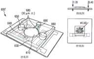
图6a和6b是根据本公开主题一些实施例的微型装置实例的结构和尺寸的示意图。6a and 6b are schematic illustrations of the structure and dimensions of an example microdevice according to some embodiments of the disclosed subject matter.
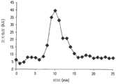
图7是根据本公开主题一个实施例的在试验中使用图6a所示微型装置的温度随时间变化图表。FIG. 7 is a graph of temperature over time in an experiment using the microdevice shown in FIG. 6a according to one embodiment of the disclosed subject matter.
图8是根据本公开主题一个实施例的温度对荧光测量(n=3,n是试验次数)影响的图表。8 is a graph of the effect of temperature on fluorescence measurements (n=3, n being the number of experiments) according to one embodiment of the disclosed subject matter.
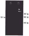
图9a和9b是根据本公开主题一个实施例的使用百日咳博德特氏菌基因组181bp DNA片段进行试验的凝胶电泳分析图表,其中,(a)显示了基于溶液的试验结果,(b)显示了基于小珠的试验结果。9a and 9b are graphs of gel electrophoresis analysis of an assay using a 181 bp DNA fragment of Bordetella pertussis genome, according to one embodiment of the disclosed subject matter, wherein (a) shows the results of a solution-based assay and (b) shows The results of the bead-based experiments are presented.
图10所示图表显示了本公开主题一个实施例中的进行基于小珠的PCR(n=3)后,退火温度对荧光强度的影响。Figure 10 is a graph showing the effect of annealing temperature on fluorescence intensity after bead-based PCR (n=3) in one embodiment of the disclosed subject matter.
图11是电泳图谱,显示了进行传统的基于溶液的PCR后,退火温度对扩增后DNA的影响,作为与基于小珠的PCR的比较。Figure 11 is an electropherogram showing the effect of annealing temperature on amplified DNA following conventional solution-based PCR, as compared to bead-based PCR.
图12所示图表显示了本公开主题一个实施例中的进行PCR后小珠浓度对小珠荧光强度的影响。Figure 12 is a graph showing the effect of bead concentration on bead fluorescence intensity following PCR in one embodiment of the disclosed subject matter.
图13是根据本公开主题一个实施例的进行PCR后小珠荧光强度关于反应混合物中模板浓度的关系图表。误差线示出了由三个实例(n=3)的平均值得出的一个标准差,根据学生t分布检验(Student’s t test),零模板(对照)和1pM模板浓度(检测极限)的反应具有差别的可能性大于95%。13 is a graph of bead fluorescence intensity versus template concentration in the reaction mixture after performing PCR, according to one embodiment of the disclosed subject matter. Error bars show one standard deviation from the mean of three instances (n=3), according to the Student's t test, the responses of zero template (control) and 1 pM template concentration (limit of detection) have The probability of difference is greater than 95%.
图14所示图表显示了根据本公开主题一个实施例的信号强度与PCR循环次数之间的关系。显示了多次试验(n=3)得出的平均值,误差线示出了标准差。Figure 14 is a graph showing the relationship between signal strength and PCR cycle number according to one embodiment of the disclosed subject matter. Means from multiple experiments (n=3) are shown and error bars show standard deviations.
图15a-15c是根据本公开主题一个实施例的微室显微照片,说明了基于小珠的PCR的过程。15a-15c are photomicrographs of microchambers illustrating the process of bead-based PCR, according to one embodiment of the disclosed subject matter.
图16是根据本公开主题一些实施例的微型装置的示意图,所述微型装置包括一个筛选室和一个扩增室。Figure 16 is a schematic diagram of a microdevice including a screening chamber and an amplification chamber according to some embodiments of the disclosed subject matter.
图17a和17b是无连接的微型装置的图像(a)和用染料检测的混合物的图像(b)。Figures 17a and 17b are images of the junction-free microdevice (a) and the mixture detected with the dye (b).
图18是根据本公开一些实施例的电泳图谱,显示了在不同洗涤中收集的含有非目标DNA的洗脱液的芯片外扩增情况。18 is an electropherogram showing off-chip amplification of eluate containing non-target DNA collected in different washes, according to some embodiments of the present disclosure.
图19是根据本公开一些实施例的电泳图谱,显示了在不同洗涤中收集的含有目标DNA的洗脱液的芯片上扩增情况。19 is an electropherogram showing on-chip amplification of eluates containing target DNA collected in different washes, according to some embodiments of the present disclosure.
图20a和20b所示图表显示了根据本公开主题一些实施例的富集后DNA与起始随机库之间对于结合亲和力的比较。Figures 20a and 20b are graphs showing a comparison of the binding affinity between the enriched DNA and the starting random pool according to some embodiments of the disclosed subject matter.
图21所示图表显示了根据本公开主题一些实施例的从荧光测量反应出的富集DNA与温度相关的结合情况。Figure 21 is a graph showing temperature-dependent binding of enriched DNA as reflected from fluorescence measurements according to some embodiments of the disclosed subject matter.
图22是根据本公开主题一些实施例的用于测量目标DNA的分离和富集的微型装置的示意图。22 is a schematic diagram of a microdevice for measuring the isolation and enrichment of target DNA according to some embodiments of the disclosed subject matter.
图23a-23j是图22所示微芯片的制造过程实例的示意图。23a-23j are schematic diagrams of an example of a fabrication process for the microchip shown in FIG. 22. FIG.
图24是图22所示微型装置制造完毕后的图片。FIG. 24 is a picture of the microdevice shown in FIG. 22 after fabrication.
图25为运行图24所示微型装置的试验方案实例。Figure 25 is an example of an experimental protocol for operating the microdevice shown in Figure 24.
图26a是根据本公开主题一个实施例的扩增后的洗脱液的电泳图谱,所述洗脱液于分离步骤中得到,含有非目标DNA;图26b是根据本公开主题实施例的不同泳道中光带强度柱状图。Figure 26a is an electrophoretic pattern of an amplified eluate according to an embodiment of the disclosed subject matter, which is obtained in the separation step and contains non-target DNA; Figure 26b is a different swimming lane according to an embodiment of the disclosed subject matter Histogram of medium light band intensity.
图27a是对照实例中得到的扩增后的洗脱液的凝胶电泳图谱;图27b是对照实例的不同泳道中光带强度柱状图。Fig. 27a is the gel electrophoresis profile of the amplified eluate obtained in the control example; Fig. 27b is a histogram of light band intensity in different swimming lanes of the control example.
图28显示了根据本公开主题一些实施例的使用不同电解质的充有凝胶的微通道中目标DNA的电泳情况。28 shows electrophoresis of target DNA in gel-filled microchannels using different electrolytes, according to some embodiments of the disclosed subject matter.
图29a-29d显示了根据本公开主题一些实施例的不同时间时通过充有凝胶的微通道电泳输送具有荧光标记的目标DNA的情况,所述微通道处于25V/cm的电场中。29a-29d show electrophoretic delivery of fluorescently labeled target DNA through a gel-filled microchannel in an electric field of 25 V/cm at different times according to some embodiments of the disclosed subject matter.
图30是根据本公开主题一些实施例的一轮分离和富集结束后从分离室和富集室获得的洗脱液的凝胶电泳图谱。30 is a gel electrophoresis pattern of eluate obtained from the separation chamber and the enrichment chamber after a round of separation and enrichment according to some embodiments of the disclosed subject matter.
图31a是PCR扩增后从富集室获得的洗脱液的凝胶电泳图谱。图31b是根据本公开主题一些实施例的洗脱液光带强度柱状图。Figure 31a is a gel electrophoresis pattern of the eluate obtained from the enrichment chamber after PCR amplification. Figure 31b is a histogram of eluate band intensity according to some embodiments of the disclosed subject matter.
图32是根据本公开主题一个实施例的微型装置实例示意图。Figure 32 is a schematic diagram of an example of a microdevice according to one embodiment of the disclosed subject matter.
图33是根据本公开主题一个实施例的微型装置实例(具有腔室和通道,所述通道中充有彩色墨水以便于观察)照片。Figure 33 is a photograph of an example of a microdevice having chambers and channels filled with colored inks for easy viewing, according to one embodiment of the disclosed subject matter.
图34a根据本公开主题一个实施例的在分离过程中获得的洗脱液的凝胶电泳图谱;图34b所示柱状图显示了根据本公开主题实施例的培养、洗涤和洗脱样本的光带强度情况。Figure 34a shows the gel electrophoresis profile of the eluate obtained during the separation process according to one embodiment of the disclosed subject matter; the histogram shown in Figure 34b shows the light bands of the cultured, washed and eluted samples according to an embodiment of the disclosed subject matter intensity situation.
图35a是根据对照实例的在分离过程中获得的洗脱液的凝胶电泳图谱;图35b所示柱状图显示了根据对照实例的培养、洗涤和洗脱样本的光带强度情况。Fig. 35a is the gel electrophoresis profile of the eluate obtained during the separation process according to the control example; the histogram shown in Fig. 35b shows the light band intensity of the cultured, washed and eluted samples according to the control example.
图36a-36c显示了根据本公开主题一个实施例的不同时间时在25V/cm电场中电泳输送具有荧光标记的目标DNA的情况;图36d是根据本公开主题实施例的在检测位点监测到的随时间变化的荧光强度图表。Figures 36a-36c show the situation of electrophoretic delivery of target DNA with fluorescent labels in a 25V/cm electric field at different times according to an embodiment of the disclosed subject matter; A graph of fluorescence intensity over time.
图37是根据本公开主题一个实施例的一轮分离和富集实例结束后从分离室和富集室获得的洗脱液的凝胶电泳图谱。Figure 37 is a gel electrophoresis pattern of the eluate obtained from the separation chamber and the enrichment chamber after one round of separation and enrichment example according to one embodiment of the disclosed subject matter.
图38a是根据本公开主题一个实施例的PCR扩增后从富集室获得的洗脱液的凝胶电泳图谱;图38b所示柱状图显示了根据本公开主题实施例的随着富集次数的增加,洗脱液的光带强度情况。Figure 38a is a gel electrophoresis profile of the eluate obtained from the enrichment chamber after PCR amplification according to an embodiment of the disclosed subject matter; The increase, the light band intensity of the eluent.
图39是根据本公开主题一个实施例的微型装置实例(充有彩色墨水以便于观察)图片。Figure 39 is a picture of an example of a microdevice (filled with colored ink for ease of viewing) according to one embodiment of the disclosed subject matter.
图40a是含有非目标DNA的洗脱液的电泳图谱,所述洗脱液从图39所示微型装置一个实例的筛选室获得。图40b是图40a中相应泳道的荧光强度柱状图。Figure 40a is an electrophoretic profile of an eluate containing non-target DNA obtained from the screening chamber of one example of the microdevice shown in Figure 39. Figure 40b is a histogram of the fluorescence intensity of the corresponding lane in Figure 40a.
图41a是根据本公开主题一个实施例的PCR腔室中小珠的显微照片;图41b和41c是根据该实施例的具有荧光标记的目标DNA杂交前(图b中)和杂交后(图c中)小珠的荧光图像(图a-c中的比例尺为100μm);图41d是根据本公开主题实施例的小珠荧光强度柱状图。Figure 41a is a photomicrograph of beads in a PCR chamber according to one embodiment of the disclosed subject matter; Figures 41b and 41c are fluorescently labeled target DNA before (panel b) and after hybridization (panel c) according to this embodiment Middle) Fluorescence images of beads (scale bar in panels a-c is 100 μm); Figure 41d is a histogram of fluorescence intensity of beads according to an embodiment of the disclosed subject matter.
图42a-42c是根据本公开主题一些实施例的(a)0、(b)10、(c)20次PCR循环后小珠的荧光图像。图42d是所述小珠相应的荧光强度柱状图。42a-42c are fluorescent images of beads after (a) 0, (b) 10, (c) 20 PCR cycles, according to some embodiments of the disclosed subject matter. Figure 42d is a histogram of the corresponding fluorescence intensity of the beads.
图43所示柱状图显示了根据本公开主题一些实施例的富集后DNA和随机DNA对于涂有IgE的小珠的结合亲和力。Figure 43 is a bar graph showing the binding affinity of enriched DNA and random DNA to IgE-coated beads according to some embodiments of the disclosed subject matter.
图44a和44b是根据本公开主题一些实施例的微型装置的结构示意图;图44c-44g是图44a和44b所示微型装置的制造实例过程示意图。所有显示尺寸均为微米。Figures 44a and 44b are structural schematic diagrams of micro-device according to some embodiments of the disclosed subject matter; Figures 44c-44g are schematic diagrams of an example manufacturing process of the micro-device shown in Figures 44a and 44b. All dimensions shown are in microns.
图45是图44a和44b中示意性显示的微型装置制造完毕后的照片。Figure 45 is a photograph of the microdevice shown schematically in Figures 44a and 44b after fabrication.
图46是在检测DNA多态位点时使用图45所示微型装置的试验方案实例。Figure 46 is an example of an assay protocol using the microdevice shown in Figure 45 in the detection of DNA polymorphic sites.
图47是使用图45所示微型装置进行校准试验中腔室温度随时间变化的图表。FIG. 47 is a graph of chamber temperature versus time for a calibration experiment using the microdevice shown in FIG. 45 .
图48a所示柱状图显示了使用图45所示微型装置进行基因分型实例中基于小珠的PCR的特征,通过在不同PCR参数下测量任意单位(a.u.)的小珠荧光强度来确定PCR特征;图48b所示柱状图显示了用NaOH从小珠除去目标DNA的洗脱效果(误差线代表基于对荧光微珠四次独立测量的标准差)。Figure 48a is a histogram showing the characteristics of bead-based PCR in an example of genotyping using the microdevice shown in Figure 45, determined by measuring the fluorescence intensity of the beads in arbitrary units (a.u.) under different PCR parameters ; Figure 48b shows a histogram showing the effect of elution with NaOH to remove target DNA from beads (error bars represent standard deviation based on four independent measurements on fluorescent beads).
图49a所示柱状图显示了使用图45所示微型装置进行基因分型实例中除盐前、除盐后和热洗脱后的小珠荧光强度;图49b是基因分型实例中的柱状形,显示了加热后用FAM标记的微珠的荧光强度;图49c是热洗脱的经FAM改性的正向引物的MALDI-TOF质谱。(a)和(b)中的误差线代表基于荧光微珠四次独立测量的标准差。The histogram shown in Figure 49a shows the bead fluorescence intensity before desalting, after desalting and after thermal elution in the genotyping example using the microdevice shown in Figure 45; Figure 49b is the bar graph in the genotyping example , shows the fluorescence intensity of microbeads labeled with FAM after heating; Fig. 49c is the MALDI-TOF mass spectrum of the thermally eluted FAM-modified forward primer. Error bars in (a) and (b) represent the standard deviation of four independent measurements based on fluorescent microbeads.
图50a是使用图45所示微型装置进行基因分型试验中突变HBB基因的MALDI-TOP质谱;图50b是未突变HBB基因的对应MALDI-TOF质谱(图中“*”表示延伸的SBE引物)。Figure 50a is the MALDI-TOP mass spectrum of the mutated HBB gene in the genotyping test using the miniature device shown in Figure 45; Figure 50b is the corresponding MALDI-TOF mass spectrum of the unmutated HBB gene ("*" in the figure indicates the extended SBE primer) .
图51显示了根据本公开主题一些实施例的可裂解生物素化ddNTP的示范性分子结构。Figure 51 shows exemplary molecular structures of cleavable biotinylated ddNTPs according to some embodiments of the disclosed subject matter.
图52a是根据本公开主题一个实施例的SNP检测装置的剖面示意图;图52b是根据本公开主题一个实施例的SNP检测装置制造完成后的照片。Fig. 52a is a schematic cross-sectional view of a SNP detection device according to an embodiment of the disclosed subject matter; Fig. 52b is a photo of the SNP detection device according to an embodiment of the disclosed subject matter after manufacture.
图53是图52b所示SNP检测装置的温度传感器的校准图表。Fig. 53 is a calibration chart of the temperature sensor of the SNP detection device shown in Fig. 52b.
图54a是图52b所示装置的SBE腔室温度试验中的时间解析跟踪曲线;图54b是图52b所示装置的SPC腔室温度试验中的时间解析跟踪曲线。Figure 54a is the time-resolved tracking curve in the SBE chamber temperature test of the device shown in Figure 52b; Figure 54b is the time-resolved tracking curve in the SPC chamber temperature test of the device shown in Figure 52b.
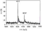
图55a是使用图52b所示SNP检测装置进行的试验中单碱基延伸产物的MALDI-TOF质谱(插图:用ddUTP-N3-生物素终结的延伸引物结构,用“*”标注的波峰是由合成引物商品中的杂质引起的);图55b是使用图52b所示SNP检测装置进行的试验中固相捕获产物和化学裂解产物的MALDI-TOF质谱(插图:裂解产物的结构);图55c是使用图52b所示SNP检测装置进行的试验中除盐后产物的MALDI-TOF质谱。Figure 55a is the MALDI-TOF mass spectrum of the single-base extension product in the experiment performed using the SNP detection device shown in Figure 52b (inset: the structure of the extension primer terminated with ddUTP-N3-biotin, the peaks marked with "*" are from caused by impurities in commercially available synthetic primers); Figure 55b is the MALDI-TOF mass spectrum of the solid-phase capture product and the chemical cleavage product in the experiment performed using the SNP detection device shown in Figure 52b (inset: structure of the cleavage product); Figure 55c is MALDI-TOF mass spectrum of the desalted product in the experiment using the SNP detection device shown in Figure 52b.
图56是使用图52b所示SNP检测装置进行的试验中结合所有步骤得到的SNP检测产物的MALDI-TOF质谱。Fig. 56 is the MALDI-TOF mass spectrum of the SNP detection product obtained by combining all the steps in the experiment using the SNP detection device shown in Fig. 52b.
具体实施方式Detailed ways
本公开主题提供了一种用于在微芯片上分离、筛选和扩增核苷酸如DNA分子的技术。更具体地,本公开主题提供了一种用于分离和富集所需DNA的基于MEMS的微型装置平台和相关方法,以进行基因分型和其它用途。The disclosed subject matter provides a technique for isolating, screening and amplifying nucleotides, such as DNA molecules, on a microchip. More specifically, the disclosed subject matter provides a MEMS-based microdevice platform and associated methods for isolating and enriching desired DNA for genotyping and other uses.
一方面,本公开主题提供了一种使用微室扩增目标DNA分子的方法,包括在第一微室内的固定在固相上的第一引物(例如微珠)。参考图1a,方法包括以下步骤:将含有目标DNA分子的第一样本引入第一微室(见110),目标DNA杂交到适于扩增目标DNA的第一引物上;将目标DNA作为模板,在第一微室内产生目标DNA的互补DNA(见120);然后将目标DNA与互补DNA分离(见130);第二引物杂交到互补DNA上(见140);将互补DNA作为模板,扩增目标DNA(见150)。In one aspect, the disclosed subject matter provides a method of amplifying a target DNA molecule using a microchamber comprising a first primer (eg, bead) immobilized on a solid phase within a first microchamber. Referring to Figure 1a, the method comprises the following steps: introducing a first sample containing target DNA molecules into a first microchamber (see 110), the target DNA is hybridized to a first primer suitable for amplifying the target DNA; using the target DNA as a template , produce the complementary DNA of the target DNA in the first microchamber (see 120); then separate the target DNA from the complementary DNA (see 130); the second primer hybridizes to the complementary DNA (see 140); use the complementary DNA as a template to amplify Amplify target DNA (see 150).
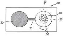
上述步骤可在微型装置(也称为微芯片)的微室(或简称为“腔室”)中进行,微型装置装有作为固相的微珠。微型装置可采用标准的微细加工技术进行制造,例如,采用PDMS软光刻技术形成具有所需形状和尺寸的腔室。例如但不局限于,微室的直径可大约在0.1mm至2mm之间,深度大约在0.05mm至0.5mm之间。用于在PCR工艺中调节温度的微加热器和温度传感器可集成在微型装置中,例如,设置于微室下方的薄膜层中。在本实施例和以下进一步描述的其他实施中,仅出于说明目的而不具局限性,图1b示意性地描绘了一个微型装置10实例,该微型装置10具有一个装有固相40的微室20,微室20位于微加热器50和温度传感器60的上方。在一些实施例中,微型装置10还可包括一个通过微通道25与微室20相连的第二微室30。在实例中对微型装置10实施例及其制造方法以及各种特点做了进一步描述。The steps described above can be performed in microchambers (or simply "chambers") of a microdevice (also known as a microchip) containing microbeads as a solid phase. Microdevices can be fabricated using standard microfabrication techniques, for example, using PDMS soft lithography to form chambers of desired shape and size. For example and without limitation, the microchambers may have a diameter between about 0.1 mm and 2 mm and a depth between about 0.05 mm and 0.5 mm. Microheaters and temperature sensors for regulating the temperature in the PCR process can be integrated in the microdevices, for example, placed in a thin film layer beneath the microchambers. In this example and other implementations described further below, for illustrative purposes only and without limitation, FIG. 1 b schematically depicts an example of a
目标DNA可以有不同的来源,包括人工合成的DNA,例如随机的寡核苷酸库或从细胞中提取的基因组DNA。来源的定位可包括样本芯片外处理也可以是芯片上预处理。Target DNA can come from different sources, including synthetic DNA such as random oligonucleotide libraries or genomic DNA extracted from cells. Source localization can include off-chip processing of samples as well as on-chip preprocessing.
微珠的功能是作为合适的引物对目标DNA(也称为“模板DNA”)进行扩增。微珠可以是涂有链霉亲和素的聚合物小珠,众所周知其对于生物素具有极高的亲和力。引物(例如反向引物)可以是生物素功能化的,固定在小珠表面上。将含有目标DNA的样本引入腔室中时,目标DNA由于分子识别(例如,沃森-克里克碱基配对)与固定到小珠上的引物杂交。样本中的其他分子,例如非目标DNA分子、细胞、小分子等,则不大可能与引物结合。由于使用了固定在小珠上的引物和PCR试剂(包括例如Taq聚合酶、脱氧核苷三磷酸和缓冲液),可基于目标DNA生成互补DNA,该互补DNA与目标DNA一起形成附着在小珠上的双链DNA(ds-DNA)。这种ds-DNA可在高温(例如大约95℃)下,发生变性(或熔化),使得目标DNA与互补DNA分离。第二引物,例如正向引物,可在较低温度(例如,大约50-62℃)下退火与互补DNA(例如,在互补DNA的游离端处)结合。然后,使用作为模板的互补DNA以及第二引物及PCR试剂在适宜的扩链温度(例如,大约72℃)下生成目标DNA的另一个复本。重复上述温度循环(熔化、退火和扩链)可实现目标DNA的扩增,即实现目标DNA以指数级进行复制。The function of the microbeads is to serve as suitable primers for the amplification of target DNA (also called "template DNA"). The microbeads may be polymeric beads coated with streptavidin, which is known to have a very high affinity for biotin. Primers (eg, reverse primers) can be biotin-functionalized and immobilized on the bead surface. When a sample containing target DNA is introduced into the chamber, the target DNA hybridizes to the primers immobilized on the beads due to molecular recognition (eg, Watson-Crick base pairing). Other molecules in the sample, such as non-target DNA molecules, cells, small molecules, etc., are less likely to bind to the primers. Thanks to the use of bead-immobilized primers and PCR reagents (including, for example, Taq polymerase, deoxynucleoside triphosphates, and buffers), complementary DNA can be generated based on the target DNA, which together forms a bead-attached double-stranded DNA (ds-DNA). This ds-DNA can be denatured (or melted) at high temperature (for example, about 95°C), so that the target DNA is separated from the complementary DNA. A second primer, such as a forward primer, can anneal to the complementary DNA (eg, at the free end of the complementary DNA) at a lower temperature (eg, about 50-62° C.). Another copy of the target DNA is then generated using the complementary DNA as template along with a second primer and PCR reagents at an appropriate chain extension temperature (eg, about 72°C). Repeating the above temperature cycles (melting, annealing, and chain extension) can achieve the amplification of the target DNA, that is, the target DNA can be replicated exponentially.
未结合的引物可用光谱检测标签(例如,荧光团)进行标记。在这种情况下,进行了多次PCR循环后,扩增的产物可为用荧光团标记的目标DNA和与小珠结合的未标记的互补链。这种做了标记的目标DNA可通过荧光光谱法进行分离。Unbound primers can be labeled with a spectrally detectable label (eg, a fluorophore). In this case, after multiple cycles of PCR, the amplified product can be the target DNA labeled with a fluorophore and the unlabeled complementary strand bound to the bead. This labeled target DNA can be isolated by fluorescence spectroscopy.
上述基于微珠的PCR步骤在微型装置中的微室中进行,便于芯片上DNA检测和操控。采用这种PCR工艺对DNA进行分离、富集和测定等这些步骤是本发明提出的设想,下文将对一些实施例进行描述。The bead-based PCR steps described above are carried out in microchambers in a microdevice, facilitating on-chip DNA detection and manipulation. Using this PCR process to separate, enrich and measure DNA is a concept proposed by the present invention, and some examples will be described below.
首先,对含有目标DNA(以及其他杂质)的样本进行处理,使目标DNA可被某些功能性分子有选择性地捕捉,这些功能性分子与目标DNA特异性结合。例如,序列特异性结合结构的分子识别利用分子分析物使复杂混合物中的这种序列得到纯化。在这种情况下,所述功能性分子可为免疫球蛋白E(IgE),目标DNA可为与IgE特异性结合的适体。这对于含有各种其他DNA序列(例如,低聚物库)的样本中的目标DNA的分离和扩增尤为有利。First, a sample containing target DNA (and other impurities) is processed so that the target DNA can be selectively captured by certain functional molecules that specifically bind to the target DNA. For example, molecular recognition of sequence-specific binding structures utilizes molecular analytes to enable the purification of such sequences from complex mixtures. In this case, the functional molecule may be immunoglobulin E (IgE), and the target DNA may be an aptamer that specifically binds to IgE. This is especially advantageous for the isolation and amplification of target DNA in samples containing various other DNA sequences (e.g., oligo pools).
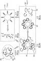
图2a-2e描述了采用这种预筛选以及随后进行扩增步骤的流程实例。预筛选可在第二腔室(也称为“分离室”或“筛选室”)中完成,例如位于同一个微型装置上并与第一腔室流体连通。功能性分子可附着在设置于第二腔室中的微珠上。当含有低聚物库200(见图2a,其中低聚物库包含目标DNA201和非目标DNA202)的样本被引入筛选室220内时,目标DNA201与附着在微珠230(见图2b)上的固定化功能性分子235相结合。未与功能性分子结合(或弱结合)的非目标DNA202可被移除,例如通过冲洗的方式进行移除(见图2c)。然后,可将已结合的目标DNA与功能性分子分离,将其输送入第一腔室210(也称为“扩增室”或“富集室”),例如通过用缓冲液洗脱的方式进行分离。扩增室210中的输送过来的目标DNA201可进行上文所述的扩增(见图2d),可将扩增产物(增加的目标DNA)分离出来(见图2e)以用于检测,或者送回筛选室220进行新一轮的分离-扩增。Figures 2a-2e depict an example of a process using such a pre-screen followed by an amplification step. Pre-screening can be accomplished in a second chamber (also referred to as a "separation chamber" or "screening chamber"), for example on the same microdevice and in fluid communication with the first chamber. Functional molecules can be attached to microbeads disposed in the second chamber. When a sample containing an oligomer library 200 (see FIG. 2a, wherein the oligomer library includes
目标DNA可以是一个适体。适体可广泛做为具有高亲和力的分析物,具有很好的目标选择控制性,可与结合目标进行人工合成,所述结合目标具有预先确定的结合特性,例如温度敏感特性。因此,可利用外部刺激(如温度、pH值或离子浓度)对适体-目标结合复合物进行干扰。例如,腔室220设置在第一温度T1以使目标适体结合,在移除未结合DNA和其他杂质后可以改变第二腔室的温度,例如升高到温度T2,温度T2高于温度T1以破坏适体的等角结构,使适体与功能性分子分离。可通过集成的微加热器和与筛选室连接的温度传感器实现对温度的控制。对于某些适体,分离温度T2可低于捕捉温度T1。在这种情况下,可通过热电制冷实现较低温度T2,例如通过微型装置中的帕尔贴(Peltier)元件实现。可选择地,与功能性分子相结合的适体可以通过试剂进行分离,例如通过碱性溶液进行分离。The target DNA can be an aptamer. Aptamers can be widely used as analytes with high affinity, have good target selection control, and can be artificially synthesized with binding targets that have predetermined binding properties, such as temperature-sensitive properties. Therefore, external stimuli such as temperature, pH or ion concentration can be used to interfere with the aptamer-target binding complex. For example, the
在实施例中,可通过电泳法将目标DNA从筛选室输送到富集室。如图3所示,连接筛选室320和富集室310的微通道340包括一个充满了凝胶350的部分。凝胶可以是任何一种适于DNA电泳的常用凝胶,例如琼脂糖凝胶。目标DNA301与固定在小珠330上的功能性分子335分离(例如热分离,由设置在筛选室320下方的微加热器337提供热量)以后,将目标DNA301通过凝胶350电泳进行输送,由施加在正极365和负极360之间的电压产生电场。只有当凝胶350中产生合适电场时才会产生目标DNA301输送。因此,这种设置可以将富集室310与筛选室320有效隔离,使得筛选室320可以独立运行(例如,冲洗、冼脱),避免产生污染富集室310中富集产物的风险。In embodiments, target DNA may be transported from the screening chamber to the enrichment chamber by electrophoresis. As shown in FIG. 3 , the
目标DNA被输送入富集室内以后,可进行又一轮的筛选-输送,以在富集室内积聚更多的目标DNA。此外或者可选择地,可使用上文中所述的方法在富集室内扩增目标DNA。如有必要,将扩增产物送回筛选室进行又一轮筛选-输送-扩增。这种从富集室至筛选室的输送可通过电泳法再次进行,例如使用微通道340和设置其内的凝胶350(以及施加在电极360和365之间的反转电场)进行,或者通过连接两腔室的充满凝胶的第二微通道进行。After the target DNA is transported into the enrichment chamber, another round of screening-transportation can be carried out to accumulate more target DNA in the enrichment chamber. Additionally or alternatively, the target DNA can be amplified within the enrichment chamber using the methods described above. If necessary, the amplified product is returned to the screening chamber for another round of screening-delivery-amplification. This transport from the enrichment chamber to the screening chamber can be performed again by electrophoresis, for
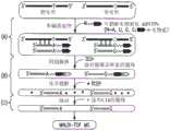
在其他实施例中,芯片上PCR法可应用于目标DNA(即,DNA具有单核苷酸多态性(SNP))中的多态位点检测,如图4a-4d所示。在这些实施例中,首先,将含有具有SNP的目标DNA401的样本可通过上文所述的基于小珠的PCR进行扩增,产生附着于小珠的包含有目标DNA401和互补DNA411的DNA(见图4a)。可将目标DNA与互补链分离(例如通过化学洗脱或变性)并冲洗掉,只留下附着在小珠上的互补DNA链411(见图4b)。然后,可引入单碱基延伸(SBE)反应物,引入等位基因特异性引物,紧挨与DNA模板上单核苷酸多态位点对应的互补DNA位点退火。接下来对这些引物进行单碱基延伸(SBE),在双脱氧核苷三磷酸(ddNTP)和酶的条件下对反应混合物进行热循环,生成仅延伸一个碱基的引物(见图4c)。可将游离的引物、盐和所有其他杂质冲洗掉,以对结合在小珠上的已延伸的和未延伸的等位基因特异性引物进行纯化,然后对已延伸的和未延伸的引物进行热力或化学洗脱(见图4d)。可对分离出的已延伸引物中的已延伸的一个碱基进行检测,例如根据已延伸引物和未延伸引物间的质量差异利用MALDI-TOF(基质辅助激光解析电离飞行时间)质谱技术进行检测,由此确定目标DNA的多态位点。In other embodiments, the on-chip PCR method can be applied to the detection of polymorphic sites in the target DNA (ie, the DNA has a single nucleotide polymorphism (SNP)), as shown in Figures 4a-4d. In these embodiments, first, a sample containing
或者,目标DNA中多态位点的检测可采用不涉及PCR的其他方法在微型装置上完成。图5所示为进行这种检测的方法的一个实施例,在图中,含有SNP的目标DNA和相应的野生型DNA按序列并排显示。可将包含有目标DNA的样本引入第一微室(“SBE室”)内。然后可以引入SBE反应物(包括例如可裂解生物素化ddNTP)和等位基因特异性引物,紧挨目标DNA的多态位点立即退火,由此各等位基因特异性引物延伸出一个碱基,得到延伸的引物(见图5a)。经过一轮或多轮热循环后,可得到延伸引物的多个复本,包括游离的延伸引物(未与目标DNA结合)。可将游离的延伸引物送至含有固相的微通道,通过进行固相捕获(SPC)纯化延伸引物。对于SPC,固相可具有附着在表面的功能性分子,所述功能性分子与延伸引物特异性结合。对于含有生物素化ddNTP的延伸引物,由于链霉亲和素对于生物素具有强亲和力,将其作为功能性分子。然后,可以将捕获的延伸引物与固相分离,例如通过化学裂解进行分离,对分离后的延伸引物进一步去盐,然后,例如根据延伸引物和未延伸引物之间的质量差异利用MALDI-TOF质谱法进行检测,由此确定目标DNA的多态位点。Alternatively, detection of polymorphic sites in target DNA can be accomplished on the microdevice using other methods that do not involve PCR. An example of a method for performing this detection is shown in Figure 5, in which the target DNA containing the SNP and the corresponding wild-type DNA are shown side-by-side in sequence. A sample containing target DNA can be introduced into the first microchamber ("SBE chamber"). SBE reagents (including, for example, cleavable biotinylated ddNTPs) and allele-specific primers can then be introduced, immediately annealing next to the polymorphic site of the target DNA, whereby each allele-specific primer is extended by one base , resulting in extended primers (see Figure 5a). After one or more rounds of thermal cycling, multiple copies of the extension primer are obtained, including the free extension primer (not bound to the target DNA). Free extension primers can be sent to a microchannel containing a solid phase, and the extension primers can be purified by performing solid phase capture (SPC). For SPC, the solid phase can have functional molecules attached to the surface that specifically bind the extended primers. For extension primers containing biotinylated ddNTPs, streptavidin was used as a functional molecule due to its strong affinity for biotin. The captured extension primers can then be separated from the solid phase, e.g. by chemical cleavage, the separated extension primers further desalted, and then e.g. The polymorphic site of the target DNA is determined by detection method.
在以下实例中将详细说明上述实施例的装置结构、制造和操作流程,其仅出于说明目的而非限制本发明。In the following examples, the device structure, manufacturing and operation process of the above-mentioned embodiments will be described in detail, which are only for the purpose of illustration and not limiting the present invention.
实例1Example 1
本实例描述了在微芯片上分离和扩增目标DNA的基于小珠的PCR,所述微芯片包括集成的加热器和温度传感器。This example describes bead-based PCR for isolation and amplification of target DNA on a microchip that includes an integrated heater and temperature sensor.
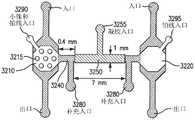
如图6所示,基于小珠的PCR芯片600包括一个微室610,该微室由聚二甲基硅氧烷(PDMS)制成,聚二甲基硅氧烷形成微室610的侧壁611并粘合到基底620(玻璃片)上,基底620具有一个集成的电阻加热器630和温度传感器640(见图6a)。加热器630具有蛇形的几何形状,覆盖微室610和大面积周围区域,在微室610中形成一个足够均匀的温度场,而电阻温度传感器640位于微室610的中心。圆柱形的微室610开口于大气中,包括两个在竖直方向上在一条直线上的直径不同的相通的隔间。下隔间可用于容纳反应物(包括表面功能化的微珠、目标DNA、PCR试剂等)。具有较大直径的上隔间可用于容纳在反应物上方的矿物油层。微室610内表面涂有一层聚合物聚氯代对二甲苯(Parylene C)。矿物油和聚对二甲苯涂层可减少水分蒸发,否则水分会向空气蒸发或通过PDMS蒸发,矿物油和聚对二甲苯还可降低气泡形成几率。聚对二甲苯还提供了一个与PCR兼容的表面,在使用添加剂(如牛血清白蛋白(BSA)和吐温)时,最大限度地减少反应组分(如DNA和Taq聚合酶)的吸附。As shown in FIG. 6, the bead-based
装置是使用标准微细制造技术进行制造的。形成加热器和温度传感器的铬层和金层(厚度为20和200nm)热力喷镀到显微镜玻璃载片上,并通过接触式光刻和湿法刻蚀技术制出图案。图案形成一个长5.67cm、宽200μm、阻值约为20Ω的加热器和一个长1.04cm、宽40μm、阻值约为30Ω的温度传感器,加热器的覆盖面积为0.242cm2。然后,采用等离子体增强化学气相沉积技术用SiO2对这些热元件进行钝化(厚度为1μm),留有开口以用于电连接,使用荫罩形成所述开口。SiO2不仅用于钝化电气元件,还为PDMS提供了有效的粘合表面。为了生产用于微液体腔室的PDMS,将PDMS预聚物以10:1的比例与固化剂混合,倾倒至一张干净的硅片上,在75℃下烘烤30分钟,然后将其从硅片上剥落。The device was fabricated using standard microfabrication techniques. Layers of chromium and gold (20 and 200 nm thick) forming heaters and temperature sensors were thermally sprayed onto microscope glass slides and patterned by contact lithography and wet etching techniques. A heater with a length of 5.67 cm, a width of 200 μm, and a resistance of about 20 Ω and a temperature sensor with a length of 1.04 cm, a width of 40 μm, and a resistance of about 30 Ω were formed by patterning. The coverage area of the heater was 0.242 cm2 . These thermal elements were then passivated (1 μm thick) withSiO2 using plasma-enhanced chemical vapor deposition, leaving openings for electrical connections, which were formed using a shadow mask.SiO2 is not only used to passivate electrical components, but also provides an effective bonding surface for PDMS. In order to produce PDMS for microfluidic chambers, the PDMS prepolymer was mixed with a curing agent at a ratio of 10:1, poured onto a clean silicon wafer, baked at 75°C for 30 minutes, and then removed from the Peeling off silicon wafers.
微流体室是由使用打孔机在PDMS中打孔形成。底层PDMS部分的厚度是1.3mm,具有一个直径为3.2mm的孔,顶层部分厚度为0.3mm,具有一个直径为4.75mm的孔(见图6a,侧视图)。然后使用紫外生成臭氧对玻璃基底和PDMS处理10分钟,并在75℃烘烤30分钟之后得以粘合。在粘合过程中,PDMS孔被对准于形成图形于玻片上的集成加热器的中心位置。接着,在保持芯片形状的条件下用化学气相沉积法将芯片涂敷一层厚度约为1μm的聚氯代对二甲苯,用透明胶带覆盖电气焊接区。Microfluidic chambers are formed by punching holes in PDMS using a hole punch. The bottom PDMS part was 1.3 mm thick with a 3.2 mm diameter hole and the top part was 0.3 mm thick with a 4.75 mm diameter hole (see Figure 6a, side view). The glass substrate and PDMS were then treated with UV-generated ozone for 10 minutes and bonded after baking at 75 °C for 30 minutes. During the bonding process, the PDMS wells were aligned to the center of the integrated heater patterned on the glass slide. Next, the chip was coated with a layer of polychlorinated p-xylylene with a thickness of about 1 μm by chemical vapor deposition method under the condition of maintaining the shape of the chip, and the electrical welding area was covered with transparent tape.
在另一种设计中,如图6b所示,微型装置600’也包括微流体通道以及在冲洗步骤中用于引入反应物和缓冲溶液的开口(包括开口650和660),通过所述微流体通道可装入小珠(通过微通道680)并阻挡小珠(通过围堰680)。要制造这种装置,需使用光学光刻技术制出一个SU-8模具,用于制造一个4μL大的PCR腔室,腔室深度为400μm,直径3.6mm。所述模具包括两个开口(650和660)和一个没有围堰的第三开口,所述两个开口将局部的竖直通道间隙限制到20μm以限制流动,作为被动围堰680阻挡腔室内的微珠流出,没有围堰的第三开口690用于微珠的装入。与前述图6a所示装置的制造方法相似,微型装置600'也用PDMS制造,形成微通道壁和支撑结构,与集成有电阻加热器630和温度传感器640的玻璃基底粘合在一起。In another design, as shown in Figure 6b, the microdevice 600' also includes microfluidic channels and openings (including
除了微型装置的设计和制造外,基于小珠的引物在设计上使得装置的运行操作快速而简便。在本实例中使用生物素-链霉亲和素组合将反向引物连接到小珠上,这种结合在这两种分子的作用下既牢固又具自发性。合成反向引物,需在5’端做双重生物素标记,然后是与核苷酸序列相邻的间隔分子。由于热变性,双生物素半族可以最大程度地减少信号丢失。间隔分子使小珠上DNA之间的横向间隔更大,因此减少了由位阻而产生的杂交问题。以合成方式形成的DNA模板用于在利用基于小珠的PCR进行DNA检测表征时获得可控的一致性结果。In addition to the design and fabrication of the micro-device, the bead-based primers are designed to allow quick and easy operation of the device. In this example a biotin-streptavidin combination was used to attach the reverse primer to the bead, and the binding was both robust and spontaneous under the influence of these two molecules. To synthesize a reverse primer, double biotinylation is required at the 5' end, followed by a spacer molecule adjacent to the nucleotide sequence. Dual biotin moieties minimize signal loss due to heat denaturation. Spacer molecules allow greater lateral spacing between the DNA on the beads, thus reducing hybridization problems due to steric hindrance. Synthetically formed DNA templates are used to obtain controlled and consistent results in DNA detection characterization using bead-based PCR.
基于小珠的PCR芯片已应用于致病DNA的检测,与百日咳博德特氏菌(B.pertussis)相关的DNA序列可通过这种检测验证。百日咳博德特氏菌是一种革兰氏阴性菌,每年在世界范围内感染大约48.5百万人(300,000人死亡)。而早期检测是治疗这种病症的关键,目前检测百日咳博德特氏菌的方法(例如细胞培养)需要数天甚至数周的周期。本公开的基于小珠的PCR芯片可以解决这个问题,能够对百日咳博德特氏菌进行快速、灵敏且特异性的检测。使用微芯片上基于小珠的PCR进行致病DNA检测包括以下步骤:将连接到小珠上的反向引物装入微芯片的微室中,暴露于原始样本(如细胞裂解液)中,该原始样本可包含有各种杂质。然后样本中的致病DNA通过其特异性地与反向引物杂交被捕获到小珠上。由于捕获是基于DNA与反向引物之间的亲和力完成,此步骤也作为纯化步骤。接着,致病DNA与芯片上的PCR反应物混合,采用荧光标记正向引物的方法对致病DNA进行基于小珠的PCR。该方法可在微珠上以指数级快速生成具有荧光标记的扩增模板复本,可以通过荧光显微镜进行检测。与在固体平面上进行的扩增相比,由于荧光团涂敷区域大幅增加,在检测中使用小珠可增强信噪比。最后,可将做了标记的模板复本与结合到小珠上的互补链通过变性进行分离,然后洗脱入纯净缓冲液,用于进一步分析,而结合到小珠上的互补链可从芯片回收,作为cDNA库保存。Bead-based PCR chips have been applied to the detection of pathogenic DNA, and DNA sequences related to Bordetella pertussis (B. pertussis) can be verified by this detection. Bordetella pertussis is a Gram-negative bacterium that infects approximately 48.5 million people (with 300,000 deaths) worldwide each year. While early detection is key to treating the condition, current methods for detecting B. pertussis, such as cell culture, require cycles of days or even weeks. The disclosed bead-based PCR chip can solve this problem, enabling rapid, sensitive and specific detection of B. pertussis. Detection of causative DNA using on-microchip bead-based PCR involves loading reverse primers attached to beads into microchambers of the microchip, exposing them to raw samples such as cell lysates, which Raw samples can contain various impurities. The disease-causing DNA in the sample is then captured onto the beads by its specific hybridization with the reverse primer. Since capture is based on the affinity between the DNA and the reverse primer, this step also serves as a purification step. Next, the disease-causing DNA is mixed with the on-chip PCR reaction, and bead-based PCR is performed on the disease-causing DNA using a fluorescently labeled forward primer. The method produces exponentially fast copies of fluorescently labeled amplified templates on microbeads that can be detected by fluorescence microscopy. The use of beads in the assay enhances the signal-to-noise ratio due to the greatly increased fluorophore-coated area compared to amplification performed on solid surfaces. Finally, the labeled copy of the template can be denatured to separate from the bead-bound complementary strand and then eluted into a clean buffer for further analysis, while the bead-bound complementary strand can be extracted from the chip. It was recovered and stored as a cDNA library.
对于与百日咳博德特氏菌相关DNA的检测,需使用以下材料和试剂。所有DNA均为冻干形式,购自位于美国爱荷华城特拉尔维尔的Integrated DNATechnologies公司。将引物用作PCR试料,用于确定百日咳博德特氏菌。使用的DNA序列如下:正向引物:5'-FAM-间隔基-GAT TCA ATA GGT TGT ATG CAT GGTT-3'(SEQ ID NO:1),反向引物:5'-双生物素-间隔基-TTC AGG CAC ACA AAC TTGATG GGC G-3'(SEQ ID NO:2),以及模板:5'-GAT TCA ATA GGT TGT ATG CATGGT TCA TCC GAA CCG GAT TTG AGA AAC TGG AAA TCG CCA ACC CCCCAG TTC ACT CAA GGA GCC CGG CCG GAT GAA CAC CCA TAA GCA TGCCCG ATT GAC CTT CCT ACG TCG ACT CGA AAT GGT CCA GCA ATT GAT CGCCCA TCA AGT TTG TGT GCC TGA A-3'(SEQ ID NO:3)。正向引物在5'端已用荧光标记羧基荧光素修饰过,而反向引物在5'端用双生物素修饰。两种分子在5'修饰端和核苷酸序列之间都含有惰性间隔分子。使用Taq酶、脱氧核苷三磷酸(dNTP)和含有适宜缓冲液(Promega GoTaq Flexi PCR混合物)的PCR反应混合物进行PCR。反向引物固定到涂有链霉亲和素的聚合物微珠(Thermo Scientific PierceProtein Research Products公司生产的超链接链霉亲和素树脂)上,直径平均为80μm。使用UV/VIS(Thermo Scientific Nanodrop公司生产)对DNA样本进行浓缩和纯化。微细制造中使用的材料包括光刻胶(Rohm&Haas Electronic Materials S1818,Microchem SU-8 2000)、PDMS预聚物(Dow Corning Sylgard184)和聚氯代对二甲苯预聚物(Kisko diX C)。For the detection of DNA associated with Bordetella pertussis, the following materials and reagents are used. All DNA was purchased in lyophilized form from Integrated DNA Technologies, Tralville, Iowa City, USA. Primers were used as PCR assays for the determination of B. pertussis. The DNA sequences used were as follows: forward primer: 5'-FAM-spacer-GAT TCA ATA GGT TGT ATG CAT GGTT-3' (SEQ ID NO: 1), reverse primer: 5'-biotin-spacer -TTC AGG CAC ACA AAC TTGATG GGC G-3'(SEQ ID NO:2), and template: 5'-GAT TCA ATA GGT TGT ATG CATGGT TCA TCC GAA CCG GAT TTG AGA AAC TGG AAA TCG CCA ACC CCCCAG TTC ACT CAA GGA GCC CGG CCG GAT GAA CAC CCA TAA GCA TGCCCG ATT GAC CTT CCT ACG TCG ACT CGA AAT GGT CCA GCA ATT GAT CGCCCA TCA AGT TTG TGT GCC TGA A-3' (SEQ ID NO: 3). The forward primer has been modified at the 5' end with the fluorescently labeled carboxyfluorescein, while the reverse primer has been modified at the 5' end with bis-biotin. Both molecules contain an inert spacer between the 5' modified end and the nucleotide sequence. PCR was performed using Taq enzyme, deoxynucleoside triphosphates (dNTPs), and a PCR reaction mix containing an appropriate buffer (Promega GoTaq Flexi PCR Mix). Reverse primers were immobilized on streptavidin-coated polymer beads (hyperlinked streptavidin resin from Thermo Scientific Pierce Protein Research Products, Inc.), with an average diameter of 80 μm. DNA samples were concentrated and purified using UV/VIS (manufactured by Thermo Scientific Nanodrop). Materials used in microfabrication include photoresists (Rohm&Haas Electronic Materials S1818, Microchem SU-8 2000), PDMS prepolymers (Dow Corning Sylgard184) and polychlorinated p-xylene prepolymers (Kisko diX C).
百日咳博德特氏菌检测的PCR反应混合物按以下步骤进行制备。将各冻干DNA样本悬置于去离子水中,稀释至所需浓度。PCR混合物由以下成分组成:5×缓冲液(2μL)、25mM的MgCl2(0.6μL)、10mM的dNTPs(0.4μL)、50μg/mL的BSA(0.4μL)、5%(体积百分比)吐温20(0.1μL)、微珠(0.5μL)、水(4.1μL)、25μM正向引物(0.4μL)、25μM反向引物(0.4μL)和酶(0.1μL)。The PCR reaction mixture for detection of Bordetella pertussis was prepared as follows. Each lyophilized DNA sample was suspended in deionized water and diluted to the desired concentration. The PCR mix consists of the following components: 5× buffer (2 μL), 25 mM MgCl2 (0.6 μL), 10 mM dNTPs (0.4 μL), 50 μg/mL BSA (0.4 μL), 5% (volume percent) Tween 20 (0.1 µL), microbeads (0.5 µL), water (4.1 µL), 25 µM forward primer (0.4 µL), 25 µM reverse primer (0.4 µL), and enzyme (0.1 µL).
将各组分与不含有酶的目标(模板)DNA(1μL合成模板DNA,浓度范围在1aM至100pM之间)混合,然后将混合物在一个暗色容器(防止荧光团标记发生光褪色)中在约0.4psi的压力下除气30分钟。此外,在PCR装置中进行的试验要在一个密闭环境中进行,以防止多余的光照射到DNA。除气以后,将酶加入混合物,用移液管将10μL的PCR样本移入芯片,然后再加入30μL的矿物油。使用集成装置(前文描述的如图6a所示的装置)进行试验期间,在没有涂敷引物的小珠存在的情况下对PCR反应混合物进行除气。将上述组分加入装置中,然后加入模板DNA,培养DNA和小珠10分钟。然后再将溶液移出(连同被围堰阻挡的小珠),引入PCR反应混合物。使用Labview控制程序进行样本温度循环,使反应得以进行。例如,反应温度由Labview程序控制,该程序利用传感器反馈保持腔室内的恒定温度场。由台式电源(安捷伦E3631A)和数字万用表卡(NI PCI-4060)提供电力和阻值测量。使用倒置荧光显微镜(尼康Diaphot300)进行全部荧光测量,使用附属数码相机(Pixelink PL-B742U)记录被激发的荧光区域影像。所述显微镜包括一个双色镜,其在激发过程中减弱高于荧光团的吸收峰波长的光(约494nm),并传递出更高的发射波长(峰值约为512nm)以供观察和测量。Mix the components with enzyme-free target (template) DNA (1 μL of synthetic template DNA at concentrations ranging from 1 aM to 100 pM) and store the mixture in a dark vessel (to prevent photofading of the fluorophore label) at approx. Degas at 0.4 psi for 30 minutes. In addition, experiments performed in a PCR setup are performed in a closed environment to prevent excess light from reaching the DNA. After degassing, the enzyme was added to the mixture, and 10 μL of the PCR sample was pipetted into the chip, followed by the addition of 30 μL of mineral oil. The PCR reaction mixture was degassed in the absence of primer-coated beads during experiments using the integrated device (device shown in Figure 6a described previously). The above components were added to the device, followed by template DNA, and the DNA and beads were incubated for 10 minutes. The solution is then removed (along with the beads blocked by the dam) and introduced into the PCR reaction mixture. The sample temperature was cycled using the Labview control program to allow the reaction to proceed. For example, the reaction temperature is controlled by a Labview program that utilizes sensor feedback to maintain a constant temperature field within the chamber. Power and resistance measurements were provided by a bench power supply (Agilent E3631A) and a digital multimeter card (NI PCI-4060). All fluorescence measurements were performed using an inverted fluorescence microscope (Nikon Diaphot300), and images of excited fluorescent regions were recorded using an attached digital camera (Pixelink PL-B742U). The microscope includes a dichroic mirror that attenuates light above the peak absorption wavelength of the fluorophore (~494nm) during excitation and passes out the higher emission wavelength (peak at ~512nm) for observation and measurement.
按照PCR,用移液管将样本移入一个0.5mL的暗色微型离心管内。用1xSSC缓冲液冲洗小珠6次,移除多余的已标记引物。此处缓冲液前所用的“×”是指大体上与标准缓冲液的文献值(文献中记录)相比的浓度。例如,10×SSC缓冲液浓度是常用缓冲液浓度的十倍,用这种方式储存可以在溶液中额外加入水或需要的试剂即可制备1×溶液(例如,1mL的10×缓冲液可加入9mL的DNA样本,得到要求DNA浓度的1×缓冲液)。将样本与缓冲液混合后对小珠进行冲洗,使小珠在重力作用下积淀,用移液管移除上层清液。将一份5μL的样本用移液管移入一个直径为30mm的玻璃片上的PDMS凹槽中,用荧光显微镜进行观察。在使用集成装置进行试验期间,使缓冲液流过腔室对微珠进行冲洗,在进行荧光检测前,微珠被围堰阻挡。显微镜要在一个封闭的环境中使用,以防止周围的光线对检测产生干扰或使荧光标记褪色。使荧光灯源对含有附着有DNA小珠的样本进行快速激发,使用附属的CCD相机显微镜记录产生的照射。在装置表征时根据荧光信号强度优化相机的曝光时间,以最大限度地增大信噪比,此处的信噪比是测得的荧光强度与背景荧光之比。使用ImageJ软件对数字图像进行分析。Following PCR, pipette the sample into a 0.5 mL dark microcentrifuge tube. Wash
首先对电阻加热器和传感器进行表征以精确控制芯片上温度。在该检测中,将微芯片置于温度可控的环境舱中,改变芯片温度。用铂电阻测温探头(HartScientific 34420A)测量舱内温度,用数字万用表(Agilent 34420A)测量芯片上电阻。温度传感器的电阻测定显示电阻与温度成线性关系。利用这些数据计算传感器的TCR为2×10⁻³℃-1。加热器的阻值约为20Ω。Resistive heaters and sensors are first characterized for precise on-chip temperature control. In this assay, the microchip is placed in a temperature-controlled environmental chamber and the temperature of the chip is varied. The temperature in the chamber was measured with a platinum resistance temperature probe (HartScientific 34420A), and the on-chip resistance was measured with a digital multimeter (Agilent 34420A). Resistance measurements of the temperature sensor showed a linear relationship between resistance and temperature. Using these data, the TCR of the sensor was calculated to be 2×10⁻³ °C-1 . The resistance of the heater is about 20Ω.
对芯片上温度测定的准确性和芯片升温速率进行检测。将一个直径为1.5mm的K型绝缘热偶探头(Omega Engineering)插入样本舱内,同时将纯水样本加入舱内。在不使用扩增试剂的情况下,按照典型PCR试验时的温度控制舱内温度(循环加热)。根据本次试验期间获得的温度变化时程(见图7)得到具有最小超调量的芯片目标温度。装置的平均加热时间常数显示为约1.4s(根据指数拟合)。此外,热电偶的读数与温度设定值相比误差为±0.5℃。这说明可以有效控制舱内温度以使扩增反应得以进行。The accuracy of the temperature measurement on the chip and the heating rate of the chip are tested. A K-type insulated thermocouple probe (Omega Engineering) with a diameter of 1.5 mm was inserted into the sample chamber, and a pure water sample was added to the chamber at the same time. In the absence of amplification reagents, the temperature in the chamber is controlled (circulatory heating) according to the temperature of a typical PCR experiment. According to the temperature change time history obtained during this test (see Figure 7), the target temperature of the chip with the minimum overshoot is obtained. The average heating time constant of the device was shown to be about 1.4 s (according to exponential fit). In addition, the error of the thermocouple reading compared with the temperature set point is ±0.5°C. This shows that the temperature in the chamber can be effectively controlled to allow the amplification reaction to proceed.
对试验条件如周围光线和温度对试验结果的影响进行研究。选择生物素-链霉亲和素作为DNA固定共价方法的简单替代选择方案,但链霉亲和素分子可由于与DNA杂交所需的高温而发生变性。对链霉亲和素与双重生物素标记的DNA这种组合执行与典型PCR试验相同的温度循环。将涂敷有链霉亲和素的小珠与1μM双重生物素标记引物和相同浓度的荧光团标记互补链混合。溶液经过温度循环,重新恢复到室温,冲洗除去溶液中的所有DNA。在图8中显示了未测试小珠(零温度循环)和经过10次、20次、30次或40次温度循环的小珠的荧光强度。(在循环中未使用PCR试剂,因此在此方法中没有产生扩增产物。)由于温度循环,在任意一个荧光单元(a.f.u.或afu)中测得的强度与基线(零温度循环)相比没有变化。这说明PCR方法中小珠表面的DNA浓度的变化可忽略不计,因此产生的误差最小。The effect of test conditions such as ambient light and temperature on test results is studied. Biotin-streptavidin was chosen as an easy alternative to covalent methods of DNA immobilization, but the streptavidin molecule can be denatured due to the high temperatures required to hybridize to DNA. This combination of streptavidin and double biotin-labeled DNA was subjected to the same temperature cycling as a typical PCR experiment. Streptavidin-coated beads were mixed with 1 μM dual biotin-labeled primer and the same concentration of fluorophore-labeled complementary strand. The solution is temperature cycled, brought back to room temperature, and washed to remove all DNA in the solution. In Figure 8 the fluorescence intensities of untested beads (zero temperature cycle) and beads subjected to 10, 20, 30 or 40 temperature cycles are shown. (No PCR reagents were used in the cycling, so no amplification product was produced in this method.) Due to the temperature cycling, the intensity measured in either fluorescent unit (a.f.u. or afu) was not compared to the baseline (zero temperature cycling). Variety. This demonstrates that the variation in the DNA concentration on the bead surface is negligible in the PCR method and therefore produces minimal error.
在一系列PCR反应中,参数各不相同,对结果进行检查以确定能够产生最大信号强度的参数,因此降低了装置的检测限制。首先,进行基于溶液的PCR(即不含有微珠)以确定装置标度和DNA引物(25个碱基)及模板(181个碱基)的序列。扩增以后,进行凝胶电泳,结果显示生成了期望长度的链(181bp)(图9a)。这表明装置温度控制的精度足以进行PCR,DNA的设计和合成都正确无误。使用基于小珠的PCR重复以上试验,得到相同的结果(图9b),然后使用生物素-链霉亲和素在95℃下通过甲酰胺浴回收DNA。接下来进行DNA乙醇析出、蒸馏水重悬和凝胶电泳。结果表明将反向引物结合到小珠未导致DNA不当扩增(例如生成伪产物)。除了与基于溶液的PCR进行比较,还采用较小反应体积(5μL)进行基于小珠的PCR试验,未发现对扩增后的平均荧光信号强度有影响。In a series of PCR reactions, the parameters are varied, and the results are examined to determine the parameters that give the greatest signal intensity, thus lowering the detection limit of the device. First, solution-based PCR (ie, without beads) was performed to determine the scale of the device and the sequences of the DNA primers (25 bases) and template (181 bases). After amplification, gel electrophoresis showed that a chain of the expected length (181 bp) was generated (Fig. 9a). This indicates that the temperature control of the device is precise enough for PCR, and that the DNA was designed and synthesized correctly. The above experiment was repeated using bead-based PCR with the same results (Fig. 9b), followed by recovery of the DNA through a formamide bath using biotin-streptavidin at 95°C. Next, ethanol precipitation of DNA, resuspension in distilled water and gel electrophoresis were performed. The results indicated that binding the reverse primer to the beads did not lead to inappropriate amplification of DNA (eg generation of spurious products). In addition to comparisons with solution-based PCR, bead-based PCR experiments were also performed using smaller reaction volumes (5 μL) and no effect on the mean fluorescence signal intensity after amplification was found.
示范性镁浓度确定为1.5mM,这与PCR研究中的典型MgCl2一致。一系列的试验还确定了恒定的保持时间或PCR循环中每个温度设定值的持续时间一致为20s,这个时间会令所使用微装置的DNA生成最有效率。An exemplary magnesium concentration was determined to be 1.5 mM, which is consistent with typicalMgCl in PCR studies. A series of experiments also determined that a constant hold time, or the duration of each temperature setting in a PCR cycle, consistently 20 s, would result in the most efficient DNA production for the microdevice used.
对退火温度对PCR中生成DNA的长度的影响进行研究。退火温度可影响引物与模板DNA的杂交;较高退火温度可使杂交更为具有特异性(引物与非特异性DNA序列的错误杂交较少),但DNA的完全杂交较少(因此,进行PCR后生成的DNA较少)。使用百日咳博德特氏菌引物进行一系列基于小珠的PCR试验,每次试验的退火温度各不相同。结果表明PCR结束后荧光强度大体上保持不变(见图10)。试验发现退火温度为54℃时各试验结果具有大的变化,在此温度下的非特异性退火可导致反应效率发生变化。The effect of annealing temperature on the length of DNA generated in PCR was studied. The annealing temperature affects the hybridization of the primers to the template DNA; higher annealing temperatures result in more specific hybridization (less primer mishybridization to nonspecific DNA sequences), but less complete hybridization of the DNA (thus, after PCR produce less DNA). A series of bead-based PCR assays were performed using Bordetella pertussis primers with varying annealing temperatures for each assay. The results showed that the fluorescence intensity remained largely unchanged after the PCR (see Figure 10). It was found that when the annealing temperature was 54°C, the test results varied greatly, and the non-specific annealing at this temperature could lead to changes in the reaction efficiency.
然后对传统的基于溶液的PCR重复进行退火温度试验,采用凝胶电泳对结果进行分析(见图11)。结果显示在52℃时无扩增,与基于小珠的PCR试验中观察到的产生高度变化的温度相接近。由于此范围的退火温度似乎产生了偶发结果,因此选择一个更高的退火温度进行进一步的试验。基于溶液的试验结果是退火温度58℃时产生的引物—二聚体最少,这是两种引物在PCR过程中杂交并延伸时的一种非特异性扩增。从基于小珠的PCR试验结果无法辨别生成的DNA的长度,因此非特异性扩增是DNA检测中假阳性读数的原因。应避免这种影响以提高检测的特异性。表1对上述研究的PCR循环参数(循环时间与温度)实例做了总结。The annealing temperature experiment was then repeated for conventional solution-based PCR, and the results were analyzed by gel electrophoresis (see Figure 11). The results showed no amplification at 52°C, which is close to the highly variable temperature observed in the bead-based PCR assay. Since this range of annealing temperatures seems to produce sporadic results, a higher annealing temperature was selected for further experiments. The solution-based assay resulted in the least primer-dimer formation at an annealing temperature of 58°C, which is a non-specific amplification when two primers hybridize and extend during PCR. The length of DNA generated cannot be discerned from bead-based PCR assay results, so non-specific amplification is the cause of false positive reads in DNA detection. This effect should be avoided to increase the specificity of the assay. Table 1 summarizes examples of PCR cycling parameters (cycle time and temperature) from the studies described above.
表1.PCR循环参数总结Table 1. Summary of PCR Cycling Parameters
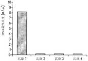
接下来,对基于小珠的PCR检测装置进行反应混合物中小珠浓度的优化。基于小珠的PCR中的固体表面引起空间效应和几何效应,因而影响反应效率。前面有关固相扩增的研究侧重于最大限度地增大DNA的最终浓度,但研究的首要问题是DNA检测。因此对反应混合物中微珠的浓度进行研究并优化,以产生最强信号。为了检验小珠浓度对荧光信号强度的影响,在一些条件(退火温度为58℃,MgCl2浓度为1.5mM,模板浓度为10pM)下使用三种不同小珠浓度进行基于小珠的PCR反应(见图12)。从图中可以看出,约200个小珠/μL时微珠上的荧光信号最强,低于或高于此浓度时信号强度都大幅降低。Next, optimization of the bead concentration in the reaction mixture was performed for the bead-based PCR detection device. Solid surfaces in bead-based PCR cause steric and geometric effects, thus affecting reaction efficiency. Previous studies on solid-phase amplification have focused on maximizing the final concentration of DNA, but the overriding issue of research is DNA detection. The concentration of beads in the reaction mixture was therefore studied and optimized to produce the strongest signal. To examine the effect of bead concentration on fluorescence signal intensity, bead-based PCR reactions were performed using three different bead concentrations under some conditions (annealing
图12中的高峰值的出现,反应混合物中微珠浓度与基于表面的PCR反应的总表面积之间的关系是主要原因。微珠浓度较高时,荧光标记DNA产物遍布大量小珠。表面积较大导致荧光信号较弱,这是因为信号强度与荧光标记的表面密度成正比。另一方面,小珠浓度较低并不意味着DNA表面浓度较高。在这些小珠浓度值下,反向引物的密度较高由于小珠表面分子间的位阻而使反应受到限制,反向引物相互间在固体表面上越来越靠近可阻碍溶液中的DNA与结合在小珠表面的引物进行杂交。除了位阻以外,小珠表面上逐渐增大的荧光团密度由于其他荧光团和核苷酸的亲近而导致淬火的发生,对荧光性产生抑制。可选择地,在反应中不使用微珠可导致一些反向引物留在溶液中,这是因为每个小珠都能承载有限数量的引物。溶液中引物对模板DNA的竞争以及位阻现象引起低效率解释了为什么小珠浓度较低时信号强度下降。图12显示出小珠浓度从200小珠/μL至20小珠/μL信号强度下降了9倍,这说明小珠浓度对传感器的DNA检测能力有巨大影响。The appearance of high peaks in Figure 12, the relationship between the concentration of beads in the reaction mixture and the total surface area of the surface-based PCR reaction is the main reason. At higher bead concentrations, the fluorescently labeled DNA product spreads over a large number of beads. A larger surface area results in a weaker fluorescent signal because the signal intensity is directly proportional to the surface density of the fluorescent label. On the other hand, a lower concentration of beads does not mean a higher concentration of DNA at the surface. At these bead concentration values, the high density of reverse primers limits the reaction due to steric hindrance between molecules on the bead surface, and the closer proximity of the reverse primers to each other on the solid surface prevents binding of DNA in solution. Primers on the bead surface hybridize. In addition to steric hindrance, the increasing fluorophore density on the bead surface leads to quenching due to the proximity of other fluorophores and nucleotides, which inhibits fluorescence. Alternatively, not using beads in the reaction can result in some reverse primers remaining in solution, since each bead can hold a limited number of primers. Competition of primers for template DNA in solution and inefficiencies caused by steric hindrance explain why the signal intensity drops at lower bead concentrations. Figure 12 shows a 9-fold drop in signal intensity from 200 beads/μL to 20 beads/μL bead concentration, which demonstrates that bead concentration has a huge impact on the DNA detection capability of the sensor.
然后,对微型装置进行试验以检测合成的基因组DNA(gDNA)。检测标准包括检测范围(可以检测到的DNA的最小浓度)以及检测所需PCR循环次数。通过改变反应物中模板DNA浓度来进行一系列的PCR反应而对装置的检测范围进行研究。试验的浓度范围是从零模板至1pM(约6×10⁵复本/μL)。PCR反应在上述的最佳试验条件下(200小珠/μL,20秒保持时间,退火温度58℃,1.5Mm的MgCl2)进行10次反应循环(约15分钟)。如图13所示,模板DNA浓度为0.1pM或更低时与零模板反应相比荧光性增强。零模板扩增产生荧光信号是因为除正确扩增的DNA以外还检测到非特异性扩增产物,例如引物—二聚体。在下文对信号强度与循环次数之间关系的讨论中将对这种影响做进一步研究。但1pM浓度的模板产生的信号与零模板对照组的信号不同,这一点通过学生t分布检验可以确定,可信度达95%。这种检测范围比使用电化学标记的PCR的范围或在平面上进行光学检测的范围小好几个数量级。Then, the tiny device was tested to detect synthetic genomic DNA (gDNA). Detection criteria include the detection range (minimum concentration of DNA that can be detected) and the number of PCR cycles required for detection. The detection range of the device was studied by performing a series of PCR reactions by varying the template DNA concentration in the reaction. The concentration range of the assay was from zero template to 1 pM (approximately 6×10⁵ copies/μL). The PCR reaction was carried out for 10 reaction cycles (approximately 15 minutes) under the above-mentioned optimum experimental conditions (200 beads/μL, 20 second hold time, annealing
除了检验装置的检测范围外,还要在检测前进行对信号强度与PCR循环次数之间关系的研究。对于这项研究,基于小珠的PCR反应按照前文所述的方法进行,但1pM的浓度(约6×10⁵复本/μL)保持不变,只改变PCR循环次数。如图14所示,循环次数为10时,信号强度远远高于本底水平,并且从10次到30次循环时信号强度增加。信号强度在10次至20次循环时出现下降。会产生许多不利影响,例如结合在表面的两个引物之间产生非特异性扩增或者生成无法复制的(不完全的)分子,上述两种情况在进一步的扩增时都会发生逆转。除对1pM的样本进行扩增外,还对无模板DNA的对照样本进行扩增,结果如图14所示。对照信号强度呈线性增加,但始终低于试验样本。In addition to checking the detection range of the device, the relationship between the signal intensity and the number of PCR cycles should be studied before the detection. For this study, bead-based PCR reactions were performed as previously described, but the concentration of 1 pM (approximately 6 x10⁵ copies/μL) was kept constant and only the number of PCR cycles was changed. As shown in Figure 14, at a cycle number of 10, the signal intensity was much higher than the background level, and the signal intensity increased from 10 to 30 cycles. Signal strength decreased between 10 and 20 cycles. Many adverse effects can occur, such as non-specific amplification between two primers bound to the surface or generation of non-replicable (incomplete) molecules, both of which are reversed by further amplification. In addition to amplifying the 1pM sample, the control sample without template DNA was also amplified, and the results are shown in FIG. 14 . Control signal intensity increased linearly, but was always lower than test samples.
图15a-15c显示了完整的微流体基于小珠的PCR过程中微室的显微照片。在图15a中,明亮区域显示出一个微室,小珠已置入该腔室,正在将DNA溶液引入微室(从顶部向底部流动)。在图15b中,DNA溶液已被引入,正通过结合在小珠上的反向引物从溶液中捕获模板DNA到小珠上。在图15c中,PCR循环结束后,用缓冲液进行洗涤,测量微珠的荧光强度。将体积为5μL的微珠(浓度约为200小珠/μL)置入微室中(如图15a所示),然后置入10pM的模板DNA(约6×106复本/μL),培养10分钟(见图15b)。然后将溶液移出,引入PCR反应混合物,此时使用集成加热器和传感器控制微室温度循环,进行10次PCR温度循环。扩增结束后,用纯缓冲液冲洗来移除反应混合物,只在微室内留下附着有荧光标记DNA的小珠(见图15c)。对荧光显微照片的分析表明小珠的荧光强度为75.9afu。然后升高微室温度并保持95℃ 5分钟,使结合在小珠上的ssDNA变性,用纯净缓冲液洗脱。用UV/VIS光谱法确定洗脱液中含有163.6ng/μL的ssDNA。从留在微室中的微珠的荧光显微照片(未图示)可以确定产物ssDNA确实已从小珠上移除。上述结果说明使用基于小珠的PCR可有助于设计出高度集成的用于DNA纯化和检测的微型装置,在芯片上过程中对缓冲液条件进行精确控制。Figures 15a-15c show photomicrographs of microchambers during complete microfluidic bead-based PCR. In Figure 15a, the bright area shows a chamber into which beads have been placed and DNA solution is being introduced into the chamber (top to bottom flow). In Figure 15b, a DNA solution has been introduced and template DNA is being captured from solution onto the beads by the reverse primer bound to the beads. In Figure 15c, after the PCR cycle was completed, the beads were washed with buffer and the fluorescence intensity of the microbeads was measured. Put microbeads with a volume of 5 μL (concentration is about 200 beads/μL) into the microchamber (as shown in Figure 15a), then put 10 pM template DNA (about 6×10⁶ copies/μL), and
实例2Example 2
在本实例中,如前文所述,使用具有筛选室和扩增室的集成微芯片对适体进行筛选和扩增,如图2所示。简单地说,是将结合序列从随机库中分离出来(图2a-2c),通过PCR进行化学扩增(图2d),然后收集单链(图2e)。可将收集到的单链置入筛选室中,以便重复以上步骤。In this example, aptamers were screened and amplified using an integrated microchip with a screening chamber and an amplification chamber as described above, as shown in FIG. 2 . Briefly, binding sequences were isolated from random pools (Fig. 2a–2c), chemically amplified by PCR (Fig. 2d), and single strands were collected (Fig. 2e). The collected single strands can be placed in the screening chamber to repeat the above steps.
在本实例中使用的微型装置(或微芯片、芯片)包括两个腔室,一个腔室(筛选室)用于筛选并分离候选适体,另一个腔室(扩增室)对这些候选适体进行扩增和收集。图16是本实例的微型装置示意图,图中筛选室1610是一个400μm高的立方体,具有两个高度不超过10μm的入口/出口和一个400μm高的用于置入微珠的入口。扩增室1620是一个4μL的圆柱体,高400μm,具有两个入口/出口。其中一个入口用于置入含有微珠的PCR反应物,而另一个的高度为10μm,可在移除上清液时阻挡微珠。在微型装置中使用微珠拦阻结构(围堰1680)可以通过在更换溶液时保留微珠上的所需核酸实现在分离操作各步骤中对缓冲条件的精确控制。两个腔室之间是一条蛇形通道(混合装置1650),其通过扩散将来自筛选室的ssDNA与PCR反应物混合。电阻加热器1630和传感器1640直接设置在各微室的下面以控制腔室的温度。腔室内部和通道表面涂敷有Parylene C以最大限度地减少反应物吸收和蒸发损失。The microdevice (or microchip, chip) used in this example consists of two chambers, one chamber (screening chamber) for screening and isolating candidate aptamers, and the other chamber (amplification chamber) for these candidate aptamers bodies were amplified and collected. Fig. 16 is a schematic diagram of the micro-device of this example, in which the
本实例试验中使用的所有DNA都购自Integrated DNA Technologies(IDT),序列如下:基因库:5'-CTA CCT ACG ATC TGA CTA GCN NNN NNN NNN NNN NNNNNN NNN NNN NNN NNN NNN NNN GCT TAC TCT CAT GTA GTT CC-3'(SEQID NO:4);正向引物:5'-FAM-间隔基-C TAC CTA CGA TCT GAC TAG C-3'(SEQ IDNO:5);反向引物:S'-双生物素-间隔基-G GAA CTA CAT GAG AGT AAG C-3'(SEQID NO:6)。基因库和引物序列基于以IgE为目标的传统SELEX规程。用于PCR的试剂包括5×GoTaq Flexi PCR混合料(Promega公司生产)、25mM的MgCl2、10mM的dNTP(Promega公司生产)、50μg/mL的BSA(Sigma公司生产)、GoTaq酶(Promega公司生产)和涂敷有链霉亲和素的聚合物微珠(Streptavidin Plus Ultralink,Pierce公司生产)。筛选步骤中使用的小珠是Bio-Rad公司生产的交联琼脂糖凝胶(Affi 10 Gel)活化介质,平均直径为80μm。用于捕获目标DNA(也称为目的蛋白)的功能性分子是人IgE(Athens Research公司生产)。All DNA used in this example assay was purchased from Integrated DNA Technologies (IDT) with the following sequences: GenBank: 5'-CTA CCT ACG ATC TGA CTA GCN NNN NNN NNN NNN NNNNNN NNN NNN NNN NNN NNN NNN GCT TAC TCT CAT GTA GTT CC-3' (SEQ ID NO:4); forward primer: 5'-FAM-spacer-C TAC CTA CGA TCT GAC TAG C-3' (SEQ ID NO:5); reverse primer: S'-bis Biotin-Spacer-G GAA CTA CAT GAG AGT AAG C-3' (SEQ ID NO: 6). Gene bank and primer sequences were based on the traditional SELEX protocol targeting IgE. Reagents for PCR include 5×GoTaq Flexi PCR mix (produced by Promega), 25mM MgCl2 , 10mM dNTP (produced by Promega), 50 μg/mL BSA (produced by Sigma), GoTaq enzyme (produced by Promega) ) and polymer beads coated with streptavidin (Streptavidin Plus Ultralink, produced by Pierce). The beads used in the screening step are cross-linked agarose gel (
使用接触式光刻技术制造所述微型装置。简单地说,是在玻璃片上涂上铬和金(厚度分别为15nm和150nm),用光学光刻技术制出图案,蚀刻制出电阻加热器和温度传感器。通过等离子体增强化学气相沉积(PECVD)涂上1μm厚的二氧化硅薄层,用硅硬质掩膜形成用于电连接的开口。使用光学光刻技术在硅片上制出分层的SU-8,形成软刻蚀模具。然后,浇铸PDMS形成微流体通道、腔室、围堰和混合装置。接下来,将PDMS流体网络结构粘结到玻璃片上,进行氧等离子体处理,进行装填前通过CVD将1μm厚的Parylene C涂敷到整个芯片上(如图17a所示)。The microdevices are fabricated using contact lithography. Simply put, the glass is coated with chromium and gold (thicknesses are 15nm and 150nm, respectively), patterned by optical lithography, and resistive heaters and temperature sensors are etched. A thin 1-μm-thick layer of silicon dioxide was coated by plasma-enhanced chemical vapor deposition (PECVD), and openings for electrical connections were formed with a silicon hard mask. The layered SU-8 is produced on the silicon wafer by optical lithography technology to form a soft etching mold. Then, PDMS was cast to form microfluidic channels, chambers, dams, and mixing devices. Next, the PDMS fluid network structure was bonded to the glass slide, treated with oxygen plasma, and 1 μm thick Parylene C was coated on the entire chip by CVD before filling (as shown in Figure 17a).
制造完成后,在环境舱中对电阻式温度传感器进行校准,用染料对混合装置进行测试,检测其混合效率(如图17b所示)。进行检测前,将涂有IgE的小珠装入筛选室直至完全装满,小珠入口用蜡密封。在进行检测前用1×磷酸盐缓冲液(PBS)冲洗小珠一次,将整个芯片暴露于50μg/mL的BSA中以防止DNA非特异性吸附的产生。然后,将腔室温度设置为所需的筛选温度(T1)37℃,将3份30μL的在经1mM MgCl2改性的1×PBS(PBSM)中的10μM DNA库溶液以每分钟5μL的速度引入筛选室。暴露于基因库后,用10份30μL的1×PBSM冲洗,在37℃下将未结合或弱结合的ssDNA除去。然后,将筛选室温度设置到洗脱温度(T2)57℃,培养5分钟后,以每分钟5μL的速度引入4份30μL改性PBS对候选适体进行洗脱。After fabrication, the resistive temperature sensor was calibrated in an environmental chamber, and the mixing device was tested with dye to check its mixing efficiency (as shown in Figure 17b). Before testing, the IgE-coated beads are loaded into the screening chamber until completely filled and the bead inlets are sealed with wax. The beads were washed once with 1× phosphate buffered saline (PBS) before detection, and the entire chip was exposed to 50 μg/mL BSA to prevent non-specific adsorption of DNA. Then, set the chamber temperature to the desired screening temperature (T1 ) of 37 °C, and dissolve three 30 μL aliquots of 10 μM DNA library solution in 1× PBS (PBSM) modified with 1 mMMgCl2 at 5 μL per min. Speed introduction into the screening room. After exposure to the gene pool, unbound or weakly bound ssDNA was removed by washing with ten 30 μL aliquots of 1× PBSM at 37°C. Then, the temperature of the screening chamber was set to the elution temperature (T2) of 57°C, and after incubation for 5 minutes, four portions of 30 μL modified PBS were introduced at a rate of 5 μL per minute to elute the candidate aptamers.
将含有候选适体的缓冲液与PCR反应试剂和微珠混合后引入扩增室。将剩余未扩增的溶液从芯片中单独分离并移除,然后保存。将扩增室填满后,用蜡将入口密封,对溶液进行热循环操作。用荧光强度确定DNA的最终表面浓度。保持腔室温度在95℃ 5分钟,使与小珠结合的DNA与微珠分离,缓冲液以每分钟1μL的速度流过将扩增的ssDNA移除。Buffers containing candidate aptamers are mixed with PCR reagents and beads and introduced into the amplification chamber. The remaining unamplified solution was separately separated from the chip and removed, then stored. After the amplification chamber is filled, the inlet is sealed with wax and the solution is thermally cycled. Determine the final surface concentration of DNA using fluorescence intensity. Keep the chamber temperature at 95°C for 5 minutes to separate the DNA bound to the beads from the beads, and flow the buffer at a rate of 1 μL per minute to remove the amplified ssDNA.
在筛选室内用另一个含有涂有IgE新小珠的新芯片进行结合分析。对含有丰富DNA库的样本再用FAM标记的正向引物进行进一步的芯片外扩增。然后,用涂有链霉亲和素的微珠进行纯化;在95℃对ssDNA进行洗脱,在1×PBSM中进行重悬。在做进一步分析前用UV/VIS吸收技术(ThermoScientific Nanodrop公司生产)对样本浓度进行测量。Binding assays were performed in the screening chamber with another new chip containing new IgE-coated beads. Samples containing abundant DNA pools were further amplified off-chip using FAM-labeled forward primers. Then, purification was performed using streptavidin-coated beads; ssDNA was eluted at 95°C and resuspended in 1×PBSM. Sample concentrations were measured using UV/VIS absorption technology (ThermoScientific Nanodrop) before further analysis.
将用于在筛选室中洗涤小珠的各缓冲液样本部分移除并保存以备分离和扩增后的检测。使用传统的PCR对这些样本进行芯片外分析,与芯片上扩增和收集同时进行。为了检测随机适体库在芯片上的分离效果,对在37℃下洗涤涂有IgE的微珠的10份缓冲液各自进行芯片外扩增并用凝胶电泳法进行检测。得到一条清晰的浓度梯度,与初始洗涤对应的荧光带亮度高于对应后续洗涤的荧光带的亮度(见图18)。这说明在筛选过程中,筛选室中DNA库的弱结合链正从与小珠结合的IgE移除,提高了筛选的严格性。洗涤液在57℃下的扩增同样可以检测筛选效果。通过对洗脱后的各候选适体样本进行扩增,证明了候选适体的出现和洗脱规程的效率。37℃下的分离洗涤结束后,在57℃对ssDNA进行洗涤,将其在芯片上与PCR试剂进行混合,然后进行17轮PCR扩增循环。四份候选适体在芯片上进行基于小珠的PCR后的微珠荧光测定显示出不连续的变化,从第一样本扩增后的强信号变化为随后检测的最弱信号(见图19)。这种突变表明在57℃第一次洗涤时就几乎完全洗脱了所需的候选适体。A portion of each buffer sample used to wash the beads in the screening chamber was removed and saved for detection after isolation and amplification. These samples were analyzed off-chip using conventional PCR, concurrently with on-chip amplification and collection. To test the separation effect of the random aptamer library on the chip, each of the 10 buffers in which the IgE-coated beads were washed at 37°C was amplified off-chip and detected by gel electrophoresis. A clear concentration gradient was obtained, and the fluorescence bands corresponding to the initial wash were brighter than those corresponding to subsequent washes (see Figure 18). This indicates that during the selection process, the weakly bound strands of the DNA pool in the selection chamber are being removed from the IgE bound to the beads, increasing the stringency of the selection. Amplification of the washing solution at 57°C can also detect the screening effect. The presence of candidate aptamers and the efficiency of the elution procedure were demonstrated by amplifying samples of each candidate aptamer after elution. After separation and washing at 37°C, the ssDNA was washed at 57°C, mixed with PCR reagents on the chip, and then 17 rounds of PCR amplification cycles were performed. Bead fluorescence assays following on-chip bead-based PCR of the four candidate aptamers showed a discontinuous change from a strong signal after amplification of the first sample to the weakest signal detected subsequently (see Figure 19 ). This mutation indicated that the desired aptamer candidate was almost completely eluted in the first wash at 57°C.
此外,还对IgE的富集候选适体库的亲和性进行了测试。为了产生足够用于测试的DNA,使用传统PCR对富集库进行芯片外扩增。对1×PBSM中的目标ssDNA进行分离和重悬后,用UV/VIS对样本浓度进行测定,使其达到1μM的标准。出于比较目的,购买在5'端做FAM修饰的随机库,在1×PBSM中洗脱至1μM。将各样本(5μL)分别置入装有新涂敷了IgE的微珠的微室中,温度保持在37℃,培养5分钟,用缓冲液冲洗,在荧光激发过程中进行显微照相。在光学显微镜下可以清晰地看到富集库的亲和性强于随机库的亲和性(见图20)。对各5μL样本培养5分钟后进行显微照片的荧光强度分析证明富集适体库对于IgE具有较强的亲和性。In addition, the affinity of the IgE-enriched library of candidate aptamers was tested. To generate sufficient DNA for testing, enriched libraries were amplified off-chip using conventional PCR. After separating and resuspending the target ssDNA in 1×PBSM, the concentration of the sample was determined by UV/VIS to make it reach the standard of 1 μM. For comparison purposes, random pools with FAM modification at the 5' end were purchased and eluted to 1 μM in 1×PBSM. Each sample (5 μL) was placed in a microchamber containing fresh IgE-coated microbeads, maintained at 37°C, incubated for 5 minutes, washed with buffer, and photomicrographed during fluorescence excitation. It can be clearly seen under the light microscope that the affinity of the enriched library is stronger than that of the random library (see Figure 20). Fluorescence intensity analysis of micrographs after incubation of each 5 μL sample for 5 minutes proved that the enriched aptamer library had a strong affinity for IgE.
除了测定富集库对于IgE的亲和性增大外,还对亲和性的温度依赖性进行了测定。和亲和性测定类似,将1μM在1×PBSM中的做了荧光标记的候选适体溶液置入装有微珠的腔室中直至荧光信号饱和,所述微珠涂敷有IgE。使纯净的缓冲液以每分钟1μL的速度流过,同时腔室温度增高3℃。当缓冲液不断流过时,腔室温度在各温度下保持5分钟。适体库显现出对于与IgE结合具有高度温度依赖性,最佳结合温度为37℃,符合预想结果(见图21)。这些对于温度敏感的候选适体与使用传统技术相比可被更快地分离且效率更高(见表2)。In addition to measuring the increase in affinity of the enriched pool for IgE, the temperature dependence of the affinity was also determined. Similar to the affinity assay, a 1 μM solution of fluorescently labeled aptamer candidates in 1×PBSM was placed into the chamber containing beads, which were coated with IgE, until the fluorescent signal was saturated. Neat buffer was flowed at a rate of 1 μL per minute while increasing the chamber temperature by 3°C. The chamber temperature was maintained at each temperature for 5 min while the buffer flowed continuously. The aptamer library showed a high temperature dependence for binding to IgE, and the optimal binding temperature was 37°C, which was in line with the expected results (see Figure 21). These temperature-sensitive aptamer candidates were isolated much faster and more efficiently than using conventional techniques (see Table 2).
表2.集成式微流体适体分离与传统方法的比较Table 2. Comparison of Integrated Microfluidic Aptamer Isolation and Traditional Methods

实例3Example 3
在本实例中,使用微流体芯片进行目标DNA的分离和富集,所述微流体芯片具有两个腔室和一个位于腔室之间的通道,所述通道中装有凝胶,如前面图3所描述。In this example, the separation and enrichment of target DNA was performed using a microfluidic chip with two chambers and a channel between the chambers filled with a gel, as shown in the previous figure 3 as described.
如图22所示,本实例所使用的微芯片包括两个微室,筛选/分离微室2210和富集微室2220,每个微室的深度均为200μm,体积均为5μL,两个微室通过一条微通道2240(长度:7mm,宽度:1mm,高度:300μm)连接。分离微室中的围堰结构(高度:40μm)在分离和富集过程中对分离室中的微珠(直径:100μm)起阻挡作用。集成在玻璃基底上的电阻加热器2230和温度传感器2235用于在对小珠上的ssDNA进行热洗脱过程中控制分离室的温度。连接通道2240通过入口2255部分地充满琼脂糖凝胶2250。一段附加的通道(长度0.6mm,宽度:0.4mm,高度:40μm)在热洗脱期间将凝固胶与加热了的腔室热隔绝。补充入口2280用于将所述附加通道填满缓冲液。由铂电极丝施加的电位差在微通道中产生电场,所述铂电极丝由铂线入口2290和2295插入微室。As shown in Figure 22, the microchip used in this example includes two microchambers, a screening/
本实例使用的微芯片是将聚二甲硅氧烷(PDMS)微流体层粘合至玻璃基底上,然后再使用标准微细技术(例如光刻技术)刻出电阻加热器和传感器图案(见图23)。要制备PDMS层的SU-8模具,需将硅片浸入食人鱼洗液(98%硫酸与30%过氧化氢的混合物,体积比3:1)1小时进行清洁。然后用去离子水冲洗硅片,放在180℃的热板上烘烤15分钟。将SU-8光刻胶层2320旋转涂布到硅片2310上,通过光掩模2330暴露于紫外线中,烘烤形成模具(图23a-23c)。将PDMS预聚物(Sylgard184,Dow Corning公司生产)涂抹到SU-8模具上,在75℃热板上烘烤1小时,从模具上剥下形成微室壁2340和通道(图23d)。另外,用热蒸发器(Auto306,BOC Edwards公司生产)将铬层(厚度:5nm)和金层(厚度:100nm)2360不断浇注到用食人鱼洗液清洗过的玻璃基底2350上。用正性光刻技术在金属层上制出图案(图23e-23h),然后通过等离子体增强化学气相沉积技术使用二氧化硅层2355(厚度:1μm)对其进行钝化(图23i)。在PDMS2340中打孔形成入口和出口后粘合到玻璃基底2350上,然后对粘合表面进行氧等离子体处理。将入口孔和出口孔连接到塑料管2380,以便于样本操作。用微吸管将熔融的琼脂糖凝胶2370由凝胶入口注入微通道,然后使其固化(图23j)。制成后的微芯片图片如图24所示。The microchip used in this example is a polydimethylsiloxane (PDMS) microfluidic layer bonded to a glass substrate, and the resistive heaters and sensors are patterned using standard microscopic techniques such as photolithography (see Fig. twenty three). To prepare the SU-8 mold of the PDMS layer, the silicon wafer needs to be cleaned by immersing it in piranha washing solution (a mixture of 98% sulfuric acid and 30% hydrogen peroxide, volume ratio 3:1) for 1 hour. Then rinse the wafer with deionized water, and bake it on a hot plate at 180°C for 15 minutes. The SU-8
为制备IgE-功能性微珠,需用1×PBS改性缓冲液用离心法冲洗200μL含有NHS活性微珠(平均直径:约100μm,GE Healthcare公司生产)的溶液三次,所述改性缓冲液含有1mM的Mg2+离子(8.1mM Na2HPO4,1.1mM Na2HPO4,138mMNaCl,2.7mM KC1,1mM MgCl2,pH7.4)。然后,将小珠与200μL的0.1μM人骨髓瘤IgE(Athens Research&Technology公司生产)在室温培养5小时。培养结束后,通过用新鲜的PBS缓冲液洗涤小珠,丢弃剩余的IgE分子。为了减少ssDNA分子与小珠非特异性结合,将小珠放入0.1M的Tris-HCl缓冲液中培养1小时以钝化未与IgE结合的表面。IgE功能化的小珠在使用前存储在4℃的PBS缓冲液中。从Integrated DNA Technologies公司购买具有荧光标记的具有随机序列的ssDNA库(97-mer,5'-GCC TGT TGT GAG CCT CCT GTC GAA–50随机碱基-TTG AGCGTT TAT TCT TGT CTC CC-3')(SEQ ID NO:7)、IgE-特异性ssDNA适体D17.4(78-mer,KD=10nM,5'-GCC TGT TGT GAG CCT CCT GTC GAA GCA CGT TTATCC GTC CCT CCT AGT GGC GTG CTT GAG CGT TTA TTC TTG TCT CCC-3')(SEQ ID NO:8)、以及正向引物(5'-GCC TGT TGT GAG CCT CCT GTC GAA-3')(SEQ ID NO:9)和反向引物(5'-GGG AGA CAA GAA TAA ACG CTC AA-3')(SEQ IDNO:10)。在整个实例中使用随机ssDNA和适体D17.4混合物(摩尔比:1000:1)以提高IgE结合位点的竞争力。将1μL的100μM随机ssDNA库与1μL的0.1μM适体D17.4在98μL的1×PBS缓冲液中混合,制备出随机ssDNA溶液。用于在微通道中电泳输送ssDNA和用于平板凝胶电泳的电泳缓冲液为0.5×TBE缓冲液(44.5mM Tris base(三羟甲基氨基甲烷),44.5mM硼酸,1.25mM EDTA,pH8.3)。在热板上将0.3克琼脂糖溶于100mL的0.5×TBE缓冲液中制备用于电泳的百分之三琼脂糖凝胶(Difco Laboratories公司生产)。To prepare IgE-functional microbeads, wash 200 μL of a solution containing NHS active microbeads (average diameter: about 100 μm, produced by GE Healthcare) three times by centrifugation with 1×PBS modified buffer. Contains 1mM Mg2+ ions (8.1mM Na2 HPO4 , 1.1mM Na2 HPO4 , 138mM NaCl, 2.7mM KC1, 1mM MgCl2 , pH7.4). Then, the beads were incubated with 200 µL of 0.1 µM human myeloma IgE (manufactured by Athens Research & Technology) for 5 hours at room temperature. After incubation, the remaining IgE molecules were discarded by washing the beads with fresh PBS buffer. In order to reduce the non-specific binding of ssDNA molecules to the beads, the beads were incubated in 0.1M Tris-HCl buffer for 1 hour to passivate the surface not bound to IgE. IgE functionalized beads were stored in PBS buffer at 4°C until use. A fluorescently labeled ssDNA library with random sequences was purchased from Integrated DNA Technologies (97-mer, 5'-GCC TGT TGT GAG CCT CCT GTC GAA–50 random bases-TTG AGCGTT TAT TCT TGT CTC CC-3') ( SEQ ID NO: 7), IgE-specific ssDNA aptamer D17.4 (78-mer, KD =10nM, 5'-GCC TGT TGT GAG CCT CCT GTC GAA GCA CGT TTATCC GTC CCT CCT AGT GGC GTG CTT GAG CGT TTA TTC TTG TCT CCC-3') (SEQ ID NO:8), and forward primer (5'-GCC TGT TGT GAG CCT CCT GTC GAA-3') (SEQ ID NO:9) and reverse primer (5 '-GGG AGA CAA GAA TAA ACG CTC AA-3') (SEQ ID NO: 10). A random ssDNA and aptamer D17.4 mixture (molar ratio: 1000:1) was used throughout the examples to increase the competitiveness of the IgE binding sites. Prepare a random ssDNA solution by mixing 1 μL of 100 μM random ssDNA library with 1 μL of 0.1 μM aptamer D17.4 in 98 μL of 1× PBS buffer. The electrophoresis buffer used to transport ssDNA in microchannels and for slab gel electrophoresis was 0.5×TBE buffer (44.5mM Tris base (trishydroxymethylaminomethane), 44.5mM boric acid, 1.25mM EDTA, pH8. 3). A 3% agarose gel (manufactured by Difco Laboratories) for electrophoresis was prepared by dissolving 0.3 g of agarose in 100 mL of 0.5×TBE buffer on a hot plate.
图25是试验方案示意图。用注射泵2510(NE 300,Harvard Apparatus公司生产)将含有ssDNA混合物和缓冲液的样本溶液引入微室中。在热洗脱过程中,通过分别与电源2520(E3631 A,Agilent Technologies公司生产)和万用表2530(34410A,Agilent Technologies公司生产)连接的电阻加热器和传感器使分离室中的温度保持在57℃,通过计算机上的基于LabVIEW的PID模块2540进行控制。铂电极与电源连接,在两个腔室之间施加电位差,诱发对ssDNA链的电泳输送。使用荧光显微镜(LSM 510,Zeiss公司生产)在通道中心对ssDNA在充满凝胶的通道的输送进行监控。Figure 25 is a schematic diagram of the experimental protocol. The sample solution containing ssDNA mixture and buffer was introduced into the microchamber with a syringe pump 2510 (
对随机ssDNA混合物中所需ssDNA分子的分离和富集按以下步骤进行。用注射器将IgE功能化的微珠由小珠入口注入分离室中,装满大约30%的腔室体积(约3×10⁴个小珠)。装载完毕后,用注射泵用1×PBS缓冲液以每分钟40μL的低流速对小珠冲洗5分钟。将随机ssDNA混合物(100μL)由入口以每分钟20μL的低流速引入腔室,从三个独立的塑料管(每个塑料管约33μL)的出口收集。将PBS缓冲液以每分钟40μL的速度注入腔室,将弱结合的DNA链从IgE小珠上冲洗掉,废弃溶液在出口的10个独立的塑料管中收集(每个塑料管约33μL)。用0.5×TBE缓冲液将两个腔室充满,通过电阻加热器将分离室加热至57℃,保持温度5分钟,将强结合的DNA链从小珠上洗脱。The separation and enrichment of desired ssDNA molecules in random ssDNA mixtures were performed as follows. Inject the IgE-functionalized microbeads from the bead inlet into the separation chamber with a syringe, filling approximately 30% of the chamber volume (approximately 3×10⁴ beads). After loading, the beads were rinsed with 1×PBS buffer at a low flow rate of 40 μL per minute for 5 minutes using a syringe pump. A random ssDNA mixture (100 μL) was introduced into the chamber from the inlet at a low flow rate of 20 μL per minute, collected from the outlets of three separate plastic tubes (approximately 33 μL each). PBS buffer was injected into the chamber at a rate of 40 μL per minute to wash away weakly bound DNA strands from the IgE beads, and the waste solution was collected in 10 separate plastic tubes at the outlet (approximately 33 μL each). Fill both chambers with 0.5× TBE buffer, heat the separation chamber to 57°C by a resistance heater, and keep the temperature for 5 minutes to elute strongly bound DNA strands from the beads.
在进行热洗脱过程中,将铂电极丝插入腔室中,施加25V/cm的电场25分钟。然后,将DNA链由充满凝胶的通道电泳输送到富集室。为了研究单轮的ssDNA分离与富集,当收集塑料管(每个塑料管约33μL)中的洗脱液时,用PBS缓冲液冲洗两个腔室。对于多轮的DNA富集,将分离室中的小珠丢弃不用,在进行下一轮分离和富集操作之前,用PBS缓冲液彻底冲洗分离室,以除去可能残留的不需要的DNA分子。然后,将新的IgE功能化的小珠引入分离室,进行下一轮DNA分离和富集。During thermal elution, a platinum electrode wire was inserted into the chamber and an electric field of 25 V/cm was applied for 25 minutes. The DNA strands are then electrophoretically transported through gel-filled channels to the enrichment chamber. To study a single round of ssDNA isolation and enrichment, both chambers were rinsed with PBS buffer when collecting the eluate in plastic tubes (approximately 33 μL each). For multiple rounds of DNA enrichment, discard the beads in the separation chamber and rinse the separation chamber thoroughly with PBS buffer before proceeding to the next round of separation and enrichment to remove unwanted DNA molecules that may remain. Then, new IgE-functionalized beads are introduced into the separation chamber for the next round of DNA isolation and enrichment.
为了分析样本结果,使用热循环仪(Mastercycler Personal,Eppendorf公司生产)通过聚合酶链反应(PCR)对各步骤中得到的具有代表性的洗脱样本进行芯片外扩增。所述PCR程序包括DNA在95℃下变性3分钟,然后进行20轮扩增。各循环由以下步骤组成:95℃变性15秒、59℃退火30秒和72℃延伸45秒。扩增结束后,将7μL PCR产物与7μL含有溴酚蓝和二甲苯蓝(Thermo Scientific公司生产)的2×DNA载入染料混合,装入充有3%琼脂糖凝胶的各个泳道。在100V下,用平板凝胶电泳装置(Mupid-exU,Advance公司生产)在0.5×TBE缓冲液进行电泳30分钟。在去离子水中用溴化乙锭对凝胶染色5分钟。使用UV照明装置(Alphalmager3400,Alpha Innotech公司生产)即可看到在凝胶中显示出代表各洗脱样本中DNA浓度的光带。使用荧光显微镜监测ssDNA在充满凝胶的通道中的电泳输送。用ImageJ软件(National Institutes of Health公司生产的免费软件)对所得图片中的凝胶光带强度和荧光强度进行分析。In order to analyze the sample results, representative eluted samples obtained in each step were subjected to off-chip amplification by polymerase chain reaction (PCR) using a thermal cycler (Mastercycler Personal, produced by Eppendorf). The PCR program included DNA denaturation at 95°C for 3 minutes, followed by 20 rounds of amplification. Each cycle consisted of the following steps: denaturation at 95°C for 15 seconds, annealing at 59°C for 30 seconds and extension at 72°C for 45 seconds. After amplification, 7 μL of PCR product was mixed with 7 μL of 2×DNA loading dye containing bromophenol blue and xylene blue (manufactured by Thermo Scientific), and loaded into each lane filled with 3% agarose gel. Electrophoresis was performed in 0.5×TBE buffer for 30 minutes with a slab gel electrophoresis device (Mupid-exU, manufactured by Advance Company) at 100V. Stain the gel with ethidium bromide for 5 min in deionized water. Using a UV illumination device (Alphalmager 3400, produced by Alpha Innotech Co.), the light bands representing the DNA concentration in each eluted sample can be seen in the gel. Electrophoretic transport of ssDNA in gel-filled channels was monitored using fluorescence microscopy. ImageJ software (free software produced by National Institutes of Health) was used to analyze the gel light band intensity and fluorescence intensity in the obtained pictures.
在分离-富集步骤中,首先在分离室中将与IgE结合的ssDNA从随机ssDNA混合物中分离出来。IgE分离是将随机DNA样本装入分离室中,然后用纯净缓冲液冲洗分离室以除去未结合的DNA。收集这些含有残留DNA的缓冲液样本,然后进行冲洗,用PCR扩增,通过平板凝胶电泳进行观察,确定分离步骤的效率。图26a所示为分离程序中收集的洗脱液中PCR产物的凝胶电泳图谱。在凝胶图片中,泳道L、P和N中的光带分别表示10碱基对(bp)DNA梯形图、阳性对照(PCR反应中模板DNA由100pmole随机ssDNA和0.1pmole D17.4适体组成)和阴性对照(PCR反应中没有模板DNA)。其他光带表示培养期间收集的洗脱液扩增样本(泳道I1)、洗涤(泳道W1-W10)、洗脱(泳道E1)以及ssDNA分离结束后用于冲洗富集室的缓冲液(泳道EC)。需要注意的是,各步骤缩写后面的数字代表收集洗脱液样本的次序。例如,“W5”中的“5”表示洗涤步骤中收集的第5个洗脱液样本。In the isolation-enrichment step, IgE-bound ssDNA is first separated from a random ssDNA mixture in a separation chamber. For IgE isolation, a random DNA sample is loaded into the separation chamber, which is then rinsed with neat buffer to remove unbound DNA. Samples of these buffers containing residual DNA were collected, rinsed, amplified by PCR, and visualized by slab gel electrophoresis to determine the efficiency of the separation step. Figure 26a shows the gel electrophoresis profile of PCR products in the eluate collected during the separation procedure. In the gel picture, the light bands in lanes L, P and N represent the 10 base pair (bp) DNA ladder, the positive control (the template DNA in the PCR reaction consists of 100 pmole random ssDNA and 0.1 pmole D17.4 aptamer, respectively ) and a negative control (no template DNA in the PCR reaction). The other bands represent the amplified samples of eluate collected during incubation (lane I1), washed (lane W1-W10), eluted (lane E1), and buffer used to rinse the enrichment chamber after ssDNA isolation (lane EC ). It should be noted that the numbers after the abbreviations of each step represent the order in which the eluate samples were collected. For example, the "5" in "W5" indicates the 5th eluate sample collected in the wash step.
泳道P和I1-E1中可见的上光带和下光带分别代表97bp随机ssDNA和78bpD17.4适体。上光带亮度高于下光带,这是因为用于分离的DNA混合物中随机ssDNA与D17.4适体的摩尔比是1000:1。泳道N中未见光带,这说明使用的试剂未被无用的DNA分子污染。此外,泳道EC中未见光带,这说明装有凝胶的通道有效地阻止了在捕获目标DNA链过程中富集室被来自分离室的无用ssDNA污染。The upper and lower bands visible in lanes P and I1-E1 represent 97 bp random ssDNA and 78 bp D17.4 aptamer, respectively. The upper band is brighter than the lower band because the molar ratio of random ssDNA to D17.4 aptamer in the DNA mixture used for isolation is 1000:1. No band of light is seen in lane N, indicating that the reagents used were not contaminated with unwanted DNA molecules. In addition, no light bands were seen in lane EC, indicating that the gel-filled channel effectively prevented the enrichment chamber from being contaminated by useless ssDNA from the separation chamber during the capture of the target DNA strands.
描绘了从泳道I1至泳道E1的97-mer随机ssDNA(上光带)光带强度的柱状图显示出了与IgE结合的ssDNA的分离过程(图26b)。在培养步骤中未与涂有IgE的微珠结合的DNA以泳道I1中的高亮度光带表示。从泳道W1至泳道W10中的光带亮度逐渐变暗说明随着洗涤的不断进行,松散结合的ssDNA分子从小珠表面移除,提高了目标ssDNA分离的严格性。泳道E1中光带强度增大说明在加热到57℃时,强结合的ssDNA分子从小珠表面洗脱。此外,在电泳通道内未发现琼脂糖凝胶受到破坏,这说明微室与装有凝胶的通道之间的通道长度足以阻止凝胶在DNA洗脱过程中发生热分解。The histogram depicting the band intensity of 97-mer random ssDNA (upper band) from lane I1 to lane E1 shows the separation of IgE-bound ssDNA (Fig. 26b). DNA that did not bind to the IgE-coated beads during the incubation step is indicated by a high-intensity band in lane I1. The brightness of the light bands from lane W1 to lane W10 gradually dimmed, indicating that loosely bound ssDNA molecules were removed from the bead surface as washing continued, increasing the stringency of target ssDNA separation. The increased intensity of the band in lane E1 indicates that strongly bound ssDNA molecules were eluted from the bead surface upon heating to 57°C. In addition, no damage to the agarose gel was observed within the electrophoresis channel, suggesting that the channel length between the microchamber and the channel containing the gel is sufficient to prevent thermal decomposition of the gel during DNA elution.
为了证实分离后的ssDNA链与IgE产生特异性结合,使用新NHS小珠重复本实例操作,该微珠表面未结合有蛋白质。凝胶图片(图27a)和柱状图(图27b)显示出在培养和早期洗涤步骤中出现明亮的光带(泳道I1-W5),在后期洗涤和洗脱步骤中没有光带(泳道W10-E1)。这说明ssDNA在NHS小珠表面弱结合,通过强烈冲洗移除。因此,图26所示的在前面实例中收集的97-mer ssDNA很可能是从随机混合物中分离出来的与IgE结合的ssDNA。与前面的结果类似,泳道EC中未见光带说明通道中的凝胶阻止了无用ssDNA进入富集室。To confirm that the isolated ssDNA strands bind specifically to IgE, this example was repeated using fresh NHS beads, which had no protein bound to their surface. Gel pictures (Fig. 27a) and histograms (Fig. 27b) show bright bands during incubation and early wash steps (lanes I1-W5), and no bands during later wash and elution steps (lanes W10- E1). This indicates that ssDNA is weakly bound to the NHS bead surface and removed by vigorous washing. Therefore, the 97-mer ssDNA collected in the previous example shown in Figure 26 is likely to be IgE-bound ssDNA isolated from a random mixture. Similar to the previous results, the absence of light bands in lane EC indicates that the gel in the channel prevents useless ssDNA from entering the enrichment chamber.
将1×PBS和0.5×TBE缓冲液作为通过装有凝胶的通道电泳输送DNA的示范性电解质进行测试。PBS缓冲液是一种强电解质(电导率:15ms/cm),由于其经常用于该方法的其他步骤,因此它在电泳中的使用可简化富集操作。可选择的,可使用TBE缓冲液(电导率约为350μs/cm)(这是因为它经常在凝胶电泳应用中使用)。1X PBS and 0.5X TBE buffer were tested as exemplary electrolytes for electrophoretic delivery of DNA through gel-filled channels. PBS buffer is a strong electrolyte (conductivity: 15ms/cm), and its use in electrophoresis simplifies enrichment as it is frequently used in other steps of the method. Alternatively, TBE buffer (conductivity about 350 μS/cm) can be used (this is because it is often used in gel electrophoresis applications).
如图28所示,将PBS用作电泳缓冲液时,在相应泳道未见光带,这说明DNA链未被输送至富集室。但是,使用TBE缓冲液时,DNA链被有效地转移至富集室,在泳道TBE可见清晰光带证明了这一点。这是因为尽管PBS的电导率比TBE高,PBS缓冲液中的盐离子(即,Na+和Mg2+)阻挡DNA链并中和其负电荷,阻止它们向阳极(即,富集室)移动。因此,在微芯片中使用0.5×TBE缓冲液用于电泳输送DNA。As shown in Figure 28, when PBS was used as the electrophoresis buffer, no light bands were seen in the corresponding lanes, which indicated that the DNA strands were not transported to the enrichment chamber. However, when using TBE buffer, the DNA strands were efficiently transferred to the enrichment chamber, as evidenced by a clear band visible in the TBE lane. This is because although PBS has a higher conductivity than TBE, the salt ions (i.e., Na+ and Mg2+ ) in the PBS buffer block the DNA strands and neutralize their negative charge, preventing them from moving toward the anode (i.e., the enrichment chamber). move. Therefore, 0.5x TBE buffer was used for electrophoretic delivery of DNA in the microchip.
确定从分离室向富集室电泳输送ssDNA所需的时间。图29a-29c所示为在装有凝胶的通道中心处在不同时间监测到的荧光标记ssDNA链的电泳输送过程荧光显微照片。在10分钟时出现的荧光强度图形高峰表明ssDNA正以大约每分钟1mm的速度通过装有凝胶的通道(图29d)。由于两个腔室之间的距离约为20mm,该微芯片至少需要20分钟将ssDNA电泳输送至富集室。为了评估本实例方案的效率,根据公式V=μE,利用测得的DNA速度计算出DNA的电泳迁移率,公式中μ为电泳迁移率,V为输送DNA的速度,E为施加的电场(即,在本实例中为25V/cm)。因此,计算得到ssDNA的电泳迁移率为6.67×10-5cm2/Vs,处于文献记录值范围内。Determine the time required for electrophoretic transport of ssDNA from the separation chamber to the enrichment chamber. Figures 29a-29c show fluorescence micrographs of the electrophoretic transport process of fluorescently labeled ssDNA strands monitored at different times in the center of the gel-filled channel. A graphical peak in fluorescence intensity at 10 minutes indicated that ssDNA was passing through the gel-filled channel at a rate of approximately 1 mm per minute (Fig. 29d). Since the distance between the two chambers is approximately 20 mm, the microchip requires at least 20 minutes for electrophoretic delivery of ssDNA to the enrichment chamber. In order to evaluate the efficiency of this example scheme, according to the formula V=μE, the electrophoretic mobility of DNA is calculated using the measured DNA velocity, where μ is the electrophoretic mobility, V is the velocity of DNA transport, and E is the applied electric field (ie , in this example 25V/cm). Therefore, the calculated electrophoretic mobility of ssDNA is 6.67×10⁻⁵ cm2 /Vs, which is within the range of literature records.
在一个单独微芯片中进行一轮完整的与IgE结合的ssDNA的分离和富集。将随机ssDNA库暴露于涂有IgE的小珠,冲洗掉弱结合链,对候选适体进行热洗脱,然后电泳输送至富集室。为了分析得到的产物,在操作结束后收集各步骤(即,培养:I,洗涤:W,洗脱:E)中的洗脱液以及用于冲洗两个腔室(即,分离室:IC,洗脱室:EC)的缓冲液。使用PCR对这些洗脱液进行化学扩增,并使用平板凝胶电泳进行观测。图30显示了扩增后的洗脱液的电泳图谱。泳道I1-I3中的光带表示在培养步骤中未与IgE结合的DNA。正如所预料的,由于大多数随机DNA未与IgE结合,这些光带显示出高水平荧光强度。泳道W1至W10指示的光带亮度逐渐减弱说明与IgE结合能力较弱的ssDNA链随着缓冲液对小珠的不断冲洗而被逐渐移除。泳道EC1中的明亮光带和泳带IC1中的较暗光带说明大多数被热洗脱的对于IgE具有高亲和力的ssDNA通过电泳输送至富集室。A complete round of isolation and enrichment of IgE-bound ssDNA was performed in a single microchip. Random ssDNA pools are exposed to IgE-coated beads, weakly bound strands are washed away, candidate aptamers are thermally eluted, and electrophoretically transported to the enrichment chamber. In order to analyze the resulting product, the eluate from each step (i.e., incubation: I, washing: W, elution: E) was collected after the operation and was used to rinse the two chambers (i.e., separation chamber: IC, Elution chamber: EC) buffer. These eluates were chemically amplified using PCR and visualized using slab gel electrophoresis. Figure 30 shows the electrophoretic pattern of the eluate after amplification. The light bands in lanes I1-I3 represent DNA that was not bound to IgE during the incubation step. As expected, these bands show high levels of fluorescence intensity since most of the random DNA is not bound to IgE. The brightness of the light bands indicated by lanes W1 to W10 gradually decreased, indicating that the ssDNA strands with weak binding ability to IgE were gradually removed as the beads were continuously washed with buffer. The bright band in lane EC1 and the darker band in lane IC1 indicate that most of the heat-eluted ssDNA with high affinity for IgE was electrophoretically transported to the enrichment chamber.
为了检测微芯片对于与IgE结合的DNA的富集能力,在芯片上进行多轮ssDNA富集。通过电泳输送对分离出来的与IgE结合的ssDNA链分别进行富集。微芯片中第1轮(泳道1)、第2轮(泳道2)和第3轮(泳道3)富集结束后所收集的扩增后洗脱液的电泳图谱如图31a所示。随着富集次数的增多,检测到富集室中的DNA浓度增大(图31b)。此外,在多轮富集结束后,观测到装有凝胶的微通道未受到损坏。To test the ability of the microchip to enrich DNA that binds to IgE, multiple rounds of ssDNA enrichment were performed on the chip. The isolated ssDNA strands bound to IgE were separately enriched by electrophoretic transport. The electropherograms of the amplified eluate collected after the first round (lane 1), the second round (lane 2) and the third round (lane 3) of enrichment in the microchip are shown in Figure 31a. As the number of enrichments increased, an increase in the DNA concentration in the enrichment chamber was detected (Fig. 31b). Furthermore, no damage to the gel-filled microchannels was observed after multiple rounds of enrichment were complete.
实例4Example 4
本实例描述了与实例3结构相似的微型装置的使用,但本实例使用化学洗脱而不是热洗脱释放已结合的候选适体。This example describes the use of a microdevice similar in structure to Example 3, but this example releases bound candidate aptamers using chemical elution rather than thermal elution.
本实例使用的微型装置包括一个筛选/分离室3210和一个富集室3220,每个腔室的深度均为200μm,体积均为5μL,腔室由一条微通道3240(1mm×7.8mm×40μm)连接(如图32所示)。微珠3215(直径:100μm)被一个坝式结构(围堰,高度为40μm)阻挡在分离室内。通道3240通过凝胶入口3255部分地充满凝胶3250(3%琼脂糖或12%聚丙烯酰胺)。在通过加热从小珠上洗脱ssDNA时,一段附加通道(0.4mm)将凝胶与加热的腔室隔绝。补充入口3280用于以缓冲液填满这部分通道。铂(Pt)电极丝由铂线入口(3290和3295)插入,为腔室提供进行电泳所需的电位差。所述微型装置可通过与实例3所描述方法基本类似的方法制造(参考图23),不同之处在于没设置传感器和加热器。制成后的微型装置图片如图33所示。The micro-device used in this example includes a screening/
在本实例中,将人骨髓瘤IgE(Athens Research&Technology公司生产)溶解于1×磷酸盐缓冲液(PBS缓冲液)直至达到最终浓度1μM。从GE Healthcare公司生产购买NHS-活化微珠,并用IgE实行功能化。从Integrated DNA Technologies公司生产购买具有随机序列的87-mer ssDNA库(5'-GCC TGT TGT GAG CCT CCTGTC GAA-N40-TTG AGC GTT TAT TCT TGT CTC CC-3')(SEQ ID NO:l1)和具有荧光标记的IgE特异性ssDNA适体D17.4引物。将1μL的随机基因库(100μM)和1μL的适体D17.4(0.1μM)在98μL的PBS缓冲液中混合,得到ssDNA混合物。In this example, human myeloma IgE (manufactured by Athens Research & Technology Co., Ltd.) was dissolved in 1×phosphate buffered saline (PBS buffer) to a final concentration of 1 μM. NHS-activated beads were purchased from GE Healthcare and functionalized with IgE. The 87-mer ssDNA library (5'-GCC TGT TGT GAG CCT CCTGTC GAA-N40-TTG AGC GTT TAT TCT TGT CTC CC-3') with random sequence was purchased from Integrated DNA Technologies company (SEQ ID NO:l1) and D17.4 primer with fluorescently labeled IgE-specific ssDNA aptamer.
对随机ssDNA混合物中所需ssDNA分子的分离和富集按以下步骤进行。用注射器将IgE-小珠由小珠入口注入分离室中,装满大约30%的腔室体积。装载完毕后,用注射泵用PBS缓冲液以每分钟20μL的低流速冲洗微珠5分钟。在整个实例中都使用具有0.1%IgE特异性适体D17.4的DNA混合物。将ssDNA混合物引入分离室,在分离室出口收集三个独立样本(样本体积约为33μL)。将PBS缓冲液以每分钟20μL的速度引入腔室,将弱结合的DNA链从IgE小珠上释放,在出品处收集废弃溶液的10个独立样本(样本体积约为30μL)。为了将强结合DNA链从小珠上洗脱,将5μL的0.5×TBE缓冲液中的0.1M NaOH引入腔室培养5分钟。将铂电极插入各腔室的铂线入口中,产生25V/cm的电场。然后将这些链通过装有凝胶的通道电泳输送至富集室。DNA电泳输送结束后,用PBS缓冲液对分离室进行冲洗,将无用DNA分子除去。为了提高富集室中与IgE结合的ssDNA浓度,重复以上分离和电泳输送步骤。对含有ssDNA分子的洗脱液样本的收集贯穿于整个实例,并采用聚合酶链反应(PCR)进行芯片外扩增。在整个操作程序中使用平板凝胶电泳比较各样本的光带强度来检定洗脱后的DNA的浓度。使用荧光显微镜监测ssDNA在充满凝胶的通道中的电泳输送。The separation and enrichment of desired ssDNA molecules in random ssDNA mixtures were performed as follows. Inject IgE-beads from the bead inlet into the separation chamber with a syringe, filling approximately 30% of the chamber volume. After loading, wash the beads with PBS buffer at a low flow rate of 20 μL per minute for 5 min using a syringe pump. A DNA mixture with 0.1% of the IgE-specific aptamer D17.4 was used throughout the examples. Introduce the ssDNA mixture into the separation chamber and collect three independent samples (sample volume approximately 33 µL) at the exit of the separation chamber. Introduce PBS buffer into the chamber at a rate of 20 µL per minute to release weakly bound DNA strands from the IgE beads and collect 10 independent samples of the discarded solution at the outlet (sample volume approximately 30 µL). To elute strongly bound DNA strands from the beads, introduce 5 µL of 0.1 M NaOH in 0.5× TBE buffer into the chamber and incubate for 5 min. A platinum electrode was inserted into the platinum wire inlet of each chamber to generate an electric field of 25 V/cm. These chains are then electrophoretically transported through gel-filled channels to the enrichment chamber. After DNA electrophoresis delivery, the separation chamber is washed with PBS buffer to remove useless DNA molecules. To increase the concentration of ssDNA bound to IgE in the enrichment chamber, repeat the above separation and electrophoretic delivery steps. Eluate samples containing ssDNA molecules were collected throughout the example and amplified off-chip using polymerase chain reaction (PCR). The concentration of eluted DNA was determined by comparing the band intensity of each sample using slab gel electrophoresis throughout the procedure. Electrophoretic transport of ssDNA in gel-filled channels was monitored using fluorescence microscopy.
首先,在分离室中将与IgE结合的ssDNA从随机ssDNA混合物中分离出来。图34a所示为分离过程中收集的洗脱液中PCR产物的凝胶电泳图谱。在图中,87-bp光带代表混合物中的随机ssDNA,而78-bp光带代表D17.4适体。泳道1和2分别为阳性对照(随机ssDNA和D17.4适体的混合物)和阴性对照(除DNA模板外的全部PCR混合物)。泳道1中的两条清晰光带是含有D17.4适体的随机ssDNA混合物的PCR产物。泳道2中未见光带,这说明试剂未被无用DNA分子污染。First, ssDNA bound to IgE is separated from a random ssDNA mixture in a separation chamber. Figure 34a shows the gel electrophoresis pattern of the PCR products in the eluate collected during the separation. In the figure, the 87-bp band represents random ssDNA in the mixture, while the 78-bp band represents the D17.4 aptamer.
图34b显示了87-mer ssDNA在培养、持续冲洗和洗脱过程中的光带强度情况。在培养步骤中未与IgE小珠结合的87-mer ssDNA显示为泳道3中的明亮光带。从泳道4至泳道6的光带强度逐渐变弱说明随着冲洗的进行,松散结合的87-merssDNA分子被从微珠表面移除,提高了分离的严格性。如泳道7所显示的,强结合的87-mer ssDNA分子被0.1M的NaOH从IgE小珠表面洗脱。Figure 34b shows the band intensity of 87-mer ssDNA during incubation, continuous washing and elution. The 87-mer ssDNA that was not bound to the IgE beads during the incubation step is shown as a bright band in
在电泳输送分离出来的DNA前,用缓冲液冲洗富集室。为了确定装有凝胶的通道是否阻止了富集室受到来自分离室的无用DNA的污染,对缓冲液进行PCR扩增并使用凝胶电泳进行分析(图34a,泳道8)。泳道中未显示光带,说明凝胶有效地阻挡了无用DNA对富集室的污染。Rinse the enrichment chamber with buffer prior to electrophoretic delivery of the separated DNA. To determine whether the gel-filled channels prevented contamination of the enrichment chamber with unwanted DNA from the separation chamber, the buffer was PCR amplified and analyzed using gel electrophoresis (Figure 34a, lane 8). There is no light band in the lane, indicating that the gel effectively blocks the contamination of the enrichment chamber by useless DNA.
为了证实洗脱后的87-mer ssDNA具有IgE特异性,使用新的表面未功能化的NHS小珠重复上述分离操作。进行所有缓冲液冲洗后,在与上述测试相同的条件下对缓冲液样本进行PCR扩增(图35)。凝胶图片和光带形状显示出明亮的培养(泳道3)光带和洗涤1(泳道4)光带,而在洗涤5和10(泳道5和6)和洗脱液(泳道7)中未观察到光带。这说明ssDNA与NHS-小珠的结合非常弱,在早期洗涤步骤中即被释放。因此,前面图34所示的样本中的收集的87-mer ssDNA是从随机混合物中分离出来的与IgE结合的ssDNA。To confirm that the eluted 87-mer ssDNA was IgE-specific, the above isolation procedure was repeated using new surface-unfunctionalized NHS beads. After all buffer washes were performed, the buffer samples were subjected to PCR amplification under the same conditions as for the test above (Figure 35). Gel picture and band shape showing bright incubation (lane 3) and wash 1 (lane 4) bands, not observed in
然后,对已分离的ssDNA通过装有凝胶的通道进行电泳输送进行研究。将两个与DC(直流)电源连接的铂电极插入分离室和富集室的铂线入口,分别形成阴极和阳极。使用倒置荧光显微镜监测装有凝胶的通道中ssDNA的电泳输送。图36a-36c所示为在通道中心处在不同时间得到的荧光显微照片。在10分钟时出现的荧光强度图形高峰表明洗脱的ssDNA正以大约每分钟1mm的速度通过装有凝胶的通道(图36d)。由于两个腔室之间的距离为20mm,该微芯片至少需要大约20分钟将ssDNA输送至富集室。Electrophoretic transport of the separated ssDNA through gel-filled channels was then studied. Insert two platinum electrodes connected to a DC (direct current) power source into the platinum wire inlets of the separation chamber and the enrichment chamber to form the cathode and anode, respectively. Monitor the electrophoretic transport of ssDNA in the gel-filled channel using an inverted fluorescence microscope. Figures 36a-36c show fluorescence micrographs taken at different times in the center of the channel. A graphical peak in fluorescence intensity at 10 minutes indicated that eluted ssDNA was passing through the gel-filled channel at a rate of approximately 1 mm per minute (Fig. 36d). Since the distance between the two chambers is 20 mm, the microchip needs at least about 20 minutes to deliver ssDNA to the enrichment chamber.
电泳输送与IgE结合的ssDNA20分钟后,用PBS缓冲液对两个腔室进行彻底冲洗,收集废弃溶液。收集的洗脱液的凝胶电泳图谱如图37所示。泳道3、4和5中的光带表示在培养期间未与IgE小珠结合的ssDNA。由于持续的冲洗,泳道6-10中的光带强度逐渐下降。泳道11中的明亮光带表示电泳输送至富集室的ssDNA。另一方面,泳道14中极其微弱的光带代表电泳结束后从分离室收集的废液。这说明大多数被捕获的ssDNA已从分离室转移至富集室。After 20 minutes of electrophoretic transport of ssDNA bound to IgE, both chambers were rinsed thoroughly with PBS buffer, and the waste solution was collected. The gel electrophoresis profile of the collected eluate is shown in Figure 37. Light bands in
为了检测芯片对DNA的富集能力,在一个单独芯片上进行多轮ssDNA分离与输送操作。一轮分离与输送结束后,将新的IgE小珠引入分离室。然后,通过重复进行分离与输送,增大富集室中被捕获的ssDNA的浓度。图38a所示为在不同芯片上进行1轮(泳道3)、2轮(泳道4)和3轮(泳道5)富集后所收集的洗脱液的凝胶电泳图谱。光带强度图表显示随着富集次数的增多,光带强度增大,这说明富集次数越多,DNA浓度越高(图38b)。In order to test the ability of the chip to enrich DNA, multiple rounds of ssDNA separation and delivery were performed on a single chip. After one round of separation and delivery is complete, new IgE beads are introduced into the separation chamber. Then, by repeating separation and delivery, the concentration of captured ssDNA in the enrichment chamber is increased. Figure 38a shows the gel electrophoresis profiles of the eluate collected after 1 round (lane 3), 2 rounds (lane 4) and 3 rounds (lane 5) of enrichment on different chips. The light band intensity graph shows that as the enrichment times increase, the light band intensity increases, which indicates that the more enrichment times, the higher the DNA concentration (Fig. 38b).
实例5Example 5
在本实例中,将基于凝胶的核酸电泳输送、基于小珠的核酸分离和聚合酶链反应(PCR)结合在一起,不需要设置复杂的流动处理元件,简化了微芯片的设计、制造和操作。本方法包括前面结合图3描述的筛选和电泳输送,还包括在富集室中利用涂有反向引物的微珠对电泳输送的候选适体进行扩增。In this example, the combination of gel-based nucleic acid electrophoretic delivery, bead-based nucleic acid separation, and polymerase chain reaction (PCR) eliminates the need for complex flow processing components and simplifies the design, fabrication, and processing of microchips. operate. This method includes the screening and electrophoretic delivery described above in conjunction with FIG. 3 , and further includes amplifying the electrophoretic transported candidate aptamers in the enrichment chamber using microbeads coated with reverse primers.
图39所示为本实例使用的微芯片图片。本试验使用的微芯片3900包括一个筛选室3910和富集/扩增室3920,每个腔室的体积为5μL,还包括深度为40μm的围堰结构,用于阻挡直径约为100μm的微珠。在筛选室的热洗脱和PCR的热循环过程中,使用集成的电阻加热器和传感器3930(Cr/Au:5/100nm)控制腔室内的温度。两个腔室通过一个装有琼脂糖的通道3950(7mm×0.8mm×40μm)连接。用于DNA电泳的电场由铂电极3990产生。微芯片采用与实例3和4相似的微细加工技术进行制造。Figure 39 shows a picture of the microchip used in this example. The
使用凝胶电泳和添加的染料对各步骤获得的扩增洗脱液进行观察,如图40a和40b所示。从泳道1中可以看到未与微珠结合的DNA,在泳道6中未见到荧光表明扩增室中的DNA未受到筛选的污染。泳道2到泳道4的光带强度减弱说明弱结合的ssDNA在洗涤过程中被逐渐从小珠除去,而泳道5中的光带代表与IgE强结合的ssDNA。施加在两个腔室间的电场将具有荧光标记的ssDNA电泳输送至PCR室。在其中,涂有反向引物的微珠将DNA捕获,如图41所示。图41a所示为PCR室中小珠的显微照片。图41b和图41c分别为具有荧光标记的DNA杂交前和杂交后的小珠荧光图片。图41a-41c中的比例尺代表100μm。图41d是ssDNA杂交前和杂交后的小珠荧光强度柱状图。DNA扩增后产生与DNA浓度相称的荧光信号,如图42所示(图42a-42c分别显示了0、10、20次PCR循环后小珠的荧光强度(图42a-42c中的比例尺为100μm);图42d为指定次数的PCR循环后小珠的荧光强度柱形图)。与富集后的ssDNA一起培养的小珠的荧光强度增大表明经过筛选的DNA与随机库相比与IgE的结合牢固得多,图43对富集后的DNA和随机DNA在相同浓度下对于涂有IgE的小珠的结合亲和力做了比较。The amplification eluate obtained in each step was observed using gel electrophoresis and added dyes, as shown in Figures 40a and 40b. The DNA not bound to the beads can be seen in
在进一步的试验中,在PCR室中将扩增后的链从小珠上分离出来(例如,通过洗脱)并电泳输送回分离室,以便进行又一轮筛选-扩增。在多轮适体筛选-输送-扩增步骤期间,未发现通道中的琼脂糖凝胶受破坏,这说明凝胶对两个腔室进行物理隔离,通过电泳有选择地输送核酸,避免了缓冲液交叉污染。经过4轮适体筛选后,随机核酸库对于IgE的结合亲和力大幅提高到13nM。用87-mer和78-mer核酸富集混合物检测结合亲和力。还对微芯片上进行的类固醇适体和MCF-7细胞的多轮筛选和富集进行检测。结果表明这种芯片上的方法可以简便而足够多功能地地筛选出多种功能性分子的适体。In a further experiment, the amplified strands are separated from the beads in the PCR chamber (eg, by elution) and electrophoretically transported back to the separation chamber for another round of selection-amplification. During multiple rounds of aptamer selection-delivery-amplification steps, no damage to the agarose gel in the channel was observed, suggesting that the gel physically separates the two chambers and selectively delivers nucleic acids by electrophoresis, avoiding buffering Liquid cross contamination. After 4 rounds of aptamer screening, the binding affinity of the random nucleic acid library for IgE was greatly increased to 13nM. Binding affinities were tested with 87-mer and 78-mer nucleic acid enrichment mixtures. Multiple rounds of selection and enrichment of steroid aptamers and MCF-7 cells performed on microchips were also tested. The results show that this method on the chip can screen out the aptamers of various functional molecules in a simple and multifunctional way.
实例6Example 6
本实例描述了基于MEMS的SNP基因分型方法,如前面结合图4所述,该方法在一个单独微室中进行微珠上的PCR、SBE和脱盐反应。This example describes a MEMS-based SNP genotyping method, as described previously in conjunction with Figure 4, that performs on-bead PCR, SBE, and desalting reactions in a single chamber.
本实例使用的微流体装置包括由PDMS制成的微室4410,所述微室位于微加热器4430和温度传感器4440上方(见图44a和44b)。所述微室(图44a和44b,高度为150μm)体积约为5μL,包括在洗涤步骤中用于阻挡微珠(直径为50-80μm)的围堰(图44a和44b,高度为15μm)。微珠表面涂有Parylene C以防止反应物蒸发损失。电阻式传感器(16.5mm L×50μm W)位于腔室中央下方,蛇形电阻加热器(296mm L×500μm W)围绕温度传感器设置。如此,对所述腔室进行加热,由传感器测量中心附近的腔室温度,完成闭环温度控制设置。The microfluidic device used in this example included a
微芯片的温度控制部分是使用标准微细加工技术进行制造的。简单地说,玻璃片4460(美国德克萨斯州休斯顿的Fisher HealthCare公司生产)用食人鱼溶液(piranha)清洗。铬(10nm)和金(100nm)薄膜4462通过热蒸发沉积并通过湿法刻蚀形成图案。然后,运用等离子体增强化学气相沉积(PECVD)法形成1μm的二氧化硅钝化层4464。最后,使用氢氟酸蚀刻二氧化硅层,露出用于导线焊接以及连接仪器至芯片上传感器和加热器的接触点。The temperature-controlled portion of the microchip is fabricated using standard microfabrication techniques. Briefly, glass slides 4460 (Fisher HealthCare, Houston, TX, USA) were cleaned with piranha solution (piranha). Chromium (10nm) and gold (100nm)
另外,所述微流体腔室用软刻蚀技术由PDMS(Sylgard184,美国密歇根州米德兰的Dow Corning公司生产)制成。将SU-8光刻胶4472(美国马萨诸塞州牛顿市的MicroChem Corp.公司生产)旋转涂布到硅片4470上并制出图案,形成制作具有微流体特征装置的模具。接下来,将PDMS预聚物溶液(微珠和固化剂以10:1的比率混合)浇注至模具上,在72℃热板上固化1小时(见图44d),形成微室4474的壁。In addition, the microfluidic chamber is made of PDMS (Sylgard184, produced by Dow Corning, Midland, Michigan, USA) using soft etching technology. Spin-coat SU-8 photoresist 4472 (manufactured by MicroChem Corp., Newton, Massachusetts, USA) onto a silicon wafer 4470 and pattern it to form a mold for making devices with microfluidic features. Next, the PDMS prepolymer solution (beads and curing agent mixed at a ratio of 10:1) was poured onto the mold and cured on a 72°C hot plate for 1 hour (see FIG. 44d ), forming the walls of the microchambers 4474.
接着,在获得的具有微流体特征的薄板上打孔形成入口和出口,用氧等离子体对粘合表面处理15秒后将薄板粘合到温度控制芯片上(见图44e)。最后,在装填链霉亲和素微珠4484前,通过化学气相沉积在微室表面涂敷一薄层Parylene C(见图44f)。制成的装置图片如图45所示。Next, holes were drilled on the obtained thin plate with microfluidic features to form inlets and outlets, and the bonding surface was treated with oxygen plasma for 15 seconds to bond the thin plate to the temperature control chip (see Figure 44e). Finally, before loading
本实例中使用的所有化学品除特殊指明外均购买自Sigma-Aldrich(美国密苏里州圣路易斯)。链霉亲和素小珠(Pierce公司生产的Streptavidin Plus UltraLinkResin)是购自Thermo Fisher Scientific Inc.公司(Rockford,1L)。双脱氧核苷三磷酸(ddNTP)购自Jena Bioscience GmbH公司(德国耶拿)。双脱氧核苷三磷酸(dNTP)和GoTaq Flexi DNA聚合酶购自Promega Corp.公司(美国威斯康星州麦迪逊)。Thermo Sequenase购自GE Healthcare公司(美国新泽西州皮斯卡塔韦),模板DNA由Integrated DNA Technologies公司生产(美国爱荷华州科拉维尔)合成和纯化,包括:HBB基因的突变型(5'-CCT CAC CAC CAA CTT CAT CCA CGT TCA CCTTGC CCC ACA GGG CAG

通过个人电脑4610上的Lab VIEW(美国德克斯州的National Instruments Corp.公司生产)程序执行比例-积分-微分(PID)算法控制位于微芯片4600下方的集成温度传感器、加热器和风扇4650实现微室的闭环温度控制。传感器的阻值由数字万用表4640(34420A,美国加利福尼亚州的Agilent Technologies Inc.公司生产)测量,加热器和风扇分别与两个直流电源4620(E3631,美国加利福尼亚州的Agilent Technologies Inc.公司生产)连接。入口与装有反应缓冲液或冲洗缓冲液的注射器连接,注射器由注射泵4630(KD210P,美国麻萨诸塞州的KD Scientific Inc.公司生产)驱动。出口与微型离心机管4670连接,用于收集基因分型产物至MALDI-TOF MS或废液。将装置从风扇处撤离后,使用设有CCD相机(型号190CU,美国新罕布什尔州的Micrometrics公司生产)的倒置落射荧光显微镜(Diaphot 300,美国纽约的Nikon Instruments Inc.公司生产)取得小珠的所有荧光图像(图46)。Through the LabVIEW (produced by National Instruments Corp., Texas, USA) program on the
用结合冲洗(B&W)缓冲液(5mM Tris-HCl,0.5mM EDTA,1M NaCl,和0.01%吐温20,pH=7.5)洗涤微室中的链霉亲和素小珠。引入B&W缓冲液中的反向引物(50pmol)并与小珠一起培养30分钟,然后用B&W缓冲液以每分钟10μL的速度冲洗10分钟。The streptavidin beads in the microchambers were washed with binding wash (B&W) buffer (5 mM Tris-HCl, 0.5 mM EDTA, 1 M NaCl, and 0.01
基于小珠的PCR进行30个热循环,步骤如下:保持95℃15秒,56℃30秒,72℃30秒。在进行热循环前和第15次和16次热循环之间引入5μL的PCR反应物样本共两次,每份样本均由0.08pmol模板、8.33pmol正向引物、1×GoTaq Flexi缓冲液、0.83单位的GoTaq Flexi DNA聚合酶、1.67nmol的dNTP和6.25nmol的MgCl2(1.26mM)。然后,用B&W缓冲液中0.15mM NaOH以每分钟5μL的速度冲洗10分钟以洗脱模板ssDNA,接着用纯净B&W缓冲液以每分钟10μL的速度洗涤10分钟,小珠上只留下互补ssDNA。Bead-based PCR was performed for 30 thermal cycles as follows: hold at 95°C for 15 seconds, 56°C for 30 seconds, and 72°C for 30 seconds. Before thermal cycling and between the 15th and 16th thermal cycles, 5 μL of PCR reaction samples were introduced twice. Each sample was composed of 0.08 pmol template, 8.33 pmol forward primer, 1 × GoTaq Flexi buffer, 0.83 units of GoTaq Flexi DNA polymerase, 1.67 nmol of dNTPs and 6.25 nmol of MgCl2 (1.26 mM). Then, wash with 0.15 mM NaOH in B&W buffer at a rate of 5 μL per minute for 10 minutes to elute template ssDNA, followed by washing with pure B&W buffer at a rate of 10 μL per minute for 10 minutes, leaving only complementary ssDNA on the beads.
为了进行SBE,在微室中使用ddNTP使SBE引物延伸一个碱基,所述引物对于HBB基因外显子1的互补序列上的SNP具有靶向性。将一份5μL的SBE反应物样本引入微室两次,一次是在进行SBE前,一次是在第5次和第6次热循环之间,按以下步骤进行10次热循环:保持90℃15秒,40℃30秒,70℃30秒。SBE反应由6.67pmol引物、16.67pmol ddNTP、1×Thermo Sequenase反应缓冲液和2.67单位的Thermo Sequenase构成。然后,用B&W缓冲液以每分钟5μL的速度冲洗微室10分钟,接下来用去离子水以每分钟5μL的速度脱盐20分钟。最后,在95℃下培养微室1分钟,然后在95℃下用去离子水以每分钟20μL的速度冲洗3分钟,对杂交后的引物进行洗脱。For SBE, the SBE primers targeting a SNP on the complementary sequence of
制造完成后对金薄膜温度传感器的温度-电阻关系进行检测。检测数据表明测得的传感器阻值(R)与温度成线性(T)关系,符合公式R=R0[1+α(T-T0)],式中R0为参考温度T0下的传感器电阻值,α为传感器电阻(TCR)的温度常数。基于典型芯片确定TCR为3.06×10⁻³℃-1,此时在参考温度21.9℃时参考电阻为83.44Ω。芯片热循环的时间解析跟踪表明通过对芯片上加热器和芯片外风扇的控制可快速而准确地达到微室温度的指定设定值(见图47)。根据指数拟合,典型温度控制芯片的热时间常数是126秒。闭环温度控制的时间常数(根据指数拟合)从56℃加热到72℃为1.4秒,从72℃加热到95℃为1.9秒,从95℃冷却到56℃为8.7秒,这说明与传统的PCR热循环(例如,在下面相关实例使用的Eppendorf
为了描述基于小珠的PCR的特征,在芯片上对反应物进行热循环,然后检测微珠荧光强度并与对照试验做比较。为了在控制条件下获得一致的结果,将模板DNA作为鉴定特征的目标序列。B&W缓冲液冲洗结束后,小珠荧光强度大大高于未经热循环、未引入酶或模板的小珠,只有原始试验的5%、7%和16%(见图48a)。这说明基于小珠的PCR技术确实对模板DNA有扩增作用,经荧光改性的引物可以实现对SNP基因分型步骤的监测。To characterize bead-based PCR, reactions were thermally cycled on-chip, and bead fluorescence intensity was measured and compared to a control assay. To obtain consistent results under controlled conditions, the template DNA was used as the target sequence for the characterization. After washing with B&W buffer, the fluorescence intensity of the beads was much higher than that of the beads without thermal cycling, enzyme or template introduction, only 5%, 7% and 16% of the original experiment (see Figure 48a). This shows that the bead-based PCR technology does amplify the template DNA, and the fluorescently modified primers can realize the monitoring of the SNP genotyping step.
进行SBE前,将PCR产生的模板ssDNA从与小珠结合的互补DNA除去。为了检验化学洗脱方法的效率,首先在传统的热循环装置中将模板ssDNA用具有荧光标记的正向引物和双生物素化的反向引物进行扩增,将扩增产物结合到琏霉亲和素小珠上,然后将这些小珠装入微室。在用缓冲液冲洗小珠前和冲洗小珠后,测量小珠的荧光强度。洗涤后小珠的荧光强度比洗脱前小珠的荧光强度低87%(见图48b),这说明大多数模板ssDNA已从小珠表面除去。为了进一步证明从小珠上除去的是模板ssDNA而不是dsDNA,向微室中引入5μL1×PCR缓冲液中的5μM经FAM改性的正向引物。在56℃培养1分钟后,用B&W缓冲液冲洗,小珠荧光强度与进行NaOH洗脱前的荧光强度接近(见图48b),这说明对模板DNA进行洗脱后,互补ssDNA仍然与小珠结合。这些结果表明使用NaOH进行芯片上化学洗脱的效率非常高。Prior to SBE, PCR-generated template ssDNA was removed from bead-bound complementary DNA. In order to test the efficiency of the chemical elution method, the template ssDNA was first amplified with a fluorescently labeled forward primer and a double-biotinylated reverse primer in a traditional thermal cycler, and the amplified product was bound to Lianmycin and primed beads, which are then loaded into microchambers. The fluorescence intensity of the beads was measured before and after washing the beads with buffer. The fluorescence intensity of the beads after washing was 87% lower than that of the beads before elution (see Figure 48b), indicating that most of the template ssDNA has been removed from the bead surface. To further demonstrate that template ssDNA and not dsDNA was removed from the beads, 5 μL of 5 μM FAM-modified forward primer in 1× PCR buffer was introduced into the microchamber. After incubating at 56°C for 1 minute and washing with B&W buffer, the fluorescence intensity of the beads was close to that before NaOH elution (see Figure 48b), which indicated that after the template DNA was eluted, the complementary ssDNA was still bound to the beads combined. These results demonstrate the very high efficiency of on-chip chemical elution using NaOH.
为了在用MALDI-TOF MS进行检测前制备出DNA溶液,将杂交后的引物去盐,然后在去离子水中热洗脱。对除盐效果和热洗脱效率进行检验以确保在该步骤中的DNA损失不会影响MS检测。首先,B&W缓冲液具有荧光标记的正向引物在小珠上与ssDNA杂交,用去离子水除盐。在95℃下进行洗涤前和洗涤后,测量小珠的荧光强度,用移液管将洗脱产物人工转移至MALDI板,用MALDI-TOFMS进行检测。在除盐过程中,用去离子水冲洗微室,荧光强度是除盐前强度的95.5%(见图49a)。然后升高腔室温度对杂交的引物进行洗脱。洗脱后,小珠的荧光强度只有除盐前强度的26%,洗脱前强度的28%(见图49a)。为了控制温度对荧光强度的影响,在热循环装置内对具有荧光标记的微珠进行不同时长的加热。如图49b所示,加热4分钟时荧光强度未产生明显变化,这说明荧光标记的强度对于升高的温度保持稳定,而对引物的洗脱是荧光强度下降的真正原因。而且,MALDI-TOF MS结束后,在6651m/z处出现一个明显的质量谱峰(见图49c)表明除盐效率高。反复试验的结果显示出相似的结果,从所有试验结果都可以看到质量谱峰的出现。这些结果证明原位除盐效果好,引物热洗脱效率高。To prepare DNA solutions prior to detection by MALDI-TOF MS, hybridized primers were desalted and then thermally eluted in deionized water. Salt removal and thermal elution efficiency were checked to ensure that DNA loss during this step does not affect MS detection. First, B&W buffer with fluorescently labeled forward primers hybridized to ssDNA on beads, desalted with deionized water. Before and after washing at 95 °C, the fluorescence intensity of the beads was measured, and the eluted product was manually transferred to a MALDI plate with a pipette, and detected by MALDI-TOF MS. During the desalination process, the microchambers were rinsed with deionized water, and the fluorescence intensity was 95.5% of the intensity before desalination (see Figure 49a). The hybridized primers are then eluted by increasing the chamber temperature. After elution, the fluorescence intensity of the beads was only 26% of the intensity before desalting and 28% of the intensity before elution (see Figure 49a). In order to control the effect of temperature on the fluorescence intensity, the fluorescently labeled microbeads were heated for different durations in a thermal cycler. As shown in Figure 49b, the fluorescence intensity did not change significantly when heated for 4 minutes, which indicated that the intensity of the fluorescent label remained stable for elevated temperature, and the elution of the primers was the real reason for the decrease of the fluorescence intensity. Moreover, after the end of MALDI-TOF MS, an obvious mass spectrum peak appeared at 6651m/z (see Figure 49c), indicating high desalination efficiency. The results of trial and error showed similar results, and the appearance of mass spectrum peaks could be seen from all test results. These results demonstrate that the in situ desalting effect is good and the thermal elution efficiency of the primers is high.
对SNP检测必需的各个步骤进行试验后,将各步骤联合,使用MALDI-TOF MS对SBE产物进行分析。从理论上说,延伸后引物的质量可根据公式mp=mr+mn-mb进行计算,式中mp为延伸后引物的质量,mr为未延伸的引物质量,mn为对应的ddNTP的质量,mb为键形成质量(175m/z)。对突变HBB基因和未突变HBB基因的SNP进行检测。由于突变的和未突变的模板DNA的目标核苷酸是腺苷和胸苷,寡双脱氧腺苷三磷酸(ddATP,M.W.=472)和双脱氧胸苷三磷酸(ddTTP,M.W.=463)分别包含在各引物中。因此,突变和未突变HBB基因的产物质量应分别为图50a所示的在4810m/z出现的明显峰值4810道尔顿((4513+472-175))和图50b所示的在4801m/z出现的明显峰值4801道尔顿((4513+463-175))。图50a和图50b中都在4513m/z出现峰值,这是由未延伸的引物引起的,这不会影响对SNP位点和核苷酸的认定。对突变的和未突变的HBB基因的反复基因分型显示出一致的质谱。因此,结果表明SNP检测步骤成功整合。After testing the individual steps necessary for SNP detection, the steps were combined to analyze the SBE product using MALDI-TOF MS. Theoretically, the quality of the extended primer can be calculated according to the formula mp = mr + mn - mb , where mp is the quality of the extended primer, mr is the quality of the unextended primer, and mn is Corresponding to the mass of ddNTP, mb is the bond forming mass (175 m/z). The SNPs of the mutated HBB gene and the non-mutated HBB gene were detected. Since the target nucleotides of mutated and unmutated template DNA are adenosine and thymidine, oligodideoxyadenosine triphosphate (ddATP, MW=472) and dideoxythymidine triphosphate (ddTTP, MW=463) were respectively included in each primer. Therefore, the mass of the products of the mutated and unmutated HBB genes should be the apparent peak of 4810 Daltons ((4513+472-175)) at 4810m/z shown in Figure 50a and 4801m/z shown in Figure 50b, respectively. An apparent peak appears at 4801 Daltons ((4513+463-175)). Both Figure 50a and Figure 50b have a peak at 4513m/z, which is caused by unextended primers, which will not affect the identification of SNP sites and nucleotides. Repeated genotyping of mutated and unmutated HBB genes showed consistent mass spectra. Thus, the results indicate that the SNP detection step was successfully integrated.
实例7Example 7
在本实例中提供了另一种检测如前文与图5相关描述中目标DNA多态位点的方法。本实例使用大量可裂解生物素化ddNTP的示范性分子结构,如图51所示。In this example, another method for detecting polymorphic sites in target DNA as described above in connection with FIG. 5 is provided. This example uses an exemplary molecular structure of a number of cleavable biotinylated ddNTPs, as shown in FIG. 51 .
如图52a(示意图)和52b(图片)所示,本实例使用的微型装置包括具有SBE腔室5210的聚二甲硅氧烷(PDMS)板5202、分别用于SPC和除盐的两个微室(5220和5230),以及用于对SBE腔室和SPC通道进行闭环温度控制的集成电阻式加热器5240和温度传感器5250。在SBE腔室和微通道5220之间以及微通道5220与5230之间设有用于阻挡微通道中微珠的围堰结构5260。As shown in Figures 52a (schematic) and 52b (pictures), the microdevice used in this example consisted of a polydimethylsiloxane (PDMS) plate 5202 with an SBE chamber 5210, two microstructures for SPC and desalination, respectively. chambers (5220 and 5230), and integrated resistive heaters 5240 and
本装置使用标准的微细加工技术进行制造。简单地说,金(100nm)和铬(5nm)薄膜通过热蒸发沉积到玻璃基底上,并通过光刻技术和湿法刻蚀形成图案,形成电阻式温度传感器和电阻加热器,然后通过等离子体增强化学气相沉积(PECVD)形成二氧化硅沉积层(1μm)对其进行钝化。接下来,用氧等离子体处理后,将PDMS板不可逆地粘合到温度控制芯片上。最后,通过化学气相沉积在所述装置的内表面涂敷一薄层Parylene C。The device is fabricated using standard micromachining techniques. Briefly, thin films of gold (100nm) and chromium (5nm) were deposited onto glass substrates by thermal evaporation and patterned by photolithography and wet etching to form resistive temperature sensors and resistive heaters, which were then passed through plasma Enhanced chemical vapor deposition (PECVD) forms a silicon dioxide deposition layer (1 μm) to passivate it. Next, after treatment with oxygen plasma, the PDMS plate was irreversibly bonded to the temperature-controlled chip. Finally, a thin layer of Parylene C was coated on the inner surface of the device by chemical vapor deposition.
制造完成后对电阻式温度传感器的温度-电阻关系进行检测,以实现精确的温度控制。金薄膜的电阻与温度成线性相关,符合公式R=R0(1+α(T-T0)),式中R是温度T时的传感器电阻值,R0是温度T0时的传感器电阻值,α是传感器的电阻温度系数(TCR)。不同温度下的SBE传感器电阻的测温值如图53所示,根据上面的公式,电阻值与温度成高度线性相关。计算得出TCR为2.74×10⁻³l/℃。SPC传感器的温度-电阻关系也呈现出线性反应,TCR等于2.76×10-31/℃。After the manufacture is completed, the temperature-resistance relationship of the resistive temperature sensor is detected to achieve precise temperature control. The resistance of the gold thin film is linearly related to the temperature, which conforms to the formula R=R0 (1+α(TT0 )), where R is the sensor resistance value at temperature T, R0 is the sensor resistance value at temperature T0 , α is the temperature coefficient of resistance (TCR) of the sensor. The measured temperature values of the SBE sensor resistance at different temperatures are shown in Figure 53. According to the above formula, the resistance value is highly linearly related to the temperature. The TCR was calculated to be 2.74×10⁻³ l/°C. The temperature-resistance relationship of the SPC sensor also showed a linear response, and the TCR was equal to 2.76×10⁻³ 1/°C.
如图54a所示,在试验中对温度跟踪历史进行追踪,表明充有缓冲液的SBE腔室通过闭环控制达到规定温度。加热的热时间常数(根据指数拟合)为3秒,冷却的热时间常数为11秒,这说明与传统的PCR热循环(例如,在下面相关实例中使用的Eppendorf
在SBE腔室内将针对抑癌基因p53外显子8的引物(5'-GATAGGACTCATCACCA-3',5163m/z)(SEQ ID NO:17)延伸一个碱基(ddUTP-N3-生物素)。将10μL的SBE溶液引入SBE腔室,按以下步骤进行10次热循环:保持90℃ 10秒,40℃ 60秒,70℃ 30秒。SBE溶液含有20pmol合成DNA模板、40pmol引物、60pmol ddUTP-N3-生物素(M.W.=1189)、1×ThermoSequenase反应缓冲液和2单位的Thermo Sequenase。正如质谱中显示的一个产物波峰(6177m/z)(图55a所示),几乎100%的引物得到延伸。A primer (5'-GATAGGACTCATCACCA-3', 5163m/z) (SEQ ID NO: 17) targeting
用ddUTP-N3-生物素终结的SBE产品(10μL,购自市售热循环装置,Eppendorf
为了确定除盐效率这个特征,将TCEP中低至0.5pmol的引物分子引入装有C18小珠的除盐通道中,然后用去离子水洗涤并用50%乙腈洗脱。从5163m/z处出现的明显质谱峰可以看出除盐效率高(见图55c),证明了所述微型装置检测低浓度突变DNA的能力。To characterize the desalting efficiency, as little as 0.5 pmol of primer molecules in TCEP were introduced into a desalting channel filled with C18 beads, which were then washed with deionized water and eluted with 50% acetonitrile. The high salt removal efficiency can be seen from the obvious mass spectrum peak at 5163 m/z (see Figure 55c), demonstrating the ability of the microdevice to detect mutant DNA at low concentrations.
对于SNP检测,如前述,引入10μL的SBE溶液,然后在微型装置依次进行SBE、SPC、化学裂解和除盐。根据质谱所示(图56)的单个明显分裂产物波峰,成功检测到SNP位点。这说明该装置能够如设计的那样进行完整的SNP检测。For SNP detection, 10 μL of SBE solution was introduced as previously described, followed by sequential SBE, SPC, chemical lysis and desalting in the microdevice. The SNP site was successfully detected based on a single distinct fragmentation product peak as shown in the mass spectrum (Figure 56). This demonstrates that the device is capable of complete SNP detection as designed.
本说明书仅阐明了本公开主题的原理。对于本领域的技术人员来说,可以出于教学目的而对实施例进行各种更改和变化。因此,本公开意在说明,而非限定本公开主题的范围。The description merely illustrates the principles of the disclosed subject matter. Various modifications and changes to the embodiments may occur for teaching purposes to those skilled in the art. Accordingly, the present disclosure is intended to illustrate, not to limit the scope of the disclosed subject matter.
Claims (35)
Applications Claiming Priority (21)
| Application Number | Priority Date | Filing Date | Title |
|---|---|---|---|
| US201161538774P | 2011-09-23 | 2011-09-23 | |
| US61/538774 | 2011-09-23 | ||
| US201161542124P | 2011-09-30 | 2011-09-30 | |
| US61/542124 | 2011-09-30 | ||
| US201261588078P | 2012-01-18 | 2012-01-18 | |
| US201261588082P | 2012-01-18 | 2012-01-18 | |
| US201261588079P | 2012-01-18 | 2012-01-18 | |
| US61/588078 | 2012-01-18 | ||
| US61/588082 | 2012-01-18 | ||
| US61/588079 | 2012-01-18 | ||
| US201261590458P | 2012-01-25 | 2012-01-25 | |
| US61/590458 | 2012-01-25 | ||
| US201261674192P | 2012-07-20 | 2012-07-20 | |
| US201261674191P | 2012-07-20 | 2012-07-20 | |
| US201261674187P | 2012-07-20 | 2012-07-20 | |
| US61/674187 | 2012-07-20 | ||
| US61/674192 | 2012-07-20 | ||
| US61/674191 | 2012-07-20 | ||
| US201261683977P | 2012-08-16 | 2012-08-16 | |
| US61/683977 | 2012-08-16 | ||
| PCT/US2012/056888WO2013044217A1 (en) | 2011-09-23 | 2012-09-24 | Isolation and enrichment of nucleic acids on microchip |
Publications (1)
| Publication Number | Publication Date |
|---|---|
| CN103732760Atrue CN103732760A (en) | 2014-04-16 |
Family
ID=47914947
Family Applications (1)
| Application Number | Title | Priority Date | Filing Date |
|---|---|---|---|
| CN201280036396.4APendingCN103732760A (en) | 2011-09-23 | 2012-09-24 | Isolation and enrichment of nucleic acids on microchip |
Country Status (4)
| Country | Link |
|---|---|
| US (1) | US20140295424A1 (en) |
| EP (1) | EP2758552A4 (en) |
| CN (1) | CN103732760A (en) |
| WO (1) | WO2013044217A1 (en) |
Cited By (4)
| Publication number | Priority date | Publication date | Assignee | Title |
|---|---|---|---|---|
| CN105505766A (en)* | 2016-01-28 | 2016-04-20 | 上海美吉逾华生物医药科技有限公司 | Automation micro-flow workstation and method for carrying out solid phase PCR reaction |
| CN106415259A (en)* | 2014-03-28 | 2017-02-15 | 通用电气健康护理生物科学股份公司 | Electrophoresis separation method |
| CN107349882A (en)* | 2017-07-21 | 2017-11-17 | 天津大学 | A kind of microlayer model for cell-free protein synthesis and preparation method thereof |
| CN115335703A (en)* | 2020-04-15 | 2022-11-11 | 普诺森公司 | Method for measuring total protein content and detecting protein by immunoassay in a microfluidic device |
Families Citing this family (5)
| Publication number | Priority date | Publication date | Assignee | Title |
|---|---|---|---|---|
| WO2013019714A1 (en) | 2011-07-29 | 2013-02-07 | The Trustees Of Columbia University In The City Of New York | Mems affinity sensor for continuous monitoring of analytes |
| WO2015057453A1 (en) | 2013-10-17 | 2015-04-23 | California Institute Of Technology | In-situ heated deposition of parylene to enhance pore penetration into silicone |
| WO2015143442A2 (en)* | 2014-03-21 | 2015-09-24 | The Trustees Of Columbia University In The City Of New York | Methods and devices for selection and isolation of aptamers |
| WO2016022696A1 (en) | 2014-08-05 | 2016-02-11 | The Trustees Of Columbia University In The City Of New York | Method of isolating aptamers for minimal residual disease detection |
| KR102385794B1 (en) | 2015-04-07 | 2022-04-11 | 셀 아이디 피티이 엘티디 | A dc heater |
Citations (3)
| Publication number | Priority date | Publication date | Assignee | Title |
|---|---|---|---|---|
| US20090011451A1 (en)* | 2004-05-13 | 2009-01-08 | Rodriguez Rodolfo R | Microfluidic device and leucocyte antigen mediated microfluidic assay |
| US20090048124A1 (en)* | 2003-01-29 | 2009-02-19 | Leamon John H | Methods of amplifying and sequencing nucleic acids |
| US20090166196A1 (en)* | 1999-04-21 | 2009-07-02 | Clinical Micro Sensors, Inc. | Use of microfluidic systems in the electrochemical detection of target analytes |
Family Cites Families (12)
| Publication number | Priority date | Publication date | Assignee | Title |
|---|---|---|---|---|
| US6132580A (en)* | 1995-09-28 | 2000-10-17 | The Regents Of The University Of California | Miniature reaction chamber and devices incorporating same |
| US6432290B1 (en)* | 1999-11-26 | 2002-08-13 | The Governors Of The University Of Alberta | Apparatus and method for trapping bead based reagents within microfluidic analysis systems |
| US7244961B2 (en)* | 2002-08-02 | 2007-07-17 | Silicon Valley Scientific | Integrated system with modular microfluidic components |
| US7338637B2 (en)* | 2003-01-31 | 2008-03-04 | Hewlett-Packard Development Company, L.P. | Microfluidic device with thin-film electronic devices |
| LT3002338T (en)* | 2006-02-02 | 2019-10-25 | Univ Leland Stanford Junior | Non-invasive fetal genetic screening by digital analysis |
| RU2394915C2 (en)* | 2006-03-24 | 2010-07-20 | Александр Борисович Четверин | Non-contact methods of detecting molecular colonies, sets of reagents and device for realising said methods |
| US8568982B2 (en)* | 2006-08-09 | 2013-10-29 | National University Of Singapore | Methods of nucleic acid synthesis using particular crowding agents and concentrations |
| AU2008210279B2 (en)* | 2007-02-02 | 2013-10-31 | Genera Biosystems Limited | Generation of nucleic acid molecules |
| US20100151465A1 (en)* | 2008-03-27 | 2010-06-17 | Jingyue Ju | Selective Capture and Release of Analytes |
| EP2138587A1 (en)* | 2008-06-23 | 2009-12-30 | Koninklijke Philips Electronics N.V. | Amplification of nucleic acids using temperature zones |
| EP2393941A2 (en)* | 2009-02-09 | 2011-12-14 | Frederic Zenhausern | Improvements in and relating to microfluidic devices for processing a sample |
| CN102803147B (en)* | 2009-06-05 | 2015-11-25 | 尹特根埃克斯有限公司 | Universal sample preparation system and the purposes in integrated analysis system |
- 2012
- 2012-09-24CNCN201280036396.4Apatent/CN103732760A/enactivePending
- 2012-09-24WOPCT/US2012/056888patent/WO2013044217A1/enactiveApplication Filing
- 2012-09-24EPEP12834427.2Apatent/EP2758552A4/ennot_activeWithdrawn
- 2014
- 2014-03-21USUS14/221,596patent/US20140295424A1/ennot_activeAbandoned
Patent Citations (3)
| Publication number | Priority date | Publication date | Assignee | Title |
|---|---|---|---|---|
| US20090166196A1 (en)* | 1999-04-21 | 2009-07-02 | Clinical Micro Sensors, Inc. | Use of microfluidic systems in the electrochemical detection of target analytes |
| US20090048124A1 (en)* | 2003-01-29 | 2009-02-19 | Leamon John H | Methods of amplifying and sequencing nucleic acids |
| US20090011451A1 (en)* | 2004-05-13 | 2009-01-08 | Rodriguez Rodolfo R | Microfluidic device and leucocyte antigen mediated microfluidic assay |
Cited By (4)
| Publication number | Priority date | Publication date | Assignee | Title |
|---|---|---|---|---|
| CN106415259A (en)* | 2014-03-28 | 2017-02-15 | 通用电气健康护理生物科学股份公司 | Electrophoresis separation method |
| CN105505766A (en)* | 2016-01-28 | 2016-04-20 | 上海美吉逾华生物医药科技有限公司 | Automation micro-flow workstation and method for carrying out solid phase PCR reaction |
| CN107349882A (en)* | 2017-07-21 | 2017-11-17 | 天津大学 | A kind of microlayer model for cell-free protein synthesis and preparation method thereof |
| CN115335703A (en)* | 2020-04-15 | 2022-11-11 | 普诺森公司 | Method for measuring total protein content and detecting protein by immunoassay in a microfluidic device |
Also Published As
| Publication number | Publication date |
|---|---|
| EP2758552A4 (en) | 2015-09-09 |
| US20140295424A1 (en) | 2014-10-02 |
| WO2013044217A1 (en) | 2013-03-28 |
| EP2758552A1 (en) | 2014-07-30 |
Similar Documents
| Publication | Publication Date | Title |
|---|---|---|
| CN103732760A (en) | Isolation and enrichment of nucleic acids on microchip | |
| US20200131572A1 (en) | Sensing apparatus for amplification and sequencing of template polynucleotides and array for amplification of template polynucleotides | |
| CN106434871B (en) | Methods and compositions for detecting target nucleic acids | |
| US6632606B1 (en) | Methods for single nucleotide polymorphism detection | |
| CN103328981B (en) | Systems and methods for automated reusable parallel biological reactions | |
| US20170354967A1 (en) | Lab-on-chip system for analyzing nucleic acid | |
| JPWO2003057875A1 (en) | PCR method by electrostatic transport, hybridization method using electrostatic transport, and devices thereof | |
| US11602747B2 (en) | Multifunctional microfluidic device for capturing target cells and analyzing genomic DNA isolated from the target cells while under flow conditions | |
| JP2001521622A (en) | Closed-loop biochemical analyzer | |
| JP2009002808A (en) | DNA measuring system and DNA measuring method | |
| US20250222451A1 (en) | Electronic assembly of long dna molecules | |
| JP2006223309A (en) | Macroporous support and its use for chemical amplification reactions | |
| US20170067091A1 (en) | Methods and devices for selection and isolation of aptamers | |
| CN101883870B (en) | Reverse flow-through platform and device thereof for rapid analysis of target analytes with enhanced sensitivity and specificity | |
| US20040019005A1 (en) | Methods for parallel measurement of genetic variations | |
| Khodakov et al. | Recent developments in nucleic acid identification using solid-phase enzymatic assays | |
| JP5187932B2 (en) | Biomolecule assay chip | |
| EP3436198A1 (en) | Surface-based detection of nucleic acid in a convection flow fluidic device | |
| US20160184829A1 (en) | Nucleic acid amplification apparatus and system | |
| Hilton et al. | Bead-based polymerase chain reaction on a microchip | |
| Zhu et al. | A MEMS-based approach to single nucleotide polymorphism genotyping | |
| WO2014078521A1 (en) | Isolation and enrichment of nucleic acids on microchip | |
| CN101151377B (en) | Nano-PCR: methods and devices for nucleic acid amplification and detection | |
| Hartanto et al. | Microfluidics-Based DNA Detection | |
| KR100438822B1 (en) | Device for assaying mutation of nucleic acids and Method for assaying nucleic acids using the device |
Legal Events
| Date | Code | Title | Description |
|---|---|---|---|
| C06 | Publication | ||
| PB01 | Publication | ||
| C10 | Entry into substantive examination | ||
| SE01 | Entry into force of request for substantive examination | ||
| C02 | Deemed withdrawal of patent application after publication (patent law 2001) | ||
| WD01 | Invention patent application deemed withdrawn after publication | Application publication date:20140416 |