CN103720529A - Arcus aortae intraoperative stent and method for manufacturing stent - Google Patents
Arcus aortae intraoperative stent and method for manufacturing stentDownload PDFInfo
- Publication number
- CN103720529A CN103720529ACN201310745529.1ACN201310745529ACN103720529ACN 103720529 ACN103720529 ACN 103720529ACN 201310745529 ACN201310745529 ACN 201310745529ACN 103720529 ACN103720529 ACN 103720529A
- Authority
- CN
- China
- Prior art keywords
- shaped
- stent
- wave
- branch
- ring
- Prior art date
- Legal status (The legal status is an assumption and is not a legal conclusion. Google has not performed a legal analysis and makes no representation as to the accuracy of the status listed.)
- Granted
Links
Images


Classifications
- A—HUMAN NECESSITIES
- A61—MEDICAL OR VETERINARY SCIENCE; HYGIENE
- A61F—FILTERS IMPLANTABLE INTO BLOOD VESSELS; PROSTHESES; DEVICES PROVIDING PATENCY TO, OR PREVENTING COLLAPSING OF, TUBULAR STRUCTURES OF THE BODY, e.g. STENTS; ORTHOPAEDIC, NURSING OR CONTRACEPTIVE DEVICES; FOMENTATION; TREATMENT OR PROTECTION OF EYES OR EARS; BANDAGES, DRESSINGS OR ABSORBENT PADS; FIRST-AID KITS
- A61F2/00—Filters implantable into blood vessels; Prostheses, i.e. artificial substitutes or replacements for parts of the body; Appliances for connecting them with the body; Devices providing patency to, or preventing collapsing of, tubular structures of the body, e.g. stents
- A61F2/02—Prostheses implantable into the body
- A61F2/04—Hollow or tubular parts of organs, e.g. bladders, tracheae, bronchi or bile ducts
- A61F2/06—Blood vessels
- A61F2/07—Stent-grafts
- A—HUMAN NECESSITIES
- A61—MEDICAL OR VETERINARY SCIENCE; HYGIENE
- A61F—FILTERS IMPLANTABLE INTO BLOOD VESSELS; PROSTHESES; DEVICES PROVIDING PATENCY TO, OR PREVENTING COLLAPSING OF, TUBULAR STRUCTURES OF THE BODY, e.g. STENTS; ORTHOPAEDIC, NURSING OR CONTRACEPTIVE DEVICES; FOMENTATION; TREATMENT OR PROTECTION OF EYES OR EARS; BANDAGES, DRESSINGS OR ABSORBENT PADS; FIRST-AID KITS
- A61F2/00—Filters implantable into blood vessels; Prostheses, i.e. artificial substitutes or replacements for parts of the body; Appliances for connecting them with the body; Devices providing patency to, or preventing collapsing of, tubular structures of the body, e.g. stents
- A61F2/82—Devices providing patency to, or preventing collapsing of, tubular structures of the body, e.g. stents
- A61F2/844—Devices providing patency to, or preventing collapsing of, tubular structures of the body, e.g. stents folded prior to deployment
- A—HUMAN NECESSITIES
- A61—MEDICAL OR VETERINARY SCIENCE; HYGIENE
- A61F—FILTERS IMPLANTABLE INTO BLOOD VESSELS; PROSTHESES; DEVICES PROVIDING PATENCY TO, OR PREVENTING COLLAPSING OF, TUBULAR STRUCTURES OF THE BODY, e.g. STENTS; ORTHOPAEDIC, NURSING OR CONTRACEPTIVE DEVICES; FOMENTATION; TREATMENT OR PROTECTION OF EYES OR EARS; BANDAGES, DRESSINGS OR ABSORBENT PADS; FIRST-AID KITS
- A61F2/00—Filters implantable into blood vessels; Prostheses, i.e. artificial substitutes or replacements for parts of the body; Appliances for connecting them with the body; Devices providing patency to, or preventing collapsing of, tubular structures of the body, e.g. stents
- A61F2/82—Devices providing patency to, or preventing collapsing of, tubular structures of the body, e.g. stents
- A61F2/86—Stents in a form characterised by the wire-like elements; Stents in the form characterised by a net-like or mesh-like structure
- A61F2/90—Stents in a form characterised by the wire-like elements; Stents in the form characterised by a net-like or mesh-like structure characterised by a net-like or mesh-like structure
- A61F2/91—Stents in a form characterised by the wire-like elements; Stents in the form characterised by a net-like or mesh-like structure characterised by a net-like or mesh-like structure made from perforated sheets or tubes, e.g. perforated by laser cuts or etched holes
- A61F2/915—Stents in a form characterised by the wire-like elements; Stents in the form characterised by a net-like or mesh-like structure characterised by a net-like or mesh-like structure made from perforated sheets or tubes, e.g. perforated by laser cuts or etched holes with bands having a meander structure, adjacent bands being connected to each other
- A—HUMAN NECESSITIES
- A61—MEDICAL OR VETERINARY SCIENCE; HYGIENE
- A61F—FILTERS IMPLANTABLE INTO BLOOD VESSELS; PROSTHESES; DEVICES PROVIDING PATENCY TO, OR PREVENTING COLLAPSING OF, TUBULAR STRUCTURES OF THE BODY, e.g. STENTS; ORTHOPAEDIC, NURSING OR CONTRACEPTIVE DEVICES; FOMENTATION; TREATMENT OR PROTECTION OF EYES OR EARS; BANDAGES, DRESSINGS OR ABSORBENT PADS; FIRST-AID KITS
- A61F2/00—Filters implantable into blood vessels; Prostheses, i.e. artificial substitutes or replacements for parts of the body; Appliances for connecting them with the body; Devices providing patency to, or preventing collapsing of, tubular structures of the body, e.g. stents
- A61F2/02—Prostheses implantable into the body
- A61F2/04—Hollow or tubular parts of organs, e.g. bladders, tracheae, bronchi or bile ducts
- A61F2/06—Blood vessels
- A—HUMAN NECESSITIES
- A61—MEDICAL OR VETERINARY SCIENCE; HYGIENE
- A61F—FILTERS IMPLANTABLE INTO BLOOD VESSELS; PROSTHESES; DEVICES PROVIDING PATENCY TO, OR PREVENTING COLLAPSING OF, TUBULAR STRUCTURES OF THE BODY, e.g. STENTS; ORTHOPAEDIC, NURSING OR CONTRACEPTIVE DEVICES; FOMENTATION; TREATMENT OR PROTECTION OF EYES OR EARS; BANDAGES, DRESSINGS OR ABSORBENT PADS; FIRST-AID KITS
- A61F2/00—Filters implantable into blood vessels; Prostheses, i.e. artificial substitutes or replacements for parts of the body; Appliances for connecting them with the body; Devices providing patency to, or preventing collapsing of, tubular structures of the body, e.g. stents
- A61F2/82—Devices providing patency to, or preventing collapsing of, tubular structures of the body, e.g. stents
- A61F2/856—Single tubular stent with a side portal passage
- A—HUMAN NECESSITIES
- A61—MEDICAL OR VETERINARY SCIENCE; HYGIENE
- A61F—FILTERS IMPLANTABLE INTO BLOOD VESSELS; PROSTHESES; DEVICES PROVIDING PATENCY TO, OR PREVENTING COLLAPSING OF, TUBULAR STRUCTURES OF THE BODY, e.g. STENTS; ORTHOPAEDIC, NURSING OR CONTRACEPTIVE DEVICES; FOMENTATION; TREATMENT OR PROTECTION OF EYES OR EARS; BANDAGES, DRESSINGS OR ABSORBENT PADS; FIRST-AID KITS
- A61F2/00—Filters implantable into blood vessels; Prostheses, i.e. artificial substitutes or replacements for parts of the body; Appliances for connecting them with the body; Devices providing patency to, or preventing collapsing of, tubular structures of the body, e.g. stents
- A61F2/82—Devices providing patency to, or preventing collapsing of, tubular structures of the body, e.g. stents
- A61F2/86—Stents in a form characterised by the wire-like elements; Stents in the form characterised by a net-like or mesh-like structure
- A61F2/89—Stents in a form characterised by the wire-like elements; Stents in the form characterised by a net-like or mesh-like structure the wire-like elements comprising two or more adjacent rings flexibly connected by separate members
- A—HUMAN NECESSITIES
- A61—MEDICAL OR VETERINARY SCIENCE; HYGIENE
- A61F—FILTERS IMPLANTABLE INTO BLOOD VESSELS; PROSTHESES; DEVICES PROVIDING PATENCY TO, OR PREVENTING COLLAPSING OF, TUBULAR STRUCTURES OF THE BODY, e.g. STENTS; ORTHOPAEDIC, NURSING OR CONTRACEPTIVE DEVICES; FOMENTATION; TREATMENT OR PROTECTION OF EYES OR EARS; BANDAGES, DRESSINGS OR ABSORBENT PADS; FIRST-AID KITS
- A61F2/00—Filters implantable into blood vessels; Prostheses, i.e. artificial substitutes or replacements for parts of the body; Appliances for connecting them with the body; Devices providing patency to, or preventing collapsing of, tubular structures of the body, e.g. stents
- A61F2/82—Devices providing patency to, or preventing collapsing of, tubular structures of the body, e.g. stents
- A61F2/86—Stents in a form characterised by the wire-like elements; Stents in the form characterised by a net-like or mesh-like structure
- A61F2/90—Stents in a form characterised by the wire-like elements; Stents in the form characterised by a net-like or mesh-like structure characterised by a net-like or mesh-like structure
- A—HUMAN NECESSITIES
- A61—MEDICAL OR VETERINARY SCIENCE; HYGIENE
- A61F—FILTERS IMPLANTABLE INTO BLOOD VESSELS; PROSTHESES; DEVICES PROVIDING PATENCY TO, OR PREVENTING COLLAPSING OF, TUBULAR STRUCTURES OF THE BODY, e.g. STENTS; ORTHOPAEDIC, NURSING OR CONTRACEPTIVE DEVICES; FOMENTATION; TREATMENT OR PROTECTION OF EYES OR EARS; BANDAGES, DRESSINGS OR ABSORBENT PADS; FIRST-AID KITS
- A61F2/00—Filters implantable into blood vessels; Prostheses, i.e. artificial substitutes or replacements for parts of the body; Appliances for connecting them with the body; Devices providing patency to, or preventing collapsing of, tubular structures of the body, e.g. stents
- A61F2/02—Prostheses implantable into the body
- A61F2/04—Hollow or tubular parts of organs, e.g. bladders, tracheae, bronchi or bile ducts
- A61F2/06—Blood vessels
- A61F2002/061—Blood vessels provided with means for allowing access to secondary lumens
- A—HUMAN NECESSITIES
- A61—MEDICAL OR VETERINARY SCIENCE; HYGIENE
- A61F—FILTERS IMPLANTABLE INTO BLOOD VESSELS; PROSTHESES; DEVICES PROVIDING PATENCY TO, OR PREVENTING COLLAPSING OF, TUBULAR STRUCTURES OF THE BODY, e.g. STENTS; ORTHOPAEDIC, NURSING OR CONTRACEPTIVE DEVICES; FOMENTATION; TREATMENT OR PROTECTION OF EYES OR EARS; BANDAGES, DRESSINGS OR ABSORBENT PADS; FIRST-AID KITS
- A61F2/00—Filters implantable into blood vessels; Prostheses, i.e. artificial substitutes or replacements for parts of the body; Appliances for connecting them with the body; Devices providing patency to, or preventing collapsing of, tubular structures of the body, e.g. stents
- A61F2/02—Prostheses implantable into the body
- A61F2/04—Hollow or tubular parts of organs, e.g. bladders, tracheae, bronchi or bile ducts
- A61F2/06—Blood vessels
- A61F2/07—Stent-grafts
- A61F2002/072—Encapsulated stents, e.g. wire or whole stent embedded in lining
- A—HUMAN NECESSITIES
- A61—MEDICAL OR VETERINARY SCIENCE; HYGIENE
- A61F—FILTERS IMPLANTABLE INTO BLOOD VESSELS; PROSTHESES; DEVICES PROVIDING PATENCY TO, OR PREVENTING COLLAPSING OF, TUBULAR STRUCTURES OF THE BODY, e.g. STENTS; ORTHOPAEDIC, NURSING OR CONTRACEPTIVE DEVICES; FOMENTATION; TREATMENT OR PROTECTION OF EYES OR EARS; BANDAGES, DRESSINGS OR ABSORBENT PADS; FIRST-AID KITS
- A61F2/00—Filters implantable into blood vessels; Prostheses, i.e. artificial substitutes or replacements for parts of the body; Appliances for connecting them with the body; Devices providing patency to, or preventing collapsing of, tubular structures of the body, e.g. stents
- A61F2/02—Prostheses implantable into the body
- A61F2/04—Hollow or tubular parts of organs, e.g. bladders, tracheae, bronchi or bile ducts
- A61F2/06—Blood vessels
- A61F2/07—Stent-grafts
- A61F2002/075—Stent-grafts the stent being loosely attached to the graft material, e.g. by stitching
- A—HUMAN NECESSITIES
- A61—MEDICAL OR VETERINARY SCIENCE; HYGIENE
- A61F—FILTERS IMPLANTABLE INTO BLOOD VESSELS; PROSTHESES; DEVICES PROVIDING PATENCY TO, OR PREVENTING COLLAPSING OF, TUBULAR STRUCTURES OF THE BODY, e.g. STENTS; ORTHOPAEDIC, NURSING OR CONTRACEPTIVE DEVICES; FOMENTATION; TREATMENT OR PROTECTION OF EYES OR EARS; BANDAGES, DRESSINGS OR ABSORBENT PADS; FIRST-AID KITS
- A61F2/00—Filters implantable into blood vessels; Prostheses, i.e. artificial substitutes or replacements for parts of the body; Appliances for connecting them with the body; Devices providing patency to, or preventing collapsing of, tubular structures of the body, e.g. stents
- A61F2/82—Devices providing patency to, or preventing collapsing of, tubular structures of the body, e.g. stents
- A61F2002/825—Devices providing patency to, or preventing collapsing of, tubular structures of the body, e.g. stents having longitudinal struts
- A—HUMAN NECESSITIES
- A61—MEDICAL OR VETERINARY SCIENCE; HYGIENE
- A61F—FILTERS IMPLANTABLE INTO BLOOD VESSELS; PROSTHESES; DEVICES PROVIDING PATENCY TO, OR PREVENTING COLLAPSING OF, TUBULAR STRUCTURES OF THE BODY, e.g. STENTS; ORTHOPAEDIC, NURSING OR CONTRACEPTIVE DEVICES; FOMENTATION; TREATMENT OR PROTECTION OF EYES OR EARS; BANDAGES, DRESSINGS OR ABSORBENT PADS; FIRST-AID KITS
- A61F2/00—Filters implantable into blood vessels; Prostheses, i.e. artificial substitutes or replacements for parts of the body; Appliances for connecting them with the body; Devices providing patency to, or preventing collapsing of, tubular structures of the body, e.g. stents
- A61F2/82—Devices providing patency to, or preventing collapsing of, tubular structures of the body, e.g. stents
- A61F2/86—Stents in a form characterised by the wire-like elements; Stents in the form characterised by a net-like or mesh-like structure
- A61F2/90—Stents in a form characterised by the wire-like elements; Stents in the form characterised by a net-like or mesh-like structure characterised by a net-like or mesh-like structure
- A61F2/91—Stents in a form characterised by the wire-like elements; Stents in the form characterised by a net-like or mesh-like structure characterised by a net-like or mesh-like structure made from perforated sheets or tubes, e.g. perforated by laser cuts or etched holes
- A61F2/915—Stents in a form characterised by the wire-like elements; Stents in the form characterised by a net-like or mesh-like structure characterised by a net-like or mesh-like structure made from perforated sheets or tubes, e.g. perforated by laser cuts or etched holes with bands having a meander structure, adjacent bands being connected to each other
- A61F2002/9155—Adjacent bands being connected to each other
- A61F2002/91583—Adjacent bands being connected to each other by a bridge, whereby at least one of its ends is connected along the length of a strut between two consecutive apices within a band
- A—HUMAN NECESSITIES
- A61—MEDICAL OR VETERINARY SCIENCE; HYGIENE
- A61F—FILTERS IMPLANTABLE INTO BLOOD VESSELS; PROSTHESES; DEVICES PROVIDING PATENCY TO, OR PREVENTING COLLAPSING OF, TUBULAR STRUCTURES OF THE BODY, e.g. STENTS; ORTHOPAEDIC, NURSING OR CONTRACEPTIVE DEVICES; FOMENTATION; TREATMENT OR PROTECTION OF EYES OR EARS; BANDAGES, DRESSINGS OR ABSORBENT PADS; FIRST-AID KITS
- A61F2210/00—Particular material properties of prostheses classified in groups A61F2/00 - A61F2/26 or A61F2/82 or A61F9/00 or A61F11/00 or subgroups thereof
- A61F2210/0076—Particular material properties of prostheses classified in groups A61F2/00 - A61F2/26 or A61F2/82 or A61F9/00 or A61F11/00 or subgroups thereof multilayered, e.g. laminated structures
- A—HUMAN NECESSITIES
- A61—MEDICAL OR VETERINARY SCIENCE; HYGIENE
- A61F—FILTERS IMPLANTABLE INTO BLOOD VESSELS; PROSTHESES; DEVICES PROVIDING PATENCY TO, OR PREVENTING COLLAPSING OF, TUBULAR STRUCTURES OF THE BODY, e.g. STENTS; ORTHOPAEDIC, NURSING OR CONTRACEPTIVE DEVICES; FOMENTATION; TREATMENT OR PROTECTION OF EYES OR EARS; BANDAGES, DRESSINGS OR ABSORBENT PADS; FIRST-AID KITS
- A61F2250/00—Special features of prostheses classified in groups A61F2/00 - A61F2/26 or A61F2/82 or A61F9/00 or A61F11/00 or subgroups thereof
- A61F2250/0014—Special features of prostheses classified in groups A61F2/00 - A61F2/26 or A61F2/82 or A61F9/00 or A61F11/00 or subgroups thereof having different values of a given property or geometrical feature, e.g. mechanical property or material property, at different locations within the same prosthesis
- A61F2250/0039—Special features of prostheses classified in groups A61F2/00 - A61F2/26 or A61F2/82 or A61F9/00 or A61F11/00 or subgroups thereof having different values of a given property or geometrical feature, e.g. mechanical property or material property, at different locations within the same prosthesis differing in diameter
Landscapes
- Health & Medical Sciences (AREA)
- Engineering & Computer Science (AREA)
- Biomedical Technology (AREA)
- Life Sciences & Earth Sciences (AREA)
- General Health & Medical Sciences (AREA)
- Transplantation (AREA)
- Heart & Thoracic Surgery (AREA)
- Vascular Medicine (AREA)
- Cardiology (AREA)
- Animal Behavior & Ethology (AREA)
- Oral & Maxillofacial Surgery (AREA)
- Public Health (AREA)
- Veterinary Medicine (AREA)
- Gastroenterology & Hepatology (AREA)
- Pulmonology (AREA)
- Physics & Mathematics (AREA)
- Optics & Photonics (AREA)
- Prostheses (AREA)
Abstract
Translated fromChinese
Description
Translated fromChinese技术领域technical field
本发明属于医疗器械领域,尤其涉及一种用于人体主动脉弓杂交手术治疗的术中支架及该支架的制造方法。The invention belongs to the field of medical devices, and in particular relates to an intraoperative stent used for hybrid operation treatment of human aortic arch and a manufacturing method of the stent.
背景技术Background technique
主动脉夹层是指由于内膜局部撕裂,受到强有力的血液冲击,内膜逐步剥离、扩展,在动脉内形成真、假两腔。按照Stanford分型方法,主动脉夹层分为A型和B型两种:A型主动脉夹层是指内膜破口位于升主动脉、主动脉弓或近段降主动脉,扩展累及升主动脉或主动脉弓部,也可延及降主动脉甚至腹主动脉;B型主动脉夹层是指内膜破口位于主动脉峡部,扩展仅累及降主动脉或延伸至腹主动脉,但不累及升主动脉和主动脉弓。根据流行病学资料,主动脉夹层的年发病率为5/10万,其中2/3为A型主动脉夹层。依此推算,我国每年新发主动脉夹层病例约75000人,其中A型主动脉夹层约为50000人。其中急性Stanford A型主动脉夹层(acute aortic dissection type A,简称AADA)是心血管外科领域最为常见和最为凶险的主动脉急症。若未经治疗,AADA发病后一周的死亡率高达50-91%;若仅接收内科保守治疗,24小时死亡率达20%,48小时死亡率可达30%。因此,AADA一经诊断,若无手术禁忌症,必须急诊外科干预。然而即使在现代医疗条件下,围术期死亡率也高达15-35%。Aortic dissection refers to the gradual peeling and expansion of the intima due to partial tearing of the intima and the impact of strong blood, forming two true and false lumens in the artery. According to the Stanford classification method, aortic dissection is divided into two types: type A and type B: type A aortic dissection refers to the intimal tear located in the ascending aorta, aortic arch or proximal descending aorta, and the extension involves the ascending aorta or aortic arch It can also extend to the descending aorta or even the abdominal aorta; type B aortic dissection refers to that the intimal tear is located in the isthmus of the aorta, and the extension only involves the descending aorta or extends to the abdominal aorta, but does not involve the ascending aorta and the abdominal aorta. aortic arch. According to epidemiological data, the annual incidence of aortic dissection is 5/100,000, of which 2/3 are type A aortic dissection. Based on this calculation, there are about 75,000 new cases of aortic dissection in my country every year, of which about 50,000 are type A aortic dissection. Acute Stanford type A aortic dissection (AADA) is the most common and dangerous aortic emergency in the field of cardiovascular surgery. If left untreated, the mortality rate of AADA one week after onset is as high as 50-91%. If only conservative medical treatment is received, the 24-hour mortality rate can reach 20%, and the 48-hour mortality rate can reach 30%. Therefore, once AADA is diagnosed, emergency surgical intervention is necessary if there are no surgical contraindications. However, even under modern medical conditions, the perioperative mortality rate is as high as 15-35%.
目前,针对A型主动脉夹层的治疗主要有两类手术方式,即传统的开放式外科手术和新型的杂交手术。在开放式外科手术方面,1983年Borst等提出了“象鼻”技术(elephant trunk technique)。对于A型主动脉夹层的患者,“象鼻”技术通常需要分I期手术、II期手术进行,I期手术对主动脉弓部进行修补,II期手术可对降主动脉进行修补。在I期手术中,需要经主动脉弓降部切口将一段人造涤纶血管植入降主动脉内,近端固定,远端旷置,目的是为远期降主动脉手术创造条件,简化II期手术操作,降低手术风险。“象鼻”技术存在的问题是向被假腔压迫得很小的真腔内植入相对比较柔软的人造涤纶血管,操作上有难度,有时甚至不能完成;并且病人在等待II期手术期间,降主动脉存在形成夹层动脉瘤的可能,甚至有破裂致死的风险,I期手术、II期手术和两次手术期间破裂致死的风险总和相当高(常常超过20%),部分病人因惧怕等原因而拒绝II期手术,而且两次手术之间的合适间期也并未最终得到公认。若在两次手术之间降主动脉扩张,“象鼻”人造涤纶血管将“漂浮”在降主动脉内,存在折叠扭曲和形成附壁血栓甚至脱落的风险,软性的人造涤纶血管对自身血管壁无支撑作用,无法促进假腔血栓化和重构。并且此手术对患者的创伤非常大,手术难度大,手术时间长,术中出血量大。At present, there are mainly two types of surgical methods for the treatment of type A aortic dissection, namely traditional open surgery and new hybrid surgery. In terms of open surgery, in 1983 Borst et al. proposed the "elephant trunk" technique (elephant trunk technique). For patients with type A aortic dissection, the "elephant trunk" technique usually requires a stage I operation and a stage II operation. The stage I operation repairs the aortic arch, and the stage II operation repairs the descending aorta. In the stage I operation, a section of artificial Dacron blood vessel needs to be implanted into the descending aorta through an incision in the descending part of the aortic arch, the proximal end is fixed, and the distal end is evacuated. The purpose is to create conditions for long-term descending aortic surgery and simplify the operation of stage II , reduce surgical risk. The problem with the "elephant trunk" technique is that it is difficult to implant a relatively soft artificial polyester blood vessel into the true lumen that is very small compressed by the false lumen, and sometimes it cannot even be completed; The descending aorta has the possibility of forming a dissecting aneurysm, and there is even a risk of rupture and death. The sum of the risk of rupture and death during the first-stage operation, the second-stage operation, and the two operations is quite high (often more than 20%), and some patients are due to fear and other reasons The stage II operation was rejected, and the appropriate interval between the two operations was not finally recognized. If the descending aorta dilates between two operations, the "elephant trunk" artificial Dacron blood vessel will "float" in the descending aorta, and there is a risk of folding and twisting, forming wall thrombus or even falling off, and the soft artificial Dacron blood vessel is harmful to itself. The vessel wall has no supporting effect and cannot promote thrombosis and remodeling of the false lumen. And this operation is very traumatic to the patient, the operation is difficult, the operation time is long, and the intraoperative blood loss is large.
1999年,一项具有里程碑意义的技术----胸主动脉腔内修复术(Thoracicendovascular aortic repair,简称TEVAR)由美国加利福尼亚州stanford大学医学院的Dake等学者首先应用于B型主动脉夹层的临床治疗。该技术应用一段带膜支架血管,借助特殊的推送和释放系统,经股动脉逆行送至降主动脉近端(左锁骨下动脉远端)。该项技术可将血流与病变位置隔绝,并且可以扩张真腔和促进假腔闭合。该项技术创伤小,患者恢复时间快,良好的效果已经为超过10年的随访结果所证实。在主动脉弓部目前也有人在研究应用单纯腔内介入治疗的方法进行治疗。然而由于主动脉弓复杂的解剖学和血流动力学特征,就目前的TEVAR技术而言,现有单纯腔内介入治疗方案主要是针对主动脉弓部部分区域的介入治疗,并且操作复杂,在实际中也没有达到推广应用,所以目前对病变延及主动脉弓部的A型主动脉夹层采用完全的腔内治疗是不能实现的。In 1999, a landmark technology - Thoracicendovascular aortic repair (TEVAR for short) was first applied to the treatment of type B aortic dissection by Dake and other scholars at the Stanford University School of Medicine in California, USA. Clinical treatment. This technology uses a piece of stent-graft vessel, which is sent retrogradely through the femoral artery to the proximal descending aorta (distal to the left subclavian artery) with the help of a special push and release system. This technique isolates the blood flow from the lesion and dilates the true lumen and facilitates closure of the false lumen. This technique has less trauma and quick recovery time for patients, and the good effect has been confirmed by more than 10 years of follow-up results. In the aortic arch, some people are currently studying the application of simple endovascular interventional therapy for treatment. However, due to the complex anatomical and hemodynamic characteristics of the aortic arch, as far as the current TEVAR technology is concerned, the existing pure endovascular interventional treatment plan is mainly for the interventional treatment of some areas of the aortic arch, and the operation is complicated, and it is not practical in practice. To achieve the popularization and application, it is currently impossible to use complete endovascular treatment for type A aortic dissection with lesions extending to the aortic arch.
为了减小上述的“象鼻”技术在降主动脉内应用的难度,有医学专家提出了综合传统外科手术和血管腔内修复手术的技术特点的杂交手术并在A型主动脉夹层的治疗中已经获得了成功和推广。例如美国专利申请US2010/0042201A1中公开了术中支架加主动脉弓替换手术,这种杂交手术的主要原理是:首先利用外科手术的方法在降主动脉内释放一个不带分支的术中支架,该术中支架依靠自身的径向支撑力可以固定在降主动脉段,还可起到扩张真腔的作用,然后用人造血管替代主动脉弓部的病变血管,人造血管的远端缝合于术中支架的近端,并将与人体血管的各个吻合口进行吻合。这种手术方法使大多数病人避免了二次手术,消除了二次手术中对病人造成的创伤。但是在主动脉弓部位仍然需要传统外科手术切除的方法,大多数病人需要五个吻合口,手术难度对很多医生仍然很大,手术过程的出血量较高,一旦多个吻合口同时出血,那么判断出血点并止血是一项非常困难的工作,而且术后也存在一定的并发症。In order to reduce the difficulty of applying the above-mentioned "elephant trunk" technique in the descending aorta, some medical experts have proposed a hybrid surgery that combines the technical characteristics of traditional surgery and endovascular repair surgery and applied it in the treatment of type A aortic dissection. Has achieved success and promotion. For example, U.S. Patent Application US2010/0042201A1 discloses intraoperative stent plus aortic arch replacement surgery. The main principle of this hybrid surgery is: first, a surgical method is used to release an intraoperative stent without branches in the descending aorta. The middle stent can be fixed on the descending aortic segment by its own radial support force, and can also play a role in expanding the true lumen. Then, artificial blood vessels are used to replace the diseased blood vessels in the aortic arch. The distal end of the artificial blood vessels is sutured to the proximal end of the stent end, and will anastomose with each anastomotic port of the human blood vessel. This surgical method allows most patients to avoid secondary surgery and eliminates the trauma caused to patients in secondary surgery. However, the traditional surgical resection method is still required in the aortic arch. Most patients need five anastomotic stomas. The operation is still very difficult for many doctors. The amount of blood loss during the operation is relatively high. Spotting and stopping the bleeding is a very difficult job, and there are certain complications after the operation.
因此目前对于A型主动脉夹层,临床上仍主要采用外科手术或者杂交手术的治疗方法,杂交手术相比传统外科手术已经得到了部分简化,但是怎样进一步简化手术操作,缩短时间,减小术中出血量,并缩短脑部血液中断的时间,是目前一个主要探索的课题。主动脉弓部解剖结构复杂,手术中血管吻合口较多是导致手术复杂和时间长的主要原因,因此通过释放支架的方法来减小吻合口的数量是目前可行的策略。因此需要一款符合主动脉弓解剖形态的带分支支架,但是因为人体主动脉弓解剖结构的特殊性,它与三个分支血管相连,分别为左锁骨下动脉、左颈总动脉、无名动脉,这三个分支血管保证对上肢和脑部的正常供血,而且这三个分支血管的间隔距离因人而异,手术的难点是术中支架的三个分支要能够安全的进入到相应的分支血管内,手术过程中以及术后分支不会发生移位和滑脱,假若术中支架中的分支从分支血管中移位甚至脱离,将造成严重的后果。Therefore, at present, for type A aortic dissection, surgery or hybrid surgery is still the main clinical treatment method. Compared with traditional surgery, hybrid surgery has been partially simplified, but how to further simplify the operation, shorten the time, and reduce the cost of surgery? Reducing the amount of bleeding and shortening the time of blood interruption in the brain is currently a major research topic. The anatomical structure of the aortic arch is complex, and the large number of vascular anastomoses during the operation is the main reason for the complexity and long time of the operation. Therefore, it is currently a feasible strategy to reduce the number of anastomotics by releasing the stent. Therefore, a stent with branches that conforms to the anatomical shape of the aortic arch is needed, but because of the particularity of the anatomical structure of the human aortic arch, it is connected to three branch vessels, namely the left subclavian artery, left common carotid artery, and innominate artery. The blood vessels ensure the normal blood supply to the upper limbs and the brain, and the distance between the three branch vessels varies from person to person. The difficulty of the operation is that the three branches of the stent must be able to safely enter the corresponding branch vessels during the operation. During and after the operation, the branches will not be displaced or slipped. If the branches in the stent are displaced or even detached from the branch vessels during the operation, serious consequences will result.
发明内容Contents of the invention
本发明的目的在于提供一种主动脉弓术中支架,用于通过杂交手术的方式对位于主动脉弓部的A型主动脉夹层疾病进行治疗,旨在解决怎样开发一款安全可靠的主动脉弓术中支架,自动适应不同病人的主动脉弓附近的血管结构,保证该术中支架在手术中可以安全的植入到主动脉弓主体和各分支血管,并且在手术后该术中支架的各个位置不会发生位移、内漏等问题。The purpose of the present invention is to provide an intraoperative stent for the aortic arch, which is used to treat type A aortic dissection disease located in the aortic arch through hybrid surgery, and aims to solve how to develop a safe and reliable intraoperative stent for the aortic arch. To adapt to the vascular structure near the aortic arch of different patients, to ensure that the intraoperative stent can be safely implanted into the main body of the aortic arch and each branch vessel during the operation, and that there will be no displacement, endoleak, etc. at each position of the intraoperative stent after the operation question.
本发明是这样实现的,一种主动脉弓术中支架,具有弹性并能被压缩,包括呈管状的主体以及一至三个呈管状的分支,在所述主体的管壁上和所述分支的管壁上具有连成一体的覆膜,所述主体的近端和远端各具有一第一开口,所述主体包括弓部段和降主段,所述分支都排列在弓部段的同一侧,所述弓部段的同一侧具有与所述分支的数量相等且与每一所述分支的第一端连接的接口,每一所述接口在所述覆膜上形成一条闭合分界线,每一所述分支的与其第一端相对的第二端具有一第二开口,所述降主段包括至少一个第一环状波形支架,所述弓部段包括与所述分支的数量相等的第二环状波形支架,每一所述分支包括至少一个第三环状波形支架,离所述主动脉弓术中支架的近端最近的一个所述分支为第一分支,在所述近端与所述第一分支之间的一部分弓部段包括一个第二环状波形支架,所述覆膜将所有的所述第一环状波形支架、所述第二环状波形支架和所述第三环状波形支架连接成一体。The present invention is achieved in this way, a stent in the aortic arch operation is elastic and can be compressed, comprising a tubular main body and one to three tubular branches, on the main body wall and the branch wall There is an integrated film on the top, the proximal end and the distal end of the main body each have a first opening, the main body includes a bow section and a descending main section, and the branches are arranged on the same side of the bow section, The same side of the arch section has an interface equal to the number of the branches and connected to the first end of each branch, each of the interfaces forms a closed dividing line on the covering film, each The second end of the branch opposite to the first end has a second opening, the descending main section includes at least one first annular wave-shaped support, and the bow section includes second The ring-shaped wave-shaped stent, each branch includes at least one third ring-shaped wave-shaped stent, the branch closest to the proximal end of the aortic arch intraoperative stent is the first branch, and the proximal end is connected to the first branch A portion of the arch segment between a branch includes a second annular wave stent, the graft covering all of the first annular wave stent, the second annular wave stent and the third annular wave stent The brackets are connected in one piece.
进一步地,所述降主段包括沿所述主体的轴向间隔排列的多个第一环状波形支架,所述多个第一环状波形支架之间连接有第一连接杆,所述第一连接杆为直线型并与所述分支在所述主体的同一侧。Further, the descending main section includes a plurality of first annular wave-shaped supports arranged at intervals along the axial direction of the main body, and first connecting rods are connected between the plurality of first annular wave-shaped supports. A connecting rod is linear and on the same side of the body as the branch.
或者,所有所述第一环状波形支架的直径是相等的或者在所述近端至所述远端的方向上是递减的。Or, the diameters of all the first ring-shaped wave-shaped stents are equal or decrease gradually in the direction from the proximal end to the distal end.
或者,离所述降主段最近的一个所述分支包括间隔排列的多个第三环状波形支架,在该分支的靠近所述降主段的一侧设置一个呈L形的第二连接杆,所述第二连接杆的长臂将该分支上的多个第三环状波形支架连接在一起,所述第二连接杆的短臂固定在所述第一连接杆上或者离该分支最近的一个第一环状波形支架上。Alternatively, one of the branches closest to the main descending section includes a plurality of third ring-shaped wave-shaped supports arranged at intervals, and an L-shaped second connecting rod is arranged on a side of the branch close to the main descending section , the long arm of the second connecting rod connects a plurality of third ring-shaped wave-shaped brackets on the branch together, and the short arm of the second connecting rod is fixed on the first connecting rod or is closest to the branch on one of the first ring-shaped waveform supports.
进一步地,每一所述分支分别被一个所述的第二环状波形支架所支撑,该第二环状波形支架上的一个波形围绕相应分支所对应的所述闭合分界线。Further, each of the branches is respectively supported by a second ring-shaped wave-shaped support, and a wave on the second ring-shaped wave-shaped support surrounds the closed dividing line corresponding to the corresponding branch.
或者,每一所述分支包括间隔排列的多个第三环状波形支架,在每一所述分支上还设置一个呈L形的第二连接杆或者一个直线型的第三连接杆,所述第二连接杆的长臂或者所述第三连接杆将所述一个分支上的多个第三环状波形支架连接在一起,所述第二连接杆的短臂固定在支撑所述一个分支的第二环状波形支架上,所述第二连接杆位于所述分支的靠近所述主体的近端的一侧。Alternatively, each of the branches includes a plurality of third ring-shaped wave-shaped supports arranged at intervals, and an L-shaped second connecting rod or a linear third connecting rod is further arranged on each of the said branches. The long arm of the second connecting rod or the third connecting rod connects a plurality of third ring-shaped wave-shaped supports on the one branch together, and the short arm of the second connecting rod is fixed on the support of the one branch. On the second ring-shaped wave-shaped support, the second connecting rod is located on a side of the branch close to the proximal end of the main body.
或者,所述波形在相应的所述闭合分界线的周围的覆膜上形成V形分界线,所述闭合分界线嵌入所述V型分界线所围成的区域内的那一部分的深度与该V型分界线所围成的区域的高度之比至少为1:2,或者,所述闭合分界线的至少一半面积位于所述V型分界线所围成的区域内。Or, the waveform forms a V-shaped dividing line on the covering film around the corresponding closed dividing line, and the depth of the part of the closed dividing line embedded in the area surrounded by the V-shaped dividing line is the same as that of the closed dividing line. The height ratio of the area enclosed by the V-shaped dividing line is at least 1:2, or at least half of the area of the closed dividing line is located in the area enclosed by the V-shaped dividing line.
进一步地,靠近每一所述分支的所述第一端的一所述第三环状波形支架与所述弓部段之间的距离范围为4mm-10mm。Further, the distance between a third ring-shaped wave-shaped support near the first end of each branch and the bow section is in the range of 4mm-10mm.
进一步地,在每一所述分支处的所述第三环状波形支架中,靠近该分支的所述第二端的一个所述第三环状波形支架的波形的高度最小且该波形的数量最多。Further, among the third ring-shaped wave-shaped supports at each branch, the wave height of a third ring-shaped wave-shaped support near the second end of the branch is the smallest and the number of waves is the largest .
进一步地,靠近每一所述分支的所述第二端的一个所述第三环状波形支架的直径沿该分支的所述第一端至所述第二端的方向逐渐减小而使该分支的所述第二端部呈缩口状。Further, the diameter of one of the third annular wave-shaped stents close to the second end of each branch gradually decreases along the direction from the first end to the second end of the branch so that the branch The second end is in the shape of a constriction.
进一步地,靠近每一所述分支的所述第一端的一个所述第三环状波形支架的直径沿该分支的所述第二端至所述第一端的方向逐渐减小而使该分支的所述第一端部呈缩口状。Further, the diameter of one of the third annular wave-shaped stents close to the first end of each branch decreases gradually along the direction from the second end to the first end of the branch, so that the The first end of the branch is in the shape of a constriction.
进一步地,所述覆膜的材料为膨体聚四氟乙烯,所述覆膜包括内层膜及外层膜,所述内层膜和外层膜结合以将所有所述环状波形支架包裹在内层膜与外层膜之间。Further, the material of the covering film is expanded polytetrafluoroethylene, and the covering film includes an inner film and an outer film, and the inner film and the outer film are combined to wrap all the ring-shaped corrugated stents between the inner and outer membranes.
进一步地,所述覆膜的材料为涤纶布,所有所述环状波形支架被缝合在所述覆膜的管壁的外侧或内侧。Further, the material of the covering film is polyester cloth, and all the ring-shaped corrugated stents are sewn on the outside or inside of the tube wall of the covering film.
进一步地,在所述近端与所述第一分支之间的一部分弓部段只有一个第二环状波形支架,在相邻两个分支之间的每一部分弓部段也只有一个第二环状波形支架。Further, there is only one second ring-shaped wave-shaped support in a part of the arch section between the proximal end and the first branch, and there is only one second ring in each part of the arch section between two adjacent branches. Shaped wave bracket.
进一步地,所述主体的近端设置有呈管状的近端织物,所述近端织物与所述覆膜连成一体,所述近端织物的第一段与所述第二环状波形支架分离,所述近端织物的第一段与的第二段固定于所述第二环状波形支架上,所述近端织物的第一段的直径比所述近端织物的第二段的直径小;所述主体的远端设置有呈管状的远端织物,所述远端织物与所述覆膜连成一体,并固定于所述第一环状波形支架上,所述远端织物与所述远端的端面对齐或者所述远端织物伸出所述远端之外。Further, the proximal end of the main body is provided with a tubular proximal fabric, the proximal fabric is integrated with the covering film, the first section of the proximal fabric is connected to the second ring-shaped wave-shaped support Separated, the first section of the proximal fabric and the second section are fixed on the second annular wave support, the diameter of the first section of the proximal fabric is larger than the second section of the proximal fabric The diameter is small; the distal end of the main body is provided with a tubular distal fabric, which is integrated with the covering film and fixed on the first ring-shaped wave-shaped support. The distal fabric Aligned with the end face of the distal end or the distal fabric protrudes beyond the distal end.
本发明的另一目的在于提供一种主动脉弓术中支架的制造方法,包括如下步骤:Another object of the present invention is to provide a method for manufacturing a stent in an aortic arch operation, comprising the steps of:
S1)提供一覆膜模具,所述覆膜模具包括第一柱体及连接于所述第一柱体上的第二柱体,所述第一柱体分为第一柱段及第二柱段,所述第二柱体的数量与所述主动脉弓术中支架的分支数量相同,所述第二柱体连接于所述第二柱段的同一侧上;S1) Provide a coating mold, the coating mold includes a first column and a second column connected to the first column, the first column is divided into a first column section and a second column segment, the number of the second column is the same as the number of branches of the stent in the aortic arch operation, and the second column is connected to the same side of the second column segment;
S2)于所述覆膜模具的外表面制作所述覆膜的内层膜;S2) making an inner film of the film covering on the outer surface of the film covering mold;
S3)提供若干所述环状波形支架,所有第一环状波形支架间隔套于所述第一柱段上,每一所述第二环状波形支架套于所述第二柱段上并将所述第二柱体间隔开,至少一个第二环状波形支架位于第一柱体的近端与第二柱体之间,每一个第二柱体匹配至少一个第二环状波形支架,且使该第二环状波形支架的其中一波形卡于该第二环状波形支架对应的所述第二柱体上,所述第三环状波形支架间隔套于每一第二柱体上;S3) Provide a plurality of the ring-shaped wave-shaped brackets, all the first ring-shaped wave-shaped brackets are placed on the first column section at intervals, each of the second annular wave-shaped brackets is placed on the second column section and The second cylinders are spaced apart, at least one second annular wave-shaped support is located between the proximal end of the first cylinder and the second cylinder, each second cylinder matches at least one second annular wave-shaped support, and One of the waveforms of the second ring-shaped wave-shaped support is clamped on the second cylinder corresponding to the second ring-shaped wave-shaped support, and the third ring-shaped wave-shaped support is sleeved on each second cylinder at intervals;
S4)将所述环状波形支架缝合在所述的内层膜上,以内层膜作为覆膜;或者,于所述覆膜模具的外表面制作所述覆膜的外层膜,并使所述外层膜与所述内层膜结合并包裹所述环状波形支架。S4) Suture the ring-shaped corrugated stent on the inner film, and use the inner film as the covering film; or, fabricate the outer film of the covering film on the outer surface of the film covering mold, and make the The outer membrane is combined with the inner membrane and wraps the ring-shaped corrugated stent.
进一步地,主动脉弓术中支架的制造方法,还包括如下步骤:Further, the manufacturing method of the stent in the aortic arch operation also includes the following steps:
S5)靠近所述近端的一个所述第二环状波形支架与设置于该第二环状波形支架上的覆膜缝合一呈管状的近端织物,所述近端织物具有与该第二环状波形支架分离的第一段以及与该第二环状波形支架缝合的第二段,所述第一段的直径比所述第二段的直径小;S5) A tubular fabric at the proximal end is sewed to the second annular wave-shaped stent near the proximal end and the covering membrane arranged on the second annular wave-shaped stent, and the fabric at the proximal end has the A first section separated from the annular wave stent and a second section sutured with the second annular wave stent, the diameter of the first section is smaller than the diameter of the second section;
S6)所述若干第一环状波形支架中的至少一部分第一环状波形支架与设置于这些第一环状波形支架的覆膜缝合一呈管状的远端织物,所述远端织物与所述远端的端面对齐或者所述远端织物伸出所述远端之外。S6) At least a part of the first annular wave-shaped stents among the plurality of first annular wave-shaped stents are sewed with a tubular distal-end fabric with the membranes arranged on these first annular wave-shaped stents, and the distal-end fabric is connected to the first annular wave-shaped stents. The end face of the distal end is aligned or the distal fabric protrudes beyond the distal end.
本发明的另一目的在于提供一种主动脉弓术中支架的制造方法,包括如下步骤:Another object of the present invention is to provide a method for manufacturing a stent in an aortic arch operation, comprising the steps of:
S1)提供一覆膜模具,所述覆膜模具包括第一柱体及连接于所述第一柱体上的第二柱体,所述第一柱体分为第一柱段及第二柱段,所述第二柱体的数量与所述主动脉弓术中支架的分支数量相同,所述第二柱体连接于所述第二柱段的同一侧上;S1) Provide a coating mold, the coating mold includes a first column and a second column connected to the first column, the first column is divided into a first column section and a second column segment, the number of the second column is the same as the number of branches of the stent in the aortic arch operation, and the second column is connected to the same side of the second column segment;
S2)提供若干所述环状波形支架,所有第一环状波形支架间隔套于所述第一柱段上,每一所述第二环状波形支架套于所述第二柱段上并将所述第二柱体间隔开,至少一个第二环状波形支架位于第一柱体的近端与第二柱体之间,每一个第二柱体匹配至少一个第二环状波形支架,且使该第二环状波形支架的其中一波形卡于该第二环状波形支架对应的所述第二柱体上,所述第三环状波形支架间隔套于每一第二柱体上;S2) Provide several ring-shaped wave-shaped brackets, all the first ring-shaped wave-shaped brackets are placed on the first column section at intervals, each of the second annular wave-shaped brackets is placed on the second column section and The second cylinders are spaced apart, at least one second annular wave-shaped support is located between the proximal end of the first cylinder and the second cylinder, each second cylinder matches at least one second annular wave-shaped support, and One of the waveforms of the second ring-shaped wave-shaped support is clamped on the second cylinder corresponding to the second ring-shaped wave-shaped support, and the third ring-shaped wave-shaped support is sleeved on each second cylinder at intervals;
S3)于所述环状波形支架的外表面套设所述覆膜;S3) Covering the outer surface of the ring-shaped corrugated stent with the covering film;
S4)将所述覆膜与所述环状波形支架缝合。S4) Suturing the graft and the ring-shaped corrugated stent.
进一步地,主动脉弓术中支架的制造方法,还包括如下步骤:Further, the manufacturing method of the stent in the aortic arch operation also includes the following steps:
S5)靠近所述近端的一个所述第二环状波形支架与设置于该第二环状波形支架上的覆膜缝合一呈管状的近端织物,所述近端织物具有与该第二环状波形支架分离的第一段以及与该第二环状波形支架缝合的第二段,所述第一段的直径比所述第二段的直径小;S5) A tubular fabric at the proximal end is sewed to the second annular wave-shaped stent near the proximal end and the covering membrane arranged on the second annular wave-shaped stent, and the fabric at the proximal end has the A first section separated from the annular wave stent and a second section sutured with the second annular wave stent, the diameter of the first section is smaller than the diameter of the second section;
S6)所述若干第一环状波形支架中的至少一部分第一环状波形支架与设置于这些第一环状波形支架的覆膜缝合一呈管状的远端织物,所述远端织物与所述远端的端面对齐或者所述远端织物伸出所述远端之外。S6) At least a part of the first annular wave-shaped stents among the plurality of first annular wave-shaped stents are sewed with a tubular distal-end fabric with the membranes arranged on these first annular wave-shaped stents, and the distal-end fabric is connected to the first annular wave-shaped stents. The end face of the distal end is aligned or the distal fabric protrudes beyond the distal end.
本发明相对于现有技术的技术效果是:本发明提供的主动脉弓术中支架能够自动适应不同病人的主动脉弓附近的血管结构,并且主动脉弓术中支架中的主体对分支保持足够的径向支撑力,保证在手术中主动脉弓术中支架上的分支能够安全的进入到分支血管内,同时在手术中以及术后避免分支从相应的分支血管中滑脱出来。本发明提供的主动脉弓术中支架具有以下优点:Compared with the prior art, the technical effect of the present invention is: the intra-aortic arch stent provided by the present invention can automatically adapt to the vascular structure near the aortic arch of different patients, and the main body in the aortic arch intra-operative stent maintains sufficient radial support force for the branches, It is ensured that the branches on the stent during the operation of the aortic arch can safely enter into the branch blood vessels, and at the same time, the branches are prevented from slipping out of the corresponding branch blood vessels during and after the operation. The stent provided by the present invention has the following advantages:
1)本发明中所述的主动脉弓术中支架,主体对分支提供了可靠的支撑力,这样可以保证分支可以完全进入分支血管内,并可避免术后远期的支架移位滑脱。1) The main body of the intraoperative aortic arch stent described in the present invention provides reliable support for the branches, which ensures that the branches can completely enter the branch vessels and avoids long-term postoperative stent displacement and slippage.
2)由于主动脉弓解剖结构复杂,不同患者的三个分支血管的间距的个体差异比较大。而主动脉弓部的A型主动脉夹层疾病通常为急诊,因此没有充足的时间为患者定制与患者的主动脉弓分支间距完全匹配的特殊产品。本发明中所述的主动脉弓术中支架,分支之间的间距可根据主动脉弓的解剖形态进行自适应调节,解决了患者主动脉弓形态个体差异比较大,急诊手术中很难匹配主动脉弓术中支架的问题。2) Due to the complex anatomical structure of the aortic arch, the distance between the three branch vessels in different patients varies greatly. However, type A aortic dissection in the aortic arch is usually an emergency, so there is not enough time to customize a special product that exactly matches the distance between the branches of the aortic arch. In the aortic arch intraoperative stent described in the present invention, the distance between branches can be adaptively adjusted according to the anatomical shape of the aortic arch, which solves the problem that the individual differences in the shape of the aortic arch are relatively large, and it is difficult to match the aortic arch intraoperative stent in emergency surgery.
3)本发明中所述的主动脉弓术中支架,主体以及分支可以采用整体的内外层的ePTFE(膨体聚四氟乙烯)膜双层覆膜技术,分支的内壁光滑,减小了血栓的形成,对脑部血管形成很好的保护作用。分支与主体之间无缝合线连接,无血液渗漏的风险。可在主体上附加近端织物,例如,在主体的近端缝有一段管状的涤纶布,这段涤纶布近端有一部分区域没有金属支架支撑,并且这一部分涤纶布直径略小于主体有金属支架弓部段部分的直径,涤纶布保证了与血管可靠的吻合,其直径略小又避免了与血管吻合时涤纶布褶皱的出现。此外,主体还可以附加远端织物,例如,主体的远端缝也可以缝合有一段管状的涤纶布,该段涤纶布的远端一小段区域也可没有金属支架支撑,该段涤纶布保证了主体的远端具有可靠的缝合性能,对于远期可能的外科手术处理提供了保证。3) The stent in the aortic arch operation described in the present invention, the main body and the branches can adopt the ePTFE (expanded polytetrafluoroethylene) film double-layer coating technology of the whole inner and outer layers, and the inner wall of the branches is smooth, which reduces the formation of thrombus , It has a good protective effect on the formation of blood vessels in the brain. There is no suture connection between the branch and the main body, no risk of blood leakage. The proximal fabric can be attached to the main body, for example, a section of tubular polyester cloth is sewn on the proximal end of the main body, and there is a part of the proximal end of this polyester cloth that is not supported by a metal bracket, and the diameter of this part of the polyester cloth is slightly smaller than that of the main body with a metal bracket The diameter of the bow section, the polyester cloth ensures reliable anastomosis with blood vessels, and its slightly smaller diameter avoids the occurrence of polyester cloth wrinkles when anastomosing with blood vessels. In addition, the main body can also be attached with a distal fabric. For example, the distal end of the main body can also be sewn with a section of tubular polyester cloth. A small area of the far end of this section of polyester cloth may not be supported by a metal bracket. This section of polyester cloth ensures The distal end of the main body has reliable suturing performance, which provides guarantee for possible long-term surgical operation.
4)本发明中所述的主动脉弓术中支架的主体的降主段可以采用锥形,更好的适应了降主动脉的解剖形态。4) The descending main section of the main body of the stent in the aortic arch operation described in the present invention can adopt a tapered shape, which better adapts to the anatomical shape of the descending aorta.
5)本发明中所述的主动脉弓术中支架的分支的最顶端的一圈环状波形支架采用小波形密排设计,保证了分支顶端良好的贴壁性能。5) The ring-shaped wave-shaped stent at the top of the branch of the stent in the aortic arch operation described in the present invention adopts a small wave-shaped close-packed design, which ensures good wall-attachment performance at the top of the branch.
6)本发明中所述的主动脉弓术中支架的分支的最顶端的一圈环状波形支架采用缩口状(最顶端直径最小),其余波形采用直管形,这样既保证了分支在主动脉弓分支血管内的稳定性,又避免了因分支的顶端对分支血管的过度扩张力而损伤分支血管。6) In the aortic arch operation described in the present invention, the ring-shaped wave-shaped stent at the top of the branch of the stent adopts a constricted shape (the diameter of the topmost part is the smallest), and the rest of the waveform adopts a straight tube shape, which ensures that the branch is in the aortic arch branch. The stability in the blood vessel avoids damage to the branch blood vessel due to the excessive expansion force of the branch top to the branch blood vessel.
7)本发明所述的主动脉弓术中支架的每个分支的最底端的一圈环状波形支架为缩口状(靠近主体一侧的直径最小),这种设计可以保证分支能够更好的进行自由摆动,从而更好的适应不同患者的主动脉弓和分支血管的解剖形态差异。7) The ring-shaped wave-shaped stent at the bottom of each branch of the stent in the aortic arch operation according to the present invention is constricted (the diameter of the side close to the main body is the smallest), and this design can ensure that the branches can be carried out better. Swing freely, so as to better adapt to the anatomical differences of the aortic arch and branch vessels in different patients.
附图说明Description of drawings
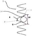
图1是本发明提供的主动脉弓术中支架的外观示意图;Fig. 1 is a schematic view of the appearance of the stent in the aortic arch operation provided by the present invention;
图2是图1的主动脉弓术中支架植入到主动脉弓部的结构和状态示意图;Fig. 2 is a schematic diagram of the structure and state of the stent implanted into the aortic arch during the aortic arch operation of Fig. 1;
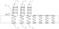
图3是本发明第一实施例提供的主动脉弓术中支架中应用的金属支架的正视图;Fig. 3 is a front view of the metal stent used in the stent in the aortic arch operation provided by the first embodiment of the present invention;
图4是图3的主动脉弓术中支架中应用的一分支以及与该分支对应的第二环状波形支架的正视图;Fig. 4 is a front view of a branch used in the stent in the aortic arch operation of Fig. 3 and a second annular wave-shaped stent corresponding to the branch;
图5是图3的俯视图;Fig. 5 is the top view of Fig. 3;
图6是图3的金属支架覆盖有内层膜后的正视图;Fig. 6 is a front view of the metal stent of Fig. 3 covered with an inner film;
图7是图3的金属支架覆盖有内层膜与外层膜后的正视图;Fig. 7 is a front view of the metal stent in Fig. 3 covered with an inner film and an outer film;
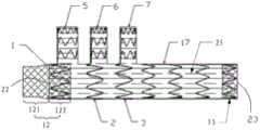
图8是本发明第一实施例提供的主动脉弓术中支架的正视图,示出涤纶布在远端布置的第一种方式;Fig. 8 is a front view of the stent in the aortic arch operation provided by the first embodiment of the present invention, showing the first way in which the polyester cloth is arranged at the distal end;
图9是本发明第一实施例提供的主动脉弓术中支架的正视图,示出涤纶布在远端布置的第二种方式;Fig. 9 is a front view of the stent in the aortic arch operation provided by the first embodiment of the present invention, showing the second way of disposing the polyester cloth at the distal end;
图10是本发明第二实施例提供的主动脉弓术中支架中应用的金属支架的正视图;Fig. 10 is a front view of the metal stent used in the stent in the aortic arch operation provided by the second embodiment of the present invention;
图11是本发明第三实施例提供的主动脉弓术中支架中应用的金属支架的正视图;Fig. 11 is a front view of the metal stent used in the stent in the aortic arch operation provided by the third embodiment of the present invention;
图12是本发明第四实施例提供的主动脉弓术中支架中应用的金属支架的正视图;Fig. 12 is a front view of the metal stent used in the aortic arch stent provided by the fourth embodiment of the present invention;
图13是本发明实施例提供的主动脉弓术中支架制造方法中应用的覆膜模具的正视图;Fig. 13 is a front view of the film-covering mold used in the manufacturing method of the stent in the aortic arch operation provided by the embodiment of the present invention;
图14是本发明第一实施例提供的主动脉弓术中支架制造方法的步骤图;Fig. 14 is a step diagram of the method for manufacturing a stent during aortic arch surgery according to the first embodiment of the present invention;
图15是本发明第二实施例提供的主动脉弓术中支架制造方法的步骤图;Fig. 15 is a step diagram of a method for manufacturing a stent during an aortic arch operation according to the second embodiment of the present invention;
图16是本发明第三实施例提供的主动脉弓术中支架制造方法的步骤图。Fig. 16 is a step diagram of a method for manufacturing a stent during an aortic arch operation according to the third embodiment of the present invention.
具体实施方式Detailed ways
为了使本发明的目的、技术方案及优点更加清楚明白,以下结合附图及实施例,对本发明进行进一步详细说明。应当理解,此处所描述的具体实施例仅仅用以解释本发明,并不用于限定本发明。In order to make the object, technical solution and advantages of the present invention clearer, the present invention will be further described in detail below in conjunction with the accompanying drawings and embodiments. It should be understood that the specific embodiments described here are only used to explain the present invention, not to limit the present invention.
请参阅图1、图5、图7,本发明第一实施例提供的主动脉弓术中支架的整体构造设计成与主动脉弓部形态相符合的结构,包括一个主体17和至少一个分支5、6、7,根据病变的累及情况,该主动脉弓术中支架可选择设置有一至三个分支。主动脉弓术中支架为覆膜支架,即主体17和分支5、6、7都是由可压缩的金属支架和金属支架表面的覆膜25组成的管状结构。所述主体17的近端22处和远端23处各具有一第一开口17a,所述主体17在其覆膜25的一侧通过接口14、15、16分别与每一所述分支的第一端a连接,每一所述分支的与所述第一端a相对的第二端b具有一第二开口4。Please refer to Fig. 1, Fig. 5 and Fig. 7, the overall structure of the stent in the aortic arch operation provided by the first embodiment of the present invention is designed to conform to the shape of the aortic arch, including a
主体17以及分支的金属支架均是由多圈环状波形支架沿着轴向排列组成,主体17上的环状波形支架与分支上的环状波形支架的排布方向是大致垂直的。每一所述环状波形支架包括首尾相接形成环状的弹性丝,所述弹性丝都贴附在覆膜25的管壁上。每一所述环状波形支架具有一对称中心轴,所述弹性丝上具有绕该对称中心轴分布且沿着轴向投影呈环状而在径向投影呈波状的若干波形。轴向投影呈环状是指所述弹性丝沿着所述环状波形支架的轴线作投影呈环状,径向投影呈波状是指所述弹性丝沿着所述环状波形支架的径向作投影呈波状。所述若干环状波形支架分为若干个第一环状波形支架31、与所述接口(14、15、16)数量相等的第二环状波形支架(1、2、3)及位于每一所述分支处且沿该分支的延伸方向间隔排列的若干个第三环状波形支架33。所述若干个第一环状波形支架31相邻并沿所述主体的延伸方向间隔排列作为第一组分布于所述主体的降主段172,所有所述第二环状波形支架1、2、3相邻并沿所述主体的延伸方向间隔排列作为第二组分布于所述主体的弓部段17。Both the
主体17包括通过覆膜25而平整连接成一体的弓部段171和降主段172,弓部段171释放到主动脉弓位置,降主段172释放到降主动脉内。主体17的近端22在弓部段171且靠近心脏,主体17的远端23在降主段172且远离心脏。所述分支(5、6、7)、所述接口(14、15、16)均与所述第二环状波形支架(1、2、3)一一对应。覆膜25上的每一所述接口处有大致为圆形的闭合分界线c,与该接口对应的第二环状波形支架上有一段波形8与该接口的闭合分界线c之间距离最近,该波形8在覆膜25上形成V型分界线d,所述闭合分界线c嵌入所述V型分界线d所围成的区域内。V型分界线d的开口宽度L3可以大于闭合分界线c的最大外径,以使闭合分界线c以内的至少一半面积位于V型分界线d所围成的区域内。V型分界线d的开口宽度L3也可以等于或略小于闭合分界线c的最大外径,闭合分界线c内面积的一小部分能够嵌入所述V型分界线d所围成的区域内。作为更普遍的实施方式,在每个分支的下方对应的主体17的位置上设有至少一个第二环状波形支架,并且其中至少一个第二环状波形支架上有一个波形8从相应的分支的第一端a相形成的接口边缘附近绕过。图5为主体17的局部俯视图,为了清楚地展示所述覆膜上的接口14与第二环状波形支架1的在其中一种实施方式下的相对位置,图中略去了覆膜,实际上,第二环状波形支架1和接口14都位于主体17的覆膜所在的管壁上。The
在本实施例中,主动脉弓术中支架包括主体17以及第一分支5、第二分支6和第三分支7,主体17以及这三个分支都呈管状结构,包括金属支架和包裹住该金属支架的覆膜25,并且第一分支5、第二分支6和第三分支7在自由状态下其轴向与主体17轴向垂直。请参阅图2,主动脉弓术中支架在主动脉弓部释放完成后的示意图,其中主体17对应人体主动脉弓部位的主动脉18,第一分支5、第二分支6和第三分支7分别对应主动脉弓上的无名动脉19、左颈总动脉20、左锁骨下动脉21这三个分支血管。主体17以及第一分支5、第二分支6和第三分支7的直径分别与相应血管内径匹配,例如主体17的近端22的直径为32mm,远端23的直径为32mm,第一分支5、第二分支6和第三分支7均呈直筒状,直径分别为18mm、14mm和14mm。In this embodiment, the stent in the aortic arch operation includes a
为了清楚地展示主动脉弓术中支架的金属支架部分,有些图未显示覆膜。请参阅图1、图3、图8,主体17以及三个分支上的金属支架分别由多圈直径相等的环状波形支架沿着轴向相互间隔排列组成,环状波形支架采用弹性镍钛合金丝编织而成,主体17上的环状波形支架选用0.55mm直径的镍钛丝编织而成,而第一分支5、第二分支6和第三分支7上的第三环状波形支架33选用0.4mm直径的镍钛丝编织而成。其中主体17包括弓部段171和与弓部段171相连的降主段172,所述弓部段171包括三个第二环状波形支架1、2、3,分别用于支撑第一分支5、第二分支6和第三分支7。该三个第一环状波形支架1、2、3是彼此独立的,并且分别位于第一分支5、第二分支6和第三分支7的第一端a的下方。优选地,为了保证三个分支之间的距离可调节,这三个第二环状波形支架1、2、3之间没有连接杆,而是紧贴在共同的覆膜25上而组成一体,这样分支可以根据分支血管之间的距离而自动调整相对位置。In order to clearly show the metal stent portion of the stent during aortic arch surgery, some figures do not show the graft. Please refer to Figure 1, Figure 3, and Figure 8, the
主体17的降主段172靠近远端23,该降主段172的第一环状波形支架31采用第一连接杆9连接在一起,所述第一连接杆9为呈直线的弹性丝。第一分支5、第二分支6和第三分支7的金属支架都是由三圈第三环状波形支架33采用连接杆组成,并且分别通过一个第二连接杆10与主体17上的第二环状波形支架1、第二环状波形支架2或第二环状波形支架3形成一对一的连接,第二连接杆10为呈L形的弹性丝。每个分支也可通过第二连接杆10的长臂来连接分支上的多个第三环状波形支架33,再通过第二连接杆10的短臂与主体上的相应一个第二环状波形支架连接。这样可以进一步提高主体17对分支提供的支撑力,第二连接杆10可以设置在的分支上的朝向主体17的近端22的一侧,既可允许分支被压缩而偏向主体17的远端23,还能保证分支恢复到与主体17大致垂直的位置,防止分支过度向远端23倾斜而不能恢复原位的风险。The descending
为了分支与主体17的夹角能够适度变化,以适应不同患者的血管形态,优选地,靠近每一所述分支的所述第一端a的一所述第三环状波形支架33与所述主体17之间的距离L4的范围为4mm-10mm,即每个分支的最底端的金属支架与主体的最小距离L4为4-10mm,以此来选择第二连接杆10的长度。通常,第三分支7需要的支撑力大于第一分支5和第二分支6所需的支撑力,第三分支7的第二连接杆10可用更粗或更硬的材料制作,而第一分支5和第二分支6的第二连接杆10可用稍细或稍软的材料制作。In order to moderately change the included angle between the branch and the
优选地,第二环状波形支架1比第一分支5更靠近主体17的近端22,第一分支5的第二连接杆10设置在靠近主体17的近端22一侧,有利于第一分支5在分支血管中的定位。为了主体17对每个分支的支撑力基本均匀分布,优选地,第二环状波形支架2比第二分支6更靠近第一分支5,第二环状波形支架3比第三分支7更靠近第二分支6。Preferably, the second annular wave-shaped
为了增强分支的贴壁性,在每一所述分支处的所述第三环状波形支架33中,靠近该分支的所述第二端b的一个所述第三环状波形支架33的波形的高度最小且该波形的数量最多,即分支顶端的第三环状波形支架33的波形排布较为密集;以第一分支5为例,请参阅图4,位于顶端的第一圈第三环状波形支架33的波形个数为九个,其余两圈第三环状波形支架33的波形个数为六个。In order to enhance the adherence of the branch, in the third ring-shaped wave-shaped
每个分支的覆膜25的底部连接到主体17侧壁的覆膜25上,并且在主体17的覆膜25上形成一个接口(14、15、16),主体17的内腔通过这个接口与分支的内腔相通。请参阅图5,第一分支5的俯视图,第二环状波形支架1的一个波形8刚好绕过主体17的覆膜25上的一个圆形的接口14,主体17的侧壁覆膜25与第一分支5的覆膜25围绕接口14而连续成一整体,也就是说,该接口14既在主体17的侧壁上,也位于第一分支5的第一端a。由于覆膜25具有一定的厚度和韧性,第一分支5对主体17的压力,通过接口14周边的覆膜25传递到第二环状波形支架1的波形8上,也就是说,第二环状波形支架1对第一分支5提供支撑力。请参阅图5,为了增加第二环状波形支架1对第一分支5的支撑作用,所述闭合分界线c嵌入所述V型分界线d所围成的区域内的深度与该V型分界线d所围成的区域的高度之比至少为50%;或者请参阅图4,接口14进入第二环状波形支架1的波形8围绕的区域内的深度L2大于波形8的高度L1的50%。这样可以增加主体17对分支的径向支撑力,使得在主体17被释放后分支可以被主体17安全的顶入相应的分支血管内,而且在分支释放后,可以防止该分支从相应的分支血管内滑脱出来。The bottom of the coating 25 of each branch is connected to the coating 25 of the
优选地,请参阅图4,第二环状波形支架1的波形8的高度L1大于圆形接口14的半径,接口14的中心也位于波形8围绕的区域内。优选地,第二环状波形支架2与第二分支6的相对位置满足同样的条件,第二环状波形支架3与第三分支7的相对位置也满足同样的条件,以增加对每个分支的径向支撑力。请参阅图5,第二环状波形支架1的围绕接口14的一个波形8的圆角半径R为3mm,这个波形8展开后的宽度L3为25mm;第一分支5的接口14直径为18mm,则接口14嵌入波形8内部的深度可以达到波形8的高度的50%以上。类似的,第二环状波形支架2、第二环状波形支架3的围绕相应接口15、16的一个波形8的圆角半径R均为3mm,这两个波形展开后的宽度均为20mm,而第二分支6和第三分支7的接口15、16直径均为14mm。这样设计可以保证主体17上的波形8有足够的宽度来容纳相应的分支的接口,保证接口嵌入主体17上的波形8内部的深度可以达到该波形8的高度的50%以上。Preferably, please refer to FIG. 4 , the height L1 of the wave 8 of the second annular wave-shaped
在金属支架制作完成后,采用覆膜模具40开始覆膜工序。覆膜模具40同主动脉弓的形态结构匹配,也是由一个主体部分和三个分支部分组成。覆膜第一个工序为制作内层膜,首先将ePTFE膜覆在模具的表面,内层膜可以是厚度0.03~0.1mm的单层ePTFE膜,或者多层较薄的ePTFE膜叠合到同等厚度,完成内层膜工序。After the metal stent is fabricated, the film covering process is started using the film covering mold 40 . The covering mold 40 matches the shape and structure of the aortic arch, and is also composed of a main body and three branch parts. The first process of film covering is to make the inner film. First, the ePTFE film is covered on the surface of the mold. The inner film can be a single-layer ePTFE film with a thickness of 0.03-0.1mm, or laminated with multiple thinner ePTFE films to the same level. Thickness, complete the inner film process.
第二个工序为将编织完成的金属支架套在覆盖了内层膜的模具表面。具体地,请参阅图6,将第二环状波形支架1和第一分支5上的金属支架、第二环状波形支架2和第二分支6上的金属支架、第二环状波形支架3和第三分支7上的金属支架、主体17上靠近远端23的其它环状波形支架分别放在图6所示的位置。优选地,模具的每个分支嵌入主体上的第二环状波形支架的波形8内部的深度超过该波形8的高度的50%。The second process is to wrap the braided metal stent on the surface of the mold covered with the inner film. Specifically, referring to Fig. 6, the second ring-shaped wave-shaped
第三个工序为外层膜工序,在金属支架外表面再覆ePTFE膜,将金属支架整体(包括主体17和三个分支的金属支架)完全包覆起来。外层膜可以是厚度0.03~0.1mm的单层ePTFE膜,或者多层较薄的ePTFE膜叠合到同等厚度。在内层膜和外层膜直接贴合的区域,覆膜25的总厚度为0.06~0.2mm。以上三个工序完成后,通过在高温中施加压力的方式,将内层和外层ePTFE覆膜贴合在一起,金属支架固定在覆膜25的中间,该工序完成后将整个支架从模具上拆下,将主体17上两端多余的ePTFE膜剪掉,形成近端22和远端23,最终如图7所示。优选地,主体17上的覆膜25的近端22至第一分支5的第一端a的距离为5-10mm。The third process is the outer membrane process, where the outer surface of the metal stent is covered with an ePTFE film to completely cover the whole metal stent (including the
ePTFE覆膜完成后,还有可选的步骤:在主体17上缝合近端织物12或者远端织物13,二者可以均为涤纶布。请参阅图8,为了便于主动脉弓术中支架的主体17的近端22与血管的缝合,覆膜完成后,在主体17的近端22缝合管状的涤纶布,即近端织物12,所述涤纶布的固定端与靠近近端22的第二环状波形支架1缝合在一起,涤纶布的自由端至第一分支5的距离为25-35mm,优选30mm。近端织物12分为第一段121与第二段122,第一段121不包含金属支架,其内没有金属支架支撑,第二段122的涤纶布围成的管内有金属支架支撑,第二段122可与第二环状波形支架1重叠并缝合在一起。第一段121内的管状涤纶布的直径为29mm,比主体17的直径略小;因为主体17的自由状态的直径会比主动脉弓部的主动脉直径略大,而涤纶管的自由端直径应与血管直径相等,为了避免血管缝合时涤纶布有褶皱,所以涤纶管的自由端的直径要比主体17近端22的直径略小。而第二段122内的管状涤纶布的直径为32mm,与主体17的直径相同。After the ePTFE covering is completed, there is an optional step: sew the
另外,在主体17的远端23缝合远端织物13,图8示出远端织物13的涤纶布在远端23布置的第一种方式,该涤纶布与主体17的降主段172的最后一圈第一环状波形支架31重叠并缝合在一起。远端织物13固定完成后,这样可以保证主体17的远端23也具有很好的缝合性能,为可能的远期外科手术提供条件。如果远端织物13刚好与主体17远端23对齐,则远期外科手术的缝线与远端织物13和主体17远端23缝合在一起。图9示出远端织物13的第二种实施方式,在主体17的远端23缝合的远端织物13分为两个区域,分别为第一区域131和第二区域132。第一区域131的涤纶布固定的位置与主体17的降主段172上最后三圈第一环状波形支架31重叠并缝合在一起,第二区域132的涤纶布具有自由端并伸出主体17的远端23一段距离,长度为8-12mm,优选10mm,并且第二区域132不包含金属支架,其内没有主体17上的第一环状波形支架31支撑。具有自由端的远端织物13固定完成后,这样可以保证主体17的远端23也具有更方便的缝合性能,远端织物13的第二区域132伸出主体17远端23一定的距离,则远期外科手术的缝线可与伸出主体17远端23的第二区域132的涤纶布缝合在一起。In addition, the far-
请参阅图10,本发明第二实施例提供的主动脉弓术中支架与第一实施例提供的主动脉弓术中支架大致相同,与第一实施例不同的是:最靠近每一所述分支的所述第二端b的一个所述第三环状波形支架33的直径沿该分支的所述第一端a至所述第二端b的方向逐渐减小而使该分支的所述第二端b处的覆膜具有锥形管壁,即该部呈缩口状。例如位于第一分支5最顶端的第三环状波形支架33的上端直径D1为16mm,下端直径D2为18mm,即上端直径D1比下端直径D2小2mm,而其它两圈第三环状波形支架33是直管形的,其直径为18mm。第二分支6、第三分支7上的结构与第一分支5相同,都是最顶端一圈第三环状波性支架为锥形缩口状设计,最上端直径比下端直径小2mm。这种设计可以保证每个分支下端两圈第三环状波形支架33与分支血管充分接触,并提供足够径向支撑,同时位于顶端的一圈第三环状波形支架的最顶端不会对分支血管有非常大的扩张力,以此减小对血管的损伤。Please refer to FIG. 10 , the stent for the aortic arch provided by the second embodiment of the present invention is substantially the same as the stent provided by the first embodiment. The diameter of the third annular wave-shaped
请参阅图11,本发明第三实施例提供的主动脉弓术中支架与第一实施例提供的主动脉弓术中支架大致相同,与第一实施例不同的是:靠近每一所述分支的所述第一端a的一个所述第三环状波形支架33的直径沿该分支的所述第二端b至所述第一端a的方向逐渐减小而使该分支的所述第一端a处的覆膜具有锥形管壁,即该部呈缩口状。例如位于第一分支5最底端的第三环状波形支架33的上端直径D2ˊ为18mm,下端直径D1ˊ为16mm,即上端直径D2ˊ比下端直径D1ˊ大2mm。第二分支6、第三分支7上的结构与第一分支5相同,下端的第三环状波形支架33都是上端直径比下端直径大2mm,这种设计可以保证三个分支在手术过程中能够更好的进行自由摆动,从而更好的适应主动脉弓上不同的分支血管解剖形态。Please refer to FIG. 11 , the intraoperative stent for the aortic arch provided by the third embodiment of the present invention is substantially the same as the intraoperative stent for the aortic arch provided by the first embodiment. The diameter of a third ring-shaped wave-shaped
此外,所有所述第一环状波形支架31的直径是在所述近端22往所述远端23的方向上是递减的,即在主体17上的第一环状波形支架31的直径逐渐缩小,整体形成一个锥度,靠近第二环状波形支架3的那个第一环状波形支架31直径D4为32mm,位于最远端的那个第一环状波形支架31的直径D3为28mm;主体17上采用锥度设计的这一段是放入降主动脉内,而人体降主动脉内径也是逐渐缩小的,因此能更好的适应降主动脉的解剖形态。In addition, the diameters of all the first ring-shaped wave-shaped
可以理解地,作为本方案中的优化设计,靠近每一所述分支的所述第二端b的一个所述第三环状波形支架33的直径沿该分支的所述第一端a至所述第二端b的方向逐渐减小而使该分支的所述第二端b部呈缩口状;靠近每一所述分支的所述第一端a的一个所述第三环状波形支架33的直径沿该分支的所述第二端b至所述第一端a的方向逐渐减小而使该分支的所述第一端a部呈缩口状。即分支的两端部均可以为缩口状设计。在分支的最底端的一圈第三环状波形支架33为缩口状设计,靠近主体17一侧的直径最小;在分支的最顶端的一圈第三环状波形支架33为缩口状设计,最顶端一侧的直径最小。It can be understood that, as an optimized design in this solution, the diameter of a third ring-shaped wave-shaped
请参阅图12,本发明第四实施例提供的主动脉弓术中支架与第一实施例提供的主动脉弓术中支架大致相同,与第一实施例不同的是:所有所述第一环状波形支架31之间连接有第一连接杆9,所述第一连接杆9为呈直线的弹性丝;所述主动脉弓术中支架还包括一第二连接杆10,所述第二连接杆10为呈L形的弹性丝,所述第二连接杆10的短臂固定于所述第一连接杆9上,所述第二连接杆10的长臂连接至与所述降主段172之间距离最近的一所述分支上的所有所述第三环状波形支架33上。即第一分支5、第二分支6、第三分支7中的金属支架与主体17中的第二环状波形支架1、2、3之间只通过覆膜25连接在一起但没有通过连接杆连接,并且第二环状波形支架1、2、3之间也没有连接杆。进一步地,所述主动脉弓术中支架还包括第三连接杆11,除与所述第二连接杆10连接的所述分支之外的其余分支上均连接有一所述第三连接杆11,每一所述第三连接杆11为呈直线的弹性丝。每个分支的管状覆膜25的底部分别固定在主体17的覆膜25上,这些覆膜25连续成一个整体并有一定的厚度和韧性,因此,第二环状波形支架1、2、3通过这个整体覆膜25给三个分支提供充分的支撑力。并且,第二环状波形支架1、2、3与分支之间没有连接杆的限制,允许每个分支相对于主体17做小幅度的角度或距离的调整,以适应不同患者的血管形态结构的差异性。Please refer to FIG. 12 , the intraoperative stent for the aortic arch provided by the fourth embodiment of the present invention is substantially the same as the intraoperative stent for the aortic arch provided by the first embodiment, and the difference from the first embodiment is that all the first annular wave-shaped
主体17上的第一环状波形支架31采用连接杆连接在一起,形成主动脉弓术中支架的降主段172。由于在手术操作中,必须先将主动脉弓术中支架的降主段172和第三分支7释放并定位在相应血管内,第三分支7的定位是三个分支中难度最高的,因此将第三分支7上的金属支架与上述降主段172通过一个第二连接杆10连接在一起,第二连接杆10为呈L形的弹性丝。所有所述第一环状波形支架31之间设置有第一连接杆9,所述第一连接杆9为呈直线的弹性丝。优选地,第一连接杆9设置在降主段172的靠近分支的一侧。第二连接杆10可以直接固定在降主段172的一个第一环状波形支架31上,但不必与第一连接杆9相连。The first annular wave-shaped
本实施例中的第二连接杆10位于第三分支7的靠近主动脉弓术中支架的远端23一侧,以约束第三分支7与所述降主段172之间的夹角,这样第三分支7在被释放后能依靠第二连接杆10的回弹力对准相应的分支血管的方向。该第二连接杆10也可用于连接第三分支7上的第三环状波形支架33,优选地,该第二连接杆10延伸固定到主动脉弓术中支架的降主段172的第一连接杆9上,进一步稳定该降主段172与第三分支7之间的夹角,并借助主体17的降主段172所提供的额外的径向支撑力将第三分支7顶入分支血管内。主动脉弓术中支架的降主段172与第三分支7之间的稳定支撑和定位,还有利于第一分支5和第二分支6的释放和定位,也允许第一分支5和第二分支6分别根据血管形态来调整角度和距离。可选地,第一连接杆9与第二连接杆10可用同一根弹性丝制作。The second connecting
本发明第五实施例提供的主动脉弓术中支架与第一实施例提供的主动脉弓术中支架大致相同,与第一实施例不同的是:主动脉弓术中支架中的覆膜整体选用涤纶布材料,代替实施例一中的ePTFE膜;将涤纶布分别围绕在主体17以及三个分支的金属支架的外表面或者内表面,然后通过缝合线与金属支架缝合在一起。与实施例一中的图9相似,在主体17的近端22,涤纶布有长25-35mm(优选30mm)区域没有环状波形支架支撑,并且直径比主体17的其余部分直径略小;在主体17的远端23,涤纶布有长8-12mm(优选10mm)的区域没有环状波形支架支撑,直径与主体17的有环状波形支架的部分直径相同。The intra-aortic arch stent provided by the fifth embodiment of the present invention is substantially the same as the aortic arch intra-operative stent provided by the first embodiment. The difference from the first embodiment is that the covering film of the aortic arch intra-operative stent is made of polyester cloth as a whole instead of The ePTFE membrane in Embodiment 1: wrap the polyester cloth around the outer surface or inner surface of the
请参阅图14,本发明第一实施例提供的主动脉弓术中支架制造方法,包括如下步骤:Please refer to FIG. 14 , the method for manufacturing a stent in the aortic arch operation provided by the first embodiment of the present invention includes the following steps:
S1)请参阅图13,提供一覆膜模具40,覆膜模具40同主动脉弓的形态结构匹配。所述覆膜模具40包括第一柱体41及连接于所述第一柱体41上的第二柱体42,所述第一柱体41分为第一柱段411及第二柱段412,所述第二柱体42的数量与所述主动脉弓术中支架的分支数量相同,所述第二柱体42连接于所述第二柱段412上。S1) Referring to FIG. 13 , a covering mold 40 is provided, and the covering mold 40 matches the shape and structure of the aortic arch. The coating mold 40 includes a first column 41 and a second column 42 connected to the first column 41, the first column 41 is divided into a first column section 411 and a second column section 412 , the number of the second column 42 is the same as the number of branches of the stent in the aortic arch operation, and the second column 42 is connected to the second column segment 412 .
S2)于所述覆膜模具40的外表面制作所述覆膜25的内层膜,将ePTFE膜覆在模具的表面,内层膜可以是厚度0.03~0.1mm的单层ePTFE膜,或者多层较薄的ePTFE膜叠合到同等厚度,完成内层膜工序。S2) Fabricate the inner film of the film covering 25 on the outer surface of the film covering mold 40, and cover the surface of the mold with an ePTFE film. The inner film can be a single-layer ePTFE film with a thickness of 0.03-0.1mm, or more The thinner ePTFE membrane is laminated to the same thickness to complete the inner membrane process.
S3)提供若干所述环状波形支架,环状波形支架采用弹性镍钛合金丝编织而成,主体17上的环状波形支架选用0.55mm直径的镍钛丝编织而成,而第一分支5、第二分支6和第三分支7上的环状波形支架33选用0.4mm直径的镍钛丝编织而成,所述若干个第一环状波形支架31间隔套于所述第一柱段411上,每一所述第二环状波形支架套于所述第二柱段412上并位于与该第二环状波形支架对应的一个所述第二柱体42的靠近所述近端的一侧,且使该第二环状波形支架的其中一波形8卡于该对应的所述第二柱体42上,所述第三环状波形支架33间隔套于每一第二柱体42上,最终如图6所示。优选地,模具的每个分支嵌入主体上的环状波形支架的波形8内部的深度超过该波形8的高度的50%。S3) Provide several ring-shaped wave-shaped brackets, the ring-shaped wave-shaped brackets are braided by elastic nickel-titanium alloy wires, the ring-shaped wave-shaped brackets on the
S4)于所述覆膜模具40的外表面制作所述覆膜25的外层膜,并使所述外层膜与所述内层膜结合并包裹所述环状波形支架。在金属支架外表面再覆ePTFE膜,将金属支架整体(包括主体17和三个分支的金属支架)完全包覆起来。外层膜可以是厚度0.03~0.1mm的单层ePTFE膜,或者多层较薄的ePTFE膜叠合到同等厚度。在内层膜和外层膜直接贴合的区域,覆膜25的总厚度为0.06~0.2mm。通过在高温中施加压力的方式,将内层和外层ePTFE覆膜贴合在一起,金属支架固定在覆膜25的中间,该工序完成后将整个支架从模具上拆下,将主体17上两端多余的ePTFE膜剪掉,形成近端22和远端23,最终如图7所示。优选地,主体17上的覆膜25的近端至第一分支5的底部的距离为5-10mm。S4) Fabricating the outer film of the covering film 25 on the outer surface of the film covering mold 40, combining the outer film with the inner film to wrap the ring-shaped corrugated stent. The outer surface of the metal stent is covered with an ePTFE film to completely cover the metal stent as a whole (including the
进一步地,还包括步骤S5)和S6),其中步骤S5)在主体17上缝合近端织物12与远端织物13,二者均为涤纶布。请参阅图8,涤纶布的固定端与靠近近端22的第二环状波形支架1缝合在一起,涤纶布的自由端至第一分支5的距离为25-35mm,优选30mm。近端织物12分为第一段121与第二段122,第二段122与近端22的第二环状波形支架1完全缝合在一起。第一段121内的管状涤纶布的直径为29mm,比主体17的直径略小。而第二段122内的管状涤纶布的直径为32mm,与主体17的直径相同。Further, steps S5) and S6) are also included, wherein in step S5), the
步骤S6)图8示出涤纶布在远端23布置的第一种方式,在主体17的远端23缝合远端织物13,远端织物13与主体17的降主段172的最后一圈第一环状波形支架31重叠并缝合在一起。图9示出涤纶布在远端23布置的第二种方式,在主体17的远端23缝合的远端织物13分为两个区域,分别为第一区域131和第二区域132。第一区域131的涤纶布固定的位置与主体17的降主段172上最后三圈第一环状波形支架31重叠并缝合在一起,第二区域132的涤纶布为伸出主体17的远端23一段距离,长度为8-12mm,优选10mm,并且第二区域132不包含金属支架,其内没有主体17上的环状波形支架支撑。Step S6) FIG. 8 shows the first way in which the polyester cloth is arranged at the
可以理解地,步骤S5)和S6)是可以替换的,替换前后效果是一样的。Understandably, steps S5) and S6) can be replaced, and the effects before and after the replacement are the same.
请参阅图15,本发明第二实施例提供的主动脉弓术中支架制造方法与第一实施例提供的主动脉弓术中支架制造方法大致相同,与第一实施例不同的是:步骤S2)与S4)。Please refer to FIG. 15 , the manufacturing method for the intraoperative stent of the aortic arch provided by the second embodiment of the present invention is substantially the same as the manufacturing method of the intraoperative stent for the aortic arch provided by the first embodiment, and the difference from the first embodiment is: steps S2) and S4) .
其中步骤S2)于所述覆膜模具40的外表面套设所述覆膜25,所述覆膜25为涤纶布。Wherein step S2) the covering film 25 is sheathed on the outer surface of the film covering mold 40, and the covering film 25 is polyester cloth.
S4)通过缝合线将所述覆膜25与所述环状波形支架缝合在一起,在主体17的近端22,涤纶布有长25-35mm(优选30mm)区域没有环状波形支架支撑,并且直径比主体17的其余部分直径略小;在主体17的远端23,涤纶布有长8-12mm(优选10mm)的区域没有环状波形支架支撑,直径与主体17的有环状波形支架的部分直径相同。S4) sew the covering film 25 and the ring-shaped wave-shaped support together by suture thread, at the
请参阅图16,本发明第三实施例提供的主动脉弓术中支架制造方法与第一实施例提供的主动脉弓术中支架制造方法大致相同,与第一实施例不同的是:步骤S2)、S3)与S4)。Please refer to FIG. 16 , the manufacturing method of the intraoperative stent for the aortic arch provided by the third embodiment of the present invention is substantially the same as the manufacturing method for the intraoperative stent of the aortic arch provided by the first embodiment, and the difference from the first embodiment is: steps S2), S3) with S4).
其中步骤S2)提供若干所述环状波形支架,环状波形支架采用弹性镍钛合金丝编织而成,主体17上的环状波形支架选用0.55mm直径的镍钛丝编织而成,而第一分支5、第二分支6和第三分支7上的环状波形支架33选用0.4mm直径的镍钛丝编织而成,所述若干个第一环状波形支架31间隔套于所述第一柱段411上,每一所述第二环状波形支架套于所述第二柱段412上并位于与该第二环状波形支架对应的一个所述第二柱体42的靠近所述近端的一侧,且使该第二环状波形支架的其中一波形8卡于该对应的所述第二柱体42上,所述第三环状波形支架33间隔套于每一第二柱体42上,最终如图6所示。优选地,模具的每个分支嵌入主体上的环状波形支架的波形8内部的深度超过该波形8的高度的50%。Wherein step S2) provides several ring-shaped wave-shaped brackets, the ring-shaped wave-shaped brackets are braided by elastic nickel-titanium alloy wires, the ring-shaped wave-shaped brackets on the
S3)于所述环状波形支架的外表面套设所述覆膜25,所述覆膜25为涤纶布。S3) Sleeve the coating 25 on the outer surface of the ring-shaped corrugated bracket, and the coating 25 is polyester cloth.
S4)通过缝合线将所述覆膜25与所述环状波形支架缝合在一起,在主体17的近端22,涤纶布有长25-35mm(优选30mm)区域没有环状波形支架支撑,并且直径比主体17的其余部分直径略小;在主体17的远端23,涤纶布有长8-12mm(优选10mm)的区域没有环状波形支架支撑,直径与主体17的有环状波形支架的部分直径相同。S4) sew the covering film 25 and the ring-shaped wave-shaped support together by suture thread, at the
本发明提供的主动脉弓术中支架能够自动适应不同病人的主动脉弓附近的血管结构,并且主动脉弓术中支架中的主体对分支保持足够的径向支撑力,保证在手术中主动脉弓术中支架上的分支能够安全的进入到分支血管内,同时在手术中以及术后避免分支从相应的分支血管中滑脱出来。本发明提供的主动脉弓术中支架具有以下优点:The stent in the aortic arch operation provided by the present invention can automatically adapt to the vascular structure near the aortic arch of different patients, and the main body in the stent in the aortic arch operation maintains sufficient radial support force for the branches to ensure that the branches on the stent in the aortic arch operation can be Safely enter the branch blood vessel, and at the same time prevent the branch from slipping out of the corresponding branch blood vessel during and after the operation. The stent provided by the present invention has the following advantages:
1)本发明中所述的主动脉弓术中支架,主体对分支提供了可靠的支撑力,这样可以保证分支可以完全进入分支血管内,并可避免术后远期的支架移位滑脱。1) The main body of the intraoperative aortic arch stent described in the present invention provides reliable support for the branches, which ensures that the branches can completely enter the branch vessels and avoids long-term postoperative stent displacement and slippage.
2)由于主动脉弓解剖结构复杂,不同患者的三个分支血管的间距的个体差异比较大。而主动脉弓部的A型主动脉夹层疾病通常为急诊,因此没有充足的时间为患者定制与患者的主动脉弓分支间距完全匹配的特殊产品。本发明中所述的主动脉弓术中支架,分支之间的间距可根据主动脉弓的解剖形态进行自适应调节,解决了患者主动脉弓形态个体差异比较大,急诊手术中很难匹配主动脉弓术中支架的问题。2) Due to the complex anatomical structure of the aortic arch, the distance between the three branch vessels in different patients varies greatly. However, type A aortic dissection in the aortic arch is usually an emergency, so there is not enough time to customize a special product that exactly matches the distance between the branches of the aortic arch. In the aortic arch intraoperative stent described in the present invention, the distance between branches can be adaptively adjusted according to the anatomical shape of the aortic arch, which solves the problem that the individual differences in the shape of the aortic arch are relatively large, and it is difficult to match the aortic arch intraoperative stent in emergency surgery.
3)本发明中所述的主动脉弓术中支架,主体以及分支可以采用整体的内外层的ePTFE膜双层覆膜技术,分支的内壁光滑,减小了血栓的形成,对脑部血管形成很好的保护作用。分支与主体之间无缝合线连接,无血液渗漏的风险。可在主体上附加近端织物,例如,在主体的近端缝有一段管状的涤纶布,这段涤纶布近端有一部分区域没有金属支架支撑,并且这一部分涤纶布直径略小于主体有金属支架弓部段部分的直径,涤纶布保证了与血管可靠的吻合,其直径略小又避免了与血管吻合时涤纶布褶皱的出现。此外,主体还可以附加远端织物,例如,主体的远端缝也可以缝合有一段管状的涤纶布,该段涤纶布的远端一小段区域也可没有金属支架支撑,该段涤纶布保证了主体的远端具有可靠的缝合性能,对于远期可能的外科手术处理提供了保证。3) The stent in the aortic arch operation described in the present invention, the main body and the branches can adopt the ePTFE membrane double-layer coating technology of the whole inner and outer layers, the inner wall of the branches is smooth, which reduces the formation of thrombus and is good for the formation of blood vessels in the brain protective effect. There is no suture connection between the branch and the main body, no risk of blood leakage. The proximal fabric can be attached to the main body, for example, a section of tubular polyester cloth is sewn on the proximal end of the main body, and there is a part of the proximal end of this polyester cloth that is not supported by a metal bracket, and the diameter of this part of the polyester cloth is slightly smaller than that of the main body with a metal bracket The diameter of the bow section, the polyester cloth ensures reliable anastomosis with blood vessels, and its slightly smaller diameter avoids the occurrence of polyester cloth wrinkles when anastomosing with blood vessels. In addition, the main body can also be attached with a distal fabric. For example, the distal end of the main body can also be sewn with a section of tubular polyester cloth. A small area of the far end of this section of polyester cloth may not be supported by a metal bracket. This section of polyester cloth ensures The distal end of the main body has reliable suturing performance, which provides guarantee for possible long-term surgical operation.
4)本发明中所述的主动脉弓术中支架的主体的降主段可以采用锥形,更好的适应了降主动脉的解剖形态。4) The descending main section of the main body of the stent in the aortic arch operation described in the present invention can adopt a tapered shape, which better adapts to the anatomical shape of the descending aorta.
5)本发明中所述的主动脉弓术中支架的分支的最顶端的一圈环状波形支架采用小波形密排设计,保证了分支顶端良好的贴壁性能。5) The ring-shaped wave-shaped stent at the top of the branch of the stent in the aortic arch operation described in the present invention adopts a small wave-shaped close-packed design, which ensures good wall-attachment performance at the top of the branch.
6)本发明中所述的主动脉弓术中支架的分支的最顶端的一圈环状波形支架采用缩口状(最顶端直径最小),其余波形采用直管形,这样既保证了分支在主动脉弓分支血管内的稳定性,又避免了因分支的顶端对分支血管的过度扩张力而损伤分支血管。6) In the aortic arch operation described in the present invention, the ring-shaped wave-shaped stent at the top of the branch of the stent adopts a constricted shape (the diameter of the topmost part is the smallest), and the rest of the waveform adopts a straight tube shape, which ensures that the branch is in the aortic arch branch. The stability in the blood vessel avoids damage to the branch blood vessel due to the excessive expansion force of the branch top to the branch blood vessel.
7)本发明所述的主动脉弓术中支架的每个分支的最底端的一圈环状波形支架为缩口状(靠近主体一侧的直径最小),这种设计可以保证分支能够更好的进行自由摆动,从而更好的适应不同患者的主动脉弓和分支血管的解剖形态差异。7) The ring-shaped wave-shaped stent at the bottom of each branch of the stent in the aortic arch operation according to the present invention is constricted (the diameter of the side close to the main body is the smallest), and this design can ensure that the branches can be carried out better. Swing freely, so as to better adapt to the anatomical differences of the aortic arch and branch vessels in different patients.
以上所述仅为本发明的较佳实施例而已,并不用以限制本发明,凡在本发明的精神和原则之内所作的任何修改、等同替换和改进等,均应包含在本发明的保护范围之内。The above descriptions are only preferred embodiments of the present invention, and are not intended to limit the present invention. Any modifications, equivalent replacements and improvements made within the spirit and principles of the present invention should be included in the protection of the present invention. within range.
Claims (18)
Translated fromChinesePriority Applications (5)
| Application Number | Priority Date | Filing Date | Title |
|---|---|---|---|
| CN201310745529.1ACN103720529B (en) | 2013-12-30 | 2013-12-30 | Arcus aortae intraoperative stent and method for manufacturing stent |
| EP14876566.2AEP3090707A4 (en) | 2013-12-30 | 2014-12-30 | Aortic arch intraoperative stent and manufacturing method thereof |
| PCT/CN2014/095609WO2015101292A1 (en) | 2013-12-30 | 2014-12-30 | Aortic arch intraoperative stent and manufacturing method thereof |
| US15/108,287US10548709B2 (en) | 2013-12-30 | 2014-12-30 | Aortic arch intraoperative stent and manufacturing method thereof |
| CN201480071638.2ACN105873543B (en) | 2013-12-30 | 2014-12-30 | The manufacture method of arch of aorta art medium-height trestle and the support |
Applications Claiming Priority (1)
| Application Number | Priority Date | Filing Date | Title |
|---|---|---|---|
| CN201310745529.1ACN103720529B (en) | 2013-12-30 | 2013-12-30 | Arcus aortae intraoperative stent and method for manufacturing stent |
Publications (2)
| Publication Number | Publication Date |
|---|---|
| CN103720529Atrue CN103720529A (en) | 2014-04-16 |
| CN103720529B CN103720529B (en) | 2017-02-08 |
Family
ID=50445001
Family Applications (2)
| Application Number | Title | Priority Date | Filing Date |
|---|---|---|---|
| CN201310745529.1AActiveCN103720529B (en) | 2013-12-30 | 2013-12-30 | Arcus aortae intraoperative stent and method for manufacturing stent |
| CN201480071638.2AActiveCN105873543B (en) | 2013-12-30 | 2014-12-30 | The manufacture method of arch of aorta art medium-height trestle and the support |
Family Applications After (1)
| Application Number | Title | Priority Date | Filing Date |
|---|---|---|---|
| CN201480071638.2AActiveCN105873543B (en) | 2013-12-30 | 2014-12-30 | The manufacture method of arch of aorta art medium-height trestle and the support |
Country Status (4)
| Country | Link |
|---|---|
| US (1) | US10548709B2 (en) |
| EP (1) | EP3090707A4 (en) |
| CN (2) | CN103720529B (en) |
| WO (1) | WO2015101292A1 (en) |
Cited By (31)
| Publication number | Priority date | Publication date | Assignee | Title |
|---|---|---|---|---|
| CN104116577A (en)* | 2014-06-27 | 2014-10-29 | 先健科技(深圳)有限公司 | Fork-type covered stent |
| WO2015101292A1 (en)* | 2013-12-30 | 2015-07-09 | 先健科技(深圳)有限公司 | Aortic arch intraoperative stent and manufacturing method thereof |
| EP3040050A1 (en)* | 2014-12-29 | 2016-07-06 | Cook Medical Technologies LLC | Support structures for prostheses with branching portions |
| CN106214287A (en)* | 2016-08-24 | 2016-12-14 | 杨威 | Dissection of aorta operation overlay film frame, conveyer device and using method |
| US20170189030A1 (en)* | 2015-12-30 | 2017-07-06 | Lifetech Scientific (Shenzhen) Co., Ltd. | Method for Treating Aortic Arch Diseases |
| WO2018001133A1 (en)* | 2016-06-28 | 2018-01-04 | 微创心脉医疗科技(上海)有限公司 | Membrane-coated stent and manufacturing method thereof |
| CN107616856A (en)* | 2017-11-08 | 2018-01-23 | 吉林大学 | A kind of belt supporting frame artificial blood vessel and the application in Stanford A type dissection of aorta clinic operation formulas |
| WO2018032358A1 (en)* | 2016-08-16 | 2018-02-22 | Dalian Corvivo Medical, Co. Ltd | Endoprosthesis having graft portion and stent graft portion |
| CN107802376A (en)* | 2016-09-08 | 2018-03-16 | 先健科技(深圳)有限公司 | Overlay film frame and preparation method thereof |
| CN107802387A (en)* | 2017-11-08 | 2018-03-16 | 有研医疗器械(北京)有限公司 | A kind of art medium-height trestle induction system and its application method |
| CN108236512A (en)* | 2016-12-27 | 2018-07-03 | 先健科技(深圳)有限公司 | Overlay film frame |
| WO2018121364A1 (en)* | 2016-12-28 | 2018-07-05 | 先健科技(深圳)有限公司 | Covered stent |
| CN108245280A (en)* | 2016-12-29 | 2018-07-06 | 先健科技(深圳)有限公司 | Overlay film frame |
| CN108392292A (en)* | 2017-08-31 | 2018-08-14 | 北京裕恒佳科技有限公司 | Side port formula branch artificial blood vessel |
| CN109009562A (en)* | 2018-08-27 | 2018-12-18 | 泉州市第医院 | Follow-on arch of aorta overlay film frame type blood vessel |
| CN109996514A (en)* | 2016-12-05 | 2019-07-09 | 美敦力瓦斯科尔勒公司 | Modularization arch of aorta prosthesis assembly and its application method |
| CN110464522A (en)* | 2018-05-11 | 2019-11-19 | 先健科技(深圳)有限公司 | Bracket in art |
| CN110623780A (en)* | 2018-05-30 | 2019-12-31 | 杭州唯强医疗科技有限公司 | Sectional type tectorial membrane stent and preparation method thereof |
| CN110801309A (en)* | 2019-10-20 | 2020-02-18 | 黄健兵 | Tectorial membrane stent blood vessel composite set for reconstructing aortic arch part and three branches |
| CN110801310A (en)* | 2019-10-21 | 2020-02-18 | 黄健兵 | Novel aortic arch part area branch tectorial membrane support type blood vessel sub-assembly |
| CN111067664A (en)* | 2019-12-25 | 2020-04-28 | 先健科技(深圳)有限公司 | stent graft |
| CN111419470A (en)* | 2020-03-17 | 2020-07-17 | 首都医科大学附属北京安贞医院 | A stent covering machine |
| CN112451188A (en)* | 2020-12-04 | 2021-03-09 | 北京市普惠生物医学工程有限公司 | Blood vessel support with branch |
| CN112587279A (en)* | 2020-12-09 | 2021-04-02 | 山东第一医科大学附属省立医院(山东省立医院) | Assembled aorta large support system |
| CN112773560A (en)* | 2021-01-29 | 2021-05-11 | 北京华脉泰科医疗器械有限公司 | Covered stent and method for manufacturing covered stent |
| CN112790892A (en)* | 2021-02-25 | 2021-05-14 | 浙江大学医学院附属第一医院 | Aortic arch in situ fenestration dedicated stent graft |
| WO2021110001A1 (en)* | 2019-12-03 | 2021-06-10 | 深圳市先健畅通医疗有限公司 | Stent graft |
| CN114569303A (en)* | 2020-12-02 | 2022-06-03 | 先健科技(深圳)有限公司 | Covered stent |
| CN115006053A (en)* | 2022-08-09 | 2022-09-06 | 北京华脉泰科医疗器械股份有限公司 | Integrated intraoperative stent and stent implantation method |
| CN116373274A (en)* | 2021-12-22 | 2023-07-04 | 先健科技(深圳)有限公司 | A bracket manufacturing tool and manufacturing method |
| CN120381357A (en)* | 2025-06-30 | 2025-07-29 | 北京脉愈医疗科技有限公司 | Ascending aorta stent |
Families Citing this family (17)
| Publication number | Priority date | Publication date | Assignee | Title |
|---|---|---|---|---|
| JP6363292B2 (en) | 2014-09-10 | 2018-07-25 | ザ クリーブランド クリニック ファウンデーションThe Cleveland ClinicFoundation | Frame structure |
| BR102016017768B1 (en)* | 2016-07-29 | 2022-09-13 | Merit Medical Systems, Inc | SYSTEM FOR FIXATION AND INTERNAL CONDUCTION OF BRANCHES FOR ENDOVASCULAR ENDOGRANTS AND CORRESPONDING ENDOPROSTHESIS FOR ENDOVASCULAR TREATMENT OF AORTIC ANEURYSMS OR DISSECTIONS |
| CN109419567B (en) | 2017-08-28 | 2024-08-23 | 先健科技(深圳)有限公司 | Lumen stent |
| EP3691569A1 (en)* | 2017-10-07 | 2020-08-12 | The Cleveland Clinic Foundation | Endovascular grafts and methods for extended aortic repair |
| CN108143447B (en)* | 2017-12-26 | 2021-04-06 | 许尚栋 | Aortic dissection distal end breach plugging device |
| CN109009563B (en)* | 2018-08-27 | 2024-01-23 | 复旦大学附属中山医院 | Improved aortic arch tectorial membrane stent type blood vessel |
| CN109730803B (en)* | 2019-01-10 | 2020-01-21 | 西安交通大学 | Metal 4D printing rapid manufacturing method for self-expansion bifurcation type vascular stent |
| FI3941392T3 (en) | 2019-03-20 | 2025-07-28 | Inqb8 Medical Tech Llc | Aortic dissection implant |
| CN113440305A (en)* | 2020-03-25 | 2021-09-28 | 脉通医疗科技(嘉兴)有限公司 | Artificial blood vessel suturing method and artificial blood vessel |
| CN114642527B (en)* | 2020-12-18 | 2025-09-16 | 先健科技(深圳)有限公司 | Tectorial membrane bracket and manufacturing method thereof |
| CN114681115B (en)* | 2020-12-30 | 2025-03-28 | 浙江脉通智造科技(集团)有限公司 | Film-covered tube shaping device and shaping method, and film-covered stent preparation method |
| CN112754738A (en)* | 2021-01-20 | 2021-05-07 | 胡佳 | Combined branched blood vessel stent and implantation method thereof |
| CN112807128A (en)* | 2021-02-08 | 2021-05-18 | 周小彪 | Branch type aorta artificial stent |
| CN116356468A (en)* | 2021-12-20 | 2023-06-30 | 浙江脉通智造科技(集团)有限公司 | Tubular fabric and preparation method thereof, sewing ring and preparation method thereof |
| CN114404105A (en)* | 2021-12-20 | 2022-04-29 | 杭州创心医学科技有限公司 | Covered stent, blood channel repairing assembly and method for expanding covered stent |
| CN116077249A (en)* | 2022-12-30 | 2023-05-09 | 江苏百优达生命科技有限公司 | Fabric covering film for vascular stent with movable opening position and preparation method thereof |
| CN118845296B (en)* | 2024-09-27 | 2024-12-10 | 北京华脉泰科医疗器械股份有限公司 | Aortic tectorial membrane support |
Citations (12)
| Publication number | Priority date | Publication date | Assignee | Title |
|---|---|---|---|---|
| US20080281395A1 (en)* | 2007-05-07 | 2008-11-13 | Boston Scientific Scimed, Inc. | Ratcheting bio cell designs |
| CN101474106A (en)* | 2008-12-15 | 2009-07-08 | 中国人民解放军第二军医大学 | Adjustable single-branch artificial aortic arch implant |
| CN201333110Y (en)* | 2008-12-15 | 2009-10-28 | 中国人民解放军第二军医大学 | Regulable double branch artificial aortic arch prosthesis |
| CN101623218A (en)* | 2009-07-03 | 2010-01-13 | 先健科技(深圳)有限公司 | Blood vessel covered stent and manufacturing method thereof |
| CN101836911A (en)* | 2009-03-18 | 2010-09-22 | 微创医疗器械(上海)有限公司 | Collateral filmed stent |
| US20100268327A1 (en)* | 2009-04-17 | 2010-10-21 | Medtronic Vascular, Inc. | Mobile External Coupling for Branch Vessel Connection |
| WO2010131823A1 (en)* | 2009-05-13 | 2010-11-18 | 연세대학교 산학협력단 | H-side branch stent |
| US20110066220A1 (en)* | 2002-02-07 | 2011-03-17 | Sentient Engineering & Technology, L.L.C. | Apparatus and methods for conduits and materials |
| CN202122626U (en)* | 2011-05-11 | 2012-01-25 | 微创医疗器械(上海)有限公司 | Intraoperative branch coated stent |
| CN102475588A (en)* | 2010-11-22 | 2012-05-30 | 大连创达技术交易市场有限公司 | Medical high-biocompatibility stent tectorial membrane and preparation method thereof |
| CN102670338A (en)* | 2012-06-11 | 2012-09-19 | 上海中山医疗科技发展公司 | Pre-bent aortic membrane-covered stent and manufacturing method thereof |
| CN103462726A (en)* | 2013-08-28 | 2013-12-25 | 苏州英络医疗器械有限公司 | Novel covered stent and manufacturing method thereof |
Family Cites Families (21)
| Publication number | Priority date | Publication date | Assignee | Title |
|---|---|---|---|---|
| US3479670A (en)* | 1966-10-19 | 1969-11-25 | Ethicon Inc | Tubular prosthetic implant having helical thermoplastic wrapping therearound |
| US4604762A (en)* | 1981-02-13 | 1986-08-12 | Thoratec Laboratories Corporation | Arterial graft prosthesis |
| US5123917A (en)* | 1990-04-27 | 1992-06-23 | Lee Peter Y | Expandable intraluminal vascular graft |
| ATE310839T1 (en)* | 1994-04-29 | 2005-12-15 | Scimed Life Systems Inc | STENT WITH COLLAGEN |
| US5667523A (en)* | 1995-04-28 | 1997-09-16 | Impra, Inc. | Dual supported intraluminal graft |
| US6010529A (en)* | 1996-12-03 | 2000-01-04 | Atrium Medical Corporation | Expandable shielded vessel support |
| US5827321A (en)* | 1997-02-07 | 1998-10-27 | Cornerstone Devices, Inc. | Non-Foreshortening intraluminal prosthesis |
| WO2002076346A1 (en)* | 2001-03-23 | 2002-10-03 | Hassan Tehrani | Branched aortic arch stent graft |
| US7090693B1 (en)* | 2001-12-20 | 2006-08-15 | Boston Scientific Santa Rosa Corp. | Endovascular graft joint and method for manufacture |
| US20060100695A1 (en)* | 2002-09-27 | 2006-05-11 | Peacock James C Iii | Implantable stent with modified ends |
| EP1567092B1 (en)* | 2002-12-04 | 2009-01-21 | Cook Incorporated | Device for treating thoracid aorta |
| US8128680B2 (en)* | 2005-01-10 | 2012-03-06 | Taheri Laduca Llc | Apparatus and method for deploying an implantable device within the body |
| CN2922835Y (en)* | 2006-03-24 | 2007-07-18 | 中国人民解放军第二军医大学 | A branched thoracic aortic endovascular isolation graft |
| CN200960184Y (en)* | 2006-09-27 | 2007-10-17 | 黄方炯 | Aortic arch three collateral support blood vessels |
| CN201026247Y (en) | 2006-10-31 | 2008-02-27 | 许尚栋 | Complex of artificial blood vessel and artificial bracket blood vessel |
| US20110270379A1 (en)* | 2010-04-29 | 2011-11-03 | Medtronic Vascular, Inc. | Mobile External Coupling for Branch Vessel Connection |
| US20120136431A1 (en)* | 2010-11-29 | 2012-05-31 | Wei-Hui Chen | Stent Graft System |
| CN102641164B (en) | 2011-02-16 | 2015-08-26 | 舒畅 | Branch type aortic stent vascular system |
| US20130032333A1 (en) | 2011-08-05 | 2013-02-07 | Halliburton Energy Services, Inc. | Methods for monitoring bacteria using opticoanalytical devices |
| CN103732181B (en)* | 2011-08-12 | 2016-05-04 | W.L.戈尔及同仁股份有限公司 | Be used for the device of the cross-sectional profiles of the approximate vascular system with branch |
| CN103720529B (en) | 2013-12-30 | 2017-02-08 | 先健科技(深圳)有限公司 | Arcus aortae intraoperative stent and method for manufacturing stent |
- 2013
- 2013-12-30CNCN201310745529.1Apatent/CN103720529B/enactiveActive
- 2014
- 2014-12-30EPEP14876566.2Apatent/EP3090707A4/enactivePending
- 2014-12-30USUS15/108,287patent/US10548709B2/enactiveActive
- 2014-12-30CNCN201480071638.2Apatent/CN105873543B/enactiveActive
- 2014-12-30WOPCT/CN2014/095609patent/WO2015101292A1/enactiveApplication Filing
Patent Citations (12)
| Publication number | Priority date | Publication date | Assignee | Title |
|---|---|---|---|---|
| US20110066220A1 (en)* | 2002-02-07 | 2011-03-17 | Sentient Engineering & Technology, L.L.C. | Apparatus and methods for conduits and materials |
| US20080281395A1 (en)* | 2007-05-07 | 2008-11-13 | Boston Scientific Scimed, Inc. | Ratcheting bio cell designs |
| CN101474106A (en)* | 2008-12-15 | 2009-07-08 | 中国人民解放军第二军医大学 | Adjustable single-branch artificial aortic arch implant |
| CN201333110Y (en)* | 2008-12-15 | 2009-10-28 | 中国人民解放军第二军医大学 | Regulable double branch artificial aortic arch prosthesis |
| CN101836911A (en)* | 2009-03-18 | 2010-09-22 | 微创医疗器械(上海)有限公司 | Collateral filmed stent |
| US20100268327A1 (en)* | 2009-04-17 | 2010-10-21 | Medtronic Vascular, Inc. | Mobile External Coupling for Branch Vessel Connection |
| WO2010131823A1 (en)* | 2009-05-13 | 2010-11-18 | 연세대학교 산학협력단 | H-side branch stent |
| CN101623218A (en)* | 2009-07-03 | 2010-01-13 | 先健科技(深圳)有限公司 | Blood vessel covered stent and manufacturing method thereof |
| CN102475588A (en)* | 2010-11-22 | 2012-05-30 | 大连创达技术交易市场有限公司 | Medical high-biocompatibility stent tectorial membrane and preparation method thereof |
| CN202122626U (en)* | 2011-05-11 | 2012-01-25 | 微创医疗器械(上海)有限公司 | Intraoperative branch coated stent |
| CN102670338A (en)* | 2012-06-11 | 2012-09-19 | 上海中山医疗科技发展公司 | Pre-bent aortic membrane-covered stent and manufacturing method thereof |
| CN103462726A (en)* | 2013-08-28 | 2013-12-25 | 苏州英络医疗器械有限公司 | Novel covered stent and manufacturing method thereof |
Cited By (46)
| Publication number | Priority date | Publication date | Assignee | Title |
|---|---|---|---|---|
| WO2015101292A1 (en)* | 2013-12-30 | 2015-07-09 | 先健科技(深圳)有限公司 | Aortic arch intraoperative stent and manufacturing method thereof |
| US10548709B2 (en) | 2013-12-30 | 2020-02-04 | Lifetech Scientific (Shenzhen) Co. Ltd. | Aortic arch intraoperative stent and manufacturing method thereof |
| CN104116577A (en)* | 2014-06-27 | 2014-10-29 | 先健科技(深圳)有限公司 | Fork-type covered stent |
| US9931231B2 (en) | 2014-12-29 | 2018-04-03 | Cook Medical Technologies Llc | Support structures for prostheses with branching portions |
| EP3040050A1 (en)* | 2014-12-29 | 2016-07-06 | Cook Medical Technologies LLC | Support structures for prostheses with branching portions |
| US20170189030A1 (en)* | 2015-12-30 | 2017-07-06 | Lifetech Scientific (Shenzhen) Co., Ltd. | Method for Treating Aortic Arch Diseases |
| US9955971B2 (en)* | 2015-12-30 | 2018-05-01 | Lifetech Scientific (Shenzhen) Co. Ltd. | Method for treating aortic arch diseases |
| WO2018001133A1 (en)* | 2016-06-28 | 2018-01-04 | 微创心脉医疗科技(上海)有限公司 | Membrane-coated stent and manufacturing method thereof |
| CN107536658A (en)* | 2016-06-28 | 2018-01-05 | 微创心脉医疗科技(上海)有限公司 | Overlay film frame and its manufacture method |
| WO2018032358A1 (en)* | 2016-08-16 | 2018-02-22 | Dalian Corvivo Medical, Co. Ltd | Endoprosthesis having graft portion and stent graft portion |
| CN106214287A (en)* | 2016-08-24 | 2016-12-14 | 杨威 | Dissection of aorta operation overlay film frame, conveyer device and using method |
| CN107802376A (en)* | 2016-09-08 | 2018-03-16 | 先健科技(深圳)有限公司 | Overlay film frame and preparation method thereof |
| CN109996514A (en)* | 2016-12-05 | 2019-07-09 | 美敦力瓦斯科尔勒公司 | Modularization arch of aorta prosthesis assembly and its application method |
| CN108236512B (en)* | 2016-12-27 | 2020-05-05 | 先健科技(深圳)有限公司 | stent graft |
| CN108236512A (en)* | 2016-12-27 | 2018-07-03 | 先健科技(深圳)有限公司 | Overlay film frame |
| US11273061B2 (en) | 2016-12-28 | 2022-03-15 | Lifetech Scientific (Shenzhen) Co., Ltd | Covered stent |
| WO2018121364A1 (en)* | 2016-12-28 | 2018-07-05 | 先健科技(深圳)有限公司 | Covered stent |
| CN108245280A (en)* | 2016-12-29 | 2018-07-06 | 先健科技(深圳)有限公司 | Overlay film frame |
| CN108392292A (en)* | 2017-08-31 | 2018-08-14 | 北京裕恒佳科技有限公司 | Side port formula branch artificial blood vessel |
| CN107802387B (en)* | 2017-11-08 | 2019-10-22 | 有研医疗器械(北京)有限公司 | Stent delivery system and its application method in a kind of art |
| CN107802387A (en)* | 2017-11-08 | 2018-03-16 | 有研医疗器械(北京)有限公司 | A kind of art medium-height trestle induction system and its application method |
| CN107616856A (en)* | 2017-11-08 | 2018-01-23 | 吉林大学 | A kind of belt supporting frame artificial blood vessel and the application in Stanford A type dissection of aorta clinic operation formulas |
| CN110464522B (en)* | 2018-05-11 | 2021-10-12 | 先健科技(深圳)有限公司 | Intraoperative stent |
| CN110464522A (en)* | 2018-05-11 | 2019-11-19 | 先健科技(深圳)有限公司 | Bracket in art |
| CN110623780A (en)* | 2018-05-30 | 2019-12-31 | 杭州唯强医疗科技有限公司 | Sectional type tectorial membrane stent and preparation method thereof |
| CN109009562B (en)* | 2018-08-27 | 2023-11-21 | 泉州市第一医院 | Improved aortic arch tectorial membrane stent type blood vessel |
| CN109009562A (en)* | 2018-08-27 | 2018-12-18 | 泉州市第医院 | Follow-on arch of aorta overlay film frame type blood vessel |
| CN110801309A (en)* | 2019-10-20 | 2020-02-18 | 黄健兵 | Tectorial membrane stent blood vessel composite set for reconstructing aortic arch part and three branches |
| CN110801310A (en)* | 2019-10-21 | 2020-02-18 | 黄健兵 | Novel aortic arch part area branch tectorial membrane support type blood vessel sub-assembly |
| CN110801310B (en)* | 2019-10-21 | 2022-03-29 | 黄健兵 | Novel aortic arch part area branch tectorial membrane support type blood vessel sub-assembly |
| WO2021110001A1 (en)* | 2019-12-03 | 2021-06-10 | 深圳市先健畅通医疗有限公司 | Stent graft |
| CN111067664B (en)* | 2019-12-25 | 2022-07-05 | 先健科技(深圳)有限公司 | stent graft |
| CN111067664A (en)* | 2019-12-25 | 2020-04-28 | 先健科技(深圳)有限公司 | stent graft |
| CN111419470B (en)* | 2020-03-17 | 2022-08-30 | 首都医科大学附属北京安贞医院 | Support laminating machine |
| CN111419470A (en)* | 2020-03-17 | 2020-07-17 | 首都医科大学附属北京安贞医院 | A stent covering machine |
| WO2022116727A1 (en)* | 2020-12-02 | 2022-06-09 | 先健科技(深圳)有限公司 | Membrane-covered stent |
| CN114569303A (en)* | 2020-12-02 | 2022-06-03 | 先健科技(深圳)有限公司 | Covered stent |
| CN114569303B (en)* | 2020-12-02 | 2025-06-03 | 先健科技(深圳)有限公司 | Stent Graft |
| CN112451188A (en)* | 2020-12-04 | 2021-03-09 | 北京市普惠生物医学工程有限公司 | Blood vessel support with branch |
| CN112587279B (en)* | 2020-12-09 | 2022-07-29 | 山东第一医科大学附属省立医院(山东省立医院) | An assembled aortic stent system |
| CN112587279A (en)* | 2020-12-09 | 2021-04-02 | 山东第一医科大学附属省立医院(山东省立医院) | Assembled aorta large support system |
| CN112773560A (en)* | 2021-01-29 | 2021-05-11 | 北京华脉泰科医疗器械有限公司 | Covered stent and method for manufacturing covered stent |
| CN112790892A (en)* | 2021-02-25 | 2021-05-14 | 浙江大学医学院附属第一医院 | Aortic arch in situ fenestration dedicated stent graft |
| CN116373274A (en)* | 2021-12-22 | 2023-07-04 | 先健科技(深圳)有限公司 | A bracket manufacturing tool and manufacturing method |
| CN115006053A (en)* | 2022-08-09 | 2022-09-06 | 北京华脉泰科医疗器械股份有限公司 | Integrated intraoperative stent and stent implantation method |
| CN120381357A (en)* | 2025-06-30 | 2025-07-29 | 北京脉愈医疗科技有限公司 | Ascending aorta stent |
Also Published As
| Publication number | Publication date |
|---|---|
| WO2015101292A1 (en) | 2015-07-09 |
| US10548709B2 (en) | 2020-02-04 |
| EP3090707A1 (en) | 2016-11-09 |
| CN103720529B (en) | 2017-02-08 |
| CN105873543A (en) | 2016-08-17 |
| EP3090707A4 (en) | 2017-07-12 |
| CN105873543B (en) | 2017-11-07 |
| US20160310258A1 (en) | 2016-10-27 |
Similar Documents
| Publication | Publication Date | Title |
|---|---|---|
| CN103720529B (en) | Arcus aortae intraoperative stent and method for manufacturing stent | |
| US8663310B2 (en) | Hybrid type A dissection device | |
| CN105726164B (en) | A plastic stent graft for aortic dissection and aortic dissection stent | |
| WO2015078289A1 (en) | Thoracic aortic covered stent | |
| KR102113718B1 (en) | Debranching great vessel stent graft and methods for use | |
| US10596015B2 (en) | Intraluminal vascular prosthesis | |
| CN107242917B (en) | Stent graft | |
| WO2019101075A1 (en) | Aortic endovascular shunt device | |
| CN107157616A (en) | A kind of aortic windowing branch overlay film frame | |
| CN105662649A (en) | Stent type blood vessel allowing branches to be inserted and connected easily | |
| CN203576697U (en) | Thoracic aorta membrane covered support frame | |
| WO2024179549A1 (en) | Branch artery reconstruction assisting device and s-c branch-first aorta reconstruction system | |
| CN205612591U (en) | Plasticity aorta intermediate layer tectorial membrane support and aorta intermediate layer support | |
| CN202960835U (en) | Covered stent for restorative treatment in integrated single-branch vascular chamber of aortic arch | |
| CN109172043A (en) | Close to the aorta ascendens bracket being anchored by innominate artery and transportation system of dissection | |
| CN108852574A (en) | Overlay film frame restraint device | |
| CN102258391A (en) | External hoop for blood vessels | |
| CN111565674A (en) | Treatment system and method for aortic dissection | |
| CN208989274U (en) | Overlay film frame restraint device | |
| CN205626187U (en) | Covered stent | |
| CN115006053B (en) | Integrated intraoperative stent | |
| CN107157615B (en) | Covered stent | |
| AU2012200174A1 (en) | Hybrid type A dissection device |
Legal Events
| Date | Code | Title | Description |
|---|---|---|---|
| C06 | Publication | ||
| PB01 | Publication | ||
| C10 | Entry into substantive examination | ||
| SE01 | Entry into force of request for substantive examination | ||
| C14 | Grant of patent or utility model | ||
| GR01 | Patent grant |