CN103702613A - Cutting simulation device and cutting simulation program - Google Patents
Cutting simulation device and cutting simulation programDownload PDFInfo
- Publication number
- CN103702613A CN103702613ACN201280036433.1ACN201280036433ACN103702613ACN 103702613 ACN103702613 ACN 103702613ACN 201280036433 ACN201280036433 ACN 201280036433ACN 103702613 ACN103702613 ACN 103702613A
- Authority
- CN
- China
- Prior art keywords
- cutting
- voxel
- object thing
- information
- display
- Prior art date
- Legal status (The legal status is an assumption and is not a legal conclusion. Google has not performed a legal analysis and makes no representation as to the accuracy of the status listed.)
- Pending
Links
Images


Classifications
- G—PHYSICS
- G09—EDUCATION; CRYPTOGRAPHY; DISPLAY; ADVERTISING; SEALS
- G09B—EDUCATIONAL OR DEMONSTRATION APPLIANCES; APPLIANCES FOR TEACHING, OR COMMUNICATING WITH, THE BLIND, DEAF OR MUTE; MODELS; PLANETARIA; GLOBES; MAPS; DIAGRAMS
- G09B23/00—Models for scientific, medical, or mathematical purposes, e.g. full-sized devices for demonstration purposes
- G09B23/28—Models for scientific, medical, or mathematical purposes, e.g. full-sized devices for demonstration purposes for medicine
- G—PHYSICS
- G06—COMPUTING OR CALCULATING; COUNTING
- G06T—IMAGE DATA PROCESSING OR GENERATION, IN GENERAL
- G06T15/00—3D [Three Dimensional] image rendering
- G06T15/08—Volume rendering
- A—HUMAN NECESSITIES
- A61—MEDICAL OR VETERINARY SCIENCE; HYGIENE
- A61B—DIAGNOSIS; SURGERY; IDENTIFICATION
- A61B34/00—Computer-aided surgery; Manipulators or robots specially adapted for use in surgery
- A61B34/10—Computer-aided planning, simulation or modelling of surgical operations
- G—PHYSICS
- G06—COMPUTING OR CALCULATING; COUNTING
- G06T—IMAGE DATA PROCESSING OR GENERATION, IN GENERAL
- G06T19/00—Manipulating 3D models or images for computer graphics
- A—HUMAN NECESSITIES
- A61—MEDICAL OR VETERINARY SCIENCE; HYGIENE
- A61B—DIAGNOSIS; SURGERY; IDENTIFICATION
- A61B34/00—Computer-aided surgery; Manipulators or robots specially adapted for use in surgery
- A61B34/10—Computer-aided planning, simulation or modelling of surgical operations
- A61B2034/101—Computer-aided simulation of surgical operations
- A—HUMAN NECESSITIES
- A61—MEDICAL OR VETERINARY SCIENCE; HYGIENE
- A61B—DIAGNOSIS; SURGERY; IDENTIFICATION
- A61B34/00—Computer-aided surgery; Manipulators or robots specially adapted for use in surgery
- A61B34/10—Computer-aided planning, simulation or modelling of surgical operations
- A61B2034/107—Visualisation of planned trajectories or target regions
- A—HUMAN NECESSITIES
- A61—MEDICAL OR VETERINARY SCIENCE; HYGIENE
- A61B—DIAGNOSIS; SURGERY; IDENTIFICATION
- A61B6/00—Apparatus or devices for radiation diagnosis; Apparatus or devices for radiation diagnosis combined with radiation therapy equipment
- A61B6/46—Arrangements for interfacing with the operator or the patient
- A61B6/461—Displaying means of special interest
- A61B6/466—Displaying means of special interest adapted to display 3D data
- A—HUMAN NECESSITIES
- A61—MEDICAL OR VETERINARY SCIENCE; HYGIENE
- A61B—DIAGNOSIS; SURGERY; IDENTIFICATION
- A61B6/00—Apparatus or devices for radiation diagnosis; Apparatus or devices for radiation diagnosis combined with radiation therapy equipment
- A61B6/52—Devices using data or image processing specially adapted for radiation diagnosis
- A61B6/5211—Devices using data or image processing specially adapted for radiation diagnosis involving processing of medical diagnostic data
- A61B6/5223—Devices using data or image processing specially adapted for radiation diagnosis involving processing of medical diagnostic data generating planar views from image data, e.g. extracting a coronal view from a 3D image
- G—PHYSICS
- G06—COMPUTING OR CALCULATING; COUNTING
- G06T—IMAGE DATA PROCESSING OR GENERATION, IN GENERAL
- G06T2210/00—Indexing scheme for image generation or computer graphics
- G06T2210/41—Medical
- G—PHYSICS
- G06—COMPUTING OR CALCULATING; COUNTING
- G06T—IMAGE DATA PROCESSING OR GENERATION, IN GENERAL
- G06T2219/00—Indexing scheme for manipulating 3D models or images for computer graphics
- G06T2219/008—Cut plane or projection plane definition
Landscapes
- Engineering & Computer Science (AREA)
- Health & Medical Sciences (AREA)
- Physics & Mathematics (AREA)
- General Physics & Mathematics (AREA)
- Theoretical Computer Science (AREA)
- Computer Graphics (AREA)
- Surgery (AREA)
- Life Sciences & Earth Sciences (AREA)
- Medical Informatics (AREA)
- General Health & Medical Sciences (AREA)
- Public Health (AREA)
- Heart & Thoracic Surgery (AREA)
- Biomedical Technology (AREA)
- Molecular Biology (AREA)
- Animal Behavior & Ethology (AREA)
- Robotics (AREA)
- Nuclear Medicine, Radiotherapy & Molecular Imaging (AREA)
- Veterinary Medicine (AREA)
- Computer Hardware Design (AREA)
- General Engineering & Computer Science (AREA)
- Software Systems (AREA)
- Chemical & Material Sciences (AREA)
- Medicinal Chemistry (AREA)
- Algebra (AREA)
- Computational Mathematics (AREA)
- Mathematical Analysis (AREA)
- Mathematical Optimization (AREA)
- Mathematical Physics (AREA)
- Pure & Applied Mathematics (AREA)
- Business, Economics & Management (AREA)
- Educational Administration (AREA)
- Educational Technology (AREA)
- Apparatus For Radiation Diagnosis (AREA)
Abstract
Description
Translated fromChinese技术领域technical field
本发明涉及例如医疗从业者进行手术模拟时灵活使用的切割模拟装置以及切割模拟程序。The present invention relates to a cutting simulation device and a cutting simulation program that can be used flexibly when, for example, medical practitioners perform surgery simulations.
背景技术Background technique
在医疗现场为了进行更为妥当的手术,能够进行手术模拟的切割模拟装置被灵活利用。In order to perform more appropriate surgery in the medical field, a cutting simulation device capable of performing surgery simulation is utilized.
现有的切割模拟装置例如具备:断层图像信息获取部,获取通过X射线CT图像或核磁共振图像(MRI图像)、PET(阳电子放射断层法)而取得的图像等的断层图像信息;存储器,其与断层图像信息获取部连接;体绘制运算部,其与存储器连接;显示器,显示体绘制运算部的运算结果;和输入部,针对在显示器显示的显示对象物进行切割指示。A conventional cutting simulation device includes, for example: a tomographic image information acquisition unit that acquires tomographic image information such as images obtained by X-ray CT images, nuclear magnetic resonance images (MRI images), PET (Positron Emission Tomography), and the like; It is connected to the tomographic image information acquisition unit; the volume rendering calculation unit is connected to the memory; the display is used to display the calculation result of the volume rendering calculation unit; and the input unit is used to instruct cutting of the display object displayed on the display.
例如,专利文献1中公开了通过输入部对在显示器被三维显示的显示对象物进行切割指示的手术模拟系统。For example,
此外,专利文献2中公开了一种图像处理装置,即便在三维显示中想要观察的部位(肿瘤等)被其他构造物(血管等)遮挡的情况下,通过使其他构造物进行半透明显示也能够良好地显示想要观察的部位。In addition,
【在先技术文献】【Prior technical literature】
【专利文献】【Patent Literature】
【专利文献1】JP特开平5-123327号公报[Patent Document 1] JP Unexamined Patent Publication No. 5-123327
【专利文献2】JP特开2003-91735号公报[Patent Document 2] JP Unexamined Publication No. 2003-91735
【发明的概要】【Summary of Invention】
但是,在上述现有的结构中,存在以下所示的问题。However, in the above conventional configuration, there are problems as shown below.
即,在上述专利文献2所公开的图像处理装置中,尽管能够从所有方向识别肿瘤等特定区域的形状,但是关于对其进行切割时的显示控制却没有任何公开。That is, in the image processing device disclosed in the above-mentioned
由此,在由专利文献2的图像处理装置所显示的三维显示中,通过专利文献1的手术模拟系统针对脏器等切割对象物进行切割的情况下,仅仅能够以该脏器的内部所存在的血管、神经等也一起被切割的状态进行显示。因此,进行了切割模拟的医生等无法信息共享对于实际手术有益的信息,有可能无法进行有效的手术模拟。Therefore, in the three-dimensional display displayed by the image processing device of
发明内容Contents of the invention
本发明的课题在于提供一种使脏器等切割对象物的内部所存在的血管、神经等非切割对象物在切割后仍可视化、从而能够详细地表现实际的手术过程的切割模拟装置以及切割模拟程序。The object of the present invention is to provide a cutting simulation device and a cutting simulation that can visualize the non-cutting objects such as blood vessels and nerves existing inside the cutting target such as an organ after cutting, thereby expressing the actual surgical procedure in detail. program.
第1发明涉及的切割模拟装置具备断层图像信息获取部、存储器、体绘制运算部、显示部、输入部和显示控制部。断层图像信息获取部获取断层图像信息。存储器与断层图像信息获取部连接,保存断层图像信息的体素信息。体绘制运算部与存储器连接,基于体素信息在与视线垂直的方向上对体素信息进行采样。显示部显示体绘制运算部的运算结果。输入部被输入针对显示部上显示的切割对象物的切割指示。显示控制部基于输入部中被输入的切割指示,将切割对象物的切割后的状态显示在显示部,并且对于切割对象物的内部包含的非切割对象物,即便包含在切割指示的切割部分中的情况下,在切割后也以切割前的状态使其显示在显示部。The cutting simulation device according to the first invention includes a tomographic image information acquisition unit, a memory, a volume rendering calculation unit, a display unit, an input unit, and a display control unit. The tomographic image information acquisition unit acquires tomographic image information. The memory is connected to the tomographic image information acquisition unit, and stores voxel information of the tomographic image information. The volume rendering computing unit is connected to the memory, and samples the voxel information in a direction perpendicular to the line of sight based on the voxel information. The display unit displays calculation results of the volume rendering calculation unit. The input unit receives a cutting instruction for the object to be cut displayed on the display unit. The display control unit displays the cut state of the object to be cut on the display unit based on the cutting instruction input to the input unit, and the non-cutting object included in the object to be cut is included in the cut portion of the cutting instruction. In the case of cutting, it is displayed on the display in the state before cutting even after cutting.
在此,例如在使用多个X射线CT图像对特定的脏器周边进行三维显示的状态下实施切割模拟时,在切割包含特定的脏器(切割对象物)及其内部的血管等(非切割对象物)的双方在内的部分的情况下,进行控制使得仅作为切割对象物的特定脏器被切割,对于血管等的非切割对象物,以切割前的状态原样显。Here, for example, when a cutting simulation is performed in a state where a specific organ is three-dimensionally displayed using a plurality of X-ray CT images, when cutting a specific organ (cutting object) and blood vessels inside it (non-cutting In the case of a portion including both sides of the cutting object), control is performed so that only the specific organ that is the cutting object is cut, and non-cutting objects such as blood vessels are displayed as they are before cutting.
在此,上述断层图像中例如包括使用X射线CT、MRI、PET等医用设备所获取的二维图像。此外,在切割对象物与非切割对象物之间的关系中,例如包括脏器和其内部存在的血管、神经等的关系。Here, the above-mentioned tomographic images include, for example, two-dimensional images acquired using medical equipment such as X-ray CT, MRI, and PET. In addition, the relationship between the object to be cut and the object not to be cut includes, for example, the relationship between an organ and blood vessels and nerves existing therein.
由此,例如在实施对三维显示的脏器的一部分进行切割的模拟时,现有技术中,以切割脏器的同时甚至该脏器的内部存在的血管和神经等的组织被切割的状态进行切割后的显示,但根据本发明,仅作为切割对象物的脏器被切割,其内部存在的非切割对象物即血管等以切割前的状态原样显示。Therefore, for example, when performing a simulation of cutting a part of a three-dimensionally displayed organ, in the prior art, the organ is cut while even tissues such as blood vessels and nerves existing inside the organ are cut. In the display after cutting, according to the present invention, only the organs to be cut are cut, and blood vessels, which are non-cut objects inside, are displayed as they are before cutting.
由此,例如在医生等实施外科手术前,能够在多个医生等之间信息共享在包含患者的实际手术部位在内的三维图像中切割部分中的血管等的位置。其结果,能够对实际的手术过程进行详细的模拟来显示。Thereby, for example, before doctors and the like perform surgical operations, information can be shared among a plurality of doctors and the like on the positions of blood vessels and the like in cut parts in the three-dimensional image including the actual operation site of the patient. As a result, a detailed simulation of the actual surgical procedure can be displayed.
第2发明涉及的切割模拟装置在第1发明涉及的切割模拟装置的基础上,在切割部分包含非切割对象物的情况下,显示控制部将切割部分的体素数据替换为非切割对象物的体素数据来显示在显示部。In the cutting simulation device according to the second invention, in the cutting simulation device according to the first invention, when the cutting part includes a non-cutting object, the display control unit replaces the voxel data of the cutting part with the non-cutting object. Voxel data is displayed on the display.
在此,在模拟上的切割处理后的显示控制中,对于切割部分中包含的非切割对象物,将切割部分的体素数据替换为非切割对象物的体素数据进行显示。Here, in the display control after the cutting process on the simulation, for the non-cutting object included in the cutting part, the voxel data of the cutting part is replaced with the voxel data of the non-cutting object and displayed.
由此,对于切割部分中包含的切割对象物,直接显示被切割处理后的状态,另一方面,对于切割部分中包含的非切割对象物,显示切割前的状态。由此,医生等能够在正确识别切割部分中包含的血管、神经等非切割对象物的位置的状态下实施实际的手术。As a result, the state of the object to be cut included in the cut portion is directly displayed after being cut, while the state before cutting is displayed for the non-cut object included in the cut portion. Accordingly, the doctor or the like can perform an actual operation while accurately recognizing the positions of non-cutting objects such as blood vessels and nerves included in the cutting portion.
第3发明涉及的切割模拟装置在第1或者第2发明涉及的切割模拟装置的基础上,对与切割对象物以及非切割对象物对应的体素信息,附加设定了在显示部显示时的颜色信息。并且,显示控制部将非切割对象物以与切割对象物的颜色不同的颜色显示在显示部。In the cutting simulation device according to the third invention, in addition to the cutting simulation device according to the first or second invention, the voxel information corresponding to the object to be cut and the object not to be cut is additionally set when displayed on the display unit. color information. Furthermore, the display control unit displays the non-cutting target object on the display unit in a color different from that of the cutting target object.
在此,为了将切割处理了切割部分之后仍被显示的非切割对象物、与按照进行了切割处理的状态被显示的切割对象物区别开识别,例如使用红色来显示血管等非切割对象物,使用不同颜色的红黑色来显示脏器等切割对象物。Here, in order to distinguish and recognize the non-cutting objects that are still displayed after cutting the cut part from the cutting objects that are displayed in the state where the cutting process has been performed, for example, non-cutting objects such as blood vessels are displayed in red, Different colors of red and black are used to display cutting objects such as organs.
由此,进行了模拟的医生等能够更为明确地识别在成为切割对象物的脏器的切割部分中作为非切割对象物的血管等是如何存在的。As a result, the doctor or the like who has performed the simulation can more clearly recognize how blood vessels and the like which are not objects to be cut exist in the excised portion of the organ which is the object to be cut.
第4发明涉及的切割模拟装置在第1至第3发明的任一个涉及的切割模拟装置的基础上,对切割对象物以及非切割对象物设定CT值的范围,该CT值的范围表示对被摄体照射X射线时的空间上的X射线吸收的程度。并且,显示控制部基于经由输入部所输入的CT值,将期望的切割对象物以及非切割对象物显示在显示部。The cutting simulation device according to the fourth invention is based on the cutting simulation device according to any one of the first to third inventions, wherein a range of CT values is set for objects to be cut and objects not to be cut, and the range of CT values indicates The degree of spatial X-ray absorption when an object is irradiated with X-rays. Then, the display control unit displays desired cutting objects and non-cutting objects on the display unit based on the CT value input via the input unit.
在此,对于作为切割对象物的脏器等、作为非切割对象物的血管或神经等,分别指定预先所设定的CT值的范围,从而切换在显示部显示的显示对象物(切割对象物、非切割对象物)。Here, the range of the CT value set in advance is designated for organs, etc., which are objects to be cut, and blood vessels, nerves, etc., which are not objects to be cut. , non-cutting objects).
在此,上述CT值是将人体中的X射线吸收程度数值化的结果,表示为将水设定为0的相对值(单位:HU)。例如,肝脏的CT值为60~70HU,肾脏的CT值为30~40HU,血液的CT值为30~50HU,骨骼的CT值为500~1000HU。Here, the above-mentioned CT value is the result of quantifying the degree of X-ray absorption in the human body, and is expressed as a relative value (unit: HU) where water is set to 0. For example, the CT value of the liver is 60-70 HU, the CT value of the kidney is 30-40 HU, the CT value of the blood is 30-50 HU, and the CT value of the bone is 500-1000 HU.
由此,例如通过鼠标、键盘等输入部对实际进行手术的脏器等输入CT值的范围,从而能够在显示部容易显示期望的脏器等,能够实施上述的切割模拟。In this way, for example, by inputting a range of CT values for an organ to be actually operated on via an input unit such as a mouse or a keyboard, a desired organ or the like can be easily displayed on the display unit, and the above-mentioned cutting simulation can be performed.
第5发明涉及的切割模拟装置在第1至第4发明的任一个所涉及的切割模拟装置的基础上,切割对象物中包含脏器或者骨骼。In the cutting simulation device according to the fifth invention, in the cutting simulation device according to any one of the first to fourth inventions, an organ or a bone is included in the object to be cut.
在此,作为成为实施上述切割模拟的对象的切割对象物,使用肝脏、肾脏、胰脏、十二指肠、胃、大肠、小肠等脏器、或者骨骼。Here, organs such as the liver, kidney, pancreas, duodenum, stomach, large intestine, and small intestine, or bones are used as objects to be cut to be subjected to the cutting simulation.
由此,能够针对需要外科手术的希望的脏器、骨骼,实施上述的切割模拟。Thereby, the above-mentioned cutting simulation can be performed on desired organs and bones requiring surgical operations.
第6发明涉及的切割模拟装置在第1至第5发明的任一个所涉及的切割模拟装置的基础上,非切割对象物中包括血管、神经。In the cutting simulation device according to the sixth invention, in the cutting simulation device according to any one of the first to fifth inventions, blood vessels and nerves are included in the non-cutting objects.
在此,作为成为实施上述切割模拟的对象的非切割对象物,例如使用作为切割对象物的脏器内部存在的血管、神经等。Here, as the non-cutting object to be subjected to the above-mentioned cutting simulation, for example, a blood vessel, a nerve, etc. existing inside an organ as a cutting target are used.
由此,能够明确地识别出需要手术的脏器等切割部分中包含的血管等如何存在。Thereby, it is possible to clearly recognize how the blood vessels and the like included in the cut portion of the organ or the like that require surgery exist.
第7发明涉及的切割模拟程序使计算机执行包括获取步骤、体绘制步骤、第1显示步骤和第2显示步骤的切割模拟方法。在获取步骤中获取断层图像信息。在体绘制步骤中,基于断层图像信息的体素信息在与视线垂直的方向上对体素信息采样。在第1显示步骤中,显示体绘制的运算结果。在第2显示步骤中,基于针对显示的切割对象物而输入的切割指示,将切割对象物的切割后的状态显示在显示部,并且对于切割对象物的内部包含的非切割对象物,即便包含在切割指示的切割部分中的情况下,在切割后仍以切割前的状态使其显示在显示部。The cutting simulation program according to the seventh invention causes a computer to execute a cutting simulation method including an acquisition step, a volume rendering step, a first display step, and a second display step. The tomographic image information is acquired in the acquiring step. In the volume rendering step, the voxel information is sampled in a direction perpendicular to the line of sight based on the voxel information of the tomographic image information. In the first display step, the calculation result of volume rendering is displayed. In the second display step, based on the cutting instruction input for the displayed cutting object, the cut state of the cutting object is displayed on the display unit, and for the non-cutting object included in the cutting object, even if it contains In the case of the cutting portion of the cutting instruction, it is displayed on the display unit in the state before cutting after cutting.
在此,例如使用多个X射线CT图像对特定的脏器周边进行三维显示的状态下实施切割模拟时,在切割包括特定的脏器(切割对象物)和其内部包含的血管等(非切割对象物)的双方在内的部分的情况下,进行控制使得仅作为切割对象物的特定脏器被切割,血管等非切割对象物以切割前状态原样被显示。Here, for example, when a cutting simulation is performed in a state where a specific organ is three-dimensionally displayed using a plurality of X-ray CT images, when the cutting includes a specific organ (cutting object) and blood vessels contained therein (non-cutting In the case of a part including both sides of the cutting target), control is performed so that only the specific organ that is the cutting target is cut, and non-cutting objects such as blood vessels are displayed as they are before cutting.
在此,上述断层图像中例如包括X射线CT图像、MRI图像、通过PET获取的图像等。此外,切割对象物与非切割对象物之间的关系中,包括例如脏器和其内部存在的血管、神经等的关系。Here, the above-mentioned tomographic images include, for example, X-ray CT images, MRI images, images obtained by PET, and the like. In addition, the relationship between the object to be cut and the object not to be cut includes, for example, the relationship between an organ and the blood vessels and nerves existing therein.
由此,例如在实施对被三维显示的脏器的一部分进行切割的模拟时,现有技术中,在切割脏器的同时以该脏器的内部连存在的血管和神经等的组织都被切割的状态进行切割后的显示,但根据本发明,仅作为切割对象物的脏器被切割,其内部存在的非切割对象物即血管等以切割前的状态原样显示。Thus, for example, when performing a simulation of cutting a part of a three-dimensionally displayed organ, in the prior art, the organ is cut simultaneously with existing tissues such as blood vessels and nerves inside the organ. However, according to the present invention, only organs that are objects to be cut are cut, and blood vessels that are not objects to be cut inside are displayed as they are before cutting.
由此,例如在医生等实施外科手术前,能够在多个医生等之间信息共享在包含患者的实际手术部位在内的三维图像中切割部分中的血管等的位置。其结果,能够对实际的手术过程进行详细的模拟来显示。Thereby, for example, before doctors and the like perform surgical operations, information can be shared among a plurality of doctors and the like on the positions of blood vessels and the like in cut parts in the three-dimensional image including the actual operation site of the patient. As a result, a detailed simulation of the actual surgical procedure can be displayed.
(发明的效果)(effect of invention)
根据本发明涉及的切割模拟装置,通过使得切割对象物的内部存在的血管、神经等可视化,从而能够详细地表现实际的手术过程。According to the cutting simulation device according to the present invention, it is possible to represent the actual surgical procedure in detail by visualizing blood vessels, nerves, and the like existing inside the object to be cut.
附图说明Description of drawings
图1是表示本发明的一实施方式涉及的个人计算机(切割模拟装置)的立体图。FIG. 1 is a perspective view showing a personal computer (cutting simulation device) according to an embodiment of the present invention.
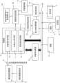
图2是图1的个人计算机的控制模块图。FIG. 2 is a control block diagram of the personal computer of FIG. 1 .
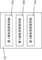
图3是表示图2的控制模块中包含的存储器内的体素标记保存部的结构的框图。FIG. 3 is a block diagram showing the configuration of a voxel label storage unit in a memory included in the control module of FIG. 2 .
图4是表示图2的控制模块中包含的存储器内的颜色信息保存部的结构的框图。4 is a block diagram showing the configuration of a color information storage unit in a memory included in the control module of FIG. 2 .
图5(a)以及(b)是图1的个人计算机的动作流程图。5( a ) and ( b ) are flowcharts of operations of the personal computer of FIG. 1 .
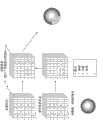
图6是说明图1的个人计算机的动作的示意图。FIG. 6 is a schematic diagram illustrating the operation of the personal computer of FIG. 1 .
图7(a)是表示在一般的切割模拟装置所显示的切割后的显示图像的一例的参考图。(b)是表示在肝脏周边存在的血管的一例的图。(c)是表示在图1的个人计算机所显示的切割后的显示图像的一例的图。FIG. 7( a ) is a reference diagram showing an example of a display image after cutting displayed on a general cutting simulation device. (b) is a diagram showing an example of blood vessels existing around the liver. (c) is a diagram showing an example of a cutout display image displayed on the personal computer of FIG. 1 .
具体实施方式Detailed ways
以下利用图1~图7(c)对本发明的一实施方式涉及的个人计算机(切割模拟装置)进行说明。Hereinafter, a personal computer (cutting simulation device) according to an embodiment of the present invention will be described using FIGS. 1 to 7( c ).
如图1所示,本实施方式涉及的个人计算机1具备显示器(显示部)2、各种输入部(键盘3、鼠标4、以及书写板5(参照图2))。As shown in FIG. 1 , a
显示器2显示X射线CT图像等的由多个断层图像形成的脏器(图1的例子中显示肾脏)等的三维图像,并且显示后述的切割模拟的结果。The
此外,如图2所示,个人计算机1在内部形成断层图像信息获取部6等的控制模块。In addition, as shown in FIG. 2 , the
断层图像信息获取部6经由体素信息提取部7而连接断层图像信息部8。也就是说,在断层图像信息部8中,被从拍摄CT或者MRI、PET等断层图像的设备提供断层图像信息,该断层图像信息通过体素信息提取部7而作为体素信息被提取。The tomographic image information acquisition unit 6 is connected to the tomographic
存储器9设置在个人计算机1内,具有体素信息保存部10、体素标记保存部11、以及颜色信息保存部12。此外,存储器9连接体绘制运算部13。The memory 9 is provided in the
体素信息保存部10保存从体素信息提取部7经由断层图像信息获取部6接收到的体素信息。The voxel
如图3所示,体素标记保存部11具有第1体素标记保存部11a、第2体素标记保存部11b、第3体素标记保存部11c。这些的第1~第3体素标记保存部11a~11c分别与后述的预先设定的CT值的范围、即成为显示对象的脏器对应地设置。例如,第1体素标记保存部11a与显示肝脏的CT值的范围对应,第2体素标记保存部11b与显示血管的CT值的范围对应,第3体素标记保存部11c与显示骨骼的CT值的范围对应。As shown in FIG. 3 , the voxel
如图4所示,颜色信息保存部12具有第1颜色信息保存部12a、第2颜色信息保存部12b、和第3颜色信息保存部12c。这些的第1~第3颜色信息保存部12a~12c与上述的第1~第3体素标记保存部11a~11c同样地,分别与后述的预先所设定的CT值的范围、即成为显示对象的脏器对应来设置。例如,第1颜色信息保存部12a与显示肝脏的CT值的范围对应,第2颜色信息保存部12b与显示血管的CT值的范围对应,第3颜色信息保存部12c与显示骨骼的CT值的范围对应。此时,第1~第3颜色信息保存部12a~12c中按成为显示对象的脏器、血管、骨骼等分别设定不同的颜色信息。例如,与肝脏对应的CT值的范围中保存红黑色的颜色信息,与血管对应的CT值的范围中保存红色的颜色信息,与骨骼对应的CT值的范围中保存白色的颜色信息。As shown in FIG. 4 , the color
再者,按成为显示对象的脏器、血管、骨骼所设定的CT值是将人体中的X射线吸收的程度数值化而得到的,表示为将水设为0的相对值(单位:HU)。例如,显示肝脏的CT值的范围是60~70HU,显示肾脏的CT值的范围是30~40HU,显示血液的CT值的范围是30~50HU,显示骨骼的CT值的范围是500~1000HU。Furthermore, the CT value set for each organ, blood vessel, and bone to be displayed is obtained by quantifying the degree of X-ray absorption in the human body, and expressed as a relative value with water as 0 (unit: HU ). For example, the range of CT values for displaying liver is 60-70HU, the range of CT values for displaying kidneys is 30-40HU, the range for displaying CT values of blood is 30-50HU, and the range of CT values for displaying bones is 500-1000HU.
体绘制运算部13基于体素信息保存部10中保存的体素信息、体素标记保存部11中保存的体素标记、和颜色信息保存部12中保存的颜色信息,获取与视线垂直、且Z方向的间隔固定的多幅的切片信息。然后,体绘制运算部13将该运算结果作为三维图像而显示在显示器2。此外,体绘制运算部13经由总线16而连接深度检测部15。Based on the voxel information stored in the voxel
深度检测部15测定后述的光线投射(ray casting)扫描距离,并且连接深度控制部17和体素标记设定部18。The depth detection unit 15 measures a ray casting scanning distance described later, and is connected to the
体素标记设定部18连接了体素标记保存部11和被切割体素标记算出显示部19。The voxel
总线16上除了连接上述的体绘制运算部13以及深度检测部15以外,还连接存储器9内的颜色信息保存部12、窗口坐标获取部20,基于从键盘3、鼠标4、书写板5等输入的内容,在显示器2显示三维图像等。In addition to connecting the above-mentioned volume
窗口坐标获取部20连接了深度检测部15和颜色信息设定部21。颜色信息设定部21连接了存储器9内的颜色信息保存部12。The window coordinate
图5(a)以及图5(b)表示用于进行本实施方式的切割模拟装置中的动作说明的控制流程。5( a ) and FIG. 5( b ) show a control flow for explaining operations in the cutting simulation device according to this embodiment.
在本实施方式的个人计算机1中,如图5(a)所示,首先,在S1中,如上述那样,输入来自断层图像信息部8的断层图像信息,该断层图像信息被提供给体素信息提取部7。In the
接着,在S2中,在体素信息提取部7中从断层图像信息提取体素信息。所提取的体素信息经由断层图像信息获取部6而保存在存储器9的体素信息保存部10中。体素信息保存部10中保存的体素信息例如是由I(x,y,z,α)构成的点的信息。此时,I是该点的亮度信息,x、y、z表示坐标点,α是透明度信息。Next, in S2 , the voxel information is extracted from the tomographic image information in the voxel
接下来,在S3中,体绘制运算部13基于体素信息保存部10所保存的体素信息,计算与视线垂直、且间隔固定的多个切片信息,来获取切片信息群。然后,切片信息群至少被暂时保存在体绘制运算部13内。Next, in S3 , the volume
再者,上述的与视线垂直的切片信息意味着与视线正交的面。例如,在将显示器2沿着铅锤方向立起的状态下,以使该显示器与脸面平行的状态进行观察时,切片信息成为与视线垂直的面。In addition, the above-mentioned slice information perpendicular to the line of sight means a plane perpendicular to the line of sight. For example, when the
这样得到的多个切片信息,如上述那样保存有由I(x,y,z,α)构成的点的信息。由此,切片信息中例如图6所示那样体素标记14在Z方向上配置多个。再者,例如图6所示的体素标记14的集合体被收纳在体素标记保存部11中。The pieces of slice information obtained in this way store point information consisting of I(x, y, z, α) as described above. Accordingly, in the slice information, a plurality of
接着,在S4中,在显示器2显示绘制像。此时,在显示器2中,通过使用鼠标4等来指定CT值的范围,以选择成为切割对象物的脏器(本实施方式中为肝脏22),如图7(a)等所示进行显示。Next, in S4 , the drawn image is displayed on the
再者,在图7(a)~图7(c)中,22为肝脏,23为血管,C表示后述的切割部分。也就是说,在本实施方式中,要实施用于对肝脏22进行手术的模拟。In addition, in FIGS. 7( a ) to 7 ( c ), 22 is a liver, 23 is a blood vessel, and C represents a cut portion described later. That is, in this embodiment, a simulation for performing an operation on the
在此,在实施一般的切割模拟时,如图7(a)所示,显示器2中显示将切割部分C中包含的肝脏22的一部分和血管23的一部分切割至规定深度之后的状态。Here, when a general cutting simulation is performed, as shown in FIG.
本实施方式中,在实施这种的肝脏22的手术模拟时,在显示器2的画面上,在实施切除肝脏22的一部分的手术之际,为了能够显示在该切割部分C如图7(b)所示的血管23是如何存在的,按以下的方式进行控制。In the present embodiment, when performing such an operation simulation of the
即,在S5中,使用鼠标4等分别设定与实施切割模拟时成为切割对象物的脏器等对应的CT值、与成为非切割对象物的血管23等对应的CT值。再者,切割对象物以及非切割对象物的设定可使用键盘3、鼠标4、或者书写板5等的任意来进行。That is, in S5 , the mouse 4 and the like are used to set the CT values corresponding to the organs and the like to be cut when the cutting simulation is performed, and the CT values corresponding to the
接下来,在S6中,使用鼠标4等来进行切割指示。再者,作为输入切割指示的输入部,可以与切割对象物以及非切割对象物的设定同样,使用键盘3、鼠标4、或者书写板5等的任意来进行。Next, in S6, cutting instructions are given using the mouse 4 or the like. In addition, as the input unit for inputting the cutting instruction, any of the
作为具体的切割指示的输入方法,通过使鼠标4在桌子上在水平方向移动,来使显示器2上显示的光标在肝脏22之上左右或者上下往复。As a specific method of inputting the cutting instruction, the mouse 4 is moved horizontally on the table, so that the cursor displayed on the
此时,鼠标4的左右方向、上下方向的运动在窗口坐标获取部20中被检测出来。然后,该信息经由深度检测部15被传送至体素标记设定部18、体素标记保存部11。由此,进行考虑了肝脏22和血管23的Z方向的位置的切割。At this time, the movement of the mouse 4 in the left and right directions and in the up and down direction is detected by the window coordinate
具体而言,在体绘制运算部13中,在与视线垂直的方向以一定间隔对体素信息进行采样(称为光线投射。)。然后,体绘制运算部13针对鼠标4移动过程中求出的全部点,在S7中由深度检测部15检测各个的光线投射扫描距离(深度)。Specifically, in the volume
然后,在S8中,判定其深度的变化率是否在一定的范围内。Then, in S8, it is judged whether the rate of change of the depth is within a certain range.
具体而言,对深度检测部15中测定的光线投射扫描距离d进行汇总,算出其梯度d。然后,进行梯度

对于阈值T,每次切割处理基于跟前的n个切割点的梯度平均和倍数系数m来决定阈值Ti。For the threshold T, each cutting process determines the threshold Ti based on the gradient average of the previous n cutting points and the multiple coefficient m.
再者,对于倍数系数m和切割点n,例如将m设定为5左右,将n设定为10等,这些数值只要根据对象图像来适当设定即可。Note that for the multiplication factor m and the cutting point n, for example, m is set to about 5, n is set to 10, etc., and these numerical values may be appropriately set according to the target image.
本实施方式中,如以上,将比较基于跟前的n个切割点的梯度平均和倍数系数m而算出的阈值Ti和梯度d而得到结果作为变化率,来决定是否实施切割。In the present embodiment, as above, the threshold value Ti calculated based on the gradient average of the previous n cutting points and the multiplier m is compared with the gradient d and get the result as the rate of change to decide whether to implement cutting.
再者,变化率的算出方法并不限定于此,只要能够确认梯度的变化状态,可以使用任意的计算式。In addition, the method of calculating the change rate is not limited to this, and any calculation formula can be used as long as the change state of the gradient can be confirmed.
此外,对于阈值T,也优选根据成为切割对象的各个脏器的特征而适当改变。由此,能够进一步提高避免误切割的精度。In addition, it is also preferable to appropriately change the threshold T according to the characteristics of each organ to be cut. Thereby, it is possible to further improve the accuracy of avoiding erroneous cutting.
在此,在上述的切割处理中,将具有规定阈值以上的变化率的点作为无效的切割点,深度控制部17对体素标记设定部18输出指示。由此,体素标记的更新被中止,不实施切割。由此,在深度检测部15检测出因医生等使用者的误操作而深度位置急剧变化的切割点的情况下,能够避免误切割。Here, in the cutting process described above, the
此时,实施切割意味着体素标记设定部18更新体素标记并保存在体素标记保存部11中。也就是说,在不实施切割的情况下,体素标记不变化。At this time, cutting means that the voxel
因此,即便使鼠标4在肝脏22上拖动的情况下,也能够避免其里侧存在的背骨等被误切割。并且,在该情况下,根据鼠标4的左右方向、上下方向的拖动次数,显示肝脏22的一部分被切割之后的图像。Therefore, even when the mouse 4 is dragged on the
再者,窗口坐标获取部20的信息经由颜色信息设定部21而传送至颜色信息保存部12,通过肝脏22的颜色变化从而能够识别肝脏22被切割之后的状态。在此,颜色信息设定部是指使用了所谓的查表的变换部。也就是说,本实施方式的个人计算机1中,如上述那样保存有由I(x,y,z,α)构成的点的信息,针对肝脏22的表面和内部由颜色信息设定部21预先设定不同的亮度信息和颜色信息。由此,在从表面切割肝脏22地进行操作的情况下,根据其切割程度,切割部分C的颜色以与其周围的颜色明显不同的颜色进行显示。Furthermore, the information of the window coordinate
接着,在S10以及S11中,与作为切割对象物的肝脏22的切割部分C对应的部分的体素信息(体素标记的值)和颜色信息标记被更新。Next, in S10 and S11 , the voxel information (the value of the voxel flag) and the color information flag of the part corresponding to the dissected part C of the
图6表示进行切割处理的情况下的体素标记和颜色信息标记的更新状态,最表面的体素标记14的大部分表示“1”的状态、也就是表示肝脏22的表面状态。此外,图6中“0”的部分表示被切割的体素。FIG. 6 shows the voxel flags and the updated states of the color information flags when the cutting process is performed, and most of the outermost voxel flags 14 indicate a state of “1”, that is, the surface state of the
本实施方式中,如上述那样,将肝脏22以及血管23各自对应的体素标记保存在体素标记保存部11的第1体素标记保存部11a、11b内,将颜色信息标记保存在颜色信息保存部12的第1颜色信息保存部12a、12b内。In this embodiment, as described above, the voxel labels corresponding to the
由此,在使用鼠标4等进行指示输入以切割肝脏22的一部分的情况下,由于肝脏22是切割对象物,因此在S10中,体素标记设定部18更新肝脏22的切割部分C的体素标记值的同时,在S11中更新肝脏22的切割部分C的颜色信息标记。由此,在显示器2的显示画面上,如图7(a)所示切除了肝脏22的切割部分C之后的肝脏22的内部以与肝脏22的表面略有不同的颜色被显示。Thus, when an instruction is input using the mouse 4 or the like to cut a part of the
在此,在本实施方式中,对于在上述的S5中被指定为非切割对象物的血管23,即便包含在肝脏22的切割部分C的情况下,通过在切割部分C置换图7(b)所示的血管23的体素标记,从而如图7(c)所示那样,没有被切割处理地在切割部分C的部分被显示。Here, in the present embodiment, even if the
具体而言,在S12中,对于被指定为非切割对象物的血管23,在切割处理后,置换切割部分C处的体素信息以及颜色信息和血管23的体素信息以及颜色信息。Specifically, in S12 , the voxel information and color information of the cut portion C and the voxel information and color information of the
由此,在显示器2的显示画面上,以仅被输入了切割指示的切割部分C中包含的肝脏22的部分被切割的状态进行显示,对于被指定为非切割对象物的血管23,以没有被切割的状态进行显示。其结果,实施手术模拟的医生等在切除作为切割对象物的肝脏22的一部(切割部分C)时,能够在多个人之间共享信息并且能够同时识别出在切割部分C内血管23位于何处。Thus, on the display screen of the
此外,本实施方式中,由于对作为切割对象物的肝脏22和作为非切割对象物的血管23设定了彼此不同的颜色信息,因此,如图7(c)所示,能够更为正确地识别以红色显示的血管23的位置、形状。In addition, in the present embodiment, since different color information is set for the
再有,本实施方式中,例如通过鼠标按键的ON/OFF能够进行切割开始/结束的切换,因此医生等使用者通过一边观察三维显示画面一边在鼠标按键ON时对鼠标4进行拖拽操作,能够容易且连续地进行希望的肝脏22的一部分区域的切割。Furthermore, in the present embodiment, for example, the ON/OFF of the mouse button can be used to switch the cutting start/end, so users such as doctors can drag the mouse 4 while viewing the three-dimensional display screen while the mouse button is ON. A desired part of the
此外,本实施方式中,在个人计算机1的电源OFF时能够进行存储器9的更新。医生等使用者若按下鼠标4的鼠标按键的同时开始拖拽,则在存储器9内仅更新体绘制运算部13的信息,能够以视觉方式对使用者提供交互式的切割功能。In addition, in the present embodiment, the memory 9 can be updated when the power of the
此时,不更新存储器9,暂时保存好工作中的体标记。然后,在使用者离开鼠标按键时,将暂时保存起来的存储器内容反映至存储器9。由此,使用者在一次的拖拽操作中,能够显示为从作为切割对象物的肝脏22的表面被切割至一定深度,能够防止以被过度切割的状态进行显示。At this time, the memory 9 is not updated, and the volume tag in operation is temporarily saved. Then, when the user releases the mouse button, the temporarily stored memory content is reflected in the memory 9 . Thereby, the user can display that the
此外,本实施方式中,将体素标记设定与初期的体素信息相同尺寸,但为了表现更为细致的切割,也可以作为更小尺寸的体素标记来生成体素标记。在本方法中,也可以不直接编辑体素信息,而使体素标记具有时刻信息,以应对Undo(返回原始)、Redo(再输入)等的操作。In addition, in this embodiment, the voxel label is set to have the same size as the initial voxel information, but in order to express a finer cut, the voxel label may be generated as a voxel label of a smaller size. In this method, the voxel information may not be directly edited, but the voxel mark has time information, so as to cope with operations such as Undo (return to original), Redo (re-input).
此外,本实施方式中,仅通过在平面上移动鼠标4就能够进行手术模拟。由此,根据这一点也能够进行适当的手术模拟。In addition, in this embodiment, surgery simulation can be performed only by moving the mouse 4 on a plane. Accordingly, appropriate surgery simulation can be performed also from this point.
[其他实施方式][Other implementations]
以上,说明了本发明的一实施方式,但本发明并不限定于上述实施方式,在不脱离发明的主旨的范围内可进行各种变更。As mentioned above, although one embodiment of this invention was described, this invention is not limited to the said embodiment, Various changes are possible in the range which does not deviate from the summary of invention.
(A)(A)
在上述实施方式中,举出了作为切割模拟装置来实现本发明的例子进行了说明。不过,本发明并不限定于此。In the above-mentioned embodiments, an example in which the present invention is realized as a cutting simulation device has been described. However, the present invention is not limited thereto.
例如,可作为使计算机执行图5(a)以及图5(b)所示的控制方法的切割模拟程序,来实现本发明。For example, the present invention can be implemented as a cutting simulation program that causes a computer to execute the control methods shown in FIG. 5( a ) and FIG. 5( b ).
再有,可作为保存该切割模拟程序的记录介质,来实现本发明。Furthermore, the present invention can be realized as a recording medium storing the cutting simulation program.
(B)(B)
上述实施方式中,对于被指定为非切割对象物的血管23,在切割处理后,控制为置换切割部分C处的体素信息以及颜色信息和血管23的体素信息以及颜色信息,以此为例进行了说明。但是,本发明并不限定于此。In the above-mentioned embodiment, for the
例如,也可以控制为仅将切割部分的体素信息置换为非切割对象物的体素信息。For example, it may be controlled so that only the voxel information of the cut part is replaced with the voxel information of the non-cut object.
其中,如图7(c)所示,对于将作为非切割对象物的血管显示得容易看清这一点,进一步优选控制成也一并置换被设定了与切割对象物不同的颜色信息的非切割对象物的颜色信息。Among them, as shown in FIG. 7( c ), in order to display blood vessels that are not objects to be cut clearly, it is further preferable to control to also replace the non-vessels that are set with color information different from the objects to be cut. Color information of the object to be cut.
(C)(C)
上述实施方式中,作为用于形成三维图像的断层图像信息,以使用了X射线CT图像的例子进行了说明。但是,本发明并不限定于此。In the above-described embodiments, an example in which X-ray CT images are used as tomographic image information for forming a three-dimensional image has been described. However, the present invention is not limited thereto.
例如,也可以利用通过没有使用放射线的核磁共振图像(MRI)而获取的断层图像信息等来形成三维图像。For example, it is also possible to form a three-dimensional image using tomographic image information or the like acquired by a nuclear magnetic resonance image (MRI) that does not use radiation.
(D)(D)
上述实施方式中,作为本发明涉及的切割模拟的一例,列举了作为切割对象物而指定肝脏22、作为非切割对象物而指定了血管23的例子进行了说明。但是,本发明并不限定于此。In the above-described embodiment, an example in which the
例如,也可以将胃、肺、肾脏、胰脏、大肠、小肠、十二指肠等的其他脏器或骨骼设定为切割对象物。或者,在实施对神经或骨骼周边存在的肿瘤等进行切割的模拟的情况下,作为非切割物也可以设定神经、或者骨骼。For example, other organs or bones such as the stomach, lungs, kidneys, pancreas, large intestine, small intestine, and duodenum may be set as cutting objects. Alternatively, when performing a simulation of cutting a tumor or the like around nerves or bones, nerves or bones may be set as non-cut objects.
再有,作为非切割对象物甚至指定了血管的种类的情况下,通过适当指定CT值,也能够以不同颜色区分来进行显示使得动脉和静脉可分辨。Furthermore, even when the type of blood vessel is designated as a non-cutting object, by appropriately designating the CT value, it is also possible to distinguish and display arteries and veins in different colors.
(E)(E)
上述实施方式中,列举了改变由鼠标4进行了切割指示的体素标记14中的切割对象物的亮度信息以及颜色信息的双方来进行说明。但是,本发明并不限定于此。In the above-mentioned embodiment, both the luminance information and the color information of the object to be cut in the
例如,也可以进行显示控制使得显示对象物的亮度信息以及颜色信息之中的至少一方变化。For example, display control may be performed such that at least one of brightness information and color information of a display object is changed.
(F)(F)
上述实施方式中,作为对被切割的体素的体积进行计算的被切割体素标记算出显示部19的输出,也可以将由鼠标4输入的切割量(体积)显示在显示器2。In the above embodiment, the cut amount (volume) input by the mouse 4 may be displayed on the
或者,也可以将由鼠标4输入的切割深度显示在显示器2。Alternatively, the cutting depth input from the mouse 4 may be displayed on the
(G)(G)
上述实施方式中,列举了一边观察在显示器2的显示画面上显示的三维图像一边实施切割模拟的例子来进行说明。但是,本发明并不限于此。In the above-described embodiment, an example in which cutting simulation is performed while observing a three-dimensional image displayed on the display screen of the
例如,也可以在表示体绘制结果的三维图像上,将二维的断层图像追加投影至显示器,针对二维的断层图像实施切割动作。For example, a two-dimensional tomographic image may be additionally projected onto a display on a three-dimensional image representing a volume rendering result, and a cutting operation may be performed on the two-dimensional tomographic image.
该情况下,也能够实施切割模拟使得切割动作被反映在三维图像上。Also in this case, cutting simulation can be performed so that the cutting action is reflected on the three-dimensional image.
(H)(H)
本发明中,也可以设置颜色信息设定部21,该颜色信息设定部21将体素信息保存部10中保存的体素信息在显示器2变换为二维或者三维图像进行显示,并且变更在显示器2显示的切割对象部中的由鼠标4指示的部分的颜色信息。In the present invention, a color
也就是说,对于显示器2上所显示的切割对象部,例如只要对作为医生而在意的部分有意地赋予颜色,并在该状态下将体素标记14的集合体保存在体素标记保存部11中即可。That is, for the part to be cut displayed on the
由此,根据从该信息得出的多方面的显示中反映出附加了颜色信息的信息。由此,能够立体地从全方位观察切割对象物中的在意的部分,并且还能够进行该切割模拟。In this way, the information to which the color information is added is reflected in the multifaceted display derived from this information. Accordingly, it is possible to three-dimensionally observe a portion of interest in the object to be cut from all directions, and to perform the cutting simulation.
(I)(I)
本发明中,也能够模拟内窥镜手术。该情况下,体绘制运算部13中将内窥镜所设置的鱼眼透镜等的聚光特性用作坐标变换表即可。In the present invention, it is also possible to simulate endoscopic surgery. In this case, the volume
(J)(J)
本发明中,也可以具有多个视点,将按各视点的每一个生成的体绘制运算部13的输出图像存储在多个存储器中,将来自存储器的输出依次显示在显示器,以生成立体图像。In the present invention, there may be a plurality of viewpoints, the output images of the volume
该情况下,也可以使用与各个图像输出同步的液晶眼镜等。In this case, liquid crystal glasses or the like that are synchronized with each image output may be used.
【工业上的可利用性】【Industrial availability】
本发明的切割模拟装置通过使切割对象物的内部存在的血管、神经等可视化,达到了能够详细地表现实际的手术过程的这一效果,因此作为实施外科手术的切割模拟的装置而被期待广泛应用。The incision simulation device of the present invention achieves the effect of expressing the actual surgical procedure in detail by visualizing the blood vessels and nerves existing inside the incision object, so it is expected to be widely used as a device for performing incision simulation in surgical operations. application.
【符号的说明】【Description of symbols】
1 个人计算机(切割模拟装置)1 personal computer (cutting simulation device)
2 显示器(显示部)2 Display (display part)
3 键盘(输入部)3 keyboard (input part)
4 鼠标(输入部)4 mouse (input part)
5 书写板(输入部)5 writing pad (input part)
6 断层图像信息获取部6 Tomographic Image Information Acquisition Department
7 体素信息提取部7 Voxel Information Extraction Department
8 断层图像信息部8 Tomographic Image Information Department
9 存储器9 memory
10 体素信息保存部10 voxel information storage department
11 体素标记保存部11 Voxel marker preservation department
11a~11c 第1~第3体素标记保存部11a~11c 1st to 3rd voxel marker storage part
12 颜色信息保存部12 Color Information Storage Department
13 体绘制运算部13 Volume Rendering Computing Department
14 体素标记14 voxel markers
15 深度检测部15 Depth Inspection Department
16 总线16 bus
17 深度控制部17 Depth Control Department
18 体素标记设定部(显示控制部)18 Voxel marker setting part (display control part)
19 被切割体素标记算出显示部19 Cut voxel mark calculation display part
20 窗口坐标获取部20 Window coordinate acquisition part
21 颜色信息设定部21 Color Information Setting Department
21a~21c 第1~第3颜色信息设定部21a~21c 1st~3rd color information setting part
22 肝脏22 liver
23 血管23 blood vessels
C 切割部分C cutting part
Claims (7)
Applications Claiming Priority (3)
| Application Number | Priority Date | Filing Date | Title |
|---|---|---|---|
| JP2011-165453 | 2011-07-28 | ||
| JP2011165453 | 2011-07-28 | ||
| PCT/JP2012/004378WO2013014868A1 (en) | 2011-07-28 | 2012-07-05 | Cutting simulation device and cutting simulation program |
Publications (1)
| Publication Number | Publication Date |
|---|---|
| CN103702613Atrue CN103702613A (en) | 2014-04-02 |
Family
ID=47600746
Family Applications (1)
| Application Number | Title | Priority Date | Filing Date |
|---|---|---|---|
| CN201280036433.1APendingCN103702613A (en) | 2011-07-28 | 2012-07-05 | Cutting simulation device and cutting simulation program |
Country Status (5)
| Country | Link |
|---|---|
| US (1) | US20140193789A1 (en) |
| EP (1) | EP2737854A1 (en) |
| JP (1) | JP6051158B2 (en) |
| CN (1) | CN103702613A (en) |
| WO (1) | WO2013014868A1 (en) |
Cited By (3)
| Publication number | Priority date | Publication date | Assignee | Title |
|---|---|---|---|---|
| CN108472001A (en)* | 2015-10-30 | 2018-08-31 | 佳能株式会社 | Medical image processing device, its control method and program |
| CN112437642A (en)* | 2018-07-26 | 2021-03-02 | 索尼公司 | Information processing apparatus, information processing method and program |
| US11200977B2 (en) | 2015-10-30 | 2021-12-14 | Canon Kabushiki Kaisha | Medical image processing apparatus, control method for the same, and program |
Families Citing this family (8)
| Publication number | Priority date | Publication date | Assignee | Title |
|---|---|---|---|---|
| WO2014200017A1 (en)* | 2013-06-11 | 2014-12-18 | Tanji Atsushi | Bone cutting assistance system, information processing device, image processing method, and image processing program |
| JP6698824B2 (en)* | 2016-04-11 | 2020-05-27 | 富士フイルム株式会社 | Image display control device, method and program |
| WO2018118858A1 (en) | 2016-12-19 | 2018-06-28 | National Board Of Medical Examiners | Medical training and performance assessment instruments, methods, and systems |
| JP7235519B2 (en)* | 2019-01-29 | 2023-03-08 | ザイオソフト株式会社 | MEDICAL IMAGE PROCESSING APPARATUS, MEDICAL IMAGE PROCESSING METHOD, AND MEDICAL IMAGE PROCESSING PROGRAM |
| CN111179402B (en)* | 2020-01-02 | 2023-07-14 | 竞技世界(北京)网络技术有限公司 | Rendering method, device and system of target object |
| CN111489442A (en)* | 2020-03-27 | 2020-08-04 | 杭州群核信息技术有限公司 | Operating system and method for accurately segmenting object |
| KR102822044B1 (en)* | 2022-08-12 | 2025-06-18 | 주식회사 데카사이트 | Method and Apparatus for Providing of Surgery Simulation |
| KR20250086927A (en)* | 2023-12-07 | 2025-06-16 | 고려대학교 산학협력단 | Apparatus for incision simulation modeling based on virtual environment |
Citations (4)
| Publication number | Priority date | Publication date | Assignee | Title |
|---|---|---|---|---|
| JPS6437678A (en)* | 1987-08-03 | 1989-02-08 | Toshiba Corp | Three-dimensional image processor |
| JPH0973556A (en)* | 1995-09-04 | 1997-03-18 | Toshiba Corp | Image processing device |
| JP2003010172A (en)* | 2001-07-04 | 2003-01-14 | Hitachi Medical Corp | Method and device for extracting and displaying specific area of internal organ |
| JP2003339644A (en)* | 2002-05-24 | 2003-12-02 | Hitachi Medical Corp | Extraction and display device of resection area of internal organ |
Family Cites Families (5)
| Publication number | Priority date | Publication date | Assignee | Title |
|---|---|---|---|---|
| JP3198130B2 (en) | 1991-11-07 | 2001-08-13 | 株式会社東芝 | Surgery simulation system |
| US6331116B1 (en)* | 1996-09-16 | 2001-12-18 | The Research Foundation Of State University Of New York | System and method for performing a three-dimensional virtual segmentation and examination |
| US6826297B2 (en)* | 2001-05-18 | 2004-11-30 | Terarecon, Inc. | Displaying three-dimensional medical images |
| JP4776834B2 (en) | 2001-09-19 | 2011-09-21 | 東芝医用システムエンジニアリング株式会社 | Image processing device |
| JP3836097B2 (en)* | 2003-09-19 | 2006-10-18 | ザイオソフト株式会社 | MEDICAL IMAGE GENERATION DEVICE AND METHOD, AND PROGRAM |
- 2012
- 2012-07-05JPJP2013525561Apatent/JP6051158B2/enactiveActive
- 2012-07-05CNCN201280036433.1Apatent/CN103702613A/enactivePending
- 2012-07-05EPEP12818406.6Apatent/EP2737854A1/ennot_activeWithdrawn
- 2012-07-05USUS14/235,246patent/US20140193789A1/ennot_activeAbandoned
- 2012-07-05WOPCT/JP2012/004378patent/WO2013014868A1/enactiveApplication Filing
Patent Citations (4)
| Publication number | Priority date | Publication date | Assignee | Title |
|---|---|---|---|---|
| JPS6437678A (en)* | 1987-08-03 | 1989-02-08 | Toshiba Corp | Three-dimensional image processor |
| JPH0973556A (en)* | 1995-09-04 | 1997-03-18 | Toshiba Corp | Image processing device |
| JP2003010172A (en)* | 2001-07-04 | 2003-01-14 | Hitachi Medical Corp | Method and device for extracting and displaying specific area of internal organ |
| JP2003339644A (en)* | 2002-05-24 | 2003-12-02 | Hitachi Medical Corp | Extraction and display device of resection area of internal organ |
Cited By (4)
| Publication number | Priority date | Publication date | Assignee | Title |
|---|---|---|---|---|
| CN108472001A (en)* | 2015-10-30 | 2018-08-31 | 佳能株式会社 | Medical image processing device, its control method and program |
| US11200977B2 (en) | 2015-10-30 | 2021-12-14 | Canon Kabushiki Kaisha | Medical image processing apparatus, control method for the same, and program |
| CN112437642A (en)* | 2018-07-26 | 2021-03-02 | 索尼公司 | Information processing apparatus, information processing method and program |
| US12053254B2 (en) | 2018-07-26 | 2024-08-06 | Sony Corporation | Information processing apparatus and information processing method |
Also Published As
| Publication number | Publication date |
|---|---|
| JP6051158B2 (en) | 2016-12-27 |
| JPWO2013014868A1 (en) | 2015-02-23 |
| US20140193789A1 (en) | 2014-07-10 |
| WO2013014868A1 (en) | 2013-01-31 |
| EP2737854A1 (en) | 2014-06-04 |
Similar Documents
| Publication | Publication Date | Title |
|---|---|---|
| US12333087B2 (en) | Interactive 3D cursor | |
| CN103702613A (en) | Cutting simulation device and cutting simulation program | |
| US11547499B2 (en) | Dynamic and interactive navigation in a surgical environment | |
| EP2573735B1 (en) | Endoscopic image processing device, method and program | |
| JP6670595B2 (en) | Medical image processing equipment | |
| US20110245660A1 (en) | Projection image generation apparatus and method, and computer readable recording medium on which is recorded program for the same | |
| EP2413285A2 (en) | Diagnosis assisting apparatus, diagnosis assisting program, and diagnosis assisting method | |
| CN113645896A (en) | System for surgical planning, surgical navigation and imaging | |
| CN109157284A (en) | A kind of brain tumor medical image three-dimensional reconstruction shows exchange method and system | |
| WO2018030015A1 (en) | Resection process estimation device and resection process navigation system | |
| WO2011118208A1 (en) | Cutting simulation device | |
| US10803645B2 (en) | Visualization of anatomical cavities | |
| CN106028943A (en) | Ultrasonic virtual endoscopic imaging system and method, and apparatus thereof | |
| CN102609623A (en) | Ablation therapy image guide device with two-dimensional image processing device | |
| JP2009022307A (en) | Medical image display device and its program | |
| CN102592060A (en) | Method for guiding equipment to process images by means of ablation treatment images | |
| RU2684760C1 (en) | Method and system for pre-operative modeling of medical procedure | |
| Skalski et al. | 3D segmentation and visualisation of mediastinal structures adjacent to tracheobronchial tree from CT data | |
| EP3326537A1 (en) | Visualization of distances to walls of anatomical cavities | |
| CN120457494A (en) | A system for virtual reality-based preoperative surgical planning | |
| Agrafiotis et al. | Virtual liver biopsy: Image processing and 3d visualization | |
| Wilson et al. | Developing an Application to Provide Interactive Threedimensional Visualisation of Bone Fractures. |
Legal Events
| Date | Code | Title | Description |
|---|---|---|---|
| C06 | Publication | ||
| PB01 | Publication | ||
| C10 | Entry into substantive examination | ||
| SE01 | Entry into force of request for substantive examination | ||
| ASS | Succession or assignment of patent right | Owner name:PANASONIC HEALTHCARE + MEDICAL EQUIPMENT CO., LTD. Free format text:FORMER OWNER: MATSUSHITA ELECTRIC INDUSTRIAL CO, LTD. Effective date:20140918 | |
| C41 | Transfer of patent application or patent right or utility model | ||
| TA01 | Transfer of patent application right | Effective date of registration:20140918 Address after:Japan Ehime Prefecture Applicant after:Panasonic Healthcare Co., Ltd Address before:Osaka Japan Applicant before:Matsushita Electric Industrial Co., Ltd. | |
| ASS | Succession or assignment of patent right | Owner name:PANASONIC HEALTHCARE HOLDINGS CO., LTD. Free format text:FORMER OWNER: PANASONIC HEALTHCARE + MEDICAL EQUIPMENT CO., LTD. Effective date:20150106 | |
| C41 | Transfer of patent application or patent right or utility model | ||
| TA01 | Transfer of patent application right | Effective date of registration:20150106 Address after:Tokyo, Japan Applicant after:Panasonic's health medical treatment is controlled interest Co., Ltd. Address before:Japan Ehime Prefecture Applicant before:Panasonic Healthcare Co., Ltd | |
| C02 | Deemed withdrawal of patent application after publication (patent law 2001) | ||
| WD01 | Invention patent application deemed withdrawn after publication | Application publication date:20140402 |