CN103654784A - Method for acquiring movement of patient during medical imaging examination - Google Patents
Method for acquiring movement of patient during medical imaging examinationDownload PDFInfo
- Publication number
- CN103654784A CN103654784ACN201310416311.1ACN201310416311ACN103654784ACN 103654784 ACN103654784 ACN 103654784ACN 201310416311 ACN201310416311 ACN 201310416311ACN 103654784 ACN103654784 ACN 103654784A
- Authority
- CN
- China
- Prior art keywords
- patient
- motion
- medical imaging
- motion sensor
- examination
- Prior art date
- Legal status (The legal status is an assumption and is not a legal conclusion. Google has not performed a legal analysis and makes no representation as to the accuracy of the status listed.)
- Granted
Links
Images

Classifications
- A—HUMAN NECESSITIES
- A61—MEDICAL OR VETERINARY SCIENCE; HYGIENE
- A61B—DIAGNOSIS; SURGERY; IDENTIFICATION
- A61B5/00—Measuring for diagnostic purposes; Identification of persons
- A61B5/72—Signal processing specially adapted for physiological signals or for diagnostic purposes
- A61B5/7271—Specific aspects of physiological measurement analysis
- A61B5/7285—Specific aspects of physiological measurement analysis for synchronizing or triggering a physiological measurement or image acquisition with a physiological event or waveform, e.g. an ECG signal
- A61B5/7292—Prospective gating, i.e. predicting the occurrence of a physiological event for use as a synchronisation signal
- A—HUMAN NECESSITIES
- A61—MEDICAL OR VETERINARY SCIENCE; HYGIENE
- A61B—DIAGNOSIS; SURGERY; IDENTIFICATION
- A61B5/00—Measuring for diagnostic purposes; Identification of persons
- A61B5/72—Signal processing specially adapted for physiological signals or for diagnostic purposes
- A61B5/7203—Signal processing specially adapted for physiological signals or for diagnostic purposes for noise prevention, reduction or removal
- A61B5/7207—Signal processing specially adapted for physiological signals or for diagnostic purposes for noise prevention, reduction or removal of noise induced by motion artifacts
- A61B5/721—Signal processing specially adapted for physiological signals or for diagnostic purposes for noise prevention, reduction or removal of noise induced by motion artifacts using a separate sensor to detect motion or using motion information derived from signals other than the physiological signal to be measured
- A—HUMAN NECESSITIES
- A61—MEDICAL OR VETERINARY SCIENCE; HYGIENE
- A61B—DIAGNOSIS; SURGERY; IDENTIFICATION
- A61B5/00—Measuring for diagnostic purposes; Identification of persons
- A61B5/05—Detecting, measuring or recording for diagnosis by means of electric currents or magnetic fields; Measuring using microwaves or radio waves
- A61B5/055—Detecting, measuring or recording for diagnosis by means of electric currents or magnetic fields; Measuring using microwaves or radio waves involving electronic [EMR] or nuclear [NMR] magnetic resonance, e.g. magnetic resonance imaging
- A—HUMAN NECESSITIES
- A61—MEDICAL OR VETERINARY SCIENCE; HYGIENE
- A61B—DIAGNOSIS; SURGERY; IDENTIFICATION
- A61B5/00—Measuring for diagnostic purposes; Identification of persons
- A61B5/103—Measuring devices for testing the shape, pattern, colour, size or movement of the body or parts thereof, for diagnostic purposes
- A61B5/11—Measuring movement of the entire body or parts thereof, e.g. head or hand tremor or mobility of a limb
- A61B5/1126—Measuring movement of the entire body or parts thereof, e.g. head or hand tremor or mobility of a limb using a particular sensing technique
- A61B5/1128—Measuring movement of the entire body or parts thereof, e.g. head or hand tremor or mobility of a limb using a particular sensing technique using image analysis
- A—HUMAN NECESSITIES
- A61—MEDICAL OR VETERINARY SCIENCE; HYGIENE
- A61B—DIAGNOSIS; SURGERY; IDENTIFICATION
- A61B6/00—Apparatus or devices for radiation diagnosis; Apparatus or devices for radiation diagnosis combined with radiation therapy equipment
- A61B6/52—Devices using data or image processing specially adapted for radiation diagnosis
- A61B6/5258—Devices using data or image processing specially adapted for radiation diagnosis involving detection or reduction of artifacts or noise
- A61B6/5264—Devices using data or image processing specially adapted for radiation diagnosis involving detection or reduction of artifacts or noise due to motion
- A61B6/527—Devices using data or image processing specially adapted for radiation diagnosis involving detection or reduction of artifacts or noise due to motion using data from a motion artifact sensor
- G—PHYSICS
- G01—MEASURING; TESTING
- G01R—MEASURING ELECTRIC VARIABLES; MEASURING MAGNETIC VARIABLES
- G01R33/00—Arrangements or instruments for measuring magnetic variables
- G01R33/20—Arrangements or instruments for measuring magnetic variables involving magnetic resonance
- G01R33/44—Arrangements or instruments for measuring magnetic variables involving magnetic resonance using nuclear magnetic resonance [NMR]
- G01R33/48—NMR imaging systems
- G01R33/54—Signal processing systems, e.g. using pulse sequences ; Generation or control of pulse sequences; Operator console
- G01R33/56—Image enhancement or correction, e.g. subtraction or averaging techniques, e.g. improvement of signal-to-noise ratio and resolution
- G01R33/565—Correction of image distortions, e.g. due to magnetic field inhomogeneities
- G01R33/56509—Correction of image distortions, e.g. due to magnetic field inhomogeneities due to motion, displacement or flow, e.g. gradient moment nulling
- G—PHYSICS
- G16—INFORMATION AND COMMUNICATION TECHNOLOGY [ICT] SPECIALLY ADAPTED FOR SPECIFIC APPLICATION FIELDS
- G16H—HEALTHCARE INFORMATICS, i.e. INFORMATION AND COMMUNICATION TECHNOLOGY [ICT] SPECIALLY ADAPTED FOR THE HANDLING OR PROCESSING OF MEDICAL OR HEALTHCARE DATA
- G16H40/00—ICT specially adapted for the management or administration of healthcare resources or facilities; ICT specially adapted for the management or operation of medical equipment or devices
- G16H40/60—ICT specially adapted for the management or administration of healthcare resources or facilities; ICT specially adapted for the management or operation of medical equipment or devices for the operation of medical equipment or devices
- G16H40/63—ICT specially adapted for the management or administration of healthcare resources or facilities; ICT specially adapted for the management or operation of medical equipment or devices for the operation of medical equipment or devices for local operation
- G—PHYSICS
- G01—MEASURING; TESTING
- G01R—MEASURING ELECTRIC VARIABLES; MEASURING MAGNETIC VARIABLES
- G01R33/00—Arrangements or instruments for measuring magnetic variables
- G01R33/20—Arrangements or instruments for measuring magnetic variables involving magnetic resonance
- G01R33/28—Details of apparatus provided for in groups G01R33/44 - G01R33/64
- G01R33/283—Intercom or optical viewing arrangements, structurally associated with NMR apparatus
Landscapes
- Health & Medical Sciences (AREA)
- Engineering & Computer Science (AREA)
- Life Sciences & Earth Sciences (AREA)
- Physics & Mathematics (AREA)
- Biomedical Technology (AREA)
- General Health & Medical Sciences (AREA)
- Medical Informatics (AREA)
- Public Health (AREA)
- Nuclear Medicine, Radiotherapy & Molecular Imaging (AREA)
- Animal Behavior & Ethology (AREA)
- Veterinary Medicine (AREA)
- Biophysics (AREA)
- Heart & Thoracic Surgery (AREA)
- Molecular Biology (AREA)
- Surgery (AREA)
- Pathology (AREA)
- Radiology & Medical Imaging (AREA)
- Signal Processing (AREA)
- Computer Vision & Pattern Recognition (AREA)
- High Energy & Nuclear Physics (AREA)
- Physiology (AREA)
- Psychiatry (AREA)
- Artificial Intelligence (AREA)
- Epidemiology (AREA)
- Business, Economics & Management (AREA)
- General Business, Economics & Management (AREA)
- Optics & Photonics (AREA)
- Primary Health Care (AREA)
- Condensed Matter Physics & Semiconductors (AREA)
- General Physics & Mathematics (AREA)
- Dentistry (AREA)
- Oral & Maxillofacial Surgery (AREA)
- Magnetic Resonance Imaging Apparatus (AREA)
Abstract
Translated fromChinese
Description
Translated fromChinese技术领域technical field
本发明涉及一种用于在借助医学成像装置、尤其是磁共振装置进行医学成像检查、尤其是磁共振检查期间采集患者的运动的方法。The invention relates to a method for detecting the movement of a patient during a medical imaging examination, in particular a magnetic resonance examination, by means of a medical imaging apparatus, in particular a magnetic resonance apparatus.
背景技术Background technique
对于磁共振成像重要的是,患者在磁共振检查的整个期间、尤其在该患者的对于磁共振检查重要的区域中不运动。患者在磁共振检查期间的运动会在磁共振图像中产生伪影,其接下来在磁共振图像的医学评价中会导致错误判读和/或有效性的降低。此外,可能必须重复磁共振检查。尤其在对具有幽闭恐惧症的患者和/或疼痛患者和/或孩童进行磁共振检查时,通常困难的是,患者对于磁共振检查的持续时间保持不运动。For magnetic resonance imaging it is important that the patient does not move during the entire duration of the magnetic resonance examination, especially in regions of the patient that are relevant for the magnetic resonance examination. Movements of the patient during the magnetic resonance examination can produce artifacts in the magnetic resonance image, which in turn can lead to misinterpretation and/or reduced validity in the medical evaluation of the magnetic resonance image. In addition, MRI examinations may have to be repeated. Especially when performing magnetic resonance examinations on claustrophobic patients and/or painful patients and/or children, it is often difficult for the patient to remain motionless for the duration of the magnetic resonance examination.
已经已知如下方法,在这些方法中,借助磁共振检查采集患者身体的运动,并且接下来改变测量序列的流程、例如匹配梯度平面。然而,这种方法必须对于每个磁共振序列来单独研发。Methods are already known in which the movement of the patient's body is recorded by means of a magnetic resonance examination and the sequence of the measurement sequence is subsequently changed, for example to adapt gradient planes. However, this method has to be developed individually for each MR sequence.
此外已知的是,借助传感器单元采集患者的运动。于是,例如从US8121361B2中已知一种用于采集患者的运动的装置,其中在患者上设置标记元件、尤其是光学的标记元件。借助探测器单元采集标记元件的位置,并且由此导出患者的位置变化和/或运动。然而,在此必须在患者的磁共振检查之前由医学操作人员对于这种测量进行准备,这导致用于磁共振检查的耗时的准备阶段。It is also known to detect the movement of the patient by means of a sensor unit. Thus, a device for detecting the movement of a patient is known, for example from US Pat. No. 8,121,361 B2, in which marking elements, in particular optical marking elements, are arranged on the patient. The position of the marking element is detected by means of the detector unit, and a position change and/or movement of the patient is derived therefrom. However, preparations for such measurements must be made by the medical operator prior to the magnetic resonance examination of the patient, which leads to a time-consuming preparation phase for the magnetic resonance examination.
发明内容Contents of the invention
本发明尤其基于如下任务:能够实现在磁共振检查期间特别可靠和省时地采集患者的运动。The invention is based, inter alia, on the object of enabling a particularly reliable and time-saving detection of the movement of a patient during a magnetic resonance examination.
本发明基于一种用于在借助医学成像装置、尤其是磁共振装置进行医学成像检查、尤其是磁共振检查期间采集患者的运动的方法,该方法具有如下方法步骤:The invention is based on a method for detecting the movement of a patient during a medical imaging examination, in particular a magnetic resonance examination, by means of a medical imaging device, in particular a magnetic resonance device, the method having the following method steps:
-在医学成像检查之前借助医学成像装置采集3D图像数据,- Acquisition of 3D image data with the aid of medical imaging devices prior to medical imaging examinations,
-根据3D图像数据计算所述患者的3D位置信息,- calculating 3D position information of said patient from the 3D image data,
-在医学成像检查期间借助至少一个运动传感器单元采集运动数据,以及- acquisition of motion data by means of at least one motion sensor unit during a medical imaging examination, and
-确定患者的运动信息,其中在计算中采用患者的3D位置信息和借助至少一个运动传感器采集的运动数据。Determining motion information of the patient, wherein the 3D position information of the patient and motion data acquired by means of at least one motion sensor are used in the calculation.
由此可以特别迅速和直接地在磁共振检查期间采集患者的运动,由此也可以直接在磁共振检查期间对运动做出响应,例如借助改变和/或匹配磁场梯度的值和/或借助对磁共振检查的部分测量的重复等来做出响应。在此,采集3D图像数据尤其理解为采集3D磁共振数据、尤其是借助磁共振装置的磁体单元进行的3D磁共振数据、纵览测量(
此外提出,在采集3D图像数据的同时借助至少一个运动传感器在医学成像检查之前采集第一运动数据。在此,在采集第一3D图像数据期间就已经可以采集患者可能的运动,并且如此实现在医学成像检查之前特别精确地对患者进行位置确定。Furthermore, it is proposed that the first motion data is recorded prior to the medical imaging examination by means of at least one motion sensor while the 3D image data are being recorded. In this case, a possible movement of the patient can already be detected during the acquisition of the first 3D image data, and this enables a particularly precise position determination of the patient prior to the medical imaging examination.
此外提出,从在医学成像检查之前采集的第一运动数据中确定第一运动信息,其表示医学成像检查的起动标准。可以如此为医学成像检查提供最优和/或理想的起动条件。特别有利地,医学成像检查的起动标准包括患者的不运动。Furthermore, it is proposed to determine a first movement information item from first movement data acquired prior to the medical imaging examination, which represents a starting criterion for the medical imaging examination. Optimal and/or ideal starting conditions for medical imaging examinations can thus be provided. Particularly advantageously, the starting criterion for the medical imaging examination includes immobility of the patient.
在本发明的另一扩展方案中提出,在医学成像检查的持续时间中连续地采集在医学成像检查期间采集的运动数据,由此有利地可以在医学成像检查的持续时间中特别快速和直接地采集患者的所不希望的位置变化和/或运动。此外,可以由此直接起动用于至少部分地补偿患者的位置变化和/或运动的相对措施,使得尤其可以消除重建过的磁共振图像中的伪影。In a further development of the invention it is provided that the motion data acquired during the medical imaging examination are continuously acquired during the medical imaging examination, whereby advantageously a particularly fast and direct Undesired position changes and/or movements of the patient are detected. Furthermore, relative measures for at least partially compensating for positional changes and/or movements of the patient can thus be initiated directly, so that in particular artifacts in the reconstructed magnetic resonance image can be eliminated.
此外提出,存储在医学成像检查期间采集的运动数据和/或由此计算出的运动信息。可以有利地将所采集的运动数据和/或由此计算出的运动信息提供用于例如评估在医学成像检查期间采集的医学图像数据、尤其是医学3D图像数据,该评估对于数据接收时间延迟地进行。运动信息尤其可以在评估在医学成像检查期间采集的医学图像数据时引起校正和/或改变各个评估参数。Furthermore, it is proposed to store motion data acquired during the medical imaging examination and/or motion information calculated therefrom. The acquired movement data and/or the movement information calculated therefrom can advantageously be made available, for example, for evaluating medical image data, in particular medical 3D image data, acquired during a medical imaging examination, the evaluation being time-delayed for data reception conduct. In particular, movement information can lead to corrections and/or changes to individual evaluation parameters during the evaluation of medical image data acquired during a medical imaging examination.
优选地,基于从在医学成像检查期间采集的运动数据中确定的患者运动信息而启动校正步骤,使得可以基于所确定的运动信息校正和/或改变测量参数和/或评估参数。校正步骤例如可以直接在医学成像检查期间呈现运动信息之后起动,和/或在评估在医学成像检查期间采集的医学图像数据时才起动,其中评估步骤可以在医学成像检查之后才进行。优选地,校正步骤至少部分自动地和/或自发地由医学成像装置的控制单元起动。Preferably, the correction step is initiated based on patient motion information determined from motion data acquired during the medical imaging examination, such that measurement parameters and/or evaluation parameters can be corrected and/or changed based on the determined motion information. The correction step can be started, for example, directly after the motion information is presented during the medical imaging examination and/or only when evaluating the medical image data acquired during the medical imaging examination, wherein the evaluation step can only be carried out after the medical imaging examination. Preferably, the correction step is initiated at least partially automatically and/or spontaneously by a control unit of the medical imaging device.
特别优选地,校正步骤包括在评估医学成像检查的医学图像数据期间校正和/或改变至少一个评估参数。这尤其在例如患者仅少量运动时会是有利的,因为在此可以在没有中断的情况下执行医学成像检查,并且仍然可以在评估医学图像数据时考虑运动校正。尤其,在此可以在评估医学图像数据时至少部分地消除基于患者运动产生的伪影。Particularly preferably, the correcting step comprises correcting and/or changing at least one evaluation parameter during the evaluation of the medical image data of the medical imaging examination. This can be advantageous especially when, for example, the patient moves only slightly, since the medical imaging examination can be carried out without interruption and the motion correction can still be taken into account when evaluating the medical image data. In particular, during the evaluation of the medical image data, artefacts arising as a result of patient movement can be at least partially eliminated.
替选地或者附加地可以设计:校正步骤包括校正和/或改变医学成像检查的测量协议的至少一个测量参数。在此,可以直接在医学成像检查的测量协议的流程期间考虑患者的位置变化和/或运动。例如,校正和/或改变医学成像检查的测量协议的至少一个测量参数可能引起医学成像检查的部分测量的中断,其中,只要患者保持不动,就重复该部分测量。此外,校正和/或改变医学成像检查的测量协议的至少一个测量参数还可以包括对于磁场梯度的校正和/或改变,使得可以基于对于磁场梯度的校正和/或改变来补偿患者的运动。校正和/或改变医学成像检查的测量协议的至少一个测量参数优选在医学成像检查的测量协议的流程期间进行,使得可以直接和特别快速地在采集医学图像数据时考虑患者当前的位置变化和/或患者的运动。此外,可以由此省去对医学图像数据的事后校正,例如在评估医学图像数据期间的校正。Alternatively or additionally, it can be provided that the correction step includes correction and/or modification of at least one measurement parameter of a measurement protocol of the medical imaging examination. In this case, position changes and/or movements of the patient can be taken into account directly during the course of the measurement protocol of the medical imaging examination. For example, correcting and/or changing at least one measurement parameter of the measurement protocol of the medical imaging examination may cause an interruption of a part of the measurement of the medical imaging examination, which is repeated as long as the patient remains still. Furthermore, the correction and/or modification of at least one measurement parameter of the measurement protocol of the medical imaging examination may also include a correction and/or modification of the magnetic field gradient, so that patient movements can be compensated based on the correction and/or modification of the magnetic field gradient. Correction and/or modification of at least one measurement parameter of the measurement protocol of the medical imaging examination is preferably carried out during the procedure of the measurement protocol of the medical imaging examination, so that a current position change of the patient and/or can be taken into account directly and particularly quickly during the acquisition of the medical image data or patient movement. Furthermore, subsequent corrections of the medical image data, for example during evaluation of the medical image data, can thereby be dispensed with.
在本发明的另一扩展方案中提出,根据借助在医学成像检查之前采集的3D图像数据和在医学成像检查之前采集的第一运动数据对患者的运动的模拟来计算潜在的运动信息,其中,该模拟至少部分地包括模型计算和/或拟合方法。这样可以基于所检测的患者在医学成像装置内的位置信息来根据所检测的运动数据的值确定多个可能的运动信息。此外,可以特别快速和可靠地反推患者的运动,其中,在该计算中可以采用相对于患者的待确定运动的自由度数目而言数目较小的不同的测量数据。此外,在此,可以特别成本有利地采集和/或确定患者的运动,因为在运动传感器单元必须通过传感器参量覆盖患者运动的每个自由度的情况下,可以有利地省去大数目的运动传感器单元。In a further development of the invention it is provided that the underlying motion information is calculated from a simulation of the patient's motion using the 3D image data acquired prior to the medical imaging examination and the first motion data acquired prior to the medical imaging examination, wherein The simulation includes, at least in part, model calculations and/or fitting methods. In this way, a plurality of possible motion information can be determined according to the value of the detected motion data based on the detected position information of the patient within the medical imaging device. Furthermore, the movement of the patient can be deduced particularly quickly and reliably, wherein a relatively small number of different measurement data can be used in the calculation relative to the number of degrees of freedom of the movement of the patient to be determined. Furthermore, the movement of the patient can be detected and/or determined particularly cost-effectively in this case, since a large number of movement sensors can advantageously be dispensed with if the movement sensor unit has to cover every degree of freedom of the movement of the patient with sensor variables. unit.
此外提出,借助该模拟来模拟患者运动的3D图像。优选地,将尤其2D运动数据用于模拟,因为在此可以基于唯一的2D照相机的使用而实现运动传感器单元在患者容纳区域内特别节省位置的布置。例如,可以借助最小二乘法(Least Squares)和/或其它优化方法来将所模拟的运动与在医学成像检查期间采集的运动数据的偏差最小化,如这尤其在2D运动数据中是有利的那样。Furthermore, it is proposed to simulate a 3D image of the patient's movement by means of the simulation. Preferably, in particular 2D motion data are used for the simulation, since a particularly space-saving arrangement of the motion sensor unit in the patient accommodation area can be achieved here due to the use of a single 2D camera. For example, the deviation of the simulated motion from the motion data acquired during the medical imaging examination can be minimized by means of the method of least squares (Least Squares) and/or other optimization methods, as this is especially advantageous with 2D motion data .
当所确定的潜在运动信息包括存储在查找表中的多个可能运动时,可以实现特别快速地访问患者的潜在的、尤其借助模拟计算的运动信息,和/或将该运动信息特别快速地与在医学成像检查期间采集的运动数据关联。优选地,查找表存储在医学成像装置的存储单元中,尤其存储在医学成像装置的评估单元中。尤其是在由多个1D运动数据组成的运动数据的情况下,可以由此实现特别快速的关联。When the determined potential motion information comprises a plurality of possible motions stored in a look-up table, a particularly fast access to the potential motion information of the patient, especially calculated by means of a simulation, and/or a particularly fast connection of this motion information with the Correlation of motion data acquired during medical imaging exams. Preferably, the look-up table is stored in a storage unit of the medical imaging device, in particular in an evaluation unit of the medical imaging device. Especially in the case of motion data consisting of a plurality of 1D motion data, a particularly fast association can thereby be achieved.
关联和/或选择多个潜在的运动信息之一可以借助在医学成像检查期间采集的运动数据进行,使得能够降低借助模型计算和/或拟合方法确定的患者运动的数目,并且优选将其限制到患者的极为可能运动上。为此可以将在医学成像检查期间采集的、在时间方面相继采集的多个运动数据用于检验待确定的运动的真实性。例如,借助在医学成像检查期间采集的运动数据和模型计算和/或拟合方法确定和/或计算运动轨迹,其中,在时间方面相继的、所采集的运动数据必须位于该运动轨迹上。Correlating and/or selecting one of a plurality of potential motion information can be performed by means of motion data acquired during a medical imaging examination, enabling the number of patient motions determined by means of model calculations and/or fitting methods to be reduced and preferably limited to the most probable motion of the patient. To this end, a plurality of motion data acquired during the medical imaging examination, acquired in temporal succession, can be used to check the plausibility of the motion to be determined. For example, a motion trajectory is determined and/or calculated by means of motion data acquired during the medical imaging examination and model calculation and/or fitting methods, wherein temporally consecutive acquired motion data must lie on this motion trajectory.
在根据本发明的方法的另一扩展方案中提出,至少一个运动传感器单元包括1D运动传感器元件和/或2D运动传感器元件,由此可以实现特别成本有利地采集运动数据。优选地,光学的1D运动传感器元件设计用于沿着唯一的维度和/或唯一的方向采集运动信息,并且光学的2D运动传感器元件设计用于沿着两个维度和/或两个方向采集运动信息,尤其是沿着2D传感器数据测量场采集运动信息。1D运动传感器元件和/或2D运动传感器元件在此可以由传统的运动传感器元件构成,例如由光学的运动传感器元件、2D照相机、超声传感器元件、激光传感器元件等构成。In a further development of the method according to the invention it is provided that at least one motion sensor unit comprises a 1D motion sensor element and/or a 2D motion sensor element, whereby particularly cost-effective acquisition of motion data can be achieved. Preferably, the optical 1D motion sensor element is designed to detect motion information along a single dimension and/or only direction, and the optical 2D motion sensor element is designed to detect motion along two dimensions and/or two directions information, especially motion information collected along the 2D sensor data measurement field. The 1D motion sensor element and/or the 2D motion sensor element can be formed here from conventional motion sensor elements, for example from optical motion sensor elements, 2D cameras, ultrasonic sensor elements, laser sensor elements or the like.
此外提出,采集借助医学成像装置采集的3D图像数据包括导航测量和/或对于层定位所需的测量。由此可以对于患者实现特别短的总检查时间,并且由此提高患者舒适度,其方式为可以借助一个测量采集不同的信息。优选地,导航测量和/或对于层定位所需的测量同样在对患者的医学成像检查之前进行。替选地或者附加地也可以借助单独的磁共振测量采集借助医学成像装置采集的3D图像数据。尤其,在此可以仅采集如下数据,这些数据具有组织到空气的过渡部的信息,并且由此尤其采集患者的外轮廓的三维信息。优选地,单独的磁共振测量具有在采集患者的外轮廓中特别高的精度,使得有利地可以省去其它信息和由此省去测量时间。Furthermore, it is proposed that the acquisition of the 3D image data acquired by means of the medical imaging device includes navigational measurements and/or measurements required for slice localization. This makes it possible to achieve a particularly short total examination time for the patient and thus increase patient comfort, since different information can be acquired with one measurement. Preferably, the navigation measurements and/or the measurements required for slice localization are likewise carried out prior to the medical imaging examination of the patient. Alternatively or additionally, 3D image data acquired by means of a medical imaging device can also be acquired by means of a separate magnetic resonance measurement. In particular, here only data can be acquired which have information on the tissue-to-air transition and thus in particular three-dimensional information on the outer contour of the patient. Preferably, the individual magnetic resonance measurements have a particularly high precision in detecting the outer contour of the patient, so that additional information and thus measurement time can advantageously be omitted.
此外,本发明基于一种医学成像装置、尤其一种磁共振装置,其具有探测器单元、患者安置装置、被探测器单元围绕的、设计用于容纳定位在患者安置装置上的患者的患者容纳区域,和至少一个运动传感器单元。Furthermore, the invention is based on a medical imaging device, in particular a magnetic resonance device, having a detector unit, a patient mounting device, a patient receptacle surrounded by the detector unit, which is designed to receive a patient positioned on the patient mounting device zone, and at least one motion sensor unit.
在此提出,医学成像装置包括运动计算单元,其中,运动计算单元设计用于按照根据本发明的方法、根据探测器单元的数据和根据至少一个运动传感器单元的数据来计算患者的运动。由此可以直接确定患者在医学成像检查期间的运动,并且只要存在患者的运动,还可以有利地在医学成像检查的其它过程期间和/或在医学图像数据的评估期间考虑患者的运动。优选地,为此在患者容纳区域内和/或围绕医学成像装置的患者容纳区域设置多个运动传感器单元。对此替选地,可以将运动传感器单元设置在局部的图像采集单元内、例如用于对患者进行磁共振检查的局部线圈内。运动传感器单元例如可以包括激光传感器单元和/或超声传感器单元和/或2D照相机和/或光学运动传感器单元等。It is proposed here that the medical imaging device comprises a movement calculation unit, wherein the movement calculation unit is designed to calculate the movement of the patient according to the method according to the invention from the data of the detector unit and from the data of at least one movement sensor unit. The movement of the patient during the medical imaging examination can thus be directly determined and, as long as there is movement of the patient, can advantageously also be taken into account during other procedures of the medical imaging examination and/or during the evaluation of the medical image data. Preferably, a plurality of motion sensor units are arranged for this purpose in the patient receiving area and/or around the patient receiving area of the medical imaging device. As an alternative thereto, the motion sensor unit can be arranged in a local image acquisition unit, for example in a local coil for the magnetic resonance examination of the patient. The motion sensor unit may eg comprise a laser sensor unit and/or an ultrasound sensor unit and/or a 2D camera and/or an optical motion sensor unit or the like.
此外提出,运动传感器单元包括至少两个运动传感器元件,其中,至少两个运动传感器元件分别具有1D运动传感器元件或者2D运动传感器元件。通过布置多个用于采集患者运动的运动信息的运动传感器元件,尤其可以可靠地采集患者的除了平移运动之外的旋转运动。此外,可以借助至少两个运动传感器元件在磁共振检查期间冗余地采集患者的运动,和/或可以从至少两个运动传感器元件所采集的运动数据中消除测量错误和/或测量不精确度。Furthermore, it is proposed that the motion sensor unit comprises at least two motion sensor elements, wherein the at least two motion sensor elements each have a 1D motion sensor element or a 2D motion sensor element. By arranging a plurality of motion sensor elements for detecting motion information of the patient's motion, in particular rotational motions of the patient other than translational motions can be reliably detected. Furthermore, the movement of the patient can be detected redundantly during the magnetic resonance examination by means of the at least two movement sensor elements and/or measurement errors and/or measurement inaccuracies can be eliminated from the movement data recorded by the at least two movement sensor elements .
附图说明Description of drawings
从下面描述的实施例中以及根据附图得出本发明的其它优点、特征和细节。Further advantages, features and details of the invention emerge from the exemplary embodiments described below and from the drawings.
其中:in:
图1以示意图示出了带有运动计算单元的医学成像装置,以及Figure 1 schematically shows a medical imaging device with a motion calculation unit, and
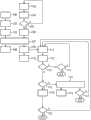
图2示出了用于采集患者运动的根据本发明的方法。FIG. 2 shows the method according to the invention for detecting patient movements.
具体实施方式Detailed ways
在图1中示出了一种根据本发明的医学成像装置,其由磁共振装置10构成。然而,本发明并不限于将医学成像装置构建成磁共振装置。更确切而言,医学成像装置也可以由计算机断层成像装置和/或PET装置(正电子发射断层成像装置)等构成。FIG. 1 shows a medical imaging system according to the invention, which is formed by a
磁共振装置10包括由磁体单元11构成的探测器单元。磁体单元11包括用于产生强且尤其恒定的主磁场13的主磁体12。此外,磁共振装置10具有用于容纳患者15的圆柱形的患者容纳区域14,其中,患者容纳区域14在圆周方向上被磁体单元11包围。患者15可以借助磁共振装置10的患者安置装置16推移到患者容纳区域14中。The
磁体单元11还具有用于产生磁场梯度的梯度线圈单元17,其用于在成像期间的位置编码。梯度线圈单元17通过梯度控制单元18来控制。此外,磁体单元11具有固定地集成在磁体单元11内的高频天线单元19和用于激励极化的高频天线控制单元20,该极化在由主磁体12产生的主磁场13中出现。高频天线单元19由高频天线控制单元20控制,并且将高频的磁共振序列入射到主要由患者容纳区域14构成的检查空间中。由此,将磁化从其平衡位置偏转。The magnet unit 11 also has a
为了控制主磁体12、梯度控制单元18和为了控制高频天线控制单元20,磁共振装置10具有由计算单元构成的系统控制单元21。系统控制单元21中央控制磁共振装置,例如执行预定的、成像的磁场梯度回波序列。例如为成像参数的控制信息以及重建的磁共振图像可以在磁共振装置10的显示单元22、例如监视器上显示。此外,磁共振装置10具有输入单元23,借助其可以在测量过程期间由操作者输入信息和/或参数。For controlling the
此外,磁共振装置10包括在当前实施例中由头部线圈装置构成的局部磁共振线圈装置24。对此替选地,局部磁共振线圈装置24也可以由膝部线圈装置、臂部线圈装置、胸部线圈装置等构成。头部线圈装置包括壳体单元25,其球状形地围绕头部线圈装置的局部患者容纳区域26。Furthermore, the
为了在磁共振检查期间采集患者15的运动,磁共振装置10具有多个运动传感器单元27、28。第一运动传感器单元27由高频天线单元19包括,并且第二运动传感器单元28由头部线圈装置包括。为此替选地,磁共振装置10也可以包括仅一个或多于两个运动传感器单元27、28,其由高频天线单元19和/或局部的磁共振线圈装置24包括。此外,运动传感器单元27、28可以还至少部分由患者安置装置16包括,和/或由至少部分地围绕磁共振装置10的患者容纳区域14和/或局部的磁共振线圈装置24的局部患者容纳区域26的其它单元包括。In order to detect the movement of the patient 15 during the magnetic resonance examination, the
高频天线单元19的运动传感器单元27包括多个运动传感器元件29,其中在图1中示例性地仅示出了运动传感器元件29中的两个。运动传感器元件29分别设计用于采集患者15的运动的运动信息,其中,为此运动传感器元件29采集由非磁体共振数据组成的运动数据。两个运动传感器元件29分别包括光学视场30用于分别采集患者15的部分区域,其中,两个运动传感器元件29中的第一个的第一光学视场30对准患者15或患者容纳区域14的第一部分区域,并且两个运动传感器元件29中的第二个的第二光学视场30对准患者15或患者容纳区域14的第二部分区域。患者15或患者容纳区域14的第一部分区域和患者15或患者容纳区域14的第二部分区域此外设置在患者容纳区域14内或患者15上的不同位置处。两个运动传感器元件29由此对于患者容纳区域14,尤其对于设置在患者容纳区域14内的患者15,具有不同的角度,以便采集患者15的运动的运动信息。The motion sensor unit 27 of the radio-
高频天线单元19包括壳体单元31,其圆柱形地围绕磁共振装置10的患者容纳区域14。两个运动传感器元件29设置在壳体单元31,其中,两个运动传感器元件29设置在高频天线单元19的如下区域中,该区域设置在壳体单元31的背对(abgewandt)患者容纳区域14的侧上。由此,两个运动传感器元件29设置在患者容纳区域14外部。The radio-
此外,两个运动传感器元件29分别由光学的1D运动传感器元件和/或光学的2D运动传感器元件组成。借助光学的1D运动传感器元件和/或光学的2D运动传感器元件,在患者容纳区域14内沿着一个维度和/或沿着两个维度采集患者15的运动的运动信息,尤其采集2D传感器数据测量场。1D运动传感器元件例如可以包括光学的运动传感器元件、超声传感器元件、激光传感器元件等。2D运动传感器元件例如可以包括2D照相机等。Furthermore, the two motion sensor elements 29 each consist of an optical 1D motion sensor element and/or an optical 2D motion sensor element. With the aid of optical 1D motion sensor elements and/or optical 2D motion sensor elements, motion information of the motion of the
高频天线单元19的运动传感器单元27具有评估单元32和数据传输单元33。数据传输单元具有数据发送单元,其带有未详细示出的天线元件用于无电缆地和/或无线地进行数据传输。借助数据传输单元33,将评估过的传感器数据和/或由运动传感器单元27接收的传感器数据传输给系统控制单元21的未详细示出的数据传输单元。The motion sensor unit 27 of the radio-
头部线圈装置的运动传感器单元28同样包括多个运动传感器元件34。这些运动传感器元件34同样由1D运动传感器元件和/或2D运动传感器元件组成。1D运动传感器元件例如可以包括光学的运动传感器元件、超声传感器元件、激光传感器元件等。2D运动传感器元件例如可以包括2D照相机等。The
头部线圈装置的壳体单元包括内部壳体单元35和外部壳体单元36,其中,在内部壳体单元35和外部壳体单元36之间存在头部线圈装置的封闭的安装空间37,在其中设置有头部线圈单元的高频线圈元件,用于采集磁共振信号。运动传感器元件34设置在内部壳体单元35和外部壳体单元36之间,其中,运动传感器元件34具有分别与其它运动传感器元件34不同的、朝向患者14和/或头部线圈装置的局部患者容纳区域26的视角,使得可以从不同的角度采集患者14在头部线圈装置的局部患者容纳区域26内的位置。The housing unit of the head coil device comprises an
头部线圈装置的运动传感器单元28具有数据传输单元38。数据传输单元38包括数据发送单元,其带有未详细示出的天线元件,用于无电缆地和/或无线地进行数据传输。借助数据传输单元38将评估过的传感器数据和/或由运动传感器元件34接收的传感器数据传输给系统控制单元21的数据传输单元。The
为了确定患者14的运动,借助系统控制单元21执行根据本发明的、用于在磁共振检查期间确定患者14的运动的方法。该用于确定患者14的运动的方法在图2中详细示出。系统控制单元21特别地设计用于执行该在磁共振检查期间确定患者14的运动的方法,其中,为此系统控制单元21包括特殊的计算机程序和特殊的软件单元,其存储在系统控制单元21的未详细示出的存储单元中,并且在系统控制单元21的未详细示出的处理器上运行。此外,为此系统控制单元21具有运动计算单元39。In order to determine the movement of the
在该方法中,首先在借助第一磁共振测量进行的第一方法步骤100中,借助磁体单元11采集3D图像数据,其中,第一磁共振测量在对患者15进行磁共振检查之前执行。第一磁共振测量由纵览测量构成,例如由导航测量和/或其它对于层定位所需的测量构成。为此,系统控制单元21具有评估软件,其特别地设计用于根据纵览测量的所采集的3D图像数据来确定患者15的外轮廓。此外,在方法步骤100中也可以借助与纵览测量和/或对于层定位所需的测量相独立地构建的单独的磁共振测量来采集3D图像数据。在此,可以仅采集如下数据,这些数据具有组织到空气的过渡部的信息,并且由此尤其采集患者15的外轮廓的三维信息。该单独的测量优选地具有在采集患者15的外轮廓中的特别高的精度,使得可以有利地省去其它信息和由此省去测量时间。In the method, 3D image data are initially acquired by means of the magnet unit 11 in a
在另一方法步骤101中,评估借助第一磁共振测量采集的第一3D图像数据,其中,评估由系统控制单元21进行,其为此包括未详细示出的评估单元。在方法步骤101中,从第一磁共振测量的第一3D图像数据中确定和/或采集设置在患者容纳区域14内的对象的、尤其是患者15的和必要时的运动传感器单元28的三维空间布置和/或三维空间轮廓。In a
接下来,在另一方法步骤102中,将设置在患者容纳区域14内的对象的三维空间布置和/或三维空间轮廓存储在系统控制单元21的未详细示出的存储单元中。Next, in a
与采集第一3D图像数据同时地,借助运动传感器单元27、28在另一方法步骤103中采集第一运动数据,其中,运动数据包括1D运动数据和/或2D运动数据。这些第一1D运动数据和/或2D运动数据同样在对患者15进行医学成像检查之前采集。第一1D运动数据和/或2D运动数据也还在对患者15进行医学成像检查之前在另一方法步骤104中被评估。接下来,在询问105中确定,患者15是否实施了运动。Simultaneously with the acquisition of the first 3D image data, first movement data are acquired by means of the
如果患者15在采集第一图像数据期间运动了,则重新借助运动传感器单元27、28采集第一运动数据,并且再次经过方法步骤103、104、105。If the patient 15 moves during the acquisition of the first image data, the first movement data are acquired again by means of the
如果患者15在采集第一图像数据期间未实施运动,则将评估过的运动数据在另一方法步骤106中同样存储在系统控制单元21的存储单元中。If the
在接下来的方法步骤107中,借助运动计算单元39计算患者15的潜在运动,其中,在该计算中采用设置在患者容纳区域14内的患者15的三维空间布置和/或三维空间轮廓以及存储和评估的运动数据。运动计算单元39特别地设计用于计算患者15的潜在运动,其中为此运动计算单元39包括特殊的计算机程序和特殊的软件单元,其在运动计算单元的未详细示出的处理器上运行。此外,为此,运动计算单元39还使用模型计算和/或拟合方法,以计算患者15的潜在运动。In the
接下来,将根据设置在患者容纳区域14内的患者15的三维空间布置和/或三维空间轮廓和所存储及评估的运动数据确定出的患者15的潜在运动在另一方法步骤108中存储和/或储存在查找表中,使得在医学成像检查期间总是保证快速和可靠地访问该表。Next, in a
此外,在患者15不运动时,在另一方法步骤109中起动磁共振检查,并且在接下来的方法步骤110中接收和/或采集医学3D图像数据、尤其是医学3D磁共振数据。在接收和/或采集医学3D磁共振数据的同时,在另一方法步骤111中还借助运动传感器单元27、28接收和/或采集其它运动数据,其中,在医学磁共振检查期间接收和/或采集这些其它运动数据。接收和/或采集这些其它运动数据在整个医学磁共振检查期间连续进行,使得在整个磁共振检查期间总是呈现患者15当前的运动信息。在医学磁共振检查期间采集的运动数据同样由1D运动数据和/或2D运动数据组成。Furthermore, while the
将在医学磁共振检查期间采集的运动数据直接在其采集之后在另一方法步骤112中由运动计算单元39评估,其中,在此借助存储在查找表中的数据给出快速的运动信息。此外,可以对运动信息在其真实性方面在运动计算单元39内进行检验。为此,沿着运动轨迹必须存在例如多个、在时间方面相继采集和/或接收的运动数据。The motion data acquired during the medical magnetic resonance examination are evaluated directly after their acquisition by the
此外,借助运动计算单元39还可以在方法步骤112中确定患者的三维图像和/或患者15的运动。在此,在患者15的运动的模拟计算中采用2D运动数据,使得结合3D图像数据确定患者15的三维图像。为了优化模拟计算,在此可以借助最小二乘法和/或其它优化方法将所模拟的运动与在医学成像检查期间采集的运动数据的偏差最小化,这尤其在2D运动数据情况下是有利的。Furthermore, a three-dimensional image of the patient and/or a movement of the patient 15 can also be determined in
在另一方法步骤113中询问,在医学磁共振检查期间是否发生了患者15的运动。如果未发生运动,则接下来还在方法步骤114中检验,医学磁共振检查是否已经结束。如果医学磁共振检查已经结束,则也将用于确定患者15在医学磁共振检查期间的运动的方法结束。如果医学磁共振检查未结束,则在方法步骤111中重新借助运动传感器单元27、28采集1D运动数据和/或2D运动数据。In a
相反,如果患者15在医学磁共振检查期间运动了,则在另一询问115中确定是否对由患者15实施的运动能够实现事后的和/或回顾性的校正,这例如在医学磁共振检查期间患者的仅小的位置变化和/或运动情况下是可能的。如果对由患者15实施的运动能够实现事后的和/或回顾性的校正,则因此在另一方法步骤116中将借助运动传感器单元27、28采集的1D运动数据和/或2D运动数据与医学磁共振检查的医学3D图像数据一起存储。对患者15在医学磁共振检查期间的运动的校正在另一方法步骤118中在评估和/或重建医学3D图像数据时才进行,使得在此在重建过的磁共振图像中消除由于患者15在医学磁共振检查期间的运动而形成的伪影。在此,系统控制单元21根据所采集的患者15的运动的运动信息来改变和/或校正用于评估和/或重建医学3D图像数据的至少一个评估参数。On the other hand, if the patient 15 moved during the medical magnetic resonance examination, it is determined in a
此外,在询问117中确定,医学磁共振检查是否已经结束。如果医学磁共振检查还未结束,则在方法步骤111-116中还采集和进一步处理1D运动数据和/或2D运动数据。Furthermore, it is determined in
如果事后校正是不可能的,则系统控制单元21在另一方法步骤119中改变和/或校正医学磁共振检查的测量协议的各个测量参数。在此,尤其将用于控制和/或实施各个磁共振序列的各个参数匹配于患者15在医学磁共振检查期间的当前运动。这例如可以引起,将用于磁场梯度的各个参数匹配于患者15在医学磁共振检查期间的运动的运动信息。此外,这种对于医学磁共振检查的测量协议内的测量参数的改变和/或校正会引起对医学磁共振检查的各个部分测量的至少部分地中断,其在患者15不运动地躺卧在患者安置装置16上时才会引起对医学磁共振检查的部分测量的恢复和/或引起医学磁共振检查的部分测量的重新开始。If a subsequent correction is not possible, the
在匹配和/或校正测量参数之后在另一方法步骤120中询问:医学磁共振检查是否已经结束。如果医学磁共振检查已经结束,则根据本发明的、用于采集患者在医学磁共振检查期间的运动的方法结束。After the adaptation and/or correction of the measurement parameters, in a
如果医学磁共振检查还未结束,则在方法步骤111-119中还采集和进一步处理1D运动数据和/或2D运动数据。If the medical magnetic resonance examination has not yet been concluded, 1D motion data and/or 2D motion data are also acquired and further processed in method steps 111 - 119 .
此外还可能的是,在方法步骤110和/或在方法步骤111中可以借助附加地设置在患者上的标记元件采集患者的运动。Furthermore, it is also possible to detect the movement of the patient in
此外还可以设计,在其中确定患者运动的方法步骤105、113中,这可以作为光学信息来借助显示单元22输出和/或通知给操作者、例如临床操作人员和/或医生。还可能的是,在方法步骤117、118、119中同样借助显示单元22将对参数、尤其是对评估参数和/或测量参数的校正和/或改变通知给操作者。Furthermore, it can be provided that in the method steps 105 , 113 in which the patient movement is determined, this can be output as optical information by means of the
Claims (17)
Translated fromChineseApplications Claiming Priority (2)
| Application Number | Priority Date | Filing Date | Title |
|---|---|---|---|
| DE102012216327.1ADE102012216327B4 (en) | 2012-09-13 | 2012-09-13 | Method for detecting movement of a patient during a medical imaging examination |
| DE102012216327.1 | 2012-09-13 |
Publications (2)
| Publication Number | Publication Date |
|---|---|
| CN103654784Atrue CN103654784A (en) | 2014-03-26 |
| CN103654784B CN103654784B (en) | 2017-01-18 |
Family
ID=50153332
Family Applications (1)
| Application Number | Title | Priority Date | Filing Date |
|---|---|---|---|
| CN201310416311.1AActiveCN103654784B (en) | 2012-09-13 | 2013-09-13 | Method for acquiring movement of patient during medical imaging examination |
Country Status (3)
| Country | Link |
|---|---|
| US (1) | US20140073904A1 (en) |
| CN (1) | CN103654784B (en) |
| DE (1) | DE102012216327B4 (en) |
Cited By (11)
| Publication number | Priority date | Publication date | Assignee | Title |
|---|---|---|---|---|
| CN104470426A (en)* | 2012-06-21 | 2015-03-25 | 皇家飞利浦有限公司 | Magnetic resonance examination system with motion detection |
| CN105361955A (en)* | 2014-08-07 | 2016-03-02 | 西门子公司 | Medical imaging unit, medical imaging device with a medical imaging unit, and method for detecting a patient movement |
| CN106659450A (en)* | 2014-09-09 | 2017-05-10 | 通用电气公司 | Multi-frame acquisition for exposure control in X-ray medical imaging |
| CN107616803A (en)* | 2016-07-13 | 2018-01-23 | 西门子保健有限责任公司 | Pass through the magnetic resonance of combination and the method for X-ray device collection and processing measurement data |
| CN110458817A (en)* | 2019-08-05 | 2019-11-15 | 上海联影医疗科技有限公司 | Qualitative forecasting method, device, equipment and the storage medium of medical image |
| CN111084634A (en)* | 2018-10-24 | 2020-05-01 | 西门子医疗有限公司 | Medical imaging device and method for operating a medical imaging device |
| CN112137621A (en)* | 2019-06-26 | 2020-12-29 | 西门子医疗有限公司 | Determination of patient motion during medical imaging measurements |
| CN113271847A (en)* | 2019-01-03 | 2021-08-17 | 皇家飞利浦有限公司 | Apparatus and method for tracking head motion in Magnetic Resonance Imaging (MRI) |
| CN113521499A (en)* | 2020-04-22 | 2021-10-22 | 西门子医疗有限公司 | Method for generating control signal |
| CN113677266A (en)* | 2019-04-11 | 2021-11-19 | 皇家飞利浦有限公司 | Combined optical image generator and optical imaging system |
| CN115024711A (en)* | 2021-03-04 | 2022-09-09 | 西门子医疗有限公司 | Imaging device, local coil and method for correcting a movement of a patient |
Families Citing this family (15)
| Publication number | Priority date | Publication date | Assignee | Title |
|---|---|---|---|---|
| US10307619B2 (en)* | 2012-02-06 | 2019-06-04 | Insightec, Ltd. | Reference-library extension during imaging of moving organs |
| DE102012216292B4 (en)* | 2012-09-13 | 2021-02-18 | Siemens Healthcare Gmbh | Magnetic resonance assembly, a magnetic resonance device with the magnetic resonance assembly and a method for determining a movement of a patient during a magnetic resonance examination |
| DE102012222375B3 (en)* | 2012-12-06 | 2014-01-30 | Siemens Aktiengesellschaft | Magnetic coil device for investigation on head of patient, has light field camera element which is provided in camera unit of magnetic coil assembly, such that camera element is arranged within receiving region surrounding shell unit |
| DE102013218432A1 (en)* | 2013-09-13 | 2015-03-19 | Siemens Aktiengesellschaft | A medical imaging device and a method for determining a position and / or movement of a patient during a medical imaging study |
| DE102014207020A1 (en) | 2014-04-11 | 2015-10-15 | Siemens Aktiengesellschaft | Method for positioning at least one local coil for recording magnetic resonance data with a magnetic resonance device and magnetic resonance system |
| EP3384307B1 (en)* | 2015-12-03 | 2021-07-28 | Koninklijke Philips N.V. | Removal of image artifacts in sense-mri |
| RU2730431C2 (en)* | 2015-12-03 | 2020-08-21 | Конинклейке Филипс Н.В. | Removal of image artifacts at sense-visualization |
| DE102017204175A1 (en)* | 2017-03-14 | 2018-09-20 | Siemens Healthcare Gmbh | A method for finding abnormalities in medical image data of a region of the patient which is arranged outside a region of the patient to be examined, and a medical imaging device for this purpose |
| EP3447520A1 (en)* | 2017-08-22 | 2019-02-27 | Koninklijke Philips N.V. | Data-driven correction of phase depending artefacts in a magnetic resonance imaging system |
| US11457871B2 (en)* | 2018-11-21 | 2022-10-04 | Enlitic, Inc. | Medical scan artifact detection system and methods for use therewith |
| US11011257B2 (en)* | 2018-11-21 | 2021-05-18 | Enlitic, Inc. | Multi-label heat map display system |
| US12011249B2 (en)* | 2020-12-29 | 2024-06-18 | Shanghai United Imaging Healthcare Co., Ltd. | Systems and methods for tomography imaging |
| US12136484B2 (en) | 2021-11-05 | 2024-11-05 | Altis Labs, Inc. | Method and apparatus utilizing image-based modeling in healthcare |
| US20250060443A1 (en)* | 2021-12-22 | 2025-02-20 | Tracinnovations A/S | Method and apparatus for optical tracking of motions of a subject |
| DE102023202427B3 (en) | 2023-03-20 | 2024-07-25 | Siemens Healthineers Ag | Method for operating an imaging modality of a magnetic resonance system, and magnetic resonance system |
Citations (8)
| Publication number | Priority date | Publication date | Assignee | Title |
|---|---|---|---|---|
| US20020167317A1 (en)* | 2001-01-26 | 2002-11-14 | Shenoy Rajendra K. | Driven equilibrium and fast-spin echo scanning |
| US20030219147A1 (en)* | 2002-05-23 | 2003-11-27 | Kabushiki Kaisha Toshiba | Object tracking apparatus and method |
| US20050054910A1 (en)* | 2003-07-14 | 2005-03-10 | Sunnybrook And Women's College Health Sciences Centre | Optical image-based position tracking for magnetic resonance imaging applications |
| DE102004006561A1 (en)* | 2004-02-06 | 2005-08-25 | Technische Universität Dresden | Three-dimensional simulation, analysis and graphical representation of changes in biological systems, whereby biological sub-system data are combined with physical condition data and stored in modules for subsequent processing |
| CN1905836A (en)* | 2004-11-12 | 2007-01-31 | 株式会社东芝 | Magnetic resonance imaging apparatus, image data correction apparatus, and image data correction method |
| US20070108978A1 (en)* | 2005-11-16 | 2007-05-17 | Macfarlane Duncan L | Apparatus and method for patient movement tracking |
| US20070280508A1 (en)* | 2006-05-19 | 2007-12-06 | Ernst Thomas M | Motion tracking system for real time adaptive imaging and spectroscopy |
| CN101352350A (en)* | 2007-07-26 | 2009-01-28 | 西门子公司 | Method for recording measurement data of a patient taking into account the movement process and related medical device |
Family Cites Families (4)
| Publication number | Priority date | Publication date | Assignee | Title |
|---|---|---|---|---|
| DE102004045495B4 (en)* | 2004-09-20 | 2015-06-18 | Siemens Aktiengesellschaft | Method and system for generating images of an organ |
| US7358732B2 (en)* | 2005-10-24 | 2008-04-15 | The General Hospital Corporation | System, method, software arrangement and computer-accessible medium for providing real-time motion correction by utilizing clover leaf navigators |
| DE102007035176B4 (en)* | 2007-07-27 | 2010-03-18 | Siemens Ag | Method for recording and processing a sequence of temporally successive image data records and magnetic resonance apparatus |
| WO2010030397A1 (en)* | 2008-09-12 | 2010-03-18 | Accuray Incorporated | Controlling x-ray imaging based on target motion |
- 2012
- 2012-09-13DEDE102012216327.1Apatent/DE102012216327B4/enactiveActive
- 2013
- 2013-09-12USUS14/024,700patent/US20140073904A1/ennot_activeAbandoned
- 2013-09-13CNCN201310416311.1Apatent/CN103654784B/enactiveActive
Patent Citations (8)
| Publication number | Priority date | Publication date | Assignee | Title |
|---|---|---|---|---|
| US20020167317A1 (en)* | 2001-01-26 | 2002-11-14 | Shenoy Rajendra K. | Driven equilibrium and fast-spin echo scanning |
| US20030219147A1 (en)* | 2002-05-23 | 2003-11-27 | Kabushiki Kaisha Toshiba | Object tracking apparatus and method |
| US20050054910A1 (en)* | 2003-07-14 | 2005-03-10 | Sunnybrook And Women's College Health Sciences Centre | Optical image-based position tracking for magnetic resonance imaging applications |
| DE102004006561A1 (en)* | 2004-02-06 | 2005-08-25 | Technische Universität Dresden | Three-dimensional simulation, analysis and graphical representation of changes in biological systems, whereby biological sub-system data are combined with physical condition data and stored in modules for subsequent processing |
| CN1905836A (en)* | 2004-11-12 | 2007-01-31 | 株式会社东芝 | Magnetic resonance imaging apparatus, image data correction apparatus, and image data correction method |
| US20070108978A1 (en)* | 2005-11-16 | 2007-05-17 | Macfarlane Duncan L | Apparatus and method for patient movement tracking |
| US20070280508A1 (en)* | 2006-05-19 | 2007-12-06 | Ernst Thomas M | Motion tracking system for real time adaptive imaging and spectroscopy |
| CN101352350A (en)* | 2007-07-26 | 2009-01-28 | 西门子公司 | Method for recording measurement data of a patient taking into account the movement process and related medical device |
Cited By (19)
| Publication number | Priority date | Publication date | Assignee | Title |
|---|---|---|---|---|
| CN104470426A (en)* | 2012-06-21 | 2015-03-25 | 皇家飞利浦有限公司 | Magnetic resonance examination system with motion detection |
| CN105361955A (en)* | 2014-08-07 | 2016-03-02 | 西门子公司 | Medical imaging unit, medical imaging device with a medical imaging unit, and method for detecting a patient movement |
| CN105361955B (en)* | 2014-08-07 | 2019-03-08 | 西门子公司 | Medical imaging assembly, medical imaging device, and method for capturing patient motion |
| US10542938B2 (en) | 2014-08-07 | 2020-01-28 | Siemens Aktiengesellschaft | Medical imaging unit, medical imaging device with a medical imaging unit, and method for detecting a patient movement |
| CN106659450A (en)* | 2014-09-09 | 2017-05-10 | 通用电气公司 | Multi-frame acquisition for exposure control in X-ray medical imaging |
| CN107616803A (en)* | 2016-07-13 | 2018-01-23 | 西门子保健有限责任公司 | Pass through the magnetic resonance of combination and the method for X-ray device collection and processing measurement data |
| CN107616803B (en)* | 2016-07-13 | 2021-01-01 | 西门子保健有限责任公司 | Method for acquiring and processing measurement data by means of a combined magnetic resonance and X-ray device |
| CN111084634B (en)* | 2018-10-24 | 2023-09-01 | 西门子医疗有限公司 | Medical imaging device and method for operating a medical imaging device |
| CN111084634A (en)* | 2018-10-24 | 2020-05-01 | 西门子医疗有限公司 | Medical imaging device and method for operating a medical imaging device |
| CN113271847A (en)* | 2019-01-03 | 2021-08-17 | 皇家飞利浦有限公司 | Apparatus and method for tracking head motion in Magnetic Resonance Imaging (MRI) |
| CN113677266A (en)* | 2019-04-11 | 2021-11-19 | 皇家飞利浦有限公司 | Combined optical image generator and optical imaging system |
| CN113677266B (en)* | 2019-04-11 | 2022-09-02 | 皇家飞利浦有限公司 | Combined optical image generator and optical imaging system |
| CN112137621A (en)* | 2019-06-26 | 2020-12-29 | 西门子医疗有限公司 | Determination of patient motion during medical imaging measurements |
| US11980456B2 (en) | 2019-06-26 | 2024-05-14 | Siemens Healthineers Ag | Determining a patient movement during a medical imaging measurement |
| CN110458817A (en)* | 2019-08-05 | 2019-11-15 | 上海联影医疗科技有限公司 | Qualitative forecasting method, device, equipment and the storage medium of medical image |
| CN113521499A (en)* | 2020-04-22 | 2021-10-22 | 西门子医疗有限公司 | Method for generating control signal |
| CN113521499B (en)* | 2020-04-22 | 2024-02-13 | 西门子医疗有限公司 | Method for generating control signals |
| US12257002B2 (en) | 2020-04-22 | 2025-03-25 | Siemens Healthineers Ag | Method for creation of a control signal |
| CN115024711A (en)* | 2021-03-04 | 2022-09-09 | 西门子医疗有限公司 | Imaging device, local coil and method for correcting a movement of a patient |
Also Published As
| Publication number | Publication date |
|---|---|
| DE102012216327A1 (en) | 2014-03-13 |
| DE102012216327B4 (en) | 2021-01-14 |
| US20140073904A1 (en) | 2014-03-13 |
| CN103654784B (en) | 2017-01-18 |
Similar Documents
| Publication | Publication Date | Title |
|---|---|---|
| CN103654784B (en) | Method for acquiring movement of patient during medical imaging examination | |
| US11185305B2 (en) | Intertial device tracking system and method of operation thereof | |
| US9658305B2 (en) | Wireless prospective motion marker | |
| CN101352350B (en) | Method for recording measured data from a patient by taking movements into account, and associated medical device | |
| US9746535B2 (en) | Magnetic resonance imaging apparatus | |
| US9626777B2 (en) | Method and apparatus to generate image data | |
| CN107468265A (en) | Position the check object to imaging method | |
| US9766308B2 (en) | Magnetic resonance unit, a magnetic resonance apparatus with the magnetic resonance unit, and a method for determination of a movement by a patient during a magnetic resonance examination | |
| CN104115020A (en) | MRI with motion correction using navigators acquired using a Dixon technique | |
| JP2011194241A (en) | System and method for automatic computation of mr imaging scan parameter | |
| US20150297120A1 (en) | Method For Tracking Motion of Subject in Real Time and for Correcting Medical Image | |
| US11419583B2 (en) | Reconstruction-free automatic multi-modality ultrasound registration | |
| CN107334473A (en) | For running the method and magnetic resonance device of magnetic resonance device | |
| CN102908144A (en) | Magnetic resonance imaging for therapy planning | |
| US10018699B2 (en) | Method and magnetic resonance apparatus for acquiring magnetic resonance data with a prospective motion correction | |
| KR20150145106A (en) | Method and appartus for registering medical images | |
| CN105361955A (en) | Medical imaging unit, medical imaging device with a medical imaging unit, and method for detecting a patient movement | |
| CN102885622B (en) | For the method for captured image data and the magnetic resonance device for implementing the method | |
| US20240090791A1 (en) | Anatomy Masking for MRI | |
| JP2014212904A (en) | Medical projection system | |
| CN112204616A (en) | Automated object monitoring for medical imaging | |
| JP2010051615A (en) | Magnetic resonance imaging apparatus | |
| WO2019048284A1 (en) | Intra-procedure calibration for image-based tracking | |
| JP2006314491A (en) | Magnetic resonance imaging system | |
| CN107843863B (en) | Magnetic resonance imaging correction method, device and equipment based on 3D topography measurement |
Legal Events
| Date | Code | Title | Description |
|---|---|---|---|
| PB01 | Publication | ||
| PB01 | Publication | ||
| C10 | Entry into substantive examination | ||
| SE01 | Entry into force of request for substantive examination | ||
| C14 | Grant of patent or utility model | ||
| GR01 | Patent grant | ||
| TR01 | Transfer of patent right | Effective date of registration:20220128 Address after:Erlangen Patentee after:Siemens Healthineers AG Address before:Munich, Germany Patentee before:SIEMENS AG | |
| TR01 | Transfer of patent right | ||
| TR01 | Transfer of patent right | Effective date of registration:20240905 Address after:German Phu F Haim Patentee after:Siemens Medical AG Country or region after:Germany Address before:Erlangen Patentee before:Siemens Healthineers AG Country or region before:Germany | |
| TR01 | Transfer of patent right |