CN103476341A - X-ray image capturing device, medical image processing device, X-ray image capturing method, and medical image processing method - Google Patents
X-ray image capturing device, medical image processing device, X-ray image capturing method, and medical image processing methodDownload PDFInfo
- Publication number
- CN103476341A CN103476341ACN2013800003817ACN201380000381ACN103476341ACN 103476341 ACN103476341 ACN 103476341ACN 2013800003817 ACN2013800003817 ACN 2013800003817ACN 201380000381 ACN201380000381 ACN 201380000381ACN 103476341 ACN103476341 ACN 103476341A
- Authority
- CN
- China
- Prior art keywords
- mentioned
- ray
- image data
- benchmark
- software
- Prior art date
- Legal status (The legal status is an assumption and is not a legal conclusion. Google has not performed a legal analysis and makes no representation as to the accuracy of the status listed.)
- Granted
Links
Images


Classifications
- A—HUMAN NECESSITIES
- A61—MEDICAL OR VETERINARY SCIENCE; HYGIENE
- A61B—DIAGNOSIS; SURGERY; IDENTIFICATION
- A61B6/00—Apparatus or devices for radiation diagnosis; Apparatus or devices for radiation diagnosis combined with radiation therapy equipment
- A61B6/54—Control of apparatus or devices for radiation diagnosis
- A—HUMAN NECESSITIES
- A61—MEDICAL OR VETERINARY SCIENCE; HYGIENE
- A61B—DIAGNOSIS; SURGERY; IDENTIFICATION
- A61B6/00—Apparatus or devices for radiation diagnosis; Apparatus or devices for radiation diagnosis combined with radiation therapy equipment
- A61B6/12—Arrangements for detecting or locating foreign bodies
- A—HUMAN NECESSITIES
- A61—MEDICAL OR VETERINARY SCIENCE; HYGIENE
- A61B—DIAGNOSIS; SURGERY; IDENTIFICATION
- A61B6/00—Apparatus or devices for radiation diagnosis; Apparatus or devices for radiation diagnosis combined with radiation therapy equipment
- A61B6/44—Constructional features of apparatus for radiation diagnosis
- A61B6/4429—Constructional features of apparatus for radiation diagnosis related to the mounting of source units and detector units
- A61B6/4435—Constructional features of apparatus for radiation diagnosis related to the mounting of source units and detector units the source unit and the detector unit being coupled by a rigid structure
- A61B6/4441—Constructional features of apparatus for radiation diagnosis related to the mounting of source units and detector units the source unit and the detector unit being coupled by a rigid structure the rigid structure being a C-arm or U-arm
- A—HUMAN NECESSITIES
- A61—MEDICAL OR VETERINARY SCIENCE; HYGIENE
- A61B—DIAGNOSIS; SURGERY; IDENTIFICATION
- A61B6/00—Apparatus or devices for radiation diagnosis; Apparatus or devices for radiation diagnosis combined with radiation therapy equipment
- A61B6/48—Diagnostic techniques
- A61B6/481—Diagnostic techniques involving the use of contrast agents
- A—HUMAN NECESSITIES
- A61—MEDICAL OR VETERINARY SCIENCE; HYGIENE
- A61B—DIAGNOSIS; SURGERY; IDENTIFICATION
- A61B6/00—Apparatus or devices for radiation diagnosis; Apparatus or devices for radiation diagnosis combined with radiation therapy equipment
- A61B6/50—Apparatus or devices for radiation diagnosis; Apparatus or devices for radiation diagnosis combined with radiation therapy equipment specially adapted for specific body parts; specially adapted for specific clinical applications
- A61B6/503—Apparatus or devices for radiation diagnosis; Apparatus or devices for radiation diagnosis combined with radiation therapy equipment specially adapted for specific body parts; specially adapted for specific clinical applications for diagnosis of the heart
- A—HUMAN NECESSITIES
- A61—MEDICAL OR VETERINARY SCIENCE; HYGIENE
- A61B—DIAGNOSIS; SURGERY; IDENTIFICATION
- A61B6/00—Apparatus or devices for radiation diagnosis; Apparatus or devices for radiation diagnosis combined with radiation therapy equipment
- A61B6/52—Devices using data or image processing specially adapted for radiation diagnosis
- A61B6/5205—Devices using data or image processing specially adapted for radiation diagnosis involving processing of raw data to produce diagnostic data
Landscapes
- Health & Medical Sciences (AREA)
- Life Sciences & Earth Sciences (AREA)
- Engineering & Computer Science (AREA)
- Medical Informatics (AREA)
- Biomedical Technology (AREA)
- Heart & Thoracic Surgery (AREA)
- High Energy & Nuclear Physics (AREA)
- Physics & Mathematics (AREA)
- Nuclear Medicine, Radiotherapy & Molecular Imaging (AREA)
- Optics & Photonics (AREA)
- Pathology (AREA)
- Radiology & Medical Imaging (AREA)
- Veterinary Medicine (AREA)
- Biophysics (AREA)
- Molecular Biology (AREA)
- Surgery (AREA)
- Animal Behavior & Ethology (AREA)
- General Health & Medical Sciences (AREA)
- Public Health (AREA)
- Computer Vision & Pattern Recognition (AREA)
- Cardiology (AREA)
- Dentistry (AREA)
- Oral & Maxillofacial Surgery (AREA)
- Apparatus For Radiation Diagnosis (AREA)
Abstract
Translated fromChinese
Description
Translated fromChinese技术领域technical field
本发明的实施方式涉及X射线摄影装置、医用图像处理装置、X射线摄影方法以及医用图像处理方法。Embodiments of the present invention relate to an X-ray imaging device, a medical image processing device, an X-ray imaging method, and a medical image processing method.
背景技术Background technique
以往,一边观察通过X射线摄影装置对被检体的体内进行摄影得到的图像一边实时地进行介入治疗的技术广为人知。例如,能够通过已插入血管的管将探针、导丝、支架、带模支架(stent graft)、人工瓣等的器具设置于被检体的体内。Conventionally, a technique for performing interventional therapy in real time while observing an image obtained by imaging the inside of a subject with an X-ray imaging device is widely known. For example, instruments such as a probe, a guide wire, a stent, a stent graft, and an artificial valve can be installed in the body of a subject through a tube inserted into a blood vessel.
作为将器具设置于体内的治疗的一种,可列举出主动脉瓣的置换。主动脉瓣的置换是通过从大腿部的血管所插入的探针将人工瓣设置于主动脉的治疗技术。使用了探针的主动脉瓣的置换术被称为TAVR(Trans-catheterAortic Valve Replacement,经导管主动脉瓣置换术)或TAVI(Trans-catheterAortic Valve Implantation,经导管主动脉瓣植入术)。As one of the treatments in which a device is installed in the body, aortic valve replacement can be mentioned. Aortic valve replacement is a treatment technique in which an artificial valve is placed in the aorta with a probe inserted from a blood vessel in the thigh. Aortic valve replacement using a probe is called TAVR (Trans-catheterAortic Valve Replacement) or TAVI (Trans-catheterAortic Valve Implantation).
在主动脉瓣的置换术中,重要的是将人工瓣高精度地留置于适当的位置。但是,在留置人工瓣时通过X射线摄影装置实时显示的X射线透视图像中,并不绘出作为人工瓣的留置目标的主动脉瓣。因此,提供了用于辅助主动脉瓣的置换术的各种应用软件。In aortic valve replacement surgery, it is important to indwell the artificial valve at an appropriate position with high precision. However, the aortic valve, which is the placement target of the artificial valve, is not drawn in the X-ray fluoroscopic image displayed in real time by the X-ray imaging device when the artificial valve is placed. Therefore, various application software for assisting the replacement of the aortic valve are provided.
例如,在市场上销售一种软件,其用于在包含预先拍摄的主动脉瓣的血管的造影图像上绘出作为人工瓣的留置目标的线,并在留置人工瓣时,使表示人工瓣的留置目标的线重叠显示于X射线透视图像上。For example, there is commercially available software for drawing a line as an indwelling target of an artificial valve on a pre-shot angiographic image of a blood vessel including the aortic valve, and when placing the artificial valve, make the line representing the artificial valve A line overlay of the indwelling target is displayed on the fluoroscopic image.
现有技术文献prior art literature
专利文献patent documents
专利文献1:日本特开2011-36433号公报Patent Document 1: Japanese Unexamined Patent Publication No. 2011-36433
发明内容Contents of the invention
发明所要解决的课题The problem to be solved by the invention
但是,在用于辅助主动脉瓣的置换术的以往技术中,需要用于在血管的X射线造影图像中确定主动脉的由用户进行的操作。具体地讲,需要通过输入装置的操作来描出主动脉瓣或输入线等的用户的输入作业。However, in the conventional technique for assisting the replacement of the aortic valve, an operation by the user for specifying the aorta in the X-ray contrast image of the blood vessel is required. Specifically, user input tasks such as drawing an aortic valve, an input line, and the like through the operation of the input device are required.
另一方面,主动脉瓣的置换术是非常复杂并伴有风险的手术。在主动脉瓣的置换术中,为了对用户的手操作进行辅助,X射线摄影装置作为用于观察被检体的体内的图像引导装置发挥作用。因此,希望以用户能够专注于其手操作的方式,减少X射线摄影装置中所要求的操作。On the other hand, aortic valve replacement is a very complex and risky operation. In an aortic valve replacement operation, an X-ray imaging device functions as an image guidance device for observing the inside of a subject in order to assist a user's manual operation. Therefore, it is desired to reduce the operations required in the X-ray imaging apparatus in such a way that the user can concentrate on the operation of his hand.
这并不限于主动脉瓣的置换术,在使用了X射线摄影装置的各种诊断或治疗等中也是相同的。This is not limited to aortic valve replacement, but also applies to various diagnoses and treatments using X-ray imaging equipment.
因此,本发明的目的在于提供一种能够减少通过用户进行的输入作业的X射线摄影装置、医用图像处理装置、X射线摄影方法以及医用图像处理方法。Therefore, an object of the present invention is to provide an X-ray imaging apparatus, a medical image processing apparatus, an X-ray imaging method, and a medical image processing method capable of reducing input work by a user.
用于解决课题的方法method used to solve the problem
本发明的实施方式的X射线摄影装置具备:X射线图像收集部、基准位置取得部以及条件设定部。X射线图像收集部使用摄影系统,对与互不相同的X射线的照射方向对应的二维的多个X射线图像数据进行收集。基准位置取得部参照上述多个X射线图像数据,求出在空间上作为基准的朝向以及作为基准的位置。条件设定部基于与上述作为基准的朝向以及上述作为基准的位置对应的信息,自动地设定上述摄影系统的控制条件以及X射线图像的图像处理条件中的至少一方。An X-ray imaging apparatus according to an embodiment of the present invention includes an X-ray image acquisition unit, a reference position acquisition unit, and a condition setting unit. The X-ray image collection unit collects a plurality of two-dimensional X-ray image data corresponding to different X-ray irradiation directions using an imaging system. The reference position acquisition unit refers to the plurality of X-ray image data to obtain a spatially reference orientation and a reference position. The condition setting unit automatically sets at least one of a control condition of the imaging system and an image processing condition of the X-ray image based on information corresponding to the reference orientation and the reference position.
此外,本发明的实施方式的医用图像处理装置具备:X射线图像收集部、基准位置取得部以及条件设定部。X射线图像收集部取得与互不相同的X射线的照射方向对应的二维的多个X射线图像数据。基准位置取得部参照上述多个X射线图像数据,求出在空间上作为基准的朝向以及作为基准的位置。条件设定部基于与上述作为基准的朝向以及上述作为基准的位置对应的信息,自动地设定X射线摄影装置所具备的摄影系统的控制条件以及X射线图像的图像处理条件中的至少一方。Furthermore, the medical image processing apparatus according to the embodiment of the present invention includes an X-ray image acquisition unit, a reference position acquisition unit, and a condition setting unit. The X-ray image acquisition unit acquires a plurality of two-dimensional X-ray image data corresponding to different X-ray irradiation directions. The reference position acquisition unit refers to the plurality of X-ray image data to obtain a spatially reference orientation and a reference position. The condition setting unit automatically sets at least one of control conditions of an imaging system included in the X-ray imaging apparatus and image processing conditions for X-ray images based on information corresponding to the reference orientation and the reference position.
此外,本发明的实施方式的X射线摄影方法具有:使用摄影系统,对与互不相同的X射线的照射方向对应的二维的多个X射线图像数据进行收集的步骤;参照上述多个X射线图像数据,求出在空间上作为基准的朝向以及作为基准的位置的步骤;以及基于与上述作为基准的朝向以及上述作为基准的位置对应的信息,自动地设定上述摄影系的控制条件以及X射线图像的图像处理条件中的至少一方的步骤。In addition, the X-ray imaging method according to the embodiment of the present invention has a step of collecting a plurality of two-dimensional X-ray image data corresponding to different X-ray irradiation directions using an imaging system; The radiographic data, a step of obtaining a reference orientation and a reference position in space; and automatically setting the control conditions and A step of at least one of the image processing conditions of the X-ray image.
此外,本发明的实施方式的医用图像处理方法具有:取得与互不相同的X射线的照射方向对应的二维的多个X射线图像数据的步骤;参照上述多个X射线图像数据,求出在空间上作为基准的朝向以及作为基准的位置步骤;以及基于与上述作为基准的朝向以及上述作为基准的位置对应的信息,自动地设定X射线摄影装置所具备的摄影系统的控制条件以及X射线图像的图像处理条件中的至少一方的步骤。In addition, the medical image processing method according to the embodiment of the present invention includes the steps of acquiring a plurality of two-dimensional X-ray image data corresponding to different X-ray irradiation directions; referring to the plurality of X-ray image data to obtain The orientation as a reference in space and the position as a reference; and based on the information corresponding to the orientation as a reference and the position as a reference above, automatically set the control conditions and X-ray imaging system of the X-ray imaging device. A step of at least one of image processing conditions for radiographic images.
附图说明Description of drawings
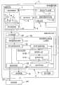
图1为本发明的实施方式的X射线摄影装置以及医用图像处理装置的构成图。FIG. 1 is a configuration diagram of an X-ray imaging apparatus and a medical image processing apparatus according to an embodiment of the present invention.
图2为表示使用了X射线摄影装置以及医用图像处理装置的主动脉瓣的置换术的流程的图。FIG. 2 is a diagram showing the flow of an aortic valve replacement using an X-ray imaging device and a medical image processing device.
图3为表示用于使用了图1所示的X射线摄影装置以及医用图像处理装置的主动脉瓣的置换术的心脏的X射线摄影的流程的流程图。FIG. 3 is a flowchart showing a flow of cardiac X-ray imaging for aortic valve replacement using the X-ray imaging apparatus and the medical image processing apparatus shown in FIG. 1 .
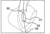
图4为表示在图1所示的X射线摄影装置以及医用图像处理装置中所取得的、绘出了设备的二维X射线图像的图像的第一例的图。FIG. 4 is a diagram showing a first example of an image in which a two-dimensional X-ray image of a device is drawn and obtained by the X-ray imaging apparatus and the medical image processing apparatus shown in FIG. 1 .
图5为表示在图1所示的X射线摄影装置以及医用图像处理装置中所取得的、绘出了设备的二维X射线图像的图像的第二例的图。FIG. 5 is a diagram showing a second example of an image in which a two-dimensional X-ray image of a device is drawn and obtained by the X-ray imaging apparatus and the medical image processing apparatus shown in FIG. 1 .
图6为表示以主动脉成为垂直方向的角度所拍摄到的X射线透视图像的例的图。FIG. 6 is a diagram showing an example of an X-ray fluoroscopic image captured at an angle in which the aorta is in the vertical direction.
图7为表示在图1所示的显示装置上,与表示两个标识器间的中心的线段一同被显示的X射线透视图像的一例的图。FIG. 7 is a diagram showing an example of an X-ray fluoroscopic image displayed on the display device shown in FIG. 1 together with a line segment indicating the center between two markers.
图8为表示使时间序列的X射线透视图像显示于固定在标识器的坐标系的例子的图。FIG. 8 is a diagram showing an example of displaying time-series X-ray fluoroscopic images on a coordinate system fixed to a marker.
图9为表示将心脏的X射线造影图像作为参照图像来指定了线段的例子的图。FIG. 9 is a diagram showing an example in which a line segment is specified using an X-ray contrast image of a heart as a reference image.
具体实施方式Detailed ways
参照附图,对本发明的实施方式的X射线摄影装置、医用图像处理装置、X射线摄影方法以及医用图像处理方法加以说明。An X-ray imaging apparatus, a medical image processing apparatus, an X-ray imaging method, and a medical image processing method according to embodiments of the present invention will be described with reference to the drawings.
(第一实施方式)(first embodiment)
图1为本发明的第一实施方式的X射线摄影装置以及医用图像处理装置的构成图。FIG. 1 is a configuration diagram of an X-ray imaging apparatus and a medical image processing apparatus according to a first embodiment of the present invention.
X射线摄影装置1具备:摄影系统2、控制系统3、数据处理系统4、输入装置5以及显示装置6。摄影系统2具有:X射线照射部7、X射线检测器8、驱动机构9以及诊疗台10。控制系统3具有:高电压产生装置11以及摄影位置控制装置12。The X-ray imaging apparatus 1 includes an imaging system 2 , a
X射线照射部7具备X射线管,并隔着已被安置在诊疗台10上的被检体O与X射线检测器8相对配置。X射线照射部7以及X射线检测器8能够一边通过驱动机构9的驱动来维持相对位置,一边改变相对于被检体O的角度以及相对位置。具体地讲,X射线照射部7以及X射线检测器8被固定于具备旋转功能的C型臂的两端。而且,X射线照射部7被构成为能够通过X射线管,自规定的角度向被检体O照射X射线,并通过X射线检测器8来检测从被检体O透射的X射线。The X-ray irradiation unit 7 is provided with an X-ray tube, and is arranged opposite to the
此外,能够通过驱动机构9来调整诊疗台10的台面的倾斜以及位置。因此,不仅调整X射线照射部7以及X射线检测器8相对于被检体O的角度,通过调整台面的角度,也能够改变相对于被检体O的X射线的照射方向。In addition, the inclination and position of the top of the medical table 10 can be adjusted by the
并且,在已被安置于诊疗台10的被检体O的附近,根据需要,设置用于向被检体O注入造影剂的造影剂注入装置13。此外,在进行使心脏以200次/每分钟的程度高速跳动的起搏(pacing)的情况下,起搏装置14被插入心脏。In addition, a contrast medium injecting
控制系统3的高电压产生装置11是通过向X射线照射部7的X射线管施加高电压,向被检体O照射具有所希望的能量的X射线的装置。摄影位置控制装置12为向驱动机构9输出控制信号来进行控制的装置。即,X射线照射部7以及X射线检测器8的旋转角度以及位置和诊疗台10的台面的倾斜以及位置是通过从摄影位置控制装置12输出到驱动机构9的控制信号来控制的。The
数据处理系统4具有A/D(analog to digital,模数)转换器15以及计算机16。但是,也有A/D转换器15与X射线检测器8形成一体化的情况。计算机16通过执行程序,作为医用图像处理装置16发挥作用。即,在X射线摄影装置1中内置有医用图像处理装置16。The
但是,也可以介由网络,将具有相同功能的独立的医用图像处理装置连接于X射线摄影装置1。此外,为了构成内置于X射线摄影装置1的医用图像处理装置16或者介由网络与X射线摄影装置1连接的医用图像处理装置,也可以使用电路。However, an independent medical image processing device having the same function may be connected to the X-ray imaging device 1 via a network. In addition, a circuit may be used to configure the medical
医用图像处理装置16具有:X射线图像生成部17、X射线图像取得部18、基准位置取得部19、软件保存部20、条件设定部21以及显示处理部22。The medical
X射线图像生成部17从X射线检测器8取入通过A/D转换器15而被数字化的X射线检测数据,具有通过进行数据处理来生成X射线图像数据的功能。特别是,在X射线图像生成部17中,能够生成主动脉瓣的置换所需的或者对其有用的X射线图像数据。The
作为具体例子,列举出包含主动脉瓣的位置的X射线透视图像数据、DSA(digital subtraction angiography,数字减影血管造影)图像数据、路线图图像数据、血管造影图像数据以及设备图像数据。在进行主动脉瓣的置换的情况下,将人工瓣留置于本来的主动脉瓣的位置的动作为操作对象。但是,作为人工瓣的留置位置的主动脉以及主动脉瓣在X射线透视图像中是不可见的。因此,根据需要,也可以通过投放造影剂来事先收集绘出了主动脉的血管造影图像数据。Specific examples include X-ray fluoroscopy image data including the position of the aortic valve, DSA (digital subtraction angiography, digital subtraction angiography) image data, road map image data, angiography image data, and device image data. In the case of aortic valve replacement, an operation to indwell the artificial valve at the original position of the aortic valve is the object of operation. However, the aorta and the aortic valve, which are the place where the artificial valve is placed, are not visible in the X-ray fluoroscopy image. Therefore, if necessary, angiographic image data in which the aorta is drawn may be collected in advance by administering a contrast medium.
DSA图像数据为造影剂注入前后的X射线图像数据的差分图像数据。路线图图像数据是为了将用于留置人工瓣的探针引导至目的位置而作为血管的造影图像数据与透视图像数据的合成图像数据来生成的血管图像数据。此外,设备图像数据为绘出了球囊、导丝或者人工瓣等设备的X射线图像数据。The DSA image data is differential image data of X-ray image data before and after contrast agent injection. The road map image data is blood vessel image data generated as composite image data of contrast image data and fluoroscopic image data of blood vessels in order to guide a probe for placing an artificial valve to a target position. In addition, the device image data is X-ray image data depicting devices such as balloons, guide wires, or artificial valves.
可以将标识器安装于在设备图像数据中所绘出的设备。因此,可以将安装于设备的标识器利用于使用了X射线图像数据的各种数据处理。此外,也可以将设备自身作为标识器而利用于各种数据处理。因此,在X射线图像生成部17中至少生成与互不相同的X射线的照射方向对应的二维(2D:two dimensional)的设备图像数据。A marker may be attached to the device depicted in the device image data. Therefore, the marker attached to the device can be used for various data processing using X-ray image data. In addition, the device itself can be used as an identifier for various data processing. Therefore, at least two-dimensional (2D: two dimensional) device image data corresponding to different X-ray irradiation directions is generated in the X-ray
而且,通过X射线图像生成部17与摄影系统2以及控制系统3协作,从而在X射线摄影装置1中具备X射线图像收集部的功能,该X射线图像收集部使用摄影系统2,收集与互不相同的X射线的照射方向对应的、绘出了设备以及多个标识器的至少一方的二维的多个X射线图像数据。Furthermore, the X-ray
X射线图像取得部18具有取得在X射线图像生成部17所生成的X射线图像数据的功能。特别是,在介由网络连接于X射线摄影装置1的独立的医用图像处理装置中,也可以省略X射线图像生成部17。在该情况下,X射线图像取得部18具备介由网络,从X射线摄影装置1中所具备的X射线图像生成部17取得X射线图像数据的功能。即,X射线图像取得部18构成为,至少与互不相同的X射线的照射方向对应地取得绘出了设备以及多个标识器的至少一方的二维的多个X射线图像数据。The X-ray
基准位置取得部19具有如下功能,即,通过分别检测从X射线图像取得部18取得的多个X射线图像数据中的设备或多个标识器的位置,求出在空间上作为基准的朝向以及作为基准的位置。在进行主动脉瓣的置换术的情况下,将作为留置于主动脉瓣的人工瓣的留置目标的朝向以及位置分别作为基准朝向以及基准位置来求出的方式是直接且有效的方法。The reference
再者,在基准位置取得部19中,并不限于伴随着造影剂的投放而收集的X射线造影图像数据,也可以根据非造影的多个X射线图像数据分别检测设备或者多个标识器的位置。若将非造影的多个X射线图像数据用于设备或者多个标识器的位置的检测,则可以省略造影剂的投放。Furthermore, in the reference
在软件保存部20中保存用于预先设定摄影系统2的控制条件或X射线图像的图像处理条件的各种应用软件。Various application software for setting control conditions of the imaging system 2 and image processing conditions for X-ray images in advance are stored in the
条件设定部21具有基于与在基准位置取得部19所求出的作为基准的朝向以及作为基准的位置对应地信息,自动地设定摄影系统2的控制条件以及X射线图像的图像处理条件中的至少一方的功能。可以将保存于软件保存部20的应用软件利用于摄影系统2的控制条件以及X射线图像的图像处理条件的设定。The
为此,条件设定部21具有软件执行部21A以及软件输入部21B。For this purpose, the
软件执行部21A具有从软件保存部20读入并执行用于自动地设定摄影系统2的控制条件以及X射线图像的图像处理条件的至少一方的必要的软件的功能。因此,在条件设定部21中,可以取得摄影系统2的控制条件以及X射线图像的图像处理条件来作为软件执行部21A的软件执行结果。The
软件输入部21B具有使用在基准位置取得部19所求出的与作为基准的朝向以及作为基准的位置对应的信息来作为向软件执行部21A中所执行的软件的输入的功能。特别是,软件输入部21B构成为,在从输入装置5取得了表示软件的执行开始的操作信息的情况下,自动地向软件输入与作为基准的朝向以及作为基准的位置的至少一方对应的输入数据。The
作为表示软件的执行开始的输入装置5的操作信息,可以列举出通过按下软件的启动按钮等而被输入的软件的启动指示信息、通过按下软件的执行开始按钮等而被输入的软件的执行开始指示信息、或激活显示了暂时启动的软件的操作画面的窗口的指示信息等。即,可以将输入装置5的所希望的操作信息作为表示软件的执行开始的操作信息来处理。As the operation information of the
结果,可以不需要由用户进行的用于执行软件的输入作业。或者,可以减少输入作业。As a result, input work by the user for executing the software may not be required. Alternatively, input jobs can be reduced.
而且,可以将在条件设定部21所设定的摄影系统2的控制条件作为摄影系统2的控制信息输出到控制系统3。由此,能够进行基于与在基准位置取得部19所求出的作为基准的朝向以及作为基准的位置对应的信息的摄影系统2的自动控制。另一方面,可以将在条件设定部21所设定的X射线图像的图像处理条件作为图像处理条件输出到显示处理部22。Furthermore, the control conditions of the imaging system 2 set in the
显示处理部22具有:从X射线图像取得部18取得X射线图像数据的功能;对所取得的X射线图像数据施以必要的图像处理并生成显示用的二维图像数据的功能;通过向显示装置6输出显示用的二维图像数据,使X射线图像显示于显示装置6的功能。特别是,显示处理部22构成为,按照在条件设定部21所设定的图像处理条件,执行对从X射线图像取得部18所取得的X射线图像数据的图像处理。由此,能够进行基于与在基准位置取得部19所求出的作为基准的朝向以及作为基准的位置对应的信息的X射线图像数据的自动图像处理以及显示。The
此外,显示处理部22构成为,能够介由网络23从其他图像诊断装置或医用图像服务器等的医用系统24取得图像数据,并使用所取得的图像数据来执行X射线图像数据的图像处理。例如,X射线CT(computedtomography,计算机断层扫描)装置能够进行用于使事先所收集的X射线CT图像与在X射线摄影装置1所拍摄的X射线图像重叠显示的图像处理。Furthermore, the
接下来,对X射线摄影装置1以及医用图像处理装置16的动作以及作用加以说明。Next, the operation and function of the X-ray imaging device 1 and the medical
图2为表示使用了X射线摄影装置1以及医用图像处理装置16的主动脉瓣的置换术的流程的图。FIG. 2 is a diagram showing the flow of an aortic valve replacement using the X-ray imaging device 1 and the medical
在图2中,横轴表示时间。如图2所示,主动脉瓣的置换术是按照计划以及手操作的顺序执行的。在计划时,作为患者的被检体O被安置于X射线摄影装置1并执行预摄像。在预摄像中,对X射线透视图像数据、DSA图像数据、路线图图像数据、血管造影图像数据以及设备图像数据等必要的X射线图像数据进行收集。此外,在计划时,人工瓣、球囊、导丝等设备通过探针被插入被检体O。因此,通过预摄像,取得绘出了设备或安装于设备的标识器的X射线图像数据。In FIG. 2, the horizontal axis represents time. As shown in Figure 2, aortic valve replacement was performed in a planned and manual sequence. At the time of planning, the subject O as a patient is placed in the X-ray imaging apparatus 1 and pre-imaging is performed. In the pre-imaging, necessary X-ray image data such as X-ray fluoroscopy image data, DSA image data, road map image data, angiography image data, and device image data are collected. In addition, at the time of planning, devices such as an artificial valve, a balloon, and a guide wire are inserted into the subject O through a probe. Thus, by pre-imaging, X-ray image data depicting the equipment or markers mounted on the equipment is obtained.
因此,在基准位置取得部19,能够从X射线图像数据对设备或安装于设备的标识器进行检测。这样一来,能够基于被检测的设备或者标识器的位置,事先求出在手操作中重要的、作为基准的朝向以及作为基准的位置来作为矢量信息或坐标信息。Therefore, in the reference
若计划结束,则开始手操作。在进行手操作时,作为图像引导,也实时地进行X射线透视图像数据的收集以及显示。此外,手操作所需的各种软件是通过软件执行部21A启动以及执行的。但是,在进行手操作时,软件的操作和向软件的输入作业成为很大的妨碍。If the plan is over, start manual operation. During manual operation, collection and display of X-ray fluoroscopic image data are also performed in real time as image guidance. In addition, various software necessary for manual operation are activated and executed by the
因此,在软件输入部21B中,能够基于在计划时预先已求出的矢量信息或者坐标信息,自动地制作向各种软件的输入数据。而且,能够使得若软件启动,则输入数据被自动地输入已启动的软件。由此,用户仅进行软件的启动等最小限度的操作,而能够专注于手操作。Therefore, in the
再者,在计划时预先启动软件,进行手操作时激活用于操作软件的窗口的情况或进行软件的执行开始指示的情况下,也能够进行同样的输入数据的自动输入。此情况下,在计划时,可以在激活窗口的定时或者指示软件的执行开始的定时的任一定时,将输入数据自动输入到软件中。Furthermore, the same automatic input of input data can also be performed when the software is activated in advance at the time of planning, and when a window for operating the software is activated during manual operation, or when an instruction to start execution of the software is given. In this case, at the time of planning, the input data may be automatically input into the software at any timing of the activation window timing or the timing of instructing the execution start of the software.
即,能够在手操作的计划时,至少预先求出作为基准的朝向以及作为基准的位置。而且,在进行手操作时,能够向用于设定摄影系统2的控制条件以及X射线图像的图像处理条件的至少一方的软件输入与作为基准的朝向以及作为基准的位置对应的输入数据。此外,输入数据的输入是能够将表示软件的执行开始的来自输入装置5的操作信息作为触发器来自动进行的。That is, at least a reference orientation and a reference position can be obtained in advance at the time of manual operation planning. In addition, at the time of manual operation, input data corresponding to a reference orientation and a reference position can be input to software for setting at least one of control conditions of the imaging system 2 and image processing conditions of X-ray images. In addition, input of input data can be automatically performed using operation information from the
以下,以基于在计划时所收集的非造影旋转DA图像数据来制作作为基准的矢量信息以及坐标信息,并基于所制作的矢量信息以及坐标信息向各种软件自动输入输入数据的情况为例,对X射线摄影装置1以及医用图像处理装置16的详细的动作以及作用加以说明。Hereinafter, taking the case of creating reference vector information and coordinate information based on non-contrast rotational DA image data collected during planning, and automatically inputting input data into various software based on the created vector information and coordinate information, as an example, Detailed operations and functions of the X-ray imaging device 1 and the medical
图3为表示用于使用了图1所示的X射线摄影装置1以及医用图像处理装置16的主动脉瓣的置换术的心脏的X射线摄影的流程的流程图。FIG. 3 is a flowchart showing a flow of cardiac X-ray imaging for aortic valve replacement using the X-ray imaging apparatus 1 and the medical
首先,进行手操作的计划。为此,将被检体O安置于诊疗台10,通过X射线摄影装置1拍摄并显示为了主动脉瓣的置换而插入的设备的确认用的X射线图像。另一方面,将设备安装于探针并插入被检体O的内部。作为插入被检体O的设备,可列举出人工瓣、球囊、导丝以及探针。球囊是先于人工瓣的插入而插入到主动脉瓣附近的设备。优选在这些设备上安装多个标识器。First, plan the hand operation. For this purpose, the subject O is placed on the examination table 10, and an X-ray image for confirmation of a device inserted for aortic valve replacement is captured and displayed by the X-ray imaging device 1 . On the other hand, the device is attached to a probe and inserted into the subject O. Examples of devices inserted into the subject O include artificial valves, balloons, guide wires, and probes. The balloon is a device that is inserted near the aortic valve prior to the insertion of the artificial valve. Preferably multiple markers are installed on these devices.
接着,在步骤S1中,医师等的用户通过对探针进行操作,使设备行进至主动脉瓣附近等的作为观察对象的血管内的规定位置并设置于此。设备被设置为长度方向与主动脉的血管的轴一致。因此,若设备为人工瓣,则人工瓣的长度方向为血管的轴方向。此外,在主动脉瓣附近使球囊扩张的情况下,导丝以及探针沿着血管的中心线。Next, in step S1 , a user such as a physician operates the probe to advance the device to a predetermined position in a blood vessel to be observed, such as near the aortic valve, and installs it there. The device is set lengthwise to coincide with the vessel axis of the aorta. Therefore, if the device is an artificial valve, the length direction of the artificial valve is the axial direction of the blood vessel. In addition, when the balloon is inflated near the aortic valve, the guide wire and the probe are along the centerline of the blood vessel.
因此,优选使得人工瓣或球囊等的设备成为插入到主动脉瓣的状态。在标识器安装于人工瓣或球囊的长度方向的两端的情况下,可以将标识器间的中点设为主动脉瓣的位置。Therefore, it is preferable to insert a device such as an artificial valve or a balloon into the aortic valve. When the markers are attached to both ends in the longitudinal direction of the artificial valve or the balloon, the midpoint between the markers may be the position of the aortic valve.
接着,在步骤S2中,根据需要,使起搏装置14工作,执行心脏的快速起搏(Rapid Pacing)。Next, in step S2, the pacing
接着,在步骤S3中,一边照射X射线一边使摄影系统2旋转,由此,收集与不同的X射线的照射角度对应的多个2D-DA图像数据。摄影系统2的旋转角度在30度的程度即足够。此外,不需要投放造影剂。Next, in step S3 , the imaging system 2 is rotated while irradiating X-rays, thereby collecting a plurality of 2D-DA image data corresponding to different X-ray irradiation angles. It is sufficient for the rotation angle of the imaging system 2 to be about 30 degrees. In addition, no contrast agent needs to be administered.
作为X射线摄影装置1的具体动作,首先,按照来自摄影位置控制装置12的控制信息,驱动机构9进行驱动。然后,诊疗台10、X射线照射部7以及X射线检测器8被定位于规定的旋转角度以及空间位置。X射线照射部7以及X射线检测器8以规定的速度进行旋转。As a specific operation of the X-ray imaging apparatus 1 , first, the
另一方面,若从高电压产生装置11向X射线照射部7的X射线管施加高电压,则X射线从X射线管向被检体O照射。从被检体O透射的X射线通过X射线检测器8被检测。在X射线检测器8所取得的X射线检测数据被输出到数据处理系统4。On the other hand, when a high voltage is applied from the
这样,X射线图像生成部17通过对在A/D转换器15被数字化的X射线检测数据的数据处理,生成X射线图像数据。在X射线图像取得部18取得所生成的X射线图像数据。In this way, the
图4为表示在图1所示的X射线摄影装置1以及医用图像处理装置16所取得的、绘出了设备的二维X射线图像的图像的第一例的图。FIG. 4 is a diagram showing a first example of an image obtained by the X-ray imaging apparatus 1 and the medical
若人工瓣被插入本来的主动脉瓣的附近,则如图4所示,不能看到主动脉以及主动脉瓣,但可以对绘出了人工瓣的非造影X射线透视图像进行摄影。在标识器安装于人工瓣的两端的情况下,也绘出各标识器。When the artificial valve is inserted near the original aortic valve, the aorta and the aortic valve cannot be seen as shown in FIG. 4 , but a non-contrast X-ray fluoroscopy image in which the artificial valve is drawn can be photographed. In the case where the markers are mounted on both ends of the prosthetic valve, the markers are also drawn.
图4表示绘出了通过探针30的操作而插入心脏的人工瓣31以及安装于人工瓣31的两端的两个标识器32、33的X射线图像的例子。再者,图4中的虚线表示所推断的心脏的轮郭,实际上是不可见的线。FIG. 4 shows an example of an X-ray image depicting the
图5为表示在图1所示的X射线摄影装置1以及医用图像处理装置16所取得的、绘出了设备的二维X射线图像的图像的第二例的图。FIG. 5 is a diagram showing a second example of an image obtained by the X-ray imaging apparatus 1 and the medical
如图5所示,若将球囊40插入主动脉瓣并使其扩张,则不能看到主动脉以及主动脉瓣,但可以对绘出了球囊40的非造影X射线透视图像进行摄影。在标识器41、42安装于球囊40的两端的情况下,也绘出各标识器41、42。在图5中的虚线也表示所推断的心脏的轮郭,实际上是不可见的线。As shown in FIG. 5 , when the
作为插入了设备的X射线图像,也可以取得绘出了人工瓣的X射线图像以及绘出了球囊的X射线图像的任一个。但是,由于球囊紧密附着于主动脉的血管壁,因此,形成主动脉的走行方向与球囊的长度方向看起来更平行的状态。所以,从更正确地把握主动脉的走行方向的观点来说,对绘出了球囊的X射线图像进行摄影是有利的。As the X-ray image in which the device is inserted, either an X-ray image in which the artificial valve is drawn or an X-ray image in which the balloon is drawn may be acquired. However, since the balloon is closely attached to the blood vessel wall of the aorta, the running direction of the aorta and the longitudinal direction of the balloon appear to be more parallel. Therefore, from the viewpoint of more accurately grasping the running direction of the aorta, it is advantageous to take an X-ray image in which the balloon is drawn.
接着,在步骤S4中,基准位置取得部19从X射线图像取得部18取得至少两帧的X射线图像数据。然后,在二维的各X射线图像数据中自动检测设备或各标识器的坐标。例如,在收集30帧的量的X射线图像数据的情况下,也可以对30帧的X射线图像数据分别跟踪设备或者标识器的坐标。此外,在检测设备或两个标识器的坐标的情况下,也可以求出表示设备的长度方向的位置或者连接标识器间的线段的算式。Next, in step S4 , the reference
再者,兼顾由于在X射线图像数据中所绘出的构造物复杂等的理由而很难跟踪设备或标识器的情况,也可以使得能够从输入装置5向基准位置取得部19输入辅助信息。In addition, it is also possible to allow input of auxiliary information from the
例如,对于一个以上的帧的X射线图像数据,可以通过输入装置5的操作来指定标识器或设备的坐标,并将其他帧的X射线图像数据中的标识器或设备的坐标作为与所指定的标识器或设备的坐标对应的坐标来进行自动检测。或者,也可以通过输入装置5的操作来指定包含标识器或设备的感兴趣区域(ROI:region of interest),并从基准位置取得部19所指定的ROI内自动检测设备或各标识器的坐标。For example, for the X-ray image data of more than one frame, the coordinates of the marker or equipment can be specified through the operation of the
接着,在步骤S5中,基准位置取得部19基于从与不同X射线的照射角度对应的多个帧的二维X射线图像数据所检测的设备或标识器的多个二维坐标位置,计算设备或标识器的三维(3D:three dimensional)空间内的坐标来作为基准位置。并且,基准位置取得部19对表示设备的朝向的线矢量或连接两个标识器间的线矢量进行计算。该计算等价于表示在空间内通过设备或多个标识器的线段的算式的计算。此外,这些计算可以根据基于对极几何(Epipolar Geometry)等的公知理论公式以几何学方式进行。Next, in step S5, the reference
接着,在步骤S6中,基准位置取得部19求出与表示设备的位置或连接标识器间的线段的位置的线矢量正交的平面的算式。此外,将三维空间坐标系中的设备或标识器的坐标投影于当前显示于显示装置6的X射线图像的二维X-Y坐标系。由此,能够推断出投影于为了人工瓣的定位而显示的X射线图像上的设备的两端或两个标识器的两点的X-Y坐标(X1,Y1)、(X2,Y2)。Next, in step S6 , the reference
由此,条件设定部21能够将在基准位置取得部19所求出的线矢量、正交于线矢量的平面以及两点的投影坐标(X1,Y1)、(X2,Y2)等的位置信息作为与作为基准的朝向以及作为基准的位置对应的信息,自动地设定摄影系统2的控制条件以及X射线图像的图像处理条件的一方或双方。Thus, the
若这样的计划结束,则能够开始手操作。在手操作中也进行X射线透视图像的摄影。即,将X射线透视图像作为引导来进行手操作。因此,设定用于对X射线透视图像进行摄影的摄影系统2的控制条件以及X射线图像的图像处理条件。When such planning is completed, manual operation can be started. Photography of X-ray fluoroscopy images is also performed in manual operation. That is, manual manipulation is performed using the X-ray fluoroscopic image as a guide. Therefore, control conditions of the imaging system 2 for imaging X-ray fluoroscopic images and image processing conditions for X-ray images are set.
可以将保存于软件保存部20的应用软件利用于摄影系统2的控制条件以及X射线图像的图像处理条件的设定。此情况下,软件执行部21A从软件保存部20读入并执行对应的软件。此外,软件输入部21B使用在基准位置取得部19作为矢量信息所求出的作为基准的朝向以及作为基准的位置来作为向软件的输入。即,软件输入部21B基于矢量信息自动地生成向各种软件的输入数据,并将所生成的输入数据自动地输入到对应软件。The application software stored in the
在此,作为具体例,对通过软件进行的七个摄影系统2的控制条件以及X射线图像的图像处理条件的设定例加以说明。Here, as a specific example, a setting example of the control conditions of the seven imaging systems 2 and the image processing conditions of the X-ray images by software will be described.
作为第一具体例,可以在条件设定部21自动地求出用于以最适合人工瓣的留置作业的观察角度显示X射线图像的摄影系统2的控制条件。此情况下,按照来自输入装置5的启动指示,软件执行部21A从软件保存部20读入并启动摄影角度的设定软件。这样,软件输入部21B自动地向摄影角度的设定软件输入正交于线矢量的平面的算式来作为输入数据。As a first specific example, the control conditions of the imaging system 2 for displaying X-ray images at an observation angle most suitable for the indwelling operation of the artificial valve may be automatically obtained by the
接着,在步骤S7-1中,条件设定部21将与作为设备的长度方向而求出的线矢量正交的平面的算式转换为以左前斜位(LAO:left anterioroblique)方向、右前斜位(RAO:right anterior oblique)方向、头部(CRA:cranial)方向以及尾部(CAU:caudal)方向为坐标轴的坐标系的算式。再者,也可以将转换前后的平面作为曲线图显示于显示装置6。Next, in step S7-1, the
所谓TAVI中的最适合的观察方向是主动脉成为垂直方向的角度、即主动脉瓣成为一条直线的角度。再者,并不是唯一地确定主动脉瓣成为一条直线的角度,而是存在多个候补。The most suitable observation direction in TAVI is the angle at which the aorta becomes the vertical direction, that is, the angle at which the aortic valve becomes a straight line. In addition, the angle at which the aortic valve becomes a straight line is not uniquely determined, but there are a plurality of candidates.
因此,在步骤S7-2中,条件设定部21求出用于在作为主动脉的走行方向所推断的线矢量的朝向垂直而主动脉瓣看起来呈一条直线的方向上照射X射线的X射线照射部7以及X射线检测器8的延伸角度。线矢量的算式或以LAO方向、RAO方向、CRA方向以及CAU方向为坐标轴的坐标系中的平面的算式被用于该计算。因此,能够自动地计算X射线照射部7以及X射线检测器8的延伸角度。Therefore, in step S7-2, the
接着,在步骤S7-3中,条件设定部21以X射线照射部7以及X射线检测器8的角度成为所求出的角度的方式,向控制系统3输出控制信息。结果,通过由控制系统3进行的控制,驱动机构9所具备的C型臂被驱动,具备X射线照射部7以及X射线检测器8的机架(gantry)旋转。然后,X射线照射部7以及X射线检测器8的角度被自动地调整成与最适合的观察方向对应的角度。Next, in step S7-3, the
因此,能够减少以往所进行的、用于由用户决定最适合的观察方向的操作。此外,能够基于非造影图像数据自动设定X射线照射部7以及X射线检测器8的最适合的角度,因此,不需要造影剂。Therefore, conventional operations for the user to determine the most suitable viewing direction can be reduced. In addition, since the optimum angles of the X-ray irradiation unit 7 and the
图6为表示以主动脉成为垂直方向的角度所拍摄的X射线透视图像的例子的图。FIG. 6 is a diagram showing an example of an X-ray fluoroscopic image captured at an angle in which the aorta is in the vertical direction.
图6表示绘出了通过探针30的操作而插入心脏的人工瓣31以及安装于人工瓣31的两端的两个标识器32、33的X射线透视图像的例子。再者,图6中的虚线表示所推断的心脏的轮郭,实际上是不可见的线。FIG. 6 shows an example of an X-ray fluoroscopic image depicting the
如图6所示,虽然不能看到主动脉以及主动脉瓣,但能够对摄影系统2进行控制,从而以主动脉成为垂直方向的角度来显示X射线透视图像。由此,用户能够以人工瓣31的长度方向成为垂直方向的方式留置人工瓣31。As shown in FIG. 6 , although the aorta and the aortic valve cannot be seen, the imaging system 2 can be controlled to display an X-ray fluoroscopic image at an angle in which the aorta is perpendicular to the direction. Thereby, the user can indwell the
作为第二具体例,在条件设定部21中,能够设定用于使多个标识器的中心重叠显示于X射线图像的图像处理条件。此情况下,按照来自输入装置5的启动指示,软件执行部21A从软件保存部20读入并启动描绘多个标识器的中心的图像处理软件。这样,软件输入部21B自动地向图像处理软件输入两个标识器的X-Y投影坐标(X1,Y1)、(X2,Y2)来作为输入数据。As a second specific example, in the
接下来,在步骤S8-1中,条件设定部21算出连接在基准位置取得部19所求出的两点的标识器的X-Y投影坐标(X1,Y1)、(X2,Y2)之间的线的垂直二等分线的算式。然后,条件设定部21设定使所算出的垂直二等分线重叠显示于显示中的X射线透视图像的图像处理条件。此外,条件设定部21向显示处理部22输出包含垂直二等分线的坐标信息的图像处理条件。Next, in step S8-1, the
另一方面,通过摄影系统2的驱动依次收集X射线透视图像数据,在X射线图像取得部18取得所收集的X射线透视图像数据。然后,X射线图像取得部18依次将所取得的X射线透视图像数据送给显示处理部22。On the other hand, X-ray fluoroscopic image data is sequentially collected by driving the imaging system 2 , and the collected X-ray fluoroscopic image data is acquired by the X-ray
接着,在步骤S8-2中,显示处理部22通过对从X射线图像取得部18依次取得的X射线透视图像数据的图像处理,生成用于使表示两个标识器间的中心的垂直二等分线重叠显示的X射线透视图像数据。所生成的X射线透视图像数据被输出到显示装置6。由此,在显示装置6显示重叠了表示两个标识器间的中心的线段的X射线透视图像。Next, in step S8-2, the
图7为表示在图1所示的显示装置6中,与表示两个标识器间的中心的线段一同被显示的X射线透视图像的一例的图。FIG. 7 is a diagram showing an example of an X-ray fluoroscopic image displayed together with a line segment indicating the center between two markers on the
图7表示在X射线投影图像上重叠显示表示两个标识器的投影位置的记号50、51以及表示标识器间的中心的垂直二等分线52的例子。因此,在通过预先已插入的球囊或探针等的设备制作了记号50、51以及垂直二等分线52的情况下,能够将垂直二等分线52用作人工瓣的留置目标。FIG. 7 shows an example in which marks 50 and 51 indicating the projection positions of two markers and a
再者,即使改变显示中的X射线透视图像的观察角度,也能够通过空间上的投影处理,随着观察角度,对记号50、51以及垂直二等分线52进行更新显示。Furthermore, even if the viewing angle of the X-ray fluoroscopic image being displayed is changed, the
此外,也能够描绘垂直二等分线52以外的线段。特别是,有时希望以被垂线分割的线段的左心室侧的长度变短的方式描绘连接与两个标识器对应的记号50、51的线段的垂线。因此,也可以使得能够可变地设定垂线的位置,以便主动脉侧的线段的长度与左心室侧的线段的长度的比例为7:3至5:5的范围。即,能够自动地设定用于使连接多个标识器的线段的中心或以规定的比例分割连接多个标识器的线段的位置重叠显示于X射线图像的图像处理条件。In addition, line segments other than the
因此,在希望专注于手操作的人工瓣的操作中,可以不进行用于在X射线透视图像上描绘人工瓣的留置目标的输入作业。Therefore, when it is desired to concentrate on the operation of the artificial valve operated by hand, it is not necessary to perform an input work for drawing an indwelling target of the artificial valve on the X-ray fluoroscopic image.
作为第三具体例,在条件设定部21中,能够设定用于使用与设备一同移动的坐标系来动态显示X射线图像的图像处理条件。此情况下,按照来自输入装置5的启动指示,软件执行部21A从软件保存部20读入并启动用于以固定于设备的坐标系显示X射线图像的图像处理软件。这样,软件输入部21B自动地向图像处理软件输入两个标识器的X-Y投影坐标(X1,Y1)、(X2,Y2)来作为输入数据。As a third specific example, in the
接着,在步骤S9-1中,条件设定部21从X射线图像取得部18取得作为显示对象的时间序列的X射线透视图像数据,并在基准位置取得部19所求出的两点的标识器的X-Y投影坐标(X1,Y1)、(X2,Y2)附近依次自动检测标识器。标识器的检测可以通过例如将以两点X-Y投影坐标(X1,Y1)、(X2,Y2)为中心的规定的区域内作为对象的、基于信号值的阈值处理等的公知的图像处理来进行。Next, in step S9-1, the
然后,条件设定部21设定用于将时间序列的X射线图像数据坐标变换至固定于所检测的标识器的位置的坐标系的图像处理条件。具体地讲,能够将随时间变化的标识器的时间序列的位置信息作为图像处理条件。所设定的图像处理条件被送给显示处理部22。Then, the
接着,在步骤S9-2中,显示处理部22从X射线图像取得部18取得作为显示对象的时间序列的X射线透视图像数据,并将其分别坐标变换为固定于伴有运动的标识器的坐标系的X射线透视图像数据。即,执行使随时间变动的标识器的位置为同一位置的、时间序列的X射线透视图像数据的运动补偿。然后,坐标变换后的时间序列的X射线透视图像依次显示于显示装置6。Next, in step S9-2, the
图8为表示以固定于标识器的坐标系显示时间序列的X射线透视图像的例子的图。FIG. 8 is a diagram showing an example of displaying time-series X-ray fluoroscopic images in a coordinate system fixed to a marker.
图8中虚线所示的箭头表示固定于由于心跳而伴有运动的两个标识器32、33的坐标系。再者,图8中的虚线表示所推断的心脏的轮郭,实际上是不可见的线。Arrows indicated by dotted lines in FIG. 8 represent coordinate systems fixed to the two
能够以静止地显示固定于图8中箭头所示的两个标识器32、33的坐标系的方式,执行时间序列的X射线透视图像数据的运动补偿。两个标识器32、33安装于通过探针30的操作而插入心脏的人工瓣31。Motion compensation of the time-series X-ray fluoroscopic image data can be performed so as to statically display the coordinate system fixed to the two
因此,能够作为动画实时地显示实际上受到心跳的影响而运动的人工瓣31看起来像停止一样的X射线透视图像。因此,能够提高人工瓣31的可见性。此外,由于对预先取得的标识器的X-Y投影坐标(X1,Y1)、(X2,Y2)的周边区域执行标识器32、33的检索,所以,能够飞跃性地提高标识器32、33的位置检测精度以及检测速度。此外,还能够回避标识器的误识别。Therefore, an X-ray fluoroscopic image in which the
作为第四具体例,在条件设定部21,伴随着使标识器的位置一致的运动补偿,对造影图像数据与透视图像数据进行合成,由此,能够设定用于生成路线图图像数据的图像处理条件。如上所述,路线图图像数据为血管的造影图像数据与透视图像数据的合成图像数据。As a fourth specific example, in the
此情况下,按照来自输入装置5的启动指示,软件执行部21A从软件保存部20读入并启动用于生成路线图图像数据的图像处理软件。这样,软件输入部21B自动地向图像处理软件输入两个标识器的X-Y投影坐标(X1,Y1)、(X2,Y2)来作为输入数据。In this case, in accordance with an activation instruction from the
接着,在步骤S10-1中,条件设定部21从X射线图像取得部18取得路线图图像数据生成用的时间序列的X射线透视图像数据,并在基准位置取得部19所求出的两点的标识器的X-Y投影坐标(X1,Y1)、(X2,Y2)附近的区域,依次自动检测标识器。Next, in step S10-1, the
此外,在预先所收集的X射线造影图像数据中,也通过阈值处理等的图像处理来检测标识器的位置。但是,在先于X射线造影图像数据的收集而获得两点的标识器的X-Y投影坐标(X1,Y1)、(X2,Y2)的情况下,能够将进行标识器的位置的检测处理的区域限定于X-Y投影坐标(X1,Y1)、(X2,Y2)附近的区域。In addition, in the X-ray contrast image data collected in advance, the position of the marker is also detected by image processing such as threshold processing. However, in the case where the X-Y projection coordinates (X1, Y1) and (X2, Y2) of the markers at two points are obtained prior to the collection of the X-ray contrast image data, the area where the marker position detection process is performed can be It is limited to the area near the X-Y projection coordinates (X1, Y1), (X2, Y2).
然后,条件设定部21设定用于将事先所收集的X射线造影图像数据以及时间序列的X射线透视图像数据坐标变换至固定于所检测的标识器的位置的坐标系的图像处理条件。具体地讲,能够将随时间变化的标识器的时间序列的位置信息作为图像处理条件。所设定的图像处理条件被送给显示处理部22。Then, the
接着,在步骤S10-2中,显示处理部22从X射线图像取得部18取得路线图图像数据生成用的X射线造影图像数据以及时间序列的X射线透视图像数据,并将其分别坐标变换为固定于标识器的坐标系的X射线造影图像数据以及X射线透视图像数据。然后,显示处理部22将坐标变换后的时间序列的X射线透视图像数据依次合成为坐标变换后的X射线造影图像数据并输出至显示装置6。由此,以标识器的位置为基准,将进行了运动补偿的路线图图像显示于显示装置6。Next, in step S10-2, the
因此,与使设备停止并显示X射线透视图像的情况同样地,在所限定的区域内从X射线透视图像数据检索标识器的位置,因此,能够飞跃性地提高标识器的位置检测精度以及检测速度。此外,还能够回避标识器的误识别。此外,能够提高软件的可靠性。Therefore, similar to the case where the device is stopped and the X-ray fluoroscopic image is displayed, the position of the marker is retrieved from the X-ray fluoroscopic image data within the limited area, so the position detection accuracy and detection accuracy of the marker can be greatly improved. speed. In addition, misrecognition of the marker can be avoided. In addition, the reliability of software can be improved.
作为第五具体例,在条件设定部21中,能够设定用于水平或垂直地显示设备的长度方向或连接多个标识器间的线段的图像处理条件。在此,对垂直显示设备的长度方向或连接多个标识器间的线段的情况加以说明,但对于水平地进行显示的情况也是相同的。As a fifth specific example, in the
此情况下,按照来自输入装置5的启动指示,软件执行部21A从软件保存部20读入并启动图像处理软件,该图像处理软件用于水平或垂直地显示设备的长度方向或连接多个标识器间的线段。这样,软件输入部21B自动地向图像处理软件输入两个标识器的X-Y投影坐标(X1,Y1)、(X2,Y2)来作为输入数据。In this case, according to the activation instruction from the
接着,在步骤S11-1中,条件设定部21从X射线图像取得部18取得作为显示对象的时间序列的X射线图像数据,并在基准位置取得部19所求出的两点的X-Y投影坐标(X1,Y1)、(X2,Y2)附近的区域,依次自动检测标识器。或者,也可以在表示设备的长度方向的线段的附近区域,通过公知的边缘检测法来检测设备的长度方向的中心线。Next, in step S11-1, the
然后,条件设定部21设定图像处理条件,该图像处理条件用于使时间序列的X射线图像数据以连接所检测的标识器间的线段或设备的长度方向的中心线成为垂直方向的方式旋转。即,将使坐标系旋转的坐标变换处理的条件设定为图像处理条件。所设定的图像处理条件被送给显示处理部22。Then, the
接着,在步骤S11-2中,显示处理部22从X射线图像取得部18取得作为显示对象的X射线图像数据,并按照图像处理条件执行用于旋转显示X射线图像数据的坐标变换处理。然后,显示处理部22将坐标变换后的X射线图像数据输出到显示装置6。由此,设备的长度方向成为垂直方向而绘出的X射线图像显示于显示装置6。显示于显示装置6的X射线图像为与图6所示的X射线透视图像相同的X射线透视图像。Next, in step S11-2, the
因此,能够提高用户的可见性。此外,可以获得与第四具体例相同的效果。Therefore, the user's visibility can be improved. In addition, the same effects as those of the fourth specific example can be obtained.
作为第六具体例,在条件设定部21中,能够自动设定摄影系统2的控制条件,该控制条件用于使得即便令C型臂旋转且变更观察角度也总是在X射线图像中绘出观察对象。此情况下,按照来自输入装置5的启动指示,软件执行部21A从软件保存部20读入并启动摄影系统2的控制条件的设定软件。As a sixth specific example, in the
TAVI中的观察对象为主动脉瓣或人工瓣。因此,在条件设定部21中,设定用于使多个标识器中的至少一个标识器的位置或设备的位置在距离摄影系统2的旋转中心的规定范围内的摄影系统2的控制条件即可。因此,软件输入部21B自动地向控制条件的设定软件输入至少一个标识器的三维空间位置或设备的三维空间位置来作为输入数据。The observation object in TAVI is the aortic valve or artificial valve. Therefore, in the
接着,在步骤S12-1中,条件设定部21自动设定摄影系统2的控制条件,以使在基准位置取得部19所求出的多个标识器中的至少一个标识器的三维空间位置或表示设备的位置的点或线段的三维空间位置位于距离C型臂的旋转中心的规定范围内。该情况下,摄影系统2的控制条件为诊疗台10的台面以及机架的至少一方的定位信息。Next, in step S12-1, the
接着,在步骤S12-2中,条件设定部21向控制系统3输出已自动设定的摄影系统2的控制条件。结果,通过由控制系统3进行的控制,驱动机构9进行驱动,诊疗台10的台面以及机架的一方或双方移动。由此,设备或安装于设备的标识器位于摄影系统2的旋转中心附近。Next, in step S12 - 2 , the
因此,无论用户对输入装置5进行操作并使摄影系统2怎样旋转,也能够总是在显示有X射线图像的画面的中央附近显示主动脉瓣或人工瓣等的观察对象。因此,在从多个方向对观察对象进行观察的情况下,可以不进行以往每当使C型臂旋转时所需要的调整X射线图像的显示位置的操作。Therefore, no matter how the imaging system 2 is rotated by the user operating the
作为第七具体例,在条件设定部21中,能够设定用于进行相对于在X射线CT装置所收集的X射线CT图像数据的对位的摄影系统2的控制条件或X射线图像的图像处理条件。即,为了进行在X射线摄影装置1所收集的X射线图像数据与在X射线CT装置所收集的X射线CT图像数据之间的对位,可以利用连接在基准位置取得部19所求出的两个标识器间的线段或表示设备的长度方向的线段。As a seventh specific example, in the
此情况下,按照来自输入装置5的启动指示,软件执行部21A从软件保存部20读入并启动进行对X射线CT图像数据的对位的软件。这样,软件输入部21B自动地向软件输入连接两个标识器间的线段或表示设备的长度方向的线段来作为输入数据。In this case, in accordance with an activation instruction from the
接着,在步骤S13-1中,条件设定部21介由网络23,从X射线CT装置、医用图像处理装置或医用图像服务器等的医用系统24取得作为对位的对象的X射线CT图像数据。Next, in step S13-1, the
可以在医用图像处理装置等的医用系统24中,从X射线CT图像数据预先检测主动脉的走行方向。所检测的主动脉的位置信息可以作为X射线CT图像数据中所附带的信息,由条件设定部21介由网络23与X射线CT图像数据一同从医用系统24取得。但是,在条件设定部21中,也可以通过公知的边缘检测处理等的图像处理从X射线CT图像数据检测主动脉的走行方向。In the
此外,在已经拍摄了作为对位的对象的X射线图像数据的情况下,条件设定部21从X射线图像取得部18取得作为对位的对象的X射线图像数据。Furthermore, when the X-ray image data to be aligned has already been captured, the
接着,在步骤S13-2中,条件设定部21基于连接在基准位置取得部19所求出的两个标识器间的线段或表示设备的长度方向的线段与在X射线CT图像数据所检测的主动脉的走行方向的几何学关系,自动设定摄影系统2的控制条件或图像处理条件。Next, in step S13-2, the
在设定摄影系统2的控制条件的情况下,对连接两个标识器间的线段或表示设备的长度方向的线段进行了投影时,设定用于对与在X射线CT图像数据所检测的主动脉的走行方向一致的投影面进行摄影的摄影系统2的控制条件。即,基于在X射线CT图像数据所绘出的主动脉的中心线的位置以及朝向,以设备的长度方向成为同一位置及朝向来进行摄影的方式,设定摄影系统2的控制条件。In the case of setting the control conditions of the imaging system 2, when a line segment connecting two markers or a line segment representing the longitudinal direction of the device is projected, set the The control conditions of the imaging system 2 for imaging on the projection plane with the same running direction of the aorta. That is, based on the position and orientation of the centerline of the aorta drawn on the X-ray CT image data, the control conditions of the imaging system 2 are set so that imaging is performed at the same position and orientation in the longitudinal direction of the device.
另一方面,在设定图像处理条件的情况下,设定用于使连接投影于作为对位的对象的二维X射线图像数据的两个标识器间的线段或表示设备的长度方向的线段与在X射线CT图像数据所绘出的主动脉的中心线一致的坐标变换处理条件。即,通过使X射线图像数据以及X射线CT图像数据的一方或双方旋转移动以及平行移动,设定用于使连接两个标识器间的线段或表示设备的长度方向的线段与主动脉的中心线一致的图像处理条件。On the other hand, in the case of setting the image processing conditions, a line segment for connecting two markers projected on the two-dimensional X-ray image data as the object of alignment or a line segment representing the longitudinal direction of the device is set. Coordinate transformation processing conditions consistent with the centerline of the aorta drawn on the X-ray CT image data. That is, by rotating one or both of the X-ray image data and the X-ray CT image data and moving them in parallel, a line segment connecting two markers or a line segment representing the longitudinal direction of the device and the center of the aorta are set. Line consistent image processing conditions.
然后,所设定的摄影系统2的控制条件被输出到控制系统3。另一方面,图像处理条件被送给显示处理部22。Then, the set control conditions of the imaging system 2 are output to the
接着,在步骤S13-3中,执行基于所设定的摄影系统2的控制条件的X射线图像数据的摄影或基于所设定的图像处理条件的X射线图像数据的图像处理。Next, in step S13-3, imaging of the X-ray image data based on the set control conditions of the imaging system 2 or image processing of the X-ray image data based on the set image processing conditions are executed.
若为X射线图像数据的摄影,则控制系统3按照摄影系统2的控制条件来控制摄影系统2。然后,收集在X射线CT图像数据所绘出的主动脉的走行方向与连接标识器间的线段或表示设备的长度方向的线段一致的X射线图像数据。In the case of imaging of X-ray image data, the
若为X射线图像数据的图像处理,则显示处理部22按照图像处理条件对X射线图像数据以及X射线CT图像数据的一方或双方施以图像处理。由此,生成在X射线CT图像数据所绘出的主动脉的走行方向与连接标识器间的线段或表示设备的长度方向的线段一致的X射线图像数据。In the case of image processing of X-ray image data, the
然后,像这样所取得的X射线图像数据以及X射线CT图像数据能够并列显示或重叠显示于显示装置6。能够在X射线CT图像中绘出难于在X射线图像中绘出的钙化部分或血栓等的物质。因此,能够进行参照了X射线CT图像的TAVI的治疗计划。此外,在X射线图像数据的摄影时与X射线CT图像数据的摄影时之间,即使被检体O的位置或样态不同,也能够自动地进行图像间的对位。Then, the X-ray image data and X-ray CT image data thus obtained can be displayed in parallel or superimposed on the
再者,在此,对相对于X射线CT图像数据的对位进行了记述,但对于通过在磁共振成像(MRI:Magnetic Resonance Imaging)装置所收集的磁共振(MR:Magnetic Resonance)图像数据等的其他方式所收集的诊断图像数据的对位,也可以同样地进行。In addition, here, the alignment with respect to the X-ray CT image data is described, but for the magnetic resonance (MR: Magnetic Resonance Imaging) image data collected by the magnetic resonance imaging (MRI: Magnetic Resonance Imaging) apparatus, etc. The alignment of diagnostic image data collected by other methods can also be performed in the same way.
如以上那样的X射线摄影装置1以不同的X射线的照射角度收集绘出了被插入主动脉瓣附近的人工瓣等的设备或多个标识器的多个X射线图像数据,通过求出设备或多个标识器的空间位置,能够自动地进行手操作时所执行的对各种软件的必要的数据输入。此外,其结果是,在手操作时,X射线摄影装置1能够自动地设定摄影系统2的控制条件以及X射线图像的图像处理条件。The X-ray imaging apparatus 1 as above collects a plurality of X-ray image data showing devices such as an artificial valve inserted near the aortic valve or a plurality of markers at different X-ray irradiation angles, and obtains the device The spatial position of multiple markers or multiple markers can automatically carry out the necessary data input to various software performed during manual operation. In addition, as a result, the X-ray imaging apparatus 1 can automatically set the control conditions of the imaging system 2 and the image processing conditions of the X-ray images during manual operation.
因此,根据X射线摄影装置1,能够减少以往为了设定摄影系统2的控制条件或X射线图像的图像处理条件而需要的、为了提取主动脉的边缘而描出轮郭的操作或指示主动脉瓣的位置这样的用户的输入作业。此外,能够无需进行输入装置的操作地自动显示作为人工瓣的留置目标的位置。由此,能够营造出可集中于手操作的环境。Therefore, according to the X-ray imaging device 1, it is possible to reduce the operation of drawing the outline of the aorta and indicating the aortic valve, which were conventionally required to set the control conditions of the imaging system 2 or the image processing conditions of the X-ray image. The location of such user input jobs. In addition, it is possible to automatically display the position of the indwelling target of the artificial valve without operating the input device. Thereby, an environment in which hands can concentrate on operation can be created.
(第二实施方式)(Second Embodiment)
在第一实施方式中,对绘出了设备以及多个标识器的至少一方的二维的多个X射线图像数据进行收集,并通过分别检测多个X射线图像数据中的设备或多个标识器的位置来求出作为基准的朝向以及作为基准的位置,但也可以基于参照多个X射线图像数据所分别指定的至少两个点的位置来求出作为基准的朝向以及作为基准的位置。In the first embodiment, a plurality of two-dimensional X-ray image data in which at least one of a device and a plurality of markers are drawn is collected, and the device or a plurality of markers in the plurality of X-ray image data are respectively detected The reference direction and the reference position may be obtained based on the position of the detector, but the reference direction and the reference position may also be obtained based on the positions of at least two points respectively designated by referring to a plurality of X-ray image data.
图9为表示将心脏的X射线造影图像作为参照图像来指定线段的例子的图。FIG. 9 is a diagram showing an example of designating a line segment using an X-ray contrast image of the heart as a reference image.
若在手操作的计划时对心脏的X射线造影图像进行摄影,则如图9所示,能够获得绘出了主动脉以及主动脉瓣的X射线图像。因此,通过输入装置5的操作,能够在X射线造影图像上手工画出线段来作为人工瓣的留置目标。这样,能够将X射线造影图像数据作为参照图像数据,在基准位置取得部19取得包含线段两端的位置的多个位置信息。当然,也可以在X射线造影图像上手工地指定两个点。When an X-ray contrast image of the heart is captured at the time of manual operation planning, an X-ray image showing the aorta and the aortic valve can be obtained as shown in FIG. 9 . Therefore, through the operation of the
而且,若通过与互不相同的X射线的照射方向对应的多个X射线造影图像数据,介由分别输入线段或点等来指定至少两个点的位置,则能够在基准位置取得部19几何学地求出在空间上作为基准的朝向以及作为基准的位置。因此,能够进行包含向与第一实施方式相同的各种软件的输入数据的自动输入在内的摄影系统2的控制条件以及X射线图像的图像处理条件的设定。Furthermore, if the positions of at least two points are specified by inputting line segments or points, respectively, from a plurality of X-ray contrast image data corresponding to different X-ray irradiation directions, the reference
再者,并不限于X射线造影图像数据,也可以参照任意的多个X射线图像数据来求出在空间上作为基准的朝向以及作为基准的位置。In addition, not limited to the X-ray contrast image data, an arbitrary plurality of X-ray image data may be referred to to obtain a spatial reference direction and a reference position.
(他的实施方式)(his implementation)
以上,对特定的实施方式进行了记述,但所述实施方式只不过是一例,并不是来限定发明的范围的。在此所述的新颖的方法以及装置可以通过各种其他样式来实现。此外,在此所述的方法以及装置的样式中,在不脱离发明的主旨的范围内,可以进行各种省略、置换以及变更。所附权利要求以及其等同物包含于发明的范围以及主旨中,其包含那些各种样式以及变形例。As mentioned above, although specific embodiment was described, the said embodiment is only an example, and does not limit the scope of invention. The novel methods and apparatus described herein can be implemented in various other ways. In addition, various omissions, substitutions, and changes may be made in the form of the methods and devices described herein without departing from the scope of the invention. The appended claims and their equivalents are included in the scope and spirit of the invention, including those various modes and modifications.
例如,如在主动脉瓣以外的心脏瓣或血管留置人工瓣的情况那样,即使在对于TAVI以外的血管系统或消化系统等各种部位的治疗或诊断中,也可以进行相同的设备或标识器的三维位置的检测。而且,能够基于所检测的设备或至少两个标识器的三维位置来自动设定摄影系统的控制条件或X射线图像的图像处理条件的一部分或全部。此外,通过参照了与不同摄影角度对应的多个X射线图像的至少两个点的位置的手工设定,也能够自动设定摄影系统的控制条件或X射线图像的图像处理条件的一部分或全部。For example, as in the case of indwelling artificial valves in heart valves or blood vessels other than the aortic valve, the same equipment and markers can be used for treatment or diagnosis of various parts of the vascular system or digestive system other than TAVI. 3D position detection. Also, part or all of the control conditions of the imaging system or the image processing conditions of the X-ray image can be automatically set based on the detected three-dimensional positions of the device or at least two markers. In addition, a part or all of the control conditions of the imaging system or the image processing conditions of the X-ray images can also be automatically set by manually setting the positions of at least two points with reference to a plurality of X-ray images corresponding to different imaging angles. .
Claims (14)
Applications Claiming Priority (3)
| Application Number | Priority Date | Filing Date | Title |
|---|---|---|---|
| JP2012-095867 | 2012-04-19 | ||
| JP2012095867 | 2012-04-19 | ||
| PCT/JP2013/060807WO2013157457A1 (en) | 2012-04-19 | 2013-04-10 | X-ray image capturing device, medical image processing device, x-ray image capturing method, and medical image processing method |
Publications (2)
| Publication Number | Publication Date |
|---|---|
| CN103476341Atrue CN103476341A (en) | 2013-12-25 |
| CN103476341B CN103476341B (en) | 2016-01-20 |
Family
ID=49383416
Family Applications (1)
| Application Number | Title | Priority Date | Filing Date |
|---|---|---|---|
| CN201380000381.7AExpired - Fee RelatedCN103476341B (en) | 2012-04-19 | 2013-04-10 | X-ray imaging device, medical image-processing apparatus, X-ray method and medical image processing method |
Country Status (4)
| Country | Link |
|---|---|
| US (1) | US9339251B2 (en) |
| JP (1) | JP2013236919A (en) |
| CN (1) | CN103476341B (en) |
| WO (1) | WO2013157457A1 (en) |
Families Citing this family (7)
| Publication number | Priority date | Publication date | Assignee | Title |
|---|---|---|---|---|
| JP6472606B2 (en)* | 2014-05-15 | 2019-02-20 | キヤノンメディカルシステムズ株式会社 | X-ray diagnostic equipment |
| RU2685373C2 (en)* | 2014-09-12 | 2019-04-17 | Конинклейке Филипс Н.В. | Analyzing aortic valve calcification |
| EP3381371A1 (en)* | 2017-03-29 | 2018-10-03 | Koninklijke Philips N.V. | Angiography panning during x-ray roadmap |
| JP7297507B2 (en)* | 2018-04-16 | 2023-06-26 | キヤノンメディカルシステムズ株式会社 | Image processing device, X-ray diagnostic device and program |
| US11515031B2 (en)* | 2018-04-16 | 2022-11-29 | Canon Medical Systems Corporation | Image processing apparatus, X-ray diagnostic apparatus, and image processing method |
| JP7607485B2 (en)* | 2021-03-24 | 2024-12-27 | 富士フイルム株式会社 | IMAGE PROCESSING APPARATUS, RADIOLOGICAL IMAGE CAPTURE SYSTEM, IMAGE PROCESSING METHOD, AND IMAGE PROCESSING PROGRAM |
| US20250232468A1 (en)* | 2022-04-05 | 2025-07-17 | Libra Science Ltd. | Device and method for guiding trans-catheter aortic valve replacement procedure |
Citations (5)
| Publication number | Priority date | Publication date | Assignee | Title |
|---|---|---|---|---|
| JP2003284716A (en)* | 2002-03-27 | 2003-10-07 | Toshiba Corp | Radiation diagnostic apparatus and therapeutic insert |
| CN1491615A (en)* | 2002-09-20 | 2004-04-28 | ��ʽ���綫֥ | X-ray diagnostic apparatus |
| CN101332093A (en)* | 2007-06-20 | 2008-12-31 | 株式会社东芝 | Medical diagnostic support device, medical diagnostic support method, and radiation diagnostic device |
| CN101539534A (en)* | 2008-02-22 | 2009-09-23 | 精工电子纳米科技有限公司 | X-ray analysis apparatus and x-ray analysis method |
| WO2011042834A1 (en)* | 2009-10-06 | 2011-04-14 | Koninklijke Philips Electronics N.V. | Automatic c-arm viewing angles for structural heart disease treatment. |
Family Cites Families (5)
| Publication number | Priority date | Publication date | Assignee | Title |
|---|---|---|---|---|
| US5389101A (en)* | 1992-04-21 | 1995-02-14 | University Of Utah | Apparatus and method for photogrammetric surgical localization |
| US20080317195A1 (en) | 2007-06-20 | 2008-12-25 | Kabushiki Kaisha Toshiba | Medical-diagnosis assisting apparatus, medical-diagnosis assisting method, and radiodiagnosis apparatus |
| JP2011036433A (en) | 2009-08-11 | 2011-02-24 | Toshiba Corp | X-ray diagnostic apparatus and method for generating x-rays diagnostic image |
| JP6169832B2 (en) | 2011-11-29 | 2017-07-26 | 東芝メディカルシステムズ株式会社 | X-ray equipment |
| JP2013158372A (en) | 2012-02-01 | 2013-08-19 | Toshiba Corp | Medical image processing device, medical image processing method and x-ray equipment |
- 2013
- 2013-04-10JPJP2013082041Apatent/JP2013236919A/enactivePending
- 2013-04-10CNCN201380000381.7Apatent/CN103476341B/ennot_activeExpired - Fee Related
- 2013-04-10WOPCT/JP2013/060807patent/WO2013157457A1/ennot_activeCeased
- 2014
- 2014-03-10USUS14/202,083patent/US9339251B2/ennot_activeExpired - Fee Related
Patent Citations (5)
| Publication number | Priority date | Publication date | Assignee | Title |
|---|---|---|---|---|
| JP2003284716A (en)* | 2002-03-27 | 2003-10-07 | Toshiba Corp | Radiation diagnostic apparatus and therapeutic insert |
| CN1491615A (en)* | 2002-09-20 | 2004-04-28 | ��ʽ���綫֥ | X-ray diagnostic apparatus |
| CN101332093A (en)* | 2007-06-20 | 2008-12-31 | 株式会社东芝 | Medical diagnostic support device, medical diagnostic support method, and radiation diagnostic device |
| CN101539534A (en)* | 2008-02-22 | 2009-09-23 | 精工电子纳米科技有限公司 | X-ray analysis apparatus and x-ray analysis method |
| WO2011042834A1 (en)* | 2009-10-06 | 2011-04-14 | Koninklijke Philips Electronics N.V. | Automatic c-arm viewing angles for structural heart disease treatment. |
Also Published As
| Publication number | Publication date |
|---|---|
| US9339251B2 (en) | 2016-05-17 |
| JP2013236919A (en) | 2013-11-28 |
| WO2013157457A1 (en) | 2013-10-24 |
| CN103476341B (en) | 2016-01-20 |
| US20140183374A1 (en) | 2014-07-03 |
Similar Documents
| Publication | Publication Date | Title |
|---|---|---|
| JP7093801B2 (en) | A system that facilitates position adjustment and guidance during surgery | |
| CN103476341B (en) | X-ray imaging device, medical image-processing apparatus, X-ray method and medical image processing method | |
| KR101458585B1 (en) | Radiopaque Hemisphere Shape Maker for Cardiovascular Diagnosis and Procedure Guiding Image Real Time Registration | |
| US8045677B2 (en) | Shifting an object for complete trajectories in rotational X-ray imaging | |
| JP6492188B2 (en) | Tracking-based 3D model enhancements | |
| US8315355B2 (en) | Method for operating C-arm systems during repeated angiographic medical procedures | |
| EP4173590A1 (en) | Method for registering a 3d medical image with a registration phantom | |
| JP6181459B2 (en) | Radiation therapy system | |
| CN100506153C (en) | Method for aligning and overlaying image data in a series of acquisitions of medical imaging | |
| CN103126692B (en) | X-ray imaging device and medical image-processing apparatus | |
| CN103239246B (en) | Medical image-processing apparatus, medical image processing method and X-ray imaging device | |
| US10405817B2 (en) | X-ray image diagnosis apparatus and medical system | |
| CN105520716B (en) | Real-time simulation of fluoroscopic images | |
| JP6349278B2 (en) | Radiation imaging apparatus, image processing method, and program | |
| JP2002136507A (en) | X-ray diagnostic equipment | |
| KR101485900B1 (en) | Image matching method between computed tomography angiography image and X-Ray angiography image based on hemisphere shaped radiopaque 3D Marker | |
| KR101703564B1 (en) | Appratus and method for displaying medical images including information of vascular structure | |
| US10765392B2 (en) | X-ray imaging apparatus and x-ray image display method | |
| JP5641707B2 (en) | X-ray diagnostic equipment | |
| KR20140120145A (en) | Cardiovascular Multi-Modality Image Registration System for Radiopaque 3D Marker Based Realtime Procedure Guiding | |
| JP6878028B2 (en) | Medical image diagnostic system and mixed reality image generator | |
| JP2008302219A (en) | Method and system for images registration | |
| JP6433745B2 (en) | Medical image processing apparatus and X-ray diagnostic apparatus | |
| CN106256325A (en) | X ray checking device and method of operating | |
| JP2014124217A (en) | X-ray image diagnostic apparatus |
Legal Events
| Date | Code | Title | Description |
|---|---|---|---|
| C06 | Publication | ||
| PB01 | Publication | ||
| C10 | Entry into substantive examination | ||
| SE01 | Entry into force of request for substantive examination | ||
| C14 | Grant of patent or utility model | ||
| GR01 | Patent grant | ||
| C41 | Transfer of patent application or patent right or utility model | ||
| TR01 | Transfer of patent right | Effective date of registration:20160805 Address after:Japan Tochigi Patentee after:TOSHIBA MEDICAL SYSTEMS Corp. Address before:Tokyo, Japan Patentee before:Toshiba Corp. Patentee before:TOSHIBA MEDICAL SYSTEMS Corp. | |
| CF01 | Termination of patent right due to non-payment of annual fee | ||
| CF01 | Termination of patent right due to non-payment of annual fee | Granted publication date:20160120 |