CN103459424A - ANTI-IL1RAP ANTIBODIES AND USES THEREOF FOR THERAPY OF HUMAN - Google Patents
ANTI-IL1RAP ANTIBODIES AND USES THEREOF FOR THERAPY OF HUMANDownload PDFInfo
- Publication number
- CN103459424A CN103459424ACN2012800141367ACN201280014136ACN103459424ACN 103459424 ACN103459424 ACN 103459424ACN 2012800141367 ACN2012800141367 ACN 2012800141367ACN 201280014136 ACN201280014136 ACN 201280014136ACN 103459424 ACN103459424 ACN 103459424A
- Authority
- CN
- China
- Prior art keywords
- cancer
- il1rap
- cells
- agent
- antibody
- Prior art date
- Legal status (The legal status is an assumption and is not a legal conclusion. Google has not performed a legal analysis and makes no representation as to the accuracy of the status listed.)
- Granted
Links
Images


Classifications
- C—CHEMISTRY; METALLURGY
- C07—ORGANIC CHEMISTRY
- C07K—PEPTIDES
- C07K16/00—Immunoglobulins [IGs], e.g. monoclonal or polyclonal antibodies
- C07K16/18—Immunoglobulins [IGs], e.g. monoclonal or polyclonal antibodies against material from animals or humans
- C07K16/28—Immunoglobulins [IGs], e.g. monoclonal or polyclonal antibodies against material from animals or humans against receptors, cell surface antigens or cell surface determinants
- C07K16/2866—Immunoglobulins [IGs], e.g. monoclonal or polyclonal antibodies against material from animals or humans against receptors, cell surface antigens or cell surface determinants against receptors for cytokines, lymphokines, interferons
- C—CHEMISTRY; METALLURGY
- C07—ORGANIC CHEMISTRY
- C07K—PEPTIDES
- C07K16/00—Immunoglobulins [IGs], e.g. monoclonal or polyclonal antibodies
- C07K16/18—Immunoglobulins [IGs], e.g. monoclonal or polyclonal antibodies against material from animals or humans
- C07K16/24—Immunoglobulins [IGs], e.g. monoclonal or polyclonal antibodies against material from animals or humans against cytokines, lymphokines or interferons
- C07K16/244—Interleukins [IL]
- C07K16/245—IL-1
- A—HUMAN NECESSITIES
- A61—MEDICAL OR VETERINARY SCIENCE; HYGIENE
- A61K—PREPARATIONS FOR MEDICAL, DENTAL OR TOILETRY PURPOSES
- A61K39/00—Medicinal preparations containing antigens or antibodies
- A61K39/395—Antibodies; Immunoglobulins; Immune serum, e.g. antilymphocytic serum
- A—HUMAN NECESSITIES
- A61—MEDICAL OR VETERINARY SCIENCE; HYGIENE
- A61K—PREPARATIONS FOR MEDICAL, DENTAL OR TOILETRY PURPOSES
- A61K47/00—Medicinal preparations characterised by the non-active ingredients used, e.g. carriers or inert additives; Targeting or modifying agents chemically bound to the active ingredient
- A61K47/50—Medicinal preparations characterised by the non-active ingredients used, e.g. carriers or inert additives; Targeting or modifying agents chemically bound to the active ingredient the non-active ingredient being chemically bound to the active ingredient, e.g. polymer-drug conjugates
- A61K47/51—Medicinal preparations characterised by the non-active ingredients used, e.g. carriers or inert additives; Targeting or modifying agents chemically bound to the active ingredient the non-active ingredient being chemically bound to the active ingredient, e.g. polymer-drug conjugates the non-active ingredient being a modifying agent
- A61K47/68—Medicinal preparations characterised by the non-active ingredients used, e.g. carriers or inert additives; Targeting or modifying agents chemically bound to the active ingredient the non-active ingredient being chemically bound to the active ingredient, e.g. polymer-drug conjugates the non-active ingredient being a modifying agent the modifying agent being an antibody, an immunoglobulin or a fragment thereof, e.g. an Fc-fragment
- A61K47/6835—Medicinal preparations characterised by the non-active ingredients used, e.g. carriers or inert additives; Targeting or modifying agents chemically bound to the active ingredient the non-active ingredient being chemically bound to the active ingredient, e.g. polymer-drug conjugates the non-active ingredient being a modifying agent the modifying agent being an antibody, an immunoglobulin or a fragment thereof, e.g. an Fc-fragment the modifying agent being an antibody or an immunoglobulin bearing at least one antigen-binding site
- A61K47/6849—Medicinal preparations characterised by the non-active ingredients used, e.g. carriers or inert additives; Targeting or modifying agents chemically bound to the active ingredient the non-active ingredient being chemically bound to the active ingredient, e.g. polymer-drug conjugates the non-active ingredient being a modifying agent the modifying agent being an antibody, an immunoglobulin or a fragment thereof, e.g. an Fc-fragment the modifying agent being an antibody or an immunoglobulin bearing at least one antigen-binding site the antibody targeting a receptor, a cell surface antigen or a cell surface determinant
- A—HUMAN NECESSITIES
- A61—MEDICAL OR VETERINARY SCIENCE; HYGIENE
- A61K—PREPARATIONS FOR MEDICAL, DENTAL OR TOILETRY PURPOSES
- A61K51/00—Preparations containing radioactive substances for use in therapy or testing in vivo
- A61K51/02—Preparations containing radioactive substances for use in therapy or testing in vivo characterised by the carrier, i.e. characterised by the agent or material covalently linked or complexing the radioactive nucleus
- A61K51/04—Organic compounds
- A61K51/08—Peptides, e.g. proteins, carriers being peptides, polyamino acids, proteins
- A61K51/10—Antibodies or immunoglobulins; Fragments thereof, the carrier being an antibody, an immunoglobulin or a fragment thereof, e.g. a camelised human single domain antibody or the Fc fragment of an antibody
- A61K51/1027—Antibodies or immunoglobulins; Fragments thereof, the carrier being an antibody, an immunoglobulin or a fragment thereof, e.g. a camelised human single domain antibody or the Fc fragment of an antibody against receptors, cell-surface antigens or cell-surface determinants
- A61K51/1033—Antibodies or immunoglobulins; Fragments thereof, the carrier being an antibody, an immunoglobulin or a fragment thereof, e.g. a camelised human single domain antibody or the Fc fragment of an antibody against receptors, cell-surface antigens or cell-surface determinants against receptors for cytokines, lymphokines or interferons
- A—HUMAN NECESSITIES
- A61—MEDICAL OR VETERINARY SCIENCE; HYGIENE
- A61K—PREPARATIONS FOR MEDICAL, DENTAL OR TOILETRY PURPOSES
- A61K51/00—Preparations containing radioactive substances for use in therapy or testing in vivo
- A61K51/02—Preparations containing radioactive substances for use in therapy or testing in vivo characterised by the carrier, i.e. characterised by the agent or material covalently linked or complexing the radioactive nucleus
- A61K51/04—Organic compounds
- A61K51/08—Peptides, e.g. proteins, carriers being peptides, polyamino acids, proteins
- A61K51/10—Antibodies or immunoglobulins; Fragments thereof, the carrier being an antibody, an immunoglobulin or a fragment thereof, e.g. a camelised human single domain antibody or the Fc fragment of an antibody
- A61K51/1093—Antibodies or immunoglobulins; Fragments thereof, the carrier being an antibody, an immunoglobulin or a fragment thereof, e.g. a camelised human single domain antibody or the Fc fragment of an antibody conjugates with carriers being antibodies
- A—HUMAN NECESSITIES
- A61—MEDICAL OR VETERINARY SCIENCE; HYGIENE
- A61P—SPECIFIC THERAPEUTIC ACTIVITY OF CHEMICAL COMPOUNDS OR MEDICINAL PREPARATIONS
- A61P35/00—Antineoplastic agents
- A—HUMAN NECESSITIES
- A61—MEDICAL OR VETERINARY SCIENCE; HYGIENE
- A61P—SPECIFIC THERAPEUTIC ACTIVITY OF CHEMICAL COMPOUNDS OR MEDICINAL PREPARATIONS
- A61P43/00—Drugs for specific purposes, not provided for in groups A61P1/00-A61P41/00
- C—CHEMISTRY; METALLURGY
- C07—ORGANIC CHEMISTRY
- C07K—PEPTIDES
- C07K16/00—Immunoglobulins [IGs], e.g. monoclonal or polyclonal antibodies
- C07K16/18—Immunoglobulins [IGs], e.g. monoclonal or polyclonal antibodies against material from animals or humans
- C07K16/28—Immunoglobulins [IGs], e.g. monoclonal or polyclonal antibodies against material from animals or humans against receptors, cell surface antigens or cell surface determinants
- C07K16/30—Immunoglobulins [IGs], e.g. monoclonal or polyclonal antibodies against material from animals or humans against receptors, cell surface antigens or cell surface determinants from tumour cells
- C07K16/3061—Blood cells
- C—CHEMISTRY; METALLURGY
- C12—BIOCHEMISTRY; BEER; SPIRITS; WINE; VINEGAR; MICROBIOLOGY; ENZYMOLOGY; MUTATION OR GENETIC ENGINEERING
- C12N—MICROORGANISMS OR ENZYMES; COMPOSITIONS THEREOF; PROPAGATING, PRESERVING, OR MAINTAINING MICROORGANISMS; MUTATION OR GENETIC ENGINEERING; CULTURE MEDIA
- C12N15/00—Mutation or genetic engineering; DNA or RNA concerning genetic engineering, vectors, e.g. plasmids, or their isolation, preparation or purification; Use of hosts therefor
- C12N15/09—Recombinant DNA-technology
- C12N15/11—DNA or RNA fragments; Modified forms thereof; Non-coding nucleic acids having a biological activity
- C12N15/115—Aptamers, i.e. nucleic acids binding a target molecule specifically and with high affinity without hybridising therewith ; Nucleic acids binding to non-nucleic acids, e.g. aptamers
- A—HUMAN NECESSITIES
- A61—MEDICAL OR VETERINARY SCIENCE; HYGIENE
- A61K—PREPARATIONS FOR MEDICAL, DENTAL OR TOILETRY PURPOSES
- A61K39/00—Medicinal preparations containing antigens or antibodies
- A61K2039/505—Medicinal preparations containing antigens or antibodies comprising antibodies
- C—CHEMISTRY; METALLURGY
- C07—ORGANIC CHEMISTRY
- C07K—PEPTIDES
- C07K2317/00—Immunoglobulins specific features
- C07K2317/20—Immunoglobulins specific features characterized by taxonomic origin
- C07K2317/24—Immunoglobulins specific features characterized by taxonomic origin containing regions, domains or residues from different species, e.g. chimeric, humanized or veneered
- C—CHEMISTRY; METALLURGY
- C07—ORGANIC CHEMISTRY
- C07K—PEPTIDES
- C07K2317/00—Immunoglobulins specific features
- C07K2317/70—Immunoglobulins specific features characterized by effect upon binding to a cell or to an antigen
- C07K2317/73—Inducing cell death, e.g. apoptosis, necrosis or inhibition of cell proliferation
- C07K2317/732—Antibody-dependent cellular cytotoxicity [ADCC]
Landscapes
- Health & Medical Sciences (AREA)
- Life Sciences & Earth Sciences (AREA)
- Chemical & Material Sciences (AREA)
- Immunology (AREA)
- Organic Chemistry (AREA)
- General Health & Medical Sciences (AREA)
- Medicinal Chemistry (AREA)
- Proteomics, Peptides & Aminoacids (AREA)
- Genetics & Genomics (AREA)
- Molecular Biology (AREA)
- Biochemistry (AREA)
- Biophysics (AREA)
- Pharmacology & Pharmacy (AREA)
- Animal Behavior & Ethology (AREA)
- Public Health (AREA)
- Veterinary Medicine (AREA)
- Engineering & Computer Science (AREA)
- Epidemiology (AREA)
- Cell Biology (AREA)
- Physics & Mathematics (AREA)
- Bioinformatics & Cheminformatics (AREA)
- Optics & Photonics (AREA)
- Biomedical Technology (AREA)
- Zoology (AREA)
- Wood Science & Technology (AREA)
- Biotechnology (AREA)
- General Engineering & Computer Science (AREA)
- Microbiology (AREA)
- Hematology (AREA)
- Chemical Kinetics & Catalysis (AREA)
- General Chemical & Material Sciences (AREA)
- Nuclear Medicine, Radiotherapy & Molecular Imaging (AREA)
- Plant Pathology (AREA)
- Mycology (AREA)
- Medicines Containing Antibodies Or Antigens For Use As Internal Diagnostic Agents (AREA)
- Medicines That Contain Protein Lipid Enzymes And Other Medicines (AREA)
- Pharmaceuticals Containing Other Organic And Inorganic Compounds (AREA)
- Peptides Or Proteins (AREA)
- Medicinal Preparation (AREA)
- Preparation Of Compounds By Using Micro-Organisms (AREA)
Abstract
Description
Translated fromChinese发明领域field of invention
本发明涉及用于实体肿瘤的治疗和诊断的试剂,所述实体肿瘤如前列腺癌、乳癌、肺癌、结直肠癌、黑素瘤、膀胱癌、脑/中枢神经系统癌症、子宫颈癌、食道癌、胃癌、头/颈癌、肾癌、肝癌、淋巴瘤、卵巢癌、胰腺癌和肉瘤。The present invention relates to reagents for the treatment and diagnosis of solid tumors such as prostate cancer, breast cancer, lung cancer, colorectal cancer, melanoma, bladder cancer, brain/central nervous system cancer, cervical cancer, esophageal cancer , stomach cancer, head/neck cancer, kidney cancer, liver cancer, lymphoma, ovarian cancer, pancreatic cancer and sarcoma.
背景background
抗药性是限制化疗在实体肿瘤中的效率的主要因素。这样的肿瘤可能在化疗前本身就是抗药性的,或最初对化疗敏感的肿瘤在治疗过程中获得了抗药性。Drug resistance is a major factor limiting the efficacy of chemotherapy in solid tumors. Such tumors may be inherently drug-resistant prior to chemotherapy, or tumors that were initially chemotherapy-sensitive may acquire resistance during treatment.
此外,在获得抗药性的过程中,肿瘤可能变得对多种化疗是交叉抵抗的,并且导致抗药性,这最终导致超过90%的患有转移性疾病的患者中的治疗失败。Furthermore, in the process of acquiring resistance, tumors may become cross-resistant to multiple chemotherapy and lead to drug resistance, which ultimately leads to treatment failure in more than 90% of patients with metastatic disease.
因此,本发明设法提供用于实体肿瘤治疗和诊断中的新试剂和方法。Accordingly, the present invention seeks to provide new reagents and methods for use in the treatment and diagnosis of solid tumors.
发明概述Summary of the invention
本发明的第一个方面提供了包含对白细胞介素-1受体辅助蛋白(IL1RAP)具有特异性的结合部分或由其组成的试剂,所述试剂用于诱导与实体肿瘤相关的细胞的细胞死亡(直接诱导或通过引发免疫系统间接诱导)和/或抑制与实体肿瘤相关的细胞的生长(即,大小)和/或增殖(即,数量),其中所述细胞表达IL1RAP。因此,本发明提供了用于治疗或预防患者实体肿瘤的试剂。A first aspect of the present invention provides an agent comprising or consisting of a binding moiety specific for interleukin-1 receptor accessory protein (IL1RAP) for use in inducing cells associated with solid tumors Death (induced directly or indirectly by priming the immune system) and/or inhibition of growth (ie, size) and/or proliferation (ie, number) of cells associated with solid tumors, wherein the cells express IL1RAP. Accordingly, the present invention provides agents for treating or preventing solid tumors in a patient.
本发明的第二个相关方面提供了包含对白细胞介素-1受体辅助蛋白(IL1RAP)具有特异性的结合部分或由其组成的试剂,所述试剂用于检测与实体肿瘤相关的细胞,其中所述细胞表达IL1RAP。In a second related aspect of the invention there is provided a reagent comprising or consisting of a binding moiety specific for interleukin-1 receptor accessory protein (IL1RAP) for use in the detection of cells associated with solid tumors, wherein said cells express IL1RAP.
“白细胞介素-1受体辅助蛋白”、“IL1RAP”和“IL1-RAP”特定地包括人IL1RAP蛋白,例如在GenBank Accession No.AAB84059,NCBI ReferenceSequence:NP_002173.1和UniProtKB/Swiss-Prot Accession No.Q9NPH3-1中公开的(另见Huang等人,1997,Proc.Natl.Acad.Sci.USA.94(24),12829-12832)。IL1RAP在科学文献中也已知为IL1R3、C3orf13、FLJ37788、IL-1RAcP和EG3556。"Interleukin-1 receptor accessory protein", "IL1RAP" and "IL1-RAP" specifically include human IL1RAP protein, for example in GenBank Accession No. AAB84059, NCBI Reference Sequence: NP_002173.1 and UniProtKB/Swiss-Prot Accession No .Q9NPH3-1 (see also Huang et al., 1997, Proc. Natl. Acad. Sci. USA. 94(24), 12829-12832). IL1RAP is also known in the scientific literature as IL1R3, C3orf13, FLJ37788, IL-1RAcP and EG3556.
“结合部分”包括能够结合至IL1RAP的所有类型的化学实体(例如寡核苷酸、多聚核苷酸、多肽、拟肽类(peptidomimetics)和小化合物)。有利地,所述结合部分在生理条件下能够选择性地(即优先地)结合至IL1RAP。所述结合部分优选对人IL1RAP具有特异性,所述人IL1RAP可位于细胞(例如实体肿瘤细胞)表面。A "binding moiety" includes all types of chemical entities (eg oligonucleotides, polynucleotides, polypeptides, peptidomimetics and small compounds) capable of binding to IL1RAP. Advantageously, said binding moiety is capable of selectively (ie preferentially) binding to IL1RAP under physiological conditions. The binding moiety is preferably specific for human IL1RAP, which may be located on the surface of a cell (eg, a solid tumor cell).
关于“与实体肿瘤相关的细胞”,我们包括实体肿瘤细胞本身。此外,这样的细胞包括病理干细胞(即,癌干细胞,或CSC)和直接或间接负责个体中产生实体肿瘤的祖细胞。CSC的实例公开于Visvader&Lindeman,2008,NatRev Cancer8:755-768中。With "cells associated with solid tumors", we include the solid tumor cells themselves. In addition, such cells include pathological stem cells (ie, cancer stem cells, or CSCs) and progenitor cells that are directly or indirectly responsible for the generation of solid tumors in an individual. Examples of CSCs are disclosed in Visvader & Lindeman, 2008, Nat Rev Cancer 8:755-768.
在本发明的第一个方面的一个实施方案中,实体肿瘤选自前列腺癌、乳癌、肺癌、结直肠癌、黑素瘤、膀胱癌、脑/中枢神经系统癌症、子宫颈癌、食道癌、胃癌、头/颈癌、肾癌、肝癌、淋巴瘤、卵巢癌、胰腺癌和肉瘤。例如,实体肿瘤可以选自前列腺、乳房、皮肤、结肠、肺、泌尿器官和子宫的癌症。在另一个实施方案中,实体肿瘤可以选自前列腺癌、黑素瘤、子宫颈癌、食道癌以及头和/或颈癌。In one embodiment of the first aspect of the invention, the solid tumor is selected from the group consisting of prostate cancer, breast cancer, lung cancer, colorectal cancer, melanoma, bladder cancer, brain/central nervous system cancer, cervical cancer, esophageal cancer, Gastric cancer, head/neck cancer, kidney cancer, liver cancer, lymphoma, ovarian cancer, pancreatic cancer, and sarcoma. For example, solid tumors may be selected from cancers of the prostate, breast, skin, colon, lung, urinary organs and uterus. In another embodiment, the solid tumor may be selected from prostate cancer, melanoma, cervical cancer, esophageal cancer, and head and/or neck cancer.
在本发明的第一个方面的再一个实施方案中,实体肿瘤是黑素瘤。In yet another embodiment of the first aspect of the invention, the solid tumor is melanoma.
对本发明的诊断方面而言,所述试剂仅仅需能够结合至存在于所述与实体肿瘤相关的细胞表面的IL1RAP就足够了(而对这些细胞没有任何功能上的影响)。For the diagnostic aspect of the invention, it is sufficient that the agent is only capable of binding to IL1RAP present on the surface of the cells associated with the solid tumor (without having any functional impact on these cells).
对本发明的治疗和预防而言,本领域技术人员应理解,所述试剂与存在于所述与实体肿瘤相关的细胞的表面的IL1RAP的结合可引起IL1RAP的生物活性的调节(即增加或减少)。然而,这样的调节作用不是必要的;例如,本发明的试剂可简单地通过结合至所述与实体肿瘤相关的细胞的表面上的IL1RAP而引起治疗和预防作用,这接下来可触发免疫系统诱导细胞死亡(例如通过ADCC和/或通过试剂中存在的细胞毒性/放射性部分来诱导)。For the treatment and prevention of the present invention, those skilled in the art will understand that the binding of the agent to IL1RAP present on the surface of the cells associated with solid tumors can result in the modulation (i.e. increase or decrease) of the biological activity of IL1RAP . However, such modulation is not necessary; for example, agents of the invention can cause therapeutic and prophylactic effects simply by binding to IL1RAP on the surface of the solid tumor-associated cells, which in turn can trigger the immune system to induce Cell death (eg induced by ADCC and/or by cytotoxic/radioactive moieties present in the agent).
“IL1RAP的生物活性”包括涉及与实体肿瘤相关的细胞上的IL1RAP的任何相互作用或信号传递事件。例如,在一个实施方案中,所述试剂能够阻断一个或多个共同受体(co-receptor)(例如IL1R1、ST2、C-KIT和/或IL1RL2)与IL1RAP的结合。"Biological activity of IL1RAP" includes any interaction or signaling event involving IL1RAP on cells associated with solid tumors. For example, in one embodiment, the agent is capable of blocking the binding of one or more co-receptors (eg, IL1R1, ST2, C-KIT and/or IL1RL2) to IL1RAP.
IL1RAP的生物活性通过本发明的试剂的这种抑制可以是完全的或部分的。例如,与未暴露于所述试剂的与实体肿瘤相关的细胞中的IL1RAP的生物活性相比,所述试剂可抑制IL1RAP的至少10%,优选至少20%、30%、40%、50%、60%、70%、80%或90%,且最优选100%的生物活性。在优选的实施方案中,与未暴露于所述试剂的与实体肿瘤相关的细胞中的IL1RAP的生物活性相比,所述试剂能够抑制IL1RAP的生物活性50%或更多。This inhibition of the biological activity of IL1RAP by the agents of the invention may be complete or partial. For example, the agent may inhibit IL1RAP by at least 10%, preferably at least 20%, 30%, 40%, 50%, 60%, 70%, 80% or 90%, and most preferably 100% biological activity. In preferred embodiments, the agent is capable of inhibiting the biological activity of IL1RAP by 50% or more compared to the biological activity of IL1RAP in cells associated with a solid tumor not exposed to the agent.
同样,应理解所述与实体肿瘤相关的细胞的生长和/或增殖的抑制可以是完全的或部分的。例如,与未暴露于所述试剂的与实体肿瘤相关的细胞的生长和/或增殖相比,所述试剂可抑制与实体肿瘤相关的细胞的至少10%,优选至少20%、30%、40%、50%、60%、70%、80%或90%,且最优选100%的生长和/或增殖。Likewise, it is understood that said inhibition of growth and/or proliferation of cells associated with solid tumors may be complete or partial. For example, the agent may inhibit the growth and/or proliferation of cells associated with a solid tumor by at least 10%, preferably at least 20%, 30%, 40%, compared to the growth and/or proliferation of cells associated with a solid tumor not exposed to the agent %, 50%, 60%, 70%, 80% or 90%, and most preferably 100% growth and/or proliferation.
在另一优选的实施方案中,所述试剂能够杀灭所述与实体肿瘤相关的细胞。具体地,所述试剂可能够诱导细胞通过凋亡或自吞噬而死亡。例如,所述试剂可通过抗体依赖细胞介导的细胞毒性(ADCC)诱导凋亡。In another preferred embodiment, said agent is capable of killing said solid tumor associated cells. In particular, the agent may be capable of inducing cell death by apoptosis or autophagy. For example, the agent can induce apoptosis through antibody-dependent cell-mediated cytotoxicity (ADCC).
如上所述,本发明的试剂可包含以下或由以下组成:构成对IL1RAP有特异性的结合部分的任何合适的化学实体。As noted above, reagents of the invention may comprise or consist of any suitable chemical entity that constitutes a binding moiety specific for IL1RAP.
检测试验化学实体和IL1RAP之间相互作用的方法是本领域已知的。例如,可用使用离子喷雾质谱/HPLC方法的超滤或其他物理和分析方法。另外,可用荧光能量共振转移(FRET)方法,其中当彼此紧密接近时通过测量荧光标记的相互作用,可测定两个荧光标记的实体之间的结合。Methods for detecting the interaction between a test chemical entity and IL1RAP are known in the art. For example, ultrafiltration using ion spray mass spectrometry/HPLC methods or other physical and analytical methods can be used. Alternatively, fluorescence resonance energy transfer (FRET) methods are available, in which binding between two fluorescently labeled entities can be determined by measuring the interaction of fluorescently labeled entities when in close proximity to each other.
检测IL1RAP与大分子(例如DNA、RNA、蛋白和磷脂)的结合的替代性方法包括表面等离子体共振测试(surface plasmon resonance assay),其例如公开于Plant等人,1995,Analyt Biochem226(2),342-348中。所述方法可使用经标记的多肽,例如经放射性标记或荧光标记标记的多肽。Alternative methods to detect binding of IL1RAP to macromolecules such as DNA, RNA, proteins, and phospholipids include surface plasmon resonance assays, as disclosed, for example, in Plant et al., 1995, Analyt Biochem 226(2), 342-348. The methods may use labeled polypeptides, eg, radioactively or fluorescently labeled polypeptides.
另一种鉴别能够结合至IL1RAP的化学实体的方法是将蛋白质暴露于该化合物并检测和/或测量该化合物与所述蛋白的任何结合的方法。可测定该化合物与多肽的结合常数。检测和/或测量(定量)化合物与多肽的结合的合适的方法是本领域技术人员已知的,并且例如可使用能够高通量操作的方法,例如基于芯片的方法。称之为VLSIPSTM的新技术已使含有成千上万或更多不同的分子探针的极小芯片的制造成为可能。这些生物芯片具有以阵列排列的探针,各探针指定特定的位置(location)。已制造出了其中各位置具有例如10微米的标度的生物芯片。可用该芯片确定靶分子是否与芯片上的任何探针发生相互作用。在所选择的试验条件下将所述阵列与靶分子接触之后,扫描设备可检测该阵列中的各位置,并确定靶分子是否已与该位置的探针发生了相互作用。Another method of identifying a chemical entity capable of binding to IL1RAP is a method of exposing a protein to the compound and detecting and/or measuring any binding of the compound to the protein. The binding constant of the compound to the polypeptide can be determined. Suitable methods of detecting and/or measuring (quantifying) the binding of compounds to polypeptides are known to those skilled in the art, and for example methods capable of high-throughput operation, such as chip-based methods, may be used. A new technology called VLSIPS(TM) has enabled the fabrication of extremely small chips containing thousands or more of different molecular probes. These biochips have probes arranged in an array, each probe assigned a specific location. Biochips have been produced in which positions have a scale of
鉴定化合物对IL1RAP是否有结合亲和性的另一方法是酵母双杂交系统,其中本发明的多肽可用于“捕获”与IL1RAP结合的蛋白。该酵母双杂交系统公开于Fields&Song,Nature340:245-246(1989)中。Another method for identifying whether a compound has binding affinity for IL1RAP is the yeast two-hybrid system, in which polypeptides of the invention can be used to "capture" proteins that bind IL1RAP. The yeast two-hybrid system is disclosed in Fields & Song, Nature 340:245-246 (1989).
在一个优选的实施方案中,所述试剂包含多肽或由多肽组成。In a preferred embodiment, said reagent comprises or consists of a polypeptide.
例如,所述试剂可包含以下或由以下组成:对IL1RAP具有结合特异性的抗体或其抗原结合片段,或所述抗体或抗原结合片段的变体、融合体或衍生物,或所述变体或其衍生物的保持对IL1RAP的结合特异性的融合体。For example, the reagent may comprise or consist of an antibody or antigen-binding fragment thereof having binding specificity for IL1RAP, or a variant, fusion or derivative of said antibody or antigen-binding fragment, or said variant or a derivative thereof that retains binding specificity for IL1RAP.
“抗体”包括基本上完整的抗体分子,以及嵌合抗体(chimaeric antibodies)、人源化抗体、人抗体(其中至少一个氨基酸相对于天然存在的人抗体发生突变)、单链抗体、双特异性抗体、抗体重链、抗体轻链、抗体重链和/或轻链的同二聚体和异二聚体、以及抗原结合片段及其衍生物。"Antibody" includes substantially intact antibody molecules, as well as chimaeric antibodies, humanized antibodies, human antibodies (in which at least one amino acid is mutated relative to a naturally occurring human antibody), single chain antibodies, bispecific Antibodies, antibody heavy chains, antibody light chains, homodimers and heterodimers of antibody heavy chains and/or light chains, and antigen-binding fragments and derivatives thereof.
“抗原结合片段”是指抗体的能够结合至IL1RAP的功能性片段。"Antigen-binding fragment" refers to a functional fragment of an antibody that is capable of binding to IL1RAP.
优选地,所述抗原结合片段选自Fv片段(例如单链Fv和二硫键键合的Fv)、Fab样片段(例如Fab片段、Fab’片段和F(ab)2片段)、单可变域(例如VH和VL域)和域抗体(dAbs,包括单一形式和二重形式[即dAb-连接基-dAb])。Preferably, the antigen-binding fragment is selected from Fv fragments (e.g. single chain Fv and disulfide-bonded Fv), Fab-like fragments (e.g. Fab fragments, Fab' fragments and F(ab)2 fragments), single variable domains (egVH andVL domains) and domain antibodies (dAbs, including single and dual formats [ie dAb-linker-dAb]).
使用抗体片段而不是整个抗体具有多重优势。所述片段较小的尺寸可改善药理性质,例如对实体组织更好的渗透。另外,抗原结合片段(例如Fab、Fv、ScFv和dAb抗体片段)可在大肠杆菌中表达和分泌,因此允许了大量所述片段容易产生。There are multiple advantages to using antibody fragments rather than whole antibodies. The smaller size of the fragments may improve pharmacological properties, eg better penetration into solid tissues. In addition, antigen-binding fragments (eg, Fab, Fv, ScFv, and dAb antibody fragments) can be expressed and secreted in E. coli, thus allowing the facile production of large quantities of such fragments.
本发明的范围还涵盖了抗体及其抗原结合片段的修饰形式,例如经聚乙二醇或其他合适的聚合物共价连接而修饰(参见下文)。Also within the scope of the invention are modified forms of antibodies and antigen-binding fragments thereof, for example modified by covalent attachment of polyethylene glycol or other suitable polymers (see below).
产生抗体和抗体片段的方法是本领域已知的。例如,抗体可通过筛选免疫球蛋白文库使用诱导抗体分子体内生成的任何一种或数种方法产生(Orlandi.等人,1989.Proc.Natl.Acad.Sci.U.S.A.86:3833-3837;Winter等人,1991,Nature349:293-299)或通过培养细胞系产生单克隆抗体分子。这些包括但不限于杂交瘤技术、人B细胞杂交瘤技术以及Epstein-Barr病毒(EBV)-杂交瘤技术(Kohler等人,1975.Nature256:4950497;Kozbor等人,1985.J.Immunol.Methods81:31-42;Cote等人,1983.Proc.Natl.Acad.Sci.USA80:2026-2030;Cole等人,1984.Mol.Cell.Biol.62:109-120)。Methods of producing antibodies and antibody fragments are known in the art. For example, antibodies can be produced by screening immunoglobulin libraries using any one or several methods that induce the production of antibody molecules in vivo (Orlandi. et al., 1989. Proc. Natl. Acad. Sci. U.S.A. 86:3833-3837; Winter et al. People, 1991, Nature 349:293-299) or by culturing cell lines to produce monoclonal antibody molecules. These include, but are not limited to, hybridoma technology, human B-cell hybridoma technology, and Epstein-Barr virus (EBV)-hybridoma technology (Kohler et al., 1975. Nature 256:4950497; Kozbor et al., 1985. J. Immunol. Methods 81: 31-42; Cote et al., 1983. Proc. Natl. Acad. Sci. USA 80:2026-2030; Cole et al., 1984. Mol. Cell. Biol. 62:109-120).
所选抗原的合适的单克隆抗体可通过已知技术制备,例如“MonoclonalAntibodies:A manual of Techniques”,H Zola(CRC Press,1988)和“MonoclonalHybridoma Antibodies:Techniques and Applications”,J G R Hurrell(CRC Press,1982)中所公开的技术。Suitable monoclonal antibodies to selected antigens can be prepared by known techniques such as "Monoclonal Antibodies: A manual of Techniques", H Zola (CRC Press, 1988) and "Monoclonal Hybridoma Antibodies: Techniques and Applications", J G R Hurrell (CRC Press, 1982).
同样,可使用本领域已知的方法获得抗体片段(参见例如,Harlow&Lane,1988,“Antibodies:A Laboratory Manual”,Cold Spring Harbor Laboratory,New York)。例如,根据本发明的抗体片段,可通过所述抗体的蛋白水解或编码该片段的DNA在大肠杆菌或哺乳动物细胞中(例如中国仓鼠卵巢细胞培养或其他蛋白表达系统)的表达而制备。或者,抗体片段可通过常规方法用胃蛋白酶或木瓜蛋白酶消化整个抗体而获得。Likewise, antibody fragments can be obtained using methods known in the art (see, eg, Harlow & Lane, 1988, "Antibodies: A Laboratory Manual", Cold Spring Harbor Laboratory, New York). For example, antibody fragments according to the present invention can be prepared by proteolysis of the antibody or expression of DNA encoding the fragments in Escherichia coli or mammalian cells (such as Chinese hamster ovary cell culture or other protein expression systems). Alternatively, antibody fragments can be obtained by pepsin or papain digestion of whole antibodies by conventional methods.
本领域技术人员应理解,对于人的治疗或诊断,优选使用人抗体或人源化抗体。非人(例如鼠)抗体的人源化形式,是优选具有衍生自非人抗体的最小部分的遗传工程的嵌合抗体或抗体片段。人源化抗体包括以下抗体:其中用具有所需功能的非人物种(供者抗体)的互补决定区的残基替代人抗体(受者抗体)的互补决定区,所述非人物种例如小鼠、大鼠或兔。在一些实例中,所述人抗体的Fv框架残基被相应的非人残基替代。人源化抗体还可包括在所述受者抗体或者所引入的互补决定区或框架序列中都没有发现的残基。通常,所述人源化抗体将包括至少一个(通常包括两个)可变区域的基本上全部,其中所有或基本上所有所述互补决定区对应于非人抗体的那些,且所有或基本上所有所述框架区对应于相关的人共有序列的那些。人源化抗体最好还包括至少一部分的抗体恒定区,例如Fc区,其通常来自人抗体(参见例如:Jones等人,1986.Nature321:522-525;Riechmann等人,1988,Nature332:323-329;Presta,1992,Curr.Op.Struct.Biol.2:593-596)。Those skilled in the art will appreciate that for human therapy or diagnosis, the use of human or humanized antibodies is preferred. Humanized forms of non-human (eg, murine) antibodies are genetically engineered chimeric antibodies or antibody fragments, preferably with minimal portions derived from the non-human antibody. Humanized antibodies include antibodies in which the complementarity determining regions of a human antibody (recipient antibody) are replaced with residues from complementarity determining regions of a non-human species (donor antibody) having the desired function, such as small mouse, rat or rabbit. In some examples, Fv framework residues of the human antibody are replaced by corresponding non-human residues. Humanized antibodies may also comprise residues which are found neither in the recipient antibody nor in the imported complementarity determining region or framework sequences. Typically, the humanized antibody will comprise substantially all of at least one, and usually two, variable domains, wherein all or substantially all of the complementarity determining regions correspond to those of a non-human antibody, and all or substantially all of the All said framework regions correspond to those of the relevant human consensus sequences. A humanized antibody preferably also includes at least a portion of an antibody constant region, such as an Fc region, which is usually derived from a human antibody (see for example: Jones et al., 1986. Nature 321:522-525; Riechmann et al., 1988, Nature 332:323-525; 329; Presta, 1992, Curr. Op. Struct. Biol. 2:593-596).
非人抗体的人源化方法是本领域已知的。通常,所述人源化抗体将一个或多个来自非人来源的氨基酸残基引入其中。这些非人氨基酸残基,通常也称为引入的残基,通常取自引入的可变区域。人源化基本上可如文献所公开那样通过相应的啮齿类互补决定区取代人互补决定区而进行(参见例如,Jones等人,1986,Nature321:522-525;Reichmann等人,1988.Nature332:323-327;Verhoeyen等人,1988,Science239:1534-1536l;US4,816,567)。因此,所述人源化抗体为嵌合抗体,其中完整人可变区域的一小部分被来自非人物种的相应序列取代。实践中,人源化抗体通常可为其中一些互补决定区残基以及可能地一些框架残基被来自啮齿类抗体中类似位点的残基取代的人抗体。Methods for humanization of non-human antibodies are known in the art. Typically, the humanized antibody has one or more amino acid residues introduced into it from a source that is non-human. These non-human amino acid residues, also often referred to as imported residues, are usually taken from the imported variable domain. Humanization can be performed essentially as disclosed in the literature by substitution of the human CDRs by the corresponding rodent CDRs (see, e.g., Jones et al., 1986, Nature 321:522-525; Reichmann et al., 1988. Nature 332: 323-327; Verhoeyen et al., 1988, Science 239:1534-15361; US 4,816,567). Thus, such humanized antibodies are chimeric antibodies in which a small portion of an intact human variable region is replaced by the corresponding sequence from a non-human species. In practice, humanized antibodies will typically be human antibodies in which some complementarity determining region residues and possibly some framework residues are substituted by residues from analogous sites in rodent antibodies.
也可使用多种本领域已知的技术鉴定人抗体,所述技术包括噬菌体展示文库(参见例如,Hoogenboom&Winter,1991,J.Mol.Biol.227:381;Marks等人,1991,J.Mol.Biol.222:581;Cole等人,1985,In:Monoclonal Antibodysand Cancer Therapy,Alan R.Liss,pp.77;Boerner等人,1991.J.Immunol.147:86-95)。Human antibodies can also be identified using a variety of techniques known in the art, including phage display libraries (see, e.g., Hoogenboom & Winter, 1991, J. Mol. Biol. 227:381; Marks et al., 1991, J. Mol. Biol. 222:581; Cole et al., 1985, In: Monoclonal Antibodys and Cancer Therapy, Alan R. Liss, pp.77; Boerner et al., 1991. J. Immunol.147:86-95).
一旦获得合适的抗体,可检测它们的活性,例如通过ELISA。Once suitable antibodies are obtained, their activity can be tested, for example by ELISA.
在本发明的第一个方面的替代性实施方案中,所述试剂包含以下或由以下组成:非免疫球蛋白结合部分,例如Skerra,Curr Opin Biotechnol.2007Aug;18(4):295-304中所公开的。In alternative embodiments of the first aspect of the invention, the reagent comprises or consists of a non-immunoglobulin binding moiety, for example in Skerra, Curr Opin Biotechnol. 2007 Aug;18(4):295-304 disclosed.
在另一替代性实施方案中,所述试剂包含适体(aptamer)或由适体组成。例如,所述试剂可包含以下或由以下组成:肽适体或核酸适体(参见Hoppe-Seyler&Butz,2000,J Mol Med.78(8):426–30;Bunka DH&StockleyPG,2006,Nat Rev Microbiol.4(8):588–96和Drabovich等人,2006,Anal Chem.78(9):3171–8)。In another alternative embodiment, the reagent comprises or consists of an aptamer. For example, the reagents may comprise or consist of a peptide aptamer or a nucleic acid aptamer (see Hoppe-Seyler & Butz, 2000, J Mol Med. 78(8): 426-30; Bunka DH & Stockley PG, 2006, Nat Rev Microbiol. 4(8):588–96 and Drabovich et al., 2006, Anal Chem. 78(9):3171–8).
在另一替代性实施方案中,所述试剂包含以下或由以下组成:小化学实体。所述具有IL1RAP结合性质的实体,可通过筛选小化合物的商业文库(例如可得自ChemBridge Corporation,San Diego,USA的文库)而鉴定。In another alternative embodiment, the reagent comprises or consists of: a small chemical entity. Such entities having IL1RAP binding properties can be identified by screening commercial libraries of small compounds (such as those available from ChemBridge Corporation, San Diego, USA).
除了所述结合部分,本发明的试剂还可含有用于提高试剂体内半衰期的部分,例如但不限于聚乙二醇(PEG)、人血清白蛋白、糖基化基团、脂肪酸和葡聚糖。使用本领域已知的方法,可将所述其他部分与所述结合部分缀合或结合。In addition to the binding moiety, the reagents of the invention may also contain moieties for increasing the half-life of the reagent in vivo, such as but not limited to polyethylene glycol (PEG), human serum albumin, glycosylation groups, fatty acids and dextran . The other moiety may be conjugated or bound to the binding moiety using methods known in the art.
同样,应理解本发明的试剂还可含有细胞毒性部分。Likewise, it is understood that the agents of the invention may also contain cytotoxic moieties.
例如,所述细胞毒性部分可包括以下或由以下组成:放射性同位素,例如砹-211、铋-212、铋-213、碘-131、钇-90、镥-177、钐-153和钯-109。For example, the cytotoxic moiety may comprise or consist of radioactive isotopes such as astatine-211, bismuth-212, bismuth-213, iodine-131, yttrium-90, lutetium-177, samarium-153, and palladium-109 .
或者,所述细胞毒性部分可包括毒素(例如皂草素(saporin)或卡奇霉素(calicheamicin))或由所述毒素组成。Alternatively, the cytotoxic moiety may comprise or consist of a toxin such as saporin or calicheamicin.
在另一替代性实施方案中,所述细胞毒性部分可包括化学治疗剂(例如抗代谢物)或由所述化学治疗剂组成。In another alternative embodiment, the cytotoxic moiety may comprise or consist of a chemotherapeutic agent (eg, an antimetabolite).
同样,应理解本发明的试剂还可含有可检测的部分。Likewise, it is understood that the reagents of the invention may also contain a detectable moiety.
例如,所述可检测的部分可包括以下或由以下组成:放射性同位素,例如锝-99m、铟-111、镓-67、镓-68、砷-72、锆-89、碘-12或铊-201。For example, the detectable moiety may comprise or consist of radioactive isotopes such as technetium-99m, indium-111, gallium-67, gallium-68, arsenic-72, zirconium-89, iodine-12, or thallium- 201.
或者,所述可检测的部分包括以下或由以下组成:顺磁性同位素,例如钆-157、锰-55、镝-162、铬-52或铁-56。Alternatively, the detectable moiety comprises or consists of a paramagnetic isotope such as gadolinium-157, manganese-55, dysprosium-162, chromium-52 or iron-56.
使用本领域已知的方法,可将细胞毒性和可检测的部分与所述结合部分缀合或结合(例如现有的免疫缀合物治疗,吉妥单抗(gemtuzumab ozogamicin)[商品名:
本发明第三个方面提供药物组合物,其包含有效量的如本发明第一和第二方面所定义的试剂,以及药学上可接受的缓冲液、稀释剂、载剂、佐剂或赋形剂。A third aspect of the present invention provides a pharmaceutical composition comprising an effective amount of the agent as defined in the first and second aspects of the present invention, and a pharmaceutically acceptable buffer, diluent, carrier, adjuvant or vehicle agent.
所述组合物中还可包括其他的化合物,包括螯合剂,例如EDTA、柠檬酸盐、EGTA或谷胱甘肽。Other compounds may also be included in the composition, including chelating agents such as EDTA, citrate, EGTA or glutathione.
所述药物组合物可以以本领域已知的方式制备,所述方式具有充分的储存稳定性并且适于给药于人和动物。例如,所述药物组合物可被冻干,例如通过冷冻干燥、喷雾干燥、喷雾冷却,或通过使用从超临界颗粒形成的颗粒形成。The pharmaceutical compositions may be prepared in a manner known in the art which is sufficiently storage stable and suitable for administration to humans and animals. For example, the pharmaceutical composition may be lyophilized, eg, by freeze drying, spray drying, spray cooling, or by using particles formed from supercritical particles.
“药学上可接受的"是指不降低本发明的试剂的IL1RAP结合活性的有效性的非毒性材料。所述药学上可接受的缓冲液、载剂或赋形剂是本领域公知的(参见Remington's Pharmaceutical Sciences,18th edition,A.R Gennaro,ED.,Mack Publishing Company(1990)和handbook of Pharmaceutical excipients,3rdedition,A.Kibbe,Ed.,Pharmaceutical Press(2000),其公开在本文中整体引用并作参考)。"Pharmaceutically acceptable" refers to non-toxic materials that do not reduce the effectiveness of the IL1RAP binding activity of the agents of the invention. Said pharmaceutically acceptable buffers, carriers or excipients are well known in the art (see Remington's Pharmaceutical Sciences, 18th edition, A.R Gennaro, ED., Mack Publishing Company (1990) and handbook of Pharmaceutical excipients, 3rd edition, A. Kibbe, Ed., Pharmaceutical Press (2000), the disclosure of which is incorporated herein by reference in its entirety).
术语"缓冲液"意指含酸-碱混合物的水溶液,其目的在于稳定pH值。缓冲液的实例为:Trizma、Bicine、Tricine、MOPS、MOPSO、MOBS、Tris、Hepes、HEPBS、MES、磷酸盐、碳酸盐、乙酸盐、柠檬酸盐、羟乙酸盐、乳酸盐、硼酸盐、ACES、ADA、酒石酸盐、AMP、AMPD、AMPSO、BES、CABS、二甲胂酸盐、CHES、DIPSO、EPPS、乙醇胺、甘氨酸、HEPPSO、咪唑、咪唑乳酸、PIPES、SSC、SSPE、POPSO、TAPS、TABS、TAPSO和TES。The term "buffer" means an aqueous solution containing an acid-base mixture, the purpose of which is to stabilize the pH. Examples of buffers are: Trizma, Bicine, Tricine, MOPS, MOPSO, MOBS, Tris, Hepes, HEPBS, MES, Phosphate, Carbonate, Acetate, Citrate, Glycolate, Lactate, Borate, ACES, ADA, Tartrate, AMP, AMPD, AMPSO, BES, CABS, Cacodylate, CHES, DIPSO, EPPS, Ethanolamine, Glycine, HEPPSO, Imidazole, Imidazole Lactic Acid, PIPES, SSC, SSPE, POPSO, TAPS, TABS, TAPSO and TES.
术语"稀释剂"意指水溶液或非水溶液,其目的在于稀释药物制剂中的所述试剂。所述稀释剂可为以下的一种或多种:盐水、水、聚乙二醇、丙二醇、乙醇或油(例如红花油、玉米油、花生油、棉子油或芝麻油)。The term "diluent" means an aqueous or non-aqueous solution, the purpose of which is to dilute the agent in the pharmaceutical formulation. The diluent may be one or more of the following: saline, water, polyethylene glycol, propylene glycol, ethanol, or an oil such as safflower oil, corn oil, peanut oil, cottonseed oil, or sesame oil.
术语"佐剂"意指加入制剂中以增加本发明的试剂的生物作用的任何化合物。所述佐剂可为与不同阴离子形成的一种或多种锌、铜或银盐,例如但不限于氟化物、氯化物、溴化物、碘化物、硫氰酸盐(tiocyanate)、亚硫酸盐、氢氧化物、磷酸盐、碳酸盐、乳酸盐、羟乙酸盐、柠檬酸盐、硼酸盐、酒石酸盐以及具有不同酰基组成的乙酸盐。所述佐剂还可为阳离子聚合物,例如阳离子纤维素醚、阳离子纤维素酯、去乙酰透明质酸、壳聚糖、阳离子树枝状聚合物(cationic dendrimers)、阳离子合成聚合物(例如聚(乙烯基咪唑))和阳离子多肽,例如多组氨酸、多聚赖氨酸、多聚精氨酸以及含这些氨基酸的肽)。The term "adjuvant" means any compound added to a formulation to increase the biological effect of an agent of the invention. The adjuvant may be one or more zinc, copper or silver salts with different anions such as but not limited to fluoride, chloride, bromide, iodide, tiocyanate, sulfite , hydroxide, phosphate, carbonate, lactate, glycolate, citrate, borate, tartrate and acetate with different acyl composition. The adjuvants may also be cationic polymers such as cationic cellulose ethers, cationic cellulose esters, hyaluronan, chitosan, cationic dendrimers, cationic synthetic polymers such as poly( vinylimidazole)) and cationic polypeptides such as polyhistidine, polylysine, polyarginine and peptides containing these amino acids).
所述赋形剂可为碳水化合物、聚合物、脂质和无机物中的一种或多种。碳水化合物的实例包括乳糖、葡萄糖、蔗糖、甘露醇和环糊精,将这些赋形剂加入所述组合物中,例如用于促进冻干。聚合物的实例为淀粉、纤维素醚、纤维素羧甲基纤维素、羟丙基甲基纤维素、羟乙基纤维素、乙基羟乙基纤维素、海藻酸盐、鹿角菜胶、透明质酸及其衍生物、聚丙烯酸、聚磺酸盐、聚乙二醇/聚氧化乙烯、聚氧化乙烯/聚氧化丙烯共聚物、不同水解度的聚乙烯醇/聚乙烯乙酸酯、和聚乙烯吡咯烷酮,其分子量全都不同,将其加入所述组合物中,例如用于粘度控制、达到生物粘附或防止脂质发生化学和蛋白水解方面的降解。脂质的实例为脂肪酸、磷脂、甘油一酯、甘油二酯、甘油三酯、神经酰胺、鞘脂类和糖脂,其酰基链长度和饱和度全都不同,蛋卵磷脂、大豆卵磷脂、氢化蛋卵磷脂和氢化大豆卵磷脂,以与聚合物相似的原因将其加入所述组合物中。无机物的实例为滑石、氧化镁、氧化锌和氧化钛,将其加入所述组合物中以得到益处,例如减少液体蓄积或获得有利的色素性质。The excipient may be one or more of carbohydrates, polymers, lipids and inorganic substances. Examples of carbohydrates include lactose, glucose, sucrose, mannitol and cyclodextrins, and these excipients are added to the composition, eg, to facilitate lyophilization. Examples of polymers are starch, cellulose ethers, cellulose carboxymethylcellulose, hydroxypropylmethylcellulose, hydroxyethylcellulose, ethylhydroxyethylcellulose, alginate, carrageenan, transparent Hyaluronic acid and its derivatives, polyacrylic acid, polysulfonate, polyethylene glycol/polyethylene oxide, polyethylene oxide/polyoxypropylene copolymer, polyvinyl alcohol/polyvinyl acetate with different degrees of hydrolysis, and polyvinyl alcohol Vinylpyrrolidone, all with different molecular weights, is added to the composition, eg, for viscosity control, to achieve bioadhesion or to prevent chemical and proteolytic degradation of lipids. Examples of lipids are fatty acids, phospholipids, monoglycerides, diglycerides, triglycerides, ceramides, sphingolipids and glycolipids, all varying in acyl chain length and degree of saturation, egg lecithin, soybean lecithin, hydrogenated Egg lecithin and hydrogenated soy lecithin were added to the composition for similar reasons as the polymers. Examples of inorganics are talc, magnesium oxide, zinc oxide and titanium oxide, which are added to the composition for benefits such as reducing liquid accumulation or obtaining favorable pigment properties.
本发明的试剂可配制成本领域已知的任何类型的药物组合物,以适于所述试剂的递送。The agents of the invention may be formulated in any type of pharmaceutical composition known in the art, suitable for delivery of the agents.
在一个实施方案中,本发明的药物组合物可为脂质体的形式,除了其他药学上可接受的载剂外,所述试剂还与两亲试剂(例如脂质)组合,所述两亲性试剂以聚集的形式存在,例如胶团,不溶性单层和液晶。用于脂质体制剂的合适的脂质包括但不限于甘油一酯、甘油二酯、硫脂类、溶血卵磷脂、磷脂、皂苷、胆汁酸等。合适的脂质还包括在极性头基经聚(乙二醇)修饰以延长血流循环时间的上述脂质。所述脂质体制剂的制备参见例如US4,235,871,其公开在本文中整体引用并作参考。In one embodiment, the pharmaceutical compositions of the present invention may be in the form of liposomes, the agent being combined with an amphiphilic agent (e.g. lipid) in addition to other pharmaceutically acceptable carriers, the amphiphilic Sexual agents exist in aggregated forms such as micelles, insoluble monolayers and liquid crystals. Suitable lipids for liposomal formulations include, but are not limited to, monoglycerides, diglycerides, sulfolipids, lysolecithin, phospholipids, saponins, bile acids, and the like. Suitable lipids also include the aforementioned lipids modified at the polar head group with poly(ethylene glycol) to prolong blood circulation time. The preparation of such liposomal formulations is described, for example, in US 4,235,871, the disclosure of which is incorporated herein by reference in its entirety.
本发明的药物组合物还可为生物可降解的微球的形式。脂肪族聚酯,例如聚(乳酸)(PLA)、聚(羟乙酸)(PGA)、PLA和PGA(PLGA)或聚(己内酯)(PCL)的共聚物和聚酐,在微球的制备当中已广泛地用作生物可降解的聚合物。所述微球的制备参见US5,851,451和EP0213303,其公开在本文中整体引用并作参考。The pharmaceutical composition of the invention may also be in the form of biodegradable microspheres. Aliphatic polyesters, such as poly(lactic acid) (PLA), poly(glycolic acid) (PGA), copolymers of PLA and PGA (PLGA) or poly(caprolactone) (PCL), and polyanhydrides, in microspheres It has been widely used as a biodegradable polymer in the preparation. The preparation of said microspheres is described in US5,851,451 and EP0213303, the disclosures of which are incorporated herein by reference in their entirety.
在另一实施方案中,本发明的药物组合物以聚合物凝胶剂的形式提供,其中聚合物,例如淀粉、纤维素醚、纤维素羧甲基纤维素、羟丙基甲基纤维素、羟乙基纤维素、乙基羟乙基纤维素、海藻酸盐、鹿角菜胶、透明质酸及其衍生物、聚丙烯酸、聚乙烯基咪唑、聚磺酸盐、聚乙二醇/聚氧化乙烯、聚氧化乙烯/聚氧化丙烯共聚物、不同水解度的聚乙烯醇/聚乙烯乙酸酯、和聚乙烯吡咯烷酮,用于将含所述试剂的溶液增稠。所述聚合物还可包括明胶或胶原。In another embodiment, the pharmaceutical composition of the present invention is provided in the form of a polymer gel, wherein polymers such as starch, cellulose ethers, cellulose carboxymethylcellulose, hydroxypropylmethylcellulose, Hydroxyethylcellulose, ethyl hydroxyethylcellulose, alginate, carrageenan, hyaluronic acid and its derivatives, polyacrylic acid, polyvinylimidazole, polysulfonate, polyethylene glycol/polyoxygen Ethylene, polyoxyethylene/polyoxypropylene copolymers, polyvinyl alcohol/polyvinyl acetate of varying degrees of hydrolysis, and polyvinylpyrrolidone were used to thicken the solutions containing the reagents. The polymer may also include gelatin or collagen.
或者,所述试剂可简单地溶解于盐水、水、聚乙二醇、丙二醇、乙醇或油(例如红花油、玉米油、花生油、棉子油或芝麻油)、黄蓍胶、和/或多种缓冲液中。Alternatively, the agent may simply be dissolved in saline, water, polyethylene glycol, propylene glycol, ethanol, or an oil (such as safflower, corn, peanut, cottonseed, or sesame oil), tragacanth, and/or polysaccharides. in a buffer.
应理解,本发明的药物组合物可包括离子和确定的pH增强所述活性试剂的作用。此外,所述组合物可进行常规药学操作(例如灭菌)和/或可包含常规佐剂(例如防腐剂、稳定剂、润湿剂、乳化剂、缓冲剂、填充剂等)。It is to be understood that the pharmaceutical compositions of the present invention may include ions and a defined pH to enhance the effect of the active agent. Furthermore, the compositions may be subjected to conventional pharmaceutical manipulations (eg sterilization) and/or may contain conventional adjuvants (eg preservatives, stabilizers, wetting agents, emulsifiers, buffers, fillers, etc.).
根据本发明的药物组合物可经本领域技术人员已知的任何合适的途径给药。因此,给药的可能途径包括肠胃外(静脉内、皮下和肌内)、局部、经眼部、经鼻、经肺、口含、口服、肠胃外、经阴道和经直肠。埋植剂给药也是可能的。The pharmaceutical compositions according to the invention may be administered by any suitable route known to those skilled in the art. Thus, possible routes of administration include parenteral (intravenous, subcutaneous and intramuscular), topical, ocular, nasal, pulmonary, buccal, oral, parenteral, vaginal and rectal. Implant administration is also possible.
在一个优选的实施方案中,所述药物组合物经肠胃外给药,例如静脉内、脑室内、关节内、动脉内、腹膜内、鞘内、心室内、胸骨内、颅内、肌内或皮下给药,或所述组合物可以通过输注技术给药。所述组合物可以以无菌水溶液的形式方便地使用,所述无菌水溶液可含有其他物质(例如充足的盐或葡萄糖)以使该溶液与血液等张。若需要,所述水溶液应合适地缓冲(优选缓冲至3至9之间的pH)。在无菌条件制备合适的肠胃外制剂,通过本领域技术人员已知的标准药学技术可轻易实现。In a preferred embodiment, the pharmaceutical composition is administered parenterally, for example intravenously, intracerebroventricularly, intraarticularly, intraarterially, intraperitoneally, intrathecally, intraventricularly, intrasternally, intracranially, intramuscularly or Administration is subcutaneous, or the composition may be administered by infusion techniques. The compositions are conveniently employed in the form of sterile aqueous solutions which may contain other substances such as sufficient saline or glucose to render the solutions isotonic with the blood. The aqueous solution should be suitably buffered (preferably to a pH between 3 and 9) if necessary. The preparation of suitable parenteral formulations under sterile conditions is readily accomplished by standard pharmaceutical techniques known to those skilled in the art.
适于肠胃外给药的制剂包括水性和非水性无菌注射溶液,其可包含抗氧化剂、缓冲液、抑菌剂和使所述制剂与预期受试者的血液等张的溶质;以及可包含助悬剂和增稠剂的水性和非水性无菌混悬剂。所述制剂可存在于单位剂量或多剂量容器(例如密封的安瓿和瓶)中,并且可储存于冻干(冷冻干燥)条件下,只需要在紧接使用之前加入无菌液体载剂(例如注射用水)。临时调配的注射溶液(extemporaneous injection solution)和混悬剂可从此前描述种类的无菌粉末、颗粒和片剂中制备。Formulations suitable for parenteral administration include aqueous and non-aqueous sterile injection solutions which may contain antioxidants, buffers, bacteriostats and solutes to render the formulation isotonic with the blood of the intended subject; and may contain Aqueous and nonaqueous sterile suspensions as suspending and thickening agents. The formulations may be presented in unit-dose or multi-dose containers, such as sealed ampoules and vials, and may be stored in a lyophilized (freeze-dried) condition requiring only the addition of the sterile liquid carrier (such as Water for Injection). Extemporaneous injection solutions and suspensions can be prepared from sterile powders, granules and tablets of the kind previously described.
因此,本发明的药物组合物尤其适于肠胃外给药,例如静脉内给药。Accordingly, the pharmaceutical compositions of the present invention are especially suitable for parenteral administration, eg intravenous administration.
或者,所述药物组合物可经鼻内或吸入给药(例如为存在于加压容器、泵、喷雾器或雾化器中的喷雾剂形式,并且同时使用合适的推进剂,例如二氯二氟甲烷、三氯氟-甲烷、二氯四氟-乙烷、氢氟烷烃例如1,1,1,2-四氟乙烷(HFA134A3)或1,1,1,2,3,3,3-七氟丙烷(HFA227EA3)、二氧化碳或其他合适的气体)。在加压喷雾剂的情况下,可通过提供阀以递送确定的量,从而确定剂量单位。所述加压容器、泵、喷雾器或雾化器中可包含所述活性多肽的溶液或混悬液,例如使用乙醇和所述推进剂的混合物作为溶剂;其还可另外包含润滑剂,例如去水山梨糖醇三油酸酯。用于吸入器或吹入器的囊和药筒(例如由明胶制成)可配制为包含本发明的化合物与合适的粉末基质(例如乳糖或淀粉)的粉末混合物。Alternatively, the pharmaceutical composition may be administered intranasally or by inhalation (e.g. as a spray in a pressurized container, pump, nebulizer or nebuliser with a suitable propellant such as dichlorodifluoro Methane, trichlorofluoro-methane, dichlorotetrafluoro-ethane, hydrofluoroalkanes such as 1,1,1,2-tetrafluoroethane (HFA134A3) or 1,1,1,2,3,3,3- Heptafluoropropane (HFA227EA3), carbon dioxide or other suitable gas). In the case of a pressurized spray, the dosage unit can be determined by providing a valve to deliver a defined quantity. The pressurized container, pump, sprayer or atomizer may contain a solution or suspension of the active polypeptide, e.g. using a mixture of ethanol and the propellant as solvent; it may additionally contain a lubricant, e.g. Sorbitan Trioleate. Capsules and cartridges (eg, of gelatin) for use in an inhaler or insufflator may be formulated containing a powder mix of the compound of the invention and a suitable powder base such as lactose or starch.
所述药物组合物将以药学有效的剂量给药于患者。本文所用的‘治疗有效量’或‘有效量’或‘治疗有效’,是指对于给定的条件和给药疗法提供治疗效果的量。其为预先确定的活性成分量,与需要的添加剂和稀释剂(即载剂或给药媒剂)联合,所述量经计算可产生所需治疗效果。另外,其意指足以降低且最优选防止在活性、功能和宿主响应方面的临床显著缺陷的量。或者,治疗有效量足以引起宿主的临床显著症状的改善。如本领域技术人员所理解,化合物的量可依其特异活性而不同。合适的剂量可包含预先确定的量的活性组合物,与所需稀释剂联合,所述量经计算可产生所需治疗效果。在生产本发明的组合物的方法和用途中,提供治疗有效量的所述活性成分。如本领域所已知,治疗有效量可由医学和兽医的普通技术人员基于患者的特征而确定,所述特征例如年龄、体重、性别、症状、并发症和其他疾病等。药学有效的剂量的给药,既可以以单个剂量单元的形式单一给药或以若干个更小的剂量单元的形式给药,也可以以特定的间隔多次给药细分的剂量。或者,所述剂量可以以较长时期内的持续输注的形式给予。The pharmaceutical composition will be administered to patients in a pharmaceutically effective dose. As used herein, 'therapeutically effective amount' or 'effective amount' or 'therapeutically effective' refers to an amount that provides a therapeutic effect for a given condition and administration regimen. It is a predetermined amount of active ingredient calculated to produce the desired therapeutic effect, in association with the required additives and diluents (ie, carrier or administration vehicle). Additionally, it means an amount sufficient to reduce, and most preferably prevent, clinically significant deficits in activity, function, and host response. Alternatively, the therapeutically effective amount is sufficient to cause amelioration of clinically significant symptoms in the host. As understood by those skilled in the art, the amount of a compound may vary depending on its specific activity. A suitable dosage may contain a predetermined amount of the active composition calculated to produce the desired therapeutic effect, in association with the required diluent. In the methods and uses of producing the compositions of the invention, a therapeutically effective amount of said active ingredient is provided. A therapeutically effective amount can be determined by one of ordinary skill in medicine and veterinary medicine based on characteristics of the patient such as age, weight, sex, symptoms, complications, and other ailments, etc., as known in the art. A pharmaceutically effective dose can be administered either as a single dose in the form of a single dose unit or in the form of several smaller dose units, or as multiple doses administered at specific intervals. Alternatively, the dose may be given as a continuous infusion over a longer period of time.
依据所用化合物效能/毒性,所述多肽可以配制为多种浓度。优选地,所述制剂包含以下浓度的所述活性试剂:0.1μM至1mM之间,更优选1μM至500μM之间、500μM至1mM之间、300μM至700μM之间、1μM至100μM之间、100μM至200μM之间、200μM至300μM之间、300μM至400μM之间、400μM至500μM之间,且最优选约500μM。The polypeptides can be formulated at various concentrations depending on the potency/toxicity of the compound used. Preferably, said formulation comprises said active agent at a concentration between 0.1 μM and 1 mM, more preferably between 1 μM and 500 μM, between 500 μM and 1 mM, between 300 μM and 700 μM, between 1 μM and 100 μM, between 100 μM and Between 200 μM, between 200 μM and 300 μM, between 300 μM and 400 μM, between 400 μM and 500 μM, and most preferably about 500 μM.
本领域技术人员将认识到本发明的药物组合物可以单独给药或结合实体肿瘤治疗中使用的其他治疗剂一起给药,所述其他治疗剂如抗代谢物、烷化剂、蒽环类和其他细胞毒性抗生素、长春碱、足叶乙甙、铂化合物、紫杉烷、拓扑异构酶I抑制剂、抗增殖免疫抑制剂、皮质类固醇、性激素和激素拮抗剂,以及其他治疗性抗体(如,曲妥珠单抗(Trastuzumab))。Those skilled in the art will recognize that the pharmaceutical compositions of the present invention may be administered alone or in combination with other therapeutic agents used in the treatment of solid tumors, such as antimetabolites, alkylating agents, anthracyclines, and Other cytotoxic antibiotics, vinblastine, etoposide, platinum compounds, taxanes, topoisomerase I inhibitors, antiproliferative immunosuppressants, corticosteroids, sex hormones and hormone antagonists, and other therapeutic antibodies (eg, , Trastuzumab).
本发明的第四个方面提供试剂盒,其包括如本发明第一和第二方面所定义的试剂或根据本发明第三方面的药物组合物。A fourth aspect of the present invention provides a kit comprising an agent as defined in the first and second aspects of the present invention or a pharmaceutical composition according to the third aspect of the present invention.
本发明的第五个方面提供如本发明第一和第二方面所定义的试剂在制备用于诱导与实体肿瘤相关的细胞的细胞死亡和/或抑制所述细胞的生长和/或增殖的药物中的用途,其中所述细胞表达IL1RAP。A fifth aspect of the present invention provides the use of the reagents as defined in the first and second aspects of the present invention in the preparation of a medicament for inducing cell death and/or inhibiting the growth and/or proliferation of cells associated with solid tumors Use in , wherein the cells express IL1RAP.
本发明的第六个相关方面提供了关于本发明第一个或第二个方面中限定的试剂在用于检测与实体肿瘤相关的细胞的诊断试剂的制备中的用途,其中所述细胞表达IL1RAP。因此,试剂用于治疗或预防患者的实体肿瘤。A sixth related aspect of the present invention provides the use of a reagent as defined in the first or second aspect of the present invention in the manufacture of a diagnostic reagent for the detection of cells associated with solid tumors, wherein said cells express IL1RAP . Accordingly, the agent is useful for treating or preventing a solid tumor in a patient.
本发明相关的第七个方面提供了关于本发明第一个或第二个方面中限定的试剂用于检测与实体肿瘤相关的细胞的用途,其中所述细胞表达IL1RAP。A related seventh aspect of the present invention provides the use of the reagent defined in the first or second aspect of the present invention for detecting cells related to solid tumors, wherein the cells express IL1RAP.
在本发明以上用途方面的一个实施方案中,实体肿瘤选自前列腺癌、乳癌、肺癌、结直肠癌、黑素瘤、膀胱癌、脑/中枢神经系统癌症、子宫颈癌、食道癌、胃癌、头/颈癌、肾癌、肝癌、淋巴瘤、卵巢癌、胰腺癌和肉瘤。例如,实体肿瘤可以选自前列腺、乳房、皮肤、结肠、肺、泌尿器官和子宫的癌症。在另一个实施方案中,实体肿瘤可以选自前列腺癌、黑素瘤、子宫颈癌、食道癌以及头和/或颈癌。In one embodiment of the above use aspect of the present invention, the solid tumor is selected from prostate cancer, breast cancer, lung cancer, colorectal cancer, melanoma, bladder cancer, brain/central nervous system cancer, cervical cancer, esophageal cancer, gastric cancer, Head/neck cancer, kidney cancer, liver cancer, lymphoma, ovarian cancer, pancreatic cancer, and sarcoma. For example, solid tumors may be selected from cancers of the prostate, breast, skin, colon, lung, urinary organs and uterus. In another embodiment, the solid tumor may be selected from prostate cancer, melanoma, cervical cancer, esophageal cancer, and head and/or neck cancer.
在本发明的第一个方面的再一个实施方案中,实体肿瘤是黑素瘤。In yet another embodiment of the first aspect of the invention, the solid tumor is melanoma.
本发明的第八个方面提供了用于诱导个体中与实体肿瘤相关的细胞的细胞死亡和/或抑制与实体肿瘤相关的细胞的生长和/或增殖的方法,其包括将有效量的关于本发明第一个或第二个方面中限定的试剂或根据本发明第三个方面的药物组合物给药于个体的步骤,其中所述细胞表达IL1RAP。An eighth aspect of the present invention provides a method for inducing cell death and/or inhibiting the growth and/or proliferation of cells associated with solid tumors in an individual, comprising introducing an effective amount of A step of administering to an individual an agent as defined in the first or second aspect of the invention, or a pharmaceutical composition according to the third aspect of the invention, wherein said cells express IL1RAP.
因此,本发明提供用于治疗实体肿瘤的方法。关于“治疗”,我们包括治疗性和预防性的患者治疗。术语“预防性”用于包括使用本文中所述的多肽或制剂预防或降低患者或受试者中实体肿瘤的可能性。Accordingly, the present invention provides methods for treating solid tumors. By "treatment," we include both curative and prophylactic treatment of patients. The term "prophylactic" is used to include the use of a polypeptide or formulation described herein to prevent or reduce the likelihood of a solid tumor in a patient or subject.
本发明的第九个方面提供了用于检测个体中与实体肿瘤相关的细胞的方法,其包括将有效量的关于本发明第一个或第二个方面中限定的试剂或根据本发明第三个方面的药物组合物给药于个体的步骤,其中所述细胞表达IL1RAP。A ninth aspect of the present invention provides a method for detecting solid tumor-associated cells in an individual, comprising administering an effective amount of the reagent as defined in the first or second aspect of the present invention or according to the third aspect of the present invention The step of administering the pharmaceutical composition of one aspect to a subject, wherein the cells express IL1RAP.
在本发明以上方法方面的一个实施方案中,实体肿瘤选自前列腺癌、乳癌、肺癌、结直肠癌、黑素瘤、膀胱癌、脑/中枢神经系统癌症、子宫颈癌、食道癌、胃癌、头/颈癌、肾癌、肝癌、淋巴瘤、卵巢癌、胰腺癌和肉瘤。例如,实体肿瘤可以选自前列腺、乳房、皮肤、结肠、肺、泌尿器官和子宫的癌症。在另一个实施方案中,实体肿瘤可以选自前列腺癌、黑素瘤、子宫颈癌、食道癌以及头和/或颈癌。In one embodiment of the above method aspects of the invention, the solid tumor is selected from the group consisting of prostate cancer, breast cancer, lung cancer, colorectal cancer, melanoma, bladder cancer, brain/central nervous system cancer, cervical cancer, esophageal cancer, gastric cancer, Head/neck cancer, kidney cancer, liver cancer, lymphoma, ovarian cancer, pancreatic cancer, and sarcoma. For example, solid tumors may be selected from cancers of the prostate, breast, skin, colon, lung, urinary organs and uterus. In another embodiment, the solid tumor may be selected from prostate cancer, melanoma, cervical cancer, esophageal cancer, and head and/or neck cancer.
在本发明的第一个方面的再一个实施方案中,实体肿瘤是黑素瘤。In yet another embodiment of the first aspect of the invention, the solid tumor is melanoma.
现描述本发明的一些方面具体化的优选的非限制性实施例,并参考下图:A preferred non-limiting embodiment of some aspects of the invention embodied now is described with reference to the following figures:
图1.P210 BCR/ABL1的表达诱导脐带血CD34+细胞中IL1RAP的表达Figure 1. The expression of P210 BCR/ABL1 induces the expression of IL1RAP in cord blood CD34+ cells
流式细胞计数分析证实,在转导三天之后,紧随着脐带血CD34+细胞中的逆转录病毒的P210 BCR/ABL1的表达IL1RAP的表达被诱导。根据点图中的门来对CD34+GFP+细胞设门。直方图显示阴性对照染色(白色)、MIG对照(浅灰色)和MIG-P210(深灰色)的IL1RAP的表达。点图中的数字显示在个体的门/象限(quadrant)中细胞的百分比。显示三个实验中一个有代表性的实验。Flow cytometric analysis confirmed that IL1RAP expression was induced following retroviral P210 BCR/ABL1 expression in cord blood CD34+ cells three days after transduction. CD34+ GFP+ cells were gated according to the gates in the dot plot. Histograms show the expression of IL1RAP for negative control staining (white), MIG control (light grey) and MIG-P210 (dark grey). Numbers in dot plots show the percentage of cells in individual gates/quadrants. One representative experiment out of three is shown.
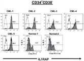
图2.IL1RAP在原始CML细胞中被上调Figure 2. IL1RAP is upregulated in naive CML cells
对5位CML患者和2例正常bm样品的CD34+细胞进行FACS分析。(A)为显示代表性的CML患者中CD34+CD38+或CD34+CD38-细胞的设门的FACS点图。(B)为显示CD34+CD38+细胞中的IL1RAP的表达的直方图。(C)为显示CD34+CD38-细胞中的IL1RAP的表达的直方图。白色表示对照染色的样品且灰色表示IL1RAP染色的样品。CD34+CD38-IL1RAP-和CD34+CD38-IL1RAP+细胞的分选门概述于直方图中。点图和直方图中的数字显示在个体的门/象限中细胞的百分比。FACS analysis of CD34+ cells from 5 CML patients and 2 normal bm samples. (A) is a FACS dot plot showing gating of CD34+ CD38+ or CD34+CD38- cells in representative CML patients. (B) is a histogram showing the expression of IL1RAP in CD34+ CD38+ cells. (C) is a histogram showing the expression of IL1RAP in CD34+ CD38- cells. White indicates control-stained samples and gray indicates IL1RAP-stained samples. Sorting gates for CD34+ CD38- IL1RAP- and CD34+ CD38- IL1RAP+ cells are outlined in histograms. Numbers in dot plots and histograms show the percentage of cells in individual gates/quadrants.
图3.在CD34+CD38-细胞区域中,IL1RAP的表达区分Ph+和Ph-CML细胞Figure 3. Expression of IL1RAP Discriminates Ph+ andPh- CML Cells in the Region of CD34+CD38- Cells
对源自5位CML患者样品的CML CD34+CD38-IL1RAP-和CD34+CD38-IL1RAP+细胞进行的Flow-drop-FISH,分别显示了BCR/ABL1-和BCR/ABL1+细胞的几乎完全的分离。黑色柱表示BCR/ABL1阴性细胞,白色柱表示BCR/ABL1阳性细胞。各柱的顶部显示了所评分的总细胞核(totalnuclei)的Ph+细胞的数量。Flow-drop-FISH on CML CD34+ CD38- IL1RAP- and CD34+ CD38- IL1RAP+ cells derived from 5 CML patient samples showed almost complete separation of BCR/ABL1- and BCR/ABL1+ cells, respectively . Black columns indicate BCR/ABL1 negative cells, and white columns indicate BCR/ABL1 positive cells. The number of Ph+ cells scored for total nuclei is shown at the top of each column.
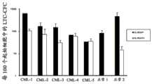
图4.IL1RAP的表达区分了正常HSC和Ph+CML干细胞Figure 4. Expression of IL1RAP differentiates normal HSCs from Ph+ CML stem cells
(A)为源自CD34+CD38-IL1RAP-和CD34+CD38-IL1RAP+细胞的LTC-CFC数量。黑色柱表示IL1RAP-细胞,白色柱表示IL1RAP+细胞。(B)为对LTC-CFC的间期FISH。黑色柱表示BCR/ABL1阴性细胞,白色柱表示BCR/ABL1阳性细胞。各柱的顶部显示了所记分的总细胞核(total nuclei)的Ph+细胞的数量。(A) is the number of LTC-CFCs derived from CD34+ CD38- IL1RAP- and CD34+ CD38- IL1RAP+ cells. Black bars representIL1RAP- cells, white bars represent IL1RAP+ cells. (B) Interphase FISH on LTC-CFC. Black columns indicate BCR/ABL1 negative cells, and white columns indicate BCR/ABL1 positive cells. The number of Ph+ cells scored for total nuclei is shown at the top of each column.
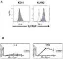
图5.通过靶向于IL1RAP的抗体杀灭CML细胞系Figure 5. Killing of CML cell lines by antibodies targeting IL1RAP
(A)为显示来自CML患者并且含费城染色体的KU812细胞上的IL1RAP的表达,与缺少费城染色体的KG-1细胞上的表达对比的直方图。白色显示对照染色的样品,灰色显示KMT-1染色的样品。白血病细胞系KG-1缺乏IL1RAP的表达,而KU812表达IL1RAP(B)。因此,KG-1中观察到低水平的抗体诱导的细胞死亡,而使用KMT-1在KU812细胞上观察到剂量依赖的ADCC作用(B)。作为非特异性ADCC作用的对照,该实验中也使用兔IgG抗体。图中显示三个独立的实验中抗体诱导的细胞死亡的平均数和标准差。(A) is a histogram showing the expression of IL1RAP on KU812 cells from a CML patient and containing the Philadelphia chromosome, compared to the expression on KG-1 cells lacking the Philadelphia chromosome. White shows control-stained samples and gray shows KMT-1-stained samples. The leukemia cell line KG-1 lacks expression of IL1RAP, whereas KU812 expresses IL1RAP (B). Thus, low levels of antibody-induced cell death were observed in KG-1, whereas a dose-dependent ADCC effect was observed on KU812 cells using KMT-1 (B). As a control for non-specific ADCC effects, rabbit IgG antibodies were also used in this experiment. The graph shows the mean and standard deviation of antibody-induced cell death from three independent experiments.
图6.通过靶向于IL1RAP的抗体杀灭CML干细胞Figure 6. Killing of CML stem cells by antibodies targeting IL1RAP
通过使用KMT-1,正常骨髓CD34+CD38-细胞对IL1RAP染色为阴性,而CML CD34+CD38+和CD34+CD38-细胞表达了IL1RAP。(A)显示代表性的实验的CML-1的直方图。白色显示对照染色的样品,灰色显示KMT-1染色的样品。与IL1RAP的表达水平相一致,使用正常骨髓CD34+CD38-细胞没有见到明显的ADCC作用,而KMT-1在CML CD34+和CD34+CD38-细胞中都诱导了强的剂量依赖的ADCC作用(B)。作为非特异性ADCC作用的对照,该实验中也使用兔IgG抗体。图中显示三个使用CML-1、CML-3、CML-4和4例正常骨髓样品的独立的实验中抗体诱导的细胞死亡的平均数和标准差。By using KMT-1, normal bone marrow CD34+CD38− cells stained negative for IL1RAP, whereas CML CD34+ CD38+ and CD34+CD38− cells expressed IL1RAP. (A) Histogram of CML-1 showing a representative experiment. White shows control-stained samples and gray shows KMT-1-stained samples. Consistent with the expression level of IL1RAP, no significant ADCC was seen using normal bone marrow CD34+ CD38- cells, whereas KMT-1 induced a strong dose-dependent ADCC in both CML CD34+ and CD34+ CD38- cells ( B). As a control for non-specific ADCC effects, rabbit IgG antibodies were also used in this experiment. The graph shows the mean and standard deviation of antibody-induced cell death in three independent experiments using CML-1, CML-3, CML-4 and 4 normal bone marrow samples.
图7.IL1RAP也在原代ALL和AML干细胞上表达Figure 7. IL1RAP is also expressed on primary ALL and AML stem cells
从确诊患者获得了急性髓性白血病(AML)细胞。(A)显示代表性的AML患者中CD34+CD38-和CD34+CD38+细胞上IL1RAP的表达。AML细胞系MONO-MAC-6和ALL细胞系REH表达了IL1RAP(B)。从确诊患者获得了急性淋巴性白血病(ALL)细胞。(C)显示代表性的Ph+ALL患者中CD34+CD38-和CD34+CD38+细胞上的IL1RAP的表达。白色显示对照染色的样品,灰色显示IL1RAP染色的样品。Acute myeloid leukemia (AML) cells were obtained from confirmed patients. (A) shows the expression of IL1RAP on CD34+CD38- and CD34+ CD38+ cells in representative AML patients. The AML cell line MONO-MAC-6 and the ALL cell line REH expressed IL1RAP (B). Acute lymphoid leukemia (ALL) cells were obtained from confirmed patients. (C) shows the expression of IL1RAP on CD34+CD38- and CD34+ CD38+ cells in a representative Ph+ ALL patient. White shows control-stained samples and gray shows IL1RAP-stained samples.
图8.通过靶向于IL1RAP的抗体杀灭AML和ALL细胞系Figure 8. Killing of AML and ALL cell lines by antibodies targeting IL1RAP
在ADCC测定中,在MONO-MAC-6和REH细胞系中都诱导了KMT-1剂量依赖性细胞死亡,这表明,靶向IL1RAP的抗体可具有比仅仅CML更宽的治疗窗。作为非特异性ADCC作用的对照,该实验中也使用兔IgG抗体。图中显示三个独立的实验中抗体诱导的细胞死亡的平均数和标准差。In the ADCC assay, KMT-1 dose-dependent cell death was induced in both MONO-MAC-6 and REH cell lines, suggesting that antibodies targeting IL1RAP may have a wider therapeutic window than CML alone. As a control for non-specific ADCC effects, rabbit IgG antibodies were also used in this experiment. The graph shows the mean and standard deviation of antibody-induced cell death from three independent experiments.
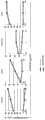
图9.通过靶向于IL1RAP的抗体杀灭AML和ALL干细胞Figure 9. Killing of AML and ALL stem cells by antibodies targeting IL1RAP
在ADCC测定中,在原代AML CD34+CD38-(A)和ALL CD34+CD38-(B)细胞中都观察到了KMT-1诱导的细胞死亡,这证实了靶向IL1RAP的抗体对于IL1RAP在其细胞表面上调的AML和ALL也具有治疗效果。作为非特异性ADCC作用的对照,该实验中也使用兔IgG抗体。图中显示特异抗体诱导的细胞死亡。In the ADCC assay, KMT-1-induced cell death was observed in both primary AML CD34+ CD38− (A) and ALL CD34+ CD38− (B) cells, confirming that antibodies targeting IL1RAP are essential for IL1RAP in its cellular Surface upregulation of AML and ALL also has a therapeutic effect. As a control for non-specific ADCC effects, rabbit IgG antibodies were also used in this experiment. The figure shows the cell death induced by the specific antibody.
图10.IL1RAP在MPD和MDS患者的白血病干细胞上表达。Figure 10. IL1RAP is expressed on leukemia stem cells from MPD and MDS patients.
(A)显示2位MPD患者(MPD-1和MPD-2)的CD34+CD38-细胞中IL1RAP的表达(伴随和不伴随JAK2突变)的等值线图(Contour plot)。(B)显示发展成为AML的MDS患者中IL1RAP的表达的直方图。白色显示对照染色的样品,灰色显示用抗IL1RAP抗体染色的样品。(A) Contour plot showing IL1RAP expression (with and without JAK2 mutation) in CD34+ CD38- cells of 2 MPD patients (MPD-1 and MPD-2). (B) Histogram showing the expression of IL1RAP in MDS patients who developed AML. White shows control-stained samples, gray shows samples stained with anti-IL1RAP antibody.
图11.来自实体肿瘤的癌细胞表面上表达了IL1RAPFigure 11. Cancer cells from solid tumors express IL1RAP on the surface
用抗人IL-1 RAcP/IL-1 R3-APC(目录号FAB676A,R&D system)(黑线)和同种型对照(灰线)将源自人实体肿瘤的不同细胞系染色。流式细胞计数分析显示了COLO829(恶性黑素瘤)、HCC1954(乳房导管癌)、NCI-8228(肺腺癌)、NCI-H716(结肠癌)、OV-90(卵巢腺癌)、H716(结肠癌)、H2228(肺腺癌)、SH-4(黑素瘤)、SR(淋巴瘤)和SW1783(星形细胞瘤)上的IL1RAP的表达。Different cell lines derived from human solid tumors were stained with anti-human IL-1 RAcP/IL-1 R3-APC (cat# FAB676A, R&D system) (black line) and isotype control (grey line). Flow cytometric analysis showed COLO829 (malignant melanoma), HCC1954 (breast duct carcinoma), NCI-8228 (lung adenocarcinoma), NCI-H716 (colon carcinoma), OV-90 (ovarian adenocarcinoma), H716 ( Expression of IL1RAP on colon carcinoma), H2228 (lung adenocarcinoma), SH-4 (melanoma), SR (lymphoma) and SW1783 (astrocytoma).
图12.来自实体肿瘤的癌细胞表面上表达了IL1RAPFigure 12. Cancer cells from solid tumors express IL1RAP on the surface
对来自用mab81.2(抗IL1RAP抗体)标记的四个不同人癌细胞系的细胞进行的流式细胞计数分析的柱状图,显示出H716(结肠癌)、H2228(肺腺癌)、HCC1954(乳房导管癌)和SH-4(黑素瘤)上的IL1RAP表达。Histogram of flow cytometric analysis of cells from four different human cancer cell lines labeled with mab81.2 (anti-IL1RAP antibody), showing H716 (colon cancer), H2228 (lung adenocarcinoma), HCC1954 ( Ductal carcinoma of the breast) and IL1RAP expression on SH-4 (melanoma).
图13.IL1RAP的抗体靶向在人癌细胞上将人NK细胞导向至ADCCFigure 13. Antibody targeting of IL1RAP directs human NK cells to ADCC on human cancer cells
图中显示了ADCC测定中通过抗人IL1RAP抗体mab81.2和人NK细胞诱导的特异性细胞死亡的程度。作为同种型对照,实验中包括了非特异性人IgG1抗体。The figure shows the extent of specific cell death induced by the anti-human IL1RAP antibody mab81.2 and human NK cells in the ADCC assay. As an isotype control, a non-specific human IgG1 antibody was included in the experiment.
图14.mAb81.2对SK-MEL-5黑素瘤细胞系的体内生长的作用Figure 14. Effect of mAb81.2 on in vivo growth of SK-MEL-5 melanoma cell line
每周两次腹膜内给药10mg/kg体重的MAb81.2。用相等体积的PBS处理对照小鼠。每个实验组含有十只小鼠。结果表示为平均肿瘤体积(mm3);误差棒表示平均值的标准误差(SEM)。MAb81.2 was administered intraperitoneally at 10 mg/kg body weight twice a week. Control mice were treated with an equal volume of PBS. Each experimental group contained ten mice. Results are expressed as mean tumor volume (mm3 ); error bars represent standard error of the mean (SEM).
实施例1Example 1
IL1RAP为慢性髓性白血病干细胞的细胞表面生物标记IL1RAP is a cell surface biomarker of chronic myelogenous leukemia stem cells
概述overview
慢性髓性白血病(CML)的以达到永久性治愈该病症为目的的治疗策略,需要完全根除CML干细胞。CML干细胞,与正常的造血干细胞(HSC)同样具有自我更新能力,代表了迄今不能用细胞表面标记与正常HSC区分的一小群白血病细胞。靶向于CML干细胞的一种策略是鉴定CML干细胞的细胞表面生物标记,未来的治疗性抗体可针对该标记。这一研究中,我们使用全基因(global gene)表达分析鉴定了,IL1RAP在原始CML CD34+细胞中被通常上调,以及由于异位P210 BCR/ABL1的表达而被上调。我们还阐明了,IL1RAP的表达将包含CML和正常HSC两者的罕见的CD34+CD38-细胞群划分为两部分:一部分具有低表达/无表达,另一部分具有较高的IL1RAP的表达。建立允许用FISH检测少量的分选的细胞中的BCR/ABL1的试验方案之后,我们观察到在CML CD34+CD38-细胞之中,IL1RAP+细胞为BCR/ABL1+,而IL1RAP-细胞几乎完全是BCR/ABL1-。通过对所述两个细胞群进一步进行长期培养-起始细胞(LTC-IC)测定,我们发现候选CML干细胞和正常HSC可预期分离。因此该研究鉴定IL1RAP为第一个可区分CML干细胞与正常HSC的细胞表面生物标记,并且为CML以及相关病症(例如急性髓性白血病(AML)、急性成淋巴细胞性白血病(ALL)、骨髓增生病(MPD)和骨髓增生异常综合征(MDS))的治疗和诊断策略开拓了新的途径。Treatment strategies for chronic myelogenous leukemia (CML) aimed at achieving a permanent cure for the disease require the complete eradication of CML stem cells. CML stem cells, which share the capacity for self-renewal with normal hematopoietic stem cells (HSCs), represent a small population of leukemic cells hitherto indistinguishable from normal HSCs by cell surface markers. One strategy to target CML stem cells is to identify cell surface biomarkers of CML stem cells against which future therapeutic antibodies can be directed. In this study, we identified using global gene expression analysis that IL1RAP is normally upregulated in naive CML CD34+ cells as well as due to ectopic P210 BCR/ABL1 expression. We also demonstrated that IL1RAP expression divides the rare CD34+CD38- cell population comprising both CML and normal HSCs into two parts: one with low/no expression and another with higher expression of IL1RAP. After establishing an assay protocol that allowed detection of BCR/ABL1 in small numbers of sorted cells by FISH, we observed that among CML CD34+CD38− cells, IL1RAP+ cells were BCR/ABL1+, whereas IL1RAP− cells were almost exclusively BCR/ABL1+ ABL1-. By further performing long-term culture-initiating cell (LTC-IC) assays on the two cell populations, we found that candidate CML stem cells and normal HSCs were predictably segregated. This study therefore identifies IL1RAP as the first cell surface biomarker that can distinguish CML stem cells from normal HSCs, and is an important marker for CML and related disorders such as acute myeloid leukemia (AML), acute lymphoblastic leukemia (ALL), myeloproliferative New avenues have been opened up in therapeutic and diagnostic strategies for myelodysplastic syndrome (MPD) and myelodysplastic syndrome (MDS).
导言preface
为了对CML干细胞鉴定细胞表面生物标记,我们进行了全基因表达分析并鉴定白细胞介素1受体辅助蛋白(IL1RAP)为首要的候选,所述IL1RAP在原始CML患者细胞中被上调,且由于异位P210BCR/ABL1的表达而被上调。通过开发出一种少数分选的细胞中检测BCR/ABL1的测定法,我们阐明IL1RAP的表达有望能够分离原始白血病细胞和正常细胞。通过长期培养-起始细胞测定,我们还阐明了IL1RAP是CML干细胞的细胞表面生物标记,首次使得预期可从正常HSC分离CML干细胞。To identify cell surface biomarkers for CML stem cells, we performed global gene expression analysis and identified
材料和方法Materials and methods
收集CML患者细胞Harvesting CML Patient Cells
分离和转导脐带血CD34+细胞Isolation and transduction of cord blood CD34+ cells
在确诊时并且在启动治疗前,在获得了知情同意之后根据本地伦理委员会批准的试验方案获得CML患者的血液样品,偶尔还获得骨髓样品。从瑞典Lund大学医院血液科和丹麦哥本哈根Rigshospitalet获得样品。定期地使用LymphoprepTM(Axis-Shield PoC AS,Oslo,Norway)根据生产者的说明书对单核细胞(MNC)进行分离,并且如此前所公开的22,使用CD34+细胞分离试剂盒(Miltenyi Biotech,Bergisch Gladbach,Germany)富集CD34+细胞,这产生了高于95%的CD34+细胞纯度。在抗体染色开始前,将单核细胞的亚级分有活力地储存于液氮中。CD34+细胞分为两部分;一部分用PBS洗涤、重新混悬于Trizol中并在-80℃冷冻,而另一部分在液氮中冷冻。在取得知情同意之后在Lund大学医院获得了来自健康志愿者的骨髓样品作为参考样品,然后如上所述地分离CD34细胞。Blood samples and occasionally bone marrow samples were obtained from CML patients at the time of diagnosis and before initiation of treatment after obtaining informed consent according to a protocol approved by the local ethics committee. Samples were obtained from the Department of Hematology, Lund University Hospital, Sweden, and Rigshospitalet, Copenhagen, Denmark. Mononuclear cells (MNC) were isolated periodically using Lymphoprep™ (Axis-Shield PoC AS, Oslo, Norway) according to the manufacturer's instructions, and as previously published22, using the CD34+ Cell Isolation Kit (Miltenyi Biotech, Bergisch Gladbach, Germany) enriched for CD34+ cells, which yielded a CD34+ cell purity greater than 95%. A subfraction of monocytes was stored viable in liquid nitrogen before antibody staining was initiated. CD34+ cells were divided into two fractions; one was washed with PBS, resuspended in Trizol and frozen at -80°C, while the other was frozen in liquid nitrogen. Bone marrow samples from healthy volunteers were obtained as reference samples at Lund University Hospital after informed consent and CD34 cells were then isolated as described above.
微阵列分析microarray analysis
使用来自瑞典Lund大学Swegene DNA Microarray Resource Center的寡核苷酸载玻片(oligonucleotide slide)进行微阵列分析。使用Pronto通用杂交试剂盒(Pronto Universal Hybridization;Corning Inc,Corning,NY)进行杂交。基本上如此前的公开23来进行RNA分离和微阵列分析。使用软件QlucoreOmics Explorer2.0(Qlucore,Lund,Sweden)进行数据可视化。Microarray analysis was performed using oligonucleotide slides from the Swegene DNA Microarray Resource Center, Lund University, Sweden. Hybridization was performed using the Pronto Universal Hybridization Kit (Pronto Universal Hybridization; Corning Inc, Corning, NY). RNA isolation and microarray analysis were performed essentially as previouslypublished23 . Data visualization was performed using the software QlucoreOmics Explorer 2.0 (Qlucore, Lund, Sweden).
流式细胞分析Flow Cytometry
在FACS Canto中进行流式细胞分析,并在FACS Aria中完成流式细胞计数细胞分选(两者都来自BD)。在细胞染色前,根据标准操作使CD34+细胞解冻,并用含2%FCS的PBS(洗涤介质)洗涤一次。使用以1:100的比例稀释的生物素标记的山羊抗人IL1RAP多克隆抗体(批次667,R&D Systems,Abingdon,UK),在冰上对所述细胞染色30分钟。接下来洗涤所述细胞,并使用1:200稀释的PE-缀合的抗生物素蛋白链菌素30分钟。使用APC-缀合的抗-CD34和FITC-缀合的抗-CD38单克隆抗体共同染色(除IL1RAP外,所有所用的抗体均购自Beckton-Dickinson Immunocytometry Systems,MountainView,CA)。在细胞分选之前,洗涤细胞两次以避免PE-缀合的抗生物素蛋白链菌素的非特异性结合。将同种型匹配对照抗体用作阴性对照。Flow cytometry analysis was performed in a FACS Canto and flow cytometry cell sorting was done in a FACS Aria (both from BD). Before cell staining, CD34+ cells were thawed according to standard procedures and washed once with PBS containing 2% FCS (washing medium). The cells were stained for 30 minutes on ice with a biotinylated goat anti-human IL1RAP polyclonal antibody (lot 667, R&D Systems, Abingdon, UK) diluted 1:100. The cells were next washed and PE-conjugated streptavidin diluted 1:200 was used for 30 minutes. Co-staining was performed using APC-conjugated anti-CD34 and FITC-conjugated anti-CD38 monoclonal antibodies (all antibodies used except IL1RAP were purchased from Beckton-Dickinson Immunocytometry Systems, Mountain View, CA). Before cell sorting, cells were washed twice to avoid non-specific binding of PE-conjugated streptavidin. An isotype-matched control antibody was used as a negative control.
细胞分选和间期FISHCell sorting and interphase FISH
保持在保湿室中,用0.01%聚L-赖氨酸(Sigma-Aldrich,Stockholm,Sweden)处理载玻片2小时,用水洗涤一次,并在37℃热板上干燥至干。接下来,用疏水笔(Daido Sangyo Co.,Ltd.Tokyo,Japan)以96孔组织培养板作为模板画圈。细胞分选之前但在室温中干燥至少2小时之后,将25μL PBS施加于环中以形成液滴。在细胞分选中,同时将30至3000个细胞直接分选入两个液滴中。为了允许所述细胞附着至表面并避免液滴干燥,将载玻片在保湿室中保持于冰上30分钟,然后用甲醇:乙酸(3:1)固定细胞10分钟。接下来,载玻片在70℃烘箱中温育过夜,然后进行FISH。使用BCR/ABL1的双色探针(Abbot,Wiesbaden,Germany)。Keeping in a humidified chamber, slides were treated with 0.01% poly-L-lysine (Sigma-Aldrich, Stockholm, Sweden) for 2 h, washed once with water, and dried to dryness on a 37 °C hot plate. Next, circles were drawn using a hydrophobic pen (Daido Sangyo Co., Ltd. Tokyo, Japan) using a 96-well tissue culture plate as a template. Before cell sorting but after drying at room temperature for at least 2 hours, 25 μL of PBS was applied to the rings to form droplets. In cell sorting, 30 to 3000 cells are sorted directly into two droplets simultaneously. To allow the cells to attach to the surface and avoid drying of the droplets, the slides were kept on ice in a humidified chamber for 30 minutes before cells were fixed with methanol:acetic acid (3:1 ) for 10 minutes. Next, slides were incubated overnight in a 70°C oven before FISH. A dual-color probe for BCR/ABL1 (Abbot, Wiesbaden, Germany) was used.
长期培养-起始细胞(LTC-IC)Long-term culture-initiating cells (LTC-IC)
如此前所公开的24,25,在补充10%FCS的RPMI-1640培养基中培养M210B4基质细胞。在细胞分选之前2天,以50,000细胞/mL的密度将基质细胞接种于96孔板的孔中的200μL含10-6M氢化可的松(Sigma-Aldrich,Stockholm,Sweden)的Myelocult培养基(Stem Cell Technologies,Vancouver,Canada)中。在细胞分选之前24小时,基质细胞经1000Rad辐照。在细胞分选中,将100-500个细胞直接分选入基质-预涂布的孔中,一式二份,并在3小时后交换100μL培养基。所述100μL培养基的交换每周重复一次。5-6周的培养后,洗涤细胞并将细胞铺板于24孔板中的甲基纤维素培养基(MethoCult H44435;Stem Cell Technologies)中。2周后,对集落的数量进行记录。集合单个孔中的集落,洗涤,施加于载玻片上的PBS液滴,然后进行上述的FISH分析。M2 10B4 stromal cells were cultured in RPMI-1640 medium supplemented with 10% FCS as previously published24,25 . 2 days before cell sorting, stromal cells were seeded at a density of 50,000 cells/mL in wells of a 96-well plate in 200 μL of Myelocult medium containing 10−6 M hydrocortisone (Sigma-Aldrich, Stockholm, Sweden) (Stem Cell Technologies, Vancouver, Canada). Stromal cells were irradiated at 1000 Rad 24 hours before cell sorting. In cell sorting, 100-500 cells were sorted directly into matrix-precoated wells in duplicate, and 100 μL of medium was exchanged after 3 hours. The 100 [mu]L medium exchange was repeated weekly. After 5-6 weeks of culture, cells were washed and plated in methylcellulose medium (MethoCult H44435; Stem Cell Technologies) in 24-well plates. After 2 weeks, the number of colonies was recorded. Colonies from individual wells were pooled, washed, applied to drops of PBS on slides, and then subjected to FISH analysis as described above.
脐带血CD34+细胞中P210 BCR/ABL1的表达Expression of P210 BCR/ABL1 in cord blood CD34+ cells
根据本地伦理委员会批准的试验方案,在获得知情同意后从正常分娩收集脐带的脐带血样品。如前所述地22富集CD34+细胞,这产生了高于95%的CD34+细胞纯度。本研究中使用RD114假型化的MSCV-IRES-GFP(MIG)和MIG-P210病毒载体23。在补充血小板生成素(TPO;50ng/mL)、干细胞因子(SCF;100ng/mL)和Flt-3-配体(FL;100ng/mL)的SFMM培养基(Stem CellTechnology,Vancouver,Canada)中培养和转导CD34+细胞,如前文所述23。Umbilical cord blood samples were collected from normal deliveries after informed consent was obtained according to the protocol approved by the local ethics committee. CD34+ cells were enriched for22 as previously described, which yielded a purity of CD34+ cells greater than 95%. RD114-pseudotyped MSCV-IRES-GFP (MIG) and MIG-P210 viral vectors were used in this study23 . Cultured in SFMM medium (Stem Cell Technology, Vancouver, Canada) supplemented with thrombopoietin (TPO; 50 ng/mL), stem cell factor (SCF; 100 ng/mL) and Flt-3-ligand (FL; 100 ng/mL) and transduce CD34+ cells as previously described23 .
结果与讨论Results and discussion
全基因表达分析鉴定CML CD34+细胞上IL1RAP的上调Global gene expression analysis identifies upregulation of IL1RAP on CML CD34+ cells
为实现鉴定Ph+CML干细胞的细胞表面生物标记的目标,已进行了很多研究上的努力(C Eaves的综述14)。用FISH检测白血病特异性BCR/ABL1融合基因之后很容易回顾性地鉴定CML中的白血病和正常细胞,这使得CML成为评估对于预期的白血病和正常细胞分离的尝试的理想的病症。然而迄今为止,还没有鉴定出能够允许CML干细胞与正常HSC预期分离的细胞表面标记。已证实全基因表达分析是一种有效的寻找新HSC标记的策略,所述新HSC标记例如区分造血干细胞和祖细胞的SLAM受体15。为了寻找编码CML干细胞的候选细胞表面生物标记的上调的基因,对比了11位CML患者样品和5例正常骨髓(bm)样品的CD34+细胞的转录概况。所鉴定的CML中上调的基因匹配基因本体(Gene Ontology,GO)类别“整合至质膜(integral to plasmamembrane)”,该类别已经过手工整理以包括所有已知的CD分子(详见材料和方法)。CML CD34+细胞中总计13个上调的基因匹配所述的“整合至质膜”的基因类别(数据未显示)。为进一步地将上调的基因与P210 BCR/ABL1表达更直接地联系起来,我们平行地产生了由于脐带血CD34+细胞中P210BCR/ABL1的表达而上调的一系列基因的列表。该分析的结果是,23个上调的基因匹配相同的GO类别基因列表(数据未显示)。有趣的是,只有一个基因即白细胞介素1受体辅助蛋白(IL1RAP)由于P210 BCR/ABL1的表达在CD34+CML细胞中和脐带血CD34+细胞中都显示了较强的上调。IL1RAP存在于两种基因列表上这一发现表明,其在原始CML细胞上的上调与P210 BCR/ABL1表达紧密联系,并表明IL1RAP是原始CML细胞上的一种新的与白血病相关的抗原。There have been many research efforts towards the goal of identifying cell surface biomarkers of Ph+ CML stem cells (reviewed by C Eaves14 ). The ease of retrospective identification of leukemic and normal cells in CML following detection of the leukemia-specific BCR/ABL1 fusion gene by FISH makes CML an ideal condition to evaluate attempts to separate prospective leukemic and normal cells. To date, however, no cell surface markers have been identified that would allow the prospective separation of CML stem cells from normal HSCs. Global gene expression analysis has proven to be an effective strategy for finding novel HSC markers such as the SLAM receptor that differentiates hematopoietic stem and progenitor cells15 . To search for upregulated genes encoding candidate cell surface biomarkers of CML stem cells, we compared the transcriptional profiles of CD34+ cells from 11 CML patient samples and 5 normal bone marrow (bm) samples. The identified upregulated genes in CML matched the Gene Ontology (GO) category 'integral to plasmamembrane', which has been manually curated to include all known CD molecules (see Materials and methods for details ). A total of 13 up-regulated genes in CML CD34+ cells matched the "plasma membrane-integrated" gene category described (data not shown). To further link upregulated genes more directly to P210BCR/ABL1 expression, we in parallel generated a list of genes upregulated due to P210BCR/ABL1 expression in cord blood CD34+ cells. As a result of this analysis, 23 upregulated genes matched the same GO category gene list (data not shown). Interestingly, only one gene,
IL1RAP在CML患者的CD34+CD38-细胞上上调,并且由于异位P210BCR/ABL1的表达而诱导IL1RAP is upregulated on CD34+ CD38- cells of CML patients and induced by ectopic P210BCR/ABL1 expression
IL1RAP是Toll样受体超家族成员之一,并且是1型白细胞介素1受体(IL-1R1)的一种公知的共同受体16。因此IL1RAP在介导促炎症细胞因子IL-1的作用中是关键的,但其还涉及介导IL-33的信号,所述IL-33是通过结合其受体ST2激活T-细胞和肥大细胞的细胞因子,其接下来与IL1RAP二聚化17。IL-1R1的激活之前已显示刺激对干扰素敏感的CML细胞的集落生长18,然而,据我们所知IL1RAP此前并未直接与CML相关联。IL1RAP is a member of the Toll-like receptor superfamily and is a well-known co-receptor for
由于P210 BCR/ABL1存在于CML细胞中作为疾病的标志,CML中可靠的细胞表面生物标记在理想情况下应直接与P210 BCR/ABL1的存在和表达相关联。与微阵列数据一致,IL1RAP的表达实际上在逆转录病毒的P210BCR/ABL1表达之后在CB CD34+细胞的细胞表面上被上调(图1)。这表明P210BCR/ABL1通过直接或间接作用调节IL1RAP的表达,这加强了其作为CML生物标记候选的地位。Since P210 BCR/ABL1 is present in CML cells as a marker of disease, reliable cell surface biomarkers in CML should ideally correlate directly with the presence and expression of P210 BCR/ABL1. Consistent with the microarray data, expression of IL1RAP was actually upregulated on the cell surface of CB CD34+ cells following retroviral P210BCR/ABL1 expression (Figure 1). This suggests that P210BCR/ABL1 regulates IL1RAP expression through direct or indirect actions, which strengthens its position as a CML biomarker candidate.
我们接下来研究了CML CD34+CD38+细胞上细胞表面IL1RAP的表达,所述细胞代表了主要的和更成熟的CD34+细胞。在该细胞群中,对比相应的正常骨髓(bm)细胞中的表达,观察到IL1RAP的上调(图2A、2B)。正常CD34+CD38+细胞显示更低的IL1RAP的表达,其部分地与CML细胞上的表达重叠。我们然后转向至正常细胞(包括HSC)的CD34+CD38-细胞区域。与前述研究一致,该细胞群显示IL1RAP低表达/无表达(图2C)19。令人震撼的是,包含Ph+CML干细胞和正常HSC两者的CML患者的CD34+CD38-细胞被划分为两个群体:一个群体具有IL1RAP的低表达/无表达,另一群体具有较高的IL1RAP表达(图2C)。在5位CML患者的外周血液(PB)中,IL1RAP阳性细胞部分构成CD34+CD38-细胞的75%至95%(n=5)。基于这些发现,我们推断IL1RAP的表达可在CML中CD34+CD38-细胞区域内区分正常细胞和白血病细胞。由于所有CML干细胞和正常HSC只发现于CD34+CD38-细胞中,所述正常细胞和白血病细胞的区分,将允许CML干细胞从正常HSC的预期分离。We next investigated the expression of cell surface IL1RAP on CML CD34+ CD38+ cells, which represent both primary and more mature CD34+ cells. In this cell population, upregulation of IL1RAP was observed compared to expression in corresponding normal bone marrow (bm) cells (Fig. 2A, 2B). Normal CD34+ CD38+ cells showed lower expression of IL1RAP, which partially overlapped with that on CML cells. We then turned to the CD34+ CD38- cell domain of normal cells, including HSCs. Consistent with previous studies, this cell population showed low/no expression of IL1RAP (Fig. 2C)19 . Strikingly, CD34+CD38- cells of CML patients containing both Ph+ CML stem cells and normal HSCs were divided into two populations: one population with low/no expression of IL1RAP and the other with higher IL1RAP expression (Fig. 2C). In the peripheral blood (PB) of 5 CML patients, the IL1RAP positive cell fraction constituted 75% to 95% of the CD34+ CD38- cells (n=5). Based on these findings, we reasoned that expression of IL1RAP could differentiate normal cells from leukemic cells within the CD34+CD38− cell region in CML. Since all CML stem cells and normal HSCs are found only in CD34+ CD38- cells, the differentiation of normal and leukemic cells would allow for the expected separation of CML stem cells from normal HSCs.
Flow-drop-FISH显示IL1RAP的表达在CML CD34+CD38-细胞内区分正常细胞和白血病细胞Flow-drop-FISH reveals IL1RAP expression in CML CD34+ CD38- cells to distinguish normal cells from leukemia cells
为检验IL1RAP的表达是否可在CML CD34+CD38-细胞区域内区分正常(Ph-)细胞和白血病(Ph+)细胞,我们建立了对少量的分选细胞进行荧光原位杂交(FISH)的新的试验方案(参见材料和方法)。该试验方案的第一步部分地基于将细胞分选入载玻片上的液滴中然后进行单细胞免疫染色的方法20。通过应用这一涉及将细胞直接分选入载玻片上的液滴中然后进行FISH的试验方案(下文称为Flow-drop-FISH),我们将少达30个细胞分选入一滴中,由此通过FISH成功地对15个细胞核进行了评分(CML-5,图3)。有趣的是,我们通过Flow-drop-FISH发现,CML CD34+CD38-IL1RAP+细胞为BCR/ABL1+,而CMLCD34+CD38-IL1RAP-细胞几乎全部是Ph-(n=5,图3)。这些数据显示,IL1RAP的表达在CML CD34+CD38-细胞区域内区分了白血病细胞和正常细胞,表明CML干细胞和正常HSC可预期分离。To test whether expression of IL1RAP could distinguish normal (Ph− ) cells from leukemic (Ph+ ) cells in the CD34+ CD38− cell region of CML, we established a new assay for fluorescence in situ hybridization (FISH) on a small number of sorted cells. experimental protocol (see Materials and methods). The first step of this protocol is based in part on the method of sorting cells into droplets on a slide followed by single cell immunostaining20 . By applying this protocol involving sorting cells directly into droplets on a glass slide followed by FISH (hereafter referred to as Flow-drop-FISH), we sorted as few as 30 cells into a drop, thereby Fifteen nuclei were successfully scored by FISH (CML-5, Figure 3). Interestingly, we found by Flow-drop-FISH that CML CD34+ CD38- IL1RAP+ cells were BCR/ABL1+ , while CMLCD34+ CD38- IL1RAP- cells were almost all Ph- (n=5, Figure 3). These data show that IL1RAP expression differentiates leukemic cells from normal cells within the CML CD34+ CD38- cell domain, suggesting a predictable separation of CML stem cells and normal HSCs.
CML干细胞为CD34+CD38-IL1RAP+,而正常HSC为CD34+CD38-IL1RAP-/低CML stem cells are CD34+ CD38- IL1RAP+ , while normal HSCs are CD34+ CD38- IL1RAP-/low
对慢性期CML干细胞的研究,目前依赖于对罕见CML患者的接近,所述患者中经长期测定后干细胞区域已主要被白血病细胞占据14。由于免疫缺陷小鼠中CML干细胞通常显示较弱的移植物移入,因此广泛使用长期培养起始细胞(long-term culture initiating cell,LTC-IC)-测试,作为候选CML干细胞检测的替代性测试。为检测CML CD34+CD38-IL1RAP+和CD34+CD38-IL1RAP-/低是否分别独特地包含候选CML干细胞和正常HSC,我们在LTC-IC测定中检测这两个细胞群。对于来自正常对照的骨髓CD34+细胞,发现与CD34+CD38-IL1RAP+细胞中相比,CD34+CD38-IL1RAP-细胞中的长期培养-集落形成细胞(LTC-CFC)的频率高出>100倍(图4A,n=2),表明正常CD34+CD38-IL1RAP-分层地(hierarchically)位于CD34+CD38-IL1RAP+细胞顶部。在CML中,与CD34+CD38-IL1RAP+细胞相比,我们观察到CD34+CD38-IL1RAP-细胞中的LTC-CFC具有平均3.6倍的更高频率(n=5,图4A),这表明对于原始细胞CML CD34+CD38-IL1RAP-细胞更加富集。重要的是,尽管CML患者样品和正常对照两者中都发现了,CD34+CD38-IL1RAP-细胞中的LTC-IC数量比CD34+CD38-IL1RAP+细胞中的LTC-IC数量更高,对CML LTC-集落进行的FISH显示了两组中Ph-和Ph+细胞几乎完全的区分(图4B)。来自CD34+CD38-IL1RAP-细胞的CML LTC-集落几乎全部为Ph-,而CD34+CD38-IL1RAP+几乎全部为Ph+。这些数据表明IL1RAP是可用于区分CML干细胞与正常HSC的新的细胞表面生物标记。Studies of stem cells in chronic phase CML currently rely on access to rare CML patients in whom the stem cell domain has been predominately occupied by leukemic cells after long-term determinations14 . Since CML stem cells often show weak engraftment in immunodeficient mice, the long-term culture initiating cell (LTC-IC)-test is widely used as a surrogate test for candidate CML stem cell detection. To test whether CML CD34+ CD38- IL1RAP+ and CD34+ CD38- IL1RAP-/low uniquely comprise candidate CML stem cells and normal HSCs, respectively, we tested these two cell populations in the LTC-IC assay. For bone marrow CD34+ cells from normal controls, a >100-fold higher frequency of long-term culture-colony-forming cells (LTC-CFCs) was found in CD34+CD38-IL1RAP-cellscompared to CD34+ CD38-IL1RAP+ cells (Fig. 4A, n=2), indicating that normal CD34+ CD38-IL1RAP- is hierarchically located on top of CD34+ CD38- IL1RAP+ cells. In CML, we observed an average 3.6-fold higher frequency of LTC-CFCs in CD34+ CD38- IL1RAP- cells compared to CD34+ CD38- IL1RAP+ cells (n = 5, Fig. 4A), suggesting that for Blasts were more enriched in CML CD34+ CD38- IL1RAP- cells. Importantly, although found in both CML patient samples and normal controls, the number of LTC-ICs in CD34+ CD38- IL1RAP- cells was higher than that in CD34+ CD38- IL1RAP+ cells, which was more important for CML FISH performed on LTC- colonies showed almost complete differentiation of Ph-and Ph+ cells in both groups (Fig. 4B). CML LTC- colonies from CD34+ CD38- IL1RAP- cells were almost all Ph- , whereas CD34+ CD38- IL1RAP+ were almost all Ph+ . These data suggest that IL1RAP is a novel cell surface biomarker that can be used to distinguish CML stem cells from normal HSCs.
本文中,我们通过全基因表达分析鉴定了新的细胞表面抗原IL1RAP,其在多重测定的挑战后,满足了成为Ph+CML干细胞的新的细胞表面生物标记的标准。基于这一发现,未来针对CML的治疗可设计为:通过使用指向IL1RAP的治疗性抗体,靶向至CML干细胞同时保留正常HSC。另外,出于诊断目的以及为了对不同治疗下的CML患者进行随访研究,可使用含抗-CD34、抗-CD38和抗-IL1RAP抗体的抗体混合物(cocktail)。重要的是,正常和CML干细胞的预期分离,将使这两种细胞群的未来的机理研究成为可能。另外,本文还显示,Flow-drop-FISH可作为在少量的分选的细胞中表征遗传畸变的有用的方法(所述少量的分选的细胞例如白血病干细胞,其为已纯化至越来越小和越来越纯的细胞群的一种细胞型21)。对于未来的研究,该方法将例如允许在多种较小的白血病干细胞和祖细胞群中检测遗传学畸变,已获得可能为多种畸变的指令提供新见解的发现,其为理解白血病生成的关键知识。另外,Flow-drop-FISH可用于在治疗中监测对白血病干细胞的治疗效果。重要的是,我们在此通过使用Flow-drop-FISH鉴定了,IL1RAP是CML干细胞区分于正常HSC的首要的细胞表面生物标记,这一发现为CML和与IL1RAP在干细胞和/或祖细胞上的上调相关的其他血液肿瘤病症开拓了新的治疗机遇。Here, we identify a novel cell surface antigen, IL1RAP, by global gene expression analysis that, after multiple assay challenge, fulfills the criteria to be a novel cell surface biomarker for Ph+ CML stem cells. Based on this finding, future treatments for CML could be designed to target CML stem cells while sparing normal HSCs by using therapeutic antibodies directed against IL1RAP. In addition, for diagnostic purposes and for follow-up studies of CML patients under different treatments, an antibody cocktail containing anti-CD34, anti-CD38 and anti-IL1RAP antibodies can be used. Importantly, the anticipated separation of normal and CML stem cells will enable future mechanistic studies of these two cell populations. In addition, it is also shown herein that Flow-drop-FISH can be a useful method for characterizing genetic aberrations in small numbers of sorted cells, such as leukemia stem cells, which have been purified to smaller and smaller and a cell type of increasingly pure cell populations21 ). For future studies, the method will, for example, allow the detection of genetic aberrations in multiple, smaller populations of leukemia stem and progenitor cells. Discoveries have been made that may provide new insights into the instruction of multiple aberrations, which are key to understanding leukemogenesis Knowledge. In addition, Flow-drop-FISH can be used to monitor the therapeutic effect on leukemia stem cells during therapy. Importantly, we identify here, using Flow-drop-FISH, that IL1RAP is the primary cell surface biomarker that differentiates CML stem cells from normal HSCs Upregulation associated with other hematologic malignancies opens up new therapeutic opportunities.
参考文献references
1.Deininger MW,Goldman JM,Melo JV.The molecular biology of chronicmyeloid leukemia.Blood.2000;96:3343-3356.1. Deininger MW, Goldman JM, Melo JV. The molecular biology of chronic myeloid leukemia. Blood. 2000;96:3343-3356.
2.Fialkow PJ,Denman AM,Jacobson RJ,Lowenthal MN.Chronic myelocyticleukemia.Origin of some lymphocytes from leukemic stem cells.J ClinInvest.1978;62:815-823.2. Fialkow PJ, Denman AM, Jacobson RJ, Lowenthal MN. Chronic myelocyteukemia. Origin of some lymphocytes from leukemic stem cells. J ClinInvest.1978;62:815-823.
3.Kavalerchik E,Goff D,Jamieson CH.Chronic myeloid leukemia stem cells.J Clin Oncol.2008;26:2911-2915.3. Kavalerchik E, Goff D, Jamieson CH. Chronic myeloid leukemia stem cells. J Clin Oncol.2008;26:2911-2915.
4.Jiang X,Zhao Y,Smith C,et al.Chronic myeloid leukemia stem cellspossess multiple unique features of resistance to BCR-ABL targetedtherapies.Leukemia.2007;21:926-935.4. Jiang X, Zhao Y, Smith C, et al. Chronic myeloid leukemia stem cells sposssess multiple unique features of resistance to BCR-ABL targeted therapies. Leukemia.2007;21:926-935.
5.Copland M,Hamilton A,Elrick LJ,et al.Dasatinib(BMS-354825)targetsan earlier progenitor population than imatinib in primary CML but does noteliminate the quiescent fraction.Blood.2006;107:4532-4539.5. Copland M, Hamilton A, Elrick LJ, et al. Dasatinib (BMS-354825) targets an earlier progenitor population than imatinib in primary CML but does noteeliminate the quiescent fraction. Blood. 2006;107:4532-4539.
6.Jin L,Hope KJ,Zhai Q,Smadja-Joffe F,Dick JE.Targeting of CD44eradicates human acute myeloid leukemic stem cells.Nat Med.2006;12:1167-1174.6. Jin L, Hope KJ, Zhai Q, Smadja-Joffe F, Dick JE. Targeting of CD44 eradicates human acute myeloid leukemic stem cells. Nat Med.2006;12:1167-1174.
7.Tavor S,Petit I,Porozov S,et al.CXCR4 regulates migration anddevelopment of human acute myelogenous leukemia stem cells intransplanted NOD/SCID mice.Cancer Res.2004;64:2817-2824.7. Tavor S, Petit I, Porozov S, et al. CXCR4 regulates migration and development of human acute myelogenous leukemia stem cells intransplanted NOD/SCID mice. Cancer Res. 2004;64:2817-2824.
8.Jin L,Lee EM,Ramshaw HS,et al.Monoclonal antibody-mediatedtargeting of CD123,IL-3 receptor alpha chain,eliminates human acutemyeloid leukemic stem cells.Cell Stem Cell.2009;5:31-42.8. Jin L, Lee EM, Ramshaw HS, et al. Monoclonal antibody-mediated targeting of CD123, IL-3 receptor alpha chain, eliminates human acutemyeloid leukemic stem cells. Cell Stem Cell. 2009;5:31-42.
9.Majeti R,Chao MP,Alizadeh AA,et al.CD47 is an adverse prognosticfactor and therapeutic antibody target on human acute myeloid leukemiastem cells.Cell.2009;138:286-299.9. Majeti R, Chao MP, Alizadeh AA, et al. CD47 is an adverse prognostic factor and therapeutic antibody target on human acute myeloid leukemiastem cells. Cell. 2009;138:286-299.
10.Hosen N,et al.CD96 is a leukemic stem cell-specific marker in humanacute myeloid leukemia.Proc Natl Acad Sci U S A.2007;104:11008-11013.10. Hosen N, et al. CD96 is a leukemic stem cell-specific marker in humanacute myeloid leukemia. Proc Natl Acad Sci U S A.2007;104:11008-11013.
11.van Rhenen A,van Dongen GA,Kelder A,et al.The novel AML stem cellassociated antigen CLL-1 aids in discrimination between normal andleukemic stem cells.Blood.2007;110:2659-2666.11. van Rhenen A, van Dongen GA, Kelder A, et al. The novel AML stem cell associated antigen CLL-1 aids in discrimination between normal andleukemic stem cells. Blood.2007;110:2659-2666.
12.Eisterer W,Jiang X,Christ O,et al.Different subsets of primary chronicmyeloid leukemia stem cells engraft immunodeficient mice and produce amodel of the human disease.Leukemia.2005;19:435-441.12. Eisterer W, Jiang X, Christ O, et al. Different subsets of primary chronic myeloid leukemia stem cells engraft immunodeficient mice and produce amodel of the human disease. Leukemia. 2005; 19: 435-441.
13.Bhatia M,Wang JC,Kapp U,Bonnet D,Dick JE.Purification of primitivehuman hematopoietic cells capable of repopulating immune-deficient mice.Proc Natl Acad Sci U S A.1997;94:5320-5325.13. Bhatia M, Wang JC, Kapp U, Bonnet D, Dick JE. Purification of primitive human hematopoietic cells capable of repopulating immune-deficient mice. Proc Natl Acad Sci U S A.1997;94:5320-5325.
14.Jiang X,Zhao Y,Forrest D,Smith C,Eaves A,Eaves C.Stem cellbiomarkers in chronic myeloid leukemia.Dis Markers.2008;24:201-216.14. Jiang X, Zhao Y, Forrest D, Smith C, Eaves A, Eaves C. Stem cell biomarkers in chronic myeloid leukemia. Dis Markers. 2008;24:201-216.
15.Kiel MJ,Yilmaz OH,Iwashita T,Terhorst C,Morrison SJ.SLAM familyreceptors distinguish hematopoietic stem and progenitor cells and revealendothelial niches for stem cells.Cell.2005;121:1109-1121.15. Kiel MJ, Yilmaz OH, Iwashita T, Terhorst C, Morrison SJ. SLAM family receptors distinguish hematopoietic stem and progenitor cells and reveal endothelial niches for stem cells. Cell.2005;121:1109-1121.
16.Subramaniam S,Stansberg C,Cunningham C.The interleukin 1 receptorfamily.Dev Comp Immunol.2004;28:415-428.16. Subramaniam S, Stansberg C, Cunningham C. The
17.Ali S,Huber M,Kollewe C,Bischoff SC,Falk W,Martin MU.IL-1 receptoraccessory protein is essential for IL-33-induced activation of T lymphocytesand mast cells.Proc Natl Acad Sci U S A.2007;104:18660-18665.17. Ali S, Huber M, Kollewe C, Bischoff SC, Falk W, Martin MU.IL-1 receptor accessory protein is essential for IL-33-induced activation of T lymphocytes and mast cells.Proc Natl Acad Sci U S A.2007; 104:18660-18665.
18.Estrov Z,et al.Suppression of chronic myelogenous leukemia colony growthby interleukin-1(IL-1)receptor antagonist and soluble IL-1 receptors:anovel application for inhibitors of IL-1 activity.Blood.1991;78:1476-1484.18. Estrov Z, et al. Suppression of chronic myelogenous leukemia colony growth by interleukin-1(IL-1) receptor antagonist and soluble IL-1 receptors: novel application for inhibitors of IL-1 activity.Blood.1991;78:1476- 1484.
19.Hystad ME,Myklebust JH,Bo TH,et al.Characterization of early stages ofhuman B cell development by gene expression profiling.J Immunol.2007;179:3662-3671.19. Hystad ME, Myklebust JH, Bo TH, et al.Characterization of early stages of human B cell development by gene expression profiling.J Immunol.2007;179:3662-3671.
20.Ema H,Morita Y,Yamazaki S,et al.Adult mouse hematopoietic stem cells:purification and single-cell assays.Nat Protoc.2006;1:2979-2987.20. Ema H, Morita Y, Yamazaki S, et al. Adult mouse hematopoietic stem cells: purification and single-cell assays. Nat Protoc.2006;1:2979-2987.
21.Dick JE.Stem cell concepts renew cancer research.Blood.2008;112:4793-4807.21. Dick JE.Stem cell concepts renew cancer research.Blood.2008;112:4793-4807.
22.Nilsson M,Karlsson S,Fan X.Functionally distinct subpopulations of cordblood CD34+ cells are transduced by adenoviral vectors with serotype5or35tropism.Mol Ther.2004;9:377-388.22. Nilsson M, Karlsson S, Fan X. Functionally distinct subpopulations of cordblood CD34+ cells are transduced by adenoviral vectors with serotype5or35tropism. Mol Ther.2004;9:377-388.
23.Jaras M,Johnels P,Agerstam H,et al.Expression of P190 and P210BCR/ABL1 in normal human CD34(+)cells induces similar gene expressionprofiles and results in a STAT5-dependent expansion of the erythroid lineage.Exp Hematol.2009;37:367-375.23. Jaras M, Johnels P, Agerstam H, et al. Expression of P190 and P210BCR/ABL1 in normal human CD34(+) cells induces similar gene expression profiles and results in a STAT5-dependent expansion of the erythroid ol lineage.20
24.Hogge DE,et al..Enhanced detection,maintenance,and differentiation ofprimitive human hematopoietic cells in cultures containing murinefibroblasts engineered to produce human steel factor,interleukin-3,andgranulocyte colony-stimulating factor.Blood.1996;88:3765-3773.24. Hogge DE, et al.. Enhanced detection, maintenance, and differentiation of primitive human hematopoietic cells in cultures containing murine fibroblasts engineered to produce human steel factor, interleukin-3, and granular colony-stimulating; .
25.Castor A,Nilsson L,Astrand-Grundstrom I,et al.Distinct patterns ofhematopoietic stem cell involvement in acute lymphoblastic leukemia.NatMed.2005;11:630-637.25. Castor A, Nilsson L, Astrand-Grundstrom I, et al. Distinct patterns of hematopoietic stem cell involvement in acute lymphoblastic leukemia. NatMed.2005;11:630-637.
实施例2Example 2
在白血病干细胞和祖细胞上的IL1RAP的抗体-靶向引起抗体依赖性细胞介导的细胞毒性(ADCC)Antibody-targeting of IL1RAP on leukemic stem and progenitor cells causes antibody-dependent cell-mediated cytotoxicity (ADCC)
概述overview
白血病的以达到永久性治愈该病症为目的的治疗策略,需要完全根除白血病干细胞。所述白血病干细胞代表了白血病细胞的一个小的群体,其迄今不能用细胞表面标记从正常造血干细胞(HSC)区分。一种靶向白血病干细胞的新观念,将鉴定白血病干细胞的细胞表面生物标记,且未来治疗性抗体将指向该生物标记(参见实施例1)。Therapeutic strategies for leukemia to achieve a permanent cure for the disease require the complete eradication of leukemia stem cells. The leukemic stem cells represent a small population of leukemic cells, hitherto indistinguishable from normal hematopoietic stem cells (HSC) by cell surface markers. A new concept for targeting leukemic stem cells will identify cell surface biomarkers of leukemic stem cells to which future therapeutic antibodies will be directed (see Example 1).
该研究中,我们生成了抗-IL1RAP抗体,并提供了以下概念验证:靶向于慢性髓性白血病(CML)干细胞、急性髓性白血病(AML)干细胞和急性成淋巴细胞性白血病(ALL)干细胞的抗-IL1RAP抗体可用于诱导抗体依赖性细胞介导的细胞毒性(ADCC),而在正常HSC中未观察到细胞毒性作用。此外,我们证明了在IL1RAP阳性细胞系KU812(CML)、MONO-MAC-6(急性髓性白血病;AML)和REH(急性成淋巴细胞细胞系;ALL)中靶向于IL1RAP的剂量依赖的ADCC。我们还阐明了MDS和MPD干细胞增加了IL1RAP的表达,这表明未来的治疗性的抗-IL1RAP靶向抗体在这些病症中将也有效。In this study, we generated anti-IL1RAP antibodies and provided proof-of-concept targeting chronic myeloid leukemia (CML) stem cells, acute myeloid leukemia (AML) stem cells, and acute lymphoblastic leukemia (ALL) stem cells The anti-IL1RAP antibody can be used to induce antibody-dependent cell-mediated cytotoxicity (ADCC), while no cytotoxic effect was observed in normal HSC. Furthermore, we demonstrated dose-dependent ADCC targeting IL1RAP in the IL1RAP-positive cell lines KU812 (CML), MONO-MAC-6 (acute myeloid leukemia; AML) and REH (acute lymphoblastic cell line; ALL) . We also demonstrate that MDS and MPD stem cells increase IL1RAP expression, suggesting that future therapeutic anti-IL1RAP targeting antibodies will also be effective in these conditions.
通过靶向于白血病干细胞上IL1RAP的抗体,该研究因此为CML、AML、ALL、MDS和MPD开拓了新的治疗机遇。This study thus opens up new therapeutic opportunities for CML, AML, ALL, MDS and MPD by targeting antibodies against IL1RAP on leukemia stem cells.
材料和方法Materials and methods
KMT-1(一种多克隆兔抗人IL1RAP抗体)的产生Production of KMT-1, a polyclonal rabbit anti-human IL1RAP antibody
用IL1RAP的胞外域免疫兔。根据标准操作纯化来自兔的血清,不同是在其中加入另外的步骤,其中将结合至免疫球蛋白域(存在于免疫蛋白上以增加半衰期)的抗体通过结合至装载免疫球蛋白的柱而舍弃。经ELISA证实,纯化的抗体与IL1RAP的胞外域结合,并且缺乏结合人免疫球蛋白域的抗体。在流式细胞计数中使用时,将PE-缀合的山羊抗-兔IgG抗体用作二级试剂(secondary reagent)。Rabbits were immunized with the extracellular domain of IL1RAP. Sera from rabbits were purified according to standard procedures, except that an additional step was added in which antibodies bound to immunoglobulin domains (present on immunoglobulin proteins to increase half-life) were discarded by binding to immunoglobulin-loaded columns. Purified antibodies bound to the extracellular domain of IL1RAP and lacked antibodies binding to human immunoglobulin domains, as confirmed by ELISA. When used in flow cytometry, a PE-conjugated goat anti-rabbit IgG antibody was used as a secondary reagent.
ADCC测定ADCC determination
ADCC测定是基于此前公开的试验方案1。简言之,根据生产者的说明书用PKH26(Sigma-Aldrich,St Louis,Missouri)标记靶细胞,并且或者将细胞直接置于96孔板的孔中,或者在CD34+CD38-细胞分选之后将细胞接种于所述孔中。KU812和KG-1细胞系和原代CD34+细胞以10,000细胞/孔接种,而原代CD34+CD38-细胞以2,000-3,000细胞/孔接种。接下来,将抗体以不同浓度加入孔中并培养20分钟,然后向各孔中加入100,000NK-效应细胞。NK细胞是在获得知情同意后从健康的志愿者提取的,所述提取是使用NK细胞阴性细胞分离试剂盒(Miltenyi Biotech,Bergisch Gladbach,Germany)根据生产者的说明书进行的。实验中使用从未经免疫的兔提纯的兔IgG抗体作为对照抗体(R&D Systems Abingdon,UK)。使用FACS CANTO流式细胞器(BD)测定用于检测细胞死亡的7-AAD阳性细胞。从至少三个独立的实验(除了图9;只有一个实验),根据以下等式计算抗体诱导的细胞死亡的平均数和标准差:(在确定的抗体浓度的7-AAD+细胞的百分比-无抗体的7-AAD+细胞的百分比)/(0.01×无抗体的7-AAD-细胞的百分比)。The ADCC assay was based on a previously published assay protocol1 . Briefly, target cells were labeled with PKH26 (Sigma-Aldrich, St Louis, Missouri) according to the manufacturer's instructions and either placed directly into the wells of a 96-well plate or after CD34+CD38- cell sorting Cells are seeded in the wells. KU812 and KG-1 cell lines and primary CD34+ cells were seeded at 10,000 cells/well, while primary CD34+CD38- cells were seeded at 2,000-3,000 cells/well. Next, antibodies were added to the wells at various concentrations and incubated for 20 minutes before adding 100,000 NK-effector cells to each well. NK cells were extracted from healthy volunteers after informed consent was obtained using the NK Cell Negative Cell Isolation Kit (Miltenyi Biotech, Bergisch Gladbach, Germany) according to the manufacturer's instructions. Rabbit IgG antibody purified from unimmunized rabbits was used as a control antibody (R&D Systems Abingdon, UK) in the experiment. 7-AAD positive cells for detection of cell death were determined using a FACS CANTO flow cytometer (BD). From at least three independent experiments (except Figure 9; only one experiment), the mean and standard deviation of antibody-induced cell death were calculated according to the following equation: (percentage of 7-AAD+ cells at defined antibody concentration - no antibody Percentage of 7-AAD+ cells)/(0.01×percentage of 7-AAD- cells without antibody).
从Lund大学医院获得来自11位AML患者和2位Ph+ALL患者的样品,且在CD34+CD38+和CD34+CD38-细胞群中使用与CML细胞的分析的相同设置来分析IL1RAP的表达。使用与KG-1和KU812细胞系相同的设置,在ADCC测定中还检测了AML细胞系MONO-MAC-6和ALL细胞系REH。Samples from 11 AML patients and 2 Ph+ ALL patients were obtained from Lund University Hospital, and the expression of IL1RAP was analyzed in CD34+ CD38+ and CD34+CD38- cell populations using the same settings as for the analysis of CML cells. Using the same setup as the KG-1 and KU812 cell lines, the AML cell line MONO-MAC-6 and the ALL cell line REH were also tested in the ADCC assay.
结果result
在CML干细胞和祖细胞上以及在CML细胞系上的IL1RAP的抗体-靶向,将NK细胞导向ADCCAntibody-targeting of IL1RAP on CML stem and progenitor cells and on CML cell lines directs NK cells to ADCC
抗体依赖性细胞介导的细胞毒性(ADCC)是先天免疫系统的保存的机理,认为一些治疗性抗体(例如对抗CD20的利妥昔单抗)至少部分地通过该机理发挥其治疗效果2。为检验使用IL1RAP作为靶点是否可实现ADCC,我们生成了多克隆兔抗人IL1RAP抗体(下文中称为KMT-1),因为与山羊抗体不同,兔抗体的Fc区域可被所述人免疫系统的细胞所识别。Antibody-dependent cell-mediated cytotoxicity (ADCC) is the mechanism by which some therapeutic antibodies, such as rituximab against CD20, are thought to exert their therapeutic effect at least in part, for the preservation of the innate immune system2 . To test whether ADCC could be achieved using IL1RAP as a target, we generated a polyclonal rabbit anti-human IL1RAP antibody (hereafter referred to as KMT-1) because, unlike goat antibodies, the Fc region of rabbit antibodies can be detected by the human immune system. identified by the cells.
正如预期的那样,在IL1RAP阴性/低的白血病细胞系KG-1中观察到低水平的ADCC,即使是在高的KMT-1浓度下(图5A、B)。相反,在表达IL1RAP的CML细胞系KU812中,在KMT-1存在下观察到自然杀伤(NK)细胞介导的ADCC(图5A,B),表明KMT-1具有通过将细胞毒性免疫细胞招募至IL1RAP+靶细胞而诱导ADCC的潜力。As expected, low levels of ADCC were observed in the IL1RAP-negative/low leukemia cell line KG-1, even at high KMT-1 concentrations (Fig. 5A,B). In contrast, in the IL1RAP-expressing CML cell line KU812, natural killer (NK) cell-mediated ADCC was observed in the presence of KMT-1 (Fig. 5A,B), suggesting that KMT-1 functions by recruiting cytotoxic immune cells to IL1RAP+ target cells to induce ADCC potential.
在来自CML患者和正常对照的原代细胞中,KMT-1显示了与前面所用的多克隆山羊抗人IL1RAP抗体相比稍弱一些但类似的染色模式(实施例1,图6A)。在ADCC测定中,平行于来自健康的对照样品的细胞,检测了来自CML-1、CML-3和CML-4的不成熟细胞(CML-2和CML-5没有留下更多的细胞)。在CML CD34+细胞中,KMT-1的结合导致的ADCC的水平比正常CD34+对照细胞中的水平高,这与IL1RAP的表达水平相关,特别是在较低的抗体浓度下(图6B)。更令人震撼的是,在干细胞富集的CD34+CD38-细胞中,KMT-1未诱导正常CD34+CD38-细胞的ADCC,而在CML CD34+CD38-细胞中清楚地观察到了剂量依赖性ADCC作用(图6B),这再次显示了与在这些细胞类型上IL1RAP表达模式的较强的相关性。In primary cells from CML patients and normal controls, KMT-1 showed a slightly weaker but similar staining pattern compared to the polyclonal goat anti-human IL1RAP antibody used previously (Example 1, Figure 6A). In the ADCC assay, immature cells from CML-1, CML-3 and CML-4 were detected in parallel to cells from healthy control samples (no more cells were left for CML-2 and CML-5). In CML CD34+ cells, KMT-1 binding resulted in higher levels of ADCC than in normal CD34+ control cells, which correlated with IL1RAP expression levels, especially at lower antibody concentrations (Fig. 6B). More strikingly, in stem cell-enriched CD34+CD38- cells, KMT-1 did not induce ADCC in normal CD34+CD38- cells, whereas a dose-dependent ADCC was clearly observed in CML CD34+CD38- cells effect ( FIG. 6B ), which again showed a strong correlation with IL1RAP expression patterns on these cell types.
靶向于AML和ALL细胞上的IL1RAP的抗体将NK细胞导向ADCCAntibodies targeting IL1RAP on AML and ALL cells direct NK cells to ADCC
在11个所测试样品的9个样品中,在AML CD34+CD38-细胞中观察到了IL1RAP的表达(图7A)。在CD34+CD38+细胞群中,观察到了类似的IL1RAP表达模式(图7A)。另外,IL1RAP在AML细胞系MONO-MAC-6和ALL细胞系REH中表达(图7B)。在全部的2个所测试样品中,在Ph+ALLCD34+CD38-细胞中也观察到了IL1RAP的表达(图7C)。使用IL1RAP作为靶点,在ADCC测定中也检验了MONOMAC-6和REH细胞系。在这两种细胞系中都观察到了靶向于IL1RAP的剂量依赖性ADCC作用(图8),表明治疗性的抗-IL1RAP靶向抗体有着比仅仅CML更宽的应用。Expression of IL1RAP was observed in AML CD34+CD38- cells in 9 out of 11 samples tested (Fig. 7A). In the CD34+ CD38+ cell population, a similar IL1RAP expression pattern was observed (Fig. 7A). In addition, IL1RAP was expressed in the AML cell line MONO-MAC-6 and the ALL cell line REH (Fig. 7B). Expression of IL1RAP was also observed in Ph+ALLCD34+CD38- cells in all 2 tested samples (Fig. 7C). MONOMAC-6 and REH cell lines were also tested in ADCC assays using IL1RAP as a target. Dose-dependent ADCC targeting IL1RAP was observed in both cell lines (Fig. 8), suggesting that therapeutic anti-IL1RAP targeting antibodies have broader application than CML alone.
我们对原代AML和ALL CD34+CD38-细胞也进行了ADCC实验,并阐明了如下原理的证据:在这些病症中,使用KMT-1可实现细胞死亡的增加(图9)。We also performed ADCC experiments on primary AML and ALL CD34+CD38- cells and demonstrated proof of principle that increased cell death could be achieved using KMT-1 in these conditions (Figure 9).
另外,用靶向IL1RAP的抗体对来自1位发展成为AML的MDS患者和2位MPD患者(其中一位为JAK2突变+)的CD34+CD38-细胞进行染色。与正常骨髓CD34+CD38-细胞相比,观察到IL1RAP的表达增加(图10,图2C)。Additionally, CD34+CD38- cells from one MDS patient who developed AML and two MPD patients (one of which was JAK2mutated+) were stained with an antibody targeting IL1RAP. Increased expression of IL1RAP was observed compared to normal bone marrow CD34+CD38- cells (Fig. 10, Fig. 2C).
讨论discuss
本研究中,我们已鉴定IL1RAP为第一个可区分候选CML干细胞与正常HSC的细胞表面生物标记,并运用这一知识来诱导CML干细胞的抗体-依赖性细胞杀灭。另外,我们鉴定IL1RAP在AML干细胞、ALL干细胞、MPD干细胞和MDS干细胞上是上调的,并且显示AML和ALL干细胞两者都可使用IL1RAP-靶向抗体进行杀灭,而正常干细胞不受影响。基于CML、ALL和AML干细胞可由靶向IL1RAP的抗体杀灭这一发现,我们预期MPD和MDS干细胞也可在ADCC测定中被杀灭。这些发现通过白血病干细胞的定向靶向为白血病患者的治疗开拓了新的观念,这一观念不同于目前所用的酪氨酸激酶抑制剂,后者优先地将细胞靶向于CML干细胞的下游3,4。In the present study, we have identified IL1RAP as the first cell surface biomarker that distinguishes candidate CML stem cells from normal HSCs and applied this knowledge to induce antibody-dependent cell killing of CML stem cells. In addition, we identified that IL1RAP is upregulated on AML stem cells, ALL stem cells, MPD stem cells and MDS stem cells, and showed that both AML and ALL stem cells can be killed using IL1RAP-targeting antibodies, while normal stem cells are not affected. Based on the finding that CML, ALL, and AML stem cells could be killed by antibodies targeting IL1RAP, we expected that MPD and MDS stem cells would also be killed in the ADCC assay. These findings open up new concepts for the treatment of leukemia patients through the directed targeting of leukemia stem cells, which differs from currently used tyrosine kinase inhibitors, which preferentially target cells downstream of CML stem cells3, 4 .
CML干细胞为何对试剂(例如Glivec)有抗性的的原因还有一部分不清楚,但可能有贡献的因素为以下特征:例如静止期和相对高水平的BCR/ABL1表达,以及这些细胞中特定膜传递蛋白的组合表达3,5,6。在给定CML干细胞的这些特征的情况下,十分需要寻找新的治疗途径以最终根除CML干细胞。由于所述抗体的作用方式独立于已知的导致CML干细胞对激酶抑制剂治疗无应答的耐药机理,一种直接靶向于CML干细胞的基于抗体的治疗可用于所述的策略。所述开发的主要限制是,完全缺乏区分CML Ph+干细胞与正常、健康的(Ph-)干细胞的细胞表面受体。我们在本文中由全基因表达分析鉴定IL1RAP是这样的靶点,并且重要的是将其表达与BCR/ABL1表达相联系(参见上述实施例1)。The reasons why CML stem cells become resistant to agents such as Glivec are still partly unclear, but factors that may contribute are features such as quiescence and relatively high levels of BCR/ABL1 expression, as well as specific membrane Combinatorial expressionof transfer proteins3,5,6. Given these characteristics of CML stem cells, there is a great need to find new therapeutic avenues to eventually eradicate CML stem cells. Since the mode of action of the antibodies is independent of known resistance mechanisms that lead to CML stem cells not responding to kinase inhibitor therapy, an antibody-based therapy that directly targets CML stem cells could be used in this strategy. A major limitation of this development is the complete lack of cell surface receptors that differentiate CML Ph+ stem cells from normal, healthy (Ph− ) stem cells. We here identify IL1RAP as such a target from global gene expression analysis and, importantly, correlate its expression with BCR/ABL1 expression (see Example 1 above).
重要的是,通过生成靶向于IL1RAP的抗体,我们在本文中首次提供了下述概念验证:可靶向于候选CML干细胞,而同时保存正常HSC。重要的是,ADCC中所述抗体的作用方式是使免疫细胞指向靶细胞杀灭,其治疗机理独立于已知的导致目前的治疗中CML对激酶抑制剂耐药的机理。因此,无论是单独使用还是与目前的疗法组合使用,CML干细胞的抗体靶向都具有根除CML干细胞的能力,最终使得CML患者得到永久性治愈。Importantly, by generating antibodies targeting IL1RAP, we here provide for the first time proof-of-concept that candidate CML stem cells can be targeted while preserving normal HSCs. Importantly, the antibodies described in ADCC act in a way that directs immune cells to target cell killing, a therapeutic mechanism independent of the mechanisms known to lead to current treatment-resistant CML to kinase inhibitors. Thus, antibody targeting of CML stem cells, whether used alone or in combination with current therapies, has the ability to eradicate CML stem cells, ultimately leading to permanent cures for CML patients.
有趣的是,我们还观察到靶向IL1RAP的抗体可引起AML(成年人中最普遍的具有预后不良的急性白血病类型)干细胞以及ALL(最普遍的儿童白血病类型)干细胞的ADCC。总之,在CML、AML、ALL、MDS和MPD中IL1RAP在具有CD34+CD38-免疫表型的白血病干细胞上表达的这一发现,以及阐明以依赖IL1RAP的方式杀灭细胞的ADCC实验,表明了可用抗-IL1RAP治疗性抗体治疗这些病症。Interestingly, we also observed that antibodies targeting IL1RAP elicited ADCC in stem cells of AML (the most prevalent type of acute leukemia with poor prognosis in adults) as well as ALL (the most prevalent type of childhood leukemia). In conclusion, the finding that IL1RAP is expressed on leukemic stem cells with a CD34+ CD38- immunophenotype in CML, AML, ALL, MDS, and MPD, together with the ADCC experiments elucidating cell killing in an IL1RAP-dependent manner, suggest a useful Anti-IL1RAP therapeutic antibodies treat these disorders.
在本文所述的ADCC实验中使用多克隆抗人IL1RAP抗体(其基本上为若干种不同单克隆抗体的混合物)。然而,本领域技术人员应理解,也可鉴定具有ADCC潜力的靶向于IL1RAP的单个的单克隆抗体。A polyclonal anti-human IL1RAP antibody (which is essentially a mixture of several different monoclonal antibodies) was used in the ADCC experiments described herein. However, it will be appreciated by those skilled in the art that individual monoclonal antibodies targeting IL1RAP that have ADCC potential can also be identified.
参考文献references
1.Wilkinson RW,Lee-MacAry AE,Davies D,Snary D,Ross EL.Antibodydependent cell-mediated cytotoxicity:a flow cytometry-based assayusing fluorophores.J Immunol Methods.2001;258:183-191.1. Wilkinson RW, Lee-MacAry AE, Davies D, Snary D, Ross EL. Antibody dependent cell-mediated cytotoxicity: a flow cytometry-based assay using fluorophores. J Immunol Methods. 2001;258:183-191.
2.Morris JC,Waldmann TA.Antibody-based therapy of leukaemia.Expert RevMol Med.2009;11:e29.2. Morris JC, Waldmann TA. Antibody-based therapy of leukaemia. Expert RevMol Med.2009;11:e29.
3.Copland M,Hamilton A,Elrick LJ,et al.Dasatinib(BMS-354825)targets anearlier progenitor population than imatinib in primary CML but does noteliminate the quiescent fraction.Blood.2006;107:4532-4539.3. Copland M, Hamilton A, Elrick LJ, et al. Dasatinib (BMS-354825) targets an earlier progenitor population than imatinib in primary CML but does noteeliminate the quiescent fraction. Blood.2006;107:4532-4539.
4.Jorgensen HG,Allan EK,Jordanides NE,Mountford JC,Holyoake TL.Nilotinib exerts equipotent antiproliferative effects to imatinib and does notinduce apoptosis in CD34+ CML cells.Blood.2007;109:4016-4019.4. Jorgensen HG, Allan EK, Jordanides NE, Mountford JC, Holyoake TL. Nilotinib exerts equipotent antiproliferative effects to imatinib and does notinduce apoptosis in CD34+ CML cells. Blood.2007;109:4016-4019.
5.Graham SM,Jorgensen HG,Allan E,et al.Primitive,quiescent,Philadelphiapositive stem cells from patients with chronic myeloid leukemiaare insensitive to STI571 in vitro.Blood.2002;99:319-325.5. Graham SM, Jorgensen HG, Allan E, et al. Primitive, quiescent, Philadelphia positive stem cells from patients with chronic myeloid leukemia are insensitive to STI571 in vitro. Blood. 2002;99:319-325.
6.Jiang X,Zhao Y,Smith C,et al.Chronic myeloid leukemia stem cells possessmultiple unique features of resistance to BCR-ABL targeted therapies.Leukemia.2007;21:926-935.6. Jiang X, Zhao Y, Smith C, et al. Chronic myeloid leukemia stem cells possess multiple unique features of resistance to BCR-ABL targeted therapies. Leukemia.2007;21:926-935.
实施例3-实体肿瘤上的基因表达Example 3 - Gene Expression on Solid Tumors
材料和方法Materials and methods
使用Oncomine搜索引擎(www.oncomine.org),我们鉴定出含有从不同肿瘤类型建立的各种细胞系的所有数据集。鉴定的最大数据集是数据集“Wooster Cell Line2”。该数据集含有308个癌细胞系,代表20种不同的肿瘤类型。所用的询问项是“IL1RAP”,报告子设定为“205277_at”。Using the Oncomine search engine (www.oncomine.org), we identified all datasets containing various cell lines established from different tumor types. The largest dataset identified was the dataset "Wooster Cell Line2". This dataset contains 308 cancer cell lines representing 20 different tumor types. The query used was "IL1RAP" and the reporter set was "205277_at".
结果result
我们共鉴定了17种不同的由符合我们的IL1RAP上调表达标准的细胞系表示的实体肿瘤类型(参见表1)。显示出IL1RAP的每个肿瘤类型内的细胞系上调的百分比范围为4%(结直肠癌)至67%(黑素瘤、前列腺癌)。在各肿瘤类型中,我们鉴定出一些人类中最常见的癌症实体,包括来自乳房、结肠、肺、前列腺和膀胱的恶性肿瘤。此外,与差的临床结果相关的一些肿瘤类型,如黑素瘤和脑肿瘤,呈现出IL1RAP的表达上调很多。We identified a total of 17 different solid tumor types represented by cell lines meeting our criteria for upregulated expression of IL1RAP (see Table 1). The percentage of cell lines within each tumor type showing upregulation of IL1RAP ranged from 4% (colorectal cancer) to 67% (melanoma, prostate cancer). Within each tumor type, we identified some of the most common cancerous entities in humans, including malignancies from the breast, colon, lung, prostate, and bladder. Furthermore, some tumor types associated with poor clinical outcomes, such as melanoma and brain tumors, exhibit much upregulation of IL1RAP expression.
结论in conclusion
我们推断出几种不同的肿瘤实体显示出IL1RAP的基因表达水平上调。We deduced that several different tumor entities showed upregulated gene expression levels of IL1RAP.
这些数据表明用针对IL1RAP的抗体的治疗将为几种不同人类癌症类型提供新治疗途径。These data suggest that treatment with antibodies against IL1RAP will provide new therapeutic avenues for several different human cancer types.
表1Table 1
表示不同肿瘤类型的308种癌细胞系中的IL1RAP的上调Upregulation of IL1RAP in 308 cancer cell lines expressing different tumor types
*使用Oncomine(www.oncomine.org)搜索308种癌细胞系上的Wooser数据集。使用的询问项为“IL1RAP”,报告子设定为“205227_at”。所用的平台是Human Genome U133 Plus2.0Arrays(Affymetrix Inc.)*Using Oncomine (www.oncomine.org) to search the Wooser dataset on 308 cancer cell lines. The query used was "IL1RAP" and the reporter set was "205227_at". The platform used was Human Genome U133 Plus2.0 Arrays (Affymetrix Inc.)
**只有呈现出与费城阳性细胞系KU812相等或更高的IL1RAP表达水平的肿瘤细胞系才评分为“上调的”。我们之前已经表明KU812在细胞表面具有上调的IL1RAP的蛋白表达(
实施例4-通过流式细胞计数分析人细胞系上的IL1RAP表达Example 4 - Analysis of IL1RAP Expression on Human Cell Lines by Flow Cytometry
材料和方法Materials and methods
试剂Reagent
来自BD Biosciences的Fc-受体阻断剂Fc-receptor blockers from BD Biosciences
抗人CD16(目录号555404)Anti-Human CD16 (Cat# 555404)
抗人CD32(目录号555447)Anti-Human CD32 (Cat# 555447)
来自BD Biosciences的APC-小鼠IgG1 k同种型对照(目录号555751)APC-Mouse IgG1 k Isotype Control from BD Biosciences (Cat# 555751)
来自R&D system的抗人IL-1 RAcP/IL-1 R3-APC(目录号FAB676A)Anti-human IL-1 RAcP/IL-1 R3-APC from R&D system (Catalog #FAB676A)
细胞系cell line
表2Table 2

在供应商推荐的培养基中,在标准条件下,培养细胞系。Cell lines were cultured under standard conditions in the medium recommended by the supplier.
FACS分析FACS analysis
将细胞(350000)重悬浮于2ml FACS缓冲液中(不含钙和镁的补充了0.5%BSA的PBS),并且在300×g下离心4分钟。丢弃上清液,并且通过在室温用30μl体积、3μg/ml浓度的抗-CD16/CD32 mAb孵育细胞5分钟来阻断Fc-受体。然后,将55μl FACS缓冲液和4μl APC-标记的同种型抗体或5μl APC-标记的针对人IL1RAP的单克隆抗体加入细胞中,并在+4℃培养30分钟。用3mlFACS缓冲液洗涤细胞,并在300×g下离心4分钟,并丢弃上清液。最后将细胞悬浮于200μl FACS缓冲液中,并且根据标准设定在FACS Cantoll流式细胞计数器(BD Biosceiences)上进行了流式细胞计数分析。Cells (350000) were resuspended in 2 ml FACS buffer (PBS supplemented with 0.5% BSA without calcium and magnesium) and centrifuged at 300 xg for 4 minutes. The supernatant was discarded and the Fc-receptors were blocked by incubating the cells with anti-CD16/CD32 mAb at a concentration of 3 μg/ml in a volume of 30 μl for 5 minutes at room temperature. Then, 55 μl of FACS buffer and 4 μl of APC-labeled isotype antibody or 5 μl of APC-labeled monoclonal antibody against human IL1RAP were added to the cells and incubated at +4°C for 30 minutes. Wash the cells with 3 ml FACS buffer and centrifuge at 300 x g for 4 min and discard the supernatant. Finally, the cells were suspended in 200 μl of FACS buffer and analyzed by flow cytometry on a FACS Cantoll flow cytometer (BD Biosciences) according to standard settings.
结果result
在所测试的实体肿瘤细胞系上的IL1RAP表达水平显示于以下的表3和图11中。IL1RAP expression levels on the solid tumor cell lines tested are shown in Table 3 and Figure 11 below.
表3table 3
不同人细胞系上的IL1RAP的表达Expression of IL1RAP on different human cell lines
数值表示平均荧光强度Values represent mean fluorescence intensity
结论in conclusion
在实体肿瘤细胞系NCI-H2228、NCI-H716、HCC1954、SR、OV-90、COLO829、SH-4和SW1783上观察到了IL1RAP的表达。与人慢性髓性白血病细胞系KU-812上的表达相比,这些细胞系上的表达水平相当或更高。Expression of IL1RAP was observed on solid tumor cell lines NCI-H2228, NCI-H716, HCC1954, SR, OV-90, COLO829, SH-4 and SW1783. Expression levels on these cell lines were comparable or higher than expression on the human chronic myelogenous leukemia cell line KU-812.
实施例5-实体肿瘤细胞上的IL1RAP的抗体靶向引起抗体依赖性细胞介导的细胞毒性(ADCC)Example 5 - Antibody targeting of IL1RAP on solid tumor cells causes antibody-dependent cell-mediated cytotoxicity (ADCC)
材料和方法Materials and methods
嵌合单克隆抗体81.2hIgG1的研发和生产Development and production of chimeric monoclonal antibody 81.2hIgG1
通过标准程序产生了分泌特异于人IL1RAP胞外部分的单克隆抗体的小鼠杂交瘤细胞系。简而言之,用由IL1RAP的胞外部分和人IgG1的Fc-部分(Pro100-Lys330)组成的融合蛋白免疫BALB/c小鼠。将脾细胞与小鼠骨髓瘤细胞系Sp2/0融合,通过用用于免疫的融合蛋白筛选来分离生产针对IL1RAP胞外部分的抗体的克隆,并用人IgG1反筛选。Mouse hybridoma cell lines secreting monoclonal antibodies specific for the extracellular portion of human IL1RAP were generated by standard procedures. Briefly, BALB/c mice were immunized with a fusion protein consisting of the extracellular part of IL1RAP and the Fc-part of human IgG1 (Pro100-Lys330). Splenocytes were fused with the mouse myeloma cell line Sp2/0, and clones producing antibodies against the extracellular portion of IL1RAP were isolated by screening with the fusion protein for immunization and counter-selected with human IgG1.
通过杂交瘤细胞系克隆81.2产生的抗体是IgG1/κ型,并且发现其对IL1RAP-阳性细胞和重组蛋白人IL1RAP(21-367)具有高特异性。从这个细胞系,分离了总RNA,并且通过PCR扩增了表示重链和轻链可变区VH和VK的cDNA,克隆并测序。Antibodies produced by the hybridoma cell line clone 81.2 were of the IgG1/κ type and were found to be highly specific for IL1RAP-positive cells and the recombinant protein human IL1RAP (21-367). From this cell line, total RNA was isolated and cDNAs representing the heavy and light chain variable regions VH and VK were amplified by PCR, cloned and sequenced.
合成了编码小鼠框内具有人κ基因恒定区的VK的遗传元件,并且克隆至质粒型哺乳动物表达载体中。Genetic elements encoding mouse VK with the constant region of the human kappa gene in frame were synthesized and cloned into a plasmid-type mammalian expression vector.
将编码小鼠VH的PCR片段与人IgG1的恒定区结合,并克隆至质粒型哺乳动物表达载体中。The PCR fragment encoding the mouse VH was combined with the constant region of human IgG1 and cloned into a plasmid-type mammalian expression vector.
用两种质粒共同转染HEK293细胞,并且在补充了100ng/ml基夫碱(kifunensine)的无血清培养基中培养细胞。通过蛋白G色谱从培养基中纯化出嵌合抗体81.2hIgG1。HEK293 cells were co-transfected with both plasmids, and cells were cultured in serum-free medium supplemented with 100 ng/ml kifunensine. The chimeric antibody 81.2hIgG1 was purified from the culture medium by protein G chromatography.
流式细胞计数flow cytometry
收集来自四种不同的人实体癌细胞系,即H2228(腺癌;非小细胞肺癌)、H716(结直肠腺癌)、HCC1954(导管乳癌)和SH-4(黑素瘤)的细胞,并用mab81.2染色,所述mab81.2是一种抗人IL1RAP抗体(Cantargia AB,Lund,瑞典)。为了检测,用抗人IgG PE-缀合的二抗(Thermo-Fisher,Waltham,MA)将细胞染色,并且使用FACS CANTO流式细胞计数器(BD ImmunocyteometrySystems,Mountain View,CA)分析细胞。Cells from four different human solid cancer cell lines, H2228 (adenocarcinoma; non-small cell lung cancer), H716 (colorectal adenocarcinoma), HCC1954 (ductal breast carcinoma), and SH-4 (melanoma), were collected and analyzed with Staining with mab81.2, an anti-human IL1RAP antibody (Cantargia AB, Lund, Sweden). For detection, cells were stained with an anti-human IgG PE-conjugated secondary antibody (Thermo-Fisher, Waltham, MA) and analyzed using a FACS CANTO flow cytometer (BD Immunocyteometry Systems, Mountain View, CA).
ADCC测定ADCC determination
ADCC测定是基于之前所述的实验方案(参见以上的实施例2)。简而言之,根据生产者的说明,用PKH26(Sigma-Aldrich,St Louis,MO)标记靶细胞,并以10,000细胞/孔的密度接种于96-孔板中。随后,将抗体以不同的浓度加入孔中,并孵育30分钟,接着将100,000个NK-效应细胞加入每个孔中。获得知情同意后,根据生产者的说明(Miltenyi Biotech,Bergisch Gladbach,德国),使用NK细胞阴性细胞分离试剂盒,从健康志愿者提取NK细胞。将非特异性人IgG1抗体用作实验中的对照(Eukeka Therapeutics,Emeryville,CA)。The ADCC assay was based on the protocol described previously (see Example 2 above). Briefly, target cells were labeled with PKH26 (Sigma-Aldrich, St Louis, MO) according to the manufacturer's instructions and seeded in 96-well plates at a density of 10,000 cells/well. Subsequently, antibodies were added to the wells at different concentrations and incubated for 30 minutes, followed by the addition of 100,000 NK-effector cells to each well. After obtaining informed consent, NK cells were isolated from healthy volunteers using the NK Cell Negative Cell Isolation Kit according to the manufacturer's instructions (Miltenyi Biotech, Bergisch Gladbach, Germany). A non-specific human IgG1 antibody was used as a control in the experiments (Eukeka Therapeutics, Emeryville, CA).
通过使用FACS CANTO流式细胞计数器(BD)检测7-AAD阳性细胞来确定细胞死亡的程度。根据以下等式计算抗体诱导的细胞死亡的水平:特定抗体浓度下的7-AAD+细胞百分比-无抗体的7-AAD+细胞百分比。The extent of cell death was determined by detecting 7-AAD positive cells using a FACS CANTO flow cytometer (BD). The level of antibody-induced cell death was calculated according to the following equation: % 7-AAD+ cells at a specific antibody concentration - % 7-AAD+ cells without antibody.
结果result
流式细胞计数中抗人IL1RAP的抗体标记了人非白血病癌细胞,并且将NK细胞导向至ADCC,导致人癌细胞的杀灭Antibody against human IL1RAP in flow cytometry labels human non-leukemic cancer cells and directs NK cells to ADCC, resulting in killing of human cancer cells
我们已经显示了KMT1(一种抗人IL1RAP的多克隆抗体)能够将NK细胞导向至ADCC,并诱导高表达IL1RAP的CML细胞系KU812的细胞死亡,但在低表达IL1RAP的KG1细胞没有此作用(参见上述实施例2)。We have shown that KMT1, a polyclonal antibody against human IL1RAP, is able to direct NK cells to ADCC and induce cell death in the IL1RAP-high CML cell line KU812 but not in IL1RAP-low KG1 cells ( See Example 2) above.
来自该研究的结果显示了不仅白血病细胞对IL1RAP介导的ADCC敏感,而且来自人实体癌症的细胞也是如此。研究了代表四种不同的人实体癌症类型的四种不同的人癌细胞,并且全部在细胞表面上显示出IL1RAP的表达(图12)。Results from this study show that not only leukemia cells are sensitive to IL1RAP-mediated ADCC, but also cells from human solid cancers. Four different human cancer cells representing four different human solid cancer types were studied and all showed expression of IL1RAP on the cell surface (Figure 12).
所有检测的四个细胞系还显示以似乎是剂量依赖性的方式对mab81.2介导的ADCC敏感,mab81.2是抗人IL1RAP的抗体(图13)。All four cell lines tested also showed sensitivity to ADCC mediated by mab81.2, an antibody against human IL1RAP, in what appeared to be a dose-dependent manner (Figure 13).
结论in conclusion
该研究证实了在几种人癌症类型的细胞表面上表达IL1RAP,所述人癌症类型包括肺癌、结肠癌、乳癌和恶性黑素瘤。This study demonstrated expression of IL1RAP on the cell surface of several human cancer types, including lung, colon, breast and malignant melanoma.
使用针对IL1RAP的抗体,通过ADCC测定中的特定NK-介导的杀灭,显示出所测试的所有四种实体肿瘤细胞系的细胞都受到靶向。Cells from all four solid tumor cell lines tested were shown to be targeted by specific NK-mediated killing in ADCC assays using antibodies against IL1RAP.
实施例6-单克隆抗体81.2在人黑素瘤SK-MEL-5异种移植物小鼠模型中的体内功效Example 6 - In vivo efficacy of monoclonal antibody 81.2 in human melanoma SK-MEL-5 xenograft mouse model
材料和方法Materials and methods
IgG1和IgG2a同种型的小鼠单克隆抗体81.2的研发和生产Development and production of mouse monoclonal antibody 81.2 of IgG1 and IgG2a isotypes
通过标准程序产生分泌特异于人IL1RAP的胞外部分的单克隆抗体的小鼠杂交瘤细胞系。简而言之,用由IL1RAP的胞外部分和人IgG1的Fc-部分(Pro100-Lys330)组成的融合蛋白免疫BALB/c小鼠。将脾细胞与小鼠骨髓瘤细胞系Sp2/0融合,通过用用于免疫的融合蛋白筛选来分离生产针对IL1RAP胞外部分的抗体的克隆,并用人IgG1反筛选。Mouse hybridoma cell lines secreting monoclonal antibodies specific for the extracellular portion of human IL1RAP were generated by standard procedures. Briefly, BALB/c mice were immunized with a fusion protein consisting of the extracellular part of IL1RAP and the Fc-part of human IgG1 (Pro100-Lys330). Splenocytes were fused with the mouse myeloma cell line Sp2/0, and clones producing antibodies against the extracellular portion of IL1RAP were isolated by screening with the fusion protein for immunization and counter-selected with human IgG1.
通过杂交瘤细胞系克隆81.2产生的抗体是IgG1/κ型,并且发现其对IL1RAP-阳性细胞和重组蛋白人IL1RAP(21-367)具有高特异性。从这个细胞系,分离了总RNA,并且通过PCR扩增了表示重链和轻链可变区VH和VK的cDNA,克隆并测序。Antibodies produced by the hybridoma cell line clone 81.2 were of the IgG1/κ type and were found to be highly specific for IL1RAP-positive cells and the recombinant protein human IL1RAP (21-367). From this cell line, total RNA was isolated and cDNAs representing the heavy and light chain variable regions VH and VK were amplified by PCR, cloned and sequenced.
合成了编码小鼠框内具有人κ基因恒定区的VK的遗传元件,并且克隆至质粒型哺乳动物表达载体中。Genetic elements encoding mouse VK with the constant region of the human kappa gene in frame were synthesized and cloned into a plasmid-type mammalian expression vector.
将编码小鼠VH的PCR片段与小鼠IgG2a的恒定区结合,并克隆至质粒型哺乳动物表达载体中。The PCR fragment encoding mouse VH was combined with the constant region of mouse IgG2a and cloned into a plasmid-type mammalian expression vector.
用两种质粒共同转染HEK293细胞,并且在无血清培养基中培养所述细胞。通过蛋白G色谱从培养基中纯化出IgG2a的小鼠抗体81.2。HEK293 cells were co-transfected with both plasmids and cultured in serum-free medium. IgG2a mouse antibody 81.2 was purified from culture medium by protein G chromatography.
流式细胞计数flow cytometry
为了证实人恶性黑素瘤细胞系SK-MEL-5上的IL1RAP表达并且将其与人CML细胞系KU812的表达相比较,根据标准程序培养两个细胞系,并且维持在对数生长期。在细胞收集时,用1-50μg/mL的小鼠IgG181.2单克隆抗体标记3.5-5.0×105细胞/mL。将IgG1同种型对照抗体用作对照。使用AccuriC6流式细胞计数器分析染色。To confirm IL1RAP expression on the human malignant melanoma cell line SK-MEL-5 and compare it to that of the human CML cell line KU812, both cell lines were cultured according to standard procedures and maintained in log phase. At the time of cell collection, label 3.5-5.0 x10⁵ cells/mL with 1-50 µg/mL of mouse IgG181.2 monoclonal antibody. An IgG1 isotype control antibody was used as a control. Staining was analyzed using an AccuriC6 flow cytometer.
试剂和治疗Reagents and Treatments
表4Table 4

人IL1RAP特异性mAb81.2或媒剂的体内给药In vivo administration of human IL1RAP-specific mAb81.2 or vehicle
给8至12-周龄的雌性CD.17SCID小鼠,每只在肋部皮下注射了50%Matrigel中的1×10⁷SK-MEL-5肿瘤细胞。在黑素瘤细胞注入后大约一周,当肿瘤达到108-128mm3的大小时,开始治疗。对20只动物进行肿瘤大小的配对,两个处理组中各自10只动物。Eight to 12-week-old female CD.17 SCID mice were each subcutaneously injected with 1 x10⁷ SK-MEL-5 tumor cells in 50% Matrigel in the flank. Treatment was initiated approximately one week after melanoma cell infusion, when tumors reached asize of 108-128 mm. Tumor size matching was performed on 20 animals, 10 animals in each of the two treatment groups.
制备了10mg/kg剂量的81.2(一种小鼠IgG2a单克隆抗体),并且使用10mL/kg的PBS体积。对照动物给予了相等体积的PBS。通过腹膜内途径给予治疗。每周两次监控肿瘤体积(通过卡尺测量)和总重量。A 10 mg/kg dose of 81.2, a mouse IgG2a monoclonal antibody, was prepared and a PBS volume of 10 mL/kg was used. Control animals were given an equal volume of PBS. Treatment was given by intraperitoneal route. Tumor volume (measured by calipers) and total weight were monitored twice weekly.
研究的终点是肿瘤生长延缓。The study endpoint was tumor growth delay.
结果result
流式细胞计数flow cytometry
表5table 5
SK-MEL-5人黑素瘤细胞系和人CML细胞系KU812上的IL1RAP的表达。数值表示荧光强度Expression of IL1RAP on the SK-MEL-5 human melanoma cell line and the human CML cell line KU812. The value represents the fluorescence intensity

示例性mAb81.2的体内活性In vivo activity of exemplary mAb81.2
自给药开始的第33天的研究分析显示,在第22天(p<0.05)、26和29天(p<0.01)和第33天(p<0.0001),与对照组相比,治疗组中肿瘤生长出现统计学上显著的延缓(参见图14)。The analysis of the study on the 33rd day since the start of administration showed that on the 22nd day (p<0.05), the 26th and 29th day (p<0.01) and the 33rd day (p<0.0001), compared with the control group, in the treatment group There was a statistically significant delay in tumor growth (see Figure 14).
结论in conclusion
通过黑素瘤肿瘤细胞系SK-MEL-5上的流式细胞计数证实了IL1RAP的表达,并且显示所述表达高于人慢性髓性白血病细胞系KU-812上的表达。Expression of IL1RAP was confirmed by flow cytometry on the melanoma tumor cell line SK-MEL-5 and shown to be higher than that on the human chronic myelogenous leukemia cell line KU-812.
体内数据表明每周给药两次剂量为10mg/kg的人IL1RAP特异性单克隆抗体81.2,引起了表达IL1RAP的人黑素瘤细胞系SK-MEL-5的肿瘤细胞生长抑制。In vivo data showed that twice weekly administration of human IL1RAP-specific monoclonal antibody 81.2 at a dose of 10 mg/kg caused tumor cell growth inhibition of the IL1RAP-expressing human melanoma cell line SK-MEL-5.
Claims (64)
Translated fromChinesePriority Applications (1)
| Application Number | Priority Date | Filing Date | Title |
|---|---|---|---|
| CN201710970452.6ACN107929730A (en) | 2011-01-19 | 2012-01-19 | Anti- IL1RAP antibody and its purposes for treating the mankind |
Applications Claiming Priority (3)
| Application Number | Priority Date | Filing Date | Title |
|---|---|---|---|
| US201161434040P | 2011-01-19 | 2011-01-19 | |
| US61/434,040 | 2011-01-19 | ||
| PCT/GB2012/050120WO2012098407A1 (en) | 2011-01-19 | 2012-01-19 | Anti - il1rap antibodies and their use for treating human |
Related Child Applications (1)
| Application Number | Title | Priority Date | Filing Date |
|---|---|---|---|
| CN201710970452.6ADivisionCN107929730A (en) | 2011-01-19 | 2012-01-19 | Anti- IL1RAP antibody and its purposes for treating the mankind |
Publications (2)
| Publication Number | Publication Date |
|---|---|
| CN103459424Atrue CN103459424A (en) | 2013-12-18 |
| CN103459424B CN103459424B (en) | 2017-11-10 |
Family
ID=45755402
Family Applications (2)
| Application Number | Title | Priority Date | Filing Date |
|---|---|---|---|
| CN201710970452.6APendingCN107929730A (en) | 2011-01-19 | 2012-01-19 | Anti- IL1RAP antibody and its purposes for treating the mankind |
| CN201280014136.7AActiveCN103459424B (en) | 2011-01-19 | 2012-01-19 | ANTI-IL1RAP ANTIBODIES AND USES THEREOF FOR THERAPY OF HUMAN |
Family Applications Before (1)
| Application Number | Title | Priority Date | Filing Date |
|---|---|---|---|
| CN201710970452.6APendingCN107929730A (en) | 2011-01-19 | 2012-01-19 | Anti- IL1RAP antibody and its purposes for treating the mankind |
Country Status (14)
| Country | Link |
|---|---|
| US (4) | US9403906B2 (en) |
| EP (3) | EP3508498A1 (en) |
| JP (1) | JP5940093B2 (en) |
| KR (1) | KR101989134B1 (en) |
| CN (2) | CN107929730A (en) |
| AU (1) | AU2012208379B2 (en) |
| BR (1) | BR112013018399B1 (en) |
| CA (1) | CA2824719C (en) |
| DK (2) | DK3020730T3 (en) |
| ES (2) | ES2714716T3 (en) |
| MX (1) | MX354450B (en) |
| PL (2) | PL2665749T3 (en) |
| RU (1) | RU2597831C2 (en) |
| WO (1) | WO2012098407A1 (en) |
Cited By (4)
| Publication number | Priority date | Publication date | Assignee | Title |
|---|---|---|---|---|
| CN106459195A (en)* | 2014-03-05 | 2017-02-22 | 坎塔吉亚有限责任公司 | Anti human interleukin-1 receptor accessory protein (IL1 RAP) antibodies and uses thereof |
| CN107074946A (en)* | 2014-08-06 | 2017-08-18 | 坎塔吉亚有限责任公司 | Novel antibodies and application thereof |
| CN109415442A (en)* | 2016-05-06 | 2019-03-01 | Mab发现股份有限公司 | Humanized anti-IL-1R 3 antibodies |
| CN114736885A (en)* | 2022-05-27 | 2022-07-12 | 四川大学华西医院 | Cell strain carrying BCR-ABL1 gene kinase region double-site mutation and construction method thereof |
Families Citing this family (45)
| Publication number | Priority date | Publication date | Assignee | Title |
|---|---|---|---|---|
| CN102046660A (en) | 2008-03-26 | 2011-05-04 | 塞勒兰特治疗公司 | Immunoglobulin and/or toll-like receptor proteins associated with myelogenous haematological proliferative disorders and uses thereof |
| GB2472856B (en) | 2009-08-21 | 2012-07-11 | Cantargia Ab | IL1-RAP modulators and uses thereof |
| WO2012098407A1 (en) | 2011-01-19 | 2012-07-26 | Cantargia Ab | Anti - il1rap antibodies and their use for treating human |
| PL2686347T3 (en) | 2011-03-16 | 2018-10-31 | Argenx Bvba | Antibodies to cd70 |
| WO2013023015A2 (en) | 2011-08-11 | 2013-02-14 | Albert Einstein College Of Medicine Of Yeshiva University | Targets for diagnosis, prognosis and therapy of acute myeloid leukemia and myelodysplastic syndromes |
| KR102775902B1 (en) | 2013-12-17 | 2025-03-06 | 크링 바이오테라퓨틱스 비.브이. | Means and methods for counteracting myeloproliferative or lymphoproliferative disorders |
| US10391168B1 (en) | 2014-08-22 | 2019-08-27 | University Of Bern | Anti-CD70 combination therapy |
| WO2016085832A1 (en)* | 2014-11-24 | 2016-06-02 | Albert Einstein College Of Medicine, Inc. | Peptides for blocking il1rap protein-protein interaction and uses thereof for treatment of disease |
| CN108026172B (en) | 2015-06-26 | 2022-04-29 | 赛诺菲生物技术公司 | Monoclonal anti-IL-1RCP antibody |
| PE20181310A1 (en)* | 2015-11-02 | 2018-08-10 | Janssen Pharmaceutica Nv | ANTI-IL1RAP ANTIBODIES, BI-SPECIFIC ANTIGEN BINDING MOLECULES THAT BIND IL1RAP AND CD3, AND USES OF THEM |
| CA3084459A1 (en)* | 2016-10-16 | 2018-04-19 | Cellerant Therapeutics, Inc. | Anti-il1-rap antibodies |
| US10736976B2 (en) | 2016-12-01 | 2020-08-11 | Regeneron Pharmaceuticals, Inc. | Radiolabeled anti-PD-L1 antibodies for immuno-PET imaging |
| US10067409B2 (en) | 2017-02-02 | 2018-09-04 | James Harrison Elias | Camera supporting base |
| EP3401332A1 (en) | 2017-05-08 | 2018-11-14 | MAB Discovery GmbH | Anti-il-1r3 antibodies for use in inflammatory conditions |
| HRP20240924T1 (en) | 2017-05-16 | 2024-10-11 | Byondis B.V. | Anti-sirpalpha antibodies |
| USD951260S1 (en) | 2017-05-19 | 2022-05-10 | James Harrison Elias | Electronic device mount |
| US11162632B2 (en) | 2017-06-02 | 2021-11-02 | James Harrison Elias | Tripod stabilizing base |
| CN110997725B (en) | 2017-06-12 | 2024-08-09 | 蓝鳍生物医药公司 | Anti-IL1RAP Antibodies and Antibody Drug Conjugates |
| CN111246882A (en)* | 2017-08-01 | 2020-06-05 | 希望之城 | Anti-IL1RAP antibody |
| GB201800649D0 (en) | 2018-01-16 | 2018-02-28 | Argenx Bvba | CD70 Combination Therapy |
| EP3837283B1 (en) | 2018-08-16 | 2024-04-17 | Cantargia AB | Anti-il1rap antibody compositions |
| TW202021618A (en) | 2018-08-17 | 2020-06-16 | 美商23與我有限公司 | Anti-il1rap antibodies and methods of use thereof |
| WO2020099653A1 (en) | 2018-11-15 | 2020-05-22 | Byondis B.V. | HUMANIZED ANTI-SIRPα ANTIBODIES |
| USD896878S1 (en) | 2018-11-29 | 2020-09-22 | James Harrison Elias | Tripod mount |
| USD888142S1 (en) | 2018-11-29 | 2020-06-23 | James Harrison Elias | Tripod mount |
| USD888815S1 (en) | 2018-11-29 | 2020-06-30 | James Harrison Elias | Tripod mount |
| USD897414S1 (en) | 2018-11-29 | 2020-09-29 | James Harrison Elias | Tripod mount |
| USD888141S1 (en) | 2018-11-29 | 2020-06-23 | James Harrison Elias | Tripod mount |
| USD900201S1 (en) | 2018-11-29 | 2020-10-27 | James Harrison Elias | Tripod mount |
| USD888140S1 (en) | 2018-11-29 | 2020-06-23 | James Harrison Elias | Tripod mount |
| USD888139S1 (en) | 2018-11-29 | 2020-06-23 | James Harrison Elias | Tripod mount |
| USD896879S1 (en) | 2018-11-30 | 2020-09-22 | James Harrison Elias | Tripod mount |
| USD888817S1 (en) | 2018-11-30 | 2020-06-30 | James Harrison Elias | Tripod mount |
| USD896880S1 (en) | 2018-11-30 | 2020-09-22 | James Harrison Elias | Tripod mount |
| USD888816S1 (en) | 2018-11-30 | 2020-06-30 | James Harrison Elias | Tripod mount |
| TWI848030B (en) | 2018-12-18 | 2024-07-11 | 比利時商阿根思公司 | CD70 combination therapy |
| US20230295313A1 (en) | 2019-06-26 | 2023-09-21 | Glaxosmithkline Intellectual Property Development Limited | Il1rap binding proteins |
| CN114599678A (en)* | 2019-08-12 | 2022-06-07 | 希望之城 | IL1RAP antibody |
| US20230303705A1 (en)* | 2020-04-10 | 2023-09-28 | Seattle Children's Hospital (dba Seattle Children's Research Institute) | Binders and chimeric antigen receptors specific for interleukin-1 receptor accessory protein |
| MX2023002901A (en) | 2020-09-14 | 2023-06-01 | Ichnos Sciences SA | Antibodies that bind to il1rap and uses thereof. |
| EP4267625A1 (en) | 2020-12-23 | 2023-11-01 | Cantargia AB | Anti-il1rap antibody |
| CN120225564A (en) | 2022-09-21 | 2025-06-27 | 赛诺菲生物技术公司 | Humanized anti-IL-1R 3 antibodies and methods of use thereof |
| WO2024231251A1 (en) | 2023-05-05 | 2024-11-14 | Cantargia Ab | Anti-il1rap antibodies |
| WO2025027529A1 (en)* | 2023-07-31 | 2025-02-06 | Advesya | Anti-il-1rap antibody drug conjugates and methods of use thereof |
| GB202402046D0 (en)* | 2024-02-14 | 2024-03-27 | Bivictrix Ltd | Therapeutic antibodies |
Citations (2)
| Publication number | Priority date | Publication date | Assignee | Title |
|---|---|---|---|---|
| CN1169754A (en)* | 1995-01-23 | 1998-01-07 | 霍夫曼-拉罗奇有限公司 | human interleukin-1 receptor accessory protein |
| CN1852974A (en)* | 2003-06-09 | 2006-10-25 | 密歇根大学董事会 | Compositions and methods for treating and diagnosing cancer |
Family Cites Families (36)
| Publication number | Priority date | Publication date | Assignee | Title |
|---|---|---|---|---|
| US4235871A (en) | 1978-02-24 | 1980-11-25 | Papahadjopoulos Demetrios P | Method of encapsulating biologically active materials in lipid vesicles |
| US4816567A (en) | 1983-04-08 | 1989-03-28 | Genentech, Inc. | Recombinant immunoglobin preparations |
| SE459005B (en) | 1985-07-12 | 1989-05-29 | Aake Rikard Lindahl | SET TO MANUFACTURE SPHERICAL POLYMER PARTICLES |
| US5094941A (en)* | 1987-12-31 | 1992-03-10 | Zymogenetics, Inc. | Monoclonal antibodies to PDGF |
| US5120525A (en)* | 1988-03-29 | 1992-06-09 | Immunomedics, Inc. | Radiolabeled antibody cytotoxic therapy of cancer |
| CA1329119C (en)* | 1988-03-29 | 1994-05-03 | Milton David Goldenberg | Cytotoxic therapy |
| US5530101A (en)* | 1988-12-28 | 1996-06-25 | Protein Design Labs, Inc. | Humanized immunoglobulins |
| GB9314623D0 (en)* | 1993-07-14 | 1993-08-25 | Nordion Int Inc | Localization and therapy with agents directed against prostate specific antigen in breast cancer |
| PE64396A1 (en)* | 1995-01-23 | 1997-01-28 | Hoffmann La Roche | INTERLEUKIN 1 RECEIVER ACCESSORY PROTEIN |
| CA2192782C (en) | 1995-12-15 | 2008-10-14 | Nobuyuki Takechi | Production of microspheres |
| US6280955B1 (en) | 1997-12-16 | 2001-08-28 | Tularik Inc. | Interleukin-1 receptor accessory proteins, nucleic acids and methods |
| US20030215453A1 (en) | 2002-05-14 | 2003-11-20 | Dedera Douglas A. | Methods of therapy and diagnosis using immunotargeting of cells expressing VpreB1 protein |
| US20030026806A1 (en) | 2000-10-27 | 2003-02-06 | Amgen Inc. | Antibodies and other selective IL-1 binding agents that allow binding to IL-1 receptor but not activation thereof |
| WO2002064630A2 (en) | 2000-10-31 | 2002-08-22 | Immunex Corporation | Il-1 receptor accessory protein |
| PL374118A1 (en) | 2001-08-07 | 2005-10-03 | Immunex Corporation | Interleukin-1 receptors in the treatment of diseases |
| WO2004100987A2 (en) | 2003-05-06 | 2004-11-25 | Regeneron Pharmaceuticals, Inc. | Methods of using il-1 antagonists to treat neointimal hyperplasia |
| KR20060031809A (en) | 2003-06-09 | 2006-04-13 | 더 리젠츠 오브 더 유니버시티 오브 미시간 | Compositions and methods for treating and diagnosing cancer |
| AU2006214034A1 (en) | 2005-02-16 | 2006-08-24 | Wyeth | Methods and systems for diagnosis, prognosis and selection of treatment of leukemia |
| WO2006110593A2 (en) | 2005-04-07 | 2006-10-19 | Macrogenics, Inc. | Biological targets for the diagnosis, treatment and prevention of cancer |
| US7612181B2 (en) | 2005-08-19 | 2009-11-03 | Abbott Laboratories | Dual variable domain immunoglobulin and uses thereof |
| EP2495257A3 (en) | 2005-08-19 | 2012-10-17 | Abbott Laboratories | Dual variable domain immunoglobulin and uses thereof |
| US8014957B2 (en) | 2005-12-15 | 2011-09-06 | Fred Hutchinson Cancer Research Center | Genes associated with progression and response in chronic myeloid leukemia and uses thereof |
| US20090324618A1 (en) | 2006-03-24 | 2009-12-31 | Armstrong Scott A | Novel signature self renewal gene expression programs |
| US8207304B2 (en) | 2006-10-19 | 2012-06-26 | Csl Limited | Antibody antagonists of interleukin-13 receptor α1 |
| EP4470556A3 (en) | 2008-01-15 | 2025-04-23 | The Board of Trustees of the Leland Stanford Junior University | Markers of acute myeloid leukemia stem cells |
| CN102046660A (en) | 2008-03-26 | 2011-05-04 | 塞勒兰特治疗公司 | Immunoglobulin and/or toll-like receptor proteins associated with myelogenous haematological proliferative disorders and uses thereof |
| EP2166021A1 (en)* | 2008-09-16 | 2010-03-24 | Ganymed Pharmaceuticals AG | Monoclonal antibodies for treatment of cancer |
| GB0823445D0 (en)* | 2008-12-23 | 2009-01-28 | Univ Surrey | Biomarker |
| GB2472856B (en) | 2009-08-21 | 2012-07-11 | Cantargia Ab | IL1-RAP modulators and uses thereof |
| WO2012098407A1 (en) | 2011-01-19 | 2012-07-26 | Cantargia Ab | Anti - il1rap antibodies and their use for treating human |
| GB201403875D0 (en) | 2014-03-05 | 2014-04-16 | Cantargia Ab | Novel antibodies and uses thereof |
| GB201413913D0 (en) | 2014-08-06 | 2014-09-17 | Cantargia Ab | Novel antibodies and uses thereof |
| CN108026172B (en) | 2015-06-26 | 2022-04-29 | 赛诺菲生物技术公司 | Monoclonal anti-IL-1RCP antibody |
| PE20181310A1 (en) | 2015-11-02 | 2018-08-10 | Janssen Pharmaceutica Nv | ANTI-IL1RAP ANTIBODIES, BI-SPECIFIC ANTIGEN BINDING MOLECULES THAT BIND IL1RAP AND CD3, AND USES OF THEM |
| CA3084459A1 (en) | 2016-10-16 | 2018-04-19 | Cellerant Therapeutics, Inc. | Anti-il1-rap antibodies |
| WO2018071910A2 (en) | 2016-10-16 | 2018-04-19 | Cellerant Therapeutics, Inc. | Anti-il1-rap antibodies |
- 2012
- 2012-01-19WOPCT/GB2012/050120patent/WO2012098407A1/enactiveApplication Filing
- 2012-01-19DKDK15197139.7Tpatent/DK3020730T3/enactive
- 2012-01-19EPEP18205957.6Apatent/EP3508498A1/enactivePending
- 2012-01-19USUS13/979,475patent/US9403906B2/enactiveActive
- 2012-01-19CNCN201710970452.6Apatent/CN107929730A/enactivePending
- 2012-01-19ESES15197139Tpatent/ES2714716T3/enactiveActive
- 2012-01-19ESES12705387.4Tpatent/ES2566538T3/enactiveActive
- 2012-01-19PLPL12705387Tpatent/PL2665749T3/enunknown
- 2012-01-19EPEP15197139.7Apatent/EP3020730B1/enactiveActive
- 2012-01-19KRKR1020137021676Apatent/KR101989134B1/enactiveActive
- 2012-01-19PLPL15197139Tpatent/PL3020730T3/enunknown
- 2012-01-19CACA2824719Apatent/CA2824719C/enactiveActive
- 2012-01-19DKDK12705387.4Tpatent/DK2665749T3/enactive
- 2012-01-19CNCN201280014136.7Apatent/CN103459424B/enactiveActive
- 2012-01-19EPEP12705387.4Apatent/EP2665749B1/enactiveActive
- 2012-01-19JPJP2013549892Apatent/JP5940093B2/enactiveActive
- 2012-01-19RURU2013138439/10Apatent/RU2597831C2/enactive
- 2012-01-19AUAU2012208379Apatent/AU2012208379B2/enactiveActive
- 2012-01-19MXMX2013008375Apatent/MX354450B/enactiveIP Right Grant
- 2012-01-19BRBR112013018399-3Apatent/BR112013018399B1/enactiveIP Right Grant
- 2016
- 2016-06-29USUS15/196,795patent/US10005841B2/enactiveActive
- 2018
- 2018-05-24USUS15/988,674patent/US10995144B2/enactiveActive
- 2021
- 2021-04-27USUS17/241,609patent/US11773174B2/enactiveActive
Patent Citations (2)
| Publication number | Priority date | Publication date | Assignee | Title |
|---|---|---|---|---|
| CN1169754A (en)* | 1995-01-23 | 1998-01-07 | 霍夫曼-拉罗奇有限公司 | human interleukin-1 receptor accessory protein |
| CN1852974A (en)* | 2003-06-09 | 2006-10-25 | 密歇根大学董事会 | Compositions and methods for treating and diagnosing cancer |
Non-Patent Citations (2)
| Title |
|---|
| MARCUS JÄRÅS ET AL: "Isolation and killing of candidate chronic myeloid leukemia stem cells by antibody targeting of IL-1 receptor accessory protein", 《PNAS》* |
| YOGANAND BALAGURUNATHAN ET AL: "Gene expression profiling based identification of cell surface targets for developing multimeric ligands in pancreatic cancer", 《MOL CANCER THER》* |
Cited By (7)
| Publication number | Priority date | Publication date | Assignee | Title |
|---|---|---|---|---|
| CN106459195A (en)* | 2014-03-05 | 2017-02-22 | 坎塔吉亚有限责任公司 | Anti human interleukin-1 receptor accessory protein (IL1 RAP) antibodies and uses thereof |
| CN106459195B (en)* | 2014-03-05 | 2018-07-20 | 坎塔吉亚有限责任公司 | Anti-human interleukin-1 receptor accessory protein (IL1 RAP) antibody and use thereof |
| CN107074946A (en)* | 2014-08-06 | 2017-08-18 | 坎塔吉亚有限责任公司 | Novel antibodies and application thereof |
| CN107074946B (en)* | 2014-08-06 | 2022-03-22 | 坎塔吉亚有限责任公司 | Novel antibodies and uses thereof |
| CN109415442A (en)* | 2016-05-06 | 2019-03-01 | Mab发现股份有限公司 | Humanized anti-IL-1R 3 antibodies |
| CN109415442B (en)* | 2016-05-06 | 2022-12-23 | 赛诺菲生物技术公司 | Humanized anti-IL-1R3 antibody |
| CN114736885A (en)* | 2022-05-27 | 2022-07-12 | 四川大学华西医院 | Cell strain carrying BCR-ABL1 gene kinase region double-site mutation and construction method thereof |
Also Published As
| Publication number | Publication date |
|---|---|
| JP5940093B2 (en) | 2016-06-29 |
| BR112013018399A2 (en) | 2017-08-01 |
| WO2012098407A1 (en) | 2012-07-26 |
| CA2824719C (en) | 2020-06-02 |
| AU2012208379B2 (en) | 2017-04-20 |
| US9403906B2 (en) | 2016-08-02 |
| PL3020730T3 (en) | 2019-06-28 |
| US20140017167A1 (en) | 2014-01-16 |
| JP2014511348A (en) | 2014-05-15 |
| EP2665749B1 (en) | 2016-01-13 |
| KR20140003550A (en) | 2014-01-09 |
| DK2665749T3 (en) | 2016-03-21 |
| BR112013018399B1 (en) | 2022-11-01 |
| ES2714716T3 (en) | 2019-05-29 |
| DK3020730T3 (en) | 2019-04-15 |
| EP2665749A1 (en) | 2013-11-27 |
| EP3508498A1 (en) | 2019-07-10 |
| KR101989134B1 (en) | 2019-06-13 |
| US20170081414A1 (en) | 2017-03-23 |
| CN107929730A (en) | 2018-04-20 |
| US10995144B2 (en) | 2021-05-04 |
| PL2665749T3 (en) | 2016-06-30 |
| CA2824719A1 (en) | 2012-07-26 |
| RU2013138439A (en) | 2015-02-27 |
| MX2013008375A (en) | 2013-08-12 |
| MX354450B (en) | 2018-03-05 |
| ES2566538T3 (en) | 2016-04-13 |
| US20210261674A1 (en) | 2021-08-26 |
| US11773174B2 (en) | 2023-10-03 |
| US20180334506A1 (en) | 2018-11-22 |
| EP3020730B1 (en) | 2018-12-26 |
| CN103459424B (en) | 2017-11-10 |
| EP3020730A1 (en) | 2016-05-18 |
| US10005841B2 (en) | 2018-06-26 |
| RU2597831C2 (en) | 2016-09-20 |
Similar Documents
| Publication | Publication Date | Title |
|---|---|---|
| US11773174B2 (en) | Anti-IL1RAP antibodies and their use for treating humans | |
| CN102639565B (en) | Expression of IL1RAP on acute and chronic myeloid leukemia cells | |
| AU2012208379A1 (en) | Anti - IL1RAP antibodies and their use for treating human |
Legal Events
| Date | Code | Title | Description |
|---|---|---|---|
| C06 | Publication | ||
| PB01 | Publication | ||
| C10 | Entry into substantive examination | ||
| SE01 | Entry into force of request for substantive examination | ||
| GR01 | Patent grant | ||
| GR01 | Patent grant | ||
| CP02 | Change in the address of a patent holder | ||
| CP02 | Change in the address of a patent holder | Address after:Lund City, Sweden 223 63 Scheelewigen 27 Patentee after:CANTARGIA AB Address before:Longde, Sweden Patentee before:CANTARGIA AB |