CN103442628A - Medical instrument for examining the cervix - Google Patents
Medical instrument for examining the cervixDownload PDFInfo
- Publication number
- CN103442628A CN103442628ACN2012800135281ACN201280013528ACN103442628ACN 103442628 ACN103442628 ACN 103442628ACN 2012800135281 ACN2012800135281 ACN 2012800135281ACN 201280013528 ACN201280013528 ACN 201280013528ACN 103442628 ACN103442628 ACN 103442628A
- Authority
- CN
- China
- Prior art keywords
- image
- acetic acid
- point
- cervix uteri
- scoring
- Prior art date
- Legal status (The legal status is an assumption and is not a legal conclusion. Google has not performed a legal analysis and makes no representation as to the accuracy of the status listed.)
- Granted
Links
Images


Classifications
- A—HUMAN NECESSITIES
- A61—MEDICAL OR VETERINARY SCIENCE; HYGIENE
- A61B—DIAGNOSIS; SURGERY; IDENTIFICATION
- A61B1/00—Instruments for performing medical examinations of the interior of cavities or tubes of the body by visual or photographical inspection, e.g. endoscopes; Illuminating arrangements therefor
- A61B1/00002—Operational features of endoscopes
- A61B1/00004—Operational features of endoscopes characterised by electronic signal processing
- A61B1/00009—Operational features of endoscopes characterised by electronic signal processing of image signals during a use of endoscope
- A61B1/000094—Operational features of endoscopes characterised by electronic signal processing of image signals during a use of endoscope extracting biological structures
- A—HUMAN NECESSITIES
- A61—MEDICAL OR VETERINARY SCIENCE; HYGIENE
- A61B—DIAGNOSIS; SURGERY; IDENTIFICATION
- A61B1/00—Instruments for performing medical examinations of the interior of cavities or tubes of the body by visual or photographical inspection, e.g. endoscopes; Illuminating arrangements therefor
- A61B1/00002—Operational features of endoscopes
- A61B1/00004—Operational features of endoscopes characterised by electronic signal processing
- A61B1/00009—Operational features of endoscopes characterised by electronic signal processing of image signals during a use of endoscope
- A61B1/000096—Operational features of endoscopes characterised by electronic signal processing of image signals during a use of endoscope using artificial intelligence
- A—HUMAN NECESSITIES
- A61—MEDICAL OR VETERINARY SCIENCE; HYGIENE
- A61B—DIAGNOSIS; SURGERY; IDENTIFICATION
- A61B1/00—Instruments for performing medical examinations of the interior of cavities or tubes of the body by visual or photographical inspection, e.g. endoscopes; Illuminating arrangements therefor
- A61B1/303—Instruments for performing medical examinations of the interior of cavities or tubes of the body by visual or photographical inspection, e.g. endoscopes; Illuminating arrangements therefor for the vagina, i.e. vaginoscopes
- G—PHYSICS
- G06—COMPUTING OR CALCULATING; COUNTING
- G06T—IMAGE DATA PROCESSING OR GENERATION, IN GENERAL
- G06T7/00—Image analysis
- G06T7/0002—Inspection of images, e.g. flaw detection
- G06T7/0012—Biomedical image inspection
- G06T7/0014—Biomedical image inspection using an image reference approach
- G06T7/0016—Biomedical image inspection using an image reference approach involving temporal comparison
- G—PHYSICS
- G06—COMPUTING OR CALCULATING; COUNTING
- G06T—IMAGE DATA PROCESSING OR GENERATION, IN GENERAL
- G06T2207/00—Indexing scheme for image analysis or image enhancement
- G06T2207/10—Image acquisition modality
- G06T2207/10016—Video; Image sequence
- G—PHYSICS
- G06—COMPUTING OR CALCULATING; COUNTING
- G06T—IMAGE DATA PROCESSING OR GENERATION, IN GENERAL
- G06T2207/00—Indexing scheme for image analysis or image enhancement
- G06T2207/10—Image acquisition modality
- G06T2207/10016—Video; Image sequence
- G06T2207/10021—Stereoscopic video; Stereoscopic image sequence
- G—PHYSICS
- G06—COMPUTING OR CALCULATING; COUNTING
- G06T—IMAGE DATA PROCESSING OR GENERATION, IN GENERAL
- G06T2207/00—Indexing scheme for image analysis or image enhancement
- G06T2207/10—Image acquisition modality
- G06T2207/10056—Microscopic image
- G—PHYSICS
- G06—COMPUTING OR CALCULATING; COUNTING
- G06T—IMAGE DATA PROCESSING OR GENERATION, IN GENERAL
- G06T2207/00—Indexing scheme for image analysis or image enhancement
- G06T2207/20—Special algorithmic details
- G06T2207/20036—Morphological image processing
- G—PHYSICS
- G06—COMPUTING OR CALCULATING; COUNTING
- G06T—IMAGE DATA PROCESSING OR GENERATION, IN GENERAL
- G06T2207/00—Indexing scheme for image analysis or image enhancement
- G06T2207/20—Special algorithmic details
- G06T2207/20081—Training; Learning
- G—PHYSICS
- G06—COMPUTING OR CALCULATING; COUNTING
- G06T—IMAGE DATA PROCESSING OR GENERATION, IN GENERAL
- G06T2207/00—Indexing scheme for image analysis or image enhancement
- G06T2207/30—Subject of image; Context of image processing
- G06T2207/30004—Biomedical image processing
- G—PHYSICS
- G06—COMPUTING OR CALCULATING; COUNTING
- G06T—IMAGE DATA PROCESSING OR GENERATION, IN GENERAL
- G06T2207/00—Indexing scheme for image analysis or image enhancement
- G06T2207/30—Subject of image; Context of image processing
- G06T2207/30004—Biomedical image processing
- G06T2207/30096—Tumor; Lesion
Landscapes
- Health & Medical Sciences (AREA)
- Engineering & Computer Science (AREA)
- Life Sciences & Earth Sciences (AREA)
- Surgery (AREA)
- Medical Informatics (AREA)
- General Health & Medical Sciences (AREA)
- Nuclear Medicine, Radiotherapy & Molecular Imaging (AREA)
- Radiology & Medical Imaging (AREA)
- Physics & Mathematics (AREA)
- Animal Behavior & Ethology (AREA)
- Molecular Biology (AREA)
- Veterinary Medicine (AREA)
- Public Health (AREA)
- Heart & Thoracic Surgery (AREA)
- Biophysics (AREA)
- Optics & Photonics (AREA)
- Pathology (AREA)
- Biomedical Technology (AREA)
- Signal Processing (AREA)
- General Physics & Mathematics (AREA)
- Theoretical Computer Science (AREA)
- Quality & Reliability (AREA)
- Reproductive Health (AREA)
- Gynecology & Obstetrics (AREA)
- Computer Vision & Pattern Recognition (AREA)
- Artificial Intelligence (AREA)
- Evolutionary Computation (AREA)
- Image Analysis (AREA)
- Image Processing (AREA)
- Endoscopes (AREA)
- Ultra Sonic Daignosis Equipment (AREA)
Abstract
Description
Translated fromChinese技术领域technical field
本发明涉及宫颈的光学检查领域,具体而言涉及阴道镜领域。The invention relates to the field of optical inspection of the cervix, in particular to the field of colposcopy.
背景技术Background technique
出现在宫颈的癌症是许多国家女性中的第一癌症。女性中约30%的癌症是由宫颈癌造成的,例如在印度,每年诊断出多于100000个新病例。Cancers that arise in the cervix are the number one cancer in women in many countries. About 30% of cancers in women are caused by cervical cancer, eg in India, more than 100000 new cases are diagnosed every year.
在针对宫颈癌的阳性筛查测试之后,妇科医师例行性地将阴道镜检查用作第二诊断步骤以识别宫颈中异常区域。阴道镜为低倍率、立体、双目视野显微镜,其具有用于子宫颈的放大视觉检查的强大光源,以帮助宫颈癌的诊断。Following a positive screening test for cervical cancer, gynecologists routinely use colposcopy as a second diagnostic step to identify abnormal areas in the cervix. A colposcope is a low power, stereoscopic, binocular field microscope with a powerful light source for magnified visual inspection of the cervix to aid in the diagnosis of cervical cancer.
在世界范围内应用的针对宫颈癌的例行测试(并且在其中使用了阴道镜)涉及向宫颈施予醋酸和碘溶液的组织反应。A routine test for cervical cancer used worldwide (and in which a colposcope is used) involves the tissue response to administration of a solution of acetic acid and iodine to the cervix.
阴道镜被用于识别提示异常组织的可视线索。它起到发光双目显微镜的作用,以放大宫颈、阴道以及外阴表面的视图。可以使用低倍率(2×至6×)获得表面结构的总体印象。利用中倍率(8×至15×)和高倍率(15×至25×)评价阴道和宫颈。常常需要较高的倍率识别可能指示存在更高级的癌前病变或癌性病变的特定血管模式。各种滤波器可用于突出宫颈表面的不同方面。Colposcopy is used to identify visual cues suggestive of abnormal tissue. It functions as a light-emitting binocular microscope to magnify the view of the cervix, vagina, and vulvar surfaces. A general impression of surface structure can be obtained using low magnification (2× to 6×). The vagina and cervix were evaluated using medium (8× to 15×) and high magnification (15× to 25×). Higher magnifications are often required to identify specific vascular patterns that may indicate the presence of higher grade precancerous or cancerous lesions. Various filters are available to accentuate different aspects of the cervical surface.
借助于例如棉棒或喷雾将醋酸(通常为3-5%)施加到宫颈。Acetic acid (usually 3-5%) is applied to the cervix with the aid of, for example, a cotton swab or spray.
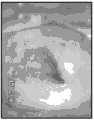
具有高风险的肿瘤形成或癌症的区域将表现为不同程度的白度,因为醋酸白与较高的核密度相关。术语“醋酸白”被用于与角化过度症或黏膜白斑病的区域的明显不同的对比中,这些区域在醋酸的施加之前呈现白色。移行带是宫颈上的关键区域,许多癌前病变和癌性病变最常在这里出现。看到移行带以及任何可视化的病变的整个范围的能力,确定是否可达到充分的阴道镜检查。Areas with a high risk of neoplasia or cancer will appear with varying degrees of whiteness, as acetowhiteness is associated with higher nuclear density. The term "acetowhite" is used in distinct contrast to areas of hyperkeratosis or leukoplakia, which appear white prior to the application of acetic acid. The transition zone is a critical area on the cervix where many precancerous and cancerous lesions most commonly occur. The ability to see the full extent of the transition zone, as well as any visualized lesions, determines whether adequate colposcopy is achievable.
在醋酸的施加之后变白或具有异常血管模式的宫颈区域常常被考虑用于活检。将碘溶液施加于宫颈以帮助突出异常的区域,并区分化生区域与可疑病变。Cervical areas that turn white or have abnormal vascular patterns after application of acetic acid are often considered for biopsy. An iodine solution is applied to the cervix to help highlight abnormal areas and differentiate metaplastic areas from suspicious lesions.
在完整的检查之后,阴道镜医师确定具有最高度可视异常的区域,并且可以使用长的活检器械从这些区实现活组织检查。大多数医生和患者都认为麻醉不是必要的。然而现在,一些阴道镜医师推荐并使用诸如利多卡因的局部麻醉剂或宫颈阻滞,以消除患者不适,尤其如果要采集许多活检样品。After a complete examination, the colposcopist identifies the areas with the most highly visible abnormalities, and a long biopsy instrument can be used to perform a biopsy from these areas. Most doctors and patients agree that anesthesia is unnecessary. Today, however, some colposcopists recommend and use local anesthetics such as lidocaine or cervical blocks to eliminate patient discomfort, especially if many biopsy samples are to be taken.
需要大量训练以正确解读根据以上方案的阴道镜测试。在诸如印度和中国的新兴市场中,培训资源和专业知识的缺乏限制了这种有效诊断工具的使用。相同的情况适用于工业化国家,在那里有资质的医疗人员短缺。Extensive training is required to correctly interpret colposcopy tests according to the above protocol. In emerging markets such as India and China, a lack of training resources and expertise limits the use of this effective diagnostic tool. The same applies to industrialized countries, where there is a shortage of qualified medical personnel.
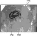
阴道镜诊断需要专业知识,并且涉及对用于子宫颈疾病的精确诊断的特定图像特征的检测。这使得该领域的自动化具有挑战性。在做出诊断所考虑的若干特征中,重要的特征之一为宫颈区域上存在的点状标记:它们是异常血管结构的标志物,并且它们的存在与宫颈的癌前病变和癌性病变的存在显著相关。Colposcopy diagnosis requires expertise and involves the detection of specific image features for precise diagnosis of cervical disease. This makes automation in this field challenging. Among the several features considered in making a diagnosis, one of the important ones is the presence of punctate marks on the cervical region: they are markers of abnormal vascular structure and their presence is associated with precancerous and cancerous lesions of the cervix. There is a significant correlation.
发明内容Contents of the invention
本发明在独立权利要求中提供了一种医疗器械、一种计算机程序产品、一种操作所述医疗器械的方法、以及一种视频配准的方法。在从属权利要求中给出了实施例。The invention provides a medical device, a computer program product, a method of operating the medical device, and a method of video registration in the independent claims. Embodiments are given in the dependent claims.
本发明可以提供一种方法,以鲁棒地识别阴道镜图像中存在的点状标记。本发明的实施例可以提供一种跟随有基于学习的框架的分级滤波途径,以检测不同种类的点状(细小/粗大/扩散的),并分割那些用于诊断辅助的区域。本发明的实施例可以将点状检测分成两个阶段:(1)粗略水平点状检测:其基于跟随有若干目标滤波步骤的感兴趣点检测以去除错误检测的目标。(2)精细水平点状检测:使用基于学习的框架以鲁棒地分类粗略检测的点状。最后的步骤是定义包含点状标记的区域。在具有不同严重性的癌前病变的35个数据集上执行测试,并且实现了关于测试集合的98.2%的平均准确性。The present invention may provide a method to robustly identify point-like markers present in colposcopy images. Embodiments of the present invention may provide a hierarchical filtering approach followed by a learning-based framework to detect different kinds of puncta (fine/coarse/diffuse) and segment those regions for diagnostic assistance. Embodiments of the present invention may divide point detection into two stages: (1) Coarse horizontal point detection: which is based on point of interest detection followed by several object filtering steps to remove falsely detected objects. (2) Fine-level point detection: use a learning-based framework to robustly classify coarsely detected points. The final step is to define the area containing the point markers. Testing was performed on 35 datasets of precancerous lesions of varying severities and achieved an average accuracy of 98.2% on the test set.
当前的阴道镜检查是主观的,并且依赖于妇科医师针对解读的知识和经验。因此,本发明的目的是消除该过程的主观性。本发明的另一目的是通过减少学习曲线并辅助诊断,增加阴道镜的可用性。本发明的另一目的是向用户(妇科医师)提供更高的置信度。本发明的再另一目的是提供针对癌症严重性程度的量化度量。Current colposcopy is subjective and relies on the knowledge and experience of the gynecologist for interpretation. Therefore, the purpose of the present invention is to eliminate the subjectivity of this process. Another object of the present invention is to increase the usability of colposcopy by reducing the learning curve and aiding in diagnosis. Another object of the invention is to provide a higher degree of confidence to the user (gynecologist). Yet another object of the present invention is to provide a quantitative measure for the severity of cancer.
这些目标通过根据本发明的系统或方法得以实现。These objectives are achieved by a system or method according to the invention.
根据本发明,提供了一种用于宫颈的光学检查的系统,所述系统包括光学放大器件、照明器件、用于至少一种刺激剂和/或造影剂的施予的施放器件、成像器件以及图像处理器件。所述图像处理器件还包括关键帧提取器件,任选地,为眩光去除器件、目标检测器件(也称作“目标检测器”)以及不透明度变化检测器件。According to the invention there is provided a system for optical examination of the cervix, said system comprising optical magnification means, illumination means, delivery means for the administration of at least one stimulating agent and/or contrast agent, imaging means and image processing device. The image processing means also includes keyframe extraction means, optionally glare removal means, object detection means (also referred to as "object detectors"), and opacity change detection means.
如本文中所用的炫光可以指镜面反射。如此,所述眩光去除器件在一些实施例中可以是镜面反射去除器件。Glare as used herein may refer to specular reflection. As such, the glare removal device may in some embodiments be a specular reflection removal device.
在优选的实施例中,所述系统还包括用于数据输入和数据输出的操作者接口器件。这种接口器件例如为显示器屏幕、键盘、鼠标、触摸屏、触摸板、控制杆等。In a preferred embodiment, the system also includes operator interface means for data input and data output. Such interface devices are, for example, monitor screens, keyboards, mice, touch screens, touchpads, joysticks and the like.
优选地,所述刺激剂和/或造影剂选自包括醋酸和/或碘溶液(优选为卢戈尔液或库勒液)的组。如根据本发明的说明书而变得明显的,本领域技术人员可以通过例行工作在相应的文献中找到备选的刺激剂和/或造影剂。因此,这种备选的刺激剂和/或造影剂也被包括在由本发明提供的保护范围内。Preferably, the stimulating and/or contrasting agent is selected from the group comprising acetic acid and/or iodine solution, preferably Lugol's solution or Kuehler's solution. As will become apparent from the description of the present invention, a person skilled in the art can find alternative stimulant and/or contrast agents in the corresponding literature by routine work. Accordingly, such alternative stimulating and/or contrasting agents are also included within the scope of protection afforded by the present invention.
在另一优选的实施例中,所述成像器件包括数字成像设备或非数字摄影机以及抓帧器。所述数字成像设备优选为数字摄影机。所述数字摄影机或非数字摄影机优选地包括CCD(电荷耦合器件)摄影机或CMOS(金属氧化物半导体)摄影机。In another preferred embodiment, the imaging device includes a digital imaging device or a non-digital video camera and a frame grabber. The digital imaging device is preferably a digital video camera. The digital or non-digital camera preferably comprises a CCD (Charge Coupled Device) camera or a CMOS (Metal Oxide Semiconductor) camera.
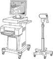
在另一优选的实施例中,所述光学放大器件为阴道镜。阴道镜可以是具有强光源的低倍率、立体、双目视野显微镜,其用于宫颈的放大视觉检查,以帮助宫颈癌的诊断。阴道镜不必须为立体的。In another preferred embodiment, the optical amplifying device is a colposcope. A colposcope can be a low power, stereoscopic, binocular field microscope with a strong light source, which is used for magnified visual inspection of the cervix to aid in the diagnosis of cervical cancer. A colposcope does not have to be stereoscopic.
在另一优选的实施例中,所述系统还包括计算机工作站,所述计算机工作站用于控制选自包括如下器件的组的器件中的至少一个:光学放大器件、照明器件、用于刺激剂的施予的施放器件、成像器件、成像处理器件、和/或操作者接口器件。In another preferred embodiment, the system further comprises a computer workstation for controlling at least one of the devices selected from the group consisting of optical amplification devices, lighting devices, Administering delivery devices, imaging devices, imaging processing devices, and/or operator interface devices.
在本发明的另一实施例中,提供了一种对患者的宫颈进行光学检查的方法,所述方法包括以下步骤:In another embodiment of the invention there is provided a method of optically examining a patient's cervix, said method comprising the steps of:
a)对宫颈施加至少一次刺激剂和/或造影剂;a) At least one application of a stimulating agent and/or a contrast agent to the cervix;
b)在刺激剂的每次施加之前和之后,采集宫颈的放大图像;b) Acquiring magnified images of the cervix before and after each application of the stimulating agent;
c)在所述图像中识别重要解剖目标,即Os、柱状区域和移行带;并且c) identifying important anatomical objects in said image, namely Os, columnar regions and transition zones; and
d)生成不透明度差异评分。d) Generate an opacity difference score.
所述患者优选为哺乳动物,具体地优选为灵长类,并且更具体地优选为人类。重要的是要提到,上文提及的步骤不必以给定顺序进行。The patient is preferably a mammal, particularly preferably a primate, and more particularly preferably a human. It is important to mention that the steps mentioned above do not have to be performed in the given order.
在本发明的另一优选的实施例中,刺激剂和/或造影剂为选自包括不透明度差异评分检测剂和/或移行带检测剂的组中的至少一个。In another preferred embodiment of the present invention, the stimulating agent and/or the contrast agent is at least one selected from the group consisting of an opacity difference score detecting agent and/or a transition zone detecting agent.
所述不透明度差异评分检测剂具有两个不同作用:第一,能够在利用所述试剂进行刺激和/或造影之后识别重要解剖目标。第二,所述不透明度差异评分检测剂能用于创建不透明度差异评分,因为醋酸白的程度与相应组织中较高的核密度相关,所述不透明度差异评分指示肿瘤形成过程或癌变过程。The opacity difference score detection agent has two distinct roles: first, it enables the identification of important anatomical targets after stimulation and/or contrast with the agent. Second, the Opacity Difference Score Detector can be used to create an Opacity Difference Score indicative of a neoplastic or carcinogenesis process since the degree of acetowhiteness correlates with higher nuclear density in the corresponding tissue.
所述移行带检测剂优选地具有深的颜色(例如,碘溶液具有深紫色或褐色,也称作“桃木褐”),并且能充当造影剂,以分别检测宫颈中的移行带,或是宫颈的面积或形状。The transition zone detection agent is preferably dark in color (for example, iodine solution has a deep purple or brown color, also known as "peachwood") and can act as a contrast agent to detect the transition zone in the cervix, or the cervical area or shape.
所述不透明度差异评分检测剂优选为醋酸,而所述移行带检测剂优选为碘溶液,例如为卢戈尔液或库勒液。因此,在根据本发明的方法的另一优选实施例中,至少一幅图像或帧是在所述不透明度差异评分检测剂的施加之前和之后采集的(为了方便,后文中将这些图像称作“醋酸前图像和醋酸后图像”,尽管所述术语也将联系其他不透明度差异评分检测剂使用),和/或是在所述移行带检测剂的施加之前和之后采集的(为了方便,后文中将这些图像称作“碘溶液前图像和碘溶液后图像”,尽管所述术语也将联系其他移行带检测剂使用)。The opacity difference score detection agent is preferably acetic acid, and the transition zone detection agent is preferably an iodine solution, such as Lugol's solution or Kuehler's solution. Therefore, in another preferred embodiment of the method according to the invention, at least one image or frame is acquired before and after the application of said opacity difference score detection agent (for convenience, these images are hereinafter referred to as "Pre-Acetic Acid and Post-Acetic Acid Images", although the term will also be used in connection with other Opacity Difference Score detection agents), and/or were acquired before and after application of the transition zone detection agent (for convenience, the post These images are referred to herein as "pre-iodine solution images and post-iodine solution images", although these terms will also be used in connection with other transition zone detection agents).
当然,对于本领域技术人员而言,能够通过例行工作从相应的文件得到备选的刺激剂和/或造影剂。能够用于检测较高核密度的区域的备选的试剂例如为亚甲蓝。这种备选的刺激剂和/或造影剂因此也被包括在由本发明提供的保护范围内。这还适用于术语“醋酸前图像和醋酸后图像”和/或在“碘溶液前图像和碘溶液后图像”之前。在针对醋酸或碘溶液的替代物被使用的情况中,将相应地改变这些术语。Of course, alternative stimulants and/or contrast agents can be derived from the corresponding documentation by routine work for a person skilled in the art. An alternative reagent that can be used to detect regions of higher nuclear density is, for example, methylene blue. Such alternative stimulating and/or contrasting agents are therefore also included within the scope of protection afforded by the present invention. This also applies to the terms "before and after acetic acid images" and/or before "before and after iodine solution images". Where alternatives to acetic acid or iodine solutions are used, these terms will be changed accordingly.
在根据本发明的方法的另一优选的实施例中,进行以下步骤中的至少一个:In another preferred embodiment of the method according to the invention, at least one of the following steps is carried out:
a)在醋酸后图像中和/或醋酸后图像中,识别Os和柱状区域;a) Identify Os and columnar regions in the post-acetic acid image and/or in the post-acetic acid image;
b)在碘溶液后图像中,识别试探性移行带;b) in the post-iodine image, identify the tentative transition zone;
c)将所述试探性移行带映射到所述醋酸前图像和所述醋酸后图像;c) mapping said tentative transition zone to said pre-acetate image and said post-acetate image;
d)通过在所述醋酸前图像和醋酸后图像中,从所述试探性移行带减去所述Os和柱状区域,识别实际移行带。d) Identify the actual transition zone by subtracting the Os and columnar regions from the tentative transition zone in the pre- and post-acetate images.
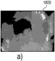
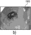
对实际移行带的识别因此为两步法:(i)在以上所列的第一步骤(步骤b)中,处理碘溶液后图像,以基于因碘溶液的施加受影响的宫颈区所描绘的颜色改变,试探性地检测移行带。使用基于颜色的K均值聚类,将从关键帧提取器获得的碘溶液后图像分割成两个聚类。选择所述两个聚类中较小的。将该聚类的凸包定义为所述试探性移行带;(ii)在以上所列的第二步骤(步骤c)中,将所述试探性移行带映射到所述醋酸前图像和醋酸后图像,并之后减去以上列出的(根据步骤a)所检测的Os和柱状上皮区域,以定义以上列出的所述实际移行带(见步骤d)。能够在识别所述移行带之前配准所述醋酸前图像和醋酸后图像。相应的图像分析过程示于图6和图7中。The identification of the actual transition zone is thus a two-step process: (i) In the first step (step b) listed above, the post-iodine solution image is processed to based on the delineation of the cervical region affected by the application of the iodine solution. Color changes, heuristic detection of transition zones. The post-iodine solution images obtained from the keyframe extractor were segmented into two clusters using color-based K-means clustering. The smaller of the two clusters is chosen. defining the convex hull of this cluster as the tentative transition zone; (ii) in the second step (step c) listed above, mapping the tentative transition zone to the pre-acetate image and post-acetate image image, and then subtract the detected Os and columnar epithelial areas listed above (according to step a) to define the actual transition zone listed above (see step d). The pre-acetate and post-acetate images can be registered prior to identifying the transition zone. The corresponding image analysis process is shown in Figures 6 and 7.
在根据本发明的方法的具体优选的实施例中,所述不透明度差异评分是通过对至少一幅醋酸前图像和一幅醋酸后图像进行图像处理而生成的。针对所述处理,基于对所述实际移行带的先前识别,优选地仅使用这些图像的实际移行带数据。In a particularly preferred embodiment of the method according to the invention, said opacity difference score is generated by image processing of at least one pre-acetic acid image and one post-acetic acid image. For said processing, preferably only the actual transition zone data of these images are used, based on a previous identification of said actual transition zone.
在根据本发明所述的方法的另一优选的实施例中,如根据本发明的方法确定的实际移行带(或者优选为其形状和/或总面积),和/或如根据本发明的方法确定的不透明度差异评分指示肿瘤形成和/或癌变过程。In another preferred embodiment of the method according to the invention, the actual transition zone (or preferably its shape and/or total area) as determined according to the method according to the invention, and/or as determined according to the method according to the invention The determined opacity difference score is indicative of tumor formation and/or cancerous processes.
通常,如在碘溶液染色以及将碘溶液染色映射到醋酸后图像之后所确定的实际移行带也能够指示癌变区域。根据该定义,如通过对至少一幅醋酸前图像和一幅醋酸后图像进行图像处理所确定的不透明度差异评分能用于确定在所述癌变区域中的白度的程度,以及因此确定癌症的程度,或严重性。Often, actual transitional zones as determined after iodine solution staining and mapping of iodine solution staining to post-acetic acid images can also indicate cancerous areas. According to this definition, the opacity difference score, as determined by image processing of at least one pre- and one post-acetate image, can be used to determine the degree of whiteness in said cancerous area, and thus the extent of the cancer. degree, or severity.
在另一实施例中,实际移行带的形状和/或总面积能够指示癌症。所述不透明度差异评分能够因此用于证实或撤销疑似肿瘤形成或癌变区域。In another embodiment, the shape and/or total area of the actual transition zone can be indicative of cancer. The opacity difference score can thus be used to confirm or withdraw suspected neoplastic or cancerous regions.
具体优选地,在成像器件和/或图像处理器件的帮助下,执行宫颈的放大图像的采集,重要解剖目标的识别和/或不透明度差异评分的生成。Particularly preferably, the acquisition of a magnified image of the cervix, the identification of important anatomical objects and/or the generation of an opacity difference score is performed with the aid of the imaging device and/or the image processing device.
优选地,阴道镜用于此目的。Preferably a colposcope is used for this purpose.
进一步优选的是,宫颈的放大图像的采集包括以下步骤:It is further preferred that the acquisition of the magnified image of the cervix comprises the steps of:
·捕获图像流;Capture image stream;
·从所述图像流识别互相关联的图像的序列;identifying a sequence of interrelated images from said image stream;
·从这些序列识别关键图像;并且identify key images from these sequences; and
·预处理所述图像。• Preprocessing the image.
对互相关联的图像的序列的识别和/或对关键帧的识别优选地包括以下步骤:The identification of the sequence of interrelated images and/or the identification of key frames preferably comprises the steps of:
·识别镜头边界;·Identify the boundary of the shot;
·分类镜头;并且· Categorize shots; and
·选择关键帧。• Select keyframes.
此外,优选地,对图像中的重要解剖目标的识别包括以下步骤:In addition, preferably, the identification of important anatomical objects in the image includes the following steps:
·使用像素的K均值聚类,基于它们的颜色,将醋酸前图像和醋酸后图像分割成2个聚类;Segment the pre- and post-acetate images into 2 clusters based on their color using K-means clustering of pixels;
·将最小的聚类标记为Os+柱状一起的上皮区域;Mark the smallest clusters as Os+columnar epithelial regions;
·迭代地去除在Os和柱状上皮区域两者内的小且分离的聚类;并且Iteratively removes small and separate clusters in both Os and columnar epithelial regions; and
·使用最小方差量化分离Os和柱状上皮。• Separation of Os and columnar epithelium using least variance quantification.
在具体优选的实施例中,不透明度差异评分的生成包括以下步骤:In a specific preferred embodiment, the generation of the opacity difference score includes the following steps:
·在醋酸后图像的移行带中识别具有主要不透明度变化的图像像素,Identify image pixels with major opacity changes in the transition zone of the post-acetic acid image,
·将所述图像像素映射到醋酸前图像的对应像素,并且mapping said image pixels to corresponding pixels of the pre-acetate image, and
·使用下式获得所述不透明度差异评分:• Obtain the Opacity Difference Score using the following formula:

进一步优选地,根据本发明的方法还包括选自包括PAP测试和/或针对人乳头瘤病毒的分子测试的组的至少一个步骤。Further preferably, the method according to the invention further comprises at least one step selected from the group comprising a PAP test and/or a molecular test against human papillomavirus.
PAP测试(得名于George Papanicolaou博士,其在上世纪上半叶发展了该技术)是用于妇科医学的筛查测试,以检测恶化前过程和恶性过程。使用窥器从子宫颈的外口以及宫颈内部收集细胞。针对异常情况,具体而言针对宫颈的肿瘤形成或癌变过程,对细胞进行组织学检查。The PAP test (named after Dr. George Papanicolaou, who developed the technique in the first half of the last century) is a screening test used in gynecology to detect premalignant and malignant processes. Cells are collected from the outer os of the cervix as well as from the inside of the cervix using a speculum. Cells are examined histologically for abnormalities, specifically neoplastic or cancerous processes of the cervix.
人乳头瘤病毒(HPV)是这样的病毒:其是能够感染人类的乳头瘤病毒家族的成员。HPV感染是几乎所有宫颈癌病例的原因。取自宫颈的样品中病毒核酸的存在指示宫颈的HPV感染,并因此是患者出现在宫颈中的肿瘤形成或癌变过程的风险的度量。现代分子诊断技术,例如PCR,允许在样品中进行对HPV的快速检测。一个这样的测试是由Qiagen制造的,并且被发布为digene HC2HPV DNA测试。Human papillomavirus (HPV) is a virus that is a member of the papillomavirus family capable of infecting humans. HPV infection is the cause of almost all cases of cervical cancer. The presence of viral nucleic acid in a sample taken from the cervix is indicative of HPV infection of the cervix and is therefore a measure of the patient's risk of developing a neoplastic or cancerous process in the cervix. Modern molecular diagnostic techniques, such as PCR, allow rapid detection of HPV in samples. One such test is manufactured by Qiagen and is published as the digene HC2HPV DNA Test.
根据本发明的方法与上文阐述的两个测试之一的组合进一步增加了宫颈中癌症检测的准确性、灵敏性和再现性。Combining the method according to the invention with one of the two tests set forth above further increases the accuracy, sensitivity and reproducibility of cancer detection in the cervix.
此外,提供了根据本发明的系统的用途,其用于执行根据本发明的方法。Furthermore, the use of the system according to the invention for carrying out the method according to the invention is provided.
在一个方面中,本发明提供了用于宫颈的光学检查的系统,所述系统包括光学放大器件、照明器件、用于至少一种刺激剂和/或造影剂的施予的施放器件、成像器件以及图像处理器件,所述图像处理器件还包括:In one aspect, the invention provides a system for optical examination of the cervix, said system comprising optical magnification means, illumination means, delivery means for administration of at least one stimulating agent and/or contrast agent, imaging means And an image processing device, the image processing device also includes:
·关键帧提取器件;· Key frame extraction device;
·任选地,眩光去除器件;· Optionally, a glare removal device;
·目标检测器件;以及an object detection device; and
·不透明度变化检测器件。· Opacity change detection device.
在另一实施例中,所述系统还包括用于数据输入和数据输出的操作者接口器件。In another embodiment, the system further includes operator interface means for data input and data output.
在另一实施例中,所述光学放大器件为阴道镜。In another embodiment, said optical magnification device is a colposcope.
在另一实施例中,所述系统还包括计算机工作站,所述计算机工作站用于控制选自包括如下器件的组的器件中的至少一个:光学放大器件、照明器件、用于刺激剂的施予的施放器件、成像器件、成像处理器件和/或操作者接口装置。In another embodiment, the system further comprises a computer workstation for controlling at least one of the devices selected from the group consisting of: optical amplification devices, lighting devices, for the administration of stimulants Delivery device, imaging device, image processing device and/or operator interface device.
在另一方面中,本发明提供了患者的宫颈的光学检查的方法,所述方法包括以下步骤:In another aspect, the invention provides a method of optical examination of a patient's cervix, said method comprising the steps of:
a)对宫颈施加至少一次刺激剂和/或造影剂;a) At least one application of a stimulating agent and/or a contrast agent to the cervix;
b)在刺激剂的每次施加之前和之后,采集宫颈的放大图像;b) Acquiring magnified images of the cervix before and after each application of the stimulating agent;
c)在所述图像中识别重要解剖目标,即Os、柱状区域和移行带;并且c) identifying important anatomical objects in said image, namely Os, columnar regions and transition zones; and
d)生成不透明度差异评分。d) Generate an opacity difference score.
在另一实施例中,所述刺激剂和/或造影剂为选自包括不透明度差异评分检测剂和/或移行带检测剂的组的至少一种。In another embodiment, the stimulating agent and/or contrast agent is at least one selected from the group comprising an opacity difference score detecting agent and/or a transition zone detecting agent.
在另一实施例中,所述不透明度差异评分检测剂为醋酸和/或所述移行带检测剂为碘溶液。In another embodiment, the opacity difference score detection agent is acetic acid and/or the transition zone detection agent is iodine solution.
在另一实施例中,至少一幅图像是在不透明度差异评分检测剂的施加之前和之后采集的(“醋酸前图像和醋酸后图像”),和/或在移行带检测剂的施加之前和之后采集的(“碘溶液前图像和碘溶液后图像”)。In another embodiment, at least one image is acquired before and after application of the Opacity Difference Score detection agent ("pre-acetic acid image and post-acetic acid image"), and/or before and after application of the transition zone detection agent. Acquired afterwards ("Before iodine solution image and after iodine solution image").
在所述方法另一实施例中,其中:In another embodiment of the method, wherein:
a)在醋酸后图像中,识别Os和柱状区域;a) In the post-acetic acid image, identifying Os and columnar regions;
b)在碘溶液后图像中,识别试探性移行带;b) in the post-iodine image, identify the tentative transition zone;
c)将所述试探性移行带映射到所述醋酸后图像;并且c) mapping the tentative transition zone to the post-acetate image; and
d)通过在醋酸前图像和醋酸后图像中,从所述试探性移行带减去所述Os和柱状区域,识别实际移行带。d) Identify the actual transition zone by subtracting the Os and columnar regions from the tentative transition zone in the pre- and post-acetate images.
在另一实施例中,不透明度差异评分是通过对至少一幅醋酸前图像和一幅醋酸后图像进行图像处理生成的。In another embodiment, the opacity difference score is generated by image processing at least one pre-acetic acid image and one post-acetic acid image.
在另一实施例中:In another embodiment:
a)实际移行带,和/或a) the actual transition zone, and/or
b)不透明度差异评分b) Opacity Difference Score
指示肿瘤形成或癌变过程。Indicates tumor formation or cancerous process.
在另一实施例中,在图像中对重要解剖目标的识别包括以下步骤:In another embodiment, the identification of important anatomical objects in the image comprises the following steps:
·使用像素的K均值聚类,基于它们的颜色,将醋酸前图像和醋酸后图像分割成2个聚类;Segment the pre- and post-acetate images into 2 clusters based on their color using K-means clustering of pixels;
·将最小聚类标记为Os+柱状一起的上皮区域;Mark minimal clusters as Os+columnar epithelial regions together;
·在Os和柱状上皮区域两者内迭代地去除小且分离的聚类;并且Iteratively removes small and separate clusters within both Os and columnar epithelial regions; and
·使用最小方差量化分离Os和柱状上皮。• Separation of Os and columnar epithelium using least variance quantification.
在另一实施例中,不透明度差异评分的生成包括以下步骤:In another embodiment, the generation of the opacity difference score includes the following steps:
·在醋酸后图像的移行带中识别具有主要不透明度变化的图像像素,Identify image pixels with major opacity changes in the transition zone of the post-acetic acid image,
·将所述图像像素映射到醋酸前图像的对应像素,并且mapping said image pixels to corresponding pixels of the pre-acetate image, and
·使用下式获得所述不透明度差异评分:• Obtain the Opacity Difference Score using the following formula:

在另一实施例中,所述方法还包括实施PAP测试和/或针对人乳头瘤病毒的分子测试。In another embodiment, the method further comprises performing a PAP test and/or a molecular test for human papillomavirus.
在另一方面中,本发明提供了根据本发明的实施例的系统,其用于执行根据本发明的实施例的方法。In another aspect, the invention provides a system according to an embodiment of the invention for performing a method according to an embodiment of the invention.
在一个方面中,本发明提供了用于检查宫颈的医疗器械。所述医疗器械包括用于采集宫颈图像的光学检查系统。本文中所用的宫颈图像囊括描述宫颈的图像。所述医疗器械还包括用于控制所述医疗器械的处理器。所述医疗器械还包括包含用于由所述处理器执行的机器可执行指令的存储器。所述指令的执行令所述处理器使用所述光学检查系统采集所述宫颈图像。所述指令的执行还令所述处理器使用数字滤波器计算感兴趣点位置的集合。在一些实施例中,所述数字滤波器为Harris角点检测器。In one aspect, the invention provides a medical device for examining the cervix. The medical device includes an optical inspection system for acquiring images of the cervix. Cervical image as used herein encompasses images depicting the cervix. The medical device also includes a processor for controlling the medical device. The medical device also includes a memory containing machine-executable instructions for execution by the processor. Execution of the instructions causes the processor to acquire the cervical image using the optical inspection system. Execution of the instructions further causes the processor to compute a set of point-of-interest locations using a digital filter. In some embodiments, the digital filter is a Harris corner detector.
所述指令的执行还令所述处理器使用所述感兴趣点位置的集合和形态滤波器计算经滤波感兴趣点位置的集合。所述形态滤波器使用所述感兴趣点的集合的光学属性,以确定哪些应被滤波。例如,点状标记应相对于背景为暗的,并且实质上是类似点的Execution of the instructions further causes the processor to calculate a set of filtered interest point locations using the set of interest point locations and a morphological filter. The morphological filter uses the optical properties of the set of interest points to determine which should be filtered. For example, a dot marker should be dark relative to the background and be dot-like in nature
所述指令的执行还令所述处理器使用所述经滤波感兴趣点位置的集合和基于邻域的滤波器计算缩减的感兴趣点位置的集合。通常,点状标记一起成组出现。排除掉远离其他点状标记的点状标记或经滤波的感兴趣点,有助于消除假阳性。Execution of the instructions further causes the processor to calculate a reduced set of point-of-interest locations using the filtered set of point-of-interest locations and a neighborhood-based filter. Usually, dot marks appear in groups together. Excluding point markers or filtered points of interest that are far from other point markers can help eliminate false positives.
在另一实施例中,所述指令的执行还使用经训练的分类模块和所述缩减的感兴趣点的集合形成经分类的感兴趣点位置的集合。训练模式检测软件模块能够用于检测或确定点状标记是否类似于现有的或先前已知的点状标记。In another embodiment, execution of the instructions further uses the trained classification module and the reduced set of points of interest to form a set of classified point-of-interest locations. The training pattern detection software module can be used to detect or determine whether a spot marker is similar to an existing or previously known spot marker.
在另一实施例中,所述指令的执行还令所述处理器使用第二基于邻域的滤波器和所述经分类的感兴趣点位置的集合计算点状位置的集合。所述第二基于邻域的滤波器检查点状标记的分组,并使用它们帮助消除假阳性。所述指令的执行还令所述处理器使用点状点位置计算点状标记区域。In another embodiment, execution of the instructions further causes the processor to calculate a set of point-like locations using a second neighborhood-based filter and the classified set of point-of-interest locations. The second neighborhood-based filter examines groupings of point-like markers and uses them to help eliminate false positives. Execution of the instructions further causes the processor to calculate a point marker area using the point positions.
在另一实施例中,所述经训练的分类模块为随机树分类模块。In another embodiment, the trained classification module is a random tree classification module.
在另一实施例中,所述指令的执行还令所述处理器在计算感兴趣点位置的第一集合之前,利用预处理滤波器生成宫颈图像内的排除位置的集合。所述数字滤波器使用所述排除位置的集合,以生成所述感兴趣点的集合。In another embodiment, execution of the instructions further causes the processor to generate a set of excluded locations within the cervical image using a pre-processing filter prior to computing the first set of point-of-interest locations. The set of excluded locations is used by the digital filter to generate the set of points of interest.
在另一实施例中,所述指令的执行还令所述处理器使用所述光学检查系统采集第二宫颈图像。所述指令的执行还令所述处理器使用所述数字滤波器计算感兴趣点位置的第二集合。所述指令的执行还令所述处理器使用所述感兴趣点位置的第二集合和所述形态滤波器计算经滤波感兴趣点位置的第二集合。所述指令的执行还令所述处理器使用所述经滤波感兴趣点位置的第二集合和所述基于邻域的滤波器计算缩减的感兴趣点位置的第二集合。所述指令的执行还令所述处理器使用所述训练分离模块和所述缩减的感兴趣点位置的第二集合计算经分类的感兴趣点位置的第二集合。所述指令的执行还令所述处理器使用第二基于邻域的滤波器和所述经分类的感兴趣点位置的第二集合计算点状位置的第二集合。使用所述点状点位置和所述第二点状点位置计算所述点状标记区域。In another embodiment, execution of the instructions further causes the processor to acquire a second cervical image using the optical inspection system. Execution of the instructions further causes the processor to calculate a second set of point-of-interest locations using the digital filter. Execution of the instructions further causes the processor to calculate a second set of filtered interest point locations using the second set of interest point locations and the morphological filter. Execution of the instructions further causes the processor to calculate a second set of reduced point-of-interest locations using the second set of filtered point-of-interest locations and the neighborhood-based filter. Execution of the instructions further causes the processor to calculate a second set of classified point-of-interest locations using the training separation module and the reduced second set of point-of-interest locations. Execution of the instructions further causes the processor to calculate a second set of point-like locations using a second neighborhood-based filter and the second set of classified point-of-interest locations. The dot marker area is calculated using the dot position and the second dot position.
在另一实施例中,所述第一宫颈图像为第一醋酸前图像。In another embodiment, said first cervical image is a first pre-acetate image.
在另一实施例中,所述第一宫颈图像为第一绿色滤波图像。In another embodiment, the first cervical image is a first green filtered image.
在另一实施例中,所述第一宫颈图像为第一醋酸后图像。所述第二宫颈图像为第二醋酸前图像。In another embodiment, said first cervical image is a first post-acetate image. The second cervical image is a second pre-acetic acid image.
在另一实施例中,所述第二宫颈图像为第二醋酸前图像。In another embodiment, said second cervical image is a second pre-acetate image.
在另一实施例中,所述第二宫颈图像为第二绿色滤波图像。In another embodiment, the second cervical image is a second green filtered image.
在另一实施例中,所述第二宫颈图像为第二醋酸后图像。In another embodiment, said second cervical image is a second post-acetate image.
在另一实施例中,所述指令的执行还令所述处理器使用图像配准模块配准所述第一图像和所述第二图像。在所述图像配准中,在所述第二图像中识别所述第一图像中的特征。In another embodiment, execution of the instructions further causes the processor to register the first image and the second image using an image registration module. In said image registration, features in said first image are identified in said second image.
在另一实施例中,所述指令的执行令所述处理器通过采集包括多个图像帧的视频数据,采集所述第一宫颈图像和所述第二宫颈图像。所述指令的执行令所述处理器从所述多个图像帧选择所述第一宫颈图像和所述第二宫颈图像。所述指令的执行还令所述处理器接收包括多个图像帧的所述视频数据。所述指令的执行还令所述处理器确定在选自所述多个图像帧的连续图像帧的每对之间的变换模型。所述指令的执行还令所述处理器从连续图像帧的每对之间的所述变换模型计算累积变换模型。所述指令的执行还令处理器使用所述累积变换模型配准所述多个图像帧中的每个。所述指令的执行还令所述处理器使用所述累积变换模型提供所述第一宫颈图像与所述第二宫颈图像的配准。本质上,所述视频数据包括许多不同的图像帧。变换模型用于计算相邻图像帧的每对之间的变化。之后累积变换模型用于计算所述第一宫颈图像与所述第二宫颈图像之间的配准。这可以具有提供所述第一宫颈图像与所述第二宫颈图像之间更准确的配准的优点。In another embodiment, execution of the instructions causes the processor to acquire the first cervical image and the second cervical image by acquiring video data comprising a plurality of image frames. Execution of the instructions causes the processor to select the first cervical image and the second cervical image from the plurality of image frames. Execution of the instructions further causes the processor to receive the video data comprising a plurality of image frames. Execution of the instructions further causes the processor to determine a transformation model between each pair of consecutive image frames selected from the plurality of image frames. Execution of the instructions further causes the processor to calculate a cumulative transformation model from the transformation model between each pair of consecutive image frames. Execution of the instructions further causes the processor to register each of the plurality of image frames using the cumulative transformation model. Execution of the instructions further causes the processor to provide registration of the first cervical image with the second cervical image using the cumulative transformation model. Essentially, the video data includes many different image frames. A transformation model is used to compute the change between each pair of adjacent image frames. The cumulative transformation model is then used to calculate the registration between said first cervical image and said second cervical image. This may have the advantage of providing a more accurate registration between the first and second cervical images.
在另一实施例中,所述指令的执行还令所述处理器检测确定连续图像帧的至少一对之间的变换模型的失败。失败可以是例如确定连续图像帧的一对之间的变换模型的能力。所述指令的执行还令所述处理器在所述失败之前,从选自所述视频数据的第一图像帧计算第一图像分割。所述指令的执行还令所述处理器在所述失败之后,从选自所述视频数据的第二图像帧计算第二图像分割。两种情况中的所述图像分割均可以使用标准图像分割软件模块来计算。例如,所述图像分割模块可以包括用于在所述图像中辨识宫颈的特定鸟类(ornithological)特征的专用代码。也可以使用诸如可变形模型或其他建模器件的东西来执行所述图像分割。In another embodiment, execution of the instructions further causes the processor to detect a failure to determine a transformation model between at least one pair of consecutive image frames. A failure may be, for example, the ability to determine a transformation model between a pair of consecutive image frames. Execution of the instructions further causes the processor to compute a first image segmentation from a first image frame selected from the video data prior to the failure. Execution of the instructions further causes the processor to compute a second image segmentation from a second image frame selected from the video data after the failure. The image segmentation in both cases can be calculated using standard image segmentation software modules. For example, the image segmentation module may include dedicated code for identifying certain ornithological features of the cervix in the image. The image segmentation may also be performed using things such as deformable phantoms or other modeling devices.
所述指令的执行还令所述处理器确定所述第一图像帧与所述第二图像帧之间的第二变换。所述指令的执行还令所述处理器使用所述第二变换校正所述累积变换模型。本质上,所述第一图像分割和所述第二图像分割提供了将所述第二图像帧配准到所述第一图像帧的方法。所述图像分割因此可以用于计算所述第一图像帧与所述第二图像帧之间的变换。这之后可以用作修复或校正所述累积变换模型的桥梁。Execution of the instructions further causes the processor to determine a second transformation between the first image frame and the second image frame. Execution of the instructions further causes the processor to correct the cumulative transformation model using the second transformation. Essentially, the first image segmentation and the second image segmentation provide a method of registering the second image frame to the first image frame. The image segmentation may thus be used to calculate a transformation between the first image frame and the second image frame. This can then be used as a bridge to repair or correct the cumulative transformation model.
在一些实施例中,以与确定相邻帧之间的变换模型相同的方式确定所述第二变换模型。In some embodiments, the second transformation model is determined in the same manner as the transformation model between adjacent frames is determined.
在另一实施例中,所述指令的执行还令所述处理器检测确定连续图像帧的至少一对之间的变换的失败。所述指令的执行还令所述处理器在所述失败之前从所述视频数据选择第一图像。所述指令的执行还令所述处理器在所述失败之后从所述视频数据选择第二图像。所述指令的执行还令所述处理器确定所述第一图像与所述第二图像之间的第二变换。所述指令的执行还令所述处理器使用所述第二变换校正所述累积变换模型。在一些实施例中,能够以与确定连续图像帧的各对之间的变换模型相同的方式执行所述第二变换。In another embodiment, execution of the instructions further causes the processor to detect a failure to determine a transition between at least one pair of consecutive image frames. Execution of the instructions further causes the processor to select a first image from the video data prior to the failure. Execution of the instructions further causes the processor to select a second image from the video data after the failure. Execution of the instructions also causes the processor to determine a second transformation between the first image and the second image. Execution of the instructions further causes the processor to correct the cumulative transformation model using the second transformation. In some embodiments, the second transformation can be performed in the same manner as determining the transformation model between pairs of consecutive image frames.
在另一实施例中,所述第一宫颈图像为造影剂前图像和/或刺激剂前图像。所述第二宫颈图像为造影剂后图像和/或刺激剂后图像。所述指令的执行还令所述处理器在所述图像中识别重要解剖目标。例如,所述重要解剖目标可以是,但不限于Os、柱状区域和移行带。所述指令的执行还令所述处理器生成不透明度差异评分。所述处理器的这些步骤可以例如用于执行对患者的宫颈进行光学检查的方法。所述方法可以包括向宫颈中施加至少一次刺激剂和/或造影剂的步骤。所述方法还可以包括在所述刺激剂的每次施加之前和之后采集宫颈的放大图像。所述方法还可以包括在所述图像中识别重要解剖目标,即Os、柱状区域和移行带。所述方法还可以包括生成不透明度差异评分的步骤。In another embodiment, said first cervical image is a pre-contrast image and/or a pre-stimulator image. The second cervical image is a post-contrast image and/or a post-stimulator image. Execution of the instructions further causes the processor to identify important anatomical objects in the image. For example, the important anatomical targets may be, but are not limited to, Os, columnar regions, and transition zones. Execution of the instructions further causes the processor to generate an opacity difference score. These steps of the processor may eg be used to perform a method of optical examination of a patient's cervix. The method may comprise the step of applying at least once a stimulating agent and/or a contrast agent into the cervix. The method may also include acquiring magnified images of the cervix before and after each application of the stimulating agent. The method may also include identifying important anatomical objects in the image, namely Os, columnar regions, and transition zones. The method may also include the step of generating an opacity difference score.
在另一实施例中,所述第二宫颈图像为示出至少部分在所述第一宫颈图像内的缩放区域的缩放图像。In another embodiment, said second cervical image is a zoomed image showing a zoomed region at least partially within said first cervical image.
在另一实施例中,所述指令的执行还令所述处理器将被叠加在所述第一宫颈图像上的点状标记区域显示在显示器上。In another embodiment, the execution of the instructions further causes the processor to display on a display the dot-shaped marking area superimposed on the first cervical image.
在另一方面中,本发明提供了一种包括由处理器执行的机器可执行指令的计算机程序产品,其用于控制检查宫颈的医疗器械。所述医疗器械包括用于采集宫颈图像的光学检查系统。所述指令的执行令所述处理器使用所述光学检查系统采集所述宫颈图像。所述指令的执行还令所述处理器使用数字滤波器计算感兴趣点位置的集合。所述指令的执行还令所述处理器使用所述感兴趣点位置的基合和形态滤波器计算经滤波感兴趣点位置的集合。所述指令的执行还令所述处理器使用所述经滤波感兴趣点位置的集合和基于邻域的滤波器计算缩减的感兴趣点位置的集合。所述指令的执行还令所述处理器使用经训练的分类模块和所述缩减的感兴趣点的集合计算经分类的感兴趣点位置的集合。In another aspect, the present invention provides a computer program product comprising machine-executable instructions executed by a processor for controlling a medical device for examining the cervix. The medical device includes an optical inspection system for acquiring images of the cervix. Execution of the instructions causes the processor to acquire the cervical image using the optical inspection system. Execution of the instructions further causes the processor to compute a set of point-of-interest locations using a digital filter. Execution of the instructions further causes the processor to compute a set of filtered interest point locations using the basis and morphological filter of the interest point locations. Execution of the instructions further causes the processor to calculate a reduced set of point-of-interest locations using the filtered set of point-of-interest locations and a neighborhood-based filter. Execution of the instructions further causes the processor to calculate a set of classified point-of-interest locations using the trained classification module and the reduced set of point-of-interest.
所述指令的执行还令所述处理器使用第二基于邻域的滤波器和所述经分类的感兴趣点位置的集合计算点状位置的集合。所述指令的执行还令所述处理器使用点状点位置计算点状标记区域。Execution of the instructions further causes the processor to calculate a set of point locations using a second neighborhood-based filter and the classified set of point-of-interest locations. Execution of the instructions further causes the processor to calculate a point marker area using the point positions.
在另一方面中,本发明提供了一种视频配准的方法,所述方法包括接收包括多个图像帧的视频数据的步骤。所述方法还包括确定选自所述多个图像帧的连续图像帧的每对之间的变换的步骤。所述方法还包括从连续图像帧的每对之间的所述变换计算累积变换模型的步骤。所述方法还包括使用所述累积变换配准所述多个图像帧的每个的步骤。该实施例可以是有益的,因为其提供了一种将所述视频数据内的帧彼此配准的有效方法。In another aspect, the present invention provides a method of video registration, the method comprising the step of receiving video data comprising a plurality of image frames. The method further comprises the step of determining a transformation between each pair of consecutive image frames selected from the plurality of image frames. The method further comprises the step of computing a cumulative transformation model from said transformation between each pair of successive image frames. The method also includes the step of registering each of the plurality of image frames using the accumulated transformation. This embodiment may be beneficial because it provides an efficient method of registering frames within the video data with each other.
在另一实施例中,使用Lucas-Kanade跟踪算法确定所述变换。In another embodiment, the transformation is determined using a Lucas-Kanade tracking algorithm.
在另一实施例中,通过跟踪SIFT特征确定所述变换。In another embodiment, the transformation is determined by tracking SIFT features.
在另一实施例中,通过同步定位和映射算法以驱动特征匹配,确定所述变换。In another embodiment, the transformation is determined by a simultaneous localization and mapping algorithm to drive feature matching.
在另一实施例中,所述方法还包括检测确定连续图像帧的至少一对之间的变换的失败的步骤。所述方法还包括在所述失败之前从选自所述视频数据的第一图像帧计算第一图像分割的步骤。所述方法还包括在所述失败之后从选自所述视频数据的第二图像帧计算第二图像分割的步骤。所述方法还包括确定所述第一图像帧与所述第二图像帧之间的第二变换的步骤。所述方法还包括使用所述第二变换校正所述累积变换的步骤。In another embodiment, the method further comprises the step of detecting a failure to determine a transformation between at least one pair of consecutive image frames. The method further comprises the step of computing a first image segmentation from a first image frame selected from said video data prior to said failure. The method further comprises the step of computing a second image segmentation from a second image frame selected from said video data after said failure. The method also includes the step of determining a second transformation between the first image frame and the second image frame. The method also includes the step of correcting the accumulated transformation using the second transformation.
在一些实施例中,能够以与确定连续图像帧的每对之间的变换的相同的方式执行所述第二变换的确定。In some embodiments, the determination of the second transformation can be performed in the same way as the transformation between each pair of consecutive image frames is determined.
在另一实施例中,所述方法还包括检测确定连续图像帧的至少一对之间的变换的失败的步骤。所述方法还包括省略从所述累积变换模型的不可靠地确定的变换的步骤。In another embodiment, the method further comprises the step of detecting a failure to determine a transformation between at least one pair of consecutive image frames. The method further comprises the step of omitting unreliably determined transformations from the cumulative transformation model.
在另一实施例中,所述方法还包括检测确定连续图像帧的至少一对之间的变换的失败的步骤。所述方法还包括在所述失败之前从选自所述视频数据的第一图像帧计算第一图像分割的步骤。所述方法还包括在所述失败之后从选自所述视频数据的第二图像帧计算第二图像分割的步骤。可以使用第三图像分割技术确定所述第一图像分割和所述第二图像分割。所述方法还包括使用所述第一图像分隔和所述第二图像分割校正所述累积变换模型的步骤。In another embodiment, the method further comprises the step of detecting a failure to determine a transformation between at least one pair of consecutive image frames. The method further comprises the step of computing a first image segmentation from a first image frame selected from said video data prior to said failure. The method further comprises the step of computing a second image segmentation from a second image frame selected from said video data after said failure. The first image segmentation and the second image segmentation may be determined using a third image segmentation technique. The method further comprises the step of correcting the cumulative transformation model using the first image segmentation and the second image segmentation.
在另一实施例中,所述方法还包括检测确定连续图像帧的至少一对之间的变换的失败的步骤。所述方法还包括在所述失败之前从所述视频数据选择第一图像帧的步骤。所述方法还包括在所述失败之后从所述视频数据选择第二图像帧的步骤。In another embodiment, the method further comprises the step of detecting a failure to determine a transformation between at least one pair of consecutive image frames. The method also includes the step of selecting a first image frame from said video data prior to said failure. The method further comprises the step of selecting a second image frame from said video data after said failure.
所述方法还包括确定所述第一图像与所述第二图像之间的第二变换的步骤。所述方法还包括使用所述第二变换校正所述累积变换模型的步骤。The method also includes the step of determining a second transformation between the first image and the second image. The method further comprises the step of calibrating the cumulative transformation model using the second transformation.
在一些实施例中,能够以与确定连续图像的每对之间的变换相同的方式执行所述第二变换。In some embodiments, the second transformation can be performed in the same manner as the transformation between each pair of consecutive images is determined.
在另一实施例中,所述第一图像分割为第一轮廓。所述第二图像分割为第二轮廓。所述指令的执行还令所述处理器确定所述第一轮廓与所述第二轮廓之间的变换。使用所述变换校正所述累积变换。In another embodiment, the first image is segmented into first contours. The second image is segmented into second contours. Execution of the instructions further causes the processor to determine a transformation between the first contour and the second contour. The cumulative transformation is corrected using the transformation.
在另一实施例中,使用基于轮廓的匹配确定所述变换。In another embodiment, the transformation is determined using contour based matching.
在另一实施例中,在感兴趣区域中执行第一图像分割和所述第二图像分割。In another embodiment, the first image segmentation and said second image segmentation are performed in the region of interest.
在另一实施例中,所述方法还包括接收感兴趣区域的步骤。这可以是从另一计算机系统或从由用户操纵的用户接口接收所述感兴趣区域。其也可以包括接收如由软件模块确定的所述感兴趣区域。In another embodiment, the method further comprises the step of receiving a region of interest. This may be receiving said region of interest from another computer system or from a user interface manipulated by a user. It may also comprise receiving said region of interest as determined by a software module.
在另一实施例中,从操作者接收所述感兴趣区域。In another embodiment, the region of interest is received from an operator.
在另一实施例中,通过使用特征识别算法,定位连续图像帧的每对之间的特征对应性,确定所述变换。进一步通过使用所述特征对应性计算所述变换,确定所述变换。In another embodiment, the transformation is determined by locating feature correspondences between each pair of consecutive image frames using a feature recognition algorithm. The transformation is further determined by computing the transformation using the feature correspondences.
在另一实施例中,所述特征识别算法能用于使用弯曲和平移映射相邻帧之间的相同特征。In another embodiment, the feature recognition algorithm can be used to map the same features between adjacent frames using warping and translation.
在另一方面中,本发明提供一种计算机程序产品,其用于执行前文提及的视频配准的方法中的任意一种。In another aspect, the present invention provides a computer program product for performing any one of the above-mentioned methods of video registration.
在另一方面中,本发明提供了一种医疗器械,包括用于采集宫颈图像的光学检查系统。所述医疗器械还包括用于控制所述医疗器械的处理器。医疗器械还包括包含用于由所述处理器执行的机器可执行指令的存储器。所述指令的执行令所述处理器采集盐水宫颈图像。所述盐水宫颈图像是在已用盐溶液冲洗宫颈之后采集的宫颈图像。所述指令的执行还令所述处理器使用所述光学检查系统采集多幅醋酸白宫颈图像。所述多幅醋酸白宫颈图像是在已用醋酸溶液冲洗宫颈之后采集的宫颈图像。可以在不同时间采集所述多幅醋酸白宫颈图像,从而能够检查或确定醋酸白动态。所述指令的执行还令所述处理器使用所述光学检查系统采集碘宫颈图像。所述碘宫颈图像是在已施加碘浴或溶液之后采集的宫颈图像。In another aspect, the invention provides a medical device comprising an optical inspection system for acquiring images of the cervix. The medical device also includes a processor for controlling the medical device. The medical device also includes a memory containing machine-executable instructions for execution by the processor. Execution of the instructions causes the processor to acquire a saline cervical image. The saline cervical image is an image of the cervix acquired after the cervix has been flushed with saline solution. Execution of the instructions further causes the processor to acquire a plurality of images of acetate white neck using the optical inspection system. The multiple acetate cervical images are cervical images acquired after the cervix has been rinsed with acetic acid solution. The plurality of acetowhite neck images can be acquired at different times so that acetowhite dynamics can be examined or determined. Execution of the instructions further causes the processor to acquire an iodine cervical image using the optical inspection system. The iodine cervical image is an image of the cervix taken after the iodine bath or solution has been applied.
所述指令的执行还令所述处理器使用所述醋酸白宫颈图像计算醋酸白动态评分。所述指令的执行还令所述处理器使用所述醋酸白宫颈图像计算醋酸白边缘评分。所述指令的执行还令所述处理器使用所述醋酸白宫颈图像和所述盐水宫颈图像计算血管模式评分。在所述醋酸白宫颈图像和所述盐水宫颈图像两者中均指出所述血管模式。如果在所述盐水宫颈图像中存在的血管模式在所述醋酸白宫颈图像中消失,这可以指示癌性肿瘤的存在。Execution of the instructions further causes the processor to calculate an acetowhite dynamic score using the acetowhite cervical image. Execution of the instructions further causes the processor to calculate an acetowhite edge score using the acetowhite neck image. Execution of the instructions further causes the processor to calculate a vascular pattern score using the acetate cervix image and the saline cervix image. The vascular pattern is noted in both the acetate cervix image and the saline cervix image. If the vascular pattern present in the saline cervical image disappears in the acetate cervical image, this may indicate the presence of a cancerous tumor.
所述指令的执行还令所述处理器使用所述碘宫颈图像计算碘染色评分。所述碘染色评分是由被宫颈中的组织摄入的碘的位置确定的评分。所述指令的执行还令所述处理器通过对所述醋酸白动态评分、所述醋酸白边缘评分、所述血管模式评分和所述碘染色评分求和,确定阴道镜指数评分。该实施例可以是有利的,因为所述阴道镜指数评分的确定可以在训练医师执行诊断或帮助医师诊断宫颈癌中提供辅助。Execution of the instructions further causes the processor to calculate an iodine staining score using the iodine cervical image. The iodine staining score is a score determined by the location of iodine uptake by tissues in the cervix. Execution of the instructions further causes the processor to determine a colposcopy index score by summing the acetowhite dynamic score, the acetowhite edge score, the vascular pattern score, and the iodine stain score. This embodiment may be advantageous because the determination of the colposcopy index score may provide assistance in training a physician to perform a diagnosis or assisting a physician in diagnosing cervical cancer.
在另一实施例中,所述医疗器械还包括显示器和用户接口。所述显示器可以是被配置用于显示信息或图形的显示器。所述用户接口囊括可以用于输入数据或用户的响应的任何设备。In another embodiment, the medical device further includes a display and a user interface. The display may be a display configured to display information or graphics. The user interface encompasses any device that can be used to input data or user responses.
在另一实施例中,所述指令的执行还令所述处理器将所述醋酸白宫颈图像中的至少一幅显示在所述显示器上。所述指令的执行还令所述处理器将所述醋酸白动态评分显示在所述显示器上。所述指令的执行还令所述处理器从用户接口接收经校正的醋酸白动态评分。至少部分地使用所述经校正的醋酸白动态评分确定所述阴道镜指数评分。该实施例可以是有利的,因为医师或医疗保健提供者可能想要复查所述醋酸白宫颈图像中的至少一幅和所述醋酸白动态评分。医师或医疗保健提供者之后可以赞同或不赞同所述醋酸白动态评分。这在训练医师中可以是有用的;其还可以在训练所述医疗器械的操作和功能中是有用的。例如,可以使用针对依赖时间的醋酸白宫颈图像的不透明度指数确定所述醋酸白动态评分。接收所述经校正的醋酸白动态评分可以提供经验数据,所述经验数据可以用于增加所述醋酸白动态的评分过程的准确性。In another embodiment, execution of the instructions further causes the processor to display on the display at least one of the images of the white neck of the acetate. Execution of the instructions further causes the processor to display the acetowhite dynamic score on the display. Execution of the instructions further causes the processor to receive a corrected acetowhite dynamic score from a user interface. The colposcopy index score is determined at least in part using the corrected acetowhite dynamic score. This embodiment may be advantageous because a physician or healthcare provider may wish to review at least one of the acetowhite cervical images and the acetowhite dynamic score. A physician or healthcare provider can then approve or disapprove of the acetowhite dynamic score. This may be useful in training physicians; it may also be useful in training the operation and function of the medical device. For example, the acetowhite dynamic score can be determined using an opacity index for a time-dependent acetowhite cervical image. Receiving the corrected acetowhite kinetics score may provide empirical data that may be used to increase the accuracy of the scoring process of the acetowhite kinetics.
在另一方面中,所述指令的执行还令所述处理器将所述醋酸白宫颈图像中的至少一幅显示在所述显示器上。所述指令的执行还令所述处理器将所述醋酸白边缘评分显示在所述显示器上。所述指令的执行还令所述处理器从所述用户接口接收经校正的醋酸白边缘评分。至少部分地使用所述经校正的醋酸白边缘评分确定所述阴道镜指数评分。该实施例可以是有利的,因为其再次可以为医师或其他医疗保健提供者提供训练机会,并且对所述经校正的醋酸白边缘评分的接收可以用于提供经验数据,所述经验数据可以提供所述醋酸白宫颈图像的更准确的评分过程。In another aspect, execution of the instructions further causes the processor to display on the display at least one of the acetate neck images. Execution of the instructions further causes the processor to display the acetate edge score on the display. Execution of the instructions further causes the processor to receive a corrected acetate edge score from the user interface. The colposcopy index score is determined at least in part using the corrected acetowhite margin score. This embodiment can be advantageous because it can again provide a training opportunity for physicians or other healthcare providers, and receipt of the corrected acetowhite margin score can be used to provide empirical data that can provide A more accurate scoring procedure of the acetic acid white cervical images.
在另一实施例中,所述指令的执行还令所述处理器将所述醋酸白宫颈图像中的至少一幅显示在所述显示器上。所述指令的执行还令所述处理器将所述血管模式评分显示在所述显示器上。所述指令的执行还令所述处理器从所述用户接口接收经校正的血管模式评分。至少部分地使用所述经校正的血管模式评分确定所述阴道镜指数评分。再一次,该实施例可以用于训练医师或医疗保健提供者,并且所述经校正的血管模式评分的接收还可以用于改进用于评分所述血管模式的算法。In another embodiment, execution of the instructions further causes the processor to display on the display at least one of the images of the white neck of the acetate. Execution of the instructions further causes the processor to display the vascular pattern score on the display. Execution of the instructions further causes the processor to receive a corrected vessel pattern score from the user interface. The colposcopy index score is determined at least in part using the corrected vascular pattern score. Again, this embodiment can be used to train physicians or healthcare providers, and receipt of the corrected vascular pattern score can also be used to improve the algorithm used to score the vascular pattern.
在另一实施例中,所述指令的执行还令所述处理器将所述醋酸白宫颈图像中的至少一幅显示在所述显示器上。所述指令的执行还令所述处理器将所述碘染色评分显示在所述显示器上。所述指令的执行还令所述处理器从所述用户接口接收经校正的碘染色评分。至少部分地使用所述经校正的碘染色评分确定所述阴道镜指数评分。该实施例可以是有利的,因为其提供了帮助训练医师和/或医疗保健提供者的方法。同样所述经校正的碘染色评分的接收可以提供对碘染色的更准确的评分。所述碘染色评分可以得自将宫颈的不用区域划分成碘摄入阳性区域和碘摄入阴性区域。所述经校正的碘染色评分可以用于帮助校正对已摄入碘的区域以及它们相对于所述移行带的位置的恰当识别。In another embodiment, execution of the instructions further causes the processor to display on the display at least one of the images of the white neck of the acetate. Execution of the instructions further causes the processor to display the iodine staining score on the display. Execution of the instructions further causes the processor to receive a corrected iodine staining score from the user interface. The colposcopy index score is determined at least in part using the corrected iodine staining score. This embodiment may be advantageous because it provides a method to help train physicians and/or healthcare providers. Also receipt of the corrected iodine staining score may provide a more accurate score for iodine staining. The iodine staining score may be obtained by dividing different areas of the cervix into iodine uptake positive areas and iodine uptake negative areas. The corrected iodine staining score can be used to aid in correcting the proper identification of iodine uptake regions and their location relative to the transition zone.
在另一实施例中,所述指令的执行还令所述处理器确定针对所述醋酸白宫颈图像的不透明度指数评分。至少部分地使用所述不透明指数评分计算所述醋酸白动态评分。In another embodiment, execution of the instructions further causes the processor to determine an opacity index score for the acetate neck image. The acetowhite dynamic score is calculated at least in part using the Opacity Index score.
在另一实施例中,所述指令的执行还令所述处理器计算针对所述醋酸白宫颈图像的依赖时间的不透明度指数。所述不透明度指数评分是通过将所述依赖时间的不透明度指数分装到预定的不透明度指数范围中,而从所述依赖时间的不透明度指数确定的。本质上,可以确定所述醋酸白宫颈图像中的每幅的所述不透明度指数。其可以被绘制或存储为所述依赖时间的不透明度指数。之后使用预定的不透明度指数范围或标准的集合,所述依赖时间的不透明度指数可以被分装到若干不同评分中的一个。该分装可以使用经验确定的标准。例如,医师能够使用醋酸白宫颈图像的集合做出诊断,并之后使用医师在大量醋酸白宫颈图像的集合上的诊断,能够确定针对具体不透明度指数评分的恰当分装。In another embodiment, execution of the instructions further causes the processor to calculate a time-dependent opacity index for the acetate neck image. The opacity index score is determined from the time-dependent opacity index by binning the time-dependent opacity index into predetermined opacity index ranges. Essentially, the opacity index can be determined for each of the acetate neck images. It can be plotted or stored as the time-dependent opacity index. The time-dependent opacity index can then be binned into one of several different scores using a predetermined set of opacity index ranges or criteria. The aliquots may use empirically determined standards. For example, a physician can use a collection of acetate cervical images to make a diagnosis, and then using the physician's diagnosis on a large collection of acetate cervical images, can determine the appropriate binning for a particular opacity index score.
在另一实施例中,所述指令的执行还令所述处理器选择所述醋酸白宫颈图像中的至少一幅。所述指令的执行还令所述处理器在所述醋酸白宫颈图像中的所述至少一幅中识别移行带。所述指令的执行还令所述处理器在所述醋酸白宫颈图像中的所述至少一幅中识别醋酸白边缘。至少部分地使用所述移行带和所述醋酸白边缘的位置计算所述醋酸白边缘评分。In another embodiment, execution of the instructions further causes the processor to select at least one of the acetate neck images. Execution of the instructions further causes the processor to identify a transition zone in the at least one of the images of the cervix of the acetate. Execution of the instructions further causes the processor to identify an acetate edge in the at least one of the acetate neck images. The acetowhite edge score is calculated using at least in part the positions of the transition zone and the acetowhite edge.
在另一实施例中,所述指令的执行还令所述处理器对至少一幅醋酸白宫颈图像进行阈值处理,以确定醋酸白边界锐度。至少部分地使用所述醋酸白边界锐度计算所述醋酸白边缘评分。In another embodiment, execution of the instructions further causes the processor to threshold at least one acetate white cervical image to determine acetate border sharpness. The acetate edge score is calculated using at least in part the acetate edge sharpness.
在另一实施例中,如果所述醋酸白边缘在所述移行带外部,则为所述醋酸白边缘评分分配第一值。如果所述醋酸白边缘在所述移行带以内,则通过比较所述醋酸白边界锐度与预定阈值,为所述醋酸白边缘评分分配第二值或第三值。该实施例可以是有利的,因为经验上使用的数据可以被用于准确地确定醋酸白边缘评分。In another embodiment, the acetowhite edge score is assigned a first value if the acetowhite edge is outside of the transition zone. If the acetowhite edge is within the transition zone, assigning a second value or a third value to the acetowhite edge score by comparing the acetowhite edge sharpness with a predetermined threshold. This embodiment can be advantageous because empirically used data can be used to accurately determine the acetowhite edge score.
在另一实施例中,所述指令的执行还令所述处理器在所述盐水宫颈图像中识别马赛克、点状标记区域和/或异型血管。所述指令的执行还令所述处理器在选自所述醋酸白宫颈图像的醋酸白宫颈图像中识别醋酸后马赛克、醋酸后点状标记区域和/或醋酸后异型血管。所述醋酸后马赛克、醋酸后点状标记区域以及醋酸后异型血管简单地是在已将醋酸浴或冲洗施加于宫颈之后可识别的马赛克、点状标记区域以及异型血管。使用以下中任意一组之间的差异,计算所述血管模式评分:所述马赛克与所述醋酸后马赛克、所述点状标记区域与所述醋酸后点状标记区域、以及所述异型血管与所述醋酸后异型血管、以及它们的组合。这可以是有利的,因为如果这些结构在已经施加醋酸之后消失,则其可以指示癌症在宫颈中的存在。In another embodiment, execution of the instructions further causes the processor to identify mosaics, stippled areas, and/or abnormal blood vessels in the saline cervical image. Execution of the instructions further causes the processor to identify a post-acetate mosaic, a post-acetate punctate region, and/or a post-acetate dysmorphic vessel in an acetate cervix image selected from the acetate cervix image. The post-acetate mosaics, post-acetate stippled areas, and post-acetate anomalies are simply the mosaics, stippled areas, and anomalous vessels that are recognizable after an acetate bath or rinse has been applied to the cervix. The vessel pattern score was calculated using the difference between any of the following groups: the mosaic versus the post-acetate mosaic, the stippled region versus the post-acetate stippled region, and the atypical vessel versus The post-acetate dysmorphic vessels, and combinations thereof. This can be advantageous because if these structures disappear after acetic acid has been applied, it can indicate the presence of cancer in the cervix.
在另一实施例中,所述指令的执行还令所述处理器在所述碘宫颈图像中识别移行带区。所述指令的执行还令所述处理器创建所述碘宫颈图像的颜色直方图。所述指令的执行还令所述处理器使用所述颜色直方图将所述碘宫颈图像阈值划分为碘阳性区域和碘阴性区域。至少部分地通过在所述移行带中识别碘阳性区域计算所述碘染色评分。In another embodiment, execution of the instructions further causes the processor to identify a zone of transition in the iodine cervical image. Execution of the instructions further causes the processor to create a color histogram of the iodine cervical image. Execution of the instructions further causes the processor to threshold the iodine cervical image into iodine positive regions and iodine negative regions using the color histogram. The iodine staining score is calculated at least in part by identifying iodine-positive regions in the transition zone.
在另一实施例中,如果所述移行带为碘阳性,则所述为碘染色评分分配第一碘值。如果所述移行带为碘阳性和碘阴性,则为所述碘染色评分分配第二碘值。如果醋酸白区域为碘阴性,则为所述碘染色评分分配第三值。In another embodiment, said assigning a first iodine value to the iodine staining score if said transition zone is iodine positive. The iodine staining score was assigned a second iodine value if the transition zone was iodine positive and iodine negative. If the acetowhite area was iodine negative, a third value was assigned to the iodine staining score.
在另一实施例中,所述指令的执行还令所述处理器使用所述用户接口接收注释。所述指令的执行还令所述处理器将所述注释显示在所述显示器上。这在训练医师和医疗保健提供者中尤其有用;其还可以在向医师提供有关特定患者的健康的信息中是有用的。In another embodiment, execution of the instructions further causes the processor to receive annotations using the user interface. Execution of the instructions further causes the processor to display the annotation on the display. This is especially useful in training physicians and healthcare providers; it may also be useful in providing physicians with information about a particular patient's health.
在另一实施例中,所述指令的执行还令所述处理器将所述注释存储在患者记录中。在一些实施例中,所述注释可以与图像一起被存储。这可以是有用的,因为专家医师或医疗保健提供者能够将注释或备注输入到患者记录中,不同的医师或医疗保健提供者可以使用所述患者记录,并在诊断或处置受试者时发现所述患者记录是有用的。In another embodiment, execution of the instructions further causes the processor to store the annotation in a patient record. In some embodiments, the annotations may be stored with the images. This can be useful because an expert physician or healthcare provider is able to enter notes or notes into a patient record that a different physician or healthcare provider can use and discover when diagnosing or disposing of a subject. The patient records are useful.
定义definition
如在本文中使用的,术语“光学放大器件”涉及能够放大光学图像的设备或算法,例如放大透镜、显微镜、或在其中以数字方式或电子方式进行放大的数字图像处理系统。As used herein, the term "optical magnification device" refers to a device or algorithm capable of magnifying an optical image, such as a magnifying lens, a microscope, or a digital image processing system in which magnification is performed digitally or electronically.
如在本文中使用的,术语“阴道镜”指具有强光源的低倍率显微镜,其用于对子宫颈的放大视觉检查,以帮助宫颈癌的诊断。在一些情况中,阴道镜可以是立体双目视野显微镜。在其他情况中,所述阴道镜为单目显微镜。As used herein, the term "colposcope" refers to a low power microscope with an intense light source used for magnified visual inspection of the cervix to aid in the diagnosis of cervical cancer. In some cases, the colposcope may be a stereoscopic binocular field microscope. In other cases, the colposcope is a monocular microscope.
术语“Os”涉及宫颈的外部孔,其为子宫腔的内部狭窄部。其对应于被称作峡部的微小狭窄,其能够在从上端到底部之间的大约中部的子宫表面上观察到。The term "Os" relates to the external orifice of the cervix, which is the internal narrowing of the uterine cavity. It corresponds to a tiny narrowing called the isthmus, which can be observed on the surface of the uterus approximately midway between the superior end and the base.
术语“柱状区域”涉及宫颈的上皮的区域。外宫颈包括非角质化的复层鳞状上皮。内宫颈(更近端,在子宫内)包括简单的柱状上皮,即所述“柱状区域”。The term "columnar region" relates to the epithelial region of the cervix. The ectocervix consists of nonkeratinized stratified squamous epithelium. The endocervix (more proximal, in the uterus) consists of a simple columnar epithelium, the "columnar zone".
术语“移行带”涉及邻近内宫颈与外宫颈的边界的区域。移行带在正常生活期间经历许多次的化生。然而,这种化生潜力增加了该区域中癌症的风险—移行带是宫颈癌发生的最常见区域。The term "transition zone" relates to the area adjacent to the border of the internal and external cervix. The transition zone undergoes metaplasia many times during normal life. However, this metaplastic potential increases the risk of cancer in this zone—the transition zone is the most common region where cervical cancer occurs.
术语“试探性移行带”涉及在碘溶液染色之后暂时检测的移行带。The term "probing transition zone" relates to a transition zone detected temporally after staining with iodine solution.
术语“实际移行带”涉及在将所述试探性移行带映射到醋酸后图像之后确定的移行带。实际移行带由于肿瘤形成过程或癌变过程的高风险,通常也被称为“癌性区域”。The term "actual transition zone" relates to the transition zone determined after mapping the tentative transition zone to the post-acetate image. The actual transition zone is also often referred to as the "cancerous zone" due to the high risk of neoplastic or carcinogenesis processes.
术语“施放器件”涉及用于以相对于时间、体积和位置受控的方式,将至少一种刺激剂和/或造影剂施加至给定目标的设备。优选地,这样的施放器件为注射器、吸量管等。The term "administration device" relates to a device for applying at least one stimulating agent and/or contrast agent to a given target in a controlled manner with respect to time, volume and location. Preferably, such dispensing devices are syringes, pipettes or the like.
术语“抓帧器”意指这样的设备,其具有将模拟视频帧成像设备或模拟扫描转换器的输出转换成数字图像以用于进一步图像处理的能力。The term "frame grabber" means a device that has the capability to convert the output of an analog video frame imaging device or an analog scan converter into a digital image for further image processing.
术语“关键帧提取器件”涉及这样的设备或算法,其能够自动地识别至少一幅醋酸前图像、醋酸后图像以及碘溶液后图像。The term "keyframe extraction device" relates to a device or algorithm capable of automatically identifying at least one pre-acetic acid image, post-acetic acid image and post-iodine solution image.
术语“眩光去除器件”涉及能够例如去除数字图像中的炫光的设备或算法。The term "glare removal device" relates to a device or an algorithm capable of removing glare, for example, in digital images.
术语“图像处理器件”(或图像处理器)涉及能够输入、计算和输出数字图像数据的数字图像处理设备或软件。The term "image processing device" (or image processor) refers to digital image processing equipment or software capable of inputting, computing and outputting digital image data.
术语“不透明度变化检测器件”(或不透明度变化检测器)涉及能够检测在例如,如本说明书中所描述的至少两幅对应图像中的不透明度变化的数字图像处理设备或软件。The term "opacity change detection device" (or opacity change detector) relates to a digital image processing device or software capable of detecting opacity changes in at least two corresponding images eg as described in this specification.
术语“目标检测器件”(或目标检测器)涉及能够在数字图像中检测和/或识别目标的数字图像处理设备或软件。The term "object detection device" (or object detector) refers to a digital image processing device or software capable of detecting and/or identifying objects in a digital image.
如在本文中使用的“计算机可读存储介质”囊括任何有形存储介质,其可以存储可由计算设备的处理器执行的指令。计算机可读存储介质可以被称为计算机可读非暂态存储介质。计算机可读存储介质也可以被称为有形计算机可读介质。在一些实施例中,计算机可读存储介质也可以能够存储能由计算设备的处理器访问的数据。计算机可读存储介质的范例包括,但不限于:软盘、穿孔带、穿孔卡、硬磁盘驱动器、固态硬盘、闪速存储器、USB拇指驱动器、随机存取存储器(RAM)、只读存储器(ROM)、光盘、磁光盘以及处理器的寄存文件。光盘的范例包括压缩盘(CD)和数字多用光盘(DVD),例如CD-ROM、CD-RW、CD-R、DVD-ROM、DVD-RW或DVD-R盘。术语计算机可读存储介质还指能够由计算机设备经由网络或通信链路访问的各种类型的记录介质。例如,可以在调制解调器上、在因特网上或在局域网上检索数据。对计算机可读存储介质的引用应被解读为可能是多个计算机可读存储介质。一个或多个程序的各种可执行部分可以被存储在不同位置。计算机可读存储介质例如可以为相同计算机系统内的多个计算机可读存储介质。计算机可读存储介质还可以是分布在多个计算机系统或计算设备间的计算机可读存储介质。A "computer-readable storage medium" as used herein encompasses any tangible storage medium that can store instructions executable by a processor of a computing device. Computer readable storage media may be referred to as computer readable non-transitory storage media. Computer readable storage media may also be referred to as tangible computer readable media. In some embodiments, a computer-readable storage medium may also be capable of storing data that is accessible by a processor of the computing device. Examples of computer readable storage media include, but are not limited to: floppy disks, punched tape, punched cards, hard disk drives, solid state drives, flash memory, USB thumb drives, random access memory (RAM), read only memory (ROM), CD-ROMs, magneto-optical disks, and register files for processors. Examples of optical discs include compact discs (CDs) and digital versatile discs (DVDs), such as CD-ROM, CD-RW, CD-R, DVD-ROM, DVD-RW or DVD-R discs. The term computer-readable storage medium also refers to various types of recording media that can be accessed by computer devices via a network or communication link. For example, data can be retrieved on a modem, on the Internet, or on a local area network. A reference to a computer-readable storage medium should be construed as possibly being a plurality of computer-readable storage media. Various executable portions of one or more programs may be stored in different locations. A computer readable storage medium can be, for example, multiple computer readable storage media within the same computer system. A computer readable storage medium can also be a computer readable storage medium distributed among multiple computer systems or computing devices.
“计算机存储器”或“存储器”是计算机可读存储介质的范例。计算机存储器是可由处理器直接访问的任何存储器。计算机存储器的范例包括,但不限于:RAM存储器、寄存器以及寄存器文件。对“计算机存储器”或“存储器”的引用应被解读为可能是多个存储器。存储器可以例如为相同计算机系统内的多个存储器。存储器还可以是分布在多个计算机系统或计算设备间的多个存储器。"Computer storage" or "memory" is an example of a computer-readable storage medium. Computer memory is any memory that is directly accessible by a processor. Examples of computer memory include, but are not limited to: RAM memory, registers, and register files. References to "computer memory" or "storage" should be read as potentially multiple memories. A memory may, for example, be multiple memories within the same computer system. The memory may also be multiple memories distributed across multiple computer systems or computing devices.
“计算机存储设备”或“存储设备”是计算机可读存储介质的范例。计算机存储设备是任意非易失性计算机可读存储介质。计算机存储设备的范例包括,但不限于:硬盘驱动器、USB拇指驱动器、软盘、智能卡、DVD、CD-ROM以及固态硬盘驱动器。在一些实施例中,计算机存储设备也可以是计算机存储器,反之亦然。对“计算机存储设备”或“存储设备”的引用应被解读为可能是多个存储设备。存储设备可以例如为相同计算机系统或计算设备内的多个存储设备。存储设备还可以是分布在多个计算机系统或计算设备间的多个存储设备。A "computer storage device" or "storage device" is an example of a computer-readable storage medium. A computer storage device is any non-volatile computer-readable storage medium. Examples of computer storage devices include, but are not limited to: hard drives, USB thumb drives, floppy disks, smart cards, DVDs, CD-ROMs, and solid state drives. In some embodiments, computer storage may also be computer memory, and vice versa. References to "computer storage" or "storage" should be construed to mean that there may be multiple storage devices. A storage device may, for example, be multiple storage devices within the same computer system or computing device. The storage device may also be multiple storage devices distributed among multiple computer systems or computing devices.
如在本文中使用的“处理器”囊括能够执行程序或机器可执行指令的电子部件。对包括“处理器”的计算设备的引用应被解读为可能包含多于一个的处理器或处理核。处理器可以例如为多核处理器。处理器也可以指单一计算机系统内或分布在多个计算机系统间的处理器的集合。术语计算设备也应被解读为可能指计算设备的集合或网络,每个计算设备均包括一个或多个处理器。许多程序具有它们的由多个处理器执行的指令,所述多个处理器可以在相同的计算设备内或其可以甚至分布在多个计算设备上。A "processor" as used herein encompasses an electronic component capable of executing a program or machine-executable instructions. References to a computing device including a "processor" should be read as possibly including more than one processor or processing core. The processor may be, for example, a multi-core processor. A processor may also refer to a collection of processors within a single computer system or distributed among multiple computer systems. The term computing device should also be read as possibly referring to a collection or network of computing devices, each computing device including one or more processors. Many programs have their instructions executed by multiple processors, which may be within the same computing device or they may even be distributed across multiple computing devices.
如本文中使用的“用户接口”是允许用户或操作者与计算机或计算机系统交互的接口。“用户接口”也可以被称为“人类接口设备”。用户接口可以向操作者提供信息或数据和/或从操作者接收信息或数据。用户接口可以使得来自操作者的输入能够被计算机接收,并且可以从计算机向用户提供输出。换言之,用户接口可以允许操作者控制或操纵计算机,并且接口可以允许计算机指示所述操作者的控制或操纵的影响。数据或信息在显示器或图形用户接口上的显示是向操作者提供信息的范例。通过键盘、鼠标、跟踪球、触摸板、指点杆、绘图板、操纵杆、游戏手柄、网络摄像头、头戴式耳机、变速杆、方向盘、踏板、有线手套、跳舞板、远程控制、一个或多个开关、一个或多个按钮、以及加速度计对数据的接收均为实现从操作者接收信息或数据的用户接口部件的范例。A "user interface" as used herein is an interface that allows a user or operator to interact with a computer or computer system. A "user interface" may also be referred to as a "human interface device". A user interface may provide information or data to and/or receive information or data from an operator. The user interface may enable input from an operator to be received by the computer, and may provide output from the computer to the user. In other words, the user interface may allow an operator to control or manipulate the computer, and the interface may allow the computer to indicate the effects of said operator's control or manipulation. The display of data or information on a display or graphical user interface is an example of providing information to an operator. via keyboard, mouse, trackball, touchpad, pointing stick, graphics tablet, joystick, gamepad, webcam, headset, gear stick, steering wheel, pedals, wired glove, dance pad, remote control, one or more A switch, one or more buttons, and receipt of data by an accelerometer are all examples of user interface components that enable receiving information or data from an operator.
如本文中使用的“硬件接口”囊括使得计算机系统的处理器能够与外部计算设备和/或装置交互,和/或控制外部计算设备和/或装置的接口。硬件接口可以允许处理器向外部计算设备和/或装置发送控制信号或指令。硬件接口也可以使得处理器能够与外部计算设备和/或装置交换数据。硬件接口的范例包括,但不限于:通用串行总线、IEEE1394端口、并行端口、IEEE1284端口、串行端口、RS-232端口、IEEE-488端口、蓝牙连接、无线局域网连接、TCP/IP连接、以太网连接、控制电压接口、MIDI接口、模拟输入接口以及数字输入接口。A "hardware interface" as used herein encompasses an interface that enables a processor of a computer system to interact with, and/or control, external computing devices and/or devices. A hardware interface may allow the processor to send control signals or instructions to external computing devices and/or devices. Hardware interfaces may also enable the processor to exchange data with external computing devices and/or devices. Examples of hardware interfaces include, but are not limited to: Universal Serial Bus, IEEE1394 port, parallel port, IEEE1284 port, serial port, RS-232 port, IEEE-488 port, Bluetooth connection, wireless LAN connection, TCP/IP connection, Ethernet connection, control voltage interface, MIDI interface, analog input interface and digital input interface.
如本文中使用的“显示器”或“显示设备”囊括适于显示图像或数据的输出设备或用户接口。显示器可以输出视觉、声音和/或触觉数据。显示器的范例包括,但不限于:计算机监视器、电视屏幕、触摸屏、触觉电子显示器、盲文屏幕、阴极射线管(CRT)、储存管、双稳显示器、电子纸、矢量显示器、平板显示器、真空荧光显示器(VF)、发光二极管(LED)显示器、电致发光显示器(ELD)、等离子显示器面板(PDP)、液晶显示器(LCD)、有机发光二极管显示器(OLED)、投影机以及头戴式显示器。A "display" or "display device" as used herein encompasses an output device or user interface suitable for displaying images or data. The display can output visual, audible and/or tactile data. Examples of displays include, but are not limited to: computer monitors, television screens, touch screens, tactile electronic displays, braille screens, cathode ray tubes (CRTs), memory tubes, bi-stable displays, electronic paper, vector displays, flat panel displays, vacuum fluorescent Displays (VF), Light Emitting Diode (LED) Displays, Electro Luminescent Displays (ELD), Plasma Display Panels (PDP), Liquid Crystal Displays (LCD), Organic Light Emitting Diode Displays (OLED), Projectors and Head Mounted Displays.
附图说明Description of drawings
本发明的这些以及其他方面将根据后文描述的实施例变得显而易见,并将参考后文描述的实施例得以阐明。在附图中:These and other aspects of the invention will be apparent from and will be elucidated with reference to the embodiments described hereinafter. In the attached picture:
图1示出Philips Goldway阴道镜;Figure 1 shows a Philips Goldway colposcope;
图2示出根据本发明的方法的流程图;Figure 2 shows a flow chart of the method according to the invention;
图3示出使阴道镜图像自动化的方法的流程图;Figure 3 shows a flowchart of a method of automating colposcopy images;
图4a示出阴道镜图像,该图像示出当醋酸被施加到宫颈时的过程;Figure 4a shows a colposcopy image showing the process as acetic acid is applied to the cervix;
图4b示出原始阴道镜图像;Figure 4b shows the original colposcopy image;
图4c示出炫光像素被去除的相同图像;Figure 4c shows the same image with glare pixels removed;
图4d示出用根据本发明的方法在阴道镜图像中识别的目标,即宫颈(1)、移行带(2)、柱状上皮(3)和Os(4);Figure 4d shows the objects identified in the colposcopy image with the method according to the invention, namely the cervix (1), transition zone (2), columnar epithelium (3) and Os (4);
图5a示出在已施加醋酸之后取得的阴道镜图像;Figure 5a shows a colposcopy image taken after acetic acid has been applied;
图5b示出一起识别的Os和柱状上皮区域;Figure 5b shows Os and columnar epithelial regions identified together;
图5c示出分开的Os和柱状区域;Figure 5c shows separated Os and columnar regions;
图5d示出在去除小且分离的聚类之后,区别开的Os和柱状上皮;Figure 5d shows the differentiated Os and columnar epithelium after removal of small and isolated clusters;
图6a示出在碘溶液后图像中识别的试探性移行带(黑线);Figure 6a shows the tentative transition zone (black line) identified in the image after iodine solution;
图6b示出被映射到醋酸后图像的试探性移行带;Figure 6b shows the tentative transition zone mapped to the acetic acid post-image;
图6c示出实际移行带;Figure 6c shows the actual transition zone;
图7a示出醋酸后图像中的移行带;Figure 7a shows the transition zone in the image after acetic acid;
图7b示出主要和次要不透明度变化的聚类移行带;Figure 7b shows clustered transition zones for primary and secondary opacity changes;
图7c示出在醋酸后移行带中具有主要不透明度变化的像素;Figure 7c shows pixels with major opacity changes in the acetic acid post-transition zone;
图7d示出醋酸前图像中的对应像素;Figure 7d shows the corresponding pixels in the pre-acetic acid image;
图8a示出醋酸前图像;Figure 8a shows the pre-acetic acid image;
图8b示出1分钟后的醋酸后图像;Figure 8b shows the post-acetic acid image after 1 minute;
图8c示出具有不透明度差异评分=18.46的不透明度图像;Figure 8c shows an opacity image with an opacity difference score = 18.46;
图8d示出醋酸前图像;Figure 8d shows the pre-acetic acid image;
图8e示出3分钟后的醋酸后图像;Figure 8e shows the post-acetic acid image after 3 minutes;
图8f示出具有43.28的不透明度差异评分的不透明度图像;Figure 8f shows an opacity image with an opacity difference score of 43.28;
图9示出图示了根据本发明的实施例的方法的流程图;Figure 9 shows a flowchart illustrating a method according to an embodiment of the invention;
图10图示了根据本发明的实施例的医疗器械;Figure 10 illustrates a medical device according to an embodiment of the invention;
图11示出宫颈图像;Figure 11 shows an image of the cervix;
图12示出本发明的实施例内的若干软件模块的关系;Figure 12 shows the relationship of several software modules within an embodiment of the invention;
图13示出图示了根据本发明的实施例的方法的流程图;Figure 13 shows a flowchart illustrating a method according to an embodiment of the invention;
图14示出如何构建被用作经训练的分类模块的RT或随机树的范例;Figure 14 shows an example of how to construct an RT or random tree used as a trained classification module;
图15示出流程图,所述流程图示出用于构建随机树分类器的特征计算;Figure 15 shows a flow chart illustrating feature computation for building a random tree classifier;
图16示出树的数量相对点状样品测试设置误差的绘图;Figure 16 shows a plot of the number of trees versus point sample test setup error;
图17示出宫颈图像;Figure 17 shows an image of the cervix;
图18示出预处理之后的图像1700;Figure 18 shows an
图19示出图像1700,其示出检测的位于图像1800中的感兴趣点;FIG. 19 shows an
图20示出形态滤波之后的图像1900;Figure 20 shows an
图21示出领域滤波之后的图像2000;Figure 21 shows an
图22示出使用RT分类器之后的图像2100;Figure 22 shows an image 2100 after using an RT classifier;
图23示出使用第二领域滤波器之后的图像2200;Figure 23 shows an
图24示出在宫颈内区域中发现的点状;Figure 24 shows puncta found in the endocervical region;
图25示出宫颈的绿色滤波图像,所述图像具有突出的点状;Figure 25 shows a green filtered image of the cervix with prominent dots;
图26示出宫颈的醋酸后图像,所述图像具有细微的点状;Figure 26 shows a post-acetate image of the cervix with subtle dots;
图27示出宫颈的缩放图像,所述图像具有低强度的点状;Figure 27 shows a zoomed image of the cervix with low intensity dots;
图28示出具有炎症且没有点状存在的宫颈图像;Figure 28 shows an image of a cervix with inflammation and no punctate presence;
图29示出醋酸后宫颈图像,其中在宫颈区域中存在有许多类似于点状的白色腺体;Figure 29 shows an image of the cervix after acetic acid, where there are many white glands resembling dots in the cervical region;
图30示出图示了根据本发明的另外的实施例的方法的流程图;Figure 30 shows a flowchart illustrating a method according to a further embodiment of the invention;
图31示出根据本发明的实施例的医疗器械的范例;Figure 31 shows an example of a medical device according to an embodiment of the invention;
图32示出图示了针对自动阴道镜检查的一般工作流程的流程图;Figure 32 shows a flowchart illustrating a general workflow for automated colposcopy;
图33图示了基于特征的跟踪的范例;Figure 33 illustrates an example of feature-based tracking;
图34示出图示了根据本发明的另外的实施例的方法的流程图;Figure 34 shows a flowchart illustrating a method according to a further embodiment of the present invention;
图35图示了根据本发明的另外的实施例的医疗器械;Figure 35 illustrates a medical device according to a further embodiment of the invention;
图36示出图示了根据本发明的另外的实施例的医疗器械的功能部件的框图;Figure 36 shows a block diagram illustrating functional components of a medical device according to a further embodiment of the invention;
图37示出图示了根据本发明的另外的实施例的方法的框图;Figure 37 shows a block diagram illustrating a method according to a further embodiment of the invention;
图38示出醋酸白宫颈图像的范例;Figure 38 shows an example of an acetate neck image;
图39示出碘宫颈图像的范例;Figure 39 shows an example of an iodine cervical image;
图40示出两个直方图;Figure 40 shows two histograms;
图41示出相同宫颈的碘宫颈图像和醋酸宫颈图像;Figure 41 shows an iodine cervical image and an acetate cervical image of the same cervix;
图42示出六幅不同的宫颈图像的系列;Figure 42 shows a series of six different cervical images;
图43示出三幅宫颈图像的系列,其图示了在醋酸前图像和醋酸后图像中对Os的检测;Figure 43 shows a series of three cervical images illustrating the detection of Os in pre- and post-acetic acid images;
图44图示了根据本发明的实施例的用户接口;Figure 44 illustrates a user interface according to an embodiment of the invention;
图45示出六幅宫颈图像的系列;Figure 45 shows a series of six cervical images;
图46示出相同醋酸白宫颈图像的三个版本,以图示对点状标记的识别;Figure 46 shows three versions of the same acetate neck image to illustrate the recognition of punctate markers;
图47示出相同醋酸白宫颈的三个版本;Figure 47 shows three versions of the same acetate white neck;
图48示出两幅相同的宫颈图像;Figure 48 shows two identical cervical images;
图49示出根据本发明的实施例的用户接口;Figure 49 illustrates a user interface according to an embodiment of the invention;
图50示出三幅醋酸白图像,其图示了湿疣性宫颈图像、尖锐边缘宫颈图像和卷起边缘宫颈图像;Figure 50 shows three acetate white images illustrating a condyloma cervix image, a sharp edge cervix image and a rolled edge cervix image;
图51示出三幅醋酸白宫颈图像,以演示不透明度指数的三个子类;Figure 51 shows three acetate neck images to demonstrate the three subclasses of the opacity index;
图52示出时间相对不透明度指数的绘图;Figure 52 shows a plot of time versus opacity index;
图53图示了图49中所示的用户接口的不同视图;Figure 53 illustrates a different view of the user interface shown in Figure 49;
图54示出宫颈图像的范例;Figure 54 shows an example of a cervical image;
图55示出图54中所示的相同宫颈图像,其中指示了马赛克区域;Figure 55 shows the same cervical image shown in Figure 54 with mosaic regions indicated;
图56图示了图49中所示的用户接口的不同视图;Figure 56 illustrates a different view of the user interface shown in Figure 49;
图57图示了图49中所示的用户接口的不同视图;Figure 57 illustrates a different view of the user interface shown in Figure 49;
图58图示了图49中所示的用户接口的不同视图;Figure 58 illustrates a different view of the user interface shown in Figure 49;
图59图示了图49中所示的用户接口的不同视图;Figure 59 illustrates a different view of the user interface shown in Figure 49;
图60图示了图49中所示的用户接口的不同视图;Figure 60 illustrates a different view of the user interface shown in Figure 49;
图61示出图示了阳性碘摄入的碘宫颈图像,示出部分碘摄入,和阴性碘摄入6106;以及Figure 61 shows an iodine cervical image illustrating positive iodine uptake, showing partial iodine uptake, and
图62示出图示了基于Reid阴道镜指数的评分的表。Figure 62 shows a table illustrating scoring based on the Reid colposcopy index.
附图标记reference sign
1 宫颈1 cervix
2 移行带2 transition zone
3 柱状上皮3 columnar epithelium
4 Os4 Os
1000 医疗器械1000 medical devices
1002 光学检查系统1002 Optical inspection system
1004 计算机1004 computer
1006 处理器1006 processor
1008 硬件接口1008 hardware interface
1010 用户接口1010 user interface
1012 计算机存储设备1012 Computer storage devices
1014 计算机存储器1014 computer memory
1016 宫颈图像1016 cervical images
1018 感兴趣点的集合1018 collection of points of interest
1020 经滤波感兴趣点的集合1020 A collection of filtered interest points
1022 缩减的感兴趣点的集合1022 A collection of reduced points of interest
1024 经分类的感兴趣点的集合1024 collection of classified points of interest
1026 点状位置的集合A collection of 1026 point positions
1028 点状标记区域1028 dotted marking area
1030 控制模块1030 control module
1032 数字滤波器1032 digital filter
1034 形态滤波器1034 Morphological Filter
1036 基于邻域的滤波器1036 Neighborhood-based filters
1038 经训练的分类模块1038 trained classification modules
1040 第二基于邻域的滤波器1040 second neighborhood-based filter
1100 宫颈图像1100 cervical images
1102 带有容易定位的点状点的区域1102 Areas with easy-to-locate dots
1104 第一区域1104 First area
1106 第二区域1106 Second area
1108 第一区域的缩放1108 Scaling of the first area
1110 点状区域1110 dotted area
1112 第二区域的缩放1112 Scaling of the second area
1114 点状区域1114 dotted area
1116 具有突出的点状点的图像11001116
1600 树的数量1600 Number of trees
1602 点状样品测试误差1602 Point sample test error
1700 宫颈图像1700 Cervical image
1702 带有点状的区域1702 Area with dots
1800 预处理之后的图像17001800 preprocessed
1900 示出所检测的感兴趣点的图像17001900 shows an
2000 形态滤波之后的图像19002000 Image after
2100 邻域滤波之后的图像20002100 Image after
2200 使用RT分类器之后的图像21002200 Image 2100 after using RT classifier
2202 感兴趣区域2202 Region of interest
2204 感兴趣区域的缩放2204 Zooming of the region of interest
2300 使用第二邻域滤波之后的图像22002300 Image after using the
2302 感兴趣区域2302 Region of Interest
2304 感兴趣区域的缩放2304 Zooming of the region of interest
2400 宫颈图像2400 cervical images
2402 感兴趣区域2402 Region of Interest
2404 区域2402的缩放图像2404 Zoomed image of
2406 带有点状位置的图像24002406
2408 带有点状区域的图像24002408 Image with
2500 宫颈图像2500 cervical images
2502 感兴趣区域2502 Region of interest
2504 区域2502的缩放图像2504 Zoomed image of
2506 带有点状位置的图像25002506 Image with
2508 带有点状区域的图像25002508 Image with
2600 宫颈图像2600 cervical images
2602 感兴趣区域2602 Region of interest
2604 区域2602的缩放图像2604 Zoomed image of area 2602
2606 带有点状位置的图像26002606 Image with point position 2600
2608 带有点状区域的图像26002608 Image with dotted areas 2600
2700 宫颈图像2700 cervical images
2702 感兴趣区域2702 Region of interest
2704 区域2702的缩放图像2704 Zoomed image of
2706 带有点状位置的图像27002706
2708 带有点状区域的图像27002708 Image with
2800 宫颈图像2800 cervical images
2802 带有点状位置的图像28002802 Image with
2804 带有点状区域的图像28002804 Image with
2900 宫颈图像2900 cervical images
2902 带有点状位置的图像29002902 Image with
2904 带有点状区域的图像29002904 Image with
3100 医疗器械3100 Medical devices
3102 视频数据3102 video data
3104 变换的集合3104 A collection of transformations
3105 累积变换3105 cumulative transformation
3106 第一图像3106 first image
3108 第二图像3108 Second image
3109 第一图像与第二图像的配准3109 Registration of the first image and the second image
3110 图像变换模块3110 image transformation module
3112 累积变换计算模块3112 cumulative transformation calculation module
3114 图像配准模型3114 Image registration model
3300 图像3300 images
3302 图像3302 images
3304 方框3304 box
3500 医疗器械3500 medical devices
3502 盐水宫颈图像3502 Saline Cervical Image
3504 醋酸白宫颈图像3504 Acetate White House Neck Image
3506 碘宫颈图像3506 Iodine Cervical Imaging
3508 醋酸白动态评分3508 Acetate white dynamic score
3510 醋酸白边缘评分3510 Acetate White Edge Score
3512 血管模式评分3512 Vascular Pattern Score
3514 碘染色评分3514 Iodine stain score
3516 阴道镜指数评分3516 Colposcopy Index Score
3520 醋酸白动态评分计算模块3520 Acetate white dynamic score calculation module
3522 醋酸白边缘评分计算模块3522 Acetate white edge score calculation module
3524 血管模式评分计算模块3524 Vascular Pattern Score Calculation Module
3526 碘染色评分计算模块3526 Iodine staining score calculation module
3528 阴道镜指数评分计算模块3528 Colposcopy Index Score Calculation Module
3800 醋酸白宫颈图像3800 Acetate white neck image
3802 宫颈区域3802 Cervical region
3900 碘宫颈图像3900 Iodine Cervical Image
3902 宫颈区域3902 Cervical region
4000 直方图4000 Histograms
4002 直方图4002 Histogram
4100 碘宫颈图像4100 Iodine Cervical Imaging
4102 醋酸宫颈图像4102 Acetate Cervical Imaging
4104 宫颈区域4104 Cervical region
4106 旧SCJ区域4106 Old SCJ Area
4202 新SCJ区域4202 New SCJ area
4302 Os4302 Os
4400 用户接口4400 User Interface
4402 宫颈图像4402 Cervical image
4404 阴道镜检查发现4404 Colposcopy findings
4406 诊断4406 Diagnosis
4500 盐水宫颈图像4500 saline cervical images
4502 醋酸白宫颈图像4502 Acetate White House Neck Image
4504 不透明度图像4504 Opacity image
4506 盐水宫颈图像4506 Saline Cervical Image
4508 醋酸白宫颈图像4508 Acetate White House Neck Image
4510 不透明度图像4510 opacity image
4600 醋酸白宫颈图像4600 Acetate White House Neck Image
4602 醋酸白宫颈图像4602 Acetate White House Neck Image
4604 醋酸白宫颈图像4604 Acetate white neck image
4606 点状标记4606 Dot Marker
4608 点状区域4608 Dotted Area
4700 醋酸白宫颈图像4700 Acetate White House Neck Image
4702 醋酸白宫颈图像4702 Acetate White House Neck Image
4704 醋酸白宫颈图像4704 Acetate White House Neck Image
4706 马赛克区域4706 Mosaic area
4708 突出显示的马赛克区域4708 Highlighted mosaic areas
4800 宫颈图像4800 cervical images
4802 宫颈图像4802 Cervical image
4804 候选白色腺体4804 candidate white gland
4808 白色腺体4808 white gland
4900 用户接口4900 User Interface
4902 宫颈图像4902 Cervical image
4904 醋酸白边缘评分4904 Acetate White Edge Score
4906 醋酸白动态评分4906 Acetate white dynamic score
4908 血管模式评分4908 Vascular Pattern Scoring
4910 碘染色评分4910 Iodine Stain Scoring
4912 评分调节选择器4912 Score adjustment selector
5002 湿疣性宫颈图像5002 Condyloma cervical image
5004 尖锐边缘宫颈图像5004 Sharp-edged cervical images
5006 卷起边缘宫颈图像5006 Rolled edge cervical image
5102 雪白宫颈图像5102 Snow White Neck Image
5104 闪亮白宫颈图像5104 Shiny White House Neck Image
5106 浓密白宫颈图像5106 Dense white neck image
5200 时间5200 hours
5202 不透明度指数5202 opacity index
5204 子类15204
5206 子类25206
5208 子类35208 Subclass 3
5400 宫颈图像5400 cervical images
5502 低置信马赛克区域5502 Low Confidence Mosaic Area
5504 中等置信马赛克区域5504 Medium Confidence Mosaic Region
5506 高置信马赛克区域5506 High Confidence Mosaic Area
5600 示出细马赛克的宫颈图像5600 shows a fine mosaic image of the cervix
5700 示出粗马赛克的宫颈图像5700 shows a coarse mosaic image of the cervix
5800 示出细点状的宫颈图像5800 Shows fine dotted images of the cervix
5900 示出粗点状的宫颈图像5900 shows a coarse dotted image of the cervix
6000 碘宫颈图像6000 iodine cervical image
6102 碘宫颈图像中的阳性碘摄入6102 Positive iodine uptake on iodine cervical images
6104 碘宫颈图像中的阳性碘摄入6104 Positive iodine uptake on iodine cervical images
6106 碘宫颈图像中的阳性碘摄入6106 Positive iodine uptake on iodine cervical images
6200 评分6200 ratings
6202 组织学6202 Histology
具体实施方式Detailed ways
根据本发明的阴道镜(参见图1)具有关键帧提取器、预处理器、目标检测器以及不透明度变化检测器。关键帧提取器将来自所述阴道镜的视频作为输入,所述视频被显示给妇科医师。以相同的光学放大提取包含宫颈区域的图像(醋酸前、醋酸后和碘溶液后)(在本申请中,词语“帧”与“图像”可互换使用)。在所述预处理器中,去除所述图像中由镜面反射造成的炫光,据此从提供良好的炫光背景比的彩色图像提取特征图像。所述特征图像中的含炫光区域被检测,并被扩展以覆盖已受所述炫光影响的所有像素。之后,通过填入对下层图像特征的估计,去除受影响区域中的炫光。A colposcope according to the invention (see Fig. 1) has a keyframe extractor, a pre-processor, an object detector and an opacity change detector. A keyframe extractor takes as input the video from the colposcope, which is displayed to the gynecologist. Extract images containing the cervical region (pre-acetic acid, post-acetic acid, and post-iodine solution) with the same optical magnification (in this application, the word "frame" is used interchangeably with "image"). In the pre-processor, the glare caused by specular reflection in the image is removed, thereby extracting a feature image from a color image that provides a good glare-background ratio. Glare-containing regions in the feature image are detected and expanded to cover all pixels that have been affected by the glare. Afterwards, glare is removed in the affected regions by filling in estimates of the underlying image features.
之后将去除炫光的图像发送到目标检测器。所述目标检测器处理所述图像,以基于使用颜色和强度信息的分割技术,识别Os、柱状上皮和移行带。The deglareed image is then sent to an object detector. The object detector processes the image to identify Os, columnar epithelium, and transition zones based on a segmentation technique using color and intensity information.
该信息之后由不透明度变化检测器用于估计移行带中的白度,并因此估计癌症的程度。所提出的方法的流程图示于图2和图3中。This information is then used by the opacity change detector to estimate the whiteness in the transition zone, and thus the extent of the cancer. The flowchart of the proposed method is shown in Fig. 2 and Fig. 3 .
关键帧提取器将来自阴道镜的2D视频作为输入。其通过识别镜头边界将所述视频分成镜头,并之后从每个镜头提取相关图像。所提取的图像的类型为具有相同放大因数的醋酸前图像、醋酸后图像和碘溶液后图像。The keyframe extractor takes as input a 2D video from a colposcope. It divides the video into shots by identifying shot boundaries, and then extracts relevant images from each shot. The types of images extracted were pre-acetic acid images, post-acetic acid images, and post-iodine solution images with the same magnification factor.
镜头边界识别器Shot Boundary Recognizer
在进行阴道镜检查同时时,能够将以不同的放大水平对宫颈的查看,以及不同化学品,例如醋酸(参见图4a)或碘溶液对所述宫颈的施加视为一般视频处理应用中的不同镜头。镜头/视图被定义为从所述阴道镜捕获的互相关联的图像的序列,其表示时间和空间中的连续动作。“视图边界”在本文中被定义为在执行所述阴道镜检查同时从一个视图切换到另一个的点。通用视频应用的语境中所提出的经典镜头边界检测算法可以用于检测镜头边界。颜色特征已被用于镜头检测。The viewing of the cervix at different levels of magnification, and the application of different chemicals, such as acetic acid (see Figure 4a) or iodine solutions, to the cervix can be considered as differences in general video processing applications while performing colposcopy. lens. A shot/view is defined as a sequence of interrelated images captured from the colposcope, representing a continuous motion in time and space. A "view boundary" is defined herein as the point at which one switches from one view to another while performing the colposcopy. Classical shot boundary detection algorithms proposed in the context of general video applications can be used to detect shot boundaries. Color features have been used for shot detection.
关键帧选择keyframe selection
一旦识别出镜头,从每个镜头自动地选择关键图像。关键帧来自以下镜头:醋酸前、醋酸后和碘溶液后。用基于宫颈的颜色、纹理、面积等的特征的集合描述镜头中的每个帧或图像。为每个帧分配评分α,所述评分为所使用的所述特征的集合的函数。选择具有最大α的帧作为镜头中的关键帧。Once the shots are identified, a key image is automatically selected from each shot. Keyframes are from the following shots: pre-acetic acid, post-acetic acid, and post-iodine solution. Each frame or image in the shot is described by a collection of features based on the color, texture, area, etc. of the cervix. Each frame is assigned a score a as a function of the set of features used. The frame with the largest alpha is chosen as the keyframe in the shot.
预处理器preprocessor
所述预处理器将由关键帧提取器选择的关键图像作为输入。宫颈图像的品质归因于包括炫光/闪烁/镜面反射的许多因素。所述炫光消除了受影响像素中的颜色信息,并因此使图像分析算法不可靠。因此,有必要从阴道镜图像去除炫光,这是在所述预处理器中完成的。炫光区域被检测为小的、饱和的并且高对比度的区域。在RGB颜色空间中打开图像“I”。所述RGB颜色空间的G分量被用作特征空间,因其提供了良好的炫光背景比。识别针对由G分量生成的直方图的局部极大值,其表示饱和的值。生成针对炫光像素的掩模,并将所述掩模应用到图像“I”上,以从其去除炫光像素(参见图4c)。The pre-processor takes as input the key images selected by the key frame extractor. The quality of cervical images is due to many factors including glare/scintillation/specular reflections. The glare removes color information in the affected pixels and thus makes image analysis algorithms unreliable. Therefore, it is necessary to remove glare from colposcopic images, which is done in the preprocessor. Glare regions are detected as small, saturated and high-contrast regions. Open image "I" in RGB color space. The G component of the RGB color space is used as the feature space because it provides a good ratio of glare to background. Identify local maxima for the histogram generated by the G component, which represent saturated values. A mask for glare pixels is generated and applied to image "I" to remove glare pixels therefrom (see Figure 4c).
目标检测器object detector
目标检测器将已从其滤波掉炫光像素的关键帧图像作为输入。所述目标检测器旨在识别宫颈(1)的有关目标,例如Os(4)、柱状上皮(3)和移行带(2),参见图4d。The object detector takes as input a keyframe image from which glare pixels have been filtered. The object detector is designed to identify relevant objects of the cervix (1), such as Os (4), columnar epithelium (3) and transition zone (2), see Fig. 4d.
Os和柱状区域检测Os and Columnar Region Detection
使用如下步骤识别Os和柱状区域:Use the following steps to identify Os and columnar regions:
·使用对像素的K均值聚类,基于像素的颜色,将醋酸后阴道镜图像分割成2个聚类;Segment post-acetic acid colposcopy images into 2 clusters based on pixel color using K-means clustering of pixels;
·将最小的聚类标记为Os+柱状上皮区域;Label the smallest cluster as Os+ columnar epithelial regions;
·使用最小方差量化分开Os和柱状上皮;并且Use minimum variance quantification to separate Os and columnar epithelium; and
·在Os和柱状上皮区域两者内迭代地去除小且分离的聚类。- Iteratively removes small and separate clusters within both Os and columnar epithelial regions.
相应的图像分析过程示于图5中。The corresponding image analysis process is shown in Figure 5.
移行带检测transition zone detection
在醋酸后图像中检测移行带为两步方法。在第一步骤中,处理碘溶液后图像,以基于在碘溶液的施加上描绘的受影响宫颈带的颜色变化,试探性地检测移行带。使用基于颜色的K均值聚类,将从关键帧提取器获得的碘溶液后图像分割成两个聚类。选择所述两个聚类中较小的。该聚类的凸包被定义为试探性移行带。这之后是第二步骤,其中将所述试探性移行带映射到醋酸前图像和醋酸后图像,并之后减去所检测的Os和柱状上皮区域,以定义实际移行带。能够在识别移行带之前配准醋酸前和醋酸后图像。相应的图像分析过程示于图6中。Detection of transitional zones in post-acetic acid images is a two-step method. In a first step, the post-iodine solution image is processed to tentatively detect the transition zone based on the color change of the affected cervical zone delineated upon application of the iodine solution. The post-iodine solution images obtained from the keyframe extractor were segmented into two clusters using color-based K-means clustering. The smaller of the two clusters is chosen. The convex hull of this cluster is defined as the tentative transition zone. This was followed by a second step in which the tentative transition zone was mapped to the pre- and post-acetate images, and the detected Os and columnar epithelial areas were then subtracted to define the actual transition zone. Ability to register pre- and post-acetate images prior to identification of the transition zone. The corresponding image analysis process is shown in Figure 6.
不透明度变化检测器Opacity Change Detector
该模块将在醋酸前图像和醋酸后图像中识别的移行带作为输入,并针对所有醋酸后图像生成相对于所述醋酸前图像的不透明度差异评分。广义地,在所述醋酸后图像中,识别移行带中显示主要不透明度变化的像素,并将其与它们在醋酸前图像中的对应像素进行比较。生成不透明度差异评分的步骤如下:This module takes as input the transition zones identified in the pre- and post-acetic acid images and generates, for all post-acetic acid images, opacity difference scores relative to the pre-acetic acid images. Broadly, in the post-acetic acid image, pixels in the transition zone showing major opacity changes are identified and compared to their corresponding pixels in the pre-acetic acid image. The steps to generate an opacity difference score are as follows:
·将醋酸后图像中的移行带的RGB值转换为Lab颜色空间(L分量密切匹配人类对亮度/白度的感知);Convert the RGB values of the transition zone in the post-acetic acid image to Lab color space (the L component closely matches human perception of lightness/whiteness);
·将移行带中的像素聚类为两个水平的发白区域,即主要不透明度变化和次要不透明度变化(以匹配临床实践的不透明白和半透明白);· Clustering of pixels in the transition zone into two levels of whitish regions, primary and secondary opacity changes (to match opaque and translucent whites in clinical practice);
·去除具有次要不透明度变化的像素;并且remove pixels with minor opacity changes; and
·在所述醋酸前图像中识别主要不透明度变化的对应像素。• Identify the corresponding pixels of major opacity changes in the pre-acetic acid image.
最终,使用下式赋予每幅醋酸后图像相对于醋酸前图像的不透明度差异评分:Finally, each post-acetate image is assigned a score for the difference in opacity relative to the pre-acetate image using the following formula:
其中:in:
I=在醋酸前图像中具有主要像素的图像;I = image with dominant pixels in the pre-acetate image;
J=在醋酸后图像中具有主要像素的图像;J = image with dominant pixels in the post-acetic acid image;
N=具有主要不透明度变化的像素的数量;N = number of pixels with major opacity changes;
r=具有主要不透明度变化的二值图像;并且r = binary image with major opacity variation; and
(i,j)=在图像I/J/r的空间域中的像素。(i, j) = pixel in the spatial domain of image I/J/r.
相应的图像分析过程示于图7和图8中。The corresponding image analysis process is shown in Fig. 7 and Fig. 8 .
图9示出图示了根据本发明的实施例的方法的流程图。在步骤900中,使用光学检查系统采集宫颈图像。接下来在步骤901中,使用数字滤波器计算感兴趣点位置的集合。接下来在步骤902中,使用所述感兴趣点位置的集合和形态滤波器计算经滤波感兴趣点位置的集合。在步骤903中,使用所述经滤波感兴趣点位置的集合和基于邻域的滤波器计算缩减的感兴趣点位置的集合。接下来在步骤904中,使用经训练的分类模块和所述缩减的感兴趣点的集合计算经分类的感兴趣点位置的集合。接下来在步骤906中,使用第二基于邻域的滤波器和所述经分类的感兴趣点位置的集合计算点状位置的集合。最后在步骤908中,使用点状点位置计算点状标记区域。Fig. 9 shows a flowchart illustrating a method according to an embodiment of the invention. In
图10示出根据本发明的实施例的医疗器械100。所述医疗器械包括光学检查系统1002和计算机1004。计算机1004包括处理器1006。处理器1006被连接到硬件接口1008、用户接口1010、计算机存储设备1012和计算机存储器1014。光学检查系统1002被连接到硬件接口1008。这使得光学检查系统1002能够将图像传送到计算机1004。利用光学检查系统1002采集的宫颈图像1016被存储在计算机存储设备1012内。从宫颈图像1016计算的感兴趣点的集合1018也在计算机存储设备1012内。计算机存储设备1012内还有已使用感兴趣点的集合1018计算的经滤波感兴趣点的集合1020。从经滤波感兴趣点的集合1020计算的缩减的感兴趣点的集合1022在计算机存储设备1012内。计算机存储设备1012内有从缩减的感兴趣点的集合1022计算的经分类的感兴趣点的集合1024。计算机存储设备1012内有从经分类的感兴趣点的集合1024计算的点状位置的集合1026。最后,在计算机存储设备1012内有已从点状位置的集合1026计算的点状标记区域1028。Fig. 10 shows a
计算机存储器1014内有控制模块。所述控制模块包含用于控制医疗器械1000的操作和功能的计算机可执行代码。计算机存储设备1014内有数字滤波器1032。数字滤波器1032用于从宫颈图像1016计算感兴趣点的集合1018。计算机存储器1014内有形态滤波器1034。形态滤波器1034包含用于从感兴趣点的集合1018计算经滤波感兴趣点的集合1020的计算机可执行代码。计算机存储器1014内有基于邻域的滤波器1036。基于邻域的滤波器1036包含用于从经滤波感兴趣点的集合1020计算缩减的感兴趣点的集合1022的计算机可执行代码。计算机存储器1014内有经训练的分类模块1038。经训练的分类模块1038包含用于从缩减的感兴趣点的集合1022计算经分类的感兴趣点的集合1024的计算机可执行代码。计算机存储器1014还包含第二基于邻域的滤波器1040。第二基于邻域的滤波器1040包含用于从经分类的感兴趣点的集合1024计算点状位置的集合1026的计算机可执行代码。控制模块1030或计算机代码之后用于从点状位置的集合1026计算点状标记区域1028。
宫颈癌是发展中国家女性的首要杀手。其高死亡率主要是由于在这些区域中缺乏认识以及缺乏筛查和诊断资源。阴道镜检查是早期宫颈癌(癌前病变或微浸润性癌症)的诊断中的重要步骤。其涉及适当地使用阴道镜观看宫颈区域并采集宫颈图像。阴道镜检查是在若干步骤中完成的,包括无准备的显微镜分析,在绿色滤波器、醋酸和卢戈尔液的使用之后的分析,并且是熟练过程。妇科医师仔细检查每个图像或视频,以基于诸如Reid指数的定义指标,收集用于累积评分的所有发现。Cervical cancer is the leading killer of women in developing countries. Its high mortality rate is largely due to lack of awareness and lack of screening and diagnostic resources in these regions. Colposcopy is an important step in the diagnosis of early cervical cancer (premalignant or minimally invasive cancer). It involves viewing the cervical region and taking images of the cervix with a colposcope in place. Colposcopy is done in several steps, including microscopic analysis without preparation, analysis following the use of green filters, acetic acid, and Lugol's solution, and is a skilled procedure. A gynecologist scrutinizes each image or video to collect all findings for cumulative scoring based on defined metrics such as the Reid Index.
阴道镜检查的解读是困难的,并因此阴道镜检查具有操作者间和操作者内的高可变性;此外据估计,要执行阴道镜检查,妇科医师需要已在监督下完成至少50次这样的检查。因此,至关重要的是拥有自动的系统,以检测图像中的异常特征,并给出准确且客观的阴道镜评估。点状和马赛克模式在计算Reid指数中具有重的权重。Interpretation of colposcopy is difficult, and colposcopy is therefore subject to high inter- and intra-operator variability; furthermore, it is estimated that to perform a colposcopy, a gynecologist needs to have performed at least 50 such colposcopies under supervision. examine. Therefore, it is crucial to have automated systems that detect abnormal features in images and give an accurate and objective colposcopy assessment. The dotted and mosaic patterns have heavy weights in calculating the Reid index.
当基质中的终末血管通过基质乳头到达上皮细胞表面时,点状出现。它们表现为在醋酸白区域中的红点。将所述点状分类为两个类型:1)细点状、2)粗点状。细点状指示正常或低等级区域,而粗点状指示高等级的癌前病变。Punctate appearance occurs when terminal vessels in the stroma pass through the stromal papilla to the epithelial surface. They appear as red spots in the acetowhite area. The punctate is classified into two types: 1) fine punctate, 2) coarse punctate. Fine dots indicate normal or low-grade areas, while thick dots indicate high-grade precancerous lesions.
点状检测是用于诊断或筛查宫颈癌的自动阴道镜图像分析的主要组成之一。这些特征能够在图像中成组或孤立存在。由于交叠以及诸如镜面反射、表面的复杂纹理和粘液分泌的噪声因素,这是具有挑战性的任务。另一个主要挑战在于在阴道镜检查程序上,点状标记的外观在各种阶段的多样性。点状的小尺寸和强度也使该任务是具有挑战性的任务。Spot detection is one of the main components of automated colposcopy image analysis for diagnosis or screening of cervical cancer. These features can exist in groups or in isolation in an image. This is a challenging task due to overlap and noise factors such as specular reflections, complex textures of surfaces and mucus secretions. Another major challenge lies in the variability in the appearance of punctate marks at various stages of the colposcopy procedure. The small size and strength of the dots also make this task a challenging one.
描述了一种在诸如醋酸前、绿色滤波器、醋酸后图像的各种阶段所采集的阴道镜图像中检测和分割点状区域的鲁棒方法。可以以至少两种方式使用该方法的结果:A robust method for detecting and segmenting punctate regions in colposcopy images acquired at various stages such as pre-acetic acid, green filter, post-acetic acid images is described. The result of this method can be used in at least two ways:
-辅助阴道镜医师将病变定级,以改善诊断,并且- assists the colposcopy physician in grading lesions to improve diagnosis, and
-输入到自动阴道镜检查系统。- Input to an automated colposcopy system.
本发明的实施例可以克服以下问题或缺陷:Embodiments of the present invention can overcome the following problems or defects:
-细微点状模式的检测。其引导阴道镜医师基于细微点状检测,适当地缩放/放大图像。- Detection of fine point patterns. It guides the colposcopist to appropriately scale/magnify the image based on fine point detection.
-在阴道镜检查程序上点状标记在各种阶段的外观。- Appearance of dot marks at various stages on the colposcopy procedure.
本发明的实施例可以提供在诸如醋酸前、绿色滤波器、醋酸后图像的各种阶段采集的阴道镜图像中检测和分割点状区域的方法。Embodiments of the present invention may provide methods for detecting and segmenting punctate regions in colposcopy images acquired at various stages such as pre-acetic acid, green filter, post-acetic acid images.
-本发明克服了当前方法的不良的灵敏度和特异性。- The present invention overcomes the poor sensitivity and specificity of current methods.
本发明的实施例可以提供客观地检测所述细微点状的方法,并且其对诸如白色腺体、镜面反射等的各种混淆模式是鲁棒的。Embodiments of the present invention can provide a method to objectively detect the fine dots and be robust to various aliasing modes such as white glands, specular reflections, and the like.
-熟练资源的缺乏。本发明的实施例可以提供至少一部分的自动系统,以帮助实现对阴道镜发现的简单解读。- Lack of skilled resources. Embodiments of the present invention may provide at least a portion of an automated system to facilitate easy interpretation of colposcopy findings.
-手动程序的低效率。本发明的实施例通过对点状模式的自动检测,可以提供具有提高的效率的系统。- Inefficiency of manual procedures. Embodiments of the present invention may provide a system with improved efficiency through automatic detection of spot patterns.
阴道镜图像中的点状模式难以定位。有时,由于它们的细微本质,它们对医生是不可见的。例如,以下被诊断为CIN2的患者的情况很好地说明了这一点。Spotted patterns in colposcopy images are difficult to locate. Sometimes, they are invisible to doctors due to their subtle nature. This is illustrated, for example, by the following case of a patient diagnosed with CIN2.
图11示出宫颈图像1100。标记为1102的区域示出了具有容易定位的点状点的区域。图像1100中标记的区域1104和1106示出具有不那么容易可视的点状标记的区域。图像1108为区域1104的放大拉近。点状区域1110被示为在图像1108内。图像1112为区域1106的拉近。图像1112内有被标记为1114的点状区域。图像1116示出具有突出的点状点的图像1100。FIG. 11 shows a
图11图示了点状模式的细微本质:图像1100示出附有“易察觉”点状标记的原始图像,图像1116示出检测的点状标记,并且图像1108和1112示出经缩放的用肉眼难以检测的区域,具有突出的点状区域的点状模式。Figure 11 illustrates the subtle nature of the point pattern:
图11图示了对检测那些细微点状模式,以识别癌前病例的系统的需要。检测这些模式的其他备选方式为拉近阴道镜,以获取不同区域的图像,这涉及更多阴道镜医师的时间,从而使得该过程的效率更低。Figure 11 illustrates the need for a system that detects those subtle punctate patterns to identify precancerous cases. Another alternative way to detect these patterns is to zoom in on the colposcope to obtain images of different areas, which involves more colposcopist time, making the process less efficient.
因此,本发明的实施例可以使阴道镜图像分析更高效更准确,以检测细微点状模式。Therefore, embodiments of the present invention can make colposcopy image analysis more efficient and accurate to detect subtle point-like patterns.
当前手动实施的其他缺陷是过程的主观性。一般地,阴道镜医师需要基于各种异常特征的存在/不存在、粒度和其他标准,给出针对各种异常特征的评分。显然,有着漏掉特征或错误识别它们的机会。本发明的实施例通过对点状模式的自动解读,可以克服这种人类主观性。Another drawback of the current manual implementation is the subjectivity of the process. Generally, colposcopy practitioners are required to give scores for various abnormal features based on their presence/absence, granularity, and other criteria. Obviously, there is a chance of missing features or misidentifying them. Embodiments of the present invention can overcome this human subjectivity through automatic interpretation of point-like patterns.
本发明的实施例可以具有如下特征:Embodiments of the present invention may have the following features:
-内置于视频阴道镜中的点状检测模块;- Point detection module built into the video colposcope;
-坐落于阴道镜的视频捕获模块与视觉显示器模块之间的点状检测模块;- a spot detection module located between the video capture module and the visual display module of the colposcope;
-基于如下想法的点状检测模块,所述想法为使用角点检测以区分正确的点状与假性点状,所述角点检测跟随着多个拒绝步骤,所述拒绝步骤涉及基于局部分析和邻域点状点的可靠性的试探法;- A point detection module based on the idea of using corner point detection followed by multiple rejection steps involving local analysis based and a heuristic for the reliability of neighboring point-like points;
-具有用于鲁棒地检测点状的两个主要阶段(粗水平和细水平)的检测模块。其滤除诸如白色腺体和镜面反射等的假阳性。- A detection module with two main stages (coarse level and fine level) for robust detection of punctateness. It filters out false positives such as white glands and specular reflections.
-具有两个或更多部件,以检测独立的点状和成组的点状的点状检测模块;- point detection modules having two or more parts to detect individual points and groups of points;
-在不同的阴道镜图像,即醋酸前、醋酸后、绿色滤波、以及缩放/放大的图像中,检测点状的点状检测模块;- Spot detection module for spot detection in different colposcopy images, i.e. pre-acetic acid, post-acetic acid, green-filtered, and zoomed/magnified images;
-突出宫颈中存在点状的区域的点状检测模块。- A spot detection module that highlights areas in the cervix where spots are present.
点状检测模块的详细框图示于图12中。图12示出本发明的实施例内的若干软件模块的关系。模块1200为从阴道镜捕获视频的视频捕获模块。该模块之后将数据传送到点状检测模块1201。点状检测模块1201之后将数据传送到阴道镜1202的视频显示器。可以预处理(1300)阴道镜图像,因其能够包含以下区域:A detailed block diagram of the spot detection module is shown in FIG. 12 . Figure 12 shows the relationship of several software modules within an embodiment of the invention.
-能够是镜面反射或醋酸白的结果的发亮/发白区域;- Shiny/whitened areas which can be the result of specular reflection or acetowhite;
-血液区域,其例外地为红色或暗红色;- areas of blood, which are exceptionally red or dark red;
-由于宫颈OS而为非常暗的区域;- very dark areas due to cervical OS;
-外部目标和宫颈的邻近区域;- the external target and the adjacent area of the cervix;
-感兴趣掩盖的宫颈区域。- Cervical area covered by interest.
针对宫颈癌的诊断,在宫颈区域中找到点状可以是有利的,因此对我们的系统的输入为经分割的宫颈区域。计算包含所有真实的或可搜索区域的掩模图像。这有助于摆脱外部目标问题。For the diagnosis of cervical cancer, it can be beneficial to find punctateness in the cervical region, so the input to our system is the segmented cervical region. Computes a mask image containing all real or searchable regions. This helps to get rid of the external target problem.
粗略水平点状检测(步骤1308)可以包括若干步骤:检测点状标记并给出具有良好灵敏度和中等特异性的合理的检测准确性。在精细水平点状检测中进一步提升所述准确性。Coarse level punctate detection (step 1308 ) may include several steps: Detect punctate markers and give reasonable detection accuracy with good sensitivity and moderate specificity. The accuracy is further improved in fine level spot detection.
感兴趣点(IP)检测(步骤1302)可以用于检测初始粗略水平点状估计。也可以使用Harris角点检测器。可以在最后的步骤中计算掩模图像上的IP。其非常灵敏但具有与点状检测有关的不良的特异性,即检测到许多为假点状的IP。图7b中示出了1022个IP。这些IP被标记为SET1。Point-of-interest (IP) detection (step 1302 ) may be used to detect an initial coarse horizontal point-like estimate. A Harris corner detector can also be used. The IP on the mask image can be computed in a final step. It is very sensitive but has poor specificity related to punctate detection, ie many false punctate IPs are detected. 1022 IPs are shown in Figure 7b. These IPs are labeled SET1.
基于形态学运算的滤波:可以在最后的步骤中通过应用形态学试探法,细化检测到的感兴趣点的集合。存在有许多在最后的步骤中检测到的点、峰、线等。我们使用的一个重要线索是点状相对于背景的暗本质,以及其点样本质。将图像置于二值形式中,以查找为暗/亮的IP、其取向以及纵横比等。该步骤给出了从1022到384个点的大量缩减。我们将经滤波的点称作SET2。Filtering based on morphological operations: The set of detected points of interest can be refined in a final step by applying morphological heuristics. There are many points, peaks, lines, etc. detected in the last step. An important cue we use is the dark nature of the point shape relative to the background, as well as its point-like quality. Put the image in binary form to find IPs that are dark/light, their orientation and aspect ratio, etc. This step gives a large reduction from 1022 to 384 points. We refer to the filtered point as SET2.
基于领域点状可靠性的滤波:观察到大多数点状成组出现。SET1中存在的点可能存在或可能不存在于SET2中。以下为所述方法的逐个步骤:Filtering based on field-point reliability: most points are observed to occur in groups. Points that exist in SET1 may or may not exist in SET2. Here are the step-by-step steps of the method:
a)选择SET2中的点“y”;a) Select point "y" in SET2;
b)分析点“y”的邻域中的点(“N(y)”),并核查它们中有多少在SET1中存在但在SET2中不存在,该数量被称作NF,即假点的数量;b) Analyze points in the neighborhood of point "y"("N(y)") and check how many of them exist in SET1 but not in SET2, this number is calledNF , the false points quantity;
c)类似地,针对SET2中的点“y”,在SET1中分析邻域点(“N(y)”),并核查它们中有多少在SET1中和SET2中存在,这些被称作NC,即正确点的数量;c) Similarly, for a point "y" in SET2, analyze the neighborhood points ("N(y)") in SET1 and check how many of them exist in SET1 and SET2, these are called NC , that is, the number of correct points;
d)如果NF>>Nc,则滤除在SET2中所选的点;d) If NF >> Nc , then filter out the points selected in SET2;
e)针对SET2中所有的点重复该程序。详见以下等式:e) Repeat the procedure for all points in SET2. See the following equation for details:


该步骤去除了错误检测的镜面反射附近的点状/角点等。该步骤给出了从384到185个点的约50%的缩减。This step removes dots/corners etc. near falsely detected specular reflections. This step gave about a 50% reduction from 384 to 185 points.
粗略水平的点状检测步骤给了我们针对点状标记的非常可能的候选。然而,临床上仍存在大量假性检测,例如白色腺体、白环、边界上的点等。它们的全部强烈相似于真实点状标记。利用任意经典的基于试探法的途径(其主要受噪声和鲁棒性的影响)都不能够滤除它们。因此,展开基于学习的框架以用于精细水平点状检测(步骤1314)。利用包含真实和假性点状范例的足够大量的范例使随机树被随机化,以进行训练。The coarse level point detection step gives us very likely candidates for point markers. However, there are still a large number of false detections clinically, such as white glands, white rings, points on the border, etc. All of them strongly resemble real point markers. They cannot be filtered out with any classical heuristic-based approach, which suffers mainly from noise and robustness. Therefore, the learning-based framework is expanded for fine-level spot detection (step 1314). The random tree is randomized for training with a sufficiently large number of examples including real and fake point-like examples.
1.)随机树:RT(随机树)是用装袋法建立的,并且利用随机选择的特征训练每个RT。1.) Random Trees: RTs (Random Trees) are built using bagging and each RT is trained with randomly selected features.
a.训练:分类器的训练和构建示于图13中。从25个数据集收集3000个样品。通过核查随着树的数量增加的测试错误率,寻找树的最优集合。a. Training: The training and construction of the classifier is shown in Figure 13. 3000 samples were collected from 25 datasets. Find the optimal set of trees by examining the test error rate as the number of trees increases.
b.测试:为了测试样品,特征向量用于使用所有RT对样品进行测试。给予每个RT选票,并且将一致意见分配给候选。计算这些选票的平均作为点是真实点状标记的概率度量。该度量被称作给定候选的强度。滤除显示低强度的点。该步骤给出了从(粗略检测的)185个点到145个点的缩减。b. Test: To test the sample, the eigenvectors are used to test the sample using all RTs. Votes are given to each RT, and consensus is assigned to candidates. Compute the average of these votes as a measure of the probability that the point is the true point-like marker. This measure is called the strength of a given candidate. Filter out points showing low intensity. This step gives a reduction from (coarsely detected) 185 points to 145 points.
2.)独立的点状检测和滤波:如已知且所提及的,点状当其成组出现时具有更大的相关性。该最终步骤通过检测其是否独立并且强度是否低,消除了假性点状。在若干细化阶段之后,几乎没有剩下独立的假阳性。因此,该步骤摆脱了许多错误检测的点状的残留。该步骤给出了从145个点到45个点的缩减。2.) Independent point detection and filtering: As known and mentioned, points are more relevant when they occur in groups. This final step eliminates spurious punctateness by detecting if they are independent and low in intensity. After several refinement stages, few independent false positives remain. Therefore, this step gets rid of many falsely detected punctate residues. This step gives a reduction from 145 points to 45 points.
3.)点状分组和区域划界:使用图像闭运算将检测的点状点的最终集合分组到一起,以形成点状区域。3.) Point grouping and region demarcation: The final set of detected point points is grouped together using image closing to form point regions.
包含主要要素的详细流程图示于图12中。在以下附图中,图示了RT(随机树)的构建以及如何选择最佳的树的数量。A detailed flowchart containing the main elements is shown in Figure 12. In the following figure, the construction of RT (random tree) and how to choose the optimal number of trees are illustrated.
图13示出图示了根据本发明的实施例的方法的流程图。首先在步骤1300中,是对图像的预处理。接下来在步骤1302中执行感兴趣点检测。接下来在步骤1304中,执行基于形态学试探法的初始滤波。接下来在步骤1306中,执行基于点状的可靠性的滤波。步骤1302、1304和1306被认为是粗略水平点状检测1308。接下来执行步骤1310。在步骤1310中,RT分类器用于分类点状标记。接下来在步骤1312中,滤除独立的点状标记。步骤1310和1312被认为是精细水平点状检测1314。在步骤1316中,将所述点状标记分组,以形成点状区域,并且将结果保存。Fig. 13 shows a flowchart illustrating a method according to an embodiment of the invention. Firstly, in
图14示出了如何建立用作经训练的分类模块的RT或随机树的范例。首先在步骤1400中,执行样品收集。接下来在步骤1402中,执行特征计算。在步骤1404中,从在其上执行测试的样品中拒绝10%。接下来在步骤1406中,选择训练集合选择。使用随机选择以引起树中的独立性。所有特征的百分之二十五被随机选择并被用于建立每个树。接下来在步骤1408中,使用所述训练集构建树,并将其存储。接下来在步骤1410中,利用所述测试集合执行交叉验证。这些是未被用于训练的数据的集合。步骤1412是带有这样的问题的决策框:已建立50个树?。如果答案为否,则所述方法返回到步骤1406,并且步骤1406、1408以及1410被重复直到50个树已被建立。如果50个树已被建立,则执行测试。Figure 14 shows an example of how to build an RT or random tree used as a trained classification module. First in
图15示出了流程图,其示出用于建立随机树分类集的特征计算。在步骤1500中,取得输入框。接下来在步骤1502中,检查41×41像素的邻域窗口。发现41×41像素的窗口是实际上的最优尺寸。接下来在步骤1504中,计算彩色像素的直方图。点状标记为红点,因此红颜色的直方图对于识别点状标记是有用的。直方图比率使所述方法在强度变化方面更加鲁棒。接下来在步骤1506中,针对所述窗口的所有颜色通道计算Gabor特征。Gabor特征是用于捕获图像的频域特征的强力竞争者。由于点状在强度上具有锐变,因此其将具有特异的傅立叶域本质或识别特征。针对每个取向使用傅立叶尺度(scale)。这是针对每个颜色通道的总共20个特征。最终在步骤1508中,所述直方图和所述Gabor特征被向量化,并形成特征向量。Fig. 15 shows a flow chart illustrating feature computation for building a random tree classification set. In
图16示出树的数量1600相对点状样品测试集合误差1602的绘图。图16中所示的测试集合误差示出了使用十个树达到了一个外来8%误差或98.2%的准确率。该图图示了十个树的使用对于准确的点状检测而言是足够的。FIG. 16 shows a plot of the number of
图16示出范例图像,以图示点状检测系统的流程。在宫颈内区域中的点状作为黑点是可视的。这些由黑圈突出显示。Figure 16 shows example images to illustrate the flow of a spot detection system. Punctations in the endocervical area are visualized as black dots. These are highlighted by black circles.
图17示出宫颈图像1700。在宫颈图像1700内的是具有点状的两个区域1702。在下一步骤中被滤除的点在黑圈中被突出显示。FIG. 17 shows a
本发明的实施例已使用具有许多内部变化的35个数据集进行了测试。分类器被训练,并且其相比专家而言达到了98.2%的测试准确性。Embodiments of the present invention have been tested using 35 datasets with many internal variations. The classifier was trained and it achieved a test accuracy of 98.2% compared to experts.
点状标记能够存在于CIN1、CIN2和CIN3情况中。其也能够存在于宫颈内区域以及宫颈外区域中。利用备注在下面示出一些示范性结果。Punctiform markers can be present in the case of CIN1, CIN2 and CIN3. It can also be present in the endocervix as well as in the extracervix. Some exemplary results are shown below with remarks.
图18示出在预处理1800之后的图像1700。FIG. 18 shows an
图19示出图像1700,其示出位于图像1800中的检测到的感兴趣点1900。所有点样目标均已被检测到,总计点=1022。FIG. 19 shows
图20示出在形态滤波2000之后的图像1900。该图示出在对上一步骤中检测到的点进行形态滤波之后的输出,点的数量=384。FIG. 20 shows an
图21示出在邻域滤波2100之后的图像2000。在应用邻域可靠性方法之后的输出,点的数量=183。FIG. 21 shows an
图22示出在使用RT分类器之后的图像2100。在图像2200中标记感兴趣区域2202。在缩放图像2204中示出区域2302。利用随机树对每个点进行分类之后的输出,点的数量=183。Figure 22 shows an image 2100 after using the RT classifier. A region of interest 2202 is marked in the
图23示出在使用第二邻域滤波器之后的图像2200。基于RT输出的独立的点状检测和滤波,点的数量=45。感兴趣区域2302被示于图像2300内。图像2304是区域2302的缩放。Fig. 23 shows an
图24示出在宫颈内区域中找到的点状。宫颈图像2400被示出。存在在图像2400中标记的感兴趣区域2402。将感兴趣区域2402拉近或放大,并将其示于图像2404中。图像2406示出具有点状位置的图像2400。图像2408示出具有点状区域的图像2400。Figure 24 shows the punctate found in the endocervical region. A
图25示出具有突出的点状的绿色滤波图像。图像2500为宫颈图像。在图像2500内标记有感兴趣区域2502。图像2504示出区域2502的拉近。图像2506示出具有点状位置的图像2500。图像2508示出具有点状区域的图像2500。Figure 25 shows a green filtered image with prominent dots.
图26示出具有细微点状的醋酸后图像。图像2600为宫颈图像。在图像2600内标记有感兴趣区域2602。图像2604为区域2602的缩放图像。图像2606为具有点状位置的图像2600。图像2608为具有点状区域的图像2600。Figure 26 shows an image after acetic acid with fine dots. Image 2600 is a cervical image. A region of interest 2602 is marked within image 2600 . Image 2604 is a zoomed image of area 2602 . Image 2606 is image 2600 with dotted locations. Image 2608 is image 2600 with dotted regions.
图27示出具有低强度点状的放大图像。图像2700为已被缩放的宫颈图像。2702为标记在图像2700中的感兴趣区域。图像2704为感兴趣区域2702的缩放。图像2706为具有点状位置的图像2700。图像2708为具有点状区域的图像2700。Figure 27 shows a magnified image with low intensity dots.
图28示出具有炎症并且不存在点状的宫颈图像2800。图像2802为具有点状位置的图像2800。图像2804为标记有点状位置的图像2800。应注意,在图像2802和图像2804中没有可见的点状位置或点状区域。Figure 28 shows an
图29示出在宫颈区域中存在有许多类似于点状的白色腺体的醋酸后图像。图像2900示出原始宫颈图像。箭头2901示出白色腺体的位置。图像2902为具有点状位置的图像2900。图像2904为具有点状区域的图像2900。应注意,在图像2902和图像2904上点状位置和点状区域是不可见的。Figure 29 shows a post-acetate image in the cervical region where many white glands resembling dots are present.
图30示出图示了根据本发明的实施例的方法的流程图。在步骤3000中,接收包括多个图像帧的视频数据。在步骤3002中,确定选自所述多个图像帧的连续图像帧的每对之间的变换。接下来在步骤3004中,从连续图像帧的每对之间的所述变换计算累积变换模型。最后在步骤3006中,使用所述累积变换配准所述多个图像帧中的每个。Figure 30 shows a flowchart illustrating a method according to an embodiment of the invention. In
图31示出根据本发明的实施例的医疗器械3100的范例。应注意,图31中所示的实施例的特征可以与图10中所示的特征组合。具体而言,在图31中所示的特征可以用于配准在检查过程期间采集的多幅图像。这些图像可以为相同的放大倍数,或者它们可以甚至在缩放图像之间。Figure 31 shows an example of a medical device 3100 according to an embodiment of the invention. It should be noted that the features of the embodiment shown in FIG. 31 may be combined with those shown in FIG. 10 . In particular, the features shown in FIG. 31 can be used to register multiple images acquired during an inspection procedure. These images can be of the same magnification, or they can even be between scaled images.
计算机1004和光学检查系统1002的功能等同于图10中所示的。计算机存储设备1012被示为包含利用光学成像系统1002采集的视频数据3102。计算机存储设备1012进一步被示为包含在视频数据3102的帧之间计算的变换的集合3104。计算机存储设备1012进一步被示为包含从变换的集合3104计算的累积变换3105。累积变换3105是第一图像3106与第二图像3108之间的,第一图像3106和第二图像3108两者还被示为存储在计算机存储设备1012中。第一图像3106和第二图像3108选自视频数据3102。计算机存储设备1012进一步被示为包含已从累积变换3106计算得到的第一图像3106与第二图像3108的配准。The functions of the
计算机存储器1014被示为包含图像变换模块3110。图像变换模块3110用于计算在视频数据3102的相邻帧之间的变换的集合3104。计算机存储器1014被示为包含累积变换计算模块3112,累积变换计算模块3112用于从变换的集合3104计算累积变换3105。计算机存储器1014进一步被示为包含图像配准模型3114。图像配准模型3114包含用于使用累积变换3105计算第一图像3106与第二图像3108之间的配准3109的计算机可执行代码。
阴道镜被广泛地用于宫颈癌诊断和筛查,尤其是在诸如印度和中国的发展中国家。图1示出来自Philips Goldway的数字阴道镜,并且在图4中示出由所述阴道镜采集的范例图像。对阴道镜视频数据的解读并不轻松,并且需要多年的专业知识。对于发展中国家而言,尤其是偏远乡村地区,可能不存在这种专业知识。Colposcopy is widely used for cervical cancer diagnosis and screening, especially in developing countries such as India and China. Figure 1 shows a digital colposcope from Philips Goldway, and in Figure 4 an example image acquired by said colposcope is shown. Interpreting colposcopy video data is not easy and requires years of expertise. For developing countries, especially remote rural areas, such expertise may not exist.
图32示出图示了用于自动阴道镜检查的一般工作流程的流程图。在步骤3200中,输入阴道镜视频。接下来,图像经历图像增强3202。这可以包括诸如闪烁去除和绿色滤波的事件。接下来在步骤3204中,执行图像配准。执行图像的对准,以在醋酸成像前后以及碘成像后,以及还在图像的缩放期间校正帧间运动。接下来在步骤3206中,处理所述图像以检测区域并提取特征。例如在醋酸白图像期间,可以伴随碘染色及可见的下层血管结构,确定边界和时间延迟。最后在步骤3208中,执行对已收集的数据的分类,并且在一些实施例中,也可以执行诊断。在一些情况中,所述诊断可以由被提供了数据的医师执行。Figure 32 shows a flowchart illustrating the general workflow for automated colposcopy. In step 3200, a colposcopy video is input. Next, the image undergoes image enhancement 3202. This can include things like flicker removal and green filtering. Next in step 3204, image registration is performed. Alignment of the images was performed to correct for interframe motion before and after acetate imaging and after iodine imaging, and also during scaling of the images. Next in step 3206, the image is processed to detect regions and extract features. Boundaries and time delays can be identified, for example during acetowhite imaging, with iodine staining and underlying vascular structures visible. Finally in step 3208, classification of the collected data is performed, and in some embodiments, diagnostics may also be performed. In some cases, the diagnosis may be performed by a physician to whom the data is provided.
图32示出针对自动阴道镜检查系统的步骤或部件,其中需要图像配准以对准输入视频帧,以便Figure 32 shows steps or components for an automated colposcopy system where image registration is required to align input video frames so that
-补偿患者的组织和身体移动;- Compensation for tissue and body movements of the patient;
-补偿摄影机运动:在阴道镜检查中,临床医师需要移动摄影机以检查宫颈的不同部位;- Compensation for camera movement: during colposcopy, the clinician needs to move the camera to examine different parts of the cervix;
-对准具有不同染色剂(盐水、醋酸、卢戈尔液)的图像;- Align images with different stains (saline, acetic acid, Lugol's solution);
-对准不同缩放的图像:在所述阴道镜检查中,临床医师需要拉近和拉远,以观看不同的诊断特征。准确的图像配准对于提取诊断特征是至关重要的。没有图像配准,可能不能够提取涉及多幅图像的诊断特征(例如,醋酸白动态)。- Alignment of images at different zooms: In the colposcopy described, the clinician needs to zoom in and out to view different diagnostic features. Accurate image registration is crucial for extracting diagnostic features. Without image registration, it may not be possible to extract diagnostic features involving multiple images (eg, acetowhite dynamics).
本发明的实施例可以提供:Embodiments of the invention may provide:
-能够用Philips Goldway的当前阴道镜产品完成准确的图像配准,无需引入任何新硬件(例如,3D成像);并且- Ability to achieve accurate image registration with Philips Goldway's current colposcopy products without introducing any new hardware (e.g., 3D imaging); and
-能够被增加到当前阴道镜产品的新特征,例如为妇科医师提供稳定的阴道镜视频。- New features that can be added to current colposcopy products, such as providing stable colposcopy video for gynecologists.
本发明的实施例可以具有以下特征:Embodiments of the invention may have the following features:
-通过迭代地联结视频中的连续帧之间的逐对配准的变换模型,完成视频配准;-Video registration is accomplished by iteratively concatenating a transformation model for pairwise registration between consecutive frames in the video;
-感兴趣区域(ROI)分割与图像配置整合;- Region of interest (ROI) segmentation and image configuration integration;
о在配准失败的情况中,ROI分割用于重新初始化图像配准;o ROI segmentation is used to reinitialize image registration in case of registration failure;
о针对配准得出的特征匹配能够用于ROI分割;o Feature matching derived for registration can be used for ROI segmentation;
-通过寻找两个帧之间的局部特征的对应性,完成图像配准;- Complete image registration by finding the correspondence of local features between two frames;
о特征匹配的三个实施例:Lukas-Kanade跟踪(KLT)、SIFT特征检测与匹配,以及SLAM(同步定位与映射);o Three embodiments of feature matching: Lukas-Kanade tracking (KLT), SIFT feature detection and matching, and SLAM (simultaneous localization and mapping);
о检查检测的特征匹配的数量作为配准的置信度/准确性水平;如果太小,则跳过对当前帧的配准。o Check number of detected feature matches as confidence/accuracy level for registration; if too small, skip registration for current frame.
阴道镜视频配准用于将所有的输入视频帧对准到参考帧(或模板帧);一般选择第一帧。备选方式是使用在阴道镜视频的第一部分(在那里分割算法(参见下文)成功地检测到感兴趣区域(例如,宫颈))中的帧。在阴道镜检查过程中,宫颈的外观显著变化,这是因为1)摄影机移动,2)由诸如醋酸的造影剂诱导的变化,3)摄影机缩放因数。因此,非常难(或不可能)对准以大的时间间隔采集的两个视频帧。提出的方案为:Colposcopic video registration is used to align all input video frames to a reference frame (or template frame); typically the first frame is chosen. An alternative is to use the frames in the first part of the colposcopy video where the segmentation algorithm (see below) successfully detects the region of interest (eg cervix). During colposcopy, the appearance of the cervix changes significantly due to 1) camera movement, 2) changes induced by contrast agents such as acetic acid, 3) camera zoom factor. Therefore, it is very difficult (or impossible) to align two video frames acquired with large time intervals. The proposals proposed are:
-进行连续帧之间的逐对配准,例如将帧n+1配准到帧n,并且保持变换模型T[n+1->n]。由于连续帧之间外观的相对小的改变,能够以高准确性完成所述配准;- Perform pairwise registration between consecutive frames, eg register frame n+1 to frame n, and keep the transformation model T[n+1 -> n]. The registration can be done with high accuracy due to relatively small changes in appearance between successive frames;
-针对给定的帧t,通过迭代地联结变换模型T[t->t-1]、T[t-1->t-2]、…T[2->1],完成与帧1的配准。- For a given frame t, by iteratively concatenating the transformation models T[t->t-1], T[t-1->t-2], ...T[2->1], complete the connection with
1)感兴趣区域(ROI)分割1) Region of interest (ROI) segmentation
将感兴趣区域(ROI)分割与图像配准整合。在输入视频帧中,首先分割与阴道镜诊断相关的感兴趣区域(例如,宫颈区域)。之后将配准聚焦在经分割的区域上。亦即,将仅在该区域中执行特征(提取和)匹配。这样的优点可以包括:Integrating region of interest (ROI) segmentation with image registration. In the input video frame, the region of interest relevant to the colposcopy diagnosis (eg, the cervical region) is first segmented. The registration is then focused on the segmented regions. That is, feature (extraction and) matching will only be performed in this region. Such advantages can include:
-通过聚焦在(与整个视频帧相比的)图像片上,加快得出特征对应性的过程。这可以减少,例如针对KLT跟踪的计算成本;- Speed up the process of deriving feature correspondences by focusing on image slices (compared to the entire video frame). This can reduce, for example, the computational cost for KLT tracking;
-减少不可靠的特征匹配或错配(即,假阳性)。例如,针对SIFT特征匹配,没有ROI分割,有可能将帧一中的ROI的一个特征与帧二中ROI外部的特征相匹配;有ROI分割,则能够避免这种错配。- Reduce unreliable feature matches or mismatches (ie, false positives). For example, for SIFT feature matching, without ROI segmentation, it is possible to match a feature of the ROI in
可以手动、半自动、或自动完成ROI分割。例如,针对半自动的,临床医师能够在视频中点击,以指示哪里是ROI。针对自动ROI分割,其能够以不同方式完成,例如在颜色空间中聚类,或使用预先训练的目标检测器。针对配准得出的特征匹配能够用于改善ROI分割,例如帧一与帧二之间所提取的特征匹配被用作对帧二中的ROI分割的额外证据(如果帧一中的所有特征均在ROI内部)。ROI segmentation can be done manually, semi-automatically, or automatically. For example, for semi-automated, the clinician can click in the video to indicate where the ROI is. For automatic ROI segmentation, it can be done in different ways, such as clustering in color space, or using a pre-trained object detector. Feature matches derived for registration can be used to improve ROI segmentation, e.g. feature matches extracted between frame one and frame two are used as additional evidence for ROI segmentation in frame two (if all features in frame one are in inside the ROI).
图像配准可能失败,例如在极大的移动(造成运动模糊)和遮挡(当临床医师正用棉球清洁宫颈时,或正施加诸如醋酸的造影剂时)的情况中。在这些情况中,将采用ROI分割,以当在输入帧中检测到ROI时重新初始化配准。作为备选方式,可以考虑基于特征的配准。Image registration can fail, for example, in cases of extreme movement (causing motion blur) and occlusions (when the clinician is cleaning the cervix with cotton balls, or is applying a contrast agent such as acetic acid). In these cases, ROI segmentation will be employed to re-initialize the registration when an ROI is detected in the input frame. As an alternative, feature-based registration can be considered.
2)特征检测与匹配2) Feature detection and matching
针对经分割的ROI,通过寻找局部特征的对应性完成配准。能够利用不同的方法实施特征匹配。For segmented ROIs, registration is done by finding correspondences of local features. Feature matching can be performed using different methods.
-在一个实施例中,采用Lucas-Kanade跟踪(或KLT跟踪器),以跟踪特征或在局部邻域中寻找对应性。- In one embodiment, Lucas-Kanade tracking (or KLT tracker) is employed to track features or find correspondences in local neighborhoods.
-在另一实施例中,SIFT特征被检测,并之后在两个视频帧之间被匹配。- In another embodiment, SIFT features are detected and then matched between two video frames.
-在另一实施例中,使用同步定位与映射法,以得出特征匹配。- In another embodiment, simultaneous localization and mapping are used to derive feature matches.
在给定射频帧中找到的特征匹配的量依赖于两个帧之间的相似性。一般地,针对两个连续视频帧之间的逐渐外观变化或小的移动,可以得出大量特征对应性。特征匹配的数量能够被用作关于配置的置信度(或准确性水平)。如果其在阈值以下,这表明两个帧之间存在较大不相似性(例如,因大的移动或遮挡造成的)。在该情况中,跳过在当前帧上的配准,继续移动到下一个输入帧。以此方式,配准误差将不会(在上文提及的迭代中)得到累积。如果已跳过了许多帧,两个帧(即,当前帧与参考帧)已变得非常不相似的风险增加,这表明配准困难或有风险。将通过ROI分割重新初始化配准,即重新设定模版帧。The amount of feature matches found in a given RF frame depends on the similarity between the two frames. In general, a large number of feature correspondences can be derived for gradual appearance changes or small movements between two consecutive video frames. The number of feature matches can be used as a confidence level (or accuracy level) on the configuration. If it is below the threshold, this indicates a large dissimilarity between the two frames (eg, due to large motion or occlusion). In this case, skip the registration on the current frame and move on to the next input frame. In this way, registration errors will not accumulate (over the iterations mentioned above). If many frames have been skipped, there is an increased risk that two frames (ie, the current frame and the reference frame) have become very dissimilar, indicating that registration is difficult or risky. The registration will be re-initialized by ROI segmentation, that is, the template frame will be reset.
3)图像配准3) Image registration
最终,基于所检测的特征匹配,估计(用于将帧一与帧二对准的)变换模型。根据视频中的图像变化,能够采用不同的变换模型。利用变换模型,将帧一与帧二配准。如上文所述,针对连续帧完成逐对图像配准。Finally, based on the detected feature matches, the transformation model (used to align frame one with frame two) is estimated. Depending on the image changes in the video, different transformation models can be employed. Using the transformation model, frame one is registered with frame two. Pairwise image registration is done for consecutive frames as described above.
图33图示了基于特征的跟踪的范例。存在并排显示的图像3300和图像3302。图像3300是在时间t的并且图像3302是在时间t+1的。由线连接的点示出对应的特征。框3304表示在两幅图像中的相同位置,并且用于图示帧3300与3302之间的运动。这种基于特征的跟踪可以用于确定选自多个图像帧的连续图像帧的每对之间的变换。Figure 33 illustrates an example of feature-based tracking. There are image 3300 and image 3302 displayed side by side. Image 3300 is at time t and image 3302 is at
图34示出图示了根据本发明的实施例的方法的流程图。首先在步骤3400中,使用光学检查系统采集盐水宫颈图像。接下来在步骤3402中,使用光学检查系统采集多幅醋酸白宫颈图像。接下来在步骤3404中,使用光学检查系统采集碘宫颈图像。接下来在步骤3406中,使用所述醋酸白宫颈图像和所述盐水宫颈图像计算醋酸白动态评分。接下来在步骤3408中,使用所述醋酸白宫颈图像计算醋酸白边缘评分。接下来在步骤3410中,使用所述醋酸白宫颈图像和所述盐水宫颈图像计算血管模式评分。接下来在步骤3412中吗,使用所述碘宫颈图像计算碘染色评分。最后在步骤3414中,通过将所述醋酸白动态评分、所述醋酸白边缘评分、所述血管模式评分和所述碘染色评分求和,确定阴道镜指数评分。Figure 34 shows a flowchart illustrating a method according to an embodiment of the invention. First in
图35图示了根据本发明另外的实施例的医疗器械3500。图35中所示的实施例非常相似于图10中所示的实施例。计算机存储设备1012和存储器1014也可以示出在图10中所示的实施例的计算机存储设备1012和计算机存储器1014中所示的数据目标和可执行代码。Figure 35 illustrates a
计算机存储设备1012被示为包含使用光学检查系统1002采集的盐水宫颈图像3502、醋酸白宫颈图像3504和碘宫颈图像3506。计算机存储设备1012进一步被示为包含醋酸白动态评分3508、醋酸白边缘评分3510、血管模式评分3512和碘染色评分3514。计算机存储设备1012进一步被示为包含阴道镜指数评分3516。
计算机存储器1014进一步被示为包含醋酸白动态评分计算模块3520。醋酸白动态评分计算模块3520包含计算机可执行代码,其使得处理器1006能够使用醋酸白宫颈图像3504和盐水宫颈图像3502计算醋酸白动态评分3508。计算机存储器1014进一步被示为包含醋酸白边缘评分计算模块3522。醋酸白边缘评分计算模块3522包含计算机可执行代码,其使得处理器1006能够使用醋酸白宫颈图像3504计算醋酸白边缘评分3508。
计算机存储器1014进一步被示为包含血管模式评分计算模块3524。血管模式评分计算模块3524包含计算机可执行代码,其使得处理器1006能够使用醋酸白宫颈图像3504和盐水宫颈图像3502计算血管模式评分3512。计算机存储器1014进一步被示为包含碘染色评分计算模块3526。碘染色评分计算模块3526包含计算机可执行代码,其使得处理器1006能够使用碘宫颈图像3506计算碘染色评分3514。计算机存储器1014进一步包含阴道镜指数评分计算模块3528。阴道镜指数评分计算模块3528包含计算机可执行代码,其使得处理器1006能够使用醋酸白动态评分3508、醋酸白边缘评分3510、血管模式评分3512和碘染色评分3514计算阴道镜指数评分3516。
图35中所描述的实施例可以用于消除该过程的主观性,并减少针对用户,例如妇科医师的学习期。这样的系统可以是有益的,因为起因于宫颈的癌症是全世界女性中第二常见的癌症。印度占了全球出现的病历的约23%。阴道镜被妇科医师用于对癌前病变及早期浸润性宫颈癌病变进行诊断/定级,并且用于识别活组织检查的位置。阴道镜医师遵循下文概括的步骤,然而所述步骤在不同设定中可以发生变化,并且可以基于他们的训练,例如在美国,跳过卢戈尔液步骤。The embodiment depicted in FIG. 35 can be used to remove the subjectivity of the process and reduce the learning period for a user, such as a gynecologist. Such a system could be beneficial because cancers arising from the cervix are the second most common cancers in women worldwide. India accounted for about 23% of the cases that emerged globally. Colposcopy is used by gynecologists for the diagnosis/grading of precancerous and early invasive cervical cancer lesions and to identify the site of biopsy. Colposcopists follow the steps outlined below, however the steps may vary in different settings and may, based on their training, skip the Lugol solution step, for example in the United States.
在阴道镜检查中有若干步骤。首先,施加生理盐水以冲洗和清洁任何分泌物。接下来,则可以采集图像。然后可以使用绿色滤波器,并且可以采集更多图像。所述绿色滤波器允许对血管模式的探查。接下来可以施加3-5%的醋酸溶液。将宫颈的外观随时间的变化,以及所述变化的持续时间和时间记录为检查的一部分。接下来可以施加卢戈尔液,并且检查碘的部分和完整摄取。最终,可以从任何异常组织区域取得活组织检查。这些步骤涉及大量的主观性。因此,存在在使用阴道镜以提供用于对宫颈癌定级的指数中所涉及的大量主观性。There are several steps in a colposcopy. First, saline solution is applied to rinse and clean any secretions. Next, images can be acquired. The green filter can then be applied and more images can be acquired. The green filter allows the detection of vessel patterns. A 3-5% acetic acid solution can be applied next. Changes in the appearance of the cervix over time, as well as the duration and timing of the changes, are recorded as part of the examination. Lugol's solution can be applied next, and partial and complete iodine uptake checked. Eventually, a biopsy can be taken from any area of abnormal tissue. These steps involve a great deal of subjectivity. Thus, there is a great deal of subjectivity involved in using colposcopy to provide an index for grading cervical cancer.
也已观察到,使用阴道镜的诊断对于新阴道镜医师具有长的学习曲线。文献中已记载了基于实际患者的交互式学习,以加速针对于新阴道镜医师的学习曲线。It has also been observed that diagnosis using a colposcope has a long learning curve for new colposcopists. Interactive learning based on actual patients has been documented in the literature to accelerate the learning curve for new colposcopists.
本发明的实施例可以提供自动生成针对阴道镜检查的指数的方法和/或系统,以对癌前病变和早期癌性病变的严重性定级,其也应加速对于新阴道镜医师的学习曲线。不同于现有的方法,所要求保护的方法可以生成由临床医师使用或复查的阴道镜指数。Embodiments of the present invention may provide a method and/or system for automatically generating an index for colposcopy to grade the severity of precancerous and early cancerous lesions, which should also speed up the learning curve for new colposcopists . Unlike existing methods, the claimed method can generate a colposcopy index that can be used or reviewed by a clinician.
针对阴道镜检查的自动评分方法可以具有以下益处:Automated scoring methods for colposcopy can have the following benefits:
·减少在对癌症的严重性定级中的观察者间变化Reduced inter-observer variability in ratings of cancer severity
·客观地评分宫颈癌,并且还自动地完成该评分Objectively score cervical cancer and also do this automatically
·以交互的方式为妇科医师提供工作训练,以加速他们的学习曲线。· Provide job training to gynecologists in an interactive way to accelerate their learning curve.
本发明的实施例可以提供自动或半自动的方法,以使用图像/视频分析算法的组合,以与阴道镜交互的方式,提供阴道镜指数。Embodiments of the present invention may provide automatic or semi-automatic methods to provide colposcopy indices in an interactive manner with the colposcope using a combination of image/video analysis algorithms.
这样的系统的特征可以包括:Characteristics of such a system may include:
1.自动评分:所提出的系统在执行阴道镜检查程序的同时分析从阴道镜视频捕获的数据,并且自动地提供阴道镜指数。1. Automatic Scoring: The proposed system analyzes the data captured from colposcopy video while performing the colposcopy procedure and automatically provides a colposcopy index.
2.针对评分计算的参数选择:对用户显示用于选择参数的标准或用于计算评分的临床发现。所述临床发现主要为醋酸白动态、醋酸白边缘、血管模式和碘染色。每个发现均与由算法提供的“置信水平”相关联。利用置信水平,医生能够知道哪个发现是有疑问的(针对自动分析软件)。针对低置信度的发现,医生能够手动核查它们,以证实所述发现。用户具有同意/不同意所选参数的选项。阴道镜医师能够集中于分析所选参数上,以给出更好的诊断。2. Parameter selection for score calculation: The user is shown the criteria for selecting parameters or the clinical findings for calculating the score. The clinical findings were mainly acetowhite dynamics, acetowhite edges, vascular pattern, and iodine staining. Each finding is associated with a "confidence level" provided by the algorithm. Using the confidence level, the physician is able to know which findings are questionable (for automated analysis software). For low confidence findings, the physician can manually review them to confirm the findings. The user has the option to agree/disagree to the selected parameters. The colposcopist can focus on analyzing selected parameters to give a better diagnosis.
3.针对所选参数的区域注释:所提出的系统将注释在图像中指示所选参数的区域。所注释的区域能够由阴道镜医师用于训练他或她自己,或其他人。3. Region Annotation for Selected Parameters: The proposed system will annotate regions in the image that indicate the selected parameters. The annotated regions can be used by the colposcopy practitioner for training him or herself, or others.
4.存储经注释的区域:这些经注释的区域将与患者的病历一起被存储,以在未来引用以跟踪异常的预后,或者甚至用于活组织检查/处置。4. Store annotated regions: These annotated regions will be stored with the patient's medical records for future reference to track the prognosis of abnormalities, or even for biopsy/disposition.
5.选择评分的类型:阴道镜医师能够从临床使用的指数(例如Reid评分、修正的Reid评分、简化的Reid评分、Swede评分等)中选择,以对癌性病变的严重性定级。5. Select the type of scoring: The colposcopist can choose from clinically used indices (eg, Reid score, modified Reid score, simplified Reid score, Swede score, etc.) to grade the severity of cancerous lesions.
6.由于实质性参数是由所述系统自动计算的,还能够包括在阴道镜检查的评分方法中的未来发展或改变。6. Since substantial parameters are automatically calculated by the system, future developments or changes in scoring methods for colposcopy can also be included.
典型的阴道镜可以具有四个部件:1)带有光源的摄影机,2)视频捕获单元,3)显示器单元,以及4)管理患者信息的数据管理单元。所述阴道镜的框图示于图36中。A typical colposcope may have four components: 1) a video camera with a light source, 2) a video capture unit, 3) a display unit, and 4) a data management unit to manage patient information. A block diagram of the colposcope is shown in FIG. 36 .
图36示出图示了根据本发明的实施例的医疗器械的功能部件的框图。框3600为摄影机和光源。所述摄影机和光源向视频捕获单元3602提供数据。来自所述视频捕获单元的图像能够被传送到视觉显示单元3604以及到运行软件的处理器,所述软件提供能够被提供到数据管理单元3608的自动评分方法3610。数据管理单元3608还能够提供数据到完成自动评分3610的处理器,并且所述自动评分的结果也可以被显示在视觉显示单元3604上。Fig. 36 shows a block diagram illustrating functional components of a medical device according to an embodiment of the present invention.
图37示出图示了根据本发明的实施例的方法的框图。首先,在步骤3700中,执行解剖区域检测。接下来在步骤3702中,执行对相关解剖发现的自动检测。接下来在步骤3704中,执行对个体发现的自动分类,并且最后在步骤3706中生成针对阴道镜检查的评分。Figure 37 shows a block diagram illustrating a method according to an embodiment of the invention. First, in
现在讨论本发明的实施例的解剖区域检测。首先,在宫颈图像中识别相关目标,即Os、移行带(TZ是由旧的鳞柱状上皮交界处(SCJ)和新SCJ确定的区域)以及宫颈。在所述图像的红色分量上使用动态阈值方案,接着是针对醋酸前和醋酸后图像的K均值聚类,自动地识别所述感兴趣区域,即宫颈区域。如果所述聚类识别了具有小于所述图像的尺寸的十分之一(其已试探性地得以确定)的尺寸的区域,则我们推断宫颈在所述图像中不可见。否则,所述聚类被认为是宫颈区域。已在201幅醋酸前和醋酸后图像上验证了该算法,并且在95%的情况中给出了准确的分割结果。在卢戈尔液图像的情况中,使用Otsu阈值以将所述宫颈区域与非宫颈区域进行区分。使用上文提及的算法,在施加醋酸的图像和施加卢戈尔液的图像中识别的宫颈区域在下文中给出:Anatomical region detection of an embodiment of the present invention is now discussed. First, the relevant targets were identified in the cervical image, namely the Os, the transition zone (TZ is the area defined by the old squamocolumnar junction (SCJ) and the new SCJ), and the cervix. The region of interest, the cervical region, was automatically identified using a dynamic thresholding scheme on the red component of the image, followed by K-means clustering for the pre- and post-acetate images. If the clustering identified a region with a size less than one-tenth the size of the image (which had been determined heuristically), we concluded that the cervix was not visible in the image. Otherwise, the cluster is considered to be the cervical region. The algorithm was validated on 201 pre- and post-acetic acid images and gave accurate segmentation results in 95% of the cases. In the case of a Logol's fluid image, an Otsu threshold was used to distinguish the cervical regions from non-cervical regions. Using the algorithm mentioned above, the cervical regions identified in the acetic acid-applied images and the Lugol's-applied images are given below:
图38示出醋酸白宫颈图像3800的范例。在图像3800中有识别的宫颈区域3802。FIG. 38 shows an example of an
图39示出碘宫颈图像3900的范例。在碘宫颈图像3900中有识别的宫颈区域3902。FIG. 39 shows an example of an iodine
旧的SCJ检测是两步骤方法。在第一步骤中,处理卢戈尔液后图像,以基于描绘在卢戈尔液的施加上的颜色变化试探性地检测移行带。使用基于颜色的K均值聚类,将所述卢戈尔液后图像分割成两个聚类,IL1和IL2。观察到,包含移行带的聚类的红色分量(IL1(R))的直方图遵循正常分布,而针对另一聚类则不是这样。为了自动地识别包含移行带的聚类,我们对正常分布进行建模。让针对两个聚类的红色分量的直方图被表示为H(IL1(R))。The old SCJ assay was a two-step method. In a first step, post-Lugol images are processed to heuristically detect transition zones based on the color changes depicted upon application of the Lugol liquid. Using color-based K-means clustering, the post-Logol images were segmented into two clusters, IL1 and IL2. It is observed that the histogram of the red component (IL1(R)) of the cluster containing the transition zone follows a normal distribution, while this is not the case for the other cluster. To automatically identify clusters containing transition zones, we model a normal distribution. Let the histograms for the red components of the two clusters be denoted as H(IL1(R)).
图40示出两个直方图4000、4002。直方图4000示出表示移行带的聚类中的红色值的正常分布。直方图4002示出不包含移行带的红色值的随机分布。在图像4000中,检查在表示移行带的聚类中的红色值的正常分布,并且在图像4002中,检查不包含移行带和H(IL2(R))的红色值的随机分布。将直方图4000、4002两者平滑,并识别在H(IL1(R))和H(IL2(R))中的峰的数量。仅包含一个峰的直方图被认为是属于包含移行带的聚类的。之后,我们迭代地去除该聚类中的小且分离的区域。将该聚类的凸包定义为IL中的试探性移行带。在第二个步骤中,使用配准技术,将在卢戈尔液图像中识别的试探性移行带映射到醋酸后图像和醋酸前图像。这在醋酸前和醋酸后图像中区分了旧SCJ区域。Figure 40 shows two
图41示出相同宫颈的碘宫颈图像4100和醋酸宫颈图像4102。在两幅图中均有识别的宫颈区域4104。还示出了被从碘后图像映射到醋酸后图像的旧SCJ4106。Figure 41 shows an iodine
新SCJ区域识别是特征提取和分类的两步过程。其将宫颈区域作为输入,以识别特征。所用的典型特征为平均红色、红色的标准偏差、空间像素位置以及红对绿和红对蓝的比率。空间像素位置、红对绿比率和红对蓝比率被应用在整个宫颈区域上,而平均红色和红色的标准偏差被应用到宫颈区域中4×4和5×5的块。在平均红色和红色的标准偏差的基础上,完成针对新SCJ区域的初始阈值处理。New SCJ region identification is a two-step process of feature extraction and classification. It takes the cervical region as input to identify features. Typical features used are mean redness, standard deviation of redness, spatial pixel location, and red-to-green and red-to-blue ratios. Spatial pixel location, red-to-green ratio, and red-to-blue ratio were applied over the entire cervical region, while mean red and standard deviation of red were applied to 4×4 and 5×5 blocks in the cervical region. Initial thresholding for new SCJ regions was done based on the mean redness and standard deviation of redness.
该具有R/G、R/B和空间位置的经阈值处理的图像给了我们使用如下函数的初始概率:概率_像素_新SCJ=图像红色.*(R2G).*(R2B).*梯度。使用该初始概率和初级分类,通过条件随机场模型计算后验概率。在已由CRF分类所述图像之后,提取被认为是新SCJ的主要区域。识别新SCJ的结果中的一些示于图42中,该图示出一系列六幅不同的宫颈图像。这些图像的每幅中,均识别了所述新SCJ区域4202。This thresholded image with R/G, R/B and spatial position gives us initial probabilities using a function like: Prob_Pixel_NewSCJ = ImageRed.*(R2G).*(R2B).*Gradient . Using this initial probability and primary classification, the posterior probability is calculated by a conditional random field model. After the image has been classified by the CRF, the main regions considered to be new SCJs are extracted. Some of the results of identifying new SCJs are shown in Figure 42, which shows a series of six different cervical images. In each of these images, the new SCJ region 4202 is identified.
在醋酸前和醋酸后图像中的Os检测被应用在所识别的新SCJ区域上。所述新SCJ区域包含柱状上皮和Os。使用最小方差量化分离这两个区域,随后是迭代地去除柱状上皮和Os中的小的分离的区域。或者,还能够使用Otsu阈值处理分离这两个区域。解剖学上,在宫颈的二维表示中,Os位于柱状上皮内部。这被用作从柱状上皮区域识别Os区域的线索。Os检测算法的结果中的一些示于图43中。Os detection in pre- and post-acetic acid images was applied to the identified new SCJ regions. The neo-SCJ region contains columnar epithelium and Os. The two regions were separated using minimum variance quantification, followed by iterative removal of small separated regions in the columnar epithelium and Os. Alternatively, the two regions can also be separated using Otsu thresholding. Anatomically, in the two-dimensional representation of the cervix, the Os are located inside the columnar epithelium. This was used as a cue to identify Os regions from columnar epithelial regions. Some of the results of the Os detection algorithm are shown in FIG. 43 .
图43示出一系列的三幅宫颈图像,其图示了在醋酸前和醋酸后图像中对Os的检测。Os被标记为4302。Figure 43 shows a series of three cervical images illustrating the detection of Os in pre- and post-acetic acid images. Os is marked as 4302.
接下来,讨论对相关阴道镜检查发现的自动检测。一旦在图像中识别出相关解剖区域时,为用户显示阴道镜检查发现的列表,所述发现是从所识别的解剖区域自动计算的。Next, automated detection of relevant colposcopy findings is discussed. Once the relevant anatomical regions are identified in the image, the user is presented with a list of colposcopy findings, which were automatically calculated from the identified anatomical regions.
图44图示了根据本发明的实施例的用户接口4400。在该用户接口4400中,显示了宫颈图像4402。区域4404示出阴道镜检查发现,或允许选择宫颈图像4402的不同区域的方框。方框4406示出可能的诊断。在该用户接口中,自动地向用户显示相关的阴道镜检查发现。FIG. 44 illustrates a
对有助于对阴道镜检查评分的相关临床发现中的一些的自动识别为如下:The automatic identification of some of the relevant clinical findings helpful in scoring colposcopy is as follows:
醋酸白动态:在醋酸前和醋酸后图像中识别的移行带是旧SCJ与新SCJ之间的区域。计算针对所有醋酸后图像相对于醋酸前图像的不透明度差异评分可以提供醋酸白上皮细胞的存在、尺寸、位置、相对于移行带的位置以及定级的信息。针对醋酸后图像的集合相对于醋酸前图像的不透明度差异评分的范例示于图45中。Acetate white dynamics: The transition zone identified in the pre- and post-acetate images is the region between the old SCJ and the new SCJ. Computing a difference in opacity score for all post-acetate images relative to pre-acetate images can provide information on the presence, size, location, position relative to the transitional zone, and grading of acetate-white epithelial cells. An example of an opacity difference score for a collection of post-acetic acid images versus pre-acetic acid images is shown in FIG. 45 .
图45示出了一系列的六幅宫颈图像4500、4502、4504、4506、4508、4510。图像4500示出醋酸前图像。图像4502示出在醋酸施加一分钟之后的相同宫颈。图像4504示出具有18.46的不透明度差异评分的不透明度图像。图像4506与图像4500相同。图像4508示出在醋酸已被施加三分钟之后的相同宫颈。图像4510示出从图像4506和4508计算的不透明度图像。图像4510的不透明度差异评分为43.28。FIG. 45 shows a series of six cervical images 4500 , 4502 , 4504 , 4506 , 4508 , 4510 . Image 4500 shows a pre-acetic acid image. Image 4502 shows the same cervix one minute after acetic acid application. Image 4504 shows an opacity image with an opacity difference score of 18.46. Image 4506 is the same as image 4500 . Image 4508 shows the same cervix three minutes after acetic acid has been applied. Image 4510 shows the opacity image calculated from images 4506 and 4508 . Image 4510 has an opacity difference score of 43.28.
该过程之后可以跟随着从醋酸白区域的边界计算特征,以将它们分类为锐利、模糊或羽化的。用于分类的特征之一是在边界中的零交叉的数量。“零交叉”是边界中这样的点,在所述点处边界的符号从正改变到负,或从负改变到正。锐利边界比模糊边界具有更多的零交叉。This process can be followed by computing features from the boundaries of the acetowhite regions to classify them as sharp, blurred or feathered. One of the features used for classification is the number of zero crossings in the boundaries. A "zero crossing" is a point in a boundary where the sign of the boundary changes from positive to negative, or from negative to positive. Sharp boundaries have more zero crossings than fuzzy boundaries.
宫颈区域上存在的点状标记是用于宫颈癌的诊断的另一重要特征。它们是异常血管构造的标志物,并且它们的存在与宫颈的癌前病变或癌性病变的存在显著相关。使用跟随有基于学习的框架的分层滤波方法,以检测不同种类的点状(细/粗/扩散性),并且分割那些区域,以用于诊断辅助。该方法可以包括两个阶段:(1)粗略水平点状检测:其基于跟随有目标滤波的若干步骤的感兴趣点检测,以去除假性检测的目标。(2)精细水平点状检测:建立基于学习的框架,以鲁棒地分类粗略检测的点状。点状检测的范例示于图46中。The presence of punctate markings on the cervical region is another important feature for the diagnosis of cervical cancer. They are markers of abnormal vascular architecture and their presence correlates significantly with the presence of precancerous or cancerous lesions of the cervix. A hierarchical filtering approach followed by a learning-based framework is used to detect different kinds of punctateness (fine/coarse/diffuse) and segment those regions for diagnostic assistance. The method may consist of two stages: (1) Coarse horizontal point detection: it is based on interest point detection followed by several steps of object filtering to remove false detected objects. (2) Fine-level point detection: A learning-based framework is built to robustly classify coarsely detected points. An example of spot detection is shown in Figure 46.
图46示出三个版本的相同醋酸白宫颈图像4600、4602和4604。图46图示了对点状标记的识别。图像4600是原始宫颈图像。图像4602图示了对个体点状标记4606的识别。图像4604示出其中存在集中的点状标记的点状标记区域4608。Figure 46 shows three versions of the same acetate
马赛克模式是有助于对癌症的严重性的计算的另一重要特征。它们的存在与癌前病变和癌性病变的存在显著相关。它们是使用基于纹理的分析被检测的。范例结果示于图47中。The mosaic pattern is another important feature that helps in the calculation of the severity of the cancer. Their presence was significantly correlated with the presence of precancerous and cancerous lesions. They are detected using texture-based analysis. Example results are shown in Figure 47.
图47示出三个版本的相同醋酸白宫颈图像4700、4702、4704。在该图像中,图示了对马赛克模式的识别。图像4700是原始图像。图像4702示出对马赛克区域4606的识别。图像4704示出突出显示了区域4708的线,在区域4708存在大量的马赛克。Figure 47 shows three versions of the same acetate
异型血管提供了用于对癌症的严重性定期的非常重要的标准。我们通过基于颜色的滤波,随后基于线的检测,对异型血管进行检测。白环和腺体的存在是移行带的正常特征,但在异常病理的情况中,所述环更宽。如下地对白环进行识别。由于所述白环是环形或椭圆形的,针对环检测的霍夫变换用于识别具有白色腺体外观的候选。在下一步骤中,基于诸如白色、尺寸、在移行带内的位置等的特定特征,滤除所识别的候选,以识别实际白色腺体。Dysmorphic vessels provide a very important criterion for routinely assessing cancer severity. We detect abnormally shaped vessels by color-based filtering followed by line-based detection. The presence of white rings and glands is a normal feature of the transition zone, but in cases of abnormal pathology, the rings are wider. White rings are identified as follows. Since the white rings are circular or elliptical, the Hough transform for ring detection is used to identify candidates with the appearance of white glands. In a next step, the identified candidates are filtered out based on specific characteristics such as whiteness, size, location within the transition zone, etc. to identify actual white glands.
图48示出了两个完全相同的宫颈图像4800、4802。在图像4800中,识别或标记了可能是白色腺体的区域4804。图像4802示出被阳性识别为白色腺体的区域4808。实质上,在图像4800中所识别的候选和白色腺体在滤除之后被识别,如示于图像4802中的。Figure 48 shows two
宫颈的碘染色的结果对于生成针对阴道镜检查的评分是重要的。未成熟的化生上皮转为碘阴性,而成熟的化生上皮转为由卢戈尔液部分地或完全地染色的褐色。低级的癌前病变为碘阴性的,而较高级的获得芥末黄染色。为了自动地确定碘变化,使用Otsu阈值处理分离所述碘阴性和阳性区域。这跟随着识别所述碘阴性区域是否在移行带以内。如果所述碘阴性区域被识别为在移行带或醋酸白区域存在于其中的区域中,则分析所述碘阴性区域的边界。我们计算在所述边界中的零交叉的总数量。如果零交叉的数量大于阈值,则将所述边界称为模糊的,否则认为其为尖锐的。The results of iodine staining of the cervix are important in generating a score for colposcopy. The immature metaplastic epithelium turns iodine negative, while the mature metaplastic epithelium turns brown, partially or completely stained by Lugol's solution. Low-grade premalignant lesions are iodine-negative, while higher-grade ones acquire mustard yellow staining. To automatically determine iodine changes, Otsu thresholding was used to separate the iodine negative and positive regions. This is followed by identifying whether the iodine negative region is within the transition zone. If the iodine-negative area is identified as being in a region in which the transition zone or acetowhite area exists, the borders of the iodine-negative area are analyzed. We count the total number of zero crossings in the boundary. If the number of zero crossings is greater than a threshold, the boundary is called blurred, otherwise it is considered sharp.
接下来,讨论对个体发现的自动分类。在该步骤中,基于对如前文所讨论的临床发现的自动检测,自动地为个体发现(醋酸白动态、醋酸白边缘、血管模式以及碘染色)中的每个给出如在下图中向用户显示的独立评分。该部分映射到上文所讨论的第二特征。系统示出这样的置信度:评分是利用所述置信度给出的。用户具有同意或不同意由所述系统提供的评分的选项。Next, automatic classification of individual findings is discussed. In this step, based on the automatic detection of clinical findings as discussed previously, for each of the individual findings (acetowhite dynamics, acetowhite margins, vessel patterns, and iodine staining) are automatically presented to the user as shown in the figure below. Independent ratings shown. This part maps to the second feature discussed above. The system shows the confidence with which the score was given. The user has the option to agree or disagree with the scoring provided by the system.
该部分的主要目的是基于四个不同的临床发现,给出对阴道镜检查记录的评分:The main purpose of this section is to provide a score for colposcopy documentation based on four distinct clinical findings:
1、边缘(由醋酸白区域的边缘表示),1. Edge (represented by the edge of the acetowhite area),
2、颜色(由醋酸白动态表示),2. Color (expressed dynamically by acetate white),
3、血管(由马赛克、点状以及异型血管表示),以及3. Vessels (represented by mosaics, dots, and odd-shaped vessels), and
4、碘染色(由对碘的颜色摄取表示)。4. Iodine staining (indicated by color uptake of iodine).
一旦识别出醋酸白区域,获得针对这些区域的边界。醋酸白边缘能够被广义地细分成如图49中所示的3个类型。图49示出根据本发明的实施例的用户接口4900。用户接口4900示出宫颈图像4902。靠近宫颈图像4902示出的是醋酸白边缘评分4904、醋酸白动态评分4906、血管模式评分4908,以及碘染色评分4910。其下方有评分调节选择器。如果医疗保健提供者不同意评估结果,则他或她能够使用评分调节选择器4912调节醋酸白边缘评分4904。使用该用户接口,能够选择不同的评分,并且能够显示图像4902,并且能够利用评分调节选择器4912修改评分。Once acetowhite regions are identified, boundaries for these regions are obtained. Acetate white edges can be broadly subdivided into 3 types as shown in FIG. 49 . FIG. 49 illustrates a
如果醋酸白边缘在移行带外部,则给出为零的评分。当所述醋酸白区域在移行带内部时,则我们计算在所述边界中的零交叉的数量。“零交叉”是所述边界中这样的点:在该点处边界的符号从正改变到负,或者从负改变到正。锐利边界比模糊边界具有更多的零交叉。区分锐利边界和模糊边界的阈值以实验方式确定。具有锐利边界的醋酸白区域被给出1的评分。由具有不同的不透明度指数的醋酸白区域围绕的模糊边界被给出2的评分。图50示出具有不同边缘评分的范例。A score of zero is given if the acetowhite edge is outside the transition zone. When the acetowhite region is inside the transition zone, then we count the number of zero crossings in the boundary. A "zero crossing" is a point in the boundary at which the sign of the boundary changes from positive to negative, or from negative to positive. Sharp boundaries have more zero crossings than fuzzy boundaries. The threshold for distinguishing between sharp and blurred boundaries is determined experimentally. Acetowhite areas with sharp borders were given a score of 1. Fuzzy borders surrounded by acetate areas with different opacity indices were given a score of 2. Figure 50 shows an example with different edge scores.
图50示出醋酸白图像5002、5004、5006,其图示了湿疣性宫颈图像5002、尖锐边缘宫颈图像5004以及卷起边缘宫颈图像5006。FIG. 50 shows acetate
醋酸白动态能够被宽泛地细分成如图51中所示的3个类型。图51示出了用于演示不透明度指数的三个子类的三幅醋酸白宫颈图像5102、5104、5106。图像5102示出雪白外观,图像5104示出闪亮白外观,并且图像5106示出浓密白外观。针对所述三个子类中的每个,所述不透明度指数示出了以下模式。这确定了针对颜色的临床发现的评分。Acetate white dynamics can be broadly subdivided into 3 types as shown in FIG. 51 . Figure 51 shows three acetate
图52示出时间5200相对不透明度指数5202的绘图。在该绘图中的是三个子类:子类15204对应于图像5102,子类25206对应于图像5104,并且子类35208对应于图像5106。FIG. 52 shows a plot of
如在图53中所示,子类1被给出0的评分,子类2被给出1的评分,并且子类3被给出2的评分。图53示出用户接口4900的另一视图。在该情况中,已选择了醋酸白动态评分4906。在该情况中,评分调节选择器4912允许用户选择醋酸白宫颈图像为雪白、闪亮白或浓密白。As shown in Figure 53,
接下来讨论对由马赛克、点状和异型血管表示的血管的识别。基于这些点状和马赛克模式的粗大度或细小度,将血管发现分成三个子类。如果所述点状和马赛克为细小的,则提供0的评分。如果所述点状和马赛克为粗大的,则给出2的评分。不存在表面血管则给出1的评分。能够通过基于颜色的滤波,随后基于线的检测,对异型血管进行检测。下图示出细/粗马赛克和模式,以及如何为它们给出评分的范例。The identification of blood vessels represented by mosaic, punctate, and anomalous vessels is discussed next. Vessel findings were divided into three subcategories based on the coarseness or fineness of these punctate and mosaic patterns. If the dots and mosaics are fine, a score of 0 is given. A score of 2 was given if the dots and mosaics were thick. A score of 1 was given for the absence of surface vessels. Abnormally shaped vessels can be detected by color-based filtering followed by line-based detection. The figure below shows an example of fine/coarse mosaics and patterns, and how they are scored.
能够观察到马赛克模式的多样化外观,并且能够为所述多样化外观给出不同的标签,例如:The diverse appearance of mosaic patterns can be observed and different labels can be given to said diverse appearance, for example:
·平铺且均匀结构的,· tiled and evenly structured,
·粗马赛克,·Rough mosaic,
·细马赛克,· Fine mosaics,
·早期马赛克部分可见且不规则结构,Early mosaics are partially visible and irregular in structure,
·造成镜面反射的凸起的马赛克,Raised mosaics causing specular reflections,
·不清楚可见的弱马赛克,A weak mosaic that is not clearly visible,
·点缀以异型/正常血管的马赛克,· Embellished with a mosaic of abnormal/normal blood vessels,
·点缀以点状的马赛克,以及· Dotted mosaics, and
·在血液或柱状上皮附近区域中的马赛克。• Mosaics in areas adjacent to blood or columnar epithelium.
鲁棒的基于学习的方法可以用于识别这些模式,该方法承载了处理诸如规模、不均匀度、粗大度、马赛克模式的外观中的镜面反射和噪声的多个变化的固有能力。本发明的实施例还可以具有与大多数类似模式区别,并且最终检测真实马赛克区域的能力。A robust learning-based approach can be used to identify these patterns, which bears the inherent ability to handle multiple variations such as scale, non-uniformity, coarseness, specularity and noise in the appearance of mosaic patterns. Embodiments of the present invention may also have the ability to distinguish from most similar patterns and ultimately detect real mosaic regions.
可以通过目标分割执行特征选择,以便解决这样的类别内变化。可以使用跟随在基于分形的特征之后的,基于梯度特征的直方图的同现的多阶段分类方法。因为对具有不同形状的目标的鲁棒区别,这可以是有益的方法。这些是在目标检测中,尤其是检测具有不同形状和取向的目标中,最成功的特征之一。整个框架是基于在近年来在模式识别领域中流行的随机森林构建的。Feature selection can be performed by object segmentation in order to account for such intra-class variation. A multi-stage classification method based on the co-occurrence of histograms of gradient features followed by fractal-based features can be used. This can be a beneficial approach because of robust discrimination against objects with different shapes. These are among the most successful features in object detection, especially in detecting objects with different shapes and orientations. The whole framework is built based on Random Forest which has been popular in the field of pattern recognition in recent years.
训练模式识别引擎或程序可以用于马赛克检测和分类。如前文相对于点状检测所提及的,用于训练学习引擎的第一步骤是训练数据创建。这是利用通过选择在其上失败的样品对分类器的连续改进,以监督的方式完成的。在总计约10,000个训练样品中,大约有五十个病例被用于训练的目的。每个样品为在被选择用于训练的点的邻域(具有60×60的窗尺寸)周围获取的彩色图像片。所有这些样品均被存储,并将用于特征计算。A trained pattern recognition engine or program can be used for mosaic detection and classification. As mentioned above with respect to spot detection, the first step for training a learning engine is training data creation. This is done in a supervised manner with continuous improvement of the classifier by selecting samples on which it fails. Out of a total of about 10,000 training samples, about fifty cases were used for training purposes. Each sample is a color image patch acquired around a neighborhood (with a window size of 60×60) of points selected for training. All these samples are stored and will be used for feature calculation.
接下来讨论特征计算。如前文所提及的,使用两种类型的特征,第一种为取向梯度的直方图的同现(cohog),其是在训练阶段中获得的所有样品上计算的。将这些与简单的60×1颜色直方图(每个通道20箱,三个颜色通道)特征混合,以使其在相对照明变化方面是鲁棒的。第二种特征的集合为分形,其帮助提取诸如线、环的目标的自然几何结构和自相似性。其已用于分类乳房超声图像。Next we discuss feature computation. As mentioned before, two types of features are used, the first is the co-occurrence (cohog) of the histogram of orientation gradients, which is computed on all samples obtained in the training phase. These are blended with simple 60×1 color histogram (20 bins per channel, three color channels) features to make it robust to relative illumination changes. The second collection of features is fractals, which help to extract the natural geometric structure and self-similarity of objects such as lines, rings. It has been used to classify breast ultrasound images.
多阶段随机森林已用于实现鲁棒性和灵敏性。所述系统包括两个阶段。第一阶段是基于这样的特征的:所述特征基于分形,并且第二阶段是基于cohog特征的。Multi-stage random forests have been used to achieve robustness and sensitivity. The system consists of two stages. The first stage is based on features that are based on fractals and the second stage is based on cohog features.
使用选自10000个测试样品中6%的随机选择的测试集合,具有分形特征的测试准确性为84%。具有cohog特征的第二分类器达到了86%的测试准确性,其也使用选自10,000个测试样品中6%的随机选择的测试集合。作为实施,训练集合和测试集合被保持为是分离的,并且是排除的。Using a randomly selected test set of 6% from 10,000 test samples, the test accuracy with fractal features was 84%. The second classifier with cohog features achieved a test accuracy of 86%, also using a randomly selected test set of 6% selected from the 10,000 test samples. As an implementation, the training and testing sets are kept separate and excluded.
图54示出宫颈图像5400的范例。图55示出指示了马赛克区域5502、5504、5506的相同的宫颈图像5400。区域5502指示这样的区域:在那里具有存在马赛克的低置信度。区域5504指示这样的区域:在那里具有存在马赛克的中等置信度。区域5506指示这样的区域:在那里具有存在马赛克的高置信度。FIG. 54 shows an example of a
图56再次图示了用户接口4900。在图56中,宫颈图像5600示出在宫颈上的细马赛克模式。FIG. 56 illustrates the
图57再次图示了用户接口4900。在该范例中,宫颈图像5700示出在宫颈上的粗马赛克模式。FIG. 57 illustrates the
图58再次图示了用户接口4900。在该范例中,宫颈图像5800示出在宫颈上的细点状标记。FIG. 58 illustrates the
图59再次图示了用户接口4900。在该范例中,宫颈图像5900示出具有粗点状标记的宫颈。FIG. 59 illustrates the
碘染色:将碘染色宽泛地细分成3种类型,如在图60和图61中所示的。图60再次图示了用户接口4900。在该范例中,宫颈图像为碘宫颈图像6000。Iodine staining: Iodine staining is broadly subdivided into 3 types, as shown in FIGS. 60 and 61 . FIG. 60 illustrates the
图61图示了三幅碘宫颈图像6102、6104、6106。图像6102示出阳性碘摄入。图像6104示出部分碘摄入。第三幅图像示出阴性碘摄入6106。图像6102对应于第一子类,图像6104对应于第二子类并且图像6106对应于第三子类。Figure 61 illustrates three iodine
为了自动确定碘变化,使用Otsu阈值处理分离碘阴性(黄色)和阳性区域(深褐色)。使用基于直方图的方法分类区域是碘阴性还是碘阳性的。这之后跟随着识别所述碘阴性区域是否在移行带内。如果在移行带中存在阳性碘摄入,则给出零的评分。如果移行带中存在黄色和褐色区域两者,则给出一的评分。如果醋酸白区域显示黄色,则给出二的评分。To automatically determine iodine changes, Otsu thresholding was used to separate iodine-negative (yellow) and positive regions (dark brown). A histogram-based approach was used to classify regions as iodine-negative or iodine-positive. This was followed by identifying whether the iodine-negative region was within the transition zone. A score of zero was given if there was positive iodine uptake in the transition zone. A score of one is given if both yellow and brown areas are present in the transition zone. A score of two is given if the acetowhite area appears yellow.
一旦给出所有四个临床发现的评分,将它们求和,以给出对阴道镜检查/记录的最终/总体评分。Once scores are given for all four clinical findings, they are summed to give a final/overall score for the colposcopy/documentation.
图62示出具有关于Reid阴道镜指数的评分的表。能够通过将醋酸白动态评分、醋酸白边缘评分、血管模式评分和碘染色评分的值求和确定评分6200。依赖于所述评分,存在可能为诊断结果的对应的组织学6202。评分为0-2,所述组织学可能为CIN1。在该情况中,病变可能为癌前的。如果评分在3与4之间,可能存在交叠的病变,并且癌症可能为CIN1或CIN2。在该情况中,患者可能想要紧接着进一步的检查。如果评分为5-8,所述组织学可能为CIN2或3,并且患者或受试者可能需要处置。Figure 62 shows a table with scores for the Reid colposcopy index. The score 6200 can be determined by summing the values of the acetowhite dynamic score, acetowhite edge score, vessel pattern score and iodine staining score. Depending on the score, there is a corresponding histology 6202 that may be diagnostic. On a score of 0-2, the histology may be CIN1. In this case, the lesion may be precancerous. If the score is between 3 and 4, there may be overlapping lesions and the cancer may be CIN1 or CIN2. In this case, the patient may want to follow up with further examinations. If the score is 5-8, the histology is likely to be
临床上使用各种阴道镜检查指标,并且阴道镜医师具有使用它们中任意一个的偏好。举几个例子:Reid评分、修正的Reid评分、简化的Reid评分、Swede评分等。在一些实施例中,可以自动地检测必要的解剖特征,从而使用户可以能够选择临床上使用的指数方法中的任意方法。Various colposcopy indices are used clinically, and the colposcopist has a preference for using any one of them. A few examples: Reid Score, Modified Reid Score, Simplified Reid Score, Swede Score, etc. In some embodiments, the necessary anatomical features may be automatically detected so that the user may be able to select any of the clinically used index methods.
尽管已在附图和前文的描述中详细说明和描述了本发明,但这种说明和描述应被认为是说明性或示范性的,而不是限制性的;本发明不限于所公开的实施例。本领域技术人员在实践要求保护的本发明时,从对附图、说明书,以及权利要求书的研究,能够理解并实现对所公开的实施例的其他变型。在权利要求书中,词语“包括”不排除其他元件或步骤,并且不定冠词“一”或“一个”不排除多个。互不相同的从属权利要求中记载了特定措施并不指示不能有利地使用这些措施的组合。权利要求书中的任何附图标记不得解释为对范围的限制。While the invention has been illustrated and described in detail in the drawings and foregoing description, such illustration and description are to be considered illustrative or exemplary and not restrictive; the invention is not limited to the disclosed embodiments . Other variations to the disclosed embodiments can be understood and effected by those skilled in the art in practicing the claimed invention, from a study of the drawings, the specification, and the claims. In the claims, the word "comprising" does not exclude other elements or steps, and the indefinite article "a" or "an" does not exclude a plurality. The mere fact that certain measures are recited in mutually different dependent claims does not indicate that a combination of these measures cannot be used to advantage. Any reference signs in the claims should not be construed as limiting the scope.
Claims (46)

Applications Claiming Priority (5)
| Application Number | Priority Date | Filing Date | Title |
|---|---|---|---|
| EP11305291 | 2011-03-16 | ||
| EP11305291.4 | 2011-03-16 | ||
| US201161567158P | 2011-12-06 | 2011-12-06 | |
| US61/567,158 | 2011-12-06 | ||
| PCT/IB2012/051149WO2012123881A2 (en) | 2011-03-16 | 2012-03-12 | Medical instrument for examining the cervix |
Publications (2)
| Publication Number | Publication Date |
|---|---|
| CN103442628Atrue CN103442628A (en) | 2013-12-11 |
| CN103442628B CN103442628B (en) | 2016-06-15 |
Family
ID=46831131
Family Applications (1)
| Application Number | Title | Priority Date | Filing Date |
|---|---|---|---|
| CN201280013528.1AExpired - Fee RelatedCN103442628B (en) | 2011-03-16 | 2012-03-12 | Medical devices used to examine the cervix |
Country Status (6)
| Country | Link |
|---|---|
| US (1) | US9107569B2 (en) |
| EP (1) | EP2685881B1 (en) |
| JP (1) | JP6231885B2 (en) |
| CN (1) | CN103442628B (en) |
| IN (1) | IN2013CN07995A (en) |
| WO (1) | WO2012123881A2 (en) |
Cited By (7)
| Publication number | Priority date | Publication date | Assignee | Title |
|---|---|---|---|---|
| CN106455958A (en)* | 2014-05-14 | 2017-02-22 | 皇家飞利浦有限公司 | Colposcopy apparatus for performing colposcopy procedure |
| CN107004267A (en)* | 2014-12-15 | 2017-08-01 | 皇家飞利浦有限公司 | Quality of image registration is controlled |
| CN107368669A (en)* | 2017-06-02 | 2017-11-21 | 吕尚格 | A kind of comprehensive inspection imaging management system of gynemetrics |
| CN110662476A (en)* | 2017-05-25 | 2020-01-07 | 日本电气株式会社 | Information processing device, control method, and program |
| CN111738980A (en)* | 2020-05-14 | 2020-10-02 | 上海依智医疗技术有限公司 | Medical image display method, computer equipment and storage medium |
| CN112867431A (en)* | 2018-06-13 | 2021-05-28 | 移动Odt公司 | Automatic detection in cervical imaging |
| CN112969404A (en)* | 2018-11-01 | 2021-06-15 | 富士胶片株式会社 | Medical image processing device, medical image processing method, medical image processing program, and diagnosis support device |
Families Citing this family (25)
| Publication number | Priority date | Publication date | Assignee | Title |
|---|---|---|---|---|
| BR112015006004A2 (en)* | 2012-09-21 | 2017-07-04 | Koninklijke Philips Nv | system and method for labeling a cervical image obtained during a colposcopy of a patient, workstation or imaging device, colposcope, and computer program product |
| JP2016503324A (en)* | 2012-11-26 | 2016-02-04 | フェリング ベスローテン フェンノートシャップ | Method and system for diagnosing uterine contraction levels using image analysis |
| CN103126646A (en)* | 2013-03-21 | 2013-06-05 | 山西瑞克普生物科技有限公司 | Image analysis system for colposcope and hysteroscope |
| CN105307554B (en) | 2013-04-18 | 2018-01-09 | 皇家飞利浦有限公司 | Acquire cervical images |
| EP2996544B1 (en) | 2013-05-15 | 2017-09-27 | Koninklijke Philips N.V. | Imaging a patient's interior |
| CN103325128B (en)* | 2013-05-16 | 2016-08-17 | 深圳市理邦精密仪器股份有限公司 | A kind of method and device of Intelligent Recognition gynecatoptron acquired image feature |
| ES2731327T3 (en) | 2013-10-11 | 2019-11-15 | Mauna Kea Tech | Method to characterize images acquired through a medical video device |
| US10028649B2 (en) | 2013-12-02 | 2018-07-24 | Welch Allyn, Inc. | Digital colposcope system |
| CN103767658B (en)* | 2013-12-30 | 2017-02-08 | 深圳市理邦精密仪器股份有限公司 | Collection method of electronic colposcope images and device |
| EP3089650B1 (en)* | 2013-12-30 | 2021-10-27 | Koninklijke Philips N.V. | Method and apparatus for colposcopic image analysis with improved reliability |
| CN103932665B (en)* | 2014-03-17 | 2016-03-02 | 深圳市理邦精密仪器股份有限公司 | A kind of display packing of electronic colposcope image and device |
| CN104217437A (en)* | 2014-09-19 | 2014-12-17 | 西安电子科技大学 | Lesion area partitioning method for prostate KVCT image |
| US10925579B2 (en)* | 2014-11-05 | 2021-02-23 | Otsuka Medical Devices Co., Ltd. | Systems and methods for real-time tracking of a target tissue using imaging before and during therapy delivery |
| EP3325938B1 (en)* | 2015-05-25 | 2022-05-11 | Natarajan, Adarsh | Sample stainer |
| EP3476272A4 (en)* | 2016-06-28 | 2019-07-31 | Sony Corporation | PICTURE |
| KR101923183B1 (en)* | 2016-12-14 | 2018-11-28 | 삼성전자주식회사 | Method and apparatus for displaying medical images |
| JP7054436B2 (en)* | 2017-12-14 | 2022-04-14 | オムロン株式会社 | Detection system, information processing device, evaluation method and program |
| CN110123254B (en)* | 2018-02-09 | 2022-04-05 | 深圳市理邦精密仪器股份有限公司 | Electronic colposcope image adjusting method and system and terminal equipment |
| US11335079B2 (en)* | 2018-03-05 | 2022-05-17 | Intel Corporation | Method and system of reflection suppression for image processing |
| JP6941233B2 (en)* | 2018-06-04 | 2021-09-29 | 富士フイルム株式会社 | Image processing equipment, endoscopic system, and image processing method |
| US12073559B2 (en)* | 2018-10-04 | 2024-08-27 | Duke University | Methods for automated detection of cervical pre-cancers with a low-cost, point-of-care, pocket colposcope |
| WO2021067334A1 (en)* | 2019-10-03 | 2021-04-08 | Nsv, Inc. | Automated process for controlling in vivo examination of the cervix and collecting image data related thereto |
| KR102268335B1 (en)* | 2019-10-28 | 2021-06-24 | 주식회사 아이도트 | Colposcopy |
| WO2022196493A1 (en)* | 2021-03-17 | 2022-09-22 | テルモ株式会社 | Program, information processing method, image diagnosis device, and trained model generation method |
| KR102625668B1 (en)* | 2021-07-07 | 2024-01-18 | 성신여자대학교 연구 산학협력단 | A capsule endoscope apparatus and supporting methods for diagnosing the lesions |
Citations (2)
| Publication number | Priority date | Publication date | Assignee | Title |
|---|---|---|---|---|
| US20020065468A1 (en)* | 2000-03-28 | 2002-05-30 | Urs Utzinger | Methods and apparatus for diagnostic multispectral digital imaging |
| CN101677837A (en)* | 2007-04-11 | 2010-03-24 | 福思光子学有限公司 | Supporting structure and workstation incorporating supporting structure for improving, objectifying and documenting in vivo examinations of uterus |
Family Cites Families (5)
| Publication number | Priority date | Publication date | Assignee | Title |
|---|---|---|---|---|
| US7260248B2 (en) | 1999-12-15 | 2007-08-21 | Medispectra, Inc. | Image processing using measures of similarity |
| JP4493408B2 (en)* | 2003-06-06 | 2010-06-30 | 富士フイルム株式会社 | Image interpretation support method, apparatus and program |
| US7664300B2 (en) | 2005-02-03 | 2010-02-16 | Sti Medical Systems, Llc | Uterine cervical cancer computer-aided-diagnosis (CAD) |
| US8131054B2 (en) | 2007-08-03 | 2012-03-06 | Sti Medical Systems, Llc | Computerized image analysis for acetic acid induced cervical intraepithelial neoplasia |
| CN102282569A (en) | 2008-10-10 | 2011-12-14 | 国际科学技术医疗系统有限责任公司 | Methods for tissue classification in cervical imagery |
- 2012
- 2012-03-12CNCN201280013528.1Apatent/CN103442628B/ennot_activeExpired - Fee Related
- 2012-03-12ININ7995CHN2013patent/IN2013CN07995A/enunknown
- 2012-03-12EPEP12710362.0Apatent/EP2685881B1/ennot_activeNot-in-force
- 2012-03-12JPJP2013558549Apatent/JP6231885B2/ennot_activeExpired - Fee Related
- 2012-03-12WOPCT/IB2012/051149patent/WO2012123881A2/enactiveApplication Filing
- 2012-03-12USUS14/004,907patent/US9107569B2/ennot_activeExpired - Fee Related
Patent Citations (2)
| Publication number | Priority date | Publication date | Assignee | Title |
|---|---|---|---|---|
| US20020065468A1 (en)* | 2000-03-28 | 2002-05-30 | Urs Utzinger | Methods and apparatus for diagnostic multispectral digital imaging |
| CN101677837A (en)* | 2007-04-11 | 2010-03-24 | 福思光子学有限公司 | Supporting structure and workstation incorporating supporting structure for improving, objectifying and documenting in vivo examinations of uterus |
Cited By (11)
| Publication number | Priority date | Publication date | Assignee | Title |
|---|---|---|---|---|
| CN106455958A (en)* | 2014-05-14 | 2017-02-22 | 皇家飞利浦有限公司 | Colposcopy apparatus for performing colposcopy procedure |
| CN107004267A (en)* | 2014-12-15 | 2017-08-01 | 皇家飞利浦有限公司 | Quality of image registration is controlled |
| CN107004267B (en)* | 2014-12-15 | 2021-02-09 | 皇家飞利浦有限公司 | Image registration quality control |
| CN110662476A (en)* | 2017-05-25 | 2020-01-07 | 日本电气株式会社 | Information processing device, control method, and program |
| US11553829B2 (en) | 2017-05-25 | 2023-01-17 | Nec Corporation | Information processing apparatus, control method and program |
| CN107368669A (en)* | 2017-06-02 | 2017-11-21 | 吕尚格 | A kind of comprehensive inspection imaging management system of gynemetrics |
| CN112867431A (en)* | 2018-06-13 | 2021-05-28 | 移动Odt公司 | Automatic detection in cervical imaging |
| CN112969404A (en)* | 2018-11-01 | 2021-06-15 | 富士胶片株式会社 | Medical image processing device, medical image processing method, medical image processing program, and diagnosis support device |
| CN112969404B (en)* | 2018-11-01 | 2024-04-12 | 富士胶片株式会社 | Medical image processing device and method, diagnosis support device, and recording medium |
| CN111738980A (en)* | 2020-05-14 | 2020-10-02 | 上海依智医疗技术有限公司 | Medical image display method, computer equipment and storage medium |
| CN111738980B (en)* | 2020-05-14 | 2023-08-04 | 北京深睿博联科技有限责任公司 | Medical image display method, computer equipment and storage medium |
Also Published As
| Publication number | Publication date |
|---|---|
| JP6231885B2 (en) | 2017-11-15 |
| US9107569B2 (en) | 2015-08-18 |
| IN2013CN07995A (en) | 2015-08-21 |
| EP2685881A2 (en) | 2014-01-22 |
| JP2014508021A (en) | 2014-04-03 |
| WO2012123881A3 (en) | 2012-12-20 |
| CN103442628B (en) | 2016-06-15 |
| WO2012123881A2 (en) | 2012-09-20 |
| US20140005477A1 (en) | 2014-01-02 |
| EP2685881B1 (en) | 2020-08-19 |
Similar Documents
| Publication | Publication Date | Title |
|---|---|---|
| CN103442628B (en) | Medical devices used to examine the cervix | |
| JP6998474B2 (en) | Computer classification of living tissue | |
| RU2633320C2 (en) | Selection of images for optical study of uterine cervix | |
| Jitpakdee et al. | A survey on hemorrhage detection in diabetic retinopathy retinal images | |
| CN111243042A (en) | Ultrasonic thyroid nodule benign and malignant characteristic visualization method based on deep learning | |
| CN109035283B (en) | A method for accurate detection and quantitative analysis of emphysema based on randomly selected partitions | |
| CN114782307A (en) | Deep learning-based enhanced CT image rectal cancer staging auxiliary diagnosis system | |
| WO2010042211A1 (en) | Methods for tissue classification in cervical imagery | |
| CN114372951A (en) | Nasopharyngeal carcinoma localization and segmentation method and system based on image segmentation convolutional neural network | |
| CN112927179A (en) | Intelligent analysis method for liver tumor | |
| Sun et al. | A novel gastric ulcer differentiation system using convolutional neural networks | |
| CN116261418A (en) | Procedures for indicating Horner's lesions, learned models and methods for their generation | |
| Sornapudi et al. | Automated cervical digitized histology whole-slide image analysis toolbox | |
| Kaur et al. | Diabetic retinopathy diagnosis through computer-aided fundus image analysis: a review | |
| KR102095730B1 (en) | Method for detecting lesion of large intestine disease based on deep learning | |
| CN112419246A (en) | Depth detection network for quantifying esophageal mucosa IPCLs blood vessel morphological distribution | |
| Pallavi et al. | Automated analysis of cervix images to grade the severity of cancer | |
| WO2013150419A1 (en) | Quality-check during medical imaging procedure | |
| Hill | Segmentation of oral optical coherence tomography with deep learning | |
| CN106204609B (en) | The processing of Laser scanning confocal microscope lung image and analysis system and method | |
| Andrade et al. | Automatic Segmentation of the Cervical Region in Colposcopic Images. | |
| Lee et al. | AI BASED GLAND DETECTION IN BARRETT’S ESOPHAGUS USING OPTICAL COHERENCE TOMOGRAPHY FOR CAPSULE ENDOSCOPY DEVICE | |
| Andrade | A Portable System for Screening of Cervical Cancer | |
| Long et al. | Technology for medical education, research, and disease screening by exploitation of biomarkers in a large collection of uterine cervix images | |
| Li et al. | Computerized image analysis for acetic acid induced intraepithelial lesions |
Legal Events
| Date | Code | Title | Description |
|---|---|---|---|
| C06 | Publication | ||
| PB01 | Publication | ||
| C10 | Entry into substantive examination | ||
| SE01 | Entry into force of request for substantive examination | ||
| C14 | Grant of patent or utility model | ||
| GR01 | Patent grant | ||
| CF01 | Termination of patent right due to non-payment of annual fee | Granted publication date:20160615 | |
| CF01 | Termination of patent right due to non-payment of annual fee |