CN103290018A - A nucleic acid aptamer specifically binding to human epidermal growth factor receptor type III mutant and its application - Google Patents
A nucleic acid aptamer specifically binding to human epidermal growth factor receptor type III mutant and its applicationDownload PDFInfo
- Publication number
- CN103290018A CN103290018ACN201310165292XACN201310165292ACN103290018ACN 103290018 ACN103290018 ACN 103290018ACN 201310165292X ACN201310165292X ACN 201310165292XACN 201310165292 ACN201310165292 ACN 201310165292ACN 103290018 ACN103290018 ACN 103290018A
- Authority
- CN
- China
- Prior art keywords
- nucleic acid
- acid aptamer
- aptamer
- growth factor
- factor receptor
- Prior art date
- Legal status (The legal status is an assumption and is not a legal conclusion. Google has not performed a legal analysis and makes no representation as to the accuracy of the status listed.)
- Granted
Links
Images


Landscapes
- Measuring Or Testing Involving Enzymes Or Micro-Organisms (AREA)
Abstract
Description
Translated fromChinese技术领域technical field
本发明属于生物医学领域,具体涉及一种与人表皮生长因子受体III型突变体特异性结合的核酸适配子及其应用。The invention belongs to the field of biomedicine, and in particular relates to a nucleic acid aptamer specifically combined with a human epidermal growth factor receptor type III mutant and an application thereof.
背景技术Background technique
指数富集配基的系统进化(systematic evolution of ligands by exponential enrichment,SELEX)技术是20世纪90年代初研制的一种新的组合化学技术,由美国的Gold和Ellington等受组合化学等的启发,构建的一种新型的体外筛选技术[1-3],通过该技术筛选获得的寡核苷酸被称为适配体(aptamer)。它通过在体外合成大容量的随机寡核苷酸文库,结合体外PCR扩增技术,与靶分子经过多轮筛选,获得高亲和力、特异性强的寡核苷酸适配子(aptamer),其靶分子包括金属离子、药物、氨基酸、碱基类似物、核苷酸和多肽等,其中蛋白质类靶分子最多,包括酶、生长因子、抗体、转录因子、细胞粘附分子和选择素等[4-5]。这种筛选方法具有简便、快速、经济等特点,与其他组合化学库如随机肽库、抗体库等文库相比,从寡核苷酸文库中筛选出的适配子具有更高的亲和力和特异性,具有良好的应用前景。Cell-SELEX技术是在SELEX技术的基础上发展来的,可以在筛选细胞特定靶分子未知的条件下进行筛选,大大提高了细胞筛选的效率和靶标的范围。与传统的SELEX技术相比,以完整细胞为靶分子的SELEX具有如下一些优点:1)筛选的靶分子范围广,可以使蛋白、肽、完整的细胞和细菌病原体,甚至在临床的病理组织切片也可以直接作为靶分子进行筛选[6];2)可以对多个靶分子同时进行筛选,同步并行的筛选方法将大大的提高工作效率,缩短筛选周期[7];3)细胞中的蛋白质可以保持其天然的构像,而非孤立的纯化蛋白,因此能够更真实地反应出蛋白的一些特性[8];4)消减策略的引进使筛选到的适配体结合特异性更强,通过反向SELEX筛选,可以有效减弱甚至消除既与靶分子结合又与靶分子类似物的结合的寡核苷酸配基,从而筛选出高度特异性结合靶分子的适配体[9]。The systematic evolution of ligands by exponential enrichment (SELEX) technology is a new combinatorial chemistry technology developed in the early 1990s. It was inspired by combinatorial chemistry by Gold and Ellington in the United States. A new type of in vitro screening technology[1-3] was constructed, and the oligonucleotide obtained by screening through this technology is called aptamer (aptamer). It synthesizes a large-capacity random oligonucleotide library in vitro, combines in vitro PCR amplification technology, and undergoes multiple rounds of screening with target molecules to obtain high-affinity and specific oligonucleotide aptamers (aptamers). Target molecules include metal ions, drugs, amino acids, base analogs, nucleotides, and polypeptides, among which protein target molecules are the most, including enzymes, growth factors, antibodies, transcription factors, cell adhesion molecules, and selectins, etc.[4 -5] . This screening method is simple, fast, and economical. Compared with other combinatorial chemical libraries such as random peptide libraries and antibody libraries, the aptamers screened from the oligonucleotide library have higher affinity and specificity. properties and has good application prospects. Cell-SELEX technology is developed on the basis of SELEX technology, which can screen cells under the condition of unknown specific target molecules, which greatly improves the efficiency of cell screening and the range of targets. Compared with the traditional SELEX technology, SELEX, which uses intact cells as target molecules, has the following advantages: 1) The range of target molecules screened is wide, and proteins, peptides, intact cells and bacterial pathogens can be detected even in clinical pathological tissue sections. It can also be directly used as a target molecule for screening[6] ; 2) Multiple target molecules can be screened at the same time, and the simultaneous and parallel screening method will greatly improve work efficiency and shorten the screening cycle[7] ; 3) Proteins in cells can It maintains its natural conformation instead of an isolated purified protein, so it can more truly reflect some properties of the protein[8] ; 4) The introduction of the reduction strategy makes the binding specificity of the screened aptamer stronger. Screening with SELEX can effectively weaken or even eliminate the oligonucleotide ligands that bind to both target molecules and target molecule analogs, thereby screening out aptamers that bind to target molecules with high specificity[9] .
在肿瘤细胞和正常细胞表面及细胞内部存在许多相同的成分和差异分子,以完整细胞为靶分子的SELEX技术可以筛选出能区别两个高度同源但有微小差异的靶组织或完整靶细胞的特异性寡核苷酸配基。筛选获得的适配体不仅可以作肿瘤特异性诊断或导向治疗,而且还可以用于肿瘤特异性标志物的鉴别和钓取[10]。因此应用Cell-SELEX技术和消减SELEX技术对胶质瘤细胞的靶标筛选和研究对胶质瘤的临床诊断和治疗及肿瘤基础研究有很大的应用价值和前景。There are many identical components and different molecules on the surface and inside of tumor cells and normal cells. The SELEX technology that uses intact cells as target molecules can screen out two highly homologous but slightly different target tissues or intact target cells. Specific oligonucleotide ligands. The screened aptamers can not only be used for tumor-specific diagnosis or guided therapy, but also can be used for the identification and fishing of tumor-specific markers[10] . Therefore, the application of Cell-SELEX technology and subtractive SELEX technology to screen and study the target of glioma cells has great application value and prospect for the clinical diagnosis and treatment of glioma and basic tumor research.
胶质瘤是颅内常见恶性肿瘤,由于肿瘤呈浸润性生长,传统治疗方法包括手术切除、放疔和化疗,其效果极为有限,神经胶质瘤在颅内各种肿瘤中最为多见。神经胶质瘤简称胶质瘤,发生于神经外胚层的肿瘤,可分为两类,一类由间质细胞形成,称为胶质瘤;另一类由实质细胞形成,称神经元肿瘤。由于从病原学与形态学上还不能将这两类肿瘤完全区别,起源于间质细胞的胶质瘤又比起源于实质细胞的神经元肿瘤常见得多,所以将神经元肿瘤包括在胶质瘤中,统称为胶质瘤。各型胶质瘤中,以星形细胞瘤最多,其次为胶质母细胞瘤,其后依次为髓母细胞瘤、室管膜瘤、少枝胶质瘤、松果体瘤、混合性胶质瘤、脉络丛乳头状瘤、未分类胶质瘤及神经元性肿瘤。各型胶质瘤的好发部位不同,如星形细胞瘤成人多见于大脑半球,儿童则多发在小脑;胶质母细胞瘤几乎均发生于大脑半球;髓母细胞瘤发生于小脑蚓部;室管膜瘤多见于第4脑室;少枝胶质瘤大多发生于在脑半球。男性较多见以脑胶质瘤,特别是在多形性胶质母细胞瘤、髓母细胞瘤,男性明显多于女性。各型胶质母细胞瘤多见于中年,室管膜瘤多见于儿童及青年,髓母细胞瘤几乎都发生在儿童。胶质瘤大多缓慢发病,自出现症状至就诊时间一般为数周至数月,少数可达数年。胶质瘤的诊断,根据其生物学特征、年龄、性别、好发部位及临床过程进行分析,在病史及体征基础上,采用电生理、超声波、放射性核素、放射学及核磁共振等辅助检查,定位正确率几乎是100%,定性诊断正确率可在90%以上。星形性胶质瘤在人类肿瘤中是最恶性的脑肿瘤,虽然在美国它的发病率在所有癌症中所占比例仅仅1.35%[11],但是自诊断之后仅有12个月的存活期,使得胶质瘤成为了癌症中最有侵略性的一种脑肿瘤[12]。胶质瘤占所有脑肿瘤的70%,其中多形性胶质母细胞瘤预后极差,其五年存活率不到3%[12]。传统的治疗方法受到放射治疗的剂量、血脑屏障对药物的不通透性等多种因素的影响,疗效受到明显限制[13]。Glioma is a common malignant intracranial tumor. Due to the invasive growth of the tumor, traditional treatment methods include surgical resection, boils and chemotherapy, but the effect is extremely limited. Glioma is the most common intracranial tumor. Glioma, referred to as glioma, is a tumor that occurs in the neuroectoderm. It can be divided into two types, one is formed by mesenchymal cells, called glioma; the other is formed by parenchymal cells, called neuronal tumors. Since the two types of tumors cannot be completely distinguished in terms of etiology and morphology, and gliomas originating from stromal cells are much more common than neuronal tumors originating from parenchymal cells, neuronal tumors are included in glial tumors. Tumors are collectively referred to as gliomas. Among the various types of glioma, astrocytoma is the most, followed by glioblastoma, followed by medulloblastoma, ependymoma, oligodendroglioma, pineal gland tumor, mixed glioma, etc. Glioma, choroid plexus papilloma, unclassified glioma, and neuronal tumor. The predilection sites of various types of gliomas are different. For example, astrocytomas are more common in the cerebral hemispheres in adults, while children are more common in the cerebellum; glioblastomas almost all occur in the cerebral hemispheres; medulloblastomas occur in the vermis of the cerebellum; Ependymomas are more common in the fourth ventricle; oligodendrogliomas mostly occur in the cerebral hemispheres. Glioma is more common in men, especially in glioblastoma multiforme and medulloblastoma, and men are significantly more than women. Various types of glioblastoma are more common in middle age, ependymoma is more common in children and young adults, and medulloblastoma almost all occurs in children. Most gliomas develop slowly, and the time from the onset of symptoms to seeing a doctor is generally several weeks to several months, and a few can reach several years. The diagnosis of glioma is analyzed according to its biological characteristics, age, gender, predilection site and clinical process. On the basis of medical history and signs, auxiliary examinations such as electrophysiology, ultrasound, radionuclides, radiology and nuclear magnetic resonance are used. , The correct rate of positioning is almost 100%, and the correct rate of qualitative diagnosis can be above 90%. Astroglioma is the most malignant brain tumor among human tumors. Although its incidence rate is only 1.35% of all cancers in the United States[11] , the survival period from diagnosis is only 12 months , making glioma the most aggressive type of brain tumor among cancers[12] . Glioma accounts for 70% of all brain tumors, among which glioblastoma multiforme has a very poor prognosis, with a five-year survival rate of less than 3%[12] . Traditional treatment methods are affected by many factors such as the dose of radiation therapy and the impermeability of the blood-brain barrier to drugs, and the curative effect is obviously limited[13] .
表皮生长因子受体(epidamal growth factor receptor,EGFR)在胶质瘤等多种恶性肿瘤中过表达,并与肿瘤恶性程相关,然而EGFR表达也见于正常组织,并存在多种EGFR突变体,限制了以EGFR为靶标的抗肿瘤治疗。EGFR基因扩增、突变及重排常见于GBM,可促进肿瘤生长、侵袭和治疗抗性。EGFRvⅢ(epidamal growth factor receptor-variantⅢ)是EGFR最常见的突变体,约占人类GBM50%,为EGFR的胞外区2-7外显子产生框内缺失而形成,胞外段缺失了267个氨基酸而丧失了与配体结合的能力,但其胞内段隔氨酸激酶仍然保持组成性激活,导致内化和降解过程的减弱,产生持续的下游信号传导,致使其瘤性显著增强[14,15]。这种突变体只在肿瘤细胞中出现,而在正常细胞中不出现,因此是一个很好的肿瘤治疗靶标[14]。目前针对EGFRvⅢ的靶向治疗研究主要包括开发下游信号传导分子特异性抑制剂、EGFRvⅢ特异性抗体、肿瘤疫苗,EGFRvⅢ特异性核酶等[16,17]。Epidermal growth factor receptor (EGFR) is overexpressed in various malignant tumors such as glioma, and is related to the malignant process of tumors. However, EGFR expression is also found in normal tissues, and there are many EGFR mutants, which limit Antitumor therapy targeting EGFR. EGFR gene amplification, mutation, and rearrangement are common in GBM and can promote tumor growth, invasion, and treatment resistance. EGFRvⅢ (epidamal growth factor receptor-variantⅢ) is the most common mutant of EGFR, accounting for about 50% of human GBM. It is formed by in-frame deletion of exons 2-7 in the extracellular region of EGFR, and the extracellular segment is missing 267 amino acids However, it loses the ability to bind to ligands, but its intracellular spacer kinase remains constitutively activated, leading to the weakening of internalization and degradation processes, resulting in continuous downstream signal transduction, resulting in a significant enhancement of its tumorigenicity[14, 15] . This mutant only appears in tumor cells, but not in normal cells, so it is a good target for tumor therapy[14] . The current research on targeted therapy for EGFRvⅢ mainly includes the development of specific inhibitors of downstream signaling molecules, EGFRvⅢ-specific antibodies, tumor vaccines, and EGFRvⅢ-specific ribozymes[16,17] .
寡核苷酸适配子已经有效的应用于临床靶标治疗[18-20]、检测和诊断等[21-24]。2005年第一个适配子的药物“Mucagen”已经被FDA批准上市,诸如对核仁蛋白有特殊效果的适配子AS1411应用于临床试验中[25],目前核酸适配子在基础研究、临床研究、药物开发中的应用在不断地增多[26-29]。目前已有很多应用Cell-SELEX方法筛选的适配子应用在临床的例子,如Tang等用Cell-SELEX技术筛选出能识别牛痘病毒感染的A549细胞的核酸适配子,它不仅能识别病毒感染的细胞,而且能识别细胞质膜上的病毒修饰成分,该适配子的成功开发有助于对病毒感染细胞的分子标志的理解[30],有学者利用EGFR和其RNA适配子的特异性结合将金纳米粒传递到肿瘤细胞内,为适配子作为运载体的进一步研究提供了证据[31],随着SELEX技术的发展,越来越多的针对肿瘤细胞相关蛋白的核酸适配子已被筛选出,并应用于临床肿瘤的诊断中[32]。寡核苷酸适配子而且在靶分子存在时,发生适应性折叠,与靶分子形成稳定性复合物[33]。适配子有很多优点:体外筛选易人工合成;靶分子范围广;高亲和力;高特异性;易于修饰;分子量小,稳定性好,易保存运输[34-39];应用范围广,小至RNA分子[40]、细胞因子[41],大到细胞[42],都能筛选其适配子。SELEX技术发展迅速,出现了组织切片SELEX[43](tissue slide-based SELEX)、复合靶SELEX[44](com-plex target SELEX)、基因组SELEX[45](genomic SELEX)等很多形式,适配子的应用将随着SELEX技术的发展而更加广泛。Oligonucleotide aptamers have been effectively used in clinical target therapy[18-20] , detection and diagnosis[21-24] . In 2005, the first aptamer drug "Mucagen" was approved by the FDA. For example, the aptamer AS1411, which has a special effect on nucleolar protein, was used in clinical trials[25] . At present, nucleic acid aptamers are used in basic research, The application in clinical research and drug development is constantly increasing[26-29] . At present, there are many clinical examples of aptamers screened by Cell-SELEX method. For example, Tang et al. used Cell-SELEX technology to screen out nucleic acid aptamers that can recognize vaccinia virus-infected A549 cells. It can not only recognize virus-infected cells, and can recognize the virus-modified components on the plasma membrane of the cell. The successful development of this aptamer will help to understand the molecular markers of virus-infected cells[30] . Some scholars have used the specificity of EGFR and its RNA aptamer Combined with the delivery of gold nanoparticles into tumor cells, it provides evidence for further research on aptamers as carriers[31] . With the development of SELEX technology, more and more nucleic acid aptamers targeting tumor cell-related proteins It has been screened out and applied to the diagnosis of clinical tumors[32] . The oligonucleotide aptamer also undergoes adaptive folding in the presence of the target molecule and forms a stable complex with the target molecule[33] . Aptamers have many advantages: easy to synthesize in vitro screening; wide range of target molecules; high affinity; high specificity; easy to modify; small molecular weight, good stability, easy storage and transportation[34-39] ; RNA molecules[40] , cytokines[41] , as large as cells[42] , can screen their aptamers. SELEX technology is developing rapidly, and many forms such as tissue slide-based SELEX[43] (tissue slide-based SELEX), composite target SELEX[44] (com-plex target SELEX), and genome SELEX[45] (genomic SELEX) have appeared. The application of the sub will be more extensive with the development of SELEX technology.
188Re(铼,rhenium)具有非常优良的核性质,分别以79%和20%的几率发射最大能量为2.11MeV和1.97MeV的β射线,同时伴随有十分适于显像的155keVγ射线。其半衰期为16.9h,可防止高的β能量对骨髓等正常组织可能引起的损伤[46],并可采用多次给药的方式以提高疗效[47]。虽然188Re和90Y具有类似的β能量,但是并没有观察到像90Y那样沉积在骨髓中引起严重的骨髓抑制现象[48]。因此188Re是理想的治疗用放射性核素[49-52]。目前很多有关188Re标记的成功应用在临床的例子,如段小艺等[53]研究了188Re诱导乳腺癌ER-75-30细胞凋亡及其与bcl-2和bax基因表达的关系,结果表明188Re能诱导乳腺癌ER-75-30细胞凋亡;边惠洁等[54]探讨188Re-β射线内照射对人肝癌细胞HCC的细胞周期阻断及诱导凋亡的作用,结果显示188Re导致的HCC细胞死亡具有典型的细胞凋亡形态学特征;王明智等[55]观察了不同辐射剂量188Re对食管癌ECA109细胞的辐射作用,发现低辐射剂量(0.1~7.0Gy)的188Re具有细胞生长抑制作用,较高剂量(>8Gy)表现出对ECA109细胞明显灭活效应。188Re标记的生长抑素类似物可缩小肿瘤体积,延长荷瘤动物生存时间。动物实验表明,荷瘤鼠内或胸膜内注射7.4MBq188Re直接标记的生长抑素类似物RC-160疗效显著[56]。小鼠静脉注射11~33MBq另一种生长抑素类似物188Re-P2045后,肿瘤的生长收到明显的抑制[57]。又有研究表明利用磁性纳米微粒作为肿瘤治疗药物的载体可将188Re及标记药物定向引入病灶,提高疗效[58]。也有很多例子表明瘤内注射188Re标记的药物在裸鼠荷瘤动物模型上对肿瘤生长有一直作用,如闵小峰等[59]将188Re-硫化铼混悬液注射到荷瘤小鼠肉瘤中,约80%~90%放射性积聚于肿瘤内,有效的抑制肿瘤生长,并无明显的副作用,对骨髓无明显的影响;于俊峰等[60]瘤内注射治疗肝癌的动物实验表明188Re在肿瘤内滞稳定性良好;188Re的放射性在肿瘤内的高保持率表明肿瘤内直接注射核素188Re制剂将是一种很有希望的肝癌治疗方法[61]。Macugen(Pegaptanibsodium)是目前唯一通过FDA批准在临床使用的适配子药物。此药经过美国Eyetech和Pfizer制药公司15年的研制,于2004年12月获得美国FDA批准上市,选择性拮抗血管内皮生长因子(VEGF),用于治疗年龄依赖性黄斑变性,对糖尿病视网膜病及视网膜静脉闭塞性有治疗作用[62]。最近有研究表明,适配子在阿尔茨海默病、脑肿瘤、重症肌无力等中枢神经系统疾病上有很前景的应用。诸多例子表明188Re药物或者核酸技术及临床应用已经很成熟,适配子在基础研究、疾病诊疗与新药研发等方面具有广阔的应用前景。188 Re (rhenium, rhenium) has very good nuclear properties, and emits β-rays with maximum energy of 2.11MeV and 1.97MeV with a probability of 79% and 20%, respectively, accompanied by 155keV γ-rays that are very suitable for imaging. Its half-life is 16.9 hours, which can prevent the possible damage caused by high β-energy to normal tissues such as bone marrow[46] , and multiple administrations can be used to improve the curative effect[47] . Although188 Re and90 Y have similar β energies, it has not been observed that90 Y is deposited in the bone marrow and causes severe bone marrow suppression[48] . Therefore188 Re is an ideal therapeutic radionuclide[49-52] . At present, there are many examples of successful clinical application of188 Re labeling. For example, Duan Xiaoyi et al[53] studied the relationship between188 Re-induced apoptosis of breast cancer ER-75-30 cells and the expression of bcl-2 and bax genes. The results showed that188 Re can induce the apoptosis of breast cancer ER-75-30 cells; Bian Huijie et al[54] explored the effect of internal irradiation of188 Re-β rays on cell cycle arrest and apoptosis induction of human liver cancer cells HCC, the results showed that188 Re-induced HCC cell death has typical morphological characteristics of apoptosis; Wang et al.[55] observed the radiation effect of different radiation doses of188 Re on esophageal cancer ECA109 cells, and found that188 Re at low radiation doses (0.1-7.0 Gy) Re has a cytostatic effect, and higher doses (>8Gy) showed a significant inactivation effect on ECA109 cells.188 Re-labeled somatostatin analogs can reduce tumor volume and prolong the survival time of tumor-bearing animals. Animal experiments showed that injection of 7.4MBq188 Re directly labeled somatostatin analogue RC-160 into tumor-bearing mice or intrapleurally had a significant curative effect[56] . After intravenous injection of 11-33MBq of another somatostatin analog188Re -P2045 in mice, the growth of tumor was significantly inhibited[57] . Another study showed that the use of magnetic nanoparticles as the carrier of tumor therapeutic drugs can direct188 Re and labeled drugs into the lesion and improve the curative effect[58] . There are also many examples showing that intratumoral injection of188 Re-labeled drugs has a constant effect on tumor growth in nude mouse tumor-bearinganimal models, such as Min Xiaofeng etal. Among them, about 80%-90% of radioactivity accumulates in the tumor, effectively inhibiting tumor growth, without obvious side effects, and has no obvious impact on bone marrow; Yu Junfeng et al.[60] animal experiments on liver cancer by intratumoral injection showed that188 The hysteresis stability of Re in the tumor is good; the high retention rate of188 Re radioactivity in the tumor indicates that the direct injection of the nuclide188 Re preparation in the tumor will be a promising method for the treatment of liver cancer[61] . Macugen (Pegaptanibsodium) is currently the only aptamer drug approved by the FDA for clinical use. After 15 years of research and development by the US Eyetech and Pfizer pharmaceutical companies, the drug was approved by the US FDA in December 2004. It selectively antagonizes vascular endothelial growth factor (VEGF) and is used for the treatment of age-dependent macular degeneration, diabetic retinopathy and Retinal vein occlusion has a therapeutic effect[62] . Recent studies have shown that aptamers have promising applications in central nervous system diseases such as Alzheimer's disease, brain tumors, and myasthenia gravis. Many examples show that188 Re drugs or nucleic acid technology and clinical applications are very mature, and aptamers have broad application prospects in basic research, disease diagnosis and treatment, and new drug development.
发明内容Contents of the invention
本发明的第一个目的是提供一种与人表皮生长因子受体III型突变体(EGFRvIII)特异性结合的核酸适配子或其化学修饰物。The first object of the present invention is to provide a nucleic acid aptamer or its chemical modification that specifically binds to human epidermal growth factor receptor type III mutant (EGFRvIII).
本发明的与人表皮生长因子受体III型突变体(EGFRvIII)特异性结合的核酸适配子或其化学修饰物,其特征在于,其核苷酸序列如SEQ ID NO:1、SEQ ID NO:2、SEQ ID NO:3或SEQ ID NO:4所示的任意一种。The nucleic acid aptamer or chemical modification thereof specifically binding to human epidermal growth factor receptor type III mutant (EGFRvIII) of the present invention is characterized in that its nucleotide sequence is as SEQ ID NO: 1, SEQ ID NO :2, any one shown in SEQ ID NO:3 or SEQ ID NO:4.
所述的化学修饰物是在上述核酸适配子中包括的至少一个核苷酸的核糖2’位上的羟基被氢原子、氟原子、-O-酰基和氨基中任意一种所取代,以及在3’端或5’端加入FCM、FITC、biotin的任意修饰。The chemical modification is that the hydroxyl group at the ribose 2' position of at least one nucleotide included in the above-mentioned nucleic acid aptamer is replaced by any one of a hydrogen atom, a fluorine atom, an -O-acyl group and an amino group, and Add any modification of FCM, FITC, biotin at the 3' end or 5' end.
本发明的第二个目的是提供上述与人表皮生长因子受体III型突变体特异性结合的核酸适配子或其化学修饰物在制备诊断过表达人表皮生长因子受体III型突变体的肿瘤细胞的传感器或试剂盒中的应用。The second object of the present invention is to provide the above-mentioned nucleic acid aptamer or its chemical modification that specifically binds to human epidermal growth factor receptor type III mutants in the preparation of diagnostic overexpression of human epidermal growth factor receptor type III mutants Application in the sensor or kit for tumor cells.
所述的肿瘤细胞,优选为人星型胶质瘤细胞。The tumor cells are preferably human astroglioma cells.
本发明的第三个目的是提供上述与人表皮生长因子受体III型突变体特异性结合的核酸适配子或其化学修饰物作为药物递送载体在制备抗过表达人表皮生长因子受体III型突变体的肿瘤细胞的药物中的应用。The third object of the present invention is to provide the above-mentioned nucleic acid aptamer or its chemical modification that specifically binds to the human epidermal growth factor receptor type III mutant as a drug delivery carrier in the preparation of anti-overexpressed human epidermal growth factor receptor III Drug application of mutant tumor cells.
所述的肿瘤细胞,优选为人星型胶质瘤细胞。The tumor cells are preferably human astroglioma cells.
本发明的第四个目的是提供一种诊断过表达人表皮生长因子受体III型突变体的肿瘤细胞的传感器,其特征在于,所述的传感器上固定有与人表皮生长因子受体III型突变体特异性结合的核酸适配子或其化学修饰物。The fourth object of the present invention is to provide a sensor for diagnosing tumor cells overexpressing human epidermal growth factor receptor type III mutants, characterized in that, the sensor is immobilized with human epidermal growth factor receptor type III The nucleic acid aptamer to which the mutant specifically binds or a chemical modification thereof.
本发明的第五个目的是提供一种诊断过表达人表皮生长因子受体III型突变体的肿瘤细胞的试剂盒,其特征在于,所述的试剂盒中含有与人表皮生长因子受体III型突变体特异性结合的核酸适配子或其化学修饰物。The fifth object of the present invention is to provide a kit for diagnosing tumor cells overexpressing human epidermal growth factor receptor type III mutants, characterized in that the kit contains human epidermal growth factor receptor III Nucleic acid aptamers or chemical modifications thereof that specifically bind to type mutants.
本发明的第六个目的是提供一种过表达人表皮生长因子受体III型突变体的肿瘤细胞的特异性的药物递送系统,其特征在于,所述的药物递送系统包含与人表皮生长因子受体III型突变体特异性结合的核酸适配子或其化学修饰物。The sixth object of the present invention is to provide a specific drug delivery system for tumor cells overexpressing human epidermal growth factor receptor type III mutants, characterized in that the drug delivery system contains human epidermal growth factor The nucleic acid aptamer or chemical modification thereof specifically bound to the receptor type III mutant.
本发明首先针对过表达EGFR的U87-EGFRvⅢ细胞,应用Cell-SELEX技术筛选适配子,并对其与细胞的特异性结合进行鉴定,然后发现所筛选的适配子具有一定的特异性,而且结合部位在细胞核外部位,最后我们筛选到了特异性及其他方面比较好的4个适配子U2、U8、U19、U31,其核苷酸序列分别如SEQ ID NO:1、SEQ ID NO:2、SEQ ID NO:3和SEQ ID NO:4所示。经过共聚焦显微镜检测4个适配子U2、U8、U19、U31能与U87Δ细胞系有特异性结合,与U87细胞系没有结合,再经Pull-down实验和免疫印迹法鉴定,这4个适配子U2、U8、U19、U31可特异性结合EGFRvIII蛋白。再通过在荷瘤裸鼠动物模型上对肿瘤生长的抑瘤效果研究说明4个适配子U2、U8、U19、U31具有抑瘤效果。再经放射性188Re标记检测其生物学分布、代谢性及靶向性说明其和U87Δ细胞系特异性结合且亲和力较强。The present invention first aims at U87-EGFRvⅢ cells overexpressing EGFR, uses Cell-SELEX technology to screen aptamers, and identifies their specific binding to cells, and then finds that the screened aptamers have certain specificity, and The binding site is outside the nucleus. Finally, we screened 4 aptamers U2, U8, U19, and U31 with good specificity and other aspects. The nucleotide sequences of these aptamers are shown in SEQ ID NO:1, SEQ ID NO:2 , shown in SEQ ID NO:3 and SEQ ID NO:4. The four aptamers U2, U8, U19, and U31 were detected by confocal microscopy to specifically bind to the U87Δ cell line, but not to the U87 cell line, and then identified by Pull-down experiments and Western blotting. Gametes U2, U8, U19, and U31 can specifically bind to EGFRvIII protein. Then, the research on the tumor growth inhibitory effect on the tumor-bearing nude mouse animal model shows that the four aptamers U2, U8, U19, and U31 have tumor inhibitory effects. Then radioactive188 Re labeling was used to detect its biological distribution, metabolism and targeting, which indicated that it specifically combined with U87Δ cell line and had a strong affinity.
本发明的与EGFRvIII特异性结合的核酸适配子仅特异性地结合U87Δ细胞系,而不与U87细胞系结合,因此可有效地应用于制备诊断表达EGFRvIII的肿瘤细胞的传感器或试剂盒,此外,还可以作为一个携带抑制EGFRvIII的肿瘤细胞药物或siRNA的载体进入表达EGFRvIII的肿瘤细胞或组织中,具有靶向的作用,具有广阔的应用前景。The nucleic acid aptamer of the present invention that specifically binds to EGFRvIII only specifically binds to the U87Δ cell line, but not to the U87 cell line, so it can be effectively applied to the preparation of sensors or kits for diagnosing tumor cells expressing EGFRvIII, and in addition , and can also be used as a carrier carrying a tumor cell drug or siRNA that inhibits EGFRvIII to enter tumor cells or tissues expressing EGFRvIII, which has a targeting effect and has broad application prospects.
附图说明Description of drawings
图1是本发明的特异性地结合过表达EGFRvIII胶质瘤细胞或组织的适配子的筛选过程的示意图,第四轮引入反筛细胞U87MG,靶细胞结合的文库被加热裂解下来作为PCR扩增的dsDNA;Fig. 1 is a schematic diagram of the screening process of an aptamer that specifically binds to overexpressed EGFRvIII glioma cells or tissues of the present invention. In the fourth round, reverse screening cells U87MG are introduced, and the library bound to target cells is heated and lysed as PCR amplification. Increased dsDNA;
图2是PCR扩增双链dsDNA和链霉亲和素磁珠分离单链ssDNA目的条带,其中A:PCR扩增双链dsDNA聚丙烯酰胺电泳图,1:pUC18DNA/Mspl/Marker;2:8cycle;3:10cycle;4:12cycle;5:14cycle;6:16cycle;7:18cycle。B:链霉亲和素磁珠分离单链ssDNA尿素变性聚丙烯酰胺凝胶电泳图;1:3μL;2:2μL;3:1μL;4:5μL:5:原库ssDNA;6:Marker.Figure 2 is PCR amplified double-stranded dsDNA and streptavidin magnetic beads to separate the target band of single-stranded ssDNA, in which A: polyacrylamide electrophoresis pattern of PCR amplified double-stranded dsDNA, 1: pUC18DNA/Mspl/Marker; 2: 8cycle; 3: 10cycle; 4: 12cycle; 5: 14cycle; 6: 16cycle; 7: 18cycle. B: urea-denatured polyacrylamide gel electrophoresis pattern of single-strand ssDNA separated by streptavidin magnetic beads; 1: 3 μL; 2: 2 μL; 3: 1 μL; 4: 5 μL: 5: original library ssDNA; 6: Marker.
图3是根据图1所示的示意图,在Cell-SELEX法第11轮中所筛选的适配子的测序结果;Fig. 3 is the sequencing result of the aptamers screened in the eleventh round of the Cell-SELEX method according to the schematic diagram shown in Fig. 1;
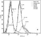
图4筛选过程中文库富集情况,流式细胞仪检测的第3、第5、第11轮文库的富集情况;Figure 4 The enrichment of the library in the screening process, the enrichment of the third, fifth and eleventh round of the library detected by flow cytometry;
图5是Cell-SELEX各轮筛选的ssDNA亚文库对U87Δ细胞系和U87细胞系的结合力流式细胞仪检测结果图;Figure 5 is a flow cytometry detection result of the binding force of the ssDNA sub-library screened in each round of Cell-SELEX to the U87Δ cell line and the U87 cell line;
图6是FITC标记的适配子U2、U8、U19、U31对U87细胞系和U87Δ细胞系的结合特异性及结合位点共聚焦显微镜检测结果图;U2A图.FITC标记的适配子U2和靶细胞U87-EGFRvⅢ结合图;U2B图.FITC标记适配子U2和反筛细胞U87-MG结合图;U8A图.FITC标记适配子U8和靶细胞U87-EGFRvⅢ结合图;U8B图.FITC标记适配子U8和反筛细胞U87-MG结合图;U19A图.FITC标记适配子U19和靶细胞U87-EGFRvⅢ结合图;U19B图.FITC标记适配子U19和反筛细胞U87-MG结合图;U31A图.FITC标记适配子U31和靶细胞U87-EGFRvⅢ结合图;U31B图.FITC标记适配子U31和反筛细胞U87-MG结合图;GNA图.FITC标记原库GN(初始ssDNA文库,以下同)和靶细胞U87-EGFRvⅢ结合图;GNB图.FITC标记原库GN和反筛细胞U87-MG结合图;Figure 6 shows the binding specificity of FITC-labeled aptamers U2, U8, U19, and U31 to U87 cell lines and U87Δ cell lines and the results of confocal microscopy of the binding sites; U2A diagram. FITC-labeled aptamers U2 and U87-EGFRvⅢ binding map of target cells; U2B map. FITC-labeled aptamer U2 and reverse screening cell U87-MG binding map; U8A map. FITC-labeled aptamer U8 and target cell U87-EGFRvⅢ binding map; U8B map. FITC labeling The binding map of aptamer U8 and reverse screening cell U87-MG; the picture of U19A. the binding map of FITC-labeled aptamer U19 and target cell U87-EGFRvⅢ; the picture of U19B. the binding map of FITC-labeled aptamer U19 and reverse screening cell U87-MG ;U31A diagram. FITC-labeled aptamer U31 and target cell U87-EGFRvⅢ binding diagram; U31B diagram. FITC-labeled aptamer U31 and reverse screening cell U87-MG binding diagram; GNA diagram. FITC-labeled original library GN (initial ssDNA library , the same below) and target cell U87-EGFRvⅢ binding diagram; GNB diagram. FITC-labeled original library GN and reverse screening cell U87-MG binding diagram;
图7生物素标记适配子特异性鉴定靶蛋白;1:原库GN钓取的反筛细胞U87MG蛋白;2:原库GN钓取的靶细胞U87-EGFRvⅢ87MG蛋白;3:适配子U2钓取的靶细胞U87-EGFRvⅢ蛋白;4:适配子U8钓取的靶细胞U87-EGFRvⅢ蛋白;5:适配子U2钓取的反筛细胞U87MG蛋白;6:适配子U8钓取的反筛细胞U87MG蛋白;Fig. 7 Specific identification of target protein by biotin-labeled aptamers; 1: U87MG protein of reverse screening cells caught by GN of the original library; 2: U87-EGFRvⅢ87MG protein of target cells caught by GN of the original library; 3: aptamer U2 U87-EGFRvⅢ protein from target cells; 4: U87-EGFRvⅢ protein from target cells caught by aptamer U8; 5: U87MG protein from reverse screening cells caught by aptamer U2; 6: reverse screened protein from U8 by aptamer U8 Sieve cell U87MG protein;
图8是裸鼠荷瘤模型的建立;A:种植U87-EGFRvⅢ细胞悬浮液前的裸鼠;B:种植U87-EGFRvⅢ细胞悬浮液两周后的裸鼠。Figure 8 is the establishment of tumor-bearing models in nude mice; A: nude mice before planting U87-EGFRvIII cell suspension; B: nude mice two weeks after planting U87-EGFRvIII cell suspension.
图9是适配子体内给药结束后各组肿瘤组织;A:无任何处理的空白对照组;B:转染试剂Invivo jet-PEITM治疗组;C:转染试剂In vivo jet-PEITM转染的原库GN治疗组;D:转染试剂Invivo jet-PEITM转染的适配子U2治疗组。Figure 9 shows the tumor tissues of each group after in vivo administration of aptamers; A: blank control group without any treatment; B: transfection reagent Invivo jet-PEITM treatment group; C: transfection reagent In vivo jet-PEITM Transfected original library GN treatment group; D: aptamer U2 treatment group transfected with transfection reagent Invivo jet-PEITM .
图10适配子体内给药前后的肿瘤体积统计结果;A:第一次给药时各组肿瘤体积大小统计,P值大于0.05;B:剥离的肿瘤体积.Blank:无任何处理的空白对照组;Reagent:In vivo jet-PEITM转染试剂治疗组;GN:转染试剂In vivo jet-PEITM转染的原库GN治疗组;AptU2:转染试剂In vivo jet-PEITM转染的适配子U2治疗组.***:与转染试剂In vivo jet-PEITM转染的适配子U2治疗组比较P值小于0.001;*:与转染试剂In vivo jet-PEITM转染的适配子U2治疗组比较P值小于0.05.Figure 10 Statistical results of tumor volume before and after in vivo administration of aptamers; A: statistics of tumor volume in each group at the first administration, P value greater than 0.05; B: stripped tumor volume. Blank: blank control without any treatment group; Reagent: In vivo jet-PEITM transfection reagent treatment group; GN: original library GN treatment group transfected with transfection reagent In vivo jet-PEITM ; AptU2: transfection reagent In vivo jet-PEITM transfected Aptamer U2 treatment group. ***: Compared with the aptamer U2 treatment group transfected with the transfection reagent In vivo jet-PEITM , the P value is less than 0.001; *: Transfected with the transfection reagent In vivo jet-PEITM The P value of the aptamer U2 treatment group was less than 0.05.
图11是适配子体内给药结束后肿瘤重量的统计结果;Blank:无任何处理的空白对照组;Reagent:转染试剂In vivo jet-PEITM治疗组;GN:转染试剂In vivo jet-PEITM转染的原库GN治疗组;AptU2:转染试剂In vivo jet-PEITM转染的适配子U2治疗组.***:与转染试剂In vivojet-PEITM转染的适配子U2治疗组比较P值小于0.001;**:与转染试剂In vivo jet-PEITM转染的适配子U2治疗组比较P值小于0.01.Figure 11 is the statistical result of tumor weight after in vivo administration of aptamers; Blank: blank control group without any treatment; Reagent: transfection reagent In vivo jet-PEITM treatment group; GN: transfection reagent In vivo jet- The original library GN treatment group transfected with PEITM ; AptU2: the aptamer U2 treatment group transfected with the transfection reagent In vivojet-PEITM .***: Adaptation with the transfection reagent In vivojet-PEITM Compared with the aptamer U2 treatment group, the P value is less than 0.001; **: The P value is less than 0.01 compared with the aptamer U2 treatment group transfected with the transfection reagent In vivo jet-PEITM .
图12是188Re标记适配子给药后的肿瘤组织体积改变;A:第一次给药时各组肿瘤体积大小统计,P值大于0.05;B:剥离的肿瘤体积.Blank:无任何处理的空白对照组;Radionuclide:瘤内注射游离的核素188Re对照组;188Re-GN:瘤内注射188Re标记的原库GN对照组;188Re-U2:瘤内注射188Re标记的适配子U2治疗组.*:与无任何处理的空白对照组比较P值小于0.01;#:与游离的核素188Re对照组比较P值小于0.01;&:与188Re标记的原库GN对照组比较P值小于0.01.Figure 12 shows the volume change of tumor tissue after administration of188 Re-labeled aptamers; A: statistics of tumor volume in each group at the first administration, P value greater than 0.05; B: volume of stripped tumor. Blank: no treatment Radionuclide: intratumoral injection of free nuclide188 Re control group;188 Re-GN: intratumoral injection of188 Re-labeled original library GN control group;188 Re-U2: intratumoral injection of188 Re-labeled appropriate Gamete U2 treatment group.*: Compared with the blank control group without any treatment, the P value is less than 0.01;# : Compared with the free nuclide188 Re control group, the P value is less than 0.01;& : Compared with the original library GN labeled with188 Re The P value for group comparison is less than 0.01.
图13是188Re标记适配子给药后剥离的肿瘤组织重量(n=9);Blank:无任何处理的空白对照组;Radionuclide:瘤内注射游离的核素188Re对照组;188Re-GN:瘤内注射188Re标记的原库GN对照组;188Re-U2:瘤内注射188Re标记的适配子U2治疗组.*:与无任何处理的空白对照组比较P值小于0.01;#:与游离的核素188Re对照组比较P值小于0.01;&:与188Re标记的原库GN对照组比较P值小于0.01.Figure 13 is the weight of tumor tissue stripped after administration of188 Re-labeled aptamers (n=9); Blank: blank control group without any treatment; Radioneclide: intratumoral injection of free nuclide188 Re control group;188 Re- GN: Intratumoral injection of188 Re-labeled original library GN control group;188 Re-U2: Intratumoral injection of188 Re-labeled aptamer U2 treatment group. *: P value less than 0.01 compared with the blank control group without any treatment;# : Compared with the free nuclide188 Re control group, the P value is less than 0.01;& : Compared with the188 Re-labeled original library GN control group, the P value is less than 0.01.
图14是SPECT显影检测标记率;A:展开剂为丙酮溶液;B:展开剂为生理盐水溶液;C:展开剂为乙醇:氨水:水=2:1:5的混合溶液;Fig. 14 is the SPECT developing detection mark rate; A: developing agent is acetone solution; B: developing agent is saline solution; C: developing agent is the mixed solution of ethanol: ammoniacal liquor: water=2:1:5;
图15是188Re标记适配子尾静脉给药的靶向性检测;A:裸鼠尾静脉注射游离核素188Re;B:裸鼠尾静脉注射188Re标记的原库GN;C:裸鼠尾静脉注射188Re标记的适配子U2;Figure 15 is the targeted detection of tail vein administration of188 Re-labeled aptamers; A: nude mice were injected with free nuclide188 Re in the tail vein; B: nude mice were injected with188 Re-labeled original library GN in the tail vein; C: nude mice188 Re-labeled aptamer U2 was injected into the mouse tail vein;
图16是188Re标记适配子瘤内给药的代谢性检测;A:在裸鼠瘤内注射188Re标记的原库GN和游离核素188Re半小时后,体内的SPECT显影情况;B:在裸鼠瘤内注射188Re标记的原库GN和游离核素188Re三小时后,体内的SPECT显影情况;Figure 16 is the metabolic detection of intratumoral administration of188 Re-labeled aptamers; A: half an hour after intratumoral injection of188 Re-labeled original library GN and free nuclide188 Re in nude mice, SPECT imaging in vivo; B : three hours after injecting188 Re-labeled former library GN and free nuclide188 Re in the tumor of nude mice, the SPECT imaging situation in the body;
图17是离体器官188Re的分布情况;A:图上半部分:离体器官实物摆放位置;图下半部分:尾静脉注射188Re标记的适配子U2在各个离体器官188Re的SPECT显影情况;B:图上半部分:离体器官实物摆放位置;图下半部分:尾静脉注射游离核素188Re在各个离体器官188Re的SPECT显影情况;C:图上半部分:离体器官实物摆放位置;图下半部分:尾静脉注射188Re标记的原库GN在各个离体器官188Re的SPECT显影情况.Figure 17 shows the distribution of188 Re in isolated organs; A: the upper part of the figure: the physical placement of the isolated organs; the lower part of the figure: the tail vein injection of188 Re-labeled aptamer U2 in each isolated organ188 Re B: The upper part of the picture: the physical placement of the isolated organs; the lower part of the picture: the SPECT development of188 Re in various isolated organs injected with free nuclide188 Re through the tail vein; C: the upper part of the picture Part: physical placement of isolated organs; lower part of the figure: tail vein injection of188 Re-labeled original library GN in each isolated organ with188 Re SPECT imaging.
具体实施方式Detailed ways
以下实施例用于进一步说明本发明,但不应理解为对本发明的限制。若未特别指明,实施例中所用的技术手段为本领域技术人员所熟知的常规手段。The following examples are used to further illustrate the present invention, but should not be construed as limiting the present invention. Unless otherwise specified, the technical means used in the embodiments are conventional means well known to those skilled in the art.
本发明的详细说明中所用的主要术语的定义如下。Definitions of main terms used in the detailed description of the present invention are as follows.
本发明使用的术语“核酸适配子”是指以高的亲和力特异性地识别其靶标的小单链寡核苷酸。The term "nucleic acid aptamer" used in the present invention refers to a small single-stranded oligonucleotide that specifically recognizes its target with high affinity.
本发明使用的术语“样品”是指待分析的可能包括胶质瘤的标记的组合物。样品的实例包括胶质瘤细胞系、胶质瘤组织、血液、血清、血浆、唾液、痰、尿液。The term "sample" as used herein refers to a composition to be analyzed that may include markers of glioma. Examples of samples include glioma cell lines, glioma tissue, blood, serum, plasma, saliva, sputum, urine.
本发明使用的词语“U87Δ细胞”是指稳定过表达EGFRvIII型突变体的U87细胞株。The term "U87Δ cell" used in the present invention refers to a U87 cell line stably overexpressing the EGFRvIII type mutant.
适配子的核苷酸总数为<100nt。如果适配子的核苷酸数量少,则适配子的化学合成和大量生产将比较容易且具有成本优势。此外,适配子将容易被化学修饰,在体内高度稳定且毒性低。除此之外,包括在适配子在内的各核苷酸可包括一个或者多个相同或不同的化学修饰,例如可在核糖的2’位进行任何原子或基团的核苷酸取代。The total number of nucleotides of the aptamers was <100 nt. If the number of nucleotides of the aptamer is small, the chemical synthesis and mass production of the aptamer will be relatively easy and cost-effective. Furthermore, aptamers will be easily chemically modified, highly stable in vivo and have low toxicity. In addition, each nucleotide included in the aptamer may include one or more identical or different chemical modifications, such as nucleotide substitution of any atom or group at the 2' position of ribose.
因此,另一个方面,本发明涉及一种诊断胶质瘤的组合物,所述组合物包含上述本发明的核酸适配子。Therefore, in another aspect, the present invention relates to a composition for diagnosing glioma, said composition comprising the above-mentioned nucleic acid aptamer of the present invention.
本发明涉及一种用核酸适配子来定量检测肿瘤细胞表面EGFRvIII的表达。例如,通过将生物素缀合到核酸适配子的末端,使标记了的核酸适配子与固定在酶联板上的来自胶质瘤细胞系的蛋白样品进行反应,然后用链霉亲和素-HRP的二抗与生物素标记的适配子反应,通过显色即可定量检测到肿瘤蛋白样品中EGFRvIII的表达量。The invention relates to a method for quantitatively detecting the expression of EGFRvIII on the surface of tumor cells by using nucleic acid aptamers. For example, by conjugating biotin to the end of the aptamer, the labeled aptamer was reacted with a protein sample from a glioma cell line immobilized on an enzyme-linked plate, and then treated with streptavidin The secondary antibody of K-HRP reacts with the biotin-labeled aptamer, and the expression of EGFRvIII in the tumor protein sample can be quantitatively detected by color development.
胶质瘤的新的生物标记可通过分析结合到核酸适配子的样品物质而检测。因此,在又一方面,本发明涉及一种用核酸适配子来检测胶质瘤细胞特异的表面生物标记的方法。例如,通过将生物素缀合到核酸适配子的末端,使核酸适配子与来自胶质瘤细胞系的膜提取蛋白样品结合,并用链霉亲和素缀合的磁性颗粒沉淀适配子,然后用质谱法分析适配子,来检测特异性地结合适配子的表面生物标记。Novel biomarkers of glioma can be detected by analyzing sample material bound to nucleic acid aptamers. Therefore, in another aspect, the present invention relates to a method for detecting specific surface biomarkers of glioma cells by using nucleic acid aptamers. For example, aptamers were bound to membrane-extracted protein samples from glioma cell lines by conjugating biotin to the ends of the aptamers, and the aptamers were precipitated with streptavidin-conjugated magnetic particles. , and then analyze the aptamers by mass spectrometry to detect surface biomarkers that specifically bind the aptamers.
同时,特异性地结合胶质瘤细胞或其组织的核酸适配子可固定在常规的支持物上,如磁珠、颗粒、试纸条,从而提供可用于诊断胶质瘤的检测传感器。因此,本发明的另一个方面涉及一种诊断胶质瘤的传感器,所述传感器上固定有与胶质瘤细胞U87Δ或组织的特异性地结合的核酸适配子。At the same time, nucleic acid aptamers that specifically bind to glioma cells or their tissues can be immobilized on conventional supports, such as magnetic beads, particles, and test strips, thereby providing detection sensors that can be used to diagnose glioma. Therefore, another aspect of the present invention relates to a sensor for diagnosing glioma, on which a nucleic acid aptamer that specifically binds to glioma cells U87Δ or tissue is immobilized.
上述固定支持无包括至少一个实质上是固相支持物,可通过任何常规的化学结合方法将核酸适配子固定到所述的固相支持物上。例如,可通过将生物素缀合到核酸适配子的末端形成缀合物,从而将缀合物固定到支持物的表面上,并将链霉亲和素固定到支持物的表上以诱导固定在支持物表面上的蛋白链霉素和生物素之间的相互作用。The above-mentioned immobilized supports include at least one substantially solid-phase support, and the nucleic acid aptamer can be immobilized on the solid-phase support by any conventional chemical combination method. For example, the conjugate can be formed by conjugating biotin to the end of the nucleic acid aptamer, thereby immobilizing the conjugate on the surface of the support, and immobilizing streptavidin on the surface of the support to induce Interaction between the protein streptomycin and biotin immobilized on the surface of the support.
同时,本发明的方法可以以试剂盒的形式提供出来,以增加可携带性。具体地,在另一个方面,本发明涉及一种诊断过表达人表皮生长因子受体Ⅲ型突变体的胶质瘤细胞的试剂盒,所述试剂盒包含与人表皮生长因子受体Ⅲ型突变体的的核酸适配子。如果需要的话,胶质瘤的诊断试剂盒可包括缓冲液和用于进行检测和分析的容器。诊断试剂盒可以是瓶、注射器类似的形式,该试剂盒可本分或全部由塑料、纸等类似物形成。传感器容器可装配有或全部或部分可分离的盖子,硕鼠盖子可以是容器的原始部件或可以是通过机械、粘着或其他方式附着与容器。容器也可以装配有塞子并通过注射针来接触内含物。检测试剂盒可包括外包装,外包装可包括有关组分用途的说明。At the same time, the method of the present invention can be provided in the form of a kit to increase portability. Specifically, in another aspect, the present invention relates to a kit for diagnosing glioma cells overexpressing human epidermal growth factor receptor type III mutants, said kit comprising a human epidermal growth factor receptor type III mutant Nucleic acid aptamers for body. The diagnostic kit for glioma may include buffers and containers for detection and analysis, if necessary. The diagnostic kit may be in the form of a bottle, syringe, or the like, and the kit may be formed partly or entirely of plastic, paper, or the like. The sensor container may be fitted with a fully or partially detachable cover which may be an original part of the container or may be mechanically, adhesively or otherwise attached to the container. The container may also be fitted with a stopper and the contents accessed by an injection needle. Assay kits may include outer packaging, which may include instructions regarding the use of the components.
另一方面,本发明涉及一种新的能介导其他药物或siRNA或病毒的细胞传递或跨膜转运的载体,如上所述的与过表达人表皮生长因子受体Ⅲ型突变体特异性结合的核酸适配子。例如,将药物与如上所述的与过表达人表皮生长因子受体Ⅲ型突变体特异性结合的核酸适配子通过共价键或者其他介质耦合在一起,加入表达EGFRvIII的肿瘤细胞或组织中,适配子特异识别受体并且被内吞进入胞内,由此将药物或siRNA携带进入细胞,达到一个靶向治疗的目标。或者,也可以将适配子、药物、高分子材料组合在一起,高分子材料包裹药物和与过表达人表皮生长因子受体Ⅲ型突变体特异性结合的核酸适配子的组合体,从而满足通过血脑屏障的需要,进入脑内组织时,与过表达人表皮生长因子受体Ⅲ型突变体特异性结合的核酸适配子和药物的复合物释放出来,适配子识别靶蛋白,从而将药物运抵目标。On the other hand, the present invention relates to a new carrier capable of mediating the cell delivery or transmembrane transport of other drugs or siRNA or virus, specifically combined with the overexpressed human epidermal growth factor receptor type III mutant as described above nucleic acid aptamers. For example, the drug is coupled with the above-mentioned nucleic acid aptamer that specifically binds to the overexpressed human epidermal growth factor receptor type III mutant through a covalent bond or other media, and added to tumor cells or tissues expressing EGFRvIII , the aptamer specifically recognizes the receptor and is endocytosed into the cell, thereby carrying the drug or siRNA into the cell to achieve a targeted therapeutic goal. Alternatively, aptamers, drugs, and polymer materials can also be combined together, and the polymer materials can wrap drugs and a combination of nucleic acid aptamers that specifically bind to overexpressed human epidermal growth factor receptor type III mutants, thereby Satisfies the need to pass through the blood-brain barrier. When entering the brain tissue, the complex of the nucleic acid aptamer and the drug that specifically binds to the overexpressed human epidermal growth factor receptor type III mutant is released, and the aptamer recognizes the target protein. Thus delivering the drug to its target.
因此,可以以本发明的与过表达人表皮生长因子受体Ⅲ型突变体特异性结合的核酸适配子来递送抗癌药物或者siRNA((Chu et al.2006)、(Zhou et al.2011)、(Zhou&Rossi2008)、)Therefore, the nucleic acid aptamer of the present invention that specifically binds to the overexpressed human epidermal growth factor receptor type III mutant can be used to deliver anticancer drugs or siRNA ((Chu et al.2006), (Zhou et al.2011 ), (Zhou&Rossi2008),)
本发明的与过表达人表皮生长因子受体Ⅲ型突变体特异性结合的核酸适配子可以其药物上可接受的盐的形式用于药物组合物中。此外,本发明的与过表达人表皮生长因子受体Ⅲ型突变体特异性结合的核酸适配子可单独使用或与其他药学活性化合物结合使用。The nucleic acid aptamer specifically binding to the overexpressed human epidermal growth factor receptor type III mutant of the present invention can be used in a pharmaceutical composition in the form of its pharmaceutically acceptable salt. In addition, the nucleic acid aptamer of the present invention that specifically binds to the overexpressed human epidermal growth factor receptor type III mutant can be used alone or in combination with other pharmaceutically active compounds.
根据本发明的与过表达人表皮生长因子受体Ⅲ型突变体特异性结合的核酸适配子的特性,结合药学高分子材料,可以开发新的剂型。如,纳米乳、包合物、缓释胶囊、脂质体等形式。开发利于透过血脑屏障的油包水型纳米乳需要水、油、乳化剂和助乳化剂等,其中天然乳化剂包括多糖类如阿拉伯胶、白蛋白、大豆磷脂、卵磷脂和胆固醇等,合成乳化剂分离子型和非离子型两类,常用的非离子型乳化剂如脂肪酸山梨坦(亲油性)、聚山梨酯(亲水性)等,而助乳化剂常可用来调节乳化剂的HLB值,并形成更小的乳滴。常用的助乳化剂有正丁醇、乙二醇、丙二醇、甘油等。开发稳定的包合物,用的材料如β-环糊精、淀粉、纤维素等等,使与过表达人表皮生长因子受体Ⅲ型突变体特异性结合的核酸适配子不易被血液中的核酸酶所降解,使药物达到缓释的作用。According to the characteristics of the nucleic acid aptamer specifically binding to the overexpressed human epidermal growth factor receptor type III mutant of the present invention, combined with pharmaceutical polymer materials, new dosage forms can be developed. For example, nanoemulsions, clathrates, sustained-release capsules, liposomes and other forms. The development of water-in-oil nanoemulsions that facilitate penetration of the blood-brain barrier requires water, oil, emulsifiers, and co-emulsifiers, among which natural emulsifiers include polysaccharides such as acacia, albumin, soybean lecithin, lecithin, and cholesterol. , Synthetic emulsifiers are separated into sub-type and non-ionic types. Commonly used non-ionic emulsifiers such as fatty acid sorbitan (lipophilic), polysorbate (hydrophilic), etc., and co-emulsifiers can often be used to adjust emulsifiers HLB value, and form smaller milk droplets. Commonly used co-emulsifiers are n-butanol, ethylene glycol, propylene glycol, glycerin, etc. Develop stable clathrates, using materials such as β-cyclodextrin, starch, cellulose, etc., so that the nucleic acid aptamer that specifically binds to the overexpressed human epidermal growth factor receptor type III mutant is not easily absorbed in the blood Degraded by the nuclease, so that the drug can achieve the effect of sustained release.
可根据患者的状况和体重、疾病的严重度、药物的类型和给药途径及周期来适用地选择本发明组合物的优选剂量和剂型,达到个体化临床用药的目的。本发明的组合物可通过多种途径给药与哺乳动物,包括小鼠、大鼠等。给药途径为:口服,注射、鞘内或脑血管给药等。The preferred dose and dosage form of the composition of the present invention can be suitably selected according to the condition and body weight of the patient, the severity of the disease, the type of drug, the administration route and cycle, so as to achieve the purpose of individualized clinical medicine. The composition of the present invention can be administered to mammals, including mice, rats, etc., through various routes. The route of administration is: oral, injection, intrathecal or cerebrovascular administration, etc.
实施例1:Example 1:
实验一、制备与过表达人表皮生长因子受体III型突变体(EGFRvIII)的胶质瘤细胞系(U87Δ)特异性结合的核酸适配子,筛选过程如图1所示,在11轮测序结果如图3所示。
1、设计并合成初始ssDNA文库和用于PCR扩增的引物1. Design and synthesize initial ssDNA library and primers for PCR amplification
设计并合成长度为76个碱基的ssDNA初始GN库:5'-ATCCAGAGTGACGCAGCA(N40)TGGACACGGTGGCTTAGT-3',初始ssDNA文库两端是固定序列,中间是40个随机序列,N代表A,T,C,G四个随机碱基。Design and synthesize the ssDNA initial GN library with a length of 76 bases: 5'-ATCCAGAGTGACGCAGCA (N40) TGGACACGGTGGCTTAGT-3', the two ends of the initial ssDNA library are fixed sequences, and the middle is 40 random sequences, N stands for A, T, C , G four random bases.
设计并合成用于扩增出dsDNA双链的引物:Design and synthesize primers for amplifying double-stranded dsDNA:
5'-FITC-Pn1:5'-ATCCAGAGTGACGCAGCA-3';5'-FITC-Pn1:5'-ATCCAGAGTGACGCAGCA-3';
5’-Biotin-Pn2:5'-ACTAAGCCACCGTGTCCA-3’5'-Biotin-Pn2: 5'-ACTAAGCCACCGTGTCCA-3'
上述初始ssDNA文库和引物均由生物公司合成。The above-mentioned initial ssDNA library and primers were synthesized by biological companies.
用PCR法,以引物5'-FITC-Pn1和引物5’-Biotin-Pn2为引物,以生物公司合成的初始ssDNA文库为模板扩增出dsDNA双链(PCR产物),大量扩增后,应用链霉亲和素磁珠分离法获得ssDNA亚文库或者适配子,双链与单链的分子量均为76bp,结果如图2所示。Using the PCR method, primers 5'-FITC-Pn1 and 5'-Biotin-Pn2 were used as primers, and the initial ssDNA library synthesized by the biological company was used as a template to amplify dsDNA double strands (PCR products). After a large amount of amplification, the application The ssDNA sub-library or aptamer was obtained by the streptavidin magnetic bead separation method, and the molecular weight of the double strand and the single strand were both 76 bp. The results are shown in Figure 2.
2、链亲和素磁珠分离法制备单链ssDNA2. Preparation of single-stranded ssDNA by streptavidin magnetic bead separation method
大约按照1ml的步骤1的PCR产物:150ul的链霉亲和素磁珠量的比例,将链霉亲和素磁珠注入柱子中,磁珠上下垫筛板以更好的过滤PCR产物。让磁珠自然状态下沉淀,但不能让磁珠里没有液体溶解;用0.5×D-PBS洗涤5次,此步操作应该用注射管轻轻的吹气赶走柱子中的气泡,尤其是出口处不能有气泡;室温下,将PCR产物循环经过磁珠过滤3次,其中一条带生物素标记的链与链霉亲和素磁珠结合,此步可以人工用注射针管加压,控制速度为1滴/每3秒;0.5ml D-PBS洗涤链霉亲和素磁珠5次,加入0.2M的NaOH400μl于37℃温箱中自然过滤30-40min,另一条带FITC标记的链被裂解下来,下面接收过滤样品的EP管中用1/10体积40μl的10%乙酸中和;接收完裂解样品之后加1/10体积的3mol/L NaAc(PH5.2),1/100体积的1mol/L MgCl2和2倍体积的无水乙醇,于-20℃冰箱沉淀过夜;14,000rpm4℃离心30min,弃上清,70%乙醇1ml溶解洗涤,于14,000rpm4℃离心30min,弃液体倒置晾干,溶于少量的三蒸水中,作为下一轮筛选的亚文库(ssDNA文库);紫外分光光度计测ssDNA浓度,7M尿素8%变性凝胶检测得到的ssDNA。Inject the streptavidin magnetic beads into the column according to the ratio of 1ml of the PCR product of step 1: 150ul of streptavidin magnetic beads, and put the magnetic beads on the top and bottom of the sieve plate to better filter the PCR product. Let the magnetic beads settle in a natural state, but don’t let the liquid in the magnetic beads dissolve; wash 5 times with 0.5×D-PBS. In this step, blow gently with the injection tube to drive away the air bubbles in the column, especially at the outlet There should be no air bubbles; at room temperature, cycle the PCR product through magnetic beads for 3 times, and one of the chains labeled with biotin will bind to streptavidin magnetic beads. This step can be manually pressurized with an injection needle, and the control speed is 1 Drops/every 3 seconds; 0.5ml D-PBS to wash the streptavidin
3、细胞selex筛选适配子3. Cell selex selection of aptamers
(1)以U87Δ为靶细胞筛选前三轮(1) The first three rounds of screening with U87Δ as the target cell
a、将步骤2的一定浓度的ssDNA文库溶解于一定量的鲑精DNA和酵母tRNA,各种具体用量见表1,于95℃变性10min后立即置于冰上5min,在冰上冷却时加入500μl的结合缓冲液(1L PBS缓冲液中含5mmol MgCl2、0.05g鲑鱼精DNA、1g BSA和4.5g葡萄糖),室温放置30min左右,之后用于筛选(第一轮筛选ssDNA文库用量为1,000pmol,以后每一轮依次为800pmol、200pmol、150pmol递减),在前三轮的筛选没有引入反筛细胞U87MG细胞;a. Dissolve the ssDNA library of a certain concentration in
表1Table 1

*第四轮引入U87MG作为反筛细胞*The fourth round introduced U87MG as reverse screening cells
b、约1×10⁶个贴壁生长过夜的U87Δ靶细胞,用洗涤缓冲液(1L PBS缓冲液中含5mmolMgCl2和4.5g葡萄糖)洗涤3次,每次1min;将已经制备好的ssDNA样品与U87Δ细胞在37℃、5%CO2培养箱中共孵育60min,使得ssDNA文库与细胞充分作用;b. About 1×10⁶ U87Δ target cells adherently grown overnight, washed 3 times with washing buffer (5mmolMgCl2 and 4.5g glucose in 1L PBS buffer), 1min each time; the prepared ssDNA samples Co-incubate with U87Δ cells in a 37°C, 5% CO2 incubator for 60 minutes, so that the ssDNA library can fully interact with the cells;
c.回收筛选文库,用洗涤缓冲液洗涤细胞3次,每次1min;用500μl的ddH2O溶解1μl蛋白酶K的样品与细胞在37℃作用15min,收集所有结合样品与细胞于1.5ml离心管中,95℃加热10min,离心收集上清,即为第一轮筛选得到的产物,用于下一轮筛选扩增dsDNA。c. Recover the screening library, wash the
用PCR法,以引物5'-FITC-Pn1和引物5’-Biotin-Pn2为引物,以第一轮筛选得到的产物为模板扩增出dsDNA双链(PCR产物),再通过链亲和素磁珠分离法从该dsDNA双链(PCR产物)制备单链ssDNA文库(具体步骤参照步骤2和表1)。用于下一轮筛选。Using the PCR method, primers 5'-FITC-Pn1 and 5'-Biotin-Pn2 were used as primers, and the product obtained in the first round of screening was used as a template to amplify dsDNA double strands (PCR products), and then passed streptavidin The single-stranded ssDNA library was prepared from the double-stranded dsDNA (PCR product) by magnetic bead separation (see
如此进行3轮筛选,得到3轮筛选后的ssDNA文库。Three rounds of screening were carried out in this way to obtain the ssDNA library after three rounds of screening.
(2)第四轮开始引入反筛细胞U87MG(2) In the fourth round, the reverse screening cell U87MG was introduced
a.按照步骤(1)中的a、b、c步骤得到经过3轮筛选后的ssDNA文库;a. Follow steps a, b, and c in step (1) to obtain the ssDNA library after three rounds of screening;
b.用洗涤缓冲液洗涤U87MG细胞3次,每次3min;将步骤(1)中得到的经过3轮筛选后的ssDNA文库与U87MG细胞在37℃、5%CO2培养箱中共孵育40min左右;洗涤缓冲液洗涤细胞3次,每次3min,小心收集液体,离心收集上清,即为该轮筛选得到的产物,用于下一轮筛选扩增dsDNA。b.
(3)用PCR法,以引物5'-FITC-Pn1和引物5’-Biotin-Pn2为引物,以步骤步骤(2)中的b得到的该轮筛选得到的产物为模板扩增出dsDNA双链(PCR产物),再通过链亲和素磁珠分离法从该dsDNA双链(PCR产物)制备带FITC标记的、与靶细胞特异性结合的ssDNA配基(具体步骤参考步骤2和表1)。用于下一轮筛选。(3) Using the PCR method, use the primer 5'-FITC-Pn1 and the primer 5'-Biotin-Pn2 as primers, and use the product obtained in this round of screening in step (2) as a template to amplify the dsDNA double Strand (PCR product), and then prepare FITC-labeled ssDNA ligand that specifically binds to target cells from the dsDNA double-strand (PCR product) by streptavidin magnetic bead separation method (refer to step 2 and Table 1 for specific steps ). for the next round of screening.
如此重复筛选8轮,连同U87Δ为靶细胞筛选的前三轮,总共筛选了11个循环。This repeated 8 rounds of screening, together with the first three rounds of screening U87Δ as the target cell, a total of 11 rounds of screening were performed.
(4)为了更好地得到特异性强、高亲和力的适配子,在逐渐的筛选过程中不断地增加洗涤次数和延长洗涤时间,缩短与细胞孵育时间和减少细胞的密度,具体见表1。(4) In order to better obtain aptamers with strong specificity and high affinity, continuously increase the number of washings and prolong the washing time during the gradual screening process, shorten the incubation time with cells and reduce the density of cells, see Table 1 for details .
利用链霉亲和素磁珠分离法获得ssDNA亚文库或者适配子,双链与单链的分子量均为76bp,结果如图3所示。The ssDNA sublibrary or aptamer was obtained by streptavidin magnetic bead separation method, and the molecular weight of the double strand and single strand were both 76 bp, the results are shown in Figure 3.
经过11轮筛选后对筛选得到的适配子进行克隆测序,所得到适配子的序列见图3。After 11 rounds of screening, the screened aptamers were cloned and sequenced, and the sequences of the obtained aptamers are shown in FIG. 3 .
如图3所示,筛选得到的适配子有4个,分别为U2(5’-ATCCAGAGTGACGCAGCATTTTGACGCTTTATCCTTTTCTTATGGCGGGATAGTTTCGTGGACACGGTGGCTTAGT-3’,具体序列如SEQ ID NO.1所示)、U8(5’-ATCCAGAGTGACGCAGCATGAATCTTTTCTTTTGGTTTTGATATTTATAGTTGGTGAATGGACACGGTGGCTTAGT-3’,具体序列如SEQ ID NO.2所示)、U19(5’-ATCCAGAGTGACGCAGCATTTGTATCCTATTTTGTTTATGTAATTGTCGTTGATCATGTGGACACGGTGGCTTAGT-3’,具体序列如SEQ ID NO.3所示)、U31(5’-ATCCAGAGTGACGCAGCATTTGTTTAATATGTTTTTTAATTCCCCTTGTGGTGTGTTGTGGACACGGTGGCTTAGT-3’,具体序列如SEQ ID NO.4所示)。As shown in Figure 3, there are 4 screened aptamers, U2 (5'-ATCCAGAGTGACGCAGCATTTTGACGCTTTATCCTTTTCTTATGGCGGGATAGTTTCGTGGACACGGTGGCTTAGT-3', the specific sequence is shown in SEQ ID NO.1), U8 (5'-ATCCAGAGTGACGCAGCATGAATCTTTTCTTTTGGTTTTGATATTTATAGTTGGTGAATGGACTA, The specific sequence is shown in SEQ ID NO.2), U19 (5'-ATCCAGAGTGACGCAGCATTTGTATCCTATTTTGTTTTATGTAATTGTCGTTGATCATGTGGACACGGTGGCTTAGT-3', the specific sequence is shown in SEQ ID NO.3), U31 (5'-ATCCAGAGTGACGCAGCATTTGTTCTTAATATGTTTTTAATTCCCCTNO'SEQGTGTGTTGTGGGACGT .4).
实验二、Cell-SELEX技术筛选得到的适配子富集检测
1、检测Cell-SELEX法的第3、5、11轮筛选得到的ssDNA亚文库对U87Δ细胞系的富集情况1. Detect the enrichment of U87Δ cell line in the ssDNA sub-library obtained in the 3rd, 5th, and 11th rounds of screening of the Cell-SELEX method
具体方法为:将细胞密度5×10⁵细胞铺在六孔板中,过夜贴壁生长;用1×PBS洗涤靶细胞U87Δ,加入适量的无酶消化液,以覆盖细胞表面为准,放置37℃适宜时间直至细胞完全脱壁,加入2倍体积的1×PBS终止消化,800rpm离心6min,弃上清,PBS重悬细胞;将FITC标记的原库GN、第3轮、第5轮、第11轮筛选的亚文库FITC-ssDNA各200pmol与U87Δ细胞系孵育40min;离心沉淀细胞,弃上清,PBS洗涤细胞3次后,300μl PBS重悬细胞;流式细胞检测仪检测原库GN及次文库与U87Δ细胞系随着筛选轮速增加的富集情况。结果如图4所示,随着筛选轮数的增加,亚文库与U87Δ细胞系具有富集作用。The specific method is as follows: Cells with a cell density of 5×10⁵ were plated in a six-well plate and grown overnight; the target cells U87Δ were washed with 1×PBS, and an appropriate amount of enzyme-free digestion solution was added to cover the cell surface, and placed for 37 ℃ for a suitable time until the cells are completely detached, add 2 times the volume of 1×PBS to stop the digestion, centrifuge at 800rpm for 6min, discard the supernatant, and resuspend the cells in PBS;
实验三、适配子U2-U31的活性检测
1、流式细胞技术检测适配子U2-U31对U87Δ细胞系、U87细胞系的结合亲和力1. The binding affinity of aptamer U2-U31 to U87Δ cell line and U87 cell line was detected by flow cytometry
具体方法:将U87Δ细胞系、U87细胞系贴壁培养过夜,消化后取5×10⁵个细胞用PBS缓冲液洗涤1次,重悬后加入终浓度为200pmol的5'端标记FITC的适配子(U2、U8、U19或U31),将所述适配子分别和培养过夜后的U87Δ细胞系、U87细胞系混合后,37℃孵育40min,再用PBS缓冲液洗涤3次,用流式细胞检测仪检测FITC的荧光强度。以初始ssDNA文库作为对照,结果如图5所示。图5所示适配子与U87细胞的结合情况,用5'标记FITC的初始ssDNA文库(initial library)做对照。从图5可以看出,适配子(U2、U8、U19或U31)与U87MG细胞的结合亲和力,与初始ssDNA文库和和空白对照差不多,曲线几乎没有位移。而适配子(U2、U8、U19或U31)与空白对照和初始ssDNA文库相比,与U87Δ细胞系的结合亲和力,曲线有着明显的位移,由此说明适配子(U2、U8、U19或U31)与U87Δ细胞系的结合亲和力强。Specific method: U87Δ cell line and U87 cell line were adhered to culture overnight, after digestion, 5×10⁵ cells were washed once with PBS buffer, resuspended and added with a final concentration of 200 pmol 5' end-labeled FITC adapter Aptamers (U2, U8, U19 or U31), mixed the aptamers with the U87Δ cell line and U87 cell line after overnight culture, incubated at 37°C for 40min, and then washed 3 times with PBS buffer, and analyzed by flow cytometry Cell detector detects the fluorescence intensity of FITC. Using the initial ssDNA library as a control, the results are shown in Figure 5. The aptamer binding to U87 cells shown in Figure 5 was compared with the initial ssDNA library (initial library) with 5'-labeled FITC. It can be seen from Figure 5 that the binding affinity of aptamers (U2, U8, U19 or U31) to U87MG cells is similar to that of the initial ssDNA library and blank control, and the curve has almost no shift. Compared with the blank control and the initial ssDNA library, the aptamers (U2, U8, U19 or U31) had a significant shift in the binding affinity curve with the U87Δ cell line, which indicated that the aptamers (U2, U8, U19 or U31) has a strong binding affinity to the U87Δ cell line.
实验四、适配子特异性鉴定
1、共聚焦显微镜检测适配子U2、U8、U19和U31,与U87Δ细胞系、U87细胞系结合的特异性及结合位点检测1. Confocal microscopy detection of aptamers U2, U8, U19 and U31, specificity and binding site detection of binding to U87Δ cell line and U87 cell line
具体方法:分别接种合适密度的U87MG和U87Δ细胞于24孔板或共聚焦小皿中贴壁过夜生长;4%多聚甲醛避光固定过夜生长于24孔板的细胞20min(孔内放有圆形的盖玻片,细胞贴壁生长于盖玻片上);用1×PBS洗涤3次×5min,3%BSA封闭1h后,弃孔内液体,用1×PBS洗涤3次×5min;将150pmol寡核苷酸适配子(U2、U8、U19或U31)溶于一定量的鲑精DNA(终浓度:0.1μg/μl)和酵母tRNA(终浓度:0.1μg/μl)于95℃加热10min,冰上5min,加300μl结合缓冲液(100ml PBS加0.1g BSA、0.45g葡萄糖、500μl1mol/L的MgCl2);将此制备好的FITC适配子(U2、U8、U19或U31)分别与U87MG和U87Δ细胞室温避光孵育40min,并放置于摇床上左右缓慢摇动,使得样品与细胞充分结合;弃孔内液体,用1×PBS洗涤3次×5min,将盖玻片倒扣在滴有10μl DAPI染液的盖玻片上;室温避光放置于铝制饭盒内,在荧光显微镜或共聚焦显微镜下观察适配子与细胞的结合情况,以原库FITC-GN(初始ssDNA文库)为对照适配子。Specific method: inoculate U87MG and U87Δ cells of appropriate density and grow adherently in 24-well plates or confocal small dishes overnight; fix cells grown in 24-well plates overnight with 4% paraformaldehyde in the dark for 20 minutes (there are circular cells placed in the wells). Coverslips, cells grow on the coverslips attached to the wall); wash 3 times with 1×PBS×5min, block with 3% BSA for 1h, discard the liquid in the well, wash 3 times×5min with 1×PBS; add 150pmol oligonuclear Aptamers (U2, U8, U19 or U31) were dissolved in a certain amount of salmon sperm DNA (final concentration: 0.1 μg/μl) and yeast tRNA (final concentration: 0.1 μg/μl), heated at 95°C for 10 min, and iced Add 300μl binding buffer (100ml PBS plus 0.1g BSA, 0.45g glucose, 500μl 1mol/L MgCl2 ) for 5min; mix the prepared FITC aptamers (U2, U8, U19 or U31) with U87MG and U87Δ cells were incubated at room temperature in the dark for 40 minutes, and placed on a shaker and shaken slowly left and right to fully combine the sample with the cells; discard the liquid in the well, wash 3 times with 1×PBS for 5 minutes, and put the coverslip upside down in a drop of 10 μl DAPI staining. placed in an aluminum lunch box at room temperature and protected from light, and observed the combination of aptamers and cells under a fluorescence microscope or confocal microscope, using the original library FITC-GN (initial ssDNA library) as a control aptamer .
如图6所示,筛选得到的适配子(U2、U8、U19或U31)与U87Δ细胞系有特异性结合,与U87细胞系没有结合,对照适配子原库FITC-GN与U87Δ细胞系、U87细胞系均没有结合。As shown in Figure 6, the screened aptamers (U2, U8, U19 or U31) have specific binding to the U87Δ cell line, but have no binding to the U87 cell line. The control aptamer original library FITC-GN and the U87Δ cell line , U87 cell lines were not combined.
实验五、Pull-down实验和免疫印迹法鉴定适配子结合的靶标
具体方法:将靶细胞U87-EGFRvⅢ(U87Δ细胞系)和反筛细胞U87MG传代过夜贴壁生长,隔天观察细胞密度与状态。待细胞密度与状态好时,用蛋白裂解液裂解细胞,并加蛋白酶抑制剂和0.5M EDTA(pH8.0)裂解15min。收集所有样品,4℃16000rpm离心1h,收集上清并进行蛋白定量。生物素磁珠用0.5×PBS洗涤3次×3min×0.6ml;1×PBS洗涤3次×3min×0.6ml,制备生物素样品:适量的结合缓冲液与3’端标记生物素适配子(U2或U8)300pmol一起和磁珠于37℃孵育30min。用1×PBS洗涤磁珠3次×3min×0.6ml,投入600μg蛋白液体(按照每300pmol生物素适配子(U2或U8)与600μg靶蛋白量比例投入),与上述磁珠于37℃孵育1h;用1×PBS洗涤磁珠5次×1min×0.6ml,弃液体加60μl的1×SDS-PAGE上样缓冲液加热煮沸10min洗脱结合蛋白后,行10%SDS-PAGE凝胶并用抗EGFR抗体进行免疫印迹实验。Specific method: The target cell U87-EGFRvⅢ (U87Δ cell line) and counter-screened cell U87MG were subcultured and grown overnight, and the cell density and state were observed the next day. When the cell density and state are good, lyse the cells with protein lysate, add protease inhibitors and 0.5M EDTA (pH8.0) to lyse for 15 minutes. All samples were collected, centrifuged at 16,000 rpm at 4°C for 1 h, and the supernatant was collected for protein quantification. Biotin magnetic beads were washed 3 times with 0.5×PBS×3min×0.6ml; washed 3 times×3min×0.6ml with 1×PBS to prepare biotin samples: an appropriate amount of binding buffer and 3’ end-labeled biotin aptamer ( U2 or U8) 300pmol and incubate with magnetic beads at 37°C for 30min. Wash the magnetic beads with 1×
结果如图7所示,筛选得到的适配子(U2或U8)可特异性结合EGFRvIII蛋白。The results are shown in Figure 7, the screened aptamers (U2 or U8) can specifically bind to EGFRvIII protein.
实施例2:Example 2:
实验1、适配子在裸鼠荷瘤动物模型上的抑制肿瘤生长的作用
具体方法:U87Δ细胞用含10%FBS的DMEM于5%CO2、37℃培养,4~6周裸鼠饲养于SPF实验屏障环境中。共28只裸鼠,分为空白组、in vivo jet-PEITM转染试剂组、原库GN组和适配子U2治疗组4组,每组7只;用改良型RPMI-1640培养液重悬U87Δ细胞,密度为100μl的1640培养液中含1.5×10⁶~2.0×10⁶个;用1ml注射器于4~6周的雄性裸鼠(BALB/c)右侧后臀部皮下注射,每只裸鼠注射100μl;2周后观察肿瘤形成情况,用游标卡尺测量肿瘤体积大小;用in vivo jet-PEITM试剂分别转染适配子U2和原库GN(初始ssDNA文库)进行瘤内注射(每隔1天注射1次,67pmol/只,共3次),以转染试剂in vivo jet-PEITM、GN原库作为对照组;1周后处死动物,手术剥离肿瘤组织,对肿瘤组织拍照以作形体大小对比;游标卡尺测量肿瘤组织体积,千分之一电子称称量肿瘤组织的重量;应用SPSS19.0统计软件对实验数据进行统计学分析。动物模型肿瘤体积和重量实验数据以平均数±标准差

结果如图8-11所示,适配子U2对肿瘤的生长有抑制作用,适配子U2治疗组与其他组进行组组间比较,均有统计学差异(P<0.05)。The results are shown in Figure 8-11, the aptamer U2 has an inhibitory effect on the growth of the tumor, and the comparison between the aptamer U2 treatment group and other groups showed statistical difference (P<0.05).
实验2:188Re标记适配子在裸鼠荷瘤动物模型上观察适配子抑瘤的作用Experiment 2:188 Re-labeled aptamers were used to observe the anti-tumor effect of aptamers in nude mouse tumor-bearing animal models
具体方法:用188Re标记适配子U2和原库GN(初始ssDNA文库)进行瘤内注射(每只200pmol,约2μg,每只给药量的标记适配子U2、GN及游离188Re的188Re放射性活度均为0.075mCi,标记率70%),以游离188Re组、188Re标记的原库GN组和空白组作为对照组。1周后处死动物,手术剥离肿瘤组织,对肿瘤组织拍照以作形体大小对比。游标卡尺测量肿瘤组织体积,千分之一电子称称量肿瘤组织的重量。SPSS13.0统计软件对实验数据统计学分析,肿瘤体积和重量数据均以平Specific method: use188 Re-labeled aptamer U2 and the original library GN (initial ssDNA library) for intratumoral injection (200 pmol per mouse, about 2 μg, each dose of labeled aptamer U2, GN and free188 ReThe 188 Re radioactivity was 0.075mCi, and the labeling rate was 70%). The free188 Re group,the 188 Re-labeled original library GN group and the blank group were used as the control group. One week later, the animals were sacrificed, and the tumor tissues were removed by operation, and photographs were taken of the tumor tissues for body size comparison. The vernier caliper measures the volume of the tumor tissue, and the thousandth electronic scale weighs the weight of the tumor tissue. SPSS13.0 statistical software statistically analyzed the experimental data, tumor volume and weight data were averaged
均数±标准差
给药结束1周后处死动物,剥离肿瘤组织,打药前的体积及剥离的肿瘤体积和重量数据用均数±标准差
实验3:188Re标记适配子,SPECT显影检测标记率、靶向性和代谢性Experiment 3:188 Re-labeled aptamers, detection of labeling rate, targeting and metabolism by SPECT imaging
具体方法:specific method:
(1)188Re标记适配子及SPECT显影检测标记率:取浓度20μg/20μl的寡核苷酸适配子U2,0.2M PH=5.0的醋酸缓冲液10μl,氯化亚锡(SnCl2浓度为3mg/0.1ml)50μl,抗坏血酸(Vit C浓度为10mg/0.1ml)100μl相混合,得样品。经188Re淋洗器取淋洗液0.672mCi/0.3ml,与样品混合,然后一起在水浴锅37℃加热1.5h,操作时注意放射性防护;标记后,用纸层析法测量标记率,双层析系统组成:固定相为3MM色谱层析纸,系统一展开剂为丙酮(标记展开点集中在底部),系统二展开剂为生理盐水(对照,标记展开点集中在底部),系统三展开剂为乙醇:氨水:水=2:1:5(标记展开点集中在顶部),用100%-丙酮展开剂的底部百分比-乙醇:氨水:水展开剂的顶部百分比=标记率,SPECT显影检测不同展开剂的标记率;(1)188 Re-labeled aptamer and detection labeling rate by SPECT imaging: take oligonucleotide aptamer U2 with a concentration of 20 μg/20 μl, 10 μl of 0.2M acetic acid buffer with pH=5.0, stannous chloride (SnCl2 concentration 3mg/0.1ml) 50μl, and ascorbic acid (Vit C concentration: 10mg/0.1ml) 100μl were mixed to obtain a sample. Take the eluent 0.672mCi/0.3ml throughthe 188 Re eluent, mix it with the sample, and then heat it together in a water bath at 37°C for 1.5h, pay attention to radioactive protection during operation; after labeling, use paper chromatography to measure the labeling rate, double Chromatography system composition: the stationary phase is 3MM chromatography paper, the developing agent of
(2)对188Re标记的适配子纯化,采用C-18Sep-Pak反相柱分析方法:加样前先用10ml无水乙醇、10ml的1mM盐酸及5ml空气清洗Sep-Pak C18反相柱后,将100μl标记好188Re的适配子U2上样后,以10ml的1mM HCl去除游离的188ReO4-,再以10ml无水乙醇与生理盐水(1:1)洗脱出标记的寡核苷酸,检测其放射性计数。计算标记率(74.49%)及纯度。(2) To purify the188 Re-labeled aptamer, use the C-18Sep-Pak reverse-phase column analysis method: wash the Sep-Pak C18 reverse-phase column with 10ml of absolute ethanol, 10ml of 1mM hydrochloric acid and 5ml of air before adding the sample Finally, after loading 100 μl of aptamer U2 labeled with188 Re, remove free188 ReO4- with 10 ml of 1 mM HCl, and then elute the labeled oligonucleus with 10 ml of absolute ethanol and saline (1:1) Nucleic acid, detect its radioactive count. Calculate the labeling rate (74.49%) and purity.
(3)SPECT显影检测瘤内及尾静脉给188Re标记适配子的靶向性及代谢性:将成瘤裸鼠固定在老鼠固定器材上;用188Re标记适配子U2和原库GN,标记适配子放射性活度为0.960mCi/0.5ml,标记率68%和70%;1ml注射器瘤内及尾静脉分别给已经标记好的适配子U2、原库GN和游离188Re,瘤内注射量200μl,尾静脉注射量300μl;SPECT显影检测尾静脉给药1小时后的靶向性与瘤内给药3小时后的代谢性。(3) SPECT imaging to detect the targeting and metabolism of188 Re-labeled aptamers in the tumor and tail vein: fix tumor-forming nude mice on mouse fixtures; use188 Re-labeled aptamer U2 and the original library GN, The radioactive activity of the labeled aptamer was 0.960mCi/0.5ml, and the labeling rate was 68% and 70%; the 1ml syringe was given to the labeled aptamer U2, the original library GN and free188 Re respectively in the tumor and the tail vein, and the intratumoral The injection volume was 200 μl, and the tail vein injection volume was 300 μl; SPECT imaging was used to detect the targeting
(4)SPECT显影检测188Re标记适配子后,离体器官188Re的分布情况(4) Distribution of188 Re in isolated organs after detection of188 Re-labeled aptamers by SPECT imaging
SPECT显影3小时后,处死裸鼠,用手术器材取出裸鼠的左右肺、左右肾、脾脏、肝脏及肿瘤;用酒精洗净各器官表面的血液,滤干表面的液体,按照一定的顺利摆放在湿润的滤纸上;SPECT显影检测各个离体器官188Re的分布情况。After 3 hours of SPECT development, the nude mice were killed, and the left and right lungs, left and right kidneys, spleen, liver and tumors of the nude mice were taken out with surgical equipment; Placed on wet filter paper; SPECT imaging to detect the distribution of188 Re in each isolated organ.
结果如下:The result is as follows:
SPECT显影检测188Re标记适配子标记率:严格按照放射性核素标记的参考文献标记适配子之后,通过层析法检测标记率,将不同的展开剂系统(丙酮,生理盐水,乙醇:氨水:水三种展开剂)的层析纸在SPECT显影检测核素188Re标记率。丙酮和生理盐水展开剂系统底部SPECT显影越清晰明显说明标记率越高,乙醇:氨水:水展开剂系统SPECT显影顶部越清晰明显表明标记率越高,结果如图14所示。结果表明展开剂为丙酮(A)溶液、生理盐水(B)的底部SPECT核素显影清晰明显说明标记率高,展开剂为乙醇:氨水:水混合溶液(C)顶部SPECT核素显影清晰明显表明标记率高。SPECT imaging detection of188 Re-labeled aptamer labeling rate: After labeling the aptamer strictly according to the radionuclide-labeled reference, the labeling rate was detected by chromatography, and different developer systems (acetone, normal saline, ethanol:ammonia water : Water, three kinds of developer) chromatographic paper in SPECT development to detect the labeling rate of nuclide188 Re. The clearer and more obvious the SPECT image at the bottom of the acetone and saline developer system indicates the higher the labeling rate. The clearer and more obvious the top of the SPECT image of the ethanol:ammonia water:water developer system indicates the higher the labeling rate. The results are shown in Figure 14. The results show that the developing agent is acetone (A) solution, and the bottom SPECT nuclide development of normal saline (B) is clear and obvious, indicating that the labeling rate is high. High marking rate.
SPECT显影检测188Re标记适配子瘤内给药的靶向性和代谢性:将188Re标记的适配子、原库GN和游离的188Re核素,通过尾静脉给药后,SPECT检测1小时后的靶向性与瘤内给药3小时后的代谢情况,结果如图15和16所示。图15结果表明将188Re标记的适配子U2尾静脉注射1小时后,在瘤内SPECT显影可检测到放射性核素显影(C),而188Re标记的原库GN(B)和游离的核素188Re(A)均未在瘤内检测到188Re核素显影,表明188Re标记的适配子U2对肿瘤具有靶向性;图16结果表明经过瘤内注射的游离核素和188Re标记的原库GN0.5小时后,瘤内还可以检测到188Re核素显影(A),3小时后瘤内基本检测不到188Re核素显影(B),在膀胱处检测到了核素显影(B),说明了注射的药物代谢了,经过膀胱排泄。Targeting and metabolism of intratumoral administration of188 Re-labeled aptamers detected by SPECT imaging: After administration of188 Re-labeled aptamers, original library GN and free188 Re nuclide through the tail vein, SPECT detection The results of the targeting after 1 hour and the metabolism after 3 hours of intratumoral administration are shown in Figures 15 and 16. The results in Figure 15 show that after 1 hour of tail vein injection of188 Re-labeled aptamer U2, radionuclide imaging can be detected in intratumoral SPECT imaging (C), while188 Re-labeled original library GN (B) and free No nuclide188 Re (A) was detected in the tumor, indicating thatthe 188Re -labeled aptamer U2 is targeted to the tumor; the results in Figure 16 show that the free nuclide and188 injected into the tumor After 0.5 hours of Re-labeled original library GN,188 Re nuclide imaging can still be detected in the tumor (A), and188 Re nuclide imaging is basically not detected in the tumor after 3 hours (B), and nuclei were detected in the bladder Phytochrome imaging (B), indicating that the injected drug is metabolized and excreted through the bladder.
裸鼠尾静脉给188Re标记的原库GN3小时候后,处死老鼠,取出左右肺、左右肾、脾脏、肝脏和肿瘤离体器官,按照一定顺序摆放在酒精湿润的滤纸上,SPECT检测188Re分布情况,结果如图17所示:肿瘤和肝脏的放射性较高,表明放射性适配子U2通过肝脏进行代谢。可以显示188Re标记的适配子U2可用于肿瘤的放射诊断。结果表明对尾静脉注射188Re标记的适配子U2(A)、游离的188Re核素(B)和原库GN(C)3小时后,裸鼠的离体器官SPECT显影检测188Re体内分布情况;188Re标记的适配子U2(A)主要在肿瘤内检测到核素显影,游离的188Re核素(B)和原库GN(C)主要在肝脏检测到核素显影。After the tail veins of nude mice were given188 Re-labeled original library GN3 when they were young, the mice were sacrificed, and the left and right lungs, left and right kidneys, spleen, liver and isolated tumor organs were taken out and placed on alcohol-moistened filter paper in a certain order, and the188 Re samples were detected by SPECT. Distribution, the results are shown in Figure 17: the radioactivity of the tumor and liver is higher, indicating that the radioactive aptamer U2 is metabolized by the liver. It can be shown that188 Re-labeled aptamer U2 can be used for radiodiagnosis of tumors. The results showed that 3 hours afterinjecting188 Re-labeled aptamer U2 (A), free188 Re nuclide (B) and original library GN (C) into the tail vein for 3 hours, the isolated organs of nude mice were detected by SPECT imaging in vivo Distribution;188 Re-labeled aptamer U2 (A) was mainly detected in the tumor, and free188 Re nuclide (B) and original library GN (C) were mainly detected in the liver.
尽管参考具体特征详细地描述了本发明,显然对本领域的技术人员来说,这种描述仅仅是优选的实施方式而并不限制本发明的范围。因此,本发明的实质范围将由所附的权利要求书及其等同来限定。Although the present invention has been described in detail with reference to specific features, it will be apparent to those skilled in the art that this description is only a preferred embodiment and does not limit the scope of the present invention. Therefore, the true scope of the present invention will be defined by the appended claims and their equivalents.


Claims (9)
Priority Applications (2)
| Application Number | Priority Date | Filing Date | Title |
|---|---|---|---|
| CN201410419307.5ACN104212800B (en) | 2013-05-07 | 2013-05-07 | Nucleic acid aptamer for specific binding with human epidermal growth factor receptor type III variant and application of nucleic acid aptamer |
| CN201310165292.XACN103290018B (en) | 2013-05-07 | 2013-05-07 | A nucleic acid aptamer specifically binding to human epidermal growth factor receptor type III mutant and its application |
Applications Claiming Priority (1)
| Application Number | Priority Date | Filing Date | Title |
|---|---|---|---|
| CN201310165292.XACN103290018B (en) | 2013-05-07 | 2013-05-07 | A nucleic acid aptamer specifically binding to human epidermal growth factor receptor type III mutant and its application |
Related Child Applications (1)
| Application Number | Title | Priority Date | Filing Date |
|---|---|---|---|
| CN201410419307.5ADivisionCN104212800B (en) | 2013-05-07 | 2013-05-07 | Nucleic acid aptamer for specific binding with human epidermal growth factor receptor type III variant and application of nucleic acid aptamer |
Publications (2)
| Publication Number | Publication Date |
|---|---|
| CN103290018Atrue CN103290018A (en) | 2013-09-11 |
| CN103290018B CN103290018B (en) | 2015-07-29 |
Family
ID=49091549
Family Applications (1)
| Application Number | Title | Priority Date | Filing Date |
|---|---|---|---|
| CN201310165292.XAActiveCN103290018B (en) | 2013-05-07 | 2013-05-07 | A nucleic acid aptamer specifically binding to human epidermal growth factor receptor type III mutant and its application |
Country Status (1)
| Country | Link |
|---|---|
| CN (1) | CN103290018B (en) |
Cited By (4)
| Publication number | Priority date | Publication date | Assignee | Title |
|---|---|---|---|---|
| CN105087596A (en)* | 2014-05-23 | 2015-11-25 | 中国医学科学院基础医学研究所 | CD20 aptamer and application thereof |
| CN105301262A (en)* | 2015-11-29 | 2016-02-03 | 卢美珍 | Kit for detecting human brain diseases |
| WO2021247335A1 (en)* | 2020-06-04 | 2021-12-09 | Aptavid Inc. | Viral diagnostics |
| CN116286831A (en)* | 2023-01-29 | 2023-06-23 | 珠海市人民医院 | Nucleic acid aptamers targeting EGFR T790M mutants and their application in the preparation of drugs for the treatment of lung cancer |
Citations (6)
| Publication number | Priority date | Publication date | Assignee | Title |
|---|---|---|---|---|
| WO1998050433A2 (en)* | 1997-05-05 | 1998-11-12 | Abgenix, Inc. | Human monoclonal antibodies to epidermal growth factor receptor |
| CN1394956A (en)* | 2001-07-09 | 2003-02-05 | 中国人民解放军军事医学科学院生物工程研究所 | Human epidermal growth factor receptor specific fusion protein and its application |
| CN1616476A (en)* | 2004-09-16 | 2005-05-18 | 浙江大学 | Small interfering ribonucleic acid molecule targeting epidermal growth factor gene and use thereof |
| WO2011123621A2 (en)* | 2010-04-01 | 2011-10-06 | Alnylam Pharmaceuticals Inc. | 2' and 5' modified monomers and oligonucleotides |
| WO2011133876A2 (en)* | 2010-04-22 | 2011-10-27 | Alnylam Pharmaceuticals, Inc. | Oligonucleotides comprising acyclic and abasic nucleosides and analogs |
| WO2012051622A2 (en)* | 2010-10-15 | 2012-04-19 | The General Hospital Corporation | Microvesicle-based assays |
- 2013
- 2013-05-07CNCN201310165292.XApatent/CN103290018B/enactiveActive
Patent Citations (6)
| Publication number | Priority date | Publication date | Assignee | Title |
|---|---|---|---|---|
| WO1998050433A2 (en)* | 1997-05-05 | 1998-11-12 | Abgenix, Inc. | Human monoclonal antibodies to epidermal growth factor receptor |
| CN1394956A (en)* | 2001-07-09 | 2003-02-05 | 中国人民解放军军事医学科学院生物工程研究所 | Human epidermal growth factor receptor specific fusion protein and its application |
| CN1616476A (en)* | 2004-09-16 | 2005-05-18 | 浙江大学 | Small interfering ribonucleic acid molecule targeting epidermal growth factor gene and use thereof |
| WO2011123621A2 (en)* | 2010-04-01 | 2011-10-06 | Alnylam Pharmaceuticals Inc. | 2' and 5' modified monomers and oligonucleotides |
| WO2011133876A2 (en)* | 2010-04-22 | 2011-10-27 | Alnylam Pharmaceuticals, Inc. | Oligonucleotides comprising acyclic and abasic nucleosides and analogs |
| WO2012051622A2 (en)* | 2010-10-15 | 2012-04-19 | The General Hospital Corporation | Microvesicle-based assays |
Non-Patent Citations (3)
| Title |
|---|
| CINDYMEYER ET AL.: "Cell-Specific Aptamers as Emerging Therapeutics", 《JOURNAL OF NUCLEIC ACIDS》, no. 2011, 31 December 2011 (2011-12-31), pages 904750* |
| LIU Y ET AL.: "Aptamers selected against the unglycosylated EGFRvIII ectodomain and delivered intracellularly reduce membrane-bound EGFRvIII and induce apoptosis", 《BIOLOGICAL CHEMISTRY》, vol. 390, no. 2, 28 February 2009 (2009-02-28), pages 137 - 144, XP009148041, DOI: doi:10.1515/BC.2009.022* |
| NA LI ET AL.: "Inhibition of Cell Proliferation by an Anti-EGFR Aptamer", 《PLOS》, 8 June 2011 (2011-06-08), pages 20299* |
Cited By (5)
| Publication number | Priority date | Publication date | Assignee | Title |
|---|---|---|---|---|
| CN105087596A (en)* | 2014-05-23 | 2015-11-25 | 中国医学科学院基础医学研究所 | CD20 aptamer and application thereof |
| CN105301262A (en)* | 2015-11-29 | 2016-02-03 | 卢美珍 | Kit for detecting human brain diseases |
| WO2021247335A1 (en)* | 2020-06-04 | 2021-12-09 | Aptavid Inc. | Viral diagnostics |
| CN116286831A (en)* | 2023-01-29 | 2023-06-23 | 珠海市人民医院 | Nucleic acid aptamers targeting EGFR T790M mutants and their application in the preparation of drugs for the treatment of lung cancer |
| CN116286831B (en)* | 2023-01-29 | 2024-02-20 | 珠海市人民医院 | Nucleic acid aptamer of targeted EGFR T790M mutant and application thereof in preparation of lung cancer treatment drugs |
Also Published As
| Publication number | Publication date |
|---|---|
| CN103290018B (en) | 2015-07-29 |
Similar Documents
| Publication | Publication Date | Title |
|---|---|---|
| Jiang et al. | GRP78-targeted ferritin nanocaged ultra-high dose of doxorubicin for hepatocellular carcinoma therapy | |
| Zhao et al. | Oligonucleotide aptamer-drug conjugates for targeted therapy of acute myeloid leukemia | |
| JP6818889B2 (en) | Peptide nucleic acid complex with improved cell permeability and pharmaceutical composition containing it | |
| CN112135907B (en) | Peptide nucleic acid complex with endosomal escape ability and use thereof | |
| Xu et al. | Development of targeted therapies in treatment of glioblastoma | |
| US11666664B2 (en) | Self assembling molecules for targeted drug delivery | |
| CN103290018B (en) | A nucleic acid aptamer specifically binding to human epidermal growth factor receptor type III mutant and its application | |
| Pham et al. | Endosomal escape of nucleic acids from extracellular vesicles mediates functional therapeutic delivery | |
| WO2025016480A1 (en) | Targeted chemical drug and preparation method therefor, pharmaceutical composition, and use of targeted chemical drug | |
| Yang et al. | Improved cellular delivery of antisense oligonucleotide for miRNA-21 imaging in vivo using cell-penetrating peptide-based nanoprobes | |
| US11618901B2 (en) | Anti-miRNA carrier conjugated with a peptide binding to a cancer cell surface protein and use thereof | |
| Santadkha et al. | Improved delivery of Mcl-1 and survivin siRNA combination in breast cancer cells with additive siRNA complexes | |
| CN104212800B (en) | Nucleic acid aptamer for specific binding with human epidermal growth factor receptor type III variant and application of nucleic acid aptamer | |
| CN118001416A (en) | PD-1 modified extracellular vesicle with dihydroartemisinin entrapped therein and preparation method thereof | |
| CN107868786B (en) | Single-stranded DNA aptamer of multidrug resistant colon cancer cell | |
| CN116555185A (en) | A kind of CAR-macrophage targeting GPC3 to block immunosuppressive signal and its preparation method and application | |
| WO2023179789A1 (en) | Preparation and anti-tumor application of gene therapy vector interfering cklf-like marvel transmembrane domain-containing protein 6 (cmtm6) expression | |
| US11890352B2 (en) | Plectin-targeted liposomes/PARP inhibitor in the treatment of cancer | |
| JP2023532032A (en) | Blood-brain barrier penetrating aptamer and its use | |
| CN103333895B (en) | Nucleic acid aptamer specifically binding to human epidermal growth factor acceptor III type mutant and applications thereof | |
| WO2009089186A2 (en) | Peptide-conjugated oligonucleotide therapeutic and method of making and using same | |
| CN119405824B (en) | Framework nucleic acid machine targeting ALKBH5 and loading cisplatin as well as preparation method and application thereof | |
| CN110628896A (en) | Application of CMDL-1, kit for diagnosing heart disease and drug for treating heart disease | |
| CN116019927B (en) | Three-drug co-assembled carrier-free drug delivery system and its preparation method and application | |
| Li | Aptamers for Targeting EBV-associated Nasopharyngeal Carcinoma |
Legal Events
| Date | Code | Title | Description |
|---|---|---|---|
| C06 | Publication | ||
| PB01 | Publication | ||
| C10 | Entry into substantive examination | ||
| SE01 | Entry into force of request for substantive examination | ||
| C14 | Grant of patent or utility model | ||
| GR01 | Patent grant |