CN103282009A - Non-invasive device for synchronizing chest compression and ventilation parameters to residual myocardial activity during cardiopulmonary resuscitation - Google Patents
Non-invasive device for synchronizing chest compression and ventilation parameters to residual myocardial activity during cardiopulmonary resuscitationDownload PDFInfo
- Publication number
- CN103282009A CN103282009ACN2011800582929ACN201180058292ACN103282009ACN 103282009 ACN103282009 ACN 103282009ACN 2011800582929 ACN2011800582929 ACN 2011800582929ACN 201180058292 ACN201180058292 ACN 201180058292ACN 103282009 ACN103282009 ACN 103282009A
- Authority
- CN
- China
- Prior art keywords
- myocardial
- patient
- activity
- therapy
- chest
- Prior art date
- Legal status (The legal status is an assumption and is not a legal conclusion. Google has not performed a legal analysis and makes no representation as to the accuracy of the status listed.)
- Granted
Links
- 230000002107myocardial effectEffects0.000titleclaimsabstractdescription144
- 238000007906compressionMethods0.000titleclaimsdescription265
- 230000006835compressionEffects0.000titleclaimsdescription265
- 238000009423ventilationMethods0.000titleclaimsdescription49
- 238000002680cardiopulmonary resuscitationMethods0.000titledescription23
- 210000002216heartAnatomy0.000claimsabstractdescription116
- 230000000694effectsEffects0.000claimsabstractdescription103
- 238000002560therapeutic procedureMethods0.000claimsabstractdescription101
- 230000000747cardiac effectEffects0.000claimsabstractdescription96
- 210000000056organAnatomy0.000claimsabstractdescription48
- 230000000004hemodynamic effectEffects0.000claimsabstractdescription44
- 230000010412perfusionEffects0.000claimsabstractdescription38
- 230000008859changeEffects0.000claimsabstractdescription14
- 210000000038chestAnatomy0.000claimsdescription251
- 238000000034methodMethods0.000claimsdescription101
- 206010058151Pulseless electrical activityDiseases0.000claimsdescription52
- 230000035939shockEffects0.000claimsdescription47
- 230000017531blood circulationEffects0.000claimsdescription43
- 230000033001locomotionEffects0.000claimsdescription40
- 230000000638stimulationEffects0.000claimsdescription37
- 230000001360synchronised effectEffects0.000claimsdescription36
- 230000000541pulsatile effectEffects0.000claimsdescription34
- 238000011282treatmentMethods0.000claimsdescription31
- 208000010496Heart ArrestDiseases0.000claimsdescription25
- 210000004165myocardiumAnatomy0.000claimsdescription23
- 230000006837decompressionEffects0.000claimsdescription16
- 230000003187abdominal effectEffects0.000claimsdescription11
- 210000004369bloodAnatomy0.000claimsdescription10
- 239000008280bloodSubstances0.000claimsdescription10
- 230000008569processEffects0.000claimsdescription10
- 238000002604ultrasonographyMethods0.000claimsdescription9
- 230000001681protective effectEffects0.000claimsdescription7
- 238000012544monitoring processMethods0.000claimsdescription6
- 238000009611phonocardiographyMethods0.000claimsdescription6
- 238000010009beatingMethods0.000claimsdescription5
- 230000005670electromagnetic radiationEffects0.000claimsdescription5
- 230000003601intercostal effectEffects0.000claimsdescription4
- 210000000481breastAnatomy0.000claimsdescription3
- 230000003111delayed effectEffects0.000claimsdescription3
- 239000012530fluidSubstances0.000claimsdescription3
- 210000005036nerveAnatomy0.000claimsdescription3
- 230000003028elevating effectEffects0.000claims2
- 210000003205muscleAnatomy0.000claims2
- 230000009278visceral effectEffects0.000claims2
- 210000004712air sacAnatomy0.000claims1
- 230000006870functionEffects0.000description23
- 230000002861ventricularEffects0.000description22
- 230000005226mechanical processes and functionsEffects0.000description16
- 239000013598vectorSubstances0.000description16
- QVGXLLKOCUKJST-UHFFFAOYSA-Natomic oxygenChemical compound[O]QVGXLLKOCUKJST-UHFFFAOYSA-N0.000description15
- 230000036772blood pressureEffects0.000description15
- 229910052760oxygenInorganic materials0.000description15
- 239000001301oxygenSubstances0.000description15
- 230000008602contractionEffects0.000description14
- 210000003414extremityAnatomy0.000description13
- 230000004044responseEffects0.000description13
- 210000001015abdomenAnatomy0.000description11
- 239000003814drugSubstances0.000description9
- 230000004217heart functionEffects0.000description9
- 229940079593drugDrugs0.000description8
- 239000000523sampleSubstances0.000description8
- 230000033764rhythmic processEffects0.000description7
- 208000003663ventricular fibrillationDiseases0.000description7
- 230000000007visual effectEffects0.000description7
- CURLTUGMZLYLDI-UHFFFAOYSA-NCarbon dioxideChemical compoundO=C=OCURLTUGMZLYLDI-UHFFFAOYSA-N0.000description6
- 230000008901benefitEffects0.000description6
- 238000002592echocardiographyMethods0.000description6
- 210000003238esophagusAnatomy0.000description6
- 238000013459approachMethods0.000description5
- 230000004872arterial blood pressureEffects0.000description5
- 238000002565electrocardiographyMethods0.000description5
- 230000010016myocardial functionEffects0.000description5
- 230000002093peripheral effectEffects0.000description5
- 206010040560shockDiseases0.000description5
- 230000004087circulationEffects0.000description4
- 238000010586diagramMethods0.000description4
- 229940124645emergency medicineDrugs0.000description4
- 230000001771impaired effectEffects0.000description4
- 210000005240left ventricleAnatomy0.000description4
- 238000003825pressingMethods0.000description4
- 230000010349pulsationEffects0.000description4
- 210000001519tissueAnatomy0.000description4
- 210000003437tracheaAnatomy0.000description4
- 238000010200validation analysisMethods0.000description4
- 239000002390adhesive tapeSubstances0.000description3
- 229910002092carbon dioxideInorganic materials0.000description3
- 239000001569carbon dioxideSubstances0.000description3
- 238000001514detection methodMethods0.000description3
- 238000006073displacement reactionMethods0.000description3
- 210000004013groinAnatomy0.000description3
- 230000006872improvementEffects0.000description3
- 230000000977initiatory effectEffects0.000description3
- 230000001788irregularEffects0.000description3
- 239000007788liquidSubstances0.000description3
- 238000005259measurementMethods0.000description3
- 239000000203mixtureSubstances0.000description3
- 238000011084recoveryMethods0.000description3
- 210000002784stomachAnatomy0.000description3
- 230000001225therapeutic effectEffects0.000description3
- 230000001960triggered effectEffects0.000description3
- 238000012795verificationMethods0.000description3
- 206010007559Cardiac failure congestiveDiseases0.000description2
- 206010010071ComaDiseases0.000description2
- 208000004246Commotio CordisDiseases0.000description2
- 239000003242anti bacterial agentSubstances0.000description2
- 229940088710antibiotic agentDrugs0.000description2
- 210000001765aortic valveAnatomy0.000description2
- 230000009286beneficial effectEffects0.000description2
- 210000004556brainAnatomy0.000description2
- 230000003925brain functionEffects0.000description2
- 230000001447compensatory effectEffects0.000description2
- 230000006378damageEffects0.000description2
- VYFYYTLLBUKUHU-UHFFFAOYSA-NdopamineChemical compoundNCCC1=CC=C(O)C(O)=C1VYFYYTLLBUKUHU-UHFFFAOYSA-N0.000description2
- 238000011221initial treatmentMethods0.000description2
- 230000002452interceptive effectEffects0.000description2
- 238000002595magnetic resonance imagingMethods0.000description2
- 230000028161membrane depolarizationEffects0.000description2
- 230000027939micturitionEffects0.000description2
- 230000004963pathophysiological conditionEffects0.000description2
- 230000000737periodic effectEffects0.000description2
- 230000004962physiological conditionEffects0.000description2
- 230000002250progressing effectEffects0.000description2
- 238000002106pulse oximetryMethods0.000description2
- 230000029058respiratory gaseous exchangeEffects0.000description2
- 230000035945sensitivityEffects0.000description2
- 230000001568sexual effectEffects0.000description2
- 230000002269spontaneous effectEffects0.000description2
- 239000000126substanceSubstances0.000description2
- 230000002537thrombolytic effectEffects0.000description2
- 230000002792vascularEffects0.000description2
- 210000003462veinAnatomy0.000description2
- UCTWMZQNUQWSLP-VIFPVBQESA-N(R)-adrenalineChemical compoundCNC[C@H](O)C1=CC=C(O)C(O)=C1UCTWMZQNUQWSLP-VIFPVBQESA-N0.000description1
- 229930182837(R)-adrenalineNatural products0.000description1
- 206010002091AnaesthesiaDiseases0.000description1
- 208000024172Cardiovascular diseaseDiseases0.000description1
- 206010012218DeliriumDiseases0.000description1
- 208000001953HypotensionDiseases0.000description1
- JVTAAEKCZFNVCJ-UHFFFAOYSA-MLactateChemical compoundCC(O)C([O-])=OJVTAAEKCZFNVCJ-UHFFFAOYSA-M0.000description1
- 206010040070Septic ShockDiseases0.000description1
- 206010049418Sudden Cardiac DeathDiseases0.000description1
- GXBMIBRIOWHPDT-UHFFFAOYSA-NVasopressinNatural productsN1C(=O)C(CC=2C=C(O)C=CC=2)NC(=O)C(N)CSSCC(C(=O)N2C(CCC2)C(=O)NC(CCCN=C(N)N)C(=O)NCC(N)=O)NC(=O)C(CC(N)=O)NC(=O)C(CCC(N)=O)NC(=O)C1CC1=CC=CC=C1GXBMIBRIOWHPDT-UHFFFAOYSA-N0.000description1
- 102000002852VasopressinsHuman genes0.000description1
- 108010004977VasopressinsProteins0.000description1
- 206010047281Ventricular arrhythmiaDiseases0.000description1
- 206010047284Ventricular asystoleDiseases0.000description1
- 238000007792additionMethods0.000description1
- 239000000853adhesiveSubstances0.000description1
- 230000001070adhesive effectEffects0.000description1
- IYIKLHRQXLHMJQ-UHFFFAOYSA-NamiodaroneChemical compoundCCCCC=1OC2=CC=CC=C2C=1C(=O)C1=CC(I)=C(OCCN(CC)CC)C(I)=C1IYIKLHRQXLHMJQ-UHFFFAOYSA-N0.000description1
- 229960005260amiodaroneDrugs0.000description1
- 230000037005anaesthesiaEffects0.000description1
- 229940124572antihypotensive agentDrugs0.000description1
- 210000000702aorta abdominalAnatomy0.000description1
- KBZOIRJILGZLEJ-LGYYRGKSSA-NargipressinChemical compoundC([C@H]1C(=O)N[C@@H](CCC(N)=O)C(=O)N[C@@H](CC(N)=O)C(=O)N[C@@H](CSSC[C@@H](C(N[C@@H](CC=2C=CC(O)=CC=2)C(=O)N1)=O)N)C(=O)N1[C@@H](CCC1)C(=O)N[C@@H](CCCN=C(N)N)C(=O)NCC(N)=O)C1=CC=CC=C1KBZOIRJILGZLEJ-LGYYRGKSSA-N0.000description1
- 206010003119arrhythmiaDiseases0.000description1
- 230000006793arrhythmiaEffects0.000description1
- 230000008321arterial blood flowEffects0.000description1
- 230000003416augmentationEffects0.000description1
- 230000003190augmentative effectEffects0.000description1
- 210000004204blood vesselAnatomy0.000description1
- 206010007625cardiogenic shockDiseases0.000description1
- 230000002802cardiorespiratory effectEffects0.000description1
- 210000000748cardiovascular systemAnatomy0.000description1
- 210000001715carotid arteryAnatomy0.000description1
- 210000003109clavicleAnatomy0.000description1
- 238000004891communicationMethods0.000description1
- 238000012790confirmationMethods0.000description1
- 238000012937correctionMethods0.000description1
- 230000003247decreasing effectEffects0.000description1
- 230000001627detrimental effectEffects0.000description1
- 230000003292diminished effectEffects0.000description1
- 201000010099diseaseDiseases0.000description1
- 208000037265diseases, disorders, signs and symptomsDiseases0.000description1
- 229960003638dopamineDrugs0.000description1
- 238000009552doppler ultrasonographyMethods0.000description1
- 210000000624ear auricleAnatomy0.000description1
- 230000002526effect on cardiovascular systemEffects0.000description1
- 238000005516engineering processMethods0.000description1
- 229960005139epinephrineDrugs0.000description1
- 230000002332fibrillatory effectEffects0.000description1
- 210000003811fingerAnatomy0.000description1
- 238000002594fluoroscopyMethods0.000description1
- 239000007789gasSubstances0.000description1
- 210000003128headAnatomy0.000description1
- 210000002837heart atriumAnatomy0.000description1
- 230000001121heart beat frequencyEffects0.000description1
- 230000010247heart contractionEffects0.000description1
- 208000019622heart diseaseDiseases0.000description1
- 230000013632homeostatic processEffects0.000description1
- 230000036543hypotensionEffects0.000description1
- 238000001361intraarterial administrationMethods0.000description1
- 238000001990intravenous administrationMethods0.000description1
- 208000028867ischemiaDiseases0.000description1
- 230000007774longtermEffects0.000description1
- 210000004072lungAnatomy0.000description1
- 238000005399mechanical ventilationMethods0.000description1
- 230000002503metabolic effectEffects0.000description1
- 239000002184metalSubstances0.000description1
- 238000012986modificationMethods0.000description1
- 230000004048modificationEffects0.000description1
- 210000003739neckAnatomy0.000description1
- 235000015097nutrientsNutrition0.000description1
- 238000005457optimizationMethods0.000description1
- 230000008816organ damageEffects0.000description1
- 210000004789organ systemAnatomy0.000description1
- 238000002496oximetryMethods0.000description1
- 238000002559palpationMethods0.000description1
- 230000007170pathologyEffects0.000description1
- 230000001991pathophysiological effectEffects0.000description1
- 210000003516pericardiumAnatomy0.000description1
- 210000005259peripheral bloodAnatomy0.000description1
- 239000011886peripheral bloodSubstances0.000description1
- 238000012545processingMethods0.000description1
- 238000004393prognosisMethods0.000description1
- 230000002062proliferating effectEffects0.000description1
- 230000035485pulse pressureEffects0.000description1
- 210000001747pupilAnatomy0.000description1
- 230000005855radiationEffects0.000description1
- 238000002601radiographyMethods0.000description1
- 230000003252repetitive effectEffects0.000description1
- 230000002336repolarizationEffects0.000description1
- 230000036303septic shockEffects0.000description1
- 210000002966serumAnatomy0.000description1
- 238000001228spectrumMethods0.000description1
- 210000001562sternumAnatomy0.000description1
- 208000014221sudden cardiac arrestDiseases0.000description1
- 239000013589supplementSubstances0.000description1
- 230000004083survival effectEffects0.000description1
- 230000002123temporal effectEffects0.000description1
- 210000000779thoracic wallAnatomy0.000description1
- 210000003371toeAnatomy0.000description1
- 230000001988toxicityEffects0.000description1
- 231100000419toxicityToxicity0.000description1
- 230000007675toxicity by organEffects0.000description1
- 231100000155toxicity by organToxicity0.000description1
- 238000002834transmittanceMethods0.000description1
- 238000011269treatment regimenMethods0.000description1
- 210000002700urineAnatomy0.000description1
- 239000005526vasoconstrictor agentSubstances0.000description1
- 229960003726vasopressinDrugs0.000description1
- 210000001631vena cava inferiorAnatomy0.000description1
- 210000002417xiphoid boneAnatomy0.000description1
Images


Classifications
- A—HUMAN NECESSITIES
- A61—MEDICAL OR VETERINARY SCIENCE; HYGIENE
- A61N—ELECTROTHERAPY; MAGNETOTHERAPY; RADIATION THERAPY; ULTRASOUND THERAPY
- A61N1/00—Electrotherapy; Circuits therefor
- A61N1/18—Applying electric currents by contact electrodes
- A61N1/32—Applying electric currents by contact electrodes alternating or intermittent currents
- A61N1/38—Applying electric currents by contact electrodes alternating or intermittent currents for producing shock effects
- A61N1/39—Heart defibrillators
- A61N1/3925—Monitoring; Protecting
- A—HUMAN NECESSITIES
- A61—MEDICAL OR VETERINARY SCIENCE; HYGIENE
- A61H—PHYSICAL THERAPY APPARATUS, e.g. DEVICES FOR LOCATING OR STIMULATING REFLEX POINTS IN THE BODY; ARTIFICIAL RESPIRATION; MASSAGE; BATHING DEVICES FOR SPECIAL THERAPEUTIC OR HYGIENIC PURPOSES OR SPECIFIC PARTS OF THE BODY
- A61H31/00—Artificial respiration by a force applied to the chest; Heart stimulation, e.g. heart massage
- A61H31/004—Heart stimulation
- A61H31/005—Heart stimulation with feedback for the user
- A—HUMAN NECESSITIES
- A61—MEDICAL OR VETERINARY SCIENCE; HYGIENE
- A61H—PHYSICAL THERAPY APPARATUS, e.g. DEVICES FOR LOCATING OR STIMULATING REFLEX POINTS IN THE BODY; ARTIFICIAL RESPIRATION; MASSAGE; BATHING DEVICES FOR SPECIAL THERAPEUTIC OR HYGIENIC PURPOSES OR SPECIFIC PARTS OF THE BODY
- A61H31/00—Artificial respiration by a force applied to the chest; Heart stimulation, e.g. heart massage
- A61H31/004—Heart stimulation
- A61H31/006—Power driven
- A—HUMAN NECESSITIES
- A61—MEDICAL OR VETERINARY SCIENCE; HYGIENE
- A61M—DEVICES FOR INTRODUCING MEDIA INTO, OR ONTO, THE BODY; DEVICES FOR TRANSDUCING BODY MEDIA OR FOR TAKING MEDIA FROM THE BODY; DEVICES FOR PRODUCING OR ENDING SLEEP OR STUPOR
- A61M16/00—Devices for influencing the respiratory system of patients by gas treatment, e.g. ventilators; Tracheal tubes
- A61M16/0051—Devices for influencing the respiratory system of patients by gas treatment, e.g. ventilators; Tracheal tubes with alarm devices
- A—HUMAN NECESSITIES
- A61—MEDICAL OR VETERINARY SCIENCE; HYGIENE
- A61M—DEVICES FOR INTRODUCING MEDIA INTO, OR ONTO, THE BODY; DEVICES FOR TRANSDUCING BODY MEDIA OR FOR TAKING MEDIA FROM THE BODY; DEVICES FOR PRODUCING OR ENDING SLEEP OR STUPOR
- A61M16/00—Devices for influencing the respiratory system of patients by gas treatment, e.g. ventilators; Tracheal tubes
- A61M16/021—Devices for influencing the respiratory system of patients by gas treatment, e.g. ventilators; Tracheal tubes operated by electrical means
- A61M16/022—Control means therefor
- A61M16/024—Control means therefor including calculation means, e.g. using a processor
- A61M16/026—Control means therefor including calculation means, e.g. using a processor specially adapted for predicting, e.g. for determining an information representative of a flow limitation during a ventilation cycle by using a root square technique or a regression analysis
- A—HUMAN NECESSITIES
- A61—MEDICAL OR VETERINARY SCIENCE; HYGIENE
- A61H—PHYSICAL THERAPY APPARATUS, e.g. DEVICES FOR LOCATING OR STIMULATING REFLEX POINTS IN THE BODY; ARTIFICIAL RESPIRATION; MASSAGE; BATHING DEVICES FOR SPECIAL THERAPEUTIC OR HYGIENIC PURPOSES OR SPECIFIC PARTS OF THE BODY
- A61H2201/00—Characteristics of apparatus not provided for in the preceding codes
- A61H2201/10—Characteristics of apparatus not provided for in the preceding codes with further special therapeutic means, e.g. electrotherapy, magneto therapy or radiation therapy, chromo therapy, infrared or ultraviolet therapy
- A—HUMAN NECESSITIES
- A61—MEDICAL OR VETERINARY SCIENCE; HYGIENE
- A61H—PHYSICAL THERAPY APPARATUS, e.g. DEVICES FOR LOCATING OR STIMULATING REFLEX POINTS IN THE BODY; ARTIFICIAL RESPIRATION; MASSAGE; BATHING DEVICES FOR SPECIAL THERAPEUTIC OR HYGIENIC PURPOSES OR SPECIFIC PARTS OF THE BODY
- A61H2201/00—Characteristics of apparatus not provided for in the preceding codes
- A61H2201/50—Control means thereof
- A61H2201/5058—Sensors or detectors
- A—HUMAN NECESSITIES
- A61—MEDICAL OR VETERINARY SCIENCE; HYGIENE
- A61H—PHYSICAL THERAPY APPARATUS, e.g. DEVICES FOR LOCATING OR STIMULATING REFLEX POINTS IN THE BODY; ARTIFICIAL RESPIRATION; MASSAGE; BATHING DEVICES FOR SPECIAL THERAPEUTIC OR HYGIENIC PURPOSES OR SPECIFIC PARTS OF THE BODY
- A61H2201/00—Characteristics of apparatus not provided for in the preceding codes
- A61H2201/50—Control means thereof
- A61H2201/5097—Control means thereof wireless
- A—HUMAN NECESSITIES
- A61—MEDICAL OR VETERINARY SCIENCE; HYGIENE
- A61H—PHYSICAL THERAPY APPARATUS, e.g. DEVICES FOR LOCATING OR STIMULATING REFLEX POINTS IN THE BODY; ARTIFICIAL RESPIRATION; MASSAGE; BATHING DEVICES FOR SPECIAL THERAPEUTIC OR HYGIENIC PURPOSES OR SPECIFIC PARTS OF THE BODY
- A61H2230/00—Measuring physical parameters of the user
- A61H2230/04—Heartbeat characteristics, e.g. E.G.C., blood pressure modulation
- A61H2230/045—Heartbeat characteristics, e.g. E.G.C., blood pressure modulation used as a control parameter for the apparatus
- A—HUMAN NECESSITIES
- A61—MEDICAL OR VETERINARY SCIENCE; HYGIENE
- A61H—PHYSICAL THERAPY APPARATUS, e.g. DEVICES FOR LOCATING OR STIMULATING REFLEX POINTS IN THE BODY; ARTIFICIAL RESPIRATION; MASSAGE; BATHING DEVICES FOR SPECIAL THERAPEUTIC OR HYGIENIC PURPOSES OR SPECIFIC PARTS OF THE BODY
- A61H2230/00—Measuring physical parameters of the user
- A61H2230/04—Heartbeat characteristics, e.g. E.G.C., blood pressure modulation
- A61H2230/06—Heartbeat rate
- A61H2230/065—Heartbeat rate used as a control parameter for the apparatus
- A—HUMAN NECESSITIES
- A61—MEDICAL OR VETERINARY SCIENCE; HYGIENE
- A61H—PHYSICAL THERAPY APPARATUS, e.g. DEVICES FOR LOCATING OR STIMULATING REFLEX POINTS IN THE BODY; ARTIFICIAL RESPIRATION; MASSAGE; BATHING DEVICES FOR SPECIAL THERAPEUTIC OR HYGIENIC PURPOSES OR SPECIFIC PARTS OF THE BODY
- A61H2230/00—Measuring physical parameters of the user
- A61H2230/20—Blood composition characteristics
- A61H2230/207—Blood composition characteristics partial O2-value
- A61H2230/208—Blood composition characteristics partial O2-value used as a control parameter for the apparatus
- A—HUMAN NECESSITIES
- A61—MEDICAL OR VETERINARY SCIENCE; HYGIENE
- A61H—PHYSICAL THERAPY APPARATUS, e.g. DEVICES FOR LOCATING OR STIMULATING REFLEX POINTS IN THE BODY; ARTIFICIAL RESPIRATION; MASSAGE; BATHING DEVICES FOR SPECIAL THERAPEUTIC OR HYGIENIC PURPOSES OR SPECIFIC PARTS OF THE BODY
- A61H2230/00—Measuring physical parameters of the user
- A61H2230/25—Blood flowrate, e.g. by Doppler effect
- A61H2230/255—Blood flowrate, e.g. by Doppler effect used as a control parameter for the apparatus
- A—HUMAN NECESSITIES
- A61—MEDICAL OR VETERINARY SCIENCE; HYGIENE
- A61M—DEVICES FOR INTRODUCING MEDIA INTO, OR ONTO, THE BODY; DEVICES FOR TRANSDUCING BODY MEDIA OR FOR TAKING MEDIA FROM THE BODY; DEVICES FOR PRODUCING OR ENDING SLEEP OR STUPOR
- A61M2230/00—Measuring parameters of the user
- A61M2230/60—Muscle strain, i.e. measured on the user
Landscapes
- Health & Medical Sciences (AREA)
- Heart & Thoracic Surgery (AREA)
- General Health & Medical Sciences (AREA)
- Cardiology (AREA)
- Veterinary Medicine (AREA)
- Public Health (AREA)
- Life Sciences & Earth Sciences (AREA)
- Animal Behavior & Ethology (AREA)
- Emergency Medicine (AREA)
- Pulmonology (AREA)
- Biomedical Technology (AREA)
- Engineering & Computer Science (AREA)
- Hematology (AREA)
- Anesthesiology (AREA)
- Epidemiology (AREA)
- Pain & Pain Management (AREA)
- Physical Education & Sports Medicine (AREA)
- Rehabilitation Therapy (AREA)
- Nuclear Medicine, Radiotherapy & Molecular Imaging (AREA)
- Radiology & Medical Imaging (AREA)
- Percussion Or Vibration Massage (AREA)
Abstract
Translated fromChinese
Description
Translated fromChinese技术领域technical field
本发明主要涉及心血管医学领域,特别涉及对患有休克至无脉性电活动(pulseless electrical activity)的心脏状况的患者的治疗,其中患者出现无生命体征以及心脏停跳并且还残留一些心肌室壁运动中的机械活动。The present invention relates generally to the field of cardiovascular medicine, and in particular to the treatment of patients with cardiac conditions ranging from shock to pulseless electrical activity, wherein the patient presents with lifeless signs and cardiac arrest with residual myocardial chambers Mechanical activity in wall motion.
背景技术Background technique
用于治疗患有心脏停跳的患者的一般方法之一是使用心脏复苏术(CPR)。在这个过程中,重复地按压患者的胸部,且经常与周期性的通气相结合。电除颤(electrical countershock)和药物的使用旨在辅助胸部按压和通气来恢复心肺功能,其成为增加生命保障的构成要素。出于许多不同的原因,CPR的效果被限制。因此,非常需要能够提高CPR效果的装置或方法。One of the common methods used to treat patients suffering from cardiac arrest is the use of Cardiac Resuscitation (CPR). During this procedure, compressions on the patient's chest are repeated, often combined with periodic ventilation. The use of electrical countershock and drugs aimed at restoring cardiorespiratory function in addition to chest compressions and ventilations is an integral element of increased life support. The effectiveness of CPR is limited for many different reasons. Therefore, there is a great need for devices or methods that can improve the effectiveness of CPR.
除了突然的心脏停跳之外,顽固性休克(后文称为“休克”)往往是致命的。例如,如果没有被适当地稳定,患有休克的人将发展成心脏停跳,由于其在本质上不是突然发生的,因而通常是致命的。急诊医学和危急护理医师主要采用试图缓解病因的方式进行对休克的治疗,这是由于其为可能有益于辅助循环的非侵入性方法。因此,同样需要治疗这些患有顽固性休克以及向心脏停跳发展的休克的患者的装置和方法。In addition to sudden cardiac arrest, refractory shock (hereinafter "shock") is often fatal. For example, if not properly stabilized, a person in shock will develop cardiac arrest, which is often fatal because it is not sudden in nature. Emergency medicine and critical care physicians treat shock primarily by attempting to alleviate the etiology because it is a noninvasive approach that may benefit circulation. Accordingly, there is also a need for devices and methods of treating these patients with refractory shock and shock progressing to cardiac arrest.
关于在患者的血压逐渐下降时开始实施CPR是否是适当的,没有普遍的共识。其缺乏被证明的疗效,并且涉及即使CPR可在某种情况下对将要心脏停跳的休克患者有益,胸部按压可能妨碍残余心脏功能。因此需要一种装置或技术来防止CPR妨碍残余心脏功能。There is no general consensus on whether it is appropriate to initiate CPR when a patient's blood pressure is gradually falling. It lacks proven efficacy and involves the possibility that chest compressions may impede residual cardiac function even though CPR may in some cases be beneficial in shock patients undergoing cardiac arrest. There is therefore a need for a device or technique to prevent CPR from interfering with residual cardiac function.
与由心室纤维性颤动造成的心脏停跳不同,无脉性电活动(pulselesselectrical activity)(PEA)为与心脏功能和血液动力有关的异质性体(heterogeneous entity)。PEA为一种临床情况,其特征为无反应并且在存有组织的心脏电活动的情况下缺乏可感测的脉搏。无脉性电活动在先前被称为电机械分离(EMD),在PEA过程中,心脏的电活动可能是或可能不是心脏机械运动(特别是心输出量)的象征。Unlike cardiac arrest caused by ventricular fibrillation, pulseless electrical activity (PEA) is a heterogeneous entity related to cardiac function and hemodynamics. PEA is a clinical condition characterized by unresponsiveness and the absence of a detectable pulse in the presence of organized cardiac electrical activity. Pulseless electrical activity was previously termed electromechanical dissociation (EMD), and the electrical activity of the heart during PEA may or may not be indicative of cardiac mechanical motion (specifically, cardiac output).
无脉性电活动不是在心脏中的完全机械静止的必要条件。在PEA中,心脏可具有常规的组织电律动,诸如室上性律动或室性律动。这些心脏律动可能与在PEA中的心脏的机械电活动并无关联。Pulseless electrical activity is not a necessary condition for complete mechanical quiescence in the heart. In PEA, the heart may have regular tissue electrical rhythms, such as supraventricular or ventricular rhythms. These cardiac rhythms may not be related to the mechanical electrical activity of the heart in PEA.
作为在PEA中的心脏机械模式的示例,患者可具有微弱的心室收缩以及可检测到的主动脉压,称为伪PEA的情况。多种不同的研究已经记载,在40%至88%的带有PEA的患者具有残余心脏机械活动(伪PEA)。在伪PEA中,患者可出现无生命体征且没有脉搏,尽管有一定程度的残余左心室功能和血液动力。患有PEA的患者的结果已经趋向于比心室纤维性颤动的患者更加严重,有可能影响CPR胸部按压以及残余心肌机械活动的势能而致使效果相互妨碍。因此需要一种装置或方法来提高在PEA中的CPR的效果。As an example of cardiac mechanical patterns in PEA, a patient may have weak ventricular contraction with detectable aortic pressure, a condition known as pseudo-PEA. Various studies have documented residual cardiac mechanical activity (pseudo-PEA) in 40% to 88% of patients with PEA. In pseudo-PEA, patients may appear lifeless and pulseless despite some degree of residual left ventricular function and hemodynamics. Outcomes in patients with PEA have tended to be more severe than in patients with ventricular fibrillation, potentially affecting the potential for CPR chest compressions and residual myocardial mechanical activity so that the effects interfere with each other. There is therefore a need for a device or method to enhance the effectiveness of CPR in PEA.
发明内容Contents of the invention
这里公开了一种用于治疗这种患有不同的涉及血液动力的心肌病理生理状况的患者的方法和系统,包括从顽固性休克中唤醒患者,以及那些出现无生命体征但仍然残留某种程度的残余心肌机械功能的患者。已经观察到,当执行配合带有心脏的残余机械活动的按压和松弛的开胸心脏按摩往往改善了心脏功能的恢复。由此推断,如果存在机械心肌功能但该功能不足,例如在PEA中,外部胸部按压似乎应该针对辅助心脏射血(cardiac ejection)(即,在其内在收缩的过程中按压胸部),并且在之后释放胸部以使得其不妨碍心室充盈(ventricular filling)。与心脏的残余机械功能不同步的CPR可导致在当左心室试图充盈时应用按压阶段,其导致基于弗-斯二氏定律(Frank-StarlingLaw)的下一射血中的明显降低的心输出量。由于胸部按压而妨碍心室充盈是有害的,其会在内部和其本身造成导致真正心脏停跳的残余机械功能的完全丧失。Disclosed herein is a method and system for treating such patients with various hemodynamically involved myocardial pathophysiological conditions, including awakening patients from refractory shock, as well as those who present with lifeless signs but still have some degree of patients with residual myocardial mechanical function. It has been observed that open heart massage tends to improve cardiac function recovery when performed in conjunction with compressions and relaxation with residual mechanical activity of the heart. It follows from this that if mechanical myocardial function is present but insufficient, as in PEA, it would appear that external chest compressions should be aimed at auxiliary cardiac ejection (i.e., compression of the chest during its intrinsic contraction) and afterward The chest is freed so that it does not interfere with ventricular filling. CPR out of sync with the residual mechanical function of the heart can lead to applying a compression phase when the left ventricle is trying to fill, which results in a significantly lower cardiac output in the next ejection based on Frank-Starling Law . Interruption of ventricular filling due to chest compressions is detrimental, causing a complete loss of residual mechanical function both internally and by itself leading to true cardiac arrest.
这里公开了一种系统,用于检测在表面上无生命体征患者的残余心肌活动并且通过机械胸部按压装置输出信号以:触发胸部按压;用语音指示何时开始这样的胸部按压,或者其他有益于与残余心肌活动同步的介入。这些其他的介入可包括但不仅限于:腹部反搏,通气,阶段性肢体按压,心肌电刺激,血管内液移,血管内气囊涨缩,内食管或内心包气囊充气,经胸电磁放射的应用等类似介入。Disclosed herein is a system for detecting residual myocardial activity in an apparently lifeless patient and outputting a signal through a mechanical chest compression device to: trigger chest compressions; verbally indicate when to initiate such chest compressions, or otherwise benefit Interventions synchronized with residual myocardial activity. These other interventions may include, but are not limited to: abdominal counterpulsation, ventilation, phased limb compressions, electrical stimulation of the myocardium, intravascular liquid displacement, intravascular balloon inflation, intraesophageal or endocardial balloon inflation, and application of transthoracic electromagnetic radiation and similar interventions.
此处公开了一种方法,用于改善患有涉及诸如无脉动电活动或休克的病理生理状况患者的心输出量,这些患者具有一些残余心肌室壁机械活动。依据所述方法,残余心肌电活动被感测以判定带有或不带有残余左或右心室泵功能的残余血管阶段性运动的存在,然而该运动具有明显的射血阶段和松弛阶段。基于被感测的心肌活动来这样重复地施加按压力,例如,在至少某些射血阶段的过程中施加按压力并且在某些松弛阶段的过程中停止按压力以允许心脏充盈,因此产生并且提高心输出量以及器官灌注。与所感测的心肌活动的同步可同样在减压过程中当患者的胸部升高时被使用。以这样的方式,提高了改善患有休克或心脏停跳的患者的结果的机会。Disclosed herein is a method for improving cardiac output in patients suffering from pathophysiological conditions involving, such as pulseless electrical activity or shock, who have some residual myocardial wall mechanical activity. According to the method, residual myocardial electrical activity is sensed to determine the presence of residual vascular phasic motion with or without residual left or right ventricular pump function, however the motion has distinct ejection phases and relaxation phases. Repeatedly applying compression force based on sensed myocardial activity, for example, applying compression force during at least some ejection phases and stopping compression force during certain relaxation phases to allow the heart to fill, thus producing and Increases cardiac output and organ perfusion. Synchronization with sensed myocardial activity may also be used during decompression when the patient's chest is raised. In this way, the chances of improving the outcome of patients suffering from shock or cardiac arrest are improved.
所述按压力可在时间区间的可变范围上被施加。例如,所述按压力可仅被施加在收缩或射血阶段的特定部分,诸如在开始部分、中间部分或结束部分。如另一示例,所述按压力可被施加在每个以及所有感测的收缩或射血阶段,或仅施加在特定收缩或射血阶段的过程中。The pressing force may be applied over a variable range of time intervals. For example, the compressive force may only be applied during a certain part of the contraction or ejection phase, such as at the beginning, middle or end. As another example, the compression force may be applied during each and all sensed contraction or ejection phases, or only during specific contraction or ejection phases.
胸部按压的开始以及按压的持续时间能够被调节以改善患者的结果。例如,开始时间和持续时间的调节可被调节为使胸部按压和其他阶段性治疗实现最优化,其中所述调节基于在一次或多次在先的胸部按压的过程中的患者情况或生理参数的反馈。所述反馈信号例如可以指示心脏射血或充盈的速率或总量,心输出量或其他的心脏或动脉血流的机械活动的指标。所述反馈信号通过逻辑电路被联接到治疗从而改变同步的阶段性治疗,例如,胸部按压,并且改变所述治疗的应用。通过改变治疗及其应用以及随后对反馈信号进行再测量,所述逻辑电路能够判定哪些对于改善心脏射血,心输出量以及其他改善患者状况的同步的治疗或多种治疗,以及同步治疗的模式是最优的和最有效的。例如,所述逻辑电路可改变每个同步的治疗和治疗组合以判定在与残余心肌同步进行同步时治疗或多种治疗中的哪种模式导致测量的心输出量最大或导致某些其他可测量的条件,这些条件指示所述阶段性治疗(多种治疗)被最优化地应用。The initiation of chest compressions and the duration of compressions can be adjusted to improve patient outcomes. For example, adjustments to start times and durations may be adjusted to optimize chest compressions and other phased therapy, wherein the adjustments are based on patient conditions or physiological parameters during one or more preceding chest compressions. feedback. The feedback signal may, for example, be indicative of the rate or amount of cardiac ejection or filling, cardiac output or other indicators of mechanical activity of the heart or arterial blood flow. The feedback signal is coupled to therapy through logic circuitry to vary the synchronized phased therapy, eg, chest compressions, and to vary the application of the therapy. By varying the treatment and its application and subsequent re-measurement of the feedback signal, the logic circuit is able to determine which simultaneous treatment or treatments, and modes of simultaneous treatment, are necessary to improve cardiac ejection, cardiac output, and otherwise improve the patient's condition is the best and most efficient. For example, the logic circuit may vary each synchronized therapy and therapy combination to determine which mode of therapy or therapies results in the greatest measured cardiac output or results in some other measurable cardiac output when synchronized with residual myocardium. conditions indicating that the phased treatment(s) are optimally applied.
心脏的电刺激可与胸部按压协同应用或在除了胸部按压以外应用。所述电刺激可与固有心脏跳动的电信号(ECG/EKG)同步,固有心脏跳动的电信号可能是缓慢的且微弱的,或者如果没有常规的心脏电信号,电刺激可与脉动性血流或心肌活动同步。例如,所述电刺激可与基于检测的脉动压力,血流或心肌活动来与动脉搏同步,诸如主动脉压(AoP)。Electrical stimulation of the heart can be used in conjunction with or in addition to chest compressions. The electrical stimulation can be synchronized with the electrical signal of the intrinsic heart beat (ECG/EKG), which can be slow and weak, or with the pulsatile blood flow if there is no regular electrical signal of the heart or synchronization of myocardial activity. For example, the electrical stimulation may be synchronized with the arterial pulse based on detection of pulsatile pressure, blood flow or myocardial activity, such as aortic pressure (AoP).
通气为另一阶段性治疗,其可基于感测的心肌活动和血液动力被应用于患者。可通过手动或机械通气机来为患者通气。所述通气可与胸部按压或其他诸如休克或伪PEA的情况下的复苏治疗同步。Ventilation is another phasic therapy that can be applied to a patient based on sensed myocardial activity and hemodynamics. The patient can be ventilated manually or by a mechanical ventilator. The ventilations may be synchronized with chest compressions or other resuscitative therapy in conditions such as shock or pseudo-PEA.
可通过使用多种不同的装置或设备来施加按压力。一些实例包括机械胸部按压装置,可充气的防护衣,神经刺激器,腹部按压装置,胸部或腹部主动式减压装置,肢体阶段性按压装置等。此外,所述按压力可被施用在胸部,腹部,肢体或背部,诸如左侧胸部,最大脉冲的心脏点等的不同位置上。Compression force can be applied by using a number of different devices or equipment. Some examples include mechanical chest compression devices, inflatable protective clothing, neurostimulators, abdominal compression devices, active chest or abdomen decompression devices, staged limb compression devices, etc. In addition, the compression force can be applied on different locations on the chest, abdomen, limbs or back, such as the left side of the chest, the heart point of maximum pulse, and the like.
所述心肌活动可通过使用多种不同的感测系统来感测,这样的系统可包括心电图,多普勒超声波扫描,体积描记,心音描记,超声波心动描记,经胸阻抗等。其可与探针结合,该探针联接到胸部,腹部,背部,四肢或其组合,或者是在身体内部定位,诸如在食道,气管或胃部中。不同类型的传感器可通过检测例如心脏电活动,收缩,其他的心脏运动,可触知的动脉脉搏来检测心肌活动。这些测量可由诸如心前区的标准位置得到,但同样也可以从食道,气管或腹部得到。指示脉动血流的皮肤、律动和呼吸的化学成分中的变化可同样被使用。The myocardial activity may be sensed using a variety of different sensing systems, such systems may include electrocardiography, Doppler ultrasonography, plethysmography, phonocardiography, echocardiography, transthoracic impedance, and the like. It may be combined with a probe attached to the chest, abdomen, back, extremities or a combination thereof, or located inside the body, such as in the esophagus, trachea or stomach. Different types of sensors detect myocardial activity by detecting, for example, electrical activity of the heart, contractions, other heart movements, and palpable arterial pulses. These measurements can be taken from standard locations such as the precordium, but can also be taken from the esophagus, trachea or abdomen. Changes in the chemical composition of the skin, rhythm and breath indicative of pulsatile blood flow may likewise be used.
可依据特定患者的特征来使传感器和算法优化地适合于感测心肌活动。此外,可在治疗的过程中改变被优化地适合于感测心肌活动的传感器。为了判定最佳指示心肌活动的优化的传感器或多个传感器,所述系统可包括算法以验证传感器并且将传感器的输出数据与所期望的患者的响应,诸如改善的心输出量相关联。为了验证传感器,所述系统可施加或提示诸如处于预先确定速率、力度或矢量的胸部按压的治疗的应用并且将传感器的输出与预期的传感器输出做比较,或判定哪个传感器产生了最精确地指示患者对预先确定的胸部按压的响应的信号。传感器的验证使得可识别和布置那些生成了最精确地测量或预测了患者的响应的信号的传感器。所述传感器可在所述治疗的初始阶段验证,并且可在患者的治疗的过程中被周期性地重新验证,诸如以常规间隔或例如在患者对于治疗的响应中产生了超过阈值的实质的改变时。Sensors and algorithms can be optimally adapted to sense myocardial activity according to specific patient characteristics. Furthermore, sensors optimally adapted to sense myocardial activity may be changed during the course of therapy. To determine an optimized sensor or sensors that best indicate myocardial activity, the system may include algorithms to validate the sensor and correlate the sensor's output data with a desired patient response, such as improved cardiac output. To validate the sensors, the system may apply or prompt the application of therapy such as chest compressions at a predetermined rate, force, or vector and compare the output of the sensor with the expected sensor output, or determine which sensor produced the most accurate indication A signal of the patient's response to predetermined chest compressions. Validation of the sensors allows identification and placement of those sensors that generate signals that most accurately measure or predict the patient's response. The sensor may be validated during an initial phase of the therapy, and may be re-validated periodically during the course of the patient's therapy, such as at regular intervals or, for example, if a substantial change in the patient's response to the therapy exceeds a threshold hour.
所述验证的传感器或验证的传感器的布置是指已经被判定为最精确地测量或预测患者的预先确定的响应的那些传感器。一旦传感器已经被确认,仅由在验证过程中识别的传感器或传感器的模式产生的信号被用于提供对于算法(其判定诸如胸部按压和通气的阶段性治疗的应用)的反馈。使用这些信号,所述算法可产生并调节用于患者的胸部按压和通气的治疗方案。所述治疗方案可指定由胸部按压施加的力度,胸部按压的频率以及由胸部按压施加的力度的形态和持续时间,与感测的心肌活动的同步化和胸部按压的调相(phasing),对胸部位置或身体的其他位置的按压,例如对腿部的按压,以及胸部按压或其他按压的矢量。所述算法可改变治疗方案以优化患者的情况,诸如增加可感测的心输出量。The validated sensors or arrangements of validated sensors refer to those sensors that have been judged to most accurately measure or predict a patient's predetermined response. Once the sensors have been validated, only the signals produced by the sensors or patterns of sensors identified during the validation process are used to provide feedback to the algorithm that determines the application of phased therapy such as chest compressions and ventilations. Using these signals, the algorithm can generate and adjust a therapy plan for the patient's chest compressions and ventilations. The therapy protocol may specify the force exerted by the chest compressions, the frequency of the chest compressions and the shape and duration of the forces exerted by the chest compressions, the synchronization with the sensed myocardial activity and the phasing of the chest compressions, for Compressions at the chest location or other locations on the body, such as compressions on the legs, and vectors for chest or other compressions. The algorithm may alter the treatment regimen to optimize the patient's condition, such as increasing sensed cardiac output.
在某些情形中,胸部按压可手动实施,诸如使用传统的CPR方法。在这样的情况下,可产生音频或视频信号以指示何时感测射血阶段。所述产生的信号可向将要施加胸部按压的救助者指示在胸部按压的过程中是否施加更大或更小的力度,或是否将按压胸部上的不同部位。以这样的方式,提供给救助者例如何时,怎样以及在何处对患者施加按压力。同步化提供的音调,音量或其他参数可被改变以在提供优化的CPR时辅助救助者。在某些情况下,胸部,腹部或四肢可同样以可选择的胸部按压方式被主动地或被动地按压或减压,并且与心脏射血或充盈相同步。In some instances, chest compressions may be performed manually, such as using traditional CPR methods. In such cases, an audio or visual signal may be generated to indicate when the ejection phase is sensed. The resulting signal may indicate to a rescuer about to apply chest compressions whether more or less force is to be applied during the chest compressions, or whether a different site on the chest is to be compressed. In this way, the rescuer is provided with information such as when, how and where to apply compression pressure to the patient. The tone, volume or other parameters of the synchronized delivery can be varied to assist the rescuer in delivering optimized CPR. In some cases, the chest, abdomen, or extremities may likewise be actively or passively compressed or decompressed in alternative chest compressions and synchronized with the ejection or inflation of the heart.
这里公开了一种系统,用于改善患有诸如无脉动电活动或休克,但仍具有残余心肌室壁运动的受损的心肌机械状况患者的心输出量和预后(prognosis)。所述系统包括心肌活动传感器,其被采用来感测心肌室壁和/或心肌瓣膜运动以判定残余心室收缩和松弛,和/或具有射血阶段和充盈阶段的泵功能的存在。所述系统可包括按压装置,其被配置为重复地对心脏施加按压力,或者通过胸内壁,胸内地通过心包,或者直接通过内窥镜和心包窗施加至心肌。此外,利用控制器接收来自心肌活动传感器的信号并且控制按压装置的操作使得所述按压装置重复地对心脏施加按压力,从而使得在至少某些射血阶段的过程中施加按压力并且在至少某些松弛阶段的过程中停止按压力以允许残余心脏充盈,因此产生并且提高心输出量以及器官灌注。Disclosed herein is a system for improving cardiac output and prognosis in patients with impaired myocardial mechanical conditions, such as pulseless electrical activity or shock, but with residual myocardial wall motion. The system includes a myocardial activity sensor employed to sense myocardial wall and/or myocardial valve motion to determine residual ventricular contraction and relaxation, and/or the presence of pump function having an ejection phase and a filling phase. The system may include a compression device configured to repeatedly apply compressive force to the heart, either through the inner thoracic wall, intrathoracically through the pericardium, or directly through the endoscope and pericardial window to the myocardium. In addition, utilizing the controller to receive signals from the myocardial activity sensor and to control the operation of the compression device such that the compression device repeatedly applies compression force to the heart such that the compression force is applied during at least some of the ejection phases and during at least some Compression forces are stopped during these relaxation phases to allow residual cardiac filling, thereby generating and increasing cardiac output and organ perfusion.
作为使用机械按压装置的选择或在患者身上布置按压装置之前施加的初始治疗,胸部按压可手动实施。在一些情况下,所述系统可包括节奏装置,其被配置为产生指示按压力何时将被施加或停止的音频和/或视频信号。这种相同的节奏系统可被用于同步诸如通气或腹部反搏术的其他治疗的阶段性治疗。Chest compressions may be performed manually as an alternative to the use of mechanical compression devices or as an initial treatment applied prior to placing a compression device on the patient. In some cases, the system may include a rhythm device configured to generate audio and/or visual signals indicating when compressive force is to be applied or ceased. This same rhythm system can be used to synchronize phasic therapy with other treatments such as ventilation or abdominal counterpulsation.
可被使用的所述心肌活动传感器包括心电图传感器,多普勒超声波扫描传感器,体积描记传感器,心音描记传感器,超声波心动描记传感器,经胸阻抗传感器,磁共振成像以及X光射线透视。这些传感器可被放置在患者的胸部,腹部,背部或四肢上,在诸如食道的体腔内部或在某些类似放射线照相或核磁共振成像的情况下与患者分开一定的距离。如果患者被配置了动脉压导管,所述控制器可同样利用同步信号。此外,所述控制器可被配置为在每个可感测的射血阶段的过程中或仅在特定的射血阶段的过程中施加按压力。作为另一选择,所述控制器可被配置为用于仅在特定的射血阶段的持续时间段施加按压力。The myocardial activity sensors that can be used include electrocardiogram sensors, Doppler ultrasound scanning sensors, plethysmographic sensors, phonocardiographic sensors, echocardiographic sensors, transthoracic impedance sensors, magnetic resonance imaging, and X-ray fluoroscopy. These sensors can be placed on the patient's chest, abdomen, back or extremities, inside a body cavity such as the esophagus or at a distance from the patient in certain situations like radiography or MRI. The controller may also utilize the synchronization signal if the patient is being prescribed an arterial pressure catheter. Furthermore, the controller may be configured to apply the compression force during each senseable ejection phase or only during specific ejection phases. Alternatively, the controller may be configured to apply the compression force only for the duration of a particular ejection phase.
所述系统可进一步包括通气装置,其被配置为基于所感测的残余心肌机械活动而对患者提供通气。所述控制器可同样改变各次通气的模式从而来优化同步。The system may further include a ventilation device configured to provide ventilation to the patient based on the sensed residual myocardial mechanical activity. The controller can also vary the pattern of ventilations to optimize synchronization.
传感器可检测由于通气或胸部按压引起的胸部的扩增和松弛。所述传感器可以是施加到胸部的塑料粘合带,其随着胸部的运动伸张和收缩。由于所述粘合带的透射率或反射的改变,带体的伸张和收缩可引起带体的电性质(例如电阻)的改变,从而被光学地检测到或者通过其他手段检测到。所述粘合带的拉伸和收缩使得粘合带传感器产生指示胸部的扩增和松弛的信号。这个信号可通过算法被使用以预测随着胸部的松弛(扩增)血液何时进入心脏或者随着胸部被压缩血液何时受迫离开心脏。Sensors detect expansion and relaxation of the chest due to ventilation or chest compressions. The sensor may be a plastic adhesive strip applied to the chest that expands and contracts with the movement of the chest. Expansion and contraction of the tape may cause changes in the electrical properties of the tape, such as electrical resistance, due to changes in the transmittance or reflectance of the adhesive tape, which can be detected optically or by other means. Stretching and contraction of the adhesive tape causes the adhesive tape sensor to generate signals indicative of enlargement and relaxation of the breast. This signal can be used by algorithms to predict when blood enters the heart as the chest relaxes (expands) or when blood is forced out of the heart as the chest is compressed.
所述阶段性装置可以为机械性的按压装置,可充气的防护衣,神经刺激器等。此外,所述系统可包括升高装置,其被配置为在松弛阶段的过程中主动地对胸部减压,或在胸部减压的过程中按压腹部。The staged device can be a mechanical compression device, an inflatable protective clothing, a nerve stimulator, and the like. Additionally, the system may include an elevation device configured to actively decompress the chest during the relaxation phase, or to compress the abdomen during chest decompression.
在另一实施例中,逻辑电路可被用于改变一个或多个阶段性治疗装置,从而优化的模式和组合能够被判定和应用。这个模式可能是可随时间变化的,并且其可能性由所述发明通过偶尔改变治疗的模式以及依据血液动力的指标或结果的预示进行调节来监测。In another embodiment, logic circuitry may be used to alter one or more of the phased therapy devices so that optimal modes and combinations can be determined and applied. This pattern may be time-variable, and its possibility is monitored by the invention by occasionally changing the pattern of treatment and adjusting according to hemodynamic indicators or predictors of outcome.
在患有心脏停跳的患者的复苏的过程中,残余左心室机械(物理)活动的存在和程度会随时间变化。所述系统可被配置为检测左心室机械活动的暂时性周期以及仅在在这些时段期间同步治疗以协助残余心肌机械活动并达到更大的心输出量。During the resuscitation of a patient from cardiac arrest, the presence and degree of residual left ventricular mechanical (physical) activity varies over time. The system can be configured to detect temporal periods of left ventricular mechanical activity and to synchronize therapy only during these periods to assist residual myocardial mechanical activity and achieve greater cardiac output.
所述传感器功能可被用于判定左心室射血的矢量以及在空间上优化胸部按压的力矢量。其可利用在胸部上定位的多普勒探针的阵列来完成以检测来自多个位置的残余心肌运动的速率并且计算这个运动的矢量。The sensor function can be used to determine the vector of left ventricular ejection and to spatially optimize the force vector of chest compressions. It can be done with an array of Doppler probes positioned on the chest to detect the rate of residual myocardial motion from multiple locations and calculate the vector of this motion.
左心室血流射血的矢量,一般来自临近锁骨远端线朝向头部内侧方向的左侧胸部第四至第六肋间隙之间的最大脉动点。这里公开的所述系统能够判断该矢量,并将胸部按压力与该矢量对齐以辅助血液的射血并且最小程度地妨碍心室充盈。The vector of left ventricular blood ejection generally comes from the point of greatest pulsation on the left side of the chest between the fourth and sixth intercostal spaces near the distal clavicle line towards the medial direction of the head. The system disclosed herein is capable of determining this vector and aligning chest compression force with this vector to assist in ejection of blood and minimally impede ventricular filling.
利用心输出量的迹象,诸如呼气末二氧化碳或重要器官血氧,所述控制器电路能够在恶化的休克的过程中施加同步治疗并且通过增加的血流判断其是否有益于患者。Using indications of cardiac output, such as end-tidal carbon dioxide or vital organ blood oxygen, the controller circuit can apply synchronized therapy during worsening shock and judge whether it would benefit the patient through increased blood flow.
这里公开了一种系统,用于治疗具有心脏和胸部的患者,所述系统包括,至少一个传感器,其用于通过检测心肌泵活动,心肌机械活动,血液动力以及器官灌注中的至少一个来监测患者的心脏活动;逻辑控制器,其接收来自至少一个传感器的信号并且产生用于控制一种或多种阶段性治疗的控制指令并且同步所述一种或多种带有监测患者的心脏活动的阶段性治疗;并且其中所述逻辑控制器执行储存在与所述逻辑控制器相联的存储器中的算法,其中所述算法使得所述逻辑控制器产生指令以改变所述一种或多种阶段性治疗的应用模式,并且之后检测由于模式的改变带来的感测的心肌泵活动,心肌机械活动,血液动力以及器官灌注中的至少一个变化,并且判定所述阶段性治疗的模式中的与感测的心肌泵活动,心肌机械活动,血液动力,器官灌注血液动力以及器官灌注的至少其中一个的期望水平相应的一个模式。Disclosed herein is a system for treating a patient having a heart and a chest, the system comprising at least one sensor for monitoring by detecting at least one of myocardial pump activity, myocardial mechanical activity, hemodynamics, and organ perfusion the patient's heart activity; a logic controller that receives signals from at least one sensor and generates control instructions for controlling one or more phasic treatments and synchronizes the one or more staged therapy; and wherein said logic controller executes an algorithm stored in a memory associated with said logic controller, wherein said algorithm causes said logic controller to generate instructions to alter said one or more stages The application mode of sexual therapy, and then detect the change of at least one of the sensed myocardial pump activity, myocardial mechanical activity, hemodynamics and organ perfusion due to the change of the mode, and determine the difference between the phased therapy mode and A pattern corresponding to a desired level of at least one of sensed myocardial pump activity, myocardial mechanical activity, hemodynamics, organ perfusion hemodynamics, and organ perfusion.
这里公开了一种方法,用于治疗休克的患者,该方法包括:感测患者的心肌运动或脉动性血流;重复地对患者施用与感测的实际心肌运动或脉动性血流同步的阶段性治疗,其中所阶段性治疗包括重复地对患者的胸部施加按压力或对患者的心脏施加电击,并且依据力或电击是否与由被感测的心肌运动或脉动性血流指示的心脏跳动一致来调节按压力或电击。A method is disclosed herein for treating a patient in shock, the method comprising: sensing myocardial motion or pulsatile blood flow in the patient; repeatedly administering to the patient a phase that is synchronized with the sensed actual myocardial motion or pulsatile blood flow Sexual therapy, where the staged therapy involves repeatedly applying compressions to the patient's chest or delivering electrical shocks to the patient's heart, depending on whether the force or shock is consistent with the heart's beating as indicated by sensed myocardial motion or pulsatile blood flow to adjust the pressure or shock.
这里公开了一种系统,用于治疗具有心脏和胸部的患者,所述系统包括,至少一个传感器,其用于通过检测心肌泵活动,心肌机械活动以及器官灌注中的至少一个来监测患者的心脏活动;逻辑控制器,其接收来自至少一个传感器的信号并且产生用于控制一种或多种阶段性治疗的控制指令并且将所述一种或多种的阶段性治疗与所监测的患者的心脏活动同步;并且其中所述逻辑控制器执行储存在与所述逻辑控制器相联的存储器中的算法,其中所述算法使得所述逻辑控制器产生指令以改变一种或多种阶段性治疗的模式,并且之后检测由于阶段性治疗的模式的改变带来的至少一个被感测的参数的变化。所述逻辑电路将在之后判定哪一个阶段性治疗的模式与被感测的心肌泵活动,心肌机械活动,血液动力,器官灌注血液动力以及器官灌注的至少其中一个的期望水平一致。Disclosed herein is a system for treating a patient having a heart and a chest, the system comprising at least one sensor for monitoring the patient's heart by detecting at least one of myocardial pump activity, myocardial mechanical activity, and organ perfusion activity; a logic controller that receives signals from at least one sensor and generates control instructions for controlling one or more phasic therapies and relates the one or more phasic therapies to the monitored patient's heart activity synchronization; and wherein the logic controller executes an algorithm stored in a memory associated with the logic controller, wherein the algorithm causes the logic controller to generate instructions to alter one or more phased treatments mode, and then detecting a change in at least one sensed parameter due to a change in mode of the phased therapy. The logic circuit will then determine which phasic therapy pattern is consistent with the sensed desired level of at least one of myocardial pump activity, myocardial mechanical activity, hemodynamics, organ perfusion hemodynamics, and organ perfusion.
此处公开了一种治疗患者的方法,包括:感测患者的心脏的心肌活动的自然速率,并且重复地对患者施加与感测的心肌活动同步的阶段性治疗,其中阶段性治疗包括施加在比感测的自然心肌活动的速率快的重复的心肌电刺激。所述方法可进一步包括感测系统,其比较在应用和未应用阶段性治疗的情况下的被感测的心肌泵活动,心肌机械活动,血液动力,器官灌注血液动力以及器官灌注的至少其中一个以判定哪个阶段性治疗最优的增加血液动力或灌注。Disclosed herein is a method of treating a patient comprising: sensing the natural rate of myocardial activity of the patient's heart and repeatedly applying to the patient a phasic therapy synchronized with the sensed myocardial activity, wherein the phasic therapy includes applying at Repeated electrical stimulation of the myocardium faster than the rate of sensed natural myocardial activity. The method may further comprise a sensing system that compares sensed at least one of myocardial pump activity, myocardial mechanical activity, hemodynamics, organ perfusion hemodynamics, and organ perfusion with and without application of the phasic therapy To determine which phase of therapy optimally increases hemodynamics or perfusion.
此处公开了一种方法,用于治疗具有心脏和胸部的患者,所述方法包括:通过由至少一个传感器检测在心肌泵活动,心肌机械活动,血液动力以及器官灌注的至少其中一个来监测患者的心脏活动;接收来自至少一个传感器的信号,基于所述信号,将一种或多种施加到被监测患者的阶段性治疗与患者的心脏活动同步;改变所述一种或多种阶段性治疗;检测由于所述一种或多种所述阶段性治疗的改变带来的感测的心肌泵活动,心肌机械活动,血液动力以及器官灌注的至少其中一个的变化;判定所述阶段性治疗的变动中的与感测的心肌泵活动,心肌机械活动,血液动力以及器官灌注的至少其中一个的所期望的水平对应的一个变动。Disclosed herein is a method for treating a patient having a heart and a chest, the method comprising: monitoring the patient by detecting at least one of myocardial pump activity, myocardial mechanical activity, hemodynamics, and organ perfusion by at least one sensor receiving a signal from at least one sensor, based on the signal, synchronizing one or more phasic therapies applied to the monitored patient with the patient's cardiac activity; changing the one or more phasic therapies ; detecting changes in at least one of sensed myocardial pump activity, myocardial mechanical activity, hemodynamics, and organ perfusion due to changes in the one or more of the phasic treatments; determining the phasic treatment A change in the change corresponds to a desired level of at least one of sensed myocardial pump activity, myocardial mechanical activity, hemodynamics, and organ perfusion.
所述方法可进一步包括比较在应用和未应用阶段性治疗的情况下的被感测的心肌泵活动,心肌机械活动,血液动力,器官灌注血液动力以及器官灌注的至少其中一个以判定哪个阶段性治疗最优的增加血液动力或灌注。The method may further comprise comparing at least one of sensed myocardial pump activity, myocardial mechanical activity, hemodynamics, organ perfusion hemodynamics, and organ perfusion with and without application of the phasic therapy to determine which phasic Treatment optimally increases hemodynamics or perfusion.
附图说明Description of drawings
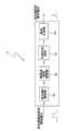
图1为依据本发明的可被用于改善患者的心输出量的系统的示例性视图。FIG. 1 is an exemplary view of a system that may be used to improve a patient's cardiac output in accordance with the present invention.
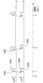
图2为依据本发明的可被用于基于来自心肌活动传感器的信号来驱动按压装置的控制器的示例性示意图。2 is an exemplary schematic diagram of a controller that may be used to drive a compression device based on a signal from a myocardial activity sensor in accordance with the present invention.
图3为示出了依据本发明的用于施加按压力的示例性时间图。FIG. 3 is a diagram showing an exemplary timing for applying pressing force according to the present invention.
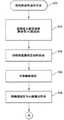
图4为示出了依据本发明的用于改善患者的心输出量的一个方法的流程图。FIG. 4 is a flowchart illustrating a method for improving a patient's cardiac output in accordance with the present invention.
图5为示出了验证用于检测心肌运动和其他患者参数的传感器的方法的流程图。5 is a flowchart illustrating a method of validating sensors for detecting myocardial motion and other patient parameters.
图6A和图6B为示例性算法的流程图,其判定何时开始胸部按压并且优化可与患者的通气和心脏的电刺激结合的胸部按压治疗过程。6A and 6B are flowcharts of exemplary algorithms that determine when to initiate chest compressions and optimize a course of chest compression therapy that may be combined with ventilation of the patient and electrical stimulation of the heart.
图7A为示出了施加为与缓慢的心脏跳动同步的改变的力的胸部按压的图表。7A is a graph showing chest compressions applied as varying forces synchronized with a slow heart beat.
图7B为示出了校正在胸部按压和心脏跳动之间的同步误差的方法的曲线图。7B is a graph illustrating a method of correcting for synchronization errors between chest compressions and heart beats.
图8为示出了将胸部按压与心脏跳动同步的方法的曲线图。8 is a graph illustrating a method of synchronizing chest compressions with a heart beat.
图9为示出了将电心脏刺激与脉动性流动或机械心肌活动同步的方法的曲线图。9 is a graph illustrating a method of synchronizing electrical cardiac stimulation with pulsatile flow or mechanical myocardial activity.
具体实施方式Detailed ways
本发明涉及的手段和装置可被用于增加患有广泛变化的休克至无脉动电活动(PEA)的疾病的患者的心输出量,其中患者出现无生命体征但仍然具有一些残余机械心脏活动。本发明一个示例性的方法是感测心脏何时跳动并在之后将胸部按压或其复苏术方法与心肌室壁的运动同步。以这样的方式,可使用不同的手段优化地使胸部按压(或其他的CPR要素)与残余左心室的功能同步以改善这样的患者的结果。因此,本发明可被用于将在胸部上或胸部周围的外部装置的按压力与残余左心室功能的射血阶段以及与松弛阶段的残余心脏充盈相同步。在另一方案中,这里公开的系统和方法提供不同的用于感测残余机械功能的手段和装置,并在之后将这些信息转换成有用的数据流,其可被用于操作复苏技术的不同组成部分,复苏技术包括辅助血流,通气,以及心脏刺激技术。The present invention relates to means and devices that can be used to increase cardiac output in patients suffering from conditions ranging widely from shock to no pulsatile electrical activity (PEA), where the patient appears lifeless but still has some residual mechanical cardiac activity. An exemplary method of the present invention is to sense when the heart is beating and then synchronize chest compressions or their resuscitative approach with the motion of the myocardial chamber wall. In this manner, different approaches may be used to optimally synchronize chest compressions (or other elements of CPR) with residual left ventricular function to improve outcomes for such patients. Thus, the present invention can be used to synchronize the compression force of an external device on or around the chest with the ejection phase of residual left ventricular function and with residual cardiac filling during the relaxation phase. In another aspect, the systems and methods disclosed herein provide various means and devices for sensing residual mechanical function and then converting this information into useful data streams that can be used to operate various resuscitation techniques. Components, resuscitative techniques include assisted blood flow, ventilation, and cardiac stimulation techniques.
这样的手段可被用在患有广泛范围的疾病的患者上。一个示例性的使用为用于这样的患者身上:其被确信处于带有无脉动电活动(PEA),不可检测到的血压的心脏停跳中,但仍然具有一定程度的残余左心室功能。然而,将被理解的是,本发明并非旨在被限制在仅用于这样的情况,而是用于某些有组织的电(但为受损的)机械心脏活动的广泛范围的情况下。Such an approach can be used on patients with a wide range of diseases. An exemplary use is in patients who are believed to be in cardiac arrest with pulseless electrical activity (PEA), undetectable blood pressure, but still have some degree of residual left ventricular function. However, it will be appreciated that the invention is not intended to be limited to use in such situations only, but rather in a broad range of situations of certain organized electrical (but impaired) mechanical heart activity.
例如,在该系列状况的一个端部为正常的自生循环,其中心输出量正常并且左心室机械和泵功能正常。在这一水平之下即为低血压并且在之后为代偿型休克。在这样的情况下,所述血压以及患者的脉搏仍然是可触知的并且其可具有良好的心输出量。然而,出于不同的原因,心输出量不能符合身体的代谢指令并且体内平衡存在风险。其通过诸如排尿减少以及血清乳酸增加的参数来证明,这些参数为器官功能不充足的标志。For example, at one end of the spectrum is normal spontaneous circulation with normal central output and normal left ventricular mechanical and pump function. Below this level is hypotension and thereafter compensatory shock. In such cases, the blood pressure as well as the patient's pulse are still palpable and they may have a good cardiac output. However, for different reasons, cardiac output does not match the body's metabolic commands and homeostasis is at risk. It is evidenced by parameters such as decreased urination and increased serum lactate, which are markers of organ insufficiency.
处于补偿休克以下的状态为非代偿型休克。在这一状态中,心肌与心血管系统不再能够提供充足的血流总量、氧气以及营养物以满足重要器官的需要,并且这些器官的功能被影响至其开始受损的程度。在这种状态下,血压例如可能为70/30mm Hg。同样,排尿可能停止,并且患者可能由于不充足的脑部功能而变得神志不清。更重要的,随着休克的发展,多器官系统开始衰竭。The state below compensatory shock is noncompensated shock. In this state, the heart muscle and cardiovascular system are no longer able to provide sufficient total blood flow, oxygen, and nutrients to meet the needs of vital organs, and the function of these organs is affected to the extent that they become impaired. In this state, the blood pressure might be, for example, 70/30 mm Hg. Also, urination may cease, and the patient may become delirious due to insufficient brain function. More importantly, as the shock progresses, multiple organ systems begin to fail.
在典型的非代偿型休克以下可以被称作“极端休克”,其濒临心脏停跳。在这种情况下,患者显示出一些残余心肌功能,其包含一些左心室射血,但心输出量对于满足重要器官的需要是完全不充足的。例如,每分钟的心输出量可能少于1升,血压可能为50/20,排尿量可能为最小值或完全缺失,并且患者可能处于麻木状态或是昏迷状态。此外,患者可能表现为带有显著减弱的脑部功能以及濒临昏迷的麻木特征的临近死亡的状态。如果对其进行治疗,极端休克将导致在数分钟的时间范围内的真正的心脏停跳。通常,在这个范围内不可能手动地触摸到动脉搏,并且这样的患者即使在心脏持续跳动的情况下也可能被临床工作人员归为PEA。Under typical uncompensated shock may be termed "extreme shock", which is on the verge of cardiac arrest. In this case, the patient shows some residual myocardial function, which includes some left ventricular ejection, but cardiac output is completely insufficient to meet the demands of vital organs. For example, cardiac output may be less than 1 liter per minute, blood pressure may be 50/20, urine output may be minimal or absent entirely, and the person may be numb or comatose. In addition, patients may present in a near-death state with markedly diminished brain function and features of anesthesia bordering on coma. If treated, extreme shock can lead to true cardiac arrest on the time scale of minutes. Typically, manual palpation of an arterial pulse in this range is not possible, and such patients may be classified as PEA by clinical staff even though the heart is beating continuously.
在极端休克的状态以下的是无脉动电活动(PEA)心脏停跳,其同样重要地具有系列状态以及血液动力的范围。例如,在其上端,PEA具有左心室机械功能和心输出量,但是不足以作为外围辐射和股骨脉搏被检测到。在仅在胸部,颈部和腹股沟可测量血压的情况下,如果动脉内导管被放置在病人体内,血压可能仅为45/25。放置在颈部或腹股沟上的多普勒探针可检测向前的血流。血流极度地不充足以至于患者通常将表现为无生命体征并且其瞳孔可能放大并变得静止。此外,尽管存在残余泵功能和向前流动,其仍表现出心脏停跳状态。PEA动力的上端与“极端休克”的下端重叠。在这样的情况下,临床工作人员可能不能区分其不同之处。示出组织电活动的心电图在病理学中为可变化的并且其QRS结构可相对正常。发明人将带有残余心肌机械活动的电动机械分离作用的术语界定为“伪EMD”。Below the state of extreme shock is pulseless electrical activity (PEA) cardiac arrest, which equally importantly has a range of states as well as hemodynamics. For example, at its upper end, PEA has left ventricular mechanical function and cardiac output, but not enough to be detected as peripheral radiation and femoral pulse. In cases where blood pressure is measurable only in the chest, neck, and groin, the blood pressure may be only 45/25 if an intra-arterial catheter is placed in the patient. A Doppler probe placed in the neck or groin detects forward blood flow. Blood flow is so inadequate that the patient will often appear lifeless and his pupils may dilate and become stationary. In addition, it exhibited cardiac arrest despite residual pump function and forward flow. The upper end of the PEA power overlaps the lower end of the "extreme shock". In such cases, clinical staff may not be able to tell the difference. The electrocardiogram showing tissue electrical activity can be variable in pathology and its QRS structure can be relatively normal. The inventors define the term "pseudo-EMD" for electromechanical dissociation with residual myocardial mechanical activity.
在PEA的级别的“上端”以下的是几乎完全缺乏左心室功能的电机械分离。由刚好在主动脉瓣之上的血管内的导管测量的血压将显示主动脉脉搏,但测量的血压仅在25/15毫米汞柱上,并且其将几乎没有关联的向前血流。在不应用CPR的情况下,本质上缺失传送到重要器官的氧气并且对于诸如脑部的器官将在数分钟内产生不可修复的损伤。这种心电图极少具有正常出现的QRS结构,并且ECG的全部模式是模糊不清的或非常规的。Below the "upper" end of the PEA scale is an almost complete lack of electromechanical dissociation of left ventricular function. Blood pressure measured by an endovascular catheter just above the aortic valve will show an aortic pulse, but the measured blood pressure is only at 25/15 mm Hg, and it will have little associated forward blood flow. Without the application of CPR, oxygen delivery to vital organs is essentially lost and irreparable damage occurs within minutes to organs such as the brain. This ECG rarely has a normally occurring QRS structure, and the overall pattern of the ECG is smeared or irregular.
PEA的最终级别为组织电律动但没有左心室机械功能。这是真正的心脏停跳。在主动脉瓣之上测量血压的导管将检测不到压力脉动并且超声波心动描记将显示没有心脏运动。此外,心输出量为零且病人处于完全的整体性缺血以及心脏停跳的状态。在不应用CPR的情况下,传送到重要器官的氧气将为零,并且对于诸如脑部的器官将在数分钟内产生不可修复的损伤。ECG的全部模式是模糊不清的或非常规的。The final level of PEA is electrical rhythm of the tissue but no mechanical function of the left ventricle. This is a real heart stop. A catheter measuring blood pressure above the aortic valve will detect no pressure pulsations and an echocardiogram will show no heart motion. In addition, cardiac output is zero and the patient is in a state of complete global ischemia and cardiac arrest. Without the application of CPR, the delivery of oxygen to vital organs would be zero, and irreparable damage would occur within minutes to organs such as the brain. The overall pattern of the ECG is ambiguous or irregular.
根据上文所述的系列状况,本发明可被用于存在一些心肌机械活动的全部情况并且同步的复苏治疗可改善心输出量。在这样的情况下,本发明可被用于检测残余机械活动并且使这样的心脏活动与诸如在CPR(包括胸部按压/减压和/或通气)中使用的复苏手段同步。因此,本发明可针对伪EMD PEA,通过休克的不同级别被利用在任何由真正的心脏停跳造成的病理生理的状况下,或被利用在带有或不带有心输出量的残余心肌机械功能的任何血液动力状态下。通过在不同的潜在的循环性的治疗中同步胸部按压和/或减压,心脏循环的射血和充盈阶段可被增加。在这样的作用下,心输出量和器官的灌注可被增大,因此改善了带有减弱的血液动力的患者的结果。According to the series of conditions described above, the present invention can be used in all situations where there is some mechanical activity of the myocardium and synchronized resuscitative therapy improves cardiac output. In such cases, the present invention can be used to detect residual mechanical activity and synchronize such cardiac activity with resuscitative measures such as those used in CPR (including chest compressions/decompression and/or ventilation). Thus, the present invention can be exploited in any pathophysiological situation resulting from true cardiac arrest for pseudo-EMD PEA through different levels of shock, or residual myocardial mechanical function with or without cardiac output. in any hemodynamic state. By synchronizing chest compressions and/or decompression during different potentially cyclical treatments, the ejection and filling phases of the cardiac cycle can be increased. In doing so, cardiac output and organ perfusion can be increased, thus improving outcomes for patients with impaired hemodynamics.
作为一个特别重要的实例,一个经常发生的并且对医生具有挑战性的临床情况为当患者由休克发展成显示出PEA心脏停跳。在这一过程的早期级别,医生趋向于以静脉药物治疗并可能以控制的通气来治疗这样的患者。这时诸如抗生素的药物可被使用到处于诸如感染性休克的患者上,诸如多巴胺的增压药物仍然为治疗的主体。但是,尽管增高了血压,但增压通常不能表示改善了这些患者的结果。这可以是由于其改善了血压但是同样增高了重要器官氧气的利用,使得没有改善在氧气供给与需求之间的整体平衡。增压药物同样对于重要器官具有大量的直接的毒性。As a particularly important example, a frequently occurring and challenging clinical situation for physicians is when a patient develops from shock to exhibit PEA cardiac arrest. At an early level in the process, physicians tend to treat such patients with intravenous medication and possibly controlled ventilation. While drugs such as antibiotics can be used on patients in conditions such as septic shock, pressor drugs such as dopamine remain the mainstay of treatment. However, despite increasing blood pressure, pressurization has generally not been shown to improve outcomes in these patients. This may be because it improves blood pressure but also increases oxygen utilization by vital organs, so that the overall balance between oxygen supply and demand is not improved. Pressor drugs also have substantial direct toxicity to vital organs.
然而,如果这些肠胃外的治疗不能稳定患者的情况,他们的休克可能不可阻挡地向前发展成越来越极端的状况并且最终变为心脏停跳。关于血压骤降的患者在哪一点应该开始接受胸部按压,许多急症医学以及临床护理的医师仍然不确定,并且其在医学文献中也是不清楚的。当然,内科医生通常在生命器官实质的损失之前不施加诸如外胸部按压的手段。这是由于CPR,特别是胸部按压,如果其以不同步的方式施加,会妨碍心脏功能,特别是心脏充盈。例如,血压为60/40的患者开始接受与心脏功能不同步的胸部按压,其可能快速地发展为完全心脏停跳。更特别的,在执行没有同步的CPR时,当左心室试图充盈时,按压阶段的施加可大大降低心脏的基于弗-斯二氏定律的下一射血的心输出量。因此,通过检测心肌机械功能,胸部按压能够与射血阶段同步,从而休克的患者可以在避免恶化其情况以及可能向心脏停跳发展的情况下被治疗。However, if these parenteral treatments fail to stabilize the patient's condition, their shock may progress inexorably to more and more extreme conditions and eventually to cardiac arrest. The point at which chest compressions should be initiated in a patient with a sudden drop in blood pressure remains uncertain to many emergency medicine and clinical care physicians and is unclear in the medical literature. Of course, physicians typically do not apply measures such as external chest compressions until loss of vital organ parenchyma. This is due to the fact that CPR, especially chest compressions, can interfere with cardiac function, particularly cardiac filling, if it is applied in an asynchronous manner. For example, a patient with blood pressure of 60/40 who begins receiving chest compressions out of synch with cardiac function may rapidly progress to complete cardiac arrest. More specifically, when performing CPR without synchronization, when the left ventricle is attempting to fill, the application of the compression phase can greatly reduce the cardiac output of the heart for the next ejection based on the Frederic-Schwartz law. Thus, by detecting myocardial mechanical function, chest compressions can be synchronized with the ejection phase so that patients in shock can be treated without worsening their condition and possibly progressing to cardiac arrest.
因此,关于在休克的级别发展中的患者应在何时开始胸部按压的问题可通过将胸部按压以及其他可能的机械附加手段与射血和松弛阶段同步来处理,从而使得临床医生可以更加确信胸部按压起到了辅助作用并且不会妨碍残余的血液循环功能。以这样的方式,临床医师不需要考虑关于何时开始胸部按压的问题。以这样的方式,本发明可起到了允许在任何形式的休克中的外部机械附加手段的使用,其为与已经施加在心源性休克的主动脉内气囊反搏术的方式类似。本发明可因此允许在医院前期以及急诊部门环境中应用这样的附加手段。Thus, the question of when chest compressions should be initiated in a patient at a developing level of shock can be addressed by synchronizing chest compressions and possibly other mechanical additions with the ejection and relaxation phases, allowing the clinician to be more confident that the chest Compression is assisted and does not interfere with residual blood circulation. In this way, the clinician does not need to think about when to start chest compressions. In this way, the present invention may function to allow the use of external mechanical adjuncts in any form of shock, in a manner similar to the way intra-aortic balloon counterpulsation has been applied in cardiogenic shock. The present invention may thus allow the application of such additional approaches in pre-hospital as well as emergency department settings.
采用同步的另外的优点在于,其可被实施为对于指向休克原因的治疗的附加手段,这些治疗诸如抗生素或溶栓,提高重要器官灌注,而这些治疗已经被实施。当然,改善血液动力将不仅仅防止器官损伤,其可改善肠胃外治疗的效果。此外,同步的胸部按压不太可能具有如升压药物带来的重大的器官毒性。An additional advantage of using synchronization is that it can be implemented as an adjunct to treatments directed at the cause of the shock, such as antibiotics or thrombolysis, increasing vital organ perfusion, which are already being administered. Of course, improving hemodynamics will not only prevent organ damage, it may improve the efficacy of parenteral therapy. Furthermore, synchronized chest compressions are less likely to have the major organ toxicity associated with vasopressor drugs.
如上文所述,本发明的一个特别应用与患有无脉动电活动(PEA)的患者相联系。PEA为心脏停跳的三个主要类型的其中之一,另外两个为心室纤维性颤动和心搏停止。PEA也被称为电机械分离(EMD)。PEA在罗森P等人的《急诊医学概念与临床实践》中已经被描述为“在心电图上存在组织电活动但没有可触知的脉搏”(Rosen P,Baker F J,Barkin R M,Braen G R,Dailey RH,Levy R C.Emergency Medicine Concepts and Clinical Practice.2nd ed.StLouis:CV Mosby,1988.)。与能够特别地被电除颤所逆转的心室纤维性颤动不同,PEA并不具有特定的对应措施。这就解释了相比较于心室纤维性颤动,处于PEA的患者往往具有更糟糕的结果。不幸的是,PEA的发生率在增加,这可能是由于早期的风险矫正正在改变心血管疾病的自然历史。在当今的一些权威机构的报告中,在急救医疗服务抵达(EMS)时,大多数处于心脏停跳的患者均为PEA。此外,大部分非心室纤维性颤动休克的患者或由心搏停止复苏的患者将在复苏的过程中的某些点上经历PEA。这些情况的组合意味着绝大多数接受用于心脏停跳治疗的促进生命维持的患者将在复苏的过程中的某些时间上具有PEA。因此,现在或是在不久的将来,PEA可在重要性上取代典型的心室纤维性颤动。或者说其已经取代了典型的心室纤维性颤动。As mentioned above, one particular application of the invention is in connection with patients suffering from pulseless electrical activity (PEA). PEA is one of three major types of cardiac arrest, the others being ventricular fibrillation and asystole. PEA is also known as electromechanical dissociation (EMD). PEA has been described by Rosen P et al in Concepts and Clinical Practice of Emergency Medicine as "the presence of tissue electrical activity but no palpable pulse on the electrocardiogram" (Rosen P, Baker F J, Barkin R M, Braen G R, Dailey RH, Levy R C. Emergency Medicine Concepts and Clinical Practice. 2nd ed. StLouis: CV Mosby, 1988.). Unlike ventricular fibrillation, which can be specifically reversed by electrical defibrillation, PEA does not have a specific counterpart. This explains why patients in PEA tend to have worse outcomes compared to those in ventricular fibrillation. Unfortunately, the incidence of PEA is increasing, possibly because early risk correction is altering the natural history of cardiovascular disease. The majority of patients in cardiac arrest are reported to be PEA at the time of emergency medical services (EMS) arrival by several authorities today. Furthermore, most patients in non-fibrillatory shock or those resuscitated from asystole will experience PEA at some point during resuscitation. The combination of these circumstances means that the vast majority of patients receiving proliferative life support for cardiac arrest will have PEA at some point during resuscitation. Therefore, PEA may replace classical ventricular fibrillation in importance now or in the near future. Or it has replaced typical ventricular fibrillation.
许多在PEA情况下的患者具有残余心脏机械活动,并且许多具有可检测到的血压。这种情况涉及到伪EMD PEA。在这样的情况下,患者会出现无生命体征并且没有脉搏。然而,其通常仍然具有一定程度的残余左心室功能。因此,本发明的一个重要特征在于感测患者何时仍然具有一些心肌功能并在之后将复苏治疗,特别是胸部的按压与心脏的残余机械功能同步。以这样的方式,CPR的按压阶段可在射血阶段的过程中产生,并且松弛阶段能够在左心室试图充盈时允许与减少的胸内压相关联的胸部的弹性回位。以这样的方式,将阶段性的复苏治疗与残余的心室射血与充盈同步,可使血液动力改善为恢复自主循环(ROSC)从而长期生存。Many patients in the setting of PEA have residual cardiac mechanical activity, and many have detectable blood pressure. This case involves pseudo-EMD PEA. In such cases, the patient is lifeless and has no pulse. However, they usually still have some degree of residual left ventricular function. Therefore, an important feature of the present invention is to sense when the patient still has some myocardial function and to thereafter synchronize resuscitative therapy, especially chest compressions, with the residual mechanical function of the heart. In this way, the compression phase of CPR can occur during the ejection phase, and the relaxation phase can allow for elastic recoil of the chest associated with reduced intrathoracic pressure as the left ventricle attempts to fill. In this way, synchronizing phased resuscitative therapy with residual ventricular ejection and filling results in hemodynamic improvements for return of spontaneous circulation (ROSC) and long-term survival.
本发明可结合多种不同的非侵入性感测技术(通过在图1中的传感器表示)以获得描述心肌室壁的模式的和/或瓣膜运动模式的实时数据,从而允许胸部按压以及其他治疗的同步,然而,如果存在血液动力的侵入性装置,诸如内动脉压或流动显示器,本发明可以起到作为在这些输出和由外部胸部按压为例的阶段性的复苏治疗之间的接口。为了在外部装置施加于胸部或身体上或在其周围的力度与残余左心室功能的射血和充盈阶段之间进行合适的同步,可使用不同的装置。残余心肌活动存在的确认可由逻辑电路通过来自多个感测医疗器械的输入数据得出。本发明可利用感测技术来采集心肌室壁功能,心肌瓣膜运动,在血管结构中的血流,重要器官氧气或能力状态,或是呼出的肺气的数据,并且这些数据可通过逻辑电路以及控制输出信号到达实现治疗的装置。由于机械残余内壁功能的模式可随时间改变,本发明可被设计成迅速识别残余功能并且基于逻辑电路的反馈来改变治疗。同样,外部胸部按压可被用于与其他手段同步,诸如腹部反搏,阶段性的肢体按压,通气,电刺激,或除此之外的其他手段以增大心脏射血及充盈。以这样的方式,患者可被稳定以允许用于诸如溶栓的初级治疗的足够的时间变得更加有效。The present invention can combine a variety of different non-invasive sensing techniques (represented by the sensors in Figure 1) to obtain real-time data describing the pattern of the myocardial wall and/or the pattern of valve motion, thereby allowing chest compressions and other therapeutic Simultaneously, however, if there are hemodynamic invasive devices such as internal arterial pressure or flow monitors, the present invention can serve as an interface between these outputs and phased resuscitative therapy exemplified by external chest compressions. For proper synchronization between the force exerted by the external device on or around the chest or body and the ejection and filling phases of residual left ventricular function, different devices may be used. Confirmation of the presence of residual myocardial activity may be derived by a logic circuit using input data from a plurality of sensing medical devices. The present invention can use sensing technology to collect myocardial wall function, myocardial valve movement, blood flow in vascular structures, vital organ oxygen or capacity status, or exhaled lung gas data, and these data can be passed through logic circuits and The control output signal reaches the device for effecting the therapy. Since the pattern of mechanical residual wall function can change over time, the present invention can be designed to quickly identify residual function and alter therapy based on feedback from the logic circuit. Likewise, external chest compressions may be used to synchronize with other means such as abdominal counterpulsation, phased limb compressions, ventilations, electrical stimulation, or other means in addition to augmenting cardiac ejection and filling. In this way, the patient can be stabilized to allow sufficient time for primary treatments such as thrombolysis to become more effective.
各种各样的设备和装置可被用于提供胸部按压。例如,各种不同类型的自动按压系统可被用于按压胸部,其包括的系统诸如卓尔循环公司(ZOLLCirculation,Inc.of Sunnyvale,Calif.)的AutoPulse复苏系统,由密歇根仪器制造的Thumper或者LUCAS装置等。此外,本发明不仅限于自动按压装置,也可结合手动方法来使用。例如,本发明可被用于提高音频和/或视频信号以指示救助者何时手动施加胸部按压。此外,在一些情况下,吸引装置可被粘着在胸部上从而使得胸部可间歇地随着胸部按压被活动地升高。A variety of devices and devices can be used to provide chest compressions. For example, various different types of automated compression systems can be used to compress the chest, including systems such as the AutoPulse resuscitation system from ZOLL Circulation, Inc. of Sunnyvale, Calif., the Thumper or LUCAS® made by Michigan Instruments. device etc. Furthermore, the present invention is not limited to automatic compression devices, but can also be used in conjunction with manual methods. For example, the present invention may be used to enhance audio and/or visual signals to indicate when the rescuer is manually applying chest compressions. Additionally, in some cases, the suction device may be adhered to the chest such that the chest may be intermittently raised actively with chest compressions.
使用手动或自动设备时,本发明可被配置为使外部胸部按压与任何心肌的残余机械活动同步,使得当心肌进入泵或心脏收缩状态,CPR处于胸部按压阶段。此外,当心脏进入充盈或心脏收缩阶段,胸部按压进入松弛阶段。传感的数据可通过逻辑电路传输,并且该电路的输出用于在心脏射血和充盈的过程中控制同步的发生。这种关系可随时间变化以优化疗效。When using manual or automated equipment, the present invention can be configured to synchronize external chest compressions with any residual mechanical activity of the myocardium such that when the myocardium enters a pump or systole state, CPR is in the chest compression phase. Also, when the heart enters the filling or systolic phase, chest compressions enter the relaxation phase. The sensed data can be transmitted through a logic circuit, and the output of this circuit is used to control the occurrence of synchronization during the ejection and filling of the heart. This relationship can be varied over time to optimize efficacy.
除了将胸部按压与残余心脏功能同步之外,本发明可同样用于将通气与残余心脏功能同步。例如,进气和呼气可与残余心肌功能同步从而增加心输出量。例如,进气可与心脏收缩同步,呼气可与心脏舒张同步。为了施加通气,本发明可使用传统的呼吸机或提供手动通气,诸如使用通气袋。在后者的情况下,关于何时施加合适的通气的音频和/或视频信号可被提供给救助者。In addition to synchronizing chest compressions with residual cardiac function, the present invention can equally be used to synchronize ventilation with residual cardiac function. For example, inspiration and expiration can be synchronized with residual myocardial function to increase cardiac output. For example, intake air may be synchronized with systole and expiration with diastole. To apply ventilation, the present invention may use a conventional ventilator or provide manual ventilation, such as using a ventilation bag. In the latter case, audio and/or visual signals may be provided to the rescuer as to when the appropriate ventilation is applied.
在同时进行胸部按压和通气的情况下,时机,频率和/或持续时间可基于特定的治疗而被改变。例如,胸部按压可发生在整个心脏收缩阶段或仅在该阶段中的一部分。此外,胸部按压可发生在每个心脏收缩阶段或仅在特定的心脏收缩阶段的过程中。类似的情形可发生在通气的情况下。控制器可使用一个或多个传感器的输出以及所利用的逻辑电路和一个或多个疗效指标,以优化在血液动力上同步的效果。In the case of simultaneous chest compressions and ventilations, the timing, frequency and/or duration may be varied based on the specific therapy. For example, chest compressions can occur throughout the systolic phase or only a portion of the phase. Furthermore, chest compressions may occur during every systole phase or only during specific systole phases. A similar situation can occur in the case of ventilation. The controller may use the output of the one or more sensors along with the utilized logic circuit and one or more efficacy indicators to optimize hemodynamically synchronized effects.
这里公开的系统可与任何治疗一起被利用,其可得益于与表面上无生命体征的患者的残余心肌机械功能的同步。其中,胸部按压和减压,腹部反搏,阶段性的肢体按压,心肌电刺激,血管内液移,血管内或心包内气囊涨缩,经胸廓的电磁辐射的应用。控制器逻辑电路可改变多种治疗中同步的模式从而判定关于增加血液动力的优化模式。The systems disclosed herein may be utilized with any therapy that may benefit from synchronization with residual myocardial mechanical function in an apparently lifeless patient. Among them, chest compression and decompression, abdominal counterpulsation, periodic limb compression, myocardial electrical stimulation, intravascular liquid displacement, intravascular or intrapericardial balloon expansion and contraction, and the application of electromagnetic radiation through the chest. The controller logic can vary the modes of synchronization among the various treatments to determine the optimal mode for increasing hemodynamics.
心肌电刺激为,例如,通过金属片或电极传输施加到胸部外部的电击,或由经改进以将心肌电刺激与例如心肌室壁功能或检测的脉动性血流同步的内部的起搏器直接向心脏施加的电信号。Electrical stimulation of the myocardium is, for example, delivery of an electrical shock applied to the outside of the chest by metal plates or electrodes, or directly by an internal pacemaker modified to synchronize electrical stimulation of the myocardium with, for example, myocardial wall function or detected pulsatile blood flow. An electrical signal applied to the heart.
为了感测心肌室壁功能,可使用多种不同的非侵入性装置和技术。例如,一个可以使用的技术为心电图(ECG)。ECG可以是具有吸引力的检测方法,这是由于其已经被用于复苏的过程中的大多数临床情况。然而,由于心肌活动在PEA过程中并不总是存在于ECG,其可能需要结合其他上文所述的其他感测手段一起使用。另一个可以使用的感测手段的实例为多普勒超声波扫描(DOP)。多普勒超声波使用超声波的多普勒频移以量化外围血管中的血流。其可与在颈部用于动脉血流,在腹股沟用于股动脉血流的传感器,或是经胸廓的或食道内的用于主动脉血流的传感器一起应用。多普勒探针可同样被放置在最大脉动的心脏点以检测心肌中的血液的运动。多普勒探针的阵列可被用于判定残余心肌机械功能的矢量并将胸部按压和关联与该矢量调准。To sense myocardial wall function, a variety of different non-invasive devices and techniques are available. For example, one technique that may be used is an electrocardiogram (ECG). ECG can be an attractive detection method since it has been used in most clinical situations during resuscitation. However, since myocardial activity is not always present in the ECG during PEA, it may need to be used in conjunction with other sensing means as described above. Another example of a sensing means that can be used is a Doppler ultrasound scan (DOP). Doppler ultrasound uses the Doppler shift of ultrasound to quantify blood flow in peripheral blood vessels. It can be used with sensors in the neck for arterial flow, in the groin for femoral flow, or transthoracic or endoesophageal for aortic flow. A Doppler probe can also be placed at the heart point of greatest pulsation to detect the movement of blood in the myocardium. An array of Doppler probes can be used to determine the vector of residual myocardial mechanical function and to align chest compressions and correlations to this vector.
动压力传感器通过感测在外围静脉中的氧气含量检测脉动性血流。由ROSS传感器感测的氧气成分作为通过外围静脉的血液脉搏。类似的,脉搏血氧定量传感器可被用于检测在脚趾,手指或耳垂中的血管的氧气含量。所述血液的氧气含量可被用于判定何止开始和终止CPR以及机械或电心脏刺激。例如,如果ROSS传感器或脉搏血氧定量传感器检测脉动性血流以及在临界值之上的氧气含量,所述系统可减小胸部按压的力度或终止胸部按压。类似的,如果ROSS或血氧定量传感器检测不到脉动性血流或氧气水平降至临界值之下,所述系统可启动手动胸部按压器或电心脏刺激。所述系统基于被感测的氧气情况的趋势来调节阶段性治疗的不同参数。Dynamic pressure sensors detect pulsatile blood flow by sensing oxygen levels in peripheral veins. The oxygen composition sensed by the ROSS sensor acts as a pulse of blood through peripheral veins. Similarly, pulse oximetry sensors can be used to detect oxygen levels in blood vessels in toes, fingers or ear lobes. The oxygen content of the blood can be used to determine when to initiate and terminate CPR and mechanical or electrical cardiac stimulation. For example, if the ROSS sensor or pulse oximetry sensor detects pulsatile blood flow and oxygen levels above a threshold, the system may reduce the force of chest compressions or terminate chest compressions. Similarly, if the ROSS or oximetry sensors fail to detect pulsatile blood flow or oxygen levels drop below a threshold, the system can initiate manual chest compressions or electrical cardiac stimulation. The system adjusts different parameters of the phasic therapy based on the sensed trend of the oxygen condition.
关于在外围血管中的脉搏的数据可被用来基于关于在心肌机械功能与脉搏压力或在外围血管中的脉动性血流之间的延迟的信息来估计残余心肌机械功能,诸如心脏射血阶段。Data on pulses in peripheral vessels can be used to estimate residual myocardial mechanical function based on information on the delay between myocardial mechanical function and pulse pressure or pulsatile blood flow in peripheral vessels, such as cardiac ejection phase .
另外的可被使用的感测手段为体积描记法(PLETH),体积描记法可通过测量带有心脏活动的经胸廓的交流电阻抗中的改变来应用。另外的可被使用的感测手段为心音描记法(PHONO)。心音描记法记录由在心脏上的听诊器检测的声能。此外还可被使用的感测手段为超声波心动描记法(ECHO),凭借超声波心动描记法或心脏的超声波图像,左心室射血能够被量化。在一些情况下,心脏功能的超声波心动描记仪的检测可与ECG结合。同样,通过使用静脉注入的微泡或其他的加强技术可改善感测性。Another sensing means that can be used is plethysmography (PLETH), which can be applied by measuring changes in the transthoracic AC impedance with cardiac activity. Another sensing means that may be used is phonocardiography (PHONO). Phonocardiography records the sound energy detected by a stethoscope over the heart. Another sensing method that can be used is echocardiography (ECHO), by means of echocardiography, or ultrasound images of the heart, left ventricular ejection can be quantified. In some cases, echocardiographic measurements of cardiac function may be combined with ECG. Likewise, sensing can be improved through the use of intravenously injected microbubbles or other augmentation techniques.
多个这样的检测系统的结合将更为理想,从而增加检测残余心肌机械功能的感测性和特异性。此外,对感测手段和诸如呼气末二氧化碳或主动脉血流的实际心输出量的指标进行比较的逻辑电路的结合也可更为理想。以这样的方式,本发明能够判定哪些感测手段的结合最多地预示了源于同步的改善。A combination of multiple such detection systems would be ideal to increase the sensitivity and specificity of detecting residual myocardial mechanical function. Additionally, a combination of logic circuits that compare the sensing means with indicators of actual cardiac output, such as end-tidal carbon dioxide or aortic blood flow, may also be desirable. In this way, the present invention is able to determine which combinations of sensing means most predict improvement from synchronization.
此外,本发明的逻辑电路能够针对实际的心输出量的指标改变同步的治疗从而判定哪些同步的治疗的模式为最有效的。其可改变在一个治疗装置中或多个治疗装置中的同步从而识别最优化的模式。In addition, the logic circuit of the present invention is able to change the simultaneous therapy according to the actual cardiac output index to determine which synchronized therapy mode is the most effective. It can vary the synchronization within one treatment device or among multiple treatment devices to identify the optimal mode.
现在参见图1,用于改善心输出量的系统10将被描述。系统10包括一个或多个传感器12,其可被用于检测残余心肌机械功能。在一些实施例中,传感器12可包括放置于患者胸部上的表面探针。传感器12可被设置在胸部的不同位置上。例如,一个位置可以是其中一个肋间隙的胸前。另一个位置可以是上腹部中的下剑突。传感器12可使用这里描述的任意技术感测心肌室壁运动,其包括超声波,多普勒技术,超声波心动描记法,体积描记法等。作为在患者胸部上设置传感器12的替代,将被理解的是,其他位置也可以同样被使用,诸如探针可以被设置在颈动脉之上的颈部上,或进入患者的食道。将同样被理解的是,传感器12可以是传感器的阵列。Referring now to FIG. 1 , a
传感器12可以是用于诸如心电图,多普勒超声波图像,体积描记法,心音描记法,超声波心动描记法,经胸阻抗等多种感测系统的一个或多个传感器。传感器12可被结合到联接到胸部,腹部,背部,四肢或其组合的探针中,或放置在身体中,诸如食道,气管,或胃部。这些多种不同的传感器通过检测例如心脏电活动,物理性收缩以及心脏的其他活动,在食道,气管,或胃部内的可触知的动脉脉搏;在皮肤内的指示脉动性血流的变化以及呼吸的律动和化学成分来检测心肌活动。
由传感器12采集的数据被传输到具有信号处理和逻辑能力的控制器14。对控制器14的其他说明将结合图2被描述在下文中。控制器14同样被电连接至可被用于对患者施加外部胸部按压的按压装置16。在一些实施例中,将被理解的是,控制器14能结合到按压装置16或任何传感器上。为了简化使用,传感器12和控制器14均可被结合到治疗装置16。此外,控制器14能够与感测和/或按压装置无线连接。在图1示出的实例中,胸部按压装置16包括接口构件18,其被联接到以重复的方式相对于胸部移动接口构件18的活塞20上。以这样的方式,胸部按压装置16可对患者重复施加胸部按压。在一些情况下,接口构件18可被配置为粘合在患者的胸部上从而随着活塞20升高接口构件18,患者的胸部将同样被升高。以这样的方式,胸部按压装置16可选择的施加胸部按压和减压。尽管在此处描述了胸部按压装置16,将被理解的是,许多不同的设备可如这里描述的以自动的方式被用于对胸部,腹部或四肢施加按压和/或减压,并且本发明并非旨在仅限定在胸部按压装置16的特定实施例。例如,存在的CPR设备的实例可以修改至与控制器14有关的功能,包括由加利福尼亚桑尼维尔复苏公司(Revivant of Sunnyvale,Calif.)的AutoPulse复苏系统或由密歇根仪器公司制造的Thumper。作为另外的选择,可充气的防护衣21可被联接至控制器14并且被配置为充放气以实施适当的同步。Data collected by the
当施加自动的胸部按压为一种选择时,本发明同样可与手动手段一起使用。在这样的情况下,控制器14可包括扬声器22和/或信号灯24,其向救助者提供关于何时施加胸部按压和/或减压的信息。例如,扬声器22可被配置为节拍器以施加重复信号,或能够给出人类能够理解的声音指示。信号灯24可被配置为重复闪烁以指示何时施加胸部按压和/或减压。同样将被理解的是,力度传感器可被放置在提供手动胸部按压的人员的手部和患者之间,使得力度、时间以及胸部按压的矢量能够被感测从而评估同步的准确性。While applying automatic chest compressions is an option, the present invention can also be used with manual means. In such cases,
胸部按压可被施加在不同的位置。实例包括胸骨的区域,旁胸骨区域,周向区域,背部等。腹部可被广泛的按压或反搏或关于特定的腹部主动脉或下腔静脉的重点区域按压或反搏。四肢可被律动性的按压。通气的模式可被改变。Chest compressions can be applied in different locations. Examples include the region of the sternum, the parasternal region, the circumferential region, the back, and the like. The abdomen may be compressed or pumped broadly or focused on specific abdominal aorta or inferior vena cava. Limbs can be pressed rhythmically. The mode of ventilation can be changed.
控制器14被配置为接收来自传感器12的数据并在之后处理信号以操作胸部按压装置16,扬声器22或信号灯24。更特别的,控制器14被配置为对外部胸部按压和/或减压与由传感器12感测的任何心肌残余机械活动进行同步。以这样的方式,当心肌进入泵或心脏收缩阶段,胸部按压装置16被配置为相对于胸部驱使接口构件18以施加胸部按压。当心脏进入再次充盈或心脏舒张阶段,控制器14被配置为升高接口构件18从而使得没有按压力被施加到胸部。能够理解的是,治疗性的脉动可被限制在每个阶段的一部分上。
防护衣21可包括可分别充气的腔室23,其中每个腔室通过导管25被联接到气泵27。导管中的阀门29通过控制器14开动以导致与阀门关联的腔室的充气和放气。通过选择性的腔室的充气和放气,在被按压的患者的胸部和背部上的特定位置能够加强胸部按压并提高心输出量。作为对带有腔室的防护衣的替代,胸部按压装置可包括被分段为多个施加胸部按压的分开的促动板18a和18b的施力接口构件18。取决于板18a和18b中的哪个被启动来按压胸部以及带有一个或多个板的哪个序列被启动,胸部按压的胸部上的位置以及由胸部按压施加的力度的矢量能够被改变以例如提高心输出量。The
系统10进一步包括通气系统26,其被联接到控制器14。例如,通气系统26可包括通气机,其与面罩28流体连通。控制器14可被配置将对进气和呼气与由传感器12检测的残余心肌功能进行同步。例如,通气系统26可被配置为在心脏收缩的过程中提供正压通气并且在心脏舒张的过程中允许呼气或反之亦然。控制器14可同样被配置为使得通气系统26与胸部按压装置16协同操作。当使用机械通气为一种选择时,本发明可同样利用其他手段,诸如通气袋,其可由患者机械性的挤压。在这样的情况下,扬声器22或信号灯24可被启动以指示救助者何时施加通气。
现在参照图2,控制器14的一个方案将被更加具体的描述。如前文所述的,控制器14接收来自传感器12的关于残余心肌室壁功能的信号。通常,来自传感器12的信号将为模拟形式。这样,控制器14可包括放大器和滤波器30,其对模拟信号进行放大和滤波。控制器14同样包括峰值或斜率检测器32,其为检测指示心肌室壁运动的模拟信号的峰值或斜率的电路系统。检测器32可被配置为在快速增大的信号振幅上触发。来自检测器32的触发的信号将通过可变时间延迟电路系统,其被供给到将模拟触发转换成固定振幅和持续时间的数字脉冲的脉冲生成器36。可变时间延迟32可被添加到这个脉冲中以允许适时调节同步。延迟的脉冲在之后被处理为数字形式的对于胸部按压装置16的输出。Referring now to FIG. 2, one version of the
控制器可结合来自数个感测系统的输入从而增加检测残余心肌机械功能的感测性和特异性。此外,将逻辑电路结合在微处理器中可更为理想,其对感测技术的组合与诸如呼气末二氧化碳或主动脉血流的实际心输出量的指标进行比较。以这样的方式,本发明能够判定哪些感测手段的结合最多地预示了来源于同步的改善。此外,本发明的逻辑电路可以是能够改变同步治疗的,并且能够将各组合与残余心肌同步的总量和测量的心输出量进行比较从而判定哪个同步治疗的模式是最有效的。The controller can combine inputs from several sensing systems to increase the sensitivity and specificity of detecting residual myocardial mechanical function. Additionally, it may be more desirable to incorporate logic circuitry in the microprocessor that compares the combination of sensing techniques with indicators of actual cardiac output such as end-tidal carbon dioxide or aortic blood flow. In this way, the invention is able to determine which combinations of sensing means most predict improvement from synchronization. In addition, the logic circuit of the present invention may be able to vary the synchronization therapy and compare the total amount of synchronization with the residual myocardium for each combination to the measured cardiac output to determine which mode of synchronization therapy is the most effective.
如前文所提及的,胸部或腹部按压和/或通气可被施加在心脏循环的不同时间的过程中并且可在自身循环上被改变。示出在图3中的动脉血压表示用于每个脉冲300的通过动脉血压增长来指示的脉动性血流。图3中的点线指对于在动脉血压内的向上的斜率302、峰值压力304以及诸如由针对压力的向下斜率的预定改变标示的压力脉冲的端部的初始增长或预定改变。例如,如在图3中示出的,可在每次传感器检测射血阶段时施加胸部按压,并且这个按压可发生在全部的整个射血阶段,如在图3中示出的A-完全循环部分。可选的,胸部按压能够仅被施加在射血循环第一半程的过程中,如示出的B-第一半循环。作为另外的选择,胸部按压能够被施加在射血循环第二半程的过程中,如示出的C-第二半循环。作为此外的选择,胸部按压能够被施加在每个射血循环的过程中,或仅在射血循环的特定的过程中,诸如每个第二,第三,或第四射血循环。同样,胸部按压的量级可被评估以判定其在全部过程中是否增加或减少。类似的情形可被用于胸部减压,腹部按压减压或反搏术,肢体按压,以及通气的阶段。As previously mentioned, chest or abdominal compressions and/or ventilations may be applied at different times during the cardiac cycle and may be varied on the native cycle. The arterial blood pressure shown in FIG. 3 represents the pulsatile blood flow indicated by the increase in arterial blood pressure for each
总而言之,通过利用传感器,或传感器的组合,表面上无生命体征的患者的残余心肌室壁功能可以被检测,并且包括胸部按压和/或减压,以及腹部反搏术和通气的阶段性的复苏治疗的应用可以被精确的控制从而使得CPR组成部分的应用提高,并且不会妨碍心脏存在的机械活动。该装置可同样具有用于带有残余生命迹象的重度休克的患者的潜在可能。In summary, by utilizing sensors, or a combination of sensors, residual myocardial wall function can be detected in apparently lifeless patients and phased resuscitation including chest compressions and/or decompression, as well as abdominal counterpulsation and ventilation Therapy application can be precisely controlled so that the application of the CPR components is enhanced without interfering with the existing mechanical activity of the heart. The device may also have potential for use in severely shocked patients with residual vital signs.
参照图4,一个用于治疗患有从休克至PEA的疾病的患者的示例性方法将被描述。首先,在示出的步骤40中评估患者以判定其是否存在任何心肌活动。如果不存在心肌活动,救助者希望考虑在示出的步骤42中的其他治疗。例如,这样的治疗能够包括在本领域众所周知的去纤颤电击。如果检测出一些心肌室壁活动,进程继续进行至步骤44,在此将判定射血阶段和充盈阶段的定时。如前文所述,其可通过使用用于感测心肌室壁活动的传感器来判定。同样可以判定的为重要器官的氧气或能量状况的矢量或基准线。依据心脏显示的活动总量,按压力可在示出的步骤46中的一个或多个射血阶段的过程中被施加到心脏。其可通过使用自动设备或通过使用手动手段来完成。在任一情况下,施加的按压力可与射血阶段同步从而按压力不会妨碍再次充盈阶段。可选的,在示出的步骤48中,按压的方式可被改变。其可包括时间,持续时间,总量,频率和矢量等。这些变量可在测量心肌室壁活动的总量之后被初始设定并且可在整个过程中依据患者的生理条件而被变化或改变。Referring to FIG. 4, an exemplary method for treating a patient suffering from shock to PEA will be described. First, the patient is assessed in
在示出的步骤50中,患者可被周期性地通气。通气的阶段可同样与在步骤44中测得的感测的射血阶段或再次充盈阶段同步。比外,通气可与胸部按压协同使用。In
在一些情况下,患者的胸部可凭借在示出的步骤52中的胸部按压以可选择的方式被主动地升高。在这样的情况下,胸部可在如步骤44中所测的充盈阶段的过程中被升高。In some cases, the patient's chest may optionally be actively raised by means of chest compressions in
作为步骤54的另一种选择,患者可被周期性的供给药物,其作为治疗的一部分。可以应用的药物的实例包括肾上腺素,血管加压素,胺碘酮等。可选的阶段性治疗可同样与残余心肌活动同步。除此之外,其可包括腹部反搏术,通气,阶段性的肢体按压,心肌电刺激,血管内液移,血管内气囊涨缩,经胸廓的电磁辐射的应用。As an alternative to step 54, the patient may be periodically administered the drug as part of the therapy. Examples of drugs that can be used include epinephrine, vasopressin, amiodarone, and the like. Optional phasic therapy can also be synchronized with residual myocardial activity. In addition, it may include abdominal counterpulsation, ventilation, phased limb compression, electrical stimulation of the myocardium, intravascular liquid displacement, intravascular balloon inflation, and transthoracic electromagnetic radiation application.
在整个过程中,患者的心脏可被连续不断的监视以判定心肌活动以及其他生理条件。例如,在步骤46,48,50,52和54的每个治疗之后,患者的情况和响应被监测。依据感测的响应和情况,这些治疗的选择和应用可被调节以达到所期望的患者的响应和情况。依据患者的情况,步骤44-45中描述的任意项目可随时间变化或停止。在步骤56中,进程中止。Throughout the procedure, the patient's heart can be continuously monitored to determine myocardial activity and other physiological conditions. For example, after each treatment of
图5为示出了由控制器14执行的示例性进程的流程图,该进程验证在图1中示出的传感器12。传感器验证进程500被实施为在控制器14的电子存储器中作为软件或固件储存的算法。传感器验证进程500可包括施加预先确定的治疗过程502进行胸部按压治疗过程,如预先确定在胸部上的力度,矢量,频率和位置的一种或多种胸部按压治疗过程。每个传感器产生信号并将信号输出至控制器,其指示由每个传感器感测的患者的情况。FIG. 5 is a flow chart illustrating an exemplary process performed by
控制器分析504输出信号以判定哪些输出信号或信号组最佳的指示患者的情况,诸如心输出量,算法500可对实际的输出信号和存储在控制器的存储器内的预期的传感器输出信号进行比较506。基于上述比较,控制器识别508生成信号的传感器,该信号准确且清楚地报告患者响应于胸部按压的治疗过程的情况。The controller analyzes 504 the output signals to determine which output signals or groups of signals are best indicative of the condition of the patient, such as cardiac output, the
在步骤510中识别的传感器被视作可依据特殊患者和PEA条件情况适合感测心肌活动的最佳传感器。传感器验证过程500可被实施在胸部按压初始阶段以及在接下来周期性地实施,特别是在心肌输出没有以预期的方式改善时。The sensors identified in
一旦传感器已经被验证,由在确认过程中识别的传感器生成的信号被用于提供算法的反馈,如在图4和图6中示出的,其判定胸部按压以及可选的同步的通气以及心脏的同步电刺激。通过使用这些信号,算法可生成并调整用于患者的胸部按压和通气的治疗过程。该治疗过程可控制由胸部按压施加的力度,胸部按压的频率,由胸部按压施加的力的形态和持续时间,与感测的心肌活动的同步和胸部按压调相,在胸部上的按压位置或身体其他按压位置,如,腿的按压以及胸部或其他按压的矢量。该算法可改变治疗过程以优化患者的情况,如增加感测的实际的心输出量或实际的血液动力,如,脉动性血流。Once the sensors have been validated, the signals generated by the sensors identified during validation are used to provide feedback to the algorithm, as shown in Figures 4 and 6, which determines chest compressions and optionally synchronized ventilation and cardiac synchronized electrical stimulation. Using these signals, an algorithm can generate and adjust a therapeutic course of chest compressions and ventilations for the patient. The therapy process can control the force exerted by chest compressions, the frequency of chest compressions, the pattern and duration of the force exerted by chest compressions, the synchronization and phasing of chest compressions with sensed myocardial activity, the location of compressions on the chest or Other compression locations on the body, such as leg compressions and vectors for chest or other compressions. The algorithm may alter the course of therapy to optimize the patient's condition, such as increasing sensed actual cardiac output or actual hemodynamics, eg, pulsatile blood flow.
图6A和图6B为示例性算法600的流程图,算法判定何时开始胸部按压,同步胸部按压和心脏活动并且优化可与患者的通气和心脏的电刺激组合的胸部按压治疗过程。在步骤602中,被施加在患有休克和其他心脏疾病的患者上的传感器被监测以检测心脏电活动,诸如心电图(ECG/EKG),并且用于直接地检测心肌运动或脉动性血流。6A and 6B are flowcharts of an
来自步骤602的传感器信号向控制器和健康监护供给器提供信息,由此判定是否开始胸部按压。例如,如果ECG信号指示稳定的,常规的心脏跳动,在步骤604和606中,控制器可判定不需要胸部按压。来自步骤602的信号可在步骤604中被分析以判定,例如是否ECG信号没有指示常规的或足够频率的心脏跳动。如果ECG信号指示非常规或罕见的心脏跳动,控制器可判定(604)需要胸部按压(606)以增大残留的自然心脏活动。The sensor signal from
此外,来自传感器的检测脉动性血流和实际的心肌运动的信号可与心脏电活动的信号进行比较以确定该心脏电活动与实际心输出量同步。如果没有可检测的心脏电活动或心脏电活动与脉动性或心脏的实际射血血流分离,控制器可依赖检测实际的心肌运动或脉动性血流的传感器以监测心脏运动和输出。控制器可实施传感器确认算法(图5)以识别产生准确的且清楚的指示心脏运动和输出的信号的传感器。In addition, signals from the sensors detecting pulsatile blood flow and actual myocardial motion can be compared to signals of cardiac electrical activity to determine that the cardiac electrical activity is in sync with actual cardiac output. If there is no detectable electrical activity of the heart or if the electrical activity of the heart is separated from the pulsatile or actual ejected blood flow of the heart, the controller may rely on sensors that detect actual myocardial motion or pulsatile blood flow to monitor cardiac motion and output. The controller may implement a sensor validation algorithm (FIG. 5) to identify sensors that produce accurate and unambiguous signals indicative of cardiac motion and output.
在胸部按压已经开始之后(步骤606),控制器执行算法(图4)以将胸部按压与所感测的心肌运动进行同步(步骤608),例如,同步到EKG/ECG信号或指示脉动性或实际的心肌运动的传感器信号。当施加胸部按压的同时,控制器依赖经验证的传感器来提供关于心脏的压缩或射血阶段和心脏的心输出量的反馈信息。After chest compressions have begun (step 606), the controller executes an algorithm (FIG. 4) to synchronize chest compressions with sensed myocardial motion (step 608), for example, to EKG/ECG signals or to indicate pulsatility or actual The sensor signal of myocardial movement. While applying chest compressions, the controller relies on proven sensors to provide feedback on the compression or ejection phase of the heart and the cardiac output of the heart.
在步骤610中,控制器执行算法(见图4)以优化胸部按压治疗过程来提高心输出量。胸部按压治疗过程可通过由关于心输出量或患者的其它情况提供信号的经验证的传感器生成的信号来优化。胸部按压治疗过程可通过改变胸部按压的参数来优化,这些参数诸如改变按压的力度和频率,在患者的胸部或其他位置的按压位置以及在按压和心脏的收缩/射血之间的同步阶段。为了优化,控制器可改变胸部按压的一个或多个参数并且分析对于由经验证的传感器生成的改变的参数的响应。In
可被改变和优化的胸部按压参数的实例包括:对胸部做出按压的深度,每次按压的持续时间,每次按压的速度,胸部按压的速率,按压的形态(诸如在按压的深度上的持续时间),在胸部上的按压位置以及在胸部按压和感测的心脏活动之间的同步阶段,当改变一个或多个这样的参数时,患者对于胸部按压的响应被检测并且对于参数设定的组合做出的判定带来最为有益的好处,诸如最强劲的动脉搏流动。Examples of chest compression parameters that can be varied and optimized include: depth of compressions to the chest, duration of each compression, speed of each compression, rate of chest compressions, compression pattern (such as duration), the location of compressions on the chest and the synchronization phase between chest compressions and sensed cardiac activity, when one or more of these parameters are varied, the patient's response to chest compressions is detected and the parameters set Combinations of the determined most beneficial benefits, such as the most robust arterial flow.
在步骤612中,控制器将患者的通气与心脏的电刺激与胸部按压和心脏的收缩/射血同步。电刺激可被重复并且与胸部按压结合。在步骤614中,传感器,例如经验证的传感器,检测或测量对于患者的按压,通气,电刺激中的一个或多个的响应。在步骤616中,做出达到所期望的患者的成效或结果的检测出或测量出的响应的判定。如果未达到所期望的成效或结果,控制器可调节按压,通气,电刺激,直到达到所期望的成效或结果。In
图7A为示出了施加的与缓慢心脏跳动同步的胸部按压的图表700。患有心律不齐的患者具有缓慢的心脏跳动702,例如每分钟跳动在55至60下以下的心脏跳动。控制器从指示脉动性血流的传感器信号中检测心脏跳动并判定该心脏跳动为缓慢的以及传感器检测提供指示微弱心脏跳动的信号的主动脉压。为了补偿缓慢的或微弱的心脏跳动,控制器生成指令706,708以启动胸部按压设备或可听见的以及可视的指令来通知何时施加胸部按压并且可选的,其指示由胸部按压施加的力度。FIG. 7A is a graph 700 showing the application of chest compressions synchronized with a slow heart beat. A patient suffering from arrhythmia has a slow heart beat 702, such as a heart beat below 55 to 60 beats per minute. The controller detects a heart beat from the sensor signal indicative of pulsatile blood flow and determines that the heart beat is slow and the sensor detects aortic pressure providing a signal indicative of a weak heart beat. In order to compensate for slow or weak heart beats, the controller generates
可听见的指令可包括计算机生成的声音指令,诸如在胸部按压的过程中的“更轻地按压”,“更重地按压”,“更深地按压”,“更浅地按压”,“更快(或更慢)地按压”以及“在胸部上的更向下(或更向上)地按压”。类似的,可视的指令可以是计算机生成的与声音指令一致的显示图像。可听见的和可视的指令可以是由监测脉动性血流,心肌活动,呼吸或患者的其他情况的传感器生成的反馈信号经计算机分析的结果。Audible commands may include computer-generated voice commands such as "press harder", "press harder", "compress deeper", "compress shallower", "faster (or Slower) and "Further down (or up) on the chest". Similarly, visual instructions may be computer-generated displayed images corresponding to voice instructions. Audible and visual instructions may be the result of computer analysis of feedback signals generated by sensors monitoring pulsatile blood flow, myocardial activity, respiration, or other conditions of the patient.
被施加的胸部按压的力度可通过在图中示出的点线706,708的长度来指示。与每个心脏跳动704一致的胸部按压708可与心脏跳动的射血阶段同步。胸部按压可不被施加在射血阶段的过程中或心脏易受心震荡(commotiocordis)影响的周期的过程中。附加的胸部按压706可被施加在自然心脏跳动之间的时段期间。这个胸部按压706的力度可足以带来接近所期望的心输出量710的心输出量。由控制器要求的胸部按压的力度水平可基于来自检测心输出量的传感器的反馈信号改变。此外,可施加在本质上比与自然心脏跳动不同相的胸部按压706的力度更低的与心脏跳动一致的胸部按压708。胸部按压706的更低的力度旨在增大心脏的自然收缩以在足够的力度上射出血液来达到所期望的心输出量的水平。控制器估算由胸部按压708(如在图8中与708关联的短点线指示的)施加的低水平的力度并且对胸部按压装置发出指令以施加特定水平的胸部按压。控制器可同样对健康护理供给器发出警报以在与心脏跳动一致的时段712期间不在胸部上施加力度以避免施加与自然心脏跳动704相反的胸部按压。The strength of the chest compressions being applied may be indicated by the length of the dotted
图7B为时间线图表,其包括指示缓慢心脏跳动的线802,指示产生大于心脏跳动频率的胸部按压的线804,指示触发胸部按压的计时器的线806以及指示错误校准计数器的线808。如由线802指示的,在这个实例中,每三(3)秒发生一次正常的心脏跳动。这个缓慢的心脏跳动可通过其ECG电信号来检测。由于心脏跳动为缓慢的,胸部按压(参见线804)以例如每两秒一次的比心脏跳动更大的频率来施加。胸部按压的更高的频率可以是心脏跳动频率的谐波。谐波频率应保持在胸部按压和自然心脏跳动之间的同步。7B is a timeline graph that includes
胸部按压与心脏跳动同步。在这一实例中,每个第三次的胸部按压810与心脏跳动一致。能够期望的是,胸部按压与心脏跳动的射血阶段同步。例如,胸部按压的开始应与在心脏循环的射血阶段之前的QRS电信号812一致。Chest compressions are synchronized with the beating of the heart. In this example, each
在系统中控制或触发胸部按压的计时器生成触发每次胸部按压的开始814和结束816的计时信号806。计时信号806在诸如大约每一秒的常规间隔处触发胸部按压,胸部按压的常规间隔可能超时而变得与心脏跳动802不同步。A timer in the system that controls or triggers chest compressions generates a
为了保持胸部按压和心脏跳动之间的同步,计时器或计数器生成出错信号808。在系统中的计时器控制或触发胸部按压。出错信号被用于测量在QRS信号812和例如是胸部按压的初始的最靠近QRS信号的胸部按压810之间的周期。如果QRS信号812和胸部按压同步,出错周期818可通过出错信号818被立即简略地示出。如果没有在大约与QRS信号相同的时间启动胸部按压,将导致较长的出错信号。作为可选择的QRS信号,出错周期818,820可基于感测的脉动性血流以及感测的机械心肌活动来判定。To maintain synchronization between chest compressions and heart beats, a timer or counter generates an
由胸部按压计时信号806触发出错时段以及特别是由胸部按压的起始信号814触发出错时段。如果胸部按压在QRS周期之前开始,出错时段802可以是正时段。正出错时段由系统施加以通过周期的持续时间延迟下一胸部按压。所述延迟应使得与下一心脏跳动一致的胸部按压与该心脏跳动同步。类似的,如果QRS周期在胸部按压信号之前开始,出错时段820可以是负时段。负的出错时段820可被施加以通过负出错时段的持续时间推进下一胸部按压的发生。该推进应使得与下一心脏跳动一致的胸部按压与该心脏跳动同步。The error period is triggered by the chest
出错时段的判定类似于依照惯例使用于控制系统的锁相环控制手法。所述延迟或推进由于对于胸部按压信号814的出错时段820,可产生在两个胸部按压之间的整个时段,或者可依据延迟或推进的长度在两个或更多的时段之间被平均地分配。类似的,如果该时段小于临界值持续时间,例如10毫秒,出错时段820可不产生延迟或推进。The determination of the error period is similar to the phase-locked loop control method conventionally used in control systems. The delay or advance, due to the
图8为示出了由线904所示的同步胸部按压902和心跳的方法。惯用的心脏跳动电信号包括:P波,QRS波,以及T波。众所周知,P波指示心房电活动(去极化作用),QRS波复合地指示心室的快速去极化作用以及心脏射血阶段的开始,以及T波指示心室的恢复(极化恢复)。胸部按压902优选发生在紧接着QRS波的射血阶段的过程中。FIG. 8 is a diagram illustrating a method of synchronizing
在T波之前的安全时段908之前,胸部按压被中止906。该时段可以是诸如10至200毫秒的较短持续时间。安全时段908被施加以确定胸部按压没有继续至T波,特别是T波的910部分,在这部分的过程中心脏易受心震荡影响,其由于在T波的过程中击打心脏而打乱心脏律动。Chest compressions are discontinued 906 prior to the
图9为示出了将电心脏刺激1002与由于心肌机械活动的主动脉压(AoP)脉冲同步的方法的图表。主动脉压(AoP)脉冲可基于ECG信号,脉动性血流和心肌活动来检测。如果心脏正在产生与心肌机械活动同步的信号,ECG的QRS信号1006可被施加以触发每个电心脏刺激脉冲1008。可选的,电刺激脉冲1008可基于脉动性血流或感测的心肌机械活动来触发。FIG. 9 is a diagram illustrating a method of synchronizing electrical
电刺激脉冲1008可以大于自然心脏跳动的频率被施加,诸如以结合图7被示出和描述的方式。此外,频率和电刺激脉冲的时机可以结合图8被示出和描述的方式来调节。
电脉冲信号1008可被施加在患者的胸部上或直接施加在每次心跳的心脏上。电信号被施加到每次心脏跳动以辅助心脏正在恢复的自然电刺激以将自然电刺激再次同步到心肌机械活动或补充自然电刺激以增加来自心肌机械活动的射血力度。The
所述电脉冲信号可以是诸如由惯用的起搏器发送的并具有小于500毫安(mA)的值的“感速脉冲”。可选的,电脉冲信号可通过发送在500mA至5A之间的脉冲来电击心脏,其类似于低能级的去纤颤脉冲。The electrical pulse signal may be a "tachypulse" such as that sent by a conventional pacemaker and having a value of less than 500 milliamps (mA). Alternatively, the electrical pulse signal can shock the heart by sending pulses between 500mA and 5A, which is similar to a low level defibrillation pulse.
用于每次心脏跳动的电脉冲信号的应用与不发出用于每次心脏跳动的电刺激的惯用起搏器装置相反。惯用起搏器仅在没有发生自然心脏跳动且计时器终止时发出电刺激。惯用起搏器在自然心脏跳动没有发生在指定时段内时发出电刺激,并且不发出与自然心脏跳动同步的电信号。The application of electrical pulse signals for each heart beat is in contrast to conventional pacemaker devices which do not deliver electrical stimulation for each heart beat. Conventional pacemakers deliver electrical stimulation only when the natural heartbeat is not occurring and the timer expires. Conventional pacemakers deliver electrical stimulation when the natural heartbeat does not occur within a specified period of time and do not send out electrical signals that are synchronized with the natural heartbeat.
为了明确和理解的目的,已经在此具体地描述本发明。然而,将被理解的是,在附加的权利要求书的范围内。可以做出特定的变化和修改。The invention has been described in detail herein for purposes of clarity and understanding. However, it is to be understood within the scope of the appended claims. Certain variations and modifications may be made.
Claims (49)
Translated fromChineseApplications Claiming Priority (5)
| Application Number | Priority Date | Filing Date | Title |
|---|---|---|---|
| US41952510P | 2010-12-03 | 2010-12-03 | |
| US61/419,525 | 2010-12-03 | ||
| US13/182,800US9259543B2 (en) | 2004-10-25 | 2011-07-14 | Non-invasive device for synchronizing chest compression and ventilation parameters to residual myocardial activity during cardiopulmonary resuscitation |
| US13/182,800 | 2011-07-14 | ||
| PCT/US2011/063291WO2012075493A1 (en) | 2010-12-03 | 2011-12-05 | Non-invasive device for synchronizing chest compression and ventilation parameters to residual myocardial activity during cardiopulmonary resuscitation |
Publications (2)
| Publication Number | Publication Date |
|---|---|
| CN103282009Atrue CN103282009A (en) | 2013-09-04 |
| CN103282009B CN103282009B (en) | 2015-08-19 |
Family
ID=45390192
Family Applications (1)
| Application Number | Title | Priority Date | Filing Date |
|---|---|---|---|
| CN201180058292.9AExpired - Fee RelatedCN103282009B (en) | 2010-12-03 | 2011-12-05 | Non-Invasive Devices for CPR |
Country Status (2)
| Country | Link |
|---|---|
| CN (1) | CN103282009B (en) |
| WO (1) | WO2012075493A1 (en) |
Cited By (15)
| Publication number | Priority date | Publication date | Assignee | Title |
|---|---|---|---|---|
| CN104840351A (en)* | 2015-05-29 | 2015-08-19 | 韩锋 | Wearable external chest compression respirator synchronous to expiratory phase |
| CN105120823A (en)* | 2012-11-26 | 2015-12-02 | 萨亚德·诺尔 | circulatory flow restoration device |
| CN105596192A (en)* | 2016-01-24 | 2016-05-25 | 李诗贤 | Angiocarpy external counterpulsation therapeutic device |
| CN105664313A (en)* | 2014-11-21 | 2016-06-15 | 中国医学科学院北京协和医院 | Respirator and ventilation control device and method thereof |
| CN106030593A (en)* | 2014-02-19 | 2016-10-12 | 德尔格制造股份两合公司 | Method for signal and data evaluation in order to determine pointer signals for pathological conditions, and corresponding apparatuses and systems |
| CN106725578A (en)* | 2016-11-25 | 2017-05-31 | 韩庆亮 | A kind of Cardiological emergency monitoring diagnosis and treatment apparatus |
| CN107438449A (en)* | 2015-04-28 | 2017-12-05 | 液体空气医疗系统公司 | Artificial ventilation's equipment with the ventilating mode suitable for cardiac massage |
| CN108697343A (en)* | 2015-12-30 | 2018-10-23 | 席恩卡顿有限公司 | Device and method for promoting wound healing |
| CN108697572A (en)* | 2016-01-29 | 2018-10-23 | 首尔大学校产学协力团 | Automatic cardiopulmonary resuscitation device and control method thereof |
| CN109125828A (en)* | 2018-06-04 | 2019-01-04 | 林青 | A kind of artificial heart control equipment and application method |
| CN109568115A (en)* | 2019-01-16 | 2019-04-05 | 陈锋 | A kind of respiratory auxiliary system |
| CN111939031A (en)* | 2020-08-19 | 2020-11-17 | 郑州铁路职业技术学院 | Special nursing physiotherapy pad of cardiovascular internal medicine |
| CN112515945A (en)* | 2020-11-30 | 2021-03-19 | 陕西科技大学 | Small animal cardiopulmonary resuscitation device and control method thereof |
| CN113768771A (en)* | 2021-06-23 | 2021-12-10 | 南京医科大学 | Guidance system and method for assisting the implementation of cardiopulmonary resuscitation |
| CN117635552A (en)* | 2023-11-20 | 2024-03-01 | 四川大学华西医院 | An auxiliary decision-making system for rapid identification and cause analysis of cardiac arrest |
Families Citing this family (5)
| Publication number | Priority date | Publication date | Assignee | Title |
|---|---|---|---|---|
| CN105491987A (en)* | 2013-06-25 | 2016-04-13 | 卓尔医疗公司 | Integrated resuscitation apparatus including perfusion monitor |
| AU2017357809B2 (en) | 2016-11-11 | 2022-09-08 | The Regents Of The University Of California | Resuscitation and ventilation asynchrony monitor |
| US11179293B2 (en) | 2017-07-28 | 2021-11-23 | Stryker Corporation | Patient support system with chest compression system and harness assembly with sensor system |
| CN111084716A (en)* | 2020-03-23 | 2020-05-01 | 清华大学 | a visceral vibrator |
| WO2022221172A1 (en)* | 2021-04-11 | 2022-10-20 | Khurana Vikas | Systems, devices and methods for enacting cyclical positive and negative pressure |
Citations (6)
| Publication number | Priority date | Publication date | Assignee | Title |
|---|---|---|---|---|
| US4928674A (en)* | 1988-11-21 | 1990-05-29 | The Johns Hopkins University | Cardiopulmonary resuscitation and assisted circulation system |
| US20040230140A1 (en)* | 2003-05-12 | 2004-11-18 | Stig Steen | Systems and procedures for treating cardiac arrest |
| US20050256415A1 (en)* | 2004-05-12 | 2005-11-17 | Qing Tan | ECG rhythm advisory method |
| US20070060785A1 (en)* | 2005-09-14 | 2007-03-15 | Freeman Gary A | Synchronization of repetitive therapeutic interventions |
| US20090062701A1 (en)* | 2007-06-29 | 2009-03-05 | Advanced Circulatory Systems, Inc. | Lower extremity compression devices, systems and methods to enhance circulation |
| US20100114220A1 (en)* | 2004-10-25 | 2010-05-06 | Norman A. Paradis | Non-invasive device for synchronizing chest compression and ventilation parameters to residual myocardial activity during cardiopulmonary resuscitation |
- 2011
- 2011-12-05CNCN201180058292.9Apatent/CN103282009B/ennot_activeExpired - Fee Related
- 2011-12-05WOPCT/US2011/063291patent/WO2012075493A1/enactiveApplication Filing
Patent Citations (6)
| Publication number | Priority date | Publication date | Assignee | Title |
|---|---|---|---|---|
| US4928674A (en)* | 1988-11-21 | 1990-05-29 | The Johns Hopkins University | Cardiopulmonary resuscitation and assisted circulation system |
| US20040230140A1 (en)* | 2003-05-12 | 2004-11-18 | Stig Steen | Systems and procedures for treating cardiac arrest |
| US20050256415A1 (en)* | 2004-05-12 | 2005-11-17 | Qing Tan | ECG rhythm advisory method |
| US20100114220A1 (en)* | 2004-10-25 | 2010-05-06 | Norman A. Paradis | Non-invasive device for synchronizing chest compression and ventilation parameters to residual myocardial activity during cardiopulmonary resuscitation |
| US20070060785A1 (en)* | 2005-09-14 | 2007-03-15 | Freeman Gary A | Synchronization of repetitive therapeutic interventions |
| US20090062701A1 (en)* | 2007-06-29 | 2009-03-05 | Advanced Circulatory Systems, Inc. | Lower extremity compression devices, systems and methods to enhance circulation |
Cited By (18)
| Publication number | Priority date | Publication date | Assignee | Title |
|---|---|---|---|---|
| CN105120823A (en)* | 2012-11-26 | 2015-12-02 | 萨亚德·诺尔 | circulatory flow restoration device |
| CN106030593A (en)* | 2014-02-19 | 2016-10-12 | 德尔格制造股份两合公司 | Method for signal and data evaluation in order to determine pointer signals for pathological conditions, and corresponding apparatuses and systems |
| CN105664313A (en)* | 2014-11-21 | 2016-06-15 | 中国医学科学院北京协和医院 | Respirator and ventilation control device and method thereof |
| CN107438449A (en)* | 2015-04-28 | 2017-12-05 | 液体空气医疗系统公司 | Artificial ventilation's equipment with the ventilating mode suitable for cardiac massage |
| CN104840351A (en)* | 2015-05-29 | 2015-08-19 | 韩锋 | Wearable external chest compression respirator synchronous to expiratory phase |
| CN108697343A (en)* | 2015-12-30 | 2018-10-23 | 席恩卡顿有限公司 | Device and method for promoting wound healing |
| CN105596192A (en)* | 2016-01-24 | 2016-05-25 | 李诗贤 | Angiocarpy external counterpulsation therapeutic device |
| CN108697572A (en)* | 2016-01-29 | 2018-10-23 | 首尔大学校产学协力团 | Automatic cardiopulmonary resuscitation device and control method thereof |
| CN108697572B (en)* | 2016-01-29 | 2020-06-19 | 首尔大学校产学协力团 | Automatic cardiopulmonary resuscitation device and control method thereof |
| US11071686B2 (en) | 2016-01-29 | 2021-07-27 | Seoul National University R&Db Foundation | Automatic cardiopulmonary resuscitation device and control method therefor |
| CN106725578A (en)* | 2016-11-25 | 2017-05-31 | 韩庆亮 | A kind of Cardiological emergency monitoring diagnosis and treatment apparatus |
| CN109125828A (en)* | 2018-06-04 | 2019-01-04 | 林青 | A kind of artificial heart control equipment and application method |
| CN109568115A (en)* | 2019-01-16 | 2019-04-05 | 陈锋 | A kind of respiratory auxiliary system |
| CN111939031A (en)* | 2020-08-19 | 2020-11-17 | 郑州铁路职业技术学院 | Special nursing physiotherapy pad of cardiovascular internal medicine |
| CN112515945A (en)* | 2020-11-30 | 2021-03-19 | 陕西科技大学 | Small animal cardiopulmonary resuscitation device and control method thereof |
| CN113768771A (en)* | 2021-06-23 | 2021-12-10 | 南京医科大学 | Guidance system and method for assisting the implementation of cardiopulmonary resuscitation |
| CN117635552A (en)* | 2023-11-20 | 2024-03-01 | 四川大学华西医院 | An auxiliary decision-making system for rapid identification and cause analysis of cardiac arrest |
| CN117635552B (en)* | 2023-11-20 | 2024-05-24 | 四川大学华西医院 | Auxiliary decision making system for rapid identification and reason analysis of sudden cardiac arrest |
Also Published As
| Publication number | Publication date |
|---|---|
| WO2012075493A1 (en) | 2012-06-07 |
| CN103282009B (en) | 2015-08-19 |
Similar Documents
| Publication | Publication Date | Title |
|---|---|---|
| US11419786B2 (en) | Non-invasive device for synchronizing chest compression and ventilation parameters to residual myocardial activity during cardiopulmonary resuscitation | |
| CN103282009B (en) | Non-Invasive Devices for CPR | |
| US8870797B2 (en) | Non-invasive device for synchronizing chest compression and ventilation parameters to residual myocardial activity during cardiopulmonary resuscitation | |
| JP6034471B2 (en) | Synchronous repetitive intervention | |
| US9364680B2 (en) | Coordinated resuscitation perfusion support | |
| CN103037760B (en) | Dynamically adjusted CPR compression parameters | |
| US20190374428A1 (en) | Systems and methods of synchronizing chest compressions with myocardial activity | |
| US11911336B2 (en) | Detection of myocardial contractions indicative of perfusion | |
| US20230149258A1 (en) | Selective activation of chest compressions synchronized with myocardial activity |
Legal Events
| Date | Code | Title | Description |
|---|---|---|---|
| C06 | Publication | ||
| PB01 | Publication | ||
| C10 | Entry into substantive examination | ||
| SE01 | Entry into force of request for substantive examination | ||
| C14 | Grant of patent or utility model | ||
| GR01 | Patent grant | ||
| C41 | Transfer of patent application or patent right or utility model | ||
| TR01 | Transfer of patent right | Effective date of registration:20160222 Address after:Massachusetts, USA Patentee after:ZOLL MEDICAL Corp. Address before:Massachusetts, USA Patentee before:ZOLL MEDICAL Corp. Patentee before:Adoneh Llc | |
| CF01 | Termination of patent right due to non-payment of annual fee | ||
| CF01 | Termination of patent right due to non-payment of annual fee | Granted publication date:20150819 |