CN103186901A - Full-automatic image segmentation method - Google Patents
Full-automatic image segmentation methodDownload PDFInfo
- Publication number
- CN103186901A CN103186901ACN2013101079942ACN201310107994ACN103186901ACN 103186901 ACN103186901 ACN 103186901ACN 2013101079942 ACN2013101079942 ACN 2013101079942ACN 201310107994 ACN201310107994 ACN 201310107994ACN 103186901 ACN103186901 ACN 103186901A
- Authority
- CN
- China
- Prior art keywords
- edge
- cartilage
- edges
- bone
- pixel
- Prior art date
- Legal status (The legal status is an assumption and is not a legal conclusion. Google has not performed a legal analysis and makes no representation as to the accuracy of the status listed.)
- Pending
Links
Images


Landscapes
- Image Analysis (AREA)
- Image Processing (AREA)
Abstract
Translated fromChinese
Description
Translated fromChinese技术领域technical field
本发明涉及图像处理技术,尤其涉及一种全自动图像分割方法。The invention relates to image processing technology, in particular to a fully automatic image segmentation method.
背景技术Background technique
随着人民生活水平的不断提高与人口老龄化的加剧,骨关节炎已成为降低中老年工作、生活能力的重要疾病,骨关节炎常伴随关节软骨的退变、破坏及形态学变化。MRI成像技术作为一种无创、非侵入式的检查方式,具有良好的空间分辨率与组织对比性,是临床上评估软骨形态、功能的重要手段。通过在MRI影像上对软骨进行分割进而计算其厚度、体积等参数,可定量评估关节软骨的形态,然而目前临床上对关节软骨进行分割主要依靠影像医生人工或是借助图像处理软件半自动的完成,不仅耗时长,而且不同人、不同时间的分割结果难免存在差异。With the continuous improvement of people's living standards and the intensification of population aging, osteoarthritis has become an important disease that reduces the work and living ability of middle-aged and elderly people. Osteoarthritis is often accompanied by degeneration, destruction and morphological changes of articular cartilage. As a non-invasive and non-invasive examination method, MRI imaging technology has good spatial resolution and tissue contrast, and is an important means to evaluate the morphology and function of cartilage clinically. By segmenting cartilage on MRI images and then calculating its thickness, volume and other parameters, the shape of articular cartilage can be quantitatively evaluated. However, the current clinical segmentation of articular cartilage is mainly done manually by radiologists or semi-automatically with the help of image processing software. Not only does it take a long time, but it is also inevitable that there will be differences in the segmentation results of different people and at different times.
由于成像方式、解剖结构等方面的复杂性,关节软骨的分割始终是一项极具挑战性的工作,目前还没有一种通用的分割方法。许多研究者针对不同MRI扫描序列及解剖平面,采用不同的分割策略,提出了基于像素特征、边缘检测、图论、图集匹配等理论的分割方法,其中基于像素分类方法分割出的软骨边缘效果较差,对噪声敏感,计算时间较长,但可区分不同软骨;基于边缘方法对相互接触的软骨及与软骨灰度相近的肌肉组织不能区分,但分割软骨的边缘效果好,有鉴于此,设计一种快速、准确、稳健的全自动软骨图像分割方法是十分必要且有意义的。Due to the complexity of imaging methods and anatomical structures, the segmentation of articular cartilage is always a very challenging task, and there is no general segmentation method yet. Many researchers have adopted different segmentation strategies for different MRI scan sequences and anatomical planes, and proposed segmentation methods based on theories such as pixel features, edge detection, graph theory, and atlas matching. It is poor, sensitive to noise, and takes a long time to calculate, but it can distinguish different cartilages; the edge-based method cannot distinguish between cartilages that are in contact with each other and muscle tissues with similar gray levels to cartilages, but the edge effect of segmenting cartilages is good. In view of this, It is necessary and meaningful to design a fast, accurate and robust automatic cartilage image segmentation method.
发明内容Contents of the invention
有鉴于此,本发明提供了一种全自动图像分割方法,通过获取待分割图像中的每条骨-软骨边缘的像素特征,并采用针对各种软骨分别构建的SVM模型根据该像素特征进行图像分割,使得使用的像素特征减少,缩小了搜索空间,从而使得分割速度快,并且不需要进行骨的预分割。In view of this, the present invention provides a fully automatic image segmentation method, by obtaining the pixel features of each bone-cartilage edge in the image to be segmented, and using SVM models constructed for various cartilages to perform image segmentation according to the pixel features. Segmentation reduces the number of pixel features used and narrows the search space, making the segmentation fast and does not require bone pre-segmentation.
本发明通过以下技术手段解决上述技术问题:The present invention solves the above technical problems by the following technical means:
本发明提供了一种全自动图像分割方法,包括:The invention provides a fully automatic image segmentation method, comprising:
输入待分割MRI影像并将其转化为灰度图像;Input the MRI image to be segmented and convert it into a grayscale image;
获取所述灰度图像中每条骨-软骨边缘的像素特征,所述像素特征包括全局像素特征和局部特征;Obtain pixel features of each bone-cartilage edge in the grayscale image, where the pixel features include global pixel features and local features;
分别构建对应于胫骨软骨、股骨软骨和髌骨软骨的二分类SVM模型;Two classification SVM models corresponding to tibial cartilage, femoral cartilage and patellar cartilage were constructed respectively;
根据所获取的各条骨-软骨边缘的像素特征,并采用所述二分类SVM模型进行图像分割。According to the acquired pixel features of each bone-cartilage edge, the binary classification SVM model is used to perform image segmentation.
进一步地,所述全局像素特征包括边缘距离和方向,所述局部像素特征包括灰度值和邻域方差;所述获取所述灰度图像中每条骨-软骨边缘的像素特征的步骤,具体包括:Further, the global pixel feature includes edge distance and direction, and the local pixel feature includes gray value and neighborhood variance; the step of acquiring the pixel feature of each bone-cartilage edge in the gray image is specifically include:
采用基于反馈大边缘数目的迭代法对所述灰度图像进行边缘检测,并提取检测到的多条大边缘;Using an iterative method based on the number of feedback large edges to perform edge detection on the gray image, and extracting a plurality of detected large edges;
根据预设识别规则对所提取的多条大边缘进行边缘识别,得到至少一条骨-软骨边缘,所述骨-软骨边缘包括胫骨-软骨边缘、股骨-软骨边缘、髌骨-软骨边缘;Performing edge recognition on the extracted multiple large edges according to preset recognition rules to obtain at least one bone-cartilage edge, the bone-cartilage edge includes a tibial-cartilage edge, a femur-cartilage edge, and a patella-cartilage edge;
根据识别出的所述至少一条骨-软骨边缘,分别计算得到各像素与对应的各条骨-软骨边缘的像素的边缘距离和方向;According to the identified at least one bone-cartilage edge, calculate the edge distance and direction between each pixel and the pixel corresponding to each bone-cartilage edge;
对所述灰度图像进行高斯滤波平滑,得到像素灰度值;performing Gaussian filter smoothing on the grayscale image to obtain pixel grayscale values;
根据滤波平滑后的灰度图像计算得到像素的邻域方差。The neighborhood variance of pixels is calculated according to the filtered and smoothed grayscale image.
进一步地,采用Canny检测器对所述灰度图像进行边缘检测,所述采用基于反馈大边缘数目的迭代法对所述灰度图像进行边缘检测,并提取检测到得多条大边缘的步骤,具体包括:Further, the Canny detector is used to perform edge detection on the gray-scale image, and the iterative method based on the number of feedback large edges is used to perform edge detection on the gray-scale image, and the step of extracting and detecting multiple large edges, Specifically include:
设置Canny检测器的初始阈值;Set the initial threshold of the Canny detector;
根据所述初始阈值进行边缘检测,并反馈所检测到的大边缘数目;Edge detection is performed according to the initial threshold, and the number of detected large edges is fed back;
判断所反馈的大边缘数目是否在预设范围内,若是,则提取所检测到的多条大边缘,否则,将所反馈的大边缘数目进行迭代得到新的阈值,再根据新的阈值进行边缘检测,并反馈所检测到的大边缘数目,直至最后所反馈的大边缘数目在预设范围内或迭代次数大于预设最大迭代次数时,提取所检测到的大边缘。Determine whether the number of large edges fed back is within the preset range, if so, extract multiple detected large edges, otherwise, iterate the number of large edges fed back to obtain a new threshold, and then perform edge processing according to the new threshold Detecting and feeding back the number of detected large edges, until the finally fed back number of large edges is within a preset range or the number of iterations is greater than the preset maximum number of iterations, the detected large edges are extracted.
进一步地,将所反馈的大边缘数目进行迭代得到新的阈值的过程中,若所反馈的该大边缘数目大于预设最大值,则提高所述Canny检测器的阈值,若所反馈的该大边缘数目小于或者等于预设最大值则降低所述Canny检测器的阈值。Further, in the process of iterating the fed back large edge number to obtain a new threshold, if the fed back large edge number is greater than the preset maximum value, then increase the threshold of the Canny detector, if the fed back large edge number If the number of edges is less than or equal to the preset maximum value, the threshold of the Canny detector is lowered.
进一步地,所述预设识别规则包括各大边缘的识别顺序依次为皮肤边缘、胫骨-软骨边缘、股骨-软骨边缘、髌骨-软骨边缘;所述预设识别规则还包括根据已识别出的大边缘对未识别出的大边缘进行约束。Further, the preset recognition rule includes that the recognition order of the major edges is skin edge, tibial-cartilage edge, femur-cartilage edge, and patella-cartilage edge; the preset recognition rule also includes Edges Constrain unrecognized large edges.
进一步地,所述根据预设识别规则对所提取的多条大边缘进行边缘识别,得到至少一条骨-软骨边缘的步骤,具体包括:Further, the step of performing edge recognition on the extracted multiple large edges according to preset recognition rules to obtain at least one bone-cartilage edge specifically includes:
首先根据皮肤边缘的特征识别出皮肤边缘,所述皮肤边缘的特征包括左侧或右侧无前景像素;First, the skin edge is identified according to the features of the skin edge, and the feature of the skin edge includes no foreground pixels on the left or right;
获取剩余的待识别各大边缘的特征参数;Obtain the characteristic parameters of the remaining major edges to be identified;
根据所述待识别各大边缘的特征参数识别出所述灰度图像中的胫骨-软骨边缘和/或股骨-软骨边缘,和/或髌骨-软骨边缘。The tibia-cartilage edge and/or the femur-cartilage edge, and/or the patella-cartilage edge in the grayscale image are identified according to the characteristic parameters of the major edges to be identified.
进一步地,所述特征参数包括开口方向特征值,位置特征值,水平方向像素数和顺时针旋转角度特征值,所述获取剩余的待识别各大边缘的特征参数的步骤,具体包括:Further, the characteristic parameters include the characteristic value of the opening direction, the characteristic value of the position, the number of pixels in the horizontal direction and the characteristic value of the clockwise rotation angle, and the step of obtaining the remaining characteristic parameters of the major edges to be identified specifically includes:
获取所述待识别各大边缘两点间的像素位置序列;Obtaining the sequence of pixel positions between the two points of the major edges to be identified;
获取所述待识别各大边缘的开口方向的特征值Popen-up,Popen-down和Pcollinear,其中,所述开口方向特征值Popen-up,Popen-down,Pcollinear分别为向上、向下、共线的中间点数与等间距选取的总中间点数之比值;Obtain the eigenvalues Popen-up , Popen-down , and Pcollinear of the opening directions of the major edges to be identified, wherein the eigenvalues Popen-up , P open-down, and Pcollinear of the opening directions are respectively upward , the ratio of the downward and collinear intermediate points to the total intermediate points selected at equal intervals;
获取所述待识别各大边缘的上下方向和左右方向的位置特征值Ppos-up和Ppos-left,其中,Ppos-up为位置序列中所有点的行平均值与行总数之比,Ppos-left为位置序列中所有点的列平均值与列总数之比;Obtain the position feature values Ppos-up and Ppos-left of the up and down directions and left and right directions of the major edges to be identified, wherein, Ppos-up is the ratio of the row average value of all points in the position sequence to the total number of rows, Ppos-left is the ratio of the column average of all points in the position sequence to the column total;
获取所述待识别各大边缘的水平方向像素数Pnum-horizontal,所述Pnum-horizontal为序列链码中方向数为0或4的数目占像素位置序列中像素总数的比例;Obtain the number of pixels in the horizontal direction Pnum-horizontal of the major edges to be identified, the Pnum-horizontal is the ratio of the number of directions in the sequence chain code being 0 or 4 to the total number of pixels in the pixel position sequence;
获取所述待识别各大边缘的顺时针旋转角度特征值Pangle-clockwise,所述Pangle-clockwise为顺时针旋转角度angle经过归一化处理得到,归一化计算式为
进一步地,所述根据所述待识别各大边缘的特征参数识别出所述灰度图像中的胫骨-软骨边缘和/或股骨-软骨边缘,和/或髌骨-软骨边缘的步骤,具体包括:Further, the step of identifying the tibial-cartilage edge and/or the femur-cartilage edge, and/or the patella-cartilage edge in the grayscale image according to the characteristic parameters of the major edges to be identified specifically includes:
对所述待识别各大边缘的开口方向特征值、位置特征值、水平方向像素数和顺时针旋转角度特征值进行加权组合,得到所述待识别各大边缘的骨-软骨边缘加权特征值Pfemur、Ptibia、Ppatella,其中,所述Pfemur、Ptibia、Ppatella的计算式分别为:Perform a weighted combination of the opening direction eigenvalues, position eigenvalues, horizontal pixel numbers, and clockwise rotation angle eigenvalues of the major edges to be identified to obtain the bone-cartilage edge weighted eigenvalue Pfemur of the major edges to be identified , Ptibia , Ppatella , wherein the calculation formulas of Pfemur , Ptibia , and Ppatella are respectively:
Pfemur=αfemurPpos-up+βfemurPangle-clockwise+θfemurPopen-up+λfemurPnum-horizontal;Pfemur = αfemur Ppos-up + βfemur Pangle-clockwise + θfemur Popen-up + λfemur Pnum-horizontal ;
Ptibia=αtibia(1-Ppos-up)+βtibiaPangle-clockwise+θtibiaPopen-down+λfemurPnum-horizontal;Ptibia =αtibia (1-Ppos-up )+βtibia Pangle-clockwise +θtibia Popen-down +λfemur Pnum-horizontal ;
Ppatella=αpatellaPpos-left+βpatellaPangle-clockwise+θpatellaPopen-down+λpatellaPnum-horizontal;Ppatella = αpatella Ppos-left + βpatella Pangle-clockwise + θpatella Popen-down + λpatella Pnum-horizontal ;
其中,α,β,θ,λ为对应特征值的权重;Among them, α, β, θ, λ are the weights of the corresponding eigenvalues;
根据计算得到的每条所述待识别大边缘的各骨-软骨边缘加权特征值确定待定骨-软骨边缘;Determine the undetermined bone-cartilage edge according to the calculated weighted eigenvalues of each bone-cartilage edge of each large edge to be identified;
判断所述待定骨-软骨边缘对应的骨-软骨边缘加权特征值是否符合预设阈值条件,若符合,则确定该待定骨-软骨边缘为对应的骨-软骨边缘。Judging whether the weighted feature value of the bone-cartilage edge corresponding to the undetermined bone-cartilage edge meets the preset threshold condition, and if so, determining the undetermined bone-cartilage edge as the corresponding bone-cartilage edge.
进一步地,在构建所述二分类SVM模型过程中,需要对选定的训练集和测试集的像素特征进行归一化处理,采用的归一化映射式为:Further, in the process of constructing the two-category SVM model, it is necessary to normalize the pixel features of the selected training set and test set, and the normalized mapping formula used is:
进一步地,在构建所述二分类SVM模型过程中,选择训练像素时,首先判断所述训练集中各图像是否包含胫骨-软骨边缘和/或股骨-软骨边缘,和/或髌骨-软骨边缘,若是则选择与其所包含的骨-软骨边缘距离8毫米内的像素进行训练。Further, in the process of constructing the binary classification SVM model, when selecting training pixels, it is first judged whether each image in the training set contains tibia-cartilage edge and/or femur-cartilage edge, and/or patella-cartilage edge, if Then select pixels within 8mm of the bone-cartilage edge it contains for training.
实施本发明的有益效果:Implement the beneficial effects of the present invention:
本发明的全自动图像分割方法通过获取图像中每条骨-软骨全局像素特征和局部像素特征,并分别针对胫骨软骨、股骨软骨和髌骨软骨建立了相应的二分类模型SVM模型,再利用该SVM模型根据获取的图像中各骨-软骨边缘的像素特征进行图像分割,不仅能够对股骨软骨、胫骨软骨及髌骨软骨进行独立分割,无需进行骨的预分割,且使用像素特征少,缩小了搜索范围,分割速度更快,也能够区分相互接触的软骨及与软骨灰度相近的肌肉组织。The fully automatic image segmentation method of the present invention obtains the global pixel features and local pixel features of each bone-cartilage in the image, and establishes corresponding binary classification model SVM models for tibial cartilage, femoral cartilage and patellar cartilage respectively, and then uses the SVM The model performs image segmentation based on the pixel features of each bone-cartilage edge in the acquired image, which not only can independently segment femoral cartilage, tibial cartilage, and patellar cartilage, but does not require bone pre-segmentation, and uses fewer pixel features, narrowing the search scope , the segmentation speed is faster, and it can also distinguish the cartilage in contact with each other and the muscle tissue with a similar gray level to the cartilage.
附图说明Description of drawings
下面结合附图和实施例对本发明作进一步描述。The present invention will be further described below in conjunction with the accompanying drawings and embodiments.
图1为本发明的一种全自动图像分割方法的一实施例的流程图;Fig. 1 is the flowchart of an embodiment of a kind of fully automatic image segmentation method of the present invention;
图2为本发明的一种全自动图像分割方法中获取骨-软骨边缘的像素特征的一实施例的流程图;Fig. 2 is the flow chart of an embodiment of obtaining the pixel feature of bone-cartilage edge in a kind of automatic image segmentation method of the present invention;
图3为本发明的一种全自动图像分割方法中采用大边缘反馈法进行边缘检测的一实施例的流程图;Fig. 3 is the flow chart of an embodiment that adopts large edge feedback method to carry out edge detection in a kind of automatic image segmentation method of the present invention;
图4为本发明的一种全自动图像分割方法中根据预设识别原则进行边缘识别的一实施例的流程图;Fig. 4 is a flowchart of an embodiment of performing edge recognition according to preset recognition principles in a fully automatic image segmentation method of the present invention;
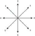
图5为8方向链码的方向数示意图。FIG. 5 is a schematic diagram of the number of directions of an 8-direction chain code.
具体实施方式Detailed ways
以下将结合附图对本发明进行详细说明。The present invention will be described in detail below in conjunction with the accompanying drawings.
参见图1,为本发明的一种全自动图像分割方法的一实施例的流程图,具体实施时,本实施例的该全自动图像分割方法具体包括步骤:Referring to Fig. 1, it is a flowchart of an embodiment of a fully automatic image segmentation method of the present invention. During specific implementation, the fully automatic image segmentation method of the present embodiment specifically includes steps:
S1,输入待分割MRI影像并将其转换为灰度图像。在一具体实施例中,将原始的DICOM图像转换为8位灰度图像。S1, input the MRI image to be segmented and convert it into a grayscale image. In one embodiment, the original DICOM image is converted to an 8-bit grayscale image.
S3,获取步骤S1中转化得到的灰度图像中每条骨-软骨边缘的像素特征,该像素特征包括全局像素特征和局部像素特征。本实施例中该全局像素特征包括像素的边缘距离和方向,局部像素特征包括灰度值和邻域方差。参见图2,该步骤S3具体包括:S3. Obtain pixel features of each bone-cartilage edge in the grayscale image converted in step S1, where the pixel features include global pixel features and local pixel features. In this embodiment, the global pixel feature includes the edge distance and direction of the pixel, and the local pixel feature includes gray value and neighborhood variance. Referring to Fig. 2, the step S3 specifically includes:
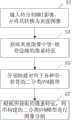
S31,采用基于反馈大边缘数目的迭代法对该灰度图像进行边缘检测,并提取检测到的多条大边缘。Canny边缘检测器是一个在边缘检测敏感性、完整性方面较为理想的检测算子,通过设置边缘的梯度阈值可对弱边缘进行去除,设置高斯滤波器的标准偏差可对噪声进行抑制。因此,本实施例中采用Canny边缘检测器来进行自适应边缘检测,具体实施时,参见图3,该步骤S31具体包括:S31. Using an iterative method based on feedback of the number of large edges to perform edge detection on the gray image, and extract a plurality of detected large edges. Canny edge detector is an ideal detection operator in terms of edge detection sensitivity and integrity. Weak edges can be removed by setting the gradient threshold of the edge, and noise can be suppressed by setting the standard deviation of the Gaussian filter. Therefore, adopt Canny edge detector to carry out self-adaptive edge detection in the present embodiment, during concrete implementation, referring to Fig. 3, this step S31 specifically comprises:
S311,设置Canny检测器的初始阈值。具体实施时,采用李二森等提出的梯度图全局阈值分割方法确定初始阈值。S311, setting an initial threshold of the Canny detector. In the specific implementation, the initial threshold is determined by using the gradient map global threshold segmentation method proposed by Li Ersen et al.
S313,对步骤S1转换得到的灰度图像进行边缘检测,并反馈检测到的大边缘数目。由于骨-软骨边缘相较于其他边缘的像素数目多,且跨度较大,因此,为了减少一些小边缘的干扰,将像素数目多,且跨度较大的边缘划分为大边缘,即骨-软骨边缘属于大边缘。S313. Perform edge detection on the grayscale image converted in step S1, and feed back the number of detected large edges. Since the bone-cartilage edge has more pixels and a larger span than other edges, in order to reduce the interference of some small edges, the edge with a large number of pixels and a large span is divided into a large edge, that is, bone-cartilage The fringe belongs to the big fringe.
S315,判断步骤S315所反馈的大边缘数目是否在预设范围内,若是,执行步骤S317a,否则,将所反馈的大边缘数目进行迭代得到新的阈值,即执行步骤S317b,S319a或者S319b。具体实施时,当迭代次数达到最大迭代次数时,则直接执行步骤S317a。S315, judging whether the number of large edges fed back in step S315 is within the preset range, if yes, execute step S317a, otherwise, iterate the number of large edges fed back to obtain a new threshold, that is, execute step S317b, S319a or S319b. During specific implementation, when the number of iterations reaches the maximum number of iterations, step S317a is directly executed.
S317a,提取步骤S15所反馈的大边缘,执行步骤S33。S317a, extract the large edge fed back in step S15, and execute step S33.
S317b,判断步骤S315所反馈的大边缘数目是否大于最大预设值,若是,则执行步骤S319a,否则执行步骤S319b。S317b, judging whether the number of large edges fed back in step S315 is greater than the maximum preset value, if yes, execute step S319a, otherwise execute step S319b.
S319a,根据所反馈的大边缘数目提高Canny检测器的阈值,执行步骤S313。S319a, increase the threshold of the Canny detector according to the number of large edges fed back, and execute step S313.
S319b,根据所反馈的大边缘数目降低Canny检测器的阈值,执行步骤S313。S319b. Decrease the threshold of the Canny detector according to the number of large edges fed back, and execute step S313.
本实施例中,当所反馈的大边缘数目不在预设范围内时,In this embodiment, when the number of large edges fed back is not within the preset range,
通过多次迭代反馈的大边缘数目来调整Canny检测器的阈值,并根据调整后得到的新的阈值再进行边缘检测直至所反馈的大边缘数目达到预设范围内或者迭代次数达到预设最大迭代次数,即通过反馈大边缘数目来反复调整Canny检测器的阈值,从而减少其他边缘的干扰,能够更加准确的获取骨-软骨边缘,也为使得后续处理速度加快。在一具体实施例中,该预设范围为2-10条,则最大预设值为10条,最小预设值为2。Adjust the threshold of the Canny detector through the number of large edges fed back through multiple iterations, and then perform edge detection according to the adjusted new threshold until the number of large edges fed back reaches the preset range or the number of iterations reaches the preset maximum iteration The number of times, that is, to repeatedly adjust the threshold of the Canny detector by feeding back the large number of edges, thereby reducing the interference of other edges, and obtaining bone-cartilage edges more accurately, and speeding up subsequent processing. In a specific embodiment, the preset range is 2-10, and the maximum preset value is 10, and the minimum preset value is 2.
本实施例检测到的多条大边缘中,可能还存在一些干扰边缘,例如血管、骨皮质-松质,以及边缘断裂、边缘“毛刺”、边缘较粗等问题,这将对后续的边缘识别带来误差,因此,在另一具体实施例中,通过上述步骤S31进行边缘检测后,还可通过形态学方法进行边缘优化以去除干扰边缘,具体实施时,可采用连通区域编码将“小边缘”去除;针对其它问题可采用去毛刺、边缘连接、细化等方法进行边缘优化。Among the multiple large edges detected in this embodiment, there may still be some interference edges, such as blood vessels, cortical-cancellous bone, and problems such as edge fracture, edge "burrs", and thicker edges, which will affect the subsequent edge recognition. Therefore, in another specific embodiment, after the edge detection is performed through the above step S31, edge optimization can also be performed by morphological methods to remove interference edges. ” removal; for other problems, methods such as deburring, edge connection, and thinning can be used for edge optimization.
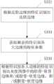
S33,根据预设识别规则对步骤S31所提取到的多条大边缘进行边缘识别,得到至少一条骨-软骨边缘。由于步骤S31所提取的大边缘包括皮肤边缘,以及胫骨-软骨边缘、股骨-软骨边缘、髌骨-软骨边缘中的一种或者两种或者三种骨-软骨边缘,以及其他干扰边缘,因此,需要对步骤S31提取的各个大边缘进行边缘识别。具体实施时,可根据边缘的强度大小与识别的难易程度确定边缘识别的顺序依次为皮肤边缘、胫骨-软骨边缘、股骨-软骨边缘、髌骨-软骨边缘,此为预设识别原则一。另外,在边缘识别过程中,还可利用已识别边缘对未识别边缘进行约束,此为预设识别原则二,例如当识别出股骨-软骨边缘时,可根据股骨位于胫骨的上方这一解剖知识进行约束,识别出髌骨-软骨边缘时,可利用髌骨位于股骨与胫骨的左侧,胫骨的上方等信息进行约束。参见图4,该步骤S33具体包括:S33. Perform edge recognition on the plurality of large edges extracted in step S31 according to preset recognition rules to obtain at least one bone-cartilage edge. Since the large edges extracted in step S31 include skin edges, and tibial-cartilage edges, femur-cartilage edges, one or two or three kinds of bone-cartilage edges in patella-cartilage edges, and other interference edges, therefore, need Perform edge recognition on each large edge extracted in step S31. During specific implementation, the order of edge recognition can be determined according to the intensity of the edge and the difficulty of recognition, which is skin edge, tibial-cartilage edge, femur-cartilage edge, and patella-cartilage edge. This is the first recognition principle. In addition, during the edge recognition process, the recognized edges can also be used to constrain the unrecognized edges. This is the second preset recognition principle. When performing constraints, when the patella-cartilage edge is identified, information such as that the patella is located on the left side of the femur and tibia, and above the tibia can be used to perform constraints. Referring to Fig. 4, this step S33 specifically includes:
S331,根据皮肤边缘的特征识别出皮肤边缘。皮肤边缘的特征包括左侧或者右侧无前景像素。具体实施时,可判断各大边缘的左侧或右侧是否有前景像素,若无前景像素,则该大边缘即为皮肤边缘,否则不为皮肤边缘,直接执行步骤S333。利用左侧或右侧无前景像素这一特征可区分90%以上的皮肤边缘,但有时有的皮肤边缘断裂或未检测到时,仅根据左侧或右侧无前景像素这一特征来识别皮肤边缘将会有误判的情况,因此,在另一具体实施例,识别皮肤边缘时,可根据皮肤边缘的上述特征,以及其他特征,例如边缘跨度大,像素多,长宽比较大等,来进行识别,从而首先识别出待分割图像中左右两侧的皮肤边缘。S331. Identify the skin edge according to the features of the skin edge. Features of the skin edge include no foreground pixels on the left or right. During specific implementation, it may be determined whether there are foreground pixels on the left or right side of each large edge. If there is no foreground pixel, the large edge is a skin edge; otherwise, it is not a skin edge, and step S333 is directly executed. More than 90% of the skin edges can be distinguished using the feature of no foreground pixels on the left or right, but sometimes when some skin edges are broken or not detected, the skin is only identified based on the feature of no foreground pixels on the left or right There will be misjudgment of the edge. Therefore, in another specific embodiment, when identifying the edge of the skin, it can be based on the above-mentioned features of the edge of the skin and other features, such as large edge span, many pixels, and large aspect ratio. Recognition is performed, so that the skin edges on the left and right sides of the image to be segmented are firstly recognized.
S333,获取剩余的待识别各大边缘的特征参数。S333. Obtain the remaining characteristic parameters of the major edges to be identified.
每种骨-软骨边缘的特征不尽相同,因此,可根据每条边缘的特征来进行边缘识别。若将不同边缘视为二维平面上的曲线,结合膝关节的解剖信息可确定各种骨-软骨边缘的特征如下表1所示:The characteristics of each bone-cartilage edge are different, therefore, edge identification can be performed according to the characteristics of each edge. If the different edges are regarded as curves on a two-dimensional plane, combined with the anatomical information of the knee joint, the characteristics of various bone-cartilage edges can be determined as shown in Table 1:
表1骨-软骨边缘的特征Table 1 Characteristics of the Bone-Cartilage Margin
由上表可知,骨-软骨边缘的特征包括开口方向、位置、水平方向数和顺时针旋转角度,其中,开口方向与抛物线的开口方向定义相似;水平方向像素数是指位于边缘(曲线)水平段的像素数目;顺时针旋转角度是指以边缘(曲线)的起始端点切线方向为参考,到终止端点时边缘(曲线)顺时针旋转的角度。要根据边缘的特征来进行边缘识别,就需要获取各边缘的特征参数,进而根据边缘的特征参数来进行边缘识别,具体实施时,步骤S333具体包括:It can be seen from the above table that the characteristics of the bone-cartilage edge include the opening direction, position, number of horizontal directions, and clockwise rotation angle, where the definition of the opening direction is similar to that of the parabola; the number of pixels in the horizontal direction refers to the number of pixels located in the horizontal section of the edge (curve). The number of pixels; the clockwise rotation angle refers to the angle at which the edge (curve) rotates clockwise when it reaches the end point, taking the tangent direction of the starting point of the edge (curve) as a reference. To perform edge recognition according to the characteristics of the edge, it is necessary to obtain the characteristic parameters of each edge, and then perform edge recognition according to the characteristic parameters of the edge. During specific implementation, step S333 specifically includes:
获取待识别各大边缘两端点间的像素位置序列。具体实施时,可采用8连通区域的轮廓跟踪方法获取各大边缘两端点间的像素位置序列。Obtain the sequence of pixel positions between the two ends of the major edges to be identified. During specific implementation, the contour tracking method of 8-connected regions can be used to obtain the sequence of pixel positions between the two ends of each major edge.
获取待识别各大边缘的开口方向的特征值Popen-up,Popen-dpwn和Pcollinear。具体实施时,可通过判断中间点位于大边缘两端点连接线的上方或下方以确定开口向下或向上,若该中间点距离端点连接线距离较近时则判断为共线。其中,该开口方向特征值Popen-up,Popen-down,Pcollinear分别为开口向上、开口向下、共线的中间点数与等间距选取的总中间点数之比值。在一具体实施例中,可等间距的选取20个中间点,分别判断开口方向,其中开口向上、开口向下、共线的中间点数与总中间点数20的比值即为对应开口方向的特征值Popen-up,Popen-down,Pcollinear。The eigenvalues Popen-up , Popen-dpwn and Pcollinear of the opening directions of the major edges to be identified are acquired. During specific implementation, it can be determined whether the opening is downward or upward by judging that the middle point is located above or below the connecting line between the two ends of the large edge. Wherein, the opening direction eigenvalues Popen-up , Popen-down , and Pcollinear are respectively the ratios of the opening-up, opening-down, and collinear intermediate points to the total intermediate points selected at equal intervals. In a specific embodiment, 20 intermediate points can be selected at equal intervals to determine the opening direction respectively, wherein the ratio of the number of middle points with opening upward, opening downward, and collinear to the total number of intermediate points 20 is the eigenvalue corresponding to the opening direction Popen-up , Popen-down , Pcollinear .
获取待识别各大边缘的上下方向和左右方向的位置特征值Ppos-up和Ppos-left,其中,Ppos-up定义为像素位置序列中所有点的行平均值与行总数之比,Ppos-left定义为像素位置序列中所有点的列平均值与列总数之比。Obtain the position eigenvalues Ppos-up and Ppos-left of the up and down directions and left and right directions of the major edges to be identified, where Ppos-up is defined as the ratio of the row average value of all points in the pixel position sequence to the total number of rows, Ppos-left is defined as the ratio of the column mean to the column total for all points in the sequence of pixel positions.
获取待识别各大边缘的水平方向像素数Pnum-horizontal。具体实施时,首先计算像素位置序列的8方向freeman链码,如图5所示,其中,水平方向即为链码的方向数为0或4,则水平方向像素数Pnum-horizontal定义为序列链码中方向数为0或4的数目占序列像素总数的比例。Obtain the number of pixels Pnum-horizontal in the horizontal direction of each major edge to be identified. During specific implementation, first calculate the 8-direction freeman chain code of the pixel position sequence, as shown in Figure 5, wherein, the horizontal direction is the direction number of the chain code is 0 or 4, then the horizontal direction pixel number Pnum-horizontal is defined as the sequence The proportion of the number of directions in the chain code that is 0 or 4 to the total number of sequence pixels.
获取待识别各大边缘的顺时针旋转角度特征值Pangle-clockwise。具体实施时,以链码的起点方向为参考方向,逐一判断下一方向数相对于上一方向数顺时针旋转角度值,当大于180度时为负,再与上一旋转角度值累加,最终得到序列的顺时针旋转角度angle,将旋转角度angle按照下式归一化处理,得到边缘位置序列顺时针旋转角度特征值:Obtain the clockwise rotation angle eigenvalue Pangle-clockwise of each major edge to be identified. During specific implementation, the starting point direction of the chain code is used as the reference direction, and the clockwise rotation angle value of the next direction number relative to the previous direction number is judged one by one. When it is greater than 180 degrees, it is negative, and then accumulated with the previous rotation angle value, finally The clockwise rotation angle angle of the sequence is obtained, and the rotation angle angle is normalized according to the following formula to obtain the clockwise rotation angle eigenvalue of the edge position sequence:
本实施例中,该大边缘的开口方向的特征值Popen-up,Popen-down和Pcollinear,上下方向和左右方向的位置特征值Ppos-up和Ppos-left,水平方向像素数Pnum-horizontal和顺时针旋转角度特征值Pangle-clockwise,即为该大边缘的特征参数。In this embodiment, the eigenvalues Popen-up , Popen-down and Pcollinear of the opening direction of the large edge, the position eigenvalues P pos-up and Ppos-left of the up-down direction and theleft-right direction, and the number of pixels in the horizontal direction Pnum-horizontal and the characteristic value of the clockwise rotation angle Pangle-clockwise are characteristic parameters of the large edge.
S335,根据待识别各大边缘的特征参数识别出该灰度图像中的胫骨-软骨边缘和/或股骨-软骨边缘,和/或髌骨-软骨边缘。具体实施时,该步骤S335具体包括:S335. Identify the tibia-cartilage edge and/or the femur-cartilage edge, and/or the patella-cartilage edge in the grayscale image according to the characteristic parameters of the major edges to be identified. During specific implementation, the step S335 specifically includes:
对获取的待识别各大边缘的上述特征值:开口方向特征值、位置特征值、水平方向像素数和顺时针旋转角度特征值,进行加权组合,得到待识别各大边缘的骨-软骨边缘加权特征值Pfemur、Ptibia、Ppatella,其各自的计算式分别如下:The above-mentioned eigenvalues of the acquired major edges to be identified: opening direction eigenvalues, position eigenvalues, number of pixels in the horizontal direction, and clockwise rotation angle eigenvalues are weighted and combined to obtain bone-cartilage edge weighted features of the major edges to be identified Values Pfemur , Ptibia , and Ppatella are calculated as follows:
Pfemur=αfemurPpos-up+βfemurPangle-clockwise+θfemurPopen-up+λfemurPnum-horizontalPfemur =αfemur Ppos-up +βfemur Pangle-clockwise +θfemur Popen-up +λfemur Pnum-horizontal
Ptibia=αtibia(1-Ppos-up)+βtibiaPangle-clockwise+θtibiaPopen-down+λfemurPnum-horizontalPtibia =αtibia (1-Ppos-up )+βtibia Pangle-clockwise +θtibia Popen-down +λfemur Pnum-horizontal
Ppatella=αpatellaPpos-left+βpatellaPangle-clockwise+θpatellaPopen-down+λpatellaPnum-horizontalPpatella =αpatella Ppos-left +βpatella Pangle-clockwise +θpatella Popen-down +λpatella Pnum-horizontal
其中,参数α,β,θ,λ为对应特征值的权重。Among them, the parameters α, β, θ, λ are the weights of the corresponding feature values.
根据计算得到的每条待识别大边缘的各骨-软骨边缘加权特征值确定待定骨-软骨边缘。由于每条待识别大边缘均有3个骨-软骨边缘特征加权特征值,具体实施时,将同一骨-软骨边缘特征加权特征值中最大的作为对应的待定骨-软骨边缘。在一具体实施例中,若要识别股骨-软骨边缘,则将各待识别大边缘中股骨-软骨边缘加权特征值Ptibta最大的一条待识别大边缘确定为待定股骨-软骨边缘;若要识别胫骨-软骨边缘,则将各待识别大边缘中胫骨-软骨边缘加权特征值Pfemur最大的一条待识别大边缘确定为待定胫骨-软骨边组;同理确定待定髌骨-软骨边缘。The undetermined bone-cartilage edge is determined according to the calculated weighted eigenvalues of each bone-cartilage edge of each large edge to be identified. Since each large edge to be identified has three bone-cartilage edge feature weighted eigenvalues, the largest among the same bone-cartilage edge feature weighted eigenvalues is used as the corresponding undetermined bone-cartilage edge. In a specific embodiment, if the femur-cartilage edge is to be identified, a large edge to be identified whose weighted characteristic value Ptibta of the femur-cartilage edge is the largest among each large edge to be identified is determined as the pending femur-cartilage edge; For the tibial-cartilage edge, the large edge to be identified with the largest weighted eigenvalue Pfemur of the tibial-cartilage edge among the large edges to be identified is determined as the undetermined tibial-cartilage edge group; similarly, the undetermined patella-cartilage edge is determined.
判断该待定骨-软骨边缘对应的骨-软骨边缘加权特征值是否符合预设的阈值条件,若符合,则该待定骨-软骨边缘即为对应于该阈值条件的骨-软骨边缘,否则确定该待分割图像不包含该骨-软骨边缘。在一具体实施例中,判断上述步骤确定的待定股骨-软骨边缘的股骨-软骨边缘加权特征值Ptibia是否大于预设的给定瓶值,若是,则确定该待定股骨-软骨边缘(即该待识别大边缘)即为股骨-软骨边缘,否则,确定该待分割图像不包含股骨-软骨边缘,即不包含股骨-软骨;同理确定胫骨-软骨边缘和髌骨-软骨边缘。Judging whether the bone-cartilage edge weighted eigenvalue corresponding to the undetermined bone-cartilage edge meets the preset threshold condition, if so, the undetermined bone-cartilage edge is the bone-cartilage edge corresponding to the threshold condition, otherwise, determine the The image to be segmented does not contain the bone-cartilage edge. In a specific embodiment, it is judged whether the femoral-cartilage edge weighted characteristic value Ptibia of the undetermined femoral-cartilage edge determined by the above steps is greater than the preset given bottle value, if so, then determine the undetermined femoral-cartilage edge (that is, the The large edge to be identified) is the femoral-cartilage edge, otherwise, it is determined that the image to be segmented does not contain the femoral-cartilage edge, that is, the femoral-cartilage edge is not included; similarly, the tibial-cartilage edge and the patella-cartilage edge are determined.
本实施例中,不同的骨-软骨边缘对应于不册的给定阈值(该给定阈值可通过训练样本数据进行实验确定),例如股骨-软骨边缘加权特征值Ptibia对应的给定阈值为0.5,则胫骨-软骨边缘加权特片值Pfemur和髌骨-软骨加权特征值Ppatella对应的给定阈值则不为0.5,且胫骨-软骨边缘加权特征值Pfemur和髌骨-软骨加权特征值Ppatella两者所对应的给定阈值也不相同。本实施例中通过高置阈值条件是为了防止未检测到骨-软骨边缘时将其它边缘误判为骨-软骨边缘。In this embodiment, different bone-cartilage edges correspond to different given thresholds (the given thresholds can be determined experimentally through training sample data), for example, the given threshold corresponding to the femoral-cartilage edge weighted eigenvalue Ptibia is 0.5, the given threshold corresponding to the tibial-cartilage edge weighted feature value Pfemur and patella-cartilage weighted feature value Ppatella is not 0.5, and the tibial-cartilage edge weighted feature value Pfemur and patella-cartilage weighted feature value P The given thresholds corresponding to bothpatella are also different. In this embodiment, the threshold condition is set high to prevent other edges from being misjudged as bone-cartilage edges when no bone-cartilage edge is detected.
本实施例中,通过对每条待识别边缘的特征参数进行加权组合,并计算得到对应的骨-软骨加权特征值,使得可根据该骨-软骨边缘加权特片值对对应骨-软骨边缘的区分能力确定,区分力越高,权重越大,从而可提高单一特征对边缘识别的准确率。In this embodiment, by weighting and combining the feature parameters of each edge to be identified, and calculating the corresponding bone-cartilage weighted feature value, the weighted feature value of the corresponding bone-cartilage edge can be calculated according to the weighted feature value of the bone-cartilage edge. The distinguishing ability is determined. The higher the distinguishing ability, the greater the weight, which can improve the accuracy of single feature for edge recognition.
S35,根据识别出的各种骨-软骨边缘,计算得到各种骨-软骨边缘的全局像素特征,即像素的边缘距离和方向。基中边缘距离d(x,y)为像素(x,y)与像素位置序列中最近点的欧氏距离,像素(x,y)的方向o(x,y)为基与边缘序列中最近点对应链码的方向数。S35. According to the identified various bone-cartilage edges, calculate global pixel features of various bone-cartilage edges, that is, edge distances and directions of pixels. The edge distance d(x, y) in the base is the Euclidean distance between the pixel (x, y) and the nearest point in the pixel position sequence, and the direction o(x, y) of the pixel (x, y) is the closest point in the base and edge sequence The point corresponds to the direction number of the chain code.
S37,对步骤S1转换得到的灰度图像进行高斯滤波平滑处理,得到像素的灰度值f(x,y)。S37, performing Gaussian filter smoothing on the grayscale image converted in step S1 to obtain the grayscale value f(x, y) of the pixel.
S39,根据步骤S37经滤波平滑后的灰度图像计算得到像素的8邻域灰度方差σ8(x,y)。具体实施时,根据滤波平滑后的灰度图像,在其8邻域内计算标准差即得到8邻域灰度方差σ8(x,y)。S39, calculate the 8-neighborhood gray variance σ8 (x, y) of the pixel according to the filtered and smoothed gray image in step S37. During specific implementation, according to the filtered and smoothed grayscale image, the standard deviation is calculated in its 8 neighborhoods to obtain the 8 neighborhood grayscale variance σ8 (x, y).
本实施例中该步即S31至S35实际上为获取灰度图像中的全局像素特征的一具体实施例,而步骤S37至步骤S39为获取灰度图像局部像素特征的一具体实施例,本实施例中获取全局像素特征的步骤S31至S35与获取局部像素特征的步骤S37-S39可不按照上述的顺序执行,也可按照其他顺序执行,即可先获取局部像素特征,后获取全局像素特征。In this embodiment, the steps S31 to S35 are actually a specific embodiment of obtaining the global pixel features in the grayscale image, and steps S37 to S39 are a specific embodiment of obtaining the local pixel features of the grayscale image. In the example, steps S31 to S35 for obtaining global pixel features and steps S37-S39 for obtaining local pixel features may not be performed in the order described above, but may also be performed in other orders, that is, the local pixel features may be obtained first, and then the global pixel features may be obtained.
S5,分别构建对应于胫骨软骨、股骨软骨和髌骨软骨的二分类SVM模型。支持向量机是由Vapnik于1995年首先担出的,通过构造正例和么例间的最优分类超平面可用于模式分类与非线性回归,与其它分类模型相比,SVM能担供好的泛化能力,具有较强的鲁棒性与有效性。因此,本实施例先择SVM进行像素分类,针对不同类型的骨-软骨(胫骨软骨、股骨软骨和髌骨软骨),分别构建相对应的二分类SVM模型,其中核函数为径向基函数。具体实施时,构建二分类SVM模型的步骤S5具体包括训练集和测试集的选定;数据预处理及模型训练。S5, construct binary classification SVM models corresponding to tibial cartilage, femoral cartilage and patellar cartilage, respectively. The support vector machine was first proposed by Vapnik in 1995. It can be used for pattern classification and nonlinear regression by constructing the optimal classification hyperplane between positive examples and negative examples. Compared with other classification models, SVM can provide good Generalization ability, strong robustness and effectiveness. Therefore, in this embodiment, SVM is first selected for pixel classification, and corresponding binary classification SVM models are respectively constructed for different types of bone-cartilage (tibial cartilage, femoral cartilage and patellar cartilage), wherein the kernel function is a radial basis function. During specific implementation, the step S5 of constructing a binary classification SVM model specifically includes selection of a training set and a test set; data preprocessing and model training.
在一具体实施例中,选定训练集与测试集时,选择已经过人工分割,胩将不同软骨标记为不同颜色的20幅MRI影像,并按照从外到内的顺序进行图像编号,编号从1至20,取其中编号为4、6、8、10、12、14、16的层面作为训练样本,3、5、7、9、11、13、15、17层面作为测试样本,用于测试算法的准确性。In a specific embodiment, when selecting a training set and a test set, select 20 MRI images that have been artificially segmented, and mark different cartilages in different colors, and number the images in order from outside to inside, numbering from 1 to 20, take layers numbered 4, 6, 8, 10, 12, 14, and 16 as training samples, and layers 3, 5, 7, 9, 11, 13, 15, and 17 as test samples for testing Algorithm accuracy.
并且需要对选定的训练集与测试集的像素特征进行归一化处理,采有的归一化映射如下所示;And it is necessary to normalize the pixel features of the selected training set and test set, and the normalized mapping is as follows;

进行模型训练时,由于不同层面,不一定三块软骨同时存在,有可能只包含股骨软骨与胫骨软骨,为了缩短训练时间,提高分割速度,需要判断图像中是否存在对应的骨-软骨边缘,从而确定是否包含对应的软骨,当不存在对应的骨-软骨边缘时则确定不包含对应软骨,进而不必利用对应的SVM模型进行图像分割。因此,在一具体实施例中,选定训练像素时,首先判断所述训练集中的该图像是否包含胫骨-软骨边缘和/或股骨-软骨边缘,和/或髌骨-软骨边缘,若是则选择与基所包含的骨-软骨对应的像素进行训练,具体判断方法可采用上述的边缘识别方法来判断,再由人工进行修正,当然也可直接由人工确定不同边缘。When performing model training, due to different layers, three cartilages may not necessarily exist at the same time, and it may only contain femoral cartilage and tibial cartilage. In order to shorten the training time and improve the segmentation speed, it is necessary to judge whether there is a corresponding bone-cartilage edge in the image, so that It is determined whether the corresponding cartilage is included, and when there is no corresponding bone-cartilage edge, it is determined that the corresponding cartilage is not included, so that image segmentation does not need to be performed using the corresponding SVM model. Therefore, in a specific embodiment, when selecting training pixels, it is first judged whether the image in the training set includes the tibia-cartilage edge and/or the femur-cartilage edge, and/or the patella-cartilage edge, and if so, select the Based on the pixels corresponding to the bone-cartilage included in the training, the specific judgment method can be judged by the above-mentioned edge recognition method, and then manually corrected, of course, different edges can also be directly determined manually.
进一步地,由于软骨仅分布于骨的表面,厚度为1-6毫米,因此,模型训练时,只需要对与骨-软骨边缘距离在8毫米(包含一定的非软骨像素)内的像素进行训练即可。此外还可根据不同骨的解剖位置进一步缩小搜索范围。Furthermore, since the cartilage is only distributed on the surface of the bone with a thickness of 1-6 mm, when training the model, it is only necessary to train the pixels within 8 mm (including certain non-cartilage pixels) from the edge of the bone-cartilage That's it. In addition, the search range can be further narrowed according to the anatomical position of different bones.
本实施例中,在选择训练像素时,通过先判断图像中是否包含对应的骨-软肌边缘,若包含,则只选择该骨-软骨边缘距离8毫米骨的像素进行训练,否则,不选择该像素,即通过减少训练的像素总数,从而缩短了训练时间。In this embodiment, when selecting training pixels, first judge whether the corresponding bone-soft muscle edge is included in the image, and if so, only select the pixel whose bone-cartilage edge is 8 mm away from the bone for training; otherwise, do not select The pixels, that is, by reducing the total number of pixels for training, thus shortening the training time.
另一方面,可通过扩大本实施例中的训练集,进一步提高分割效果。On the other hand, the segmentation effect can be further improved by expanding the training set in this embodiment.
本实施例中,也可首先构建好二分类SVM模型后,再执行上述步骤S1-S3。In this embodiment, the binary classification SVM model may also be constructed first, and then the above steps S1-S3 are performed.
S7,根据步骤S3中所获取的像素特征,并采用步骤S5中构建的二分类SVM模型进行图像分割。由于上述步骤S3的子步骤S335中,已经根据待识别各大边缘的特征参数识别出该待分割图像中所包含的骨-软骨边缘的类型,即得知该待分割图像中所包含的骨-软骨类型,且由于软骨仅分布于骨的表面,厚度为1-6毫米,因此,具体实施时,仅对于骨-软骨边缘8毫米内的像素进行分类,即仅对可能为软骨的像素进行分类,并标记不同的颜色。S7, according to the pixel features obtained in step S3, and using the binary classification SVM model constructed in step S5 to perform image segmentation. Since in the sub-step S335 of the above step S3, the type of the bone-cartilage edge contained in the image to be segmented has been identified according to the characteristic parameters of the major edges to be identified, that is, the bone-cartilage edge type contained in the image to be segmented is known. Cartilage type, and since cartilage is only distributed on the surface of the bone with a thickness of 1-6 mm, therefore, only pixels within 8 mm of the bone-cartilage edge are classified, that is, only pixels that may be cartilage are classified , and mark a different color.
由于采用SCM模型进行初步分割后,还可能存在孔洞、边缘不平滑及像素重复分类的部题。因此,本实施例中,该全自动图像分割方法还可包括分类后处理步骤。具体实施时,像素重复分类是由于髌骨软骨与股骨软骨、股骨软骨与胫骨软骨在某些层面解剖上存在接角,在对相互接角部位的像素应用SVM模型分类后可能将一个像素同时判定为两种软骨,对此根据边缘距离将该像素划分为边缘距离小的一类。对于孔洞及边缘不平滑部题采用描黑算子(当某一背景像素的8邻域有至少5个前景像素时,则该像素为前景像素;否则为背景像素)进行下理。After the initial segmentation using the SCM model, there may still be some problems such as holes, uneven edges, and repeated pixel classification. Therefore, in this embodiment, the fully automatic image segmentation method may further include a post-classification processing step. In the specific implementation, the repeated classification of pixels is due to the anatomical angles between patellar cartilage and femoral cartilage, femoral cartilage and tibial cartilage at certain levels. After applying the SVM model classification to the pixels at the mutual joint angles, a pixel may be simultaneously determined as Two types of cartilage, for which the pixel is classified into a class with a small edge distance according to the edge distance. For holes and uneven edges, the blackening operator is used (when there are at least 5 foreground pixels in the 8 neighborhoods of a certain background pixel, the pixel is a foreground pixel; otherwise, it is a background pixel) for processing.
本实施例的全自动图像分割方法,通过获取的图像中各骨-软骨边缘的像素特征,针对不同的软骨构建对应二分类SVM模型,并根据获取的图像中骨-软骨边缘的像素特征利用该SVM模型进行图像分割,其中,获取各骨-软骨边缘的像素特征时,首先采用边缘栓测和边缘识识将各骨-软骨边缘从图像中检测识别出来,再获取根据训别出的各条骨-软骨边缘来计算其像素特征:边缘中离、方向、灰度值和邻域方差,从而利用SVM模块进行图像分割时,SCM模型只需要根据这些像素特征对骨-软骨边缘对应的像素进行分类即可,使用较少的像素特征,且缩小了搜萦范围,使得分割速度更快,且由于针对不同的软骨建立了不同有SVM模型,且获取像素特征时,也识别出不同的骨-软骨边缘,从而可对股骨软骨、胫骨软骨及髌骨软骨进行独立分割,也无需进行骨的预分害,也能够区分相互接角的软骨及与软骨灰度相近的肌肉组织。根据本实施例的全自动图像分割方法,一台主频为2.8Ghz PC上对测试亲集进行分割仅需约150s,而有经验的放射医师进行分割花费约1h,且使用本实施例的方法进行股骨软骨、胫骨软骨、髌骨软骨自动分割与手工分割的Dice相似性系数分别为0.78、0.76、0.74,具有较好的一致性。In the fully automatic image segmentation method of this embodiment, through the pixel features of each bone-cartilage edge in the acquired image, a corresponding binary classification SVM model is constructed for different cartilages, and the pixel feature of the bone-cartilage edge in the acquired image is used. The SVM model performs image segmentation. When obtaining the pixel features of each bone-cartilage edge, firstly use edge bolt detection and edge recognition to detect and identify each bone-cartilage edge from the image, and then obtain each bone-cartilage edge identified according to training. The bone-cartilage edge is used to calculate its pixel features: edge distance, direction, gray value and neighborhood variance, so that when using the SVM module for image segmentation, the SCM model only needs to perform pixel corresponding to the bone-cartilage edge according to these pixel features. Classification is enough, using less pixel features, and narrowing the search range, making the segmentation faster, and because different SVM models have been established for different cartilages, and when obtaining pixel features, different bones are also identified- Cartilage edge, so that the femoral cartilage, tibial cartilage and patellar cartilage can be segmented independently, and there is no need to pre-segment the bone, and it is also possible to distinguish the cartilage that meets each other and the muscle tissue that is similar to the gray scale of the cartilage. According to the fully automatic image segmentation method of this embodiment, it only takes about 150s to segment the test set on a PC with a main frequency of 2.8Ghz, and it takes about 1h for experienced radiologists to segment, and the method of this embodiment is used The Dice similarity coefficients for automatic and manual segmentation of femoral cartilage, tibial cartilage, and patellar cartilage were 0.78, 0.76, and 0.74, respectively, showing good consistency.
软骨病变时骨-软骨边缘依然存在,因而本实施例的方法可进一步推广至对病变软骨的分割;根据分割的结果可计算软骨厚度、体积等形态参数,为临床诊断、科研提供定量的软骨数据。The bone-cartilage edge still exists when cartilage is diseased, so the method of this embodiment can be further extended to the segmentation of diseased cartilage; according to the segmentation results, the cartilage thickness, volume and other morphological parameters can be calculated to provide quantitative cartilage data for clinical diagnosis and scientific research .
最后说明的是,以上实施例仅用以说明本发明的技术方案而非限制,尽管参照较佳实施例对本发明进行了详细说明,本领域的普通技术人员应当理解,可以对本发明的技术方案进行修改或者等同替换,而不脱离本发明技术方案的宗旨和范围,其均应涵盖在本发明的权利要求范围当中。Finally, it is noted that the above embodiments are only used to illustrate the technical solutions of the present invention without limitation. Although the present invention has been described in detail with reference to the preferred embodiments, those of ordinary skill in the art should understand that the technical solutions of the present invention can be carried out Modifications or equivalent replacements without departing from the spirit and scope of the technical solution of the present invention shall be covered by the claims of the present invention.
Claims (10)
Translated fromChinese
Priority Applications (1)
| Application Number | Priority Date | Filing Date | Title |
|---|---|---|---|
| CN2013101079942ACN103186901A (en) | 2013-03-29 | 2013-03-29 | Full-automatic image segmentation method |
Applications Claiming Priority (1)
| Application Number | Priority Date | Filing Date | Title |
|---|---|---|---|
| CN2013101079942ACN103186901A (en) | 2013-03-29 | 2013-03-29 | Full-automatic image segmentation method |
Publications (1)
| Publication Number | Publication Date |
|---|---|
| CN103186901Atrue CN103186901A (en) | 2013-07-03 |
Family
ID=48678059
Family Applications (1)
| Application Number | Title | Priority Date | Filing Date |
|---|---|---|---|
| CN2013101079942APendingCN103186901A (en) | 2013-03-29 | 2013-03-29 | Full-automatic image segmentation method |
Country Status (1)
| Country | Link |
|---|---|
| CN (1) | CN103186901A (en) |
Cited By (14)
| Publication number | Priority date | Publication date | Assignee | Title |
|---|---|---|---|---|
| CN103440665A (en)* | 2013-09-13 | 2013-12-11 | 重庆大学 | Automatic segmentation method of knee joint cartilage image |
| CN104809740A (en)* | 2015-05-26 | 2015-07-29 | 重庆大学 | Automatic knee cartilage image partitioning method based on SVM (support vector machine) and elastic region growth |
| CN105184799A (en)* | 2015-09-18 | 2015-12-23 | 浙江工商大学 | Modified non-supervision brain tumour MRI (Magnetic Resonance Imaging) image segmentation method |
| CN105225234A (en)* | 2015-09-18 | 2016-01-06 | 浙江工商大学 | Based on the lung tumor identification method of support vector machine MRI Iamge Segmentation |
| CN106327495A (en)* | 2016-08-26 | 2017-01-11 | 穆达文 | Biological bone recognition method, device and system |
| CN108921198A (en)* | 2018-06-08 | 2018-11-30 | 山东师范大学 | commodity image classification method, server and system based on deep learning |
| CN109313710A (en)* | 2018-02-02 | 2019-02-05 | 深圳蓝胖子机器人有限公司 | Model of Target Recognition training method, target identification method, equipment and robot |
| CN109528195A (en)* | 2018-11-08 | 2019-03-29 | 殷晓亮 | In a kind of MRI/CT image bone boundary and method for distinguishing is known with reference to bone mark object |
| CN109741352A (en)* | 2018-12-25 | 2019-05-10 | 深圳市第二人民医院 | Image segmentation method for cartilage damage repair based on multimodal magnetic resonance |
| CN109844809A (en)* | 2017-11-30 | 2019-06-04 | 深圳配天智能技术研究院有限公司 | A kind of image processing method and device, computer readable storage medium |
| CN110443790A (en)* | 2019-08-01 | 2019-11-12 | 北京灵医灵科技有限公司 | Cartilage recognition methods and identifying system in a kind of medical image |
| CN111028278A (en)* | 2018-10-09 | 2020-04-17 | 武汉大学中南医院 | Method for providing human body joint data based on tomography technology |
| CN109377505B (en)* | 2018-10-29 | 2021-07-06 | 哈尔滨理工大学 | A MRI brain tumor image segmentation method based on multi-feature discrimination |
| CN113962970A (en)* | 2021-10-27 | 2022-01-21 | 江苏中烟工业有限责任公司 | A method and device for detecting sundries in finished cigarettes based on machine vision |
Citations (4)
| Publication number | Priority date | Publication date | Assignee | Title |
|---|---|---|---|---|
| WO2006104707A2 (en)* | 2005-03-24 | 2006-10-05 | Image Metrics Limited | Method and system for characterization of knee joint morphology |
| US20110254845A1 (en)* | 2010-04-16 | 2011-10-20 | Hitachi Medical Corporation | Image processing method and image processing apparatus |
| CN102314609A (en)* | 2011-09-13 | 2012-01-11 | 中国科学院地理科学与资源研究所 | Skeleton extraction method and device for polygonal image |
| CN102968783A (en)* | 2012-10-15 | 2013-03-13 | 深圳市旭东数字医学影像技术有限公司 | Method and system for automatically segmenting bones from abdomen image data |
- 2013
- 2013-03-29CNCN2013101079942Apatent/CN103186901A/enactivePending
Patent Citations (4)
| Publication number | Priority date | Publication date | Assignee | Title |
|---|---|---|---|---|
| WO2006104707A2 (en)* | 2005-03-24 | 2006-10-05 | Image Metrics Limited | Method and system for characterization of knee joint morphology |
| US20110254845A1 (en)* | 2010-04-16 | 2011-10-20 | Hitachi Medical Corporation | Image processing method and image processing apparatus |
| CN102314609A (en)* | 2011-09-13 | 2012-01-11 | 中国科学院地理科学与资源研究所 | Skeleton extraction method and device for polygonal image |
| CN102968783A (en)* | 2012-10-15 | 2013-03-13 | 深圳市旭东数字医学影像技术有限公司 | Method and system for automatically segmenting bones from abdomen image data |
Non-Patent Citations (1)
| Title |
|---|
| 庞剑飞等: ""基于边缘检测与支持向量机的关节软骨自动分割算法研究"", 《中国学术期刊(光盘版)》, 19 March 2013 (2013-03-19)* |
Cited By (18)
| Publication number | Priority date | Publication date | Assignee | Title |
|---|---|---|---|---|
| CN103440665A (en)* | 2013-09-13 | 2013-12-11 | 重庆大学 | Automatic segmentation method of knee joint cartilage image |
| CN103440665B (en)* | 2013-09-13 | 2016-09-14 | 重庆大学 | Automatic segmentation method of knee joint cartilage image |
| CN104809740A (en)* | 2015-05-26 | 2015-07-29 | 重庆大学 | Automatic knee cartilage image partitioning method based on SVM (support vector machine) and elastic region growth |
| CN104809740B (en)* | 2015-05-26 | 2017-12-08 | 重庆大学 | Knee cartilage image automatic segmentation method based on SVM and Hookean region growth |
| CN105184799A (en)* | 2015-09-18 | 2015-12-23 | 浙江工商大学 | Modified non-supervision brain tumour MRI (Magnetic Resonance Imaging) image segmentation method |
| CN105225234A (en)* | 2015-09-18 | 2016-01-06 | 浙江工商大学 | Based on the lung tumor identification method of support vector machine MRI Iamge Segmentation |
| CN106327495A (en)* | 2016-08-26 | 2017-01-11 | 穆达文 | Biological bone recognition method, device and system |
| WO2019104616A1 (en)* | 2017-11-30 | 2019-06-06 | 深圳配天智能技术研究院有限公司 | Image processing method and device and computer-readable storage medium |
| CN109844809A (en)* | 2017-11-30 | 2019-06-04 | 深圳配天智能技术研究院有限公司 | A kind of image processing method and device, computer readable storage medium |
| CN109844809B (en)* | 2017-11-30 | 2022-04-15 | 深圳配天智能技术研究院有限公司 | An image processing method and device, and a computer-readable storage medium |
| CN109313710A (en)* | 2018-02-02 | 2019-02-05 | 深圳蓝胖子机器人有限公司 | Model of Target Recognition training method, target identification method, equipment and robot |
| CN108921198A (en)* | 2018-06-08 | 2018-11-30 | 山东师范大学 | commodity image classification method, server and system based on deep learning |
| CN111028278A (en)* | 2018-10-09 | 2020-04-17 | 武汉大学中南医院 | Method for providing human body joint data based on tomography technology |
| CN109377505B (en)* | 2018-10-29 | 2021-07-06 | 哈尔滨理工大学 | A MRI brain tumor image segmentation method based on multi-feature discrimination |
| CN109528195A (en)* | 2018-11-08 | 2019-03-29 | 殷晓亮 | In a kind of MRI/CT image bone boundary and method for distinguishing is known with reference to bone mark object |
| CN109741352A (en)* | 2018-12-25 | 2019-05-10 | 深圳市第二人民医院 | Image segmentation method for cartilage damage repair based on multimodal magnetic resonance |
| CN110443790A (en)* | 2019-08-01 | 2019-11-12 | 北京灵医灵科技有限公司 | Cartilage recognition methods and identifying system in a kind of medical image |
| CN113962970A (en)* | 2021-10-27 | 2022-01-21 | 江苏中烟工业有限责任公司 | A method and device for detecting sundries in finished cigarettes based on machine vision |
Similar Documents
| Publication | Publication Date | Title |
|---|---|---|
| CN103186901A (en) | Full-automatic image segmentation method | |
| Zortea et al. | A simple weighted thresholding method for the segmentation of pigmented skin lesions in macroscopic images | |
| CN103440665B (en) | Automatic segmentation method of knee joint cartilage image | |
| CN104809740B (en) | Knee cartilage image automatic segmentation method based on SVM and Hookean region growth | |
| Melo et al. | Microaneurysm detection in color eye fundus images for diabetic retinopathy screening | |
| Greenspan et al. | Automatic detection of anatomical landmarks in uterine cervix images | |
| Ribas et al. | A complex network based approach for knee Osteoarthritis detection: Data from the Osteoarthritis initiative | |
| CN104751178A (en) | Pulmonary nodule detection device and method based on shape template matching and combining classifier | |
| CN102737379A (en) | A CT Image Segmentation Method Based on Adaptive Learning | |
| CN104299242B (en) | Fluoroscopic visualization eye fundus image extracting method based on NGC ACM | |
| CN104794708A (en) | Atherosclerosis plaque composition dividing method based on multi-feature learning | |
| CN113380401B (en) | Method, device and medium for classifying benign and malignant breast tumors based on ultrasound images | |
| CN106340000A (en) | Bone age assessment method | |
| KR102690591B1 (en) | Automatic detection and segmentation method of urinary stones based on deep learning | |
| CN113870194A (en) | Deep layer characteristic and superficial layer LBP characteristic fused breast tumor ultrasonic image processing device | |
| Šerifović-Trbalić et al. | Classification of benign and malignant masses in breast mammograms | |
| Antony et al. | Feature learning to automatically assess radiographic knee osteoarthritis severity | |
| Xue et al. | Comparative performance analysis of cervix ROI extraction and specular reflection removal algorithms for uterine cervix image analysis | |
| Fang et al. | A multitarget interested region extraction method for wrist X-ray images based on optimized AlexNet and two-class combined model | |
| Alam et al. | A novel automated system to detect breast cancer from ultrasound images using deep fused features with super resolution | |
| CN109816665B (en) | A method and device for fast segmentation of optical coherence tomography images | |
| CN105225234A (en) | Based on the lung tumor identification method of support vector machine MRI Iamge Segmentation | |
| Ahmad et al. | Brain tumor detection & features extraction from MR images using segmentation, image optimization & classification techniques | |
| Jyoti et al. | A systematic review of trending technologies in non-invasive automatic brain tumor detection | |
| Htun et al. | Automatic Detection and Classification of Tibia Bone Fracture from X-ray Images by using Gray Level Co-occurrence Matrix |
Legal Events
| Date | Code | Title | Description |
|---|---|---|---|
| C06 | Publication | ||
| PB01 | Publication | ||
| C10 | Entry into substantive examination | ||
| SE01 | Entry into force of request for substantive examination | ||
| C12 | Rejection of a patent application after its publication | ||
| RJ01 | Rejection of invention patent application after publication | Application publication date:20130703 |