CN103153589A - Method for manufacturing three-dimensional molded model and support tool for medical treatment, medical training, research, and education - Google Patents
Method for manufacturing three-dimensional molded model and support tool for medical treatment, medical training, research, and educationDownload PDFInfo
- Publication number
- CN103153589A CN103153589ACN2012800032567ACN201280003256ACN103153589ACN 103153589 ACN103153589 ACN 103153589ACN 2012800032567 ACN2012800032567 ACN 2012800032567ACN 201280003256 ACN201280003256 ACN 201280003256ACN 103153589 ACN103153589 ACN 103153589A
- Authority
- CN
- China
- Prior art keywords
- internal structure
- dimensional
- definition
- shape data
- dimensional modeling
- Prior art date
- Legal status (The legal status is an assumption and is not a legal conclusion. Google has not performed a legal analysis and makes no representation as to the accuracy of the status listed.)
- Granted
Links
Images


Classifications
- G—PHYSICS
- G09—EDUCATION; CRYPTOGRAPHY; DISPLAY; ADVERTISING; SEALS
- G09B—EDUCATIONAL OR DEMONSTRATION APPLIANCES; APPLIANCES FOR TEACHING, OR COMMUNICATING WITH, THE BLIND, DEAF OR MUTE; MODELS; PLANETARIA; GLOBES; MAPS; DIAGRAMS
- G09B23/00—Models for scientific, medical, or mathematical purposes, e.g. full-sized devices for demonstration purposes
- G09B23/28—Models for scientific, medical, or mathematical purposes, e.g. full-sized devices for demonstration purposes for medicine
- G09B23/30—Anatomical models
- B—PERFORMING OPERATIONS; TRANSPORTING
- B29—WORKING OF PLASTICS; WORKING OF SUBSTANCES IN A PLASTIC STATE IN GENERAL
- B29C—SHAPING OR JOINING OF PLASTICS; SHAPING OF MATERIAL IN A PLASTIC STATE, NOT OTHERWISE PROVIDED FOR; AFTER-TREATMENT OF THE SHAPED PRODUCTS, e.g. REPAIRING
- B29C67/00—Shaping techniques not covered by groups B29C39/00 - B29C65/00, B29C70/00 or B29C73/00
- A—HUMAN NECESSITIES
- A61—MEDICAL OR VETERINARY SCIENCE; HYGIENE
- A61B—DIAGNOSIS; SURGERY; IDENTIFICATION
- A61B5/00—Measuring for diagnostic purposes; Identification of persons
- A61B5/05—Detecting, measuring or recording for diagnosis by means of electric currents or magnetic fields; Measuring using microwaves or radio waves
- A61B5/055—Detecting, measuring or recording for diagnosis by means of electric currents or magnetic fields; Measuring using microwaves or radio waves involving electronic [EMR] or nuclear [NMR] magnetic resonance, e.g. magnetic resonance imaging
- A—HUMAN NECESSITIES
- A61—MEDICAL OR VETERINARY SCIENCE; HYGIENE
- A61B—DIAGNOSIS; SURGERY; IDENTIFICATION
- A61B6/00—Apparatus or devices for radiation diagnosis; Apparatus or devices for radiation diagnosis combined with radiation therapy equipment
- A61B6/02—Arrangements for diagnosis sequentially in different planes; Stereoscopic radiation diagnosis
- A61B6/03—Computed tomography [CT]
- B—PERFORMING OPERATIONS; TRANSPORTING
- B29—WORKING OF PLASTICS; WORKING OF SUBSTANCES IN A PLASTIC STATE IN GENERAL
- B29C—SHAPING OR JOINING OF PLASTICS; SHAPING OF MATERIAL IN A PLASTIC STATE, NOT OTHERWISE PROVIDED FOR; AFTER-TREATMENT OF THE SHAPED PRODUCTS, e.g. REPAIRING
- B29C64/00—Additive manufacturing, i.e. manufacturing of three-dimensional [3D] objects by additive deposition, additive agglomeration or additive layering, e.g. by 3D printing, stereolithography or selective laser sintering
- B29C64/10—Processes of additive manufacturing
- B29C64/106—Processes of additive manufacturing using only liquids or viscous materials, e.g. depositing a continuous bead of viscous material
- B29C64/112—Processes of additive manufacturing using only liquids or viscous materials, e.g. depositing a continuous bead of viscous material using individual droplets, e.g. from jetting heads
- G—PHYSICS
- G09—EDUCATION; CRYPTOGRAPHY; DISPLAY; ADVERTISING; SEALS
- G09B—EDUCATIONAL OR DEMONSTRATION APPLIANCES; APPLIANCES FOR TEACHING, OR COMMUNICATING WITH, THE BLIND, DEAF OR MUTE; MODELS; PLANETARIA; GLOBES; MAPS; DIAGRAMS
- G09B23/00—Models for scientific, medical, or mathematical purposes, e.g. full-sized devices for demonstration purposes
- G09B23/28—Models for scientific, medical, or mathematical purposes, e.g. full-sized devices for demonstration purposes for medicine
- B—PERFORMING OPERATIONS; TRANSPORTING
- B33—ADDITIVE MANUFACTURING TECHNOLOGY
- B33Y—ADDITIVE MANUFACTURING, i.e. MANUFACTURING OF THREE-DIMENSIONAL [3-D] OBJECTS BY ADDITIVE DEPOSITION, ADDITIVE AGGLOMERATION OR ADDITIVE LAYERING, e.g. BY 3-D PRINTING, STEREOLITHOGRAPHY OR SELECTIVE LASER SINTERING
- B33Y50/00—Data acquisition or data processing for additive manufacturing
- B—PERFORMING OPERATIONS; TRANSPORTING
- B33—ADDITIVE MANUFACTURING TECHNOLOGY
- B33Y—ADDITIVE MANUFACTURING, i.e. MANUFACTURING OF THREE-DIMENSIONAL [3-D] OBJECTS BY ADDITIVE DEPOSITION, ADDITIVE AGGLOMERATION OR ADDITIVE LAYERING, e.g. BY 3-D PRINTING, STEREOLITHOGRAPHY OR SELECTIVE LASER SINTERING
- B33Y80/00—Products made by additive manufacturing
Landscapes
- Engineering & Computer Science (AREA)
- Physics & Mathematics (AREA)
- Health & Medical Sciences (AREA)
- Chemical & Material Sciences (AREA)
- General Physics & Mathematics (AREA)
- Medical Informatics (AREA)
- Life Sciences & Earth Sciences (AREA)
- General Health & Medical Sciences (AREA)
- Materials Engineering (AREA)
- Educational Technology (AREA)
- Pure & Applied Mathematics (AREA)
- Business, Economics & Management (AREA)
- Mathematical Physics (AREA)
- Mathematical Optimization (AREA)
- Mathematical Analysis (AREA)
- Educational Administration (AREA)
- Theoretical Computer Science (AREA)
- Computational Mathematics (AREA)
- Algebra (AREA)
- Medicinal Chemistry (AREA)
- Nuclear Medicine, Radiotherapy & Molecular Imaging (AREA)
- Optics & Photonics (AREA)
- Mechanical Engineering (AREA)
- Biomedical Technology (AREA)
- Surgery (AREA)
- Molecular Biology (AREA)
- Heart & Thoracic Surgery (AREA)
- Public Health (AREA)
- Pathology (AREA)
- Biophysics (AREA)
- Radiology & Medical Imaging (AREA)
- High Energy & Nuclear Physics (AREA)
- Animal Behavior & Ethology (AREA)
- Veterinary Medicine (AREA)
- Manufacturing & Machinery (AREA)
- Instructional Devices (AREA)
Abstract
Description
Translated fromChinese技术领域technical field
本发明是关于一种利用X射线CT或MRI等医疗用诊断装置取得的医学图像的三维数字化数据,使用复数种原材料进行造型制成三维造型模具的技术。The present invention relates to a technique of making a three-dimensional modeling mold by using the three-dimensional digitized data of medical images obtained by medical diagnostic devices such as X-ray CT or MRI, and using a plurality of raw materials for molding.
背景技术Background technique
在医疗领域中将患部或身体特定部位进行三维可视化,如在知情同意、决定治疗方案,医疗教育、医学研究等领域的需求在不断增长。特别是利用三维造型模具实现的三维可视化时,不仅是视觉,将立体形状用手进行实际接触观察,可以获得更多计算机图像无法传达的信息。In the medical field, the three-dimensional visualization of the affected part or specific parts of the body, such as in the fields of informed consent, decision of treatment plan, medical education, medical research and other fields, is constantly increasing. Especially when using 3D modeling molds to achieve 3D visualization, not only vision, but also actual contact observation of three-dimensional shapes with hands can obtain more information that cannot be conveyed by computer images.
通常的X射线CT或MRI(磁共振成像)等医疗用诊断仪器的标准采用的是DICOM(数字影像和通信标准)、生成三维形状数据,在此基础上通过层叠式粉末成型机使用石膏基材料,实现高速、高精度的制作出医疗用三维造型模具是已公开的。The general standard of medical diagnostic equipment such as X-ray CT or MRI (magnetic resonance imaging) adopts DICOM (Digital Imaging and Communication Standard), generates three-dimensional shape data, and uses gypsum-based materials with a laminated powder molding machine , realizing high-speed, high-precision manufacturing of three-dimensional modeling molds for medical use has been disclosed.
但是,还存在着类似有内部结构的肝脏等复杂器官的柔软度的模拟、或者无法向处理器官等医生或护士提供器官等的触感信息等问题。However, there are still problems such as the simulation of the softness of complex organs such as the liver with internal structures, or the inability to provide tactile information such as organs to doctors and nurses who treat organs.
近年来,随着医疗与工程学的融合·协作的飞速进步,已有提议有效利用先进的工程学计算方法的仿真程序。该仿真程序,将要进行手术对象的生物体部位用多边形构建在计算机屏幕上,模拟类似肝脏等器官的表面。In recent years, with the rapid progress in the integration and collaboration of medical science and engineering, simulation programs that effectively utilize advanced engineering calculation methods have been proposed. This simulation program builds the body part of the subject to be operated on with polygons on the computer screen, simulating the surface of organs such as the liver.
另一方面,通过同时喷射多种树脂,组合硬质树脂和软质树脂,采用机械性质不同的树脂制作出三维造型模具的三维打印机也已公开。使用该种三维打印机,不仅与该形状结构相关的表面、其内部结构也可以再现出来。但是,可以再现出有着内部结构的肝脏等复杂器官的柔软度、或骨头硬度等装置还很少。On the other hand, a three-dimensional printer that produces a three-dimensional modeling mold using resins with different mechanical properties by simultaneously injecting multiple resins, combining hard resins and soft resins, has also been disclosed. Using this kind of 3D printer, not only the surface related to the shape structure, but also its internal structure can be reproduced. However, there are still few devices that can reproduce the softness of complex organs such as the liver with internal structures, or the hardness of bones.
在已公开技术中、在网状结构中支撑造型用模具材料的支撑材料上、将一层有着复数种的模具材料支撑固定在各自的造型位置上,在被固定的一层模具材料上放置下一层的支撑材料,然后在下一层的支撑材料上将下一层的有着复数种类的模具材料支撑固定在各自的造型位置上,依次重复进行模具材料的固定,各层进行叠加后将支撑材料溶解去除,制作出多色·多材料、不同部位其硬度也不同的结构复杂的立体物体的三维造型物体的制作方法已经存在。但是,在特许文献1的方法中,要制作不同部位硬度不同的模具,需要准备符合该部位硬度或软度的模具材料。In the disclosed technology, on the support material supporting the mold material for molding in the network structure, a layer of mold materials with multiple types is supported and fixed at their respective modeling positions, and placed on the fixed layer of mold materials. One layer of support material, and then support and fix the next layer of multiple types of mold materials on the support material of the next layer at their respective modeling positions, repeat the fixing of the mold material in turn, and after each layer is superimposed, the support material Dissolution and removal, and the production method of three-dimensional modeling objects with multi-color, multi-material, and different parts with different hardnesses and complex structures already exists. However, in the method of
【专利文献1】再表2005/037529号公报【Patent Document 1】Reformation No. 2005/037529
发明内容Contents of the invention
发明所要解决的技术问题The technical problem to be solved by the invention
传统的三维造型模具的目的,重点一直放在确认形状上。产品设计师或产品设计人员所追求的是可以将流畅的线条或精致的部件进行再现的高精度造型模具。可是,仅靠外形对触感或使用感进行想象是很困难的。需要不仅是形状,该物体的触感、质感等能与现实的物体接近的造型模具。The purpose of traditional 3D modeling molds has been focusing on confirming the shape. Product designers or product designers are looking for high-precision molding dies that can reproduce smooth lines or delicate parts. However, it is difficult to imagine the tactility or usability based on the appearance alone. There is a need for a molding die that is close to a real object in terms of not only the shape but also the tactility and texture of the object.
鉴于上述问题,本发明的目的是在于提供一种使用复数种机械性质不同的材料,利用可以制造三维造型模具的三维打印机,通过对材料进行组合,可以将医生等专家所掌握的对器官等的触感进行再现的三维造型模具的制作方法。In view of the above-mentioned problems, the purpose of the present invention is to provide a kind of material with different mechanical properties, using a three-dimensional printer that can manufacture three-dimensional modeling molds, by combining the materials, it is possible to combine the functions of organs, etc., which are mastered by doctors and other experts. A method of making a three-dimensional modeling mold that reproduces the feeling of touch.
解决问题的方法way of solving the problem
为了解决上述课题,本发明的三维造型模具制作方法是通过利用至少使用2种类型的原材料作为模具材料的三维打印机、制作三维造型模具的制作方法,至少包括以下的1)~6)步骤。In order to solve the above-mentioned problems, the method for manufacturing a three-dimensional modeling mold of the present invention is a method for manufacturing a three-dimensional modeling mold by using a three-dimensional printer using at least two types of raw materials as mold materials, and at least includes the following 1) to 6) steps.
1)诊断装置取得的二维数据的亮度信息中提取出造型对象物体的生物体部位的三维形状,生成生物体部位及其内部结构部位的三维形状数据的形状数据生成步骤1) Step of generating shape data by extracting the three-dimensional shape of the biological part of the object to be modeled from the brightness information of the two-dimensional data obtained by the diagnostic device, and generating three-dimensional shape data of the biological part and its internal structure
2)功能对生物体部位及其内部结构部位的三维形状数据进行编辑的形状数据编辑步骤2) Function Shape data editing procedure for editing three-dimensional shape data of body parts and internal structural parts
3)分别对生物体部位以及内部结构部位、创建指定原始形状的至少包括内部结构模式、模式尺寸和模式间隔作为参数的触感等效参数表的表创建步骤3) The table creation step of creating a tactile equivalent parameter table for the biological body part and the internal structure part, which at least includes the internal structure mode, the mode size and the mode interval as parameters of the specified original shape
4)分别对生物体部位以及内部结构部位的造型所使用的模具材料的材料类别以及混合比率进行定义、追加在触感等效参数表的材料类别定义步骤4) Define the material type and mixing ratio of the mold material used for the modeling of the biological body part and the internal structure part, and add the material type definition step in the tactile equivalent parameter table
5)根据触感等效参数表的参数生成原始形状数据,将从数据编辑步骤取得的生物体部位及内部结构部位的部位数据与原始形状数据进行并集、差集、交集中的任何一种布尔运算的布尔运算步骤5) Generate the original shape data according to the parameters of the tactile equivalent parameter table, and perform any Boolean combination, subtraction, or intersection of the part data of the body parts and internal structure parts obtained from the data editing step and the original shape data. Boolean steps for operations
6)根据布尔运算步骤取得的生物体部位以及内部结构部位的三维形状数据,采用由材料类别定义步骤所定义的材料,使用三维打印机进行造型的造型步骤6) A modeling step of modeling with a 3D printer using the material defined in the material category definition step based on the three-dimensional shape data of the body parts and internal structure parts obtained in the Boolean operation step
在此,医疗用诊断装置指的是CT,MRI,PET(正电子发射计算机断层扫描)等,采用医疗用数字化图像格式和通讯协议标准的DICOM(数字影像和通信标准)、从二维数据的亮度信息中提取造型对象物体的生物体部位的三维形状。Here, medical diagnostic devices refer to CT, MRI, PET (Positron Emission Computed Tomography), etc., which adopt DICOM (Digital Imaging and Communication Standard), which is a digital image format and communication protocol standard for medical use, from two-dimensional data The three-dimensional shape of the living body part of the molding target object is extracted from the brightness information.
在此,由于MRI图像中软组织的分辨率比CT图像高,可以获取更多组织的物理属性。此外,PET主要是用于癌症诊断等,在制作癌变器官的三维造型模具时,CT/MRI图像一起使用。可以使用CT图像或MRI图像、PET图像中的任意一种,或者是进行组合使用。可以将其中一种用与生物体部位的轮廓,另外一种作为内部结构部位的组织图像使用。Here, since the resolution of soft tissues in MRI images is higher than that in CT images, more physical properties of tissues can be obtained. In addition, PET is mainly used for cancer diagnosis, etc., and CT/MRI images are used together when making three-dimensional modeling molds of cancerous organs. Any one of CT images, MRI images, and PET images, or a combination thereof can be used. One of them can be used for the outline of the body part, and the other can be used for the tissue image of the internal structure part.
关于上述1)所述的形状数据生成步骤指的是、根据从医疗用诊断装置取得二维数据中的亮度信息提取出造型对象物体的生物体部位的三维形状,生成生物体部位及其内部结构部位的三维形状数据的形状数据生成步骤。可以通过医疗用诊断装置取得包括DICOM格式的亮度信息的断层图像的点云数据,将这些断层图像进行层叠提取出造型对象物体的生物体部位的三维形状。然后,利用市场上销售的三维图像CAD软件,分别生成生物体部位及其内部结构部位的三维形状数据。在使用三维图像CAD软件分别生成生物体部位及其内部结构部位的三维形状数据时,进行调整曲面方向、或删除多余的外壳、或增补缝隙、或细化补丁、平滑轮廓形状等处理,例如用STL(Standard Triangulated Language)数据形式进行数据保存。The shape data generating step described in 1) above refers to extracting the three-dimensional shape of the biological part of the object to be modeled based on the brightness information in the two-dimensional data obtained from the medical diagnostic device, and generating the biological part and its internal structure. A step of generating shape data of three-dimensional shape data of a part. Point cloud data of tomographic images including brightness information in DICOM format can be acquired by a medical diagnostic device, and the three-dimensional shapes of biological parts of modeling objects can be extracted by stacking these tomographic images. Then, using commercially available 3D image CAD software, the 3D shape data of the body part and its internal structure part are respectively generated. When using the 3D image CAD software to generate the 3D shape data of the body part and its internal structure part, adjust the direction of the surface, or delete the redundant shell, or add gaps, or refine the patch, smooth the contour shape, etc., for example, use STL (Standard Triangulated Language) data format for data storage.
接下来,关于上述2)所述的数据编辑步骤指的是、通过造型功能对上述1)步骤中取得的三维形状数据进行编辑。在此,造型功能指的是市场上销售的三维图像CAD软件所具备的功能,包括创建截面形状(包括一部分截面)、或创建平面、曲面、原始形状、或形状切割、添加文字·符号、或添加标记符号等功能。Next, the data editing step described in the above 2) refers to editing the three-dimensional shape data obtained in the above 1) step by using the modeling function. Here, the modeling function refers to the functions of 3D image CAD software sold on the market, including creating cross-sectional shapes (including a part of cross-sections), creating planes, curved surfaces, original shapes, or cutting shapes, adding text and symbols, or Add features such as marker symbols.
在所述形状数据编辑步骤中,作为优先,至少具备标准文字·符号的添加处理、标记符号的添加处理、尺寸符合的添加处理、条形码的追加处理等功能。本发明的三维造型模具,目的是在医疗领域中将患部或身体的特定部位进行三维可视化,设想使用在知情同意、决定治疗方案、医疗教育、医学研究等场合。将文字·符号添加在三维形状数据上,比如将患者等的个人信息或拍摄日期等可以作为文字信息附加在三维造型模具上。此外,可将标记符号添加在三维形状数据上,比如在知情同意、决定治疗方案时可以将有用的信息附加在三维造型模具上。此外,可将条形码添加在三维形状数据上,比如可将患者的信息附加在三维造型模具上。In the shape data editing step, it is preferable to have at least the functions of adding standard characters and symbols, adding symbols, adding dimensions, adding barcodes, and the like. The purpose of the three-dimensional modeling mold of the present invention is to perform three-dimensional visualization of the affected part or a specific part of the body in the medical field. Add characters and symbols to 3D shape data. For example, personal information such as patients and shooting dates can be added as character information to 3D modeling molds. In addition, marker symbols can be added to the 3D shape data, for example, useful information can be added to the 3D modeling mold when informed consent and treatment plan are decided. In addition, barcodes can be added to three-dimensional shape data, for example, patient information can be added to three-dimensional modeling molds.
接下来,关于上述3)所述的表创建步骤指的是、对各个生物体部位及内部结构部位分别创建触感等效参数表的步骤。在此,触感等效参数表指的是对生物体部位及其内部结构部位的触感进行再现时所需的信息数据。具体讲是,在触感等效参数表中,指定原始形状的包括内部结构模式定义和、该模式的尺寸定义和、该模式的间隔定义。Next, the table creation step described in 3) above refers to a step of creating a tactile equivalent parameter table for each body part and internal structure part. Here, the tactile sensation equivalent parameter table refers to the information data required for reproducing the tactile sensation of a biological body part and its internal structural parts. Specifically, in the tactile equivalent parameter table, the designation of the original shape includes the definition of the internal structure mode, the size definition of the mode and the interval definition of the mode.
在此,生物体部位指的是包括人类或动物、植物等生物体组织。例如,以人类为例,肝脏或心脏等器官可列入生物体部位。其他,狗或猫的宠物等的器官或植物的果实也可列入。此外,内部结构部位指的是生物体组织的内部的结构部位,例如骨头或脂肪或血管等。Here, a living body part refers to living body tissues including human beings, animals, plants, and the like. For example, in the case of humans, organs such as the liver or heart could be included as body parts. Others, the organs of pets such as dogs or cats or the fruits of plants can also be included. In addition, an internal structural site refers to a structural site inside a living tissue, such as bone, fat, or blood vessel.
此外,指定原始形状的内部结构模式定义是将圆柱体或长方体等原始形状的识别号作为内部结构进行定义。在此,原始形状包括圆柱体、椭圆形、矩形、长方体、棱柱、球体、楔形体、棱锥、圆锥等与几何学相关的基本形状和、将基本形状进行组合的组合形状或独特形状等应用形状。In addition, the definition of the internal structure pattern specifying the original shape defines the identification number of the original shape such as a cylinder or a cuboid as the internal structure. Here, primitive shapes include basic shapes related to geometry such as cylinder, ellipse, rectangle, cuboid, prism, sphere, wedge, pyramid, and cone, and application shapes such as compound shapes that combine basic shapes or unique shapes. .
此外,模式尺寸定义指的是原始形状物体的大小,例如,如果是球体的话就以半径来定义,长方体的话就用高、宽、厚度来定义。此外,模式间隔定义指的是各个原始形状物体的配置间隔,例如相邻原始形状物体就以重心间的距离或侧面间的距离来定义。In addition, the pattern size definition refers to the size of the original shape object. For example, if it is a sphere, it is defined by radius, and if it is a cuboid, it is defined by height, width, and thickness. In addition, the pattern interval definition refers to the arrangement interval of each original shape object, for example, the distance between adjacent original shape objects is defined by the distance between centers of gravity or the distance between sides.
接下来,关于上述4)所述的材料类别定义步骤指的是、分别对用于生物体部位及内部结构部位造型用的模具材料的材料类型和混合比例进行定义并追加在触感等效参数的步骤。Next, the material category definition step described in the above 4) refers to defining the material type and mixing ratio of the mold materials used for modeling the body parts and internal structure parts respectively, and adding them to the tactile equivalent parameters. step.
在此,所谓原材料,例如树脂是最适用的。树脂可以选择硬质或软质树脂等。但是,并不限定于树脂,石膏粉末、塑料粉末、金属粉末、蜡也是可以的。例如,假设以树脂作为原材料使用时,会受到三维造型所使用的三维打印机的限制,有可能无法自由选用所需树脂。因此,对材料的混合比例进行定义、实现树脂的混合。Here, so-called raw materials such as resins are most suitable. Resin can choose hard or soft resin etc. However, it is not limited to resin, and gypsum powder, plastic powder, metal powder, and wax are also possible. For example, if resin is used as a raw material, there may be restrictions on the 3D printer used for 3D modeling, and it may not be possible to freely select the desired resin. Therefore, the mixing ratio of materials is defined and the mixing of resins is realized.
此外,模具材料指的是形成三维造型模具形状的造型用材料。此外,支撑材料指的是模具材料在造型时需要的支撑材料。模具材料有半透明、着色、柔韧性、刚度等多种材料。另一方面,所使用的支撑材料在材料硬化后可以简单去除。要从造型物体上去除支撑材料时可使用超声波清洗机或裂解液等。模具材料或支撑材料,比如丙烯酸树脂是比较适用。要对周围被其他结构包围住的内部结构进行造型时,使用支撑材料作为上述原材料进行造型也是可行的。In addition, the mold material refers to a molding material that forms the shape of a three-dimensional molding mold. In addition, the support material refers to the support material required for the molding of the mold material. Mold materials include translucent, colored, flexible, rigid and other materials. On the other hand, the support material used can be easily removed after the material has hardened. To remove support material from modeling objects, use an ultrasonic cleaner or a lysis solution, etc. A mold material or support material such as acrylic resin is suitable. When modeling an internal structure surrounded by other structures, it is also possible to use a support material as the above-mentioned raw material for modeling.
此外,对混合比率进行定义指的是,例如使用可以同时喷射2种树脂的模具材料的三维打印机时,对树脂的混合比例进行控制,可获得理想的硬度或柔软度。In addition, defining the mixing ratio means, for example, when using a 3D printer that can inject two kinds of resin mold materials at the same time, controlling the mixing ratio of the resins can obtain the desired hardness or softness.
此外,关于模具材料,作为优选,至少有1种是由透光性材料构成。通过透光性材料可观察到透明的内部结构、实现提高空间意识。作为比较容易把握内部结构的材料,对知情同意、决定治疗方案、医疗教育,医学研究等场合时提供帮助,本发明的模具材料至少有1种是由透光性材料构成。Moreover, it is preferable that at least one type of mold material is made of a translucent material. Through the light-transmitting material, the transparent internal structure can be observed, and spatial awareness can be improved. As a material that is relatively easy to grasp the internal structure, it provides assistance in situations such as informed consent, decision-making of treatment plans, medical education, and medical research. At least one of the mold materials of the present invention is composed of a light-transmitting material.
接下来,关于上述5)所述的布尔运算步骤指的是、通过触感等效参数表的参数生成的原始形状数据,将数据编辑步骤中取得的生物体部位及其内部结构部位的部位数据、与原始形状数据中进行并集,差集,交集中任意一种的布尔运算步骤。Next, the Boolean operation step described in the above 5) refers to the original shape data generated by the parameters of the tactile equivalent parameter table, the part data of the biological part and its internal structure part obtained in the data editing step, Perform any Boolean operation steps of union, subtraction, and intersection with the original shape data.
在此,布尔运算指的是在三维计算机图形或CAD中,对具有相同体积的物体执行并集、差集、交集运算进行造型的运算方法。此外,并集是和其他形状成为一体的运算方法,差集是削减其他形状的运算方法,交集是留下与其他形状的重叠部分的运算方法。Here, the Boolean operation refers to an operation method for modeling by performing union, difference, and intersection operations on objects with the same volume in three-dimensional computer graphics or CAD. In addition, union is an operation method for integrating with other shapes, subtraction is an operation method for reducing other shapes, and intersection is an operation method for leaving overlapping parts with other shapes.
本发明的特征是,为了让造型模具使用的材料的硬度接近实物触感,对感触等效参数表进行定义、通过执行相应的布尔运算,对接近实物触感的材料进行定义。The feature of the present invention is that, in order to make the hardness of the material used in the molding die close to the real touch, the equivalent parameter table of touch is defined, and the material close to the real touch is defined by performing corresponding Boolean operations.
接下来,关于上述6)所述的造型步骤指的是、根据布尔运算步骤取得的生物体部位以及内部结构部位的三维形状数据、采用材料类别定义步骤所定义的材料通过三维打印机进行造型的步骤。Next, the modeling step described in the above 6) refers to the step of modeling with a three-dimensional printer using the material defined in the material category definition step based on the three-dimensional shape data of the body parts and internal structural parts obtained in the Boolean operation step .
三维打印机是对三维物体进行造型的打印机,有使用光固化的丙烯酸类树脂的紫外线固化型喷墨方式、还有使用ABS树脂的热熔化层压方式、还有使用粉末的固定方式,并不限定上述方式的打印机,只要是可以使用复数种材料进行三维造型的打印机都是可以的。Three-dimensional printers are printers that shape three-dimensional objects. There are UV-curable inkjet methods using photocurable acrylic resins, hot-melt lamination methods using ABS resins, and fixing methods using powders. Any printer of the above-mentioned mode may be used as long as it can perform three-dimensional modeling using a plurality of materials.
此外,模具材料所使用的原材料,作为优选,最好是选择可以控制颜色、透明度、高软质、X线透视透明度、超声波的灵敏度、闪光的灵敏度、热丝的灵明度、导电性能等参数的材料。In addition, the raw materials used for the mold material, as a preference, should be selected to control parameters such as color, transparency, high softness, X-ray perspective transparency, ultrasonic sensitivity, flash sensitivity, hot wire sensitivity, and electrical conductivity. Material.
此外,在所述三维造型模具的制作方法中所涉及的形状数据生成步骤中,将内部结构部位的三维形状尺寸缩小至小于生物体部位的内部结构部位的体积,这样能从生物体部位中只抽取出内部结构。将内部结构部位的三维形状尺寸缩小后所产生的缝隙部分在进行造型时填充支撑材料,等固化后去除。In addition, in the step of generating shape data involved in the manufacturing method of the three-dimensional modeling mold, the size of the three-dimensional shape of the internal structure part is reduced to be smaller than the volume of the internal structure part of the biological part, so that only Extract the internal structure. The gaps generated by reducing the three-dimensional shape and size of the internal structure are filled with support materials during modeling, and removed after curing.
例如,将生物体部位分割成2部分、可以从2部分的断面部分只抽取出内部结构。这样可以只对内部结构进行更详细的确认。For example, a body part can be divided into two parts, and only the internal structure can be extracted from the cross-sectional parts of the two parts. This allows a more detailed confirmation of only the internal structure.
在所述本发明的三维造型模具的制作方法中,若要对生物体部位以及内部结构部位中的任意一个部位赋予软质感触时,在布尔运算步骤中,将所定义的第1软质材料的部位数据与所定义的第2软质材料的原始形状数据进行并集运算。In the manufacturing method of the three-dimensional modeling mold of the present invention, if a soft texture is to be given to any part of the body part and the internal structure part, in the Boolean operation step, the defined first soft material The position data of , and the defined original shape data of the second soft material are combined.
将所定义的第1软质材料的部位数据与所定义的第2软质材料的原始形状数据进行并集运算指的是、比如在像支撑材料的柔软材料里添加类似橡胶的柔软材料。所需的柔软度或切割·切开感使用混合了橡胶的模具材料制成的原始形状、再现接近实际柔软度的触感。Performing the union operation of the defined portion data of the first soft material and the defined original shape data of the second soft material means, for example, adding a soft material like rubber to a soft material like a support material. The desired softness or feeling of cutting and cutting is reproduced in the original shape using a mold material mixed with rubber, and the touch close to the actual softness is reproduced.
如上所述,原始形状的种类包括圆柱体、椭圆形、矩形、长方体、棱柱、球体、楔形体、棱锥、圆锥等与几何学相关的基本形状和、将基本形状进行组合的组合形状或独特形状等应用形状,有多种多样。通过调整所述原始形状的种类或大小、混合个数·量、混合方法、获得所需柔软度的触感。As mentioned above, the types of primitive shapes include basic shapes related to geometry such as cylinders, ellipses, rectangles, cuboids, prisms, spheres, wedges, pyramids, and cones, and combined shapes or unique shapes that combine basic shapes. There are many kinds of application shapes. By adjusting the type or size of the original shape, the number and quantity of mixing, and the mixing method, the desired softness of touch can be obtained.
这些主要用于再现脂肪、肌肉、血管等柔软组织的感觉。These are mainly used to reproduce the feel of soft tissues such as fat, muscle, blood vessels, etc.
在所述本发明的三维造型模具的制作方法中,若要对生物体部位及其内部结构部位中的任意一个部位赋予软质感触时,在布尔运算步骤中,将所定义的第1软质材料的部位数据与、材料未定义的原始形状数据进行差集运算。在与材料未定义的原始形状数据进行差集运算时,原始形状数据会自动定义支撑材料。如果原始形状的部位是密封的内部结构,就无需清洗去除支撑材料。因此,将支撑材料的柔软特性附加在整体形状内部的特性。另一方面,如果原始形状的部位不是密封的内部结构,例如,如果是格子状部位里面有缝隙的结构时,需要进行洗净去除支撑材料,原始形状的部位为空。例如,在类似橡胶的柔软的模具材料所形成的部位中去除原始形状。从所需的柔软度或切割·切开感使用混合了橡胶的模具材料制成的部位去除原始形状,实现再现接近实际柔软度的触感。通过调整所述原始形状的种类或大小、混合个数·量、混合方法、获得所需柔软度的触感。In the manufacturing method of the three-dimensional modeling mold of the present invention, if a soft texture is to be given to any part of the body part and its internal structure part, in the Boolean operation step, the defined first soft texture The difference operation is performed between the part data of the material and the original shape data of which the material is not defined. The raw shape data automatically defines the support material when subtracted from the raw shape data where the material is not defined. If the original shape is a sealed internal structure, no cleaning is required to remove the support material. Thus, the softness of the supporting material is added to the properties of the interior of the overall shape. On the other hand, if the original shape of the part is not a sealed internal structure, for example, if it is a structure with gaps in the lattice-like part, it needs to be washed to remove the support material, and the original shape of the part is empty. For example, the original shape is removed in the area formed by the soft rubber-like mold material. By removing the original shape from the desired softness or cutting/cutting feeling using rubber-mixed mold material, the touch feeling close to the actual softness can be reproduced. By adjusting the type or size of the original shape, the number and quantity of mixing, and the mixing method, the desired softness of touch can be obtained.
这些主要用于再现脂肪、肌肉、血管等柔软组织的感觉。These are mainly used to reproduce the feel of soft tissues such as fat, muscle, blood vessels, etc.
在所述本发明的三维造型模具的制作方法中,若要对生物体部位以及内部结构部位中的任意一个部位赋予软质感触时,在布尔运算步骤中,将所定义的第1硬质材料的部位数据与所定义的第2硬质材料的原始形状部位数据进行并集运算。In the manufacturing method of the three-dimensional modeling mold of the present invention, if a soft texture is to be given to any part of the body part and the internal structure part, in the Boolean operation step, the defined first hard material The position data of the second hard material and the defined original shape position data of the second hard material are combined.
将所定义的第1硬质材料的部位数据与所定义的第2硬质材料的原始形状部位数据进行并集运算指的是往硬的造型材料中加入更硬的造型材料。将实际所需硬度通过混合硬质造型材料制成的原始形状、可再现与实际硬度等效的触感。通过调整原始形状的种类或大小、混合个数·量、混合方法,达到所需的软硬感觉。Performing the union operation of the defined part data of the first hard material and the defined original shape part data of the second hard material refers to adding a harder modeling material to the hard modeling material. The original shape made by mixing the actual required hardness with hard molding materials can reproduce the tactile feeling equivalent to the actual hardness. By adjusting the type or size of the original shape, the number and amount of mixing, and the mixing method, the required soft and hard feeling can be achieved.
这些主要用于再现骨头等硬组织的感觉,或是对再现切割·切开感时有用。These are mainly used to reproduce the feeling of hard tissues such as bones, or to reproduce the feeling of cutting and cutting.
在所述本发明的三维造型模具的制作方法中,若要在对生物体部位以及内部结构部位中的任意一个部位赋予软质感触时,在布尔运算步骤中,将所定义的第1硬质材料的部位数据与材料未定义的原始形状部位数据进行差集运算。In the manufacturing method of the three-dimensional modeling mold of the present invention, if a soft texture is to be given to any part of the body part and the internal structure part, in the Boolean operation step, the defined first hard Subtraction operation is performed between the position data of the material and the undefined original shape position data of the material.
将所定义的第1硬质材料的部位数据与材料未定义的原始形状部位数据进行差集运算指的是将硬质材料制成的形状部位里面的内存物以精致的原始形状拉出。可再现接近实际切割·切开感的等效的触感。通过调整原始形状的种类或大小、拉出个数·量、拉出方法,达到所需的软硬感觉。Performing a subtraction operation on the part data of the defined first hard material and the undefined original shape part data refers to pulling out the internal content in the shape part made of the hard material in a refined original shape. Equivalent tactile feel close to actual cutting and cutting can be reproduced. By adjusting the type or size of the original shape, the number and amount of pulling out, and the pulling out method, the desired soft and hard feeling can be achieved.
这些主要用于再现骨头等硬组织的感觉,或是对再现切割·切开感时有用。These are mainly used to reproduce the feeling of hard tissues such as bones, or to reproduce the feeling of cutting and cutting.
接下来,就对制作三维造型模具时特别有用的生物体部位、内部结构的定义进行说明。Next, the definitions of living body parts and internal structures that are particularly useful for making three-dimensional modeling molds will be explained.
在所述本发明的三维造型模具制作方法中涉及的表创建步骤中,当生物体部位是肝脏时,从肝实质、肝静脉、门脉、胆管、病变部位中选择1个以上的部位作为所述内部结构部位,并对所选部位进行内部结构模式定义、模式尺寸定义、模式间隔定义。In the table creation step involved in the three-dimensional modeling mold manufacturing method of the present invention, when the body part is the liver, one or more parts are selected from the liver parenchyma, hepatic vein, portal vein, bile duct, and diseased part as the part. Describe the internal structure part, and define the internal structure mode, mode size definition and mode interval definition for the selected part.
当生物体部位是肝脏时,作为优选,至少对肝动脉、肝静脉、门静脉、还有病变部位进行定义。如果对肝实质、胆管也进行定义的话会更理想。When the body part is the liver, at least hepatic artery, hepatic vein, portal vein, and diseased part are preferably defined. It would be more ideal if the liver parenchyma and bile ducts were also defined.
此外,在上述本发明的3维造型模具制作方法中涉及的表创建步骤中,当生物体部位是孕妇腹部时,从子宫、胎儿、挤带、胎盘、羊水、血管、皮下脂肪中选择1个以上的部位作为内部结构部位,并对所选部位进行所述内部结构模式定义、模式尺寸定义、模式间隔定义。In addition, in the table creation step involved in the above-mentioned 3D modeling mold manufacturing method of the present invention, when the biological part is the abdomen of a pregnant woman, one of the following is selected from the uterus, fetus, extruded belt, placenta, amniotic fluid, blood vessel, and subcutaneous fat The above parts are used as internal structure parts, and the internal structure mode definition, mode size definition, and mode interval definition are performed on the selected parts.
通过再现怀孕期间的胎儿的触感的造型,可作为出生前诊断或剖腹产时的安全确认或、在出生时脐带是否缠住胎儿的脖子时进行判断、出生过程的确认等。此外,所制做的三维造型模具可作为分娩后的纪念制成造型纪念册。By reproducing the feel of the fetus during pregnancy, it can be used for prenatal diagnosis, safety confirmation at the time of caesarean section, judgment of whether the umbilical cord is wrapped around the neck of the fetus at birth, confirmation of the birth process, etc. In addition, the prepared three-dimensional modeling mold can be used as a memorial book after childbirth.
此外,在所述本发明的三维造型模具制作方法中涉及的表创建步骤中,当生物体部位是乳房部位时,从皮下脂肪、乳腺、乳管、淋巴管、淋巴结、乳房肿瘤中选择1个以上的部位作为内部结构,并对所选部位进行所述内部结构模式定义、模式尺寸定义、模式间隔定义。作为优选,除了对皮下脂肪、乳腺、乳房肿瘤进行定义,也对淋巴管进行定义会更理想。In addition, in the table creation step involved in the three-dimensional modeling mold manufacturing method of the present invention, when the body part is a breast part, one of subcutaneous fat, mammary gland, milk duct, lymphatic vessel, lymph node, and breast tumor is selected. The above parts are used as the internal structure, and the internal structure mode definition, mode size definition, and mode interval definition are performed on the selected parts. Preferably, in addition to defining subcutaneous fat, mammary glands, and breast tumors, it would be more ideal to also define lymphatic vessels.
通过再现乳房肿瘤部位的硬度,可作为医学教育的一个环节,对乳房肿瘤的检查的培训或实际对患者进行病情说明时使用。By reproducing the hardness of the breast tumor site, it can be used as a part of medical education, training on breast tumor examination or actually explaining the patient's condition.
此外,在上述本发明的3维造型模具制作方法中涉及的表创建步骤中,当所述生物体部位是四肢部位时,从构成四肢关节的皮肤、皮下脂肪、动脉、静脉、肌肉、骨、腱中选择1个以上的部位作为所述内部结构,并对所选部位进行所述内部结构模式定义、模式尺寸定义、模式间隔定义。In addition, in the table creation step involved in the above-mentioned 3D modeling mold manufacturing method of the present invention, when the body part is a limb part, the skin, subcutaneous fat, arteries, veins, muscles, bones, Select one or more parts in the tendon as the internal structure, and define the internal structure mode, mode size, and mode interval for the selected parts.
通过再现皮肤、内部的皮下脂肪或血管的柔软度的触感,可作为医生或护士等在注射、抽血的培训时使用。By reproducing the softness of the skin, internal subcutaneous fat, and blood vessels, it can be used as a training for doctors, nurses, etc. in injections and blood draws.
此外,所制做的三维造型模具可作为义肢或义足使用。In addition, the manufactured three-dimensional modeling mold can be used as a prosthetic limb or prosthetic foot.
此外,在所述本发明的三维造型模具制作方法中涉及的表创建步骤中,当生物体部位是喉咙部位时,从食道、气管、软骨中选择1个以上的部位作为内部结构部位,并对所选部位进行所述内部结构模式定义、模式尺寸定义、模式间隔定义。In addition, in the table creation step involved in the method of manufacturing a three-dimensional modeling mold of the present invention, when the body part is the throat part, one or more parts from the esophagus, trachea, and cartilage are selected as internal structural parts, and the Define the internal structure mode, the mode size and the mode interval at the selected part.
通过再现食道、气管的形状和触感,可作为医生或护士等对肺炎患者进行插入吸气管的培训时使用。By reproducing the shape and feel of the esophagus and trachea, it can be used as a training for doctors, nurses, etc. to insert the suction tube for patients with pneumonia.
此外,在上述本发明的三维造型模具制作方法中涉及的表创建步骤中,当生物体部位是脸部时,从皮肤、皮下脂肪、肌肉、软骨、骨、血管中选择1个以上的部位作为内部结构部位,并对所选部位进行所述内部结构模式定义、模式尺寸定义、模式间隔定义。In addition, in the table creation step involved in the above-mentioned three-dimensional modeling mold manufacturing method of the present invention, when the body part is a face, one or more parts are selected from skin, subcutaneous fat, muscle, cartilage, bone, and blood vessels as The internal structure part, and define the internal structure mode, mode size definition and mode interval definition for the selected part.
可作为对脸部的美容整形外科手术前的确认。Can be used as a confirmation before cosmetic plastic surgery on the face.
此外,在所述本发明的三维造型模具制作方法中涉及的表创建步骤中,当生物体部位是牙齿和牙龈部位时,从牙齿、齿肉、齿槽骨、血管、神经、颧骨肌肉、舌中选择1个以上的部位作为内部结构部位,并对所选部位进行所述内部结构模式定义、模式尺寸定义、模式间隔定义。In addition, in the table creation step involved in the three-dimensional modeling mold manufacturing method of the present invention, when the body parts are teeth and gum parts, teeth, gingiva, alveolar bone, blood vessels, nerves, zygomatic muscles, One or more parts of the tongue are selected as internal structure parts, and the internal structure pattern definition, pattern size definition, and pattern interval definition are performed on the selected parts.
在所述本发明的三维造型模具制作方法中涉及的表创建步骤中,当生物体部位是消化管部位时,从消化管内壁或消化管外壁中选择1个以上的部位作为内部结构部位,并对所选部位进行所述内部结构模式定义、模式尺寸定义、模式间隔定义。In the table creation step involved in the three-dimensional modeling mold manufacturing method of the present invention, when the part of the living body is a part of the digestive canal, select one or more parts from the inner wall of the digestive canal or the outer wall of the digestive canal as the internal structure part, and The internal structure pattern definition, pattern size definition, and pattern interval definition are performed on the selected part.
此外,在上述本发明的3维造型模具制作方法中涉及的表创建步骤中,当生物体部位是头部时,从毛发、眼球、脑、脑血管、皮肤、皮下脂肪、肌肉、耳软骨、鼻软骨、头盖骨中选择1个以上的部位作为内部结构部位,并对所选部位进行所述内部结构模式定义、模式尺寸定义、模式间隔定义。In addition, in the table creation step involved in the above-mentioned 3D modeling mold manufacturing method of the present invention, when the body part is the head, hair, eyeballs, brain, cerebral blood vessels, skin, subcutaneous fat, muscle, ear cartilage, Select one or more parts from the nasal cartilage and cranium as internal structure parts, and define the internal structure pattern, pattern size, and pattern interval for the selected parts.
可作为对脸部的美容整形外科手术前的确认。Can be used as a confirmation before cosmetic plastic surgery on the face.
此外,从本发明的其他观点来看,也提供一种将根据所述三维造型模具的制作方法制成的三维造型模具按照所定厚度进行切片后的切片模具、或是将切片后的切片模具进行层叠还原到原来的形状、并通过轴自由的来回绕轴转动的医疗、医学培训、科研和教育用支持工具。In addition, from another viewpoint of the present invention, there is also provided a slicing mold in which the three-dimensional modeling mold manufactured according to the method for manufacturing the three-dimensional modeling mold is sliced into predetermined thicknesses, or a slicing mold in which the sliced slicing mold is sliced is provided. A support tool for medical treatment, medical training, scientific research and education that is stacked back to its original shape and freely rotates back and forth through the shaft.
将制成的三维造型模具切片后的各个部位进行层叠,可以同时掌握整体形状和断面结构,作为诊断或医疗培训支持工具是有用的。By laminating each part after slicing the produced 3D modeling mold, the overall shape and cross-sectional structure can be grasped at the same time, which is useful as a support tool for diagnosis or medical training.
此外,从本发明的其他观点来看,提供一种从医疗诊断装置取得的二维数据的亮度信息中抽取出造型对象物体的生物体部位的三维形状、生成生物体部位及其内部结构部位的三维形状数据的形状数据生成步骤和、对通过造型功能取得的三维形状数据进行编辑的形状数据编辑步骤和、对各个生物体部位及其内部结构部位造型使用的模型材料的材料类别和混合比例进行定义并追加在触感等效参数表的材料类别定义步骤和、使用材料类别定义步骤所定义的材料通过三维打印机进行造型的造型步骤,利用一种至少使用2种类型的原材料作为模具材料的三维打印机的三维造型模具的制作方法,将根据该方法制得的三维造型模具按照一定厚度切片后的切片模具层叠成原来的三维形状、并通过轴自由的来回绕轴转动的医疗、医学培训、科研和教育用支持工具。In addition, from another aspect of the present invention, there is provided a method for extracting the three-dimensional shape of a living body part of an object to be modeled from brightness information of two-dimensional data acquired by a medical diagnostic device, and generating the living body part and its internal structural parts. Steps for generating shape data of three-dimensional shape data, steps of editing shape data for editing three-dimensional shape data acquired by a modeling function, and performing a process for determining the material type and mixing ratio of model materials used for modeling individual body parts and their internal structural parts defining and adding the material category definition step in the tactile equivalent parameter table and the modeling step of using the material defined in the material category definition step to model with a three-dimensional printer, using a three-dimensional printer that uses at least two types of raw materials as mold materials The manufacturing method of the three-dimensional modeling mold, the three-dimensional modeling mold obtained according to the method is sliced according to a certain thickness and the slice mold is laminated into the original three-dimensional shape, and the medical treatment, medical training, scientific research and Support tools for education.
将制成的三维造型模具切片后的各个部位进行层叠、可以同时掌握整体形状和断面结构,作为诊断或医疗培训支持工具是有用的。By layering each part of the sliced 3D modeling mold, the overall shape and cross-sectional structure can be grasped at the same time, which is useful as a support tool for diagnosis or medical training.
接下来,就本发明的三维造型模具的制作程序进行说明。Next, the manufacturing procedure of the three-dimensional modeling mold of the present invention will be described.
本发明的三维造型模具制作程序是通过一种至少使用2种类型的原材料作为模具材料的三维打印机制作三维造型模具的制作程序、通过计算机运行以下a)~e)的程序。The three-dimensional modeling mold production program of the present invention is a production program for producing three-dimensional modeling molds by a three-dimensional printer using at least two types of raw materials as mold materials, and the following programs a) to e) are run by a computer.
a)根据从医疗用诊断装置取得二维数据中的亮度信息提取出造型对象物体的生物体部位的三维形状,生成生物体部位及其内部结构部位的三维形状数据的形状数据生成程序a) A shape data creation program that extracts the three-dimensional shape of the biological body part of the object to be modeled based on the brightness information in the two-dimensional data obtained from the medical diagnostic device, and generates three-dimensional shape data of the biological body part and its internal structural parts
b)对通过造型功能对生物体部位及其内部结构部位的三维形状数据进行编辑的形状数据编辑程序b) A shape data editing program for editing three-dimensional shape data of body parts and internal structural parts through modeling functions
c)分别对生物体部位及其内部结构部位创建指定原始形状的至少包括内部结构模式、模式尺寸和模式间隔作为参数的触感等效参数表的表创建程序c) A table creation program for creating a tactile equivalent parameter table specifying an original shape for an organism part and its internal structure part, including at least an internal structure mode, a mode size, and a mode interval as parameters
d)提示输入各个生物体部位以及内部结构部位造型所使用的模具材料的材料类别及混合比例进行定义,追加定义在触感等效参数表的材料类别定义程序d) Prompt to input the material category and mixing ratio of the mold materials used in the modeling of various biological parts and internal structure parts to define, and add the material category definition program defined in the tactile equivalent parameter table
e)根据触感等效参数表的参数生成原始形状数据,将数据编辑步骤取得的生物体部位以及内部结构部位的部位数据与原始形状数据进行并集、差集、交集中的任意一种布尔运算的布尔运算程序e) Generate original shape data according to the parameters in the tactile equivalent parameter table, and perform any Boolean operation of union, difference, and intersection on the part data of the body parts and internal structure parts obtained in the data editing step and the original shape data Boolean operations
通过计算机运行上述的a)~e)程序,可以有效利用从医疗诊断装置取得的图像数据生成触感接近的造型模具的造型数据。通过上述的e)的布尔运算程序取得的生物体部位及其内部结构部位的三维形状数据和、将上述d)的材料类别定义程序所定义的材料数据发送到三维打印机,通过三维打印机制成接近触感的三维造型模具。By running the above-mentioned programs a) to e) on a computer, image data acquired from a medical diagnostic device can be effectively used to generate molding data of a molding die with close tactile sensation. The three-dimensional shape data of the biological body part and its internal structure parts obtained by the Boolean operation program of e) above and the material data defined by the material category definition program of d) above are sent to the three-dimensional printer, and the three-dimensional printer is used to make a close Tactile 3D modeling molds.
发明效果Invention effect
发明通过至少使用2种类型的原材料作为模具材料的三维打印机、可制作出与医生等专家所掌握的对器官等的触感等效的造型模具。By inventing a 3D printer that uses at least two types of raw materials as mold materials, it is possible to create a modeling mold that is equivalent to the touch of organs, etc. that experts such as doctors have mastered.
由此,比如在诊断时,肝脏器官的三维造型模具对病理进行三维的评价,可有效的用于诊断·决定治疗方案以及治疗效果的判定。特别是对手术前决定切除部位或决定手术方案都是有帮助的。此外,可以有效用在以手术为主的诊断治疗技术的实践。还有在器官移植手术中,作为替代器官应用在有容量依赖型的技术中。也可用于对患者或患者家属进行说明。Thus, for example, in the case of diagnosis, the three-dimensional model of the liver organ can be used for three-dimensional evaluation of the pathology, which can be effectively used for diagnosis, determination of treatment plan and judgment of treatment effect. In particular, it is helpful to decide the resection site or decide the operation plan before the operation. In addition, it can be effectively used in the practice of surgical-based diagnostic and therapeutic techniques. Also in organ transplant surgery, it is used as a replacement organ in a capacity-dependent technique. It can also be used to explain to a patient or patient's family.
此外,在实际的外科手术现场,要进行手术的部位因为血液或其他器官的影响造成视野受阻,在很多情况下需要依靠触感和经验。在这种情况下,事先准备好再现了患者的部位及其形状和触感的三维造型模具,医生通过目视确认造型模具,在确认完治疗部位的形状及其触感后,实际可放心的对患者实施治疗手术。In addition, at the actual surgical site, the field of view is obstructed by blood or other organs, and in many cases it is necessary to rely on touch and experience. In this case, a three-dimensional modeling mold that reproduces the patient's part, its shape and feel is prepared in advance, and the doctor can actually treat the patient with peace of mind after confirming the shape of the treatment part and the feeling of the model by visually confirming the shape and feel of the patient. Perform therapeutic surgery.
并且,在医学教育方面,实际接触根据本发明制作的三维造型模具,可以理解器官的立体结构或其触感,特别是可以模拟体验触感。Moreover, in terms of medical education, by actually contacting the three-dimensional modeling mold made according to the present invention, the three-dimensional structure of the organ or its tactile sensation can be understood, especially the tactile sensation can be simulated and experienced.
附图说明Description of drawings
【图1】本发明所述的三维造型模具的制作方法的流程图。[FIG. 1] A flow chart of a method for manufacturing a three-dimensional modeling mold according to the present invention.
【图2】实施例1所述的三维造型模具的制作装置的结构图。[ Fig. 2 ] A structural diagram of a manufacturing apparatus for a three-dimensional modeling mold described in Example 1.
【图3】实施例1所述的三维造型模具的制作方法的流程图。[ Fig. 3 ] A flow chart of the method for manufacturing the three-dimensional modeling mold described in Example 1.
【图4】实施例1所述的三维造型模具制作装置中所搭载的程序的说明图。[FIG. 4] An explanatory diagram of a program installed in the three-dimensional modeling mold manufacturing apparatus described in Example 1. [FIG.
【图5】一个三维造型模具的例子。[Fig. 5] An example of a three-dimensional modeling mold.
【图6】肝脏的三维造型模具的触感等效参数表的说明图。[ Fig. 6 ] An explanatory diagram of a tactile equivalent parameter table of the three-dimensional modeling mold of the liver.
【图7】骨头的三维造型模具的触感等效参数表的说明图。[ Fig. 7 ] An explanatory diagram of a tactile equivalent parameter table of a three-dimensional modeling mold of a bone.
【图8】一个骨头的三维造型模具的例子。[Fig. 8] An example of a three-dimensional modeling mold of a bone.
【图9】一个肌肉的三维造型模具的例子。[Figure 9] An example of a three-dimensional modeling mold for muscles.
【图10】医疗、医学培训、科研和教育用支持工具的外观图。[FIG. 10] External view of support tools for medical treatment, medical training, scientific research and education.
【图11】肝脏模型的外观图。[Fig. 11] Appearance of the liver model.
【图12】孕妇腹部模具的外观图。[Figure 12] Appearance of the pregnant woman's abdomen mold.
【图13】脸部模具以及上半身的外观图(1)。[Figure 13] The appearance of the face mold and upper body (1).
【图14】脸部模具以及上半身的外观图(2)。[Figure 14] The appearance of the face mold and upper body (2).
【图15】骨头模具的说明图。[Fig. 15] An explanatory diagram of a bone mold.
具体实施方式Detailed ways
以下,就本发明的实施形态一边参照图面进行详细说明。此外,本发明的范围,不限定于以下的实施例或图示例,可以有其他的变更。Hereinafter, embodiments of the present invention will be described in detail with reference to the drawings. In addition, the scope of the present invention is not limited to the following examples or illustrated examples, and other modifications are possible.
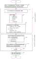
图1示意的是本发明所述的三维造型模具的制作方法的流程图。如图1所示的,本发明所述的三维造型模具的制作方法实施以下的步骤1~步骤6中的S1~S8。在此,S2~S3是为了实现三维形状可视化所进行的设定,S4~S7是为了达到触感等效而进行的设定。Fig. 1 schematically is a flow chart of the manufacturing method of the three-dimensional modeling mold described in the present invention. As shown in FIG. 1 , the method for manufacturing a three-dimensional modeling mold according to the present invention implements steps S1 to S8 in the following
<步骤1:形状数据生成步骤11><Step 1: Shape
(S1)从医疗用诊断装置取得的二维数据的亮度信息中提取出造型对象物体的三维形状。(S1) Extracting the three-dimensional shape of the object to be modeled from the brightness information of the two-dimensional data acquired by the medical diagnostic device.
从CT或MRI装置中提取出包括DICOM格式的亮度信息的断层图像的点云数据(像素点云数据),将这些断层图像进行层积提取造型对象物体的生物体部位的三维形状。Point cloud data (pixel point cloud data) of tomographic images including brightness information in DICOM format are extracted from CT or MRI devices, and the tomographic images are layered to extract the three-dimensional shape of the biological body part of the modeling object.
(S2)生成生物体部位及其内部结构部位的三维形状数据。(S2) Generating three-dimensional shape data of a body part and its internal structural part.
利用市场上销售的三维图像CAD软件,对生物体部位及其内部结构部位的三维形状数据进行调整曲面方向、删除多余外壳、增补缝隙、细化补丁、平滑轮廓形状等处理、生成STL数据形式的数据。Use the 3D image CAD software sold in the market to adjust the surface direction, delete redundant shells, add gaps, refine patches, smooth outline shapes, etc., and generate STL data for the 3D shape data of biological parts and their internal structures. data.
<步骤2:形状数据进行编辑步骤12><Step 2: Edit
(S3)通过造型功能对三维形状数据进行编辑。(S3) Edit the three-dimensional shape data by the modeling function.
利用市场上销售的三维图像CAD软件,通过添加文字·符号,可将患者等的个人信息或拍摄日期等以文字信息添加在三维形状数据里。通过添加标记符号,可将决定的治疗方案等有用的信息添加在三维形状数据里。通过添加定标尺,实际进行手术时将有用的信息添加在三维形状数据里。通过添加条形码,可将患者信息添加在三维形状数据里。By adding characters and symbols using commercially available 3D image CAD software, personal information such as patients and shooting dates can be added to 3D shape data as character information. By adding marker symbols, useful information such as the determined treatment plan can be added to the 3D shape data. By adding scales, useful information is added to the 3D shape data during actual surgery. By adding a barcode, patient information can be added to the 3D shape data.
<步骤3:表创建步骤13><Step 3:
(S4)对各个生物体部位及内部结构创建触觉等效参数表。(S4) Create a tactile equivalent parameter table for each body part and internal structure.
利用市场上销售的三维图像CAD软件,生成触感等效参数表。触感等效参数表对指定原始形状进行内部结构模式定义和、模式尺寸定义和、模式间隔定义。事先准备一定的模式,用户可根据模式号码等进行选择。Use the 3D image CAD software sold in the market to generate the tactile equivalent parameter table. The tactile equivalent parameter table defines internal structure mode, mode size definition and mode interval definition for the specified original shape. A certain pattern is prepared in advance, and the user can select it according to the pattern number or the like.
<步骤4:材料类别定义步骤14><Step 4: Material
(S5)对各个生物体部位及其内部结构部位造型使用的模具材料进行材料类别及混合比例的定义。(S5) Define the material category and mixing ratio of the mold materials used in the modeling of various body parts and their internal structural parts.
此外,原材料采用树脂作为模具材料使用时,可以在能控制颜色、透明度、高软质、X线透视透明度、超声波的灵敏度、闪光的灵敏度、热丝的灵明度、导电性能等参数的各种树脂中进行选择。此外,支撑材料也可作为造型用的模具材料使用。In addition, when the raw material is made of resin as the mold material, it can be used in various resins that can control parameters such as color, transparency, high softness, X-ray transparency, ultrasonic sensitivity, flash sensitivity, hot wire sensitivity, and electrical conductivity. to choose from. In addition, the support material can also be used as a mold material for molding.
可同时喷射2种类型树脂的模具材料时,在选择2种模具材料使用的树脂的同时,也对混合比例(例如,树脂A:树脂B=1:3)进行定义。For mold materials that can inject two types of resins at the same time, define the mixing ratio (for example, resin A: resin B = 1:3) while selecting the resins used for the two types of mold materials.
<步骤5:布尔运算步骤15><Step 5:
(S6)根据触感等效参数表中的参数生成原始形状数据。(S6) Generate original shape data according to the parameters in the tactile equivalent parameter table.
根据触感等效参数表的数据决定原始形状。原始形状有使用模具材料制成的和、不用模具材料制成而是用支撑材料制成的、也有将支撑材料去除后从模具形状中取出原始形状。Determine the original shape according to the data in the tactile equivalent parameter table. The original shape is made using the mold material and, not made of the mold material but with the support material, or the original shape is taken from the mold shape after the support material is removed.
(S7)将生物体部位·内部结构部位的部位数据与原始形状数据进行布尔运算。(S7) Boolean operation is performed on the part data of the body part/internal structure part and the original shape data.
对使用模具材料或支撑材料制成的部位数据,增加或去除原始形状。For part data made using mold material or support material, add or remove the original shape.
<步骤6:造型步骤16><Step 6:
(S8)使用所定义的材料通过三维打印机进行造型。(S8) Modeling is performed by a three-dimensional printer using the defined material.
有效利用市场上销售的三维打印机制作三维造型模具。例如,所使用的三维打印机,可使用OBJET社制造的三维打印机。通过所需三维造型模具的内部结构的各个三维形状数据和所定义的材料使用三维打印机进行造型。Make effective use of 3D printers sold in the market to make 3D modeling molds. For example, as the 3D printer to be used, a 3D printer manufactured by OBJET Corporation can be used. The three-dimensional printer is used for modeling through the various three-dimensional shape data of the internal structure of the required three-dimensional modeling mold and the defined materials.
【实施例1】【Example 1】
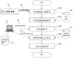
图2示意的是关于实施例1所涉及的三维造型模具的制作装置的结构图。如图2所示的,三维造型模具的制作装置指的是具备中央处理器1、存储部3、显示部6、输入部5、硬盘驱动器7的一般的计算机。更具体说就是,输入部5是键盘和鼠标、显示部6是液晶显示屏。存储部3是记忆存储数据、具备三维形状数据等CAD数据存储部31和、布尔运算所需的原始形状数据存储部32和、触感等效参数存储部333。三维形状模具设计部2指的是程序,从硬盘驱动器7读取到存储部3由计算机运行。FIG. 2 is a schematic diagram showing the structure of the three-dimensional modeling mold manufacturing apparatus related to the first embodiment. As shown in FIG. 2 , the manufacturing device of a three-dimensional modeling mold refers to a general computer including a
在三维形状模具设计部2中,通过显示部6提示用户输入树脂类别的树脂种类输入部21和、提示输入作为触感等效参数的主要构成要素的内部结构参数输入部22和、进行布尔运算的布尔运算部23。In the three-dimensional shape
图3示意的是实施例1所涉及的关于三维造型模具的制作方法的流程图。Fig. 3 schematically is a flow chart of the manufacturing method of the three-dimensional modeling mold involved in the first embodiment.
如图3所示的,三维造型模具的制作方法的流程由可视部分的设定(S12~S14)和触感类似部分的设定(S21~S23)的2部分构成,这2个部分同时进行设定。As shown in Figure 3, the flow of the manufacturing method of the three-dimensional modeling mold consists of two parts: the setting of the visible part (S12-S14) and the setting of the similar part of the touch (S21-S23), and these two parts are carried out simultaneously set up.
首先,从CT/MRI装置的输出数据(DICOM数据)的亮度信息中提取出造型对象物体的生物体部位的三维形状进行数据化(S11)。接着,关于可视化部分的设定,对各个人体部位及其内部器官生成STL数据(S12)。修改STL数据(S13)。通过造型功能对STL数据进行编辑(S14)。在图3所示的流程中,S13和S14都有实施。另外,省略S13只实施S14也是可以的。First, the three-dimensional shape of the living body part of the object to be modeled is extracted from the brightness information of the output data (DICOM data) of the CT/MRI apparatus and converted into data ( S11 ). Next, regarding the setting of the part to be visualized, STL data is generated for each human body part and its internal organs ( S12 ). Modify STL data (S13). The STL data is edited by the modeling function (S14). In the flow shown in Fig. 3, both S13 and S14 are implemented. In addition, it is also possible to omit S13 and only implement S14.
此外,关于触感类似部分的设定,将各个生物体部位及内部结构部位的内部构造,包括内部结构模式、模式尺寸、模式间隔定义在触感等效参数表(S21)。将各个生物体部位及其内部结构部位造型使用的树脂类别和混合比例定义在触觉等效参数表(S22)。In addition, regarding the setting of tactile similar parts, the internal structure of each biological part and internal structure part, including internal structure mode, mode size, and mode interval, is defined in the tactile equivalent parameter table ( S21 ). Resin types and mixing ratios used for modeling various body parts and their internal structural parts are defined in the tactile equivalent parameter table (S22).
完成可视化部分的设定和感触类似部分的设定后,进行布尔运算(S23)。将布尔运算后的三维形状数据发送到三维打印机开始进行三维造型模具的造型(S15)。After the setting of the visualization part and the setting of the feeling similar part are completed, a Boolean operation is performed ( S23 ). The three-dimensional shape data after the Boolean operation is sent to the three-dimensional printer to start modeling the three-dimensional modeling mold ( S15 ).
此外,图4示意的是实施例1所涉及的三维造型模具制作装置所搭载的程序的说明图。In addition, FIG. 4 is an explanatory diagram showing a program mounted in the three-dimensional modeling mold production device according to the first embodiment.
从CT或MRI装置(CT/MRI装置41)导入DICOM数据42。根据形状数据生成程序(S41)以STL数据文件格式保存在计算机内部的硬盘驱动器。使用市场上销售的三维形状软件读入STL数据,通过造型功能对形状数据进行编辑(形状数据编辑步骤:S42)。
此外,在完成上述操作后,或者是同时进行上述操作,在计算机终端使用市场上销售的制表软件45,创建触感等效表(表创建程序:S43)。此外,输入树脂类别或混合比例表。In addition, after the above operations are completed, or at the same time, the
树脂类别定义程序:S44)。根据S43和S44,创建触感等价参数的数据库(D/B)。然后,根据布尔运算步骤制得所需的三维造型模具。Resin class definition procedure: S44). According to S43 and S44, a database (D/B) of tactile sensation equivalent parameters is created. Then, the required three-dimensional modeling mold is made according to the Boolean operation steps.
图5示意的是一个三维造型模具的例子。图5(1)是将肝脏的三维造型模具的一部分进行扩大后的图像。上面标有关于患者信息的标准文字·符号51。此外,图5(2)中,在三维造型模具内部中嵌入定标尺5的样子。除了类似定标尺等的寸法标记以外,也可嵌入条形码。Figure 5 schematically shows an example of a three-dimensional modeling mold. Fig. 5(1) is an enlarged image of a part of the three-dimensional modeling mold of the liver. Standard characters and
图6示意的是关于肝脏的三维造型模具的触感等效参数表的说明图。FIG. 6 is an explanatory diagram of a tactile equivalent parameter table of the three-dimensional modeling mold of the liver.
图6(1)将1个正常肝脏三维造型模具时所需的内部结构参数的例子通过表进行说明的。此外图6(2)将1个肝脏的截面图的三维造型模具所需的内部结构参数的例子通过表进行说明。图6(3)将1个肝癌的三维造型模具所需的内部结构参数的例子通过表进行说明。Fig. 6(1) illustrates an example of internal structural parameters required for a normal liver three-dimensional modeling mold through a table. In addition, FIG. 6(2) shows an example of internal structural parameters required for a three-dimensional modeling mold of a cross-sectional view of a liver in a table. Fig. 6(3) shows an example of internal structural parameters required for a three-dimensional modeling mold of one liver cancer in a table.
根据图6(1)的表中数据所示,由肝实质、肝静脉和门静脉作为肝脏内部结构,使用树脂A和树脂B的2种材料制造三维造型模具。肝实质、肝静脉和门静脉使用树脂A或树脂B中的任意一种,或者是树脂A和树脂B混合的树脂进行造型。具体是,肝实质用树脂A(树脂A和树脂B以1对0混合)进行造型,肝静脉用树脂B(树脂A和树脂B以0对1混合)进行造型,门静脉使用树脂A和树脂B以3对1的比例混合成的混合树脂进行造型。肝实质、肝静脉和门静脉的三维形状的进行并集的布尔运算,制成肝脏模具。According to the data in the table in Figure 6(1), the liver parenchyma, hepatic vein and portal vein are used as the internal structure of the liver, and two materials, resin A and resin B, are used to manufacture a three-dimensional modeling mold. The liver parenchyma, hepatic vein, and portal vein are modeled using either resin A or resin B, or a mixture of resin A and resin B. Specifically, the liver parenchyma is modeled with resin A (resin A and resin B are mixed 1 to 0), the hepatic vein is modeled with resin B (resin A and resin B are mixed 0 to 1), and the portal vein is modeled with resin A and resin B Modeling with a mixed resin mixed at a ratio of 3 to 1. The Boolean operation of the union of the three-dimensional shapes of the liver parenchyma, hepatic vein, and portal vein was performed to make a liver mold.
此外,关于各个内部结构,如图6(1)所示的以内部结构模式、模式尺寸、模式间隔作为参数。例如,肝实质一边1.5mm的立方体有0.5mm的间隔的内部结构模式。将该内部结构模式与肝实质的三维形状进行并集或差集的布尔运算,可制得与实物的触感接近的模具。如果是并集的布尔运算时,以模具材料作为内部结构模式的树脂作使用。如果是差集的布尔运算时,以支撑材料作为内部结构的树脂使用。In addition, regarding each internal structure, as shown in FIG. 6(1), the internal structure pattern, pattern size, and pattern interval are used as parameters. For example, a cube of 1.5 mm on one side of the liver parenchyma has internal structural patterns at intervals of 0.5 mm. The Boolean operation of union or subtraction of the internal structural model and the three-dimensional shape of the liver parenchyma can be used to produce a mold with a touch close to the real thing. If it is a Boolean operation of union, the mold material is used as the resin of the internal structure mode. For the Boolean operation of difference, use the supporting material as the resin of the internal structure.
图6(2)的表中数据与图6(1)的表中数据相同。图6(2)是肝脏的截面图的三维造型模具、从截面部分可以对内部结构进行详细观察。内部结构的各个造型模具的截面部分都露在外部,全部使用模具材料进行造型。例如支撑材料无法作为原材料进行造型。The data in the table of FIG. 6(2) is the same as the data in the table of FIG. 6(1). Fig. 6(2) is a three-dimensional modeling mold of a cross-sectional view of the liver, and the internal structure can be observed in detail from the cross-sectional part. The cross-sectional parts of each molding mold of the internal structure are all exposed to the outside, and all mold materials are used for molding. Support materials, for example, cannot be modeled as raw materials.
根据图6(3)的表中数据所示,包括图6(1)的表中数据在内,还追加了癌症的病变部位数据。根据图6(3)的表中数据所示,癌症部位的材料采用支撑材料。由于癌症周围整体被肝实质包围着,使用支撑材料对癌症部位进行造型。According to the data in the table of FIG. 6(3), the data of the lesion site of the cancer is added in addition to the data in the table of FIG. 6(1). According to the data in the table of Fig. 6(3), the material of the cancer site adopts the supporting material. Since the cancer is surrounded by liver parenchyma as a whole, support materials are used to shape the cancer site.
图7示意的是骨头的三维造型模具的触感等效参数表的说明图。此外,图8和图9,分别示意的是一个骨头和肌肉的三维造型模具的例子。FIG. 7 is an explanatory diagram of a tactile equivalent parameter table of a three-dimensional modeling mold of a bone. In addition, FIG. 8 and FIG. 9 respectively schematically show an example of a three-dimensional modeling mold for bones and muscles.
图7(1)(a)示意的是正常骨头的造型模具图像。此外,图7(1)(b)的表示意的是一个正常骨头的造型模具的触感等价参数表的例子。根据图7(1)(b),将为原始形状的内部结构模式定义为立方体。模式尺寸为1.5mm、模式间隔为1mm。使用的树脂是,将树脂A和树脂B经过1对1比例混合的树脂。Figure 7(1)(a) schematically shows the modeling mold image of a normal bone. In addition, the representation in Fig. 7(1)(b) is intended to be an example of a tactile equivalent parameter table of a normal bone modeling mold. According to Fig. 7(1)(b), the internal structure mode will be defined as a cube for the original shape. The pattern size is 1.5 mm, and the pattern interval is 1 mm. The resin used is a resin obtained by mixing resin A and resin B at a ratio of 1:1.
这种情况,生物体部位的骨头本身形状是采用树脂A和树脂B经过1对1比例混合的树脂建成,如果是与原始形状的立方体的差集,去除立方体骨头,制成与骨头硬度等效的造型模具。在此,模式尺寸为立方体的一边的长度。此外模式间隔为立方体的侧面与侧面的间隔。In this case, the shape of the bone itself of the biological body part is made of resin A and resin B mixed in a 1:1 ratio. If it is the difference from the original shape of the cube, remove the cube bone and make it equivalent to the hardness of the bone. modeling mold. Here, the pattern size is the length of one side of the cube. In addition, the mode spacing is the side-to-side spacing of the cube.
图7(2)(c)示意的是患有骨质疏松的骨头的造型模具图像。此外,图7(2)(d)示意的是一个患有骨质疏松的骨的造型模具的触感等价参数表的例子。根据图7(2)(d),将为原始形状的内部结构模式定义为立方体。模式尺寸为4mm、模式间隔为0.5mm。使用的树脂是将树脂A和树脂B经过1对1比例混合的树脂。Fig. 7(2)(c) schematically shows the modeling mold image of the bone suffering from osteoporosis. In addition, FIG. 7(2)(d) schematically shows an example of a tactile equivalent parameter table of a bone modeling mold with osteoporosis. According to Fig. 7(2)(d), the internal structure mode will be defined as a cube for the original shape. The pattern size is 4 mm, and the pattern interval is 0.5 mm. The resin used is a resin in which Resin A and Resin B are mixed in a 1:1 ratio.
这种情况,生物体部位的骨头本身形状是采用树脂A和树脂B经过1对1比例混合的树脂建成,提取与原始形状的立方体的差集,去除立方体,制成与骨头硬度等效的造型模具。In this case, the shape of the bone itself of the biological body is made of resin A and resin B mixed in a 1:1 ratio, and the difference with the original shape of the cube is extracted, and the cube is removed to make a shape equivalent to the hardness of the bone mold.
在此,模式尺寸为立方体的一边的长度。此外模式间隔为立方体的侧面和侧面的间隔。Here, the pattern size is the length of one side of the cube. In addition, the pattern spacing is the side of the cube and the spacing of the sides.
患有骨质疏松的骨头的造型模具与正常骨头的造型模具比起,所去除的原始形状的体积大,并且间隙变大以调节硬度。Compared with the molding mold of normal bone, the molding mold of bone with osteoporosis has a larger volume of the original shape removed, and the gap is enlarged to adjust the hardness.
图8示意的是骨头模具61的垂直于长度方向的截面(图8(2))和内部结构的概念图(图8(3))。骨模具61的截面图是只用树脂C形成的截面模样(图8(2)(a))和、通过变换原始形状尺寸提取差集的截面模样(图8(2)(b)~(d))。这种情况,如果没有定义原始形状的材料,会自动定义支撑材料进行造型,等模具材料硬化后支撑材料会被去除,结果是只去除原始形状的尺寸大小部分。因此,随着图图8(2)(a)至图8(2)(d)的变化,骨模具的硬度会逐渐变脆弱。FIG. 8 schematically shows a cross-section ( FIG. 8 ( 2 )) perpendicular to the length direction of the
图9示意的是肌肉模具62垂直于长度方向的截面(图9(2))与内部结构的概念图(图8(3))。肌肉模具62的截面图,只用支撑材料形成的截面模样(图9(2)(a))和、通过变化比支撑材料硬的树脂C构成的原始形状的模式间隔提取并集后的截面模样(图9(2)(b)~(d))。图9(2)(a)柔软度最高,随着图9(2)(b)至图9(2)(d)的变化,原始形状的结构模式增多,肌肉模具也会逐渐变硬。FIG. 9 schematically shows a cross section ( FIG. 9 ( 2 )) and a conceptual diagram of the internal structure ( FIG. 8 ( 3 )) of the muscle mold 62 perpendicular to the length direction. A cross-sectional view of the muscle mold 62, a cross-sectional pattern formed using only the support material (Fig. 9(2)(a)) and a cross-sectional pattern obtained by extracting and combining the pattern intervals of the original shape formed by changing the resin C harder than the support material (Figure 9(2)(b)-(d)). Figure 9(2)(a) has the highest softness, and with the change from Figure 9(2)(b) to Figure 9(2)(d), the structural patterns of the original shape increase, and the muscle mold will gradually become harder.
图10示意的是医疗、医学培训、科研和教育用支持工具的外观图。将根据本发明的三维造型模具制作方法制得的关于人体的躯体周围至胸部周围的三维造型模具按照一定厚度进行切片后的模具73。脊椎和肺部的截面可以得到确认。将这些切片后的模具还原到原来的三维形状视为层叠物70,通过相当于脊椎的轴71自由回转在轴71。由此,可同时掌握整体形状和截面结构,可有效应用在诊疗支持或作为医学培训的支持工具。Fig. 10 is a diagram showing the appearance of support tools for medical treatment, medical training, scientific research and education. The
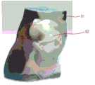
图11意的是肝脏模具的外观图。形状整体为肝实质80,肝实质80使用透光性树脂制成。在肝实质80内部可以确认有肝静脉等的内部结构形状。肝实质80的模具材料采用软质的透光性树脂,为了使肝实质80可以与实物触感更加接近,根据触感等价参数对肝实质80的内部设定内部结构模式。Figure 11 is intended to be the external view of the liver mold. The overall shape is the liver parenchyma 80, and the liver parenchyma 80 is made of translucent resin. Internal structural shapes such as hepatic veins can be confirmed inside the liver parenchyma 80 . The mold material of the liver parenchyma 80 is soft translucent resin. In order to make the liver parenchyma 80 feel closer to the real thing, an internal structural model is set for the inside of the liver parenchyma 80 according to the equivalent parameters of touch.
此外,图12示意的是孕妇腹部模具的外观图。形状整体呈现的是孕妇的腹部切出形状。孕妇的腹部91采用透光性树脂制成。从该模具可以确认孕妇的腹部91及其内部存在胎儿91的样子。关于胎儿的模样,其形状、大小、所处方向、手脚的样子、腹部的形状中的位置关系是一目了然。In addition, Fig. 12 schematically shows the appearance of the pregnant woman's abdomen mold. The overall shape presents the cut-out shape of a pregnant woman's abdomen. The abdomen 91 of the pregnant woman is made of light-transmitting resin. From this mold, the
孕妇的腹部91的模具材料采用软质的透光性树脂,为了使孕妇的腹部91更加接近实物触感,根据触感等价参数对胎儿92的内部设定内部结构模式。The mold material of the
图13和图14示意的是脸部模型以及上半身的外观图。101是头盖骨、102是耳软骨、103是鼻软骨、104是肱、105是心脏。此外,还可确认到肋骨、胸骨、其他骨、牙齿、肺等部位。从头部至上半身、臂膀部位,整体的皮肤以及耳朵,鼻子的软骨部分采用透明树脂进行造型的。此外,内部结构的头盖骨或肋骨,胸骨等骨头部采用硬质树脂造型的。此外,心脏使用软质树脂造型。各个造型模具,为了更加接近实物触感,根据触感等价参数对其内部设定内部结构模式。Figures 13 and 14 show the appearance of the face model and the upper body. 101 is the cranium, 102 is the ear cartilage, 103 is the nasal cartilage, 104 is the humerus, and 105 is the heart. In addition, ribs, sternum, other bones, teeth, lungs, etc. can be confirmed. From the head to the upper body, the arms, the overall skin, ears, and the cartilage of the nose are molded with transparent resin. In addition, bones such as the skull, ribs, and sternum of the internal structure are molded with hard resin. In addition, the heart is molded using soft resin. For each modeling mold, in order to get closer to the physical touch, the internal structure mode is set according to the equivalent parameters of touch.
图15示意的是骨头模具的说明图。如图15所示的骨头模具,骨头表面分有成皮质骨部分110及其内部的松质骨部分(112,114)构成。松质骨部分分成松质骨表面部分112(与皮质骨相邻的一定厚度部分)和松质骨内部部分114。皮质骨部分110是用硬质树脂造型。为了使皮质骨部分110的硬度与实物触感更加接近,根据触感等效参数,在其内部设定内部结构模式。Fig. 15 is an explanatory view showing a bone mold. In the bone mold shown in FIG. 15 , the surface of the bone is divided into a cortical bone part 110 and an inner cancellous bone part ( 112 , 114 ). The cancellous bone portion is divided into a cancellous bone surface portion 112 (thickness portion adjacent to the cortical bone) and a cancellous bone inner portion 114 . The cortical bone portion 110 is molded with hard resin. In order to make the hardness of the cortical bone part 110 closer to the physical touch, an internal structure mode is set inside it according to the equivalent parameters of touch.
此外,松质骨表面部分112设有与皮质骨部分110不同的内部结构模式。此外,松质骨内部114最为脆弱,为了使该触感与实物触感更加接近,根据触感等效参数,设有内部结构参数。如图15(b)所示,松质骨内部114,内部结构模式的原始形状被去除呈格子状。Furthermore, the cancellous bone surface portion 112 is provided with a different internal structural pattern than the cortical bone portion 110 . In addition, the interior 114 of the cancellous bone is the most fragile, and in order to make the touch feel closer to the real touch, internal structural parameters are set according to the equivalent parameters of the touch. As shown in FIG. 15( b ), in the interior 114 of the cancellous bone, the original shape of the internal structural pattern is removed to form a lattice.
目前,使用根据本发明制作的三维造型模具,在与大学附属医院合作中已经证实在医疗领域中可以缩短治疗时间等。当骨盆骨折时实施植入结合手术,对已实施5例进行评级,通常需要6~7小时的手术,可缩短至5~6小时(也就是说缩短1个小时)。缩短手术时间,可减轻患者负担,另外也手术后的恢复也很乐观的。今后,在各种医疗现场,根据本发明制造的三维造型模具应该是可以得到很好的应用。At present, using the three-dimensional modeling mold made according to the present invention, it has been confirmed in the cooperation with the university affiliated hospital that the treatment time etc. can be shortened in the medical field. When the pelvic fracture is combined with implant surgery, 5 cases have been graded, and the operation usually takes 6 to 7 hours, which can be shortened to 5 to 6 hours (that is, shortened by 1 hour). Shortening the operation time can reduce the burden on the patient, and the recovery after the operation is also very optimistic. In the future, in various medical sites, the three-dimensional modeling mold manufactured according to the present invention should be well applied.
工业应用性Industrial Applicability
根据本发明制作的三维造型模具,对知情同意、决定治疗方案、诊疗支援、医学教育或医学研究或一般的教育支持工作都是有帮助的。通过体验模拟触感,作为解剖学或外科学的教材也是很有用的。例如,用类似橡胶的软质树脂制成的血管和实际器官,不仅可以对立体解剖实际用手接触掌握提供支持,设置在拉帕洛手术用训练模拟器内,作为显微镜下的非常好的模拟训练。The three-dimensional modeling mold made according to the present invention is helpful for informed consent, decision on treatment plan, diagnosis and treatment support, medical education or medical research or general educational support work. It is also useful as a teaching material for anatomy or surgery by experiencing simulated touch. For example, blood vessels and actual organs made of rubber-like soft resin, which not only provide support for the actual hand-to-hand grasp of stereoscopic anatomy, are set in the training simulator for Rapallo surgery as a very good simulation under the microscope train.
图中符号说明Explanation of symbols in the figure
11 形状数据生成步骤11 Shape data generation steps
12 形状数据编辑步骤12 Steps for editing shape data
13 表创建步骤13 table creation steps
14 材料类别定义步骤14 Material category definition steps
15 布尔运算步骤15 Boolean operation steps
16 造型步骤16 modeling steps
51 标准文字·记号51 Standard characters and symbols
52 定标器52 scaler
61 骨头模型61 bone model
62 肌肉模型62 muscle models
70 层积物70 laminates
71 轴71 shaft
73 按照一定厚度进行切片后的模具73 The mold after slicing according to a certain thickness
80 肝实质80 liver parenchyma
91 孕妇的腹部91 Pregnant belly
92 胎儿92 fetus
101 头盖骨101 skull
102 耳软骨102 ear cartilage
103 鼻软骨103 Nasal cartilage
104 肱104 brachial
105 心脏105 heart
110 骨皮质部分110 Cortical part of bone
112 松质骨的表面部分112 Superficial part of cancellous bone
114 松质骨内部114 Internal cancellous bone
Claims (20)
Applications Claiming Priority (3)
| Application Number | Priority Date | Filing Date | Title |
|---|---|---|---|
| JP2011078876 | 2011-03-31 | ||
| JP2011-078876 | 2011-03-31 | ||
| PCT/JP2012/002211WO2012132463A1 (en) | 2011-03-31 | 2012-03-30 | Method for manufacturing three-dimensional molded model and support tool for medical treatment, medical training, research, and education |
Publications (2)
| Publication Number | Publication Date |
|---|---|
| CN103153589Atrue CN103153589A (en) | 2013-06-12 |
| CN103153589B CN103153589B (en) | 2015-05-27 |
Family
ID=46930233
Family Applications (1)
| Application Number | Title | Priority Date | Filing Date |
|---|---|---|---|
| CN201280003256.7AExpired - Fee RelatedCN103153589B (en) | 2011-03-31 | 2012-03-30 | Method for manufacturing three-dimensional molded model and support tool for medical treatment, medical training, research, and education |
Country Status (6)
| Country | Link |
|---|---|
| US (1) | US9183764B2 (en) |
| EP (1) | EP2692509A4 (en) |
| JP (1) | JP5239037B2 (en) |
| KR (1) | KR101458729B1 (en) |
| CN (1) | CN103153589B (en) |
| WO (1) | WO2012132463A1 (en) |
Cited By (23)
| Publication number | Priority date | Publication date | Assignee | Title |
|---|---|---|---|---|
| CN103721334A (en)* | 2013-12-12 | 2014-04-16 | 殷琴 | Bronchial catheter manufacturing and application method |
| CN104346491A (en)* | 2014-05-19 | 2015-02-11 | 宜春学院 | Three-dimensional model based on Top-Down machine part expressing method and preparation method of three-dimensional model |
| CN104546222A (en)* | 2015-02-02 | 2015-04-29 | 深圳华明生物科技有限公司 | Artificial cornea and manufacturing method thereof |
| CN104783924A (en)* | 2015-04-24 | 2015-07-22 | 杭州捷诺飞生物科技有限公司 | Breast prosthesis manufacturing method based on three-dimensional printing technology |
| CN104821124A (en)* | 2015-03-16 | 2015-08-05 | 北京大学口腔医学院 | Simulated major salivary gland model device and preparation method thereof |
| CN105096715A (en)* | 2014-05-15 | 2015-11-25 | 朱一帆 | Functional human organ model based on 3D printing technology and manufacturing method |
| CN105390056A (en)* | 2015-10-16 | 2016-03-09 | 南方医科大学 | High-fidelity belly deformation mold for verifying precision of deformable registration algorithm and preparing method thereof |
| CN105719550A (en)* | 2016-04-29 | 2016-06-29 | 王洛 | Tumor-reductive surgery exercise model and manufacturing method thereof |
| CN105877990A (en)* | 2016-05-26 | 2016-08-24 | 上海金怡医疗科技有限公司 | Chest-variable heart pressure effect detection system for mechanical press equipment |
| CN106182838A (en)* | 2016-07-25 | 2016-12-07 | 青岛三帝生物科技有限公司 | The anti-displacement breast prosthesis forming method printed based on 3D and breast prosthesis |
| CN106384554A (en)* | 2016-10-08 | 2017-02-08 | 上海光韵达数字医疗科技有限公司 | Operation training model and manufacturing method thereof and operation navigation system |
| CN106710406A (en)* | 2015-07-21 | 2017-05-24 | 上海微创医疗器械(集团)有限公司 | Vascular model and manufacturing method thereof, and spraying equipment |
| CN106827500A (en)* | 2017-01-19 | 2017-06-13 | 西安交通大学 | A kind of skull bone substitute multiple degrees of freedom 3D printing method |
| CN107209647A (en)* | 2015-01-29 | 2017-09-26 | 惠普发展公司有限责任合伙企业 | Handle the object for printing |
| CN107657880A (en)* | 2016-07-26 | 2018-02-02 | 上海光韵达数字医疗科技有限公司 | A kind of manufacturing process of human organ model |
| CN104821124B (en)* | 2015-03-16 | 2018-02-09 | 北京大学口腔医学院 | One kind emulation major salivary glands model equipment and preparation method |
| CN108288427A (en)* | 2018-01-06 | 2018-07-17 | 无锡市第二人民医院 | A kind of production method of 3 D-printing transtracheal mirror lymph node puncture training pattern |
| CN108697373A (en)* | 2016-02-17 | 2018-10-23 | 皇家飞利浦有限公司 | Physics 3D anatomical structure models manufacture |
| CN108735061A (en)* | 2017-04-13 | 2018-11-02 | 中南大学 | The method for preparing transparent soft human body viscera model based on 3D printing |
| CN109003324A (en)* | 2017-06-06 | 2018-12-14 | 富士施乐株式会社 | Path data generating means and method for three-dimensional modeling |
| CN109637315A (en)* | 2018-12-14 | 2019-04-16 | 王成燕 | A kind of prostate cancer simulation model and preparation method thereof |
| TWI709115B (en)* | 2019-09-27 | 2020-11-01 | 國立成功大學 | Method of fabricating light-transmitting blood vessel model |
| CN112297436A (en)* | 2019-07-25 | 2021-02-02 | 富士施乐株式会社 | Information processing apparatus, recording medium, and information processing method |
Families Citing this family (98)
| Publication number | Priority date | Publication date | Assignee | Title |
|---|---|---|---|---|
| EP1900732A4 (en) | 2005-06-24 | 2009-11-18 | Toyama Chemical Co Ltd | NOVEL HETEROCYCLIC NITROGENIC COMPOUND AND SALT THEREOF |
| EP2622594B1 (en) | 2010-10-01 | 2018-08-22 | Applied Medical Resources Corporation | Portable laparoscopic trainer |
| JP5783627B2 (en)* | 2011-04-26 | 2015-09-24 | 国立大学法人 岡山大学 | Human body modeling system |
| US9218753B2 (en) | 2011-10-21 | 2015-12-22 | Applied Medical Resources Corporation | Simulated tissue structure for surgical training |
| KR101953187B1 (en) | 2011-12-20 | 2019-02-28 | 어플라이드 메디컬 리소시스 코포레이션 | Advanced surgical simulation |
| CA2880277A1 (en) | 2012-08-03 | 2014-02-06 | Applied Medical Resources Corporation | Simulated stapling and energy based ligation for surgical training |
| JP2015532450A (en) | 2012-09-26 | 2015-11-09 | アプライド メディカル リソーシーズ コーポレイション | Surgical training model for laparoscopic procedures |
| US9959786B2 (en) | 2012-09-27 | 2018-05-01 | Applied Medical Resources Corporation | Surgical training model for laparoscopic procedures |
| WO2014052612A1 (en) | 2012-09-27 | 2014-04-03 | Applied Medical Resources Corporation | Surgical training model for laparoscopic procedures |
| US10679520B2 (en) | 2012-09-27 | 2020-06-09 | Applied Medical Resources Corporation | Surgical training model for laparoscopic procedures |
| EP2901439A1 (en) | 2012-09-28 | 2015-08-05 | Applied Medical Resources Corporation | Surgical training model for laparoscopic procedures |
| CA2885314C (en) | 2012-09-28 | 2021-01-19 | Applied Medical Resources Corporation | Surgical training model for transluminal laparoscopic procedures |
| US9226797B2 (en)* | 2012-11-04 | 2016-01-05 | Miba Medical Inc. | Computer aided implantation of body implants |
| JP2016503685A (en) | 2012-12-28 | 2016-02-08 | サイバーハート, インコーポレイテッド | Planning of kidney treatment by radiosurgery based on blood-tissue surface |
| AU2014224004B2 (en) | 2013-03-01 | 2018-04-05 | Applied Medical Resources Corporation | Advanced surgical simulation constructions and methods |
| US11048829B2 (en)* | 2013-03-15 | 2021-06-29 | Kemeera Llc | 3D printing systems and methods for fabricating injection molds |
| CA3139494A1 (en) | 2013-05-15 | 2014-11-20 | Applied Medical Resources Corporation | Hernia model |
| KR101477909B1 (en) | 2013-06-03 | 2014-12-30 | 주식회사 만도 | Radar apparatus and antenna apparatus |
| KR102607634B1 (en) | 2013-06-18 | 2023-11-29 | 어플라이드 메디컬 리소시스 코포레이션 | Gallbladder model for teaching and practicing surgical procedures |
| US10198966B2 (en) | 2013-07-24 | 2019-02-05 | Applied Medical Resources Corporation | Advanced first entry model for surgical simulation |
| AU2014293036B2 (en) | 2013-07-24 | 2017-12-21 | Applied Medical Resources Corporation | First entry model |
| JP6331324B2 (en)* | 2013-10-21 | 2018-05-30 | 大日本印刷株式会社 | Method and system for adding and confirming unique code for output object output from 3D printer |
| JP5620594B1 (en)* | 2014-01-31 | 2014-11-05 | 丸井 智敬 | A three-dimensional structure produced by a 3D printer, an information transmission system using the three-dimensional structure. |
| ES2891756T3 (en) | 2014-03-26 | 2022-01-31 | Applied Med Resources | Simulated dissectable tissue |
| JP6579746B2 (en)* | 2014-04-16 | 2019-09-25 | 株式会社ミマキエンジニアリング | Manufacturing method of three-dimensional structure and three-dimensional structure |
| JP6425916B2 (en)* | 2014-05-16 | 2018-11-21 | キヤノンメディカルシステムズ株式会社 | PROCESSING APPARATUS, IMAGE PROCESSING METHOD, PROGRAM, AND FORMING APPARATUS |
| JP5759055B1 (en)* | 2014-05-26 | 2015-08-05 | サンアロー株式会社 | Organ model |
| KR101635188B1 (en)* | 2014-07-11 | 2016-07-04 | (주)쓰리디스토리 | Unborn child sculpture printing service system and a method thereof |
| US20160070822A1 (en)* | 2014-09-09 | 2016-03-10 | Primesmith Oy | Method, Apparatus and Computer Program Code for Design and Visualization of a Physical Object |
| CN104269092B (en)* | 2014-09-19 | 2016-05-25 | 成都学创伟业科技有限公司 | The auxiliary skeleton model preparation method of operation |
| JP6457785B2 (en)* | 2014-11-11 | 2019-01-23 | 株式会社ファソテック | Biological texture model production method and biological texture model production program |
| WO2016077636A1 (en)* | 2014-11-13 | 2016-05-19 | The Board Of Trustees Of The Leland Stanford Junior University | Miniaturized phantoms for quantitative image analysis and quality control |
| AU2015347077B2 (en) | 2014-11-13 | 2021-08-12 | Applied Medical Resources Corporation | Simulated tissue models and methods |
| KR102586607B1 (en) | 2015-02-19 | 2023-10-10 | 어플라이드 메디컬 리소시스 코포레이션 | Simulated tissue structures and methods |
| US10842585B2 (en) | 2015-03-24 | 2020-11-24 | University Of Tsukuba | Model, manufacturing system, information processing device, manufacturing method, information processing method, program, and recording medium |
| KR101828033B1 (en)* | 2015-04-02 | 2018-02-12 | 울산대학교 산학협력단 | Apparatus for modeling three dimensional shape of internal body and method |
| JP6213516B2 (en)* | 2015-04-16 | 2017-10-18 | キヤノンマーケティングジャパン株式会社 | MEDICAL IMAGE MANAGEMENT SYSTEM, ITS CONTROL METHOD, AND PROGRAM, AND INFORMATION PROCESSING DEVICE, ITS CONTROL METHOD, AND PROGRAM |
| US10864659B1 (en)* | 2015-05-12 | 2020-12-15 | Jacques Zaneveld | Methods and systems for creating anatomical models |
| US10350833B1 (en) | 2015-05-12 | 2019-07-16 | Jacques Zaneveld | Methods and systems for creating anatomical models |
| ES2716924T3 (en) | 2015-05-14 | 2019-06-18 | Applied Med Resources | Synthetic tissue structures for training and electrosurgical stimulation |
| JP6821324B2 (en)* | 2015-05-22 | 2021-01-27 | キヤノン株式会社 | Image processing equipment, image processing methods and programs |
| JP6360003B2 (en)* | 2015-06-04 | 2018-07-18 | 満 入江 | Medical three-dimensional model and manufacturing method thereof |
| AU2016276771B2 (en) | 2015-06-09 | 2022-02-03 | Applied Medical Resources Corporation | Hysterectomy model |
| WO2017010299A1 (en)* | 2015-07-14 | 2017-01-19 | 富士フイルム株式会社 | Method for producing vascular model of organism and method for producing model of organ of organism |
| WO2017010298A1 (en)* | 2015-07-14 | 2017-01-19 | 富士フイルム株式会社 | Method for producing model of organ of organism |
| KR102697097B1 (en) | 2015-07-16 | 2024-08-21 | 어플라이드 메디컬 리소시스 코포레이션 | Simulated exciseable tissue |
| WO2017015438A1 (en) | 2015-07-22 | 2017-01-26 | Applied Medical Resources Corporation | Appendectomy model |
| CN105160977A (en)* | 2015-08-05 | 2015-12-16 | 成都嘉逸科技有限公司 | A 3D teaching system of human anatomy |
| JP6526516B2 (en)* | 2015-08-06 | 2019-06-05 | キヤノンメディカルシステムズ株式会社 | Medical image processing device |
| JP6613727B2 (en)* | 2015-08-28 | 2019-12-04 | 大日本印刷株式会社 | Data reduction device for 3D object modeling |
| KR20250099424A (en) | 2015-10-02 | 2025-07-01 | 어플라이드 메디컬 리소시스 코포레이션 | Hysterectomy Model |
| ES2608203B1 (en)* | 2015-10-05 | 2018-01-11 | Servicio Andaluz De Salud | Customizable device for intervention simulation |
| US10733911B2 (en) | 2015-10-14 | 2020-08-04 | Humanetics Innovative Solutions, Inc. | Three-dimensional ribs and method of three-dimensional printing of ribs for crash test dummy |
| US10395561B2 (en) | 2015-12-07 | 2019-08-27 | Humanetics Innovative Solutions, Inc. | Three-dimensionally printed internal organs for crash test dummy |
| JP6480311B2 (en)* | 2015-11-18 | 2019-03-06 | 富士フイルム株式会社 | Support member design apparatus, method, program, structure forming apparatus, and structure manufacturing method |
| JP6886975B2 (en) | 2015-11-20 | 2021-06-16 | アプライド メディカル リソーシーズ コーポレイション | Simulated incisable tissue |
| FR3047162B1 (en)* | 2016-02-03 | 2018-02-16 | Institut De Radioprotection Et De Surete Nucleaire | THYROIDIAN FANTOM, METHOD FOR MANUFACTURING SAME, GLOBAL FANTOM COMPRISING SUCH A THYROID FANTOME AND CORRESPONDING FANTOM FAMILIES |
| WO2017138558A1 (en)* | 2016-02-09 | 2017-08-17 | パナソニックヘルスケアホールディングス株式会社 | Three-dimensional image processing device, three-dimensional image processing method, and three-dimensional image processing program |
| CN105997313B (en)* | 2016-05-03 | 2017-10-27 | 柴凡 | External prosthese of 3D printing breast and preparation method thereof |
| JP6918443B2 (en)* | 2016-05-09 | 2021-08-11 | キヤノンメディカルシステムズ株式会社 | Medical information processing device |
| JPWO2017216967A1 (en)* | 2016-06-17 | 2019-04-04 | オリンパス株式会社 | Simulated living body, production apparatus for laminated resin structure, and production method for laminated resin structure |
| ES2946810T3 (en) | 2016-06-27 | 2023-07-26 | Applied Med Resources | simulated abdominal wall |
| EP3273430A1 (en)* | 2016-07-19 | 2018-01-24 | Humanetics Innovative Solutions, Inc. | Three-dimensional components and method of three-dimensional printing of components for crash test dummy |
| ES2769080T5 (en)* | 2016-07-19 | 2022-11-07 | Humanetics Innovative Solutions Inc | 3D printed meat and internal organs for crash test dummies |
| US12076912B2 (en) | 2016-10-18 | 2024-09-03 | Autodesk, Inc. | Systems and methods of cellular-hull infill structure generation for additive manufacturing |
| US10885407B2 (en)* | 2016-11-23 | 2021-01-05 | Simbionix Ltd. | Method and system for three-dimensional print oriented image segmentation |
| JP6864823B2 (en)* | 2017-01-18 | 2021-04-28 | 富士フイルムビジネスイノベーション株式会社 | Information processing equipment, information processing programs and 3D modeling systems |
| ES3004046T3 (en) | 2017-02-14 | 2025-03-11 | Applied Med Resources | Laparoscopic training system |
| US10556418B2 (en)* | 2017-02-14 | 2020-02-11 | Autodesk, Inc. | Systems and methods of open-cell internal structure and closed-cell internal structure generation for additive manufacturing |
| US10847057B2 (en) | 2017-02-23 | 2020-11-24 | Applied Medical Resources Corporation | Synthetic tissue structures for electrosurgical training and simulation |
| KR101918119B1 (en)* | 2017-03-10 | 2019-02-08 | 주식회사 모르페우스 | Method, system and non-transitory computer-readable recording medium for estimating anatomical layer of face |
| WO2018164329A1 (en)* | 2017-03-10 | 2018-09-13 | 주식회사 모르페우스 | Method and system for providing information on face by using anatomical layer, and non-transitory computer-readable recording medium |
| US10933579B2 (en) | 2017-03-10 | 2021-03-02 | Prellis Biologics, Inc. | Methods and systems for printing biological material |
| US11085018B2 (en) | 2017-03-10 | 2021-08-10 | Prellis Biologics, Inc. | Three-dimensional printed organs, devices, and matrices |
| CN111032687A (en) | 2017-05-25 | 2020-04-17 | 普瑞利思生物制品公司 | Three-dimensional printed organs, devices and substrates |
| WO2018222779A1 (en)* | 2017-05-30 | 2018-12-06 | Dignity Health | Systems and methods for constructing a synthetic anatomical model with predetermined anatomic, biomechanical, and physiological properties |
| GB2564155B (en) | 2017-07-06 | 2020-10-14 | Sony Interactive Entertainment Inc | System and method of 3D print modelling |
| JP2019115984A (en)* | 2017-12-26 | 2019-07-18 | 株式会社びーふる | Three dimentional formed object and manufacturing method therefor |
| CN108437469A (en)* | 2018-03-12 | 2018-08-24 | 保定翰阳科技有限公司 | A kind of individual customized type method based on 3D technology |
| WO2019204611A1 (en)* | 2018-04-18 | 2019-10-24 | Arizona Board Of Regents On Behalf Of The University Of Arizona | Mixed-reality endoscope and surgical tools with haptic feedback for integrated virtual-reality visual and haptic surgical simulation |
| KR102078159B1 (en)* | 2018-04-27 | 2020-02-17 | 한림대학교 산학협력단 | System for manufacturing Breast Implant using 3D Printer |
| JP6637116B2 (en)* | 2018-06-21 | 2020-01-29 | 社会福祉法人兵庫県社会福祉事業団 | Medical three-dimensional model and manufacturing method thereof |
| WO2020028436A1 (en) | 2018-07-31 | 2020-02-06 | Prellis Biologics, Inc. | Optically-induced auto-encapsulation |
| CN109345932B (en)* | 2018-08-29 | 2021-09-28 | 中国科学院自动化研究所 | Medical model based on 3D printing and manufacturing method thereof |
| US11564848B2 (en)* | 2018-09-27 | 2023-01-31 | Exosseus Llc | Method of producing a custom-fit orthopedic immobilization device |
| JP7360125B2 (en)* | 2018-10-10 | 2023-10-12 | Jsr株式会社 | Information processing devices, systems, object manufacturing methods, information processing methods, and programs |
| US10315353B1 (en) | 2018-11-13 | 2019-06-11 | SmileDirectClub LLC | Systems and methods for thermoforming dental aligners |
| US11007042B2 (en) | 2019-02-06 | 2021-05-18 | Sdc U.S. Smilepay Spv | Systems and methods for marking models for dental aligner fabrication |
| US10482192B1 (en) | 2019-02-12 | 2019-11-19 | SmileDirectClub LLC | Systems and methods for selecting and marking a location on a dental aligner |
| US20220189101A1 (en)* | 2019-08-16 | 2022-06-16 | Hewlett-Packard Development Company, L.P. | Three-dimensional object marking |
| FR3106232B1 (en)* | 2020-01-13 | 2022-08-26 | Virtualisurg | DRIVE DEVICE |
| JP7543767B2 (en)* | 2020-08-07 | 2024-09-03 | 株式会社リコー | Organ Models and Training Systems |
| JP7580688B2 (en)* | 2020-08-26 | 2024-11-12 | 大輝 吉本 | Method for creating 3D shape data for a solid model, method for manufacturing a solid model, and method for manufacturing a solid model |
| JP7092841B2 (en)* | 2020-10-13 | 2022-06-28 | 昌平 岩部 | Pediatric skeleton model |
| KR102293201B1 (en) | 2020-11-20 | 2021-08-25 | 이마고웍스 주식회사 | Method of generating three dimensional model for digital dentistry using virtual bridge based multi input boolean operation |
| WO2022265345A1 (en)* | 2021-06-18 | 2022-12-22 | 가톨릭대학교 산학협력단 | System for outputting model of fetal image by using 3d printer, and device and method which generate file for 3d printer output |
| JP2023046998A (en)* | 2021-09-24 | 2023-04-05 | 学校法人慈恵大学 | Program, information processing device, method, modeling system, and display control device |
| CN116851777B (en)* | 2023-06-21 | 2025-07-15 | 华南理工大学 | SLM multi-scale digital tissue structure customization method based on ultrasonic compounding |
Citations (6)
| Publication number | Priority date | Publication date | Assignee | Title |
|---|---|---|---|---|
| JP2001030696A (en)* | 1999-07-21 | 2001-02-06 | Sony Corp | Modeling apparatus and modeling method |
| KR20030061580A (en)* | 2002-01-15 | 2003-07-22 | 유용석 | Inplant finite element method |
| JP2007021705A (en)* | 2005-07-21 | 2007-02-01 | Roland Dg Corp | 3D modeling method and 3D modeling apparatus |
| CN101594817A (en)* | 2007-04-24 | 2009-12-02 | 奥林巴斯医疗株式会社 | Medical image processing device and medical image processing method |
| JP2010042065A (en)* | 2008-08-08 | 2010-02-25 | Toshiba Corp | Medical image processor, processing method |
| JP2010184090A (en)* | 2009-02-13 | 2010-08-26 | Osaka Univ | Method and device for generating three-dimensional dentition image |
Family Cites Families (25)
| Publication number | Priority date | Publication date | Assignee | Title |
|---|---|---|---|---|
| JPH0511689A (en) | 1991-06-29 | 1993-01-22 | Sekisui Chem Co Ltd | Manufacture of solid model of internal organ or the like |
| BE1008372A3 (en)* | 1994-04-19 | 1996-04-02 | Materialise Nv | METHOD FOR MANUFACTURING A perfected MEDICAL MODEL BASED ON DIGITAL IMAGE INFORMATION OF A BODY. |
| KR20010038344A (en)* | 1999-10-25 | 2001-05-15 | 김남국 | Method and Apparatus for Forming Objects Similar to Things in Human Body |
| AU2002239810A1 (en)* | 2001-01-02 | 2002-07-16 | The Charles Stark Draper Laboratory, Inc. | Tissue engineering of three-dimensional vascularized using microfabricated polymer assembly technology |
| JP2003011237A (en) | 2001-07-03 | 2003-01-15 | Kuraray Co Ltd | Manufacturing method for three-dimensional objects |
| US20050186361A1 (en)* | 2002-05-10 | 2005-08-25 | Toshio Fukuda | Three-dimensional model |
| JP4575295B2 (en)* | 2003-07-31 | 2010-11-04 | 独立行政法人理化学研究所 | Artificial bone forming method by powder lamination method |
| JP4193944B2 (en) | 2003-10-20 | 2008-12-10 | 独立行政法人産業技術総合研究所 | Method for producing three-dimensional structure and three-dimensional structure |
| EP1690206A4 (en)* | 2003-11-14 | 2008-07-02 | Univ Drexel | METHODS AND DEVICES FOR COMPUTERIZED TISSUE ENGINEERING FOR MODELING, DESIGNING AND REALIZING IN FREE FORM OF TISSUE SCAFFOLDING, CONSTRUCTION AND DEVICES |
| WO2006088946A2 (en)* | 2005-02-14 | 2006-08-24 | Mayo Medical Ventures | Tissue support structure |
| JP2006343434A (en)* | 2005-06-07 | 2006-12-21 | Toin Gakuen | Method for manufacturing entity model of human body diseased part |
| WO2007045000A2 (en)* | 2005-10-14 | 2007-04-19 | Vantus Technology Corporation | Personal fit medical implants and orthopedic surgical instruments and methods for making |
| US7718351B2 (en)* | 2006-03-14 | 2010-05-18 | Agency For Science, Technology & Research | Three-dimensional fabrication of biocompatible structures in anatomical shapes and dimensions for tissue engineering and organ replacement |
| WO2008069759A1 (en)* | 2006-12-05 | 2008-06-12 | Nanyang Technological University | Manufacturing three-dimensional scaffolds using electrospinning at low temperatures |
| WO2008075303A1 (en)* | 2006-12-21 | 2008-06-26 | Koninklijke Philips Electronics N.V. | Anatomically and functionally accurate soft tissue phantoms and method for generating same |
| CN101981608B (en) | 2008-03-28 | 2014-08-27 | 泰尔茂株式会社 | Three-dimensional model of biological tissue and its manufacturing method |
| EP2110079A1 (en) | 2008-04-14 | 2009-10-21 | DKFZ Deutsches Krebsforschungszentrum, Stiftung des Öffentlichen Rechts | Method to derive anatomical and/or pathological structures from data of imaging technologies |
| WO2010060080A1 (en)* | 2008-11-24 | 2010-05-27 | Immunotrex Corporation | Three dimensional tissue generation |
| KR100994935B1 (en)* | 2008-11-26 | 2010-11-19 | 한국과학기술원 | Manufacturing Method of Large 3D Sculptures by Stacking Laminated Surfaces |
| US9161822B2 (en)* | 2008-11-26 | 2015-10-20 | In'tech Industries, Inc. | Conformance model |
| US20110136162A1 (en)* | 2009-08-31 | 2011-06-09 | Drexel University | Compositions and Methods for Functionalized Patterning of Tissue Engineering Substrates Including Bioprinting Cell-Laden Constructs for Multicompartment Tissue Chambers |
| US8691974B2 (en)* | 2009-09-28 | 2014-04-08 | Virginia Tech Intellectual Properties, Inc. | Three-dimensional bioprinting of biosynthetic cellulose (BC) implants and scaffolds for tissue engineering |
| US8260589B1 (en)* | 2009-12-24 | 2012-09-04 | Indian Institute Of Technology Madras | Methods and systems for modeling a physical object |
| RU2560393C2 (en)* | 2010-10-21 | 2015-08-20 | Органово, Инк. | Devices, systems and methods of making fabric |
| US8579620B2 (en)* | 2011-03-02 | 2013-11-12 | Andy Wu | Single-action three-dimensional model printing methods |
- 2012
- 2012-03-30WOPCT/JP2012/002211patent/WO2012132463A1/enactiveApplication Filing
- 2012-03-30USUS13/823,652patent/US9183764B2/ennot_activeExpired - Fee Related
- 2012-03-30EPEP12765020.8Apatent/EP2692509A4/ennot_activeCeased
- 2012-03-30KRKR1020137007081Apatent/KR101458729B1/ennot_activeExpired - Fee Related
- 2012-03-30CNCN201280003256.7Apatent/CN103153589B/ennot_activeExpired - Fee Related
- 2012-03-30JPJP2012538117Apatent/JP5239037B2/ennot_activeExpired - Fee Related
Patent Citations (6)
| Publication number | Priority date | Publication date | Assignee | Title |
|---|---|---|---|---|
| JP2001030696A (en)* | 1999-07-21 | 2001-02-06 | Sony Corp | Modeling apparatus and modeling method |
| KR20030061580A (en)* | 2002-01-15 | 2003-07-22 | 유용석 | Inplant finite element method |
| JP2007021705A (en)* | 2005-07-21 | 2007-02-01 | Roland Dg Corp | 3D modeling method and 3D modeling apparatus |
| CN101594817A (en)* | 2007-04-24 | 2009-12-02 | 奥林巴斯医疗株式会社 | Medical image processing device and medical image processing method |
| JP2010042065A (en)* | 2008-08-08 | 2010-02-25 | Toshiba Corp | Medical image processor, processing method |
| JP2010184090A (en)* | 2009-02-13 | 2010-08-26 | Osaka Univ | Method and device for generating three-dimensional dentition image |
Cited By (26)
| Publication number | Priority date | Publication date | Assignee | Title |
|---|---|---|---|---|
| CN103721334B (en)* | 2013-12-12 | 2016-03-09 | 殷琴 | A kind of manufacture method of bronchial catheter |
| CN103721334A (en)* | 2013-12-12 | 2014-04-16 | 殷琴 | Bronchial catheter manufacturing and application method |
| CN105096715A (en)* | 2014-05-15 | 2015-11-25 | 朱一帆 | Functional human organ model based on 3D printing technology and manufacturing method |
| CN104346491A (en)* | 2014-05-19 | 2015-02-11 | 宜春学院 | Three-dimensional model based on Top-Down machine part expressing method and preparation method of three-dimensional model |
| CN107209647A (en)* | 2015-01-29 | 2017-09-26 | 惠普发展公司有限责任合伙企业 | Handle the object for printing |
| CN104546222A (en)* | 2015-02-02 | 2015-04-29 | 深圳华明生物科技有限公司 | Artificial cornea and manufacturing method thereof |
| CN104821124A (en)* | 2015-03-16 | 2015-08-05 | 北京大学口腔医学院 | Simulated major salivary gland model device and preparation method thereof |
| CN104821124B (en)* | 2015-03-16 | 2018-02-09 | 北京大学口腔医学院 | One kind emulation major salivary glands model equipment and preparation method |
| CN104783924B (en)* | 2015-04-24 | 2017-01-18 | 杭州捷诺飞生物科技有限公司 | Breast prosthesis manufacturing method based on three-dimensional printing technology |
| CN104783924A (en)* | 2015-04-24 | 2015-07-22 | 杭州捷诺飞生物科技有限公司 | Breast prosthesis manufacturing method based on three-dimensional printing technology |
| CN106710406A (en)* | 2015-07-21 | 2017-05-24 | 上海微创医疗器械(集团)有限公司 | Vascular model and manufacturing method thereof, and spraying equipment |
| CN105390056A (en)* | 2015-10-16 | 2016-03-09 | 南方医科大学 | High-fidelity belly deformation mold for verifying precision of deformable registration algorithm and preparing method thereof |
| CN108697373A (en)* | 2016-02-17 | 2018-10-23 | 皇家飞利浦有限公司 | Physics 3D anatomical structure models manufacture |
| CN105719550A (en)* | 2016-04-29 | 2016-06-29 | 王洛 | Tumor-reductive surgery exercise model and manufacturing method thereof |
| CN105719550B (en)* | 2016-04-29 | 2019-06-28 | 王洛 | Tumor resection practices model and its manufacturing method |
| CN105877990A (en)* | 2016-05-26 | 2016-08-24 | 上海金怡医疗科技有限公司 | Chest-variable heart pressure effect detection system for mechanical press equipment |
| CN106182838A (en)* | 2016-07-25 | 2016-12-07 | 青岛三帝生物科技有限公司 | The anti-displacement breast prosthesis forming method printed based on 3D and breast prosthesis |
| CN107657880A (en)* | 2016-07-26 | 2018-02-02 | 上海光韵达数字医疗科技有限公司 | A kind of manufacturing process of human organ model |
| CN106384554A (en)* | 2016-10-08 | 2017-02-08 | 上海光韵达数字医疗科技有限公司 | Operation training model and manufacturing method thereof and operation navigation system |
| CN106827500A (en)* | 2017-01-19 | 2017-06-13 | 西安交通大学 | A kind of skull bone substitute multiple degrees of freedom 3D printing method |
| CN108735061A (en)* | 2017-04-13 | 2018-11-02 | 中南大学 | The method for preparing transparent soft human body viscera model based on 3D printing |
| CN109003324A (en)* | 2017-06-06 | 2018-12-14 | 富士施乐株式会社 | Path data generating means and method for three-dimensional modeling |
| CN108288427A (en)* | 2018-01-06 | 2018-07-17 | 无锡市第二人民医院 | A kind of production method of 3 D-printing transtracheal mirror lymph node puncture training pattern |
| CN109637315A (en)* | 2018-12-14 | 2019-04-16 | 王成燕 | A kind of prostate cancer simulation model and preparation method thereof |
| CN112297436A (en)* | 2019-07-25 | 2021-02-02 | 富士施乐株式会社 | Information processing apparatus, recording medium, and information processing method |
| TWI709115B (en)* | 2019-09-27 | 2020-11-01 | 國立成功大學 | Method of fabricating light-transmitting blood vessel model |
Also Published As
| Publication number | Publication date |
|---|---|
| CN103153589B (en) | 2015-05-27 |
| EP2692509A4 (en) | 2014-10-22 |
| JPWO2012132463A1 (en) | 2014-07-24 |
| JP5239037B2 (en) | 2013-07-17 |
| KR20130063531A (en) | 2013-06-14 |
| WO2012132463A1 (en) | 2012-10-04 |
| US9183764B2 (en) | 2015-11-10 |
| KR101458729B1 (en) | 2014-11-05 |
| EP2692509A1 (en) | 2014-02-05 |
| US20140017651A1 (en) | 2014-01-16 |
Similar Documents
| Publication | Publication Date | Title |
|---|---|---|
| CN103153589B (en) | Method for manufacturing three-dimensional molded model and support tool for medical treatment, medical training, research, and education | |
| Haleem et al. | 3D scanning applications in medical field: a literature-based review | |
| Ebert et al. | Getting in touch—3D printing in forensic imaging | |
| US20180168730A1 (en) | System and method for medical procedure planning | |
| CN109345932B (en) | Medical model based on 3D printing and manufacturing method thereof | |
| Żukowska et al. | Additive manufacturing of 3D anatomical models—review of processes, materials and applications | |
| US10864659B1 (en) | Methods and systems for creating anatomical models | |
| Tokgöz et al. | Cosmetic and reconstructive facial plastic surgery related simulation and optimization efforts | |
| Raos et al. | Additive manufacturing of medical models-applications in rhinology | |
| JP7437826B2 (en) | Organ model for surgical practice | |
| JP6457785B2 (en) | Biological texture model production method and biological texture model production program | |
| JP2017096997A (en) | Manufacturing method of human body model (simulator), human body model | |
| Lambrecht et al. | Haptic model fabrication for undergraduate and postgraduate teaching | |
| CN115188232A (en) | Medical teaching comprehensive training system and method based on MR-3D printing technology | |
| Riedle et al. | Generation of 3D silicone models of anatomic soft tissue structures-A comparison of direct 3D printing and molding techniques | |
| Sampath et al. | 3D Printing in Medicine | |
| Gargiulo et al. | Use of 3 dimensional models and navigation system in preparation for brain surgery | |
| Wyleżoł et al. | Computer aided and 3D printing in pre-operative planning of orbital reconstruction surgery | |
| Tsioukas et al. | The long and winding road from CT and MRI images to 3D models | |
| WO2015037978A1 (en) | An anatomical model | |
| Żukowska et al. | Methodology of the Rapid Manufacturing of an Individualized Anatomical Model of the Tongue with a Tumor for the Preparation of an Organ Reconstruction Operation | |
| TW201933299A (en) | Medical preoperative simulation model and method for molding the same capable of accurately reflecting the pathological characteristics of a patient's organs | |
| Macko et al. | 3D technology implementation in medicine | |
| Treichel et al. | Planning ortheses using customized biomodels for the treatment of laminitis in equines | |
| JP2023046998A (en) | Program, information processing device, method, modeling system, and display control device |
Legal Events
| Date | Code | Title | Description |
|---|---|---|---|
| C06 | Publication | ||
| PB01 | Publication | ||
| C10 | Entry into substantive examination | ||
| SE01 | Entry into force of request for substantive examination | ||
| C14 | Grant of patent or utility model | ||
| GR01 | Patent grant | ||
| CF01 | Termination of patent right due to non-payment of annual fee | ||
| CF01 | Termination of patent right due to non-payment of annual fee | Granted publication date:20150527 |