CN103153184A - Devices and methods for respiratory variation monitoring by measurement of respiratory volumes, motion and variability - Google Patents
Devices and methods for respiratory variation monitoring by measurement of respiratory volumes, motion and variabilityDownload PDFInfo
- Publication number
- CN103153184A CN103153184ACN2011800393984ACN201180039398ACN103153184ACN 103153184 ACN103153184 ACN 103153184ACN 2011800393984 ACN2011800393984 ACN 2011800393984ACN 201180039398 ACN201180039398 ACN 201180039398ACN 103153184 ACN103153184 ACN 103153184A
- Authority
- CN
- China
- Prior art keywords
- patient
- impedance
- respiratory
- breathing
- volume
- Prior art date
- Legal status (The legal status is an assumption and is not a legal conclusion. Google has not performed a legal analysis and makes no representation as to the accuracy of the status listed.)
- Granted
Links
- 230000000241respiratory effectEffects0.000titleclaimsabstractdescription171
- 238000000034methodMethods0.000titleclaimsabstractdescription99
- 238000005259measurementMethods0.000titleclaimsdescription183
- 230000033001locomotionEffects0.000titleclaimsdescription73
- 238000012544monitoring processMethods0.000titleclaimsdescription67
- 238000002847impedance measurementMethods0.000claimsabstractdescription25
- 238000001914filtrationMethods0.000claimsabstractdescription5
- 230000029058respiratory gaseous exchangeEffects0.000claimsdescription233
- 210000000038chestAnatomy0.000claimsdescription93
- 239000000523sampleSubstances0.000claimsdescription65
- 210000004072lungAnatomy0.000claimsdescription64
- 238000004458analytical methodMethods0.000claimsdescription57
- 230000008859changeEffects0.000claimsdescription42
- 210000001015abdomenAnatomy0.000claimsdescription39
- 238000013125spirometryMethods0.000claimsdescription35
- 238000009423ventilationMethods0.000claimsdescription35
- 229940079593drugDrugs0.000claimsdescription34
- 239000003814drugSubstances0.000claimsdescription34
- 230000000694effectsEffects0.000claimsdescription26
- 208000037265diseases, disorders, signs and symptomsDiseases0.000claimsdescription21
- 238000003384imaging methodMethods0.000claimsdescription21
- 201000010099diseaseDiseases0.000claimsdescription20
- 230000036387respiratory rateEffects0.000claimsdescription18
- 238000001356surgical procedureMethods0.000claimsdescription17
- 206010002091AnaesthesiaDiseases0.000claimsdescription16
- 208000019693Lung diseaseDiseases0.000claimsdescription16
- 230000003187abdominal effectEffects0.000claimsdescription15
- 230000037005anaesthesiaEffects0.000claimsdescription15
- 238000002489impedance cardiographyMethods0.000claimsdescription15
- 230000003434inspiratory effectEffects0.000claimsdescription14
- 210000000115thoracic cavityAnatomy0.000claimsdescription14
- 238000004364calculation methodMethods0.000claimsdescription13
- 238000002560therapeutic procedureMethods0.000claimsdescription12
- 238000002106pulse oximetryMethods0.000claimsdescription11
- 208000004756Respiratory InsufficiencyDiseases0.000claimsdescription10
- 230000001225therapeutic effectEffects0.000claimsdescription9
- 230000002596correlated effectEffects0.000claimsdescription8
- 238000001514detection methodMethods0.000claimsdescription8
- 230000036592analgesiaEffects0.000claimsdescription6
- 230000006378damageEffects0.000claimsdescription6
- 201000004193respiratory failureDiseases0.000claimsdescription6
- 208000027418Wounds and injuryDiseases0.000claimsdescription5
- 230000003044adaptive effectEffects0.000claimsdescription5
- 208000014674injuryDiseases0.000claimsdescription5
- 238000011871bio-impedance analysisMethods0.000claimsdescription4
- 238000009826distributionMethods0.000claimsdescription4
- 238000011156evaluationMethods0.000claimsdescription4
- 206010051093Cardiopulmonary failureDiseases0.000claimsdescription3
- 230000000202analgesic effectEffects0.000claimsdescription3
- 238000002483medicationMethods0.000claims4
- 230000002708enhancing effectEffects0.000claims1
- 230000000977initiatory effectEffects0.000claims1
- 230000003750conditioning effectEffects0.000abstract1
- 238000012360testing methodMethods0.000description41
- CURLTUGMZLYLDI-UHFFFAOYSA-NCarbon dioxideChemical compoundO=C=OCURLTUGMZLYLDI-UHFFFAOYSA-N0.000description38
- 230000000747cardiac effectEffects0.000description32
- 238000004422calculation algorithmMethods0.000description28
- 210000002417xiphoid boneAnatomy0.000description24
- 230000036541healthEffects0.000description21
- 229910002092carbon dioxideInorganic materials0.000description19
- 230000008901benefitEffects0.000description15
- 238000011084recoveryMethods0.000description15
- 238000003745diagnosisMethods0.000description13
- 238000012545processingMethods0.000description12
- 210000001519tissueAnatomy0.000description12
- 208000006673asthmaDiseases0.000description10
- 230000006870functionEffects0.000description10
- 206010019280Heart failuresDiseases0.000description9
- 206010035664PneumoniaDiseases0.000description9
- 206010007559Cardiac failure congestiveDiseases0.000description8
- 238000004891communicationMethods0.000description8
- 230000006866deteriorationEffects0.000description8
- 238000007726management methodMethods0.000description8
- 230000002980postoperative effectEffects0.000description8
- 230000002685pulmonary effectEffects0.000description8
- 208000006545Chronic Obstructive Pulmonary DiseaseDiseases0.000description7
- 230000009471actionEffects0.000description7
- 230000003542behavioural effectEffects0.000description7
- 230000000875corresponding effectEffects0.000description7
- 238000010586diagramMethods0.000description7
- 210000003205muscleAnatomy0.000description7
- 230000004199lung functionEffects0.000description6
- 238000003860storageMethods0.000description6
- 208000037656Respiratory SoundsDiseases0.000description5
- 238000013459approachMethods0.000description5
- 210000000988bone and boneAnatomy0.000description5
- 210000003109clavicleAnatomy0.000description5
- 238000007405data analysisMethods0.000description5
- 238000002592echocardiographyMethods0.000description5
- 230000036571hydrationEffects0.000description5
- 238000006703hydration reactionMethods0.000description5
- 238000005399mechanical ventilationMethods0.000description5
- 230000002503metabolic effectEffects0.000description5
- 230000002093peripheral effectEffects0.000description5
- 230000002829reductive effectEffects0.000description5
- 238000012549trainingMethods0.000description5
- 208000031229CardiomyopathiesDiseases0.000description4
- 206010019027HaemothoraxDiseases0.000description4
- 206010021133HypoventilationDiseases0.000description4
- 206010028980NeoplasmDiseases0.000description4
- 206010038678Respiratory depressionDiseases0.000description4
- 239000008280bloodSubstances0.000description4
- 210000004369bloodAnatomy0.000description4
- 230000036772blood pressureEffects0.000description4
- 239000004020conductorSubstances0.000description4
- 230000035487diastolic blood pressureEffects0.000description4
- 239000012530fluidSubstances0.000description4
- 208000000122hyperventilationDiseases0.000description4
- 230000000870hyperventilationEffects0.000description4
- 230000004807localizationEffects0.000description4
- 238000013123lung function testMethods0.000description4
- 230000000414obstructive effectEffects0.000description4
- 229940124583pain medicationDrugs0.000description4
- 230000036581peripheral resistanceEffects0.000description4
- 230000010363phase shiftEffects0.000description4
- 230000035479physiological effects, processes and functionsEffects0.000description4
- 201000003144pneumothoraxDiseases0.000description4
- 238000005070samplingMethods0.000description4
- 230000035882stressEffects0.000description4
- 230000035488systolic blood pressureEffects0.000description4
- 238000002627tracheal intubationMethods0.000description4
- 201000003883Cystic fibrosisDiseases0.000description3
- 206010014561EmphysemaDiseases0.000description3
- 206010037423Pulmonary oedemaDiseases0.000description3
- 208000030934Restrictive pulmonary diseaseDiseases0.000description3
- 208000027790Rib fractureDiseases0.000description3
- 206010047924WheezingDiseases0.000description3
- 230000005856abnormalityEffects0.000description3
- 230000001154acute effectEffects0.000description3
- 210000000577adipose tissueAnatomy0.000description3
- 229940035676analgesicsDrugs0.000description3
- 239000000730antalgic agentSubstances0.000description3
- QVGXLLKOCUKJST-UHFFFAOYSA-Natomic oxygenChemical compound[O]QVGXLLKOCUKJST-UHFFFAOYSA-N0.000description3
- 230000037396body weightEffects0.000description3
- 230000003247decreasing effectEffects0.000description3
- 230000006735deficitEffects0.000description3
- 230000001419dependent effectEffects0.000description3
- 238000006073displacement reactionMethods0.000description3
- 230000002526effect on cardiovascular systemEffects0.000description3
- 239000004744fabricSubstances0.000description3
- 239000006260foamSubstances0.000description3
- 230000006872improvementEffects0.000description3
- 230000000670limiting effectEffects0.000description3
- 230000007774longtermEffects0.000description3
- 239000000463materialSubstances0.000description3
- 230000004048modificationEffects0.000description3
- 238000012986modificationMethods0.000description3
- 238000012806monitoring deviceMethods0.000description3
- 230000000474nursing effectEffects0.000description3
- 238000005457optimizationMethods0.000description3
- 229910052760oxygenInorganic materials0.000description3
- 239000001301oxygenSubstances0.000description3
- 238000006213oxygenation reactionMethods0.000description3
- 208000005333pulmonary edemaDiseases0.000description3
- 230000009325pulmonary functionEffects0.000description3
- 238000000275quality assuranceMethods0.000description3
- 238000011002quantificationMethods0.000description3
- 238000011160researchMethods0.000description3
- 230000001360synchronised effectEffects0.000description3
- 230000003519ventilatory effectEffects0.000description3
- 206010051228ChylothoraxDiseases0.000description2
- 208000034656ContusionsDiseases0.000description2
- 208000028399Critical IllnessDiseases0.000description2
- 208000032843HemorrhageDiseases0.000description2
- 206010048612HydrothoraxDiseases0.000description2
- 206010020591HypercapniaDiseases0.000description2
- 241001465754MetazoaSpecies0.000description2
- PXHVJJICTQNCMI-UHFFFAOYSA-NNickelChemical compound[Ni]PXHVJJICTQNCMI-UHFFFAOYSA-N0.000description2
- 208000011623Obstructive Lung diseaseDiseases0.000description2
- 206010039740ScreamingDiseases0.000description2
- 229910021607Silver chlorideInorganic materials0.000description2
- 208000003443UnconsciousnessDiseases0.000description2
- 230000002159abnormal effectEffects0.000description2
- 210000003484anatomyAnatomy0.000description2
- 229940035674anestheticsDrugs0.000description2
- 230000004872arterial blood pressureEffects0.000description2
- 238000013528artificial neural networkMethods0.000description2
- 229940049706benzodiazepineDrugs0.000description2
- 150000001557benzodiazepinesChemical class0.000description2
- 239000002876beta blockerSubstances0.000description2
- 230000037237body shapeEffects0.000description2
- 230000001914calming effectEffects0.000description2
- 230000001684chronic effectEffects0.000description2
- 230000000295complement effectEffects0.000description2
- 239000002131composite materialSubstances0.000description2
- 230000009519contusionEffects0.000description2
- 238000013480data collectionMethods0.000description2
- VYFYYTLLBUKUHU-UHFFFAOYSA-NdopamineChemical compoundNCCC1=CC=C(O)C(O)=C1VYFYYTLLBUKUHU-UHFFFAOYSA-N0.000description2
- 238000012377drug deliveryMethods0.000description2
- 230000007613environmental effectEffects0.000description2
- 238000001595flow curveMethods0.000description2
- 238000002695general anesthesiaMethods0.000description2
- 239000003193general anesthetic agentSubstances0.000description2
- 239000000416hydrocolloidSubstances0.000description2
- 239000000017hydrogelSubstances0.000description2
- 239000007943implantSubstances0.000description2
- 230000003993interactionEffects0.000description2
- 208000028867ischemiaDiseases0.000description2
- 230000007246mechanismEffects0.000description2
- 230000004060metabolic processEffects0.000description2
- 230000000116mitigating effectEffects0.000description2
- 239000000203mixtureSubstances0.000description2
- 230000000926neurological effectEffects0.000description2
- 230000003287optical effectEffects0.000description2
- 230000007170pathologyEffects0.000description2
- 230000010412perfusionEffects0.000description2
- 230000035790physiological processes and functionsEffects0.000description2
- 210000004910pleural fluidAnatomy0.000description2
- 239000000047productSubstances0.000description2
- OLBCVFGFOZPWHH-UHFFFAOYSA-NpropofolChemical compoundCC(C)C1=CC=CC(C(C)C)=C1OOLBCVFGFOZPWHH-UHFFFAOYSA-N0.000description2
- 229960004134propofolDrugs0.000description2
- 238000009613pulmonary function testMethods0.000description2
- 208000002815pulmonary hypertensionDiseases0.000description2
- 230000005855radiationEffects0.000description2
- 230000004202respiratory functionEffects0.000description2
- 210000002345respiratory systemAnatomy0.000description2
- 230000000284resting effectEffects0.000description2
- HKZLPVFGJNLROG-UHFFFAOYSA-Msilver monochlorideChemical compound[Cl-].[Ag+]HKZLPVFGJNLROG-UHFFFAOYSA-M0.000description2
- 230000000391smoking effectEffects0.000description2
- 239000000021stimulantSubstances0.000description2
- 230000004936stimulating effectEffects0.000description2
- 238000009662stress testingMethods0.000description2
- 230000008685targetingEffects0.000description2
- 238000012546transferMethods0.000description2
- 230000001960triggered effectEffects0.000description2
- 201000008827tuberculosisDiseases0.000description2
- 230000005740tumor formationEffects0.000description2
- 230000002861ventricularEffects0.000description2
- 230000000007visual effectEffects0.000description2
- 206010001052Acute respiratory distress syndromeDiseases0.000description1
- 206010067484Adverse reactionDiseases0.000description1
- 208000000884Airway ObstructionDiseases0.000description1
- 206010002198Anaphylactic reactionDiseases0.000description1
- 206010003598AtelectasisDiseases0.000description1
- 206010048962Brain oedemaDiseases0.000description1
- OKTJSMMVPCPJKN-UHFFFAOYSA-NCarbonChemical compound[C]OKTJSMMVPCPJKN-UHFFFAOYSA-N0.000description1
- 208000002177CataractDiseases0.000description1
- DBAKFASWICGISY-BTJKTKAUSA-NChlorpheniramine maleateChemical compoundOC(=O)\C=C/C(O)=O.C=1C=CC=NC=1C(CCN(C)C)C1=CC=C(Cl)C=C1DBAKFASWICGISY-BTJKTKAUSA-N0.000description1
- 208000014085Chronic respiratory diseaseDiseases0.000description1
- RYGMFSIKBFXOCR-UHFFFAOYSA-NCopperChemical compound[Cu]RYGMFSIKBFXOCR-UHFFFAOYSA-N0.000description1
- 206010011224CoughDiseases0.000description1
- 206010011376CrepitationsDiseases0.000description1
- 208000001380Diabetic KetoacidosisDiseases0.000description1
- 208000000059DyspneaDiseases0.000description1
- 206010013975DyspnoeasDiseases0.000description1
- 206010014568EmpyemaDiseases0.000description1
- 206010016654FibrosisDiseases0.000description1
- 206010020751HypersensitivityDiseases0.000description1
- 206010021143HypoxiaDiseases0.000description1
- 206010022562Intermittent claudicationDiseases0.000description1
- 208000029523Interstitial Lung diseaseDiseases0.000description1
- 206010048858Ischaemic cardiomyopathyDiseases0.000description1
- 206010027406MesotheliomaDiseases0.000description1
- 206010053159Organ failureDiseases0.000description1
- 208000004550Postoperative PainDiseases0.000description1
- 208000007123Pulmonary AtelectasisDiseases0.000description1
- 208000010378Pulmonary EmbolismDiseases0.000description1
- 206010037370Pulmonary contusionDiseases0.000description1
- 208000019155Radiation injuryDiseases0.000description1
- 208000013616Respiratory Distress SyndromeDiseases0.000description1
- 206010038669Respiratory arrestDiseases0.000description1
- 206010070774Respiratory tract oedemaDiseases0.000description1
- 206010038748Restrictive cardiomyopathyDiseases0.000description1
- 206010039897SedationDiseases0.000description1
- 206010041349SomnolenceDiseases0.000description1
- 206010066901Treatment failureDiseases0.000description1
- 239000000853adhesiveSubstances0.000description1
- 230000001070adhesive effectEffects0.000description1
- 239000000695adrenergic alpha-agonistSubstances0.000description1
- 239000000808adrenergic beta-agonistSubstances0.000description1
- 201000000028adult respiratory distress syndromeDiseases0.000description1
- 230000006838adverse reactionEffects0.000description1
- 208000026935allergic diseaseDiseases0.000description1
- 230000007815allergyEffects0.000description1
- 239000002160alpha blockerSubstances0.000description1
- 230000003444anaesthetic effectEffects0.000description1
- 230000036783anaphylactic responseEffects0.000description1
- 208000003455anaphylaxisDiseases0.000description1
- 238000003491arrayMethods0.000description1
- 230000006793arrhythmiaEffects0.000description1
- 206010003119arrhythmiaDiseases0.000description1
- 229940097320beta blocking agentDrugs0.000description1
- 239000011230binding agentSubstances0.000description1
- 230000008827biological functionEffects0.000description1
- 230000005540biological transmissionEffects0.000description1
- 230000036760body temperatureEffects0.000description1
- 210000004556brainAnatomy0.000description1
- 208000006752brain edemaDiseases0.000description1
- 208000030303breathing problemsDiseases0.000description1
- 239000004044bronchoconstricting agentSubstances0.000description1
- 230000003435bronchoconstrictive effectEffects0.000description1
- 229940124630bronchodilatorDrugs0.000description1
- 239000000168bronchodilator agentSubstances0.000description1
- 206010006475bronchopulmonary dysplasiaDiseases0.000description1
- 201000011510cancerDiseases0.000description1
- 229910052799carbonInorganic materials0.000description1
- 239000001569carbon dioxideSubstances0.000description1
- 206010061592cardiac fibrillationDiseases0.000description1
- 238000007675cardiac surgeryMethods0.000description1
- 230000002802cardiorespiratory effectEffects0.000description1
- 239000002327cardiovascular agentSubstances0.000description1
- 229940125692cardiovascular agentDrugs0.000description1
- 230000009084cardiovascular functionEffects0.000description1
- 238000013131cardiovascular procedureMethods0.000description1
- 210000000748cardiovascular systemAnatomy0.000description1
- 238000013153catheter ablationMethods0.000description1
- 229960003291chlorphenamineDrugs0.000description1
- 208000024980claudicationDiseases0.000description1
- 239000003086colorantSubstances0.000description1
- 230000000052comparative effectEffects0.000description1
- 238000013211curve analysisMethods0.000description1
- 238000013479data entryMethods0.000description1
- 238000003066decision treeMethods0.000description1
- 230000000994depressogenic effectEffects0.000description1
- 230000002542deteriorative effectEffects0.000description1
- 230000001627detrimental effectEffects0.000description1
- 238000011161developmentMethods0.000description1
- 238000002059diagnostic imagingMethods0.000description1
- 230000003205diastolic effectEffects0.000description1
- 208000035475disorderDiseases0.000description1
- 229960003638dopamineDrugs0.000description1
- 238000009509drug developmentMethods0.000description1
- 230000009977dual effectEffects0.000description1
- 230000004064dysfunctionEffects0.000description1
- 238000013399early diagnosisMethods0.000description1
- 230000002433effect on respirationEffects0.000description1
- 230000005611electricityEffects0.000description1
- 239000007772electrode materialSubstances0.000description1
- 239000003792electrolyteSubstances0.000description1
- 238000005516engineering processMethods0.000description1
- 230000001747exhibiting effectEffects0.000description1
- 210000003414extremityAnatomy0.000description1
- 230000002600fibrillogenic effectEffects0.000description1
- 230000004761fibrosisEffects0.000description1
- 210000001061foreheadAnatomy0.000description1
- 210000001035gastrointestinal tractAnatomy0.000description1
- 239000000499gelSubstances0.000description1
- 230000003861general physiologyEffects0.000description1
- 230000007274generation of a signal involved in cell-cell signalingEffects0.000description1
- 230000002068genetic effectEffects0.000description1
- 230000036449good healthEffects0.000description1
- 238000009499grossingMethods0.000description1
- 210000003128headAnatomy0.000description1
- 208000019622heart diseaseDiseases0.000description1
- 206010020871hypertrophic cardiomyopathyDiseases0.000description1
- 230000007954hypoxiaEffects0.000description1
- 238000011065in-situ storageMethods0.000description1
- 208000015181infectious diseaseDiseases0.000description1
- 230000002458infectious effectEffects0.000description1
- 238000001802infusionMethods0.000description1
- 239000011810insulating materialSubstances0.000description1
- 230000003601intercostal effectEffects0.000description1
- 239000010416ion conductorSubstances0.000description1
- 238000002955isolationMethods0.000description1
- 230000002045lasting effectEffects0.000description1
- 230000003902lesionEffects0.000description1
- 210000004185liverAnatomy0.000description1
- 238000002690local anesthesiaMethods0.000description1
- 210000003750lower gastrointestinal tractAnatomy0.000description1
- 230000028161membrane depolarizationEffects0.000description1
- 230000005055memory storageEffects0.000description1
- 238000000491multivariate analysisMethods0.000description1
- 208000031225myocardial ischemiaDiseases0.000description1
- 239000004081narcotic agentSubstances0.000description1
- 230000003533narcotic effectEffects0.000description1
- 239000006199nebulizerSubstances0.000description1
- 210000003739neckAnatomy0.000description1
- 229910052759nickelInorganic materials0.000description1
- 238000005312nonlinear dynamicMethods0.000description1
- 210000000056organAnatomy0.000description1
- 230000000399orthopedic effectEffects0.000description1
- 229940124641pain relieverDrugs0.000description1
- 230000036961partial effectEffects0.000description1
- 230000007310pathophysiologyEffects0.000description1
- 230000037361pathwayEffects0.000description1
- 230000000144pharmacologic effectEffects0.000description1
- 210000003105phrenic nerveAnatomy0.000description1
- 230000004962physiological conditionEffects0.000description1
- 210000004224pleuraAnatomy0.000description1
- 230000001144postural effectEffects0.000description1
- 230000002028prematureEffects0.000description1
- 238000003825pressingMethods0.000description1
- 238000000513principal component analysisMethods0.000description1
- 230000008569processEffects0.000description1
- 230000002035prolonged effectEffects0.000description1
- 208000005069pulmonary fibrosisDiseases0.000description1
- 238000007674radiofrequency ablationMethods0.000description1
- 238000001959radiotherapyMethods0.000description1
- 230000009467reductionEffects0.000description1
- 238000002271resectionMethods0.000description1
- 208000023504respiratory system diseaseDiseases0.000description1
- 230000004044responseEffects0.000description1
- 230000033764rhythmic processEffects0.000description1
- 238000013214routine measurementMethods0.000description1
- 210000001991scapulaAnatomy0.000description1
- 238000013077scoring methodMethods0.000description1
- 230000036280sedationEffects0.000description1
- 230000035939shockEffects0.000description1
- 208000013220shortness of breathDiseases0.000description1
- 230000011664signalingEffects0.000description1
- 230000037067skin hydrationEffects0.000description1
- 239000007787solidSubstances0.000description1
- 238000004611spectroscopical analysisMethods0.000description1
- 238000010183spectrum analysisMethods0.000description1
- 238000007619statistical methodMethods0.000description1
- 230000000638stimulationEffects0.000description1
- 239000013589supplementSubstances0.000description1
- 230000000153supplemental effectEffects0.000description1
- 230000009897systematic effectEffects0.000description1
- 208000008203tachypneaDiseases0.000description1
- 206010043089tachypnoeaDiseases0.000description1
- 238000002626targeted therapyMethods0.000description1
- 238000012956testing procedureMethods0.000description1
- 210000000779thoracic wallAnatomy0.000description1
- 239000003204tranquilizing agentSubstances0.000description1
- 230000002936tranquilizing effectEffects0.000description1
- 210000002438upper gastrointestinal tractAnatomy0.000description1
- 210000002700urineAnatomy0.000description1
- 230000002792vascularEffects0.000description1
- 230000001755vocal effectEffects0.000description1
- 230000002747voluntary effectEffects0.000description1
- XLYOFNOQVPJJNP-UHFFFAOYSA-NwaterSubstancesOXLYOFNOQVPJJNP-UHFFFAOYSA-N0.000description1
Images


Classifications
- A—HUMAN NECESSITIES
- A61—MEDICAL OR VETERINARY SCIENCE; HYGIENE
- A61B—DIAGNOSIS; SURGERY; IDENTIFICATION
- A61B5/00—Measuring for diagnostic purposes; Identification of persons
- A61B5/02—Detecting, measuring or recording for evaluating the cardiovascular system, e.g. pulse, heart rate, blood pressure or blood flow
- A61B5/0205—Simultaneously evaluating both cardiovascular conditions and different types of body conditions, e.g. heart and respiratory condition
- A—HUMAN NECESSITIES
- A61—MEDICAL OR VETERINARY SCIENCE; HYGIENE
- A61B—DIAGNOSIS; SURGERY; IDENTIFICATION
- A61B5/00—Measuring for diagnostic purposes; Identification of persons
- A61B5/0002—Remote monitoring of patients using telemetry, e.g. transmission of vital signals via a communication network
- A61B5/0015—Remote monitoring of patients using telemetry, e.g. transmission of vital signals via a communication network characterised by features of the telemetry system
- A61B5/0022—Monitoring a patient using a global network, e.g. telephone networks, internet
- A—HUMAN NECESSITIES
- A61—MEDICAL OR VETERINARY SCIENCE; HYGIENE
- A61B—DIAGNOSIS; SURGERY; IDENTIFICATION
- A61B5/00—Measuring for diagnostic purposes; Identification of persons
- A61B5/0002—Remote monitoring of patients using telemetry, e.g. transmission of vital signals via a communication network
- A61B5/0015—Remote monitoring of patients using telemetry, e.g. transmission of vital signals via a communication network characterised by features of the telemetry system
- A61B5/0024—Remote monitoring of patients using telemetry, e.g. transmission of vital signals via a communication network characterised by features of the telemetry system for multiple sensor units attached to the patient, e.g. using a body or personal area network
- A—HUMAN NECESSITIES
- A61—MEDICAL OR VETERINARY SCIENCE; HYGIENE
- A61B—DIAGNOSIS; SURGERY; IDENTIFICATION
- A61B5/00—Measuring for diagnostic purposes; Identification of persons
- A61B5/0033—Features or image-related aspects of imaging apparatus, e.g. for MRI, optical tomography or impedance tomography apparatus; Arrangements of imaging apparatus in a room
- A61B5/0036—Features or image-related aspects of imaging apparatus, e.g. for MRI, optical tomography or impedance tomography apparatus; Arrangements of imaging apparatus in a room including treatment, e.g., using an implantable medical device, ablating, ventilating
- A—HUMAN NECESSITIES
- A61—MEDICAL OR VETERINARY SCIENCE; HYGIENE
- A61B—DIAGNOSIS; SURGERY; IDENTIFICATION
- A61B5/00—Measuring for diagnostic purposes; Identification of persons
- A61B5/02—Detecting, measuring or recording for evaluating the cardiovascular system, e.g. pulse, heart rate, blood pressure or blood flow
- A61B5/0205—Simultaneously evaluating both cardiovascular conditions and different types of body conditions, e.g. heart and respiratory condition
- A61B5/02055—Simultaneously evaluating both cardiovascular condition and temperature
- A—HUMAN NECESSITIES
- A61—MEDICAL OR VETERINARY SCIENCE; HYGIENE
- A61B—DIAGNOSIS; SURGERY; IDENTIFICATION
- A61B5/00—Measuring for diagnostic purposes; Identification of persons
- A61B5/05—Detecting, measuring or recording for diagnosis by means of electric currents or magnetic fields; Measuring using microwaves or radio waves
- A61B5/053—Measuring electrical impedance or conductance of a portion of the body
- A61B5/0535—Impedance plethysmography
- A—HUMAN NECESSITIES
- A61—MEDICAL OR VETERINARY SCIENCE; HYGIENE
- A61B—DIAGNOSIS; SURGERY; IDENTIFICATION
- A61B5/00—Measuring for diagnostic purposes; Identification of persons
- A61B5/08—Measuring devices for evaluating the respiratory organs
- A—HUMAN NECESSITIES
- A61—MEDICAL OR VETERINARY SCIENCE; HYGIENE
- A61B—DIAGNOSIS; SURGERY; IDENTIFICATION
- A61B5/00—Measuring for diagnostic purposes; Identification of persons
- A61B5/08—Measuring devices for evaluating the respiratory organs
- A61B5/0803—Recording apparatus specially adapted therefor
- A—HUMAN NECESSITIES
- A61—MEDICAL OR VETERINARY SCIENCE; HYGIENE
- A61B—DIAGNOSIS; SURGERY; IDENTIFICATION
- A61B5/00—Measuring for diagnostic purposes; Identification of persons
- A61B5/08—Measuring devices for evaluating the respiratory organs
- A61B5/0816—Measuring devices for examining respiratory frequency
- A—HUMAN NECESSITIES
- A61—MEDICAL OR VETERINARY SCIENCE; HYGIENE
- A61B—DIAGNOSIS; SURGERY; IDENTIFICATION
- A61B5/00—Measuring for diagnostic purposes; Identification of persons
- A61B5/08—Measuring devices for evaluating the respiratory organs
- A61B5/085—Measuring impedance of respiratory organs or lung elasticity
- A61B5/086—Measuring impedance of respiratory organs or lung elasticity by impedance pneumography
- A—HUMAN NECESSITIES
- A61—MEDICAL OR VETERINARY SCIENCE; HYGIENE
- A61B—DIAGNOSIS; SURGERY; IDENTIFICATION
- A61B5/00—Measuring for diagnostic purposes; Identification of persons
- A61B5/08—Measuring devices for evaluating the respiratory organs
- A61B5/087—Measuring breath flow
- A—HUMAN NECESSITIES
- A61—MEDICAL OR VETERINARY SCIENCE; HYGIENE
- A61B—DIAGNOSIS; SURGERY; IDENTIFICATION
- A61B5/00—Measuring for diagnostic purposes; Identification of persons
- A61B5/08—Measuring devices for evaluating the respiratory organs
- A61B5/091—Measuring volume of inspired or expired gases, e.g. to determine lung capacity
- A—HUMAN NECESSITIES
- A61—MEDICAL OR VETERINARY SCIENCE; HYGIENE
- A61B—DIAGNOSIS; SURGERY; IDENTIFICATION
- A61B5/00—Measuring for diagnostic purposes; Identification of persons
- A61B5/145—Measuring characteristics of blood in vivo, e.g. gas concentration or pH-value ; Measuring characteristics of body fluids or tissues, e.g. interstitial fluid or cerebral tissue
- A61B5/14542—Measuring characteristics of blood in vivo, e.g. gas concentration or pH-value ; Measuring characteristics of body fluids or tissues, e.g. interstitial fluid or cerebral tissue for measuring blood gases
- A—HUMAN NECESSITIES
- A61—MEDICAL OR VETERINARY SCIENCE; HYGIENE
- A61B—DIAGNOSIS; SURGERY; IDENTIFICATION
- A61B5/00—Measuring for diagnostic purposes; Identification of persons
- A61B5/48—Other medical applications
- A61B5/4848—Monitoring or testing the effects of treatment, e.g. of medication
- A—HUMAN NECESSITIES
- A61—MEDICAL OR VETERINARY SCIENCE; HYGIENE
- A61B—DIAGNOSIS; SURGERY; IDENTIFICATION
- A61B5/00—Measuring for diagnostic purposes; Identification of persons
- A61B5/68—Arrangements of detecting, measuring or recording means, e.g. sensors, in relation to patient
- A61B5/6801—Arrangements of detecting, measuring or recording means, e.g. sensors, in relation to patient specially adapted to be attached to or worn on the body surface
- A61B5/6813—Specially adapted to be attached to a specific body part
- A61B5/6823—Trunk, e.g., chest, back, abdomen, hip
- A—HUMAN NECESSITIES
- A61—MEDICAL OR VETERINARY SCIENCE; HYGIENE
- A61B—DIAGNOSIS; SURGERY; IDENTIFICATION
- A61B5/00—Measuring for diagnostic purposes; Identification of persons
- A61B5/72—Signal processing specially adapted for physiological signals or for diagnostic purposes
- A61B5/7271—Specific aspects of physiological measurement analysis
- A61B5/7278—Artificial waveform generation or derivation, e.g. synthesizing signals from measured signals
- A—HUMAN NECESSITIES
- A61—MEDICAL OR VETERINARY SCIENCE; HYGIENE
- A61B—DIAGNOSIS; SURGERY; IDENTIFICATION
- A61B5/00—Measuring for diagnostic purposes; Identification of persons
- A61B5/72—Signal processing specially adapted for physiological signals or for diagnostic purposes
- A61B5/7271—Specific aspects of physiological measurement analysis
- A61B5/7282—Event detection, e.g. detecting unique waveforms indicative of a medical condition
- A—HUMAN NECESSITIES
- A61—MEDICAL OR VETERINARY SCIENCE; HYGIENE
- A61B—DIAGNOSIS; SURGERY; IDENTIFICATION
- A61B5/00—Measuring for diagnostic purposes; Identification of persons
- A61B5/74—Details of notification to user or communication with user or patient; User input means
- A61B5/746—Alarms related to a physiological condition, e.g. details of setting alarm thresholds or avoiding false alarms
- A—HUMAN NECESSITIES
- A61—MEDICAL OR VETERINARY SCIENCE; HYGIENE
- A61M—DEVICES FOR INTRODUCING MEDIA INTO, OR ONTO, THE BODY; DEVICES FOR TRANSDUCING BODY MEDIA OR FOR TAKING MEDIA FROM THE BODY; DEVICES FOR PRODUCING OR ENDING SLEEP OR STUPOR
- A61M11/00—Sprayers or atomisers specially adapted for therapeutic purposes
- A—HUMAN NECESSITIES
- A61—MEDICAL OR VETERINARY SCIENCE; HYGIENE
- A61M—DEVICES FOR INTRODUCING MEDIA INTO, OR ONTO, THE BODY; DEVICES FOR TRANSDUCING BODY MEDIA OR FOR TAKING MEDIA FROM THE BODY; DEVICES FOR PRODUCING OR ENDING SLEEP OR STUPOR
- A61M16/00—Devices for influencing the respiratory system of patients by gas treatment, e.g. ventilators; Tracheal tubes
- A61M16/0003—Accessories therefor, e.g. sensors, vibrators, negative pressure
- A61M16/0006—Accessories therefor, e.g. sensors, vibrators, negative pressure with means for creating vibrations in patients' airways
- A—HUMAN NECESSITIES
- A61—MEDICAL OR VETERINARY SCIENCE; HYGIENE
- A61M—DEVICES FOR INTRODUCING MEDIA INTO, OR ONTO, THE BODY; DEVICES FOR TRANSDUCING BODY MEDIA OR FOR TAKING MEDIA FROM THE BODY; DEVICES FOR PRODUCING OR ENDING SLEEP OR STUPOR
- A61M16/00—Devices for influencing the respiratory system of patients by gas treatment, e.g. ventilators; Tracheal tubes
- A61M16/0051—Devices for influencing the respiratory system of patients by gas treatment, e.g. ventilators; Tracheal tubes with alarm devices
- A—HUMAN NECESSITIES
- A61—MEDICAL OR VETERINARY SCIENCE; HYGIENE
- A61M—DEVICES FOR INTRODUCING MEDIA INTO, OR ONTO, THE BODY; DEVICES FOR TRANSDUCING BODY MEDIA OR FOR TAKING MEDIA FROM THE BODY; DEVICES FOR PRODUCING OR ENDING SLEEP OR STUPOR
- A61M16/00—Devices for influencing the respiratory system of patients by gas treatment, e.g. ventilators; Tracheal tubes
- A61M16/021—Devices for influencing the respiratory system of patients by gas treatment, e.g. ventilators; Tracheal tubes operated by electrical means
- A61M16/022—Control means therefor
- A61M16/024—Control means therefor including calculation means, e.g. using a processor
- A61M16/026—Control means therefor including calculation means, e.g. using a processor specially adapted for predicting, e.g. for determining an information representative of a flow limitation during a ventilation cycle by using a root square technique or a regression analysis
- A—HUMAN NECESSITIES
- A61—MEDICAL OR VETERINARY SCIENCE; HYGIENE
- A61M—DEVICES FOR INTRODUCING MEDIA INTO, OR ONTO, THE BODY; DEVICES FOR TRANSDUCING BODY MEDIA OR FOR TAKING MEDIA FROM THE BODY; DEVICES FOR PRODUCING OR ENDING SLEEP OR STUPOR
- A61M5/00—Devices for bringing media into the body in a subcutaneous, intra-vascular or intramuscular way; Accessories therefor, e.g. filling or cleaning devices, arm-rests
- A61M5/14—Infusion devices, e.g. infusing by gravity; Blood infusion; Accessories therefor
- A61M5/142—Pressure infusion, e.g. using pumps
- A61M5/14244—Pressure infusion, e.g. using pumps adapted to be carried by the patient, e.g. portable on the body
- A61M5/14276—Pressure infusion, e.g. using pumps adapted to be carried by the patient, e.g. portable on the body specially adapted for implantation
- A—HUMAN NECESSITIES
- A61—MEDICAL OR VETERINARY SCIENCE; HYGIENE
- A61M—DEVICES FOR INTRODUCING MEDIA INTO, OR ONTO, THE BODY; DEVICES FOR TRANSDUCING BODY MEDIA OR FOR TAKING MEDIA FROM THE BODY; DEVICES FOR PRODUCING OR ENDING SLEEP OR STUPOR
- A61M5/00—Devices for bringing media into the body in a subcutaneous, intra-vascular or intramuscular way; Accessories therefor, e.g. filling or cleaning devices, arm-rests
- A61M5/14—Infusion devices, e.g. infusing by gravity; Blood infusion; Accessories therefor
- A61M5/168—Means for controlling media flow to the body or for metering media to the body, e.g. drip meters, counters ; Monitoring media flow to the body
- G—PHYSICS
- G16—INFORMATION AND COMMUNICATION TECHNOLOGY [ICT] SPECIALLY ADAPTED FOR SPECIFIC APPLICATION FIELDS
- G16H—HEALTHCARE INFORMATICS, i.e. INFORMATION AND COMMUNICATION TECHNOLOGY [ICT] SPECIALLY ADAPTED FOR THE HANDLING OR PROCESSING OF MEDICAL OR HEALTHCARE DATA
- G16H20/00—ICT specially adapted for therapies or health-improving plans, e.g. for handling prescriptions, for steering therapy or for monitoring patient compliance
- G16H20/30—ICT specially adapted for therapies or health-improving plans, e.g. for handling prescriptions, for steering therapy or for monitoring patient compliance relating to physical therapies or activities, e.g. physiotherapy, acupressure or exercising
- G—PHYSICS
- G16—INFORMATION AND COMMUNICATION TECHNOLOGY [ICT] SPECIALLY ADAPTED FOR SPECIFIC APPLICATION FIELDS
- G16H—HEALTHCARE INFORMATICS, i.e. INFORMATION AND COMMUNICATION TECHNOLOGY [ICT] SPECIALLY ADAPTED FOR THE HANDLING OR PROCESSING OF MEDICAL OR HEALTHCARE DATA
- G16H20/00—ICT specially adapted for therapies or health-improving plans, e.g. for handling prescriptions, for steering therapy or for monitoring patient compliance
- G16H20/40—ICT specially adapted for therapies or health-improving plans, e.g. for handling prescriptions, for steering therapy or for monitoring patient compliance relating to mechanical, radiation or invasive therapies, e.g. surgery, laser therapy, dialysis or acupuncture
- G—PHYSICS
- G16—INFORMATION AND COMMUNICATION TECHNOLOGY [ICT] SPECIALLY ADAPTED FOR SPECIFIC APPLICATION FIELDS
- G16H—HEALTHCARE INFORMATICS, i.e. INFORMATION AND COMMUNICATION TECHNOLOGY [ICT] SPECIALLY ADAPTED FOR THE HANDLING OR PROCESSING OF MEDICAL OR HEALTHCARE DATA
- G16H40/00—ICT specially adapted for the management or administration of healthcare resources or facilities; ICT specially adapted for the management or operation of medical equipment or devices
- G16H40/40—ICT specially adapted for the management or administration of healthcare resources or facilities; ICT specially adapted for the management or operation of medical equipment or devices for the management of medical equipment or devices, e.g. scheduling maintenance or upgrades
- G—PHYSICS
- G16—INFORMATION AND COMMUNICATION TECHNOLOGY [ICT] SPECIALLY ADAPTED FOR SPECIFIC APPLICATION FIELDS
- G16H—HEALTHCARE INFORMATICS, i.e. INFORMATION AND COMMUNICATION TECHNOLOGY [ICT] SPECIALLY ADAPTED FOR THE HANDLING OR PROCESSING OF MEDICAL OR HEALTHCARE DATA
- G16H40/00—ICT specially adapted for the management or administration of healthcare resources or facilities; ICT specially adapted for the management or operation of medical equipment or devices
- G16H40/60—ICT specially adapted for the management or administration of healthcare resources or facilities; ICT specially adapted for the management or operation of medical equipment or devices for the operation of medical equipment or devices
- G16H40/63—ICT specially adapted for the management or administration of healthcare resources or facilities; ICT specially adapted for the management or operation of medical equipment or devices for the operation of medical equipment or devices for local operation
- G—PHYSICS
- G16—INFORMATION AND COMMUNICATION TECHNOLOGY [ICT] SPECIALLY ADAPTED FOR SPECIFIC APPLICATION FIELDS
- G16Z—INFORMATION AND COMMUNICATION TECHNOLOGY [ICT] SPECIALLY ADAPTED FOR SPECIFIC APPLICATION FIELDS, NOT OTHERWISE PROVIDED FOR
- G16Z99/00—Subject matter not provided for in other main groups of this subclass
- A—HUMAN NECESSITIES
- A61—MEDICAL OR VETERINARY SCIENCE; HYGIENE
- A61B—DIAGNOSIS; SURGERY; IDENTIFICATION
- A61B2560/00—Constructional details of operational features of apparatus; Accessories for medical measuring apparatus
- A61B2560/02—Operational features
- A61B2560/0223—Operational features of calibration, e.g. protocols for calibrating sensors
- A61B2560/0238—Means for recording calibration data
- A—HUMAN NECESSITIES
- A61—MEDICAL OR VETERINARY SCIENCE; HYGIENE
- A61B—DIAGNOSIS; SURGERY; IDENTIFICATION
- A61B2562/00—Details of sensors; Constructional details of sensor housings or probes; Accessories for sensors
- A61B2562/02—Details of sensors specially adapted for in-vivo measurements
- A61B2562/0209—Special features of electrodes classified in A61B5/24, A61B5/25, A61B5/283, A61B5/291, A61B5/296, A61B5/053
- A61B2562/0215—Silver or silver chloride containing
- A—HUMAN NECESSITIES
- A61—MEDICAL OR VETERINARY SCIENCE; HYGIENE
- A61B—DIAGNOSIS; SURGERY; IDENTIFICATION
- A61B5/00—Measuring for diagnostic purposes; Identification of persons
- A61B5/02—Detecting, measuring or recording for evaluating the cardiovascular system, e.g. pulse, heart rate, blood pressure or blood flow
- A61B5/021—Measuring pressure in heart or blood vessels
- A—HUMAN NECESSITIES
- A61—MEDICAL OR VETERINARY SCIENCE; HYGIENE
- A61B—DIAGNOSIS; SURGERY; IDENTIFICATION
- A61B5/00—Measuring for diagnostic purposes; Identification of persons
- A61B5/02—Detecting, measuring or recording for evaluating the cardiovascular system, e.g. pulse, heart rate, blood pressure or blood flow
- A61B5/024—Measuring pulse rate or heart rate
- A61B5/02416—Measuring pulse rate or heart rate using photoplethysmograph signals, e.g. generated by infrared radiation
- A—HUMAN NECESSITIES
- A61—MEDICAL OR VETERINARY SCIENCE; HYGIENE
- A61B—DIAGNOSIS; SURGERY; IDENTIFICATION
- A61B5/00—Measuring for diagnostic purposes; Identification of persons
- A61B5/24—Detecting, measuring or recording bioelectric or biomagnetic signals of the body or parts thereof
- A61B5/316—Modalities, i.e. specific diagnostic methods
- A61B5/318—Heart-related electrical modalities, e.g. electrocardiography [ECG]
- A—HUMAN NECESSITIES
- A61—MEDICAL OR VETERINARY SCIENCE; HYGIENE
- A61B—DIAGNOSIS; SURGERY; IDENTIFICATION
- A61B5/00—Measuring for diagnostic purposes; Identification of persons
- A61B5/24—Detecting, measuring or recording bioelectric or biomagnetic signals of the body or parts thereof
- A61B5/316—Modalities, i.e. specific diagnostic methods
- A61B5/369—Electroencephalography [EEG]
- A—HUMAN NECESSITIES
- A61—MEDICAL OR VETERINARY SCIENCE; HYGIENE
- A61M—DEVICES FOR INTRODUCING MEDIA INTO, OR ONTO, THE BODY; DEVICES FOR TRANSDUCING BODY MEDIA OR FOR TAKING MEDIA FROM THE BODY; DEVICES FOR PRODUCING OR ENDING SLEEP OR STUPOR
- A61M16/00—Devices for influencing the respiratory system of patients by gas treatment, e.g. ventilators; Tracheal tubes
- A61M16/04—Tracheal tubes
- A61M16/0463—Tracheal tubes combined with suction tubes, catheters or the like; Outside connections
- A—HUMAN NECESSITIES
- A61—MEDICAL OR VETERINARY SCIENCE; HYGIENE
- A61M—DEVICES FOR INTRODUCING MEDIA INTO, OR ONTO, THE BODY; DEVICES FOR TRANSDUCING BODY MEDIA OR FOR TAKING MEDIA FROM THE BODY; DEVICES FOR PRODUCING OR ENDING SLEEP OR STUPOR
- A61M16/00—Devices for influencing the respiratory system of patients by gas treatment, e.g. ventilators; Tracheal tubes
- A61M16/0003—Accessories therefor, e.g. sensors, vibrators, negative pressure
- A61M2016/003—Accessories therefor, e.g. sensors, vibrators, negative pressure with a flowmeter
- A61M2016/0033—Accessories therefor, e.g. sensors, vibrators, negative pressure with a flowmeter electrical
- A61M2016/0036—Accessories therefor, e.g. sensors, vibrators, negative pressure with a flowmeter electrical in the breathing tube and used in both inspiratory and expiratory phase
- A—HUMAN NECESSITIES
- A61—MEDICAL OR VETERINARY SCIENCE; HYGIENE
- A61M—DEVICES FOR INTRODUCING MEDIA INTO, OR ONTO, THE BODY; DEVICES FOR TRANSDUCING BODY MEDIA OR FOR TAKING MEDIA FROM THE BODY; DEVICES FOR PRODUCING OR ENDING SLEEP OR STUPOR
- A61M2205/00—General characteristics of the apparatus
- A61M2205/02—General characteristics of the apparatus characterised by a particular materials
- A61M2205/0238—General characteristics of the apparatus characterised by a particular materials the material being a coating or protective layer
- A—HUMAN NECESSITIES
- A61—MEDICAL OR VETERINARY SCIENCE; HYGIENE
- A61M—DEVICES FOR INTRODUCING MEDIA INTO, OR ONTO, THE BODY; DEVICES FOR TRANSDUCING BODY MEDIA OR FOR TAKING MEDIA FROM THE BODY; DEVICES FOR PRODUCING OR ENDING SLEEP OR STUPOR
- A61M2205/00—General characteristics of the apparatus
- A61M2205/18—General characteristics of the apparatus with alarm
- A—HUMAN NECESSITIES
- A61—MEDICAL OR VETERINARY SCIENCE; HYGIENE
- A61M—DEVICES FOR INTRODUCING MEDIA INTO, OR ONTO, THE BODY; DEVICES FOR TRANSDUCING BODY MEDIA OR FOR TAKING MEDIA FROM THE BODY; DEVICES FOR PRODUCING OR ENDING SLEEP OR STUPOR
- A61M2205/00—General characteristics of the apparatus
- A61M2205/33—Controlling, regulating or measuring
- A61M2205/3375—Acoustical, e.g. ultrasonic, measuring means
- A—HUMAN NECESSITIES
- A61—MEDICAL OR VETERINARY SCIENCE; HYGIENE
- A61M—DEVICES FOR INTRODUCING MEDIA INTO, OR ONTO, THE BODY; DEVICES FOR TRANSDUCING BODY MEDIA OR FOR TAKING MEDIA FROM THE BODY; DEVICES FOR PRODUCING OR ENDING SLEEP OR STUPOR
- A61M2205/00—General characteristics of the apparatus
- A61M2205/35—Communication
- A61M2205/3546—Range
- A61M2205/3569—Range sublocal, e.g. between console and disposable
- A—HUMAN NECESSITIES
- A61—MEDICAL OR VETERINARY SCIENCE; HYGIENE
- A61M—DEVICES FOR INTRODUCING MEDIA INTO, OR ONTO, THE BODY; DEVICES FOR TRANSDUCING BODY MEDIA OR FOR TAKING MEDIA FROM THE BODY; DEVICES FOR PRODUCING OR ENDING SLEEP OR STUPOR
- A61M2205/00—General characteristics of the apparatus
- A61M2205/35—Communication
- A61M2205/3576—Communication with non implanted data transmission devices, e.g. using external transmitter or receiver
- A61M2205/3592—Communication with non implanted data transmission devices, e.g. using external transmitter or receiver using telemetric means, e.g. radio or optical transmission
- A—HUMAN NECESSITIES
- A61—MEDICAL OR VETERINARY SCIENCE; HYGIENE
- A61M—DEVICES FOR INTRODUCING MEDIA INTO, OR ONTO, THE BODY; DEVICES FOR TRANSDUCING BODY MEDIA OR FOR TAKING MEDIA FROM THE BODY; DEVICES FOR PRODUCING OR ENDING SLEEP OR STUPOR
- A61M2205/00—General characteristics of the apparatus
- A61M2205/50—General characteristics of the apparatus with microprocessors or computers
- A—HUMAN NECESSITIES
- A61—MEDICAL OR VETERINARY SCIENCE; HYGIENE
- A61M—DEVICES FOR INTRODUCING MEDIA INTO, OR ONTO, THE BODY; DEVICES FOR TRANSDUCING BODY MEDIA OR FOR TAKING MEDIA FROM THE BODY; DEVICES FOR PRODUCING OR ENDING SLEEP OR STUPOR
- A61M2230/00—Measuring parameters of the user
- A61M2230/04—Heartbeat characteristics, e.g. ECG, blood pressure modulation
- A61M2230/06—Heartbeat rate only
- A—HUMAN NECESSITIES
- A61—MEDICAL OR VETERINARY SCIENCE; HYGIENE
- A61M—DEVICES FOR INTRODUCING MEDIA INTO, OR ONTO, THE BODY; DEVICES FOR TRANSDUCING BODY MEDIA OR FOR TAKING MEDIA FROM THE BODY; DEVICES FOR PRODUCING OR ENDING SLEEP OR STUPOR
- A61M2230/00—Measuring parameters of the user
- A61M2230/08—Other bio-electrical signals
- A61M2230/10—Electroencephalographic signals
- A—HUMAN NECESSITIES
- A61—MEDICAL OR VETERINARY SCIENCE; HYGIENE
- A61M—DEVICES FOR INTRODUCING MEDIA INTO, OR ONTO, THE BODY; DEVICES FOR TRANSDUCING BODY MEDIA OR FOR TAKING MEDIA FROM THE BODY; DEVICES FOR PRODUCING OR ENDING SLEEP OR STUPOR
- A61M2230/00—Measuring parameters of the user
- A61M2230/20—Blood composition characteristics
- A61M2230/205—Blood composition characteristics partial oxygen pressure (P-O2)
- A—HUMAN NECESSITIES
- A61—MEDICAL OR VETERINARY SCIENCE; HYGIENE
- A61M—DEVICES FOR INTRODUCING MEDIA INTO, OR ONTO, THE BODY; DEVICES FOR TRANSDUCING BODY MEDIA OR FOR TAKING MEDIA FROM THE BODY; DEVICES FOR PRODUCING OR ENDING SLEEP OR STUPOR
- A61M2230/00—Measuring parameters of the user
- A61M2230/30—Blood pressure
- A—HUMAN NECESSITIES
- A61—MEDICAL OR VETERINARY SCIENCE; HYGIENE
- A61M—DEVICES FOR INTRODUCING MEDIA INTO, OR ONTO, THE BODY; DEVICES FOR TRANSDUCING BODY MEDIA OR FOR TAKING MEDIA FROM THE BODY; DEVICES FOR PRODUCING OR ENDING SLEEP OR STUPOR
- A61M2230/00—Measuring parameters of the user
- A61M2230/65—Impedance, e.g. conductivity, capacity
Landscapes
- Health & Medical Sciences (AREA)
- Life Sciences & Earth Sciences (AREA)
- Engineering & Computer Science (AREA)
- Public Health (AREA)
- General Health & Medical Sciences (AREA)
- Biomedical Technology (AREA)
- Heart & Thoracic Surgery (AREA)
- Animal Behavior & Ethology (AREA)
- Veterinary Medicine (AREA)
- Medical Informatics (AREA)
- Physics & Mathematics (AREA)
- Biophysics (AREA)
- Surgery (AREA)
- Pathology (AREA)
- Molecular Biology (AREA)
- Pulmonology (AREA)
- Physiology (AREA)
- Hematology (AREA)
- Anesthesiology (AREA)
- Cardiology (AREA)
- Nuclear Medicine, Radiotherapy & Molecular Imaging (AREA)
- Epidemiology (AREA)
- Primary Health Care (AREA)
- Radiology & Medical Imaging (AREA)
- Emergency Medicine (AREA)
- Business, Economics & Management (AREA)
- General Business, Economics & Management (AREA)
- Computer Vision & Pattern Recognition (AREA)
- Psychiatry (AREA)
- Signal Processing (AREA)
- Artificial Intelligence (AREA)
- Vascular Medicine (AREA)
- Computer Networks & Wireless Communication (AREA)
- Optics & Photonics (AREA)
- Physical Education & Sports Medicine (AREA)
- Urology & Nephrology (AREA)
- Measurement Of The Respiration, Hearing Ability, Form, And Blood Characteristics Of Living Organisms (AREA)
- Measurement And Recording Of Electrical Phenomena And Electrical Characteristics Of The Living Body (AREA)
- Measuring And Recording Apparatus For Diagnosis (AREA)
- Pain & Pain Management (AREA)
Abstract
Translated fromChinese
Description
Translated fromChinese相关申请引用Related Application Citations
本申请要求2010年8月13日提交且题为“Devices and Methods for Respiratory Variation Monitoring by Measurement of Respiratory Volumes, Motion and Variability”的编号为61/373548的临时美国申请、2011年3月7日提交且题为“Respiratory Variation Monitoring Instrument”的编号为61/449811的临时美国申请、2011年4月28日提交且题为“Systems and Methods of Respiratory Monitoring”的编号为61/480105的临时美国申请、和2011年7月20日提交且题为“Use of Impedance Measurements for Measuring Intrathoracic Volume in Emergency Cardiovascular Care”的编号为61/509952的临时美国申请的优先权,其全部被整体地结合到本文中。This application calls upon Provisional U.S. Application No. 61/373548, filed August 13, 2010 and entitled "Devices and Methods for Respiratory Variation Monitoring by Measurement of Respiratory Volumes, Motion and Variability," filed March 7, 2011 and Provisional U.S. Application No. 61/449811, entitled "Respiratory Variation Monitoring Instrument," Provisional U.S. Application No. 61/480105, filed April 28, 2011, and entitled "Systems and Methods of Respiratory Monitoring," and 2011 Priority to Provisional U.S. Application No. 61/509952, filed July 20, 1999, and entitled "Use of Impedance Measurements for Measuring Intrathoracic Volume in Emergency Cardiovascular Care," which is hereby incorporated in its entirety.
技术领域technical field
本发明针对用于收集和分析数据以评估人和/或动物主体的呼吸状态和健康的方法和设备。本发明结合了阻抗体积描记法、阻抗呼吸描记法、电阻抗信号的声学和数据分析且由其构成。The present invention is directed to methods and apparatus for collecting and analyzing data to assess the respiratory status and health of a human and/or animal subject. The present invention combines and consists of impedance plethysmography, impedance pneumography, acoustic and data analysis of electrical impedance signals.
背景技术Background technique
生理监视—历史和演进Physiological Surveillance—History and Evolution
病人监视是基本的,因为其提供对病人恶化的警告,并允许有早期干预的机会,大大地改善了病人恢复结果。例如,现代监视设备可以检测异常心率、血液氧饱和度以及体温,其可以将否则将不引人注意的恶化对临床医生进行告警。Patient monitoring is essential because it provides warning of patient deterioration and allows the opportunity for early intervention, greatly improving patient recovery outcomes. For example, modern monitoring equipment can detect abnormal heart rate, blood oxygen saturation, and body temperature, which can alert clinicians to deterioration that would otherwise go unnoticed.
病人监视的最早记录显示古代埃及人早在1550 BC就意识到外周脉搏与心搏之间的相关性。在实现监视方面的下一次显著进步之前经过了三千年,伽利略使用钟摆来测量脉搏率。在1887年,沃勒确定了通过使用电极,他可以被动地记录跨胸腔的电活动并使信号与来自心脏的活动相关。沃勒的发现为使用电信号作为用以测量生理信号的方法铺平道路。然而,在科学家认识到在临床环境中监视生理信号的优点之前将仍花费时间。The earliest records of patient monitoring show that ancient Egyptians were aware of the correlation between the peripheral pulse and the heartbeat as early as 1550 BC. Three thousand years passed before the next significant advance in surveillance, Galileo, used a pendulum to measure pulse rate. In 1887, Waller determined that by using electrodes, he could passively record electrical activity across the chest cavity and correlate the signal with activity from the heart. Waller's discovery paved the way for the use of electrical signals as a method to measure physiological signals. However, it will still take time before scientists realize the advantages of monitoring physiological signals in a clinical setting.
在1925年,麦肯齐强调诸如脉搏率和血压的生理信号的连续记录和监视的重要性。他具体地强调这些信号的图形表示在病人状况的评估中是重要的。在二十世纪60年代,随着计算机的到来,病人监视器随着被同时记录的多个生命指征的实时图形显示的添加而改善。还向监视器中结合了警报器并在诸如脉搏率或血压的信号达到某个阈值时将该警报器进行触发。In 1925, Mackenzie emphasized the importance of continuous recording and monitoring of physiological signals such as pulse rate and blood pressure. He specifically emphasizes that the graphical representation of these signals is important in the assessment of the patient's condition. With the advent of computers in the 1960's, patient monitors improved with the addition of real-time graphical displays of multiple vital signs being recorded simultaneously. An alarm is also incorporated into the monitor and triggered when a signal such as pulse rate or blood pressure reaches a certain threshold.
在外科手术期间对病人使用第一病人监视器。随着显示出病人恢复结果得以改善,生命体征的监视波及医院的其他区域,诸如重症监护室和急诊部。例如,在手术室中首先广泛使用脉搏血氧测定法作为无创地连续测量病人的氧合作用的方法。脉搏血氧测定法快速地变成用于全身麻醉的给药的护理标准,并且随后波及医院的其他部分,包括恢复室和重症监护室。A first patient monitor is used with a patient during a surgical procedure. As patient recovery outcomes have shown to improve, monitoring of vital signs has spread to other areas of the hospital, such as intensive care units and emergency departments. For example, pulse oximetry was first widely used in the operating room as a method of non-invasively and continuously measuring a patient's oxygenation. Pulse oximetry quickly became the standard of care for the administration of general anesthesia and subsequently spread to other parts of the hospital, including recovery and intensive care units.
对改善的病人监视的增长的需要Growing need for improved patient monitoring
出现在急诊部的垂危病人的数目正以很大的速率增加,并且这些病人要求密切的监视。已经估计急诊部中的在1—8%之间的病人要求执行重症护理程序,诸如心血管程序或胸廓和呼吸程序(机械通气、导管插入、动脉插管)。The number of critically ill patients presenting to emergency departments is increasing at a significant rate, and these patients require close monitoring. It has been estimated that between 1-8% of patients in emergency departments require intensive care procedures such as cardiovascular procedures or thoracic and respiratory procedures (mechanical ventilation, catheterization, arterial cannulation).
诸如死亡概率模型(MPM)、急性生理和慢性健康教化(APACHE)、简化急性生理评分(SAPS)和治疗干预评分系统(TISS)的生理评分已经显示出病人恢复结果方面的显著改善。通过在有病病人的疾病早期(甚至在器官衰竭或休克之前)使用生理评分和生命体征来监视有病病人改善恢复结果。病人的密切监视允许识别病人恶化和管理适当治疗。Physiological scores such as the Probability of Mortality Model (MPM), Acute Physiological and Chronic Health Education (APACHE), Simplified Acute Physiological Score (SAPS), and Therapeutic Intervention Scoring System (TISS) have shown significant improvements in patient recovery outcomes. Improve recovery outcomes by monitoring sick patients early in their illness, even before organ failure or shock, using physiological scores and vital signs. Close monitoring of the patient allows identification of patient deterioration and administration of appropriate therapy.
然而,当前评分方法在约15%的ICU病人中并未准确地预测病人恢复结果,并且这对于在呼吸重症监护室中的病人而言可能更坏,呼吸重症监护室在医院中为具有急性呼吸衰竭的许多病人提供护理。此外,诸如血液氧合作用的当前监视生命体征上的差异在呼吸或循环损害的进展的后期发生。病人恶化的最早征兆常常是病人的呼吸努力或呼吸模式中的变化。However, current scoring methods do not accurately predict patient recovery outcomes in about 15% of ICU patients, and this may be worse for patients in respiratory intensive care units, which are hospitals with acute respiratory distress syndrome. Nursing for many patients who are failing. Furthermore, differences in currently monitored vital signs such as blood oxygenation occur late in the progression of respiratory or circulatory impairment. The earliest sign of patient deterioration is often a change in the patient's breathing effort or breathing pattern.
呼吸速率被视为病人健康的生命指示符且被用来评估病人状态。然而,单独的呼吸速率未能指示重要的生理变化,诸如呼吸量上的变化。从连续容量测量导出的度量已经显示出具有用于在大范围的临床应用中确定病人状态的巨大潜力。然而,当前不存在能够准确地且方便地确定呼吸容量的适当系统,这刺激了对能够追踪呼吸容量上的变化的无创呼吸监视器的需要。Respiration rate is considered a vital indicator of patient health and is used to assess patient status. However, respiratory rate alone fails to indicate important physiological changes, such as changes in respiratory volume. Metrics derived from continuous volumetric measurements have shown great potential for determining patient status in a wide range of clinical applications. However, currently no suitable system exists that can accurately and conveniently determine respiratory volume, stimulating the need for non-invasive respiratory monitors that can track changes in respiratory volume.
当前方法的缺点Disadvantages of the current approach
当前,用诸如肺活量测定法和呼吸末CO2测量的方法来监视病人的呼吸状态。这些方法使用起来常常是不方便的且是不准确的。虽然呼吸末CO2监视在麻醉期间和在多种环境中的对插管病人的评估中是有用的,但其对于未通气病人而言是不准确的。肺活量计和呼吸速度描记器在其测量结果方面受到限制,其高度取决于病人努力和由临床医生进行的适当指导。有效的培训和质量保证是成功的肺活量测定法的必需品。然而,在临床实践中不一定如其在探索研究和肺功能实验室中那样实施这两个先决条件。因此,质量保证对于防止误导性结果而言是必不可少的。Currently, a patient's respiratory status is monitored with methods such as spirometry and end-tidalCO2 measurement. These methods are often inconvenient and inaccurate to use. While end-tidalCO2 monitoring is useful during anesthesia and in the assessment of intubated patients in a variety of settings, it is inaccurate for non-ventilated patients. Spirometers and pneumotachometers are limited in their measurement results, which are highly dependent on patient effort and proper guidance by the clinician. Effective training and quality assurance are essential for successful spirometry. However, these two prerequisites are not necessarily implemented in clinical practice as they are in exploratory research and lung function laboratories. Therefore, quality assurance is essential to prevent misleading results.
肺活量测定法是最常执行的肺功能测试。肺活量计和呼吸速度描记器可以给出对呼吸量的直接测量结果。其涉及通过在空气进入和离开病人的身体时测量空气的体积或流量来评估病人的呼吸模式。肺活量测定程序和策略是由美国胸科协会(ATS)和欧洲呼吸协会(ERS)标准化的。肺活量测定可以提供用于评估呼吸健康和诊断呼吸病理学的重要度量。主流肺活量计的主要缺点是其要求病人通过管子进行呼吸,使得能够测量他的呼吸的容量和/或流速。通过设备的呼吸引入对呼吸流动的阻力,并且改变病人的呼吸模式。因此,不可能使用这些设备来准确地测量病人的正常呼吸。通过设备的呼吸要求有意识的顺从的病人。并且,为了记录由ATS和ERS提出的度量,病人必须经历费力的呼吸策略,其使得大多数年长的、新生的和COPD病人不能经历此类检查。该程序的恢复结果也是高度可变的,取决于病人努力和指导以及操作员技能和经验。ATS还推荐用于实施肺活量测定法的保健专业人员的广泛培训。并且,许多内科医生并不具有对于准确地解释从肺功能测试获得的数据所需的技能。根据美国胸科协会,主体体内变化性的最大来源是测试的不适当执行。因此,肺功能测试中的大部分病人体内和病人间变化性是由人为错误产生的。基于阻抗的呼吸监视填补了重要的空白,因为当前肺活量测定法测量由于对病人合作和通过管子的呼吸的要求而不能提供连续测量。因此,需要一种在延长时间段内提供未插管病人体内的近实时信息(对比持续一分钟或以下的肺活量测定试验)的设备,所述近实时信息能够显示出与诱发测试或治疗干预有关的呼吸中的变化。Spirometry is the most commonly performed lung function test. Spirometers and pneumotachometers can give direct measurements of respiratory volume. It involves assessing a patient's breathing pattern by measuring the volume or flow of air as it enters and leaves the patient's body. Spirometry procedures and strategies are standardized by the American Thoracic Society (ATS) and the European Respiratory Society (ERS). Spirometry can provide an important measure for assessing respiratory health and diagnosing respiratory pathology. The main disadvantage of mainstream spirometers is that they require the patient to breathe through a tube so that the volume and/or flow rate of his breath can be measured. Breathing through the device introduces resistance to the flow of breath and changes the patient's breathing pattern. Therefore, it is not possible to accurately measure a patient's normal breathing using these devices. Breathing through the device requires conscious compliance from the patient. Also, in order to record the metrics proposed by the ATS and ERS, the patient must undergo strenuous breathing maneuvers, which precludes most elderly, neonatal and COPD patients from undergoing such examinations. Recovery outcomes from this procedure are also highly variable, depending on patient effort and guidance as well as operator skill and experience. The ATS also recommends extensive training for health care professionals performing spirometry. Also, many physicians do not possess the skills required to accurately interpret data obtained from lung function tests. According to the American Thoracic Society, the largest source of in-subject variability is improper performance of the test. Thus, much of the intra-patient and inter-patient variability in pulmonary function testing is due to human error. Impedance-based respiration monitoring fills an important gap because current spirometry measurements do not provide continuous measurements due to the requirements for patient cooperation and breathing through the tube. Therefore, there is a need for a device that provides near real-time information (compared to a spirometry test lasting a minute or less) in a non-intubated patient over an extended period of time that can be shown to be relevant to provocative testing or therapeutic intervention changes in breathing.
为了获取可接受的肺活量测定法测量结果,如ATS标准所规定的,保健专业人员必须具有广泛的训练并进行进修课程。一个群组显示对于完成培训研习班的那些人而言,可接受的肺活量测定法测量的量明显更大(41%对比17%)。即使利用可接受的肺活量测定法测量,由主要内科医生进行的数据解释也有50%的时间被肺脏专家视为不正确的。然而,注意到的是,当收集了适当的肺活量测定法测量结果时,来自计算机算法的辅助显示出解释呼吸描记图方面的改善。To obtain acceptable spirometry measurements, as specified by the ATS standards, healthcare professionals must have extensive training and follow-up courses. One cohort showed significantly greater volumes of acceptable spirometry measurements for those who completed the training workshop (41% vs 17%). Even with acceptable spirometry measurements, data interpretation by leading physicians is deemed incorrect 50% of the time by pulmonary specialists. However, it was noted that assistance from computer algorithms showed improvements in interpreting spirographs when appropriate spirometry measurements were collected.
初级护理诊所需要严格的培训以获取可接受的肺活量测定法测量结果并进行准确的解释。然而,用以培训大量人并实施令人满意的质量保证的资源是不切实际且效率低的。即使在专门研究背景下,技术人员的表现也随时间推移而下降。Primary care clinics require rigorous training to obtain acceptable spirometry measurements and to interpret them accurately. However, the resources to train large numbers of people and implement satisfactory quality assurance are impractical and inefficient. Even in specialized research settings, technician performance declined over time.
除由于病人和保健提供者而引起的人为错误之外,肺活量测定法包含毁坏呼吸变化性测量结果的系统误差。由呼吸模式和变化性实现的有用呼吸测量结果已被表明由于诸如面罩或管嘴(mouthpiece)的气道附件而是复杂的。并且,在利用这些设备的测量期间所涉及的不适和不方便阻止它们被用于例行测量或作为长期监视器。诸如热敏电阻器或应变仪的其他微创技术已被用来预测容量上的变化,但是这些方法提供关于呼吸量的不良信息。呼吸带还在测量呼吸量方面已经显示出前景,但是群组已显示其不那么准确且具有比来自阻抗呼吸描记法的测量结果更大的变化性。因此,需要一种能够以最小的病人和临床医生交互在长时间段内测量容量的系统。In addition to human error due to patients and healthcare providers, spirometry contains systematic errors that corrupt respiratory variability measurements. Useful respiration measurements achieved by breathing patterns and variability have been shown to be complicated by airway accessories such as masks or mouthpieces. Also, the discomfort and inconvenience involved during measurements with these devices prevents them from being used for routine measurements or as long-term monitors. Other minimally invasive techniques such as thermistors or strain gauges have been used to predict changes in volume, but these methods provide poor information on respiratory volume. Respiratory belts have also shown promise in measuring respiration volume, but groups have shown it to be less accurate and have greater variability than measurements from impedance pneumography. Therefore, there is a need for a system capable of measuring volume over long periods of time with minimal patient and clinician interaction.
肺功能测试和术前、术后护理Lung function tests and pre- and post-operative care
术前护理以识别什么病人特性可能在手术期间将病人置于风险中并使那些风险最小化为中心。病史、吸烟史、年龄及其他参数决定在术前护理中所采取的步骤。具体地,年长病人和具有肺病的病人在被置于用于手术的呼吸机下时可能冒着呼吸复杂化的风险。为了弄清楚这些病人以便手术,执行诸如肺活量测定法的肺功能测试,其给出更多的信息以确定病人是否能够利用呼吸机。还可以采取胸部x射线。然而,这些测试不能在手术中间或者在被麻醉病人或不能或将不会合作的那些人中进行重复。测试在术后背景下可能是不舒适的,并且对病人恢复可能是破坏性的。Preoperative care centers on identifying what patient characteristics may place the patient at risk during surgery and minimizing those risks. Medical history, smoking history, age, and other parameters determine the steps taken in preoperative care. In particular, elderly patients and patients with lung disease may risk respiratory complications when placed under a ventilator for surgery. To clarify these patients for surgery, lung function tests such as spirometry are performed, which give more information to determine whether the patient can utilize a ventilator. A chest x-ray may also be taken. However, these tests cannot be repeated in the middle of surgery or in anesthetized patients or those who cannot or will not cooperate. Testing can be uncomfortable in a postoperative setting and can be disruptive to patient recovery.
呼吸末CO2和病人监视End TidalCO2 and Patient Monitoring
呼吸末CO2是用于确定病人的肺部状态的有用度量。该值被作为百分比或部分压力而呈现,并且使用二氧化碳图(capnograph)监视器对其连续地测量,可以将该二氧化碳图监视器与其他病人监视设备耦合。这些仪器产生二氧化碳描记图,其表示CO2浓度的波形。二氧化碳图比较呼出空气和动脉血内的二氧化碳浓度。然后分析二氧化碳描记图以诊断诸如换气过度和换气不足的呼吸方面的问题。呼吸末CO2中的趋势对评估呼吸机性能和识别药物活性、插管方面的技术问题以及气道堵塞是特别有用的。美国麻醉师协会(ASA)命令了在使用气管内导管或喉罩的任何时间监视呼吸末CO2,并且对于涉及全身麻醉的任何治疗而言呼吸末CO2也是非常鼓励的。对于病人通气的监视而言,二氧化碳图还已被证明比脉搏血氧测定法更有用。遗憾的是,其一般是不准确的且在未通气病人中是难以实现的,并且其他补充呼吸监视方法将具有很大实用性。End-tidalCO2 is a useful metric for determining the state of a patient's lungs. This value is presented as a percentage or partial pressure and is measured continuously using a capnograph monitor, which can be coupled with other patient monitoring equipment. These instruments produce a capnogram, which represents a waveform ofCO2 concentration. A capnogram compares the concentration of carbon dioxide in exhaled air and arterial blood. The capnogram is then analyzed to diagnose breathing problems such as hyperventilation and hypoventilation. Trends in end-tidalCO2 are particularly useful for assessing ventilator performance and identifying drug activity, technical problems with intubation, and airway obstruction. TheAmerican Society of Anesthesiologists (ASA) mandates monitoring of end-tidalCO2 anytime an endotracheal tube or laryngeal mask is used, and is also strongly encouraged for any therapy involving general anesthesia. Capnography has also been shown to be more useful than pulse oximetry for monitoring of patient ventilation. Unfortunately, it is generally inaccurate and difficult to achieve in non-ventilated patients, and other methods of supplemental respiratory monitoring would be of great utility.
超声心电图echocardiogram
Fenichel等人确定了呼吸运动如果未受控制的话能够引起与超声心电图的干扰。呼吸运动可以阻止通过肺扩张的前部回波,并且其改变换能器射线相对于心脏的入射角。对超声波心动描记信号的这些影响能够降低所记录的或从超声心电图推断的测量结果的准确度。将超声心动描记法与呼吸循环的准确测量结果组合可以允许成像设备补偿呼吸运动。Fenichel et al. determined that respiratory motion can cause interference with echocardiography if uncontrolled. Breathing motion can prevent anterior echoes through lung expansion, and it changes the angle of incidence of the transducer rays relative to the heart. These effects on the echocardiographic signal can reduce the accuracy of measurements recorded or inferred from the echocardiogram. Combining echocardiography with accurate measurements of the respiratory cycle may allow imaging devices to compensate for respiratory motion.
阻抗呼吸描记法Impedance pneumography
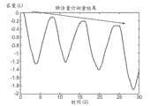
阻抗呼吸描记法是能够在不妨碍气流的情况下产生呼吸量跟踪、不要求与气流的接触以及不限制身体移动的简单方法。此外,也许能够进行反映肺的功能性残余容量的测量。Impedance pneumography is a simple method capable of producing respiratory volume tracking without obstructing airflow, requiring no contact with airflow, and not restricting body movement. In addition, it may be possible to make measurements that reflect the functional residual volume of the lung.
在尝试测量心脏活动的同时,Atzler和Lehmann注意到随呼吸而变的经胸电阻抗。他们将呼吸阻抗变化视为伪像并在进行测量的同时让病人停止呼吸。在1940年,在同样研究心脏阻抗的同时,Nyboer在其测量中注意到相同的呼吸阻抗伪像。通过作为通过同时地记录两者而使用肺活量计来使经胸阻抗上的变化与容量上的变化相关的第一人,他确认了伪像的起源。通过作为量化地使呼吸量和经胸阻抗相关的第一研究者,Goldensohn和Zablow使阻抗呼吸描记法更进一步。他们报告了分离心脏信号伪像方面的困难,并且还注意到身体移动期间的伪像。然而,在通过最小平方回归将阻抗变化与呼吸量变化相比较之后,他们重要地确定两者是线性相关的。其他群组已经证实了经胸阻抗变化与呼吸之间的线性关系,并且已经发现可以由胸阻抗信号来解释肺活量测定信号的约90%。虽然该关系已被表明是线性的,但许多群组发现用于病人体内和病人间的校准常数在试验之间是高度可变的。校准常数的这些差异可以归因于各种生理和电极特性,必须将其考虑在内。While attempting to measure cardiac activity, Atzler and Lehmann noticed a transthoracic electrical impedance that varied with respiration. They interpreted changes in respiratory impedance as artifacts and had the patient stop breathing while the measurement was being made. In 1940, while also studying cardiac impedance, Nyboer noticed the same respiratory impedance artifact in his measurements. By being the first person to use a spirometer to correlate changes in transthoracic impedance with changes in volume by recording both simultaneously, he confirmed the origin of the artifact. Goldensohn and Zablow took impedance pneumography a step further by being the first investigators to quantitatively correlate respiratory volume and transthoracic impedance. They reported difficulties in isolating cardiac signal artifacts and also noted artifacts during body movement. However, after comparing changes in impedance with changes in respiratory volume by least-squares regression, they importantly determined that the two were linearly related. Other groups have demonstrated a linear relationship between transthoracic impedance changes and respiration, and it has been found that approximately 90% of the spirometry signal can be explained by the thoracic impedance signal. Although the relationship has been shown to be linear, many groups found that the calibration constants used within and between patients were highly variable between trials. These differences in calibration constants can be attributed to various physiological and electrode characteristics, which must be taken into account.
经胸阻抗理论transthoracic impedance theory
电阻抗是被定义为电阻(R)、实部以及阻抗(X)、虚部(Z=R+jX=|Z|ejΘ)的和的复量。其被用作与交流电相对的测量结果。在数学上,由类似于欧姆定律的以下等式来测量阻抗:Electrical impedance is a complex quantity defined as the resistance (R), real part, and the sum of impedance (X), imaginary part (Z=R+jX=|Z|ejΘ ). It is used as a measurement against alternating current. Mathematically, impedance is measured by the following equation similar to Ohm's law:
Z=V/I (1)Z=V/I (1)
其中,电压=V,电流=I,并且阻抗=Z。可以根据简单电路来确定具有未知阻抗的导电的对象。在同时地测量跨对象的电压并使用等式(1)的同时跨对象施加已知交流电产生了阻抗。胸表示容积导体,并且因此,可以应用支配离子导电体的定律。另外,呼吸期间的器官移动和胸廓的扩大产生能够被测量的传导率上的变化。可以通过引入已知电流并用电极来测量跨胸的电压上的变化来测量跨胸的阻抗。Where voltage=V, current=I, and impedance=Z. Conductive objects with unknown impedance can be determined from simple circuits. Impedance is created by applying a known alternating current across the subject while simultaneously measuring the voltage across the subject and using equation (1). The chest represents a volume conductor, and therefore the laws governing ionic conductors can be applied. In addition, organ movement and expansion of the thorax during respiration produce changes in conductance that can be measured. Transthoracic impedance can be measured by introducing a known current and using electrodes to measure the change in voltage across the chest.
经胸阻抗信号的起源Origin of transthoracic impedance signal
构成胸和腹的组织层全部影响经胸阻抗的测量结果。每个组织具有影响电极之间的电流流动方向的不同传导率。从最外层开始,身体的表面被皮肤覆盖,其呈现出高电阻率,但是仅约1mm厚。在皮肤下面的是脂肪层,其也具有高电阻率。然而,此层的厚度是高度可变的,并且取决于对象的身体位置和身体类型。从后面移动到前面,在皮肤和脂肪层下面的是姿势肌,其是各向异性的。其在纵向方向上具有低电阻率,但是在所有其他方向上具有高电阻率,这导致在平行于皮肤的方向上传导电流的趋势。在肌肉下面的是肋骨,作为骨头,其是高度绝缘的。因此,通过胸的电流只能在骨之间流动。一旦电流到达肺,则假设电流穿过血液,血液具有任何身体组织的最低电阻中的一个。肺的充气改变肺的尺寸和电流流动的通道,并且使其本身表现为能够测量的电阻或阻抗中的变化。The layers of tissue that make up the thorax and abdomen all contribute to the measurement of transthoracic impedance. Each tissue has a different conductivity that affects the direction of current flow between the electrodes. Starting from the outermost layer, the surface of the body is covered by skin, which exhibits high electrical resistivity, but is only about 1 mm thick. Beneath the skin is a layer of fat, which also has a high electrical resistivity. However, the thickness of this layer is highly variable and depends on the subject's body position and body type. Moving from the back to the front, beneath the skin and fat layer are the postural muscles, which are anisotropic. It has low resistivity in the longitudinal direction, but high resistivity in all other directions, which leads to a tendency to conduct current in a direction parallel to the skin. Beneath the muscles are the ribs, which, as bones, are highly insulating. Therefore, the current through the chest can only flow between the bones. Once the current reaches the lungs, it is assumed that the current passes through the blood, which has one of the lowest electrical resistances of any body tissue. Inflation of the lungs changes the size of the lungs and the pathways through which electrical current flows, and manifests itself as a change in electrical resistance or impedance that can be measured.
由于组织的各向异性性质,通过胸的径向电流流动比将预期的少得多。大部分电流绕着胸经过而不是通过胸。结果,阻抗变化来自胸围上的变化、肺尺寸上的变化以及横膈膜-肝质量的移动。较低的胸层面处的测量归因于横膈膜和肝的移动,并且在较高的胸层面处,其归因于肺的充气和扩张。因此,阻抗信号是来自肺的扩张和充气的变化与横膈膜-肝质量的移动的和。需要腹和胸分量以便观察正常呼吸信号。另外,上部和下部胸中的阻抗变化的不同起源可以解释为什么在较高胸层面处观察到更大的线性。Radial current flow through the thorax is much less than would be expected due to the anisotropic nature of the tissue. Most of the current flows around the chest rather than through it. As a result, impedance changes result from changes in chest circumference, changes in lung size, and shifts in the diaphragm-liver mass. Measurements at the lower thoracic level are due to movement of the diaphragm and liver, and at higher thoracic levels it is due to inflation and expansion of the lungs. Thus, the impedance signal is the sum of changes from expansion and inflation of the lungs and movement of the diaphragm-liver mass. Abdominal and thoracic components are required in order to observe normal respiratory signals. Additionally, different origins of impedance variation in the upper and lower thorax may explain why greater linearity is observed at higher thoracic levels.
电极放置的影响Effect of Electrode Placement
用被附着于病人的皮肤的电极来测量经胸阻抗。Geddes等人确定电极刺激频率由于生理组织考虑而不应在20 kHz以下。其是安全问题且消除了来自生物电事件的干扰。另外,发现对象的阻抗测量结果根据对象位置而不同,包括坐、仰卧和站立。已经表明针对给定的容量上的变化,仰卧躺着在呼吸期间产生最大的信号振幅和最低的信噪比。Transthoracic impedance is measured with electrodes attached to the patient's skin. Geddes et al. determined that the electrode stimulation frequency should not be below 20 kHz due to physiological tissue considerations. It is a safety issue and eliminates interference from bioelectric events. Additionally, it was found that the subject's impedance measurements varied depending on the subject's position, including sitting, supine, and standing. It has been shown that for a given change in volume, lying supine produces the greatest signal amplitude and the lowest signal-to-noise ratio during respiration.
另一潜在信号伪像来自主体移动,其可能移动电极并干扰校准。此外,电极移动在肥胖和年长病人中可能更为普遍,这可能要求长期监视时段期间的反复指导重新校准。由于试验之间的校准变化性,某些人已经提出应针对给定的主体姿势和电极放置而对每个个体执行校准。然而,群组能够表明小心的病人体内电极放置能够将测量结果之间的阻抗减小至1%左右。Another potential signal artifact comes from subject movement, which can move the electrodes and interfere with the calibration. Furthermore, electrode movement may be more common in obese and elderly patients, which may require repeated guided recalibration during long-term monitoring periods. Due to trial-to-trial variability in calibration, some have suggested that calibration should be performed per individual for a given subject pose and electrode placement. However, the group was able to show that careful intra-patient electrode placement can reduce the impedance between measurements to around 1%.
尽管具有相同的电极放置,用于不同尺寸的个体的校准常数和信号振幅显示出变化性。所确定的是,针对容量上给定的变化的阻抗上的变化对于薄胸的人而言是最大的且对于体型更大的人而言较小。这些观察的差异可能是由于较大主体内的电极与肺之间的较大量的电阻性组织、诸如脂肪组织和肌肉而引起的,针对较大的主体产生用于容量上的给定变化的阻抗上的总体较小的百分比变化。另一方面,值得注意的是在儿童体内,阻抗轨迹的心脏分量比成年人体内的心脏分量更大。这可能是由于与在儿童体内相比在成年人体内的心脏周围的更大脂肪沉积而引起的,该脂肪沉积用于使心脏免于被结合到阻抗测量中。Calibration constants and signal amplitudes for individuals of different sizes showed variability despite having the same electrode placement. It was determined that the change in impedance for a given change in volume was greatest for thin-chested people and smaller for larger people. These observed differences may be due to the larger amount of resistive tissue, such as adipose tissue and muscle, between the electrodes and the lungs within the larger subject for which impedance is produced for a given change in volume An overall smaller percentage change in . On the other hand, it is noteworthy that the cardiac component of the impedance trace is larger in children than in adults. This may be due to greater fat deposits around the heart in adults than in children, which serve to protect the heart from being incorporated into the impedance measurement.
在第六根肋骨的层面处被附接于腋中线的电极在呼吸期间产生最大阻抗变化。然而,两个变量之间的最大线性是通过将电极较高地放置在胸上而获得的。尽管报告了高程度的线性,但已经报告了呼吸期间的阻抗变化的大标准偏差。然而,在阻抗测量结果中所观察的变化性与在诸如血压的其他生命体征中的测量结果中看到的那些相当。群组已经表明阻抗呼吸描记法对于临床目的而言是足够准确的。此外,在自从这些研究以来的40年中,电极材料和阻抗测量的信号处理已经被大大改善,产生甚至更可靠的测量结果。数字信号处理允许实时阻抗测量结果的近即时滤波和平滑化,这允许伪像和噪声的最小化。近年来,已经在长期病人监视中成功地使用呼吸阻抗。只要电极保持相对不动,阻抗中的变化与容量中的变化的关系在长时间段内是稳定的。Electrodes attached to the midaxillary line at the level of the sixth rib produce the greatest change in impedance during respiration. However, the greatest linearity between the two variables was obtained by placing the electrodes higher on the chest. Despite reporting a high degree of linearity, large standard deviations in impedance variation during respiration have been reported. However, the variability observed in impedance measurements was comparable to that seen in measurements of other vital signs such as blood pressure. Groups have shown that impedance pneumography is sufficiently accurate for clinical purposes. Furthermore, in the 40 years since these studies, electrode materials and signal processing for impedance measurements have been greatly improved, yielding even more reliable measurements. Digital signal processing allows near-instantaneous filtering and smoothing of real-time impedance measurements, which allows minimization of artifacts and noise. In recent years, respiratory impedance has been used successfully in long-term patient monitoring. The change in impedance versus change in capacity relationship is stable over long periods of time as long as the electrodes remain relatively still.
有源声学系统Active Acoustic System
声学在与肺的关系中的最常见使用是评估通过使用听诊器获取的发源于肺中的声音。肺组织的一个被频繁忽视的性质是其充当声滤波器的能力。其在不同的程度上使在其中通过的声音的各种频率衰减。在衰减的水平与肺中的空气量之间存在关系。胸壁的运动也导致通过胸的声信号的频移。The most common use of acoustics in relation to the lungs is the evaluation of sounds originating in the lungs acquired through the use of a stethoscope. A frequently overlooked property of lung tissue is its ability to act as an acoustic filter. It attenuates to varying degrees the various frequencies of sound passing through it. There is a relationship between the level of attenuation and the amount of air in the lungs. Movement of the chest wall also causes a frequency shift of the acoustic signal passing through the chest.
用于检测异常的潜力Potential for detecting anomalies
可以从用阻抗呼吸描记法来监视病人呼吸的容量轨迹中提取诸如用力肺活量(forced vital capacity,FVC)和第一秒用力呼气量(FEV1)的许多有用指示符。FVC和FEV1是通常由肺活量计测量的两个基准指示符,并且被用来诊断和监视诸如COPD、哮喘和肺气肿的疾病。除监视呼吸之外,阻抗呼吸描记法还可以同时地记录来自同一电极的心电图。Many useful indicators such as forced vital capacity (FVC) and forced expiratory volume in one second (FEV1) can be extracted from volume traces that monitor patient respiration with impedance pneumography. FVC and FEV1 are two baseline indicators commonly measured by spirometers and are used to diagnose and monitor diseases such as COPD, asthma and emphysema. In addition to monitoring respiration, impedance pneumography can simultaneously record an ECG from the same electrode.
呼吸间变化性breath-to-breath variability
诸如呼吸间变化性、变化系数、标准偏差以及潮气量直方图的对称性的计算已被表明取决于年龄和呼吸健康。与正常主体相比,已经表明这些参数中的某些,特别是变化系数,在具有肺结核、肺炎、肺气肿和哮喘的病人中是明显不同的。此外,在本文中已经注意到只要电极不在病人上移动,阻抗测量结果是令人满意的。一般地,许多群组已经确定健康主体在呼吸模式方面显示出比处于肺病状态中的主体更大的变化性。Calculations such as breath-to-breath variability, coefficient of variation, standard deviation, and symmetry of tidal volume histograms have been shown to depend on age and respiratory health. Some of these parameters, especially the coefficient of variation, have been shown to be significantly different in patients with tuberculosis, pneumonia, emphysema and asthma compared to normal subjects. Furthermore, it has been noted herein that impedance measurements are satisfactory as long as the electrodes do not move on the patient. In general, many groups have determined that healthy subjects exhibit greater variability in breathing patterns than subjects in pulmonary disease states.
呼吸波形的非线性分析已在广泛的应用中使用。在检查非线性、生理数据的规则性时,研究已经表明在肺病状态内,病人展示出呼吸间复杂性的下降。在慢性阻塞性肺病、限制性肺病内以及在未能完成来自机械通气的除管法的病人内已经证明了复杂性上的此降低。降低的变化性还已被确定为镇静和痛觉丧失的结果。广泛地说,正常病人比受某种形式肺病或损害所折磨的那些人具有更大的呼吸间变化性。Nonlinear analysis of respiratory waveforms has been used in a wide range of applications. When examining the regularity of non-linear, physiological data, studies have shown that within pulmonary disease states, patients exhibit a decrease in interbreathing complexity. This reduction in complexity has been demonstrated in chronic obstructive pulmonary disease, restrictive lung disease, and in patients who failed to complete extubation from mechanical ventilation. Reduced variability has also been identified as a consequence of sedation and analgesia. Broadly speaking, normal patients have greater breath-to-breath variability than those afflicted with some form of lung disease or impairment.
呼吸模式是非线性的,类似于任何生理数据,因为其受到身体内的大量调控剂影响。在呼吸间变化性的分析内,使用各种熵度量来测量信号内的不规则性和可再现性的量。这些度量可以不仅在评估呼吸间变化、而且在评估呼吸内变化性以及曲线的量值、周期性以及空间位置时在RVM潮气量追踪的分析内被使用。Breathing patterns are non-linear, similar to any physiological data, as they are influenced by a large number of regulators in the body. Within the analysis of breath-to-breath variability, various entropy measures are used to measure the amount of irregularity and reproducibility within the signal. These metrics can be used within the analysis of RVM tidal volume tracking in assessing not only inter-breath variation, but also intra-breath variability and the magnitude, periodicity, and spatial location of the curve.
基于标准化病人特性数据(Crapo)的系统的通用校准允许产生复杂性指数以及单个病人与被定义为正常复杂性水平的东西的比较。此指数将被用来帮助临床医生确定除管的适当时间,确定心肺疾病的严重度,以及也在治疗的评估之内。此指数将独立于其中通过基于阻抗的设备、加速度计、呼吸机或成像设备而收集了数据的方法。还可以针对特定病人对该系统进行校准并在检测任何呼吸参数内的快速变化的同时集中于主体体内变化性。Universal calibration of the system based on standardized patient characteristic data (Crapo) allowed the generation of complexity indices and the comparison of individual patients with what was defined as normal complexity levels. This index will be used to help clinicians determine the appropriate time for extubation, determine the severity of cardiorespiratory disease, and also within the evaluation of treatment. This index will be independent of the method in which the data was collected by impedance-based devices, accelerometers, ventilators, or imaging devices. It is also possible to calibrate the system for a specific patient and focus on intra-subject variability while detecting rapid changes in any respiratory parameter.
呼吸间间隔的非线性分析Nonlinear Analysis of Interbreath Intervals
除变化性度量之外,某些群组已发现即时呼吸间间隔的非线性分析与脱离机械呼吸机的成功是高度相关的。这些度量是肺健康的有用指示符且能够帮助临床判定。不能使病人从机械呼吸机分离在约20%的病人中发生,并且用以预测成功分离的当前方法是不好的,并且几乎无助于内科医生的判定。在对机械通气下的33个主体的大于24小时的研究中,已发现24个主体成功地脱离通气,而8个主体失败(来自一个主体的数据被去除)。将失败的原因列举为五个主体中缺氧以及对于其余三个的呼吸急促、高碳酸血以及上气道水肿,其全部是可能被阻抗呼吸描记系统所潜在地识别的疾病。此研究中的主要发现是对于未能从机械呼吸机分离的那些人的即时呼吸间隔的非线性分析明显比被成功地分离的那些人更规则。此外,已经表明呼吸速率在两个群组之间是不同的。从阻抗呼吸描记法测量结果的非线性分析导出的度量能够成功地预测病人恢复结果。另外,已经表明这些度量是稳健的,并且不会在引入诸如咳嗽的伪像时显著地改变。In addition to measures of variability, certain groups have found that nonlinear analysis of immediate breath-to-breath intervals is highly correlated with success off mechanical ventilator. These measures are useful indicators of lung health and can aid in clinical decisions. Failure to isolate a patient from a mechanical ventilator occurs in approximately 20% of patients, and current methods to predict successful isolation are poor and of little help to physicians' judgment. In a study of 33 subjects under mechanical ventilation over 24 hours, 24 subjects were found to be successfully weaned from ventilation, while 8 subjects failed (data from one subject was removed). Reasons for failure were listed as hypoxia in five subjects and tachypnea, hypercapnia, and upper airway edema for the remaining three, all diseases that could potentially be identified by the impedance pneumography system. The main finding in this study was that the non-linear analysis of immediate breath intervals was significantly more regular for those who failed to separate from the mechanical ventilator than those who were successfully separated. Furthermore, it has been shown that the respiration rate differs between the two cohorts. Metrics derived from nonlinear analysis of impedance pneumography measurements were able to successfully predict patient recovery outcomes. Additionally, it has been shown that these metrics are robust and do not change significantly when artifacts such as coughing are introduced.
减少通气状态的检测Reduce detection of ventilatory status
由阻抗呼吸描记法产生的呼吸轨迹以及主体的平均阻抗能够指示减少通气的状态或胸中的液量上的变化。此类监视将对被麻醉病人的护理有用。在被麻醉或不移动的病人体内用阻抗呼吸描记法进行的呼吸监视被表明在长时段内是准确且可靠的,尤其是在手术之后的恢复室中的关键时段期间。研究者已经确定胸或肺中的流体可能导致阻抗中的可测量变化,其可以被用来确定对于恢复室中的病人的常见问题,诸如肺水肿或肺炎。The respiratory trace produced by impedance pneumography and the subject's average impedance can indicate a state of reduced ventilation or a change in fluid volume in the chest. Such monitoring would be useful in the care of anesthetized patients. Respiration monitoring with impedance pneumography in anesthetized or immobile patients has been shown to be accurate and reliable over long periods of time, especially during the critical period in the recovery room after surgery. Researchers have determined that fluid in the chest or lungs can cause measurable changes in impedance, which can be used to identify common problems for patients in the recovery room, such as pulmonary edema or pneumonia.
除测量胸中的液量上的变化之外,潮气量和上气道阻力上的变化在阻抗测量中是立即显而易见的。研究者发现被麻醉病人的气管内夹紧仍产生减少的阻抗信号,尽管病人努力呼吸,从而给出通气的正确指示。还已表明阻抗测量结果提供每个肺的通气的定量评定。在具有单侧的肺病变的病人中观察到阻抗测量结果上的差异,其中在胸的受损伤侧上的一对电极比正常的一侧产生不那么明显的信号。In addition to measuring changes in fluid volume in the chest, changes in tidal volume and upper airway resistance are immediately apparent in impedance measurements. The researchers found that endotracheal clamping in anesthetized patients produced a reduced impedance signal despite the patient's effort to breathe, thereby giving the correct indication for ventilation. Impedance measurements have also been shown to provide a quantitative assessment of the ventilation of each lung. Differences in impedance measurements were observed in patients with unilateral lung lesions, where a pair of electrodes on the injured side of the chest produced a less pronounced signal than the normal side.
呼吸监视器respiratory monitor
虽然某些接触探针记录呼吸速率,但迄今为止,尚未具体地发明用以记录或分析呼吸模式或变化性、使呼吸模式或变化性与生理条件或生命力相关、或使用呼吸模式或变化性来预测即将发生的崩溃的设备或方法。心率变化性算法仅报告逐个搏动的心率上的变化。期望的是使用呼吸速率变化性算法来将变化性结合在呼吸运动的呼吸强度、速率以及位置中。如通过强度方面、速率方面、呼吸努力的局部化方面或者这些参数中的任何一个的变化性方面的变化所注意到的呼吸方面的已标记异常,提供呼吸或心血管衰竭的预警,并且可以呈现出针对早期干预的机会。用以记录这些变化的设备的开发和使这些呼吸变化与疾病或损伤的严重度相关的算法的产生将不仅提供有用的战场工具,而且提供用以帮助评估和治疗垂危病人的医院重症护理背景中的重要性之一。诊所或家庭背景中的使用可以对不那么垂危的病人有用,其仍然将受益于此类监视。例如,如果病人被过度麻醉,则呼吸速率下降且呼吸变得“浅”。呼吸速率和呼吸努力随着由于肺水肿或对于肺顺应性损失的其他原因而引起的硬肺和不良换气而上升。然而,作为被客观地监视的唯一参数的速率的含义常常未被足够快地识别以最好地治疗病人。需要一种可以提供呼吸功的实时、定量评定并分析任何或所有这些参数中的呼吸速率、强度、局部化或变化性的趋势的系统,以用于早期诊断和干预以及治疗监视。需要此类系统来判断麻醉的深度或者麻药或其他镇痛药物的充足或过度剂量。While some contact probes record breathing rate, to date, nothing has been specifically invented to record or analyze breathing patterns or variability, correlate breathing patterns or variability to physiological conditions or vitality, or use breathing patterns or variability to Apparatus or method for predicting impending crashes. The heart rate variability algorithm reports only the beat-to-beat variation in heart rate. It is desirable to use a breath rate variability algorithm to incorporate variability in the breath intensity, rate, and position of the breathing motion. Flagged abnormalities in respiration, as noted by changes in intensity, rate, localization of respiratory effort, or variability in any of these parameters, provide early warning of respiratory or cardiovascular failure and can present opportunities for early intervention. The development of devices to record these changes and the generation of algorithms to correlate these respiratory changes with the severity of disease or injury would provide useful tools not only on the battlefield, but also in the context of hospital critical care to aid in the assessment and treatment of dying patients. one of the importance. Use in a clinic or home setting may be useful for less critically ill patients who would still benefit from such monitoring. For example, if a patient is overanesthetized, the breathing rate drops and breathing becomes "shallow." Respiratory rate and respiratory effort rise with stiff lungs and poor ventilation due to pulmonary edema or other causes for loss of lung compliance. However, the implications of velocity, the only parameter being objectively monitored, are often not recognized quickly enough to best treat the patient. There is a need for a system that can provide real-time, quantitative assessment of work of breathing and analyze trends in respiration rate, intensity, localization or variability in any or all of these parameters for early diagnosis and intervention as well as therapy monitoring. Such systems are needed to judge the depth of anesthesia or the adequacy or overdose of anesthesia or other analgesic drugs.
PCA和反馈控制PCA and Feedback Control
病人自控镇痛(PCA)是包括病人反馈的术后疼痛控制的方法。麻醉剂的给药能够抑制呼吸、心率以及血压,因此需要小心和密切的监视。系统包括包含可以泵送到病人的IV线中的止痛药的计算机化泵。一般地,除恒定剂量的镇止痛药之外,病人可以按下按钮以接收以附加药物形式的护理。然而,如果病人变得过于困倦,则劝阻病人不要按下按钮,因为这可能阻止对于更快速恢复的治疗。还存在就地保护,其限制在给定时间量内给予病人的药物的量以防止过度剂量。可以使用脉搏血氧计、呼吸速率和二氧化碳图监视器来警告由止痛药所引起的呼吸抑制并切断PCA剂量,但是均具有关于至少准确度、有效性以及实施方式的严格限制。Patient-controlled analgesia (PCA) is an approach to postoperative pain control that includes patient feedback. Administration of anesthetics can suppress respiration, heart rate, and blood pressure, so careful and close monitoring is required. The system includes a computerized pump containing pain medication that can be pumped into the patient's IV line. Typically, a patient can press a button to receive care in the form of an additional medication in addition to a constant dose of analgesic medication. However, if the patient becomes too sleepy, the patient is discouraged from pressing the button, as this may prevent treatment for a more rapid recovery. There are also in situ safeguards, which limit the amount of drug administered to a patient in a given amount of time to prevent overdosing. Pulse oximeter, respiration rate and capnography monitors can be used to warn of respiratory depression caused by pain medication and to cut off PCA dose, but all have severe limitations on at least accuracy, effectiveness and implementation.
发明内容Contents of the invention
下文所述的方法和设备提供了准确地计算呼吸量、测量各种呼吸参数和检测呼吸异常的技术。病人或主体监视是必不可少的且遍及医院部门快速地传播。具体地,在重症监护室中、在麻醉程序期间以及术后时期需要呼吸监视。呼吸速率已被用来评定病人状态,但是单独的呼吸速率并未指示重要的呼吸变化,并且因此需要监视其他呼吸参数。可以指示诸如肺活量测定法和呼吸末CO2测量结果的附加呼吸参数的当前方法是不方便、不准确或不可能在镇静的病人或不能或将不会合作的那些人身上实现。能够以方便且准确的方式检测附加呼吸参数的呼吸监视设备将大大地有益于病人监视和病状的检测。The methods and devices described below provide techniques for accurately calculating respiration volume, measuring various respiration parameters, and detecting respiration abnormalities. Patient or subject monitoring is essential and spreads rapidly throughout the hospital sector. In particular, respiratory monitoring is required in the intensive care unit, during anesthesia procedures, and in the postoperative period. Respiration rate has been used to assess patient status, but respiration rate alone does not indicate significant respiratory changes, and therefore other respiratory parameters need to be monitored. Current methods that can indicate additional respiratory parameters such as spirometry and end-tidalCO2 measurements are inconvenient, inaccurate or impossible to implement in sedated patients or those who cannot or will not cooperate. A respiratory monitoring device capable of detecting additional respiratory parameters in a convenient and accurate manner would greatly benefit patient monitoring and detection of pathology.
优选地,该设备提供至少一个附加呼吸参数的测量结果。此呼吸参数是通过报告主体的呼吸速率、主体的呼吸压力、主体的呼吸流量、主体的呼吸末CO2、主体的舌下CO2、主体的呼吸强度,与这些相关或对这些进行测量的测量结果而被反映的。Preferably, the device provides a measurement of at least one additional breathing parameter. This respiratory parameter is measured by reporting, relating to, or measuring the subject's respiratory rate, subject's respiratory pressure, subject's respiratory flow, subject's end-tidalCO2 , subject's sublingualCO2 , subject's respiratory intensity results are reflected.
该设备的一个实施例基于测量结果来提供数据,其报告以下参数、与这些参数相关或对这些参数进行测量,或者报告以下参数的变化性、变化或复杂性,与其相关或对其进行测量:主体的呼吸曲线的形状、主体的呼吸曲线的形状上的变化、基于主体的吸入容量的呼吸曲线、基于主体的呼出容量的呼吸曲线、基于主体的吸入压力的呼吸曲线、基于主体的呼出压力的呼吸曲线、基于主体的吸入流量的呼吸曲线、基于主体的呼出流量的呼吸曲线、基于通过成像测量的主体胸部的运动的呼吸曲线、基于由放置在胸部上的接触传感器测量的主体的胸部运动的呼吸曲线、基于通过成像测量的主体的腹部运动的呼吸曲线、基于由放置在腹部上的接触传感器测量的主体的腹部运动的呼吸曲线、基于通过成像测量的主体的胸部和腹部两者的运动的呼吸曲线、基于由放置在胸部和腹部上的接触传感器测量的主体的胸部和腹部的运动的呼吸曲线、主体的呼吸间间隔的变化、主体的阻抗和容量信号之间的相位滞后、主体的阻抗和容量信号之间的相位滞后的变化,及其组合。An embodiment of the device provides data based on measurements reporting, relating to, or measuring, or reporting variability, change, or complexity of, relating to, or measuring: The shape of the subject's breathing curve, the change in the shape of the subject's breathing curve, the breathing curve based on the subject's inhalation volume, the breathing curve based on the subject's exhalation volume, the breathing curve based on the subject's inhalation pressure, the variation of the subject's exhalation pressure A breathing curve, a breathing curve based on the subject's inhaled flow, a breathing curve based on the subject's exhaled flow, a breathing curve based on the movement of the subject's chest measured by imaging, a breathing curve based on the movement of the subject's chest measured by a contact sensor placed on the chest respiration curve, respiration curve based on the subject's abdominal movement measured by imaging, a respiration curve based on the subject's abdominal movement measured by a contact sensor placed on the abdomen, a respiration curve based on the movement of both the subject's chest and abdomen measured by imaging Breathing curve, breathing curve based on movement of the subject's chest and abdomen measured by contact sensors placed on the chest and abdomen, variation of the subject's interbreathing interval, phase lag between the subject's impedance and volume signals, the subject's impedance and the change in phase lag between the capacity signal, and combinations thereof.
在优选实施例中,所述设备提供了至少一个附加呼吸参数的呼吸参数上的变化、变化性或复杂性的测量结果。此呼吸参数是通过报告主体的呼吸速率、主体的呼吸压力、主体的呼吸流量、与这些相关或对这些进行测量的测量结果,与主体的呼吸末CO2相关的测量结果,与主体的舌下CO2、主体的呼吸强度、主体的呼吸深度、主体的呼吸局部化相关的测量结果而被反映的。In a preferred embodiment, the device provides a measure of change, variability or complexity in a breathing parameter of at least one additional breathing parameter. This respiratory parameter is obtained by reporting the subject's respiratory rate, the subject's respiratory pressure, the subject's respiratory flow, measurements related to or measured against these, measurements related to the subject's end-tidalCO2 , related to the subject's sublingual CO2 , subject's respiration intensity, subject's respiration depth, and subject's respiration localization-related measurements are reflected.
所述设备的一个实施例提供了报告以下各项、与之相关或对其进行测量的测量结果:主体的呼吸间间隔的变化、主体的阻抗和容量信号之间的相位滞后、主体的阻抗和容量信号之间的相位滞后的变化、以及其组合。One embodiment of the device provides measurements that report, relate to, or measure changes in the subject's interbreathing interval, phase lag between the subject's impedance and volume signals, the subject's impedance and Variations in phase lag between volume signals, and combinations thereof.
优选地,监视设备将使用以下各项中的一个或多个:阻抗呼吸描记法、声学、呼吸末CO2测量结果、用以测量呼吸量及其他呼吸参数的脉搏血氧测定法。优选地,该设备将利用信号处理算法来使原始阻抗数据的噪声和电干扰最小化以产生在此后将被称为呼吸振动监视(RVM)测量结果、轨迹或曲线的平滑、清楚的轨迹。优选地,使用呼吸参数来计算用于肺活量测定法测试的ATS标准化值,包括但不限于用力肺活量(FVC)、一秒用力呼气量(FEV1)、六秒用力呼气量(FEV6)、用力呼气流量(FEF)、峰值呼气流量(PEF)、用力吸气肺活量(FIVC)、峰值吸气流量(PIF)、潮气量(VT)、吸气量(IC)、呼气储备量(ERV)、吸气储备量(IRV)、缓慢肺活量(SVC)、最大通气量(MVV)、肺活量(VC)以及每分钟呼吸(BPM)。Preferably, the monitoring device will use one or more of: impedance pneumography, acoustics, end-tidalCO2 measurements, pulse oximetry to measure respiratory volume and other respiratory parameters. Preferably, the device will utilize signal processing algorithms to minimize noise and electrical interference of the raw impedance data to produce a smooth, clear trace which will hereafter be referred to as a Respiratory Vibration Monitoring (RVM) measurement, trace or curve. Preferably, respiratory parameters are used to calculate ATS normalized values for spirometry testing, including but not limited to forced vital capacity (FVC), forced expiratory volume in one second (FEV1), forced expiratory volume in six seconds (FEV6), forced expiratory volume in six seconds (FEV6), forced Expiratory flow (FEF), peak expiratory flow (PEF), forced inspiratory vital capacity (FIVC), peak inspiratory flow (PIF), tidal volume (VT), inspiratory volume (IC), expiratory reserve volume (ERV ), Inspiratory Reserve (IRV), Slow Vital Capacity (SVC), Maximum Ventilation (MVV), Vital Capacity (VC) and Breaths Per Minute (BPM).
更优选地,所述设备将被用来随时间推移监视潮气量、呼吸速率和每分钟通气量,更优选地在一小时内、在4、6、8、12、24小时内。最优选地,所述设备将能够随时间推移而适应于并报告呼吸参数或其变化、变化性或复杂性上的变化。More preferably, the device will be used to monitor tidal volume, respiratory rate and minute ventilation over time, more preferably within one hour, within 4, 6, 8, 12, 24 hours. Most preferably, the device will be able to adapt to and report changes in breathing parameters or their variation, variability or complexity over time.
优选地,所述设备将能够测量不稳定且不顺畅和一致的呼吸。优选地,将近实时地、在10秒下、在20秒下、在30秒下、在1分钟下、在2分钟下报告这些测量结果。Preferably, the device will be able to measure erratic and uneven and consistent breathing. Preferably, these measurements are reported in near real time, at 10 seconds, at 20 seconds, at 30 seconds, at 1 minute, at 2 minutes.
优选地,所述设备将输入测量参数以计算用于呼吸充分性的唯一指数,在病人诊断和监视中利用的呼吸充分性指数(RSI)。特别地,对于不能遵守程序的病人而言,将获取简单的潮式呼吸样本,这不要求指导和顺从性。RSI测量结果将被护理人员用来调整护理计划。Preferably, the device will input measured parameters to calculate a unique index for respiratory sufficiency, the Respiratory Sufficiency Index (RSI) utilized in patient diagnosis and monitoring. In particular, for patients who are unable to comply with the procedure, a simple tidal breath sample will be taken, which does not require instruction and compliance. RSI measurements will be used by nursing staff to adjust care plans.
在一个实施例中,呼吸变化监视使用阻抗呼吸描记来提供在肺功能实验室中发现的类似类型的呼吸评定。虽然不能在不经历规定策略的情况下直接计算由美国胸科协会(ATS)推荐的肺活量测定法度量,但当诊断一定范围的肺病状态时,可以使用阻抗轨迹中的其他信息来代替这些度量或作为对其的补充。用阻抗体积描记法进行的呼吸变化监视提供了对肺活量测定法的新型无创、非阻塞性替代方式,具有可以检查无意识或不顺从病人的附加优点。虽然病人不能在处于此类状态的同时执行与在肺活量测定法中相同的策略,但可以利用潮气量的阻抗轨迹来测量或估计由肺活量测定法测试给定的呼吸参数,诸如FEV1(一秒用力呼气量)和FVC(用力肺活量)。此外,FEV1和FVC测量结果的实用性在于提供在量化肺功能水平和在阻塞性和限制性肺部失调之间进行区分时有用的度量。在哮喘(阻塞性肺部失调)中,1秒用力呼气量(FEV1)通常减小,用力肺活量(FVC)通常是正常的,并且比值FEV1/FVC减小。在限制性失调中,FEV1和FVC两者都减小,留下正常FEV1/FVC。RVM测量结果可以在不要求病人向机器中呼吸的情况下输送FEV1和FVC测量结果,所述机器具有要求注意保持在嘴中的管嘴,这影响结果。RVM在吸入或呼出空气的呼吸通道中不具有任何设备,其可能妨碍或改变呼吸的流量或容量或者可能污染气流。实际上,其是感兴趣的肺通气充分性和充足性的测量结果,并且使用RVM、不同参数或除FEV1或FVC之外的不同参数来提供类似或更好的数据以帮助测量或监视肺功能以帮助限制性或阻塞性气道疾病的诊断和管理。这些数据是从呼吸曲线的形状、相移或呼吸间变化性的分析获得的。In one embodiment, respiratory variation monitoring uses impedance pneumography to provide a similar type of respiration assessment found in pulmonary function laboratories. Although the spirometry measures recommended by the American Thoracic Society (ATS) cannot be directly calculated without undergoing prescribed strategies, other information from the impedance trace can be used in place of these measures when diagnosing a range of lung disease states or as a supplement to it. Monitoring of respiratory changes by impedance plethysmography offers a novel non-invasive, non-obstructive alternative to spirometry, with the added advantage of being able to examine unconscious or noncompliant patients. Although the patient cannot perform the same strategies as in spirometry while in such a state, the impedance trace of tidal volume can be used to measure or estimate a respiratory parameter given by a spirometry test, such as FEV1 (forced in one second expiratory volume) and FVC (forced vital capacity). Furthermore, the utility of FEV1 and FVC measurements is to provide measures useful in quantifying levels of lung function and distinguishing between obstructive and restrictive lung disorders. In asthma (an obstructive pulmonary disorder), forced expiratory volume in 1 second (FEV1) is usually decreased, forced vital capacity (FVC) is usually normal, and the ratio FEV1/FVC is decreased. In restrictive disorders, both FEV1 and FVC are reduced, leaving normal FEV1/FVC. RVM measurements can deliver FEV1 and FVC measurements without requiring the patient to breathe into a machine with a nozzle that requires attention to remain in the mouth, which affects the results. The RVM does not have any devices in the breathing passage of inhaled or exhaled air that could impede or alter the flow or volume of the breath or that could contaminate the airflow. In effect, it is the measure of lung adequacy and adequacy of interest, and uses RVM, a different parameter, or a different parameter other than FEV1 or FVC to provide similar or better data to help measure or monitor lung function To aid in the diagnosis and management of restrictive or obstructive airway disease. These data are obtained from the analysis of the shape, phase shift, or breath-to-breath variability of the respiration curve.
另一方法使用声学来提供呼吸变化监视。可以使用一种包括在病人的胸部上的扬声器和扩音器的装置来监视肺和胸的声学性质以估计肺容量。如阻抗呼吸描记法的情况一样,声学呼吸监视不妨碍气流,并且可以对无意识和不顺从的病人使用。除估计肺容量之外,还可以使用扩音器来评定与病人的状况相关联的其他参数并检测呼吸衰减的征兆。Another approach uses acoustics to provide respiratory change monitoring. A device comprising a loudspeaker and a microphone on the patient's chest can be used to monitor the acoustic properties of the lungs and chest to estimate lung volumes. As in the case of impedance pneumography, acoustic respiration monitoring does not impede airflow and can be used on unconscious and noncompliant patients. In addition to estimating lung volumes, the microphone can also be used to assess other parameters associated with the patient's condition and to detect signs of respiratory decay.
已经研究了阻抗体积描记法达约半个世纪。特别有兴趣的努力是在给定“标度因子”的情况下从阻抗数据产生容量曲线的能力。此标度因子是已经先前通过将阻抗轨迹与使用肺活量计同时地记录的容量数据相比较所导出的。然而,所采取的新型方法是在给定病人生理和历史及其他可测量参数的情况下替代地计算“RVM校准系数”,所述可测量参数诸如身高、体重、BMI、BSA、年龄、性别、民族以及其他物理特性或生理、新陈代谢或实验室参数。此策略将用于容量变化的相对阻抗的重要性与胸的总体阻抗进行分离。基于阻抗的无创设备监视呼吸频率和潮气量并代替对诊断和病人监视两者有用且不那么依赖于操作员技能的基于标准肺活量测定法的值进行服务。还可以从在标准肺功能测试期间用肺活量计、呼吸速度描记器或其他呼吸描记器获取的测量结果或从来自用于插管病人的呼吸机的读数导出RVM校准系数。Impedance plethysmography has been studied for about half a century. A particularly interesting endeavor is the ability to generate capacity curves from impedance data given a "scaling factor". This scaling factor has been previously derived by comparing the impedance trace with volume data recorded simultaneously using a spirometer. However, the novel approach taken is to instead calculate an "RVM calibration factor" given a patient's physiology and history and other measurable parameters such as height, weight, BMI, BSA, age, sex, Ethnicity and other physical characteristics or physiological, metabolic or laboratory parameters. This strategy decouples the importance of relative impedance for volume changes from the overall impedance of the chest. Impedance-based non-invasive devices monitor respiratory rate and tidal volume and serve in place of standard spirometry-based values that are useful for both diagnostics and patient monitoring and are less dependent on operator skill. RVM calibration coefficients can also be derived from measurements taken with a spirometer, pneumotachometer, or other pneumograph during standard pulmonary function testing, or from readings from a ventilator used to intubate a patient.
对内科医生和诊断有用的一个新RVM测量是对称为呼吸健康指数(RHI)的RVM的新型方法。此指数利用RVM数据和病人参数来产生针对呼吸健康的百分比值。该值是根据利用正常肺活量测定图的表格所导出的值,用于改变在Knudsen、Crapo等的作品中发现的病人人口统计信息。例如,针对潮式呼吸,发现了用于与病人相同身高、体重、性别以及民族的人的正常值,并且然后通过将实际值除以表列值以产生百分比来与实际值相比较。RHI中的峰峰值变化应接近于100%以指示良好的健康。One new measure of RVM that is useful to physicians and diagnostics is a novel approach to RVM called the Respiratory Health Index (RHI). This index utilizes RVM data and patient parameters to generate a percentage value for respiratory health. This value was derived from a table using normal spirometry charts for varying patient demographics found in the works of Knudsen, Crapo et al. For example, for tidal breathing, normal values for a person of the same height, weight, sex and ethnicity as the patient are found and then compared to the actual value by dividing the actual value by the tabulated value to yield a percentage. The peak-to-peak variation in the RHI should be close to 100% to indicate good health.
优选地,至少一个呼吸参数的分析包括使所述至少一个呼吸参数与预定义呼吸状况相关。优选地,该分析提供对诊断的辅助。更优选地,该分析提供诊断。优选地,该分析提供将与其他监视或临床数据集成以充当治疗管理方面的辅助的信息。Preferably, the analysis of at least one breathing parameter comprises correlating said at least one breathing parameter with a predefined breathing condition. Preferably, the analysis provides an aid to diagnosis. More preferably, the analysis provides a diagnosis. Preferably, this analysis provides information that will be integrated with other monitoring or clinical data to serve as an aid in treatment management.
优选地,所述分析提供信息以指导治疗的管理。优选地,设备提供未来病人状态的预测。更优选地,该预测是主体的生命力的预测、损伤严重度的预测、主体的崩溃可能性的预测、主体遭受呼吸衰竭的可能性的预测、主体的麻醉深度的预测、主体的药物剂量水平的预测、主体的心肺衰竭的可能性的预测、用于与治疗病人相关联的设备的设备故障的可能性的预测、正在进行中的治疗的适当性或不适当性的预测,或其组合。Preferably, the analysis provides information to guide the management of therapy. Preferably, the device provides predictions of future patient states. More preferably, the prediction is a prediction of the subject's vitality, a prediction of the severity of the injury, a prediction of the subject's likelihood of collapsing, a prediction of the subject's likelihood of suffering respiratory failure, a prediction of the subject's depth of anesthesia, a prediction of the subject's drug dose level A prediction, a prediction of the likelihood of cardiorespiratory failure of the subject, a prediction of the likelihood of equipment failure of equipment used in connection with treating the patient, a prediction of the appropriateness or inappropriateness of ongoing therapy, or a combination thereof.
优选地,该设备识别与诸如COPD、哮喘、肺气肿、肺结核、局部急性肺炎、呼吸急促、高碳酸血、肺水肿、肺炎、单侧肺病变、即将发生或存在的呼吸衰竭和气道阻塞的病状相关联的呼吸模式。Preferably, the device identifies conditions associated with, for example, COPD, asthma, emphysema, tuberculosis, localized acute pneumonia, shortness of breath, hypercapnia, pulmonary edema, pneumonia, unilateral lung disease, impending or existing respiratory failure, and airway obstruction. Breathing patterns associated with the condition.
优选地,该设备识别与诸如心脏、神经或新陈代谢(诸如充血性心力衰竭、心肌病、糖尿病性酮酸中毒、脑水肿)的病状相关联的呼吸模式。Preferably, the device identifies breathing patterns associated with conditions such as cardiac, neurological or metabolic (such as congestive heart failure, cardiomyopathy, diabetic ketoacidosis, cerebral edema).
附图说明Description of drawings
图1是本发明的四引线实施例的透视图。Figure 1 is a perspective view of a four-lead embodiment of the invention.
图2是后面左至右电极配置的示图。Figure 2 is a diagram of the rear left to right electrode configuration.
图3是后面右侧垂直电极配置的示图。Figure 3 is a diagram of the rear right vertical electrode configuration.
图4是前后电极配置的示图。Figure 4 is a diagram of front and rear electrode configurations.
图5是前面右侧垂直电极配置的示图。Figure 5 is a diagram of the front right vertical electrode configuration.
图6是被复用器相互连接的两个四引线配置的透视图。Figure 6 is a perspective view of two four-lead configurations interconnected by a multiplexer.
图7是ICG电极配置的示图。Figure 7 is a diagram of an ICG electrode configuration.
图8是被连接到肺活量计的本发明的四引线实施例的透视图。Figure 8 is a perspective view of a four lead embodiment of the invention connected to a spirometer.
图9是被连接到呼吸机的本发明的四引线实施例的透视图。Figure 9 is a perspective view of a four-lead embodiment of the present invention connected to a ventilator.
图10是用于缓慢、正常以及不稳定呼吸策略的RVM测量结果(阻抗)对比容量曲线图。Figure 10 is a graph of RVM measurements (impedance) versus volume for slow, normal, and unstable breathing strategies.
图11是对于正常呼吸的针对时间的一组RVM和容量曲线图。Figure 11 is a set of RVM and volume graphs versus time for normal breathing.
图12是对于缓慢呼吸的针对时间的一组RVM和容量曲线图。Figure 12 is a set of RVM and volume graphs over time for slow breathing.
图13是对于不稳定呼吸的针对时间的一组RVM和容量曲线图。Figure 13 is a set of RVM and volume graphs versus time for unstable breaths.
图14是对于四个不同电极配置的针对BMI的校准系数的曲线图。Figure 14 is a graph of calibration coefficients for BMI for four different electrode configurations.
图15是展示出容量漂移的肺活量测定曲线图。Figure 15 is a graph of spirometry showing volume drift.
图16是受容量漂移影响的容量对比阻抗的曲线图。Figure 16 is a graph of capacitance versus impedance as affected by capacitance drift.
图17是针对容量漂移进行修正的肺活量测定曲线图。Figure 17 is a graph of spirometry curves corrected for volume drift.
图18是容量对比阻抗的曲线图,其比较对于容量漂移未修正和已修正的数据。Figure 18 is a graph of capacity versus impedance comparing uncorrected and corrected data for capacity drift.
图19描述用于本发明的数据分析的流程图。Figure 19 depicts a flow chart for data analysis of the present invention.
图20是利用扬声器和扩音器的本发明的优选实施例。Figure 20 is a preferred embodiment of the invention utilizing a loudspeaker and microphone.
图21是利用扬声器和扩音器阵列的本发明的优选实施例。Figure 21 is a preferred embodiment of the invention utilizing an array of loudspeakers and microphones.
图22是利用扬声器阵列和扩音器的本发明的优选实施例。Figure 22 is a preferred embodiment of the invention utilizing a loudspeaker array and a loudspeaker.
图23是利用用于传感器的背心的本发明的优选实施例。Figure 23 is a preferred embodiment of the invention utilizing a vest for sensors.
图24是利用用于传感器的被构建到布块中的阵列的本发明的优选实施例。Figure 24 is a preferred embodiment of the invention utilizing an array for sensors built into a cloth patch.
图25是利用传感器网的本发明的优选实施例。Figure 25 is a preferred embodiment of the invention utilizing a sensor network.
图26是利用无线发射机和接收机的本发明的优选实施例。Figure 26 is a preferred embodiment of the invention utilizing a wireless transmitter and receiver.
图27示出用于同时记录数据的阻抗对比时间和容量对比时间的图表。Figure 27 shows graphs of impedance versus time and capacity versus time for simultaneous recording of data.
图28图示出本发明的系统的实施例。Figure 28 illustrates an embodiment of the system of the present invention.
图29图示出本发明的设备的实施例。Figure 29 illustrates an embodiment of a device of the present invention.
图30—32图示出本发明的设备的优选实施例。Figures 30-32 illustrate a preferred embodiment of the apparatus of the present invention.
图33—38描绘了引线放置的不同实施例。33-38 depict different embodiments of lead placement.
具体实施方式Detailed ways
本发明的一个实施例针对用于评定病人、个体或动物的设备,其通过在身体上放置多个电极引线和/或扬声器和扩音器来收集阻抗测量结果。优选地,至少一个阻抗测量元件和扩音器/扬声器在功能上被连接到可编程元件,其被编程以提供主体的至少一个呼吸参数的评定。One embodiment of the invention is directed to an apparatus for assessing a patient, individual or animal that collects impedance measurements by placing multiple electrode leads and/or speakers and microphones on the body. Preferably, at least one impedance measuring element and a microphone/speaker are functionally connected to a programmable element programmed to provide an assessment of at least one breathing parameter of the subject.
优选地,阻抗测量结果基于多个远程探针数据组,并且其中,所述可编程元件还被编程为增强所述多个远程探针数据组中的至少一个;或者使所述多个远程探针数据组中的至少一个稳定;或针对动态范围和信噪比(SNR)值分析所述多个远程探针数据组中的每一个。优选地,所述设备探针被保持在若干引线配置中。在一个实施例中,引线配置上的变化允许根据主体和所正执行的测试而有灵活性。在其他实施例中,引线配置上的变化允许病人解剖结构上的变化性。优选地,设备保持设置以识别有效的引线配置。优选地,设备保持设置以识别有效的引线附接。Preferably, the impedance measurements are based on a plurality of remote probe data sets, and wherein said programmable element is further programmed to enhance at least one of said plurality of remote probe data sets; or to cause said plurality of remote probe data sets to at least one of the probe data sets is stabilized; or each of the plurality of remote probe data sets is analyzed for dynamic range and signal-to-noise ratio (SNR) values. Preferably, the device probes are held in several lead configurations. In one embodiment, variations in lead configuration allow for flexibility depending on the subject and the test being performed. In other embodiments, variations in lead configuration allow for variability in patient anatomy. Preferably, the device maintains settings to identify valid lead configurations. Preferably, the device maintains a setting to recognize valid lead attachment.
优选地,如在被嵌入机器中的协议中描述的设备或方法下达关于引线放置的命令。优选地,由设备来检验适当的引线接触。优选地,设备警告操作员关于不充分或不适当的引线放置。Preferably, a device or method as described in a protocol embedded in the machine commands for lead placement. Preferably, proper lead contact is verified by the device. Preferably, the device alerts the operator of insufficient or improper lead placement.
优选地,设备连续地或间歇性地进行监视并保持警报以指示呼吸参数何时反映通气或其他生命机能上的损失。该警报是基于呼吸充分性指数、每分钟通气量、呼吸速率、潮气量、吸气量或流量参数、呼气量或流量参数、呼吸率的变化性、容量、流量或所生成的其他参数而设定的。例如,如果监视器检测到与换气不足相关联的呼吸频率或深度或每分钟通气量方面的下降或检测到将暗示换气过度的这些参数中的任何一个或全部方面的增加,则进行警报。当基于到呼吸机或肺活量计的特定个体校准来将病人的当前呼吸状态与基线水平相比较时,在医院楼层上使用警报。优选地,基于针对给定个体从呼吸机或肺活量计获取的参数来设置警报。更优选地,基线水平是基于以下各项中的一个或多个:人口统计信息、生理或体型参数。警报还被用来针对被确定为对病人有害的点处的麻醉诱发的呼吸抑制而进行报警。优选地,值的范围(超过该值,警报将被触发)是针对以下各项中的一个或多个由医师或护理人员所选择的:呼吸速率、潮气量、每分钟通气量、呼吸充分性指数、呼吸曲线的形状、熵、分形(fractal)、或与呼吸变化性或复杂性相关联的其他分析参数。Preferably, the device monitors continuously or intermittently and maintains an alarm to indicate when respiratory parameters reflect a loss in ventilation or other vital functions. The alarm is based on respiratory adequacy index, minute ventilation, respiratory rate, tidal volume, inspiratory volume or flow parameter, expiratory volume or flow parameter, variability in respiratory rate, volume, flow, or other parameters generated set. For example, if the monitor detects a decrease in respiratory rate or depth or minute ventilation associated with hypoventilation or detects an increase in any or all of these parameters that would suggest hyperventilation, an alert is issued . Alarms are used on hospital floors when a patient's current respiratory state is compared to a baseline level based on a specific individual calibration to a ventilator or spirometer. Preferably, the alarms are set based on parameters obtained from a ventilator or spirometer for a given individual. More preferably, the baseline level is based on one or more of: demographic information, physiological or body shape parameters. Alarms are also used to alert for anesthesia-induced respiratory depression at points determined to be detrimental to the patient. Preferably, the range of values beyond which an alarm will be triggered is selected by a physician or paramedic for one or more of the following: respiratory rate, tidal volume, minute ventilation, respiratory adequacy Exponential, shape of the respiratory curve, entropy, fractal, or other analytical parameters associated with respiratory variability or complexity.
在另一实施例中,将在任何给定时间点处所获取的RVM测量结果记录为基线。奖这些记录值与由内科医生或病人状态的其他保健工作人员所产生的主观印象相关。随后,监视RVM并设置警报以便如果注意到呼吸量、每分钟通气量曲线特性或变化性方面的10%、20%或其他所选百分比变化,则向保健工作人员报警。In another embodiment, the RVM measurements taken at any given point in time are recorded as a baseline. These recorded values correlate with subjective impressions made by physicians or other health care workers of the patient's state. Subsequently, the RVM is monitored and an alarm is set to alert the healthcare worker if a 10%, 20%, or other selected percentage change in respiratory volume, minute ventilation curve characteristics or variability is noted.
以下举例说明本发明的实施例,但不应将其视为限制本发明的范围。The following examples illustrate examples of the present invention, but they should not be construed as limiting the scope of the present invention.
阻抗体积描记器Impedance plethysmograph
如在本文中体现并广泛描述的内容提供了本发明的详细实施例。该实施例仅仅是可以以各种和替代形式来体现的本发明的示例。因此,不存在特定结构和功能细节应具有限制性的意图,相反,意图是其提供用于权利要求的基础,并且作为用于教导本领域的技术人员来各种地采用本发明的代表性基础。As embodied and broadly described herein, provides detailed embodiments of the invention. This embodiment is merely an example of the invention that can be embodied in various and alternative forms. Therefore, it is not intended that specific structural and functional details should be limiting, but rather, are intended to provide a basis for the claims and serve as a representative basis for teaching one skilled in the art to variously employ the present invention .
本发明优选地包括阻抗呼吸描记器,其具有集成电子装置以将测量的阻抗值转换成容量并通过采用数据的数值或图形表示的电子接口或打印的报告来向最终用户显示容量。阻抗测量设备包括电路、至少一个微处理器和优选地包括至少四个引线。优选地,其中,至少两个引线被用于向主体的身体中注入电流且至少两个被用于读取所述病人的身体的电压响应。The invention preferably includes an impedance pneumograph with integrated electronics to convert measured impedance values into volumes and display the volumes to the end user through an electronic interface using numerical or graphical representation of the data or a printed report. The impedance measurement device includes circuitry, at least one microprocessor and preferably at least four leads. Preferably, wherein at least two leads are used to inject current into the subject's body and at least two are used to read the voltage response of the patient's body.
在一个实施例中,所述设备优选地包括集成模块以模拟病人并允许自动化系统测试和演示。自动化系统测试改善了设备的性能并保证其在使用之前正确地运行。In one embodiment, the device preferably includes integrated modules to simulate a patient and allow automated system testing and demonstration. Automated system testing improves equipment performance and ensures it operates correctly before use.
在优选实施例中,设备利用模拟除法器来补偿注入电流中的轻微偏差并增加所获取数据的准确度。优选实施例中的模拟除法器将被放置在解调器之后和整流器之前。在其他实施例中,可以将模拟除法器放置在电路中的其他位置上,包括但不限于在精密整流器之后或解调器之前。In a preferred embodiment, the device utilizes an analog divider to compensate for slight deviations in the injected current and increase the accuracy of the acquired data. The analog divider in the preferred embodiment would be placed after the demodulator and before the rectifier. In other embodiments, the analog divider may be placed elsewhere in the circuit, including but not limited to after the precision rectifier or before the demodulator.
在优选实施例中,设备利用被微处理器驱动的自适应电子装置以保持电路中的不同放大器上的适当增益以防止信号离开该范围。微处理器跟踪每个硬件放大器处的设定增益并在其计算期间适当地对其进行补偿,使得其始终输出适当的值。In a preferred embodiment, the device utilizes adaptive electronics driven by a microprocessor to maintain proper gain on the various amplifiers in the circuit to prevent signals from leaving this range. The microprocessor tracks the set gain at each hardware amplifier and compensates it appropriately during its calculations so that it always outputs the proper value.
优选地经由数字接口(例如USB、火线(Fire wire)、串行、并行或其他种类的数字接口)将阻抗测量设备连接到计算机。数字接口被用来防止数据在传输期间的讹误。优选地将此接口上的通信加密以进一步保证数据完整性以及保护本发明免于仿造模块(测量设备或计算机)的使用。The impedance measuring device is preferably connected to the computer via a digital interface such as USB, Fire wire, serial, parallel or other kind of digital interface. Digital interfaces are used to prevent corruption of data during transmission. Communications over this interface are preferably encrypted to further ensure data integrity and protect the invention from use by counterfeit modules (measurement devices or computers).
现在更详细地参考本发明的优选实施例,在图1中,示出了阻抗体积描记器,包括射频阻抗计1、包含在被链接到该阻抗计的PC上的可编程元件2,将该阻抗计通过四个引线、即第一引线3、第二引线4、第三引线5以及第四引线6连接到病人。每个引线优选地被连接到表面电极,即第一表面电极、第二表面电极、第三表面电极以及第四表面电极。Referring now in more detail to the preferred embodiment of the present invention, in Figure 1 there is shown an impedance plethysmograph comprising a radio
更详细地,仍参考图1的实施例,电极可以由诸如AgCl的导电材料制成,涂敷有粘合剂,即诸如水凝胶或水胶体的导电材料。引线可以由诸如铜线的任何导电材料制成,并且优选地涂敷有诸如橡胶的绝缘材料。在优选实施例中,利用无线电极来提供电流并收集和发射数据。优选地,将此引线组成与蓝牙技术和接收机耦合。In more detail, still referring to the embodiment of FIG. 1 , the electrodes may be made of a conductive material such as AgCl, coated with a binder, ie a conductive material such as hydrogel or hydrocolloid. The leads may be made of any conductive material such as copper wire and are preferably coated with an insulating material such as rubber. In a preferred embodiment, wireless electrodes are utilized to provide electrical current and collect and transmit data. Preferably, this lead composition is coupled with Bluetooth technology and a receiver.
引线1和4被连接到具有优选地大于20 KHz的恒定频率的电流源,其大到足以避免与生物信令相干扰。电流源的振幅优选地小于50 mA,并且在将引起所选频率下的纤维性颤动的水平以下。引线2和3之间的差动电压被用来根据欧姆定律而计算阻抗。通过对由阻抗计获取的电压测量结果进行采样,可编程元件(诸如PC)跟踪与诸如心跳和呼吸的生物机能相对应的胸阻抗上的变化并对其进行绘图。然后使用阻抗上的变化来监视肺功能。优选地,通过在本文中展示的方法来对设备进行校准以计算肺容量并将其显示给操作员。
参考图28,示例性和优选系统包括至少一个通用计算设备100,其包括处理单元(CPU)120,以及系统总线110,其将包括诸如只读存储器(ROM)140和随机存取存储器(RAM)150的系统存储器的各种系统部件耦合到处理单元120。其他系统存储器130也可以加以使用。本发明优选地在具有一个以上的CPU 120的计算设备上或在被联网在一起以提供更大处理能力的计算设备群组或集群上操作。系统总线110可以是若干类型的总线结构中的任何一个,包括存储器总线或存储器控制器、外围总线以及使用各种总线架构中的任何一个的本地总线。存储在ROM 140等中的基本输入/输出(BIOS)优选地提供基本例程,其帮助诸如在启动期间在计算设备100内的元件之间传输信息。计算设备100还优选地包括诸如硬盘驱动器160、磁盘驱动器、光盘驱动器、磁带驱动器等的存储设备。存储设备160被驱动接口连接到系统总线110。驱动器和关联的计算机可读介质提供计算机可读指令、数据结构、程序模块和用于计算设备100的其他数据的非易失性存储。基本部件对于本领域的技术人员而言是已知的,并且根据设备的类型想到适当的变化,诸如设备是否是小型、手持式计算设备、台式计算机、膝上型计算机、计算机服务器、无线设备、网络启用设备或无线电话等。Referring to Figure 28, an exemplary and preferred system includes at least one general
在某些实施例中,优选地由单个CPU来控制系统,然而,在其他实施例中,由一个或多个微处理器(MP)来控制系统的一个或多个部件。另外,可以使用CPU和MP的组合。优选地,MP是嵌入式微控制器,然而,还可以使用能够处理命令的其他设备。In some embodiments, a single CPU preferably controls the system, however, in other embodiments, one or more microprocessors (MPs) control one or more components of the system. Alternatively, a combination of CPU and MP can be used. Preferably, the MP is an embedded microcontroller, however, other devices capable of processing commands may also be used.
虽然在本文中所述的示例性环境采用硬盘,但本领域的技术人员应认识到的是在示例性操作环境中还可以使用其他类型的计算机可读介质,其能够存储可被计算机访问的数据,其他类型的计算机可读介质诸如是磁带盒、闪速存储卡、数字多功能磁盘、盒式磁带、随机存取存储器(RAM)、只读存储器(ROM)、包含位流的电缆或无线信号,等等。为了能够实现与计算设备100的用户交互,输入设备190表示任何数目的输入机构,诸如用于语音的扩音器、用于手势或图形输入的触摸屏、电信号传感器、键盘、鼠标、运动输入、语音等。设备输出端170可以是对于本领域的技术人员而言已知的多个输出机构中的一个或多个,例如打印机、监视器、投影仪、扬声器以及绘图仪。在某些实施例中,该输出可以经由网络接口,例如上传到网站、发送电子邮件,被附接于其他电子文件或放置在其他电子文件内、以及发送SMS或MMS消息。在某些情况下,多模系统使得用户能够提供多个类型的输入以与计算设备100通信。通信接口180一般控制和管理用户输入和系统输出。不存在对在任何特定硬件布置上操作的本发明的限制,并且因此这里的基本特征可以随着改进的硬件或固件布置被开发而被其容易地取代。Although the exemplary environment described herein utilizes a hard disk, those skilled in the art will recognize that other types of computer-readable media capable of storing data that can be accessed by a computer can also be used in the exemplary operating environment. , other types of computer-readable media such as magnetic tape cartridges, flash memory cards, digital versatile disks, cassette tapes, random access memory (RAM), read only memory (ROM), cables containing a bit stream, or wireless signals ,etc. To enable user interaction with
在本发明的范围内的实施例还可以包括用于载送或具有存储在其上面的计算机可执行指令或数据结构的计算机可读介质。此类计算机可读介质可以是能够被通用或专用计算机访问的任何可用介质。以示例而非限制的方式,此类计算机可读介质可以包括RAM、ROM、EEPROM、CD-ROM或其他光盘存储器、磁盘存储器或其他磁存储设备,或者能够用来以计算机可执行指令或数据结构的形式载送或存储期望程序代码装置的任何其他介质。当通过网络或另一通信连接(硬布线、无线或其组合)向计算机传输或提供信息时,计算机适当地将该连接视为计算机可读介质。因此,适当地将任何此类连接称为计算机可读介质。以上的组合也应包括在计算机可读介质的范围内。Embodiments within the scope of the present invention may also include computer-readable media for carrying or having computer-executable instructions or data structures stored thereon. Such computer-readable media can be any available media that can be accessed by a general purpose or special purpose computer. By way of example and not limitation, such computer-readable media may include RAM, ROM, EEPROM, CD-ROM or other optical disk storage, magnetic disk storage or other magnetic storage devices, or may be used to store computer-executable instructions or data structures any other medium that carries or stores the desired program code means. When information is transmitted or provided to a computer over a network or another communications connection (hardwired, wireless, or a combination thereof), the computer properly considers that connection to be a computer-readable medium. Thus, any such connection is properly termed a computer-readable medium. Combinations of the above should also be included within the scope of computer-readable media.
计算机可执行指令包括例如促使通用计算机、专用计算机或专用处理设备执行某个功能或功能组的指令和数据。计算机可执行指令还包括在独立或网络环境中被计算机执行的程序模块。一般地,程序模块包括执行特定任务或实现特定抽象数据类型的例程、程序、对象、部件以及数据结构。计算机可执行指令、关联数据结构以及程序模块表示用于执行本文公开的方法的步骤的程序代码装置的示例。此类可执行指令和关联数据结构的特定顺序表示用于实现在此类步骤中描述的功能的对应动作的示例。Computer-executable instructions include, for example, instructions and data which cause a general purpose computer, special purpose computer, or special purpose processing device to perform a certain function or group of functions. Computer-executable instructions also include program modules that are executed by computers in stand-alone or network environments. Generally, program modules include routines, programs, objects, components, and data structures that perform particular tasks or implement particular abstract data types. Computer-executable instructions, associated data structures, and program modules represent examples of the program code means for executing steps of the methods disclosed herein. The particular sequence of such executable instructions and associated data structures represents examples of corresponding acts for implementing the functions described in such steps.
本领域的技术人员将认识到的是可以用许多类型的计算机系统配置在网络计算环境中实践本发明的其他实施例,这些计算机系统配置包括个人计算机、手持式设备、多处理器系统、基于微处理器或可编程的消费电子设备、网络PC、微型计算机、大型机计算机,等等。网络可以包括因特网、一个或多个局域网(“LAN”)、一个或多个城域网(“MAN”)、一个或多个广域网(“WAN”)、一个或多个内部网,等等。还可以在分布式计算环境中实践实施例,其中,由通过通信网络(由硬布线链路、无线链路或由其组合)链接的本地和远程处理设备来执行任务。在分布式计算环境中,可以将程序模块定位于本地和远程存储器存储设备两者中。Those skilled in the art will recognize that other embodiments of the invention can be practiced in a network computing environment with many types of computer system configurations, including personal computers, handheld devices, multiprocessor systems, microprocessor-based Processor or programmable consumer electronics, network PCs, microcomputers, mainframe computers, and more. A network may include the Internet, one or more local area networks ("LANs"), one or more metropolitan area networks ("MANs"), one or more wide area networks ("WANs"), one or more intranets, and the like. Embodiments may also be practiced in distributed computing environments where tasks are performed by local and remote processing devices that are linked through a communications network (either by hardwired links, wireless links, or a combination thereof). In a distributed computing environment, program modules may be located in both local and remote memory storage devices.
图2是本发明的系统200的实施例的示意图。电源源自于信号源205。优选地,使用可调整函数发生器210(例如XR2206芯片)来生成电源。函数发生器210优选地是可经由微处理器(MP)275或手动地调整的。在某些实施例中,可以调谐函数发生器以便改善信号。调谐可以发生一次或多次。可以使用生物阻抗频谱学来检测不同频率下的水合作用的水平,其可以被用来对函数发生器210进行校准。类似地,可以计算体脂肪百分比。信号源205还包括电流发生器215(例如Howland电路)。电流发生器215优选地保持源电流恒定,尽管有焊盘接点上的变化(除非接点完全坏掉)。在优选实施例中,可以调谐电流发生器215以改善性能,这可以手动地或由MP 275自动地完成。在优选实施例中,监视焊盘接点质量,并且当焊盘接点坏掉或者质量过差而使电子装置不能进行补偿时产生警告。信号源205还可以包括电流监视器220以计算阻抗。在优选实施例中,信号源205还包括病人模拟器225。病人模拟器225可以用与真实病人类似的参数来模拟阻抗上的变化。可以将病人模拟器225用于测试系统200以及电路的校准。FIG. 2 is a schematic diagram of an embodiment of a
来自信号源205的信号通过病人230且被传感器235接收到。优选地,传感器230包括输入放大器240。输入放大器240抑制不良或可变焊盘接点对测量结果的影响。优选地由MP 275来控制输入放大器240的增益以向其他模块提供增强的信号。传感器230优选地还包括信号滤波器245以去除来自电力网等的干扰。信号滤波器245可以是标准高通滤波器(如在图30上)、解调器(如在图31上)或另一信号滤波器。然后将同步解调器用于检测生物阻抗变化并剥离信号中的运动伪像。Signals from
在优选实施例中,将信号分离到两个路径中(如在图32上)。第一路径使用发生器信号作为载波将测量信号解调。第二路径在解调之前使用90度相位旋转电路。可以使用电压至RMS转换器将已解调信号转换成RMS值。单独地测量,将信号加和并且然后计算平方根。这允许补偿主体中的任何相移以及电阻和电抗的单独测量,这提供用于运动伪像补偿的有价值信息以及水合作用水平、脂肪百分比和校准系数计算。In a preferred embodiment, the signal is split into two paths (as on Figure 32). The first path demodulates the measurement signal using the generator signal as a carrier. The second path uses a 90 degree phase rotation circuit prior to demodulation. The demodulated signal can be converted to an RMS value using a voltage to RMS converter. Measured individually, the signals are summed and the square root is then calculated. This allows compensation for any phase shift in the subject and separate measurements of resistance and reactance, which provide valuable information for motion artifact compensation as well as hydration level, fat percentage and calibration factor calculations.
另外,传感器230可以包括模拟除法器250,其将测量电压信号除以来自电流监视电路的信号以计算阻抗。传感器230优选地还包括精密整流器或具有低通滤波器的均方根至直流(RMS至DC)芯片255以去除载波频率。传感器230的输出优选地是与病人的阻抗成比例的DC信号。传感器230还可以包括带通滤波器260以通过滤出不对应于呼吸的信号部分来仅选择呼吸速率。可以手动地或由MP 275自动地对带通滤波器260进行校准。优选地,传感器230包括由MP 275控制以调节多个探针对的复用器265。优选地,存在2个探针对,然而可想到更多或更少的探针对。传感器230还可以包括输出放大器270。优选地由MP 275来控制输出放大器270,并向模数转换器(ADC)280提供信号以用于高精度数字化。使用过采样来减少可能源自于不同源(例如,热、电子、生物或EM干扰)的测量噪声。MP 275命令ADC以尽可能高的节奏获取测量结果,并且然后在对应于采样频率的时间间隔内对所获得数据求平均。采样频率是其被阻抗测量设备呈现给计算机时的阻抗采样的频率。优选地将该频率设置得足够高以监视呼吸的所有微小特征。Additionally,
使用可控增益和过采样优选地允许系统以极高的有效精度来测量阻抗(对于当前实现方式而言估计为28位、或者十亿分之四)。Using controllable gain and oversampling preferably allows the system to measure impedance with very high effective precision (estimated at 28 bits, or four parts per billion for current implementations).
信号源205和传感器230两者都被MP 275控制。MP 275优选地包括监视信号处理的至少一个ADC 280以及用以控制数字电位计、复用器、运算放大器、信号发生器及其他设备的至少一个数字输出端285。优选地,MP 275和计算机接口(例如,经由USB接口、串行接口或无线接口)。Both
在优选实施例中,设备具有测量和记录其他参数的能力,所述其他参数包括但不限于:心输出量、呼吸末CO2、氧灌注量、ECG以及心脏的其他电生理测量结果。在优选实施例中,阻抗测量设备同时地测量阻抗心动描记和阻抗呼吸描记。优选地,在屏幕上显示附加参数。优选地,以有意义的方式将呼吸阻抗数据与附加参数组合以充当对诊断的辅助。优选地,使用单独的阻抗数据或将其与一个或多个附加参数组合来提供疾病状态的诊断。In a preferred embodiment, the device has the capability to measure and record other parameters including, but not limited to: cardiac output, end tidal CO2, oxygen perfusion, ECG, and other electrophysiological measurements of the heart. In a preferred embodiment, the impedance measuring device measures impedance cardiography and impedance pneumography simultaneously. Preferably, additional parameters are displayed on the screen. Preferably, the breathing impedance data is combined with additional parameters in a meaningful way to serve as an aid to diagnosis. Preferably, a diagnosis of the disease state is provided using the impedance data alone or in combination with one or more additional parameters.
在一个实施例中,独立地从胸的每一侧获取测量结果并将其用来既评估一般肺状态又评估右和左肺充气或胸扩张之间的差异。在肋骨骨折(其中可以存在归因于包括肺挫伤的伤害的变化)的情况下,其示例是由于夹板或气胸而引起的运动上的下降,其中,独立地监视胸的两侧以提供侧特定数据。可以评估局部化肺病症的其他来源,包括肺炎、水胸、乳糜胸、血胸、血/气胸、肺膨胀不全、肿瘤以及辐射损伤。在另一实施例中,将来自设备的信息与来自超声心电图、放射性核素研究或对心脏进行成像的其他方法的信息一起使用。在优选实施例中,设备帮助利用以下各项中的一个来诊断心肌缺血:心电图、高级电生理研究、心导管插入术、超声心电图、应力测试、放射性核素测试、CT、MRI、通过阻抗测量进行的心排血量监视。在一个实施例中,所述设备提供用来帮助收集随呼吸而变的其他信号的信息,诸如呼吸音、心脏的信息、辐射检测设备、放射治疗设备、切除设备。In one embodiment, measurements are taken independently from each side of the chest and used to assess both general lung status and differences between right and left lung inflation or chest expansion. In the case of rib fractures (where there may be variations due to injury including lung contusions), examples of which are decreased motion due to splinting or pneumothorax, where both sides of the chest are monitored independently to provide side-specific data. Other sources of localized lung disease can be evaluated, including pneumonia, hydrothorax, chylothorax, hemothorax, hemothorax/pneumothorax, atelectasis, tumor, and radiation injury. In another embodiment, the information from the device is used with information from an echocardiogram, radionuclide study, or other method of imaging the heart. In a preferred embodiment, the device facilitates the diagnosis of myocardial ischemia using one of the following: electrocardiogram, advanced electrophysiological studies, cardiac catheterization, echocardiography, stress testing, radionuclide testing, CT, MRI, through impedance Cardiac output monitoring performed by measurement. In one embodiment, the device provides information to help collect other signals that vary with respiration, such as breath sounds, cardiac information, radiation detection equipment, radiotherapy equipment, resection equipment.
在优选实施例中,设备可以帮助由另一模态进行的定时或数据收集和/或使用呼吸曲线的特性来修正所收集的数据。In a preferred embodiment, the device can assist with timing or data collection by another modality and/or use characteristics of the breathing curve to correct the collected data.
在一个实施例中,所述设备提供与将与心脏搏动间变化性或复杂性相结合地使用的呼吸间变化性或呼吸复杂性有关的信息,以提供关于心脏、肺系统或整个新陈代谢或神经状态的其他不可得的信息。In one embodiment, the device provides information about breath-to-breath variability or complexity to be used in conjunction with heart beat-to-beat variability or complexity to provide information on the heart, pulmonary system, or overall metabolic or neurological Other unavailable information about the status.
引线配置Lead configuration
提出的呼吸参数评估技术依赖于参数与测量阻抗之间的高度线性关系。并不是对于每个电极放置而言都是如此。进行广泛的研究以选择优选地满足以下条件的最好电极放置。The proposed respiratory parameter assessment technique relies on a highly linear relationship between the parameter and the measured impedance. This is not true for every electrode placement. Extensive research was performed to select the best electrode placement preferably satisfying the following conditions.
1) 呼吸量与测量阻抗变化之间的高度线性关系(即在96%以上的相关值)。1) Highly linear relationship between respiratory volume and measured impedance change (i.e. a correlation value above 96%).
2) 由于病人运动而引起的低水平的伪像。2) Low level artifacts due to patient motion.
3) 反复电极施加之间的低变化。3) Low variation between repeated electrode applications.
4) 常见临床情况下的容易应用。4) Easy application in common clinical situations.
用于与“通用校准”一起使用的能力,其在没有用呼吸机/肺活量计的初步校准的情况下可靠地确定取决于可测量病人身体参数的标度因子。Capability for use with "Universal Calibration", which reliably determines scale factors that depend on measurable patient physical parameters without initial calibration with the ventilator/spirometer.
优选地,电极在第六根肋骨的层面处被水平地附接于腋中线。优选地,将一个电极放置在稳定位置处,诸如直接在锁骨下面或在上横骨处,并且将另一电极放置在胸廓的底部处或腋中线处的剑突的层面处。然而,可以将电极较高地或较低地放置在胸上。此外,可以按照其他位置或配置(例如垂直地沿着胸、跨过胸以一定角度、或者从在病人前面的位置至在病人背面的位置)来放置电极,取决于要测试的主体、要执行的测试以及其他生理关注方面(例如,如果病人具有起搏器或其他人造设备)。Preferably, the electrodes are attached horizontally to the midaxillary line at the level of the sixth rib. Preferably, one electrode is placed at a stable location, such as directly below the clavicle or at the superior transverse bone, and the other electrode is placed at the floor of the thorax or at the level of the xiphoid process at the midaxillary line. However, the electrodes can be placed higher or lower on the chest. In addition, electrodes may be placed in other positions or configurations (such as vertically along the chest, at an angle across the chest, or from a position in front of the patient to a position on the back of the patient), depending on the subject to be tested, the and other physiological concerns (for example, if the patient has a pacemaker or other artificial device).
优选地,至少一个阻抗测量元件存在于一个或多个电极引线上。优选地,以线性阵列、网格状图案或以受解剖影响的配置来布置两个或更多电极。优选地,以线性阵列来布置四个远程探针。在另一实施例中,将多个电极引线布置为网、背心或阵列。优选地,将一个或多个探针、电极引线或传感器放置在主体的胸或腹部上。优选地,设备使用单次使用电极。在其他实施例中,电极是水凝胶、水胶体或固体凝胶。优选地,电极利用AgCl、镍或碳传感器。优选地,电极带有软布、泡沫、多孔带、透明带背衬或另一粘合剂。优选地,存在用于成年人和新生儿的不同的、尺寸适当的电极,其中成年人电极大于新生儿电极,其优选地为1''乘3/8''或以下(2.54 cm乘0.95 cm或以下)。在其他实施例中,传感器电极与向身体输送电脉冲的探针相同,或者不同于该输送电极,或者是无线的且向远程传感器发射数据。在另一实施例中,输送探针本身是传感器。在一个实施例中,刺激电极是电池供电的。优选地,在30秒、连续地、间歇性地、在达到主体呼吸的至少3、5、10、20或50个内、在达到主体呼吸的至少100个内、在达到主体呼吸的至少1000个的持续时间内或在另一持续时间内,记录至少一个呼吸参数。优选地,同时记录主体的心阻抗图。Preferably, at least one impedance measuring element is present on one or more electrode leads. Preferably, two or more electrodes are arranged in a linear array, in a grid-like pattern or in an anatomically influenced configuration. Preferably, four remote probes are arranged in a linear array. In another embodiment, a plurality of electrode leads are arranged as a mesh, vest or array. Preferably, one or more probes, electrode leads or sensors are placed on the subject's chest or abdomen. Preferably, the device uses single use electrodes. In other embodiments, the electrodes are hydrogels, hydrocolloids or solid gels. Preferably, the electrodes utilize AgCl, nickel or carbon sensors. Preferably, the electrodes are backed with soft cloth, foam, porous tape, transparent tape or another adhesive. Preferably, there are different, appropriately sized electrodes for adults and neonates, where the adult electrodes are larger than the neonate electrodes, which are preferably 1" by 3/8" or less (2.54 cm by 0.95 cm or below). In other embodiments, the sensor electrodes are the same as the probes that deliver electrical pulses to the body, or are different from the delivery electrodes, or are wireless and transmit data to remote sensors. In another embodiment, the delivery probe itself is a sensor. In one embodiment, the stimulating electrodes are battery powered. Preferably, within 30 seconds, continuously, intermittently, within at least 3, 5, 10, 20 or 50 breaths to the subject, within at least 100 breaths to the subject, at least 1000 breaths to the subject For a duration of , or for another duration, at least one respiratory parameter is recorded. Preferably, an impedance cardiogram of the subject is simultaneously recorded.
优选地,所述至少一个阻抗测量元件包括一个或多个远程探针或电极引线,或者与标准EKG引线类似或与被用于测量心脏阻抗的引线类似的引线,并且其中,所述可编程元件被进一步编程为分析从一个或多个远程探针或电极引线收集的一个或多个远程探针或电极引线数据组。Preferably, said at least one impedance measuring element comprises one or more remote probes or electrode leads, or leads similar to standard EKG leads or to leads used to measure cardiac impedance, and wherein said programmable element is further programmed to analyze one or more remote probe or electrode lead data sets collected from the one or more remote probe or electrode leads.
在任何实施例中,引线配置对于设备的性能而言是关键的。优选地,将一个或多个引线放置在胸上。在一个实施例中,将引线放置在胸或腹部上以从诸如胸或腹部的身体的不同部位测量呼吸。与呼吸相关联的身体运动的位置上的差异产生在临床上对生理状态的诊断和疾病的监视有用的信息。将引线以交替配置放置在胸、颈和头上。在一个实施例中,引线是基于解剖位置以不同的配置进行放置的,并且根据特定测量距离或解剖界标或两者的组合而进行间隔。在一个实施例中,实现了相对于身体尺寸的间距修改。优选地,使这些修改与解剖界标相关。在优选实施例中,间距对于从新生儿至肥胖病人(在从250g至400kg的范围变动)的所有尺寸的病人而言保持相对相同。在另一实施例中,间距基于反映身体尺寸和体质的算法而变化。其他配置具有确定一个半胸对比另一个半胸的差分运动的优点,其在诊断或监视诸如气胸、血胸、脓胸、癌症的单侧或不对称病症时有用。In any embodiment, the lead configuration is critical to the performance of the device. Preferably, one or more leads are placed on the chest. In one embodiment, leads are placed on the chest or abdomen to measure respiration from different parts of the body such as the chest or abdomen. Differences in the location of body movements associated with respiration yield information that is clinically useful for diagnosis of physiological states and monitoring of disease. Place the leads in an alternating configuration on the chest, neck, and head. In one embodiment, leads are placed in different configurations based on anatomical location and spaced according to specific measured distances or anatomical landmarks or a combination of both. In one embodiment, spacing modification relative to body size is achieved. Preferably, these modifications are related to anatomical landmarks. In a preferred embodiment, the spacing remains relatively the same for all patient sizes from neonates to obese patients (ranging from 250g to 400kg). In another embodiment, the spacing varies based on an algorithm reflecting body size and constitution. Other configurations have the advantage of determining differential motion of one hemithorax versus the other, which is useful in diagnosing or monitoring unilateral or asymmetric conditions such as pneumothorax, hemothorax, empyema, cancer.
现在参考图2,示出了具有称为后面左至右(PLR)的特定电极配置的一个实施例,其中,第一电极7被放置于剑突(xiphoid process)的层面处的在脊柱左侧6英寸处,第二电极8被放置在剑突的层面处的在脊柱左侧2英寸处,第三电极9被放置在剑突的层面处的在脊柱右侧2英寸处,并且第四电极10被放置在具有剑突的脊柱层面的右侧六英寸处。以此配置来放置电极的优点是在信号的读取和高水平信号时将两个肺考虑在内。Referring now to FIG. 2 , there is shown one embodiment with a particular electrode configuration known as posterior left-to-right (PLR), wherein a first electrode 7 is placed on the left side of the spine at the level of the xiphoid process. 6 inches, a second electrode 8 was placed at the level of the
参考图3,示出了称为后面垂直向右(PVR)的第二特定电极配置,其中,第一电极11被放置在腋中线与刚好在肩胛骨下面的脊柱间的中间,第二电极12被放置在电极1下面两英寸,第三电极13被放置在电极2下面两英寸,并且第四电极14被放置在电极3下面。此配置的优点是由于胸扩张和较少心脏干扰而引起的电极移动的减少。此位置具有电极之间的很少到没有容量变化和较少心脏噪声的益处。Referring to FIG. 3 , a second specific electrode configuration known as Posterior Vertical Right (PVR) is shown, wherein a
参考图4,示出了称为前面至后面(AP)的第三特定电极配置,其中,第一电极15被放置在剑突的层面处的右腋中线的右侧6英寸处,第二电极16被放置在剑突的层面处的右腋中线的右侧2英寸处,第三电极17被放置在剑突的层面处的右腋中线的左侧2英寸处,并且第四电极18被放置在剑突层面处的右腋中线的左侧2英寸处。此位置捕捉最大的容量变化,这对呼吸局部化的确定有用。Referring to FIG. 4, a third specific electrode configuration called anterior to posterior (AP) is shown, wherein a
参考图5,示出了称为前面垂直向右(AVC)的第四特定电极放置,其中,第一电极19被直接放置在剑突与腋中线间的中间的锁骨下面,第三电极20与第一电极成直线地被放置在剑突层面处,第二电极21被放置在第三电极以上4英寸处,并且第四电极22被放置在第三电极下面4英寸处。此位置对新生儿及其他病人有用,其特性防止操作员将引线放置在后面。其他四探针位置被垂直地和水平地放置在腹部和胸上,相互等距离或者在具体测量的距离处。探针位置还被放置在生理界标处,诸如肠骨脊或第三肋间隙。腹部和胸两者上的探针放置允许确定胸与腹部呼吸之间的关系。此关系帮助诊断和治疗的监视。Referring to FIG. 5 , a fourth specific electrode placement known as anterior vertical right (AVC) is shown, in which the
除上述四探针配置之外,可以通过在位置之间等距地添加探针,例如与所述放置成直线地在与每个电极相距两英寸处以AP配置在电极1和2、2和3、3和4之间添加电极,来将这些配置修改成包括更多探针。在许多电极的情况下,可以将其相互等距地以网格图案放置:下面将进一步讨论此配置。用于2个或更多引线的其他放置包括在胸周围的诸如剑突的恒定高度处的等距离点处。用于24引线系统的特定放置在线性阵列内,其具有分别在胸和背上线性地等距间隔的12个引线。可以在网或背心内实现此类网格或阵列以便被病人穿戴。在一个实施例中,所述设备提供描述引线放置替代方式的表格,并且提供测量设备以帮助探针放置。在一个实施例中,由具有位置传感器和/或能够确定从一个传感器至另一个或多个传感器的距离的传感器的引线来自动地确认引线之间的测量距离。In addition to the four-probe configuration described above, probes can be added equidistantly between locations, for example in an AP configuration at
现在参考图6,示出了若干个电极的配置23,其借助于模拟复用器24被连接在一起,并被连接到射频阻抗计25和诸如PC的可编程元件26。示出了实现在先前的图中、图2和3中所示的引线和复用器配置的设备的实施例。在图6中,每个引线借助于复用器被连接到若干个不同的电极。此配置的优点是其允许设备以数字方式切换DAS的电子输入和输出,并且有效地切换电极配置,以便几乎同时地在若干个方向上收集关于阻抗的数据。例如,12电极系统由不同的四组引线组成,其中第一组在每个配置中将通往相应的第一电极,第二组引线在每个配置中将通往相应的第二电极,以此类推。Referring now to Figure 6, there is shown an
还使得电极配置与胸、腹部和肢体上的解剖位置相对应,诸如图7中所示的静止ICG位置,其中第一电极27被放置在前额上,第二电极28在左锁骨上面,第三电极29在具有剑突的腋中线层面上,并且第四电极40在直接在肠骨脊上面的腋中线上。Electrode configurations are also made to correspond to anatomical locations on the chest, abdomen, and extremities, such as the resting ICG location shown in FIG.
每个电极配置将以不同方式受到运动的影响。例如,右臂的移动将引起任何引线放置上的运动伪像,其跨右侧的胸肌、背阔肌、斜方肌以及胸和上背的其他肌肉来跟踪阻抗。通过注意从不同的引线布置同时地记录的信号的形状、导数或量值之间的差异,可以识别局部运动伪像并从阻抗信号减去。Each electrode configuration will be affected by motion differently. For example, movement of the right arm will cause motion artifacts on any lead placement that tracks impedance across the right pectoralis, latissimus dorsi, trapezius, and other muscles of the chest and upper back. By noting the differences between the shape, derivative or magnitude of signals recorded simultaneously from different lead arrangements, local motion artifacts can be identified and subtracted from the impedance signal.
在一个实施例中,以直线条来制造探针,其在每端处具有输送和传感器对,并且输送和传感器电极之间具有固定距离以形成离散焊盘。在优选实施例中,在两个焊盘之间存在可以伸展以允许基于解剖界标的适当病人特定定位的柔顺条。优选地,材料一旦被伸展,将保持其扩展的配置。In one embodiment, the probes are fabricated in straight lines with a feed and sensor pair at each end with a fixed distance between the feed and sensor electrodes to form discrete pads. In a preferred embodiment, there is a compliant strip between the two pads that can stretch to allow proper patient-specific positioning based on anatomical landmarks. Preferably, the material, once stretched, will retain its expanded configuration.
探针probe
现在参考图23,示出了设备的实施例,其中,将被体现为表面电极、扬声器和/或扩音器的一个或多个远程探针集成到使用电缆连接到阻抗体积描记器47的背心46中。本实施例的优点是由背心的制造商来确定引线的位置,并且因此对其进行标准化。也就是说,背心的使用消除了相对于引线配置的操作员误差。在替代实施例中,探针和致动器是无线的。在替代实施例中,背心还包括覆盖腹部的引线。Referring now to FIG. 23 , an embodiment of a device is shown in which one or more remote probes embodied as surface electrodes, speakers and/or microphones are integrated into a vest connected to an
现在参考图24,示出了设备的实施例,其中将一个或多个远程探针被集成到阵列48中,其中,电极被轻轻地压到病人皮肤上的柔顺布块或网状物块所连接。此配置的益处是电极间距离被阵列制造商标准化,因此减轻了相对于电极配置的操作员相关误差。Referring now to FIG. 24 , an embodiment of the device is shown in which one or more remote probes are integrated into an
现在参考图25,示出了设备的实施例,其中,一个或多个远程探针被线所相互连接,形成能够快速地且有效地施加于病人皮肤的网49。所述实施例的益处是电极间距离以及电极与彼此的相对位置被标准化,因此减轻了操作员相关误差的影响。在另一实施例中,线的弹性伸展提供用于不同身体体质的探针调整。优选地,伸展材料将提供将在材料上读取的距离的测量结果,或者通过将关于伸展的信息中继到设备来提供距离的测量结果。优选地,该线将具有附接的位移传感器,诸如线性位移换能器或应变仪,其在功能上被连接到可编程元件以中继关于网的每个线被伸展的长度的信息。优选地,可编程元件被进一步编程以说明从位移传感器向其中继的引线放置上的变化。Referring now to Figure 25, there is shown an embodiment of the device in which one or more remote probes are interconnected by wires forming a
现在参考图26,示出了设备的实施例,其中,一个或多个远程探针在功能上被连接到远程发射机50,并且其中,可编程元件51被连接到远程接收机。针对该系统提出的通信协议的范围从有限范围至若干个节点的巨大联网系统变动。这提供了用于无限数量的使用情况的基础。在远程通信协议的一个实施例中,使用诸如蓝牙v4.0的近程高频系统。这模拟了RS - 232有线连接将提供的内容的无线解决方案。这能够快速地且安全地实现近距离内的两个设备的通信。在另一实施例中,使用大致服从802.11的协议来生成由最近设备所组成的网状网络。此网状网络将所有设备结合在给定单元中。单元尺寸没有限制,因为个别节点的添加增加了范围(范围和单元尺寸是成正比的,因为网络是由节点本身所包括和支配的—不要求底层基础设施)。只有巨大的外围层离开此网络。这意味着为了省略该外围层,最近的当前连接节点必须明确地在用于外围层用以通信的范围之外。这些服务、特别是硬件能够在不使用主CPU的情况下运行/轮询(使电池使用最小化)。这是有用的,因为当设备没有正被读取时,其只能充当中继节点。系统的该性质使功率要求最小化(增加服务寿命),支持不对称链路/路径,并且使得每个节点能够扮演多个角色,以便有益于网络。Referring now to FIG. 26, there is shown an embodiment of an apparatus in which one or more remote probes are functionally connected to a remote transmitter 50 and in which a programmable element 51 is connected to a remote receiver. The communication protocols proposed for this system range from a limited scope to a huge networked system of several nodes. This provides the basis for an unlimited number of use cases. In one embodiment of the long range communication protocol, a short range high frequency system such as Bluetooth v4.0 is used. This simulates a wireless solution for what an RS-232 wired connection would provide. This enables fast and secure communication between two devices within close range. In another embodiment, a generally 802.11 compliant protocol is used to generate a mesh network of nearest devices. This mesh network combines all devices in a given unit. There is no limit to cell size, as the addition of individual nodes increases range (range and cell size are proportional, since the network is comprised and governed by the nodes themselves—no underlying infrastructure is required). Only the huge peripheral layer leaves this network. This means that in order to omit the peripheral layer, the nearest currently connected node must be explicitly out of range for the peripheral layer to communicate with. These services, especially the hardware, are able to run/polling without using the main CPU (minimizing battery usage). This is useful because it can only act as a relay node when the device is not being read. This nature of the system minimizes power requirements (increases service life), supports asymmetric links/paths, and enables each node to play multiple roles for the benefit of the network.
另一实施例要求到LAN或WAN网络的连接,并且远程程序被用户驱动事件(按钮按下等)所催化。这在与设备特定信息耦合的每个电话上生成用于数据交易的数字收据的唯一标识符。此信息被补充以GPS位置来辨别设备位置。由于数据传输是由双方在与GPS信息耦合的精确时间发起的,所以系统能够通过位置、UID和设备标识符来安全地识别双方。所有方法利用匿名试探法和加密来进行保护。这将防止数据的嗅探、即由“中间人”攻击所呈现出的问题。Another embodiment requires a connection to a LAN or WAN network, and the remote procedure is catalyzed by a user actuated event (button press, etc.). This generates a unique identifier on each phone coupled with device specific information for digital receipts of data transactions. This information is supplemented with GPS location to identify the device location. Since data transfers are initiated by both parties at precise times coupled with GPS information, the system is able to securely identify both parties by location, UID, and device identifier. All methods are protected using anonymity heuristics and encryption. This will prevent sniffing of data, a problem presented by "man-in-the-middle" attacks.
设备的另一实施例利用植入身体中的一个或多个电探针。在本发明的一个实施例中,植入探针被连接到心脏起搏器。在另一实施例中,植入探针被连接到内部自动化除颤器。在另一实施例中,植入探针被连接到膈神经刺激器。在另一实施例中,植入探针被连接到用于止痛药、局部麻醉、氯苯氨丁酸或其他药物的输送泵。在另一实施例中,植入探针被连接到另一植入电子设备。优选地,连接是无线的。Another embodiment of the device utilizes one or more electrical probes implanted in the body. In one embodiment of the invention, an implanted probe is connected to a cardiac pacemaker. In another embodiment, the implanted probe is connected to an internal automated defibrillator. In another embodiment, the implanted probe is connected to a phrenic nerve stimulator. In another embodiment, the implanted probe is connected to a delivery pump for pain medication, local anesthesia, chlorphenamine, or other medication. In another embodiment, the implant probe is connected to another implant electronics. Preferably, the connection is wireless.
现在参考图33,示出了电极配置XidMar。配置XidMar是双信道配置,具有在剑突上的电极1和与电极1水平对准的在右腋中线上的电极4。电极2a在电极1的左侧1英寸处,而电极3a在电极4右侧1英寸处。电极2a和3a被用来记录信道a上的电压信号。信道b是使用在相应信道a电极下面1英寸处找到的电极2b和3b进行记录的。Referring now to FIG. 33, the electrode configuration XidMar is shown. Configuration XidMar is a dual channel configuration with
图34示出了其中电极1刚好位于上横骨下面且电极4与剑突水平对准地位于右腋中线上的StnMar电极配置。电极2a位于电极1下面1英寸处,并且电极3a位于电极4的右侧1英寸处。信道b与信道a成约45度的角。电极2b位于剑突上且电极3b位于电极3a下面1英寸处。Figure 34 shows a StnMar electrode configuration in which
图35示出了其中电极1刚好位于上横骨下面且电极4位于胸廓底部处的下右腋中线上的StnIMar电极位置。电极2a位于电极1下面1英寸处,并且3a位于4的右侧1英寸处。电极2b位于剑突上且电极3b位于电极3a下面1英寸处。Figure 35 shows the StnIMar electrode locations where
图36示出了其中电极1位于刚好在锁骨下的右锁骨中线上且电极4与剑突水平对准地位于右腋中线上的McrMar电极配置。电极2a位于电极1下面1英寸处且电极3a位于电极4右侧1英寸处。电极2b位于剑突上且电极3b位于电极3a下面1英寸处。Figure 36 shows a McrMar electrode configuration in which
图37示出了其中电极1位于刚好在锁骨下面的右锁骨中线上且电极4位于大约在胸廓底部处的下腋中线上的McrIMar电极配置。电极2a位于电极1下面1英寸处且电极3a位于电极4右侧1英寸处。电极2b位于剑突上且电极3b位于电极3a下面1英寸处。Figure 37 shows a McrIMar electrode configuration in which
图38示出了其中电极1位于刚好在锁骨下面左锁骨中线上且电极4与剑突水平对准地位于右腋中线上的MclMar电极配置。电极2a位于电极1下面1英寸处且电极3a位于电极4右侧1英寸处。电极2b位于剑突上且电极3b位于电极3a下面1英寸处。Figure 38 shows a MclMar electrode configuration in which
图34—38中所示的电极配置可以利用信道a、信道b或同时地利用两者来测量数据。The electrode configurations shown in Figures 34-38 can measure data using channel a, channel b, or both simultaneously.
有源声学系统Active Acoustic System
针对肺容量的声学测量,优选地,设备包括至少一个扬声器和至少一个扩音器。优选地,所述至少一个扬声器和扩音器被布置为网、背心或阵列。优选地,所述至少一个扬声器在离散频率之间切换或广播广谱噪声。优选地,许多扬声器是同时活动的,广播不同的声信号。优选地,许多扩音器是同时活动的,并且记录可以与肺容量以及肺的病症相关的胸的测量的声学性质。优选地,扩音器还记录发源于肺中的声音,诸如喘息声、尖叫声以及劈啪声,其可以是许多慢性和急性肺病的指示符。优选地,肺声音随着其被活动信号修改而被记录和识别。优选地,算法分析喘息声、尖叫声以及劈啪声的数目和位置以预测哮喘及其他肺病。在一个实施例中,将声学数据与阻抗数据相组合以帮助相对于呼吸循环来安排声学测量的时间。在一个实施例中,出于疾病的诊断和监视的目的将声学数据与阻抗数据组合。其示例是充血性心力衰竭,其中,僵硬产生阻抗曲线中的特性变化,并且还存在与充血性心力衰竭相关联的肺音上的变化。数据的组合提供附加信息。For acoustic measurement of lung volumes, preferably the device comprises at least one loudspeaker and at least one microphone. Preferably, said at least one loudspeaker and loudspeaker are arranged as a net, vest or array. Preferably, said at least one speaker switches between discrete frequencies or broadcasts broad spectrum noise. Preferably, a number of speakers are active simultaneously, broadcasting different acoustic signals. Preferably, a number of microphones are active simultaneously and record acoustic properties of the chest which can be correlated to lung volumes and disease of the lungs. Preferably, the microphone also records sounds originating in the lungs, such as wheezing, screaming and crackling, which can be indicators of many chronic and acute lung diseases. Preferably, lung sounds are recorded and recognized as they are modified by the activity signal. Preferably, the algorithm analyzes the number and location of wheezes, screams and crackles to predict asthma and other lung diseases. In one embodiment, acoustic data is combined with impedance data to aid in timing acoustic measurements relative to the breathing cycle. In one embodiment, acoustic data is combined with impedance data for purposes of diagnosis and monitoring of disease. An example of this is congestive heart failure, where stiffness produces a characteristic change in the impedance curve, and there are also changes in lung sounds associated with congestive heart failure. The combination of data provides additional information.
现在参考图20,示出了其中扬声器38被附着于病人的胸且用声音阻尼泡沫39隔离的设备。扩音器40被附着于病人的背部并用声音阻尼泡沫隔离。扬声器和扩音器两者在功能上被连接到可编程元件41,例如具有诸如MATLAB的安装的分析软件的计算机。输出元件实时地向操作员提供关于病人的呼吸的数据。扬声器生成由扩音器记录的声信号。信号生成和记录被可编程元件定时和同步。分析软件使用所记录声波的特征来评估胸的声学性质,其可以被用来评估肺容量。所述信号特征包括但不限于:频率相关相移以及振幅衰减。优选地,扬声器在声音的离散频率之间切换或者生成广谱白噪声。Referring now to FIG. 20 , an apparatus is shown in which a
在设备的另一实施例中,还使用扩音器来检测源自于肺内的声音,诸如劈啪声、尖叫声和喘息声。在一个实施例中,设备的可编程元件将采用软件算法来检测关联的声学模式并告知内科医生。在一个实施例中,声学系统也将与基于阻抗的系统对接。In another embodiment of the device, a microphone is also used to detect sounds originating in the lungs, such as crackling, screaming and wheezing. In one embodiment, the programmable elements of the device will employ software algorithms to detect associated acoustic patterns and inform the physician. In one embodiment, the acoustic system will also interface with the impedance based system.
现在参考图21,示出了其中使用扩音器阵列42来记录从胸的不同区域发射的声音的设备的实施例。优选地,扩音器同时地进行记录。优选地,可编程元件43选择具有最好信噪比的扩音器以用于分析。优选地,可编程元件将来自不同信道的数据组合以便使肺容量估计的准确度最大化,并对包括肿瘤形成、出血和组织恶化的肺的病症进行局部化。Referring now to FIG. 21 , there is shown an embodiment of an apparatus in which a
现在参考图22,示出了其中使用扬声器阵列44来生成声波的设备的实施例。优选地,可编程元件45个别地控制每个扬声器,并且在扬声器之间切换以允许设备在许多不同方向上测量胸的声学性质。优选地,可编程元件将用唯一频率的信号同时地激活每个扬声器,使得可以在所记录信号中将来自每个扬声器的信号分离。优选地,可编程元件将来自不同信道的数据组合以便使肺容量估计的准确度最大化,并对包括肿瘤形成、出血和组织恶化的肺的病症进行局部化。Referring now to FIG. 22, an embodiment of an apparatus in which a
病人数据输入Patient Data Entry
优选地,设备软件保持用户友好的GUI(图形用户接口)。优选地,GUI包含色彩编码系统以帮助操作员快速地进行诊断和判定以用于病人护理。在一个实施例中,GUI呈现数值RVM测量结果。在一个实施例中,GUI呈现呼吸充分性指数(RSI)。在一个实施例中,GUI呈现呼吸波形。Preferably, the device software maintains a user-friendly GUI (Graphical User Interface). Preferably, the GUI incorporates a color coding system to assist the operator in rapid diagnosis and decision making for patient care. In one embodiment, the GUI presents numerical RVM measurements. In one embodiment, the GUI presents a Respiratory Sufficiency Index (RSI). In one embodiment, the GUI presents a respiratory waveform.
在存在于设备的所有实施例中的软件中,优选地由用户在测试之前记录病人数据。提示用户输入病人数据。所记录的数据包括以下各项中的任何一个或全部:病人身高、体重、最大吸气期间的胸围、标准呼气末期间的胸围、年龄、性别、民族以及吸烟史。在一个实施例中,测试时的姿势还被输入到可编程GUI内的设备中。姿势上的变化可能导致不同的呼吸模式和潮气量。设备接受诸如仰卧和坐着和站立的姿势输入。按照多个姿势测试病人的能力对诸如新生儿或迟钝病人的不顺从病人是有帮助的。In the software present in all embodiments of the device, the patient data is preferably recorded by the user prior to the test. Prompt the user to enter patient data. Recorded data included any or all of the following: patient height, weight, chest circumference during maximal inspiration, chest circumference during standard end-expiration, age, sex, ethnicity, and smoking history. In one embodiment, the gesture at the time of testing is also entered into the device within the programmable GUI. Variations in posture may result in different breathing patterns and tidal volumes. The device accepts posture inputs such as lying on your back and sitting and standing. The ability to test a patient in multiple positions is helpful for non-compliant patients such as neonates or retarded patients.
在一个实施例中,设备计算BMI。在优选实施例中,设备中或查找表上的算法计算“校准系数”,其针对病人尺寸和身体体质进行修正以提供通用校准以输送绝对测量结果。可以通过将病人信息与从施加的探针所记录的数据进行组合来获得校准系数。优选地,还输入探针的物理位置。在数据获取期间,校准算法可以确认数据以及其与输入的病人信息的一致性,并且可以建议与所记录的数据最一致的输入参数的组合,以及用于操作员重新检查病人的信息的建议。在正在获取数据时,校准算法可以建议和/或执行基于从探针所记录和/或由操作员作为正常或异常所提供的信号模式的重新调整。在另一实施例中,设备计算身体形状或尺寸的BSA或另一指数。在一个实施例中,系统基于上述病人数据来显示用于病人结果的预测性值。在一个实施例中,设备还提供针对在显示结果内的这些值的百分比比较,以进一步基于由Knudsen、Crapo或其他人所产生的肺活量测定数据的标准表格来将病人参数或状况告知临床医生。在一个实施例中,输入病人的人口统计信息和/或身体测量结果,并且设备建议引线配置和/或引线的间距和/或用于该病人的引线的尺寸或特性。In one embodiment, the device calculates BMI. In a preferred embodiment, an algorithm in the device or on a look-up table calculates a "calibration factor" that corrects for patient size and anatomy to provide a universal calibration to deliver absolute measurements. Calibration coefficients can be obtained by combining patient information with data recorded from applied probes. Preferably, the physical location of the probe is also entered. During data acquisition, a calibration algorithm can confirm the data and its consistency with the entered patient information, and can suggest the combination of input parameters that is most consistent with the recorded data, as well as suggestions for the operator to recheck the patient's information. While data is being acquired, the calibration algorithm may suggest and/or perform readjustments based on signal patterns recorded from the probe and/or provided by the operator as normal or abnormal. In another embodiment, the device calculates a BSA or another index of body shape or size. In one embodiment, the system displays predictive values for patient outcomes based on the patient data described above. In one embodiment, the device also provides percentage comparisons for these values within the displayed results to further inform the clinician of patient parameters or conditions based on standard tables of spirometry data produced by Knudsen, Crapo, or others. In one embodiment, patient demographic information and/or body measurements are input and the device suggests a lead configuration and/or lead spacing and/or lead size or characteristics for that patient.
校准方法Calibration method
以新型方式来计算校准系数。在优选实施例中,设备包含自动地对设备进行校准的电路和软件。在一个实施例中,由通过生物电阻抗分析、在各种频率下测量组织阻抗的过程所获取的数据来帮助校准。在本实施例中,可以使用来自生物电阻抗分析的数据来计算主体的某些特性,包括但不限于水合作用水平、基线阻抗和身体组成。低水平的水合作用促使身体的电阻抗更大。身体内的高水平的脂肪还将引起身体的平均电阻抗的增加,但是很可能随着电穿过最小电阻路径而在总阻抗上减小。肌肉比脂肪有更多的血管,并且包含更有传导性的电解液,因此肌肉多的病人的身体将比肌肉不太多的类似尺寸的人具有低得多的电阻抗。基于这些输入来换算校准因数使其更准确。Calibration coefficients are calculated in a novel way. In a preferred embodiment, the device contains circuitry and software that automatically calibrates the device. In one embodiment, calibration is aided by data acquired by bioelectrical impedance analysis, a process of measuring tissue impedance at various frequencies. In this embodiment, data from bioelectrical impedance analysis can be used to calculate certain characteristics of the subject, including but not limited to hydration levels, baseline impedance, and body composition. Low levels of hydration contribute to greater electrical resistance in the body. High levels of fat in the body will also cause an increase in the body's average electrical impedance, but likely a decrease in overall impedance as electricity travels through the path of least resistance. Muscle is more vascular than fat and contains more conductive electrolytes, so a patient with more muscle will have a much lower electrical impedance than a similarly sized person with less muscle. Based on these inputs, the calibration factor is scaled to make it more accurate.
本发明的设备的校准优选地包括用于基于身体组织的新陈代谢要求的潮气量或每分钟呼吸机容量的预测。预测优选地涉及将病人的测量体重或理想体重乘以一定空气量或单位体重所需的每分钟的空气量。理想体重是根据病人的身高、种族和/或年龄而确定的,并且还可以用Devine、Robinson、Hamwi和Miller公式中的一个或多个来确定。Calibration of the device of the present invention preferably includes predictions for tidal volume or minute ventilator volumes based on metabolic requirements of body tissue. Prediction preferably involves multiplying the patient's measured or ideal body weight by a certain air volume or volume of air per minute required per unit of body weight. Ideal body weight is determined based on the patient's height, race, and/or age, and can also be determined using one or more of the Devine, Robinson, Hamwi, and Miller formulas.
在一个实施例中,根据病人的人口统计信息来计算校准系数,包括但不限于:性别、年龄和种族。在另一实施例中,根据病人的生理测量结果来计算校准系数,包括但不限于在呼吸循环的不同点处测量的身高、体重、胸围、体脂肪百分比、体表面积以及体质量指数。在另一实施例中,基于在不同点处记录的ECG信号的测量值来计算校准系数。更详细地,由胸和腹部上的各种位置处的电极来记录ECG。在一个实施例中,使用不同电极处的差分电压记录来计算平均基线阻抗并在各种方向上估计病人的胸的电阻率。在另一实施例中,基于在双极配置、四极配置或包括2个或更多引线的其他配置中的电极之间所测量的对外部电流源的病人基线阻抗来计算校准系数。这些电极的位置以一定范围的配置被放置在全身上。在另一实施例中,将人口统计特性与基线阻抗测量结果组合以用于校准。在另一实施例中,将解剖信息与基线阻抗测量结果组合以用于校准。在优选实施例中,将在肺活量计或呼吸机上记录的已知容量与人口统计信息和基线阻抗组合。In one embodiment, calibration coefficients are calculated based on patient demographic information including, but not limited to: gender, age, and race. In another embodiment, calibration coefficients are calculated from physiological measurements of the patient, including but not limited to height, weight, chest circumference, percent body fat, body surface area, and body mass index measured at various points in the breathing cycle. In another embodiment, calibration coefficients are calculated based on measurements of ECG signals recorded at different points. In more detail, the ECG is recorded from electrodes at various locations on the chest and abdomen. In one embodiment, the differential voltage recordings at different electrodes are used to calculate the average baseline impedance and estimate the resistivity of the patient's chest in various directions. In another embodiment, the calibration coefficients are calculated based on the patient's baseline impedance to an external current source measured between electrodes in a bipolar configuration, a quadrupole configuration, or other configurations including 2 or more leads. These electrode locations are placed throughout the body in a range of configurations. In another embodiment, demographic characteristics are combined with baseline impedance measurements for calibration. In another embodiment, anatomical information is combined with baseline impedance measurements for calibration. In a preferred embodiment, the known volume recorded on a spirometer or ventilator is combined with demographic information and baseline impedance.
优选地进行校准的进行中或间歇性检查。在设备的优选实施例中,用每个样本的记录来重新计算校准。在另一实施例中,基于定时器功能来规则地对设备进行重新校准。在另一实施例中,每当基线阻抗以诸如10%的某个阈值而从基线变化时,对设备进行重新校准。在另一实施例中,每当潮气量或每分钟容量以诸如20%的某个阈值而从基线水平或预测水平变化时,对设备进行重新校准,其中,使用由Krappo、Knudson及其他人所公布的公式来计算预测值。An ongoing or intermittent check of calibration is preferably performed. In a preferred embodiment of the device, the calibration is recalculated with each sample recording. In another embodiment, the device is regularly recalibrated based on a timer function. In another embodiment, the device is recalibrated whenever the baseline impedance changes from baseline by some threshold, such as 10%. In another embodiment, the device is recalibrated whenever the tidal volume or minute volume changes from a baseline or predicted level by some threshold, such as 20%, using the method described by Krappo, Knudson, et al. Published formulas to calculate predicted values.
可以进行校准的进行中或间隙性检查。优选地,这涉及对内部幻象的内部检查。In-progress or interim checks of calibration can be performed. Preferably, this involves an internal check for internal phantoms.
优选地,使用基线阻抗的进行中或间歇性检查来重新校准或重新确认校准。优选地,使用个别地或组合地来自每个半胸的进行中或间歇性读数来重新校准或提供用于重新校准的数据。Preferably, ongoing or intermittent checks of baseline impedance are used to recalibrate or reconfirm calibration. Preferably, ongoing or intermittent readings from each hemi-thorax, individually or in combination, are used to recalibrate or provide data for recalibration.
优选地,自动地或通过警告护理人员要求的修改或要求由护理人员将采取附加步骤来执行重新校准,诸如用呼吸机或肺活量计进行重新校准。Preferably, the recalibration is performed either automatically or by alerting the caregiver to the required modification or requiring additional steps to be taken by the caregiver, such as recalibration with a ventilator or spirometer.
在一个实施例中,通过测量电极对来完成校准。在另一实施例中,通过附加电极来完成校准。在另一实施例中,全部或部分地通过重新确定测量电极的目的并使用传感器作为输送电极且使用输送电极作为传感器电极来完成校准。In one embodiment, calibration is accomplished by measuring electrode pairs. In another embodiment, calibration is accomplished with additional electrodes. In another embodiment, the calibration is done entirely or in part by re-purposing the measurement electrodes and using the sensor as the delivery electrode and the delivery electrode as the sensor electrode.
优选地,将校准电极放置在腹部和胸上的特定位置上和/或分开特定距离处。在另一实施例中,将引线中的一个或多个分开指定距离放置在前额上。在设备的另一实施例中,使用有或没有心脏容量的估计的情况下跨越可接受电极配置的ICG信号的量值来确定基线阻抗并针对呼吸量对RVM数据进行校准。优选地,使用5个前述方法的组合来计算校准系数。Preferably, the calibration electrodes are placed at specific locations on the abdomen and chest and/or at a specific distance apart. In another embodiment, one or more of the leads are placed on the forehead a specified distance apart. In another embodiment of the device, the magnitude of the ICG signal across acceptable electrode configurations with or without estimation of heart volume is used to determine baseline impedance and calibrate RVM data for respiratory volume. Preferably, the calibration coefficients are calculated using a combination of the 5 aforementioned methods.
通用校准Universal Calibration
虽然呼吸和阻抗变化之间的关系是高度线性的,那些值之间的“标度因子”从一个病人到另一个显著变化。对于同一病人而言还存在每日变化。每日变化在一定程度上与由RMV设备所测量的生理参数相关,并且可以被显著地补偿。用于同一病人的剩余每日变化小于典型的测量误差。在优选实施例中,可以用现有辅助测量结果来管理此剩余变化。在优选实施例中,可以使用由前述方法中的任何一个进行的进行中或间歇性重新校准来管理此剩余变化。Although the relationship between respiration and impedance changes is highly linear, the "scale factor" between those values varies significantly from one patient to another. There is also daily variation within the same patient. Diurnal variations are to some extent related to the physiological parameters measured by the RMV device and can be significantly compensated for. The residual daily variation for the same patient is less than typical measurement errors. In a preferred embodiment, this remaining variation can be managed with existing auxiliary measurements. In a preferred embodiment, ongoing or intermittent recalibration by any of the aforementioned methods may be used to manage this remaining variation.
在一个实施例中,“标度因子”以约一个数量级在病人之间变化。在优选实施例中,可以通过具有肺活量计或呼吸机数据或其他数据组的初步校准来精密地确定此因数。在优选实施例中,在没有初步校准的情况下将RMV设备用于呼吸参数的测量。优选地,将根据可测量病人生理参数来推断此因数的可靠程序用于校准。此类程序允许以足够的精密度来确定“比例参数”以满足用于所有提出的设备应用的测量要求。In one embodiment, the "scaling factor" varies from patient to patient by about an order of magnitude. In a preferred embodiment, this factor can be precisely determined by preliminary calibration with spirometer or ventilator data or other data sets. In a preferred embodiment, the RMV device is used for the measurement of respiratory parameters without preliminary calibration. Preferably, a reliable procedure for deriving this factor from measurable patient physiological parameters is used for calibration. Such procedures allow the "scale parameter" to be determined with sufficient precision to meet the measurement requirements for all proposed device applications.
在一个实施例中,对从包括阻抗体积描记法、放置在身体上的加速度计、视频图像、声信号或跟踪胸、腹部或其他身体部分的运动的其他手段的技术所导出的呼吸运动的测量结果进行校准或将其与评定呼吸状态的另一技术相关联。在优选实施例中,用肺活量测定法来对从阻抗测量结果导出的呼吸运动检测进行校准。在一个实施例中,对呼吸运动检测进行校准或将其与呼吸末CO2测量结果相关。在一个实施例中,对呼吸运动检测进行校准或将其与流量和/或容量的呼吸机测量结果相关。在一个实施例中,用全身体积描记器对呼吸运动进行校准。在一个实施例中,与标准肺活量测定法测量结果相结合地获取给定病人的基线RVM测量结果,并导出用于该特定病人的校准系数。稍后在术后时期或其他中,使用校准系数来获得用于该病人的量化的肺容量测量结果。在优选实施例中,将此类校准系数与当前基线阻抗或其他生理测量结果进行组合以用于进行中或间歇性校准。在一个实施例中,使用术前测量结果来导出校准系数,其然后被单独地或与其他数据相组合地用来获得量化的肺容量测量结果以在手术之后的病人管理中或在其他情况下使用。在另一实施例中,根据从机械呼吸机记录的测量结果,从在插管病人身上获得的肺容量或流量测量结果导出校准系数。In one embodiment, measurements of respiratory motion derived from techniques including impedance plethysmography, accelerometers placed on the body, video images, acoustic signals, or other means of tracking the motion of the chest, abdomen, or other body parts The results are calibrated or correlated with another technique for assessing respiratory status. In a preferred embodiment, spirometry is used to calibrate respiratory motion detection derived from impedance measurements. In one embodiment, respiratory motion detection is calibrated or correlated to end-tidal CO2 measurements. In one embodiment, respiratory motion detection is calibrated or correlated to ventilator measurements of flow and/or volume. In one embodiment, respiratory motion is calibrated with a whole body plethysmograph. In one embodiment, baseline RVM measurements for a given patient are taken in conjunction with standard spirometry measurements and calibration coefficients for that particular patient are derived. Later in the post-operative period or otherwise, the calibration coefficients are used to obtain quantified lung volume measurements for the patient. In a preferred embodiment, such calibration coefficients are combined with current baseline impedance or other physiological measurements for ongoing or intermittent calibration. In one embodiment, preoperative measurements are used to derive calibration coefficients, which are then used alone or in combination with other data to obtain quantified lung volume measurements for use in patient management following surgery or otherwise use. In another embodiment, calibration coefficients are derived from lung volume or flow measurements obtained on an intubated patient based on measurements recorded from a mechanical ventilator.
优选地,设备被链接到肺活量计、呼吸机或呼吸速度描记器以提供容量或流量校准。优选地,该将该设备链接到肺活量计或呼吸机或呼吸速度描记器以提供容量校准。在一个实施例中,操作员将使病人通过以下各项中的一个或多个的短暂呼吸测试体系:至少一个潮式呼吸样本、至少一个用力肺活量(FVC)样本、每分钟通气量样本的至少一个测量结果以及至少一个最大随意通气(MVV)样本。将相对于阻抗测量结果基于肺活量计测试的结果来对设备进行校准。在优选实施例中,将从在潮式呼吸期间获取的测量结果来实现校准。特别地,对于不能遵守程序的病人而言,将获取简单的潮式呼吸样本,这不要求指导和顺从性。在15秒、30秒、60秒或另一时间帧内收集潮式呼吸样本。Preferably, the device is linked to a spirometer, ventilator or pneumotachometer to provide volume or flow calibration. Preferably, the device is linked to a spirometer or ventilator or pneumotachometer to provide volume calibration. In one embodiment, the operator will subject the patient to a short breath test regime of one or more of: at least one tidal breath sample, at least one forced vital capacity (FVC) sample, at least one minute ventilation sample One measurement and at least one sample of maximum voluntary ventilation (MVV). The device will be calibrated based on the results of the spirometer test relative to the impedance measurements. In a preferred embodiment, calibration will be achieved from measurements taken during tidal breathing. In particular, for patients who are unable to comply with the procedure, a simple tidal breath sample will be taken, which does not require instruction and compliance. Tidal breathing samples are collected over 15 seconds, 30 seconds, 60 seconds or another time frame.
在一个实施例中,基于组合肺活量测定法和RVM数据来计算用于给定个体的校准系数并将其应用于输送用于在未来时间进行的RVM测量的绝对容量测量结果。优选地,将使用硬件所固有的校准能力和从设备导出的当前测量结果来在未来时间对此绝对容量测量结果进行确认或修改。在优选实施例中,基于病人人口统计信息、在Knudsen、crapo等人的作品中找到的用于变化的病人人口统计信息的现有正常肺活量测定法数据、和/或其他解剖或生理测量结果来将算法应用于RVM数据,以在不需要用肺活量计或呼吸机进行单独校准的情况下提供通用校准以输送绝对容量测量结果。In one embodiment, a calibration factor for a given individual is calculated based on combined spirometry and RVM data and applied to deliver absolute volume measurements for RVM measurements taken at future times. Preferably, this absolute capacity measurement will be confirmed or revised at a future time using calibration capabilities inherent in the hardware and current measurements derived from the device. In a preferred embodiment, based on patient demographic information, existing normal spirometry data for varying patient demographic information found in Knudsen, Crapo et al., and/or other anatomical or physiological measurements Algorithms are applied to RVM data to provide a universal calibration to deliver absolute volume measurements without the need for separate calibration with spirometers or ventilators.
优选地,可以与ECG或ICG数据相结合地使用设备以通过利用诸如心率和SNR的参数导出ECG和ICG来产生阻抗数据的进一步校准。优选地,ECG或ICG数据将帮助确认适当的电极放置。在另一实施例中,使用心脏的电活动来增强设备校准。优选地,设备可以测量以下心脏、肺及其他生理参数和特征:心率(HR)、基线阻抗、阻抗量值、射血前期(PEP)、左心室射血时间(LVET)、收缩时间定量(STR)、心搏量(SV)、心输出量(CO)、心脏指数(CI)、胸液含量(TFC)、收缩血压(SBP)、舒张血压(DBP)、平均动脉压(MAP)、平均中心静脉压(CVP)、全身血管阻力(SVR)、心率压乘积(RPP)、健康指数(HI)、心搏量指数(SVI)以及波形准确度值(WAV)。根据用于这些特征的病人特性计算的基线值被用来导出校准系数以及计算总体呼吸充分性的指数。相反,RVM数据可以被用来增强ICG数据的准确度或实用性,ICG数据诸如是心率(HR)、基线阻抗、阻抗量值、射血前期(PEP)、左心室射血时间(LVET)、收缩时间定量(STR)、心搏量(SV)、心输出量(CO)、心脏指数(CI)、胸液含量(TFC)、收缩血压(SBP)、舒张血压(DBP)、平均动脉压(MAP)、平均中心静脉压(CVP)、全身血管阻力(SVR)、心率压乘积(RPP)、健康指数(HI)、心搏量指数(SVI)以及波形准确度值(WAV)。Preferably, the device can be used in conjunction with ECG or ICG data to generate further calibration of impedance data by deriving ECG and ICG using parameters such as heart rate and SNR. Preferably, ECG or ICG data will help confirm proper electrode placement. In another embodiment, the electrical activity of the heart is used to enhance device calibration. Preferably, the device measures the following cardiac, pulmonary and other physiological parameters and characteristics: heart rate (HR), baseline impedance, impedance magnitude, pre-ejection period (PEP), left ventricular ejection time (LVET), systolic time quantification (STR ), stroke volume (SV), cardiac output (CO), cardiac index (CI), pleural fluid content (TFC), systolic blood pressure (SBP), diastolic blood pressure (DBP), mean arterial pressure (MAP), mean center Venous Pressure (CVP), Systemic Vascular Resistance (SVR), Heart Rate Pressure Product (RPP), Health Index (HI), Stroke Volume Index (SVI), and Waveform Accuracy Value (WAV). Baseline values calculated from patient characteristics for these characteristics were used to derive calibration coefficients and to calculate an index of overall respiratory adequacy. Instead, RVM data can be used to enhance the accuracy or usefulness of ICG data such as heart rate (HR), baseline impedance, impedance magnitude, pre-ejection pre-ejection (PEP), left ventricular ejection time (LVET), Systolic time quantification (STR), stroke volume (SV), cardiac output (CO), cardiac index (CI), pleural fluid content (TFC), systolic blood pressure (SBP), diastolic blood pressure (DBP), mean arterial pressure ( MAP), mean central venous pressure (CVP), systemic vascular resistance (SVR), heart rate pressure product (RPP), health index (HI), stroke volume index (SVI), and waveform accuracy value (WAV).
特别地,针对不能服从更复杂程序的病人,获取静止时的呼吸的简单潮式呼吸样本,这不要求指导和顺从性。这些数据的分析提供了否则不能获得的关于肺生理学和呼吸状态的信息。In particular, simple tidal breath samples of breath at rest are obtained for patients who cannot comply with more complex procedures, which do not require instruction and compliance. Analysis of these data provides otherwise unobtainable information on lung physiology and respiratory status.
现在参考图8,示出了两者都在功能上被连接到同一可编程元件33的阻抗体积描记器31和肺活量计32。优选地与阻抗体积描记器的阻抗读取同时或几乎同时地对来自肺活量计的容量数据进行采样。现在参考图9,示出了被连接到两者都在功能上被连接到可编程元件36的呼吸机34以及阻抗体积描记器35的病人。与阻抗体积描记器的阻抗读取同时地对呼吸机的容量进行采样。现在参考图10中的图表,示出了用于经历各种呼吸策略的给定病人的容量对比阻抗的图表,同时使用阻抗体积描记器和肺活量计来同时地收集数据。具有随时间而变的容量的由图11表示的轨迹是正常呼吸。由图12表示的轨迹是缓慢呼吸且由图13表示的轨迹是不稳定呼吸。在一个实施例中,使用最佳拟合线37的斜率作为RVM校准系数以根据阻抗来计算容量。在另一实施例中,使用利用斜率、形状和/或其他曲线特性和/或病人的其他人口统计或身体体质特性的算法来计算校准系数。Referring now to FIG. 8 , there is shown an impedance plethysmograph 31 and a spirometer 32 both functionally connected to the same programmable element 33 . The volumetric data from the spirometer is preferably sampled at or near the same time as the impedance reading of the impedance plethysmograph. Referring now to FIG. 9 , there is shown a patient connected to a ventilator 34 and an
在一个实施例中,从呼吸机或肺活量计获得用于潮气量或每分钟通气量的简单数值以供在设备的校准中使用。一个实施例包括组合系统,其中,借助于肺活量计、呼吸速度描记器、呼吸机或类似设备和被用来从用于给定个体的RVM测量结果产生用于计算绝对容量的单独校准系数的组合数据,来同时地、近乎同时地或顺序地获取RVM和容量测量结果。In one embodiment, simple values for tidal volume or minute ventilation are obtained from a ventilator or spirometer for use in the calibration of the device. One embodiment includes a combined system wherein, with the aid of a spirometer, pneumotachometer, ventilator or similar device and a combination of individual calibration coefficients used to calculate absolute volume is used to generate from RVM measurements for a given individual data to acquire RVM and capacity measurements simultaneously, near-simultaneously, or sequentially.
示例:Example:
在小规模研究中已经利用一种校准方法。获取并记录身高、体重、处于最大吸气和正常呼气的胸围、从胸骨上切迹至剑突的距离、从中锁骨下面至腋中线中的胸廓末端的距离、从胸廓的末端至腋中线中的肠骨脊的距离、以及肚脐处的腹围的测量结果。电极按照上文所讨论的后面左至右、后面右侧垂直以及前面-后面以及ICG配置来进行定位。阻抗测量设备的四个探针被连接到对应于以上配置中的一个的电极。ICG位置首先被连接且仅用来测量处于仰卧位的主体的静止ICG。然后将引线重配置成连接至后面左至右位置。一旦引线被正确地定位且主体是仰卧的,则主体执行由阻抗测量设备和肺活量计在约30秒的采样时间内同时地测量的呼吸测试。所执行的呼吸测试是正常潮式呼吸(3回)、不稳定呼吸(2回)、缓慢呼吸(2回)、用力肺活量(FVC)(3回)、以及最大肺通气量(MVV)(2回)。根据ATS程序来执行FVC和MVV。由钟式肺活量计来测量正常、不稳定以及缓慢测试,并且由涡轮肺活量计来测量FVC和MVV。优选地,可以在满足ATS标准的任何类型的肺活量计上一起运行所有校准。一旦所有呼吸测试完成,则将引线重新定位于新的配置,并且再次运行测试直至所有配置已被测试。针对阻抗数据和涡轮肺活量计数据在PC上且针对钟式肺活量计数据在另一PC上收集数据。然后将该数据合并到一个PC上且加载到MATLAB中。优选地,使用利用信号处理的MATLAB或其他软件包。优选地,将数据加载到PC或其他计算站上。一旦数据被合并,则使用基于GUI的程序将来自每个呼吸测试的阻抗和容量数据匹配在一起。通过使用MATLAB来比较阻抗和容量轨迹,针对每次测试运行产生相关系数和校准系数。然后在Excel中利用此数据来基于病人特性而预测校准系数。优选地,可以将数据输入到具有统计包的任何软件中并在该软件中对其进行分析。A calibration method has been utilized in small studies. Obtain and record height, weight, chest circumference at maximal inspiration and normal expiration, distance from suprasternal notch to xiphoid process, distance from mid-clavian to end of thorax in midaxillary line, distance from end of thorax to midaxillary line The distance of the iliac ridge, and the measurement of abdominal circumference at the navel. Electrodes were positioned in the posterior left-to-right, posterior right vertical, and anterior-posterior and ICG configurations discussed above. Four probes of the impedance measuring device were connected to electrodes corresponding to one of the above configurations. The ICG position was first linked and only used to measure the resting ICG of the subject in the supine position. The leads are then reconfigured to connect to the rear left-to-right position. Once the leads are properly positioned and the subject is supine, the subject performs a breath test measured simultaneously by the impedance measurement device and the spirometer over a sampling time of about 30 seconds. The breath tests performed were normal tidal breathing (3 counts), unstable breathing (2 counts), slow breathing (2 counts), forced vital capacity (FVC) (3 counts), and maximum pulmonary volume (MVV) (2 counts). back). FVC and MVV are performed according to ATS procedures. Normal, unstable, and slow tests were measured by bell spirometer, and FVC and MVV were measured by turbine spirometer. Preferably, all calibrations can be run together on any type of spirometer meeting ATS standards. Once all breath tests are complete, the leads are repositioned to the new configuration, and the test is run again until all configurations have been tested. Data was collected on a PC for impedance data and turbine spirometer data and on another PC for bell spirometer data. The data was then consolidated on a PC and loaded into MATLAB. Preferably, MATLAB or other software packages utilizing signal processing are used. Preferably, the data is loaded onto a PC or other computing station. Once the data was merged, the impedance and volume data from each breath test were matched together using a GUI-based program. Correlation and calibration coefficients were generated for each test run by using MATLAB to compare the impedance and capacity traces. This data was then utilized in Excel to predict calibration coefficients based on patient characteristics. Preferably, the data can be entered into and analyzed in any software with a statistical package.
现在参考图14,描绘了用于7个病人的BMI对比校准系数的图表。在x轴上示出了BMI,并且在y轴上示出了校准系数。配置D(如先前所述的PRR放置)中的身高与校准系数之间的线性关系指示其在确定校准系数时的实用性。诸如身高、体重、体表面积、种族,性别、胸围、乳房间距离、年龄的其他生理参数也与校准系数具有重要关系,并且在一个实施例中,这些参数中的任何或全部帮助校准系数的准确确定。使用统计分析和专家系统的组合来基于所述生理参数的输入而确定给定病人的相关系数。此类方法可以包括主成分分析、人工神经网络、模糊逻辑以及基因编程和模式分析。在优选实施例中,使用来自初步研究的测试数据来训练专家系统。在优选实施例中,使用关于病人人口统计和肺功能的现有数据来训练专家系统。优选地,使用来自初步研究的测试数据和现有肺功能数据组的组合来训练专家系统。Referring now to FIG. 14 , depicted is a graph of BMI versus calibration coefficients for 7 patients. BMI is shown on the x-axis and calibration coefficients are shown on the y-axis. The linear relationship between height and calibration coefficients in configuration D (PRR placement as previously described) indicates its usefulness in determining calibration coefficients. Other physiological parameters such as height, weight, body surface area, race, sex, bust, inter-breast distance, age also have an important relationship with the calibration coefficients, and in one embodiment, any or all of these parameters contribute to the accuracy of the calibration coefficients Sure. A correlation coefficient for a given patient is determined based on the input of the physiological parameters using a combination of statistical analysis and an expert system. Such methods can include principal component analysis, artificial neural networks, fuzzy logic, and genetic programming and pattern analysis. In a preferred embodiment, the expert system is trained using test data from preliminary studies. In a preferred embodiment, the expert system is trained using existing data on patient demographics and lung function. Preferably, the expert system is trained using a combination of test data from a preliminary study and an existing lung function data set.
某些肺活量计遇到的一个问题是容量漂移,其中,更大的空气量被吸入而不是被呼出。另外,延长的肺活量测定法测试提供对肺流量的阻力上的增加,其能够改变生理和/或能够改变呼吸流量和/或容量。这些模式可以通过改变容量而使用于测试的相关系数中断,使得其趋势向下,同时阻抗轨迹保持恒定。图15示出了展示出容量漂移的容量曲线。图16示出了用于该组的容量对比阻抗曲线,其中,容量漂移损害曲线图的拟合。在一个实施例中,设备通过减去具有恒定斜率值的线来修正该问题。在使用这种平均流量方法之后,曲线趋势并未向上或向下,如在图17中看到的,并且容量对比阻抗数据保持更紧密,如在图18中看到的,并且容量对比阻抗数据保持更紧密,给出更高的相关和更好的相关系数。在一个实施例中,在校准中使用容量漂移减法。在一个实施例中,在导出校准系数时使用容量漂移减法。还通过区别容量曲线以获得流量、减去在开始和终点处具有相同肺容量的间隔之间的DC偏移、并且然后求积分以在没有漂移伪像的情况下获得流量,来实现相同的实用性。One problem encountered with some spirometers is volume drift, wherein a greater volume of air is inhaled rather than exhaled. Additionally, prolonged spirometry testing provides an increase in resistance to lung flow that can alter physiology and/or can alter respiratory flow and/or volume. These modes can disrupt the correlation coefficient used for the test by changing the volume such that it trends downward while the impedance locus remains constant. Figure 15 shows capacity curves exhibiting capacity drift. Figure 16 shows the capacity versus impedance curves for this group, where capacity drift impairs the fitting of the curves. In one embodiment, the device corrects this problem by subtracting lines with constant slope values. After using this average flow method, the curves did not trend up or down, as seen in Figure 17, and the capacity vs. impedance data remained tighter, as seen in Figure 18, and the capacity vs. impedance data Keeping it tighter gives higher correlations and better correlation coefficients. In one embodiment, volumetric drift subtraction is used in the calibration. In one embodiment, volume drift subtraction is used in deriving the calibration coefficients. The same utility is also achieved by differentiating the volume curves to obtain flow, subtracting the DC offset between intervals with the same lung volume at the start and end, and then integrating to obtain flow without drift artifacts sex.
在设备的另一实施例中,通过基于由本领域的技术人员已知的Knudsen、Crapo等人创建的肺活量测定数据的标准表格来将RVM数据轨迹与被与用于病人的潮气量、FVC、FEV1等的预测值相比较的计算值相比较来确定校准系数。In another embodiment of the device, the RVM data traces are correlated with tidal volume, FVC, FEV1 for the patient by a standard table based on spirometry data created by Knudsen, Crapo et al. known to those skilled in the art. The predicted values are compared with the calculated values to determine the calibration coefficients.
数据分析data analysis
现在参考图19,示出了通过分析软件来显示数据的进展的流程图。由阻抗计来记录原始数据,使用模数转换器对原始数据进行数字化,并且将其通过标准数据端口输入到可编程元件。数据处理剥离噪声和运动伪像的信号。分析算法计算容量轨迹以及医学相关信息,包括但不限于:阻抗的频域和时域图和/或所计算容量轨迹、呼吸速率、潮气量以及每分钟通气量。在一个实施例中,将阻抗转换成容量轨迹的分析算法利用与肺活量计或呼吸机数据相结合的校准或者在另一实施例中利用基于生理参数的校准。该算法产生相关系数,其在与阻抗数据相乘时将阻抗换算成容量标度。另外,该算法将以上度量的变化性考虑在内,并且自动地计算标准化呼吸充分性指数(RSI)。此RSI包含将来自一个或多个测量结果的信息进行集成和/或个别地和组合地利用以下测量结果的可接受值的范围以提供与呼吸充分性或不充分性有关的单个数:如先前所规定的呼吸速率、呼吸容量、呼吸曲线特性、呼吸变化性或复杂性。Referring now to FIG. 19 , a flow diagram of the progression of data displayed by the analysis software is shown. Raw data is recorded by the impedance meter, digitized using an analog-to-digital converter, and input to the programmable element through a standard data port. Data processing strips the signal of noise and motion artifacts. The analysis algorithm calculates volume trajectories as well as medically relevant information including, but not limited to: frequency and time domain plots of impedance and/or calculated volume trajectories, respiratory rate, tidal volume, and minute ventilation. In one embodiment, the analysis algorithm that converts impedance to volume traces utilizes calibrations combined with spirometer or ventilator data or in another embodiment utilizes calibrations based on physiological parameters. The algorithm produces a correlation coefficient that, when multiplied by the impedance data, converts impedance to a capacity scale. Additionally, the algorithm takes into account the variability of the above metrics and automatically calculates the Normalized Respiratory Sufficiency Index (RSI). This RSI involves integrating information from one or more measurements and/or using ranges of acceptable values for the following measurements individually and in combination to provide a single number related to the adequacy or insufficiency of breathing: Prescribed respiration rate, respiration volume, respiration profile characteristics, respiration variability or complexity.
在一个实施例中,在RSI的计算中使用以下方法中的一个:与先前测量结果相比的病人状态中的变化、与先前测量结果相比的病人状态中的变化的二阶导数、多变量分析、模式分析、频谱分析、神经网络、用于个体的自学系统、用于病人全体的自学系统。In one embodiment, one of the following methods is used in the calculation of the RSI: change in patient state compared to previous measurements, second derivative of change in patient state compared to previous measurements, multivariate Analysis, Pattern Analysis, Spectrum Analysis, Neural Networks, Self-Learning System for Individuals, Self-Learning System for Entire Patients.
在一个实施例中,RSI还包括来自以下各项的数据:氧饱和度、TcpO2、TcpCO2、呼吸末CO2、舌下CO2、心率、心输出量、胶体渗透压、皮肤水合作用、身体水合作用以及BMI。此指数的优点是其能够被未经训练的人员理解,并且其能够被链接到警报以在快速恶化的健康的情况下通知内科医生或其他护理人员。在计算之后,已处理度量传递至可以被体现为打印机的输出模块,或者在屏幕上显示或通过口头、视觉或文本消息发送来传达。In one embodiment, the RSI also includes data from: oxygen saturation, TcpO2, TcpCO2, end tidal CO2, sublingual CO2, heart rate, cardiac output, oncotic pressure, skin hydration, body hydration Use as well as BMI. The advantage of this index is that it can be understood by untrained personnel, and it can be linked to an alarm to notify a physician or other nursing staff in case of rapidly deteriorating health. After calculation, the processed metrics are passed to an output module which may be embodied as a printer, or displayed on a screen or communicated by verbal, visual or text messaging.
在一个实施例中,设备记录在呼吸的吸气或呼气阶段期间所记录的曲线中的模式。在一个实施例中,设备记录呼吸的速率、容量和/或位置上的呼吸变化性中的模式。在一个实施例中,在呼吸曲线的形状中记录该模式。在一个实施例中,模式分析包括从吸气的斜率导出的值。在一个实施例中,模式分析包括从呼吸的斜率导出的值。在一个实施例中,模式分析包括可以包括以下各项中的任何或全部的参数的组合:呼吸速率、每分钟通气量、潮气量、吸气斜率、呼气斜率、呼吸变化性。在一个实施例中,在提供通气充足性的标准化定量测量的呼吸健康指数(RHI)的计算内使用这些参数。在一个实施例中,将RHI与在呼吸下降至被认为充足以下、或在被认为充足的范围内(如果病人经历非常突然的变化)时响起的警报相耦合。在一个实施例中,设备提供用以计算RHI的信息。优选地,该设备计算和显示RHI。在一个实施例中,基于病人特性针对通用校准来比较呼吸健康指数。在一个实施例中,RHI在系统针对特定性人而进行校准的情况下提供定量数据。In one embodiment, the device records patterns in the curves recorded during the inhalation or exhalation phase of respiration. In one embodiment, the device records the rate of respiration, volume and/or patterns in respiration variability over location. In one embodiment, this pattern is recorded in the shape of the breathing curve. In one embodiment, the pattern analysis includes values derived from the slope of the inspiration. In one embodiment, the pattern analysis includes values derived from the slope of the breath. In one embodiment, the pattern analysis includes a combination of parameters that may include any or all of: respiratory rate, minute ventilation, tidal volume, inspiratory slope, expiratory slope, respiratory variability. In one embodiment, these parameters are used within the calculation of a Respiratory Health Index (RHI) that provides a standardized quantitative measure of ventilatory adequacy. In one embodiment, the RHI is coupled to an alarm that sounds when respiration falls below what is considered adequate, or within a range considered adequate if the patient experiences a very sudden change. In one embodiment, the device provides information used to calculate the RHI. Preferably, the device calculates and displays RHI. In one embodiment, the respiratory health index is compared against a universal calibration based on patient characteristics. In one embodiment, the RHI provides quantitative data where the system is calibrated for a specific person.
现在参考图27,示出了阻抗信号和容量信号的时间延迟或相位滞后。在此特定图中,发现延迟为0.012秒。容量与阻抗信号之间的相位滞后是在一个实施例中解决的重要问题。由于胸膜和肺组织的弹性和电容性质,在阻抗和容量信号之间存在时间滞后,其在横膈膜移动与肺中的空气流动之间产生轻微的延迟。在一个实施例中,使用此相位差作为肺刚性和气道阻力的度量。频率相位分析允许用户找到相位角。较大相位偏移指示对运动的高程度的气道阻力。相位角的计算是通过将同时记录和同步的RVM曲线与由肺活量计、呼吸速度描记器、呼吸机或类似设备所记录的流量、容量或压力曲线相比较而实现的。在一个实施例中,容量与阻抗信号之间的相位滞后是被用来针对给定个体对系统进行校准的算法的分量。在一个实施例中,使用相位滞后来对系统进行校准以用于通用校准。当使用外部压力、流量或容量测量设备来计算校准系数时,导向曲线以相位滞后的量值进行移位,从而在时间上与追踪曲线相关。本实施例增加了校准算法的准确度。当未将外部压力、流量或容量测量设备用于校准时,基于病人特性来计算虚拟相位滞后,病人特性包括人口统计信息、生理测量结果以及肺功能测试度量。Referring now to FIG. 27, the time delay or phase lag of the impedance signal and the capacity signal is shown. In this particular graph, the latency was found to be 0.012 seconds. The phase lag between the capacitance and impedance signals is an important issue addressed in one embodiment. Due to the elastic and capacitive nature of the pleura and lung tissue, there is a time lag between the impedance and volume signals, which creates a slight delay between the movement of the diaphragm and the flow of air in the lungs. In one embodiment, this phase difference is used as a measure of lung stiffness and airway resistance. Frequency phase analysis allows the user to find the phase angle. Larger phase shifts indicate a high degree of airway resistance to movement. The phase angle is calculated by comparing the simultaneously recorded and synchronized RVM curves with flow, volume or pressure curves recorded by a spirometer, pneumotachometer, ventilator or similar device. In one embodiment, the phase lag between the volume and impedance signals is a component of the algorithm used to calibrate the system for a given individual. In one embodiment, the system is calibrated using phase lag for universal calibration. When an external pressure, flow or volume measuring device is used to calculate the calibration coefficient, the steering curve is shifted by the magnitude of the phase lag to be related in time to the tracking curve. This embodiment increases the accuracy of the calibration algorithm. When no external pressure, flow, or volume measurement devices are used for calibration, the virtual phase lag is calculated based on patient characteristics, including demographic information, physiological measurements, and pulmonary function test metrics.
在一个实施例中,由RVM算法在对阻抗和容量两者进行调准时对相位滞后进行修正。在一个实施例中,将相位滞后独立地呈现为标准化指数以举例说明肺顺从性和刚性的度量。在一个实施例中,在呼吸健康指数内将相位滞后数据集成为呼吸状态的度量。In one embodiment, phase lag is corrected by the RVM algorithm in aligning both impedance and capacitance. In one embodiment, the phase lag is independently presented as a normalized index to illustrate measures of lung compliance and stiffness. In one embodiment, the phase lag data is integrated as a measure of respiratory status within a respiratory health index.
在一个实施例中,将频域分析应用于RVM测量结果。优选地,向操作员显示诸如傅立叶变换的至少一个频域图。优选地,向操作员显示诸如摄谱仪的RVM数据的至少一个2维频域图像,其中,一个维度是频率且另一个是时间,并且用色彩来表示每个位置处的信号的量值。优选地,使用频域信息来评定呼吸健康或病症。优选地,如果频域数据指示病人健康的快速恶化,则警报将向医学专业人员报警。In one embodiment, frequency domain analysis is applied to the RVM measurements. Preferably, at least one frequency domain plot, such as a Fourier transform, is displayed to the operator. Preferably at least one 2-dimensional frequency domain image of RVM data, such as a spectrograph, is displayed to the operator, where one dimension is frequency and the other is time, with colors representing the magnitude of the signal at each location. Preferably, the respiratory health or condition is assessed using frequency domain information. Preferably, the alert will alert a medical professional if the frequency domain data indicates a rapid deterioration of the patient's health.
在优选实施例中,使用RVM测量结果作为用于复杂性分析的基础。在一个实施例中,单独地对RVM信号执行复杂性分析。优选地,与其他生理测量结果相结合地使用RVM测量结果,其他生理测量结果诸如是心率、尿输出、EKG信号、心阻抗图、EEG或其他脑监视信号。In a preferred embodiment, RVM measurements are used as a basis for complexity analysis. In one embodiment, complexity analysis is performed on the RVM signal alone. Preferably, the RVM measurements are used in conjunction with other physiological measurements, such as heart rate, urine output, EKG signals, impedance cardiogram, EEG or other brain monitoring signals.
在优选实施例中,与由用来治疗或监视病人的设备所提供的数据相组合地利用RVM测量结果作为复杂性分析的分量,所述数据包括:病人生成的呼吸压力的呼吸机测量结果、病人生成的呼吸流量的呼吸机测量结果、病人生成的呼吸量的呼吸机测量结果、呼吸机生成的呼吸压力的呼吸机测量结果、呼吸机生成的呼吸流量的呼吸机测量结果、呼吸机生成的呼吸量的呼吸机测量结果、灌注泵或用来治疗病人的其他设备,可以使用RVM测量结果来将呼吸间变化性进行量化。设备的一个实施例被用来沿着呼吸曲线定义指定点,用该指定点来计算呼吸速率方面的呼吸间变化性,诸如吸气的峰值或呼气的最低点。优选地,自动地识别每个呼吸的峰值或最低点。在一个实施例中,设备提供描述吸入容量方面的呼吸间变化性的数据。在一个实施例中,设备提供描述呼吸量或流量曲线的斜率或其他特性方面的呼吸间变化性或复杂性的数据。在一个实施例中,通过用相同或不同电极对从身体上的不同位置收集数据,设备提供用来计算与呼吸努力的位置相关联的变化性或复杂性,呼吸努力的位置诸如是胸对比腹部或一个半胸对比另一个半胸。优选地,设备计算这些参数中的一个或多个的呼吸间变化性或复杂性。优选地,设备以易于由用户解释的形式来呈现变化性或复杂性分析。在一个实施例中,设备将来自以下各项之中的变化性或复杂性的一个以上的来源的数据组合:呼吸速率、呼吸容量、呼吸努力的位置、呼吸容量或流量曲线的斜率或其他特性,以提供呼吸功能的高级评定。在一个实施例中,设备间歇性地或连续地分析变化性或复杂性数据,并且以诸如每10分钟、每30分钟或每小时的间隔呈现该数据。优选地,设备在小于10分钟、小于5分钟、小于1分钟内或近实时地呈现变化性分析。在一个实施例中,可以通过线性或非线性分析方法来将任何呼吸参数的变化性或复杂性进行量化。优选地,可以通过非线性动态分析来将任何呼吸参数的变化性或复杂性进行量化。在一个实施例中,近似熵被设备用于数据分析。在一个实施例中,将数据的变化性或复杂性分析与容量数据组合以提供呼吸功能的组合指数。在一个实施例中,将变化性或复杂性分析数据与其他参数组合并呈现为呼吸充分性指数或呼吸健康指数。In a preferred embodiment, RVM measurements are utilized as components of the complexity analysis in combination with data provided by devices used to treat or monitor patients, including: ventilator measurements of patient-generated breathing pressure, Ventilator measurement of patient-generated breath flow, ventilator measurement of patient-generated breath volume, ventilator measurement of ventilator-generated breath pressure, ventilator measurement of ventilator-generated breath flow, ventilator-generated Ventilator measurements of respiratory volume, perfusion pumps, or other devices used to treat patients can use RVM measurements to quantify breath-to-breath variability. One embodiment of the device is used to define designated points along the respiration curve with which to calculate breath-to-breath variability in respiration rate, such as the peak of inspiration or the nadir of exhalation. Preferably, the peak or nadir of each breath is automatically identified. In one embodiment, the device provides data describing breath-to-breath variability in inspiratory volume. In one embodiment, the device provides data describing breath-to-breath variability or complexity in the slope or other characteristic of the breath volume or flow curve. In one embodiment, by collecting data from different locations on the body with the same or different electrode pairs, the device provides a means to calculate the variability or complexity associated with the location of breathing effort, such as chest versus abdomen Or one half-breast versus the other. Preferably, the device calculates breath-to-breath variability or complexity of one or more of these parameters. Preferably, the device presents the variability or complexity analysis in a form that is easily interpreted by the user. In one embodiment, the device combines data from more than one source of variability or complexity among: respiration rate, respiration volume, location of respiration effort, slope or other characteristic of the respiration volume or flow curve , to provide an advanced assessment of respiratory function. In one embodiment, the device analyzes the variability or complexity data intermittently or continuously, and presents the data at intervals such as every 10 minutes, every 30 minutes, or every hour. Preferably, the device presents variability analysis in less than 10 minutes, less than 5 minutes, less than 1 minute, or near real time. In one embodiment, the variability or complexity of any respiratory parameter can be quantified by linear or nonlinear analysis methods. Preferably, the variability or complexity of any breathing parameter can be quantified by nonlinear dynamic analysis. In one embodiment, the approximate entropy is used by the device for data analysis. In one embodiment, the variability or complexity analysis of the data is combined with the volume data to provide a combined index of respiratory function. In one embodiment, the variability or complexity analysis data is combined with other parameters and presented as a respiratory adequacy index or a respiratory health index.
在优选实施例中,利用RVM信号的RVM测量结果或复杂性分析作为在有目的的治疗中使用的信息的至少一部分。在优选实施例中,RVM测量结果或RVM信号的复杂性分析提供用于判定支持的信息。在优选实施例中,利用RVM测量结果或RVM信号的复杂性分析作为对于受控环路系统所需的病人数据的至少一部分。In a preferred embodiment, RVM measurements or complexity analysis of the RVM signal are utilized as at least part of the information used in the targeted therapy. In a preferred embodiment, complexity analysis of RVM measurements or RVM signals provides information for decision support. In a preferred embodiment, complexity analysis of RVM measurements or RVM signals is utilized as at least part of the patient data required for the controlled loop system.
成像中的使用use in imaging
在设备的一个实施例中,通过一个或多个方法来测量呼吸循环,方法包括但不限于阻抗呼吸描记法、呼吸末CO2或脉搏血氧测定法,同时使用可以被体现为2D回波、3D回波或任何其他类型的超声心动描记法的超声心动描记法来对心脏进行成像或以其他方式进行测量。来自超声心电图的时间系列数据基于由呼吸监视器记录的呼吸运动而被标记为具有某个准确度等级。在一个实施例中,丢弃在准确度阈值以下的超声心动描记数据。在另一实施例中,基于其准确度等级对超声心动描记数据进行加权,其中,最不准确的数据被加权最低。设备基于最准确的超声心电图数据生成心脏和心脏运动的合成图像或视频。在一个实施例中,在一个以上的心动周期内记录超声心动描记数据,然后在分析和准确度评级之后,将最好的数据用于生成心脏的合成图像或心动周期的视频。In one embodiment of the device, the respiratory cycle is measured by one or more methods including, but not limited to, impedance pneumography, end-tidalCO2 , or pulse oximetry, while using 3D echo or any other type of echocardiography to image or otherwise take measurements of the heart. The time-series data from the echocardiogram is labeled with a certain level of accuracy based on the respiratory motion recorded by the respiratory monitor. In one embodiment, echocardiographic data below an accuracy threshold are discarded. In another embodiment, the echocardiographic data are weighted based on their level of accuracy, with the least accurate data being weighted lowest. The device generates a composite image or video of the heart and heart movement based on the most accurate echocardiogram data available. In one embodiment, echocardiographic data is recorded over more than one cardiac cycle, and then after analysis and accuracy rating, the best data is used to generate a composite image of the heart or a video of the cardiac cycle.
其他实施例包括出于改善准确度的目的而将呼吸循环测量和量化与其他心脏成像技术进行组合。心脏成像的方法可以包括多普勒流量测量、放射性核素研究、选通CT以及选通MRI。其他实施例包括将由RVM进行的呼吸循环测量与胸、腹部及其他身体部分的其他诊断或治疗模态进行组合,包括诊断CT或MRI、导管指引治疗、指引心脏消融、肿瘤的射频消融、肿瘤的放射。在优选实施例中,将RVM和心脏阻抗数据一起用于诊断成像或解剖指引治疗的数据收集或数据分析的定时。Other embodiments include combining respiratory cycle measurement and quantification with other cardiac imaging techniques for the purpose of improving accuracy. Methods of cardiac imaging may include Doppler flow measurements, radionuclide studies, gated CT, and gated MRI. Other embodiments include combining respiratory cycle measurements by RVM with other diagnostic or therapeutic modalities of the chest, abdomen, and other body parts, including diagnostic CT or MRI, catheter-guided therapy, guided cardiac ablation, radiofrequency ablation of tumors, tumor radiation. In a preferred embodiment, RVM and cardiac impedance data are used together for timing of data collection or data analysis for diagnostic imaging or anatomically guided therapy.
在设备的另一实施例中,使用呼吸阻抗测量结果或来自RVM测量结果的复杂性分析的数据来生成肺的图像。在设备的另一实施例中,使用来自RVM测量结果和心脏阻抗测量结果的复杂性分析的数据来生成心脏和肺的图像。在优选实施例中,同时地对心脏和肺进行成像。在一个实施例中,将设备用于生成心脏和/或肺的2D图像、视频或模型。在优选实施例中,设备生成心脏和/或肺的3D图像、视频或模型。In another embodiment of the device the image of the lungs is generated using the respiratory impedance measurements or data from complexity analysis of the RVM measurements. In another embodiment of the device, the images of the heart and lungs are generated using data from the complexity analysis of the RVM measurements and cardiac impedance measurements. In a preferred embodiment, the heart and lungs are imaged simultaneously. In one embodiment, the device is used to generate 2D images, videos or models of the heart and/or lungs. In a preferred embodiment, the device generates a 3D image, video or model of the heart and/or lungs.
检测病症和改善监视Detecting Conditions and Improving Surveillance
在一个实施例中,设备提供RVM数据,其在有或没有变化性或复杂性分析的情况下被用来帮助决策,诸如用于机械通气的除管或插管。在一个实施例中,设备提供RVM数据,其在有或没有变化性或复杂性分析的情况下帮助关于药物给药或其他治疗干预的决策。在一个实施例中,设备单独地或与容量数据一起使用变化性或复杂性信息作为开环或闭环控制系统的一部分以调整通气设置。在一个实施例中,设备单独地或与容量数据或由RVM提供的呼吸曲线的其他分析一起使用变化性或复杂性信息作为开环或闭环控制系统的一部分以调整药物的剂量。本实施例对于早产儿是有用的,以使压力呼吸机的管理最优化,并且对具有无套囊气管内插管的病人是有用的。在一个实施例中,所述设备单独地或与容量数据或由RVM提供的呼吸曲线的其他分析一起使用变化性或复杂性信息作为病人管理系统的一部分,其监视病人状态、推荐药物输送且然后重新评定病人以指引进一步动作。In one embodiment, the device provides RVM data that is used, with or without variability or complexity analysis, to aid in decision-making, such as extubation or intubation for mechanical ventilation. In one embodiment, the device provides RVM data that aids in decisions regarding drug dosing or other therapeutic intervention with or without variability or complexity analysis. In one embodiment, the device uses the variability or complexity information alone or with volume data as part of an open loop or closed loop control system to adjust ventilation settings. In one embodiment, the device uses variability or complexity information as part of an open-loop or closed-loop control system to adjust the dose of the drug, alone or with volume data or other analysis of the respiratory profile provided by the RVM. This embodiment is useful for premature infants to optimize pressure ventilator management, and for patients with uncuffed endotracheal tubes. In one embodiment, the device uses variability or complexity information alone or with volume data or other analysis of the respiratory profile provided by the RVM as part of a patient management system that monitors patient status, recommends drug delivery and then Reassess the patient to guide further action.
在一个实施例中,设备使用单独的RVM信号的变化性或复杂性分析、单独的容量数据、单独的曲线分析或组合地使用这些中的任何来触发指示病人状态中的变化的警报。在另一实施例中,使用符号分布熵或每字位熵来测量时间系列内的模式的概率。在另一实施例中,使用分布方法的相似性。在一个实施例中,设备在其检测到呼吸复杂性上的变化或在指定阈值以下的呼吸复杂性或与肺部病症或疾病状态相关联的更受约束的呼吸模式时使警报响起。在一个实施例中,设备在其检测到超过指定阈值的呼吸和心率复杂性的组合测量结果上的变化时使警报响起。In one embodiment, the device uses variability or complexity analysis of the RVM signal alone, volume data alone, curve analysis alone, or any of these in combination to trigger an alarm indicative of a change in patient status. In another embodiment, the probability of a pattern within a time series is measured using symbol distribution entropy or bit-per-word entropy. In another embodiment, a similarity of distribution method is used. In one embodiment, the device sounds an alarm when it detects a change in breathing complexity or breathing complexity below a specified threshold or a more restricted breathing pattern associated with a pulmonary disorder or disease state. In one embodiment, the device sounds an alarm when it detects a change in the combined measure of respiration and heart rate complexity that exceeds a specified threshold.
在一个实施例中,将RVM测量结果集成到开放或闭合反馈环路中以通过针对呼吸停止的警告标志而监视通气来保证安全药物剂量而报告通气的充足性。在优选实施例中,将RVM集成到具有呼吸机的系统中,其提供进行呼吸机调整的开放或闭合反馈环路。RVM测量结果与呼吸机或肺活量计生成的容量或流量测量结果之间的差可以用来提供用于诊断和治疗的指导的信息。通过在有或没有来自呼吸末CO2或脉搏血氧测定法测量结果的附加信息的情况下使用RVM监视,本实施例通过逐渐地减少通气支持并观察RVM及其他参数来自动地使得病人脱离,并且将用于除管的准备就绪警告给内科医生,或者警告未能继续进行。具有脉搏血氧测定法或ETCO2或两者的此组合系统可以被用作开环或闭环系统以输送麻醉药或其他呼吸镇定药物,诸如苯二氮或异丙酚(propofol)。In one embodiment, RVM measurements are integrated into an open or closed feedback loop to report the adequacy of ventilation to ensure safe drug dosing by monitoring ventilation for warning signs of respiratory arrest. In a preferred embodiment, the RVM is integrated into the system with the ventilator, which provides an open or closed feedback loop for ventilator adjustments. The difference between the RVM measurements and the volume or flow measurements generated by the ventilator or spirometer can be used to provide information for diagnosis and guidance of treatment. By using RVM monitoring with or without additional information from end-tidalCO2 or pulse oximetry measurements, this embodiment automatically weans the patient by tapering ventilatory support and observing RVM and other parameters, And the physician is alerted to the readiness for extubation, or to the failure to proceed. This combined system with pulse oximetry or ETCO2 or both can be used as an open or closed loop system to deliver anesthetics or other respiratory calming drugs such as benzodiazepines or propofol.
在一个实施例中,分析算法检测保持在专家系统数据库中的特定呼吸模式的存在,并且告知内科医生或其他保健提供者关于关联病症的可能性的信息。在一个实施例中,识别用于给定病症的呼吸模式且在优选实施例中将其量化。在另一实施例中,对病症进行局部化。In one embodiment, an analysis algorithm detects the presence of a particular breathing pattern maintained in an expert system database and informs a physician or other healthcare provider about the likelihood of an associated condition. In one embodiment, the breathing pattern for a given condition is identified and, in a preferred embodiment, quantified. In another embodiment, the condition is localized.
在优选实施例中,设备识别与呼吸容量、曲线、变化性或复杂性或RVM数据的其他分析有关的特定模式。In a preferred embodiment, the device identifies specific patterns related to respiratory volume, curves, variability or complexity, or other analysis of RVM data.
在一个实施例中,设备识别与濒临呼吸衰减或呼吸停止相关联的模式并输送可听和/或可见警报或警告。在一个实施例中,设备分析呼吸数据或数据中的趋势并进行用于插管和机械通气的推荐。在一个实施例中,设备分析呼吸模式数据并调整麻醉药或诸如异丙酚的其他呼吸镇定药物的注入水平。In one embodiment, the device identifies patterns associated with imminent respiratory decay or cessation of breathing and delivers an audible and/or visual alarm or warning. In one embodiment, the device analyzes respiratory data or trends in the data and makes recommendations for intubation and mechanical ventilation. In one embodiment, the device analyzes the breathing pattern data and adjusts the infusion level of anesthetic or other breathing calming drug such as propofol.
在一个实施例中,设备识别与特定疾病实体或病症相关联的呼吸模式,特定疾病实体或病症诸如是充血性心力衰竭或哮喘或COPD或麻醉药诱发呼吸抑制或濒临呼吸衰竭。在一个实施例中,设备向内科医生警告此病症。在一个实施例中,设备将病症的程度量化。在一个实施例中,设备识别充血性心力衰竭的模式并提供关于随时间或随关联治疗干预而趋向于改善或恶化的数据。In one embodiment, the device identifies breathing patterns associated with a particular disease entity or condition, such as congestive heart failure or asthma or COPD or narcotic drug induced respiratory depression or imminent respiratory failure. In one embodiment, the device alerts the physician to the condition. In one embodiment, the device quantifies the extent of the condition. In one embodiment, the device identifies patterns of congestive heart failure and provides data on trends toward improvement or worsening over time or with associated therapeutic interventions.
优选地,设备的阻抗测量元件可以产生阻抗心动描记器(ICG)测量结果。优选地,设备检测与心率变化性相关联的阻抗变化性。优选地,设备检测与呼吸波形的变化性或其他呼吸参数相关联的阻抗变化性,并且利用心率和呼吸速率、容量或波形变化性来预测心脏、呼吸和肺复杂化。优选地,设备保持警报达到与不安全肺变化性或复杂性或组合心率和呼吸变化性或复杂性相关联的预定极限。Preferably, the impedance measuring element of the device may produce an impedance cardiography (ICG) measurement. Preferably, the device detects impedance variability associated with heart rate variability. Preferably, the device detects impedance variability associated with variability in respiratory waveforms or other respiratory parameters and utilizes heart and respiratory rate, volume or waveform variability to predict cardiac, respiratory and pulmonary complications. Preferably, the device maintains the alarm up to predetermined limits associated with unsafe lung variability or complexity or combined heart rate and respiration variability or complexity.
在另一实施例中,除主观评定之外或作为其替代使用呼吸末CO2(ETCO2)以确定RVM基线。在一个实施例中,将RVM与ETCO2测量结果耦合以提供关于呼吸状态的附加信息。In another embodiment, end tidal CO2 (ETCO2 ) is used in addition to or instead of the subjective assessment to determine the RVM baseline. In one embodiment, RVM is coupled with ETCO2 measurements to provide additional information about respiratory status.
在另一实施例中,将RVM与脉搏血氧测定法耦合以提供关于氧合作用的通气/呼吸两者的信息。更复杂的RVM系统将标准RVM测量结果与ETCO2或脉搏血氧测定法或两者耦合。此组合设备提供关于用于被镇定病人的呼吸的进一步信息并加强病人监视。In another embodiment, RVM is coupled with pulse oximetry to provide both ventilation/breathing information on oxygenation. More sophisticated RVM systems couple standard RVM measurements with ETCO2 or pulse oximetry or both. This combined device provides further information on breathing for a sedated patient and enhances patient monitoring.
在优选实施例中,使用肺容量和每分钟通气量的测量结果来以定量方式在除管之后评定病人的充足性。每分钟通气量具体地被用于正在经历手术的病人。优选地,获得潮气量或每分钟通气量的术前测量结果作为用于特定病人的基线。优选地,在术后使用基线作为术前和术后呼吸状态之间的比较。使用潮气量或每分钟通气量的趋势来在手术或程序期间或在麻醉后护理室中、重症护理室中或医院楼层上的术后恢复期间监视病人。此趋势给出了病人呼吸与程序前基线相比的差和变化的准确度量,并且可以表示病人何时回到基线呼吸水平。在优选实施例中,设备通过定义特定于该病人的充足的呼吸水平来直接地帮助内科医生做出适当的除管决定。在一个实施例中,将绝对肺容量与从病人特性导出的预校准数据相比较,并且用在确定限制性和/或阻塞性肺病及其他呼吸状况的存在。绝对容量数据尤其是可以在PACU和ICU内有用,作为对现有定量数据的补充。In a preferred embodiment, measurements of lung volumes and minute ventilation are used to quantitatively assess patient adequacy after extubation. Minute ventilation is used specifically for patients undergoing surgery. Preferably, a preoperative measurement of tidal volume or minute ventilation is obtained as a baseline for a particular patient. Preferably, a baseline is used postoperatively as a comparison between preoperative and postoperative respiratory status. Use trends in tidal volume or minute ventilation to monitor patients during surgery or procedures or during postoperative recovery in the post-anesthesia care unit, in the intensive care unit, or on the hospital floor. This trend gives an accurate measure of the difference and change in the patient's breathing from the pre-procedural baseline and can indicate when the patient has returned to baseline breathing levels. In a preferred embodiment, the device directly assists the physician in making appropriate extubation decisions by defining adequate breathing levels specific to that patient. In one embodiment, absolute lung volumes are compared to pre-calibrated data derived from patient characteristics and used to determine the presence of restrictive and/or obstructive lung disease and other respiratory conditions. Absolute volume data can be especially useful within the PACU and ICU as a complement to existing quantitative data.
在PCA反馈和药物剂量最优化中的使用Use in PCA Feedback and Drug Dose Optimization
设备的一个用途是使用由本文所列技术中的一个、若干个或组合所测量和记录的心脏和/或呼吸数据,以确定一个或多个药物或其他医学干预对病人的影响。在实施例中,使用呼吸监视器来判断镇痛药对身体的副作用,并且防止或帮助防止由于有害反应或过度剂量而引起的呼吸衰竭或其他损害。One use of the device is to use cardiac and/or respiratory data measured and recorded by one, several or a combination of the techniques listed herein to determine the effect of one or more drugs or other medical interventions on a patient. In an embodiment, a respiratory monitor is used to judge the side effects of analgesics on the body and to prevent or help prevent respiratory failure or other damage due to adverse reactions or overdoses.
在优选实施例中,使设备与病人控制镇痛(PCA)系统配对或集成到其中。这是以电子方式通过本发明的设备与电子PCA系统之间的通信或由集成监视器/PCA系统或由指示病人正在被给予PCA的监视器中的设置来实现的。在本实施例中,基于由设备所预测的呼吸或其他复杂化的风险来限制镇痛或麻醉的给药。如果PCA系统不是电子的,或者正在由人员输送镇痛药,则设备进行关于何时呼吸复杂化的风险高且应降低剂量的推荐。In a preferred embodiment, the device is paired with or integrated into a Patient Controlled Analgesia (PCA) system. This is accomplished electronically through communication between the device of the invention and the electronic PCA system or by an integrated monitor/PCA system or by a setting in the monitor indicating that the patient is being administered PCA. In this embodiment, the administration of analgesia or anesthesia is limited based on the risk of respiratory or other complications predicted by the device. If the PCA system is not electronic, or analgesics are being delivered by a human, the device makes recommendations about when the risk of respiratory complications is high and the dose should be lowered.
本发明的设备的另一实施例是诊断/治疗平台。将监视设备与以下各项中的一个或多个配对:医药体系、治疗体系、吸气器的使用、喷雾器的使用、靶向呼吸系统的药物使用、靶向心血管系统的药物的使用、靶向哮喘、COPD、CHF、囊性纤维化、支气管肺发育异常、肺动脉高血压、其他肺病的药物的使用。设备的本实施例被用来判断可能的医学或非医学干预对呼吸状态或呼吸健康的有效性,并且当病人处于复杂化的风险时,建议用于最优化的体系上的变化和/或建议适当的干预。Another embodiment of the device of the present invention is a diagnostic/therapeutic platform. Pair the monitoring device with one or more of the following: medication regimes, therapeutic regimes, use of inhalers, use of nebulizers, use of drugs targeting the respiratory system, use of drugs targeting the cardiovascular system, Use of drugs for asthma, COPD, CHF, cystic fibrosis, bronchopulmonary dysplasia, pulmonary hypertension, other lung diseases. This embodiment of the device is used to judge the effectiveness of possible medical or non-medical interventions on respiratory status or respiratory health, and to suggest institutional changes and/or recommendations for optimization when the patient is at risk of complications Appropriate intervention.
在一个实施例中,使RVM与行为算法或包括关于以下病人医学状态中的任何的信息、环境因素以及人口统计群体的或一般病人的行为因素的算法配对。在优选实施例中,上述算法中的一个可以表示用于获得RVM测量结果的必要性。更优选地,与行为/医学/环境算法数据相结合地使用RVM测量结果以提供信息来指示动作或治疗。设备的本实施例的使用的示例将是一种算法,其包括病人的先前呼吸复杂化或慢性呼吸疾病,和/或与已知将使所述状况加剧的行为事件一起作为输入的过敏性反应。通过包括来自病人时间表的信息(例如,在过敏季节期间参加户外事件或参与体育竞赛),系统推荐他进行RVM测量,然后进行关于是保持正常药物定量还是对其增加的推荐。软件还可以针对事件推荐病人将药物随身携带,并且一般地提醒病人服用他的药物。另一示例可以是病人具有哮喘发作或其他呼吸复杂化。可以利用RVM数据通过任何测量参数来评定此发作的严重度,所述测量参数包括每分钟通气量、潮气量、用于吸气对比呼气的时间(即比值)、正常呼吸期间的呼吸曲线的形状、可能最深呼吸或其他呼吸策略期间的呼吸曲线的形状。数据然后可以独立地提示或与其他信息相结合地被用来进行用于病人执行包括以下各项中的一个的动作的判定:无所作为、休息、使用吸气器、服用药物、使用喷雾器、去医院。关于所要求的动作的信息可以是针对具有类似机能失调的特定病人或一群病人、具有类似人口统计的病人、具有特定医学、解剖或行为轮廓的病人或一般病人而设计的行为或其他算法的一部分。优选地,在动作之后,指示病人重复RVM测量以评定治疗的充足性。优选地,将病人的重复测量结果与治疗或其他干预之前的测量结果相比较,并注意变化。单独地或与其他病人数据相组合地使用来自此比较的附加信息或仅仅在治疗之后获取的数据来进行进一步的医学判定或用于动作的推荐。In one embodiment, the RVM is paired with a behavioral algorithm or algorithm that includes information on any of the patient's medical status, environmental factors, and demographic or general patient behavioral factors. In a preferred embodiment, one of the above algorithms may represent the necessity for obtaining RVM measurements. More preferably, RVM measurements are used in conjunction with behavioral/medical/environmental algorithm data to provide information indicative of action or treatment. An example of the use of this embodiment of the device would be an algorithm that includes as input a patient's previous respiratory complications or chronic respiratory disease, and/or anaphylaxis along with behavioral events known to exacerbate the condition . By including information from the patient's schedule (eg, participation in outdoor events or participation in sports competitions during allergy season), the system recommends him for RVM measurements and then makes a recommendation as to whether to maintain normal drug dosing or increase it. The software may also recommend that the patient take his medication with him for the event and generally remind the patient to take his medication. Another example may be a patient having an asthma attack or other respiratory complication. RVM data can be used to assess the severity of this episode by any measure including minute ventilation, tidal volume, time for inhalation versus exhalation (i.e. ratio), respiratory curve during normal respiration shape, the shape of the breathing curve during the deepest breath possible or other breathing strategies. The data can then be used to make a decision for the patient to perform an action including one of: inaction, rest, use of an inhaler, taking medication, use of a nebulizer, going to the hospital, either alone or in combination with other information . Information about the requested action may be part of a behavioral or other algorithm designed for specific patients or groups of patients with similar dysfunctions, patients with similar demographics, patients with specific medical, anatomical, or behavioral profiles, or patients in general . Preferably, after the action, the patient is instructed to repeat the RVM measurement to assess the adequacy of the treatment. Preferably, the patient's repeated measurements are compared to measurements taken prior to the treatment or other intervention and changes are noted. Additional information from this comparison or data acquired only after treatment is used alone or in combination with other patient data to make further medical decisions or recommendations for actions.
例如,哮喘病患者正具有症状并决定或被疾病管理算法指示获得RVM测量结果。该RVM数据被设备所分析、独立地利用或与哮喘病患者的历史基线或获取的最后测量结果相比较。基于这些,在有或没有诸如心率的其他病人特定输入的情况下,设备推荐他使用其吸气器。然后获取第二组RVM数据。将RVM数据与在治疗之前获取的先前RVM数据相比较。设备然后遵循决策树并告知病人他已好转且不需要进一步的治疗、他需要重复剂量、他需要呼叫他的内科医生、或者他立即需要去医院。在优选实施例中,将RVM数据与针对人口统计或针对特定病人所开发的行为算法相组合以使用于病人的推荐最优化 。For example, an asthma patient is symptomatic and decides or is instructed by a disease management algorithm to obtain RVM measurements. This RVM data is analyzed by the device, utilized independently or compared to the asthma patient's historical baseline or last measurement taken. Based on these, the device recommends him to use its inhaler with or without other patient-specific inputs such as heart rate. Then get the second set of RVM data. The RVM data are compared to previous RVM data obtained prior to treatment. The device then follows the decision tree and informs the patient that he is better and does not need further treatment, that he needs a repeat dose, that he needs to call his physician, or that he needs to go to the hospital immediately. In a preferred embodiment, RVM data is combined with demographic or patient-specific behavioral algorithms developed to optimize recommendations for patients.
PACU/ICU使用PACU/ICU use
在一个实施例中,在术后麻醉护理室(PACU)背景内使用所述设备作为独立监视器或作为对现有监视器的伴随物或结合在其中。在PACU内,计算RVM容量并针对将BMI、身高、体重、胸围及其他参数考虑在内所导出的预先校准数据来比较RVM容量。使用设备来对支持PACU内的决策的现有定量数据进行补充。在一个实施例中,在手术室内,使RVM数据与呼吸末二氧化碳测量结果相关以提供呼吸状态的更全面评定。使用包括每分钟通气量的RVM导出测量结果来比较手术或程序之前、期间和之后的病人状态并将麻醉/麻醉药诱发的呼吸抑制的效应编成文档。通过提供用于某些判定的定量理由,包括对重新插管的判定,使用RVM来支持由PACU中的临床医生所进行的更主观评定。作为用于呼吸状态下降的监视器和用于重新插管或执行另一干预以改善呼吸状态的需要的警报,该设备还支持关于医院楼层上的病人的主观评定。优选地,RVM测量结果将帮助麻醉性止痛药、诸如苯二氮的镇定药或具有呼吸抑制效果的其他药物的调节。在一个实施例中,在ICU背景内实现关于PACU中的RVM的上述使用,ICU背景诸如是新生儿ICU、手术ICU、医学ICU、肺ICU、心脏ICU、冠心病护理室、儿科ICU以及神经外科ICU。在另一实施例中,在逐步观察室(step down unit)或标准医院用床的背景中使用RVM设备以跟随呼吸状态。In one embodiment, the device is used in the context of a post-operative anesthesia care unit (PACU) as a stand-alone monitor or as an accompaniment to or incorporated into an existing monitor. Within the PACU, RVM volumes are calculated and compared against pre-calibrated data derived taking into account BMI, height, weight, chest circumference, and other parameters. Use devices to complement existing quantitative data that supports decision-making within the PACU. In one embodiment, in the operating room, RVM data is correlated with end-tidal capnography to provide a more comprehensive assessment of respiratory status. Use RVM-derived measurements including minute ventilation to compare patient status before, during, and after surgery or procedures and to document the effects of anesthesia/anesthesia-induced respiratory depression. RVM was used to support a more subjective assessment by clinicians in the PACU by providing quantitative justification for certain decisions, including decisions for reintubation. The device also supports a subjective assessment of the patient on the hospital floor as a monitor for a drop in respiratory status and an alert for the need to re-intubate or perform another intervention to improve the respiratory status. Preferably, RVM measurements will aid in the adjustment of narcotic pain relievers, tranquilizers such as benzodiazepines, or other drugs with a respiratory depressant effect. In one embodiment, the above described use with respect to the RVM in the PACU is implemented within an ICU context such as Neonatal ICU, Surgical ICU, Medical ICU, Pulmonary ICU, Cardiac ICU, Coronary Care Unit, Pediatric ICU, and Neurosurgery ICU. In another embodiment, the RVM device is used in the context of a step down unit or a standard hospital bed to follow breathing states.
稍后在术后时期或其他中,将包括潮气量、呼吸速率、每分钟通气量、呼吸间间隔或容量上的变化性、或RVM信号复杂性的呼吸模式的测量结果与在手术之前测量的基线值相比较。这可以通过定义对该病人特定的充足呼吸水平是什么来直接帮助除管判定。在设备的另一实施例中,RVM监视识别一般与呼吸机相关联的问题,诸如不良气管内导管定位、换气过度、换气不足、再呼吸和漏气。该系统还通过胸管或无套囊管来识别漏气。漏气将促使向下趋势出现在任何直接容量测量结果上,该直接容量测量结果将不会存在于阻抗轨迹上,因此设备能够检测和报告直接测量容量或流量的设备中的漏气。在优选实施例中,系统识别对半胸特定的异常和趋势,诸如与以下病症有关的那些:气胸、肺挫伤、肋骨骨折、血胸、乳糜胸、水胸以及肺炎。Later in the postoperative period or otherwise, compare measurements of breathing patterns including tidal volume, respiratory rate, minute ventilation, variability in interbreathing intervals or volumes, or RVM signal complexity with those measured before surgery. compared to baseline values. This can directly aid extubation decisions by defining what is the adequate breathing level for that patient. In another embodiment of the device, RVM monitoring identifies problems generally associated with ventilators, such as poor endotracheal tube positioning, hyperventilation, hypoventilation, rebreathing and air leaks. The system also identifies air leaks through chest tubes or uncuffed tubes. Leakage will cause a downward trend to appear on any direct volume measurement that would not exist on the impedance trace, so the device is able to detect and report air leaks in devices that directly measure volume or flow. In a preferred embodiment, the system identifies abnormalities and trends specific to the hemithorax, such as those associated with the following conditions: pneumothorax, lung contusion, rib fractures, hemothorax, chylothorax, hydrothorax, and pneumonia.
在一个实施例中,在被监视麻醉护理(MAC)期间使用所述设备来监视呼吸状态,帮助药物和流体给药,提供即将发生或现有的呼吸损害或衰竭的指示,并在必要时帮助插管的判定。In one embodiment, the device is used during monitored anesthesia care (MAC) to monitor respiratory status, facilitate drug and fluid administration, provide indication of impending or existing respiratory impairment or failure, and assist Intubation determination.
在设备的另一实施例中,RVM监视识别一般与呼吸机相关联的问题,诸如不良气管内导管定位、换气过度、换气不足、再呼吸和漏气。在一个实施例中,将RVM测量结果与从呼吸机导出的数据组合以提供关于生理的附加数据。其示例是可以记录RVM测量结果对比在呼吸机上测量的吸气或呼气流量或容量方面的差,以便以定量方式评定“呼吸功”。In another embodiment of the device, RVM monitoring identifies problems generally associated with ventilators, such as poor endotracheal tube positioning, hyperventilation, hypoventilation, rebreathing and air leaks. In one embodiment, RVM measurements are combined with data derived from the ventilator to provide additional data on physiology. An example of this is that the difference in RVM measurements versus inspiratory or expiratory flow or volume measured on the ventilator can be recorded in order to assess "work of breathing" in a quantitative manner.
在另一实施例中,在手术之后在病人体内获取RVM测量结果以监视病人恢复,该病人仍在麻醉或止痛药物的影响下。记录正常术前状况期间的用于病人的基线潮气量曲线提供了用于手术期间和之后的监视的比较基线。返回类似潮气量曲线的是在被拿掉呼吸机之后的呼吸恢复的一个信号。在本发明的本实施例中,使用设备来评估除管的成功并确定是否有必要再插管。在本文中描述的发明允许无创地且在不在吸入/呼出空气流中或妨碍气道流或污染气道回路的情况下进行这些测量。In another embodiment, RVM measurements are taken in a patient after surgery to monitor the recovery of the patient, who is still under the influence of anesthesia or pain medication. Recording the baseline tidal volume curve for the patient during normal preoperative conditions provides a comparative baseline for monitoring during and after surgery. Returning to a similar tidal volume curve is a sign of resumption of breathing after being taken off the ventilator. In this embodiment of the invention, a device is used to assess the success of extubation and determine if reintubation is necessary. The invention described herein allows these measurements to be made non-invasively and without being in the inhaled/exhaled air flow or obstructing the airway flow or contaminating the airway circuit.
在一个实施例中,在门诊手术中心内使用该设备,具体地朝着接收被监视麻醉护理的病人调整,包括正在经历整形外科程序、白内障手术及上和下胃肠道(GI tract)的内窥镜检查的病人。In one embodiment, the device is used in an ambulatory surgery center, specifically geared toward patients receiving monitored anesthesia care, including those undergoing orthopedic procedures, cataract surgery, and the interior of the upper and lower gastrointestinal tract (GI tract). Patient under speculum examination.
诊断使用diagnostic use
在一个实施例中,使用所述设备来将基于执行的测试期间的呼吸参数进行量化。在优选实施例中,使用所述设备来将包括应力测试的心血管功能的测试中的呼吸参数进行量化。在优选实施例中,与以下测试中的一个相组合地使用所述设备以评定测试对呼吸的影响。在优选实施例中,设备报告锻炼或类似于多巴胺的特定药物对身体的总体生理或新陈代谢的影响,其在由呼吸容量、模式、速率或其组合上的变化所反映时而进行,包括呼吸间变化性/复杂性的高级分析、基于分形或熵的分析,如在其他地方所述的。在优选实施例中,使用所述设备来评估给定水平的锻炼或药理应力的安全。In one embodiment, the device is used to quantify breathing parameters based on a performance-based test period. In a preferred embodiment, the device is used to quantify respiratory parameters in tests of cardiovascular function including stress tests. In a preferred embodiment, the device is used in combination with one of the following tests to assess the test's effect on respiration. In a preferred embodiment, the device reports the effect of exercise or specific drugs like dopamine on the body's general physiology or metabolism as reflected by changes in breathing volume, pattern, rate, or a combination thereof, including breath-to-breath variations Advanced analysis of sex/complexity, fractal- or entropy-based analysis, as described elsewhere. In a preferred embodiment, the device is used to assess the safety of a given level of exercise or pharmacological stress.
在优选实施例中,与标准肺功能测试一致地来执行RVM测量结果的变化性或复杂性分析。在优选实施例中,与诸如应力测试、用于跛行的行走测试或其他基于执行的测试的标准心血管生理测试一致地,在有或没有心率变化性/复杂性分析的情况下执行RVM测量结果的变化性或复杂性分析。In a preferred embodiment, variability or complexity analysis of RVM measurements is performed in concert with standard pulmonary function tests. In a preferred embodiment, RVM measurements are performed with or without heart rate variability/complexity analysis in line with standard cardiovascular physiologic testing such as stress testing, walking tests for claudication, or other performance-based testing analysis of variability or complexity.
在优选实施例中,使用设备来出于诊断目的评估药物对包括支气管扩张剂的呼吸系统的影响、治疗的监视、包括对心脏和肺两者的影响的最优化。更优选地,以上设备将由阻抗或所描述的其他方法所获得的呼吸信息与关于心率、心率变化性、局部缺血或心律不齐的EKG证明的EKG信息组合。在优选实施例中,使用所述设备来评估如在激发测试中的支气管收缩剂的效果。在各种实施例中,设备获得连续或间歇性RVM测量结果。在优选实施例中,设备提供RVM数据的趋势。In a preferred embodiment, the device is used to assess the effects of drugs on the respiratory system including bronchodilators, monitoring of therapy, optimization including effects on both heart and lungs for diagnostic purposes. More preferably, the above device combines respiratory information obtained by impedance or other methods described with EKG information on heart rate, heart rate variability, ischemia or EKG evidence of arrhythmia. In a preferred embodiment, the device is used to assess the effect of a bronchoconstrictor as in a challenge test. In various embodiments, the device obtains continuous or intermittent RVM measurements. In a preferred embodiment, the appliance provides trending of RVM data.
在优选实施例中,使用所述设备来评估新陈代谢刺激剂、包括β阻断剂、阿尔法肾上腺素能激动药或阻断剂、β肾上腺素能激动药或阻断剂的心血管药物的效果。在优选实施例中,在应力测试期间使用设备来证明投入的努力水平或证明相对于肺系统的不安全条件以终止或修改该测试。被引入到病人的应力是由包括但不限于锻炼和/或药物输送的各种手段所产生的。在优选实施例中,设备指示或用早先所述的其他技术进行工作以指示总体锻炼的水平。在优选实施例中,使用所述设备作为用于测量锻炼或其他刺激剂对肺系统的影响的自立设备。In a preferred embodiment, the device is used to assess the effect of metabolic stimulants, cardiovascular drugs including beta blockers, alpha adrenergic agonists or blockers, beta adrenergic agonists or blockers. In a preferred embodiment, the device is used during a stress test to demonstrate the level of effort put in or to demonstrate an unsafe condition relative to the pulmonary system to terminate or modify the test. The stress introduced to the patient is produced by various means including, but not limited to, exercise and/or drug delivery. In a preferred embodiment, the device indicates or operates with other techniques as described earlier to indicate the level of overall exercise. In a preferred embodiment, the device is used as a self-standing device for measuring the effects of exercise or other stimulants on the pulmonary system.
在设备的另一实施例中,将呼吸信息与心脏信息组合以定义关于与心脏病相关联的EKG变化的运用水平。在设备的另一实施例中,所述系统将呼吸信息与心脏信息组合以确定运动员的运用水平。In another embodiment of the device, respiratory information is combined with cardiac information to define a level of exercise with respect to EKG changes associated with heart disease. In another embodiment of the device, the system combines respiratory information with cardiac information to determine an athlete's exercise level.
在另一实施例中,所述设备提供锻炼水平对总体健康或对心脏状态的潜在负面影响的警告,其在有或者没有在家中、运动场、军队环境或医院背景之外将呼吸信号与心脏阻抗或EKG测量结果配对的情况下。设备的一个实施例是霍尔特监视器,其输出用于以下各项中的一个或多个的值:呼吸努力、活动水平、生理状态或与不同的节奏、去极化或其他心脏病理生理学相关联的新陈代谢。In another embodiment, the device provides a warning of the potential negative impact of exercise levels on general health or on heart condition, with or without combining respiratory signals with cardiac impedance outside of a home, sports field, military setting, or hospital setting. Or in the case of paired EKG measurements. One embodiment of a device is a Holter monitor that outputs values for one or more of the following: respiratory effort, activity level, physiological state or rhythm, depolarization, or other cardiac pathophysiology associated metabolism.
本发明的一个实施例类似于霍尔特监视器,其在医院、家中或其他背景中在几个小时至几天内监视一个或多个生理参数。将设备的一个实施例与霍尔特监视器或特级护理监视器组合,其具体地监视与心力衰竭有关的代偿失调效应。设备的类似实施例监视和输出“肺水”的测量结果。在一个实施例中,将设备包括在用于充血性心力衰竭的疾病管理系统中。One embodiment of the present invention is similar to a Holter monitor that monitors one or more physiological parameters over hours to days in a hospital, home, or other setting. One embodiment of the device is combined with a Holter monitor or an intensive care monitor, which specifically monitors the decompensated effects associated with heart failure. A similar embodiment of the device monitors and outputs "lung water" measurements. In one embodiment, the device is included in a disease management system for congestive heart failure.
在最优选实施例中,设备提供连续测量,其可以长时间段运行,并且可以输送证明锻炼或药物的效果的时间曲线以用于诊断、治疗监视或药物开发。In the most preferred embodiment, the device provides continuous measurements, which can run over long periods of time, and can deliver time profiles demonstrating the effects of exercise or medication for use in diagnosis, treatment monitoring, or drug development.
设备的一个实施例提供具有各种疾病状态的用于病人的几分钟至几小时内的趋势数据,所述疾病状态包括慢性阻塞性肺病、充血性心力衰竭、肺动脉高血压、肺纤维化、囊性纤维化、间质性肺病、限制性肺病、间皮瘤、胸手术后、心脏手术后、开胸术后、胸廓造口术后、肋骨骨折后、肺挫伤后、肺栓塞后、心脏局部缺血、心肌病、缺血性心肌病、限制性心肌病、舒张心肌病、感染性心肌病、肥厚性心肌病。优选地,所述设备提供与干预或激发测试程序有关的关于这些疾病状态中的呼吸中的变化的信息。One embodiment of the device provides trend data over a period of minutes to hours for patients with various disease states including chronic obstructive pulmonary disease, congestive heart failure, pulmonary hypertension, pulmonary fibrosis, cystic fibrosis, interstitial lung disease, restrictive lung disease, mesothelioma, post-thoracic surgery, post-cardiac surgery, post-thoracotomy, post-thoracostomy, post-rib fracture, post-pulmonary contusion, post-pulmonary embolism, local heart Ischemia, cardiomyopathy, ischemic cardiomyopathy, restrictive cardiomyopathy, diastolic cardiomyopathy, infectious cardiomyopathy, hypertrophic cardiomyopathy. Preferably, the device provides information on changes in respiration in these disease states in relation to intervention or challenge testing procedures.
在本发明的设备的一个实施例中,使用所述系统来诊断各种疾病。在优选实施例中,使用所述设备来评定发展中的肺炎的风险。在另一实施例中,使用所述设备来评定肺炎治疗无效的风险且建议修正动作。本发明的另一实施例被用于与包括但不限于:肺炎、心力衰竭、囊性纤维化、间质性纤维化或其他疾病的疾病相关联的功能性恶化或恢复的评估。In one embodiment of the device of the invention, the system is used to diagnose various diseases. In a preferred embodiment, the device is used to assess the risk of developing pneumonia. In another embodiment, the device is used to assess the risk of pneumonia treatment failure and recommend corrective action. Another embodiment of the present invention is used in the assessment of functional deterioration or recovery associated with diseases including, but not limited to, pneumonia, heart failure, cystic fibrosis, interstitial fibrosis, or other diseases.
在一个实施例中,植入所述设备。在优选实施例中,从类似于起搏器的电池对设备供电。在一个实施例中,将所述设备与起搏器或除颤器组合。在一个实施例中,使用外部部件来调整或校准或询问所述设备。In one embodiment, the device is implanted. In a preferred embodiment, the device is powered from a pacemaker-like battery. In one embodiment, the device is combined with a pacemaker or defibrillator. In one embodiment, external components are used to adjust or calibrate or interrogate the device.
下面阐述本发明的其他实施例和技术优点,并且其根据附图和随后的本发明的描述可以是显而易见的,或者可以从本发明的实施中学习。Additional embodiments and technical advantages of the invention are set forth below, and may be apparent from the accompanying drawings and the description of the invention that follows, or may be learned by practice of the invention.
根据在本文中公开的本发明的说明书和实践的考虑,本发明的其他实施例和用途对于本领域的技术人员而言将是显而易见的。在本文中引用的所有参考文献被具体地且完全地通过引用进行结合,这些参考文献包括所有公开、美国和外国专利和专利申请,包括在2008年9月12日提交的编号PCT/US2008/76224的国际申请、在2010年3月9日提交的编号12/677, 16的美国申请、在2007年9月12日提交的编号60/971,642的美国临时申请、以及在2007年9月18日提交的编号60/973,292的美国临时申请。每当被使用时,术语“包括(comprising)“意图包括术语“由...组成”和“本质上由...组成”。此外,术语“包括”、“包含”和“含有”并不意图是限制性的。意图在于将说明书和示例仅视为示例性的,其中由以下权利要求来指示本发明的真实范围和精神。Other embodiments and uses of the invention will be apparent to those skilled in the art from consideration of the specification and practice of the invention disclosed herein. All references cited herein are specifically and completely incorporated by reference, including all published, U.S. and foreign patents and patent applications, including number PCT/US2008/76224 filed September 12, 2008 , U.S. Application No. 12/677,16 filed March 9, 2010, U.S. Provisional Application No. 60/971,642 filed September 12, 2007, and U.S. Provisional Application No. 60/971,642 filed September 18, 2007 U.S. Provisional Application No. 60/973,292. Whenever used, the term "comprising" is intended to include the terms "consisting of" and "consisting essentially of". Furthermore, the terms "comprising", "comprising" and "containing" are not intended to be limiting. It is intended that the specification and examples be considered exemplary only, with a true scope and spirit of the invention being indicated by the following claims.
Claims (65)
Translated fromChineseApplications Claiming Priority (15)
| Application Number | Priority Date | Filing Date | Title |
|---|---|---|---|
| US37354810P | 2010-08-13 | 2010-08-13 | |
| US61/373548 | 2010-08-13 | ||
| US201161449811P | 2011-03-07 | 2011-03-07 | |
| US61/449811 | 2011-03-07 | ||
| US201161480105P | 2011-04-28 | 2011-04-28 | |
| US61/480105 | 2011-04-28 | ||
| US201161509952P | 2011-07-20 | 2011-07-20 | |
| US61/509952 | 2011-07-20 | ||
| PCT/US2011/047812WO2012021900A1 (en) | 2010-08-13 | 2011-08-15 | Devices and methods for respiratory variation monitoring by measurement of respiratory volumes, motion and variability |
| US201361809025P | 2013-04-05 | 2013-04-05 | |
| US14/246,862US10702166B1 (en) | 2010-08-13 | 2014-04-07 | Devices and methods for respiratory variation monitoring by measurement of respiratory volumes, motion and variability |
| US201562215847P | 2015-09-09 | 2015-09-09 | |
| US15/255,413US20160367186A1 (en) | 2010-08-13 | 2016-09-02 | Devices and methods for non-invasive ventilation therapy |
| US201662416416P | 2016-11-02 | 2016-11-02 | |
| US15/802,061US11129536B2 (en) | 2010-08-13 | 2017-11-02 | Respiratory early warning scoring systems and methods |
Publications (2)
| Publication Number | Publication Date |
|---|---|
| CN103153184Atrue CN103153184A (en) | 2013-06-12 |
| CN103153184B CN103153184B (en) | 2016-04-27 |
Family
ID=57586769
Family Applications (1)
| Application Number | Title | Priority Date | Filing Date |
|---|---|---|---|
| CN201180039398.4AActiveCN103153184B (en) | 2010-08-13 | 2011-08-15 | For being carried out equipment and the method for breathing vibration supervision by respiratory capacity, motion and variational measurement |
Country Status (15)
| Country | Link |
|---|---|
| US (4) | US10271739B2 (en) |
| EP (1) | EP2603138B1 (en) |
| JP (3) | JP5944899B2 (en) |
| KR (1) | KR101805078B1 (en) |
| CN (1) | CN103153184B (en) |
| AU (1) | AU2011289159C1 (en) |
| CA (2) | CA2805124C (en) |
| CL (1) | CL2013000205A1 (en) |
| DK (1) | DK2603138T3 (en) |
| ES (1) | ES2660013T3 (en) |
| MX (1) | MX2013001557A (en) |
| PL (1) | PL2603138T3 (en) |
| PT (1) | PT2603138T (en) |
| WO (1) | WO2012021900A1 (en) |
| ZA (1) | ZA201300054B (en) |
Cited By (44)
| Publication number | Priority date | Publication date | Assignee | Title |
|---|---|---|---|---|
| CN103690169A (en)* | 2013-12-31 | 2014-04-02 | 中国科学院深圳先进技术研究院 | Respiration information detection method and system |
| CN103976737A (en)* | 2014-05-28 | 2014-08-13 | 中山大学 | Method and system for analyzing correlation between respiration impedance of left lung and right lung |
| CN104138259A (en)* | 2014-07-02 | 2014-11-12 | 中山大学 | Chest breathing signal collecting method and chest breathing signal collecting system without being influenced by sleeping posture |
| CN105136251A (en)* | 2014-06-09 | 2015-12-09 | 浙江海洋学院 | Method for detecting air mass flowmeter |
| CN105148371A (en)* | 2015-11-03 | 2015-12-16 | 陈宏� | Intelligent household breathing machine system |
| CN105286839A (en)* | 2014-03-24 | 2016-02-03 | 三星电子株式会社 | Adjustable sensor support structure for optimizing skin contact |
| CN105828710A (en)* | 2013-11-26 | 2016-08-03 | 心脏起搏器股份公司 | Detection of chronic obstructive pulmonary disease exacerbations from breathing patterns |
| CN106295237A (en)* | 2016-09-23 | 2017-01-04 | 申昆玲 | A kind of provide the method for application method information, Apparatus and system |
| CN106415695A (en)* | 2014-03-06 | 2017-02-15 | 呼吸运动公司 | Methods and devices for displaying trend and variability in a physiological dataset |
| CN106456017A (en)* | 2014-05-15 | 2017-02-22 | 加利福尼亚大学董事会 | Multisensor physiological monitoring systems and methods |
| CN106540364A (en)* | 2016-11-07 | 2017-03-29 | 中国人民解放军第三军医大学 | The method and system of respirator is controlled using thoracic impedance signal |
| CN106572813A (en)* | 2014-03-28 | 2017-04-19 | 沙普医疗系统公司 | Pattern recognition system for quantifying the likelihood of the contribution of multiple possible forms of chronic disease to patient reported dyspnea |
| CN106659410A (en)* | 2014-05-28 | 2017-05-10 | 洛杉矶儿童医院 | Method, system and software for assessing extubation failure |
| CN107072593A (en)* | 2014-11-25 | 2017-08-18 | 金宙科技有限公司 | Device and method for measuring functions of human respiratory system |
| CN107106053A (en)* | 2015-01-10 | 2017-08-29 | 黛博拉·杜连 | Method and apparatus for automated functional measurement for diagnosis and verification of patient treatment and outcomes |
| CN107205694A (en)* | 2015-02-12 | 2017-09-26 | 皇家飞利浦有限公司 | Estimate while the respiration parameter being fitted by the region of respiration parameter |
| CN107427627A (en)* | 2014-11-04 | 2017-12-01 | 呼吸运动公司 | The automation IV administrations and the activation of IV pipe clamps of respiration parameter guiding |
| CN108186012A (en)* | 2018-02-01 | 2018-06-22 | 深圳市禹欣鑫电子有限公司 | The sleep apnea intelligence wearable device moved based on brain electricity and chest and abdomen |
| CN109069030A (en)* | 2016-02-18 | 2018-12-21 | 皇家飞利浦有限公司 | Estimate and asynchronous detection algorithm via using measurement of central venous pressure method to enhance respiration parameter |
| CN109171677A (en)* | 2018-09-20 | 2019-01-11 | 芯海科技(深圳)股份有限公司 | A kind of Portable type measurement unit and the hemodynamic parameter measurement method based on the device |
| CN109452936A (en)* | 2018-12-15 | 2019-03-12 | 浙江大学台州研究院 | Breathing and heart stroke combined monitoring equipment |
| CN109718437A (en)* | 2018-12-28 | 2019-05-07 | 北京谊安医疗系统股份有限公司 | Respiration parameter adjusting method, device and the Breathing Suppotion equipment of Breathing Suppotion equipment |
| CN109718438A (en)* | 2018-12-28 | 2019-05-07 | 北京谊安医疗系统股份有限公司 | Breathing Suppotion equipment automatization pings the method and system of gas |
| CN109996488A (en)* | 2016-11-02 | 2019-07-09 | 呼吸运动公司 | Respiratory warning scoring system and method |
| CN110087540A (en)* | 2016-11-18 | 2019-08-02 | 百来 | Method and apparatus for pulmonary function test (pft) |
| CN110279398A (en)* | 2019-06-05 | 2019-09-27 | 安徽华米信息科技有限公司 | Wearable device |
| CN110996781A (en)* | 2017-06-07 | 2020-04-10 | 呼吸运动公司 | Respiratory volume monitors and ventilators |
| CN111013104A (en)* | 2019-12-30 | 2020-04-17 | 中国科学院合肥物质科学研究院 | Respiratory muscle training system in motion state and use method thereof |
| CN111134673A (en)* | 2018-11-01 | 2020-05-12 | 德尔格制造股份两合公司 | Electrical Impedance Tomography (EIT) apparatus and method with cardiac region determination |
| CN111194182A (en)* | 2017-06-07 | 2020-05-22 | 呼吸运动公司 | Electrode spacing for bioimpedance measurements |
| CN111261300A (en)* | 2020-01-16 | 2020-06-09 | 复旦大学附属中山医院 | Methods for obtaining normal predicted values of pulmonary function parameters |
| CN111655126A (en)* | 2018-02-05 | 2020-09-11 | 三星电子株式会社 | Estimating body composition on mobile devices |
| CN112040872A (en)* | 2018-04-27 | 2020-12-04 | 瑞斯派尔实验室股份有限公司 | Systems, devices and methods for performing active auscultation and detecting acoustic energy measurements |
| CN112292077A (en)* | 2018-06-18 | 2021-01-29 | 皇家飞利浦有限公司 | Reduce the inhalation of pollutants when you travel |
| CN112639990A (en)* | 2018-04-30 | 2021-04-09 | 小利兰·斯坦福大学托管委员会 | System and method for maintaining health using personal digital phenotype |
| CN113194836A (en)* | 2018-12-11 | 2021-07-30 | Eko.Ai私人有限公司 | Automated clinical workflow for identifying and analyzing 2D and Doppler modality echocardiography images for automated cardiac measurements and diagnosis, prediction and prognosis of cardiac disease |
| US11185253B2 (en) | 2013-07-25 | 2021-11-30 | Shape Medical Systems, Inc. | Pattern recognition system for quantifying the likelihood of the contribution of multiple possible forms of chronic disease to patient reported dyspnea |
| CN114027824A (en)* | 2021-12-16 | 2022-02-11 | 上海交通大学 | Universal lung ventilation volume and transthoracic electrical impedance linear model construction method and application |
| CN114129146A (en)* | 2021-11-25 | 2022-03-04 | 点奇生物医疗科技(北京)有限公司 | Analysis device, analysis method and medium for impedance data of lung perfusion |
| CN114375461A (en)* | 2019-09-09 | 2022-04-19 | 皇家飞利浦有限公司 | Inspiratory metrics for chest X-ray images |
| CN115211812A (en)* | 2022-06-21 | 2022-10-21 | 上海贝氪若宝健康科技有限公司 | Co-sleep snore identification method and system based on snore and respiratory sound characteristic comparison |
| CN115334977A (en)* | 2020-03-27 | 2022-11-11 | 泰尔茂株式会社 | Living body function estimation device and living body function estimation method |
| US11497439B2 (en) | 2019-03-05 | 2022-11-15 | Shape Medical Systems, Inc. | Pattern recognition system for classifying the functional status of patients with chronic heart, lung, and pulmonary vascular diseases |
| CN116098601A (en)* | 2023-02-09 | 2023-05-12 | 山东埃尔法智慧医疗科技有限公司 | Verification method and equipment for noninvasive cardiac output parameters |
Families Citing this family (220)
| Publication number | Priority date | Publication date | Assignee | Title |
|---|---|---|---|---|
| US8202248B2 (en) | 2004-08-18 | 2012-06-19 | Sequana Medical Ag | Dialysis implant and methods of use |
| US20100324437A1 (en)* | 2007-09-12 | 2010-12-23 | Freeman Jenny E | Device and method for assessing physiological parameters |
| EP2465434B1 (en)* | 2009-08-13 | 2020-08-05 | Hidetsugu Asanoi | Device for calculating respiratory waveform information and medical device using respiratory waveform information |
| US9314170B2 (en) | 2010-05-07 | 2016-04-19 | Atcor Medical Pty Ltd | Brachial cuff |
| US8676285B2 (en)* | 2010-07-28 | 2014-03-18 | Covidien Lp | Methods for validating patient identity |
| US10702166B1 (en)* | 2010-08-13 | 2020-07-07 | Respiratory Motion, Inc. | Devices and methods for respiratory variation monitoring by measurement of respiratory volumes, motion and variability |
| US11723542B2 (en) | 2010-08-13 | 2023-08-15 | Respiratory Motion, Inc. | Advanced respiratory monitor and system |
| ES2660013T3 (en)* | 2010-08-13 | 2018-03-20 | Respiratory Motion, Inc. | Devices and methods for monitoring respiratory variation by measuring respiratory volumes, movement and variability |
| US9861293B2 (en)* | 2011-04-28 | 2018-01-09 | Myolex Inc. | Sensors, including disposable sensors, for measuring tissue |
| CA2843806C (en) | 2011-07-20 | 2017-08-22 | Respiratory Motion, Inc. | Impedance measuring devices and methods for emergency cardiovascular care |
| DE102011110486A1 (en)* | 2011-08-17 | 2013-02-21 | Daimler Ag | Method and device for monitoring at least one vehicle occupant and method for operating at least one assistance device |
| GB201116860D0 (en)* | 2011-09-30 | 2011-11-09 | Guy S And St Thomas Nhs Foundation Trust | Patent monitoring method and monitoring device |
| US8585635B2 (en) | 2012-02-15 | 2013-11-19 | Sequana Medical Ag | Systems and methods for treating chronic liver failure based on peritoneal dialysis |
| WO2013123403A1 (en)* | 2012-02-15 | 2013-08-22 | Sanford-Burnham Medical Research Institute | Theranostics platform and methods of use |
| WO2013120134A1 (en)* | 2012-02-17 | 2013-08-22 | Visecor Pty Ltd | Respiratory monitoring |
| FI124977B (en)* | 2012-04-03 | 2015-04-15 | Tide Medical Oy | Measurement of lung volume changes with impedance pneumography |
| WO2013177300A1 (en) | 2012-05-22 | 2013-11-28 | Sparo Labs | Spirometer system and methods of data analysis |
| WO2013181300A1 (en)* | 2012-05-29 | 2013-12-05 | The Board Of Trustees Of The Leland Stanford Jr. University | Apparatus, systems, and methods for monitoring extravascular lung water |
| RU2641975C2 (en) | 2012-06-08 | 2018-01-23 | Конинклейке Филипс Н.В. | Method and system for lung function monitoring |
| CN102743178B (en)* | 2012-07-25 | 2014-04-30 | 中国科学院合肥物质科学研究院 | Lung function detector with self-help function and measuring method thereof |
| US12343130B2 (en)* | 2012-09-07 | 2025-07-01 | Respiratory Motion, Inc. | Spacing of electrodes for bioimpedance measurements |
| US20200237251A9 (en)* | 2012-09-07 | 2020-07-30 | Respiratory Motion, Inc. | Electrode Padset Guide |
| AU2013312218C1 (en)* | 2012-09-07 | 2019-01-31 | Respiratory Motion, Inc. | Electrode padset |
| US9787568B2 (en) | 2012-11-05 | 2017-10-10 | Cercacor Laboratories, Inc. | Physiological test credit method |
| US20140128757A1 (en)* | 2012-11-06 | 2014-05-08 | Perminova Inc. | System for electrophysiology that includes software module and body-worn monitor |
| CN102973402B (en)* | 2012-11-09 | 2014-10-22 | 中国人民解放军第三军医大学 | Method and auxiliary device of determining cardiopulmonary resuscitation quality parameters during process of cardiopulmonary resuscitation |
| CN103070683B (en)* | 2012-12-20 | 2015-06-03 | 中山大学 | Sleep breathing mode identification method and device based on bioelectrical impedance |
| US20140228657A1 (en) | 2013-02-09 | 2014-08-14 | Spire, Inc. | System and method for monitoring respiration |
| JP6099422B2 (en)* | 2013-02-12 | 2017-03-22 | 住友理工株式会社 | POSITION DETECTION DEVICE, RESPIRATION MEASUREMENT DEVICE, AND HEART RATE MEASUREMENT DEVICE |
| US20140230818A1 (en)* | 2013-02-15 | 2014-08-21 | Covidien Lp | Methods for prediction of ventilation treatment inadequacy |
| EP2989569B1 (en) | 2013-04-24 | 2021-06-23 | Fresenius Kabi Deutschland GmbH | Method of operating a control device for controlling an infusion device |
| US20140323824A1 (en)* | 2013-04-25 | 2014-10-30 | Covidien Lp | Systems and methods for determining fluid responsiveness |
| US11183287B2 (en) | 2013-06-13 | 2021-11-23 | Carefusion 303, Inc. | Analytics regarding patient care |
| WO2014201371A1 (en)* | 2013-06-13 | 2014-12-18 | Intelidigital Corp. | System for optimal physical exercise and training |
| US10777313B2 (en) | 2013-06-13 | 2020-09-15 | Carefusion 303, Inc. | Analytics regarding ventilated patients |
| KR101576710B1 (en)* | 2013-11-27 | 2015-12-21 | (주)도넛시스템엘에스아이 | Apparatus and systemp for helping exercise |
| EP3077934B1 (en)* | 2013-12-06 | 2021-06-02 | Cardiac Pacemakers, Inc. | Drug titration and patient monitoring in chronic obstructive pulmonary disease |
| US10154655B2 (en) | 2014-02-24 | 2018-12-18 | Equus Global Holdings Llc | Mobile animal surveillance and distress monitoring |
| US9629340B2 (en)* | 2014-02-24 | 2017-04-25 | Equus Global Holdings Llc | Mobile animal surveillance and distress monitoring |
| AT515126B1 (en)* | 2014-04-11 | 2015-06-15 | Ems Handels Ges M B H | Measuring and control device for a ventilator |
| KR101653869B1 (en)* | 2014-07-08 | 2016-09-02 | 이귀선 | Auxiliary apparatus for treating hypertension with breathing inducing function |
| US20160045155A1 (en)* | 2014-08-13 | 2016-02-18 | Dymedix Corporation | Method of assessing respiratory effort |
| US10328292B2 (en) | 2014-08-27 | 2019-06-25 | Honeywell International Inc. | Multi-sensor based motion sensing in SCBA |
| KR102290277B1 (en)* | 2014-08-27 | 2021-08-17 | 삼성전자주식회사 | Method and apparatus for processing biosignal |
| US10028665B2 (en)* | 2014-09-12 | 2018-07-24 | Panasonic Intellectual Property Management Co., Ltd. | Biosignal determining device and biosignal determining method |
| JP2017528278A (en)* | 2014-09-29 | 2017-09-28 | ノニン・メディカル・インコーポレーテッド | Interruption detection for physiological sensors |
| US10149647B2 (en)* | 2014-10-08 | 2018-12-11 | Phyzq Research Inc. | Weaning readiness indicator, sleeping status recording device, and air providing system applying nonlinear time-frequency analysis |
| US20180000409A1 (en)* | 2014-12-18 | 2018-01-04 | Quantium Medical Sl | Apparatus for the Assessment of the Level of Pain and Nociception During General Anesthesia Using Electroencephalogram, Plethysmographic Impedance Cardiography, Heart Rate Variability and the Concentration or Biophase of the Analgesics |
| JP6639787B2 (en)* | 2015-02-09 | 2020-02-05 | 株式会社槌屋 | How to measure respiration |
| USD820447S1 (en) | 2015-03-23 | 2018-06-12 | Sparo, Inc. | Spirometer device |
| US10194835B2 (en)* | 2015-04-01 | 2019-02-05 | Medical Research Infrastructure And Health Services Fund Of The Tel Aviv Medical Center | Method of monitoring volumetric change of a lung |
| JP6770527B2 (en) | 2015-04-16 | 2020-10-14 | コーニンクレッカ フィリップス エヌ ヴェKoninklijke Philips N.V. | Devices, systems, methods and computer programs to detect the disease of interest |
| JP6465726B2 (en)* | 2015-04-17 | 2019-02-06 | 日本光電工業株式会社 | Biological information monitor |
| ES2843688T3 (en)* | 2015-06-03 | 2021-07-20 | Laboratori Di Informatica Applicata Di Giuseppe Capasso | System for the early detection and prediction of the approach of exacerbations in patients suffering from chronic obstructive bronchopneumopathy |
| US10292369B1 (en) | 2015-06-30 | 2019-05-21 | Vium, Inc. | Non-contact detection of physiological characteristics of experimental animals |
| CN108135534B (en) | 2015-08-26 | 2022-04-05 | 瑞思迈传感器技术有限公司 | System and method for monitoring and managing chronic diseases |
| WO2017037565A1 (en)* | 2015-08-31 | 2017-03-09 | Koninklijke Philips N.V. | Monitoring device with multi-parameter hyperventilation alert |
| CN105078461B (en)* | 2015-09-07 | 2017-10-17 | 四川大学 | A kind of monitoring of respiration sensor and breathing guiding System and method for |
| ES3014605T3 (en) | 2015-10-08 | 2025-04-23 | Cordio Medical Ltd | Assessment of a pulmonary condition by speech analysis |
| US10604011B2 (en) | 2015-10-13 | 2020-03-31 | Consumer Safety Technology, Llc | Networked intoxication vehicle immobilization |
| GB201519985D0 (en)* | 2015-11-12 | 2015-12-30 | Respinor As | Ultrasonic method and apparatus for respiration monitoring |
| WO2017083735A1 (en)* | 2015-11-13 | 2017-05-18 | Children's Medical Center Corporation | System and methods for extubation device utilization following liberation from mechanical ventilation |
| CA3008240A1 (en)* | 2015-12-15 | 2017-06-22 | Koninklijke Philips N.V. | Methods and apparatus for calibrating a medical monitoring device |
| KR20180094082A (en)* | 2015-12-15 | 2018-08-22 | 레스퍼러토리 모션 인코포레이티드 | Assessment of respiratory rate monitoring to detect dyspnea and eliminate false alarms before pulse oximetry |
| FR3047654B1 (en)* | 2016-02-12 | 2025-01-17 | Commissariat Energie Atomique | DEVICE FOR DEVELOPING AN INDICATOR OF THE REGULARITY OF A RESPIRATORY SIGNAL OF AN INDIVIDUAL INTENDED TO BE USED TO DETERMINE A STATE OF THE INDIVIDUAL |
| US10667723B2 (en) | 2016-02-19 | 2020-06-02 | Covidien Lp | Systems and methods for video-based monitoring of vital signs |
| WO2017172755A1 (en)* | 2016-04-01 | 2017-10-05 | Cardiac Pacemakers, Inc. | Multi-disease patient management |
| WO2017199243A1 (en)* | 2016-05-15 | 2017-11-23 | Sensible Medical Innovations Ltd. | Impedance measurement |
| EP3485215B1 (en) | 2016-07-12 | 2023-06-07 | Alexander Poltorak | System and method for maintaining efficiency of a heat sink |
| CN109963497A (en)* | 2016-08-01 | 2019-07-02 | 呼吸运动公司 | Advanced breathing monitor and system |
| CA3034358A1 (en)* | 2016-08-26 | 2018-03-01 | Sequana Medical Ag | Systems and methods for managing and analyzing data generated by an implantable device |
| US10716922B2 (en) | 2016-08-26 | 2020-07-21 | Sequana Medical Nv | Implantable fluid management system having clog resistant catheters, and methods of using same |
| US10663440B2 (en) | 2016-09-09 | 2020-05-26 | Consumer Safety Technology, Llc | Secure data handling in a breath alcohol calibration station |
| US10877008B2 (en) | 2016-09-09 | 2020-12-29 | Consumer Safety Technology, Llc | Reference gas management in a breath alcohol calibration station |
| US11241198B2 (en)* | 2016-09-20 | 2022-02-08 | Nokia Technologies Oy | Method and apparatus for synchronizing impedance cardiography with electrocardiography to lower patient auxiliary current |
| DE102016118673A1 (en)* | 2016-09-30 | 2018-04-05 | Isar-M Gmbh | System for the early detection of post-operative bleeding |
| JP7186174B2 (en)* | 2016-12-05 | 2022-12-08 | メディパインズ コーポレイション | Systems and methods for respiration measurements using respiratory gas samples |
| US20190313949A1 (en) | 2016-12-08 | 2019-10-17 | Boehringer Ingelheim International Gmbh | System and method for facilitating detection of a respiratory status |
| SG10202112770TA (en) | 2016-12-15 | 2021-12-30 | Baxter Int | System and method for monitoring and determining patient parameters from sensed venous waveform |
| TWI598073B (en)* | 2016-12-15 | 2017-09-11 | 財團法人工業技術研究院 | Physiological signal measurement method and physiological signal measurement device |
| DE102016015368A1 (en)* | 2016-12-22 | 2018-06-28 | Drägerwerk AG & Co. KGaA | Medical device and method for operating a medical device |
| US20200054238A1 (en)* | 2017-01-24 | 2020-02-20 | Analog Devices, Inc. | Multi-sensor device for monitoring health |
| GB2569921B (en)* | 2017-02-03 | 2022-06-01 | Bruin Biometrics Llc | Bisymmetric comparison of sub-epidermal moisture values |
| US10993660B2 (en) | 2017-02-07 | 2021-05-04 | Spire, Inc. | System for physiological monitoring |
| JP6938940B2 (en)* | 2017-02-15 | 2021-09-22 | コニカミノルタ株式会社 | Dynamic analysis system |
| JP7187493B2 (en)* | 2017-03-02 | 2022-12-12 | アトコア メディカル ピーティーワイ リミテッド | Non-invasive brachial blood pressure measurement |
| RU174788U1 (en)* | 2017-04-24 | 2017-11-02 | Роман Евгеньевич Антипов | DEVICE FOR PSYCHOPHYSICAL STATE OPERATOR CONTROL |
| WO2018201078A1 (en)* | 2017-04-28 | 2018-11-01 | Masimo Corporation | Spot check measurement system |
| EP3612246B1 (en) | 2017-05-24 | 2020-12-30 | Sequana Medical NV | Direct sodium removal method, solution and apparatus to reduce fluid overload in heart failure patients |
| US11559618B2 (en) | 2017-05-24 | 2023-01-24 | Sequana Medical Nv | Formulations and methods for direct sodium removal in patients having severe renal dysfunction |
| US20190000349A1 (en)* | 2017-06-28 | 2019-01-03 | Incyphae Inc. | Diagnosis tailoring of health and disease |
| CN110891476A (en)* | 2017-07-14 | 2020-03-17 | 公立大学法人横滨市立大学 | Severe severity estimation device and severe severity estimation program |
| DE102017007224A1 (en)* | 2017-08-02 | 2019-02-07 | Drägerwerk AG & Co. KGaA | Apparatus and method for determining difference ratios based on EIT data |
| CA3072866A1 (en)* | 2017-08-14 | 2019-02-21 | Oridion Medical 1987 Ltd. | Volumetric capnography |
| US11723579B2 (en) | 2017-09-19 | 2023-08-15 | Neuroenhancement Lab, LLC | Method and apparatus for neuroenhancement |
| US10864337B2 (en) | 2017-09-20 | 2020-12-15 | ART MEDICAL Ltd. | Systems and methods for tracking spontaneous breathing in a mechanically ventilated patient |
| SE545659C2 (en) | 2017-11-13 | 2023-11-28 | Univ Pretoria | System and method for asthma warning comprising adjusting of trigger levels |
| US10939824B2 (en) | 2017-11-13 | 2021-03-09 | Covidien Lp | Systems and methods for video-based monitoring of a patient |
| US10426424B2 (en) | 2017-11-21 | 2019-10-01 | General Electric Company | System and method for generating and performing imaging protocol simulations |
| US11717686B2 (en) | 2017-12-04 | 2023-08-08 | Neuroenhancement Lab, LLC | Method and apparatus for neuroenhancement to facilitate learning and performance |
| EP3723600A1 (en)* | 2017-12-15 | 2020-10-21 | Respiratory Motion, Inc. | Devices and methods of calculating and displaying continuously monitored tidal breathing flow-volume loops (tbfvl) obtained by non-invasive impedance-based respiratory volume monitoring |
| US11273283B2 (en) | 2017-12-31 | 2022-03-15 | Neuroenhancement Lab, LLC | Method and apparatus for neuroenhancement to enhance emotional response |
| US12280219B2 (en) | 2017-12-31 | 2025-04-22 | NeuroLight, Inc. | Method and apparatus for neuroenhancement to enhance emotional response |
| US11712176B2 (en) | 2018-01-08 | 2023-08-01 | Covidien, LP | Systems and methods for video-based non-contact tidal volume monitoring |
| JP6847875B2 (en)* | 2018-01-24 | 2021-03-24 | インテル コーポレイション | Wearable computer equipment |
| JP2021520950A (en)* | 2018-01-26 | 2021-08-26 | ユニバーシティ ド エクス‐マルセイユ(エーエムユー) | A method for measuring a patient's sedation |
| KR102105887B1 (en)* | 2018-01-29 | 2020-05-29 | 주식회사 바이랩 | Non-invasive mechanical ventilation system to measure change of air voloume in lungs and obstruction degree of airway and operating method thereof |
| EP3524146B1 (en)* | 2018-02-09 | 2021-08-11 | Stichting IMEC Nederland | A system and a method for respiratory monitoring of a subject |
| WO2019161065A1 (en) | 2018-02-16 | 2019-08-22 | University Of Louisville Research Foundation, Inc. | Respiratory training and airway pressure monitoring device |
| US11908581B2 (en) | 2018-04-10 | 2024-02-20 | Hill-Rom Services, Inc. | Patient risk assessment based on data from multiple sources in a healthcare facility |
| US11504071B2 (en) | 2018-04-10 | 2022-11-22 | Hill-Rom Services, Inc. | Patient risk assessment based on data from multiple sources in a healthcare facility |
| EP3781018B1 (en)* | 2018-04-14 | 2025-06-18 | Y Michael Lee | System and method for monitoring and treating head, spine and body health and wellness |
| US11364361B2 (en) | 2018-04-20 | 2022-06-21 | Neuroenhancement Lab, LLC | System and method for inducing sleep by transplanting mental states |
| RU183867U1 (en)* | 2018-04-25 | 2018-10-05 | ФЕДЕРАЛЬНОЕ ГОСУДАРСТВЕННОЕ КАЗЕННОЕ ВОЕННОЕ ОБРАЗОВАТЕЛЬНОЕ УЧРЕЖДЕНИЕ ВЫСШЕГО ОБРАЗОВАНИЯ Военная академия Ракетных войск стратегического назначения имени Петра Великого МИНИСТЕРСТВА ОБОРОНЫ РОССИЙСКОЙ ФЕДЕРАЦИИ | DEVICE FOR PSYCHOPHYSICAL STATE OPERATOR CONTROL |
| US11039754B2 (en) | 2018-05-14 | 2021-06-22 | Baxter International Inc. | System and method for monitoring and determining patient parameters from sensed venous waveform |
| WO2019226956A1 (en)* | 2018-05-23 | 2019-11-28 | University Of Washington | Respiratory failure detection systems and associated methods |
| US10932729B2 (en) | 2018-06-06 | 2021-03-02 | Masimo Corporation | Opioid overdose monitoring |
| US12097043B2 (en) | 2018-06-06 | 2024-09-24 | Masimo Corporation | Locating a locally stored medication |
| KR102214111B1 (en)* | 2018-06-07 | 2021-02-09 | 메디타코리아 주식회사 | Medical diagrnostic apparatus |
| US11510584B2 (en) | 2018-06-15 | 2022-11-29 | Covidien Lp | Systems and methods for video-based patient monitoring during surgery |
| KR102219685B1 (en)* | 2018-06-18 | 2021-02-24 | 주식회사 바이랩 | System and method of monitoring hypopnea |
| US11942222B2 (en)* | 2018-06-18 | 2024-03-26 | Zoll Medical Corporation | Medical device for estimating risk of patient deterioration |
| US11266840B2 (en) | 2018-06-27 | 2022-03-08 | Arizona Board Of Regents On Behalf Of Arizona State University | Wireless cardiac pace making |
| RU2693442C1 (en)* | 2018-06-29 | 2019-07-02 | Федеральное Государственное Бюджетное Научное Учреждение "Федеральный Научно-Клинический Центр Реаниматологии И Реабилитологии" (Фнкц Рр) | Method for determination of readiness of weaning from equipment of artificial ventilation of light patients with cerebral injuries |
| US12419537B2 (en)* | 2018-07-31 | 2025-09-23 | The Trustees Of Dartmouth College | Device for automatically detecting lung function variability |
| WO2020033613A1 (en) | 2018-08-09 | 2020-02-13 | Covidien Lp | Video-based patient monitoring systems and associated methods for detecting and monitoring breathing |
| US11000191B2 (en) | 2018-08-20 | 2021-05-11 | Thomas Jefferson University | Acoustic sensor and ventilation monitoring system |
| WO2020041167A2 (en) | 2018-08-20 | 2020-02-27 | Thomas Jefferson University | Acoustic sensor and ventilation monitoring system |
| US11213225B2 (en) | 2018-08-20 | 2022-01-04 | Thomas Jefferson University | Acoustic sensor and ventilation monitoring system |
| CN112655053B (en)* | 2018-09-07 | 2024-11-19 | 莱文尼尔研究有限公司 | Method and arrangement for respiration measurement |
| EP3849410A4 (en) | 2018-09-14 | 2022-11-02 | Neuroenhancement Lab, LLC | SLEEP ENHANCEMENT SYSTEM AND METHOD |
| US10847177B2 (en) | 2018-10-11 | 2020-11-24 | Cordio Medical Ltd. | Estimating lung volume by speech analysis |
| US11464410B2 (en) | 2018-10-12 | 2022-10-11 | Masimo Corporation | Medical systems and methods |
| CN109394187B (en)* | 2018-11-08 | 2021-10-22 | 重庆医科大学 | Wearable cardiovascular health monitoring system based on single side signal detection |
| CN111317440A (en)* | 2018-12-13 | 2020-06-23 | 通用电气公司 | Early warning method for patients, monitoring device using the method, and readable storage medium |
| US11617520B2 (en) | 2018-12-14 | 2023-04-04 | Covidien Lp | Depth sensing visualization modes for non-contact monitoring |
| US11406307B2 (en)* | 2018-12-21 | 2022-08-09 | Biosense Webster (Israel) Ltd. | Impedance measurements using burst pulses to prevent noise on ECG |
| US11315275B2 (en) | 2019-01-28 | 2022-04-26 | Covidien Lp | Edge handling methods for associated depth sensing camera devices, systems, and methods |
| JP7466548B2 (en)* | 2019-01-29 | 2024-04-12 | コーニンクレッカ フィリップス エヌ ヴェ | Apparatus and system for generating a respiratory alarm |
| US20220133223A1 (en)* | 2019-02-15 | 2022-05-05 | Children's Medical Center Corporation | Summarial scores for an emr platform |
| KR102264402B1 (en)* | 2019-02-21 | 2021-06-14 | 주식회사 바이랩 | Measuring device using eit electrode |
| US11024327B2 (en) | 2019-03-12 | 2021-06-01 | Cordio Medical Ltd. | Diagnostic techniques based on speech models |
| US11011188B2 (en) | 2019-03-12 | 2021-05-18 | Cordio Medical Ltd. | Diagnostic techniques based on speech-sample alignment |
| US11696713B2 (en) | 2019-03-15 | 2023-07-11 | Arizona Board Of Regents On Behalf Of Arizona State University | Contour electrocorticography (ECoG) array |
| US11428588B2 (en) | 2019-03-28 | 2022-08-30 | Arizona Board Of Regents On Behalf Of Arizona State University | Fully-passive pressure sensors and methods for their use |
| US11547307B2 (en)* | 2019-04-29 | 2023-01-10 | Technion Research And Development Foundation Ltd. | Quantification of the respiratory effort from hemodynamic measurements |
| US11567156B2 (en) | 2019-05-08 | 2023-01-31 | Shanghai United Imaging Healthcare Co., Ltd. | Systems and methods for magnetic resonance imaging |
| CN110009709B (en) | 2019-05-08 | 2023-07-07 | 上海联影医疗科技股份有限公司 | Medical image imaging method and system |
| US12042669B2 (en) | 2019-05-08 | 2024-07-23 | Shanghai United Imaging Healthcare Co., Ltd. | Systems and methods for motion tracking in radiation therapy |
| US20210137450A1 (en)* | 2019-05-13 | 2021-05-13 | Aditya Agarwal | Breath monitoring and feedback application and methods |
| US11786694B2 (en) | 2019-05-24 | 2023-10-17 | NeuroLight, Inc. | Device, method, and app for facilitating sleep |
| US20200383647A1 (en)* | 2019-06-07 | 2020-12-10 | Respiratory Motion, Inc. | Device and method for clinical evaluation |
| KR102318976B1 (en)* | 2019-06-17 | 2021-11-02 | 주식회사 소연메디칼 | Breating apparatus for ventilator |
| US11918346B2 (en) | 2019-08-12 | 2024-03-05 | Samsung Electronics Co., Ltd. | Methods and systems for pulmonary condition assessment |
| US11484271B2 (en)* | 2019-08-20 | 2022-11-01 | West Affum Holdings Dac | Alert presentation based on ancillary device conditions |
| WO2021059284A1 (en)* | 2019-09-25 | 2021-04-01 | Omnysense Ltd. | Oral device for measuring respiratory sounds |
| US20240099592A1 (en)* | 2019-10-15 | 2024-03-28 | The Children's Medical Center Corporation | Monitoring of breathing and heart function |
| US20230346252A1 (en)* | 2019-10-18 | 2023-11-02 | Timple Medical B.V. | Systems and methods for determining a respiratory effort of a patient |
| IL293622A (en) | 2019-12-05 | 2022-08-01 | Disati Medical Inc | Systems and methods for determining the degree of respiratory effort exerted by a patient while breathing |
| CN110960213B (en)* | 2019-12-10 | 2022-03-11 | 天津理工大学 | Pelvic floor probe, pelvic floor all-in-one machine, and pelvic floor probe anti-counterfeiting system and method |
| US20210225489A1 (en)* | 2020-01-22 | 2021-07-22 | Koninklijke Philips N.V. | Determining the likelihood of patient self-extubation |
| US11484208B2 (en) | 2020-01-31 | 2022-11-01 | Covidien Lp | Attached sensor activation of additionally-streamed physiological parameters from non-contact monitoring systems and associated devices, systems, and methods |
| US11564587B2 (en)* | 2020-02-06 | 2023-01-31 | Dorit Arad | Physical methods for living tissue inactivation and detection, and physical methods in use for the detection and inactivation of living bodies (like Ebola and 2019 Coronavirus) in living systems and non-living systems thereof |
| US12076112B2 (en)* | 2020-02-10 | 2024-09-03 | Samsung Electronics Co., Ltd. | System and method for conducting on-device spirometry test |
| US11890078B2 (en)* | 2020-02-10 | 2024-02-06 | Samsung Electronics Co., Ltd. | System and method for conducting on-device spirometry test |
| IL295739A (en) | 2020-02-20 | 2022-10-01 | Univ Leland Stanford Junior | A system and method for targeting and treating abnormal biological rhythms |
| WO2021178195A1 (en)* | 2020-03-02 | 2021-09-10 | Makani Science Inc. | Establishment of a baseline measurement of respiratory activity for efficient measurement of changes |
| US11484211B2 (en) | 2020-03-03 | 2022-11-01 | Cordio Medical Ltd. | Diagnosis of medical conditions using voice recordings and auscultation |
| US11278695B2 (en) | 2020-03-12 | 2022-03-22 | Jeff Majdali | Oxygen therapy administration methods and related apparatus |
| US12390114B2 (en) | 2020-03-20 | 2025-08-19 | Masimo Corporation | Wearable device for monitoring health status |
| JP7472573B2 (en)* | 2020-03-23 | 2024-04-23 | コニカミノルタ株式会社 | PROGRAM, DYNAMIC ANALYSIS DEVICE AND DIAGNOSTIC SUPPORT SYSTEM |
| SMT202400070T1 (en) | 2020-03-26 | 2024-03-13 | Univ Vanderbilt | Human monoclonal antibodies to severe acute respiratory syndrome coronavirus 2 (sars-cov-2) |
| CN115666394A (en)* | 2020-04-22 | 2023-01-31 | 戴尔哈修墨医学研究内结构和服务有限公司 | Pulmonary airway clearance |
| CN115668386A (en)* | 2020-05-26 | 2023-01-31 | 爱思阿尔法数字医疗科技有限公司 | Digital device and application program for cancer cachexia therapy and method of use thereof |
| US12427085B2 (en)* | 2020-06-25 | 2025-09-30 | Physio-Control, Inc. | Low noise measurement of impedance of a patient |
| US11417342B2 (en) | 2020-06-29 | 2022-08-16 | Cordio Medical Ltd. | Synthesizing patient-specific speech models |
| CN116528756A (en)* | 2020-07-08 | 2023-08-01 | 费雪派克医疗保健有限公司 | Improvements relating to respiratory support |
| US12357194B2 (en) | 2020-07-09 | 2025-07-15 | Covidien Lp | Informative display for non-contact patient monitoring |
| WO2022046872A1 (en)* | 2020-08-26 | 2022-03-03 | The Johns Hopkins University | Methods, systems, and related computer program products for evaluating respiratory pattern |
| EP4221586A1 (en)* | 2020-09-30 | 2023-08-09 | Cardiac Pacemakers, Inc. | Breath classification systems and methods |
| FI20205993A1 (en) | 2020-10-09 | 2022-04-10 | Datahammer Oy | Training of a computerized model and non-invasive detection of a life-threatening condition through the use of the trained computerized model |
| US11763947B2 (en) | 2020-10-14 | 2023-09-19 | Etiometry Inc. | System and method for providing clinical decision support |
| US12334105B2 (en) | 2020-11-23 | 2025-06-17 | Cordio Medical Ltd. | Detecting impaired physiological function by speech analysis |
| CN116669625A (en)* | 2020-12-29 | 2023-08-29 | 深圳迈瑞生物医疗电子股份有限公司 | Physiological signal processing method, device, monitor and computer readable storage medium |
| US12390124B2 (en) | 2021-01-27 | 2025-08-19 | Covidien Lp | Systems and methods for non-contact respiratory monitoring |
| IT202100005726A1 (en) | 2021-03-11 | 2022-09-11 | Marco Ceccarelli | PORTABLE DEVICE FOR MEASUREMENT OF THE MOVEMENT OF HUMAN RIBS |
| US20220293264A1 (en)* | 2021-03-12 | 2022-09-15 | PulManage, Inc. | Spirometry data systems, methods, and apparatuses |
| CN113069105B (en)* | 2021-03-26 | 2022-03-04 | 北京理工大学 | Method for detecting smoking behavior of driver by using loudspeaker and microphone of smart phone |
| TWI753806B (en)* | 2021-04-01 | 2022-01-21 | 達爾生技股份有限公司 | Phlegm device |
| IT202100008585A1 (en)* | 2021-04-07 | 2022-10-07 | Marco Ceccarelli | VENTILATION HOLTER DEVICE |
| EP4312769B1 (en) | 2021-04-16 | 2025-04-09 | Physcade, Inc. | Personalized heart rhythm therapy |
| WO2022225082A1 (en)* | 2021-04-22 | 2022-10-27 | 젠트리 주식회사 | Wearable biological diagnosis system, device, and method |
| US20220362497A1 (en)* | 2021-05-17 | 2022-11-17 | Ion Heat SAS | Monitoring device and system |
| US20220369968A1 (en)* | 2021-05-19 | 2022-11-24 | Biosense Webster (Israel) Ltd. | Catheter with blood o2/co2 concentration measurement |
| US11864925B2 (en) | 2021-07-14 | 2024-01-09 | Unitedhealth Group Incorporated | Machine learning techniques for detecting splinting activity |
| DE102021120512A1 (en)* | 2021-08-06 | 2023-02-09 | Drägerwerk AG & Co. KGaA | Evaluation unit for a medical system |
| KR102565228B1 (en)* | 2021-08-23 | 2023-08-09 | 주식회사 바이랩 | Apparatus and method for measuring cardiopulmonary functions |
| US12148525B2 (en) | 2021-09-04 | 2024-11-19 | Lasarrus Clinic And Research Center Inc. | Wearable multi-modal system for remote monitoring of patients with chronic obstructive pulmonary disease |
| EP4436471A1 (en)* | 2021-11-23 | 2024-10-02 | Nox Medical ehf. | Methods and systems of calibrating respiratory measurements to determine flow, ventilation and/or endotypes |
| JP2023087155A (en)* | 2021-12-13 | 2023-06-23 | コニカミノルタ株式会社 | Image analysis device and program |
| US12374128B2 (en) | 2021-12-21 | 2025-07-29 | Covidien Lp | Non-contact depth sensing monitoring in vehicles |
| CN114190951B (en)* | 2021-12-31 | 2023-11-03 | 上海联影医疗科技股份有限公司 | ECG signal processing methods, systems, devices and media |
| US20250065136A1 (en)* | 2022-01-02 | 2025-02-27 | Exact Neuro LLC | Device and Method for Current Steering in an Implantable Device by an External Generator |
| US12311759B1 (en) | 2022-02-02 | 2025-05-27 | Consumer Safety Technology, Llc | Wireless vehicle interface for immobilization system |
| TW202342510A (en) | 2022-02-18 | 2023-11-01 | 英商Rq生物科技有限公司 | Antibodies |
| JP7085783B1 (en) | 2022-03-01 | 2022-06-17 | ナチュラルポスチャー合同会社 | Pleural effusion estimation system, pleural effusion estimation device, intrathoracic estimation device, pleural effusion estimation method and program |
| US12274545B2 (en) | 2022-04-28 | 2025-04-15 | Gmeci, Llc | Methods and systems for detection of atelectasis in flight |
| US11642477B1 (en)* | 2022-05-17 | 2023-05-09 | Telesair, Inc. | Measurement device and method for monitoring condition of patient |
| KR102850644B1 (en)* | 2022-09-30 | 2025-08-25 | 김경인 | Detection method of the stereotypic behavior of cat using the sensor |
| KR102850645B1 (en)* | 2022-09-30 | 2025-08-25 | 김경인 | Method for providing treatment service of stereotypic behavior for cat |
| EP4353149A1 (en)* | 2022-10-10 | 2024-04-17 | Technische Hochschule Mittelhessen | Method and apparatus for determining lung ventilation |
| US12340902B2 (en)* | 2022-12-02 | 2025-06-24 | Hand Held Products, Inc. | Methods, apparatuses, and computer program products for monitoring environmental conditions |
| US20240390611A1 (en)* | 2023-05-23 | 2024-11-28 | Jonathan David Piteo TARPY | ARI smart respiratory inhaler with integrated spacer |
| WO2025107049A1 (en)* | 2023-11-24 | 2025-05-30 | Magnamed Tecnologia Medica S.A. | Method and system for detecting patient-ventilator asynchronies during mechanical ventilation |
| CN118121183B (en)* | 2024-03-08 | 2024-12-10 | 广州医科大学 | Pulmonary function assessment method and system based on pulmonary impedance image time characteristics |
| CN117951588B (en)* | 2024-03-27 | 2024-07-30 | 南方电网科学研究院有限责任公司 | OIP casing health status assessment model modeling method, device, terminal and medium |
| CN118629625B (en)* | 2024-06-03 | 2025-02-25 | 首都医科大学附属北京朝阳医院 | A method, device, storage medium and electronic device for respiratory path coding |
| CN118299076B (en)* | 2024-06-04 | 2024-08-13 | 自贡市第一人民医院 | An intelligent monitoring method for preventing accidental extubation of patients |
| TWI884832B (en)* | 2024-07-17 | 2025-05-21 | 國立臺灣大學 | Analysis system for evaluating the state of physiological tissues |
| CN119655740B (en)* | 2025-02-24 | 2025-04-25 | 陕西省人民医院(陕西省临床医学研究院) | Intelligent processing method and system for physiological data of patient suffering from respiratory critical disease |
Citations (6)
| Publication number | Priority date | Publication date | Assignee | Title |
|---|---|---|---|---|
| US20050081847A1 (en)* | 2003-09-18 | 2005-04-21 | Kent Lee | Automatic activation of medical processes |
| CN101208044A (en)* | 2005-06-10 | 2008-06-25 | 泰来松儿童健康研究院 | Method of measuring acoustic impedance of the respiratory system and method of diagnosing or monitoring treatment of respiratory diseases or disorders |
| WO2009036312A1 (en)* | 2007-09-12 | 2009-03-19 | Freeman Jenny E | Device and method for assessing physiological parameters |
| US20090149748A1 (en)* | 2005-08-25 | 2009-06-11 | Virginia Commonwealth University | Portable Pulmonary Injury diagnostic Devices And Methods |
| WO2009124297A1 (en)* | 2008-04-03 | 2009-10-08 | Kai Sensors, Inc. | Non-contact physiologic motion sensors and methods for use |
| US20090326353A1 (en)* | 2008-06-30 | 2009-12-31 | Nellcor Puritan Bennett Ireland | Processing and detecting baseline changes in signals |
Family Cites Families (94)
| Publication number | Priority date | Publication date | Assignee | Title |
|---|---|---|---|---|
| US593441A (en) | 1897-11-09 | Shade-hanger | ||
| US3433217A (en) | 1965-09-30 | 1969-03-18 | Gen Electric | Respiration monitor |
| BE757682A (en)* | 1969-10-23 | 1971-04-01 | Ieram Sarl | RHEOPLETHYSMOGRAPHY DEVICE |
| US3690143A (en)* | 1970-11-27 | 1972-09-12 | American Optical Corp | Self calibrating tidal volume impedance pneumograph |
| US3871359A (en)* | 1973-06-25 | 1975-03-18 | Interscience Technology Corp | Impedance measuring system |
| JPS51114156A (en)* | 1975-03-31 | 1976-10-07 | Toshiba Corp | Device for detecting the amount of ventilation |
| US4291691A (en)* | 1978-10-30 | 1981-09-29 | Novametrix Medical Systems, Inc. | Combined respirator and catheter suction adapter |
| JPS55103833A (en)* | 1979-02-01 | 1980-08-08 | Nippon Kouden Kogyo Kk | Single electrode impedance plestimograph |
| CN1034665A (en)* | 1988-01-08 | 1989-08-16 | 北京航空学院 | Automatic and woundless cardiac-function measuring instrument |
| US5058583A (en) | 1990-07-13 | 1991-10-22 | Geddes Leslie A | Multiple monopolar system and method of measuring stroke volume of the heart |
| IL102300A (en) | 1992-06-24 | 1996-07-23 | N I Medical Ltd | Non-invasive system for determining of the main cardiorespiratory parameters of the human body |
| US5735284A (en) | 1992-06-24 | 1998-04-07 | N.I. Medical Ltd. | Method and system for non-invasive determination of the main cardiorespiratory parameters of the human body |
| US6000396A (en)* | 1995-08-17 | 1999-12-14 | University Of Florida | Hybrid microprocessor controlled ventilator unit |
| JP3480940B2 (en)* | 1996-02-09 | 2003-12-22 | アデザ・バイオメデイカル・コーポレイシヨン | How to select medical and biochemical diagnostic tests using neural network related applications |
| US6168568B1 (en) | 1996-10-04 | 2001-01-02 | Karmel Medical Acoustic Technologies Ltd. | Phonopneumograph system |
| US6809462B2 (en) | 2000-04-05 | 2004-10-26 | Sri International | Electroactive polymer sensors |
| SE9802827D0 (en)* | 1998-08-25 | 1998-08-25 | Siemens Elema Ab | ventilator |
| US6980679B2 (en) | 1998-10-23 | 2005-12-27 | Varian Medical System Technologies, Inc. | Method and system for monitoring breathing activity of a subject |
| DE19857090A1 (en)* | 1998-12-10 | 2000-06-29 | Stephan Boehm | Procedure for the regional determination of alveolar opening and closing of the lungs |
| AU2622800A (en) | 1999-01-21 | 2000-08-07 | Metasensors, Inc. | Non-invasive cardiac output and pulmonary function monitoring using respired gasanalysis techniques and physiological modeling |
| US6173198B1 (en) | 1999-01-29 | 2001-01-09 | Baxter International Inc. | Apparatus and method for the accurate placement of biomedical sensors |
| US6219408B1 (en)* | 1999-05-28 | 2001-04-17 | Paul Kurth | Apparatus and method for simultaneously transmitting biomedical data and human voice over conventional telephone lines |
| US6366803B1 (en) | 1999-12-23 | 2002-04-02 | Agere Systems Guardian Corp. | Predictive probe stabilization relative to subject movement |
| US6286806B1 (en) | 2000-01-20 | 2001-09-11 | Dan E. Corcoran | Adjustable sensor supporting apparatus and method |
| WO2001080741A1 (en)* | 2000-04-20 | 2001-11-01 | Pulmosonix Pty Ltd | Method and apparatus for determining conditions of biological tissues |
| US20060070623A1 (en)* | 2000-04-20 | 2006-04-06 | Wilkinson Malcolm H | Method and apparatus for determining a bodily characteristic or condition |
| US6821254B2 (en) | 2000-07-21 | 2004-11-23 | Institute Of Critical Care Medicine | Cardiac/respiratory arrest detector |
| US6402969B1 (en) | 2000-08-15 | 2002-06-11 | Sandia Corporation | Surface—micromachined rotatable member having a low-contact-area hub |
| EP1337296B1 (en)* | 2000-11-17 | 2007-07-25 | Medtronic, Inc. | Apparatus for monitoring heart rate and abnormal respiration |
| US8548576B2 (en) | 2000-12-15 | 2013-10-01 | Cardiac Pacemakers, Inc. | System and method for correlation of patient health information and implant device data |
| EP1372472B1 (en) | 2001-04-02 | 2006-04-26 | N.I. MEDICAL Ltd. | Device for determining hemodynamic state |
| FR2823660A1 (en) | 2001-04-18 | 2002-10-25 | Pneumopartners | Analysis system for respiratory sounds includes sampling and processing module producing sound parameters for comparison with database |
| US6731985B2 (en)* | 2001-10-16 | 2004-05-04 | Pacesetter, Inc. | Implantable cardiac stimulation system and method for automatic capture verification calibration |
| US7096061B2 (en) | 2002-07-03 | 2006-08-22 | Tel-Aviv University Future Technology Development L.P. | Apparatus for monitoring CHF patients using bio-impedance technique |
| AU2003262625A1 (en) | 2002-08-01 | 2004-02-23 | California Institute Of Technology | Remote-sensing method and device |
| DE10238310A1 (en) | 2002-08-21 | 2004-03-04 | Erich Jaeger Gmbh | electrode assembly |
| US6976963B2 (en) | 2002-09-30 | 2005-12-20 | Clift Vaughan L | Apparatus and method for precision vital signs determination |
| US6827695B2 (en) | 2002-10-25 | 2004-12-07 | Revivant Corporation | Method of determining depth of compressions during cardio-pulmonary resuscitation |
| US7510531B2 (en)* | 2003-09-18 | 2009-03-31 | Cardiac Pacemakers, Inc. | System and method for discrimination of central and obstructive disordered breathing events |
| US8002553B2 (en) | 2003-08-18 | 2011-08-23 | Cardiac Pacemakers, Inc. | Sleep quality data collection and evaluation |
| EP1670547B1 (en) | 2003-08-18 | 2008-11-12 | Cardiac Pacemakers, Inc. | Patient monitoring system |
| US7367953B2 (en) | 2003-11-26 | 2008-05-06 | Ge Medical Systems Global Technology Company | Method and system for determining a period of interest using multiple inputs |
| US7329226B1 (en)* | 2004-07-06 | 2008-02-12 | Cardiac Pacemakers, Inc. | System and method for assessing pulmonary performance through transthoracic impedance monitoring |
| EP1778083B1 (en) | 2004-07-15 | 2010-05-26 | Laerdal Medical AS | Method and system for monitoring ventilations |
| GB0416888D0 (en) | 2004-07-29 | 2004-09-01 | Rolls Royce Plc | Controlling a plurality of devices |
| US7361146B1 (en)* | 2004-11-24 | 2008-04-22 | Pacesetter, Inc. | System and method for detecting abnormal respiration via respiratory parameters derived from intracardiac electrogram signals |
| DE112006000890A5 (en)* | 2005-02-10 | 2008-01-10 | Weinmann Geräte für Medizin GmbH + Co. KG | Apparatus for ventilation and method for controlling a ventilator |
| US7196317B1 (en) | 2005-03-25 | 2007-03-27 | Virginia Tech Intellectual Properties, Inc. | System, device, and method for detecting perturbations |
| US7785262B2 (en) | 2005-04-25 | 2010-08-31 | University Of Florida Research Foundation, Inc. | Method and apparatus for diagnosing respiratory disorders and determining the degree of exacerbations |
| US7603170B2 (en)* | 2005-04-26 | 2009-10-13 | Cardiac Pacemakers, Inc. | Calibration of impedance monitoring of respiratory volumes using thoracic D.C. impedance |
| US7733224B2 (en)* | 2006-06-30 | 2010-06-08 | Bao Tran | Mesh network personal emergency response appliance |
| EP1945650A1 (en) | 2005-11-02 | 2008-07-23 | F.Hoffmann-La Roche Ag | Bidentate c, p chiral phosphine ligands |
| US7766840B2 (en) | 2005-12-01 | 2010-08-03 | Cardiac Pacemakers, Inc. | Method and system for heart failure status evaluation based on a disordered breathing index |
| DE102006004415A1 (en)* | 2006-01-31 | 2007-08-09 | Up Management Gmbh & Co Med-Systems Kg | Apparatus for evaluating a hemodynamic condition of a patient using cardiopulmonary interaction |
| US8010190B2 (en) | 2006-05-26 | 2011-08-30 | Cardiac Science Corporation | CPR feedback method and apparatus |
| WO2007147505A2 (en) | 2006-06-21 | 2007-12-27 | Universität Bern | A system for controlling administration of anaesthesia |
| EP2063761A2 (en) | 2006-09-05 | 2009-06-03 | N.I. MEDICAL Ltd. | Medical instrument |
| EA014638B1 (en) | 2006-09-05 | 2010-12-30 | Н.И. Медикал Лтд. | Method and system for non-invasive measurement of cardiac parameters |
| US7764996B2 (en)* | 2006-10-31 | 2010-07-27 | Cardiac Pacemakers, Inc. | Monitoring of chronobiological rhythms for disease and drug management using one or more implantable device |
| US8852094B2 (en)* | 2006-12-22 | 2014-10-07 | Masimo Corporation | Physiological parameter system |
| US7988634B1 (en)* | 2007-01-10 | 2011-08-02 | Pacesetter, Inc. | Adaptive cancellation of a signal component |
| US9943234B2 (en)* | 2007-04-17 | 2018-04-17 | Cardiac Pacemakers, Inc. | Using respiration distress manifestations for heart failure detection |
| US7530956B2 (en) | 2007-06-15 | 2009-05-12 | Cardiac Pacemakers, Inc. | Daytime/nighttime respiration rate monitoring |
| US8034006B2 (en) | 2007-06-15 | 2011-10-11 | Board Of Regents, The University Of Texas System | Cardiopulmonary resuscitation sensor |
| EP2018825A1 (en) | 2007-07-26 | 2009-01-28 | Ecole Polytechnique Fédérale de Lausanne (EPFL) | Methods and device to quantify human physical activity pattern |
| US8147416B2 (en) | 2007-08-31 | 2012-04-03 | Pacesetter, Inc. | Implantable systemic blood pressure measurement systems and methods |
| US8808193B2 (en)* | 2007-09-11 | 2014-08-19 | Carefusion 207, Inc. | Regional oxygen uptake/perfusion measuring device and method |
| US20090264789A1 (en) | 2007-09-26 | 2009-10-22 | Medtronic, Inc. | Therapy program selection |
| EP2249696B1 (en) | 2008-02-14 | 2017-06-07 | N.I. MEDICAL Ltd. | Method and system for use in monitoring left ventricular dysfunction |
| JP5198162B2 (en)* | 2008-03-10 | 2013-05-15 | チェスト株式会社 | Respiratory impedance measuring device and measuring method thereof |
| US20100152600A1 (en)* | 2008-04-03 | 2010-06-17 | Kai Sensors, Inc. | Non-contact physiologic motion sensors and methods for use |
| WO2009146214A1 (en)* | 2008-04-18 | 2009-12-03 | Corventis, Inc. | Method and apparatus to measure bioelectric impedance of patient tissue |
| US8700118B2 (en) | 2008-05-01 | 2014-04-15 | 3M Innovative Properties Company | Biomedical sensor system |
| EP2317913B1 (en)* | 2008-08-01 | 2013-11-20 | Cardiac Pacemakers, Inc. | Apparatus for detection of myocardial ischemia upon exertion |
| EP2348970A1 (en)* | 2008-10-10 | 2011-08-03 | Regents of the University of Minnesota | Improved monitor of heart failure using bioimpedance |
| NL2002225C2 (en) | 2008-11-19 | 2010-05-21 | Emergency Pulmonary Care B V | Apparatus and system for monitoring breathing or ventilation, defibrillator device, apparatus and system for monitoring chest compressions, valve apparatus. |
| CN101496767B (en) | 2009-02-27 | 2010-08-18 | 武汉依瑞德医疗设备新技术有限公司 | External cardiac massage rhythm prompting device and method thereof |
| US8876742B2 (en) | 2009-03-06 | 2014-11-04 | Physio-Control, Inc. | Measurement of a compression parameter for CPR on a surface |
| EP2228009B1 (en)* | 2009-03-09 | 2018-05-16 | Drägerwerk AG & Co. KGaA | Apparatus and method to determine functional lung characteristics |
| US8615295B2 (en) | 2009-03-17 | 2013-12-24 | Cardiothrive, Inc. | External defibrillator |
| US8888700B2 (en)* | 2010-04-19 | 2014-11-18 | Sotera Wireless, Inc. | Body-worn monitor for measuring respiratory rate |
| ES2660013T3 (en)* | 2010-08-13 | 2018-03-20 | Respiratory Motion, Inc. | Devices and methods for monitoring respiratory variation by measuring respiratory volumes, movement and variability |
| AU2015264875B2 (en) | 2010-08-13 | 2017-10-12 | Respiratory Motion, Inc. | Devices and methods for respiratory variation monitoring by measurement of respiratory volumes, motion and variability |
| CA2808457C (en) | 2010-08-17 | 2018-09-04 | University Of Florida Research Foundation, Inc. | Intelligent drug and/or fluid delivery system to optimize medical treatment or therapy using pharmacodynamic and/or pharmacokinetic data |
| GB201018774D0 (en)* | 2010-11-05 | 2010-12-22 | Learning Clinic The Ltd | A system and method for monitoring the health of a hospital patient |
| US8830254B2 (en) | 2012-01-24 | 2014-09-09 | Ayasdi, Inc. | Systems and methods for graph rendering |
| AU2013312218C1 (en) | 2012-09-07 | 2019-01-31 | Respiratory Motion, Inc. | Electrode padset |
| US20160135715A1 (en)* | 2013-07-02 | 2016-05-19 | Tide Medical Oy | Method for respiratory measurement |
| US10055549B2 (en) | 2013-10-10 | 2018-08-21 | Wireless Medical Monitoring, Inc. | Method and apparatus for wireless health monitoring and emergent condition prediction |
| JP2017537753A (en) | 2014-11-04 | 2017-12-21 | レスピラトリー・モーション・インコーポレイテッド | Automatic intravenous administration and intravenous tube clamp activation induced by respiratory parameters |
| EP3892198B1 (en)* | 2014-11-14 | 2024-03-06 | ZOLL Medical Corporation | Medical premonitory event estimation |
| MX2017012734A (en)* | 2015-04-08 | 2017-11-30 | Koninklijke Philips Nv | Cardiovascular deterioration warning score. |
| US10413254B2 (en)* | 2016-07-08 | 2019-09-17 | General Electric Company | Dynamic automated adjustment of alarm threshold levels |
| CA3051425A1 (en)* | 2017-02-02 | 2018-08-09 | PreNav, Inc. | Tracking image collection for digital capture of environments, and associated systems and methods |
- 2011
- 2011-08-15ESES11817179.2Tpatent/ES2660013T3/enactiveActive
- 2011-08-15CACA2805124Apatent/CA2805124C/ennot_activeExpired - Fee Related
- 2011-08-15USUS13/210,360patent/US10271739B2/enactiveActive
- 2011-08-15EPEP11817179.2Apatent/EP2603138B1/enactiveActive
- 2011-08-15CNCN201180039398.4Apatent/CN103153184B/enactiveActive
- 2011-08-15JPJP2013524265Apatent/JP5944899B2/ennot_activeExpired - Fee Related
- 2011-08-15DKDK11817179.2Tpatent/DK2603138T3/enactive
- 2011-08-15MXMX2013001557Apatent/MX2013001557A/enactiveIP Right Grant
- 2011-08-15CACA2981390Apatent/CA2981390C/ennot_activeExpired - Fee Related
- 2011-08-15PLPL11817179Tpatent/PL2603138T3/enunknown
- 2011-08-15KRKR1020137005103Apatent/KR101805078B1/enactiveActive
- 2011-08-15AUAU2011289159Apatent/AU2011289159C1/ennot_activeCeased
- 2011-08-15PTPT118171792Tpatent/PT2603138T/enunknown
- 2011-08-15WOPCT/US2011/047812patent/WO2012021900A1/enactiveApplication Filing
- 2013
- 2013-01-03ZAZA2013/00054Apatent/ZA201300054B/enunknown
- 2013-01-21CLCL2013000205Apatent/CL2013000205A1/enunknown
- 2016
- 2016-05-25JPJP2016103987Apatent/JP2016179187A/enactivePending
- 2016-09-02USUS15/255,413patent/US20160367186A1/enactivePending
- 2017
- 2017-11-02USUS15/802,061patent/US11129536B2/enactiveActive
- 2021
- 2021-09-27USUS17/486,174patent/US20220248966A1/enactivePending
- 2022
- 2022-06-30JPJP2022105554Apatent/JP7461072B2/enactiveActive
Patent Citations (6)
| Publication number | Priority date | Publication date | Assignee | Title |
|---|---|---|---|---|
| US20050081847A1 (en)* | 2003-09-18 | 2005-04-21 | Kent Lee | Automatic activation of medical processes |
| CN101208044A (en)* | 2005-06-10 | 2008-06-25 | 泰来松儿童健康研究院 | Method of measuring acoustic impedance of the respiratory system and method of diagnosing or monitoring treatment of respiratory diseases or disorders |
| US20090149748A1 (en)* | 2005-08-25 | 2009-06-11 | Virginia Commonwealth University | Portable Pulmonary Injury diagnostic Devices And Methods |
| WO2009036312A1 (en)* | 2007-09-12 | 2009-03-19 | Freeman Jenny E | Device and method for assessing physiological parameters |
| WO2009124297A1 (en)* | 2008-04-03 | 2009-10-08 | Kai Sensors, Inc. | Non-contact physiologic motion sensors and methods for use |
| US20090326353A1 (en)* | 2008-06-30 | 2009-12-31 | Nellcor Puritan Bennett Ireland | Processing and detecting baseline changes in signals |
Cited By (59)
| Publication number | Priority date | Publication date | Assignee | Title |
|---|---|---|---|---|
| US11185253B2 (en) | 2013-07-25 | 2021-11-30 | Shape Medical Systems, Inc. | Pattern recognition system for quantifying the likelihood of the contribution of multiple possible forms of chronic disease to patient reported dyspnea |
| CN105828710A (en)* | 2013-11-26 | 2016-08-03 | 心脏起搏器股份公司 | Detection of chronic obstructive pulmonary disease exacerbations from breathing patterns |
| CN103690169A (en)* | 2013-12-31 | 2014-04-02 | 中国科学院深圳先进技术研究院 | Respiration information detection method and system |
| CN103690169B (en)* | 2013-12-31 | 2015-12-30 | 中国科学院深圳先进技术研究院 | Respiratory information detecting method |
| CN106415695A (en)* | 2014-03-06 | 2017-02-15 | 呼吸运动公司 | Methods and devices for displaying trend and variability in a physiological dataset |
| CN105286839A (en)* | 2014-03-24 | 2016-02-03 | 三星电子株式会社 | Adjustable sensor support structure for optimizing skin contact |
| CN106572813B (en)* | 2014-03-28 | 2020-07-17 | 沙普医疗系统公司 | Pattern recognition system for quantifying likelihood of contribution of multiple possible forms of chronic disease to a patient reported dyspnea |
| CN106572813A (en)* | 2014-03-28 | 2017-04-19 | 沙普医疗系统公司 | Pattern recognition system for quantifying the likelihood of the contribution of multiple possible forms of chronic disease to patient reported dyspnea |
| US11197616B2 (en) | 2014-05-15 | 2021-12-14 | The Regents Of The University Of California | Multisensor physiological monitoring systems and methods |
| CN106456017A (en)* | 2014-05-15 | 2017-02-22 | 加利福尼亚大学董事会 | Multisensor physiological monitoring systems and methods |
| CN106659410A (en)* | 2014-05-28 | 2017-05-10 | 洛杉矶儿童医院 | Method, system and software for assessing extubation failure |
| CN103976737B (en)* | 2014-05-28 | 2015-11-18 | 中山大学 | A kind of pulmo respiratory impedance correlation analysis and system |
| CN103976737A (en)* | 2014-05-28 | 2014-08-13 | 中山大学 | Method and system for analyzing correlation between respiration impedance of left lung and right lung |
| CN105136251A (en)* | 2014-06-09 | 2015-12-09 | 浙江海洋学院 | Method for detecting air mass flowmeter |
| CN104138259B (en)* | 2014-07-02 | 2016-08-31 | 中山大学 | The chest breath signal acquisition method not affected by sleeping posture and system |
| CN104138259A (en)* | 2014-07-02 | 2014-11-12 | 中山大学 | Chest breathing signal collecting method and chest breathing signal collecting system without being influenced by sleeping posture |
| CN107427627A (en)* | 2014-11-04 | 2017-12-01 | 呼吸运动公司 | The automation IV administrations and the activation of IV pipe clamps of respiration parameter guiding |
| CN107072593A (en)* | 2014-11-25 | 2017-08-18 | 金宙科技有限公司 | Device and method for measuring functions of human respiratory system |
| CN107106053A (en)* | 2015-01-10 | 2017-08-29 | 黛博拉·杜连 | Method and apparatus for automated functional measurement for diagnosis and verification of patient treatment and outcomes |
| CN107106053B (en)* | 2015-01-10 | 2021-01-08 | 黛博拉·杜连 | Method and apparatus for automated functional measurement for diagnosing and validating patient treatment and outcome |
| CN107205694A (en)* | 2015-02-12 | 2017-09-26 | 皇家飞利浦有限公司 | Estimate while the respiration parameter being fitted by the region of respiration parameter |
| CN105148371A (en)* | 2015-11-03 | 2015-12-16 | 陈宏� | Intelligent household breathing machine system |
| CN109069030B (en)* | 2016-02-18 | 2021-10-19 | 皇家飞利浦有限公司 | Enhancing respiratory parameter estimation and asynchronous detection algorithms via use of central venous pressure manometry |
| CN109069030A (en)* | 2016-02-18 | 2018-12-21 | 皇家飞利浦有限公司 | Estimate and asynchronous detection algorithm via using measurement of central venous pressure method to enhance respiration parameter |
| CN106295237A (en)* | 2016-09-23 | 2017-01-04 | 申昆玲 | A kind of provide the method for application method information, Apparatus and system |
| CN109996488A (en)* | 2016-11-02 | 2019-07-09 | 呼吸运动公司 | Respiratory warning scoring system and method |
| CN106540364A (en)* | 2016-11-07 | 2017-03-29 | 中国人民解放军第三军医大学 | The method and system of respirator is controlled using thoracic impedance signal |
| CN110087540A (en)* | 2016-11-18 | 2019-08-02 | 百来 | Method and apparatus for pulmonary function test (pft) |
| CN111194182A (en)* | 2017-06-07 | 2020-05-22 | 呼吸运动公司 | Electrode spacing for bioimpedance measurements |
| CN110996781A (en)* | 2017-06-07 | 2020-04-10 | 呼吸运动公司 | Respiratory volume monitors and ventilators |
| CN108186012A (en)* | 2018-02-01 | 2018-06-22 | 深圳市禹欣鑫电子有限公司 | The sleep apnea intelligence wearable device moved based on brain electricity and chest and abdomen |
| CN111655126A (en)* | 2018-02-05 | 2020-09-11 | 三星电子株式会社 | Estimating body composition on mobile devices |
| CN111655126B (en)* | 2018-02-05 | 2023-09-29 | 三星电子株式会社 | Estimating body composition on a mobile device |
| CN112040872A (en)* | 2018-04-27 | 2020-12-04 | 瑞斯派尔实验室股份有限公司 | Systems, devices and methods for performing active auscultation and detecting acoustic energy measurements |
| CN112639990B (en)* | 2018-04-30 | 2025-04-15 | 小利兰·斯坦福大学托管委员会 | Systems and methods for maintaining health using a personal digital phenotype |
| CN112639990A (en)* | 2018-04-30 | 2021-04-09 | 小利兰·斯坦福大学托管委员会 | System and method for maintaining health using personal digital phenotype |
| CN112292077A (en)* | 2018-06-18 | 2021-01-29 | 皇家飞利浦有限公司 | Reduce the inhalation of pollutants when you travel |
| CN109171677A (en)* | 2018-09-20 | 2019-01-11 | 芯海科技(深圳)股份有限公司 | A kind of Portable type measurement unit and the hemodynamic parameter measurement method based on the device |
| CN111134673A (en)* | 2018-11-01 | 2020-05-12 | 德尔格制造股份两合公司 | Electrical Impedance Tomography (EIT) apparatus and method with cardiac region determination |
| CN111134673B (en)* | 2018-11-01 | 2023-10-17 | 德尔格制造股份两合公司 | Electrical Impedance Tomography (EIT) apparatus and method with cardiac region determination |
| CN113194836A (en)* | 2018-12-11 | 2021-07-30 | Eko.Ai私人有限公司 | Automated clinical workflow for identifying and analyzing 2D and Doppler modality echocardiography images for automated cardiac measurements and diagnosis, prediction and prognosis of cardiac disease |
| CN113194836B (en)* | 2018-12-11 | 2024-01-02 | Eko.Ai私人有限公司 | Automated clinical workflow |
| CN109452936A (en)* | 2018-12-15 | 2019-03-12 | 浙江大学台州研究院 | Breathing and heart stroke combined monitoring equipment |
| CN109718438A (en)* | 2018-12-28 | 2019-05-07 | 北京谊安医疗系统股份有限公司 | Breathing Suppotion equipment automatization pings the method and system of gas |
| CN109718437A (en)* | 2018-12-28 | 2019-05-07 | 北京谊安医疗系统股份有限公司 | Respiration parameter adjusting method, device and the Breathing Suppotion equipment of Breathing Suppotion equipment |
| US11497439B2 (en) | 2019-03-05 | 2022-11-15 | Shape Medical Systems, Inc. | Pattern recognition system for classifying the functional status of patients with chronic heart, lung, and pulmonary vascular diseases |
| CN110279398A (en)* | 2019-06-05 | 2019-09-27 | 安徽华米信息科技有限公司 | Wearable device |
| CN114375461A (en)* | 2019-09-09 | 2022-04-19 | 皇家飞利浦有限公司 | Inspiratory metrics for chest X-ray images |
| CN111013104A (en)* | 2019-12-30 | 2020-04-17 | 中国科学院合肥物质科学研究院 | Respiratory muscle training system in motion state and use method thereof |
| CN111013104B (en)* | 2019-12-30 | 2021-06-25 | 中国科学院合肥物质科学研究院 | Respiratory muscle training system under exercise state and method of using the same |
| CN111261300A (en)* | 2020-01-16 | 2020-06-09 | 复旦大学附属中山医院 | Methods for obtaining normal predicted values of pulmonary function parameters |
| CN115334977A (en)* | 2020-03-27 | 2022-11-11 | 泰尔茂株式会社 | Living body function estimation device and living body function estimation method |
| CN114129146B (en)* | 2021-11-25 | 2025-04-15 | 点奇生物医疗科技(北京)有限公司 | Analysis device, analysis method and medium for impedance data of lung perfusion |
| CN114129146A (en)* | 2021-11-25 | 2022-03-04 | 点奇生物医疗科技(北京)有限公司 | Analysis device, analysis method and medium for impedance data of lung perfusion |
| CN114027824A (en)* | 2021-12-16 | 2022-02-11 | 上海交通大学 | Universal lung ventilation volume and transthoracic electrical impedance linear model construction method and application |
| CN114027824B (en)* | 2021-12-16 | 2023-08-18 | 上海交通大学 | Construction method and application of linear model of universal pulmonary ventilation and transthoracic electrical impedance |
| CN115211812A (en)* | 2022-06-21 | 2022-10-21 | 上海贝氪若宝健康科技有限公司 | Co-sleep snore identification method and system based on snore and respiratory sound characteristic comparison |
| CN116098601B (en)* | 2023-02-09 | 2023-09-19 | 山东埃尔法智慧医疗科技有限公司 | Verification method and equipment for noninvasive cardiac output parameters |
| CN116098601A (en)* | 2023-02-09 | 2023-05-12 | 山东埃尔法智慧医疗科技有限公司 | Verification method and equipment for noninvasive cardiac output parameters |
Also Published As
| Publication number | Publication date |
|---|---|
| JP2016179187A (en) | 2016-10-13 |
| AU2011289159C1 (en) | 2015-12-10 |
| AU2011289159B2 (en) | 2015-09-03 |
| JP5944899B2 (en) | 2016-07-05 |
| JP2014502854A (en) | 2014-02-06 |
| WO2012021900A1 (en) | 2012-02-16 |
| CL2013000205A1 (en) | 2013-08-23 |
| CA2981390A1 (en) | 2012-02-16 |
| JP7461072B2 (en) | 2024-04-03 |
| DK2603138T3 (en) | 2018-02-05 |
| PT2603138T (en) | 2018-02-26 |
| EP2603138A4 (en) | 2014-03-19 |
| KR20130108273A (en) | 2013-10-02 |
| US10271739B2 (en) | 2019-04-30 |
| AU2011289159A1 (en) | 2013-02-14 |
| US11129536B2 (en) | 2021-09-28 |
| US20120041279A1 (en) | 2012-02-16 |
| EP2603138A1 (en) | 2013-06-19 |
| US20220248966A1 (en) | 2022-08-11 |
| JP2022130633A (en) | 2022-09-06 |
| MX2013001557A (en) | 2013-06-28 |
| EP2603138B1 (en) | 2017-11-01 |
| ES2660013T3 (en) | 2018-03-20 |
| CN103153184B (en) | 2016-04-27 |
| US20180098739A1 (en) | 2018-04-12 |
| CA2805124A1 (en) | 2012-02-16 |
| CA2981390C (en) | 2020-07-14 |
| PL2603138T3 (en) | 2018-04-30 |
| US20160367186A1 (en) | 2016-12-22 |
| KR101805078B1 (en) | 2018-01-10 |
| CA2805124C (en) | 2017-11-14 |
| ZA201300054B (en) | 2013-09-25 |
Similar Documents
| Publication | Publication Date | Title |
|---|---|---|
| JP7461072B2 (en) | Respiratory early warning scoring system and method | |
| CN109996488B (en) | Breath early warning scoring system and method | |
| US20200329977A1 (en) | Devices and methods for respiratory variation monitoring by measurement of respiratory volumes, motion and variability | |
| AU2017279693B2 (en) | Devices and methods for respiratory variation monitoring by measurement of respiratory volumes, motion and variability | |
| JP2023153893A (en) | Ventilation therapy systems and methods | |
| US20180280646A1 (en) | Respiratory volume monitor and ventilator | |
| US20200383647A1 (en) | Device and method for clinical evaluation |
Legal Events
| Date | Code | Title | Description |
|---|---|---|---|
| C06 | Publication | ||
| PB01 | Publication | ||
| C10 | Entry into substantive examination | ||
| SE01 | Entry into force of request for substantive examination | ||
| C14 | Grant of patent or utility model | ||
| GR01 | Patent grant |