CN103140184A - System and method for temperature feedback for adaptive radio frequency ablation - Google Patents
System and method for temperature feedback for adaptive radio frequency ablationDownload PDFInfo
- Publication number
- CN103140184A CN103140184ACN2011800468069ACN201180046806ACN103140184ACN 103140184 ACN103140184 ACN 103140184ACN 2011800468069 ACN2011800468069 ACN 2011800468069ACN 201180046806 ACN201180046806 ACN 201180046806ACN 103140184 ACN103140184 ACN 103140184A
- Authority
- CN
- China
- Prior art keywords
- shape
- ablation
- volume
- temperature
- ptv
- Prior art date
- Legal status (The legal status is an assumption and is not a legal conclusion. Google has not performed a legal analysis and makes no representation as to the accuracy of the status listed.)
- Granted
Links
Images


Classifications
- A—HUMAN NECESSITIES
- A61—MEDICAL OR VETERINARY SCIENCE; HYGIENE
- A61B—DIAGNOSIS; SURGERY; IDENTIFICATION
- A61B18/00—Surgical instruments, devices or methods for transferring non-mechanical forms of energy to or from the body
- A61B18/04—Surgical instruments, devices or methods for transferring non-mechanical forms of energy to or from the body by heating
- A61B18/12—Surgical instruments, devices or methods for transferring non-mechanical forms of energy to or from the body by heating by passing a current through the tissue to be heated, e.g. high-frequency current
- A61B18/1206—Generators therefor
- A—HUMAN NECESSITIES
- A61—MEDICAL OR VETERINARY SCIENCE; HYGIENE
- A61B—DIAGNOSIS; SURGERY; IDENTIFICATION
- A61B18/00—Surgical instruments, devices or methods for transferring non-mechanical forms of energy to or from the body
- A61B18/18—Surgical instruments, devices or methods for transferring non-mechanical forms of energy to or from the body by applying electromagnetic radiation, e.g. microwaves
- A—HUMAN NECESSITIES
- A61—MEDICAL OR VETERINARY SCIENCE; HYGIENE
- A61B—DIAGNOSIS; SURGERY; IDENTIFICATION
- A61B18/00—Surgical instruments, devices or methods for transferring non-mechanical forms of energy to or from the body
- A61B2018/00571—Surgical instruments, devices or methods for transferring non-mechanical forms of energy to or from the body for achieving a particular surgical effect
- A61B2018/00577—Ablation
- A—HUMAN NECESSITIES
- A61—MEDICAL OR VETERINARY SCIENCE; HYGIENE
- A61B—DIAGNOSIS; SURGERY; IDENTIFICATION
- A61B18/00—Surgical instruments, devices or methods for transferring non-mechanical forms of energy to or from the body
- A61B2018/00636—Sensing and controlling the application of energy
- A61B2018/00773—Sensed parameters
- A61B2018/00791—Temperature
- A61B2018/00797—Temperature measured by multiple temperature sensors
- A—HUMAN NECESSITIES
- A61—MEDICAL OR VETERINARY SCIENCE; HYGIENE
- A61B—DIAGNOSIS; SURGERY; IDENTIFICATION
- A61B34/00—Computer-aided surgery; Manipulators or robots specially adapted for use in surgery
- A61B34/10—Computer-aided planning, simulation or modelling of surgical operations
- A61B2034/101—Computer-aided simulation of surgical operations
- A61B2034/102—Modelling of surgical devices, implants or prosthesis
- A61B2034/104—Modelling the effect of the tool, e.g. the effect of an implanted prosthesis or for predicting the effect of ablation or burring
- A—HUMAN NECESSITIES
- A61—MEDICAL OR VETERINARY SCIENCE; HYGIENE
- A61B—DIAGNOSIS; SURGERY; IDENTIFICATION
- A61B34/00—Computer-aided surgery; Manipulators or robots specially adapted for use in surgery
- A61B34/20—Surgical navigation systems; Devices for tracking or guiding surgical instruments, e.g. for frameless stereotaxis
- A61B2034/2046—Tracking techniques
- A61B2034/2051—Electromagnetic tracking systems
- A—HUMAN NECESSITIES
- A61—MEDICAL OR VETERINARY SCIENCE; HYGIENE
- A61B—DIAGNOSIS; SURGERY; IDENTIFICATION
- A61B90/00—Instruments, implements or accessories specially adapted for surgery or diagnosis and not covered by any of the groups A61B1/00 - A61B50/00, e.g. for luxation treatment or for protecting wound edges
- A61B90/36—Image-producing devices or illumination devices not otherwise provided for
- A61B90/37—Surgical systems with images on a monitor during operation
- A61B2090/378—Surgical systems with images on a monitor during operation using ultrasound
- A—HUMAN NECESSITIES
- A61—MEDICAL OR VETERINARY SCIENCE; HYGIENE
- A61B—DIAGNOSIS; SURGERY; IDENTIFICATION
- A61B90/00—Instruments, implements or accessories specially adapted for surgery or diagnosis and not covered by any of the groups A61B1/00 - A61B50/00, e.g. for luxation treatment or for protecting wound edges
- A61B90/36—Image-producing devices or illumination devices not otherwise provided for
- A61B90/37—Surgical systems with images on a monitor during operation
Landscapes
- Health & Medical Sciences (AREA)
- Surgery (AREA)
- Life Sciences & Earth Sciences (AREA)
- Engineering & Computer Science (AREA)
- Medical Informatics (AREA)
- Molecular Biology (AREA)
- Nuclear Medicine, Radiotherapy & Molecular Imaging (AREA)
- Veterinary Medicine (AREA)
- Biomedical Technology (AREA)
- Heart & Thoracic Surgery (AREA)
- Physics & Mathematics (AREA)
- Otolaryngology (AREA)
- Animal Behavior & Ethology (AREA)
- General Health & Medical Sciences (AREA)
- Public Health (AREA)
- Plasma & Fusion (AREA)
- Electromagnetism (AREA)
- Surgical Instruments (AREA)
- Measuring And Recording Apparatus For Diagnosis (AREA)
- Electrotherapy Devices (AREA)
Abstract
Description
Translated fromChinese技术领域technical field
本公开涉及医疗领域,更具体而言,涉及一种采用温度反馈规划消融程序的系统和方法。The present disclosure relates to the medical field, and more particularly, to a system and method for planning an ablation procedure using temperature feedback.
背景技术Background technique
近年来已经越来越多地执行诸如射频消融(RFA)的消融手术作为侵入性更强的外科手术的替代。在RFA期间,将具有未绝缘顶端的电极插入要在超声、计算断层摄影(CT)或磁共振成像(MRI)引导下消融的肿瘤或病灶中。在放置电极时,向顶端施加射频电流,引起组织发热,在60℃以上引起细胞死亡。Ablation procedures such as radiofrequency ablation (RFA) have been increasingly performed in recent years as an alternative to more invasive surgical procedures. During RFA, electrodes with uninsulated tips are inserted into the tumor or lesion to be ablated under ultrasound, computed tomography (CT), or magnetic resonance imaging (MRI) guidance. When the electrodes are placed, a radio frequency current is applied to the tip, which causes tissue heating and cell death above 60°C.
为了破坏比针尖周围体积更大的肿瘤,需要反复重新定位针尖以消融肿瘤的不同部分,被处理体积彼此部分交叠。需要重复这个过程,直到整个肿瘤被该组消融覆盖为止,也称为“合成消融”。To destroy tumors that are larger than the surrounding volume of the needle tip, it is necessary to repeatedly reposition the needle tip to ablate different parts of the tumor, with the volumes being treated partially overlapping each other. This process needs to be repeated until the entire tumor is covered by this set of ablations, also known as "synthetic ablations".
当前,利用来自超声(US)或CT的基本成像指导进行这些复合消融,但通常没有导航辅助,且没有定量的或计算机化的规划。程序的结果很大程度上取决于医生的直觉和经验。合成消融规划和执行的过程很困难,已经有人指出,利用(更小的)个体消融完全覆盖规划目标体积(PTV)通常需要次数大到惊人的消融。Currently, these compound ablations are performed with basic imaging guidance from ultrasound (US) or CT, but often without navigational assistance, and without quantitative or computerized planning. The results of the procedure depend largely on the doctor's intuition and experience. The process of synthetic ablation planning and execution is difficult, and it has been pointed out that complete coverage of the planning target volume (PTV) with (smaller) individual ablations often requires a prohibitively large number of ablations.
于是,不能保证“心智规划的”合成消融实际完全覆盖PTV,或者以最佳方式覆盖PTV,即利用最小次数的消融覆盖(每次消融花费12和20分钟之间)。而且,由于根据“心智规划”执行或放置消融探针时的不精确性,实际实现的PTV覆盖可能不足以根除肿瘤,导致局部肿瘤复发。Thus, there is no guarantee that the "mentally planned" synthetic ablation actually fully covers the PTV, or covers the PTV optimally, ie with a minimum number of ablative covers (each ablation takes between 12 and 20 minutes). Moreover, due to inaccuracies in performing or placing ablation probes according to “mental planning,” the actual achieved PTV coverage may not be sufficient for tumor eradication, leading to local tumor recurrence.
已经开发了RFA规划系统以解决计算最佳次数(通常尽可能地少)和放置多次交叠消融以覆盖整个肿瘤体积加上肿瘤周围大约0.5-1.0cm的安全裕度(这个组合体积被称为规划目标体积(PTV))而不对肿瘤周围的健康组织造成过度损伤的问题。规划系统使得医生能够对规划的消融对肿瘤覆盖的影响进行可视化并定量评估。RFA planning systems have been developed to account for calculating the optimal number of times (usually as few as possible) and placing multiple overlapping ablations to cover the entire tumor volume plus a safety margin of approximately 0.5–1.0 cm around the tumor (this combined volume is called The problem of planning the target volume (PTV) without causing undue damage to the healthy tissue surrounding the tumor. The planning system enables physicians to visualize and quantitatively assess the impact of planned ablation on tumor coverage.
还开发了采用一种形式的对RFA电极顶端和超声换能器探头位置和取向进行空间跟踪(例如,电磁/光学器件)的导航系统,以通过例如将RFA电极位置直接参照事先采集的医学图像(例如CT扫描)的三维坐标来改善常规图像引导。不过,这些导航系统独立于规划系统而存在,因此在执行或迭代改善程序计划时没有用处。Navigation systems employing a form of spatial tracking (e.g., electromagnetic/optical) of the position and orientation of the RFA electrode tip and the ultrasound transducer probe have also been developed to allow, for example, direct reference of the RFA electrode position to previously acquired medical images. (e.g. CT scans) to improve conventional image guidance. However, these navigation systems exist independently of the planning system and are therefore not useful in executing or iteratively improving program plans.
在射频消融(RFA)程序期间实际实现的消融的尺寸或形状受到与肿瘤体积相邻的血管中血流存在的影响。将消融附近血流的冷却效应称为“热沉”效应。热沉影响会改变消融的总体形状,可能导致处理不完整。这可能导致肿瘤的复发。为了确保完全处理,实现的消融形状需要覆盖整个PTV。通常通过在程序期间执行对比度CT或对比度US扫描来发现残余(未消融)PTV区域。不过,使用造影剂可能对一些患者而言无法指示或容忍度较差。因此,需要所实现消融形状/尺寸的有意义指示符以确保不涉及使用造影剂或额外CT扫描的完全处理。The size or shape of the ablation actually achieved during a radiofrequency ablation (RFA) procedure is influenced by the presence of blood flow in vessels adjacent to the tumor volume. The cooling effect of ablating nearby blood flow is referred to as the "heat sink" effect. Heat sink effects can alter the overall shape of the ablation, potentially resulting in incomplete processing. This can lead to recurrence of the tumor. To ensure complete treatment, the achieved ablation shape needs to cover the entire PTV. Areas of residual (non-ablated) PTV are usually found by performing a contrast CT or contrast US scan during the procedure. However, the use of contrast agents may not be indicated or tolerated poorly by some patients. Therefore, a meaningful indicator of the achieved ablation shape/size is needed to ensure a complete treatment that does not involve the use of contrast agents or additional CT scans.
发明内容Contents of the invention
根据本原理,提供了用于消融的系统和方法,包括利用消融探针消融目标并收集目标周围的温度信息。基于温度信息确定消融体积的形状。在显示器上相对于目标的图像显示形状。In accordance with the present principles, systems and methods for ablation are provided, including ablating a target with an ablation probe and collecting temperature information around the target. The shape of the ablation volume is determined based on the temperature information. The shape is displayed on the display relative to the image of the target.
在一个实施例中,消融系统包括消融探针和耦合到探针的射频发生器,以为探针供电并配置成从目标体积周围的组织收集温度信息。在所述探针上安装标记物,以为成像模态提供参考位置,从而可以确定收集温度信息的位置。配置一模块以基于所述温度信息确定消融体积的形状并在显示器上相对于目标体积的图像显示所述形状。In one embodiment, an ablation system includes an ablation probe and a radio frequency generator coupled to the probe to power the probe and configured to collect temperature information from tissue surrounding a target volume. Markers are mounted on the probe to provide a reference location for the imaging modality so that the location at which temperature information is collected can be determined. A module is configured to determine a shape of the ablation volume based on the temperature information and display the shape on a display relative to the image of the target volume.
在另一实施例中,工作站包括处理器和耦合到处理器的存储器。存储器被配置成存储并利用所述处理器执行模块以基于在消融过程期间测量的温度信息确定消融体积的形状并在显示器上相对于目标体积的图像显示所述形状。In another embodiment, a workstation includes a processor and memory coupled to the processor. The memory is configured to store and execute with the processor a module to determine a shape of the ablation volume based on temperature information measured during an ablation procedure and to display the shape on a display relative to an image of the target volume.
从其例示性实施例的以下详细描述,本公开的这些和其他目的、特征和优点将变得显而易见,要结合附图阅读其描述。These and other objects, features and advantages of the present disclosure will become apparent from the following detailed description of exemplary embodiments thereof, which description should be read in conjunction with the accompanying drawings.
附图说明Description of drawings
将参考以下附图在优选实施例的以下描述中给出本公开,附图中:The present disclosure will be presented in the following description of a preferred embodiment with reference to the following drawings in which:
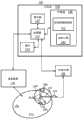
图1是方框图,示出了根据一个例示性实施例采用温度反馈的消融系统;FIG. 1 is a block diagram illustrating an ablation system employing temperature feedback according to an exemplary embodiment;
图2是示出了根据一个例示性实施例利用RFA探针通过多次消融进行的消融程序的图示,部署探针,使得标记物与特定的尖头对准;FIG. 2 is a diagram illustrating an ablation procedure using an RFA probe through multiple ablations, the probe being deployed such that markers are aligned with specific prongs, according to an exemplary embodiment;
图3是曲线图,示出了根据一个例示性实施例要在计算新的消融形状时采用的消融直径和温度之间的关系;3 is a graph illustrating the relationship between ablation diameter and temperature to be employed in calculating a new ablation shape according to an exemplary embodiment;
图4是图表和曲线图,示出了RFA探针中嵌入的五个温度传感器的每个的测得温度,对其进行内插以形成根据一个例示性实施例的曲线;4 is a graph and graph showing measured temperatures for each of the five temperature sensors embedded in the RFA probe, interpolated to form a curve according to an illustrative embodiment;
图5A是曲线图,示出了图4的曲线和圆形轮廓线,示出了符合消融的标称或预期形状的每个传感器上的均等温度;FIG. 5A is a graph showing the curve and circular contour lines of FIG. 4 showing the equal temperature on each sensor conforming to the nominal or expected shape of the ablation;
图5B是曲线图,示出了图4的曲线和由样条计算的调整的轮廓线,用于根据这些原理调整消融形状;Figure 5B is a graph showing the curve of Figure 4 and the adjusted contour calculated by splines for adjusting the ablation shape according to these principles;
图6是流程图,示出了根据一个例示性实施例采用温度反馈的消融方法;以及FIG. 6 is a flowchart illustrating a method of ablation employing temperature feedback, according to an exemplary embodiment; and
图7是例示性示出了图像的图,该图像具有规划处置体积(PTV)并具有叠加在目标区上的多个消融形状,从而可以根据这些原理进行进一步的消融规划。Fig. 7 is a diagram schematically showing an image with a planned treatment volume (PTV) with multiple ablation shapes superimposed on the target zone so that further ablation planning can be performed according to these principles.
具体实施方式Detailed ways
本公开描述了用于规划利用多次精确放置的射频消融(RFA)进行最佳肿瘤覆盖的系统和方法。市售RFA电极生成的消融体积由制造商标称定义为具有特定直径的球体或椭球体形状。不过,在实际程序中,实现的消融形状/尺寸随着患者而变化并取决于肿瘤局部环境的热沉效应,例如与血管的接近程度、这些血管中的尺寸或血流速率等。The present disclosure describes systems and methods for planning optimal tumor coverage with multiple precisely placed radiofrequency ablation (RFA). The ablation volume generated by commercially available RFA electrodes is nominally defined by the manufacturer as a sphere or ellipsoid shape with a specific diameter. However, in practical procedures, the achieved ablation shape/size varies patient by patient and depends on the heat sink effect of the local tumor environment, such as proximity to blood vessels, size or blood flow rate in these vessels, etc.
根据本原理,RFA电极可以嵌入温度传感器以在消融期间监测实现的温度。由RF发生器使用这一温度信息,RF发生器驱动电极以调整消融过程。也可以由临床医师定性地监测温度以判断消融过程的安全性、有效性、完成等。在一个实施例中,以定量方式集成温度测量以估计完成单次消融时实现的消融形状/尺寸。使用这一信息计算残余肿瘤体积,允许更新的计划以计算最佳地覆盖残余肿瘤的必要消融次数和放置。根据本原理,采用来自RFA电极的温度测量估计实现的消融尺寸/形状以用作反馈系统。According to the present principles, RFA electrodes can be embedded with temperature sensors to monitor the achieved temperature during ablation. This temperature information is used by the RF generator, which drives the electrodes to adjust the ablation process. Temperature may also be monitored qualitatively by a clinician to judge the safety, effectiveness, completion, etc. of the ablation procedure. In one embodiment, temperature measurements are integrated in a quantitative manner to estimate the ablated shape/size achieved when a single ablation is performed. Using this information to calculate the residual tumor volume allows for an updated plan to calculate the number of ablations and placements necessary to optimally cover the residual tumor. According to the present principles, the achieved ablation size/shape is estimated using temperature measurements from the RFA electrodes to serve as a feedback system.
RFA探针可以包括嵌入针形电极顶端(或个体尖中)中的温度传感器,以感测被消融组织的温度。对于多尖RFA探针(例如,来自AngiodynamicsTM的StarburstTM XL/XLi RFA电极),驱动RFA探针的RF发生器使用消融完成的标准。一种这样的标准是,多个传感器间的平均温度测量值大于用户为预指定时间量指定的目标温度。The RFA probe may include a temperature sensor embedded in the needle electrode tip (or in the individual tip) to sense the temperature of the ablated tissue. For multi-tip RFA probes (eg, Starburst™ XL/XLi RFA electrodes from Angiodynamics™ ), the RF generator driving the RFA probe uses the standard of ablation completion. One such criterion is that the average temperature measurement across multiple sensors is greater than a user-specified target temperature for a pre-specified amount of time.
在一个特别有用的实施例中,可以使用在消融结束时每个尖头上测量的温度来感测由于存在附近血管中的血流造成的任何热损失。大血管附近的尖头将可能记录到与远离任何血管的尖头相比更低的组织中实现的温度。可以在RF发生器上连续显示这种温度测量值,RF发生器在消融期间向组织中插入的RFA探针供电。在消融期间或在完成消融时有利地采用温度测量以估计形状相对于制造商针对对应RF发生器驱动的具体RFA电极指定的标称或预定形状/尺寸而言的可能变化。In one particularly useful embodiment, the temperature measured on each tine at the end of the ablation can be used to sense any heat loss due to the presence of blood flow in nearby vessels. A cusp near a large vessel will likely register a lower temperature achieved in tissue than a cusp farther from any vessel. This temperature measurement can be continuously displayed on the RF generator that powers the RFA probe inserted in the tissue during ablation. Temperature measurements are advantageously employed during ablation or upon completion of ablation to estimate possible changes in shape relative to the nominal or predetermined shape/dimensions specified by the manufacturer for the particular RFA electrode driven by the corresponding RF generator.
应当理解,将按照医疗仪器,尤其是消融仪器来描述本发明;不过,本发明的教导要宽得多,适用于利用热或热生成技术或冷却技术,例如低温消融等破坏组织时采用的任何仪器。具体而言,本原理适用于身体中所有区域,例如肺、胃肠道、排泄器官、血管等中的程序。图中描绘的元件可以实现于硬件和软件的各种组合中并提供可以在单个元件或多个元件中组合的功能。It should be understood that the present invention will be described in terms of a medical instrument, particularly an ablation instrument; however, the teachings of the present invention are much broader and apply to any instrument used to destroy tissue using heat or heat generating techniques or cooling techniques, such as cryogenic ablation. instrument. In particular, the principles apply to procedures in all areas of the body, such as the lungs, gastrointestinal tract, excretory organs, blood vessels, and the like. Elements depicted in the figures may be implemented in various combinations of hardware and software and provide functions that may be combined in a single element or in multiple elements.
可以利用专用硬件以及能够执行与适当软件相关联的软件的硬件提供图中所示的各种元件的功能。在由处理器提供时,可以由单个专用处理器,由单个共享处理器,或由多个个体处理器(其中一些可能是共享)提供功能。此外,具体的使用术语“处理器”或“控制器”不应被解释为排他地指能够执行软件的硬件,而是可能暗含地包括,但不限于数字信号处理器(“DSP”)硬件、用于存储软件的只读存储器(“ROM”)、随机存取存储器(“RAM”)、非易失性存储器等。The functions of the various elements shown in the figures can be provided by dedicated hardware as well as hardware capable of executing software in association with appropriate software. When provided by a processor, the functionality may be provided by a single dedicated processor, by a single shared processor, or by multiple individual processors, some of which may be shared. Furthermore, specific use of the terms "processor" or "controller" should not be construed to refer exclusively to hardware capable of executing software, but may implicitly include, but is not limited to, digital signal processor ("DSP") hardware, Read-only memory (“ROM”), random-access memory (“RAM”), non-volatile memory, etc., used to store software.
此外,这里提到原理、方面和本发明实施例的所有陈述以及其具体范例都意在涵盖其等价结构和功能。此外,这样的等价物应包括当前已知的等价物以及将来开发的等价物(即,开发的执行相同功能的任何元件,不论结构如何)。于是,例如,本领域的技术人员将认识到,这里给出的方框图代表体现本发明原理的例示性系统部件和/或电路的概念图。类似地,要认识到,任何流程图等表示各种过程,基本可以在计算机可读存储介质中表示各种过程并由计算机或处理器这样执行,无论是否明确示出了这样的计算机或处理器。Moreover, all statements herein referring to principles, aspects, and embodiments of the invention, as well as specific examples thereof, are intended to encompass equivalent structures and functions thereof. Additionally, it is intended that such equivalents include both currently known equivalents as well as equivalents developed in the future (ie, any elements developed that perform the same function, regardless of structure). Thus, for example, it will be appreciated by those skilled in the art that the block diagrams presented herein represent conceptual views of illustrative system components and/or circuits embodying the principles of the invention. Similarly, it is to be appreciated that any flow diagrams and the like represent various processes, which can generally be represented in a computer-readable storage medium and so executed by a computer or processor, whether or not such computer or processor is explicitly shown .
此外,本发明的实施例可以采取能够从计算机可用或计算机可读存储介质访问的计算机程序产品的形式,提供程序代码,供计算机或任何指令执行系统使用或结合其使用。出于本说明书的目的,计算机可用或计算机可读存储介质可以是可以包括、存储、传送、传播或传输程序的任何设备,程序供指令执行系统、设备或装置使用或结合其使用。该介质可以是电子的、磁性的、光学的、电磁的、红外的或半导体系统(或设备或装置)或传播介质。计算机可读介质的范例包括半导体或固态存储器、磁带、可移除计算机盘、随机存取存储器(RAM)、只读存储器(ROM)、刚性磁盘和光盘。当前光盘的范例包括高密度磁盘–只读存储器(CD-ROM)、高密度磁盘–读/写(CD-R/W)和DVD。Furthermore, embodiments of the invention may take the form of a computer program product accessible from a computer-usable or computer-readable storage medium, providing program code for use by or in connection with a computer or any instruction execution system. For the purposes of this specification, a computer-usable or computer-readable storage medium may be any device that can contain, store, communicate, propagate or transport the program for use by or in connection with the instruction execution system, device or apparatus. The medium may be an electronic, magnetic, optical, electromagnetic, infrared or semiconductor system (or device or arrangement) or a propagation medium. Examples of computer readable media include semiconductor or solid state memory, magnetic tape, removable computer disks, random access memory (RAM), read only memory (ROM), rigid magnetic disks, and optical disks. Current examples of optical disks include Compact Disk - Read Only Memory (CD-ROM), Compact Disk - Read/Write (CD-R/W) and DVD.
现在参考附图,其中类似数字表示相同或类似元件,一开始参考图1,例示性地示出了系统100,其能够利用温度反馈定量地估计消融形状和尺寸。系统100包括射频消融(RFA)探针102(例如单尖头或多尖头探针),具有一个或多个嵌入的温度传感器104(例如,沿着单个尖头或在多个尖头上安装)。温度传感器104优选安装在RFA探针102上或其中,但可以采用测量温度的其他方式。例如,可以在该区域中插入独立的装置(例如针)以测量组织温度。Referring now to the drawings, in which like numerals represent the same or similar elements, and initially to FIG. 1 , there is illustratively shown a
其他装置可以包括具有传感器或成像装备的导管或装置,以通过接触或间接测量(例如热成像)提供温度测量。在一个实施例中,探针102可以包括仅单个温度传感器(例如,在单针或三元组群探针中)。如果RFA探针102没有嵌入的温度传感器104,可以采用插入目标体积附近,在其顶端具有嵌入的温度传感器104的精细量规的针,以确定消融区周围的温度分布。Other devices may include catheters or devices with sensors or imaging equipment to provide temperature measurements by contact or indirect measurements such as thermal imaging. In one embodiment,
RF发生器106向工作站108传送温度数据,工作站108利用例如显示器110向用户提供规划/导航/消融反馈信息。工作站108可以包括计算机处理器112、显示器110、用户接口114(例如鼠标、键盘等)和用于存储数据和软件的存储器120。存储器120包括软件,其可以包括轮廓线模块122,配置成采用温度反馈信息确定围绕消融部位的消融体积的最可能形状/尺寸。The
模块122相对于固定在探针102别处的标记物124确定温度传感器104的位置。通过这种方式,针对进行给定测量,已知温度传感器相对于标记物124的位置。模块122能够确定探针102上的温度传感器104以及标记物124的位置,探针102在利用医学成像模态126(例如CT、超声)的具体消融处置期间被部署。成像模态126包括扫描机或其他成像装置(例如CT、US、X射线等)。可以为扫描采用超声或(旋转)X射线成像(例如,替代CT)。如果采用多尖头探针102,不必从CT数据逐个提取尖头,例如,利用EM跟踪,如果已知温度传感器相对于标记物124的位置。
标记物124可以可选地包括空间跟踪系统/装置,可用于确定RFA探针相对于医学图像的位置(而非使用RFA探针的CT图像)。例如,可以在RFA探针上放置6自由度电磁(EM)跟踪传感器作为标记物124。然后可以相对于这个固定的EM跟踪的传感器识别(一次标定)RFA尖头203上的温度传感器104的位置。这样能够在消融期间知道温度传感器104的位置,并能够在利用到EM跟踪传感器的空间配准采集的任何模态图像上在空间上精确配准温度数据。
模块122从RF发生器106收集连续温度数据并显示叠加在成像扫描上的温度数据,示出插入到PTV中的RFA探针102。利用RFA探针上部署的标记物在空间上正确的位置示出了叠加的温度数据。可以将温度数据转换成沿平行赤道面上的径向的估计消融直径以表示实现的消融形状。可以显示区域中的特定点的温度数据,也可以在CT扫描和PTV上叠加消融形状,以显示PTV内部的被消融体素的体积。RF消融反馈方法可以从整个PTV减去PTV内部的被消融体素以计算残余PTV。残余PTV代表未处置的区域,并且是对更新RFA计划的输入。此外,温度反馈或计算的消融体积可以采用其他成像信息,例如,实时超声回声反射性,以显示被消融区域。
基于温度反馈,模块122能够基于温度数据计算实现的消融形状或处置区域。使用受试者内部区域的映射体积或其他图像,可以通过在针对病灶或肿瘤的规划目标体积(PTV)上叠加温度分布而在显示器110上对温度数据进行可视化。使用温度数据,模块122计算到利用RFA探针102消融的形状和尺寸的变换。这种变换的消融形状可以叠加在PTV上,使得临床医师或医生能够对受影响区域的更精确版本进行可视化。Based on the temperature feedback,
模块122还规定,通过减去也在原始PTV内部且在被变换消融形状内部的所有消融体素来计算和可视化残余PTV。利用用于反馈的变换消融形状执行消融程序期间对残余PTV的计算。
RFA规划工具130可以存储在存储器120中并计算消融的最佳次数和放置以在程序期间进行消融时覆盖整个PTV或残余PTV。采用制造商指定的理想尺寸和形状的消融来规划RFA程序。在未灌注和完美均质组织的假设下,可以实现这个目的;但对于在灌注的非均质组织中消融,这种形状是不切实际的。附近血管中的血流通过局部冷却消融区改变消融的形状。如果传感器104在特定尖头(对于多尖头探针102而言)中测量的最高温度低于50℃,那么被感测区域周围的组织不太可能被消融。系统100通过将传感器的3D位置几何配准到插入探针的组织区域实现来自每个传感器104的温度读数的相关。这样能够确定温度图并在消融形状上叠加,因此,从制造商指定的预定形状估计形状的可能变化。
使用温度作为反馈手段允许医生采用探针102或具有其嵌入的温度传感器104的消融装置自身来估计消融形状134。这样允许在消融区周围的图像中对解剖特征上叠加的实际温度进行可视化。针对每次温度测量估计消融半径,计算与那个变换的消融形状对应的3维体积。估计残余的PTV136不需要造影剂超声或造影剂CT扫描,因为可以利用温度反馈控制其,从而估计沿着消融特定方向的热损失影响,并提供了基于来自RFA探针102的温度反馈估计消融形状的计算。Using temperature as a means of feedback allows the physician to estimate the
参考图2并继续参考图1,例示性地示出了多尖头探针102。探针102的尖头203可以包括温度传感器104。从RF发生器106向外部计算机(工作站108)提供温度数据,计算机进行RFA程序规划、导航和反馈。可以通过任何适当的方法,例如USB、串行端口、并行端口、网络接口等提供这种数据。多尖头RFA探针102的形状可以是圆柱形对称的。温度传感器104布置于这些尖头中的一些上,但我们事先不知道哪个传感器指示特定尖头203上的温度。标记物124位于RFA探针102上,将来自传感器的温度精确地对准到特定的尖头。例如,如果多尖头探针具有五个温度传感器,标记为#1,2,3,4和5,那么制定规约,即标记物124与记录传感器#1上的温度读数的尖头203共同取向。这个标记物124应当由例如在CT扫描中容易辨别的材料制造,因为它应当与部署的尖头相关(在CT下这是容易可见的),由此与沿该特定尖头203方向的温度测量相关。Referring to FIG. 2 with continued reference to FIG. 1 , a
标记物124应当牢固附着于RFA探针102而不会干扰RFA探针102的治疗功能。可能的位置可以是探针柄或距探针柄最近的绝缘轴上。图2中将标记物124示为与标记为#1的尖头203对准。The
由探针制造商指定由发生器106驱动的消融探针102,以产生具有指定消融直径的球状体或椭球体形状的消融。椭球体形状的消融具有三个独立的直径。球状体形状的消融具有三个直径,其中两个是相同的。在实践中,将圆柱形对称消融指定为球状体。垂直于RFA探针102的轴(柄到针尖)的消融直径是赤道直径,平行于RFA探针102的轴的消融直径是极直径。
在图2中,第一轮219在远端将消融探头102推进到病灶210的组织中,消融程序导致热消融212。然后,在近端缩回探针以进一步执行额外的消融214和216。程序继续进行新的轮次220,实现消融218。继续这样的操作,直到完全处置病灶210为止。被消融的组织优选延伸超过病灶210,但未必破坏健康组织。In FIG. 2 , the ablation procedure results in thermal ablation 212 as the first round 219 advances the
对于每次消融,通常利用消融终止方法编程控制RF发生器106,例如该方法让用户指定在认为消融完成之前探针记录的平均温度超过特定值所需的时间。一旦满足指定的条件,RF发生器106就切断供应给RFA探针102的电力。在理想状态下,如果所有温度传感器104都记录了从开始消融到消融结束温度的均匀升高,并且如果所有对称设置的传感器104上记录的最终温度都相等,那么预计所实现消融的赤道和极直径是最佳的。可以假设这些最佳直径等于制造商指定的消融直径,或者可以从先前的标定(calibration)研究或通过利用有限元模型的模拟研究获得它们。For each ablation, the
对于单个温度传感器探针102,可以使用温度数据直接修改赤道面中被消融区的单个直径。这使得总体消融形状保持为球状体,但考虑了影响总体消融尺寸的组织中微灌注的影响。如果由独立于探针102的多个针或其他装置测量温度,可以通过相对于标定研究等中的理想温度估计消融期间记录的实际温度来推断消融形状(例如,在图3中)。从消融电极顶端到温度传感器的距离将影响温度。For a single
例如,可以在均质未灌注组织上进行标定研究(即预计所有尖头203都记录几乎相等的温度),其中,将来自组织学/消融后造影剂CT或MR扫描的实际消融直径与恰好在发生器断电以标记完成消融之前消融结束时记录的温度测量相关。这种标定研究会根据尖头上记录的平均温度记录实际消融直径。For example, a calibration study (i.e., all
参考图3,例示性示出了消融直径与温度之间的关系曲线300。曲线300将消融期间测量的温度与消融形状的几何形状相关。可以向模块122中编写这样的函数以实现图形显示,示出CT或MRI扫描中的被消融区域。可以采用具有不同的温度分布图或与其他条件相关的其他曲线(例如,不对称的温度分布)以调整所显示的形状和尺寸。Referring to FIG. 3 , a
也可以采用其他信息(例如患者的临床病况、总体灌注等)或模型(例如在RFA探针附近给定分段血管下的热传递的有限元建模)以确定在存在血流的情况下消融可能被如何改变的关系。例如,在距消融已知距离、取向、流量等处有血管的冷却效应时的有限元建模(FEM)使得能够计算温度传感器位置处的温度。Other information (e.g., patient's clinical condition, overall perfusion, etc.) or models (e.g., finite element modeling of heat transfer under a given segmented vessel in the vicinity of the RFA probe) can also be employed to determine the ablation rate in the presence of blood flow. how the relationship might be changed. For example, finite element modeling (FEM) with the cooling effect of a vessel at a known distance from ablation, orientation, flow, etc. enables calculation of the temperature at the temperature sensor location.
知道了来自RFA探针102的温度数据的不对称性,就可以推断附近的血流,可以利用FEM模型关系替代将消融直径与从图3的标定研究导出的温度相关的关系来调整消融形状。Knowing the asymmetry of the temperature data from the
再次参考图2并继续参考图1,可以使用CT扫描在向临床医师(或介入式放射科医师)批准的精确位置推进探针时确认RFA探针102的位置。临床医师也可以使用RFA规划工具130像图2中那样对一组交叠的消融进行计算和可视化。CT扫描示出了所部署RFA探针102连同包括标记物124的所暴露尖头的每个的最终位置。标记物的取向会与尖头203上的特定温度传感器(例如,假设是编号为#1的传感器)以及不同尖头上的其他温度传感器104(相对于传感器#1处于已知的几何位置)对准。可以在CT扫描上分割或跟踪尖头203。每个尖头上的温度传感器104的已知空间位置对应于组织中的特定体素。可以从RFA探针的设计获知这个位置,例如,可以在尖头的顶端嵌入温度传感器104,或者可以距尖头的顶端已知距离。几何配准允许本系统/方法在CT扫描中的特定组织点上叠加从RF发生器106传送的特定温度值。Referring again to FIG. 2 with continued reference to FIG. 1 , a CT scan can be used to confirm the position of the
一旦通过几何方式将来自传感器104的温度配准到组织中的特定体素,然后估计对消融形状的影响。如果在组织中均匀地部署所有尖头,那么温度传感器104(通常对称设置于每个尖头203上)将落在唯一的三维平面上。因此,与这些感测的温度点对应的体素将落在平行于理想消融球状体的赤道平面的几何平面上。Once the temperature from the
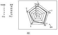
参考图4,曲线图302示出了在消融中的某个点从五个传感器位置#1-5记录的温度数据曲线304的范例。这个范例示出了范围从65℃-90℃的不同温度,平均温度为79.2℃。可以编程控制RF发生器(106)以在平均温度高于60℃超过5分钟时终止消融。这被认为是成功的消融。不过,尖头#3(5点钟位置)上的温度为90℃,相对于尖头#4(7点钟位置),仅为65℃。这意味着消融形状可能有一些不均匀性。Referring to FIG. 4 ,
例如,使用图3的函数,我们现在可以利用内插法(例如线性、样条等)为这种温度分布估计可能的消融形状。例如,如果对于105℃的温度(根据图3)要实现均匀的最大消融直径4.0cm,消融直径在所有方向上将是均匀的4cm。图5A示出了均一的圆401,指示在105℃温度下球状消融4cm的赤道直径。不过,对于图4的温度分布(曲线304),将如图5B中所示变换消融直径。图5B示出了样条内插402以及线性内插304。在图5A和5B中,垂直线刻度408示出了从0到5cm的实现直径,传感元件编号1-5分布于多边形外部。如果传感器测量到105度C(来自图3),消融形状的直径为4cm。对所有温度测量进行这种操作以将温度相关到消融体积的直径。For example, using the function of Figure 3, we can now estimate possible ablation shapes for this temperature distribution using interpolation methods (e.g. linear, spline, etc.). For example, if a uniform maximum ablation diameter of 4.0 cm is to be achieved for a temperature of 105°C (according to Figure 3), the ablation diameter will be a uniform 4 cm in all directions. Figure 5A shows a uniform circle 401 indicating an equatorial diameter of 4 cm for spherical ablation at a temperature of 105°C. However, for the temperature profile of Figure 4 (curve 304), the ablation diameter would be transformed as shown in Figure 5B. FIG. 5B shows
这个平面中的消融形状不再是圆,而是一般化的2维闭合轮廓线(402)。这条轮廓线402反映了平行于消融赤道平面的单个平面上的消融形状。沿着离开赤道平面的平行平面,使用这种被变换形状的成比例缩小版本。比例常数仅仅是该平面上原始均一圆相对于赤道平面上圆直径的直径。这产生了沿消融球状体极轴的变形消融形状。The ablation shape in this plane is no longer a circle, but a generalized 2-dimensional closed contour ( 402 ). This
从数学上讲,可以利用模块122确定变换的消融形状而执行的步骤如下:Mathematically, the steps that can be performed to determine the transformed ablation
1、消融球状体在每个赤道平面(x-y平面)中具有圆形轮廓线。针的轴表示z坐标。圆的直径随着极坐标(z)而改变。假设D(z)是表示在值z(极值)处均一消融(圆形轮廓线)的直径的函数。消融的赤道平面为固定值z=z0。1. The ablation spheroid has circular outlines in each equatorial plane (xy plane). The axis of the needle represents the z coordinate. The diameter of the circle varies with the polar coordinate (z). Suppose D(z) is a function representing the diameter of a uniform ablation (circular contour line) at a value z (extreme). The equatorial plane for ablation is a fixed value z=z0 .
2、在赤道平面上利用观测的温度分布(图4)和测得的将消融直径与观测温度相关的标定(图3)计算变换的轮廓线f(x,y,z=z0)。2. Calculate the transformed contour line f(x, y, z=z0) on the equatorial plane using the observed temperature distribution (Fig. 4) and the measured calibration (Fig. 3) that relates the ablation diameter to the observed temperature.
3、对于平行于赤道平面的所有平面,由值D(z=z0+d)/D(z=z0)按比例缩放相对于圆中心的f(x,y,z=z0+d)上的变换轮廓线点。这样造成了扭曲的消融形状,与均一的消融球状体相比,扭曲的消融形状是在所有赤道平面上按比例缩小的。3. For all planes parallel to the equatorial plane, scale f(x, y, z=z0 +d) relative to the center of the circle by the value D(z=z 0 +d)/D(z=z0 ) ) on transformed contour points. This results in a distorted ablation shape that is scaled down in all equatorial planes compared to a uniform ablation spheroid.
这些多个轮廓线产生3维消融形状,反映沿传感器位置的测得温度的影响。残余PTV是减去这个3维消融形状中包括的所有体素之后原始PTV剩余的部分。These multiple contours generate a 3-dimensional ablation shape reflecting the effect of measured temperature along the sensor location. The residual PTV is what remains of the original PTV after subtracting all voxels included in this 3D ablation shape.
根据一个实施例,单尖头传感器(例如参见图7)可以包括其上设置一个或多个温度传感器的针或导管。如果沿着针的极轴而非赤道平面定位一个或多个温度传感器,可以使用个体温度测量值按比例缩放传感器所在赤道平面中标准消融形状的标称直径。对于球状体形状的标准消融,赤道平面(包括温度传感器的位置)中的消融轮廓线是具有标称直径D(z=z1)的圆。图3建立了针对给定传感器位置的测得温度和该赤道平面中实现的消融直径之间的关系。针对给定传感器位置测量的实际温度决定了针对传感器平面的所实现消融直径。与传感器平面中实现的消融直径成比例地缩放针对其他平面实现的消融直径。According to one embodiment, a single-tip sensor (see, eg, FIG. 7 ) may comprise a needle or catheter on which one or more temperature sensors are disposed. If one or more temperature sensors are positioned along the needle's polar axis rather than the equatorial plane, individual temperature measurements can be used to scale the nominal diameter of the standard ablation shape in the equatorial plane where the sensors are located. For standard ablation in the shape of a spheroid, the ablation contour in the equatorial plane (including the location of the temperature sensor) is a circle with a nominal diameter D (z=z1). Figure 3 establishes the relationship between the measured temperature for a given sensor location and the ablation diameter achieved in that equatorial plane. The actual temperature measured for a given sensor location determines the achieved ablation diameter for the sensor plane. The ablation diameter achieved for the other planes is scaled proportionally to the ablation diameter achieved in the sensor plane.
参考图6,根据一个实施例示出了使用温度信息作为反馈消融组织的方法。在方框502中,任选地开发初步处置计划以处置目标组织。这可以包括在受试者体内界定规划处置体积(PTV)。在方框504中,将消融探针引导到受试者体内的目标组织。Referring to FIG. 6 , a method of ablating tissue using temperature information as feedback is shown according to one embodiment. In block 502, a preliminary treatment plan is optionally developed to treat the target tissue. This can include defining a planned treatment volume (PTV) within the subject. In block 504, an ablation probe is directed to a target tissue in a subject.
在方框508,在选择的位置,为消融探针供电以破坏组织,优选根据处置计划供电。在方框510中,在消融期间(和消融之后),在目标组织处或附近监测温度。在目标周围利用温度传感器收集温度信息,温度传感器可以安装在探针上或独立放置(例如针等)。如果温度传感器是独立放置的,可以接近目标插入它们,以测量消融体积周围的温度。也可以利用多尖头消融探针上的温度传感器测量温度信息。在方框511中,针对已知位置收集温度信息。可以通过多种方式确定位置,例如,可以采用标记物或跟踪器识别温度传感器的位置并向利用成像模态采集的图像提供空间配准,将传感器位置配准到被处置组织中的空间位置。At block 508, at the selected location, the ablation probe is powered to destroy tissue, preferably according to the treatment plan. In block 510, temperature is monitored at or near the target tissue during (and after) ablation. Temperature information is collected around the target using temperature sensors, which can be mounted on probes or placed independently (such as needles, etc.). If temperature sensors are placed independently, they can be inserted close to the target to measure the temperature around the ablation volume. Temperature information can also be measured using a temperature sensor on the multi-tipped ablation probe. In block 511, temperature information is collected for known locations. The location can be determined in a number of ways, for example, markers or trackers can be used to identify the location of the temperature sensor and provide spatial registration to images acquired with the imaging modality, registering the sensor location to the spatial location in the treated tissue.
在方框512中,基于温度信息确定消融体积的形状。采用温度信息确定是否需要因为温度分布的扭曲而修改消融形状。这些扭曲可以是血流、受试者解剖或其他影响的结果。可以在方框514中通过根据消融几何形状和温度之间的关系变换标准形状消融体积来确定消融体积的形状。该关系可以包括标定研究、模型,例如有限元模型等。在一个实施例中,在方框516中,通过根据该关系利用内插计算消融轮廓线并成比例地调整其他区域中的形状以提供消融的形状来变换或调整标准形状。In block 512, the shape of the ablation volume is determined based on the temperature information. The temperature information is used to determine whether the ablation shape needs to be modified due to the distortion of the temperature distribution. These distortions may be the result of blood flow, subject anatomy, or other influences. The shape of the ablation volume may be determined in block 514 by transforming the standard shape ablation volume according to the relationship between the ablation geometry and temperature. The relationship may include calibration studies, models, such as finite element models, and the like. In one embodiment, in block 516, the standard shape is transformed or adjusted by interpolating the ablation contour from the relationship and proportionally adjusting the shape in other regions to provide the ablated shape.
在方框520中,在显示器上相对于目标的图像显示形状及其扭曲等。通过这种方式,可以看出被消融区域的更精确图示,这减小了程序之后未处置肿瘤或其他组织仍然存在的机会。显示的图像可以包括在方框522中叠加在受试者的三维图像上的消融体积的形状。通过这种方式,可以相对于图像、规划目标体积(PTV)、其他消融等对目标的被消融部分进行可视化。In block 520, the shape, its distortion, etc. are displayed on a display relative to the image of the target. In this way, a more accurate representation of the area being ablated can be seen, which reduces the chance of untreated tumor or other tissue remaining after the procedure. The displayed image may include the shape of the ablation volume superimposed on the three-dimensional image of the subject in block 522 . In this way, the ablated portion of the target can be visualized relative to the image, planning target volume (PTV), other ablations, etc.
在方框524中,可以根据PTV继续后续消融并加以显示。优选从视觉上将PTV与流程期间估计的多个变换消融形状比较。图7示出了这种显示器是例示性范例。多个变换的消融形状界定残余PTV。在方框526中计算具有额外消融的计划以覆盖残余PTV,确保处置原始PTV的全部。In block 524, subsequent ablation may be continued and displayed based on the PTV. The PTV is preferably compared visually to a plurality of transformed ablation shapes estimated during the procedure. Figure 7 shows that such a display is an illustrative example. The plurality of transformed ablation shapes define a residual PTV. A plan with additional ablation is calculated in block 526 to cover the residual PTV, ensuring that all of the original PTV is treated.
参考图7,示出了例示性显示图像602。图像602包括患者610内部目标区域604的CT扫描或其他图像。在这一实施例中,采用单尖头装置620。单尖头装置620沿其长度可以包括一个或多个传感器104。该装置可以是针、导管或其他装置。也可以采用标记物124以辅助定位装置620。产生并在图像602中示出PTV区域612。此外,利用PTV示出通过温度反馈修改的消融体积614以促进根据本原理的高度精确手术程序。Referring to FIG. 7 , an
在解释所附的权利要求时,应当理解:In interpreting the appended claims, it should be understood that:
a)“包括”一词不排除有给定权利要求中列出的那些之外的其他元件或动作;a) the word "comprising" does not exclude the presence of other elements or acts than those listed in a given claim;
b)元件前的“一”一词不排除存在多个这样的元件;b) the word "a" preceding an element does not exclude the presence of a plurality of such elements;
c)权利要求中的任何附图标记都不限制其范围;c) any reference signs in the claims do not limit their scope;
d)可以由同一项目或硬件或软件实现的结构或功能代表几个“模块”;并且d) structures or functions that can be implemented by the same item or hardware or software represent several "modules"; and
e)除非具体指出,并不要求动作有具体的顺序。e) Unless specifically stated, no specific order of actions is required.
已经描述了具有温度反馈以用于自适应射频消融的系统和方法的优选实施例(意在是例示性的而非限制性的),要指出的是,本领域的技术人员根据以上教导能够做出修改和变化。因此要理解,可以在所披露的公开的特定实施例中做出改变,这些改变在所附权利要求勾勒出的所公开实施例的范围之内。这样描述完专利法要求的细节和特性之后,在所附权利要求中阐述专利证书主张并希望保护的范围。Having described preferred embodiments of systems and methods with temperature feedback for adaptive radiofrequency ablation (intended to be illustrative and not limiting), it is noted that those skilled in the art, given the above teachings, will be able to do modifications and variations. It is therefore to be understood that changes may be made in the particular embodiments disclosed which are within the scope of the disclosed embodiments outlined by the appended claims. Having thus described the details and particularity required by the patent laws, what is claimed and desired protected by Letters Patent is set forth in the appended claims.
Claims (31)
Applications Claiming Priority (3)
| Application Number | Priority Date | Filing Date | Title |
|---|---|---|---|
| US38754910P | 2010-09-29 | 2010-09-29 | |
| US61/387,549 | 2010-09-29 | ||
| PCT/IB2011/054163WO2012042443A1 (en) | 2010-09-29 | 2011-09-22 | System and method for temperature feedback for adaptive radio frequency ablation |
Publications (2)
| Publication Number | Publication Date |
|---|---|
| CN103140184Atrue CN103140184A (en) | 2013-06-05 |
| CN103140184B CN103140184B (en) | 2016-05-11 |
Family
ID=44802331
Family Applications (1)
| Application Number | Title | Priority Date | Filing Date |
|---|---|---|---|
| CN201180046806.9AExpired - Fee RelatedCN103140184B (en) | 2010-09-29 | 2011-09-22 | The temperature feedback system melting for adaptive RF |
Country Status (6)
| Country | Link |
|---|---|
| US (1) | US10561462B2 (en) |
| EP (1) | EP2621388B1 (en) |
| JP (1) | JP6448905B2 (en) |
| CN (1) | CN103140184B (en) |
| BR (1) | BR112013007027A2 (en) |
| WO (1) | WO2012042443A1 (en) |
Cited By (9)
| Publication number | Priority date | Publication date | Assignee | Title |
|---|---|---|---|---|
| WO2015066994A1 (en)* | 2013-11-05 | 2015-05-14 | 深圳迈瑞生物医疗电子股份有限公司 | Ultrasonic intervention ablation system and working method therefor |
| CN106659529A (en)* | 2014-07-18 | 2017-05-10 | 奥林巴斯株式会社 | Device for ultrasonic energy therapy and method for ultrasonic energy therapy |
| CN107028654A (en)* | 2015-12-24 | 2017-08-11 | 韦伯斯特生物官能(以色列)有限公司 | Temperature during estimation ablation |
| CN112734909A (en)* | 2020-12-31 | 2021-04-30 | 杭州堃博生物科技有限公司 | Radio frequency operation prompting method, electronic device and computer readable storage medium |
| CN112741681A (en)* | 2020-12-31 | 2021-05-04 | 杭州堃博生物科技有限公司 | Radio frequency operation prompting method, device and system based on multi-pole radio frequency ablation catheter and storage medium |
| CN112790858A (en)* | 2020-12-31 | 2021-05-14 | 杭州堃博生物科技有限公司 | Ablation parameter configuration method, device, system and computer readable storage medium |
| CN112842514A (en)* | 2020-12-31 | 2021-05-28 | 杭州堃博生物科技有限公司 | Ablation operation prompting method, electronic device and computer readable storage medium |
| CN114259295A (en)* | 2021-12-16 | 2022-04-01 | 杭州堃博生物科技有限公司 | Detection processing method, device, system, equipment and medium for radio frequency ablation |
| CN117179797A (en)* | 2023-11-08 | 2023-12-08 | 北京唯迈医疗设备有限公司 | C-shaped arm X-ray machine |
Families Citing this family (15)
| Publication number | Priority date | Publication date | Assignee | Title |
|---|---|---|---|---|
| CN103717167B (en)* | 2011-07-28 | 2017-06-27 | 皇家飞利浦有限公司 | Ablation planning system |
| GB2514714A (en)* | 2012-03-29 | 2014-12-03 | Spiration Inc | Apparatuses, methods, and systems for the identification and treatment of pulmonary tissue |
| JP6391215B2 (en)* | 2012-05-22 | 2018-09-19 | コヴィディエン リミテッド パートナーシップ | Temperature-based ablation completion algorithm |
| US9498182B2 (en)* | 2012-05-22 | 2016-11-22 | Covidien Lp | Systems and methods for planning and navigation |
| US9259287B2 (en)* | 2013-04-02 | 2016-02-16 | Siemens Aktiengesellschaft | Patient specific planning and simulation of ablative procedures |
| US10098685B2 (en) | 2013-10-30 | 2018-10-16 | Medtronic Cryocath Lp | Feedback system for cryoablation of cardiac tissue |
| CN107635503B (en) | 2015-05-12 | 2021-09-07 | 纳维斯国际有限公司 | Damage estimation by dielectric property analysis |
| US11445911B2 (en)* | 2016-05-25 | 2022-09-20 | Ikomed Technologies Inc. | System for treating unwanted tissue |
| WO2018092071A1 (en)* | 2016-11-16 | 2018-05-24 | Navix International Limited | Estimators for ablation effectiveness |
| US11648062B2 (en)* | 2017-11-09 | 2023-05-16 | Acessa Health Inc. | System for controlling ablation treatment and visualization |
| US20200022774A1 (en)* | 2018-07-19 | 2020-01-23 | David Douglas | Implantable markers to aid surgical operations |
| CN113271881A (en)* | 2019-01-07 | 2021-08-17 | 柯惠有限合伙公司 | System for monitoring ablation progress using a remote temperature probe |
| US10842572B1 (en)* | 2019-11-25 | 2020-11-24 | Farapulse, Inc. | Methods, systems, and apparatuses for tracking ablation devices and generating lesion lines |
| US20220241001A1 (en)* | 2019-12-31 | 2022-08-04 | Broncus Medical Inc. | Lung tumor ablation method |
| CN116077170B (en)* | 2023-01-08 | 2024-02-06 | 天津市鹰泰利安康医疗科技有限责任公司 | Ablation regulation control method and system |
Citations (5)
| Publication number | Priority date | Publication date | Assignee | Title |
|---|---|---|---|---|
| US20020077627A1 (en)* | 2000-07-25 | 2002-06-20 | Johnson Theodore C. | Method for detecting and treating tumors using localized impedance measurement |
| CN1596085A (en)* | 2001-09-28 | 2005-03-16 | 锐达医疗系统公司 | Impedance controlled tissue ablation apparatus and method |
| US20090118613A1 (en)* | 2007-11-01 | 2009-05-07 | Tyco Healthcare Group Lp | Method for Volume Determination and Geometric Reconstruction |
| US20100063496A1 (en)* | 2007-01-24 | 2010-03-11 | Koninklijke Philips Electronics N. V. | Rf ablation planner |
| US20110306969A1 (en)* | 2010-06-09 | 2011-12-15 | Tyco Healthcare Group Lp | System and method for directing energy to tissue and method of assessing ablation size as a function of temperature information associated with an energy applicator |
Family Cites Families (12)
| Publication number | Priority date | Publication date | Assignee | Title |
|---|---|---|---|---|
| US6241725B1 (en)* | 1993-12-15 | 2001-06-05 | Sherwood Services Ag | High frequency thermal ablation of cancerous tumors and functional targets with image data assistance |
| US6575969B1 (en) | 1995-05-04 | 2003-06-10 | Sherwood Services Ag | Cool-tip radiofrequency thermosurgery electrode system for tumor ablation |
| US5800484A (en)* | 1995-08-15 | 1998-09-01 | Rita Medical Systems, Inc. | Multiple antenna ablation apparatus with expanded electrodes |
| US5810804A (en) | 1995-08-15 | 1998-09-22 | Rita Medical Systems | Multiple antenna ablation apparatus and method with cooling element |
| US5833688A (en)* | 1997-02-24 | 1998-11-10 | Boston Scientific Corporation | Sensing temperature with plurality of catheter sensors |
| US7160296B2 (en) | 2001-05-10 | 2007-01-09 | Rita Medical Systems, Inc. | Tissue ablation apparatus and method |
| US7306593B2 (en) | 2002-10-21 | 2007-12-11 | Biosense, Inc. | Prediction and assessment of ablation of cardiac tissue |
| US20060155267A1 (en)* | 2005-01-10 | 2006-07-13 | Nir Berzak | Thermal mapping of a cryoablation volume, for image-guided cryosurgery |
| US20060200121A1 (en) | 2005-03-03 | 2006-09-07 | Mowery Thomas M | Navigable, multi-positional and variable tissue ablation apparatus and methods |
| US8556888B2 (en)* | 2006-08-04 | 2013-10-15 | INTIO, Inc. | Methods and apparatuses for performing and monitoring thermal ablation |
| US8346370B2 (en) | 2008-09-30 | 2013-01-01 | Vivant Medical, Inc. | Delivered energy generator for microwave ablation |
| US8690776B2 (en) | 2009-02-17 | 2014-04-08 | Inneroptic Technology, Inc. | Systems, methods, apparatuses, and computer-readable media for image guided surgery |
- 2011
- 2011-09-22CNCN201180046806.9Apatent/CN103140184B/ennot_activeExpired - Fee Related
- 2011-09-22USUS13/825,353patent/US10561462B2/enactiveActive
- 2011-09-22WOPCT/IB2011/054163patent/WO2012042443A1/enactiveApplication Filing
- 2011-09-22JPJP2013530832Apatent/JP6448905B2/enactiveActive
- 2011-09-22BRBR112013007027-7Apatent/BR112013007027A2/ennot_activeApplication Discontinuation
- 2011-09-22EPEP11770521.0Apatent/EP2621388B1/enactiveActive
Patent Citations (7)
| Publication number | Priority date | Publication date | Assignee | Title |
|---|---|---|---|---|
| US20020077627A1 (en)* | 2000-07-25 | 2002-06-20 | Johnson Theodore C. | Method for detecting and treating tumors using localized impedance measurement |
| CN1596085A (en)* | 2001-09-28 | 2005-03-16 | 锐达医疗系统公司 | Impedance controlled tissue ablation apparatus and method |
| US7344533B2 (en)* | 2001-09-28 | 2008-03-18 | Angiodynamics, Inc. | Impedance controlled tissue ablation apparatus and method |
| US20100063496A1 (en)* | 2007-01-24 | 2010-03-11 | Koninklijke Philips Electronics N. V. | Rf ablation planner |
| CN101795636A (en)* | 2007-01-24 | 2010-08-04 | 皇家飞利浦电子股份有限公司 | The RF ablation planner |
| US20090118613A1 (en)* | 2007-11-01 | 2009-05-07 | Tyco Healthcare Group Lp | Method for Volume Determination and Geometric Reconstruction |
| US20110306969A1 (en)* | 2010-06-09 | 2011-12-15 | Tyco Healthcare Group Lp | System and method for directing energy to tissue and method of assessing ablation size as a function of temperature information associated with an energy applicator |
Cited By (17)
| Publication number | Priority date | Publication date | Assignee | Title |
|---|---|---|---|---|
| WO2015066994A1 (en)* | 2013-11-05 | 2015-05-14 | 深圳迈瑞生物医疗电子股份有限公司 | Ultrasonic intervention ablation system and working method therefor |
| CN106659529A (en)* | 2014-07-18 | 2017-05-10 | 奥林巴斯株式会社 | Device for ultrasonic energy therapy and method for ultrasonic energy therapy |
| CN107028654A (en)* | 2015-12-24 | 2017-08-11 | 韦伯斯特生物官能(以色列)有限公司 | Temperature during estimation ablation |
| CN112734909B (en)* | 2020-12-31 | 2022-02-01 | 杭州堃博生物科技有限公司 | Radio frequency operation prompting method, electronic device and computer readable storage medium |
| WO2022143840A1 (en)* | 2020-12-31 | 2022-07-07 | 杭州堃博生物科技有限公司 | Radio-frequency operation prompting method based on multi-pole radio-frequency ablation catheter, apparatus, system, and storage medium |
| CN112790858A (en)* | 2020-12-31 | 2021-05-14 | 杭州堃博生物科技有限公司 | Ablation parameter configuration method, device, system and computer readable storage medium |
| CN112842514A (en)* | 2020-12-31 | 2021-05-28 | 杭州堃博生物科技有限公司 | Ablation operation prompting method, electronic device and computer readable storage medium |
| CN112790858B (en)* | 2020-12-31 | 2021-11-09 | 杭州堃博生物科技有限公司 | Ablation parameter configuration method, device, system and computer readable storage medium |
| CN112734909A (en)* | 2020-12-31 | 2021-04-30 | 杭州堃博生物科技有限公司 | Radio frequency operation prompting method, electronic device and computer readable storage medium |
| CN114942710A (en)* | 2020-12-31 | 2022-08-26 | 杭州堃博生物科技有限公司 | Ablation operation prompting method, electronic device and computer readable storage medium |
| CN112741681A (en)* | 2020-12-31 | 2021-05-04 | 杭州堃博生物科技有限公司 | Radio frequency operation prompting method, device and system based on multi-pole radio frequency ablation catheter and storage medium |
| WO2022141691A1 (en)* | 2020-12-31 | 2022-07-07 | 杭州堃博生物科技有限公司 | Radio frequency operation prompting method, electronic device, and computer-readable storage medium |
| CN112741681B (en)* | 2020-12-31 | 2022-07-12 | 杭州堃博生物科技有限公司 | Electronic device, radio frequency operation prompting system and storage medium |
| CN114259295A (en)* | 2021-12-16 | 2022-04-01 | 杭州堃博生物科技有限公司 | Detection processing method, device, system, equipment and medium for radio frequency ablation |
| CN114259295B (en)* | 2021-12-16 | 2024-08-09 | 杭州堃博生物科技有限公司 | Detection processing method, device, system, equipment and medium for radio frequency ablation |
| CN117179797A (en)* | 2023-11-08 | 2023-12-08 | 北京唯迈医疗设备有限公司 | C-shaped arm X-ray machine |
| CN117179797B (en)* | 2023-11-08 | 2024-02-06 | 北京唯迈医疗设备有限公司 | C-shaped arm X-ray machine |
Also Published As
| Publication number | Publication date |
|---|---|
| CN103140184B (en) | 2016-05-11 |
| JP2013540517A (en) | 2013-11-07 |
| EP2621388B1 (en) | 2021-11-10 |
| WO2012042443A1 (en) | 2012-04-05 |
| JP6448905B2 (en) | 2019-01-09 |
| EP2621388A1 (en) | 2013-08-07 |
| US10561462B2 (en) | 2020-02-18 |
| BR112013007027A2 (en) | 2020-06-16 |
| US20130184700A1 (en) | 2013-07-18 |
Similar Documents
| Publication | Publication Date | Title |
|---|---|---|
| CN103140184B (en) | The temperature feedback system melting for adaptive RF | |
| CN109419501B (en) | Advanced Current Location (ACL) automatic map rotation for detecting holes in Current Position Map (CPM) maps | |
| JP5685546B2 (en) | A feedback system that integrates interventional planning and navigation | |
| EP2919694B1 (en) | Temperature distribution determining apparatus and method | |
| US10321962B2 (en) | Method for volume determination and geometric reconstruction | |
| EP1554986B1 (en) | Prediction and assessment of ablation of cardiac tissue | |
| EP3016593B1 (en) | Temperature distribution determining apparatus. | |
| CN103732162B (en) | For sensing or disposing Displacement Feedback equipment and the method delivering probe | |
| JP7366535B2 (en) | Graphical user interface (GUI) for displaying the estimated proximity of the cardiac catheter to the esophagus | |
| JP2014511111A (en) | System and method for planning an interventional procedure | |
| EP3104938B1 (en) | Heat sink parameter determination apparatus | |
| US11617621B2 (en) | System and method for multi-probe guidance | |
| JP7366534B2 (en) | Estimating the proximity of the cardiac catheter to the esophagus | |
| Chen et al. | Characterization of tracked radiofrequency ablation in phantom | |
| HK1078451B (en) | Prediction and assessment of ablation of cardiac tissue |
Legal Events
| Date | Code | Title | Description |
|---|---|---|---|
| C06 | Publication | ||
| PB01 | Publication | ||
| C10 | Entry into substantive examination | ||
| SE01 | Entry into force of request for substantive examination | ||
| C14 | Grant of patent or utility model | ||
| GR01 | Patent grant | ||
| CF01 | Termination of patent right due to non-payment of annual fee | ||
| CF01 | Termination of patent right due to non-payment of annual fee | Granted publication date:20160511 |