CN103040455A - Operating microscope monitoring system - Google Patents
Operating microscope monitoring systemDownload PDFInfo
- Publication number
- CN103040455A CN103040455ACN2013100203853ACN201310020385ACN103040455ACN 103040455 ACN103040455 ACN 103040455ACN 2013100203853 ACN2013100203853 ACN 2013100203853ACN 201310020385 ACN201310020385 ACN 201310020385ACN 103040455 ACN103040455 ACN 103040455A
- Authority
- CN
- China
- Prior art keywords
- operating microscope
- laser diode
- monitoring system
- blood flow
- ccd camera
- Prior art date
- Legal status (The legal status is an assumption and is not a legal conclusion. Google has not performed a legal analysis and makes no representation as to the accuracy of the status listed.)
- Pending
Links
Images
Landscapes
- Measuring Pulse, Heart Rate, Blood Pressure Or Blood Flow (AREA)
- Microscoopes, Condenser (AREA)
Abstract
Translated fromChineseDescription
Translated fromChinese技术领域technical field
本发明涉及生物组织血流成像技术领域,尤其涉及一种手术显微镜监测系统。The invention relates to the technical field of biological tissue blood flow imaging, in particular to an operating microscope monitoring system.
背景技术Background technique
在外科手术过程中,术中的血流、血管监测至关重要。局部血流变化对于判断外科手术病人的病情变化及预后有着非常重要的意义。在动脉瘤夹闭术或者血管旁路移植术的手术过程中,脑皮层血流的监测可以帮助评估血流是否恢复到术前水平。此外,也可以通过脑皮层血流进行功能性成像,来定位运动和感觉区域。During surgical operations, intraoperative blood flow and blood vessel monitoring are crucial. Changes in local blood flow are of great significance for judging the changes and prognosis of surgical patients. During aneurysm clipping or vascular bypass grafting, monitoring of cortical blood flow can help assess whether blood flow has returned to preoperative levels. In addition, functional imaging of blood flow through the cortex can be performed to localize motor and sensory areas.
然而能够在手术过程中无干扰的实时获取血流图像的手段并不多。目前在神经外科手术中常用的是吲哚菁绿(ICG)术血管造影,是一种评价脑血管内血流的术中监测技术,这一技术与手术显微镜整合后,可以提供术中动脉瘤、载瘤动脉以及周围相关动脉的实时血流信息,图像清晰,解析度高,可以有效地监测动脉瘤夹闭后是否残留、载瘤动脉是否狭窄及吻合血管、穿通血管是否通畅,对指导手术操作、提高手术质量和改善患者预后有着重要意义。但是该方法缺少定量分析,采集时间过长并且需要注射造影剂,这些缺点限制了它的应用。However, there are not many methods that can obtain blood flow images in real time without interference during the operation. Currently, indocyanine green (ICG) angiography is commonly used in neurosurgery, which is an intraoperative monitoring technique for evaluating blood flow in cerebral vessels. , the real-time blood flow information of the parent artery and surrounding related arteries, the image is clear and the resolution is high, it can effectively monitor whether the aneurysm remains after clipping, whether the parent artery is stenotic, and whether the anastomotic blood vessel and the perforating blood vessel are unobstructed. It is of great significance to improve the operation, improve the quality of surgery and improve the prognosis of patients. However, the lack of quantitative analysis of this method, the long acquisition time and the need to inject contrast agents limit its application.
激光多普勒技术可以定量的测量血流信息,但是此技术局限于单点测量,如要对大面积区域的血管流速进行监测则需加扫描装置,限制了成像的时间或空间分辨率且易对手术过程产生干扰。Laser Doppler technology can quantitatively measure blood flow information, but this technology is limited to single-point measurement. If you want to monitor the blood vessel flow velocity in a large area, you need to add a scanning device, which limits the temporal or spatial resolution of imaging and is easy to detect. interfere with the surgical procedure.
发明内容Contents of the invention
本发明所要解决的技术问题是提供了一种手术显微镜监测系统,以解决手术显微镜在监测的过程中得到的血流图像缺少定量分析以及局限于单点测量的问题。The technical problem to be solved by the present invention is to provide an operating microscope monitoring system to solve the problems that the blood flow images obtained by the operating microscope in the monitoring process lack quantitative analysis and are limited to single-point measurement.
为了解决上述技术问题,本发明的技术方案是:一种手术显微镜监测系统,包括手术显微镜,手术显微镜监测系统还包括:激光照明模块,包括激光二极管以及与所述激光二极管连接的激光二极管驱动器,所述激光二极管与所述手术显微镜连接;CCD相机,与所述手术显微镜连接;计算机,与所述CCD相机电信连接;滤光片,设置在所述CCD相机的成像路径中。In order to solve the above technical problems, the technical solution of the present invention is: a surgical microscope monitoring system, including a surgical microscope, and the surgical microscope monitoring system also includes: a laser lighting module, including a laser diode and a laser diode driver connected to the laser diode, The laser diode is connected with the operating microscope; the CCD camera is connected with the operating microscope; the computer is connected with the CCD camera for telecommunication; the optical filter is arranged in the imaging path of the CCD camera.
进一步的,手术显微镜监测系统还包括多触点液晶显示器,并通过VGA连接线与所述计算机连接。Further, the operating microscope monitoring system also includes a multi-touch liquid crystal display, which is connected to the computer through a VGA connection line.
进一步的,手术显微镜监测系统还包括血流异常报警单元,所述血流异常报警单元集成在所述计算机内。Further, the operating microscope monitoring system also includes an abnormal blood flow alarm unit integrated in the computer.
进一步的,所述激光二极管发出光的波长为785nm。Further, the wavelength of light emitted by the laser diode is 785nm.
进一步的,所述激光二极管通过适配器与所述手术显微镜连接。Further, the laser diode is connected to the operating microscope through an adapter.
本发明提供的手术显微镜监测系统,通过将CCD相机拍摄到的原始激光散斑图像传输至计算机,计算机计算原始激光散斑图像数据得到血流分布图像,能够实时定量的得到具有高时间空间分辨率的血流信息。本发明提供的手术显微镜监测系统,能够方便地将CCD相机集成到手术显微镜上,从而在不影响手术的情况下对血流和血管的变化进行监测。The surgical microscope monitoring system provided by the present invention transmits the original laser speckle image captured by the CCD camera to the computer, and the computer calculates the original laser speckle image data to obtain the blood flow distribution image, which can obtain real-time and quantitative images with high temporal and spatial resolution. blood flow information. The operating microscope monitoring system provided by the present invention can conveniently integrate a CCD camera into the operating microscope, so as to monitor the changes of blood flow and blood vessels without affecting the operation.
附图说明Description of drawings
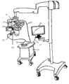
图1为本发明实施例提供的手术显微镜监测系统的结构示意图。Fig. 1 is a schematic structural diagram of an operating microscope monitoring system provided by an embodiment of the present invention.
具体实施方式Detailed ways
以下结合附图和具体实施例对本发明提出的一种手术显微镜监测系统作进一步详细说明。根据下面说明和权利要求书,本发明的优点和特征将更清楚。需说明的是,附图均采用非常简化的形式且均使用非精准的比率,仅用于方便、明晰地辅助说明本发明实施例的目的。A monitoring system for an operating microscope proposed by the present invention will be described in further detail below in conjunction with the accompanying drawings and specific embodiments. Advantages and features of the present invention will be apparent from the following description and claims. It should be noted that all the drawings are in very simplified form and use imprecise ratios, which are only used for the purpose of conveniently and clearly assisting in describing the embodiments of the present invention.
本发明的核心思想在于,本发明提供的手术显微镜监测系统,通过将CCD相机拍摄到的原始激光散斑图像传输至计算机,计算机计算原始激光散斑图像数据得到血流分布图像,能够实时定量的得到具有高时间空间分辨率的血流信息。本发明提供的手术显微镜监测系统,能够方便地将CCD相机集成到手术显微镜上,从而在不影响手术的情况下对血流和血管的变化进行监测。The core idea of the present invention is that the surgical microscope monitoring system provided by the present invention transmits the original laser speckle image captured by the CCD camera to the computer, and the computer calculates the original laser speckle image data to obtain the blood flow distribution image, which can real-time quantitative Obtain blood flow information with high spatial and temporal resolution. The operating microscope monitoring system provided by the present invention can conveniently integrate a CCD camera into the operating microscope, so as to monitor the changes of blood flow and blood vessels without affecting the operation.
图1为本发明实施例提供的手术显微镜监测系统的结构示意图。如图1所示,一种手术显微镜监测系统,包括手术显微镜11,手术显微镜监测系统还包括:激光照明模块,包括激光二极管12以及与所述激光二极管12连接的激光二极管驱动器13,所述激光二极管12与所述手术显微镜11连接;CCD相机14,与所述手术显微镜11连接;计算机15,与所述CCD相机14电信连接;滤光片(图中未示出),设置在所述CCD相机14的成像路径中。Fig. 1 is a schematic structural diagram of an operating microscope monitoring system provided by an embodiment of the present invention. As shown in Figure 1, a kind of operating microscope monitoring system, comprises
进一步地,所述激光二极管12通过适配器121与所述手术显微镜11连接,在本实施例中,所述激光二极管12发出光的波长为785nm,通过手术显微镜11内部的特殊设计,将785nm的激光光路照射到手术监测区域。Further, the laser diode 12 is connected to the
在本实施例中,所述手术显微镜11配备CCD接口18,将高分辨率、高帧速率、高信噪比的CCD相机14连接到手术显微镜11,与激光二极管12配合工作,对需要实时观测血流变化的手术区域的散斑图像进行连续采集并通过数据线19连接到计算机15,通过计算机15进行后续的数据计算和处理。具体地,通过设置在CCD相机成像路径中的滤光片滤除激光散斑成像所需特定波长激光之外的杂光,以使计算机15采集到的散斑图像精确。计算机15在采集散斑图像前设置CCD相机14的相关参数,如帧速率、分辨率、曝光时间等参数,同时,计算机15也可以设置激光二极管12的相关参数,以控制激光的强度,如激光光源的亮度等参数。In this embodiment, the
进一步的,本发明实施例提供的手术显微镜监测系统还包括多触点液晶显示器16,并通过VGA连接线17与所述计算机15连接。操作者通过多触点液晶显示器16可随时观察血流分布情况,并可在多触点液晶显示器16上通过手触的方式选定感兴趣的区域,观察该区域血流速度的定量变化情况。Further, the operating microscope monitoring system provided by the embodiment of the present invention also includes a multi-touch
进一步的,本发明实施例提供的手术显微镜监测系统还包括血流异常报警单元(图中未示出),所述血流异常报警单元集成在所述计算机15内。计算机15在运行的过程中,可以实时地对血流变化情况进行评估,一旦发现异常情况,报警系统会及时进行声音以及画面的警告和提示。具体地,操作者选择感兴趣的区域,确定感兴趣区域的血流值的基线水平,并设置出发警报的阈值,例如该阈值为基线水平的20%,计算机15对感兴趣区域血流值进行实时监测,如果发现血流值低于阈值,则发出警报。Furthermore, the surgical microscope monitoring system provided by the embodiment of the present invention also includes an abnormal blood flow alarm unit (not shown in the figure), and the abnormal blood flow alarm unit is integrated in the
本发明实施例提供的手术显微镜监测系统在传统手术显微镜基础上,通过激光照明模块以及CCD相机,并基于激光散斑衬比成像技术对术中的血流和血管变化情况进行实时监测,实现了外科手术过程中血流的定量实时监测,区分动静脉,及时发现血流出现的异常情况,帮助手术操作及早发现问题,采取措施,并且能够在手术中避免对重要血管的意外损伤。The surgical microscope monitoring system provided by the embodiment of the present invention is based on the traditional surgical microscope, through the laser illumination module and the CCD camera, and based on the laser speckle contrast imaging technology, real-time monitoring of blood flow and blood vessel changes in the operation is realized. Quantitative and real-time monitoring of blood flow during surgery can distinguish arteries and veins, detect abnormalities in blood flow in time, help surgical operations to detect problems early, take measures, and avoid accidental damage to important blood vessels during surgery.
显然,本领域的技术人员可以对发明进行各种改动和变型而不脱离本发明的精神和范围。这样,倘若本发明的这些修改和变型属于本发明权利要求及其等同技术的范围之内,则本发明也意图包含这些改动和变型在内。Obviously, those skilled in the art can make various changes and modifications to the invention without departing from the spirit and scope of the invention. Thus, if these modifications and variations of the present invention fall within the scope of the claims of the present invention and their equivalent technologies, the present invention also intends to include these modifications and variations.
Claims (5)
Priority Applications (1)
| Application Number | Priority Date | Filing Date | Title |
|---|---|---|---|
| CN2013100203853ACN103040455A (en) | 2013-01-18 | 2013-01-18 | Operating microscope monitoring system |
Applications Claiming Priority (1)
| Application Number | Priority Date | Filing Date | Title |
|---|---|---|---|
| CN2013100203853ACN103040455A (en) | 2013-01-18 | 2013-01-18 | Operating microscope monitoring system |
Publications (1)
| Publication Number | Publication Date |
|---|---|
| CN103040455Atrue CN103040455A (en) | 2013-04-17 |
Family
ID=48053477
Family Applications (1)
| Application Number | Title | Priority Date | Filing Date |
|---|---|---|---|
| CN2013100203853APendingCN103040455A (en) | 2013-01-18 | 2013-01-18 | Operating microscope monitoring system |
Country Status (1)
| Country | Link |
|---|---|
| CN (1) | CN103040455A (en) |
Cited By (3)
| Publication number | Priority date | Publication date | Assignee | Title |
|---|---|---|---|---|
| CN103297705A (en)* | 2013-05-21 | 2013-09-11 | 东莞市三基音响科技有限公司 | Method and system for real-time processing of professional-grade network audio and video in large venues |
| CN106842532A (en)* | 2015-11-18 | 2017-06-13 | 三鹰光器株式会社 | Stereovision device is used in operation |
| CN110200707A (en)* | 2019-06-28 | 2019-09-06 | 上海德芬生物科技有限公司 | A kind of operating microscope system and imaging method showing blood flow information |
Citations (7)
| Publication number | Priority date | Publication date | Assignee | Title |
|---|---|---|---|---|
| WO2005051190A1 (en)* | 2003-11-21 | 2005-06-09 | Kings College Hospital Nhs Trust | Blood flow monitoring equipment |
| WO2007009234A1 (en)* | 2005-07-18 | 2007-01-25 | Andreas Mandelis | Method and apparatus using infrared photothermal radiometry (ptr) and modulated laser luminescence (lum) for diagnostics of defects in teeth |
| CN101156769A (en)* | 2007-08-02 | 2008-04-09 | 太原特玛茹电子科技有限公司 | A instrument of laser speckle eyeground blood flow measurement |
| CN101784227A (en)* | 2007-07-06 | 2010-07-21 | 工业研究有限公司 | laser speckle imaging systems and methods |
| CN102232856A (en)* | 2010-05-06 | 2011-11-09 | 高春平 | Double-frequency ultrasonic multi-dimensional focused cerebrovascular thrombolytic system |
| CN202277329U (en)* | 2011-10-31 | 2012-06-20 | 深圳市理邦精密仪器股份有限公司 | Medical diagnosis instrument supporting third-generation communication technology |
| CN102961171A (en)* | 2012-11-02 | 2013-03-13 | 中国人民解放军总医院 | Quantitative blood stream limiting device |
- 2013
- 2013-01-18CNCN2013100203853Apatent/CN103040455A/enactivePending
Patent Citations (7)
| Publication number | Priority date | Publication date | Assignee | Title |
|---|---|---|---|---|
| WO2005051190A1 (en)* | 2003-11-21 | 2005-06-09 | Kings College Hospital Nhs Trust | Blood flow monitoring equipment |
| WO2007009234A1 (en)* | 2005-07-18 | 2007-01-25 | Andreas Mandelis | Method and apparatus using infrared photothermal radiometry (ptr) and modulated laser luminescence (lum) for diagnostics of defects in teeth |
| CN101784227A (en)* | 2007-07-06 | 2010-07-21 | 工业研究有限公司 | laser speckle imaging systems and methods |
| CN101156769A (en)* | 2007-08-02 | 2008-04-09 | 太原特玛茹电子科技有限公司 | A instrument of laser speckle eyeground blood flow measurement |
| CN102232856A (en)* | 2010-05-06 | 2011-11-09 | 高春平 | Double-frequency ultrasonic multi-dimensional focused cerebrovascular thrombolytic system |
| CN202277329U (en)* | 2011-10-31 | 2012-06-20 | 深圳市理邦精密仪器股份有限公司 | Medical diagnosis instrument supporting third-generation communication technology |
| CN102961171A (en)* | 2012-11-02 | 2013-03-13 | 中国人民解放军总医院 | Quantitative blood stream limiting device |
Non-Patent Citations (3)
| Title |
|---|
| ANDREW K.DUNN,ET AL.: "Dynamic Imaging of Cerebral Blood Flow Using Laser Speckle", 《JOURNAL OF CEREBRAL BLOOD FLOW AND METABOLISM》* |
| PARTHASARATHY ET AL.: "Laser speckle contrast imaging of cerebral blood flow in humans during neurosurgery: a pilot clinical study", 《 JOURNAL OF BIOMEDICAL OPTICS》* |
| 张红艳: "面向临床应用的激光散斑血流成像系统研究", 《中国博士学位论文全文数据库》* |
Cited By (4)
| Publication number | Priority date | Publication date | Assignee | Title |
|---|---|---|---|---|
| CN103297705A (en)* | 2013-05-21 | 2013-09-11 | 东莞市三基音响科技有限公司 | Method and system for real-time processing of professional-grade network audio and video in large venues |
| CN106842532A (en)* | 2015-11-18 | 2017-06-13 | 三鹰光器株式会社 | Stereovision device is used in operation |
| CN106842532B (en)* | 2015-11-18 | 2020-12-22 | 三鹰光器株式会社 | Stereoscopic observation device for operation |
| CN110200707A (en)* | 2019-06-28 | 2019-09-06 | 上海德芬生物科技有限公司 | A kind of operating microscope system and imaging method showing blood flow information |
Similar Documents
| Publication | Publication Date | Title |
|---|---|---|
| Eriksson et al. | Non-invasive imaging of microcirculation: a technology review | |
| AU2016335761B2 (en) | System and method for rapid examination of vasculature and particulate flow using laser speckle contrast imaging | |
| RU2481056C2 (en) | Device for image processing, method of image processing, device for capturing tomogram, programme and carrier for programme recording | |
| EP2821006B1 (en) | Funduscopic device | |
| EP2460460B1 (en) | Ophthalmological observation device | |
| CN103417196B (en) | Venous visualizer and visualizing method | |
| JP4615865B2 (en) | Characterization of moving substances in a stationary background | |
| EP2638848B1 (en) | Fundus image processing device and fundus observation device | |
| WO2015166503A1 (en) | Multimodal transcranial brain optical imaging | |
| JP2012511361A (en) | Apparatus for infrared vision of anatomical structures and signal processing method thereof | |
| Jiang et al. | Automated segmentation and fractal analysis of high-resolution non-invasive capillary perfusion maps of the human retina | |
| US11024416B2 (en) | Tomographic image processing device, ophthalmic device comprising the same and non-transitory computer-readable recording medium storing computer-readable instructions for tomographic image processing device | |
| JP2016049255A (en) | Optometer | |
| Schraven et al. | Continuous intraoperative perfusion monitoring of free microvascular anastomosed fasciocutaneous flaps using remote photoplethysmography | |
| JPWO2020090729A1 (en) | Medical image processing equipment, medical image processing methods and programs, diagnostic support equipment | |
| CN201624637U (en) | Tunable light source combined with confocal laser synchronous fundus angiography system | |
| Tao et al. | Intraoperative monitoring cerebral blood flow during the treatment of brain arteriovenous malformations in hybrid operating room by laser speckle contrast imaging | |
| CN103040455A (en) | Operating microscope monitoring system | |
| JP6402025B2 (en) | Blood flow measuring device | |
| KR102389961B1 (en) | Apparatus and method for measuring blood flow based on infrared imaging | |
| JP6747576B2 (en) | Imaging equipment | |
| CN216777062U (en) | Rapid imaging system for human skin laser speckle blood flow | |
| KR101663376B1 (en) | Method and apparatus for mapping retinal vessels | |
| KR101488775B1 (en) | Segmental analysis and disease diagnosis by representative blood flow dynamics value | |
| US20200187797A1 (en) | Fluorescence imaging processing and computation for surgery |
Legal Events
| Date | Code | Title | Description |
|---|---|---|---|
| C06 | Publication | ||
| PB01 | Publication | ||
| C10 | Entry into substantive examination | ||
| SE01 | Entry into force of request for substantive examination | ||
| C12 | Rejection of a patent application after its publication | ||
| RJ01 | Rejection of invention patent application after publication | Application publication date:20130417 |