CN102858252A - Property determination device for determining the property of an object - Google Patents
Property determination device for determining the property of an objectDownload PDFInfo
- Publication number
- CN102858252A CN102858252ACN2011800208199ACN201180020819ACN102858252ACN 102858252 ACN102858252 ACN 102858252ACN 2011800208199 ACN2011800208199 ACN 2011800208199ACN 201180020819 ACN201180020819 ACN 201180020819ACN 102858252 ACN102858252 ACN 102858252A
- Authority
- CN
- China
- Prior art keywords
- character
- order
- determined
- determining
- tissue
- Prior art date
- Legal status (The legal status is an assumption and is not a legal conclusion. Google has not performed a legal analysis and makes no representation as to the accuracy of the status listed.)
- Granted
Links
Images


Classifications
- A—HUMAN NECESSITIES
- A61—MEDICAL OR VETERINARY SCIENCE; HYGIENE
- A61B—DIAGNOSIS; SURGERY; IDENTIFICATION
- A61B17/00—Surgical instruments, devices or methods
- A61B17/34—Trocars; Puncturing needles
- A61B17/3403—Needle locating or guiding means
- A—HUMAN NECESSITIES
- A61—MEDICAL OR VETERINARY SCIENCE; HYGIENE
- A61B—DIAGNOSIS; SURGERY; IDENTIFICATION
- A61B34/00—Computer-aided surgery; Manipulators or robots specially adapted for use in surgery
- A61B34/70—Manipulators specially adapted for use in surgery
- A61B34/71—Manipulators operated by drive cable mechanisms
- A—HUMAN NECESSITIES
- A61—MEDICAL OR VETERINARY SCIENCE; HYGIENE
- A61B—DIAGNOSIS; SURGERY; IDENTIFICATION
- A61B8/00—Diagnosis using ultrasonic, sonic or infrasonic waves
- A61B8/08—Clinical applications
- A61B8/0833—Clinical applications involving detecting or locating foreign bodies or organic structures
- A—HUMAN NECESSITIES
- A61—MEDICAL OR VETERINARY SCIENCE; HYGIENE
- A61B—DIAGNOSIS; SURGERY; IDENTIFICATION
- A61B8/00—Diagnosis using ultrasonic, sonic or infrasonic waves
- A61B8/12—Diagnosis using ultrasonic, sonic or infrasonic waves in body cavities or body tracts, e.g. by using catheters
- A—HUMAN NECESSITIES
- A61—MEDICAL OR VETERINARY SCIENCE; HYGIENE
- A61B—DIAGNOSIS; SURGERY; IDENTIFICATION
- A61B90/00—Instruments, implements or accessories specially adapted for surgery or diagnosis and not covered by any of the groups A61B1/00 - A61B50/00, e.g. for luxation treatment or for protecting wound edges
- A61B90/36—Image-producing devices or illumination devices not otherwise provided for
- A61B90/37—Surgical systems with images on a monitor during operation
- A—HUMAN NECESSITIES
- A61—MEDICAL OR VETERINARY SCIENCE; HYGIENE
- A61B—DIAGNOSIS; SURGERY; IDENTIFICATION
- A61B18/00—Surgical instruments, devices or methods for transferring non-mechanical forms of energy to or from the body
- A61B18/02—Surgical instruments, devices or methods for transferring non-mechanical forms of energy to or from the body by cooling, e.g. cryogenic techniques
- A61B18/0206—Surgical instruments, devices or methods for transferring non-mechanical forms of energy to or from the body by cooling, e.g. cryogenic techniques ultrasonic, e.g. for destroying tissue or enhancing freezing
- A—HUMAN NECESSITIES
- A61—MEDICAL OR VETERINARY SCIENCE; HYGIENE
- A61B—DIAGNOSIS; SURGERY; IDENTIFICATION
- A61B18/00—Surgical instruments, devices or methods for transferring non-mechanical forms of energy to or from the body
- A61B18/04—Surgical instruments, devices or methods for transferring non-mechanical forms of energy to or from the body by heating
- A61B18/12—Surgical instruments, devices or methods for transferring non-mechanical forms of energy to or from the body by heating by passing a current through the tissue to be heated, e.g. high-frequency current
- A61B18/14—Probes or electrodes therefor
- A61B18/1492—Probes or electrodes therefor having a flexible, catheter-like structure, e.g. for heart ablation
- A—HUMAN NECESSITIES
- A61—MEDICAL OR VETERINARY SCIENCE; HYGIENE
- A61B—DIAGNOSIS; SURGERY; IDENTIFICATION
- A61B17/00—Surgical instruments, devices or methods
- A61B2017/00017—Electrical control of surgical instruments
- A61B2017/00022—Sensing or detecting at the treatment site
- A61B2017/00039—Electric or electromagnetic phenomena other than conductivity, e.g. capacity, inductivity, Hall effect
- A61B2017/00044—Sensing electrocardiography, i.e. ECG
- A61B2017/00048—Spectral analysis
- A61B2017/00053—Mapping
- A—HUMAN NECESSITIES
- A61—MEDICAL OR VETERINARY SCIENCE; HYGIENE
- A61B—DIAGNOSIS; SURGERY; IDENTIFICATION
- A61B17/00—Surgical instruments, devices or methods
- A61B2017/00017—Electrical control of surgical instruments
- A61B2017/00022—Sensing or detecting at the treatment site
- A61B2017/00106—Sensing or detecting at the treatment site ultrasonic
- A—HUMAN NECESSITIES
- A61—MEDICAL OR VETERINARY SCIENCE; HYGIENE
- A61B—DIAGNOSIS; SURGERY; IDENTIFICATION
- A61B17/00—Surgical instruments, devices or methods
- A61B17/34—Trocars; Puncturing needles
- A61B17/3403—Needle locating or guiding means
- A61B2017/3413—Needle locating or guiding means guided by ultrasound
- A—HUMAN NECESSITIES
- A61—MEDICAL OR VETERINARY SCIENCE; HYGIENE
- A61B—DIAGNOSIS; SURGERY; IDENTIFICATION
- A61B18/00—Surgical instruments, devices or methods for transferring non-mechanical forms of energy to or from the body
- A61B2018/00571—Surgical instruments, devices or methods for transferring non-mechanical forms of energy to or from the body for achieving a particular surgical effect
- A61B2018/00577—Ablation
- A—HUMAN NECESSITIES
- A61—MEDICAL OR VETERINARY SCIENCE; HYGIENE
- A61B—DIAGNOSIS; SURGERY; IDENTIFICATION
- A61B18/00—Surgical instruments, devices or methods for transferring non-mechanical forms of energy to or from the body
- A61B18/02—Surgical instruments, devices or methods for transferring non-mechanical forms of energy to or from the body by cooling, e.g. cryogenic techniques
- A61B2018/0212—Surgical instruments, devices or methods for transferring non-mechanical forms of energy to or from the body by cooling, e.g. cryogenic techniques using an instrument inserted into a body lumen, e.g. catheter
- A—HUMAN NECESSITIES
- A61—MEDICAL OR VETERINARY SCIENCE; HYGIENE
- A61B—DIAGNOSIS; SURGERY; IDENTIFICATION
- A61B18/00—Surgical instruments, devices or methods for transferring non-mechanical forms of energy to or from the body
- A61B18/18—Surgical instruments, devices or methods for transferring non-mechanical forms of energy to or from the body by applying electromagnetic radiation, e.g. microwaves
- A61B18/1815—Surgical instruments, devices or methods for transferring non-mechanical forms of energy to or from the body by applying electromagnetic radiation, e.g. microwaves using microwaves
- A61B2018/1861—Surgical instruments, devices or methods for transferring non-mechanical forms of energy to or from the body by applying electromagnetic radiation, e.g. microwaves using microwaves with an instrument inserted into a body lumen or cavity, e.g. a catheter
- A—HUMAN NECESSITIES
- A61—MEDICAL OR VETERINARY SCIENCE; HYGIENE
- A61B—DIAGNOSIS; SURGERY; IDENTIFICATION
- A61B34/00—Computer-aided surgery; Manipulators or robots specially adapted for use in surgery
- A61B34/20—Surgical navigation systems; Devices for tracking or guiding surgical instruments, e.g. for frameless stereotaxis
- A61B2034/2046—Tracking techniques
- A61B2034/2051—Electromagnetic tracking systems
- A—HUMAN NECESSITIES
- A61—MEDICAL OR VETERINARY SCIENCE; HYGIENE
- A61B—DIAGNOSIS; SURGERY; IDENTIFICATION
- A61B34/00—Computer-aided surgery; Manipulators or robots specially adapted for use in surgery
- A61B34/20—Surgical navigation systems; Devices for tracking or guiding surgical instruments, e.g. for frameless stereotaxis
- A61B2034/2046—Tracking techniques
- A61B2034/2065—Tracking using image or pattern recognition
- A—HUMAN NECESSITIES
- A61—MEDICAL OR VETERINARY SCIENCE; HYGIENE
- A61B—DIAGNOSIS; SURGERY; IDENTIFICATION
- A61B34/00—Computer-aided surgery; Manipulators or robots specially adapted for use in surgery
- A61B34/30—Surgical robots
- A61B2034/301—Surgical robots for introducing or steering flexible instruments inserted into the body, e.g. catheters or endoscopes
- A—HUMAN NECESSITIES
- A61—MEDICAL OR VETERINARY SCIENCE; HYGIENE
- A61B—DIAGNOSIS; SURGERY; IDENTIFICATION
- A61B90/00—Instruments, implements or accessories specially adapted for surgery or diagnosis and not covered by any of the groups A61B1/00 - A61B50/00, e.g. for luxation treatment or for protecting wound edges
- A61B90/36—Image-producing devices or illumination devices not otherwise provided for
- A61B90/37—Surgical systems with images on a monitor during operation
- A61B2090/374—NMR or MRI
- A—HUMAN NECESSITIES
- A61—MEDICAL OR VETERINARY SCIENCE; HYGIENE
- A61B—DIAGNOSIS; SURGERY; IDENTIFICATION
- A61B90/00—Instruments, implements or accessories specially adapted for surgery or diagnosis and not covered by any of the groups A61B1/00 - A61B50/00, e.g. for luxation treatment or for protecting wound edges
- A61B90/36—Image-producing devices or illumination devices not otherwise provided for
- A61B90/37—Surgical systems with images on a monitor during operation
- A61B2090/376—Surgical systems with images on a monitor during operation using X-rays, e.g. fluoroscopy
- A61B2090/3762—Surgical systems with images on a monitor during operation using X-rays, e.g. fluoroscopy using computed tomography systems [CT]
- A—HUMAN NECESSITIES
- A61—MEDICAL OR VETERINARY SCIENCE; HYGIENE
- A61B—DIAGNOSIS; SURGERY; IDENTIFICATION
- A61B90/00—Instruments, implements or accessories specially adapted for surgery or diagnosis and not covered by any of the groups A61B1/00 - A61B50/00, e.g. for luxation treatment or for protecting wound edges
- A61B90/36—Image-producing devices or illumination devices not otherwise provided for
- A61B90/37—Surgical systems with images on a monitor during operation
- A61B2090/378—Surgical systems with images on a monitor during operation using ultrasound
- A61B2090/3782—Surgical systems with images on a monitor during operation using ultrasound transmitter or receiver in catheter or minimal invasive instrument
- A61B2090/3784—Surgical systems with images on a monitor during operation using ultrasound transmitter or receiver in catheter or minimal invasive instrument both receiver and transmitter being in the instrument or receiver being also transmitter
- A—HUMAN NECESSITIES
- A61—MEDICAL OR VETERINARY SCIENCE; HYGIENE
- A61B—DIAGNOSIS; SURGERY; IDENTIFICATION
- A61B2217/00—General characteristics of surgical instruments
- A61B2217/002—Auxiliary appliance
- A61B2217/007—Auxiliary appliance with irrigation system
- A—HUMAN NECESSITIES
- A61—MEDICAL OR VETERINARY SCIENCE; HYGIENE
- A61B—DIAGNOSIS; SURGERY; IDENTIFICATION
- A61B6/00—Apparatus or devices for radiation diagnosis; Apparatus or devices for radiation diagnosis combined with radiation therapy equipment
- A61B6/50—Apparatus or devices for radiation diagnosis; Apparatus or devices for radiation diagnosis combined with radiation therapy equipment specially adapted for specific body parts; specially adapted for specific clinical applications
- A61B6/503—Apparatus or devices for radiation diagnosis; Apparatus or devices for radiation diagnosis combined with radiation therapy equipment specially adapted for specific body parts; specially adapted for specific clinical applications for diagnosis of the heart
- A—HUMAN NECESSITIES
- A61—MEDICAL OR VETERINARY SCIENCE; HYGIENE
- A61B—DIAGNOSIS; SURGERY; IDENTIFICATION
- A61B6/00—Apparatus or devices for radiation diagnosis; Apparatus or devices for radiation diagnosis combined with radiation therapy equipment
- A61B6/50—Apparatus or devices for radiation diagnosis; Apparatus or devices for radiation diagnosis combined with radiation therapy equipment specially adapted for specific body parts; specially adapted for specific clinical applications
- A61B6/504—Apparatus or devices for radiation diagnosis; Apparatus or devices for radiation diagnosis combined with radiation therapy equipment specially adapted for specific body parts; specially adapted for specific clinical applications for diagnosis of blood vessels, e.g. by angiography
- A—HUMAN NECESSITIES
- A61—MEDICAL OR VETERINARY SCIENCE; HYGIENE
- A61B—DIAGNOSIS; SURGERY; IDENTIFICATION
- A61B8/00—Diagnosis using ultrasonic, sonic or infrasonic waves
- A61B8/08—Clinical applications
- A61B8/0891—Clinical applications for diagnosis of blood vessels
- A—HUMAN NECESSITIES
- A61—MEDICAL OR VETERINARY SCIENCE; HYGIENE
- A61B—DIAGNOSIS; SURGERY; IDENTIFICATION
- A61B8/00—Diagnosis using ultrasonic, sonic or infrasonic waves
- A61B8/44—Constructional features of the ultrasonic, sonic or infrasonic diagnostic device
- A61B8/4444—Constructional features of the ultrasonic, sonic or infrasonic diagnostic device related to the probe
- A61B8/445—Details of catheter construction
- A—HUMAN NECESSITIES
- A61—MEDICAL OR VETERINARY SCIENCE; HYGIENE
- A61B—DIAGNOSIS; SURGERY; IDENTIFICATION
- A61B8/00—Diagnosis using ultrasonic, sonic or infrasonic waves
- A61B8/44—Constructional features of the ultrasonic, sonic or infrasonic diagnostic device
- A61B8/4444—Constructional features of the ultrasonic, sonic or infrasonic diagnostic device related to the probe
- A61B8/4472—Wireless probes
- A—HUMAN NECESSITIES
- A61—MEDICAL OR VETERINARY SCIENCE; HYGIENE
- A61B—DIAGNOSIS; SURGERY; IDENTIFICATION
- A61B8/00—Diagnosis using ultrasonic, sonic or infrasonic waves
- A61B8/48—Diagnostic techniques
- A61B8/486—Diagnostic techniques involving arbitrary m-mode
Landscapes
- Health & Medical Sciences (AREA)
- Life Sciences & Earth Sciences (AREA)
- Surgery (AREA)
- Engineering & Computer Science (AREA)
- Nuclear Medicine, Radiotherapy & Molecular Imaging (AREA)
- Animal Behavior & Ethology (AREA)
- Heart & Thoracic Surgery (AREA)
- Medical Informatics (AREA)
- Molecular Biology (AREA)
- General Health & Medical Sciences (AREA)
- Public Health (AREA)
- Veterinary Medicine (AREA)
- Biomedical Technology (AREA)
- Pathology (AREA)
- Radiology & Medical Imaging (AREA)
- Physics & Mathematics (AREA)
- Biophysics (AREA)
- Gynecology & Obstetrics (AREA)
- Oral & Maxillofacial Surgery (AREA)
- Robotics (AREA)
- Ultra Sonic Daignosis Equipment (AREA)
- Investigating Or Analyzing Materials By The Use Of Ultrasonic Waves (AREA)
Abstract
Description
Translated fromChinese技术领域technical field
本发明涉及一种用于确定对象的性质的性质确定装置、方法和计算机程序。本发明还涉及包括性质确定装置的对象影响装置、相应的对象影响方法和相应的对象影响计算机程序。The present invention relates to a property determination device, method and computer program for determining properties of an object. The invention also relates to an object influencing device comprising a property determining device, a corresponding object influencing method and a corresponding object influencing computer program.
背景技术Background technique
WO 2006/064495 A1公开了一种在心脏热消融过程期间监测组织的热损伤的方法。在消融组织的特定部分同时,采集该组织相邻部分的超声图像,并且从这些超声图像提取指示组织相邻部分对热的生物学响应的参数。在实施例中,所述参数为气泡的累积,并且基于这个参数确定组织相邻部分的损伤。这种监测方法的目的是检测对组织相邻部分的不希望损伤。该监测方法因而通过观测超声图像中的空间和时间变化并通过将这些变化关联到气泡形成来监测组织的可能损伤。这种监测导致确定组织可能损伤中的不精确性,因为气泡形成仅反映组织中的高温,而不是直接与组织损伤程度相关联。WO 2006/064495 A1 discloses a method of monitoring thermal damage to tissue during cardiac thermal ablation procedures. While a particular portion of tissue is being ablated, ultrasound images of adjacent portions of the tissue are acquired, and parameters indicative of the biological response of the adjacent portion of tissue to heat are extracted from the ultrasound images. In an embodiment, said parameter is the accumulation of gas bubbles, and based on this parameter damage to adjacent parts of the tissue is determined. The purpose of this monitoring method is to detect unwanted damage to adjacent parts of the tissue. The monitoring method thus monitors for possible damage to tissue by observing spatial and temporal changes in the ultrasound images and by correlating these changes to bubble formation. Such monitoring leads to inaccuracies in determining possible damage to the tissue, since bubble formation only reflects high temperatures in the tissue, rather than directly correlating to the extent of tissue damage.
发明内容Contents of the invention
本发明的目的是提供一种性质确定装置,其中可以以改善的精度确定对象的性质。本发明的另一目的是提供一种包括性质确定装置的对象影响装置并提供相应的方法和计算机程序。It is an object of the present invention to provide a property determination device in which properties of objects can be determined with improved accuracy. Another object of the present invention is to provide an object influencing device comprising a property determining device and to provide a corresponding method and computer program.
在本发明的第一方面中,提供一种用于确定对象的性质的性质确定装置,其中所述性质确定装置包括:In a first aspect of the present invention there is provided a property determining device for determining a property of an object, wherein said property determining device comprises:
-超声信号提供单元,其用于提供通过如下方式产生的超声信号- an ultrasonic signal providing unit for providing an ultrasonic signal generated by
-向所述对象发出超声脉冲,- sending ultrasound pulses to said object,
-从所述对象接收回波序列,以及- receiving a sequence of echoes from said object, and
-根据所接收的回波序列生成所述超声信号,- generating said ultrasound signal from the received echo sequence,
-散射确定单元,其用于确定至少一个散射值,该至少一个散射值指示灌注对象的流体对超声脉冲的散射,其中所述散射确定单元适于根据所述超声信号确定至少一个散射值,- a scatter determination unit for determining at least one scatter value indicative of the scatter of an ultrasound pulse by a fluid perfusing the subject, wherein said scatter determination unit is adapted to determine at least one scatter value from said ultrasound signal,
-性质确定单元,其用于根据所述至少一个散射值确定所述对象的性质。- A property determination unit for determining a property of said object from said at least one scatter value.
由于散射确定单元确定指示流体导致的超声脉冲散射的至少一个散射值,并且由于性质确定单元适于根据所述至少一个散射值确定对象的性质,因而能够基于所述至少一个散射值更直接地确定与流体灌注相关的性质。因而,不必使用非常间接的手段,像检测气泡形成,来确定对象的性质,气泡形成反映组织中高温,但其不直接与例如组织损伤程度相关联。这增大了确定对象的性质的精度。Since the scatter determination unit determines at least one scatter value indicative of the scattering of the ultrasound pulse by the fluid, and since the property determination unit is adapted to determine a property of the object from the at least one scatter value, it is possible to determine more directly based on the at least one scatter value Properties related to fluid perfusion. Thus, it is not necessary to use very indirect means, like detecting bubble formation, which reflects high temperature in the tissue, but which is not directly linked eg to the degree of tissue damage, to determine the nature of the object. This increases the precision with which properties of objects are determined.
散射确定单元可适于确定若干散射值,其中性质确定单元可适于基于若干散射值确定性质。The scatter determination unit may be adapted to determine a number of scatter values, wherein the property determination unit may be adapted to determine a property based on the number of scatter values.
超声信号提供单元可以是存储单元,在其中已经存储了超声信号,或者超声信号提供单元可以是数据接收单元,像有线或无线数据连接,用于接收所测量的超声信号。此外,超声信号提供单元可以由用于生成超声信号的一个或若干超声换能器构成,其中同一超声换能器能够发送超声脉冲并接收回波序列,或者第一超声换能器能够发送超声脉冲,并且另一个第二超声换能器能够接收回波序列。The ultrasound signal providing unit may be a memory unit, in which the ultrasound signals are already stored, or the ultrasound signal providing unit may be a data receiving unit, like a wired or wireless data connection, for receiving the measured ultrasound signals. Furthermore, the ultrasonic signal providing unit may consist of one or several ultrasonic transducers for generating ultrasonic signals, wherein the same ultrasonic transducer is capable of sending ultrasonic pulses and receiving echo sequences, or the first ultrasonic transducer is capable of sending ultrasonic pulses , and another second ultrasonic transducer capable of receiving the echo sequence.
超声信号提供单元优选地适于针对不同时间和对象内的不同深度提供超声信号。所提供的超声信号优选地是M模式图像。The ultrasound signal providing unit is preferably adapted to provide ultrasound signals for different times and for different depths within the object. The ultrasound signals provided are preferably M-mode images.
对象优选地是人或动物的器官,其中所述器官被像血液的体液灌注。特别地,对象优选地是心脏,其中心脏的组织被血液灌注。还优选地,性质确定装置适于基于至少一个散射值确定流体对对象的灌注程度,特别是,毛细管灌注程度,来作为性质。特别是,性质确定单元适于确定对象的哪个部分被灌注,对象的哪个部分未被灌注。由于所述至少一个散射值指示流体对超声脉冲的散射,因而可以基于所述至少一个散射值确定对象的灌注程度,特别是,对象或对象的一部分是被灌注还是未被灌注,即,如果对象未被流体灌注,散射确定单元能够确定指示没有流体的散射值,并且性质确定单元能够确定例如对象未被灌注;如果对象被流体灌注,散射确定单元能够确定指示存在流体的散射值,性质确定单元能够确定例如对象被灌注。The object is preferably an organ of a human or animal, wherein said organ is perfused with a bodily fluid like blood. In particular, the subject is preferably the heart, wherein the tissue of the heart is perfused with blood. It is also preferred that the property determining means is adapted to determine as a property the degree of perfusion of the object by the fluid, in particular the degree of capillary perfusion, based on at least one scatter value. In particular, the property determination unit is adapted to determine which part of the object is perfused and which part of the object is not perfused. Since the at least one scatter value is indicative of the scattering of the ultrasound pulse by the fluid, it is possible to determine the degree of perfusion of the object based on the at least one scatter value, in particular whether the object or a part of the object is perfused or not, i.e. if the object Not perfused with fluid, the scatter determining unit can determine a scatter value indicating the absence of fluid, and the property determining unit can determine, for example, that the object is not perfused; if the object is perfused with fluid, the scatter determining unit can determine a scatter value indicating the presence of fluid, the property determining unit It can be determined, for example, that the subject is perfused.
还优选地,所述对象是生物学对象,像心脏或另一包括组织的器官,其中所述性质确定单元适于基于所述至少一个散射值确定所述组织的一部分包括消融组织还是未被消融组织,作为对象的性质。特别是,性质确定单元适于确定组织的哪个部分被消融,组织的哪个部分未被消融。通过消融可以修改对象的灌注,其中修改灌注能够修改流体对超声脉冲的散射,从而修改所述至少一个散射值。因此可以使用所述至少一个散射值的修改来确定消融程度。例如,通过校准测量,可以确定消融过程开始之后,哪些散射值或散射值的哪些变化,对应于消融程度,其中确定散射值,而消融程度是已知的。可以认为这些确定的散射值是校准值,其中然后能够使用校准值根据实际确定的散射值确定消融程度。Also preferably, said object is a biological object, like a heart or another organ comprising tissue, wherein said property determination unit is adapted to determine, based on said at least one scatter value, that a portion of said tissue comprises ablated tissue or is not ablated Organization, as a property of objects. In particular, the property determination unit is adapted to determine which part of the tissue was ablated and which part of the tissue was not ablated. The perfusion of the subject may be modified by ablation, wherein modifying the perfusion modifies scattering of the ultrasound pulse by the fluid, thereby modifying the at least one scattering value. The modification of the at least one scatter value can thus be used to determine the degree of ablation. For example, by calibration measurements it can be determined which scatter values or which changes in scatter values correspond to the degree of ablation after the start of the ablation process, wherein the scatter values are determined and the degree of ablation is known. These determined scatter values can be considered as calibration values, wherein the calibration values can then be used to determine the degree of ablation from the actually determined scatter values.
可以向组织应用增强通过组织的灌注的手段。这样的手段例如是像异丙基肾上腺素的血管扩张剂。如果增强灌洗,则可以增大消融组织的超声脉冲散射和未被消融组织的超声脉冲散射的差异,由于未被消融组织的灌注和未被消融组织流体造成的相应的散射将增大,而由于被消融组织未被灌注或仅被灌注一点,因此消融组织区域中的散射将不会增大或仅增大一点。这进一步改善了在被消融组织和未被消融组织间区分的精度。Means that enhance perfusion through the tissue can be applied to the tissue. Such means are, for example, vasodilators like isoproterenol. If the perfusion is enhanced, the difference between the scattering of ultrasonic pulses from the ablated tissue and the non-ablated tissue can be increased, and the corresponding scattering due to the perfusion of the non-ablated tissue and the fluid of the non-ablated tissue will increase, while Since the ablated tissue is not perfused or is perfused only a little, the scatter in the region of the ablated tissue will increase no or only a little. This further improves the accuracy of distinguishing between ablated and non-ablated tissue.
还优选地,性质确定单元适于确定组织的哪部分是被消融组织,组织的哪部分是未被消融组织,并根据组织的这些所确定的部分确定消融深度。由于在确定了组织的哪个部分是被消融组织,组织的哪个部分是未被消融组织之后,就知道组织之内的被消融和未被消融区域的空间分布,因此可以根据所确定的组织的被消融和未被消融部分容易地确定消融深度。Also preferably, the property determination unit is adapted to determine which part of tissue is ablated tissue and which part of tissue is non-ablated tissue, and to determine the ablation depth from these determined parts of tissue. Since after determining which part of the tissue is the ablated tissue and which part of the tissue is the non-ablated tissue, the spatial distribution of the ablated and non-ablated regions within the tissue is known, so the determined ablated tissue can be The ablation and non-ablation portions easily determine the ablation depth.
还优选地,所提供的超声信号表示流体在a)对象之内不同深度和b)不同时间中的至少一个时造成的散射,其中由对应于a)不同深度和b)不同时间中的至少一个的采样窗口对超声信号采样,其中所述散射值确定单元适于确定采样窗口的散射值,其中对于相应的采样窗口,基于所述超声信号中对应于相应采样窗口的部分确定至少一个散射值,并且其中所述性质确定单元适于基于针对相应采样窗口确定的至少一个散射值确定针对相应采样窗口的性质。例如,如果超声信号是M模式图像,可以由对应于特定深度范围和特定时间范围的若干采样窗口对所述M模式图像采样。对于采样窗口中的每个,可以确定至少一个散射值,其中对于采样窗口中的每个,可以基于针对相应采样窗口所确定的至少一个散射值确定性质,特别是,相应采样窗口之内的组织被消融还是未被消融。这样允许随着时间并在不同深度监测性质。特别是,可以实时监测性质。例如,可以实时监测消融深度。Also preferably, the ultrasound signal provided is representative of scattering by the fluid at at least one of a) different depths within the object and b) different times, wherein The sampling window samples the ultrasound signal, wherein the scatter value determination unit is adapted to determine the scatter value of the sampling window, wherein for the corresponding sampling window, at least one scatter value is determined based on a portion of the ultrasound signal corresponding to the corresponding sampling window, And wherein the property determination unit is adapted to determine a property for a respective sampling window based on at least one scatter value determined for the respective sampling window. For example, if the ultrasound signal is an M-mode image, the M-mode image may be sampled by several sampling windows corresponding to a specific depth range and a specific time range. For each of the sampling windows at least one scatter value can be determined, wherein for each of the sampling windows a property can be determined based on the at least one scatter value determined for the corresponding sampling window, in particular the tissue within the corresponding sampling window To be ablated or not to be ablated. This allows monitoring of properties over time and at different depths. In particular, properties can be monitored in real time. For example, ablation depth can be monitored in real time.
采样窗口优选地是交叠的,因为那样可以增大确定对象性质的分辨率,而不减小采样窗口的尺寸。不过,采样窗口也可以是不交叠的。The sampling windows are preferably overlapping, as that increases the resolution at which the properties of the object can be determined without reducing the size of the sampling windows. However, the sampling windows may also be non-overlapping.
还优选地,采样窗口中的每个对应于所述超声信号的若干超声强度,其中所述散射确定单元适于根据相应采样窗口内的超声强度直方图确定采样窗口的至少一个散射值。因而,优选地针对采样窗口中的每个,根据相应采样窗口的超声强度的直方图确定至少一个散射值。特别是,散射确定单元适于基于一阶直方图和二阶直方图中的至少一个确定至少一个散射值。也可以使用更高阶统计来确定至少一个散射值,例如,可以使用Gabor滤波方法确定所述至少一个散射值。Also preferably, each of the sampling windows corresponds to several ultrasound intensities of said ultrasound signal, wherein said scatter determination unit is adapted to determine at least one scatter value of a sampling window from a histogram of ultrasound intensities within the corresponding sampling window. Thus, preferably for each of the sampling windows at least one scatter value is determined from the histogram of the ultrasound intensities of the corresponding sampling window. In particular, the scatter determination unit is adapted to determine at least one scatter value based on at least one of a first order histogram and a second order histogram. Higher order statistics may also be used to determine the at least one scatter value, for example the at least one scatter value may be determined using a Gabor filtering method.
还优选地,所述散射确定单元适于确定以下值中的至少一个作为所述至少一个散射值:一阶直方图的一阶平均值、一阶直方图的一阶方差、一阶直方图的一阶熵、二阶直方图的二阶熵、二阶直方图的二阶能量、二阶直方图的二阶均一性、二阶直方图的二阶对比度、二阶直方图的二阶聚类趋势、二阶直方图的二阶形状、二阶直方图的二阶相关性以及二阶直方图的二阶相关性导数。Also preferably, the scatter determination unit is adapted to determine at least one of the following values as the at least one scatter value: a first-order mean of a first-order histogram, a first-order variance of a first-order histogram, a first-order variance of a first-order histogram, First-order entropy, second-order entropy of second-order histograms, second-order energy of second-order histograms, second-order uniformity of second-order histograms, second-order contrast of second-order histograms, second-order clustering of second-order histograms Trend, second-order shape of second-order histogram, second-order correlation of second-order histogram, and derivative of second-order correlation of second-order histogram.
在实施例中,采样窗口中的每个对应于所述超声信号的若干超声强度,其中所述散射确定单元适于根据相应采样窗口之内的超声强度之和确定采样窗口的至少一个散射值。因而,除了使用基于直方图的散射值之外或作为替代,也可以使用取决于相应采样窗口之内超声强度之和的散射值。例如,散射值可以是相应采样窗口之内所有超声强度之和或超声强度之积的和,其中每对的超声强度中的至少一个位于相应采样窗口之内,且其中每个积包括对应于采集时间的超声强度,如果对象是心脏组织,所述采集时间由对象的心脏循环周期分隔。如果一起使用基于直方图的散射值和优选地不基于直方图的基于这些和的散射值确定对象的性质,则可以进一步改善确定对象性质的精度。In an embodiment, each of the sampling windows corresponds to several ultrasound intensities of said ultrasound signal, wherein said scatter determination unit is adapted to determine at least one scatter value of a sampling window from a sum of ultrasound intensities within the corresponding sampling window. Thus, in addition to or instead of using histogram-based scatter values, it is also possible to use scatter values that depend on the sum of ultrasound intensities within the respective sampling window. For example, the scatter value may be the sum of all ultrasound intensities within a corresponding sampling window or the sum of products of ultrasound intensities, where at least one of the ultrasound intensities of each pair is within the corresponding sampling window, and where each product includes the corresponding Ultrasound intensity over time, if the subject is cardiac tissue, the acquisition times are separated by the subject's cardiac cycle. The accuracy of determining the property of the object can be further improved if the histogram-based scatter values and the scatter values based on these sums, preferably not based on the histogram, are used together to determine the property of the object.
还优选地,性质确定单元适于向采样窗口应用聚类分析,其中根据针对相应采样窗口确定的至少一个散射值对采样窗口分聚类并向采样窗口的聚类分配性质。性质确定单元可适于执行聚类算法,例如K均值聚类,用于对散射值分组。如果对于每个采样窗口,仅确定单个散射值,则对单个散射值应用聚类算法,如果对于每个采样窗口确定了若干散射值,针对单个采样窗口确定的散射值形成多维特征矢量,则向针对若干采样窗口确定的多维特征矢量应用聚类算法。聚类算法可能分别得到散射值或多维特征矢量的第一聚类,并且因而得到相应的采样窗口的第一聚类,以及分别得到散射值或多维特征矢量的第二聚类,并且因而得到相应的采样窗口的第二聚类。采样窗口的第一聚类可表示被消融组织,采样窗口的第二聚类可表示未被消融组织。可以根据与阈值的比较,确定聚类表示被消融还是未被消融组织,可以通过校准测量确定阈值。因而,可以通过执行阈值处理向采样窗口的聚类分配对象的性质。还可以在开始消融之前首先应用聚类分析,获得表示未被消融组织的第一组聚类。然后,在执行消融过程时连续应用聚类分析。如果聚类分析获得不属于第一组聚类的新聚类,可以向这些新聚类分配性质“被消融组织”。Also preferably, the property determination unit is adapted to apply a cluster analysis to the sampling windows, wherein the sampling windows are clustered according to at least one scatter value determined for the respective sampling window and a property is assigned to the clusters of the sampling windows. The property determination unit may be adapted to perform a clustering algorithm, such as K-means clustering, for grouping the scatter values. If for each sampling window only a single scatter value is determined, a clustering algorithm is applied to the single scatter value, if for each sampling window several scatter values are determined, the scatter values determined for a single sampling window form a multidimensional feature vector, then to A clustering algorithm is applied to the multidimensional feature vectors determined for several sampling windows. A clustering algorithm may obtain a first cluster of scatter values or multidimensional feature vectors, respectively, and thus a first cluster of corresponding sampling windows, and a second cluster of scatter values or multidimensional feature vectors, respectively, and thus a corresponding The sampling window for the second clustering. A first cluster of sampling windows may represent ablated tissue and a second cluster of sampling windows may represent non-ablated tissue. A determination that a cluster represents ablated or non-ablated tissue may be determined based on a comparison with a threshold, which may be determined by a calibration measurement. Thus, it is possible to assign properties of objects to clusters of sampling windows by performing thresholding. It is also possible to first apply cluster analysis before starting ablation, obtaining a first set of clusters representing non-ablated tissue. Then, cluster analysis is applied continuously while performing the ablation procedure. If the cluster analysis yields new clusters that do not belong to the first set of clusters, the property "ablated tissue" can be assigned to these new clusters.
性质确定单元可适于基于至少一个散射值与至少一个阈值的比较来确定性质。例如,可以利用上述采样窗口对超声信号采样并且可以针对每个采样窗口确定至少一个散射值。可以定义:如果采样窗口的散射值高于阈值,则对应于该采样窗口的组织未被消融,如果散射值低于阈值,则对应于该采样窗口的组织被消融。如果针对同一采样窗口确定了若干散射值,则可以针对每个散射值提供阈值,并且针对每个散射值,可以确定相应散射值高于还是低于相应阈值。例如,如果采样窗口的大部分散射值高于相应阈值,则可以定义对应于该采样窗口的组织未被消融,并且例如,如果大部分散射值低于相应阈值,则可以定义对应于采样窗口的组织被消融。所述一个或若干阈值例如可以通过校准测量确定。如果针对采样窗口确定了若干散射值,则可以将它们组合成多维特征矢量,即,针对每个采样窗口,可以定义多维特征矢量,其中可以将多维特征矢量与阈值矢量比较,以确定相应的采样窗口对应于被消融组织还是未被消融组织。The property determining unit may be adapted to determine the property based on a comparison of at least one scatter value with at least one threshold. For example, the ultrasound signal may be sampled using the aforementioned sampling windows and at least one scatter value may be determined for each sampling window. It can be defined that if the scatter value of the sampling window is higher than the threshold, the tissue corresponding to the sampling window is not ablated, and if the scatter value is lower than the threshold, the tissue corresponding to the sampling window is ablated. If several scatter values are determined for the same sampling window, a threshold may be provided for each scatter value and for each scatter value it may be determined whether the respective scatter value is above or below the respective threshold. For example, if most of the scatter values of the sampling window are above the corresponding threshold, it can be defined that the tissue corresponding to the sampling window is not ablated, and for example, if most of the scatter values are below the corresponding threshold, it can be defined that the tissue corresponding to the sampling window is not ablated. Tissue is ablated. Said one or several threshold values may eg be determined by calibration measurements. If several scatter values are determined for a sampling window, they can be combined into a multidimensional feature vector, i.e., for each sampling window, a multidimensional feature vector can be defined, where the multidimensional feature vector can be compared with a threshold vector to determine the corresponding sampling The windows correspond to ablated or non-ablated tissue.
还优选地,所述超声信号提供单元适于提供通过使用频率大于10MHz的超声波产生的超声信号。超声波优选地具有20到40MHz频率范围之内的频率,特别是30MHz的频率。利用这些相对较高的超声频率,得到增大的超声信号分辨率。由于超声信号的分辨率的增大,因而能够在超声信号中更好地识别流体造成的超声脉冲散射导致的超声信号模式。因此改善了从超声信号提取至少一个散射值,因而改善了所确定的性质的质量。Also preferably, the ultrasonic signal providing unit is adapted to provide an ultrasonic signal generated by using ultrasonic waves with a frequency greater than 10 MHz. The ultrasound preferably has a frequency in the frequency range of 20 to 40 MHz, in particular a frequency of 30 MHz. With these relatively high ultrasound frequencies, increased ultrasound signal resolution results. Due to the increased resolution of the ultrasound signal, ultrasound signal patterns resulting from scattering of ultrasound pulses by the fluid can be better identified in the ultrasound signal. Extraction of at least one scatter value from the ultrasound signal is thus improved, thus improving the quality of the determined property.
还优选地,所述超声信号提供单元是集成到导管或针中的超声换能器。这允许使用要用于确定例如患者体内心脏或另一器官的性质的性质确定装置,其中至少超声换能器可以被引入患者体内。Also preferably, the ultrasound signal providing unit is an ultrasound transducer integrated into a catheter or needle. This allows the use of a property determining device to be used for determining properties of eg the heart or another organ in a patient in which at least the ultrasound transducer can be introduced.
性质确定单元还可适于基于所确定的至少一个散射值确定对象的特定部分是否包括血管,特别是,动脉或静脉。特别是,可以将所述至少一个散射值与和血管相关联的预定义散射值范围比较,其中,如果至少一个散射值在预定义散射值范围之内,则确定对象的一部分是血管。也可以通过校准测量确定预定义散射值范围,其中在对象类型已知的同时确定散射值。The property determination unit may also be adapted to determine whether a certain portion of the object comprises a blood vessel, in particular an artery or a vein, based on the determined at least one scatter value. In particular, the at least one scatter value may be compared to a predefined scatter value range associated with blood vessels, wherein if the at least one scatter value is within the predefined scatter value range, it is determined that the part of the object is a blood vessel. Predefined scatter value ranges can also be determined by calibration measurements, wherein the scatter values are determined while the object type is known.
在本发明的另一方面中,提供了一种用于影响对象的对象影响装置,其中对象影响装置包括用于影响对象的对象影响单元和权利要求1所述的性质确定装置。对象影响装置优选地是用于消融人或动物的器官例如心脏的消融装置。对象影响单元优选地包括消融电极和连接到消融电极的能量源,用于通过例如射频(RF)能量加热对象。除了适于执行RF消融过程,消融装置也可适于执行另一种消融,例如光学消融、冷冻消融、超声消融、微波消融等。In another aspect of the present invention, there is provided an object influencing device for influencing an object, wherein the object influencing device comprises an object influencing unit for influencing an object and the property determining device according to
对象影响装置优选地包括用于控制对象影响单元的控制单元,其中控制单元适于根据对象的性质控制对象影响单元,所述对象的性质由性质确定装置的性质确定单元确定。如上所述,对象的性质例如是消融深度。如果对象影响装置是根据消融深度控制的消融装置,那么可以控制消融装置使得能够达到期望的消融深度。此外,可以确定血管,特别是,动脉或静脉是否位于消融电极前方,作为对象的性质。例如,可以控制消融装置,使得如果在消融电极前方检测到血管,则不开始消融或停止消融。总地来说,通过根据所确定的对象的性质控制对象的消融,可以改善对象的消融。The object influencing means preferably comprises a control unit for controlling the object influencing unit, wherein the control unit is adapted to control the object influencing unit according to a property of the object determined by the property determining unit of the property determining means. As mentioned above, the property of the object is eg ablation depth. If the object influencing device is an ablation device controlled according to the ablation depth, the ablation device can be controlled such that a desired ablation depth can be achieved. Furthermore, it can be determined as a property of the object whether a blood vessel, in particular an artery or a vein, is located in front of the ablation electrode. For example, the ablation device may be controlled such that if a blood vessel is detected in front of the ablation electrodes, ablation is not started or ablation is stopped. In general, object ablation can be improved by controlling object ablation according to determined object properties.
在本发明的另一方面中,提供了一种用于确定对象的性质的性质确定方法,其中所述性质确定方法包括:In another aspect of the present invention, a property determination method for determining a property of an object is provided, wherein the property determination method comprises:
-提供通过如下方式产生的超声信号-Provide an ultrasonic signal generated by
-向所述对象发出超声脉冲,- sending ultrasound pulses to said object,
-从所述对象接收回波序列,以及- receiving a sequence of echoes from said object, and
-根据接收的回波序列生成所述超声信号,- generating said ultrasound signal from a received echo sequence,
-确定至少一个散射值,该至少一个散射值指示灌注对象的流体导致的超声脉冲的散射,其中根据所述超声信号确定所述至少一个散射值,- determining at least one scatter value indicative of a scatter of an ultrasound pulse caused by a fluid perfusing the subject, wherein said at least one scatter value is determined from said ultrasound signal,
-根据所述至少一个散射值确定对象的性质。- Determining a property of the object from said at least one scatter value.
在本发明的另一方面中,提供了一种用于影响对象的对象影响方法,其中对象影响方法包括影响对象和权利要求12所述的性质确定方法的步骤。优选地,对象影响方法包括根据所确定的对象的性质控制对对象的影响的步骤。对象影响方法优选地是用于消融对象的消融方法,其中,优选地根据所确定的对象的性质,特别是,根据所确定的消融深度,控制对象的消融。可以实时确定消融深度,以便在消融对象时控制对象的消融。In another aspect of the present invention there is provided an object influencing method for influencing an object, wherein the object influencing method comprises the steps of influencing the object and the property determining method as claimed in
在本发明的另一方面中,提供了一种用于确定对象的性质的性质确定计算机程序,其中性质确定计算机程序包括程序代码模块,该程序代码模块用于当计算机程序在控制权利要求1所述的性质确定装置的计算机上运行时,令所述性质确定装置执行权利要求12所述的性质确定方法的步骤。In another aspect of the present invention, there is provided a property determination computer program for determining a property of an object, wherein the property determination computer program comprises a program code module for use when the computer program controls the When running on the computer of the property determination device described above, the property determination device is made to execute the steps of the property determination method described in
在本发明的另一方面中,提供了一种用于影响对象的对象影响计算机程序,其中对象影响计算机程序包括程序代码模块,该程序代码模块用于当计算机程序在控制权利要求11所述的对象影响装置的计算机上运行时,令所述对象影响装置执行权利要求13所述的对象影响方法的步骤。In another aspect of the present invention, there is provided an object influencing computer program for influencing an object, wherein the object influencing computer program comprises a program code module for when the computer program controls the object described in
将要理解,权利要求1所述的性质确定装置、权利要求11所述的对象影响装置、权利要求12所述的性质确定方法、权利要求13所述的对象影响方法、权利要求14所述的性质确定计算机程序和权利要求15所述的对象影响计算机程序具有类似和/或相同的优选实施例,特别是,如从属权利要求中所定义的那样。It will be understood that the property determining apparatus as claimed in
将要理解,本发明的优选实施例也可以是从属权利要求与相应独立权利要求的任意组合。It will be understood that a preferred embodiment of the invention can also be any combination of the dependent claims with the corresponding independent claim.
参考下文所描述的实施例,本发明的这些和其他方面将变得显而易见并得到阐述。These and other aspects of the invention will be apparent from and elucidated with reference to the embodiments described hereinafter.
附图说明Description of drawings
在附图中:In the attached picture:
图1示意性和示范性示出了用于消融对象的消融装置的实施例;Fig. 1 schematically and exemplarily shows an embodiment of an ablation device for ablation of an object;
图2示意性和示范性示出了消融装置的导管顶端的实施例;Figure 2 schematically and exemplarily shows an embodiment of a catheter tip of an ablation device;
图3示范性示出了包括被消融组织和未被消融组织的组织的M模式图像;Fig. 3 exemplarily shows an M-mode image of tissue including ablated tissue and non-ablated tissue;
图4到13示出了取决于时间的若干散射值;Figures 4 to 13 show several scatter values as a function of time;
图14到16示范性示出了对象的若干M模式图像和对应的聚类结果;Figures 14 to 16 exemplarily show several M-mode images of objects and corresponding clustering results;
图17和18示出的流程图示范性例示了聚类算法;The flowcharts shown in Figures 17 and 18 exemplarily illustrate the clustering algorithm;
图19示出了M模式图像的A线;Figure 19 shows the A-line of the M-mode image;
图20示意性和示范性示出了用于确定对象的性质的性质确定装置的实施例;Figure 20 schematically and exemplarily shows an embodiment of a property determining device for determining a property of an object;
图21示意性和示范性示出了性质确定装置的导管顶端的实施例;Figure 21 schematically and exemplarily shows an embodiment of a catheter tip of a property determining device;
图22示出的流程图示范性图示了用于确定对象的性质的性质确定方法的实施例;Figure 22 shows a flowchart exemplarily illustrating an embodiment of a property determination method for determining a property of an object;
图23示出的流程图示范性图示了用于消融对象的消融方法;以及The flowchart shown in FIG. 23 exemplarily illustrates an ablation method for ablating an object; and
图24示意性和示范性示出了活检针顶端的实施例。Figure 24 schematically and exemplarily shows an embodiment of a biopsy needle tip.
具体实施方式Detailed ways
图1示意性和示范性示出了用于消融对象的消融装置1。消融装置1包括用于提供对象3的图像的图像提供单元2,在这一实施例中,对象是人20的心脏。消融装置1还包括用于向心脏3的内壁施加能量的导管21。图2中示意性和示范性示出了导管21的顶端22。导管顶端22包括消融电极4,用于在位置5处向心脏3的壁施加能量以消融该壁。消融电极4经由电连接23与能量源24连接,用于在位置5处提供电能。优选地,能量源24、电连接23和消融电极4适于在位置5处向心脏3施加射频(RF)能量。电连接23优选是导线。消融电极4、电连接23和能量源24形成对象影响单元。Fig. 1 schematically and exemplarily shows an
导管顶端22还包括用于提供超声信号的超声信号提供单元18。超声信号提供单元18是超声换能器,其适于向对象发出超声脉冲,从对象接收回波序列,并根据接收的回波序列产生超声信号。在这一实施例中,超声信号提供单元18适于生成表示在不同深度和不同时间位置5处心脏组织的超声性质的M模式图像。The
超声换能器18适于利用频率大于10MHz的超声波提供超声信号,即M模式图像。超声波优选具有20到40MHz频率范围之内的频率,特别是30MHz的频率。The
导管顶端包括灌洗开口50、51、53,用于允许灌洗流体离开导管顶端。由灌洗流体提供单元52提供灌洗流体。灌洗流体提供单元52提供的灌洗流体被引入导管21中,导向导管顶端22并通过灌洗开口50、51、53离开导管顶端22。灌洗流体不仅用于灌洗的目的,而且用作声学介质,为超声脉冲和回波序列界定声学路径。优选地通过灌洗开口53传输超声脉冲和回波序列。代替提供灌洗开口53,也可以使用像聚甲基戊烯的声学透明材料。The catheter tip includes
图像提供单元2优选地适于提供心脏3的电子解剖图。在这一实施例中,图像提供单元2是存储电子解剖图的存储单元。可以通过例如用计算机断层摄影系统、磁共振成像系统、核成像系统或超声成像系统产生心脏3的三维图像,或通过基于阻抗、磁性或电磁的导管顶端位置跟踪,以及通过在心脏壁上的不同位置测量心脏的电学性质,来产生电子解剖图,其中在心脏三维图像中的相应位置处使所测得的电学性质可视化。The
例如,电子解剖图可以是反映解剖学基质的激活序列的激活图。从这种激活图可以推导出传导模式,传导模式揭示出例如最近激活或折返波的区域。可以使用来自激活图的信息识别应当施加能量的消融目标。For example, the electronic anatomical map may be an activation map that reflects the activation sequence of the anatomical matrix. From this activation map a conduction pattern can be deduced which reveals, for example, areas of recent activation or reentrant waves. The information from the activation map can be used to identify ablation targets to which energy should be applied.
消融装置1还包括用于在不同位置定位消融电极4的定位单元6、7。定位单元包括具有X射线源25和X射线探测器26的X射线荧光检查系统6。X射线源25发射X射线束27,其横穿包括导管21的顶端22的心脏3。横穿过心脏3的X射线束被X射线探测器26探测到。X射线探测器26根据探测到的X射线束生成电信号,荧光检查控制单元28使用该电信号生成X射线投影图像。荧光检查控制单元28还适于控制X射线源25和X射线探测器26。X射线源25和X射线探测器26可适于能够绕患者20旋转,以允许X射线荧光检查系统6在不同方向上生成X射线投影图像。X射线荧光检查系统例如是计算机断层摄影荧光检查系统或C型臂荧光检查系统。向位置确定单元7提供X射线投影图像以确定导管顶端22,特别是,消融电极4和/或超声换能器18在心脏3之内的位置。为了基于所提供的X射线投影图像确定导管顶端22在心脏3之内的位置,可以使用已知的位置确定方法。例如,可以在不同的X射线投影图像中识别导管顶端22,这允许位置确定单元确定导致导管顶端22的相应投影的X射线路径。位置确定单元7可适于从这些路径的交点确定心脏3之内导管顶端22的位置。或者,可以从X射线投影图像,例如利用反向投影算法生成心脏3之内导管顶端22的三维图像,其中位置确定单元7可适于通过在所生成的三维图像中识别心脏3之内的导管顶端22来确定导管顶端22在心脏3之内的位置。位置确定单元7还可适于确定导管顶端22的取向。The
在其他实施例中,定位单元可以包括其他器件,例如,磁共振成像系统或在导管顶端22处的位置传感器,用于确定导管顶端22在心脏3之内的位置以及任选还确定其取向。定位单元可适于允许实时定位导管顶端22。In other embodiments the localization unit may comprise other means, eg a magnetic resonance imaging system or a position sensor at the
消融装置1还包括导航单元29,导航单元用于将导管21,特别是导管顶端22导航到对象3之内的期望位置。导航单元29可适于允许用户根据导管顶端22的确定的位置和优选的取向,完全手动地或半自动地对导管21导航。导管22包括内置的引导器件(图1中未示出),可以由导航单元29控制引导器件。例如,可以利用操舵索操纵和导航导管29,以便将导管顶端22引导到对象3之内的期望位置。The
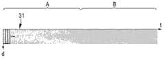
消融装置1还包括散射确定单元8,散射确定单元用于确定指示心脏组织之内血液对超声脉冲的散射的至少一个散射值,其中散射确定单元8适于根据超声信号确定至少一个散射值。图3示意性和示范性地示出了M模式图像的超声信号30。M模式图像根据时间t和组织之内的不同深度d示出了超声信号的超声强度。在A指示的第一时间段中,组织被血液灌注,在B指示的第二时间段中,组织未被血液灌注。血液灌注的组织对应于未被消融的组织,无血液灌注的组织对应于被消融的组织。由采样窗口31对M模式图像采样,采样窗口31不交叠并沿着M模式图像的整个深度范围延伸。散射值确定单元8适于根据相应采样窗口31之内的超声强度的直方图针对每个采样窗口3确定若干散射值。特别是,散射确定单元8适于基于一阶直方图和二阶直方图中的至少一个确定散射值。然而,也可以使用更高阶统计确定散射值,例如,可以使用Gabor滤波方法确定所述至少一个散射值。The
散射确定单元8可适于确定以下值中的若干作为相应采样窗口31的散射值:一阶直方图的一阶平均值m1,一阶直方图的一阶方差m2,一阶直方图的一阶熵m3,二阶直方图的二阶熵n1,二阶直方图的二阶能量n2,二阶直方图的二阶均一性n3,二阶直方图的二阶对比度n4,二阶直方图的二阶聚类趋势n5,二阶直方图的二阶形状n6,二阶直方图的二阶相关性n7以及二阶直方图的二阶相关性导数n8和n9。The
一阶直方图是标准直方图,其中对于不同的超声强度,即对于不同的超声强度箱(bin),确定具有强度I的像素数量P(I),即位于相应的强度箱中的像素数量P(I)。这个一阶直方图的一阶平均值m1可以由以下公式定义:The first-order histogram is a standard histogram in which for different ultrasound intensities, i.e. for different ultrasound intensity bins (bins), the number of pixels P(I) with intensity I is determined, i.e. the number of pixels located in the corresponding intensity bin P (I). The first-order meanm1 of this first-order histogram can be defined by the following formula:
m1=∑IP(I), (1)m1 =∑IP(I), (1)
可以由以下公式定义一阶方差m2和一阶熵m3:The first-order variance m2 and first-order entropy m3 can be defined by the following formulas:
m2=∑(I-m1)2P(I)以及 (2)m2 =∑(Im1 )2 P(I) and (2)
m3=-∑P(I)log2P(I). (3)m3 =-∑P(I)log2 P(I). (3)
在公式(1)到(3)中,在不同超声强度I上进行求和。In equations (1) to (3), the summation is performed over different ultrasound intensities I.
二阶值优选地基于所谓的共生矩阵,例如,在S.Theodoridis等人的“Pattern Recognition”一书(Academic Press,2003)中公开了这种矩阵。二阶值还考虑M模式图像中超声强度的相对位置,并且二阶值基于二阶直方图,可以由以下公式定义:The second-order value is preferably based on a so-called co-occurrence matrix, such a matrix is disclosed, for example, in the book "Pattern Recognition" by S. Theodoridis et al. (Academic Press, 2003). The second-order value also takes into account the relative location of the ultrasound intensity in the M-mode image, and is based on a second-order histogram, which can be defined by the following formula:
其中变量i和j指示超声强度。给定距离例如由校准测量预定义,其中尝试不同的预定义距离,直到依赖于二阶直方图的所确定的性质尽可能好地匹配对象的已知性质。优选地,给定的距离是一个像素,即处于给定距离的像素对优选是直接相邻的像素。where the variables i and j indicate the ultrasound intensity. The given distance is eg predefined by a calibration measurement, wherein different predefined distances are tried until the determined properties dependent on the second order histogram match the known properties of the object as well as possible. Preferably, the given distance is one pixel, ie pairs of pixels at a given distance are preferably directly adjacent pixels.
二阶熵n1,二阶能量n2,二阶均一性n3,二阶对比度n4,二阶聚类趋势n5,二阶聚类形状n6,二阶相关性n7和二阶相关性导数n8、n9,可以由以下公式定义:Second-order entropy n1 , second-order energy n2 , second-order uniformity n3 , second-order contrast n4 , second-order clustering tendency n5 , second-order clustering shape n6 , second-order correlation n7 and second-order The correlation derivatives n8 and n9 can be defined by the following formula:
n1=-∑P(i,j)lnP(i,j). (5)n1 =-∑P(i,j)lnP(i,j). (5)
n2=∑(i-j)2P(i,j), (6)n2 =∑(ij)2 P(i, j), (6)
n5=∑(i+j-2μ)2P(i,j),其中
n6=∑(i+j-2μ)3P(i,j), (10)n6 =∑(i+j-2μ)3 P(i, j), (10)
n8=-∑P(i,j)1n(∑iP(i,j)∑jP(i,j))和 (12)n8 =-∑P(i,j)1n(∑i P(i,j)∑j P(i,j)) and (12)
n9=-∑(∑iP(i,j)∑jP(i,j))1n(∑iP(i,j)∑jP(i,j)). (13)n9 =-∑(∑i P(i,j)∑j P(i,j))1n(∑i P(i,j)∑j P(i,j)). (13)
如果没有特别指出,在公式(5)到(13)中,求和是在超声强度i和j上进行的。公式(5)、(12)和(13)中使用的对数也可以具有另一个底数。If not specified otherwise, in equations (5) to (13), the summation is performed over ultrasound intensities i and j. The logarithms used in equations (5), (12) and (13) can also have another base.
二阶散射值提供指示M模式图像的模式特征的值,该模式涉及M模式图像像素的空间排列,而不是仅仅涉及对比度。这些二阶统计描述模式的随机性、规则性和取向特性。The second order scatter values provide values indicative of the pattern characteristic of the M-mode image, which pattern relates to the spatial arrangement of the M-mode image pixels rather than just the contrast. These second-order statistics describe the randomness, regularity, and orientation properties of patterns.
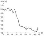
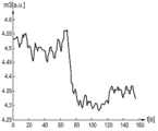
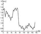
应当指出,散射确定单元8未必确定所有上述一阶值和二阶值。优选地,散射确定单元适于仅确定散射值的种类,其允许确定对象的期望性质。可以通过校准测量确定可用于确定对象的期望性质的散射值种类,其中确定几类散射值,而对象的性质是已知的。在实施例中,校准测量表明,可以使用散射值m1到m3和n3到n9确定组织被消融还是未被消融。在图4到13中示意性和示范性示出了这些散射值。在这些图中,根据以秒为单位的时间,以任意单位示出了相应散射值。在所有这些图中,在大约60到80秒之间可以看到未被消融组织和消融组织之间的转变。It should be noted that the
散射确定单元8还可适于根据相应采样窗口之内的超声强度之和而非根据直方图确定采样窗口的至少一个散射值。例如,该至少一个散射值可以是相应采样窗口之内所有超声强度之和。也可以由以下公式定义所述至少一个散射值:The
f=∑ItIt-p, (14)f=∑It Itp , (14)
其中It指示时刻t时M模式图像的像素的超声强度,It-p指示时刻t-p时M模式图像的像素的超声强度,其中p指示两次相继心跳之间的时间。在公式(14)中,求和在所有ItIt-p对上进行,其中对应于It的像素是位于相应采样窗口之内的。可以由心电图仪确定两次相继心跳之间的时间,心电图仪在图1中由附图标记为9的框示意性和示范性表示。在其他实施例中,消融装置可以不包括心电图仪9,并且可以从例如M模式图像确定两次相继心跳之间的时间。where It indicates the ultrasound intensity of a pixel of the M-mode image at time t, Itp indicates the ultrasound intensity of a pixel of the M-mode image at time tp, where p indicates the time between two consecutive heartbeats. In equation (14), the summation is performed over all It Itp pairs where the pixel corresponding to It is located within the corresponding sampling window. The time between two successive heartbeats can be determined by an electrocardiograph, which is schematically and exemplarily indicated by the box referenced 9 in FIG. 1 . In other embodiments, the ablation device may not comprise an
消融装置还包括性质确定单元15,性质确定单元用于根据散射确定单元8所确定的散射值确定对象3的性质。在这一实施例中,性质确定单元15适于基于散射值确定组织的一部分是被消融组织还是未被消融组织作为对象的性质。通过消融可以修改组织的灌注,其中灌注的修改改变了超声脉冲的散射,从而修改了散射值。因此可以将散射值用于确定组织是被消融还是未被消融。The ablation device further comprises a
在这一实施例中,性质确定单元15适于针对采样窗口的每个散射值确定组织被消融或组织未被消融的投票。对于这些值中的每个,通过将相应值与预定义阈值比较来进行投票,即,对于每个值进行二元阈值处理,以便针对每个值确定投票值。性质确定单元15适于基于投票的大多数确定最终结果,即对应于相应采样窗口的组织被消融还是未被消融。这意味着,如果针对采样窗口已经确定被消融的比未被消融的投票更多,则最终结果是,对应于相应采样窗口的组织被消融,反之亦然。预定义的阈值可以通过例如校准测量来确定。In this embodiment, the
性质确定单元还可适于将若干散射值组合到多维特征矢量,即,针对每个采样窗口,可以定义多维特征矢量,其中可以将多维特征矢量与预定义阈值矢量比较,以确定相应的采样窗口对应于被消融组织还是未被消融组织。同样,可以通过,例如校准测量,来确定这个预定义阈值矢量。The property determination unit can also be adapted to combine several scatter values into a multidimensional feature vector, i.e. for each sampling window a multidimensional feature vector can be defined, wherein the multidimensional feature vector can be compared with a predefined threshold vector to determine the corresponding sampling window Corresponds to ablated tissue or non-ablated tissue. Again, this predefined threshold vector can be determined eg by calibration measurements.
性质确定单元还可适于向采样窗口应用聚类分析,其中根据多维特征矢量对采样窗口分聚类,并向采样窗口的聚类分配性质。例如,聚类分析可能获得采样窗口的两个聚类,其中向这些聚类之一分配性质“被消融组织”,向聚类中的另一个分配性质“未被消融组织”。可以根据与阈值的比较情况确定聚类表示消融还是未被消融组织,其中,例如,可以对聚类的多维特征矢量求平均以生成平均矢量,并且其中可以将聚类的平均矢量与阈值矢量比较,通过校准测量确定阈值矢量。因而,通过阈值处理可以向采样窗口分配对象的性质。还可以在开始消融之前首先应用聚类分析,获得表示未被消融组织的第一组聚类。然后,可以连续应用聚类分析,同时执行消融过程。如果聚类分析获得不属于第一组聚类的新聚类,可以向这些新聚类分配性质“被消融组织”。The property determination unit may also be adapted to apply a cluster analysis to the sampling windows, wherein the sampling windows are clustered according to the multidimensional feature vector and assign properties to the clusters of the sampling windows. For example, a cluster analysis might obtain two clusters of sampling windows, wherein the property "ablated tissue" is assigned to one of these clusters, and the property "non-ablated tissue" is assigned to the other of the clusters. A cluster can be determined to represent ablated or non-ablated tissue based on a comparison to a threshold, where, for example, the multidimensional feature vectors of the clusters can be averaged to generate an average vector, and wherein the cluster's average vector can be compared to the threshold vector , the threshold vector is determined from calibration measurements. Thus, the properties of the object can be assigned to the sampling windows by thresholding. It is also possible to first apply cluster analysis before starting ablation, obtaining a first set of clusters representing non-ablated tissue. Then, cluster analysis can be applied continuously while the ablation process is performed. If the cluster analysis yields new clusters that do not belong to the first set of clusters, the property "ablated tissue" can be assigned to these new clusters.
在实施例中,性质确定单元适于应用后续聚类分析技术,以确定组织的哪个部分被消融,以及组织的哪个部分未被消融。将参考图14到18描述聚类分析技术,其中在图14到16中,上部示出了M模式图像,下部示出了聚类分析技术的结果,且其中图17和18示出的流程图图示了聚类分析技术的若干步骤。In an embodiment, the property determination unit is adapted to apply a subsequent cluster analysis technique to determine which part of the tissue was ablated and which part of the tissue was not ablated. The cluster analysis technique will be described with reference to FIGS. 14 to 18, wherein in FIGS. Several steps of the cluster analysis technique are illustrated.
散射确定单元确定若干采样窗口131的散射值。采样窗口131对整个M模式图像采样并且是交叠的。采样窗口131的交叠增大了最终聚类结果的分辨率。采样窗口对应于特定深度范围和特定时间范围。采样窗口131可以具有相同宽度和相同长度,或者采样窗口的宽度和长度可以是不同的。此外,可以由具有不同尺寸和/或不同形状的不同采样窗口对M模式图像采样。优选地,窗口的宽度覆盖至少一个心跳周期,其中采样窗口可以相对于心跳对准。例如,每个采样窗口可以开始于相对于相应收缩周期的相同时间偏移。在实施例中,采样窗口在深度方向的尺寸大约为0.2mm。The scatter determination unit determines scatter values for
在开始消融之前执行第一聚类分析技术,其中在图14的上部中示出了对应的M模式图像。在下文中将参考图17中所示的流程图描述这种第一聚类分析技术。A first cluster analysis technique is performed before starting the ablation, for which the corresponding M-mode image is shown in the upper part of FIG. 14 . This first cluster analysis technique will be described hereinafter with reference to the flowchart shown in FIG. 17 .
第一聚类分析技术开始于步骤101,方式是提供示出开始消融之前的组织的M模式图像,利用采样窗口对M模式图像采样,并针对每个样本窗口确定包括散射值的多维特征矢量。The first cluster analysis technique begins at
在步骤102中,选择采样窗口并从而选择对应的初始特征矢量。可以随机地进行这种选择。在步骤103中,将初始特征矢量,并且因而,所选择的采样窗口,分配为第一聚类。在步骤104中,确定哪些特征矢量被布置在包含多维特征矢量的多维特征矢量空间中的初始特征矢量周围的区域之内。如果特征矢量是二维矢量,则该区域优选是圆,或者如果特征矢量的维度大于二,是超球面。可以通过校准测量来确定区域的尺寸。In
在步骤105中,将所确定的特征矢量(位于初始特征矢量周围的区域之内)分配给第一聚类,在步骤106中,对第一聚类之内的特征矢量求平均,以计算第一聚类的平均矢量。在步骤107中,确定是否满足终止标准。终止标准是,例如,是否已达到预定义的迭代次数或是否已满足收敛标准。收敛标准例如是,初始特征矢量和平均特征矢量之间的差或实际确定的平均特征矢量和先前确定的平均特征矢量之间的差低于预定义的阈值。如果不满足终止标准,聚类分析再次执行步骤104到107,其中,现在并不使用初始特征矢量而是使用实际确定的平均特征矢量,即在步骤104中在实际确定的平均特征矢量周围的区域之内确定特征矢量,在步骤105中,向第一聚类分配尚未分配给第一聚类的实际平均矢量周围区域中的特征矢量,在步骤106中,通过对第一聚类的特征矢量求平均来计算新的平均特征矢量。In
如果在步骤107中满足终止标准,则确定了第一聚类,该方法继续步骤108。在108中,确定是否己将所有特征矢量,因而将所有采样窗口,分配给聚类。如果未将所有特征矢量分配给聚类,则基于尚未分配给聚类的剩余特征矢量执行步骤102到107,以便确定另一个聚类。因此执行步骤102到108,直到所有特征矢量,因而采样窗口,都分配给聚类。在向聚类分配所有特征矢量之后,第一聚类分析技术结束于步骤109。由于将第一聚类分析技术应用于示出开始消融前的组织的M模式图像,因而通过执行步骤101到109确定的聚类对应于未被消融的组织。If in
图15和16示出的M模式图像不仅示出了开始消融之前的组织,而且示出了开始消融之后的组织。在执行消融时,连续生成超声信号并采样。于是,对采样窗口进行连续采集并针对每个采样窗口计算特征矢量,即连续确定特征矢量,其中根据第二聚类分析技术对连续新确定的特征矢量聚类,在下文中将参考图18中所示的流程图描述所述第二聚类分析技术。The M-mode images shown in FIGS. 15 and 16 not only show the tissue before starting ablation, but also show the tissue after starting ablation. Ultrasound signals are continuously generated and sampled while the ablation is being performed. Then, the sampling windows are continuously collected and the feature vectors are calculated for each sampling window, that is, the feature vectors are continuously determined, wherein the continuously newly determined feature vectors are clustered according to the second cluster analysis technique, hereinafter referred to in FIG. 18 The flow chart shown describes the second cluster analysis technique.
在步骤201中,向实际确定的特征矢量和已经确定的聚类的每个平均矢量应用相似性度量。相似性度量例如是实际确定的特征矢量和相应平均特征矢量之间的绝对差,其中,如果这种绝对差低于预定义阈值,认为实际确定的特征矢量类似于相应的平均特征矢量。如果实际确定的特征矢量类似于特定聚类的平均特征矢量,则在步骤202中向特定聚类分配实际确定的特征矢量,其中在步骤203中,考虑新分配的实际确定的特征矢量,为这个特定聚类计算新的平均特征矢量。In step 201 a similarity measure is applied to the actually determined feature vector and each mean vector of the already determined clusters. The similarity measure is, for example, the absolute difference between the actually determined feature vector and the corresponding average feature vector, wherein an actually determined feature vector is considered similar to the corresponding average feature vector if this absolute difference is below a predefined threshold. If the actually determined eigenvector is similar to the average eigenvector of a particular cluster, the actual determined eigenvector is assigned to the particular cluster in
如果在步骤201中,确定实际确定的特征矢量不类似于现有聚类的任何平均特征矢量,则该方法继续步骤204。在步骤204中,无法分配给现有聚类的实际确定的特征矢量定义新聚类,并且该实际确定的特征矢量被定义为这个新聚类的平均特征矢量。If in
在图14中可以示范性地看到,在开始消融之前,存在若干聚类40...47,它们可能属于未被消融组织的不同结构。在图15和16的下部,可以看到新聚类48,它是在开始消融之后生成的。因此性质确定单元将这个新聚类48分配给“被消融组织”,将其他聚类40…47分配给“未被消融”。It can be seen exemplarily in Fig. 14 that before starting the ablation there are
消融装置1还包括显示器10,显示器用于示出确定的性质。特别是,显示器10适于显示组织的哪些部分被消融,组织的哪些部分未被消融。The
性质确定单元优选地还适于根据所确定的被消融部分和未被消融部分确定消融深度。由于从M模式图像已知消融部分和未被消融部分的位置,所以能够容易地确定消融深度,即从组织外表面开始消融的组织深度。显示器10还可适于显示这个消融深度。The property determination unit is preferably further adapted to determine the ablation depth from the determined ablated and non-ablated portions. Since the positions of the ablated portion and the non-ablated portion are known from the M-mode image, the ablation depth, ie the depth of tissue ablated from the outer surface of the tissue, can be easily determined. The
消融装置1还包括用于根据M模式图像的A线确定心壁的厚度的心壁厚度确定单元54。A线由M模式图像的超声强度定义,表示在固定时间t的超声信号的幅度。图19中示意性和示范性示出了这样的A线61,其中根据任意单位的深度d以任意单位示出了A线的幅度a。The
62和63表示的A线61的区域对应于心壁的前后表面。区域64是由超声脉冲直接生成的。The area of
图19中示出的A线61允许确定相对于超声换能器18的位置的前后表面62、63的位置,超声换能器18发射超声脉冲并接收回波。区域64中的第一测得幅度界定超声单元的位置。区域64之后是包括基本为零的幅度的区域,之后不久,区域63中的幅度再次增大,标志着对象处的第一反射,即标志着对象的前表面。接着是区域65,包括对应于心壁组织之内反射的小幅度,然后在区域62中,幅度再次显著增大,由此标志着心壁的后表面。于是,A线61允许基于区域62和63确定前后表面的位置。心壁厚度确定单元优选地适于确定在包括基本零幅度值的区域之后区域63中越来越大幅度的位置,作为对象前表面的位置。然后,幅度在区域65中显著减小,确定幅度下次显著增大的位置(区域62)作为心壁后表面的位置。换言之,在区域64中超声单元的换能器振铃之后,接着是“平静期”。这个平静期接下来终止于与前表面相关联的区域63中的反射。在区域63中的这种反射之后,出现时期65,其标志是超声强度的快速且小的温度变化。特别是,时期65中信号的包络倾向于在强度上以指数衰减。在时期65的末尾,在区域62中再次观察到与后表面相关联的强反射。可以预定义阈值,尤其是可以预定义相对阈值,其中如果“平静期”之后的反射超过相应预定义阈值,则检测到前表面,且其中如果在时期65末尾信号超过相应阈值,则检测到后表面。可以利用具有已知前表面和后表面位置的壁,通过校准测量预定义阈值。The A-line 61 shown in Figure 19 allows determining the position of the front and
在实施例中,性质确定单元还可适于根据所确定的消融深度和所确定的心壁前表面和后表面位置确定心壁的透壁程度。In an embodiment, the property determination unit is further adapted to determine the degree of transmurality of the heart wall based on the determined ablation depth and the determined positions of the anterior surface and the posterior surface of the heart wall.
消融装置1还包括控制单元11,控制单元用于根据性质确定单元确定的对象的性质控制对象的消融。特别是,控制单元11可适于根据确定的对象组织的被消融部分和未被消融部分控制对象组织的消融。例如,如果应当消融对象组织的某一部分,控制单元11能够控制对象组织的消融使得RF能量被施加直到完全消融相应部分。为了执行消融过程的这种控制,优选地实时确定组织的被消融部分和组织的未被消融部分。控制单元11还可适于根据透壁程度控制心壁的消融。特别是,控制单元11可适于控制消融,从而完全消融心壁,而不消融下方组织,即,控制单元可适于控制消融装置使得达到100%的透壁性。The
消融装置1中用于确定对象的性质,特别是,用于确定对象的被消融部分和未被消融部分的单元和元件,构成集成到消融装置中的性质确定装置。不过,性质确定装置也可以是未集成到消融装置中的独立装置。The units and elements used in the
图20中示意性和示范性示出了独立的性质确定装置。图20中所示的性质确定装置90的元件和单元类似于上文参考图1所述的对应元件和单元,并由相同的附图标记表示。为了详细描述性质确定装置90,因此参考图1的以上描述。A separate property determination device is shown schematically and exemplarily in FIG. 20 . Elements and units of the property determining device 90 shown in FIG. 20 are similar to corresponding elements and units described above with reference to FIG. 1 and are denoted by the same reference numerals. For a detailed description of the property determining means 90 reference is therefore made to the above description of FIG. 1 .
图21示意性和示范性示出了性质确定装置90的导管70的导管顶端71的实施例。性质确定装置90的导管70的导管顶端71的元件和单元类似于上文参考图2所述的对应元件和单元,因此由相似的附图标记表示。为了详细描述这些元件和单元,因此参考上文参考图2提供的描述。与消融装置的导管顶端不同,性质确定装置90的导管顶端不包括消融电极。FIG. 21 shows schematically and exemplarily an embodiment of a
在下文中,将参考图22中示出的流程图示范性描述性质确定方法的实施例。Hereinafter, an embodiment of the property determination method will be exemplarily described with reference to the flowchart shown in FIG. 22 .
在步骤301中,通过以下方式来提供超声信号:向对象3发出超声脉冲、从对象3接收回波序列并根据接收的回波序列生成超声信号。由超声换能器18提供超声信号,即M模式图像。在步骤302中,根据提供的超声信号确定至少一个散射值,该至少一个散射值指示对象3的流体导致的超声脉冲的散射。在步骤303中,根据对象的一个或若干散射值,确定对象的性质,特别是对象的被消融部分和对象的未被消融部分。In
在下文中,将参考图23所示的流程图示范性描述用于消融对象的消融方法的实施例。在步骤301中,提供超声信号,在步骤302中,基于所提供的超声信号确定至少一个散射值;在步骤303中,根据至所述至少一个散射值确定对象的性质。步骤301至303类似于上文参考图22所述的步骤,是循环执行的,以便连续确定对象的哪个部分被消融,对象的哪个部分未被消融。向消融装置1的控制单元11提供这种消融信息,其中通过根据所确定的消融信息控制能量源24来控制步骤304中的对象消融,特别是,通过消融电极4施加RF能量。因而连续确定消融信息并将其用于控制消融过程。Hereinafter, an embodiment of an ablation method for ablating an object will be exemplarily described with reference to the flowchart shown in FIG. 23 . In
针对心脏心律不齐的已知导管消融过程中的一个主要缺点是生成伤口时缺少关于伤口质量的足够信息。此外,利用已知的方法,很难在重做消融过程时找回旧的伤口部位。当前,治疗学家依赖其自己的专业知识确定消融的最佳参数,例如功率、温度和持续时间。不过,这些设置有很大变化,例如,因为例如局部心壁厚度、血流造成的局部冷却、导管和组织之间的接触等的患者内和患者间差异。A major drawback in known catheter ablation procedures for cardiac arrhythmias is the lack of sufficient information about the wound quality when the wound is generated. Furthermore, with known methods it is very difficult to retrieve the old wound site when redoing the ablation procedure. Currently, therapists rely on their own expertise to determine the optimal parameters for ablation, such as power, temperature and duration. However, these settings vary widely, eg, because of intra-patient and inter-patient variations such as local heart wall thickness, local cooling by blood flow, contact between catheter and tissue, and the like.
在现有技术中,两个主要的治疗相关问题源于部位的加热不足或加热过度。在加热不足的情况下,组织未充分凝结以形成治疗学家期望的心律失常阻塞损伤。这可能导致患者体内持久或复发的症状、以及后续治疗的需要、更长的住院治疗时间和中风和栓塞的更大风险。另一个极端情况,过热,会导致处理部位组织的破裂,向血流中释放可能威胁生命的颗粒,或对相邻器官和组织造成损伤。In the prior art, two main treatment-related problems stem from under- or over-heating of the site. With insufficient heating, the tissue does not coagulate sufficiently to form the arrhythmia blocking injury that therapists expect. This can lead to persistent or recurring symptoms in patients, as well as the need for follow-up treatment, longer hospital stays and a greater risk of stroke and embolism. At the other extreme, overheating can lead to rupture of tissue at the treatment site, releasing potentially life-threatening particles into the bloodstream, or causing damage to adjacent organs and tissues.
根据本发明的消融装置提供了改善的控制。它提供了关于组织中伤口进展的反馈,能够提供关于伤口相对于处理部位处组织厚度的深度信息,并能够防止导管消融过程中加热不足和过热造成的伤害和死亡。Ablation devices according to the present invention provide improved control. It provides feedback on wound progression in tissue, can provide information on the depth of the wound relative to tissue thickness at the treatment site, and can prevent injury and death from under- and overheating during catheter ablation.
在消融期间,血管结构并且因而毛细管灌注,一般被凝固性坏死完全破坏。相反,具有血细胞的毛细管灌注向未被消融的健康组织供应氧和代谢物。被消融的组织因此一般没有毛细管灌注或毛细管灌注至少被大大减少,未被消融组织包括正常毛细管灌注而且一般未被减少。所述至少一个散射值优选地指示灌注组织的血液细胞造成的超声脉冲散射,因此也指示消融信息,特别是,无论组织是被消融组织还是未被消融组织。因而,如上文更详细的描述,性质确定单元能够基于所述至少一个散射值确定组织为被消融组织还是未被消融组织。During ablation, the vascular structure, and thus capillary perfusion, is generally completely destroyed by coagulation necrosis. In contrast, capillary perfusion with blood cells supplies oxygen and metabolites to unablated healthy tissue. Ablated tissue thus generally has no capillary perfusion or at least substantially reduced capillary perfusion, and non-ablated tissue includes normal capillary perfusion and generally not reduced. The at least one scatter value is preferably indicative of the scattering of the ultrasound pulse by blood cells perfusing the tissue and thus also of the ablation information, in particular whether the tissue is ablated or non-ablated tissue. Thus, as described in more detail above, the property determination unit is able to determine whether the tissue is ablated tissue or non-ablated tissue based on the at least one scatter value.
如上所述,(红)血细胞的运动对超声的散射有贡献,尤其是在更高超声(US)频率(>10MHz)。因此,由流速变化或归因于被破坏的血管系统的完全没有运动所导致的由消融引起的血细胞运动的变化,影响US的散射。可以将这个特征用作组织损伤的标记。As mentioned above, the movement of (red) blood cells contributes to the scattering of ultrasound, especially at higher ultrasound (US) frequencies (>10MHz). Thus, changes in the motion of blood cells induced by ablation, either by changes in flow velocity or by complete lack of motion due to disrupted vasculature, affect the scattering of US. This feature can be used as a marker of tissue damage.
在实施例中,性质确定单元适于识别血管。由于至少一个散射值可以指示血细胞导致的超声脉冲散射,因而性质确定单元可适于根据至少一个散射值识别血管,特别是动脉或静脉。例如,通过校准测量,可以确定哪些散射值对应于血管,并且在实际确定了(如由校准确定的)对应于血管的散射值时,性质确定单元能够识别血管。消融装置的控制单元可适于使得,在性质确定单元在应当开始或继续消融过程的位置识别了血管时,在显示器上或像声学输出单元的另一输出单元上产生警报。通过这种方式,在将消融电极定位在血管上或非常接近时,可以通知医生,并可以避免对血管应用消融过程。应当避免对血管应用消融程序,因为这可能导致,例如,缺血。In an embodiment, the property determining unit is adapted to identify blood vessels. Since the at least one scatter value may be indicative of the scattering of the ultrasound pulse by the blood cells, the property determination unit may be adapted to identify a blood vessel, in particular an artery or a vein, from the at least one scatter value. For example, by calibration measurements it can be determined which scatter values correspond to blood vessels, and when the scatter values corresponding to blood vessels are actually determined (as determined by the calibration), the property determination unit is able to identify blood vessels. The control unit of the ablation device may be adapted such that an alarm is generated on the display or on another output unit like the acoustic output unit when the property determination unit identifies a blood vessel at a location where the ablation procedure should be started or continued. In this way, when an ablation electrode is positioned on or very close to a blood vessel, the physician can be informed and application of an ablation procedure to the blood vessel can be avoided. Applying ablation procedures to blood vessels should be avoided as this may result in, for example, ischemia.
消融装置和性质确定装置提供了分析和量化血液灌注和非血液灌注组织的散射模式变化的自动方式。如前所述,这种模式差异为区分消融前的健康心脏组织和消融导致的凝结消融组织提供了重要信息。模式差异优选基于实时M模式超声图像的质地(texture)性质的统计分析,尤其是基于在两种状况之间给出清晰区别的二阶统计性质。这使得能够优选地仅基于统计分析区分健康和凝结的组织,因此能够用于消融期间和/或之后的伤口监测目的,特别是,用于控制伤口深度。The ablation device and property determination device provide an automated way to analyze and quantify changes in scattering patterns of blood-perfused and non-blood-perfused tissue. As mentioned previously, this pattern difference provides important information for distinguishing healthy cardiac tissue before ablation from clotted ablated tissue resulting from ablation. The mode difference is preferably based on a statistical analysis of the texture properties of the real-time M-mode ultrasound images, especially based on second order statistical properties that give a clear distinction between the two conditions. This enables a distinction between healthy and coagulated tissue, preferably based solely on statistical analysis, and thus can be used for wound monitoring purposes during and/or after ablation, in particular, for controlling wound depth.
至少一个散射值优选是从超声原始数据确定的,因为它保留了用于表征散射模式的高频信息。因而,用于确定至少一个散射值的超声信号优选地是由尚未处理,例如,尚未滤波的超声原始数据表示的。The at least one scatter value is preferably determined from the ultrasound raw data since it preserves high frequency information for characterizing the scatter pattern. Thus, the ultrasound signal used to determine the at least one scatter value is preferably represented by ultrasound raw data that has not been processed, eg not filtered.
尽管在上述实施例中,基于例如一阶和二阶直方图确定散射值,但也可以基于其他基于质地的分析(其可以包括更高阶统计)确定散射值。例如,可以使用Gabor滤波方法,其中特定频带可能能够捕获被消融组织和未被消融组织之间的主要差异。Although in the embodiments described above, scatter values were determined based on, for example, first and second order histograms, scatter values may also be determined based on other texture-based analyses, which may include higher order statistics. For example, Gabor filtering methods may be used, where specific frequency bands may be able to capture the main differences between ablated and non-ablated tissue.
尽管在上述实施例中,超声换能器被集成到导管中,但超声换能器也可以集成到例如针中。Although in the above-described embodiments the ultrasound transducer is integrated into the catheter, the ultrasound transducer may also be integrated into eg a needle.
再次参考图1,灌洗流体提供单元52可适于在灌洗流体中增加血管扩张剂,例如异丙基肾上腺素。当恰在开始消融之前应用一剂血管扩张剂时,目标位置的局部灌注增大。通过这种方式,可以增强消融前后血液诱发的超声散射的差异,从而便于分析,即便于性质确定单元基于至少一个散射值区分被消融组织和未被消融组织。Referring again to FIG. 1 , the irrigation
尽管在上述实施例中,性质确定装置被集成到消融装置中,但在其他实施例中,性质确定装置也可以集成到另一对象影响装置中,例如,集成到用于执行活检的活检装置中。活检装置包括活检针,其中超声换能器可以集成到活检针的顶端中。活检装置可被用于例如肿瘤学中,用于区分被消融肿瘤与未被消融肿瘤。典型地,肿瘤的特征在于超灌注组织围绕的凝结组织的致密核心。这种周围组织是活检所取的靶组织。周围超灌注组织可以散射超声脉冲,可以基于所得的超声信号确定至少一个散射值。基于这至少一个散射值,可以确定肿瘤的哪个部分是凝结组织的致密核心,肿瘤的哪个部分是必须要进行活检的超灌注组织。Although in the above described embodiments the property determining device is integrated into the ablation device, in other embodiments the property determining device may also be integrated into another object affecting device, e.g. a biopsy device for performing a biopsy . The biopsy device includes a biopsy needle, wherein an ultrasound transducer can be integrated into the tip of the biopsy needle. Biopsy devices may be used, for example, in oncology to differentiate ablated tumors from non-ablated tumors. Typically, tumors are characterized by a dense core of coagulated tissue surrounded by hyperperfused tissue. This surrounding tissue is the target tissue for biopsy. Surrounding hyperperfused tissue may scatter the ultrasound pulse, and at least one scatter value may be determined based on the resulting ultrasound signal. Based on the at least one scatter value, it can be determined which part of the tumor is the dense core of coagulated tissue and which part of the tumor is the hyperperfused tissue which has to be biopsied.
图24示意性和示范性示出了活检针顶端80的实施例。活检针包括位于活检针顶端80的外部区域中的多个超声换能器18。这个外部区域围绕中央腔82。中央腔用于引入活检仪器,以获取组织3的一部分,并将这个部分转移到人体外部。超声换能器18通过向组织发出超声脉冲,从组织接收回波序列,并根据接收的回波序列生成超声信号,来提供超声信号。通过电连接23向散射确定单元发射超声信号。散射确定单元确定至少一个散射值,该至少一个散射值指示组织之内流体导致的超声脉冲散射,其中根据超声信号确定所述至少一个散射值,并且性质确定单元根据上文参考图1和2所述的至少一个散射值确定组织的性质。特别是,性质确定单元确定肿瘤的哪个部分是凝结组织的致密核心,对于灌注而言,该致密核心对应于被消融组织,并且肿瘤的哪个部分是超灌注组织,必须要从该超灌注组织进行活检,并且对于灌洗而言,该超灌注组织对应于未被消融组织。活检装置优选还包括上文参考图1所述的图像提供单元2、定位单元6、7和导航单元29。在活检针顶端的远端和超声换能器18之间,提供声学透明的材料81,例如聚甲基戊烯,界定超声换能器18和活检针外部之间的声学路径。FIG. 24 schematically and exemplarily shows an embodiment of a
尽管在上述实施例中,心壁前后表面的位置从A线的幅度确定,但是在其他实施例中,可以通过其他方式确定这些位置。例如,可以基于深度上A线的频谱分析确定心壁前后表面的位置。Although in the embodiments described above the locations of the anterior and posterior surfaces of the heart wall are determined from the magnitude of the A-line, in other embodiments these locations may be determined by other means. For example, the location of the anterior and posterior surfaces of the heart wall can be determined based on spectral analysis of the A-line in depth.
可以在治疗例如心律不齐和肿瘤消融期间的组织成像中使用消融装置和性质确定装置。在这些过程中,希望在程序期间跟踪伤口形成的进展。Ablation devices and property determination devices may be used in tissue imaging during treatments such as arrhythmias and tumor ablation. In these procedures, it is desirable to follow the progression of wound formation during the procedure.
通过研究附图、公开和所附权利要求,本领域的技术人员在实践请求保护的本发明时能够理解和实现所公开实施例的其他变化。Other variations to the disclosed embodiments can be understood and effected by those skilled in the art in practicing the claimed invention, from a study of the drawings, the disclosure, and the appended claims.
在权利要求中,“包括”一词不排除其他元件或步骤,不定冠词“一”不排除多个。In the claims, the word "comprising" does not exclude other elements or steps, and the indefinite article "a" or "a" does not exclude a plurality.
可以由任何其他数量的单元或设备进行一个或若干单元或设备执行的确定,例如散射值和对象的性质的确定。例如,可以由单个单元或由任何其他数量的不同单元进行散射值的确定和对象的性质的确定。可以将确定和/或根据消融方法的消融装置的控制和/或根据性质确定方法的性质确定装置的控制实现为计算机程序的程序代码模块和/或专用硬件。Determinations performed by one or several units or devices, such as determinations of scatter values and properties of objects, may be performed by any other number of units or devices. For example, the determination of the scatter value and the determination of the property of the object may be performed by a single unit or by any other number of different units. The determination and/or the control of the ablation device according to the ablation method and/or the control of the property determination device according to the property determination method may be implemented as program code modules of a computer program and/or as dedicated hardware.
单个单元或设备可以完成权利要求中记载的若干项目的功能。在互不相同的从属权利要求中记载特定手段的仅有事实并不表示不能有利地使用这些手段的组合。A single unit or device may fulfill the functions of several items recited in the claims. The mere fact that certain measures are recited in mutually different dependent claims does not indicate that a combination of these measured cannot be used to advantage.
权利要求中的任何附图标记不应被解释为限制范围。Any reference signs in the claims should not be construed as limiting the scope.
本发明涉及一种用于确定对象的性质的性质确定装置,所述对象优选地是心脏。超声信号提供单元提供对象的超声信号,散射确定单元根据超声信号确定至少一个散射值,该至少一个散射值指示由灌注对象的流体导致的超声脉冲的散射。性质确定单元根据至少一个散射值确定对象的性质。与基于气泡形成的组织损伤检测方法相比,基于流体导致的超声脉冲散射可以较为直接地确定与灌洗相关的对象的性质,例如组织被消融还是未被消融,由此增大确定对象的性质的精度。The invention relates to a property determination device for determining a property of an object, preferably a heart. The ultrasound signal providing unit provides ultrasound signals of the object, from which the scatter determining unit determines at least one scatter value indicative of scattering of ultrasound pulses caused by fluid perfusing the object. A property determination unit determines a property of the object from at least one scatter value. Compared with tissue damage detection methods based on bubble formation, fluid-induced ultrasonic pulse scattering can more directly determine the properties of objects related to lavage, such as whether tissue is ablated or not, thereby increasing the ability to determine the properties of objects. accuracy.
Claims (15)
Applications Claiming Priority (5)
| Application Number | Priority Date | Filing Date | Title |
|---|---|---|---|
| EP10161318 | 2010-04-28 | ||
| EP10161318.0 | 2010-04-28 | ||
| EP10173025 | 2010-08-17 | ||
| EP10173025.7 | 2010-08-17 | ||
| PCT/IB2011/051640WO2011135482A1 (en) | 2010-04-28 | 2011-04-15 | Property determining apparatus for determining a property of an object |
Publications (2)
| Publication Number | Publication Date |
|---|---|
| CN102858252Atrue CN102858252A (en) | 2013-01-02 |
| CN102858252B CN102858252B (en) | 2015-05-20 |
Family
ID=44120284
Family Applications (1)
| Application Number | Title | Priority Date | Filing Date |
|---|---|---|---|
| CN201180020819.9AActiveCN102858252B (en) | 2010-04-28 | 2011-04-15 | Property determination device for determining the property of an object |
Country Status (8)
| Country | Link |
|---|---|
| US (1) | US10335192B2 (en) |
| EP (1) | EP2563229B1 (en) |
| JP (1) | JP6118247B2 (en) |
| CN (1) | CN102858252B (en) |
| BR (1) | BR112012027273A8 (en) |
| RU (1) | RU2567268C2 (en) |
| TW (1) | TW201143721A (en) |
| WO (1) | WO2011135482A1 (en) |
Cited By (1)
| Publication number | Priority date | Publication date | Assignee | Title |
|---|---|---|---|---|
| WO2019119429A1 (en)* | 2017-12-22 | 2019-06-27 | 中国科学院深圳先进技术研究院 | Dual-transducer compensation imaging method and ultrasonic imaging system |
Families Citing this family (14)
| Publication number | Priority date | Publication date | Assignee | Title |
|---|---|---|---|---|
| WO2011135482A1 (en) | 2010-04-28 | 2011-11-03 | Koninklijke Philips Electronics N.V. | Property determining apparatus for determining a property of an object |
| WO2013005500A1 (en)* | 2011-07-06 | 2013-01-10 | オリンパスメディカルシステムズ株式会社 | Sampling device |
| EP2911589A1 (en)* | 2012-10-23 | 2015-09-02 | Koninklijke Philips N.V. | Spatial configuration determination apparatus |
| CN115715689B (en) | 2016-11-11 | 2025-01-17 | 杰尼索尼克斯公司 | Tissue controlled treatment and dynamic interaction and comparison with tissue and/or treatment data |
| US11083517B2 (en)* | 2017-01-19 | 2021-08-10 | Biosense Webster (Israel) Ltd. | Enhancing efficiency of repeat ablation by merging current and previous maps |
| EP3614940B1 (en)* | 2017-04-28 | 2024-11-20 | Arrinex, Inc. | Systems for locating blood vessels in the treatment of rhinitis |
| JP2020518385A (en)* | 2017-05-04 | 2020-06-25 | ガイネソニックス, インコーポレイテッド | A method for monitoring ablation progression using Doppler ultrasound |
| EP3716854B1 (en)* | 2017-11-29 | 2025-05-28 | Avent, Inc. | System and method for mounting an ultrasound transducer on a needle |
| WO2020109519A1 (en) | 2018-11-30 | 2020-06-04 | Koninklijke Philips N.V. | Apparatus and method for detecting bone fracture |
| CN110313937B (en) | 2019-08-01 | 2023-03-24 | 无锡海斯凯尔医学技术有限公司 | Ultrasonic signal processing method, device, equipment and storage medium |
| EP4031046A1 (en)* | 2019-11-05 | 2022-07-27 | Boston Scientific Scimed, Inc. | Medical systems for ablating tissue |
| CN111281426B (en)* | 2020-02-28 | 2022-09-09 | 中国科学院苏州生物医学工程技术研究所 | Ultrasound imaging and image analysis methods of superficial nerves |
| CN111429432B (en)* | 2020-03-24 | 2024-05-03 | 聚融医疗科技(杭州)有限公司 | Thermal ablation area monitoring method and system based on radio frequency processing and fuzzy clustering |
| CN118267748B (en)* | 2024-05-31 | 2024-08-13 | 四川省农业科学院茶叶研究所 | Method for extracting secondary metabolites of tea trees |
Citations (11)
| Publication number | Priority date | Publication date | Assignee | Title |
|---|---|---|---|---|
| WO1995001126A1 (en)* | 1993-06-29 | 1995-01-12 | Institute Of Cancer Research: Royal Cancer Hospital | Apparatus for monitoring ultrasonic surgical ablation |
| US20040030227A1 (en)* | 2002-05-16 | 2004-02-12 | Barbara Ann Karmanos Cancer Institute | Method and apparatus for combined diagnostic and therapeutic ultrasound system incorporating noninvasive thermometry, ablation control and automation |
| US20050283074A1 (en)* | 2004-06-22 | 2005-12-22 | Siemens Medical Solutions Usa, Inc. | Ultrasound feedback for tissue ablation procedures |
| CN1809317A (en)* | 2003-04-17 | 2006-07-26 | 布赖汉姆妇女医院 | Shear mode therapeutic ultrasound |
| WO2006087649A1 (en)* | 2005-02-17 | 2006-08-24 | Koninklijke Philips Electronics, N.V. | Method and apparatus for the visualization of the focus generated using focused ultrasound |
| TW200732011A (en)* | 2006-02-20 | 2007-09-01 | Chiung-Nien Chen | Orientation system for focal point position of non-invasive therapeutical ultrasonic |
| US20080154131A1 (en)* | 2006-12-20 | 2008-06-26 | General Electric Company | Methods for enhancement of visibility of ablation regions |
| US20080253529A1 (en)* | 2007-04-11 | 2008-10-16 | Searete Llc, A Limited Liability Corporation Of The State Of Delaware | Ablating based at least partially on compton scattered x-ray visualizing, imaging, or information providing |
| US20090093807A1 (en)* | 2007-10-03 | 2009-04-09 | Searete Llc, A Limited Liability Corporation Of The State Of Delaware | Vasculature and lymphatic system imaging and ablation |
| CN101453942A (en)* | 2006-05-30 | 2009-06-10 | 皇家飞利浦电子股份有限公司 | Apparatus for depth-resolved measurements of properties of tissue |
| CN101500502A (en)* | 2006-08-11 | 2009-08-05 | 皇家飞利浦电子股份有限公司 | Image-based power feedback for optimal ultrasound imaging of radio frequency tissue ablation |
Family Cites Families (14)
| Publication number | Priority date | Publication date | Assignee | Title |
|---|---|---|---|---|
| JPH02215448A (en)* | 1989-02-16 | 1990-08-28 | Fujitsu Ltd | Ultrasound diagnostic equipment |
| JP4723156B2 (en)* | 2000-03-31 | 2011-07-13 | アンジオ ダイナミクス インコーポレイテッド | Tissue biopsy and treatment equipment |
| US7367944B2 (en)* | 2004-12-13 | 2008-05-06 | Tel Hashomer Medical Research Infrastructure And Services Ltd. | Method and system for monitoring ablation of tissues |
| US8320652B2 (en) | 2005-04-01 | 2012-11-27 | Koninklijke Philips Electronics N.V. | Method, a system and a computer program for segmenting a structure in a Dataset |
| US7918850B2 (en) | 2006-02-17 | 2011-04-05 | Biosense Wabster, Inc. | Lesion assessment by pacing |
| US20070239007A1 (en)* | 2006-03-03 | 2007-10-11 | Silverman Ronald H | Ultrasound method for enhanced visualization of thermal lesions and other features of biological tissues |
| JP4797194B2 (en)* | 2006-05-09 | 2011-10-19 | 独立行政法人産業技術総合研究所 | Biological tissue evaluation system using ultrasonic tomographic images |
| EP2540246B8 (en)* | 2006-05-12 | 2020-10-07 | Vytronus, Inc. | Device for ablating body tissue |
| EP2142921B1 (en)* | 2007-04-13 | 2020-01-01 | Centre Hospitalier De L'Universite de Montreal | Method of ultrasound scatterer characterization |
| JP4960162B2 (en)* | 2007-07-17 | 2012-06-27 | 株式会社東芝 | Ultrasonic diagnostic equipment |
| JP5158679B2 (en)* | 2007-09-14 | 2013-03-06 | 国立大学法人岐阜大学 | Image processing apparatus, image processing program, storage medium, and ultrasonic diagnostic apparatus |
| US8469889B2 (en)* | 2008-03-27 | 2013-06-25 | Panasonic Corporation | Ultrasonograph that chooses tracking waveforms for attribute value calculations |
| US20100160768A1 (en)* | 2008-12-24 | 2010-06-24 | Marrouche Nassir F | Therapeutic outcome assessment for atrial fibrillation |
| WO2011135482A1 (en) | 2010-04-28 | 2011-11-03 | Koninklijke Philips Electronics N.V. | Property determining apparatus for determining a property of an object |
- 2011
- 2011-04-15WOPCT/IB2011/051640patent/WO2011135482A1/enactiveApplication Filing
- 2011-04-15BRBR112012027273Apatent/BR112012027273A8/ennot_activeIP Right Cessation
- 2011-04-15USUS13/643,116patent/US10335192B2/enactiveActive
- 2011-04-15RURU2012150798/14Apatent/RU2567268C2/ennot_activeIP Right Cessation
- 2011-04-15CNCN201180020819.9Apatent/CN102858252B/enactiveActive
- 2011-04-15EPEP11722547.4Apatent/EP2563229B1/enactiveActive
- 2011-04-15JPJP2013506780Apatent/JP6118247B2/enactiveActive
- 2011-04-25TWTW100114326Apatent/TW201143721A/enunknown
Patent Citations (12)
| Publication number | Priority date | Publication date | Assignee | Title |
|---|---|---|---|---|
| WO1995001126A1 (en)* | 1993-06-29 | 1995-01-12 | Institute Of Cancer Research: Royal Cancer Hospital | Apparatus for monitoring ultrasonic surgical ablation |
| US20040030227A1 (en)* | 2002-05-16 | 2004-02-12 | Barbara Ann Karmanos Cancer Institute | Method and apparatus for combined diagnostic and therapeutic ultrasound system incorporating noninvasive thermometry, ablation control and automation |
| CN1809317A (en)* | 2003-04-17 | 2006-07-26 | 布赖汉姆妇女医院 | Shear mode therapeutic ultrasound |
| US20050283074A1 (en)* | 2004-06-22 | 2005-12-22 | Siemens Medical Solutions Usa, Inc. | Ultrasound feedback for tissue ablation procedures |
| WO2006087649A1 (en)* | 2005-02-17 | 2006-08-24 | Koninklijke Philips Electronics, N.V. | Method and apparatus for the visualization of the focus generated using focused ultrasound |
| CN101119767A (en)* | 2005-02-17 | 2008-02-06 | 皇家飞利浦电子股份有限公司 | Method and apparatus for visualizing a focus generated using focused ultrasound |
| TW200732011A (en)* | 2006-02-20 | 2007-09-01 | Chiung-Nien Chen | Orientation system for focal point position of non-invasive therapeutical ultrasonic |
| CN101453942A (en)* | 2006-05-30 | 2009-06-10 | 皇家飞利浦电子股份有限公司 | Apparatus for depth-resolved measurements of properties of tissue |
| CN101500502A (en)* | 2006-08-11 | 2009-08-05 | 皇家飞利浦电子股份有限公司 | Image-based power feedback for optimal ultrasound imaging of radio frequency tissue ablation |
| US20080154131A1 (en)* | 2006-12-20 | 2008-06-26 | General Electric Company | Methods for enhancement of visibility of ablation regions |
| US20080253529A1 (en)* | 2007-04-11 | 2008-10-16 | Searete Llc, A Limited Liability Corporation Of The State Of Delaware | Ablating based at least partially on compton scattered x-ray visualizing, imaging, or information providing |
| US20090093807A1 (en)* | 2007-10-03 | 2009-04-09 | Searete Llc, A Limited Liability Corporation Of The State Of Delaware | Vasculature and lymphatic system imaging and ablation |
Cited By (1)
| Publication number | Priority date | Publication date | Assignee | Title |
|---|---|---|---|---|
| WO2019119429A1 (en)* | 2017-12-22 | 2019-06-27 | 中国科学院深圳先进技术研究院 | Dual-transducer compensation imaging method and ultrasonic imaging system |
Also Published As
| Publication number | Publication date |
|---|---|
| TW201143721A (en) | 2011-12-16 |
| EP2563229A1 (en) | 2013-03-06 |
| RU2567268C2 (en) | 2015-11-10 |
| JP6118247B2 (en) | 2017-04-19 |
| CN102858252B (en) | 2015-05-20 |
| JP2013529943A (en) | 2013-07-25 |
| WO2011135482A1 (en) | 2011-11-03 |
| BR112012027273A2 (en) | 2016-08-09 |
| BR112012027273A8 (en) | 2016-09-13 |
| RU2012150798A (en) | 2014-06-10 |
| US10335192B2 (en) | 2019-07-02 |
| US20130041259A1 (en) | 2013-02-14 |
| EP2563229B1 (en) | 2019-07-24 |
Similar Documents
| Publication | Publication Date | Title |
|---|---|---|
| CN102858252B (en) | Property determination device for determining the property of an object | |
| US20230200899A1 (en) | Device, system, and method for imaging and tissue characterization of ablated tissue | |
| RU2572748C2 (en) | Power-supply device for power supply to object | |
| CN102421356B (en) | Apparatus, method and computer program for determining a property of a heart | |
| US9763642B2 (en) | Property determination apparatus for determining a property of an object | |
| JP5819035B2 (en) | Computer program, computer readable medium and apparatus for generating complex fragmented atrial electrograms | |
| US7306593B2 (en) | Prediction and assessment of ablation of cardiac tissue | |
| JP6366591B2 (en) | Space shape determination instrument | |
| EP2437675B1 (en) | Visualization apparatus and method | |
| US20060116576A1 (en) | System and use thereof to provide indication of proximity between catheter and location of interest in 3-D space | |
| JP2008535560A (en) | 3D imaging for guided interventional medical devices in body volume | |
| WO1995002995A1 (en) | Apparatus and method for treating cardiac arrhythmias | |
| US20150305646A1 (en) | Checking for perforation of the epicardium using magnetic resonance imaging | |
| US12387835B2 (en) | Assessing lesions formed in an ablation procedure |
Legal Events
| Date | Code | Title | Description |
|---|---|---|---|
| C06 | Publication | ||
| PB01 | Publication | ||
| C10 | Entry into substantive examination | ||
| SE01 | Entry into force of request for substantive examination | ||
| C14 | Grant of patent or utility model | ||
| GR01 | Patent grant |