CN102406949B - A targeted-tracking multimodal diagnostic nano-imaging drug - Google Patents
A targeted-tracking multimodal diagnostic nano-imaging drugDownload PDFInfo
- Publication number
- CN102406949B CN102406949BCN 201010292945CN201010292945ACN102406949BCN 102406949 BCN102406949 BCN 102406949BCN 201010292945CN201010292945CN 201010292945CN 201010292945 ACN201010292945 ACN 201010292945ACN 102406949 BCN102406949 BCN 102406949B
- Authority
- CN
- China
- Prior art keywords
- peg
- angiopep
- imaging
- dendrimer
- group
- Prior art date
- Legal status (The legal status is an assumption and is not a legal conclusion. Google has not performed a legal analysis and makes no representation as to the accuracy of the status listed.)
- Expired - Fee Related
Links
Images


Landscapes
- Medicines Containing Antibodies Or Antigens For Use As Internal Diagnostic Agents (AREA)
Abstract
Translated fromChineseDescription
Translated fromChinese技术领域technical field
本发明属医学诊断药物领域,涉及纳米影像药物,具体涉及一种靶向示踪的多模式诊断纳米影像药物。本发明包括荧光/磁性多模式纳米药物的合成及表征,体外细胞毒性检测,癌细胞内吞效率,活体内受体介导的跨越血脑屏障功能评价,纳米影像药物对原位脑肿瘤的被动及主动受体介导靶向效率,以及在脑肿瘤多模式示踪过程中的应用。The invention belongs to the field of medical diagnostic drugs, relates to nano-imaging drugs, in particular to a multi-mode diagnostic nano-imaging drug for target tracing. The invention includes the synthesis and characterization of fluorescent/magnetic multimodal nano-medicines, in vitro cytotoxicity detection, cancer cell endocytosis efficiency, in vivo receptor-mediated cross-blood-brain barrier function evaluation, and the passive effect of nano-imaging drugs on orthotopic brain tumors. and active receptor-mediated targeting efficiency, as well as its application in multimodal tracking of brain tumors.
背景技术Background technique
脑神经胶质瘤是脑肿瘤中最常见(总发生率的69%)和最致命(平均5年存活率为5%)的一类肿瘤,其具有高恶性和高复发性特点。手术切除是目前治疗脑胶质瘤的最主要手段。但由于脑胶质瘤的弥漫性和浸润性等特点,手术前对肿瘤的无创精确定位及手术过程中对肿瘤的准确切除变得十分困难。Brain glioma is the most common (69% of the total incidence) and deadliest (average 5-year survival rate is 5%) type of brain tumor, which is characterized by high malignancy and high recurrence. Surgical resection is currently the most important method for the treatment of glioma. However, due to the diffuse and infiltrating characteristics of glioma, it is very difficult to accurately locate the tumor before surgery and to remove the tumor accurately during surgery.
磁共振显像(MRI)是目前手术前脑胶质瘤定位的主要手段。但临床上使用MRI准确描绘肿瘤的边界受到现有钆造影剂循环时间短、无靶向特异性和血脑屏障穿透能力差等因素的限制。研究显示,多数早期胶质瘤和20-30%的晚期胶质瘤血脑屏障并未明显破坏,现有造影剂对此类肿瘤难以有效地实现准确示踪。Magnetic resonance imaging (MRI) is currently the main means of glioma localization before surgery. However, the clinical use of MRI to accurately delineate tumor boundaries is limited by factors such as the short circulation time of existing gadolinium contrast agents, no target specificity, and poor penetration of the blood-brain barrier. Studies have shown that most early-stage gliomas and 20-30% of late-stage gliomas have no obvious damage to the blood-brain barrier, and it is difficult for existing contrast agents to effectively and accurately track such tumors.
有研究发现多肽angiopep-2做为低密度脂蛋白受体相关蛋白(LRP)的配体表现出比转铁蛋白(transferrin)和抑肽酶(ApoE)等脑靶向配体更强的跨血脑屏障效率[Demeule,M et al,J.Pharmacol.Exp.Ther.2008,324,1064;Demeule,M et al,J.Neurochem.2008,106,1534;Ke,Wet al,Biomaterials 2009,30,6976]。更重要的是,LRP不仅在脑毛细血管内皮细胞上表达,它在胶质瘤中也高度表达[Maletinska,L et al,Cancer Res.2000,60,2300]。现有技术表明多肽angiopep-2作为靶向基团能够有效地提高基因药物对完好BBB的穿越效率[Jiang,C.et al,Biomaterials,2008,29,238;Jiang,C.et al.,Biomaterials,2009,30,6976]并取得良好的治疗效果。因此标记有angiopep-2的影像药物有望通过受体介导作用穿越血脑屏障并对实现对脑胶质瘤的靶向性示踪。Studies have found that the polypeptide angiopep-2, as a ligand of low-density lipoprotein receptor-related protein (LRP), exhibits stronger cross-blood activity than brain-targeted ligands such as transferrin and aprotinin (ApoE). Brain barrier efficiency [Demeule, M et al, J.Pharmacol.Exp.Ther.2008, 324, 1064; Demeule, M et al, J.Neurochem.2008, 106, 1534; Ke, Wet al,
树枝状大分子(dendrimers)具有高度支化、结构规整、单分散、多位点修饰,有单一的分子量等特点。聚乙二胺树枝状大分子(PAMAM dendrimer)做为一种新的药物载体可以标记多个影像基团或靶向基团(标记位点的多少取决于PAMAM的代数)以达到更高的靶向性和成像灵敏度。已有文献报道:标记有钆配合物的低代数树枝状高分子,包括G2(二代,直径:3nm)、G3(三代,5nm)和G4(四代,6nm)其在体内循环时间较短并通过肾脏快速排泄出体外,但由于减少了血管外渗,与小分子造影剂如Gd-DTPA(二乙烯三胺五乙酸螯合的钆剂),Gd-DOTA(1,4,7,10-四氮杂环十二烷-1,4,7,10-四乙酸螯合的钆剂)相比,它们能更好地显示血管结构;中等代数的树枝状高分子造影剂,包括Gd-DTPA标记的G5(五代,7nm)和G6(六代,9nm)树枝状高分子,其体内循环时间延长到30分钟以上并通过肾脏和肝胆管共同排泄出体外;而对于G7(七代,11nm)和G8(八代,13nm)等高代数树枝状高分子而言,其几乎完全是通过肝胆系统排泄;更高代数的树枝状高分子则被肝脾等的网状内皮系统所捕获而很难用于影像示踪用途。Dendrimers have the characteristics of highly branched, regular structure, monodisperse, multi-site modification, and single molecular weight. Polyethylenediamine dendrimer (PAMAM dendrimer) as a new drug carrier can label multiple imaging groups or targeting groups (the number of labeling sites depends on the generation number of PAMAM) to achieve higher targets. tropism and imaging sensitivity. It has been reported in the literature that low-generation dendrimers labeled with gadolinium complexes, including G2 (second generation, diameter: 3nm), G3 (third generation, 5nm) and G4 (fourth generation, 6nm), have a shorter circulation time in the body And excreted quickly through the kidneys, but due to the reduction of vascular extravasation, it can be combined with small molecule contrast agents such as Gd-DTPA (gadolinium chelated with diethylenetriaminepentaacetic acid), Gd-DOTA (1, 4, 7, 10 -Tetraazacyclododecane-1,4,7,10-tetraacetic acid chelated gadolinium agents), which are better at visualizing vascular structures; medium algebra dendrimer contrast agents, including Gd- DTPA-labeled G5 (fifth generation, 7nm) and G6 (sixth generation, 9nm) dendrimers, the circulation time in the body is extended to more than 30 minutes and are excreted out of the body through the kidney and hepatic bile duct; while for G7 (seventh generation, 11nm ) and G8 (eight generations, 13nm) and other high-generation dendrimers are almost completely excreted through the hepatobiliary system; higher-generation dendrimers are captured by the reticuloendothelial system of the liver and spleen and are difficult to excrete. For image tracing purposes.
近年来,近红外荧光(Near-Infrared Fluorescence)探针在生物医学领域的应用悄然兴起。活体组织对波长范围在700~1000nm的近红外光吸收较弱,因此近红外荧光能够穿透较深的组织,从而得到灵敏度更高,且信噪比更好的图像。与光学成像技术相比,磁共振成像(MRI)具有高空间/时间分辨率,无探测深度和角度限制,易临床转化等优点,但灵敏度较低。In recent years, the application of near-infrared fluorescence (Near-Infrared Fluorescence) probes in the field of biomedicine has quietly risen. Living tissue has weak absorption of near-infrared light in the wavelength range of 700-1000nm, so near-infrared fluorescence can penetrate deeper tissues, thereby obtaining images with higher sensitivity and better signal-to-noise ratio. Compared with optical imaging techniques, magnetic resonance imaging (MRI) has the advantages of high spatial/temporal resolution, no limitation of detection depth and angle, and easy clinical translation, but its sensitivity is low.
综上所述,研制一类在手术前可对脑肿瘤的位置、形态及边缘无创示踪的,并且在手术过程中可实现影像指导下脑肿瘤准确切除的影像药物对胶质瘤的早期诊断和治疗将具有重要的意义;构建能够在完整血脑屏障条件下对脑肿瘤实现准确示踪的影像药物对胶质瘤的早期诊断和手术准确切除显得尤为重要。To sum up, developing a class of imaging drugs that can non-invasively trace the location, shape and edge of brain tumors before surgery, and can realize accurate brain tumor resection under image guidance during surgery can be used for early diagnosis of glioma. And treatment will be of great significance; the construction of imaging drugs that can accurately track brain tumors under the condition of complete blood-brain barrier is particularly important for early diagnosis and accurate surgical resection of glioma.
目前尚未见有关以angiopep-2为BBB跨越基团同时标记有光学和顺磁性双功能影像基团的纳米探针的报道。So far, there are no reports about nanoprobes labeled with optical and paramagnetic bifunctional imaging moieties using angiopep-2 as the BBB spanning moiety.
发明内容Contents of the invention
本发明的目的在于克服现有技术的不足,提供一种靶向示踪的多模式诊断纳米影像药物,尤其涉及具有跨越血脑屏障(B B B)能力并标记有光学和磁性双功能影像基团的纳米影像药物,其用于原位脑胶质瘤的无创动态示踪,尤其对血脑屏障未受破坏条件下的神经胶质瘤具有靶向示踪功能。The purpose of the present invention is to overcome the deficiencies in the prior art, to provide a targeted tracer multi-mode diagnostic nano-imaging drug, especially related to the ability to cross the blood-brain barrier (BBB) and labeled with optical and magnetic dual-function imaging substrates A group of nano-imaging drugs, which are used for non-invasive dynamic tracing of glioma in situ, especially for glioma under the condition that the blood-brain barrier is not damaged.
本发明所述的血脑屏障未受破坏条件下的神经胶质瘤包括血脑屏障未破坏的毛细胞型星形细胞瘤(I级);低度弥漫型星形细胞瘤(II级);间变型星形细胞瘤(III级)和多型性胶质母细胞瘤(IV级)。The glioma under the condition that the blood-brain barrier is not damaged according to the present invention includes pilocytic astrocytoma (grade I) without blood-brain barrier damage; low-grade diffuse astrocytoma (grade II); Anaplastic astrocytoma (grade III) and glioblastoma multiforme (grade IV).
具体而言,本发明的一种靶向示踪的多模式诊断纳米影像药物,其特征在于,选择G5做为探针载体优化目标影像药物的血循环时间和被动靶向性(enhanced permeabilityand retention effect),通过下述方法制备:Specifically, a targeted tracer multimodal diagnostic nano-imaging drug of the present invention is characterized in that G5 is selected as the probe carrier to optimize the blood circulation time and passive targeting (enhanced permeability and retention effect) of the target imaging drug , prepared by the following method:
将近红外荧光基团Cy5.5(羰花青类近红外荧光染料5.5)和Gd-DOTA顺磁性基团标记在G5树枝状高分子上,通过双功能的聚乙二醇PEG连接angiopep-2多肽和G5树枝状高分子,制成具有跨越血脑屏障(BBB)能力并标记有光学和磁性双功能影像基团的纳米影像药物。The near-infrared fluorescent group Cy5.5 (carbocyanine-based near-infrared fluorescent dye 5.5) and the Gd-DOTA paramagnetic group were labeled on the G5 dendrimer, and the angiopep-2 polypeptide was linked to the bifunctional polyethylene glycol PEG and G5 dendrimers to make a nano-imaging drug that has the ability to cross the blood-brain barrier (BBB) and is labeled with optical and magnetic dual-function imaging groups.
本发明的纳米影像药物能达到较好的肿瘤示踪信噪比和灵敏度。The nano-imaging drug of the invention can achieve better signal-to-noise ratio and sensitivity for tumor tracing.
本发明中,具有跨BBB功能的目标纳米探针Den-angio和不具备跨BBB功能的参比探针合成路线如下:In the present invention, the synthesis route of the target nanoprobe Den-angio with cross-BBB function and the reference probe without cross-BBB function is as follows:

标记angiopep-2靶头的纳米探针合成路线Synthetic route of nanoprobes labeling the target head of angiopep-2

未标记angiopep-2靶头的参比纳米探针合成路线Reference nanoprobe synthesis route for unlabeled angiopep-2 target head
本发明所述的目标影像药物的结构为Den-(NIRP)x-(MRI CA)y-(PEG-angiopep-2)z,The structure of the target imaging drug described in the present invention is Den-(NIRP)x -(MRI CA)y -(PEG-angiopep-2)z ,
其中,Den:代表作为影像药物载体的树枝状高分子,本发明的一个实施例中,Den为2-8代的聚酰胺胺树枝状高分子;Among them, Den: represents a dendrimer as an imaging drug carrier. In one embodiment of the present invention, Den is a polyamidoamine dendrimer of 2-8 generations;
NIRP为近红外荧光基团,x代表标记在载体上荧光基团数目,NIRP以酰胺键形式标记到树枝状高分子上;所述近红外荧光基团包括IR783、Cy5.5等在内的羰花青类近红外荧光染料;NIRP is a near-infrared fluorescent group, x represents the number of fluorescent groups labeled on the carrier, and NIRP is labeled on the dendrimer in the form of an amide bond; the near-infrared fluorescent group includes IR783, Cy5.5, etc. Cyanine near-infrared fluorescent dyes;
MRI CA为T1加权磁共振造影基团,y代表标记在载体上磁共振基团数目;所述CA以酰胺键形式标记到树枝状高分子上;所述MRI CA为包括Gd-DOTA在内的钆金属配合物。MRI CA is a T1-weighted magnetic resonance imaging group, and y represents the number of magnetic resonance groups marked on the carrier; the CA is marked on the dendritic polymer in the form of an amide bond; the MRI CA is the Gadolinium metal complexes.
PEG为聚乙二醇,angiopep-2为可跨越血脑屏障并靶向脑肿瘤的肽链;树枝状高分子和angiopep-2多肽通过双功能化的PEG桥连在一起;z代表标记在载体上的PEG-angiopep-2数目。PEG is polyethylene glycol, and angiopep-2 is a peptide chain that can cross the blood-brain barrier and target brain tumors; dendrimers and angiopep-2 polypeptides are bridged together by bifunctional PEG; z represents the label in the carrier The number of PEG-angiopep-2 on
本发明中,双功能化PEG的两端分别是伯胺基和马来酰亚胺;所述PEG上伯胺基与3-(2-吡啶二巯基)丙酸N-羟基琥珀酰亚胺酯反应得到修饰有吡啶二巯基的PEG;PEG上的马来酰亚胺与树枝状高分子上伯胺基反应得到Den-PEG中间产物;In the present invention, the two ends of the bifunctionalized PEG are respectively primary amino group and maleimide; the primary amino group and 3-(2-pyridine dimercapto) propionic acid N-hydroxysuccinimide ester on the described PEG The reaction obtains PEG modified with pyridyl dimercapto; the maleimide on the PEG reacts with the primary amine group on the dendrimer to obtain a Den-PEG intermediate;
本发明中,angiopep-2肽链其氨基酸序列为TFFYGGSRGKRNNFKTEEY,在其C端引入半胱氨酸得到序列为TFFYGGSRGKRNNFKTEEYC的新肽链;新肽链半胱氨酸上的巯基与标记在树枝状高分子上的2-吡啶二巯基缩合得到通过PEG桥连的Den-PEG-angiopep-2化合物。In the present invention, the amino acid sequence of the angiopep-2 peptide chain is TFFYGGSRGKRNNFKTEEY, and cysteine is introduced into its C-terminus to obtain a new peptide chain whose sequence is TFFYGGSRGKRNNFKTEEYC; Condensation of the 2-pyridinedithiol on the above gives the Den-PEG-angiopep-2 compound bridged by PEG.
本发明中,标记在影像纳米药物上的angiopep-2可特异性识别脑血管内皮细胞和脑胶质瘤细胞上低密度脂蛋白相关受体,是实现跨血脑屏障的和肿瘤靶向性的关键。In the present invention, the angiopep-2 labeled on the imaging nanomedicine can specifically recognize low-density lipoprotein-related receptors on cerebrovascular endothelial cells and brain glioma cells, and is capable of crossing the blood-brain barrier and tumor targeting The essential.
本发明中,多模式指磁共振成像和近红外荧光光学成像,诊断药物可被磁共振成像和光学成像同时无创监测。In the present invention, multi-modality refers to magnetic resonance imaging and near-infrared fluorescence optical imaging, and diagnostic drugs can be simultaneously non-invasively monitored by magnetic resonance imaging and optical imaging.
本发明中,纳米药物上标记的angiopep-2肽链可与脑血管内皮细胞上表达的低密度脂蛋白相关受体结合,并通过受体介导的内吞作用实现跨BBB功能;纳米药物由angiopep-2肽链引导跨越BBB后,可与脑肿瘤细胞上高表达的低密度脂蛋白相关受体结合实现探针的肿瘤主动靶向作用。In the present invention, the angiopep-2 peptide chain labeled on the nano-medicine can bind to low-density lipoprotein-related receptors expressed on cerebrovascular endothelial cells, and realize cross-BBB function through receptor-mediated endocytosis; the nano-medicine consists of After the angiopep-2 peptide chain guides across the BBB, it can bind to the high-expressed low-density lipoprotein-related receptors on brain tumor cells to realize the active tumor targeting effect of the probe.
本发明中,近红外荧光基团Cy5.5(羰花青类近红外荧光染料5.5)和Gd-DOTA顺磁性基团被标记在G5树枝状高分子上,具有跨BBB功能的angiopep-2多肽和树枝状高分子则通过双功能的聚乙二醇PEG连接在一起。PEG链不但改善了纳米探针的水溶性,适当提高了其血液循环时间,同时也尽可能减少了树枝状大分子由于立体位阻效应对angiopep-2肽链的血脑屏障跨越效率的影响。由于探针上标记了红色荧光基团rhodamine(罗丹明),当用荧光显微镜观察离体细胞或组织切片时,很容易追踪此纳米探针。In the present invention, the near-infrared fluorescent group Cy5.5 (carbocyanine near-infrared fluorescent dye 5.5) and the Gd-DOTA paramagnetic group are marked on the G5 dendrimer, and the angiopep-2 polypeptide with cross-BBB function and dendrimers are linked together by bifunctional polyethylene glycol PEG. The PEG chain not only improves the water solubility of the nanoprobe, appropriately increases its blood circulation time, but also minimizes the influence of the dendritic macromolecule on the blood-brain barrier crossing efficiency of the angiopep-2 peptide chain due to the steric hindrance effect. Since the probe is marked with a red fluorescent group rhodamine (rhodamine), it is easy to track the nanoprobe when observing isolated cells or tissue slices with a fluorescence microscope.
本发明还具有如下优点:The present invention also has the following advantages:
本发明中采用的光学成像的优点恰好克服MRI在示踪灵敏度方面的限制,而MR高分辩率的优点又弥补了光学成像的不足。因此本发明的多功能分子影像探针技术,尤其是将MR和光学成像相结合的分子影像探针技术,既能克服单一成像技术灵敏度或分辨率的限制,同时还能通过影像对比得到更丰富的生理和病理信息。与氧化铁产生的MR负向信号相比,钆配合物产生的MR正向信号较少受到病变组织干扰,且解剖结构清晰、分辨率高。本发明以Gd-DOTA配合物作为磁共振成像基团,由于所述的Gd-DOTA配合物具有良好的热力学稳定性,不易与生理内源性的Zn2+,Ca2+等离子发生交换并游离出毒性的Gd3+离子。The advantage of optical imaging adopted in the present invention just overcomes the limitation of MRI in tracer sensitivity, and the advantage of high resolution of MR makes up for the deficiency of optical imaging. Therefore, the multifunctional molecular imaging probe technology of the present invention, especially the molecular imaging probe technology combining MR and optical imaging, can not only overcome the limitation of the sensitivity or resolution of a single imaging technology, but also obtain more abundant information through image comparison. Physiological and pathological information. Compared with the negative MR signals produced by iron oxide, the positive MR signals produced by gadolinium complexes are less disturbed by diseased tissues, and have clear anatomical structures and high resolution. The present invention uses the Gd-DOTA complex as the magnetic resonance imaging group. Since the Gd-DOTA complex has good thermodynamic stability, it is not easy to exchange and dissociate with the physiological endogenous Zn2+ and Ca2+ ions. out of toxic Gd3+ ions.
附图说明Description of drawings
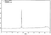
图1是C-angiopep-2的ESI-MS图谱,Figure 1 is the ESI-MS spectrum of C-angiopep-2,
其中,多肽TFFYGGSRGKRNNFKTEEYC分子量:2404.6Da。Among them, the molecular weight of the polypeptide TFFYGGSRGKRNNFKTEEYC: 2404.6Da.
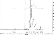
图2是C-angiopep-2的分析型HPLC图谱,Fig. 2 is the analytical HPLC pattern of C-angiopep-2,
其中,色谱方法:色谱柱:YMC HPLC COLUMN 150×4.6mml.D;流动相A:0.1%TFA乙腈溶液;流动相B:0.1%TFA水溶液;流速:0.7mL/min;时间:45分钟,在214和280nm处探测;洗脱程序:在0到26分钟,洗脱程序从90%到65%的B线性变化后,27-32分钟,维持10%的B冲洗柱子,最后,用90%的B冲洗柱子至平衡。Among them, the chromatographic method: chromatographic column:
图3是C-angiopep-2的1H NMR核磁图。Fig. 3 is the1 H NMR nuclear magnetic spectrum of C-angiopep-2.
图4是化合物2的1H NMR核磁图。FIG. 4 is the1 H NMR spectrum of
图5是化合物3的1H NMR核磁图。Fig. 5 is the1 H NMR nuclear magnetic spectrum of
图6是化合物4的1H NMR核磁图,Fig. 6 is the1 H NMR nuclear magnetic pattern of compound 4,
其中,苯丙氨酸芳香族质子峰在7.4-6.6ppm。Among them, the aromatic proton peak of phenylalanine is at 7.4-6.6ppm.
图7是G5的1H NMR核磁图,其中,G5的光谱在3.3-2.2ppm。Fig. 7 is the1 H NMR nuclear magnetic spectrum of G5, wherein the spectrum of G5 is at 3.3-2.2ppm.
图8是化合物7的1H NMR核磁图,其中,PEG的O-CH2基团光谱在3.7ppm。Fig. 8 isthe 1 H NMR nuclear magnetic spectrum of
图9是化合物8的1H NMR核磁图。Fig. 9 is the1 H NMR nuclear magnetic spectrum of
图10是纳米探针的流体动力学粒径分布和Zeta电位图,Fig. 10 is the hydrodynamic particle size distribution and Zeta potential figure of nanoprobe,
其中,用动态光散射的方法来测定纳米探针的流体动力学粒径分布(A)和Zeta电位(B),Den-Angio和Den-PEG的平均粒径分别是14.2nm和9.3nm,平均Zeta电位是+11.6mV和+16.7mV。Among them, the method of dynamic light scattering is used to measure the hydrodynamic particle size distribution (A) and Zeta potential (B) of the nanoprobes. The average particle sizes of Den-Angio and Den-PEG are 14.2nm and 9.3nm, respectively, and the average Zeta potentials are +11.6mV and +16.7mV.
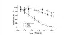
图11表明纳米探针对人类脑胶质瘤U87MG肿瘤细胞的细胞毒性,Figure 11 shows the cytotoxicity of nanoprobes to human glioma U87MG tumor cells,
其中,Den-Angio和Den-PEG对人类脑胶质瘤U87MG肿瘤细胞的细胞毒性均比未修饰的G5明显低,数据用平均值±方差表示(每个浓度的实验n=8)。Among them, the cytotoxicity of Den-Angio and Den-PEG to human glioma U87MG tumor cells was significantly lower than that of unmodified G5, and the data were represented by mean ± variance (n=8 for each concentration experiment).
图12是细胞摄取纳米探针及竞争实验的共聚焦荧光显微镜成像,Figure 12 is the confocal fluorescence microscope imaging of the cell uptake nanoprobe and competition experiment,
其中,Den-Angio通过LRP-受体介导的细胞内吞而高效进入人脑胶质瘤U87MG肿瘤细胞,(A)对用2μM Den-Angio在4℃培养了15分钟的活细胞以及先用2μM RAP处理半小时再用Den-Angio处理15分钟(均在4℃)的活细胞进行共聚焦荧光显微镜成像,标尺:20ìm,用2μM Den-Angio或者Den-PEG在37℃处理了1小时(B)和24小时(C)的活细胞的共聚焦荧光显微镜图像;(D)用纳米探针37℃处理过的活细胞的时间依赖的荧光强度;荧光强度用NIH Image J软件来定量;数据用平均值±方差表示,n>32,标尺:方差。Among them, Den-Angio efficiently enters human glioma U87MG tumor cells through LRP-receptor-mediated endocytosis. Live cells treated with 2 μM RAP for half an hour and then treated with Den-Angio for 15 minutes (both at 4°C) were imaged by confocal fluorescence microscopy, scale bar: 20 μm, treated with 2 μM Den-Angio or Den-PEG at 37°C for 1 hour ( B) Confocal fluorescence microscopy images of living cells at 24 hours (C); (D) time-dependent fluorescence intensity of living cells treated with nanoprobes at 37°C; fluorescence intensity was quantified using NIH Image J software; data Expressed as mean ± variance, n>32, scale: variance.
图13表明荷瘤裸鼠注射纳米探针后光学和磁共振成像的T/N信号比,Figure 13 shows the T/N signal ratios of optical and magnetic resonance imaging in tumor-bearing nude mice injected with nanoprobes,
其中,体内的磁共振和光学成像均证实,Den-Angio相对于Den-PEG有更高的T/N信号比;纳米探针经尾静脉注射后,体内的时间依赖的标准化的肿瘤和周围正常脑组织的近红外荧光强度比(A)和T1加权的磁共振信号比(B)(T/N比值);T/N值用注射前的比值标准化,数据用平均值±方差表示(n=3);注射剂量磁共振显像1.2mg/mouse(相当于0.05mmol[Gd3+]/mouse),而光学显像0.4mg/mouse。Among them, both magnetic resonance and optical imaging in vivo confirmed that Den-Angio has a higher T/N signal ratio than Den-PEG; after the nanoprobe was injected through the tail vein, the time-dependent normalized tumor and surrounding normal in vivo The near-infrared fluorescence intensity ratio of brain tissue (A) and the T1-weighted magnetic resonance signal ratio (B) (T/N ratio); 3) The injection dose for magnetic resonance imaging is 1.2mg/mouse (equivalent to 0.05mmol[Gd3+ ]/mouse), while for optical imaging it is 0.4mg/mouse.
图14是正常小鼠中,两种纳米探针在各个时间点T1加权的磁共振图像,以及表明皮层和海马时间依赖的T1加权磁共振信号在各个时间点的变化,Figure 14 is the T1-weighted magnetic resonance images of the two nanoprobes at various time points in normal mice, and the time-dependent changes in the T1-weighted magnetic resonance signals of the cortex and hippocampus at various time points,
其中,Den-Angio在血脑屏障完整的正常小鼠中表现出比Den-PEG更高的大脑摄取率;(A)正常小鼠大脑在纳米探针注射前和注射后30分钟,2小时和24小时代表性的T1加权磁共振图像(1.2mg/mouse,相当于0.05mmol[Gd3+]/mouse);红色箭头指向脑室,白色箭头指向海马;(B)&(C)时间依赖的T1加权的磁共振信号在正常小鼠尾静脉注射纳米探针前、后在皮层和海马区域的变化(n=4)。Among them, Den-Angio showed a higher brain uptake rate than Den-PEG in normal mice with intact blood-brain barrier; (A) normal mouse brain before and after nanoprobe
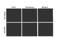
图15是荷瘤小鼠激光共聚焦荧光显微镜图像,Figure 15 is a laser confocal fluorescence microscope image of a tumor-bearing mouse,
其中,共聚焦荧光显微镜图像显示,Den-Angio比Den-PEG显示出更好的脑肿瘤成像能力;图片显示的是脑部种植有U87MG肿瘤的荷瘤裸鼠在尾静脉注射(4nmol/mouse)Den-Angio(上排)和Den-PEG(下排)24h后的荧光显微镜图片;罗丹明标记的探针显示出红色荧光,细胞核的DAPI染色显示出蓝色荧光;组织切片的核染色有助于界定肿瘤和正常脑组织的边界。Among them, confocal fluorescence microscope images show that Den-Angio has better brain tumor imaging ability than Den-PEG; the picture shows that the tumor-bearing nude mice with U87MG tumors implanted in the brain were injected into the tail vein (4nmol/mouse) Fluorescence microscope images of Den-Angio (upper row) and Den-PEG (lower row) after 24h; rhodamine-labeled probes showed red fluorescence, and DAPI staining of nuclei showed blue fluorescence; nuclear staining of tissue sections was helpful Used to define the boundary between tumor and normal brain tissue.
具体实施方式Detailed ways
通过下述实施例将有助于进一步理解本发明,但并不限制本发明的内容。The following examples will help to further understand the present invention, but do not limit the content of the present invention.
实施例1半胱氨酸修饰的可穿越BBB多肽angiopep-2的合成Example 1 Synthesis of cysteine-modified cross-BBB polypeptide angiopep-2
为了将具有BBB穿越能力的angiopep-2修饰到G5树枝状高分子上,同时也不影响其与低密度脂蛋白相关受体结合的特异性,采用Boc保护的固相多肽合成方法合成C端修饰有一个半胱氨酸残基的angiopep-2:TFFYGGSRGKRNNFKTEEYC(MW=2402Da)。In order to modify angiopep-2 with BBB crossing ability to G5 dendrimer without affecting its binding specificity to low-density lipoprotein-associated receptors, a C-terminal modification was synthesized by Boc-protected solid-phase peptide synthesis Angiopep-2 with one cysteine residue: TFFYGGSRGKRNNFKTEEYC (MW = 2402 Da).
1.称取0.156g Boc-L-Cys(pMeBzl)-PAM树脂(取代度0.8mmol/g,约0.125mmol),用DMF(N,N-二甲基甲酰胺)溶胀20分钟,并洗涤数次;1. Weigh 0.156g Boc-L-Cys(pMeBzl)-PAM resin (degree of substitution 0.8mmol/g, about 0.125mmol), swell with DMF (N,N-dimethylformamide) for 20 minutes, and wash several times Second-rate;
2.连续用TFA(三氟乙酸)脱保护2次,加量至浸没树脂,每次搅拌1分钟,DMF洗涤数次;2. Deprotect with TFA (trifluoroacetic acid) twice continuously, add the amount until the resin is submerged, stir for 1 minute each time, and wash with DMF several times;
3.称取所需氨基酸1.1mmol,用2ml的HBTU(苯并三氮唑-N,N,N’,N’-四甲基脲六氟磷酸盐)和0.5ml的DIEA(N,N-二异丙基乙胺)溶解,加入树脂中,恒温振荡器上反应20分钟;3. Weigh 1.1mmol of the required amino acid, and use 2ml of HBTU (benzotriazole-N, N, N', N'-tetramethyluronium hexafluorophosphate) and 0.5ml of DIEA (N, N- Diisopropylethylamine) was dissolved, added in the resin, and reacted for 20 minutes on a constant temperature oscillator;
4.用DMF洗涤。挑取芝麻大小的树脂进行NT(茚三酮)检测确定是否反应完全,NT检测液:A液为8ml苯酚加2ml乙醇,B液为吡啶,C液为1g茚三酮加10ml乙醇,NT检测:挑取的树脂中加入A液10ul,B液20ul,C液10ul,120℃电烤约1分钟,若树脂变蓝则反应不完全,需要重新称取此氨基酸进行反应;若树脂仍为黄色,则反应完全,重复2、3、4;4. Wash with DMF. Pick sesame-sized resin for NT (ninhydrin) detection to determine whether the reaction is complete. NT detection solution: A liquid is 8ml phenol plus 2ml ethanol, B liquid is pyridine, C liquid is 1g ninhydrin plus 10ml ethanol, NT detection : Add 10ul of liquid A, 20ul of liquid B, and 10ul of liquid C to the picked resin, bake at 120°C for about 1 minute, if the resin turns blue, the reaction is incomplete, and the amino acid needs to be re-weighed for reaction; if the resin is still yellow , then the reaction is complete,
依次反应的氨基酸分别是Boc-Tyr(Br-Z)(有保护基团的酪氨酸)、Boc-Glu(OcHex)(有保护基团的谷氨酸)、Boc-Glu(OcHex)、Boc-Thr(Bzl)(有保护基团的苏氨酸)、Boc-Lys(Cl-Z)(有保护基团的赖氨酸)、Boc-Phe(有保护基团的苯丙氨酸)、Boc-Asn(Xan)-OH(有保护基团的天冬氨酸)、Boc-Asn(Xan)-OH、Boc-Arg(Tos)-OH(有保护基团的精氨酸)、Boc-Lys(Cl-Z)、Boc-Gly(有保护基团的甘氨酸)、Boc-Arg(Tos)-OH、Boc-Ser(Bzl)(有保护基团的丝氨酸)、Boc-Gly、Boc-Gl y、Boc-Tyr(Br-Z)、Boc-Phe、Boc-Phe、Boc-Thr(Bzl),得到的未去保护的线状肽链为:H-Thr(Bzl)-Phe-Phe-Tyr(Br-Z)-Gly-Gly-Ser(Bzl)-Arg(Tos)-Gly-Lys(Cl-Z)-Arg(Tos)-Asn(Xan)-Asn(Xan)-Phe-Lys(Cl-Z)-Thr(Bzl)-Glu(OcHex)-Glu(OcHex)-Tyr(Br-Z)-Cys(PMeBzl)-OH;The amino acids that react in sequence are Boc-Tyr (Br-Z) (tyrosine with protective group), Boc-Glu (OcHex) (glutamic acid with protective group), Boc-Glu (OcHex), Boc -Thr(Bzl) (threonine with a protective group), Boc-Lys(Cl-Z) (lysine with a protective group), Boc-Phe (phenylalanine with a protective group), Boc-Asn(Xan)-OH (aspartic acid with protective group), Boc-Asn(Xan)-OH, Boc-Arg(Tos)-OH (arginine with protective group), Boc- Lys(Cl-Z), Boc-Gly (glycine with protective group), Boc-Arg(Tos)-OH, Boc-Ser(Bzl) (serine with protective group), Boc-Gly, Boc-Gl y, Boc-Tyr(Br-Z), Boc-Phe, Boc-Phe, Boc-Thr(Bzl), the obtained undeprotected linear peptide chain is: H-Thr(Bzl)-Phe-Phe-Tyr (Br-Z)-Gly-Gly-Ser(Bzl)-Arg(Tos)-Gly-Lys(Cl-Z)-Arg(Tos)-Asn(Xan)-Asn(Xan)-Phe-Lys(Cl- Z)-Thr(Bzl)-Glu(OcHex)-Glu(OcHex)-Tyr(Br-Z)-Cys(PMeBzl)-OH;
5.同上,用TFA脱保护两次,用甲醇洗涤树脂数次,再用之浸泡树脂15~20分钟,抽干甲醇(约30分钟),放入塑料筒中,并加入适量的p-cresol(4-甲基苯酚)、加入磁子,挂到切割仪上,并使塑料筒浸没入液氮中,抽真空,通HF(氢氟酸),待HF在塑料筒中冷凝约2ml,关闭HF,在冰水浴中磁力搅拌1小时,抽吸HF(约30分钟),冰乙醚沉淀产物,用砂芯漏斗过滤得沉淀,适量50%乙腈溶解,旋蒸去乙腈,冻干,得到粗产物约280mg。5. As above, use TFA to deprotect twice, wash the resin several times with methanol, then soak the resin for 15-20 minutes, drain the methanol (about 30 minutes), put it into a plastic cylinder, and add an appropriate amount of p-cresol ( 4-methylphenol), add magnets, hang it on the cutter, and immerse the plastic cylinder into liquid nitrogen, vacuumize, pass HF (hydrofluoric acid), wait for HF to condense about 2ml in the plastic cylinder, turn off HF, Stir magnetically in an ice-water bath for 1 hour, pump HF (about 30 minutes), precipitate the product with ice ether, filter with a sand core funnel to obtain the precipitate, dissolve an appropriate amount of 50% acetonitrile, spin evaporate the acetonitrile, and freeze-dry to obtain about 280 mg of the crude product .
6.称取100mg粗产物,适量蒸馏水溶解,制备型HPLC(高效液相色谱仪)纯化、冻干,产物的纯度通过分析型HPLC证实;ESI-MS中有一个单一的802.5[M3+]峰,计算分子量为2404.6[M+H+],ESI-MS和分析型HPLC结果如图1、2所示;C-angiopep-2的1HNMR核磁图如图3所示。6. Weigh 100 mg of the crude product, dissolve in appropriate amount of distilled water, purify by preparative HPLC (high performance liquid chromatography), and lyophilize. The purity of the product is confirmed by analytical HPLC; there is a single 802.5 [M3+ ] in ESI-MS The peak, the calculated molecular weight is 2404.6 [M+H+ ], the results of ESI-MS and analytical HPLC are shown in Figures 1 and 2;the 1 H NMR spectrum of C-angiopep-2 is shown in Figure 3.
制备型HPLC纯化方法:制备柱:SymmetryR 300A C187μm 19×300mm steal Column;流动相A:5%乙腈,流动相B:35%乙腈;洗脱程序:0-60分钟0%B-100%B;流速:10ml/min;柱温:25℃;检测:UV 214nm和280nm。Preparative HPLC purification method: Preparative column: SymmetryR 300A C187μm 19×300mm steal Column; mobile phase A: 5% acetonitrile, mobile phase B: 35% acetonitrile; elution program: 0% B-100% B in 0-60 minutes ; Flow rate: 10ml/min; Column temperature: 25°C; Detection: UV 214nm and 280nm.
分析型HPLC方法:如图2所示。Analytical HPLC method: as shown in Figure 2.
实施例2化合物1的合成The synthesis of
2.1mg(6.8×10⁻⁶mol,1.3倍)的SPDP(N-琥珀酰亚胺基3-(2-吡啶基二硫)丙酸酯溶于300μL的DMF中,缓慢逐滴加入磁力搅拌下的1.0mL的10.4mg(5.2×10⁻⁶mol,分子量认为是2KDa)NH2-PEG2k-Malemide(氨基-聚乙二醇2k-马来酰亚胺)的1X PBS(pH 7.4)溶液中。常温下反应45分钟后,形成一端是马来酰亚胺,另一端是3-(2-吡啶基二硫)丙酸酯的PEG衍生物。2.1 mg (6.8×10⁻⁶ mol, 1.3 times) of SPDP (N-succinimidyl 3-(2-pyridyl dithio)propionate was dissolved in 300 μL of DMF, and slowly added dropwise under magnetic stirring 1.0mL of 10.4mg (5.2×10⁻⁶ mol, molecular weight is considered to be 2KDa) NH2 -PEG2k -Malemide (amino-polyethylene glycol2k -maleimide) in 1X PBS (pH 7.4) solution After reacting at room temperature for 45 minutes, a PEG derivative with maleimide at one end and 3-(2-pyridyl dithio)propionate at the other end was formed.
实施例3化合物2的合成The synthesis of
将以上反应液直接加入1.0mL的11.6mg(4×10⁻⁷mol)的树枝状大分子PAMAM G5的1X PBS(pH 7.4~8.0)溶液中。常温下搅拌1小时后,形成化合物2,其中SPDP通过PEG连接到G5树枝状高分子表面。用分子量为10,000Da的滤膜超滤管离心纯化(4000转/分钟,30分钟×3次)得到目标产物。G5和PEG之间的摩尔比通过化合物2的1H NMR图谱中的质子积分来计算。化合物2中SPDP的标记水平通过DTT(二硫苏糖醇)实验来定量。简化的操作过程为:过量的DTT加入化合物2的PBS溶液中,搅拌15分钟,测量上述溶液在343nm处的吸光度。SPDP和G5之间的摩尔比通过公式R=ΔA343/8080×Cdendrimer来计算,R代表摩尔比,ΔA343代表DTT加入前后343nm处吸光度的变化,Cdendrimer代表G5的摩尔浓度,数值8080代表吡啶-2-硫酮在343nm处的消光系数。实验结果表明平均每个G5上标记有8个SPDP。化合物2的1H NMR核磁图如图4所示。The above reaction solution was directly added to 1.0 mL of a 1X PBS (pH 7.4-8.0) solution of 11.6 mg (4×10⁻⁷ mol) dendrimer PAMAM G5. After stirring at room temperature for 1 hour,
实施例4化合物3的合成The synthesis of embodiment 4
0.4mg(8×10⁻⁷mol,2.0倍.)的rhodamine-NHS(罗丹明-N-羟基琥珀酰亚胺酯)和1.2mg(8.0×10⁻⁷mol,2.0倍)的Cy5.5-NHS溶解在50μL无水DMF中,然后缓慢逐滴加入1.0mL化合物2的HEPES(4-羟乙基哌嗪乙磺酸)缓冲液(0.1M,pH 8.3)。常温下搅拌1小时,荧光基团标记的G5用分子量10,000Da的超滤管纯化(4000转/分钟),并浓缩到约2.0mL 0.5M的HEPES中(pH 8.3)。50.7mg(5.12×10⁻⁵mol,128倍.)DOTA-NHS白色粉末逐渐加入到上述溶液中,pH计监测溶液的pH值,并用5.0M NaOH(氢氧化钠)调节溶液pH值在8.5左右。常温下避光搅拌4小时后,混合物用分子量10,000Da的离心超滤管纯化得到罗丹明、Cy5.5和DOTA修饰的紫色树枝状大分子3溶液。通过对1H NMR光谱计算得出每个G5分子上标记了约94个DOTA螯合剂。另外,根据朗伯-比尔定律,平均有1.4个罗丹明和1.1个Cy5.5标记到化合物3上。罗丹明和Cy5.5在水溶液中的吸光系数分别为(ε552=60,000M-1cm-1)和(ε675=250,000M-1cm-1)。化合物3的1H NMR核磁图如图5所示。0.4mg (8×10⁻⁷ mol, 2.0 times.) of rhodamine-NHS (rhodamine-N-hydroxysuccinimide ester) and 1.2mg (8.0×10⁻⁷ mol, 2.0 times) of Cy5.5- NHS was dissolved in 50 μL of anhydrous DMF, and then 1.0 mL of
实施例5化合物4的合成The synthesis of
将化合物3溶解在2mL的PBS溶液中,同时逐滴加入溶解于200μL DMF中的Cysteine-angiopep-2(12mg,5.2×10-6mol,13倍.)。室温下避光搅拌过夜。Angiopep-2标记的G5化合物经超滤纯化后,化合物4中的多肽angiopep-2标记率通过1H NMR谱图加以计算。化合物四种,平均有5.6个angiopep-2标记到一个G5分子上。
化合物4的1H NMR核磁图如图6所示。The1 H NMR spectrum of compound 4 is shown in FIG. 6 .
实施例6化合物5的合成The synthesis of
12.7mg Gd2(CO3)3(6×10⁻⁵mol,64倍.)加入溶于2mL化合物4溶液中。此悬浊液在60℃下避光搅拌12小时。多余的Gd2(CO3)3离心除去(2000转/分钟,8分钟),上清液经超滤纯化后,获得Den-Angio化合物,产率大约为82%(根据G5计算)。12.7 mg Gd2 (CO3 )3 (6×10⁻⁵ mol, 64 times.) was added and dissolved in 2 mL of compound 4 solution. The suspension was stirred at 60°C for 12 hours in the dark. Excess Gd2 (CO3 )3 was removed by centrifugation (2000 rpm, 8 minutes), and the supernatant was purified by ultrafiltration to obtain the Den-Angio compound with a yield of about 82% (calculated based on G5).
实施例7化合物6的合成The synthesis of
0.4mg(8×10⁻⁷mol,2.0倍.)rhodamine-NHS溶于50μL无水DMF中,并缓慢逐滴加入到1mL 11.6mg(4×10⁻⁷mol)G5(0.1M HEPES)中(pH 8.3)。常温下搅拌1小时后,混合液离心超滤纯化得到罗丹明标记的G5树枝状高分子。1.2mg(8.0×10⁻⁷mol,2.0倍)Cy5.5-NHS溶于50μL无水DMF中,同上述反应一样加入到G5的1mL0.1MHEPES溶液中。常温下搅拌1小时后,连接有双荧光基团的树枝状大分子经超滤纯化得到化合物6的紫色溶液。通过吸光光度法计算平均有1.5个罗丹明和1.1个Cy5.5标记到一个化合物6分子上。化合物6的1H NMR核磁图如图7所示。0.4 mg (8×10⁻⁷ mol, 2.0 times.) rhodamine-NHS was dissolved in 50 μL of anhydrous DMF, and slowly added dropwise to 1 mL of 11.6 mg (4×10⁻⁷ mol) G5 (0.1M HEPES) ( pH 8.3). After stirring at room temperature for 1 hour, the mixture was purified by centrifugation and ultrafiltration to obtain rhodamine-labeled G5 dendrimers. 1.2 mg (8.0×10⁻⁷ mol, 2.0 times) of Cy5.5-NHS was dissolved in 50 μL of anhydrous DMF, and added to 1 mL of 0.1M HEPES solution of G5 as in the above reaction. After stirring at room temperature for 1 hour, the dendrimers linked with dual fluorescent groups were purified by ultrafiltration to obtain a purple solution of
实施例8化合物7的合成The synthesis of
化合物6溶解在2.0mL 1X PBS(pH7.4)中,加入溶于2.0mL PBS中的10.4mg(5.2×10⁻⁶mol,13倍.)PEG2K-NHS。常温下搅拌1小时后,产物经超滤纯化得到紫色化合物7溶液(产率是82%,约18.9mg,3.28×10-7mol)。化合物7中G5和PEG间的摩尔比通过它们在1H NMR光谱中的质子积分定量。
化合物7的1H NMR核磁图如图8所示。The1 H NMR spectrum of
实施例9化合物8的合成The synthesis of
3.28×10⁻⁷mol化合物7溶于2mL 0.5M HEPES缓冲液中(pH 8.3)。41.6mg(4.2×10⁻⁵mol,128倍)的DOTA-NHS以固体形式加入以上溶液中。pH计监测反应体系pH值,并用5.0M NaOH溶液调节在8.5左右。反应液在常温下搅拌过夜,离心超滤纯化后得到产率为92%的化合物8。标记到G5上的DOTA数量通过1H NMR谱中的质子积分来定量,约为95个。3.28×10⁻⁷ mol of
化合物8的1H NMR核磁图如图9所示。The1 H NMR spectrum of
实施例10化合物9的合成The synthesis of
9.5mg(1.93×10⁻⁵mol,64倍.)Gd2(CO3)3加入到溶于4mL 0.1M HEPES(pH 8.3)的3.0×10⁻⁷mol(基于G5的摩尔数)的化合物8中。此悬浊液在60℃下避光搅拌12小时。多余的Gd2(CO3)3经离心沉淀除去,上清液经离心超滤纯化,产率为95%(基于G5的摩尔数)。Add 9.5mg (1.93×10⁻⁵ mol, 64 times.) of Gd2 (CO3 )3 to 3.0×10⁻⁷ mol (based on the moles of G5) of
实施例11纳米探针粒径分布和Zeta电位的测定The measurement of embodiment 11 nanoprobe particle size distribution and Zeta potential
纳米探针和未修饰的G5树枝状大分子在水溶液中的粒径通过动态光散射的方法测定。溶于蒸馏水的2.0mg/ml的牛血清白蛋白标准溶液对设备进行校准。样品用孔径为0.45μm的滤膜纯化后并用1X PBS稀释到100g/mL。纳米探针的粒径和分布通过动态光散射仪加以测定。在测定纳米探针的表面电荷时,纳米探针溶液用0.45μm的滤头过滤并稀释到10mM的NaCl溶液中。The particle sizes of nanoprobes and unmodified G5 dendrimers in aqueous solution were determined by dynamic light scattering. The equipment was calibrated with a 2.0 mg/ml bovine serum albumin standard solution in distilled water. Samples were purified with a 0.45 μm filter and diluted to 100 g/mL with 1X PBS. The particle size and distribution of the nanoprobes were determined by a dynamic light scattering instrument. When measuring the surface charge of the nanoprobes, the nanoprobe solution was filtered through a 0.45 μm filter and diluted into 10 mM NaCl solution.
Den-Angio和Den-PEG的平均直径是14.2nm和9.3nm,平均Zeta电位是+11.6和+16.7mV。The average diameters of Den-Angio and Den-PEG are 14.2nm and 9.3nm, and the average Zeta potentials are +11.6 and +16.7mV.
纳米探针的粒径分布和Zeta电位如图10所示。The particle size distribution and Zeta potential of the nanoprobes are shown in Fig. 10 .
实施例12用ICP-AES测定纳米探针中的Gd3+含量
纳米探针中的Gd3+含量用Hitachi P-4010型号ICP-AES(Inductively CoupledPlasma Atomic Emission Spectroscopy,电感耦合等离子体发射光谱仪)进行测定,射频能量为1100W,喷雾器气流速度为0.9L/min。准备好用3%硝酸溶解的Gd3+浓度分别为1,5,10,20,50,100,200ppm的标准溶液,标绘相应的色谱峰来绘制标准曲线以对应Gd3+含量。0.1mM的纳米探针母液用3%的硝酸稀释100倍。样品的Gd3+含量通过对Gd3+标准曲线拟合得到。实验结果显示,Gd3+-DOTA配合物在探针Den-Angio和Den-PEG中的含量均平均达95个。The Gd3+ content in the nanoprobe was measured with Hitachi P-4010 model ICP-AES (Inductively Coupled Plasma Atomic Emission Spectroscopy, Inductively Coupled Plasma Atomic Emission Spectroscopy), the radio frequency energy was 1100W, and the airflow velocity of the nebulizer was 0.9L/min. Prepare standard solutions with Gd3+ concentrations of 1, 5, 10, 20, 50, 100, and 200 ppm dissolved in 3% nitric acid, and plot the corresponding chromatographic peaks to draw a standard curve corresponding to the Gd3+ content. The 0.1 mM nanoprobe stock solution was diluted 100 times with 3% nitric acid. The Gd3+ content of the samples was obtained by fitting the Gd3+ standard curve. The experimental results showed that the average content of Gd3+ -DOTA complexes in the probes Den-Angio and Den-PEG reached 95.
实施例13检测纳米探针的驰豫率Embodiment 13 detects the relaxation rate of the nanoprobe
纳米探针和商品化的磁共振造影剂Gd3+-DOTA的纵向驰豫率根据方程r1p=(1/Tsample-1/TPBS)/[Gd].来计算。其中r1p为纵向磁豫率,Tsample为溶液的纵向磁豫时间,TPBS为PBS溶液的磁豫时间。将探针稀释成四种不同浓度的PBS溶液(pH 7.4),其T1值在4.7T的磁共振上测定。根据ICP-AES测定的Gd3+浓度描绘出纳米探针的(1/Tsample-1/TPBS)值,从而得出纳米探针的驰豫率。Gd3+-DOTA,Den-Angio和Den-PEG的纵向驰豫率分别为4.7,6.9 and 7.4mM-1s-1。The longitudinal relaxation rates of the nanoprobe and the commercial magnetic resonance contrast agent Gd3+ -DOTA were calculated according to the equation r1p =(1/Tsample −1/TPBS )/[Gd]. Where r1p is the longitudinal magnetic coefficient, Tsample is the longitudinal magnetic time of the solution, and TPBS is the magnetic time of the PBS solution. The probe was diluted into four different concentrations of PBS solutions (pH 7.4), and its T1 value was measured at 4.7T magnetic resonance. The (1/Tsample −1/TPBS ) value of the nanoprobe is plotted according to the Gd3+ concentration determined by ICP-AES, thereby obtaining the relaxation rate of the nanoprobe. The longitudinal relaxation rates of Gd3+ -DOTA, Den-Angio and Den-PEG are 4.7, 6.9 and 7.4mM-1 s-1 , respectively.
实施例14体外细胞毒性实验Example 14 In vitro cytotoxicity test
1.细胞培养人类胶质母细胞瘤U87MG细胞系用最低基础培养基在75-cm2培养瓶中单层培养,即用加有10%胎牛血清(FBS)、2mM L-谷氨酰胺、1%青、链霉素(Invitrogen,Carlsbad,CA)的Alpha 1X培养基(MEM,Mediatech,Manassas,VA),置于有充分湿气的含5%CO2的37℃培养箱中培养。当细胞长满80%的面积时可以消化收集,以使细胞保持在指数增长状态。1. Cell culture The human glioblastoma U87MG cell line is cultured in a single layer in a 75-cm2 culture flask with the minimum basal medium, which is added with 10% fetal bovine serum (FBS), 2mM L-glutamine, Alpha 1X medium (MEM, Mediatech, Manassas, VA) with 1% penicillin and streptomycin (Invitrogen, Carlsbad, CA) was cultured in a 37°C incubator with sufficient humidity and 5% CO2 . Cells can be digested and harvested when they are 80% full to keep the cells in a state of exponential growth.
2.体外细胞毒性实验MTT(3-(4,5-二甲基噻唑-2)-2,5-二苯基四氮唑溴盐)细胞增殖实验用来测定用纳米探针和未修饰的G5对照处理后的细胞的活力。处于指数增长期的细胞单层用0.25%胰酶消化收集,得到单细胞悬液。用血细胞计数器和普通光学显微镜(OLYMPUS BH-2)来计数细胞。优化细胞数量以使得在整个MTT实验中,细胞处于指数增长期。因此,用适量细胞培养液重悬细胞,在96孔板的每个孔中加入含有约2×10³个细胞的100μL单细胞悬液。每个浓度准备了8个复孔。细胞贴壁24小时后,用纳米探针或者未修饰的G5来处理这些细胞。样品溶液用
实施例15活细胞摄取纳米探针的共聚焦荧光显微镜成像Example 15 Confocal fluorescence microscopy imaging of nanoprobe uptake by living cells
为了避免固定过程中出现的假阳性,所有的实验都在活的U87MG细胞中进行。细胞(2×10⁴)培养在35mm的玻璃底培养皿(14mm小孔,MatTek,Ashland,MA)中,大概长满皿底的50%,用2ml溶解有2μM纳米探针的10%FBS培养液在4℃或37℃培养细胞一定的时间。孵育结束后,用HBSS(Hank’s Balance Salt Solution,Hank’s平衡盐溶液)洗涤三次,并加入1ml无酚红培养基。立即用共聚焦荧光显微镜观察细胞。To avoid false positives during fixation, all experiments were performed in live U87MG cells. Cells (2×10⁴ ) were cultured in a 35mm glass-bottom culture dish (14mm well, MatTek, Ashland, MA), which covered about 50% of the bottom of the dish, and cultured with 2ml of 10% FBS dissolved in 2μM nanoprobes The cells were incubated at 4°C or 37°C for a certain period of time. After incubation, wash three times with HBSS (Hank's Balance Salt Solution, Hank's Balanced Salt Solution), and add 1 ml of phenol red-free medium. Immediately observe the cells with a confocal fluorescence microscope.
实施例16体外细胞摄取纳米探针的竞争实验Example 16 Competition experiment of in vitro cell uptake of nanoprobes
培养在35mm玻璃底培养皿中的活U87MG细胞先用溶解有2μM低密度脂蛋白受体缔合性蛋白(RAP)的常规培养液在4℃培养30分钟,RAP用作LRP(低密度脂蛋白受体相关蛋白)受体的通用拮抗剂。孵育结束后,洗涤细胞并加入溶解有2μM RAP和2μM Den-Angio的培养液继续在4℃培养15分钟。孵育结束后,细胞用HBSS洗涤三次,并用共聚焦荧光显微镜观察。Live U87MG cells cultured in 35mm glass-bottomed culture dishes were first cultured at 4°C for 30 minutes with conventional culture medium dissolved with 2 μM low-density lipoprotein receptor-associated protein (RAP), and RAP was used as LRP (low-density lipoprotein Receptor-associated protein) receptor general antagonists. After the incubation, the cells were washed and added to the medium dissolved in 2 μM RAP and 2 μM Den-Angio and continued to incubate at 4°C for 15 minutes. After incubation, cells were washed three times with HBSS and observed with a confocal fluorescence microscope.
细胞摄取纳米探针及竞争实验的共聚焦荧光显微镜成像结果如图12所示。The confocal fluorescence microscopy imaging results of the nanoprobe uptake by cells and competition experiments are shown in Figure 12.
实施例17小鼠模型和肿瘤原位种植Example 17 Mouse model and tumor orthotopic implantation
所有的动物实验都依照复旦大学伦理委员会评估和认可的指南进行。人源U87MG胶质细胞瘤细胞(1.0×10⁶细胞重悬于5μl PBS)在有小鼠衔接器的立体定位仪的协助下被接种到裸鼠的右侧纹状体(前囟点旁开1.8mm,往前0.6mm,深3mm)。接种后14-18天,颅内肿瘤长到直径0.2-0.5mm大小,即进行显像实验。All animal experiments were performed in accordance with the guidelines evaluated and approved by the Ethics Committee of Fudan University. Human U87MG glioma cells (1.0×10⁶ cells resuspended in 5 μl PBS) were inoculated into the right striatum of nude mice (opened next to bregma) with the assistance of a stereotaxic apparatus with mouse adapters. 1.8mm, forward 0.6mm, deep 3mm). 14-18 days after inoculation, when the intracranial tumor grows to a size of 0.2-0.5 mm in diameter, the imaging experiment is performed.
实施例18体内和离体的光学显像研究Example 18 In Vivo and Ex Vivo Optical Imaging Research
光学显像研究在柯达公司的活体光学成像系统上进行,设置感光滤光片为630nm,而发射谱带通滤色片为700nm。成像前,小鼠用氯胺酮(25mg/kg)和乙酰丙嗪(2.5mg/kg)的混合麻醉剂麻醉,并且脸朝上固定在成像板上。X线显像(曝光时间30秒),白光摄影(曝光时间0.2秒)和近红外荧光显像(曝光时间15秒)分别在一样的大小的视野(FOV)下摄取(FOV=12.8cm,f/stop=4,Bin=high resolution),时间点包括注射前和全身注射纳米探针后的几个选定的时间点,每只小鼠注射4.0nmol的纳米探针(基于G5的摩尔数)。X线成像和近红外荧光成像叠加起来来确定位于颅内部分的大小。时间依赖的肿瘤和周围正常脑组织的荧光强度比(T/N比值)用纳米探针注射前的值进行标准化。最后,小鼠被处死并心脏灌流PFA进行预固定。小鼠的大脑被小心地剥离出来,而主要的脏器包括心、肝、脾、肺、肾和肌肉则用组织切片器(Braintree Scientific Inc.,Braintree,MA)切成约厚1-2mm。荧光显微镜下肿瘤和周围正常脑组织的荧光强度由ImageJ软件(National Institutes of Health,Bethesda,MD)定量。The optical imaging research was carried out on Kodak's in vivo optical imaging system, and the photosensitive filter was set at 630nm, while the emission bandpass filter was set at 700nm. Before imaging, mice were anesthetized with a mixture of ketamine (25 mg/kg) and acepromazine (2.5 mg/kg) and fixed face up on the imaging plate. X-ray imaging (
实施例19磁共振成像Example 19 Magnetic resonance imaging
体内磁共振成像在Bruker Biospec 47/30磁共振仪上进行。实验前,小鼠尾静脉用自制的导管系统置管,该导管系统用一个小的T形接头(Cole-Parmer,Vernon Hills,IL)连接,使得纳米探针溶液在装置中的死容积最小化(小于50μL)。麻醉的小鼠的头被固定在自制的螺线管线圈里,小鼠在磁体中的体温通过一个温度调节装置加热板维持,呼吸则通过Bruke PhysioGard系统进行持续监控。小鼠被安置在磁共振线圈里后,异氟烷(0.5-2%)和100%的氧气一起给予,并且持续的呼吸监测随时进行调整。每只老鼠从尾静脉注射0.05mmol/kg[Gd3+],总共0.25mL体积的纳米探针PBS溶液,收集注射前、后脑部动态T1加权像。观察T1加权的磁共振信号在注射后0-120分钟和24小时的增强情况。冠状为的脑部影像以1mm厚来获取图像,使用了一个自旋回波脉冲序列,视野(FOV)2cm×2cm,矩阵128×128,TR=300ms,TE=11ms,NA=8。3D的T1加权像用一个快速小角度激发成像序列(FLASH)来获得,flip angle=45°,FOV=1.5cm×1.5cm×1.5cm,矩阵128×128×32,TR=35ms,TE=6.2ms,NA=8。感兴趣区(ROI)在时间点的增强强度(IE)通过以下公式表示IE=(RI(t)-RI(0))/RI(0)×100%,其中,RI(t)对应于在各个时间点测定的标准化的信号强度,而RI(0)是纳米探针注射前标准化的信号强度。时间依赖的肿瘤和周围正常脑组织之间的荧光强度比(T/N比值)用纳米探针注射前的值进行标准化。In vivo magnetic resonance imaging was performed on a Bruker Biospec 47/30 magnetic resonance machine. Before the experiment, the mouse tail vein was cannulated with a homemade catheter system connected with a small T-connector (Cole-Parmer, Vernon Hills, IL) to minimize the dead volume of the nanoprobe solution in the device (less than 50 μL). The head of the anesthetized mouse is fixed in a homemade solenoid coil, the body temperature of the mouse in the magnet is maintained by a thermostat heating plate, and the respiration is continuously monitored by the Bruke PhysioGard system. After mice were placed in the magnetic resonance coil, isoflurane (0.5-2%) was administered along with 100% oxygen, and continuous respiratory monitoring was adjusted over time. Each mouse was injected with 0.05mmol/kg[Gd3+ ] through the tail vein, with a total volume of 0.25mL of nanoprobe PBS solution, and the dynamic T1-weighted images of the brain before and after the injection were collected. Observe the enhancement of T1-weighted MR signals at 0-120 minutes and 24 hours after injection. Coronal brain images were acquired at 1mm thickness using a spin-echo pulse sequence, field of view (FOV) 2cm×2cm, matrix 128×128, TR=300ms, TE=11ms, NA=8. 3D T1 The weighted image is obtained by a fast small-angle excitation imaging sequence (FLASH), flip angle=45°, FOV=1.5cm×1.5cm×1.5cm, matrix 128×128×32, TR=35ms, TE=6.2ms, NA =8. The enhancement intensity (IE) of the region of interest (ROI) at the time point is represented by the following formula: IE=(RI(t)-RI(0))/RI(0)×100%, where RI(t) corresponds to The normalized signal intensity measured at each time point, while RI(0) is the normalized signal intensity before nanoprobe injection. The time-dependent ratio of fluorescence intensity between tumor and surrounding normal brain tissue (T/N ratio) was normalized to the value before nanoprobe injection.
光学和磁共振成像的T/N信号比变化如图13所示。The T/N signal ratio changes for optical and magnetic resonance imaging are shown in Fig. 13.
两种纳米探针在各个时间点T1加权的磁共振像,以及皮层和海马时间依赖的T1加权磁共振信号在各个时间点的变化如图14所示。The T1-weighted magnetic resonance images of the two nanoprobes at various time points, and the time-dependent changes of the T1-weighted magnetic resonance signals of the cortex and hippocampus at various time points are shown in Figure 14 .
实施例18冰冻大脑切片共聚焦显微镜成像Example 18 Confocal microscope imaging of frozen brain slices
体内成像后,常规处理小鼠大脑,浸在4%PFA(多聚甲醛)中12小时,再放入30%的蔗糖溶液中脱水至沉底,然后冰冻切片厚15μm,包埋切片并用Leica TCS SP2激光共聚焦显微镜(Leica Inc.,Wetzlar,Germany)观察,所用的镜头是HCXPL APO CS 40×1.25oil immersion lens和HCPL APO CS 10×0.40immersion lens。罗丹明用543nm激光激发,发射光则用560nm带通滤色片的光电倍增管检测。同时,用405nm激光激发DAPI,发射光则用具有490nm二色分光镜和420-480nm带通滤色片的二级光电倍增管检测。同时,共聚焦Z轴方向进行0.8μm厚的扫描。图15显示了冰冻切片的激光共聚焦荧光显微镜图像。After in vivo imaging, the mouse brain was routinely processed, immersed in 4% PFA (paraformaldehyde) for 12 hours, then placed in 30% sucrose solution to dehydrate to the bottom, and then the frozen section was 15 μm thick, embedded and sliced with Leica TCS Observed by SP2 laser confocal microscope (Leica Inc., Wetzlar, Germany), the lenses used are
实施例19组织学成像Example 19 Histological Imaging
用纳米探针处理过的小鼠大脑被离体并浸没在formalin(福尔马林)和PFA的混合液中(体积1∶9混合)固定适当时间。固定好的组织用石蜡包埋并切成3-4μm厚。切片进行H&E染色,并用Leica MZ75高性能立体显微镜2.5X和5.0X的物镜观察。Mouse brains treated with nanoprobes were isolated and immersed in a mixture of formalin (formalin) and PFA (1:9 volume mix) for proper time fixation. Fixed tissues were embedded in paraffin and sectioned 3-4 μm thick. Sections were stained with H&E and observed with Leica MZ75 high-performance stereomicroscope with 2.5X and 5.0X objective lenses.
结果显示,本发明能克服单一成像技术灵敏度或分辨率的限制,同时还能通过影像对比得到更丰富的生理和病理信息。The results show that the invention can overcome the limitation of the sensitivity or resolution of a single imaging technique, and can obtain more abundant physiological and pathological information through image comparison.

Claims (3)
Priority Applications (1)
| Application Number | Priority Date | Filing Date | Title |
|---|---|---|---|
| CN 201010292945CN102406949B (en) | 2010-09-21 | 2010-09-21 | A targeted-tracking multimodal diagnostic nano-imaging drug |
Applications Claiming Priority (1)
| Application Number | Priority Date | Filing Date | Title |
|---|---|---|---|
| CN 201010292945CN102406949B (en) | 2010-09-21 | 2010-09-21 | A targeted-tracking multimodal diagnostic nano-imaging drug |
Publications (2)
| Publication Number | Publication Date |
|---|---|
| CN102406949A CN102406949A (en) | 2012-04-11 |
| CN102406949Btrue CN102406949B (en) | 2013-05-29 |
Family
ID=45909481
Family Applications (1)
| Application Number | Title | Priority Date | Filing Date |
|---|---|---|---|
| CN 201010292945Expired - Fee RelatedCN102406949B (en) | 2010-09-21 | 2010-09-21 | A targeted-tracking multimodal diagnostic nano-imaging drug |
Country Status (1)
| Country | Link |
|---|---|
| CN (1) | CN102406949B (en) |
Cited By (1)
| Publication number | Priority date | Publication date | Assignee | Title |
|---|---|---|---|---|
| CN109395101A (en)* | 2018-09-27 | 2019-03-01 | 复旦大学附属华山医院 | Target the preparation method of the mr contrast agent of blood-brain barrier and glioma |
Families Citing this family (25)
| Publication number | Priority date | Publication date | Assignee | Title |
|---|---|---|---|---|
| MXPA05007322A (en) | 2003-01-06 | 2006-02-17 | Angiochem Inc | A method for transporting a compound across the blood-brain barrier. |
| US9365634B2 (en) | 2007-05-29 | 2016-06-14 | Angiochem Inc. | Aprotinin-like polypeptides for delivering agents conjugated thereto to tissues |
| CN102026667B (en) | 2008-04-18 | 2014-06-25 | 安吉奥开米公司 | Pharmaceutical compositions of paclitaxel, paclitaxel analogs or paclitaxel conjugates and related methods of preparation and use |
| EP2346906A4 (en) | 2008-10-15 | 2013-04-24 | Angiochem Inc | Conjugates of glp-1 agonists and uses thereof |
| WO2010043049A1 (en) | 2008-10-15 | 2010-04-22 | Angiochem Inc. | Etoposide and doxorubicin conjugates for drug delivery |
| MX2011005963A (en) | 2008-12-05 | 2011-09-01 | Angiochem Inc | Conjugates of neurotensin or neurotensin analogs and uses thereof. |
| JP2012512185A (en) | 2008-12-17 | 2012-05-31 | アンジオケム インコーポレーテッド | Membrane type 1 matrix metalloprotein inhibitor and use thereof |
| BRPI1015295A2 (en) | 2009-04-20 | 2016-05-31 | Angiochem Inc | tracing ovarian cancer using an anticancer agent conjugated to an angiopep-2 analogue. |
| BRPI1015918A2 (en) | 2009-07-02 | 2019-09-24 | Angiochem Inc | multimeric peptide conjugates and uses thereof |
| AU2013302270A1 (en) | 2012-08-14 | 2015-03-26 | Angiochem Inc. | Peptide-dendrimer conjugates and uses thereof |
| CN104511026B (en)* | 2013-09-29 | 2018-07-24 | 复旦大学 | Modify adenosine receptor agonist nano-probe and its preparation method and application |
| CN103495188B (en)* | 2013-09-29 | 2015-03-25 | 无锡米度生物技术有限公司 | Medical polymer material with radioactive nuclide marker, and preparation method as well as application thereof |
| JP6823055B2 (en) | 2015-06-15 | 2021-01-27 | アンジオケム インコーポレーテッド | How to treat soft meningeal carcinomatosis |
| CN106902360A (en)* | 2015-12-22 | 2017-06-30 | 上海交通大学 | Magnetic resonance, fluorescence double-developing nanogel probe and preparation method thereof |
| CN107837403B (en)* | 2016-09-21 | 2020-10-20 | 复旦大学 | Dual-modality nanoprobes for image-guided brain tumor resection |
| CN110124036B (en)* | 2019-06-10 | 2020-11-20 | 西安电子科技大学 | Multifunctional magnetic nanoprobe and preparation method thereof |
| CN111253464B (en)* | 2020-01-06 | 2022-02-01 | 江苏省原子医学研究所 | Gamma-glutamyl transpeptidase targeted molecular probe and preparation method and application thereof |
| CN111892645B (en)* | 2020-06-16 | 2021-12-21 | 南方科技大学 | Organic coordination compound, preparation method and application thereof, probe |
| CN114366822A (en)* | 2020-10-15 | 2022-04-19 | 中国科学院宁波材料技术与工程研究所慈溪生物医学工程研究所 | An active targeting multimodal molecular imaging probe and its preparation method and application |
| CN112494664A (en)* | 2020-12-14 | 2021-03-16 | 武汉工程大学 | Polysaccharide magnetic resonance and fluorescence dual-mode imaging diagnostic agent, preparation method thereof and diagnostic agent |
| CN112546242B (en)* | 2020-12-14 | 2023-06-06 | 武汉工程大学 | A nonapeptide-based magnetic resonance and fluorescence dual-mode imaging diagnostic agent and its preparation method |
| CN116920087A (en)* | 2022-04-07 | 2023-10-24 | 中国科学院理化技术研究所 | A gadolinium-based composite material with microwave-responsive heat generation and dual-mode imaging effects and its preparation and use |
| CN115025249B (en)* | 2022-05-12 | 2024-02-20 | 深圳市第二人民医院(深圳市转化医学研究院) | Targeting probe and preparation method and application thereof |
| EP4536278A2 (en) | 2022-06-07 | 2025-04-16 | Actinium Pharmaceuticals, Inc. | Bifunctional chelators and conjugates |
| CN117756927B (en)* | 2023-08-03 | 2025-02-18 | 汕头大学医学院第二附属医院 | MRI-CEST polypeptide probe for targeted recognition of abnormal alpha-synuclein in parkinsonism brain, imaging method and imaging equipment |
- 2010
- 2010-09-21CNCN 201010292945patent/CN102406949B/ennot_activeExpired - Fee Related
Non-Patent Citations (6)
| Title |
|---|
| Cong Li, et al..Multimodal Image-Guided Enzyme/Prodrug Cancer Therapy.《J. AM. CHEM. SOC.》.2006,第128卷15072-15073. |
| Gene delivery targeted to the brain using an Angiopep-conjugated polyethyleneglycol-modified polyamidoamine dendrimer;Weilun Ke, et al.;《Biomaterials》;20090917;第30卷;6976-6985* |
| Hu Yang.Nanoparticle-Mediated Brain-Specific Drug Delivery, Imaging, and Diagnosis.《Pharm Res》.2010,第27卷1759-1771. |
| Multimodal Image-Guided Enzyme/Prodrug Cancer Therapy;Cong Li, et al.;《J. AM. CHEM. SOC.》;20061103;第128卷;15072-15073* |
| Nanoparticle-Mediated Brain-Specific Drug Delivery, Imaging, and Diagnosis;Hu Yang;《Pharm Res》;20100701;第27卷;1759-1771* |
| Weilun Ke, et al..Gene delivery targeted to the brain using an Angiopep-conjugated polyethyleneglycol-modified polyamidoamine dendrimer.《Biomaterials》.2009,第30卷6976-6985. |
Cited By (1)
| Publication number | Priority date | Publication date | Assignee | Title |
|---|---|---|---|---|
| CN109395101A (en)* | 2018-09-27 | 2019-03-01 | 复旦大学附属华山医院 | Target the preparation method of the mr contrast agent of blood-brain barrier and glioma |
Also Published As
| Publication number | Publication date |
|---|---|
| CN102406949A (en) | 2012-04-11 |
Similar Documents
| Publication | Publication Date | Title |
|---|---|---|
| CN102406949B (en) | A targeted-tracking multimodal diagnostic nano-imaging drug | |
| CN103083689B (en) | A kind of for brain tumor diagnosis across the multi-modal Nano medication of blood brain barrier targeting | |
| Oostendorp et al. | Quantitative molecular magnetic resonance imaging of tumor angiogenesis using cNGR-labeled paramagnetic quantum dots | |
| Li et al. | Peptide-enhanced tumor accumulation of upconversion nanoparticles for sensitive upconversion luminescence/magnetic resonance dual-mode bioimaging of colorectal tumors | |
| CN111450264B (en) | A dual-modality nanoprobe targeting glioblastoma and its preparation method | |
| CN101991867B (en) | Multi-mode targeted probe for early hepatic fibrosis diagnosis and preparation method thereof | |
| Xing et al. | An “imaging-biopsy” strategy for colorectal tumor reconfirmation by multipurpose paramagnetic quantum dots | |
| Wang et al. | A novel plectin/integrin-targeted bispecific molecular probe for magnetic resonance/near-infrared imaging of pancreatic cancer | |
| KR20170029430A (en) | Library of ph responsive polymers and nanoprobes thereof | |
| US9011816B2 (en) | Fibronectin targeting contrast agent | |
| Yu et al. | Accurate detection and delineation boundary of renal cell carcinoma based on dual-targeted magnetic-fluorescent carbon dots | |
| CN111450263B (en) | Magnetic resonance/fluorescence bimodal nanoprobe and preparation method thereof | |
| CN108623661A (en) | A kind of bispecific peptide molecule probe of targeted pancreatic cancerous swelling oncocyte and application | |
| CN111004307A (en) | Indocyanine green compound for treating early brain glioma and preparation method and application thereof | |
| CN111205411A (en) | Blood vessel and tumor enhancement macromolecule magnetic resonance contrast agent and preparation method and application thereof | |
| Li et al. | Near‐Infrared Organic Room Temperature Phosphorescent Probes Targeting Fibroblast Activation Protein for Surgical Navigation of Liver Cancer | |
| Cai et al. | A transferrin-target magnetic/fluorescent dual-mode probe significantly enhances the diagnosis of non-small cell lung cancer | |
| Dong et al. | Targeted MRI and chemotherapy of ovarian cancer with clinic available nano-drug based nanoprobe | |
| TW201511774A (en) | Radiolabeled active targeting pharmaceutical composition and the use thereof | |
| CN106310297B (en) | Multifunctional macromolecule prodrug nanoscale medicine delivery system and preparation method and purposes | |
| CN101773677A (en) | In vivo tumor imaging target molecule and specific probe thereof | |
| Lee et al. | Multifunctional dendrimer-peptide conjugates for MET receptor-specific imaging of cancer cells | |
| Yin et al. | Magnetic resonance/fluorescence dual-modality contrast agents targeting αvβ6-overexpressing tumors based on A20FMDV2 peptide as a ligand | |
| CN101703784A (en) | Magnetic resonance tumor targeted contrast agent and preparation method thereof | |
| US20110236316A1 (en) | Peptide targeting imaging agents and methods of use thereof |
Legal Events
| Date | Code | Title | Description |
|---|---|---|---|
| C06 | Publication | ||
| PB01 | Publication | ||
| C10 | Entry into substantive examination | ||
| SE01 | Entry into force of request for substantive examination | ||
| C53 | Correction of patent for invention or patent application | ||
| CB03 | Change of inventor or designer information | Inventor after:Li Cong Inventor after:Yan Huihui Inventor after:Gao Xihui Inventor after:Wei Xunbin Inventor before:Li Cong Inventor before:Wei Xunbin Inventor before:Yan Huihui | |
| COR | Change of bibliographic data | Free format text:CORRECT: INVENTOR; FROM: LI CONG WEI XUNBIN YAN HUIHUI TO: LI CONG YAN HUIHUI GAO XIHUI WEI XUNBIN | |
| C14 | Grant of patent or utility model | ||
| GR01 | Patent grant | ||
| CF01 | Termination of patent right due to non-payment of annual fee | Granted publication date:20130529 Termination date:20150921 | |
| EXPY | Termination of patent right or utility model |