CN102283667B - Medical image display device and X-ray computed tomography device - Google Patents
Medical image display device and X-ray computed tomography deviceDownload PDFInfo
- Publication number
- CN102283667B CN102283667BCN201110162648.5ACN201110162648ACN102283667BCN 102283667 BCN102283667 BCN 102283667BCN 201110162648 ACN201110162648 ACN 201110162648ACN 102283667 BCN102283667 BCN 102283667B
- Authority
- CN
- China
- Prior art keywords
- mentioned
- during
- interval
- rejecting rate
- series
- Prior art date
- Legal status (The legal status is an assumption and is not a legal conclusion. Google has not performed a legal analysis and makes no representation as to the accuracy of the status listed.)
- Active
Links
Images


Classifications
- A—HUMAN NECESSITIES
- A61—MEDICAL OR VETERINARY SCIENCE; HYGIENE
- A61B—DIAGNOSIS; SURGERY; IDENTIFICATION
- A61B6/00—Apparatus or devices for radiation diagnosis; Apparatus or devices for radiation diagnosis combined with radiation therapy equipment
- A61B6/46—Arrangements for interfacing with the operator or the patient
- A61B6/467—Arrangements for interfacing with the operator or the patient characterised by special input means
- A61B6/469—Arrangements for interfacing with the operator or the patient characterised by special input means for selecting a region of interest [ROI]
- A—HUMAN NECESSITIES
- A61—MEDICAL OR VETERINARY SCIENCE; HYGIENE
- A61B—DIAGNOSIS; SURGERY; IDENTIFICATION
- A61B6/00—Apparatus or devices for radiation diagnosis; Apparatus or devices for radiation diagnosis combined with radiation therapy equipment
- A61B6/46—Arrangements for interfacing with the operator or the patient
- A61B6/461—Displaying means of special interest
- A—HUMAN NECESSITIES
- A61—MEDICAL OR VETERINARY SCIENCE; HYGIENE
- A61B—DIAGNOSIS; SURGERY; IDENTIFICATION
- A61B6/00—Apparatus or devices for radiation diagnosis; Apparatus or devices for radiation diagnosis combined with radiation therapy equipment
- A61B6/48—Diagnostic techniques
- A61B6/481—Diagnostic techniques involving the use of contrast agents
- A—HUMAN NECESSITIES
- A61—MEDICAL OR VETERINARY SCIENCE; HYGIENE
- A61B—DIAGNOSIS; SURGERY; IDENTIFICATION
- A61B6/00—Apparatus or devices for radiation diagnosis; Apparatus or devices for radiation diagnosis combined with radiation therapy equipment
- A61B6/48—Diagnostic techniques
- A61B6/486—Diagnostic techniques involving generating temporal series of image data
- G—PHYSICS
- G16—INFORMATION AND COMMUNICATION TECHNOLOGY [ICT] SPECIALLY ADAPTED FOR SPECIFIC APPLICATION FIELDS
- G16H—HEALTHCARE INFORMATICS, i.e. INFORMATION AND COMMUNICATION TECHNOLOGY [ICT] SPECIALLY ADAPTED FOR THE HANDLING OR PROCESSING OF MEDICAL OR HEALTHCARE DATA
- G16H30/00—ICT specially adapted for the handling or processing of medical images
- G16H30/40—ICT specially adapted for the handling or processing of medical images for processing medical images, e.g. editing
Landscapes
- Health & Medical Sciences (AREA)
- Life Sciences & Earth Sciences (AREA)
- Engineering & Computer Science (AREA)
- Medical Informatics (AREA)
- General Health & Medical Sciences (AREA)
- Public Health (AREA)
- Nuclear Medicine, Radiotherapy & Molecular Imaging (AREA)
- Radiology & Medical Imaging (AREA)
- High Energy & Nuclear Physics (AREA)
- Surgery (AREA)
- Veterinary Medicine (AREA)
- Biophysics (AREA)
- Animal Behavior & Ethology (AREA)
- Optics & Photonics (AREA)
- Pathology (AREA)
- Biomedical Technology (AREA)
- Heart & Thoracic Surgery (AREA)
- Molecular Biology (AREA)
- Physics & Mathematics (AREA)
- Human Computer Interaction (AREA)
- Epidemiology (AREA)
- Primary Health Care (AREA)
- Apparatus For Radiation Diagnosis (AREA)
- Measuring And Recording Apparatus For Diagnosis (AREA)
Abstract
Translated fromChinese
Description
Translated fromChinese本发明基于并要求享受申请号为2010-137733、申请日为2010年6月16日的日本专利申请的优先权,该在先专利申请的所有内容通过参考包含在本申请中。This application is based upon and claims the benefit of priority from Japanese Patent Application No. 2010-137733, filed June 16, 2010, the entire contents of which are hereby incorporated by reference.
技术领域technical field
本发明涉及一种医用图像显示装置以及X射线计算机断层摄影装置。The invention relates to a medical image display device and an X-ray computed tomography device.
背景技术Background technique
本发明涉及将通过3000帧/秒等的超高速摄影而收集到的大量的医用图像作为显示对象的医用图像显示装置以及X射线计算机断层摄影装置。The present invention relates to a medical image display device and an X-ray computed tomography device for displaying a large number of medical images collected by ultra-high-speed imaging at 3000 frames/second or the like.
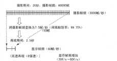
作为医用图像摄影,例如在造影检查中用于确认造影剂的流入流出的动态的动态摄影需要30~180秒左右的摄影。确认关节的活动需要5~10秒左右的摄影。即使是摄影时间为例如20秒,3000帧/秒等的超高速摄影会也产生60000帧的大量的医用图像。As medical imaging, for example, dynamic imaging for checking the inflow and outflow of a contrast agent in a contrast examination requires about 30 to 180 seconds of imaging. It takes about 5 to 10 seconds of photography to confirm the movement of the joints. Even ultra-high-speed photography with a photography time of, for example, 20 seconds and 3000 frames/second generates a large number of medical images of 60000 frames.
众所周知,人的眼睛能够识别的速度是30帧/秒左右。若将通过超高速摄影收集到的60000帧的大量的医用图像以该显示帧频(30帧/秒)来显示,则需要30分钟以上的时间。As we all know, the speed that human eyes can recognize is about 30 frames per second. It takes more than 30 minutes to display a large number of medical images of 60,000 frames collected by ultra-high-speed photography at this display frame rate (30 frames/second).
发明内容Contents of the invention
本发明的目的在于,在不降低判读精度的情况下提高通过超高速摄影收集到的医用图像的判读效率。An object of the present invention is to improve the interpretation efficiency of medical images collected by ultra-high-speed photography without reducing the accuracy of interpretation.
通常,本发明的医用图像显示装置具备:间隔剔除处理部,对一系列的医用图像实施间隔剔除处理;显示部,将实施了上述间隔剔除处理后的一系列的医用图像作为动态图像来显示;以及控制部,以使上述间隔剔除处理中的间隔剔除率在时间轴上变化的方式,控制上述间隔剔除处理部。Generally, the medical image display device of the present invention includes: a thinning processing unit that performs thinning processing on a series of medical images; a display unit that displays the series of medical images that have been subjected to the thinning processing as a dynamic image; And the control unit controls the thinning processing unit so that the thinning rate in the thinning processing changes on the time axis.
发明效果Invention effect
能够提高通过超高速摄影而收集到的医用图像的判读效率的判读精度。It is possible to improve the interpretation accuracy of the interpretation efficiency of medical images collected by ultra-high-speed photography.
附图说明Description of drawings
图1是表示本发明的实施方式的图像显示装置的构成的图。FIG. 1 is a diagram showing the configuration of an image display device according to an embodiment of the present invention.
图2是表示本实施方式中的摄影帧频的图。FIG. 2 is a diagram showing the shooting frame rate in this embodiment.
图3是表示本实施方式中的低速再现(1/100倍速)的图。FIG. 3 is a diagram showing low-speed playback (1/100x speed) in this embodiment.
图4是表示本实施方式中的低速再现(1/10倍速)的图。FIG. 4 is a diagram showing low-speed playback (1/10x speed) in this embodiment.
图5是表示本实施方式中的等速再现的图。FIG. 5 is a diagram showing constant-speed playback in this embodiment.
图6是表示本实施方式中的高速再现(2倍速)的图。FIG. 6 is a diagram showing high-speed playback (2x speed) in this embodiment.
图7是表示本实施方式中的高速再现(4倍速)的图。FIG. 7 is a diagram showing high-speed reproduction (quadruple speed) in this embodiment.
图8是表示本实施方式中的低速再现(1/50倍速)的图。FIG. 8 is a diagram showing low-speed reproduction (1/50x speed) in this embodiment.
图9是表示本实施方式中的伴随着显示帧频增加的等速再现的图。FIG. 9 is a diagram showing constant-speed playback accompanied by an increase in the display frame rate in this embodiment.
图10是表示本实施方式中的伴随着显示帧频增加的高速再现(4倍速)的图。FIG. 10 is a diagram showing high-speed reproduction (4x speed) accompanied by an increase in the display frame rate in the present embodiment.
图11是表示本实施方式中的伴随着显示帧频增加的高速再现(8倍速)的图。FIG. 11 is a diagram showing high-speed reproduction (8x speed) accompanied by an increase in the display frame rate in the present embodiment.
图12是表示本实施方式中的用于设定关注区域(ROI)的图像选择画面的图。FIG. 12 is a diagram showing an image selection screen for setting a region of interest (ROI) in this embodiment.
图13是表示在图12中选择出的图像上设定的关注区域的图。FIG. 13 is a diagram showing a region of interest set on the image selected in FIG. 12 .
图14是表示与图13的关注区域有关的像素值(造影浓度)的时间变化和基于该时间变化而确定出的关注期间的图。FIG. 14 is a diagram showing temporal changes in pixel values (contrast density) related to the region of interest in FIG. 13 and a period of interest specified based on the temporal changes.
图15是表示图14的关注期间及该关注期间以外的期间各自的再现速度的图。FIG. 15 is a diagram showing playback speeds of the period of interest in FIG. 14 and periods other than the period of interest.
图16是表示与图13的关注区域有关的像素值(造影浓度)的时间变化和基于该时间变化而确定出的关注期间的图。FIG. 16 is a diagram showing temporal changes in pixel values (contrast density) related to the region of interest in FIG. 13 and a period of interest specified based on the temporal changes.
图17是表示图16的关注期间及该关注期间以外的期间各自的再现速度的图。FIG. 17 is a diagram showing playback speeds of the period of interest in FIG. 16 and periods other than the period of interest.
图18是表示在本实施方式中的关注期间的其他的决定规则的概念的图。FIG. 18 is a diagram showing the concept of another decision rule for the period of interest in this embodiment.
图19是表示根据图18的规则而决定出的关注期间的图。FIG. 19 is a diagram showing periods of interest determined based on the rule in FIG. 18 .
图20是表示本实施方式的间隔剔除率的经时变化的一例的图。FIG. 20 is a graph showing an example of the temporal change of the thinning rate in this embodiment.
图21是表示本实施方式的间隔剔除率的经时性变化的其他的例子的图。FIG. 21 is a graph showing another example of the temporal change of the thinning rate in this embodiment.
符号说明Symbol Description
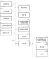
1...系统控制部,2...图像存储部,3...操作部,4...ROI设定部,5...TDC制作部,6...关注期间设定部,7...帧频决定部,8...帧频变换处理部,9...再现图像存储部,10...显示部,11...接口部1...system control section, 2...image storage section, 3...operation section, 4...ROI setting section, 5...TDC creation section, 6...attention period setting section, 7...frame rate determination section, 8...frame rate conversion processing section, 9...reproduced image storage section, 10...display section, 11...interface section
具体实施方式Detailed ways
通常,本实施方式的医用图像显示装置具备:对一系列的医用图像实施(perform)间隔剔除处理(thin out processing)的间隔剔除处理部;将实施了间隔剔除处理后的一系列的医用图像作为动态图像来显示的显示部;以及以间隔剔除处理中的间隔剔除率在时间轴上变化的方式控制间隔剔除处理部的控制部。Generally, the medical image display device according to this embodiment includes: a thinning processing unit that performs thin out processing on a series of medical images; A display unit that displays a moving image; and a control unit that controls the thinning processing unit so that the thinning rate in the thinning process changes on the time axis.
摄影帧频(imaging frame rate)是指,通过X射线诊断装置等医用图像摄影装置在1秒内所产生的帧的数量,也称为原始帧频。显示帧频(displaying frame rate)是指,通过包含显示器等的显示部在1秒内切换的帧的数量,其取决于显示部的性能。另外,本实施方式适用了间隔剔除处理。通过间隔剔除处理,减少了构成一系列的医用图像的帧的总数。即,间隔剔除处理是使一系列的医用图像的帧频减少的处理,换言之,是扩大与一系列的医用图像有关的帧间隔的处理。具体地讲,是下述处理中的任一个处理:按每个单位期间,典型的是1秒,从该期间中所含的多个医用图像提取1张或者规定张数的医用图像作为显示对象图像,将这以外的医用图像从显示对象中排除的处理;按每个单位期间生成将该期间中所含的多个医用图像的加法平均图像作为显示对象图像的处理。此外,将单位期间内拍摄到的原始的一系列的医用图像的帧数设为N,将在显示部上显示的显示对象图像的帧数设为n,则间隔剔除率定义为,从帧数N中减去帧数n后得到的帧数占帧数N的比例(N-n)/N。The imaging frame rate refers to the number of frames generated by a medical imaging device such as an X-ray diagnostic device within 1 second, and is also called the original frame rate. The displaying frame rate refers to the number of frames switched within 1 second by a display unit including a display and the like, and depends on the performance of the display unit. In addition, this embodiment applies thinning processing. Through the thinning process, the total number of frames constituting a series of medical images is reduced. That is, the thinning process is a process of reducing the frame rate of a series of medical images, in other words, a process of increasing the frame interval of a series of medical images. Specifically, it is any one of the following processes: for each unit period, typically 1 second, one or a predetermined number of medical images are extracted from a plurality of medical images included in the period as display objects. An image is a process of excluding other medical images from display targets; and a process of generating, for each unit period, an averaged image of a plurality of medical images included in the period as a display target image. In addition, assuming that the number of frames of the original series of medical images captured in the unit period is N, and the number of frames of the display target image displayed on the display unit is n, the thinning rate is defined as, from the number of frames The ratio of the number of frames obtained after subtracting the number of frames n from N to the number of frames N (N-n)/N.
本实施方式的特征在于,如图20、图21所示,对于原始的一系列的医用图像,能够按单位期间分别设定间隔剔除率。本实施方式的特征在于,以间隔剔除率在时间轴上变化的方式适用间隔剔除处理。在图20、图21的例子中,一系列的医用图像以高于显示帧频(30帧/秒)的摄影帧频(3000帧/秒)被产生。间隔剔除率为99%,即,在单位时间内的、典型地是1秒内的3000帧中,排除2970帧,提取等间隔的30帧作为显示对象图像,以30帧/秒的显示帧频进行显示时,摄影对象的活动以与被摄影时的时间尺度(scale)相同的时间尺度来再现。另一方面,间隔剔除率为0%,即,将单位时间内,典型地1秒内的3000帧的全部帧作为显示对象图像,以30帧/秒的显示帧频来显示时,摄影对象的活动以被摄影时的时间尺度的100倍的时间尺度被缓慢再现。在图20的例子中,从时刻tm开始到时刻tm十1为止的期间的间隔剔除率设定为0%,其他的期间的间隔剔除率为99%。判读者对对于诊断而言重要度高的期间设定间隔剔除率0%,对重要度低的期间设定间隔剔除率99%,从而能够提高判读作业的效率。如图21所示,间隔剔除率的变化(variation)可以任意地增加。The present embodiment is characterized in that, as shown in FIGS. 20 and 21 , thinning rates can be set for each unit period for a series of original medical images. The present embodiment is characterized in that the thinning process is applied so that the thinning rate changes on the time axis. In the examples shown in FIG. 20 and FIG. 21 , a series of medical images are generated at an imaging frame rate (3000 frames/sec) higher than the display frame rate (30 frames/sec). The thinning rate is 99%, that is, in a unit time, typically 3000 frames within 1 second, 2970 frames are excluded, and 30 frames at equal intervals are extracted as the display object image at a display frame rate of 30 frames per second When displaying, the motion of the subject to be photographed is reproduced on the same time scale as when the photograph was taken. On the other hand, when the thinning rate is 0%, that is, when all the frames of 3000 frames within a unit time, typically 1 second, are used as the display target image and displayed at a display frame rate of 30 frames/second, the photographic target Activity is slowly reproduced on a
以下,详细地说明本实施方式。Hereinafter, this embodiment will be described in detail.
如图1所示,本实施方式的医用图像显示装置经由接口部11与外部的超高速摄影对应的X射线诊断装置21、磁共振诊断装置(MRI)22等图像产生装置以及图像保管通信系统(PACS)23连接。这些X射线诊断装置21以及磁共振诊断装置22如图2所示,装备了以例如3000帧/秒的一定的帧频反复摄影,产生构成动态图像的一系列的医用图像的超高速动态图像摄影功能。医用图像典型来讲是二维图像,也可以是三维图像。在医用图像是三维图像时,该说明中“帧”和“帧频”分别被改读为“体积(volume)”、“体积率(volume rate)”。As shown in FIG. 1 , the medical image display device according to this embodiment communicates with an image generation device such as an X-ray diagnostic device 21 and a magnetic resonance diagnostic device (MRI) 22 compatible with external ultra-high-speed imaging via an
另外,帧频被定义为单位时间内的、在此为1秒内的帧数。基于这些X射线诊断装置21以及磁共振诊断装置22等图像产生装置的超高速动态图像摄影功能而产生的一系列的医用图像的数据,从图像产生装置直接地,或者经由图像保管通信系统23地发送至本实施方式的医用图像显示装置。图像存储部2设置为用于存储经由接口部11而接收到的原始的一系列的医用图像的数据。In addition, the frame rate is defined as the number of frames per unit time, here, 1 second. The data of a series of medical images generated based on the ultra-high-speed dynamic image photography function of these image generating devices such as the X-ray diagnostic device 21 and the magnetic resonance
本实施方式的医用图像显示装置,除了图像存储部2和接口部11之外,包括控制中枢的系统控制部1、操作部3、ROI设定部4、TDC制作部5、关注期间设定部6、帧频决定部7、帧频变换处理部8、再现图像存储部9和显示部10。操作部3具有键盘、鼠标等定位设备(pointing device)。操作者对操作部3进行操作,从而对医用图像显示装置输入各种指示。例如,经由操作部3,选择一系列的医用图像中的特定的一张(1帧)的医用图像来作为关注区域指定用图像。ROI设定部4设置为用于在图像坐标系上表现关注区域(ROI)的位置、形状以及大小,所述关注区域(ROI)是在经由操作部3而确定的医用图像上指定的区域。The medical image display device of this embodiment includes, in addition to the
TDC制作部5以一系列的医用图像整体作为对象,制作由ROI设定部4设定出的关注区域内的像素值的时间变化(时间-密度曲线(TDC:Time-Density Curve))。关注区域中包含多个像素时,关于像素值频度分布上的中间值、表示最高频度的像素值、最大值、最小值、或者平均像素值等任意值,制作时间变化。另外,该时间变化也可以如后所述那样是与多个骨区域之间的距离有关的时间变化。The
关注期间设定部6基于由TDC制作部5制作的时间变化,在跨越一系列的医用图像整体的摄影期间中设定关注高的至少一个部分期间(关注期间)。另外,关注期间设定部6也可以按照操作者经由操作部3指定出的期间来设定该关注期间。The period of
构成摄影期间的一部分的关注期间内的动态图像以等速或者低速被再现。摄影期间中所含的关注期间以外的期间内的动态图像以高速被再现。等速再现是指以与摄影所需要的时间等价的时间来再现动态图像,即,例如将摄影时需要10秒的动态图像以与摄影时间等价的10秒时间进行再现。摄影时间是指从摄影开始时刻到摄影结束时刻之间的时间的长度。再现时间是指从再现开始时刻到再现结束时刻之间的时间的长度。The moving images in the period of interest constituting a part of the shooting period are reproduced at a constant speed or at a low speed. Movies in periods other than the period of interest included in the shooting period are reproduced at high speed. Constant-speed reproduction refers to reproducing a moving image in a time equivalent to the time required for shooting, that is, for example, reproducing a moving image that requires 10 seconds for shooting in 10 seconds equivalent to the shooting time. The shooting time refers to the length of time from the shooting start time to the shooting end time. The playback time refers to the length of time from the playback start time to the playback end time.
低速再现是指以比摄影所需要的时间长的时间来再现动态图像,即,例如将摄影时需要10秒的动态图像以比摄影时间长的20秒的时间进行再现。高速再现是指以比摄影时所需要的时间短的时间来再现动态图像,即,例如将摄影时需要10秒的动态图像以比摄影时间短的5秒的时间进行再现。Low-speed reproduction refers to reproducing a moving image in a time longer than that required for shooting, that is, for example, reproducing a moving image that takes 10 seconds for shooting in 20 seconds longer than the shooting time. High-speed playback refers to reproducing a moving image in a time shorter than that required for shooting, that is, for example, reproducing a moving image that takes 10 seconds for shooting in 5 seconds shorter than the shooting time.
另外,再现速度定义为,摄影时间相对于再现时间的比例n(n=摄影时间/再现时间)。一般来讲,再现速度表现为“n倍速”。本实施方式中,对于低速再现和高速再现分别准备了多个再现速度。再现速度通过由帧频变换处理部9设定的间隔剔除率以及显示部10的显示帧频来决定。帧频变换处理部9通过对拍摄到的原始的一系列的医用图像实施间隔剔除处理或者平滑化处理,从而对拍摄到的原始的一系列的医用图像进行间隔剔除处理。另外,即使显示部10的显示帧频被固定为例如30帧/秒,但也可以从例如30帧/秒和60帧/秒这2个种类中来选择。在此利用前者进行说明。In addition, the playback speed is defined as the ratio n of the shooting time to the playback time (n=shooting time/playback time). In general, the reproduction speed is expressed as "n-time speed". In this embodiment, a plurality of playback speeds are prepared for low-speed playback and high-speed playback. The playback speed is determined by the thinning rate set by the frame rate
本实施方式中,通过超高速摄影而采用了3000帧/秒等非常高的摄影帧频,此外显示部10的显示帧频为30帧/秒或者60帧/秒,这与摄影帧频相比非常低。在显示帧频固定时,显示部10上所显示的动态图像的最终的再现速度由摄影帧频和间隔剔除率来决定。In this embodiment, a very high shooting frame rate such as 3000 frames/second is adopted by ultra-high-speed shooting, and the display frame rate of the
帧频决定部7对于构成摄影时间的一部分期间的至少一个关注期间以及构成摄影时间的其他部分的期间的期间(低关注期间),分别分配间隔剔除率。如上所述,帧频被定义为每单位时间的帧数。与其他的低关注期间被分配的间隔剔除率相比,关注期间被分配的间隔剔除率设定得较小。换言之,关注期间的再现速度设定得比低关注期间的再现速度慢,低关注期间的再现速度设定得比关注期间的再现速度快。为了使动态图像再现容纳(收まる)在由操作者、典型地是判读者经由操作部3指定的再现时间框内,帧频决定部7对关注期间和低关注期间分配不同的间隔剔除率。详细内容将在后面说明。The frame rate determination unit 7 assigns a thinning rate to at least one period of interest constituting a part of the imaging time and a period (period of low interest) constituting the other part of the imaging time. As mentioned above, the frame rate is defined as the number of frames per unit of time. The thinning rate assigned to the attention period is set to be smaller than the thinning ratio assigned to the other low attention periods. In other words, the playback speed of the attention period is set slower than that of the low attention period, and the playback speed of the low attention period is set faster than that of the attention period. The frame rate determination unit 7 assigns different thinning rates to the attention period and the low attention period in order to accommodate moving image reproduction within the reproduction time frame designated by the operator, typically the judge, via the
帧频变换部8通过对原始的一系列的医用图像实施间隔剔除处理或者平滑化处理,以由帧频决定部7决定出的间隔剔除率,对拍摄到的原始的一系列的医用图像进行间隔剔除处理。间隔剔除处理是使一系列的医用图像的帧频减少的处理,具体地讲,是下述任一种处理:按每个单位期间,典型的是1秒,从该期间所含的多个医用图像中提取1张或者规定张数的医用图像来作为显示对象图像,并将这以外的医用图像从显示对象中排除的处理;按每个单位期间,生成该期间所含的多个医用图像的加法平均图像来作为显示对象图像的处理。这里以前者的情况进行说明。在将单位期间(1秒)内所含的帧数设为N,该期间的显示对象图像的帧数设为n的情况下,间隔剔除率是(N-n)/N。The frame
再现图像存储部9设置为用于存储被帧频变换部8变换后的再现对象的一系列的医用图像的数据。再现对象的一系列的医用图像的帧数比原始的一系列的医用图像的帧数少。此外,关注期间内的医用图像的帧数比低关注期间内的医用图像的帧数多。The reproduced
再现图像存储部9中所存储的再现对象的一系列的医用图像的数据,基于系统控制部1的控制,与显示部10的垂直同步信号同步地,以一定的周期一帧一帧地依次被供给至显示部10。The data of a series of medical images to be reproduced stored in the reproduced
图3~图7示出了在显示帧频固定为30帧/秒时的多个再现速度。在图3所示的最慢的低速再现中,所拍摄的全部的帧被作为显示对象。间隔剔除率为0。该情况下,再现速度是1/100倍速,所显示的脏器等的活动看上去被非常缓慢地再现。图4所示的低速再现中间隔剔除率设定为90%。该情况下,再现速度是1/10倍速,所显示的脏器等的活动看上去被缓慢地再现。3 to 7 show a plurality of reproduction speeds when the display frame rate is fixed at 30 frames/second. In the slowest low-speed playback shown in FIG. 3 , all captured frames are displayed. Thinning rate is 0. In this case, the reproduction speed is 1/100 times speed, and the movement of the displayed organs or the like appears to be reproduced very slowly. The thinning rate in low-speed reproduction shown in FIG. 4 is set to 90%. In this case, the reproduction speed is 1/10 times speed, and the movement of the displayed organs or the like appears to be reproduced slowly.
在图5所示的等速再现中,间隔剔除率设定为99%。该情况下,再现速度是等速,所显示的脏器等的活动看上去以与实际的活动相同的速度被再现。In the constant-speed reproduction shown in FIG. 5, the thinning rate is set to 99%. In this case, the reproduction speed is constant, and the displayed movement of the organs, etc., appears to be reproduced at the same speed as the actual movement.
在图6所示高速再现中,间隔剔除率设定为99.5%。该情况下,再现速度是2倍速,所显示的脏器等的活动看上去以2倍的速度被再现。在图7所示的高速再现中,间隔剔除率设定为99.75%。该情况下,再现速度是4倍速,所显示的脏器等的活动看上去以4倍的速度被再现。In the high-speed reproduction shown in FIG. 6, the thinning rate is set to 99.5%. In this case, the playback speed is 2x speed, and the displayed movement of the organs, etc., appears to be reproduced at 2x speed. In the high-speed reproduction shown in FIG. 7, the thinning rate is set to 99.75%. In this case, the playback speed is quadruple speed, and the displayed movement of the organs, etc., appears to be reproduced at quadruple speed.
图8~图11示出了将显示帧频从30帧/秒变更(增加)为60帧/秒时的多个再现速度所对应的显示帧频。在图8所示的例子中,没有进行间隔剔除。间隔剔除率是0%。该情况下,再现速度为1/50倍速,所显示的脏器等的活动看上去以仅快于图3的例子的速度被非常缓慢地再现。8 to 11 show display frame rates corresponding to a plurality of playback speeds when the display frame rate is changed (increased) from 30 frames/second to 60 frames/second. In the example shown in FIG. 8, thinning is not performed. Thinning rate is 0%. In this case, the reproduction speed is 1/50 times speed, and the displayed movement of the organs etc. appears to be reproduced very slowly at a speed only faster than the example in FIG. 3 .
图9所示的显示帧频变更为非常高的60帧/秒的显示帧频,伴随着该变更,与图5所示的例子相同地实现了等速再现。间隔剔除率设定为98%。The display frame rate shown in FIG. 9 is changed to a very high display frame rate of 60 frames/second. With this change, constant-speed playback is realized similarly to the example shown in FIG. 5 . Thinning rate is set to 98%.
在图10所示的例子中,显示帧频从30帧/秒变更为60帧/秒。与图7所示的例子相同地实现了4倍速再现。但是,间隔剔除率设定为99.5%。In the example shown in FIG. 10, the display frame rate is changed from 30 frames/second to 60 frames/second. Quadruple speed reproduction is realized in the same manner as in the example shown in FIG. 7 . However, the thinning rate is set to 99.5%.
图11所示的最快的高速再现的显示帧频实现了8倍速再现。间隔剔除率设定为99.75%。The display frame rate of the fastest high-speed reproduction shown in FIG. 11 realizes 8-times-speed reproduction. Thinning rate is set to 99.75%.
在显示部10的显示帧频固定为30帧/秒时,系统控制部1对帧频决定部7指示将间隔剔除率的选择项限制为图3~图7所示的5个种类的间隔剔除率。When the display frame rate of the
在显示部10的显示帧频能够在30帧/秒和60帧/秒之间进行选择,并且指定了判读者经由操作部10将显示帧频增加为60帧/秒时,系统控制部1对帧频决定部7指示将间隔剔除率的选择项限制为图8~图11所示的4个种类的间隔剔除率。When the display frame rate of the
另外,在显示部10的显示帧频能够在30帧/秒和60帧/秒之间选择,并且允许了判读者经由操作部10将显示帧频增加至60帧/秒时,系统控制部1将显示帧频选择为30帧/秒和60帧/秒的任一方。在再现的整体期间内显示帧频是固定的。在判读者所指定的再现时间框相对于摄影时间的比例为例如2倍以上时,系统控制部1将显示帧频选择为30帧/秒。在判读者所指定的再现时间框相对于摄影时间的比例为例如不足2倍时,系统控制部1将显示帧频选择为60帧/秒,向显示帧频的选择项中增加8倍速再现。In addition, when the display frame rate of the
接着,对关注期间的设定例进行说明。首先,基于系统控制部1的控制,从通过摄影而产生的一系列的医用图像中在时间轴上离散地选择规定数量的医用图像。选择出的规定数量的医用图像在未图示的帧频变换处理部中被提供给矩阵尺度(matrix size)的减低处理。由此产生规定数量的缩略图(thumbnail)医用图像,如图12所示在显示部10上被一览显示。判读者操作操作部31来选择确定的医用图像。如图13所示,选择出的医用图像在显示部10的显示框整个区域中以原始的矩阵尺度或者比缩略图高的矩阵尺度被显示。判读者操作操作部3,在所显示的医用图像上的任意的位置以任意的形状及大小指定1个或者多个关注区域(ROI)。在此,以指定了2个关注区域的情况进行说明。经由操作部3在特定的医用图像上指定的关注区域(ROI)的位置、形状及大小由ROI设定部4通过图像坐标系上的表现进行设定。Next, a setting example of the period of interest will be described. First, based on the control of the
通过TDC制作部5,将拍摄到的构成动态图像的全部的一系列的医用图像作为对象,制作由ROI设定部4设定的关注区域内的像素值的时间变化(TDC)。在关注区域中包含多个像素时,典型地是制作与平均像素值有关的时间变化(TDC)。制作出的TDC被显示在显示部10上。The
图14示出了与2个关注区域(ROI.1、ROI.2)有关的2个TDC。图14的例子示出了基于造影摄影的TDC。例如在将造影剂急剧流入关注区域的期间设定为关注期间时,通过关注期间设定部6,将表示TDC的微分值是规定的阈值以上的期间设定为关注期间T1、T2。另外,也可以在显示部10所显示的TDC上由操作者经由操作部3设定至少一个关注期间。此外,也可以由操作者经由操作部3对关注期间设定部6所设定的关注期间T1、T2进行修正。Figure 14
对于设定的关注期间T1、T2,通过帧频决定部7,分配由判读者初始设定的用于关注期间的显示帧频,对于关注期间T1、T2以外的低关注期间,分配由判读者初始设定的用于低关注期间的间隔剔除率。例如,对关注期间T1、T2分配图4示出的1/10倍速的低速再现,对低关注期间分配图6示出的2倍速的高速再现。当基于这些初始的间隔剔除率而计算出的总再现时间容纳在预先指定的再现时间框内时,确定这些初始的间隔剔除率。当计算出的总再现时间超过了预先指定的再现时间框时,按照规定的规则来变更这些初始的间隔剔除率。首先,作为第一顺位,将用于低关注期间的间隔剔除率降低一个等级。在该例子中,实现图6所示的2倍速的高速再现的间隔剔除率被变更为实现图7所示的4倍速的高速再现的间隔剔除率。当基于变更后的间隔剔除率而再次计算的总再现时间容纳在预先指定的再现时间框内时,确定该间隔剔除率。当再计算出的总再现时间超过了预先指定的再现时间框时,将用于关注期间T1、T2的间隔剔除率降低一个等级(step)。在该例子中,实现图4所示的1/10倍速的低速再现的间隔剔除率被变更为实现图5所示的等速再现的间隔剔除率。当基于该变更后的间隔剔除率而再次计算的总再现时间容纳在预先指定的再现时间框内时,确定该显示帧频。当最后总再现时间超过了再现时间框时,显示表示该超过的注意消息。当判读者输入了“OK”指示时,确定该间隔剔除率。当判读者输入了“NG”指示时,系统控制部1显示TDC,促使进行关注期间T1、T2的缩短化操作,或者从再现中排除的一部分期间的指定操作。For the set attention period T1, T2, the display frame rate for the period of interest initially set by the interpreter is allocated by the frame rate determination part 7, and for the low attention period other than the attention period T1, T2, the display frame rate for the period of interest set by the interpreter is allocated. The initial thinning rate to use for periods of low concern. For example, the 1/10-speed low-speed playback shown in FIG. 4 is allocated to the focused periods T1 and T2, and the 2-fold high-speed playback shown in FIG. 6 is allocated to the low focused period. These initial thinning rates are determined when the total reproduction time calculated based on these initial thinning rates fits within a pre-designated reproduction time frame. When the calculated total reproduction time exceeds a predetermined reproduction time frame, these initial thinning rates are changed according to a predetermined rule. First, as a first order, the thinning rate for periods of low interest is lowered by one notch. In this example, the thinning rate for achieving high-speed playback at 2x speed shown in FIG. 6 is changed to the thinning-out rate for high-speed playback at 4x speed shown in FIG. 7 . The thinning-out rate is determined when the total reproduction time recalculated based on the changed thinning-out rate is accommodated within a pre-designated reproduction time frame. When the recalculated total reproduction time exceeds the pre-specified reproduction time frame, the thinning rate for the periods of interest T1, T2 is reduced by one step. In this example, the thinning rate for achieving low-speed reproduction at 1/10x speed shown in FIG. 4 is changed to the thinning-out rate for realizing constant-speed reproduction shown in FIG. 5 . The display frame rate is determined when the total reproduction time recalculated based on the changed thinning rate is accommodated within a pre-designated reproduction time frame. When the final total reproduction time exceeds the reproduction time frame, a caution message indicating the excess is displayed. This thinning rate is determined when the interpreter has input an indication of "OK". When the judge inputs an "NG" instruction, the
在此,为了便于说明,假设对关注期间T1、T2确定了用于实现图4所示的1/10倍速的低速再现的间隔剔除率,对低关注期间确定了用于实现图6所示的2倍速的高速再现的间隔剔除率的情况来说明。Here, for the sake of explanation, it is assumed that the thinning rate for realizing the low-speed reproduction of 1/10x speed shown in FIG. The case of the thinning rate of high-speed reproduction at 2x speed will be described.
若对于关注期间T1、T2和低关注期间分别确定了间隔剔除率,则按照确定出的间隔剔除率,通过帧频变换部8对原始的一系列的医用图像执行间隔剔除处理。按照与对于关注期间T1、T2而确定出的间隔剔除率相对应的间隔剔除率,从关注期间T1、T2内所拍摄到的多个医用图像中,相对于时间轴等间隔地,在此以10帧中取1帧的比例等间隔地提取再现对象图像,将其他的医用图像从再现对象中排除,即被间隔剔除。同样,从低关注期间内所拍摄到的多个医用图像中,相对于时间轴等间隔地,在此以200帧取1帧的比例等间隔地提取再现对象图像,将其他的医用图像从再现对象中排除,即被间隔剔除。If thinning rates are determined for the periods of interest T1 , T2 and the period of low interest, thinning processing is performed on the original series of medical images by the frame
通过帧频变换部8提取出的一系列的再现对象图像暂时存储在再现图像存储部9,基于系统控制部1的控制,与显示部10的垂直同步信号同步地,以一定的周期一帧一帧地依次提供给显示部10。由此,如图15所示,在关注期间中被缓慢再现,在低关注期间中被高速再现。这样,成为再现对象的一系列的医用图像的再现速度,关于再现时间轴,按照其关注程度被赋予缓急地发生变动。A series of playback target images extracted by the frame
在该例子中,通过造影剂动态地观察血管等时,将希望观察的动脈瘤、AVM等设定为ROI,在造影剂到达该ROI之前,基于高速再现来观察造影剂的流动,在造影剂流入ROI,该流入即将迎来峰值之前为止的期间能够详细地观察血流的动态。然后,基于高速再现来观察造影剂的流动。这样,对于通过超高速摄影而收集到的一系列的医用图像,能够对关注高的重要的动态进行缓慢地判读,即便对于关注不是那么高的动态也没有排除再现,而是进行高速再现。由此,能够在抑制判读精度的降低的同时提高判读效率。In this example, when a blood vessel etc. is dynamically observed with a contrast agent, an aneurysm, AVM, etc. to be observed is set as an ROI, and before the contrast agent reaches the ROI, the flow of the contrast agent is observed based on high-speed reproduction, The agent flows into the ROI, and the dynamics of the blood flow can be observed in detail until the inflow peaks. Then, the flow of the contrast agent is observed based on high-speed reproduction. In this way, for a series of medical images collected by ultra-high-speed photography, it is possible to slowly interpret important movements of high interest, and perform high-speed reproduction even for movements of lesser interest, without excluding reproduction. Thereby, it is possible to improve interpretation efficiency while suppressing a decrease in interpretation accuracy.
另外,也可以在将造影剂流入期间设定为关注期间的同时,也将造影剂流出期间设定为关注期间。该情况下,如图16所示,有时多个关注期间重叠(overlap)。在图16的例子中,与第二ROI有关的造影剂流出的关注期间T2-1和与第一ROI有关的造影剂流入的关注期间T1-2重叠。该情况下,将包含关注期间T2-1和关注期间T1-2两者的期间设定为关注期间T2。在设定出的3个关注期间T1、T2、T3中,如图17所示进行缓慢再现,在低关注期间进行高速再现。In addition, while setting the contrast medium inflow period as the period of interest, the contrast medium outflow period may also be set as the period of interest. In this case, as shown in FIG. 16 , a plurality of periods of interest may overlap (overlap). In the example of FIG. 16 , the period T2 - 1 of interest for the outflow of the contrast medium related to the second ROI overlaps with the period T1 - 2 of interest for the inflow of the contrast medium related to the first ROI. In this case, a period including both the period of interest T2-1 and the period of interest T1-2 is set as the period of interest T2. In the set three focus periods T1, T2, T3, slow playback is performed as shown in FIG. 17, and high-speed playback is performed in the low focus periods.
在该例子中,在利用造影剂动态观察血管等时,将希望观察的动脈瘤、AVM等设定为ROI,在造影剂到达该ROI之前,基于高速再现来观察造影剂的流动,在造影剂进入的期间,通过低速再现能够详细地观察造影剂的动态。在造影浓度不发生变化后,再通过高速再现以实现时间的缩短,然后在造影剂流出的期间,通过低速再现来详细观察造影剂流出的样子,进而在造影浓度从关注区域充分流出以后的期间,通过高速再现能够实现时间的缩短。即,在重要的期间以低速进行再现从而提高判读精度,另一方面,在不太重要的期间以高速进行再现从而提高判读效率。由于被认为不太重要的期间的动态图像部分也没有从再现对象中被排除,因此即使该期间中有重要的动态,也能够降低漏看的危险性。In this example, when using a contrast agent to dynamically observe a blood vessel, etc., an aneurysm, AVM, etc. to be observed is set as an ROI, and before the contrast agent reaches the ROI, the flow of the contrast agent is observed based on high-speed reproduction, During the injection of the contrast agent, the dynamics of the contrast agent can be observed in detail by low-speed reproduction. After the contrast density does not change, the time can be shortened by high-speed reproduction, and then during the outflow of the contrast agent, the low-speed reproduction can be used to observe the outflow of the contrast agent in detail, and then after the contrast density has fully flowed out from the area of interest , time can be shortened by high-speed reproduction. That is, playback is performed at a low speed during an important period to improve interpretation accuracy, while playback is performed at a high speed during a less important period to improve interpretation efficiency. Since moving image portions in periods considered to be less important are not excluded from reproduction targets, even if there is important movement in this period, the risk of missing it can be reduced.
关注期间的设定方法还可以考虑除此之外的多种方法。关节检查中,有时虽然有疼痛但通过通常摄影没有异常,此时需要一边活动关节一边摄影,希望对该活动进行动态图像观察(确认在某位置骨与骨接触或关节错位的情况来计划治疗)。调查骨与骨的间隙,仅对间隙变无的期间进行缓慢显示。对于关节的情况而言,间隙最小的位置是希望重点观察的位置,在间隙的距离的时间变化中需要从微分值表示负的最大值的时期开始到表示正的最大值的时期为止的期间总是缓慢显示。As a method of setting the period of interest, various methods other than this can be considered. During the joint examination, sometimes there is pain, but there is no abnormality in normal photography. At this time, it is necessary to take pictures while moving the joints. I want to observe the movement with dynamic images (confirm bone-to-bone contact at a certain position or joint dislocation to plan treatment) . Check the bone-to-bone gap and slowly display only the period when the gap disappears. In the case of joints, the position where the gap is the smallest is the position to focus on. In the time change of the distance of the gap, the period from the time when the differential value shows a negative maximum value to the time when it shows a positive maximum value needs to be total. is displayed slowly.
例如,如图18、图19所示,在关节观察中,关节部分的2个骨区域的距离是重要的。计算在特定的医用图像上设定的2个骨区域之间的距离,制作其时间变化。将2个骨区域比规定距离接近的期间设定为关注期间。即,对骨彼此干涉或变窄的活动进行缓慢再现,对除此之外的骨彼此之间一定程度分离的期间进行高速再现。For example, as shown in FIGS. 18 and 19 , in joint observation, the distance between two bone regions in the joint portion is important. Calculate the distance between two bone regions set on a specific medical image, and create its temporal change. A period in which two bone regions are closer than a predetermined distance is set as the period of interest. That is, the movement in which the bones interfere with each other or becomes narrow is slowly reproduced, and the other periods in which the bones are separated to a certain extent are reproduced at high speed.
如上所述,在对通过超高速摄影而产生的一系列的医用图像进行再现时,根据是否是重要的期间,使间隔剔除率关于时间轴而变动,使再现速度变化。换言之,通过关于时间轴使间隔剔除率或平滑化率变化,能够实现一边提高诊断效率,一边高效地观察大量的医疗图像数据。As described above, when reproducing a series of medical images generated by super high-speed photography, the thinning-out rate is varied with respect to the time axis and the reproduction speed is changed depending on whether it is an important period or not. In other words, by changing the thinning rate or the smoothing rate with respect to the time axis, it is possible to efficiently observe a large amount of medical image data while improving diagnosis efficiency.
虽然对特定的实施方式进行了说明,但是这些实施方式仅仅是作为例子来说明的,并没有一体限定本发明的范围。事实上,在不脱离本发明的精神的情况下,在此说明的新的方法和系统能够通过各种其他方式,进一步通过对在此说明的新的方法的系统进行各种省略、追加以及改变来实施。所附的权利要求及其等同的方案试图覆盖所有这种方式或变形例,它们落入本发明的范围和宗旨内。Although specific embodiments have been described, these embodiments are merely illustrative and do not limit the scope of the present invention as a whole. In fact, without departing from the spirit of the present invention, the new method and system described here can be used in various other ways, further by making various omissions, additions and changes to the new method and system described here to implement. The appended claims and their equivalents are intended to cover all such modes or modifications which fall within the scope and spirit of the invention.
Claims (14)
Applications Claiming Priority (2)
| Application Number | Priority Date | Filing Date | Title |
|---|---|---|---|
| JP137733/2010 | 2010-06-16 | ||
| JP2010137733 | 2010-06-16 |
Publications (2)
| Publication Number | Publication Date |
|---|---|
| CN102283667A CN102283667A (en) | 2011-12-21 |
| CN102283667Btrue CN102283667B (en) | 2014-04-23 |
Family
ID=45328674
Family Applications (1)
| Application Number | Title | Priority Date | Filing Date |
|---|---|---|---|
| CN201110162648.5AActiveCN102283667B (en) | 2010-06-16 | 2011-06-16 | Medical image display device and X-ray computed tomography device |
Country Status (3)
| Country | Link |
|---|---|
| US (1) | US9814434B2 (en) |
| JP (1) | JP5725981B2 (en) |
| CN (1) | CN102283667B (en) |
Families Citing this family (18)
| Publication number | Priority date | Publication date | Assignee | Title |
|---|---|---|---|---|
| JP2013026727A (en)* | 2011-07-19 | 2013-02-04 | Sony Corp | Display device and display method |
| US20140253544A1 (en)* | 2012-01-27 | 2014-09-11 | Kabushiki Kaisha Toshiba | Medical image processing apparatus |
| CN103239253B (en)* | 2012-02-14 | 2015-07-15 | 株式会社东芝 | Medical image diagnostic apparatus |
| JP5981162B2 (en)* | 2012-02-24 | 2016-08-31 | 東芝メディカルシステムズ株式会社 | X-ray CT system |
| US9691433B2 (en)* | 2014-04-18 | 2017-06-27 | Toshiba Medical Systems Corporation | Medical image diagnosis apparatus and medical image proccessing apparatus |
| JP6664873B2 (en)* | 2014-11-21 | 2020-03-13 | キヤノンメディカルシステムズ株式会社 | Image processing apparatus, X-ray diagnostic apparatus, and image processing program |
| DE102015202494B4 (en)* | 2015-02-12 | 2023-10-19 | Siemens Healthcare Gmbh | Evaluation of a dynamic contrast agent distribution |
| JP2018015079A (en)* | 2016-07-26 | 2018-02-01 | コニカミノルタ株式会社 | Image management device, image display system, and image display method |
| US11723579B2 (en) | 2017-09-19 | 2023-08-15 | Neuroenhancement Lab, LLC | Method and apparatus for neuroenhancement |
| US11717686B2 (en) | 2017-12-04 | 2023-08-08 | Neuroenhancement Lab, LLC | Method and apparatus for neuroenhancement to facilitate learning and performance |
| US11273283B2 (en) | 2017-12-31 | 2022-03-15 | Neuroenhancement Lab, LLC | Method and apparatus for neuroenhancement to enhance emotional response |
| US12280219B2 (en) | 2017-12-31 | 2025-04-22 | NeuroLight, Inc. | Method and apparatus for neuroenhancement to enhance emotional response |
| KR101898580B1 (en)* | 2018-01-22 | 2018-09-13 | 주식회사 뷰노 | Method for facilitating image view and apparatus using the same |
| US11364361B2 (en) | 2018-04-20 | 2022-06-21 | Neuroenhancement Lab, LLC | System and method for inducing sleep by transplanting mental states |
| EP3849410A4 (en) | 2018-09-14 | 2022-11-02 | Neuroenhancement Lab, LLC | SLEEP ENHANCEMENT SYSTEM AND METHOD |
| JP7473313B2 (en)* | 2018-10-05 | 2024-04-23 | キヤノンメディカルシステムズ株式会社 | Medical image processing device, medical image processing method, and medical image processing program |
| JP7484537B2 (en)* | 2020-07-31 | 2024-05-16 | コニカミノルタ株式会社 | Radiation image processing device, radiation image processing method and program |
| CN113397581B (en)* | 2021-08-19 | 2021-11-30 | 浙江太美医疗科技股份有限公司 | Method and device for reconstructing medical dynamic image |
Family Cites Families (25)
| Publication number | Priority date | Publication date | Assignee | Title |
|---|---|---|---|---|
| JPH07284050A (en)* | 1994-04-08 | 1995-10-27 | Fuji Photo Film Co Ltd | Device and method for displaying image |
| JPH07289546A (en) | 1994-04-22 | 1995-11-07 | Hitachi Medical Corp | Ultrasonic diagnostic system |
| US5592523A (en)* | 1994-12-06 | 1997-01-07 | Picker International, Inc. | Two dimensional detector array for CT scanners |
| JP2001120547A (en) | 1999-10-27 | 2001-05-08 | Hitachi Medical Corp | Ultrasonograph |
| JP2001242253A (en)* | 1999-12-24 | 2001-09-07 | Toshiba Corp | Radiation detector and X-ray CT device |
| US6426991B1 (en)* | 2000-11-16 | 2002-07-30 | Koninklijke Philips Electronics N.V. | Back-illuminated photodiodes for computed tomography detectors |
| JP2002281289A (en)* | 2001-03-15 | 2002-09-27 | Konica Corp | Medical image generator, medical image processor and medical network system |
| JP5132515B2 (en)* | 2001-12-20 | 2013-01-30 | 株式会社東芝 | X-ray computed tomography system |
| JP2004073490A (en)* | 2002-08-19 | 2004-03-11 | Canon Inc | X-ray imaging apparatus, X-ray imaging method, and computer-readable storage medium |
| EP1713270A4 (en)* | 2004-02-04 | 2009-06-03 | Panasonic Corp | REPRODUCTION DEVICE AND REPRODUCTION METHOD |
| JP2005277733A (en)* | 2004-03-24 | 2005-10-06 | Seiko Epson Corp | Moving image processing device |
| US20060013462A1 (en)* | 2004-07-15 | 2006-01-19 | Navid Sadikali | Image display system and method |
| US7372937B2 (en)* | 2004-07-16 | 2008-05-13 | University Of Iowa Research Foundation | Systems and methods of non-standard spiral cone-beam computed tomograpy (CT) |
| JP4575124B2 (en)* | 2004-11-29 | 2010-11-04 | オリンパス株式会社 | Image display device |
| JP4725183B2 (en)* | 2005-05-11 | 2011-07-13 | ソニー株式会社 | Imaging apparatus and method |
| JP5011482B2 (en)* | 2005-07-19 | 2012-08-29 | ジーイー・メディカル・システムズ・グローバル・テクノロジー・カンパニー・エルエルシー | X-ray CT system |
| JP4887727B2 (en)* | 2005-10-20 | 2012-02-29 | ソニー株式会社 | Image signal processing apparatus, camera system, and image signal processing method |
| JP5091644B2 (en)* | 2006-12-04 | 2012-12-05 | 株式会社東芝 | X-ray computed tomography apparatus and medical image processing apparatus |
| US7403589B1 (en)* | 2007-03-27 | 2008-07-22 | General Electric Company | Photon counting CT detector using solid-state photomultiplier and scintillator |
| JP5111924B2 (en)* | 2007-04-05 | 2013-01-09 | 株式会社東芝 | Medical diagnostic imaging equipment |
| JP5523681B2 (en)* | 2007-07-05 | 2014-06-18 | 株式会社東芝 | Medical image processing device |
| JP5337374B2 (en)* | 2007-12-12 | 2013-11-06 | 株式会社東芝 | Medical information reproduction display system |
| JP5035987B2 (en)* | 2008-01-28 | 2012-09-26 | 富士フイルム株式会社 | Capsule endoscope and operation control method of capsule endoscope |
| JP5288955B2 (en)* | 2008-09-09 | 2013-09-11 | キヤノン株式会社 | Solid-state imaging device, imaging system, and driving method of solid-state imaging device |
| CN101600115A (en)* | 2009-06-29 | 2009-12-09 | 中国航空工业集团公司洛阳电光设备研究所 | A kind of method of eliminating periodic characteristic block of image stabilization system |
- 2011
- 2011-06-07JPJP2011127481Apatent/JP5725981B2/enactiveActive
- 2011-06-16USUS13/161,870patent/US9814434B2/enactiveActive
- 2011-06-16CNCN201110162648.5Apatent/CN102283667B/enactiveActive
Also Published As
| Publication number | Publication date |
|---|---|
| US20110311021A1 (en) | 2011-12-22 |
| JP2012020115A (en) | 2012-02-02 |
| CN102283667A (en) | 2011-12-21 |
| JP5725981B2 (en) | 2015-05-27 |
| US9814434B2 (en) | 2017-11-14 |
Similar Documents
| Publication | Publication Date | Title |
|---|---|---|
| CN102283667B (en) | Medical image display device and X-ray computed tomography device | |
| US9433393B2 (en) | Image processing apparatus and X-ray diagnosis apparatus | |
| JP5652227B2 (en) | Image processing apparatus and method, and program | |
| CN109938759B (en) | Medical image processing device and X-ray diagnostic device | |
| WO2003071779A1 (en) | System and method for generating movie loop display from medical image data | |
| CN114512221B (en) | Dental arch line image generation method and device and computer equipment | |
| JP2014061290A (en) | Medical diagnostic image processing device | |
| CN104240271B (en) | Medical image-processing apparatus | |
| CN114926448B (en) | A method for extracting feature points from capsule endoscope images | |
| JP2016097077A (en) | Image processing device and x-ray diagnostic device | |
| US8934699B2 (en) | Information processing apparatus, information processing method, program, and recording medium | |
| JP7246912B2 (en) | Medical information processing device and medical information processing system | |
| JP2014079312A (en) | Image processing apparatus and program | |
| JP2020010726A (en) | Confidence determination in medical image video clip measurement based upon video clip image quality | |
| JP6172990B2 (en) | Image recording apparatus, image recording processing control method, and program thereof | |
| JP2008142417A (en) | Image display control device, image display control program, and image display control method | |
| US9269395B2 (en) | Display control apparatus, display apparatus, and method for controlling the same | |
| JP2019180545A (en) | Prediction device, prediction method, and prediction program | |
| US9905001B2 (en) | Image processing apparatus and image processing method | |
| JP7433750B2 (en) | Video clip selector used for medical image creation and diagnosis | |
| US20210334959A1 (en) | Inference apparatus, medical apparatus, and program | |
| JP2013039353A (en) | Image processing system, image storing device, and medical image diagnostic device | |
| JP2023037550A (en) | Method and apparatus for protecting privacy of ophthalmic patient and storage medium | |
| CN1531904A (en) | Method of operating imaging medical examination device | |
| JP5303192B2 (en) | Medical image system and image compression method for medical image system |
Legal Events
| Date | Code | Title | Description |
|---|---|---|---|
| C06 | Publication | ||
| PB01 | Publication | ||
| C10 | Entry into substantive examination | ||
| SE01 | Entry into force of request for substantive examination | ||
| C14 | Grant of patent or utility model | ||
| GR01 | Patent grant | ||
| C41 | Transfer of patent application or patent right or utility model | ||
| TR01 | Transfer of patent right | Effective date of registration:20160721 Address after:Japan Tochigi Patentee after:Toshiba Medical System Co., Ltd. Address before:Tokyo, Japan, Japan Patentee before:Toshiba Corp Patentee before:Toshiba Medical System Co., Ltd. |