CN102140139B - Antitumor human single-chain antibody - Google Patents
Antitumor human single-chain antibodyDownload PDFInfo
- Publication number
- CN102140139B CN102140139BCN 201010104607CN201010104607ACN102140139BCN 102140139 BCN102140139 BCN 102140139BCN 201010104607CN201010104607CN 201010104607CN 201010104607 ACN201010104607 ACN 201010104607ACN 102140139 BCN102140139 BCN 102140139B
- Authority
- CN
- China
- Prior art keywords
- cdk4
- antibody
- cells
- human
- chain antibody
- Prior art date
- Legal status (The legal status is an assumption and is not a legal conclusion. Google has not performed a legal analysis and makes no representation as to the accuracy of the status listed.)
- Active
Links
- 230000000259anti-tumor effectEffects0.000titleclaimsabstractdescription19
- 108090000623proteins and genesProteins0.000claimsabstractdescription67
- 102000013701Cyclin-Dependent Kinase 4Human genes0.000claimsabstractdescription54
- 108010025464Cyclin-Dependent Kinase 4Proteins0.000claimsabstractdescription54
- 230000000694effectsEffects0.000claimsabstractdescription30
- 238000002360preparation methodMethods0.000claimsabstractdescription9
- 206010006187Breast cancerDiseases0.000claimsabstractdescription8
- 208000026310Breast neoplasmDiseases0.000claimsabstractdescription8
- 108010077850Nuclear Localization SignalsProteins0.000claimsabstractdescription7
- 239000002773nucleotideSubstances0.000claimsabstractdescription6
- 125000003729nucleotide groupChemical group0.000claimsabstractdescription6
- 206010008342Cervix carcinomaDiseases0.000claimsabstractdescription5
- 208000006105Uterine Cervical NeoplasmsDiseases0.000claimsabstractdescription5
- 201000010881cervical cancerDiseases0.000claimsabstractdescription5
- DLZKEQQWXODGGZ-KCJUWKMLSA-N2-[[(2r)-2-[[(2s)-2-amino-3-(4-hydroxyphenyl)propanoyl]amino]propanoyl]amino]acetic acidChemical compoundOC(=O)CNC(=O)[C@@H](C)NC(=O)[C@@H](N)CC1=CC=C(O)C=C1DLZKEQQWXODGGZ-KCJUWKMLSA-N0.000claimsabstractdescription4
- 108091026890Coding regionProteins0.000claimsabstractdescription3
- 229940079593drugDrugs0.000claimsabstractdescription3
- 239000003814drugSubstances0.000claimsabstractdescription3
- 206010028980NeoplasmDiseases0.000abstractdescription10
- 238000011282treatmentMethods0.000abstractdescription5
- 231100000053low toxicityToxicity0.000abstractdescription2
- 230000001225therapeutic effectEffects0.000abstractdescription2
- 210000004027cellAnatomy0.000description152
- 239000006228supernatantSubstances0.000description36
- 239000000243solutionSubstances0.000description30
- 230000027455bindingEffects0.000description29
- RAXXELZNTBOGNW-UHFFFAOYSA-NimidazoleNatural productsC1=CNC=N1RAXXELZNTBOGNW-UHFFFAOYSA-N0.000description27
- 102000004169proteins and genesHuman genes0.000description27
- 230000014509gene expressionEffects0.000description26
- 239000013612plasmidSubstances0.000description24
- 210000004881tumor cellAnatomy0.000description24
- 238000002474experimental methodMethods0.000description23
- 238000000034methodMethods0.000description23
- 238000001262western blotMethods0.000description23
- 101000715942Homo sapiens Cyclin-dependent kinase 4Proteins0.000description22
- 102000048992human CDK4Human genes0.000description22
- 108020004414DNAProteins0.000description21
- 239000000020NitrocelluloseSubstances0.000description21
- 230000003834intracellular effectEffects0.000description21
- 239000012528membraneSubstances0.000description21
- 229920001220nitrocellulosPolymers0.000description21
- LFQSCWFLJHTTHZ-UHFFFAOYSA-NEthanolChemical compoundCCOLFQSCWFLJHTTHZ-UHFFFAOYSA-N0.000description19
- 238000012546transferMethods0.000description19
- UQLDLKMNUJERMK-UHFFFAOYSA-Ldi(octadecanoyloxy)leadChemical compound[Pb+2].CCCCCCCCCCCCCCCCCC([O-])=O.CCCCCCCCCCCCCCCCCC([O-])=OUQLDLKMNUJERMK-UHFFFAOYSA-L0.000description18
- 239000012634fragmentSubstances0.000description18
- 238000003752polymerase chain reactionMethods0.000description18
- 239000000427antigenSubstances0.000description17
- 108091007433antigensProteins0.000description17
- 102000036639antigensHuman genes0.000description17
- 238000001514detection methodMethods0.000description15
- 239000000203mixtureSubstances0.000description15
- 238000002415sodium dodecyl sulfate polyacrylamide gel electrophoresisMethods0.000description15
- 238000002965ELISAMethods0.000description14
- 230000001580bacterial effectEffects0.000description14
- 239000000047productSubstances0.000description14
- 241000283707CapraSpecies0.000description13
- 238000005516engineering processMethods0.000description13
- 239000002609mediumSubstances0.000description13
- 238000000246agarose gel electrophoresisMethods0.000description12
- 238000004458analytical methodMethods0.000description12
- XLYOFNOQVPJJNP-UHFFFAOYSA-NwaterSubstancesOXLYOFNOQVPJJNP-UHFFFAOYSA-N0.000description12
- 241000283973Oryctolagus cuniculusSpecies0.000description11
- 230000005764inhibitory processEffects0.000description11
- 239000000463materialSubstances0.000description11
- 238000000746purificationMethods0.000description11
- 108010085238ActinsProteins0.000description10
- 101000702488Rattus norvegicus High affinity cationic amino acid transporter 1Proteins0.000description10
- YBYRMVIVWMBXKQ-UHFFFAOYSA-Nphenylmethanesulfonyl fluorideChemical compoundFS(=O)(=O)CC1=CC=CC=C1YBYRMVIVWMBXKQ-UHFFFAOYSA-N0.000description10
- 230000035755proliferationEffects0.000description10
- 238000003757reverse transcription PCRMethods0.000description10
- 238000012216screeningMethods0.000description10
- 239000000872bufferSubstances0.000description9
- 238000006243chemical reactionMethods0.000description9
- 239000013604expression vectorSubstances0.000description9
- 229940126619mouse monoclonal antibodyDrugs0.000description9
- 238000011084recoveryMethods0.000description9
- 235000020183skimmed milkNutrition0.000description9
- 239000013598vectorSubstances0.000description9
- 102000007469ActinsHuman genes0.000description8
- 239000006144Dulbecco’s modified Eagle's mediumSubstances0.000description8
- 230000006907apoptotic processEffects0.000description8
- 239000003153chemical reaction reagentSubstances0.000description8
- 238000010790dilutionMethods0.000description8
- 239000012895dilutionSubstances0.000description8
- 238000001962electrophoresisMethods0.000description8
- BPHPUYQFMNQIOC-NXRLNHOXSA-Nisopropyl beta-D-thiogalactopyranosideChemical compoundCC(C)S[C@@H]1O[C@H](CO)[C@H](O)[C@H](O)[C@H]1OBPHPUYQFMNQIOC-NXRLNHOXSA-N0.000description8
- 239000013642negative controlSubstances0.000description8
- 239000008188pelletSubstances0.000description8
- ZLSOONVQLWLPMF-UHFFFAOYSA-M3-(3,8-diamino-6-phenylphenanthridin-5-ium-5-yl)propyl-diethyl-methylazanium;dibromideChemical compound[Br-].[Br-].C12=CC(N)=CC=C2C2=CC=C(N)C=C2[N+](CCC[N+](C)(CC)CC)=C1C1=CC=CC=C1ZLSOONVQLWLPMF-UHFFFAOYSA-M0.000description7
- 239000013592cell lysateSubstances0.000description7
- 238000000605extractionMethods0.000description7
- 238000003199nucleic acid amplification methodMethods0.000description7
- 210000004940nucleusAnatomy0.000description7
- IAZDPXIOMUYVGZ-UHFFFAOYSA-NDimethylsulphoxideChemical compoundCS(C)=OIAZDPXIOMUYVGZ-UHFFFAOYSA-N0.000description6
- 108010058846OvalbuminProteins0.000description6
- 230000018199S phaseEffects0.000description6
- 239000001963growth mediumSubstances0.000description6
- 238000001114immunoprecipitationMethods0.000description6
- 239000007788liquidSubstances0.000description6
- 229940092253ovalbuminDrugs0.000description6
- 229920006395saturated elastomerPolymers0.000description6
- 238000012408PCR amplificationMethods0.000description5
- 101710120037Toxin CcdBProteins0.000description5
- 102000004142TrypsinHuman genes0.000description5
- 108090000631TrypsinProteins0.000description5
- 238000002835absorbanceMethods0.000description5
- 230000003321amplificationEffects0.000description5
- 239000012148binding bufferSubstances0.000description5
- 230000022131cell cycleEffects0.000description5
- 238000000684flow cytometryMethods0.000description5
- 239000003112inhibitorSubstances0.000description5
- 239000006166lysateSubstances0.000description5
- 238000010369molecular cloningMethods0.000description5
- 239000012071phaseSubstances0.000description5
- 239000002244precipitateSubstances0.000description5
- 238000012163sequencing techniqueMethods0.000description5
- 210000002966serumAnatomy0.000description5
- 238000001890transfectionMethods0.000description5
- 239000012588trypsinSubstances0.000description5
- 239000011534wash bufferSubstances0.000description5
- PRDFBSVERLRRMY-UHFFFAOYSA-N2'-(4-ethoxyphenyl)-5-(4-methylpiperazin-1-yl)-2,5'-bibenzimidazoleChemical compoundC1=CC(OCC)=CC=C1C1=NC2=CC=C(C=3NC4=CC(=CC=C4N=3)N3CCN(C)CC3)C=C2N1PRDFBSVERLRRMY-UHFFFAOYSA-N0.000description4
- 108090000672Annexin A5Proteins0.000description4
- 102000004121Annexin A5Human genes0.000description4
- 102000004190EnzymesHuman genes0.000description4
- 108090000790EnzymesProteins0.000description4
- 101710088172HTH-type transcriptional regulator RipAProteins0.000description4
- 150000001413amino acidsChemical class0.000description4
- 230000003698anagen phaseEffects0.000description4
- 239000003242anti bacterial agentSubstances0.000description4
- 229940088710antibiotic agentDrugs0.000description4
- 244000309466calfSpecies0.000description4
- 238000004113cell cultureMethods0.000description4
- 238000010367cloningMethods0.000description4
- 230000002860competitive effectEffects0.000description4
- 238000011161developmentMethods0.000description4
- 230000018109developmental processEffects0.000description4
- 238000001976enzyme digestionMethods0.000description4
- 238000010166immunofluorescenceMethods0.000description4
- 238000000338in vitroMethods0.000description4
- 239000002184metalSubstances0.000description4
- 229910052751metalInorganic materials0.000description4
- 230000008569processEffects0.000description4
- 238000010814radioimmunoprecipitation assayMethods0.000description4
- 230000002441reversible effectEffects0.000description4
- 150000003384small moleculesChemical class0.000description4
- 230000010473stable expressionEffects0.000description4
- 108091032973(ribonucleotides)n+mProteins0.000description3
- 229920000936AgarosePolymers0.000description3
- 101150108519CDK4 geneProteins0.000description3
- 102000008857FerritinHuman genes0.000description3
- 108050000784FerritinProteins0.000description3
- 238000008416FerritinMethods0.000description3
- PEDCQBHIVMGVHV-UHFFFAOYSA-NGlycerineChemical compoundOCC(O)COPEDCQBHIVMGVHV-UHFFFAOYSA-N0.000description3
- 230000001640apoptogenic effectEffects0.000description3
- 230000008901benefitEffects0.000description3
- 230000000903blocking effectEffects0.000description3
- 230000004663cell proliferationEffects0.000description3
- 238000005119centrifugationMethods0.000description3
- 238000012512characterization methodMethods0.000description3
- 238000000749co-immunoprecipitationMethods0.000description3
- 238000004737colorimetric analysisMethods0.000description3
- 230000006957competitive inhibitionEffects0.000description3
- 238000010276constructionMethods0.000description3
- 230000003247decreasing effectEffects0.000description3
- 238000013461designMethods0.000description3
- 230000009368gene silencing by RNAEffects0.000description3
- 239000012160loading bufferSubstances0.000description3
- 239000013641positive controlSubstances0.000description3
- 238000001556precipitationMethods0.000description3
- 108091008146restriction endonucleasesProteins0.000description3
- 239000000758substrateSubstances0.000description3
- 238000002560therapeutic procedureMethods0.000description3
- 230000007704transitionEffects0.000description3
- 238000005406washingMethods0.000description3
- 108020005544Antisense RNAProteins0.000description2
- 241000894006BacteriaSpecies0.000description2
- HEDRZPFGACZZDS-UHFFFAOYSA-NChloroformChemical compoundClC(Cl)ClHEDRZPFGACZZDS-UHFFFAOYSA-N0.000description2
- 102000012410DNA LigasesHuman genes0.000description2
- 108010061982DNA LigasesProteins0.000description2
- 230000010190G1 phaseEffects0.000description2
- KFZMGEQAYNKOFK-UHFFFAOYSA-NIsopropanolChemical compoundCC(C)OKFZMGEQAYNKOFK-UHFFFAOYSA-N0.000description2
- 239000006142Luria-Bertani AgarSubstances0.000description2
- 208000009869Neu-Laxova syndromeDiseases0.000description2
- PXHVJJICTQNCMI-UHFFFAOYSA-NNickelChemical compound[Ni]PXHVJJICTQNCMI-UHFFFAOYSA-N0.000description2
- 108020002230Pancreatic RibonucleaseProteins0.000description2
- 102000005891Pancreatic ribonucleaseHuman genes0.000description2
- 238000012228RNA interference-mediated gene silencingMethods0.000description2
- 238000010240RT-PCR analysisMethods0.000description2
- 229920002684SepharosePolymers0.000description2
- 108010006785Taq PolymeraseProteins0.000description2
- 239000007983Tris bufferSubstances0.000description2
- 230000009471actionEffects0.000description2
- 230000004913activationEffects0.000description2
- 229960000723ampicillinDrugs0.000description2
- AVKUERGKIZMTKX-NJBDSQKTSA-NampicillinChemical compoundC1([C@@H](N)C(=O)N[C@H]2[C@H]3SC([C@@H](N3C2=O)C(O)=O)(C)C)=CC=CC=C1AVKUERGKIZMTKX-NJBDSQKTSA-N0.000description2
- 230000000692anti-sense effectEffects0.000description2
- 230000004071biological effectEffects0.000description2
- 230000025084cell cycle arrestEffects0.000description2
- 230000010261cell growthEffects0.000description2
- 230000008859changeEffects0.000description2
- 239000003184complementary RNASubstances0.000description2
- 238000012258culturingMethods0.000description2
- 238000005520cutting processMethods0.000description2
- 238000011033desaltingMethods0.000description2
- 230000029087digestionEffects0.000description2
- 238000004090dissolutionMethods0.000description2
- 239000012149elution bufferSubstances0.000description2
- 239000013613expression plasmidSubstances0.000description2
- MHMNJMPURVTYEJ-UHFFFAOYSA-Nfluorescein-5-isothiocyanateChemical compoundO1C(=O)C2=CC(N=C=S)=CC=C2C21C1=CC=C(O)C=C1OC1=CC(O)=CC=C21MHMNJMPURVTYEJ-UHFFFAOYSA-N0.000description2
- 239000000499gelSubstances0.000description2
- 238000001415gene therapyMethods0.000description2
- 239000011521glassSubstances0.000description2
- 230000012010growthEffects0.000description2
- 238000003306harvestingMethods0.000description2
- 238000003384imaging methodMethods0.000description2
- 230000005847immunogenicityEffects0.000description2
- 238000011534incubationMethods0.000description2
- 230000006698inductionEffects0.000description2
- 230000002401inhibitory effectEffects0.000description2
- 238000011081inoculationMethods0.000description2
- 238000011068loading methodMethods0.000description2
- 230000004807localizationEffects0.000description2
- 238000004020luminiscence typeMethods0.000description2
- 239000003550markerSubstances0.000description2
- 108020004999messenger RNAProteins0.000description2
- VMGAPWLDMVPYIA-HIDZBRGKSA-Nn'-amino-n-iminomethanimidamideChemical compoundN\N=C\N=NVMGAPWLDMVPYIA-HIDZBRGKSA-N0.000description2
- 108020004707nucleic acidsProteins0.000description2
- 102000039446nucleic acidsHuman genes0.000description2
- 150000007523nucleic acidsChemical class0.000description2
- 230000030648nucleus localizationEffects0.000description2
- 239000000843powderSubstances0.000description2
- 238000003259recombinant expressionMethods0.000description2
- BOLDJAUMGUJJKM-LSDHHAIUSA-Nrenifolin DNatural productsCC(=C)[C@@H]1Cc2c(O)c(O)ccc2[C@H]1CC(=O)c3ccc(O)cc3OBOLDJAUMGUJJKM-LSDHHAIUSA-N0.000description2
- 238000011160researchMethods0.000description2
- 238000007619statistical methodMethods0.000description2
- 230000008685targetingEffects0.000description2
- 231100000331toxicToxicity0.000description2
- 230000002588toxic effectEffects0.000description2
- LENZDBCJOHFCAS-UHFFFAOYSA-NtrisChemical compoundOCC(N)(CO)COLENZDBCJOHFCAS-UHFFFAOYSA-N0.000description2
- 238000011144upstream manufacturingMethods0.000description2
- GEZHEQNLKAOMCA-RRZNCOCZSA-N(-)-gambogic acidChemical compoundC([C@@H]1[C@]2([C@@](C3=O)(C\C=C(\C)C(O)=O)OC1(C)C)O1)[C@H]3C=C2C(=O)C2=C1C(CC=C(C)C)=C1O[C@@](CCC=C(C)C)(C)C=CC1=C2OGEZHEQNLKAOMCA-RRZNCOCZSA-N0.000description1
- 102000000412AnnexinHuman genes0.000description1
- 108050008874AnnexinProteins0.000description1
- 108010077805Bacterial ProteinsProteins0.000description1
- 108091007914CDKsProteins0.000description1
- BVKZGUZCCUSVTD-UHFFFAOYSA-LCarbonateChemical compound[O-]C([O-])=OBVKZGUZCCUSVTD-UHFFFAOYSA-L0.000description1
- 108090000994Catalytic RNAProteins0.000description1
- 102000053642Catalytic RNAHuman genes0.000description1
- 108020004705CodonProteins0.000description1
- 102000002554Cyclin AHuman genes0.000description1
- 108010068192Cyclin AProteins0.000description1
- 102000002427Cyclin BHuman genes0.000description1
- 108010068150Cyclin BProteins0.000description1
- 102000003910Cyclin DHuman genes0.000description1
- 108090000259Cyclin DProteins0.000description1
- 102000003909Cyclin EHuman genes0.000description1
- 108090000257Cyclin EProteins0.000description1
- 102000015792Cyclin-Dependent Kinase 2Human genes0.000description1
- 108010024986Cyclin-Dependent Kinase 2Proteins0.000description1
- 102000003903Cyclin-dependent kinasesHuman genes0.000description1
- 108090000266Cyclin-dependent kinasesProteins0.000description1
- 108010014303DNA-directed DNA polymeraseProteins0.000description1
- 102000016928DNA-directed DNA polymeraseHuman genes0.000description1
- 108010054576Deoxyribonuclease EcoRIProteins0.000description1
- 241000588724Escherichia coliSpecies0.000description1
- 241000620209Escherichia coli DH5[alpha]Species0.000description1
- 241000729176Fagopyrum dibotrysSpecies0.000description1
- 238000012413Fluorescence activated cell sorting analysisMethods0.000description1
- 230000037057G1 phase arrestEffects0.000description1
- 239000012981Hank's balanced salt solutionSubstances0.000description1
- 239000012097Lipofectamine 2000Substances0.000description1
- 108700026244Open Reading FramesProteins0.000description1
- 108010030762PSTAIR peptideProteins0.000description1
- 229930040373ParaformaldehydeNatural products0.000description1
- 108091000080PhosphotransferaseProteins0.000description1
- 239000004793PolystyreneSubstances0.000description1
- 108091030071RNAIProteins0.000description1
- 108020004459Small interfering RNAProteins0.000description1
- 108091081024Start codonProteins0.000description1
- 241000700605VirusesSpecies0.000description1
- 238000001261affinity purificationMethods0.000description1
- 239000011543agarose gelSubstances0.000description1
- BFNBIHQBYMNNAN-UHFFFAOYSA-Nammonium sulfateChemical compoundN.N.OS(O)(=O)=OBFNBIHQBYMNNAN-UHFFFAOYSA-N0.000description1
- 229910052921ammonium sulfateInorganic materials0.000description1
- 238000012870ammonium sulfate precipitationMethods0.000description1
- 235000011130ammonium sulphateNutrition0.000description1
- 238000013459approachMethods0.000description1
- 239000008346aqueous phaseSubstances0.000description1
- 230000031018biological processes and functionsEffects0.000description1
- 230000015572biosynthetic processEffects0.000description1
- 238000009835boilingMethods0.000description1
- 239000007853buffer solutionSubstances0.000description1
- 238000004364calculation methodMethods0.000description1
- 229940041514candida albicans extractDrugs0.000description1
- 230000003197catalytic effectEffects0.000description1
- 230000018486cell cycle phaseEffects0.000description1
- 230000006369cell cycle progressionEffects0.000description1
- 239000006285cell suspensionSubstances0.000description1
- 210000000349chromosomeAnatomy0.000description1
- 238000003776cleavage reactionMethods0.000description1
- 230000009194climbingEffects0.000description1
- 238000011490co-immunoprecipitation assayMethods0.000description1
- 230000009137competitive bindingEffects0.000description1
- 239000002299complementary DNASubstances0.000description1
- 150000001875compoundsChemical class0.000description1
- NKLPQNGYXWVELD-UHFFFAOYSA-Mcoomassie brilliant blueChemical compound[Na+].C1=CC(OCC)=CC=C1NC1=CC=C(C(=C2C=CC(C=C2)=[N+](CC)CC=2C=C(C=CC=2)S([O-])(=O)=O)C=2C=CC(=CC=2)N(CC)CC=2C=C(C=CC=2)S([O-])(=O)=O)C=C1NKLPQNGYXWVELD-UHFFFAOYSA-M0.000description1
- 239000013078crystalSubstances0.000description1
- 210000004748cultured cellAnatomy0.000description1
- 230000009089cytolysisEffects0.000description1
- 210000000805cytoplasmAnatomy0.000description1
- 210000003104cytoplasmic structureAnatomy0.000description1
- 230000007423decreaseEffects0.000description1
- 239000008367deionised waterSubstances0.000description1
- 229910021641deionized waterInorganic materials0.000description1
- 238000000502dialysisMethods0.000description1
- 238000009826distributionMethods0.000description1
- 238000012137double-stainingMethods0.000description1
- 238000001035dryingMethods0.000description1
- 239000003480eluentSubstances0.000description1
- 210000002472endoplasmic reticulumAnatomy0.000description1
- 239000002158endotoxinSubstances0.000description1
- ZMMJGEGLRURXTF-UHFFFAOYSA-Nethidium bromideChemical compound[Br-].C12=CC(N)=CC=C2C2=CC=C(N)C=C2[N+](CC)=C1C1=CC=CC=C1ZMMJGEGLRURXTF-UHFFFAOYSA-N0.000description1
- 229960005542ethidium bromideDrugs0.000description1
- 230000003203everyday effectEffects0.000description1
- 238000013401experimental designMethods0.000description1
- 238000010195expression analysisMethods0.000description1
- 239000000284extractSubstances0.000description1
- 238000001943fluorescence-activated cell sortingMethods0.000description1
- 238000010353genetic engineeringMethods0.000description1
- 239000003292glueSubstances0.000description1
- 230000036541healthEffects0.000description1
- 239000012535impuritySubstances0.000description1
- 230000000415inactivating effectEffects0.000description1
- 230000002779inactivationEffects0.000description1
- 230000002452interceptive effectEffects0.000description1
- 239000002502liposomeSubstances0.000description1
- 239000006193liquid solutionSubstances0.000description1
- 230000002934lysing effectEffects0.000description1
- 230000014759maintenance of locationEffects0.000description1
- 230000003211malignant effectEffects0.000description1
- 230000001404mediated effectEffects0.000description1
- 235000013336milkNutrition0.000description1
- 239000008267milkSubstances0.000description1
- 210000004080milkAnatomy0.000description1
- 210000003470mitochondriaAnatomy0.000description1
- 239000011259mixed solutionSubstances0.000description1
- 230000035772mutationEffects0.000description1
- 229910052759nickelInorganic materials0.000description1
- 230000036963noncompetitive effectEffects0.000description1
- 230000009871nonspecific bindingEffects0.000description1
- 238000011017operating methodMethods0.000description1
- 210000003463organelleAnatomy0.000description1
- 238000004091panningMethods0.000description1
- 229920002866paraformaldehydePolymers0.000description1
- 102000020233phosphotransferaseHuman genes0.000description1
- 239000004033plasticSubstances0.000description1
- 229920002704polyhistidinePolymers0.000description1
- 229920002223polystyrenePolymers0.000description1
- 108090000765processed proteins & peptidesProteins0.000description1
- 238000012545processingMethods0.000description1
- 230000009465prokaryotic expressionEffects0.000description1
- 239000012474protein markerSubstances0.000description1
- 239000012460protein solutionSubstances0.000description1
- 239000011541reaction mixtureSubstances0.000description1
- 230000002829reductive effectEffects0.000description1
- 230000001105regulatory effectEffects0.000description1
- 238000010839reverse transcriptionMethods0.000description1
- 108091092562ribozymeProteins0.000description1
- 238000002390rotary evaporationMethods0.000description1
- 230000007017scissionEffects0.000description1
- 230000028327secretionEffects0.000description1
- 230000035939shockEffects0.000description1
- 239000002356single layerSubstances0.000description1
- 239000007790solid phaseSubstances0.000description1
- 230000009870specific bindingEffects0.000description1
- 239000012192staining solutionSubstances0.000description1
- 239000007858starting materialSubstances0.000description1
- 230000003068static effectEffects0.000description1
- 238000003786synthesis reactionMethods0.000description1
- 238000012353t testMethods0.000description1
- 238000002626targeted therapyMethods0.000description1
- 238000012360testing methodMethods0.000description1
- 229940126585therapeutic drugDrugs0.000description1
- 231100000419toxicityToxicity0.000description1
- 230000001988toxicityEffects0.000description1
- 230000009466transformationEffects0.000description1
- 239000012137tryptoneSubstances0.000description1
- 230000004565tumor cell growthEffects0.000description1
- 230000004614tumor growthEffects0.000description1
- 239000012138yeast extractSubstances0.000description1
Images


Landscapes
- Peptides Or Proteins (AREA)
Abstract
Translated fromChineseDescription
Translated fromChinese技术领域technical field
本发明涉及一种人源抗体。具体地说是一种具有抗肿瘤活性的特异性识别CDK4蛋白的人源单链抗体的基因获得及活性表征。The present invention relates to a human antibody. Specifically, it is the gene acquisition and activity characterization of a human single-chain antibody that specifically recognizes CDK4 protein with anti-tumor activity.
背景技术Background technique
肿瘤是威胁人类生命健康的主要杀手,目前尚缺乏有效治疗手段。Tumor is the main killer that threatens human life and health, and currently there is no effective treatment.
肿瘤细胞恶性增殖的主要原因之一是细胞周期失控。Evan GI等2001年在Nature,2001,411:342-348中提出细胞周期调控机制的核心是周期蛋白依赖性蛋白激酶(cyclin-dependent kinases,CDKs)时相性激活。CDKs是一类Ser/Thr蛋白激酶,其活性依赖于与周期蛋白(Cyclin)的结合。CDKs的异常活化将导致细胞周期失控,从而诱发肿瘤的形成。Malumbers M等2005年在Trends in Biochemical Sciences,2005,30(11):630-641中总结提出,目前已发现11种同源的CDK分子,即CDK1~11。其中Matsushime等1992年在Cell,1992,71(2):323-334中最早分离得到CDK4基因,CDK4基因定位于染色体12q13~14,mRNA序列全长为2.14kb,其cDNA开放读码框架为912bp,编码303个氨基酸的蛋白质,分子量约为34kD,与CDK2非常接近。CDK4主要存在三个功能区:在9~28个残基之间,是ATP的结合部位和该酶的活性部位;在42~51个残基之间,是调节亚基的结合部位;在177~208个残基之间,是p13的结合部位。因为CDK4分子中缺乏典型的PSTAIR序列,故CyclinA、Cyclin B和Cyclin E均不能激活CDK4,只有Cyclin D家族成员对CDK4具有活化功能。One of the main reasons for the malignant proliferation of tumor cells is the uncontrolled cell cycle. Evan GI et al. proposed in Nature, 2001, 411: 342-348 in 2001 that the core of the cell cycle regulation mechanism is the phasic activation of cyclin-dependent kinases (CDKs). CDKs are a class of Ser/Thr protein kinases whose activity depends on the combination with Cyclin. Abnormal activation of CDKs will lead to uncontrolled cell cycle, thereby inducing tumor formation. Malumbers M et al. summarized in Trends in Biochemical Sciences, 2005, 30(11): 630-641 in 2005 that 11 homologous CDK molecules have been discovered, namely CDK1-11. Among them, Matsushime et al. first isolated the CDK4 gene in Cell, 1992, 71(2): 323-334 in 1992. The CDK4 gene is located on chromosome 12q13-14. The full length of the mRNA sequence is 2.14kb, and its cDNA open reading frame is 912bp , encoding a protein of 303 amino acids, with a molecular weight of about 34kD, which is very close to CDK2. CDK4 mainly has three functional regions: between 9 and 28 residues, it is the binding site of ATP and the active site of the enzyme; between 42 and 51 residues, it is the binding site of the regulatory subunit; between 177 Between ~208 residues is the binding site of p13. Because the CDK4 molecule lacks the typical PSTAIR sequence, none of Cyclin A, Cyclin B, and Cyclin E can activate CDK4, and only members of the Cyclin D family can activate CDK4.
Sherr CJ and Roberts M 2004年在Genes Dev,2004,18:2699-2711中指出CDK4作为细胞进入增殖周期第一个被激活的周期蛋白依赖性蛋白激酶,通过与CyclinD1结合形成有活性的CyclinD1/CDK4激酶复合物,促进细胞G0→S期的转换,是细胞周期进程中G1/S期转换的限速因子。Deshpande A等2005年在Oncogene,2005,24:2909-2915中提出在许多肿瘤细胞中存在CDK4过度表达和过度活化,与肿瘤的发生、发展有着密切关系。Bonin S等2006年在Virchows Archiv,2006,448(5):539-544中发现CDK4与肿瘤的预后密切相关。美国Reddy等人2005年在Cancer Res,2005,65(22):10174-10178的研究结果显示CDK4的表达是导致一些乳腺癌发生的必要因子,并提出抑制CDK4活性的治疗可能是治疗乳腺癌高度特异性的途径。哈佛医学院的Yu等2006年在Cancer Cell,2006,9(1):23-32中对CDK4基因敲除鼠模型的最新研究结果表明,乳腺癌的发生、发展需要CDK4激酶的持续活化,因此,Malumbres M和Barbacid M 2006年在Cancer Cell,2006,9(1):2-4中也提出抑制CDK4激酶活性将有利于乳腺癌治疗。这些结果都为CDK4作为肿瘤治疗的一个有效靶点提供了实验依据。Sherr CJ and Roberts M pointed out in Genes Dev, 2004, 18: 2699-2711 in 2004 that CDK4 is the first cyclin-dependent protein kinase activated when cells enter the proliferation cycle, and forms active CyclinD1/CDK4 by combining with CyclinD1 Kinase complex, which promotes the transition of cells from G0→S phase, is the rate-limiting factor of G1/S phase transition in cell cycle progression. Deshpande A et al. proposed in Oncogene, 2005, 24: 2909-2915 in 2005 that CDK4 is overexpressed and overactivated in many tumor cells, which is closely related to the occurrence and development of tumors. Bonin S et al. found that CDK4 is closely related to the prognosis of tumors in Virchows Archiv, 2006, 448(5): 539-544 in 2006. The research results of American Reddy et al. in Cancer Res, 2005, 65(22): 10174-10178 in 2005 showed that the expression of CDK4 is a necessary factor leading to the occurrence of some breast cancers, and proposed that the treatment of inhibiting CDK4 activity may be the treatment for high-grade breast cancer. specific pathway. Yu et al. from Harvard Medical School in 2006 in Cancer Cell, 2006, 9 (1): 23-32, the latest research results on the CDK4 gene knockout mouse model showed that the occurrence and development of breast cancer requires the continuous activation of CDK4 kinase, so , Malumbres M and Barbacid M also proposed in Cancer Cell, 2006, 9(1): 2-4 in 2006 that inhibition of CDK4 kinase activity will be beneficial to the treatment of breast cancer. These results provide an experimental basis for CDK4 as an effective target for tumor therapy.
Dai Y和Grant S 2004年在Curr Oncol Rep,2004,6(2):123中提出抑制CDK4催化活性的小分子化合物能够阻滞肿瘤细胞增殖,诱导细胞凋亡,抑制CDKs活性的研究将为肿瘤治疗药物的设计提供新思路。桑建利等1999年在科学通报,1999,44(2):184中利用反义RNA抑制乳腺癌细胞中CDK4基因的表达,致使细胞的增殖速率和致瘤性明显降低,抑制肿瘤细胞增殖。Grillo M等2006年在Breast Cancer Res Treat,2006,95(2):185-194中证实通过应用针对CDK4的siRNA作用于肿瘤细胞时,肿瘤细胞的生长均得到了控制。这些研究成果表明,抑制CDK4活性均能抑制肿瘤细胞增殖,CDK4是肿瘤治疗的一个有效靶点。Dai Y and Grant S proposed in Curr Oncol Rep, 2004, 6(2):123 in 2004 that small molecular compounds that inhibit the catalytic activity of CDK4 can block tumor cell proliferation and induce cell apoptosis. The design of therapeutic drugs provides new ideas. In Science Bulletin, 1999, 44 (2): 184, Sang Jianli et al. used antisense RNA to inhibit the expression of CDK4 gene in breast cancer cells, resulting in a significant decrease in the proliferation rate and tumorigenicity of the cells and inhibiting the proliferation of tumor cells. Grillo M et al. confirmed in Breast Cancer Res Treat, 2006, 95(2): 185-194 in 2006 that when siRNA against CDK4 was applied to tumor cells, the growth of tumor cells was controlled. These research results show that inhibiting the activity of CDK4 can inhibit the proliferation of tumor cells, and CDK4 is an effective target for tumor therapy.
但上述抑制CDK4活性的抗肿瘤治疗研究中存在如下问题和缺点:首先,小分子抑制剂的特异性和肿瘤细胞靶向性问题很难解决,这样就会产生明显的毒副作用;其次反义技术及RNA干扰技术存在相对不稳定、作用范围较窄等问题。因而不能实现CDK4的靶向、高效灭活。However, there are the following problems and shortcomings in the research on anti-tumor therapy that inhibits CDK4 activity: First, the specificity of small molecule inhibitors and the targeting of tumor cells are difficult to solve, which will cause obvious toxic and side effects; secondly, antisense technology And RNA interference technology has problems such as relative instability and narrow range of action. Therefore, the targeted and efficient inactivation of CDK4 cannot be achieved.
单链抗体是用基因工程方法制备的小分子抗体,是由弹性连接肽(一般为12-15个氨基酸)将抗体的重链可变区(VH)与轻链可变区(VL)连接而成的重组抗体,其分子量只相当于原天然抗体的六分之一,但单链抗体含有全部的抗原结合位点,所以单链抗体最大程度的保留了抗体的抗原结合活性,是具有亲本抗体抗原结合活性的最小片段。单链抗体出现以后,人们在研究单链抗体的过程中,发现单链抗体具有一些无可比拟的优越性,所以人们就利用现有技术对单链抗体进行了改造和修饰,以完善单链抗体的功能和发展新的用途,其中胞内抗体(intracellular antibody or intrabody)技术是近年来,随着人们对抗体工程和细胞内信号传导的深入研究,派生出一项全新的可阻断细胞内重要靶蛋白的抗体技术。胞内抗体技术可以通过添加细胞核定位信号或内质网滞留信号等方法对抗体分子进行适当修饰,使之定向分布于细胞核、细胞浆或某些细胞器中,从而特异性干扰或阻断分布于该部位的某些生物大分子的活性或加工、分泌过程,引起细胞的一系列生物过程发生改变,从而达到结合和失活任一胞质结构的作用,实现重要靶分子的表型敲除。它是继反义RNA、特异性核酶、显性负突变、自杀基因等技术之后又一新型基因治疗途径。并且与新发展起来的RNAi技术相比,胞内抗体能够高特异性、高稳定性地作用于细胞内的蛋白质,在抗肿瘤基因治疗中具有巨大潜力和应用前景。A single-chain antibody is a small molecule antibody prepared by genetic engineering. It is formed by connecting the heavy chain variable region (VH) of the antibody to the light chain variable region (VL) by an elastic linker peptide (generally 12-15 amino acids). The molecular weight of the resulting recombinant antibody is only one-sixth of that of the original natural antibody, but the single-chain antibody contains all the antigen-binding sites, so the single-chain antibody retains the antigen-binding activity of the antibody to the greatest extent, and is a parent antibody. Minimal fragment with antigen binding activity. After the emergence of single-chain antibodies, people found that single-chain antibodies have some incomparable advantages in the process of studying single-chain antibodies, so people used existing technologies to transform and modify single-chain antibodies to improve single-chain antibodies. The function and development of new uses of antibodies, among which intracellular antibody (intracellular antibody or intrabody) technology is a new technology that can block intracellular Antibody technology for important target proteins. Intrabody technology can properly modify antibody molecules by adding nuclear localization signals or endoplasmic reticulum retention signals, so that they can be distributed in the nucleus, cytoplasm or certain organelles, thereby specifically interfering with or blocking the distribution in these cells. The activity or processing and secretion process of some biomacromolecules in the site will cause a series of biological processes in the cell to change, so as to achieve the effect of binding and inactivating any cytoplasmic structure, and realize the phenotype knockout of important target molecules. It is another new gene therapy approach after antisense RNA, specific ribozyme, dominant negative mutation, suicide gene and other technologies. And compared with the newly developed RNAi technology, intracellular antibodies can act on proteins in cells with high specificity and high stability, and have great potential and application prospects in anti-tumor gene therapy.
发明内容Contents of the invention
鉴于上述抑制CDK4活性的小分子抑制剂存在的缺乏特异性和肿瘤细胞靶向性造成的毒副作用及反义技术及RNA干扰技术存在的相对不稳定、作用范围较窄等问题,本发明的目的是提供一种低毒、高效、特异、稳定的抑制CDK4的活性分子,即抗CDK4人源单链抗体,并实现其在细胞内有效敲除CDK4的表型功能,从而达到抑制肿瘤细胞增殖的目的。为了实现上述目的,本发明的技术方案是:In view of the lack of specificity of the above-mentioned small molecule inhibitors that inhibit CDK4 activity, the toxicity and side effects caused by tumor cell targeting, and the relative instability and narrow range of action of antisense technology and RNA interference technology, the purpose of the present invention It is to provide a low-toxic, efficient, specific and stable active molecule that inhibits CDK4, that is, anti-CDK4 human single-chain antibody, and realize its phenotype function of effectively knocking out CDK4 in cells, so as to inhibit the proliferation of tumor cells Purpose. In order to achieve the above object, technical scheme of the present invention is:
一种抗肿瘤人源单链抗体:是特异性抗人CDK4人源单链抗体,其核苷酸序列如Sequence NO.1所述。An anti-tumor human single-chain antibody: it is a specific anti-human CDK4 human single-chain antibody, and its nucleotide sequence is as described in Sequence NO.1.
本发明的一种抗肿瘤人源单链抗体:还包含核定位信号肽NLS、E-tag标签肽基因编码序列,其核苷酸序列如Sequence NO.2所述。An anti-tumor human single-chain antibody of the present invention: it also includes nuclear localization signal peptide NLS, E-tag tag peptide gene coding sequence, and its nucleotide sequence is as described in Sequence NO.2.
一种抗肿瘤人源单链抗体,在制备具有抗肿瘤作用的药物中的应用。An anti-tumor human single-chain antibody, and its application in the preparation of drugs with anti-tumor effects.
本发明所述的抗肿瘤人源单链抗体的应用,所述的肿瘤为人乳腺癌和人宫颈癌。For the application of the anti-tumor human single-chain antibody of the present invention, the tumors are human breast cancer and human cervical cancer.
以重组人CDK4蛋白为抗原,通过“吸附-洗脱-扩增”淘选过程从人源噬菌体抗体库中筛选获得特异性抗人CDK4蛋白的单链抗体基因ANK4,进一步通过原核表达、ELISA、Western blot和免疫沉淀等技术,对ANK4蛋白进行活性表征,即获得特异性的抗人CDK4人源性单链抗体。由于细胞核是CDK4的主要活化功能区,为了实现ANK4特异性干扰或阻断分布于细胞核的CDK4的活性,实现CDK4的表型敲除,从而发挥ANK4的抗肿瘤作用,进一步以ANK4为模板,通过聚合酶链式反应(PCR)克隆入细胞核定位信号,E-tag检测信号,添加相应酶切位点,即获得抗CDK4人源胞内单链抗体基因NANK4,进而将其亚克隆到真核表达载体pcDNA3.1中,从而构建了重组细胞核定位型抗CDK4人源胞内单链抗体基因的真核表达载体pNANK4。将构建的真核表达载体pNANK4转染人类乳腺癌MCF-7细胞株和人类宫颈癌HeLa细胞株后,对重组细胞核定位型抗CDK4人源胞内抗体基因的表达情况、定位情况以及体外生物活性进行了系列研究,结果表明,抗CDK4人源单链抗体ANK4添加细胞核定位信号后获得的细胞核定位型抗CDK4人源胞内单链抗体基因(NANK4)能够在肿瘤细胞中有效表达并实现ANK4的目标定位。该基因的稳定表达能够显著抑制细胞生长和增殖,引起细胞周期阻滞,并明显诱导细胞凋亡。Using the recombinant human CDK4 protein as the antigen, through the "adsorption-elution-amplification" panning process, the single-chain antibody gene ANK4 specific for human CDK4 protein was screened from the human phage antibody library, and further obtained through prokaryotic expression, ELISA, Western blot and immunoprecipitation and other techniques were used to characterize the activity of ANK4 protein, that is, to obtain specific anti-human CDK4 human single-chain antibody. Since the nucleus is the main activation functional area of CDK4, in order to achieve ANK4 specific interference or block the activity of CDK4 distributed in the nucleus, to achieve the phenotype knockout of CDK4, so as to exert the anti-tumor effect of ANK4, further use ANK4 as a template, through Polymerase chain reaction (PCR) cloning into the nucleus localization signal, E-tag detection signal, adding the corresponding enzyme cutting site, that is, obtaining the anti-CDK4 human intracellular single chain antibody gene NANK4, and then subcloning it into eukaryotic expression In the vector pcDNA3.1, the eukaryotic expression vector pNANK4 of the recombinant nuclear-localized anti-CDK4 human intracellular single-chain antibody gene was constructed. After the constructed eukaryotic expression vector pNANK4 was transfected into human breast cancer MCF-7 cell line and human cervical cancer HeLa cell line, the expression, localization and in vitro biological activity of recombinant nuclear-localized anti-CDK4 human intracellular antibody gene A series of studies were carried out, and the results showed that the nuclear-localized anti-CDK4 human intracellular single-chain antibody gene (NANK4) obtained after adding the nuclear localization signal to the anti-CDK4 human single-chain antibody ANK4 can be effectively expressed in tumor cells and realize the activation of ANK4. target setting. The stable expression of this gene can significantly inhibit cell growth and proliferation, cause cell cycle arrest, and obviously induce cell apoptosis.
本发明具有如下优点:本发明中所获得的人源抗CDK4单链抗体ANK4是一种低毒、高效、特异的抑制CDK4的活性的分子,由于该抗体为人源性抗体,避免了异源抗体用于人体的毒副作用,该抗体是小分子的单链抗体,具有分子量小、免疫原性低等优点,克服了以往单克隆抗体分子量大、免疫原性高的缺点,另外可以通过引入细胞核定位信号,进一步重组到真核表达载体中,使该单链抗体在细胞内特定部位高效、稳定结合、灭活CDK4功能,为肿瘤治疗开辟新途径。另外,还可以通过使用肿瘤特异性真核表达载体,进行该抗体的肿瘤靶向治疗,实现该治疗基因的可控性,这样不仅能充分发挥抗体的抑瘤作用,而且具有肿瘤特异性,确保治疗的安全有效。The present invention has the following advantages: the humanized anti-CDK4 single-chain antibody ANK4 obtained in the present invention is a low-toxicity, high-efficiency, and specific molecule that inhibits the activity of CDK4. Since the antibody is a human antibody, heterologous antibodies are avoided. It is used for the toxic and side effects of the human body. The antibody is a small molecule single-chain antibody, which has the advantages of small molecular weight and low immunogenicity. It overcomes the shortcomings of previous monoclonal antibodies with large molecular weight and high immunogenicity. The signal is further recombined into the eukaryotic expression vector, so that the single-chain antibody can efficiently and stably bind to and inactivate the CDK4 function at a specific site in the cell, opening up a new way for tumor treatment. In addition, the tumor-targeted therapy of the antibody can be carried out by using the tumor-specific eukaryotic expression vector to realize the controllability of the therapeutic gene, which can not only give full play to the anti-tumor effect of the antibody, but also has tumor specificity, ensuring The treatment is safe and effective.
附图说明Description of drawings
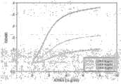
图1.噬菌体抗体与CDK4抗原结合能力测定。Figure 1. Determination of the binding ability of phage antibody to CDK4 antigen.
图2.ELISA法检测可溶性单链抗体与CDK4蛋白的结合活性。Figure 2. Detection of binding activity of soluble scFv to CDK4 protein by ELISA method.
图3.SDS-PAGE分析ANK4的可溶性表达及纯化。1.蛋白分子量标准;2为ANK4/HB2151载体对照菌;3和4分别为ANK4/HB2151IPTG诱导前、后菌体沉淀;5.ANK4/HB2151IPTG诱导上清;6.样本5经镍柱流出液;7.40mM咪唑洗涤流出液;8.500mM咪唑洗脱液。Figure 3. SDS-PAGE analysis of soluble expression and purification of ANK4. 1. Protein molecular weight standard; 2 is the ANK4/HB2151 carrier control bacteria; 3 and 4 are the bacterial precipitation before and after ANK4/HB2151IPTG induction; 5. ANK4/HB2151IPTG induced supernatant; 6. The effluent of
图4.Western blot鉴定ANK4。1.ANK4/HB2151未经IPTG诱导上清;2.ANK4/HB2151IPTG诱导上清;3.纯化的ANK4蛋白。Figure 4. Western blot identification of ANK4. 1. ANK4/HB2151 supernatant not induced by IPTG; 2. ANK4/HB2151 IPTG-induced supernatant; 3. Purified ANK4 protein.
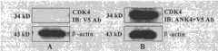
图5.Westernblot检测纯化的ANK4与重组人CDK4结合活性。1.pET28a-CDK4/BL21IPTG诱导前菌体沉淀;2.纯化的重组人CDK4蛋白。V5Ab:抗V5抗体。Figure 5. Western blot detection of the binding activity between purified ANK4 and recombinant human CDK4. 1. Precipitation of pET28a-CDK4/BL21IPTG induced bacteria; 2. Purified recombinant human CDK4 protein. V5Ab: Anti-V5 antibody.
图6.ANK4的亲和力测定。Figure 6. Affinity determination of ANK4.
图7.抗CDK4多克隆抗体对ANK4的竞争性抑制。A.只加入抗CDK4多克隆抗体;B.同时加入抗CDK4多克隆抗体和ANK4;C.只加入ANK4。*p<0.01 vs A or C。Figure 7. Competitive inhibition of ANK4 by anti-CDK4 polyclonal antibodies. A. Add only anti-CDK4 polyclonal antibody; B. Add anti-CDK4 polyclonal antibody and ANK4 at the same time; C. Add only ANK4. *p<0.01 vs A or C.
图8.Western blot检测纯化的ANK4与MCF-7细胞内源性表达的CDK4结合活性。A.阴性对照组,NC膜不与ANK4蛋白孵育,直接与抗V5抗体作用。B.实验组,NC膜首先与ANK4蛋白孵育,然后与抗V5抗体反应。以β-actin作为细胞裂解液Western blot蛋白加样的内参对照。V5Ab:抗V5抗体。Figure 8. Western blot detection of the binding activity of purified ANK4 to CDK4 endogenously expressed in MCF-7 cells. A. Negative control group, NC membrane was not incubated with ANK4 protein, and directly reacted with anti-V5 antibody. B. Experimental group, NC membrane was first incubated with ANK4 protein, and then reacted with anti-V5 antibody. β-actin was used as an internal control for Western blot protein loading of cell lysates. V5Ab: Anti-V5 antibody.
图9.免疫共沉淀分析ANK4与细胞内源性表达的CDK4结合活性。V5Ab:抗V5抗体;CDK4Ab:抗CDK4抗体。Figure 9. Co-immunoprecipitation analysis of the binding activity of ANK4 to CDK4 endogenously expressed in cells. V5Ab: anti-V5 antibody; CDK4Ab: anti-CDK4 antibody.
图10.NANK4的PCR扩增及重组表达载体pNANK4构建。其中A.PCR扩增结果。1.DL-2000Marker;2.PCR产物;3.纯化的PCR产物;B.重组表达载体pNANK4的鉴定。1.1kb DNA ladder marker;2.pcDNA3.1;3.p NANK4;4.pcDNA3.1/Hind III+EcoRI;5.pNANK4/Hind III+EcoR I;6.NANK4/Hind III+EcoR I;7.DL-2000Marker。Figure 10. PCR amplification of NANK4 and construction of recombinant expression vector pNANK4. Where A. PCR amplification results. 1. DL-2000Marker; 2. PCR product; 3. Purified PCR product; B. Identification of recombinant expression vector pNANK4. 1.1kb DNA ladder marker; 2.pcDNA3.1; 3.pNANK4; 4.pcDNA3.1/Hind III+EcoRI; 5.pNANK4/Hind III+EcoR I; 6.NANK4/Hind III+EcoR I; 7. DL-2000 Marker.
图11.RT-PCR分析NANK4在稳定转染细胞中的表达。Figure 11. RT-PCR analysis of NANK4 expression in stably transfected cells.
图12.免疫荧光检测NANK4在稳定转染细胞中的表达及定位。A.MCF-7细胞;B.HeLa细胞。Hoechst 33342与细胞核区域结合,发出蓝色荧光。抗E-Tag抗体和FITC标记的二抗检测细胞内表达的NANK4,发出绿色荧光,Merge是将同一区域的蓝色荧光和绿色荧光叠加。Figure 12. Immunofluorescence detection of expression and localization of NANK4 in stably transfected cells. A. MCF-7 cells; B. HeLa cells.
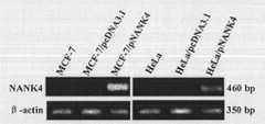
图13.Western-blot分析NANK4在稳定转染细胞中的表达。1.MCF-7;2.MCF-7/pcDNA3.1;3.MCF-7/pNANK4;4.HeLa;5.HeLa/pcDNA3.1;6.HeLa/pNANK4。Figure 13. Western-blot analysis of the expression of NANK4 in stably transfected cells. 1. MCF-7; 2. MCF-7/pcDNA3.1; 3. MCF-7/pNANK4; 4. HeLa; 5. HeLa/pcDNA3.1; 6. HeLa/pNANK4.
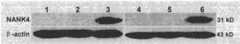
图14.免疫共沉淀实验检测NANK4与CDK4在细胞内的结合。1.MCF-7;2.MCF-7/pcDNA3.1;3.MCF-7/pNANK4;4.HeLa;5.HeLa/pcDNA3.1;6.HeLa/pNANK4。Figure 14. Co-immunoprecipitation assay to detect the binding of NANK4 and CDK4 in cells. 1. MCF-7; 2. MCF-7/pcDNA3.1; 3. MCF-7/pNANK4; 4. HeLa; 5. HeLa/pcDNA3.1; 6. HeLa/pNANK4.
图15A.MTT法测定NANK4对MCF-7细胞的体外增殖活性影响。Figure 15A. MTT method to determine the effect of NANK4 on the proliferation activity of MCF-7 cells in vitro.
图15B.MTT法测定NANK4对HeLa细胞的体外增殖活性影响。Figure 15B. MTT method to determine the effect of NANK4 on the proliferation activity of HeLa cells in vitro.
图16.流式细胞仪检测NANK4对细胞周期影响。1.MCF-7;2.MCF-7/pcDNA3.1;3.MCF-7/pNANK4;4.HeLa;5.HeLa/pcDNA3.1;6.HeLa/pNANK4。*p<0.01 vs MCF-7orMCF-7/pcDNA3.1at the same phase,n=3;**p<0.01 vs HeLa or HeLa/pcDNA3.1 atthe same phase,n=3。Figure 16. The effect of NANK4 on the cell cycle detected by flow cytometry. 1. MCF-7; 2. MCF-7/pcDNA3.1; 3. MCF-7/pNANK4; 4. HeLa; 5. HeLa/pcDNA3.1; 6. HeLa/pNANK4. *p<0.01 vs MCF-7 or MCF-7/pcDNA3.1 at the same phase, n=3; **p<0.01 vs HeLa or HeLa/pcDNA3.1 at the same phase, n=3.
图17.Annexin-V检测NANK4对细胞凋亡的影响。其中A为各组代表性图片;B为统计学分析结果。1.MCF-7;2.MCF-7/pcDNA3.1;3.MCF-7/pNANK4;4.HeLa;5.HeLa/pcDNA3.1;6.HeLa/pNANK4。*p<0.01 vs MCF-7 or MCF-7/pcDNA3.1,n=3;**p<0.01 vs HeLa or HeLa/pcDNA3.1,n=3。Figure 17. Annexin-V detects the effect of NANK4 on cell apoptosis. Among them, A is the representative picture of each group; B is the result of statistical analysis. 1. MCF-7; 2. MCF-7/pcDNA3.1; 3. MCF-7/pNANK4; 4. HeLa; 5. HeLa/pcDNA3.1; 6. HeLa/pNANK4. *p<0.01 vs MCF-7 or MCF-7/pcDNA3.1, n=3; **p<0.01 vs HeLa or HeLa/pcDNA3.1, n=3.
具体实施方式Detailed ways
实施例1特异性抗人CDK4人源单链抗体的制备,为了便于阅读,本专利“特异性抗人CDK4人源单链抗体”以下简称“ANK4”Example 1 Preparation of specific anti-human CDK4 human single-chain antibody, for ease of reading, the patent "specific anti-human CDK4 human single-chain antibody" hereinafter referred to as "ANK4"
一、实验材料1. Experimental materials
1.质粒和菌株1. Plasmids and strains
大肠杆菌(Escherichia coli)DH5α、HB2151和XL1-Blue购自北京鼎国生物技术有限责任公司。pUC119质粒购自大连宝生物工程有限公司。辅助噬菌体VCSM13购自Stratagene公司。Escherichia coli DH5α, HB2151 and XL1-Blue were purchased from Beijing Dingguo Biotechnology Co., Ltd. The pUC119 plasmid was purchased from Dalian Bao Biological Engineering Co., Ltd. Helper phage VCSM13 was purchased from Stratagene.
2.分子克隆主要相关试剂2. Main related reagents for molecular cloning
细菌培养用胰化蛋白胨(tryptone),酵母提取物(yeast extract),购自Oxid公司。原核表达并用Ni-NTA Agarose纯化的重组人CDK4蛋白参照曹玉华等2008年在吉林大学学报(理学版),2008,46(5):992-996中的方法制备;HRP/Anti-M13单克隆抗体购自Pharmacia公司,抗V5单克隆抗体为Invitrogen公司产品;anti-CDK4antibody购自Santa Cruz Biotechnology公司。铁蛋白(Fer)和卵清蛋白(OA)、OPD、考马斯亮蓝R-250、G-250、PMSF、Tris、BSA、抗生素(Amp、Tet)购自Sigma公司。His TrapHP Kit、PD-10脱盐柱购自Amersham Bioscience。脱脂奶粉购自Bio-Rad公司。硝酸纤维素(NC)膜购自Amersham Bioscience。ECL发光试剂盒购自Transgen生物技术公司。蛋白Marker、HRP标记羊抗小鼠IgG、Eppendorf管、移液器枪尖等塑料耗材购自北京鼎国生物技术有限责任公司。其他试剂均为分析纯的国产生化试剂。质粒提取、纯化试剂盒,购自Promega公司。Tryptone and yeast extract for bacterial culture were purchased from Oxid Company. The recombinant human CDK4 protein expressed in prokaryotic and purified by Ni-NTA Agarose was prepared according to the method of Cao Yuhua et al. in Jilin University Journal (Natural Science Edition), 2008, 46(5): 992-996 in 2008; HRP/Anti-M13 monoclonal antibody Purchased from Pharmacia Company, anti-V5 monoclonal antibody was purchased from Invitrogen Company; anti-CDK4antibody was purchased from Santa Cruz Biotechnology Company. Ferritin (Fer) and ovalbumin (OA), OPD, Coomassie brilliant blue R-250, G-250, PMSF, Tris, BSA, antibiotics (Amp, Tet) were purchased from Sigma. His TrapHP Kit and PD-10 desalting column were purchased from Amersham Bioscience. Skimmed milk powder was purchased from Bio-Rad. Nitrocellulose (NC) membranes were purchased from Amersham Bioscience. ECL luminescence kit was purchased from Transgen Biotechnology Company. Protein Marker, HRP-labeled goat anti-mouse IgG, Eppendorf tube, pipette tip and other plastic consumables were purchased from Beijing Dingguo Biotechnology Co., Ltd. Other reagents are analytically pure domestic chemical reagents. Plasmid extraction and purification kits were purchased from Promega.
3.相关溶液的配制参照《分子克隆实验指南》。3. For the preparation of related solutions, refer to the "Molecular Cloning Experiment Guide".
二、实验方法2. Experimental method
1.抗CDK4人源单链抗体基因ANK4的筛选1. Screening of anti-CDK4 human single chain antibody gene ANK4
噬菌体人源单链抗体库的获得具体参照文献Sblattero D等Nat Biotechnol,2000,18:74-80及Sblattero D等1998年在Immunotechnology,1998,3:271-278的方法进行,通过滴度测定表明,最终获得库容为1×10¹¹的噬菌体人源单链抗体库。The acquisition of the phage human single-chain antibody library refers to the method of Sblattero D et al. Nat Biotechnol, 2000, 18: 74-80 and Sblattero D et al. 1998 in Immunotechnology, 1998, 3: 271-278. , and finally a phage human single-chain antibody library with a library capacity of 1×10¹¹ was obtained.
抗体库的筛选采用固相化抗原免疫吸附筛选法,将人CDK4蛋白用0.05mol/L碳酸盐缓冲液稀释至100μg/ml,以1ml包被免疫管(NUNC公司),参照李春英等在中国皮肤性病学杂志,2005,19:388-390中所述方法对抗体库进行4轮“吸附-洗脱-扩增”筛选。每一轮筛选均测定次级库滴度,并计算噬菌体投入/产出比(回收率),作为特异性噬菌体抗体富集的指标。The screening of the antibody library adopts the solid-phase antigen immunoadsorption screening method. The human CDK4 protein is diluted to 100 μg/ml with 0.05 mol/L carbonate buffer solution, and the immune tube (NUNC company) is coated with 1 ml. Refer to Li Chunying et al. in China The method described in Journal of Dermatology and Venereology, 2005, 19: 388-390 carried out 4 rounds of "adsorption-elution-amplification" screening of the antibody library. The titer of the secondary library was measured in each round of screening, and the phage input/output ratio (recovery rate) was calculated as an indicator of specific phage antibody enrichment.
从第4轮筛选的菌落培养盘中随机挑取100个克隆,接种在2×YT中培养,用辅助病毒VCSM13超感染诱导培养过夜后,收集上清为噬菌体抗体。以10μg/ml的人CDK4为抗原包被ELISA板,经1%BSA封闭后加入待测噬菌体抗体上清,以HRP/Anti-M13单克隆抗体为二抗,孵育、洗涤后OPD底物显色;显色阳性的克隆再以人铁蛋白(Fer)和卵清蛋白(OA)与目的抗原CDK4同时包被ELISA板,鉴定其抗原抗体反应的特异性,所用二抗为HRP/Anti-M13单克隆抗体,加入OPD显色后,读取A490值。100 clones were randomly selected from the colony culture plate of the fourth round of screening, inoculated in 2×YT for culture, and superinfected with the helper virus VCSM13 to induce culture overnight, and the supernatant was collected as phage antibody. Coat the ELISA plate with 10 μg/ml human CDK4 as the antigen, block with 1% BSA, add the supernatant of the phage antibody to be tested, use the HRP/Anti-M13 monoclonal antibody as the secondary antibody, incubate and wash the OPD substrate for color development ; The color-positive clones were then coated with human ferritin (Fer) and ovalbumin (OA) and the target antigen CDK4 at the same time on the ELISA plate to identify the specificity of the antigen-antibody reaction. The secondary antibody used was HRP/Anti-M13 monoclonal After cloning the antibody, add OPD for color development, and read the A490 value.
将A490值阳性的特异性结合目的抗原的噬菌体抗体上清感染对数生长期的HB2151菌株,37℃孵育30min后涂布2×YT平板(50μg/ml Amp)。次日挑取单个菌落于2×YT培养液中培养过夜后以1∶100稀释转接,培养到对数生长期,加入IPTG至终浓度为1mmol/L,30℃诱导培养过夜,离心收集上清即为可溶性单链抗体。以2μg/ml人CDK4包被ELISA板,封闭后加入待检测表达上清,37℃孵育1h,洗涤后加入适当稀释的Anti-V5单克隆抗体,37℃孵育1小时,洗涤后加入适当稀释的HRP-羊抗小鼠IgG,加入OPD显色后,读取A490值。选择结合活性最高的克隆的噬菌抗体上清感染对数生长期的XL1-Blue菌株,37℃孵育30min后涂布2×YT平板(20μg/ml Amp,10μg/ml Tet)。次日挑取单个菌落于2×YT培养液(20μg/ml Amp,10μg/ml Tet)中培养过夜后,保存菌种并按Wizard Plus SV Minipreps DNA Purification System试剂盒操作说明提取质粒,由上海生工生物工程有限公司进行测序。Infect the HB2151 strain in the logarithmic growth phase with the phage antibody supernatant with positive A490 value and specifically binding to the target antigen, incubate at 37°C for 30 min, and spread 2×YT plates (50 μg/ml Amp). The next day, pick a single colony and culture it overnight in 2×YT culture medium, then transfer it at 1:100 dilution, culture to logarithmic growth phase, add IPTG to a final concentration of 1 mmol/L, induce culture at 30°C overnight, and collect by centrifugation Serum is a soluble single-chain antibody. Coat the ELISA plate with 2 μg/ml human CDK4, add the expression supernatant to be detected after blocking, incubate at 37°C for 1 hour, add appropriate dilution of Anti-V5 monoclonal antibody after washing, incubate at 37°C for 1 hour, add appropriate dilution of Anti-V5 monoclonal antibody after washing HRP-goat anti-mouse IgG, after adding OPD for color development, read the A490 value. The phage antibody supernatant of the clone with the highest binding activity was selected to infect the XL1-Blue strain in the logarithmic growth phase, incubated at 37°C for 30 min, and then spread on 2×YT plates (20 μg/ml Amp, 10 μg/ml Tet). The next day, a single colony was picked and cultured overnight in 2×YT culture medium (20 μg/ml Amp, 10 μg/ml Tet), and the strain was preserved and the plasmid was extracted according to the instructions of the Wizard Plus SV Minipreps DNA Purification System kit, provided by Shanghai Sheng Engineering Bioengineering Co., Ltd. for sequencing.
2.抗CDK4单链抗体ANK4的可溶性表达与纯化2. Soluble expression and purification of anti-CDK4 single chain antibody ANK4
将ANK4克隆接种于400ml含Amp+抗性的2×YT培养基中,1mM的IPTG诱导过夜后离心收集上清。上清中加入40%-60%的硫酸铵进行低温沉淀,12000rpm离心20min收集沉淀的蛋白,沉淀的蛋白经pH7.4的0.01M PBS溶解,透析、除盐;由于表达的抗CDK4单链抗体的C末端含有His-Tag,所以沉淀的蛋白经除盐后可以用His Trap HPKit进行纯化。纯化时,His Trap HP Kit柱先用含10mmol/L咪唑的Binding Buffer平衡,将经过过滤的蛋白溶液上柱,然后依次用Binding Buffer和含有咪唑的洗涤Buffer分别上柱清涤杂蛋白,以500mmol/L咪唑作为洗脱液,收集各个流出组份并进行SDS-PAGE电泳,确定最佳洗涤浓度。纯化的蛋白用Bradford比色法测定蛋白含量。The ANK4 clone was inoculated in 400 ml of 2×YT medium containing Amp+ resistance, induced with 1 mM IPTG overnight, and the supernatant was collected by centrifugation. Add 40%-60% ammonium sulfate to the supernatant for cryoprecipitation, centrifuge at 12000rpm for 20min to collect the precipitated protein, dissolve the precipitated protein in 0.01M PBS with pH 7.4, dialyze and desalt; due to the expressed anti-CDK4 single chain antibody The C-terminus contains His-Tag, so the precipitated protein can be purified with His Trap HPKit after desalting. When purifying, the His Trap HP Kit column is first equilibrated with Binding Buffer containing 10mmol/L imidazole, then put the filtered protein solution on the column, and then use Binding Buffer and washing buffer containing imidazole to wash the impurity protein separately, with 500mmol /L imidazole was used as the eluent, and each effluent fraction was collected and subjected to SDS-PAGE electrophoresis to determine the optimal washing concentration. The protein content of the purified protein was determined by Bradford colorimetric method.
3.Western blot分析鉴定ANK4抗体3. Western blot analysis to identify ANK4 antibody
将纯化的蛋白经12%的SDS-PAGE电泳后;半干式转移槽100mA转移2h,将蛋白转移到硝酸纤维素(NC)膜上;取出NC膜,PBST洗涤之后放入5%脱脂奶粉-PBST中室温封闭1h;将NC膜取出放入抗V5单克隆抗体(1∶5000稀释于5%脱脂奶粉-PBST中)中室温孵育2h;取出,PBST振荡洗涤3次,每次10min;再将NC膜放入HRP标记的羊抗小鼠IgG中(1∶5000稀释于5%脱脂奶粉-PBST中)室温孵育1h;PBST中洗涤3次,每次10min;ECL发光试剂盒检测目的条带的出现。After the purified protein was subjected to 12% SDS-PAGE electrophoresis; the semi-dry transfer tank was transferred at 100mA for 2 hours, and the protein was transferred to the nitrocellulose (NC) membrane; the NC membrane was taken out, washed with PBST and then put into 5% skimmed milk powder- Block in PBST for 1 h at room temperature; take out the NC membrane and put it in anti-V5 monoclonal antibody (1:5000 diluted in 5% skimmed milk powder-PBST) and incubate at room temperature for 2 h; take it out, shake and wash with PBST for 3 times, each time for 10 min; The NC membrane was placed in HRP-labeled goat anti-mouse IgG (diluted 1:5000 in 5% skimmed milk powder-PBST) and incubated at room temperature for 1 h; washed 3 times in PBST, 10 min each time; ECL luminescence kit was used to detect the target band Appear.
三、结果与分析3. Results and Analysis
1.抗CDK4人源噬菌体抗体的筛选1. Screening of anti-CDK4 human phage antibody
经过4轮筛选,噬菌体抗体回收率得到约70倍富集,从最后1轮挑取的100个克隆制备噬菌体抗体,用ELISA方法进行初筛,结果发现有15个可与人CDK4抗原结合的克隆,再用铁蛋白(Fer)和卵清蛋白(OA)作为对照抗原进行噬菌体ELISA鉴定,仅9个克隆的噬菌体抗体能与人CDK4特异性结合,与无关抗原不具备结合特性(附图1)。After 4 rounds of screening, the recovery rate of phage antibody was enriched by about 70 times. The phage antibody was prepared from 100 clones picked in the last round, and was screened by ELISA method. As a result, 15 clones that could bind to human CDK4 antigen were found , and then use ferritin (Fer) and ovalbumin (OA) as control antigens for phage ELISA identification, only 9 cloned phage antibodies can specifically bind to human CDK4, and have no binding properties to irrelevant antigens (Figure 1) .
2.可溶性单链抗体的表达及结合活性2. Expression and binding activity of soluble scFv
为了检测获得的噬菌体抗体与人CDK4的结合活性,将9个阳性克隆的噬菌体抗体上清感染无琥珀抑制的菌株HB2151,IPTG诱导可溶性单链抗体表达,以人CDK4包被ELISA板,以可溶性表达上清、Anti-V5单克隆抗体和HRP-羊抗小鼠IgG为抗体,进行ELISA检测,结果显示只有4个克隆具有特异性结合活性(附图2)。经测序和后续分析结果显示,结合活性最高的7号克隆的基因全长744bp(序列见Sequence No.1),编码248个氨基酸。该抗体的轻链为λ链,能够与CDK4特异性结合,因此我们将其确定为抗CDK4人源单链抗体,命名为ANK4,并进行了一系列研究。In order to detect the binding activity of the obtained phage antibody to human CDK4, the phage antibody supernatants of 9 positive clones were infected with the strain HB2151 without amber inhibition, and the expression of soluble single-chain antibody was induced by IPTG, and the ELISA plate was coated with human CDK4 to express in soluble The supernatant, Anti-V5 monoclonal antibody and HRP-goat anti-mouse IgG were antibodies, and ELISA detection was performed, and the results showed that only 4 clones had specific binding activity (accompanying drawing 2). The results of sequencing and subsequent analysis showed that the gene of clone No. 7 with the highest binding activity was 744 bp in full length (see Sequence No.1 for the sequence), encoding 248 amino acids. The light chain of this antibody is a λ chain, which can specifically bind to CDK4, so we identified it as an anti-CDK4 human single-chain antibody, named it ANK4, and conducted a series of studies.
3.抗CDK4人源单链抗体ANK4的可溶性表达、纯化及鉴定3. Soluble expression, purification and identification of anti-CDK4 human single chain antibody ANK4
将ANK4/HB2151的IPTG诱导上清进行硫酸铵沉淀,沉淀经过溶解和透析后进行亲和纯化。由于细菌蛋白可能含多聚组氨酸,且存在非特异性结合,为了得到纯度较高的蛋白,曾进行过多次纯化条件摸索。当以含10mM咪唑的结合缓冲液、含40mM咪唑的洗涤缓冲液、含500mM咪唑的洗脱缓冲液纯化效果较好。结果如附图3所示,经SDS-PAGE分析,500mM咪唑洗脱下来的溶液在约30kD处出现一条明显的蛋白条带。经过BandScan扫描分析,蛋白纯度约为96%。将纯化的抗CDK4单链抗体经过SDS-PAGE电泳后,以抗V5Tag单克隆抗体为一抗对其进行Western blot分析。结果如附图4所示,IPTG诱导的ANK4/HB2151上清和纯化后的抗CDK4单链抗体样品泳道在30kDa处有特异性目的条带出现,而在未经诱导的ANK4/HB2151上清中未检测到目的条带。这些结果表明已成功实现了抗CDK4单链抗体的可溶性表达和纯化。纯化得到的ANK4蛋白保存于-80℃,用于活性表征。The IPTG-induced supernatant of ANK4/HB2151 was subjected to ammonium sulfate precipitation, and the precipitate was subjected to affinity purification after dissolution and dialysis. Since the bacterial protein may contain polyhistidine, and there is non-specific binding, in order to obtain a protein with higher purity, many purification conditions have been explored. The purification effect is better when the binding buffer containing 10mM imidazole, the washing buffer containing 40mM imidazole, and the elution buffer containing 500mM imidazole are better. The results are shown in Figure 3, by SDS-PAGE analysis, the solution eluted with 500mM imidazole has an obvious protein band at about 30kD. After BandScan scanning analysis, the protein purity is about 96%. After the purified anti-CDK4 single-chain antibody was subjected to SDS-PAGE electrophoresis, it was analyzed by Western blot using anti-V5Tag monoclonal antibody as the primary antibody. The results are shown in Figure 4, the IPTG-induced ANK4/HB2151 supernatant and the purified anti-CDK4 single-chain antibody sample lanes had specific target bands at 30kDa, but not in the uninduced ANK4/HB2151 supernatant. Target band detected. These results indicate that soluble expression and purification of anti-CDK4 scFv have been successfully achieved. The purified ANK4 protein was stored at -80°C for activity characterization.
实施例2抗CDK4人源单链抗体ANK4的活性表征Example 2 Characterization of the activity of the anti-CDK4 human single-chain antibody ANK4
一、实验材料1. Experimental materials
pET28a-CDK4/BL21(DE3)菌株参照曹玉华等2008年在吉林大学学报(理学版),2008,46(5):992-996中的方法制备。人乳腺癌MCF-7细胞株为吉林大学免疫教研室提供。细胞培养用DMEM、小牛血清购自Gibco公司。DTT、胰蛋白酶、Protein G on
二、实验方法2. Experimental method
1.Western blot分析纯化的抗CDK4单链抗体ANK4与重组人CDK4的结合活性1. Western blot analysis of the binding activity of purified anti-CDK4 single chain antibody ANK4 to recombinant human CDK4
重组人CDK4蛋白经12%的SDS-PAGE电泳后;半干式转移槽100mA转移2h,将蛋白转移到硝酸纤维素(NC)膜上;取出NC膜,PBST洗涤之后放入5%脱脂奶粉-PBST中室温封闭1h;将NC膜取出放入ANK4(20μg/ml稀释于5%脱脂奶粉-PBST中)中室温孵育2h;再以抗V5抗体(1∶5000稀释于5%脱脂奶粉-PBST中)为一抗,HRP标记的羊抗小鼠IgG为二抗,ECL发光试剂盒检测目的条带的出现。After the recombinant human CDK4 protein was subjected to 12% SDS-PAGE electrophoresis; the semi-dry transfer tank was transferred at 100mA for 2 hours, and the protein was transferred to the nitrocellulose (NC) membrane; the NC membrane was taken out, washed with PBST and put into 5% skimmed milk powder- Block in PBST for 1 h at room temperature; take out the NC membrane and place it in ANK4 (20 μg/ml diluted in 5% skim milk powder-PBST) and incubate at room temperature for 2 h; ) was the primary antibody, HRP-labeled goat anti-mouse IgG was the secondary antibody, and the ECL luminescent kit was used to detect the appearance of the target band.
2.抗CDK4单链抗体ANK4亲和力的测定2. Determination of ANK4 Affinity of Anti-CDK4 Single Chain Antibody
首先用经过2n梯度稀释的CDK4蛋白100μl于4℃包被聚苯乙烯板过夜;次日弃上清,用0.05%BSA-PBST 200μl封闭1h;再加入经过2n梯度稀释的抗CDK4单链抗体100μl室温孵育2h;弃上清,加入抗V5鼠单克隆抗体(以0.05%BSA-PBST按1∶5000稀释)孵育2h;弃上清,再加入50μl HRP-羊抗小鼠IgG(以0.05%BSA-PBST按1∶5000稀释);以OPD为底物避光显色,读取OD490的值。然后按照公式(n[Ab1]-[Ab])/(n-1)计算抗CDK4单链抗体的亲和常数,n为抗CDK4单链抗体的稀释倍数,Ab和Ab1分别表示当抗原浓度为Ag和Ag1时,1/2MaxOD490对应的抗CDK4单链抗体浓度。First, use 100 μl of CDK4 protein that has been diluted in a 2n gradient to coat a polystyrene plate overnight at 4°C; discard the supernatant the next day, and block with 200 μl of 0.05% BSA-PBST for 1 hour; then add 100 μl of anti-CDK4 single-chain antibody that has been diluted in a 2n gradient Incubate at room temperature for 2 h; discard the supernatant, add anti-V5 mouse monoclonal antibody (diluted at 1:5000 with 0.05% BSA-PBST) and incubate for 2 h; discard the supernatant, then add 50 μl of HRP-goat anti-mouse IgG (dilute with 0.05% BSA - PBST was diluted 1:5000); OPD was used as the substrate to develop color in the dark, and the value of OD490 was read. Then calculate the affinity constant of the anti-CDK4 single-chain antibody according to the formula (n[Ab1]-[Ab])/(n-1), n is the dilution factor of the anti-CDK4 single-chain antibody, and Ab and Ab1 represent respectively when the antigen concentration is For Ag and Ag1, the concentration of anti-CDK4 single chain antibody corresponding to 1/2MaxOD490.
3.ELISA法检测抗CDK4多克隆抗体与抗CDK4单链抗体ANK4的竞争性结合3. ELISA method to detect the competitive binding of anti-CDK4 polyclonal antibody and anti-CDK4 single-chain antibody ANK4
以10μg/ml的重组人CDK4蛋白包被ELISA板并4℃过夜;次日以0.05%BSA-PBST封闭2h;然后每孔加入50μl纯化的抗CDK4单链抗体ANK4(10μg/ml),加入等体积的兔抗CDK4多克隆抗体作为竞争性抑制剂(以0.05%BSA-PBST按1∶2000稀释)共同孵育(所得A490的值即抑制后A值),以不加竞争性抑制剂作为阳性对照(所得A490的值即抑制前A值),以不加抗CDK4单链抗体而只加兔抗CDK4多克隆抗体为阴性对照,孵育2h后加入50μl抗V5鼠单克隆抗体(以0.05%BSA-PBST按1∶5000稀释)孵育2h,再加入50μl HRP-羊抗小鼠IgG(以0.05%BSA-PBST按1∶5000稀释),以OPD为底物避光显色,读取A490的值。用以下公式计算抑制率:抑制率=100%×(抑制前A值-抑制后A值)/抑制前A值Coat the ELISA plate with 10 μg/ml recombinant human CDK4 protein and overnight at 4°C; block with 0.05% BSA-PBST for 2 hours the next day; then add 50 μl of purified anti-CDK4 single-chain antibody ANK4 (10 μg/ml) to each well, add etc. The volume of rabbit anti-CDK4 polyclonal antibody was used as a competitive inhibitor (diluted with 0.05% BSA-PBST at 1:2000) to incubate together (the obtained A490 value is the A value after inhibition), and no competitive inhibitor was used as a positive control (The obtained value of A490 is the A value before inhibition), with no anti-CDK4 single-chain antibody but only rabbit anti-CDK4 polyclonal antibody as a negative control, 50 μl anti-V5 mouse monoclonal antibody (with 0.05% BSA- PBST was diluted 1:5000) and incubated for 2 hours, then 50 μl of HRP-goat anti-mouse IgG (diluted with 0.05% BSA-PBST was diluted 1:5000) was added, and OPD was used as the substrate to develop color in the dark, and the value of A490 was read . Calculate the inhibition rate with the following formula: inhibition rate=100%×(A value before inhibition-A value after inhibition)/A value before inhibition
4.MCF-7细胞的培养4. Culture of MCF-7 cells
MCF-7细胞培养于含10%小牛血清的DMEM完全培养基中,37℃饱和湿度、5%CO2孵箱中培养。细胞贴壁生长,每3-5天胰蛋白酶消化细胞,传代培养。MCF-7 cells were cultured in complete DMEM medium containing 10% calf serum in a 37°C saturated humidity and 5% CO2 incubator. Cells adhered to the wall, digested with trypsin every 3-5 days, and subcultured.
5.Western blot实验5.Western blot experiment
将收集的MCF-7细胞(每管约2×10⁶个)加入20μl SDS-PAGE上样缓冲液,混匀后冰浴30min,沸水煮样3-5min后进行12%SDS-PAGE电泳;半干式转移槽100mA转移2h,将蛋白转移到硝酸纤维素(NC)膜上;取出NC膜,PBST洗涤之后放入5%脱脂奶粉-PBST中室温封闭1h;将NC膜取出放入抗CDK4单链抗体ANK4(20μg/ml稀释于5%脱脂奶粉-PBST中)中室温孵育2h;再以抗V5抗体(1∶5000稀释于5%脱脂奶粉-PBST中)为一抗,HRP标记的羊抗小鼠IgG为二抗,ECL发光试剂盒检测目的条带的出现。Add 20 μl SDS-PAGE loading buffer to the collected MCF-7 cells (approximately 2×10⁶ cells in each tube), mix well, and ice-bath for 30 minutes, boil the sample in boiling water for 3-5 minutes, and conduct 12% SDS-PAGE electrophoresis;
6.免疫沉淀实验6. Immunoprecipitation experiments
将培养在6孔板中对数生长的MCF-7细胞用含1mM PMSF的冷PBS洗涤两次,每孔加入500μl细胞裂解液,用细胞刮子将其刮出,放于1.5ml Eppendorf管中,冰浴40min,20%的能量超声两次,每次10s;15000rpm 4℃离心10min;取上清,加入纯化的抗CDK4单链抗体ANK4(20μg/ml),于4℃缓慢颠倒2h;再加入2μl抗V5鼠单克隆抗体,于4℃缓慢颠倒2h;再加入20μl Protein G on
三、结果与分析3. Results and Analysis
1.纯化的抗CDK4单链抗体ANK4具有与重组人CDK4的结合活性1. Purified anti-CDK4 single chain antibody ANK4 has binding activity to recombinant human CDK4
为了进一步检测抗CDK4单链抗体与重组人CDK4蛋白结合活性,以未经IPTG诱导的pET28a-CDK4/BL21(DE3)菌体沉淀为阴性对照,将其和纯化的重组人CDK4蛋白经SDS-PAGE后,转移到NC膜上,首先与抗CDK4单链抗体结合,进而与抗V5抗体和HRP标记的羊抗小鼠IgG反应,ECL显影观察,纯化的重组人CDK4蛋白样品在约34kD处出现一明显特异性条带,而阴性对照组中未发现目的条带(附图5),这一结果进一步验证了纯化的抗CDK4单链抗体可以特异性结合人重组CDK4蛋白。In order to further detect the binding activity of anti-CDK4 single chain antibody to recombinant human CDK4 protein, pET28a-CDK4/BL21(DE3) bacterial cell precipitation without IPTG induction was used as a negative control, and it and the purified recombinant human CDK4 protein were subjected to SDS-PAGE Afterwards, it was transferred to NC membrane, first combined with anti-CDK4 single-chain antibody, and then reacted with anti-V5 antibody and HRP-labeled goat anti-mouse IgG, and observed by ECL imaging, the purified recombinant human CDK4 protein sample appeared at about 34kD. The specific band was obvious, but no target band was found in the negative control group (Fig. 5), this result further verified that the purified anti-CDK4 single chain antibody could specifically bind to human recombinant CDK4 protein.
2.抗CDK4单链抗体ANK4亲和力的测定2. Determination of ANK4 Affinity of Anti-CDK4 Single Chain Antibody
应用非竞争性ELISA法,以抗体的浓度为横坐标,以抗原抗体反应的OD490值为纵坐标作标准曲线(附图6),根据曲线及计算公式(n[Ab1]-[Ab])/(n-1)来计算抗CDK4单链抗体的亲和常数,n为抗CDK4单链抗体的稀释倍数,Ab和Ab1分别表示当抗原浓度为Ag和Ag1时,1/2MaxOD490的抗体浓度。通过计算得到亲和力的值分别为(1.79±0.42)×10-8mol/L。Apply the non-competitive ELISA method, take the concentration of the antibody as the abscissa, and use the OD490 value of the antigen-antibody reaction as the ordinate to make the standard curve (accompanying drawing 6), according to the curve and the calculation formula (n[Ab1]-[Ab])/ (n-1) to calculate the affinity constant of the anti-CDK4 single-chain antibody, n is the dilution factor of the anti-CDK4 single-chain antibody, Ab and Ab1 represent the antibody concentration of 1/2MaxOD490 when the antigen concentration is Ag and Ag1 respectively. The calculated affinity values were (1.79±0.42)×10-8 mol/L.
3.抗CDK4多克隆抗体竞争性抑制抗CDK4单链抗体ANK4与抗原结合3. Anti-CDK4 polyclonal antibody competitively inhibits the binding of anti-CDK4 single-chain antibody ANK4 to antigen
为了检测抗CDK4多克隆抗体与抗CDK4单链抗体竞争性结合CDK4的能力,进行了竞争性ELISA实验。纯化的抗CDK4单链抗体中混入兔抗CDK4多克隆抗体作为竞争性抑制剂(以0.1%BSA-PBST按1∶2000稀释)为实验组即抑制后,以只加抗CDK4单链抗体不加兔抗CDK4多克隆抗体作为阳性对照即抑制前,检测兔抗CDK4多克隆抗体与抗CDK4单链抗体竞争结合重组人CDK4的能力,结果如附图7所示,根据公式(抑制率=(抑制前A值-抑制后A值)/抑制前A值x100%)可以计算出兔抗CDK4多克隆抗体对抗CDK4单链抗体ANK4的竞争性抑制率为38.67%。实验组与阳性对照有显著性差异(p<0.01)。从竞争性抑制率可以看出,兔抗CDK4多克隆抗体能够竞争性抑制抗CDK4单链抗体ANK4结合重组人CDK4。In order to detect the ability of anti-CDK4 polyclonal antibody to compete with anti-CDK4 single chain antibody to bind to CDK4, a competitive ELISA experiment was performed. The purified anti-CDK4 single-chain antibody was mixed with rabbit anti-CDK4 polyclonal antibody as a competitive inhibitor (diluted with 0.1% BSA-PBST at 1:2000) as the experimental group. After inhibition, only anti-CDK4 single-chain antibody was added without Before the rabbit anti-CDK4 polyclonal antibody is used as a positive control, that is, before inhibition, the ability of the rabbit anti-CDK4 polyclonal antibody to compete with the anti-CDK4 single-chain antibody to bind to recombinant human CDK4 is detected. The results are shown in Figure 7. According to the formula (inhibition rate=(inhibition The competitive inhibition rate of rabbit anti-CDK4 polyclonal antibody against CDK4 single chain antibody ANK4 can be calculated as 38.67%. There was a significant difference between the experimental group and the positive control (p<0.01). It can be seen from the competitive inhibition rate that the rabbit anti-CDK4 polyclonal antibody can competitively inhibit the binding of the anti-CDK4 single chain antibody ANK4 to recombinant human CDK4.
4.抗CDK4单链抗体ANK4能与肿瘤细胞内CDK4蛋白结合4. Anti-CDK4 single chain antibody ANK4 can bind to CDK4 protein in tumor cells
由于本实验所用的抗CDK4单链抗体是以原核表达的重组人CDK4蛋白为抗原从人源噬菌体抗体库中筛选得到的,因此须对其是否具有结合天然状态下的CDK4蛋白的能力进行检测。为此,我们分别采用了Western blot和免疫沉淀技术来检测纯化的抗CDK4单链抗体与肿瘤细胞内表达的CDK4是否有相互作用。Since the anti-CDK4 single-chain antibody used in this experiment was screened from the human phage antibody library using prokaryotically expressed recombinant human CDK4 protein as an antigen, it was necessary to detect whether it has the ability to bind to the natural CDK4 protein. To this end, we used Western blot and immunoprecipitation techniques to detect whether the purified anti-CDK4 single-chain antibody interacted with CDK4 expressed in tumor cells.
4.1 Western blot4.1 Western blot
为了检测抗CDK4单链抗体与细胞内源的CDK4蛋白结合活性,将收集得到的MCF-7细胞裂解液经SDS-PAGE电泳,转移到NC膜上,首先与抗CDK4单链抗体孵育,进而与抗V5抗体和HRP标记的羊抗小鼠IgG反应,ECL发光试剂盒显影。结果附图8所示,在约34kD处出现一明显特异性条带(附图8B),而在阴性对照组(未与ANK4反应,只加入抗V5抗体)中未发现目的条带(附图8A),以β-actin作为细胞裂解液Westernblot蛋白加样的内参对照,各组样品中β-actin均检测出清晰的β-actin条带。In order to detect the binding activity of the anti-CDK4 single-chain antibody to the endogenous CDK4 protein of the cells, the collected MCF-7 cell lysate was subjected to SDS-PAGE electrophoresis, transferred to the NC membrane, firstly incubated with the anti-CDK4 single-chain antibody, and then mixed with Reaction of anti-V5 antibody and HRP-labeled goat anti-mouse IgG, developed with ECL luminescent kit. As shown in the results accompanying drawing 8, an obvious specific band appears at about 34kD place (accompanying drawing 8B), and in the negative control group (does not react with ANK4, only anti-V5 antibody is added) no band of interest is found (accompanying drawing 8A), β-actin was used as the internal control of Western blot protein loading in cell lysate, and clear β-actin bands were detected in samples of each group.
4.2免疫沉淀4.2 Immunoprecipitation
将收集得到的MCF-7细胞裂解后在上清中依次加入抗CDK4单链抗体,V5单克隆抗体和Protein G on

实施例3 抗CDK4人源胞内单链抗体基因的克隆及重组真核表达载体的构建。为了便于阅读,本专利“抗CDK4人源胞内单链抗体基因”以下简称“NANK4”Example 3 Cloning of anti-CDK4 human intracellular single chain antibody gene and construction of recombinant eukaryotic expression vector. For ease of reading, the patent "anti-CDK4 human intracellular single-chain antibody gene" is hereinafter referred to as "NANK4"
一、实验材料1. Experimental materials
1.菌株和质粒1. Strains and Plasmids
pcDNA3.1(+)质粒购自Invitrogen公司;大肠杆菌E.coli JM109购自北京鼎国生物技术有限公司。pcDNA3.1 (+) plasmid was purchased from Invitrogen; Escherichia coli E.coli JM109 was purchased from Beijing Dingguo Biotechnology Co., Ltd.
2.分子克隆主要相关试剂2. Main related reagents for molecular cloning
限制性内切酶Hind III、EcoR I,T4DNA连接酶及其相应缓冲液,Taq DNA聚合酶及其相应缓冲液,dNTP购自大连宝生物工程有限公司。质粒提取纯化试剂盒购自Promega公司。DNA回收试剂盒、核酸分子量标准(1kb Ladder和DL-2000DNAMarker)购自北京鼎国生物技术有限公司。琼脂糖凝胶电泳所需琼脂糖、溴化乙锭、Tris等购自Sigma公司。其余试剂及材料参照实施例1和2。Restriction endonuclease Hind III, EcoR I, T4 DNA ligase and its corresponding buffer, Taq DNA polymerase and its corresponding buffer, and dNTP were purchased from Dalian Bao Biological Engineering Co., Ltd. Plasmid extraction and purification kits were purchased from Promega. DNA recovery kit, nucleic acid molecular weight standard (1kb Ladder and DL-2000DNAMarker) were purchased from Beijing Dingguo Biotechnology Co., Ltd. Agarose, ethidium bromide, Tris, etc. required for agarose gel electrophoresis were purchased from Sigma. Refer to Examples 1 and 2 for the rest of the reagents and materials.
2.引物设计2. Primer Design
本实验所用的引物由上海生工生物工程技术服务有限公司合成。具体如下:以P1(FP)(5’-CCCAAGCTTATGGATCCGAAGAAGAAACGTAAGGTTCCGAAGAAGAAACGTAAGGTTCAGTCTGTGCTGACGCAGCC-3’)为正向引物,以P2(RP)(5’-GGAATTCTTAACGCGGTTCCAGCGGATCCGGATACGGCACCGGCGCACCTGAAGAGACAGTGACCGGGGTTCC-3’)为反向引物引入核定位序列、检测标签及相应酶切位点构建胞内抗体NANK4。The primers used in this experiment were synthesized by Shanghai Sangon Bioengineering Technology Service Co., Ltd. The details are as follows: P1 (FP) (5'-CCCAAGCTTATGGATCCGAAGAAGAAACGTAAGGTTCCGAAGAAGAAACGTAAGGTTCAGTCTGTGCTGACGCAGCC-3') was used as the forward primer, P2 (RP) (5'-GGAATTCTTAACGCGGTTCCAGCGGATCCGGATACGGCACCGGCGCACCTGAAGAGACAGTGACCGGG corresponding reverse primer sequence for the introduction of the nuclear localization detection primer and GTTCC-3') was used as the sequence The cleavage site was used to construct the intrabody NANK4.
二、实验方法2. Experimental method
(一)NANK4基因片段的获得(1) Obtaining NANK4 gene fragments
1.聚合酶链式反应(PCR)1. Polymerase Chain Reaction (PCR)
为了通过定向克隆的方法制备抗人CDK4胞内单链抗体基因NANK4,并能构建入真核表达载体pcDNA3.1中,我们设计了P1/P2两条引物。引物设计根据抗CDK4人源单链抗体基因序列及不同亚细胞区滞留型胞内抗体基因序列,并依据PCR引物设计原则引入符合阅读框的起始密码子和终止密码子以及相应的酶切位点。具体序列见材料部分。In order to prepare the anti-human CDK4 intracellular single-chain antibody gene NANK4 by directional cloning method and construct it into the eukaryotic expression vector pcDNA3.1, we designed two primers, P1/P2. The primers were designed according to the anti-CDK4 human single-chain antibody gene sequence and the intracellular antibody gene sequence of different subcellular regions, and the initiation codon and stop codon in line with the reading frame and the corresponding enzyme cutting site were introduced according to the principles of PCR primer design point. See the Materials section for specific sequences.
以从人源噬菌体单链抗体库中筛选到的抗CDK4人源单链抗体基因ANK4序列为模板,用PCR的方法扩增细胞核定位的抗人CDK4胞内单链抗体(NANK4)基因。Using the anti-CDK4 human single-chain antibody gene ANK4 sequence screened from the human phage single-chain antibody library as a template, the nuclear localized anti-human CDK4 intracellular single-chain antibody (NANK4) gene was amplified by PCR.
PCR反应体系:10×Ex 10μlPCR reaction system: 10×Ex 10μl
Taq buffer 8μlTaq buffer 8μl
dNTPs(2.5mM each) 2μl(respectively)dNTPs(2.5mM each) 2μl(respectively)
P1and P2(20μM) 0.5μlP1and P2(20μM) 0.5μl
Template(100ng/μl) 0.5μlTemplate (100ng/μl) 0.5μl
ExTaq(5U/μl) 77μlExTaq(5U/μl) 77μl
dd H2Odd H2 O
Total 100μlTotal 100μl
操作程序:(1)94℃,4min;(2)94℃,45sec;(3)55℃,45sec;(4)72℃,45sec;(5)72℃,10min.其中(2)-(4)进行30个循环。Operating procedures: (1) 94°C, 4min; (2) 94°C, 45sec; (3) 55°C, 45sec; (4) 72°C, 45sec; (5) 72°C, 10min. ) for 30 cycles.
2.PCR扩增产物的回收2. Recovery of PCR amplification products
PCR产物经1%琼脂糖凝胶电泳分离、按照DNA回收纯化试剂盒回收并检验。The PCR products were separated by 1% agarose gel electrophoresis, recovered and tested according to the DNA recovery and purification kit.
(1)电泳结束后,在凝胶成像仪上用手术刀切取含目的片段的琼脂糖;(2)放入预先称量好的Eppendorf管中,称重,捣碎,按1∶3(重量/mg∶体积/μl)的比例加入溶液A;(3)50℃金属浴10min,至胶完全溶解,在室温下,加入溶液B,充分混匀,溶液分别转移至离心柱内静置2min,8500rpm离心1min弃液体(溶液若一次加不完,可以分两次离心);(4)弃液体,加入500μl溶液C,8500rpm离心1min弃液体;重复一次;(5)12000rpm离心1min,甩干剩余液体,除去残余酒精;(6)将离心柱置于新的离心管中室温敞盖8min,使乙醇挥发尽;(7)加预热溶液D,15000rpm离心10min,管底即为目的DNA。取少量DNA进行1%琼脂糖凝胶电泳,检验回收片段的纯度与浓度。(1) After electrophoresis, use a scalpel on the gel imager to cut out the agarose containing the target fragment; (2) Put it into a pre-weighed Eppendorf tube, weigh it, mash it, and press 1:3 (weight Add solution A at the ratio of /mg:volume/μl); (3) 50°C metal bath for 10min, until the glue is completely dissolved, add solution B at room temperature, mix well, transfer the solution to the spin column and let stand for 2min, Centrifuge at 8500rpm for 1min and discard the liquid (if the solution cannot be added at one time, it can be centrifuged twice); (4) Discard the liquid, add 500μl solution C, centrifuge at 8500rpm for 1min and discard the liquid; repeat once; (5) Centrifuge at 12000rpm for 1min, dry the remaining (6) Put the spin column in a new centrifuge tube at room temperature and open the lid for 8 minutes to evaporate the ethanol; (7) Add preheated solution D and centrifuge at 15,000 rpm for 10 minutes, and the bottom of the tube is the target DNA. Take a small amount of DNA for 1% agarose gel electrophoresis to check the purity and concentration of the recovered fragments.
3.目的片段的酶切与回收3. Digestion and recovery of target fragments
回收的PCR扩增片段产物的HindIII和EcoR I双酶切反应体系HindIII and EcoRI double enzyme digestion reaction system of recovered PCR amplified fragment product
ddH2O 12μlddH2 O 12μl
10×M buffer 4μl10×M buffer 4μl
HindIII 2μlHindIII 2μl
EcoR I 2μlEcoR I 2μl
回收的PCR扩增片段 20μlRecovered PCR amplified fragments 20 μl
40μl40μl
上述混合液于37℃金属浴消化1.5-2h,产物进行1%琼脂糖凝胶电泳,回收约870bp的目的片段(方法同上),1%琼脂糖凝胶电泳检验其回收效果,-20℃保存备用。The above mixed solution was digested in a metal bath at 37°C for 1.5-2h, and the product was subjected to 1% agarose gel electrophoresis to recover the target fragment of about 870bp (the method is the same as above), and the recovery effect was checked by 1% agarose gel electrophoresis, and stored at -20°C spare.
(二)pcDNA3.1载体片段的获得(2) Acquisition of pcDNA3.1 vector fragment
将质粒pcDNA3.1进行Hind III和EcoR I双酶切,37℃消化1.5-2h,1%琼脂糖凝胶,98mA电泳30-40min分离约5.4kb的目的片段。Plasmid pcDNA3.1 was digested with Hind III and EcoR I, digested at 37°C for 1.5-2h, followed by 1% agarose gel, and electrophoresed at 98mA for 30-40min to separate the target fragment of about 5.4kb.
载体质粒pcDNA3.1的酶切体系如下:The enzyme digestion system of vector plasmid pcDNA3.1 is as follows:
dd H2O 12μldd H2 O 12μl
10×M buffer 4μl10×M buffer 4μl
Hind III 2μlHind III 2μl
EcoR I 2μlEcoR I 2μl
pcDNA3.1 20μlpcDNA3.1 20μl
40μl40μl
用DNA回收试剂盒回收载体片段pcDNA3.1,方法同上。1%琼脂糖凝胶电泳,检验回收片段。The vector fragment pcDNA3.1 was recovered with a DNA recovery kit, and the method was the same as above. 1% agarose gel electrophoresis to check the recovered fragments.
(三)载体与目的基因的连接:将目的基因片段和pcDNA3.1酶切大片段通过T4DNA连接酶连接,获得重组质粒pNANK4。构建过程见图1。连接体系如下:(3) Ligation of the vector and the target gene: the target gene fragment and the pcDNA3.1 digested large fragment were connected by T4 DNA ligase to obtain the recombinant plasmid pNANK4. The construction process is shown in Figure 1. The connection system is as follows:
dd H2O 8.5μldd H2 O 8.5 μl
T4ligase buffer 2μlT4ligase buffer 2μl
T4ligase 1μlT4ligase 1μl
pcDNA3.1片段 0.5μlpcDNA3.1 fragment 0.5μl
NANK4片段 8μl
20μl20μl
混匀,16℃金属浴连接12-16h。Mix well and connect to a metal bath at 16°C for 12-16h.
(四)JM109感受态细胞的制备(4) Preparation of JM109 Competent Cells
(1)以无菌接种环刮取冻存于-80℃冰箱的JM109菌种,划线接种于不含氨苄青霉素(Amp)的LB琼脂平板,37℃培养16h左右;(2)挑取单一菌落接种到5ml液体LB培养基中,37℃180rpm振摇培养至OD600=0.4;(3)将细菌培养管取出,迅速置于冰浴中15min以上,使培养物冷却到0℃;(4)在无菌条件下将细菌培养液转移到无菌、冰预冷的1.5ml Eppendorf管中(以下操作均需无菌),4℃、4000rpm离心10min,弃上清;(5)以500μl冰预冷的0.1mol/L CaCl2溶液重悬细菌沉淀,冰浴30min;(6)4℃、4000rpm离心10min,收集菌体,经80μl冰预冷的CaCl2重新悬浮菌体,将感受态细胞置于4℃冰箱冰浴中备用。(1) Use a sterile inoculation loop to scrape out JM109 strains frozen in a -80°C refrigerator, inoculate them on LB agar plates without ampicillin (Amp), and culture them at 37°C for about 16 hours; (2) pick a single Inoculate the colony into 5ml of liquid LB medium, shake and culture at 37°C at 180rpm until OD600 =0.4; (3) Take out the bacterial culture tube and put it in an ice bath for more than 15 minutes to cool the culture to 0°C; (4 ) Under aseptic conditions, transfer the bacterial culture solution to a sterile, ice-cooled 1.5ml Eppendorf tube (the following operations need to be sterile), centrifuge at 4°C and 4000rpm for 10min, and discard the supernatant; Pre-cooled 0.1mol/L CaCl2 solution to resuspend the bacterial pellet, ice bath for 30min; (6) 4°C, 4000rpm centrifuge for 10min, collect the bacterial cells, resuspend the bacterial cells with 80μl ice-cooled CaCl2 , and transfer the competent cells Store in a 4°C refrigerator in an ice bath for later use.
(五)质粒转化(5) Plasmid transformation
(1)将每管80μl感受态细胞加入到20μl连接产物中,轻轻混匀,冰浴30min;(2)将管放入42℃金属浴中,热休克2min;(3)快速转移到冰浴中,冷却2min;(4)每管加入900μl 37℃预热的LB液体培养基,37℃、150rpm振荡培养45min;(5)4000rpm、4℃离心5min;(6)弃700-800μl上清,用余下上清重悬细胞,涂布于含100μg/ml氨卞青霉素抗性(Amp)的LB琼脂培养基中,静置10min;37℃倒置培养12~16h,出现重组子菌落。转化菌盘4℃保存备用。(1) Add 80 μl of competent cells to 20 μl of the ligation product, mix gently, and ice-bath for 30 minutes; (2) Put the tube in a metal bath at 42°C and heat shock for 2 minutes; (3) Quickly transfer to ice Cool in the bath for 2 minutes; (4) Add 900 μl LB liquid medium preheated at 37°C to each tube, shake and incubate at 37°C, 150 rpm for 45 minutes; (5) Centrifuge at 4000 rpm, 4°C for 5 minutes; (6) Discard 700-800 μl supernatant , resuspend the cells with the remaining supernatant, smear them on LB agar medium containing 100 μg/ml ampicillin resistance (Amp), and let stand for 10 minutes; culture them upside down at 37°C for 12 to 16 hours, and recombinant colonies appear. The transformed bacterial plates were stored at 4°C for later use.
(六)表达质粒pNANK4的鉴定(6) Identification of expression plasmid pNANK4
1.表达质粒pNANK4的小量提取与纯化(参照《分子克隆实验指南》碱裂解法小量提取质粒)1. Mini-extraction and purification of expression plasmid pNANK4 (refer to "Molecular Cloning Experiment Guide" alkaline lysis method for mini-extraction of plasmids)
(1)挑取转化细胞单个阳性克隆,接种于5ml含100μg/ml Amp+的LB液体培养基中,37℃、180-200rpm振荡培养12~16h;(2)菌液分装于1.5ml Eppendorf管中,15000rpm、4℃离心5min收菌体沉淀;(3)加入0.1mL冰预冷的溶液I,吹悬沉淀,静置5min;(4)加入现配的0.2mL溶液II,颠倒混匀;(5)加入0.15mL冰预冷溶液III,轻轻颠倒混匀;(6)冰浴15min,15000rpm 4℃离心5min;(7)收集上清,加入2.5倍体积的无水乙醇,室温静置20min;(8)15000rpm 4℃离心15min;(9)弃上清,加入2.5倍体积预冷的70%乙醇洗涤,15000rpm 4℃离心5min;(10)弃上清,控干,真空旋转蒸发干燥;(11)沉淀溶于50μl TE中(含50μg/ml RNaseA),37℃30min,1%琼脂糖凝胶电泳检验,-20℃保存备用。(1) Pick a single positive clone of the transformed cell, inoculate it in 5ml LB liquid medium containing 100μg/ml Amp+ , shake and culture at 37°C, 180-200rpm for 12-16h; (2) Dispense the bacterial solution into 1.5ml Eppendorf In the tube, centrifuge at 15,000rpm at 4°C for 5 minutes to collect the bacterial cell pellet; (3) Add 0.1mL of ice-precooled solution I, blow and suspend the precipitate, and let stand for 5min; (4) Add 0.2mL of solution II, and mix evenly by inversion ; (5) Add 0.15mL of ice-precooled solution III, gently invert to mix; (6) Ice bath for 15min, 15000rpm 4 ℃ centrifugation for 5min; (7) Collect the supernatant, add 2.5 times the volume of absolute ethanol, room temperature static Place for 20 minutes; (8) Centrifuge at 15,000 rpm at 4°C for 15 minutes; (9) Discard the supernatant, add 2.5 times the volume of pre-cooled 70% ethanol to wash, and centrifuge at 15,000 rpm at 4°C for 5 minutes; (10) Discard the supernatant, control to dry, and vacuum rotary evaporation Drying; (11) The precipitate was dissolved in 50 μl TE (containing 50 μg/ml RNaseA), 37°C for 30 min, tested by 1% agarose gel electrophoresis, and stored at -20°C for later use.
2.重组质粒的酶切鉴定2. Enzyme digestion and identification of recombinant plasmids
利用限制性内切酶消化法初步检验提取的重组质粒。将重组质粒进行Hind III和EcoR I双酶切,37℃消化1.5-2h,1%琼脂糖凝胶,98mA电泳30-40min,凝胶成像分析仪上观测。The extracted recombinant plasmids were initially tested by restriction endonuclease digestion. The recombinant plasmid was digested with Hind III and EcoR I, digested at 37°C for 1.5-2h, followed by 1% agarose gel electrophoresis at 98mA for 30-40min, and observed on a gel imaging analyzer.
pNANK4质粒的Hind III和EcoR I双酶切反应体系:Hind III and EcoR I double enzyme digestion reaction system of pNANK4 plasmid:
dd H2O 6μldd H2 O 6μl
10×M buffer 2μl10×M buffer 2μl
Hind III 1μlHind III 1μl
EcoR I 1μlEcoR I 1μl
pNANK4 10μlpNANK4 10μl
20μl20μl
3.重组质粒的序列测定3. Sequencing of recombinant plasmids
由上海生工生物工程有限公司进行测序。Sequencing was performed by Shanghai Sangon Bioengineering Co., Ltd.
三、结果与分析3. Results and analysis
为了获得定位于细胞核的抗CDK4人源胞内单链抗体基因,我们以抗CDK4人源单链抗体ANK4基因为模板,用含有细胞核定位信号、Hind III和EcoR I酶切位点序列以及E-tag标签肽序列的引物,通过PCR的方法扩增细胞核定位型抗CDK4人源胞内单链抗体NANK4基因。PCR产物经琼脂糖凝胶电泳后发现,在约860bp处有一明显的DNA条带(见附图10A);产物回收后克隆到真核表达质粒pcDNA3.1的CMV启动子下游的限制性内切酶Hind III和EcoRI之间;阳性重组质粒经限制性内切酶Hind III和EcoRI双酶切,琼脂糖凝胶电泳发现,在5400bp和840bp处分别出现一条明显的DNA条带(见附图10B)。进一步通过T7和BGH通用引物正反双向测通载体pNANK4上的目的片段,测序结果表明,插入序列与实验设计一致,并且读码框完全正确,这说明通过PCR扩增成功地引入了细胞核定位信号、E-tag检测标签等,NANK4基因全长834bp(序列见Sequence No.2),编码278个氨基酸。这说明我们已成功获得了重组胞内抗体NANK4基因的真核表达载体pNANK4。In order to obtain the anti-CDK4 human intracellular single-chain antibody gene localized in the nucleus, we used the anti-CDK4 human single-chain antibody ANK4 gene as a template, and used sequences containing nuclear localization signals, Hind III and EcoR I restriction sites, and E- The primer of the tag tag peptide sequence is used to amplify the nucleus-localized anti-CDK4 human intracellular single-chain antibody NANK4 gene by PCR. After the PCR product was subjected to agarose gel electrophoresis, it was found that there was an obvious DNA band at about 860bp (see accompanying drawing 10A); Between the enzyme Hind III and EcoRI; the positive recombinant plasmid was double-digested by the restriction endonuclease Hind III and EcoRI, and agarose gel electrophoresis found that an obvious DNA band appeared respectively at 5400bp and 840bp (see accompanying drawing 10B ). Further, the target fragment on the vector pNANK4 was detected by T7 and BGH universal primers in forward and reverse directions. The sequencing results showed that the inserted sequence was consistent with the experimental design, and the reading frame was completely correct, which indicated that the nuclear localization signal was successfully introduced by PCR amplification. , E-tag detection tags, etc., the full length of the NANK4 gene is 834bp (see Sequence No.2 for the sequence), encoding 278 amino acids. This shows that we have successfully obtained the eukaryotic expression vector pNANK4 of the recombinant intracellular antibody NANK4 gene.
实施例4 抗CDK4单链抗体抗肿瘤活性分析Example 4 Anti-tumor activity analysis of anti-CDK4 single chain antibody
一、实验材料1. Experimental materials
1.分子生物学相关试剂及材料1. Reagents and materials related to molecular biology
去内毒素质粒提取试剂盒购自OMEGA公司;逆转录PCR(RT-PCR)试剂盒购自大连宝生物工程有限公司;其余参照实施例1-3。The endotoxin-free plasmid extraction kit was purchased from OMEGA Company; the reverse transcription PCR (RT-PCR) kit was purchased from Dalian Bao Biological Engineering Co., Ltd.; the rest refer to Examples 1-3.
2.细胞培养及转染及活性检测试剂及材料2. Reagents and materials for cell culture, transfection and activity detection
脂质体(Lipofectamine 2000)、G418、Trizol为Invitrogen公司产品;DMSO、MTT、溴化丙啶(PI)、RNase A、Hoechst 33342购自Sigma公司;RIPA裂解液购自碧云天生物技术有限公司;抗E-tag抗体购自Phamarcia公司;FITC标记山羊抗小鼠IgG购自北京中杉金桥生物技术公司;Annexin V凋亡检测试剂盒购自南京凯基生物技术有限公司;其余同实施例1-3。Lipofectamine 2000, G418, and Trizol were products of Invitrogen; DMSO, MTT, propidium bromide (PI), RNase A, and
3.RT-PCR所需引物由上海生工生物工程技术服务有限公司合成。3. The primers required for RT-PCR were synthesized by Shanghai Sangon Bioengineering Technology Service Co., Ltd.
(1)RT-PCR扩增胞内抗体引物序列:正向引物P3为5’-CCCAAGCTTATGGGATGGAGCTGT-3’,反向引物P4为5’-GGTACCGCTCGAGGATAACT-3’,扩增产物为460bp;(2)扩增细胞内β-actin合成的引物序列:正向引物P5为5’-TGGAATCCTGTGGCATCCATGAAAC-3’,反向引物P6为5’-TAAAACGCAGCTCAGTAACAGTCCG-3’。(1) RT-PCR amplification intracellular antibody primer sequence: forward primer P3 is 5'-CCCAAGCTTATGGGATGGAGCTGT-3', reverse primer P4 is 5'-GGTACCGCTCGAGGATAACT-3', and the amplified product is 460bp; (2) Primer sequences for β-actin synthesis in the cells: forward primer P5 is 5'-TGGAATCCTGTGGCATCCATGAAAC-3', and reverse primer P6 is 5'-TAAAACGCAGCTCAGTAACAGTCCG-3'.
4.相关溶液的配制参照《分子克隆实验指南》。4. For the preparation of related solutions, refer to the "Molecular Cloning Experiment Guide".
二实验方法Two experimental methods
1.转染用质粒的准备(参照去内毒素质粒提取试剂盒说明书)1. Preparation of plasmids for transfection (refer to the instructions of the endotoxin-free plasmid extraction kit)
1.1使用前准备工作1.1 Preparation before use
将试剂盒中附带的RNaseA加入Solution I中,4℃保存;向DNA Washing Buffer中加入60mL无水乙醇进行稀释,稀释后的DNA Washing Buffer室温保存。Add the RNaseA attached to the kit to Solution I and store at 4°C; add 60mL absolute ethanol to the DNA Washing Buffer for dilution, and store the diluted DNA Washing Buffer at room temperature.
1.2操作方案(所有步骤按照试剂盒说明书在室温下进行)1.2 Operation scheme (all steps are carried out at room temperature according to the kit instructions)
(1)过夜培养的细菌培养液转移到一个干净的5mL离心管中,5000g室温离心10min,弃上清、收集菌体沉淀;(2)加入500μL Solution I,将菌体沉淀悬起,并将其转移至1.5mL离心管中;(3)加入500μL SolutionII,上下颠倒四次,室温静置2min;(4)加入250μL冰浴的Buffer N3,上下颠倒四次,直到出现白色絮状沉淀,12000g,4℃,离心10min;(5)小心地将上清转移到一个1.5mL离心管中,加入0.1体积的ETR Solution,冰浴10min,其间颠倒几次;(6)将裂解液42℃孵育5min,12000g离心3min,ETR Solution会在底部形成一蓝色条带;(7)将上清转移至1.5mL离心管中,并加入0.5体积的无水乙醇,充分混匀,室温静置1~2min;(8)将以上混合物700μL转移到一个套在2mL收集管中柱II中,室温离心,10000g,1min;将流出液倒掉,按上述操作,将剩余溶液再转移到柱中并离心;(9)用500μL HB Buffer洗柱,室温离心,10000g,1min;(10)倒掉流出液,用700μL含乙醇的DNA Wash Buffer洗柱,室温离心,10000g,1min,倒掉流出液,重复此操作一次;(11)倒掉流出液,将空柱离心13000g,3min,以干燥柱的介质,特别是将乙醇除去;(12)将柱置于一个新的1.5mL离心管中,加入100μL的Endotoxin-Free Elution Buffer(70℃预热),室温静置2min,13000g,离心3min以洗脱DNA;(13)弃回收柱,洗脱DNA保存于-20℃备用。(1) Transfer the bacterial culture solution cultivated overnight to a clean 5mL centrifuge tube, centrifuge at 5000g room temperature for 10min, discard the supernatant, and collect the bacterial pellet; (2) Add 500μL Solution I, suspend the bacterial pellet, and Transfer it to a 1.5mL centrifuge tube; (3) Add 500μL Solution II, turn it upside down four times, and let it stand at room temperature for 2min; (4) Add 250μL Buffer N3 in ice bath, turn it upside down four times until white flocculent precipitate appears, 12000g , 4°C, centrifuge for 10min; (5) Carefully transfer the supernatant to a 1.5mL centrifuge tube, add 0.1 volume of ETR Solution, ice-bath for 10min, and invert several times; (6) Incubate the lysate at 42°C for 5min , Centrifuge at 12000g for 3min, ETR Solution will form a blue band at the bottom; (7) Transfer the supernatant to a 1.5mL centrifuge tube, add 0.5 volume of absolute ethanol, mix well, and let stand at room temperature for 1~2min (8) Transfer 700 μL of the above mixture to a column II set in a 2mL collection tube, centrifuge at room temperature, 10000g, 1min; pour out the effluent, and transfer the remaining solution to the column and centrifuge according to the above operation; 9) Wash the column with 500μL HB Buffer, centrifuge at room temperature, 10000g, 1min; (10) Pour off the effluent, wash the column with 700μL ethanol-containing DNA Wash Buffer, centrifuge at room temperature, 10000g, 1min, pour off the effluent, repeat the operation Once; (11) Pour off the effluent, centrifuge the empty column at 13000g for 3min to dry the medium of the column, especially remove ethanol; (12) Put the column in a new 1.5mL centrifuge tube, add 100μL of Endotoxin -Free Elution Buffer (preheated at 70°C), let stand at room temperature for 2 minutes, centrifuge at 13000g for 3 minutes to elute DNA; (13) discard the recovery column, and store the eluted DNA at -20°C for later use.
1.3DNA的产量与质量1.3 DNA yield and quality
把抽提得到的DNA样品稀释100倍,分别测量其在260nm和280nm处的吸光度,得到DNA浓度(μg/mL)和A260与A280的比值。A260与A280的比值可显示核酸的纯度,本实验要求的比值应该在1.8-2.0之间。Dilute the extracted
2.细胞培养2. Cell culture
MCF-7细胞和HeLa细胞培养于含10%小牛血清的DMEM培养液中,37℃饱和湿度、5% CO2孵箱中培养。细胞贴壁生长,每2-4天胰蛋白酶消化细胞,传代培养。MCF-7 cells and HeLa cells were cultured in DMEM medium containing 10% calf serum in an incubator with saturated humidity at 37°C and 5% CO2. Cells adhered to the wall, digested with trypsin every 2-4 days, and subcultured.
3.稳定转染细胞株的建立3. Establishment of stable transfected cell lines
将生长状态良好的24h前换液的MCF-7细胞按1×10⁶cells/孔的密度、HeLa细胞按1×105cells/孔的密度分别接种至6孔板中,37℃、饱和湿度、5%CO2孵箱中培养,当6孔板内细胞生长达到90-95%汇合时即可转染。具体如下:Inoculate MCF-7 cells and HeLa cells in a 6-well plate at a density of 1×10⁶ cells/well and HeLa cells at a density of 1×10⁵ cells/well in a well-growth state and change the medium 24 hours ago, respectively, at 37°C and saturated humidity , 5% CO2 incubator culture, when the cells in the 6-well plate grow to 90-95% confluence, they can be transfected. details as follows:
(1)配制溶液A:将4μg质粒DNA与不含抗生素的DMEM培养液轻轻混合至总体积为250μL;(2)配制溶液B:将10μL LipofectAMINETM2000与240μL不含抗生素的DMEM培养液混合,室温孵育5min;(3)将溶液B加入到溶液A中,充分混合,室温孵育20min;(4)将6孔板中的细胞用无血清不含抗生素的DMEM培养液洗两遍,每孔加入2mL不含抗生素的DMEM培养液;(5)加入上述混合物,37℃、饱和湿度,5%CO2培养;(6)于转染后72h将转染细胞消化,按照1∶10传代培养,同时加入600μg/mLG 418进行抗性筛选,每3天换液一次并保持G 418的浓度。约2周后,空对照组细胞全部死亡,出现阳性克隆,逐步降低G 418浓度至200μg/mL维持筛选。扩大培养稳定转染细胞株,冻存,相关检测备用。(1) Prepare solution A: Gently mix 4 μg of plasmid DNA with DMEM medium without antibiotics to a total volume of 250 μL; (2) Prepare solution B:
4.ANAK4的稳定表达分析4. Stable expression analysis of ANAK4
4.1RT-PCR检测4.1 RT-PCR detection
4.1.1总RNA的提取4.1.1 Extraction of total RNA
将筛选到的稳定转染的MCF-7细胞组、HeLa细胞组分别消化,冷PBS洗2次后,加入0.5ml Trizol提取液,室温放置5min;加入0.1ml氯仿剧烈震荡15s,室温静置3min;4℃12000g离心15min;上层水相转入新的离心管内,加入0.25ml异丙醇,混匀,室温放置10min;4℃12000g离心10min;沉淀用1ml 75%乙醇(DEPC水配制)洗一次;4℃10000g离心5min;空气中干燥,加入适量DEPC水溶解(55-60℃水浴10min助溶),存于-80℃备用。The screened stable transfected MCF-7 cell group and HeLa cell group were digested separately, washed twice with cold PBS, added 0.5ml Trizol extract, left at room temperature for 5min; added 0.1ml of chloroform to shake vigorously for 15s, and left at room temperature for 3min Centrifuge at 12000g at 4°C for 15min; transfer the upper aqueous phase into a new centrifuge tube, add 0.25ml of isopropanol, mix well, and place at room temperature for 10min; centrifuge at 12000g at 4°C for 10min; wash the precipitate once with 1ml of 75% ethanol (prepared with DEPC water) Centrifuge at 10,000 g for 5 min at 4°C; dry in air, add appropriate amount of DEPC water to dissolve (water bath at 55-60°C for 10 min to aid dissolution), and store at -80°C for later use.
4.1.2RT-PCR4.1.2RT-PCR
以提取的mRNA为起始物,按照RT-PCR试剂盒说明,分别加入逆转录反应所需的各种成分进行RT反应,其条件为:30℃10min,45℃30min,99℃5min,5℃5min;94℃2min。从反应混合液中各取5μl放入新的PCR管中,依次加入100pmol的上下游引物P3和P4或β-actin的上下游引物P5和P6,5μl PCR缓冲液(10×),2.5mmol/L的dNTP和U的DNA聚合酶(Taq DNA Polymerase),最终体积为50μl;放置PCR仪中进行PCR扩增,反应参数为94℃30S,50℃30S,72℃1min,30个循环;最后于72℃延伸10min。扩增后进行1%琼脂糖凝胶电泳检验。Using the extracted mRNA as the starting material, according to the instructions of the RT-PCR kit, add various components required for the reverse transcription reaction to carry out the RT reaction. The conditions are: 30°C for 10 minutes, 45°C for 30 minutes, 99°C for 5 minutes, and 5°C 5min; 2min at 94°
5.间接免疫荧光实验5. Indirect Immunofluorescence Experiment
将无菌处理的载玻片放到6孔培养板中,细胞按适当密度分别接种至6孔板中,37℃饱和湿度、5%CO2孵箱中培养,细胞可自然贴附于载玻片上。当6孔板内细胞生长达到80%以上汇合时将细胞爬片从6孔培养板中取出,37℃D-Hanks洗涤2次,5min/次;将D-Hanks液吸出,4%多聚甲醛固定30min;dd H2O洗涤3次,分别为10min,10min,5min;风机吹干,-20℃保存备用;-20℃取出片子室温水化20min;PBST洗涤10min,PBS洗涤3次,每次5min;0.01%胰酶消化40s,PBS洗涤3次,每次5min;3%H2O2室温阻断20min,PBS洗涤3次,每次5min;3%BSA-PBST封闭20min,PBST洗涤3次,每次10min;加入PBST稀释(1∶125)的抗E-tag鼠单克隆抗体,4℃过夜;PBST洗涤3次,每次10min;加入PBST稀释(1∶100)的FITC-山羊抗小鼠IgG,避光室温20min;PBST洗涤3次,每次10min;加入2μg/mL的Hoechst 33342,室温避光20min;PBST充分洗涤;甘油封片,上机观察。Put the aseptically treated glass slides in a 6-well culture plate, inoculate the cells into the 6-well plate at an appropriate density, and culture them in a 37°C saturated humidity, 5%CO2 incubator, and the cells can naturally attach to the glass slides Chip. When the cells in the 6-well plate reached more than 80% confluence, the cell slides were taken out from the 6-well culture plate, washed twice with D-Hanks at 37°C, 5min each time; the D-Hanks solution was aspirated and washed with 4% paraformaldehyde Fix for 30 minutes; wash with dd H2 O for 3 times, 10 minutes, 10 minutes, and 5 minutes respectively; blow dry with a fan, and store at -20°C for later use; take out the slice at -20°C and hydrate at room temperature for 20 minutes; wash with PBST for 10 minutes, and wash with PBS for 3 times, each time 5min; digested with 0.01% trypsin for 40s, washed 3 times with PBS, 5min each time; blocked with 3% H2 O2 at room temperature for 20min, washed 3 times with PBS, 5min each time; blocked with 3% BSA-PBST for 20min, washed 3 times with PBST , 10 min each time; add anti-E-tag mouse monoclonal antibody diluted in PBST (1:125), overnight at 4°C; wash 3 times with PBST, 10 min each time; add FITC-goat anti-mouse antibody diluted in PBST (1:100) Mouse IgG, protected from light for 20 minutes at room temperature; washed 3 times with PBST, 10 minutes each time; added 2 μg/mL Hoechst 33342, protected from light at room temperature for 20 minutes; fully washed with PBST; sealed with glycerol, and observed on the machine.
6.Western-blot分析6. Western-blot analysis
收集稳定转染的MCF-7细胞,冷PBS洗2次;加入约500μL的RIPA强裂解液(含1mM PMSF),冰上裂解40min-1h;超声破碎,能量20%,10s,两次;15000rpm离心3-5min,收获上清液;进行SDS-PAGE,用半干式电转仪将蛋白转移到硝酸纤维素膜上;转膜后,将膜置于PBST中缓摇15min;用含5%脱脂奶粉的PBST室温封闭1h;膜置于PBST中缓摇3次,每次10min;加入封闭液稀释的一抗,室温缓摇2h;膜置于PBST中缓摇3次,每次10min;加入封闭液稀释(1∶5000)的HRP标记的羊抗小鼠IgG,室温缓摇1h;膜置于PBST中缓摇3次,每次10min;利用ECL试剂盒显影。注:抗E-tag鼠单克隆抗体稀释比例(1∶1000),抗β-actin鼠单克隆抗体稀释比例(1∶5000)。Collect stably transfected MCF-7 cells, wash them twice with cold PBS; add about 500 μL of RIPA strong lysate (containing 1 mM PMSF), and lyse on ice for 40 min-1 h; sonicate, energy 20%, 10 s, twice; 15000 rpm Centrifuge for 3-5min, and harvest the supernatant; perform SDS-PAGE, and transfer the protein to the nitrocellulose membrane with a semi-dry electroporator; after transferring the membrane, place the membrane in PBST for 15min; The milk powder was blocked with PBST at room temperature for 1 hour; the membrane was placed in PBST and slowly shaken 3 times, 10 minutes each time; the primary antibody diluted in blocking solution was added, and slowly shaken at room temperature for 2 hours; Dilute HRP-labeled goat anti-mouse IgG in liquid solution (1:5000), shake slowly at room temperature for 1 h; place the membrane in PBST and shake slowly for 3 times, each time for 10 min; use ECL kit to develop. Note: Anti-E-tag mouse monoclonal antibody dilution ratio (1:1000), anti-β-actin mouse monoclonal antibody dilution ratio (1:5000).
7.免疫共沉淀实验7. Co-immunoprecipitation experiment
将培养在6孔板中对数生长的细胞用含1mM PMSF的冷PBS洗涤两次,每孔加入500μl弱的RIPA裂解液(含1mM PMSF),用细胞刮子将其刮出,放于1.5ml Eppendorf管中,冰浴40min,20%的能量超声两次,每次10s;15000rpm 4℃离心5min;取上清,向各样品上清中加入4μL抗E-tag抗体,4℃,缓慢颠倒,1h以上;加入20μL Protein Gon Sepharose,4℃,缓慢颠倒,过夜;8000rpm,4℃,离心3min,弃掉上清;用500μL弱的RIPA裂解液洗涤沉淀一次,再用0.01M PBS洗涤两次,每次10min;向沉淀中加入20μL 1×SDS-PAGE上样缓冲液,进行SDS-PAGE,之后将蛋白转移到NC膜上,参照上述实验方法,以抗CDK4多克隆抗体为一抗,HRP标记的羊抗兔IgG为二抗,进行Western-blot实验。Wash the logarithmically grown cells in a 6-well plate twice with cold PBS containing 1mM PMSF, add 500 μl of weak RIPA lysate (containing 1mM PMSF) to each well, scrape it out with a cell scraper, and place it at 1.5 ml Eppendorf tube, ice-bath for 40min, sonicate twice at 20% energy, 10s each time; centrifuge at 15000rpm at 4°C for 5min; take the supernatant, add 4μL anti-E-tag antibody to the supernatant of each sample, at 4°C, slowly invert , more than 1h; add 20μL Protein Gon Sepharose, 4℃, slowly invert, overnight; 8000rpm, 4℃, centrifuge for 3min, discard the supernatant; wash the pellet once with 500μL weak RIPA lysate, and then wash twice with 0.01M PBS , 10 min each time; add 20
8.MTT法检测细胞增殖活性8. MTT method to detect cell proliferation activity
(1)接种细胞:用0.25%胰蛋白酶消化单层培养细胞,用含10%小牛血清的DMEM培养液配成单个细胞悬液,MCF-7细胞按7×10³cells/孔;HeLa细胞按5×103cells/孔的密度接种至96孔培养板中,200μL/孔,每种设10个平行孔,同时以200μL/孔培养液作为阴性对照。(2)培养细胞:将培养板移入CO2孵箱中,在37℃、5%CO2及饱和湿度条件下,分别在培养4h、24h、48h、72h、96h、120h后取出培养板,每孔加入5mg/mL MTT溶液20μL,放回培养箱继续培养。(3)继续培养4h后,小心吸弃孔内培养液。加入DMSO(150μL/孔)溶解MTT,振荡10min混匀,使结晶物充分溶解。(4)比色:选择492nm波长,在酶联免疫检测仪上调定各孔吸收值,记录结果。以培养液孔光吸收值作为阴性对照,计算每种细胞在492nm波长处的平均光吸收值,以时间为横轴,492nm光吸收值为纵轴绘制细胞生长曲线。(1) Cell inoculation: Digest monolayer cultured cells with 0.25% trypsin, prepare a single cell suspension with DMEM medium containing 10% calf serum, MCF-7 cells at 7×10³ cells/well; HeLa cells Inoculate into 96-well culture plate at a density of 5×10³ cells/well, 200 μL/well, set 10 parallel wells for each type, and use 200 μL/well culture solution as a negative control. (2) Cultivate cells: move the culture plate into theCO2 incubator, under the conditions of 37°C, 5%CO2 and saturated humidity, take out the culture plate after culturing for 4h, 24h, 48h, 72h, 96h, and 120h respectively. Add 20 μL of 5 mg/mL MTT solution to the well, and put it back into the incubator to continue culturing. (3) After continuing to cultivate for 4 hours, carefully aspirate and discard the culture medium in the well. Add DMSO (150 μL/well) to dissolve MTT, shake for 10 minutes and mix well to fully dissolve the crystals. (4) Colorimetry: select a wavelength of 492nm, adjust the absorbance value of each well on the enzyme-linked immunosorbent assay instrument, and record the result. Taking the light absorbance value of the culture medium well as a negative control, calculate the average light absorbance value of each cell at a wavelength of 492nm, draw the cell growth curve with time as the horizontal axis and the 492nm light absorbance value as the vertical axis.
9.FACS分析细胞周期9. FACS analysis of cell cycle
收获细胞并用冷PBS洗涤2次,然后用70%冷乙醇4℃固定过夜,检测前用PBS洗净乙醇,重悬于200μL含50μg/mL的PI染液的PBS中,加入RNase A至终浓度50μg/mL,避光孵育30min,上流式细胞分析仪检测细胞周期的变化情况。Harvest the cells and wash them twice with cold PBS, then fix with 70% cold ethanol overnight at 4°C, wash the ethanol with PBS before detection, resuspend in 200 μL PBS containing 50 μg/mL PI staining solution, add RNase A to the final concentration 50 μg/mL, incubate in the dark for 30 minutes, and detect changes in the cell cycle on a flow cytometer.
10.Annexin V/PI双染流式细胞术检测分析10. Annexin V/PI double staining flow cytometry analysis
(1)收集稳定转染的细胞,冷PBS洗涤细胞2次,收集5×10⁵个细胞;(2)吸取250μL的2×Binding Buffer和250μL的灭菌去离子水混匀;(3)用上述500μL的1×Binding Buffer悬浮细胞;(4)加入1μL Annexin V-EGFP混匀,加入5μL PI,混匀;(5)避光反应10min;(6)用流式细胞仪检测细胞的凋亡情况。(1) Collect the stably transfected cells, wash the cells twice with cold PBS, and collect 5×10⁵ cells; (2) Pipette 250 μL of 2×Binding Buffer and 250 μL of sterilized deionized water to mix; (3) Suspend cells in the above 500
三、结果与分析3. Results and analysis
1.pNANK4在肿瘤细胞中的稳定表达1. Stable expression of pNANK4 in tumor cells
为了检测NANK4的表达情况以及其体外生物学活性,以脂质体LipofectameTM 2000介导重组质粒pNANK4及空对照质粒pcDNA3.1转染人乳腺癌细胞株MCF-7细胞和人子宫颈癌细胞株HeLa细胞,经G 418抗性筛选,获得稳定转染的阳性细胞克隆,为了鉴定这些克隆,扩大培养后进行如下实验。In order to detect the expression of NANK4 and its biological activity in vitro, the recombinant plasmid pNANK4 mediated by liposome LipofectameTM 2000 and the empty control plasmid pcDNA3.1 were transfected into human breast cancer cell line MCF-7 cells and human cervical cancer cell line HeLa Cells were screened for G 418 resistance to obtain stably transfected positive cell clones. In order to identify these clones, the following experiments were carried out after expanding culture.
1.1RT-PCR检测NANK4的表达1.1 RT-PCR detection of NANK4 expression
为了检测NANK4在肿瘤细胞内的稳定表达,我们分别提取了稳定转染细胞总RNA,以P3和P4为引物进行了RT-PCR分析,并且在实验中以β-actin基因作为内参照。PCR产物经1%琼脂糖凝胶电泳后发现,在以特异性扩增β-actin的相应引物(P5,P6)进行RT-PCR扩增时,所有样品均能扩增出350bp的β-actin片段,而且每组内各样品扩增出的条带亮度基本一致(附图11),而在以特异性扩增NANK4的相应引物进行RT-PCR扩增时,只有稳定转染pNANK4的细胞,才能扩增出约460bp的产物,而以空载体转染的细胞或空细胞的RNA为模板时均无该条带的出现(附图11),这一结果在mRNA水平上证明了我们选择的细胞克隆为稳定表达NANK4的细胞株,表明已初步获得稳定表达NANK4的细胞克隆MCF-7/pNANK4细胞株和HeLa/pNANK4细胞株。In order to detect the stable expression of NANK4 in tumor cells, we extracted the total RNA of stably transfected cells, and performed RT-PCR analysis with P3 and P4 as primers, and used β-actin gene as an internal reference in the experiment. After the PCR products were subjected to 1% agarose gel electrophoresis, it was found that when the corresponding primers (P5, P6) for specific amplification of β-actin were used for RT-PCR amplification, all samples could amplify 350bp β-actin fragments, and the brightness of the bands amplified by each sample in each group is basically the same (Fig. 11), and when RT-PCR amplification is performed with the corresponding primers that specifically amplify NANK4, only cells stably transfected with pNANK4, The product of about 460bp can be amplified, but there is no appearance of this band when the cells transfected with empty vector or the RNA of empty cells are used as templates (accompanying drawing 11), this result proves that we have selected The cell clones are cell lines stably expressing NANK4, indicating that the cell clones MCF-7/pNANK4 cell line and HeLa/pNANK4 cell line stably expressing NANK4 have been initially obtained.
1.2间接免疫荧光实验分析1.2 Analysis of indirect immunofluorescence experiments
以RT-PCR结果呈阳性的细胞为材料,经细胞爬片等一系列处理,以抗E-tag的鼠单克隆抗体为一抗,FITC-山羊抗小鼠IgG为二抗,进行间接免疫荧光实验分析。Hoechst33342与细胞核区域结合,发出蓝色荧光。荧光显微镜观察发现,pNANK4稳定转染的MCF-7细胞和HeLa细胞的细胞核区域均发出明亮的黄绿色荧光,而pcDNA3.1稳定转染的MCF-7细胞和HeLa细胞以及空对照的MCF-7细胞和HeLa细胞的细胞核区域没有荧光(附图12),这表明NANK4基因在稳定转染的肿瘤细胞中得到有效表达,并且其表达产物定位在细胞核,进一步验证了我们所获得的阳性细胞株。Cells with positive RT-PCR results were used as materials, and after a series of treatments such as cell climbing, anti-E-tag mouse monoclonal antibody was used as the primary antibody, and FITC-goat anti-mouse IgG was used as the secondary antibody for indirect immunofluorescence experiment analysis. Hoechst33342 binds to the nuclear region and fluoresces blue. Fluorescent microscope observation showed that the nuclei of MCF-7 cells and HeLa cells stably transfected with pNANK4 emitted bright yellow-green fluorescence, while MCF-7 cells and HeLa cells stably transfected with pcDNA3.1 and MCF-7 cells of the empty control Cells and HeLa cells had no fluorescence in the nucleus area (Fig. 12), which indicated that the NANK4 gene was effectively expressed in the stably transfected tumor cells, and its expression product was localized in the nucleus, further verifying the positive cell lines we obtained.
1.3Western-blot分析1.3 Western-blot analysis
为了进一步检测NANK4在稳定转染细胞株中的表达,我们分别裂解稳定转染pNANK4以及稳定转染pcDNA3.1的MCF-7细胞和HeLa细胞,并以空细胞作对照,用抗E-tag鼠单克隆抗体进行Western-blot检测,同时以各样品内β-actin的表达水平为内参照。结果如附图13所示,各样品内β-actin基因均有表达,且表达量均一。而在所筛选到的稳定转染pNANK4的MCF-7细胞和HeLa细胞的细胞裂解液中检测到NANK4基因的表达,即在大约31kD的位置上检测到一目的条带。而稳定转染pcDNA3.1以及空细胞的细胞裂解液中并没有检测到相关目的条带的产生。此结果证明了在稳定转染pNANK4的MCF-7细胞和HeLa细胞中得到了稳定的表达,同时也进一步验证了我们所获得的阳性细胞株。In order to further detect the expression of NANK4 in stably transfected cell lines, we lysed MCF-7 cells and HeLa cells stably transfected with pNANK4 and stably transfected with pcDNA3. The monoclonal antibody was detected by Western-blot, and the expression level of β-actin in each sample was used as an internal reference. The results are shown in Figure 13, the β-actin gene was expressed in each sample, and the expression level was uniform. However, the expression of NANK4 gene was detected in the cell lysates of MCF-7 cells and HeLa cells stably transfected with pNANK4, that is, a target band was detected at the position of about 31kD. However, no relevant target bands were detected in the cell lysates of stably transfected pcDNA3.1 and empty cells. This result proved that pNANK4 was stably expressed in MCF-7 cells and HeLa cells stably transfected with pNANK4, and also further verified the positive cell lines we obtained.
2.胞内抗体NANK4能够与肿瘤细胞内的CDK4蛋白结合2. The intracellular antibody NANK4 can bind to the CDK4 protein in tumor cells
为了检测MCF-7细胞和HeLa细胞内胞内抗体NANK4的表达及其能否在细胞内与CDK4结合,我们进行了免疫共沉淀实验。我们以稳定表达NANK4的MCF-7细胞和HeLa细胞为材料,以转染了pcDNA3.1空载体的细胞及空细胞做对照,将细胞进行裂解后,加入抗E-tag的鼠单克隆抗体,进行孵育,之后加入Protein G-Sepharose进行结合,然后以抗CDK4多克隆抗体为一抗,HRP标记的羊抗兔IgG为二抗进行Western-blot实验,结果发现在约34kD的位置处可以出现一明显的条带,即为CDK4与NANK4结合的CDK4蛋白。结果如附图14所示,这说明胞内抗体NANK4在MCF-7细胞及HeLa细胞内得到了表达,并且与抗原CDK4结合。In order to detect the expression of intracellular antibody NANK4 in MCF-7 cells and HeLa cells and whether it can bind to CDK4 in cells, we performed co-immunoprecipitation experiments. We used MCF-7 cells and HeLa cells stably expressing NANK4 as materials, and cells transfected with pcDNA3.1 empty vector and empty cells were used as controls. After the cells were lysed, anti-E-tag mouse monoclonal antibodies were added. After incubation, Protein G-Sepharose was added for binding, and then the anti-CDK4 polyclonal antibody was used as the primary antibody, and HRP-labeled goat anti-rabbit IgG was used as the secondary antibody for Western-blot experiments. It was found that a The obvious band is the CDK4 protein combined with CDK4 and NANK4. The results are shown in Figure 14, which indicated that the intracellular antibody NANK4 was expressed in MCF-7 cells and HeLa cells, and combined with the antigen CDK4.
4.NANK4抑制肿瘤细胞生长4. NANK4 inhibits tumor cell growth
我们采用MTT比色法测定活细胞线粒体内Formazan含量以检测肿瘤细胞群体增殖能力。选用对数生长期的MCF-7细胞、HeLa细胞以5-7×10³cells/孔的细胞量铺96孔板,每种细胞分为3组(pNANK4转染、pcDNA3.1转染,空细胞组),共设置1-6d六个时相点,每天测定的比色值取10个重复孔的平均数,绘制生长曲线,平行组间进行t检验。结果如附图15所示,与pcDNA3.1转染细胞组和空细胞组相比,pNANK4组细胞Formazan的生成量随时间的延长明显的减少,显著差异(p<0.01)。这些结果说明,NANK4的表达对转染细胞的生长起到显著的抑制作用。We used MTT colorimetry to measure the content of Formazan in the mitochondria of living cells to detect the proliferation ability of tumor cell populations. MCF-7 cells and HeLa cells in the logarithmic growth phase were selected to spread 96-well plates with a cell volume of 5-7×10³ cells/well, and each cell was divided into 3 groups (pNANK4 transfection, pcDNA3.1 transfection, empty Cell group), a total of six time points from 1 to 6 days were set, the colorimetric value measured every day was the average of 10 replicate wells, the growth curve was drawn, and the t test was carried out between parallel groups. The results are shown in Figure 15. Compared with the pcDNA3.1 transfected cell group and the empty cell group, the amount of Formazan produced by the cells in the pNANK4 group decreased significantly with time, and the difference was significant (p<0.01). These results indicated that the expression of NANK4 significantly inhibited the growth of transfected cells.
5.NANK4引起肿瘤细胞的细胞周期阻滞5. NANK4 causes cell cycle arrest in tumor cells
为了检测NANK4基因的表达对肿瘤细胞细胞周期的影响,我们通过流式细胞仪测定细胞中DNA含量,从而获得各细胞周期时相的百分比。首先将空细胞、筛选的含有空载体pcDNA3.1的阳性细胞株以及表达NANK4基因的阳性细胞株分别扩增培养,收获细胞经冷PBS洗涤、70%冷乙醇固定过夜、PI染色后,进行FACS分析。结果如图3.10所示,表达NANK4基因的阳性细胞株与对照组相比,G0-G1期的细胞比例增加,S期细胞比例减少。在MCF-7细胞中,G0-G1期细胞由40.01±1.0%增加到64.91±2.4%,而S期细胞由40.12±1.3%减至24.37±3.2%,与对照组相比,差异显著(p<0.01)(附图16);在HeLa细胞中,G0-G1期细胞由40.70±2.1%增加到67.10±3.9%,S期细胞由42.06±3.2%减至24.58±3.4%,与对照组相比,差异显著(p<0.01)(附图16)。这一结果说明,NANK4基因的表达可以引起肿瘤细胞G1期阻滞,抑制肿瘤细胞由G1期向S期过渡,从而抑制肿瘤细胞增殖。In order to detect the effect of the expression of NANK4 gene on the cell cycle of tumor cells, we measured the DNA content in the cells by flow cytometry, so as to obtain the percentage of each cell cycle phase. First, the empty cells, the screened positive cell lines containing the empty vector pcDNA3.1, and the positive cell lines expressing the NANK4 gene were amplified and cultured respectively, and the harvested cells were washed with cold PBS, fixed overnight with 70% cold ethanol, and stained with PI before performing FACS analyze. The results are shown in Figure 3.10. Compared with the control group, the positive cell lines expressing NANK4 gene increased the proportion of cells in G0-G1 phase and decreased the proportion of cells in S phase. In MCF-7 cells, G0-G1 phase cells increased from 40.01±1.0% to 64.91±2.4%, while S phase cells decreased from 40.12±1.3% to 24.37±3.2%, compared with the control group, the difference was significant (p <0.01) (accompanying drawing 16); In HeLa cell, G0-G1 phase cell increases to 67.10 ± 3.9% by 40.70 ± 2.1%, and S phase cell reduces to 24.58 ± 3.4% by 42.06 ± 3.2%, compared with the control group ratio, the difference was significant (p<0.01) (accompanying drawing 16). This result shows that the expression of NANK4 gene can cause the G1 phase arrest of tumor cells, inhibit the transition of tumor cells from G1 phase to S phase, and thus inhibit the proliferation of tumor cells.
6.NANK4能够诱导肿瘤细胞凋亡6. NANK4 can induce tumor cell apoptosis
样品经处理后,用流式细胞仪检测,以Annexin V为横坐标,PI为纵坐标作图发现,MCF-7空细胞的自然凋亡率为4.01±0.9%,表达NANK4基因的阳性细胞株的凋亡率为26.23±1.4%(附图17A);HeLa空细胞的自然凋亡率为3.98±0.4%,表达NANK4基因的阳性细胞株的凋亡率为25.75±2.0%(附图17A)。经统计学分析,与对照组相比,差异显著(p<0.01)(附图17B)。本结果说明NANK4基因的表达显著诱导肿瘤细胞凋亡。After the samples were processed, they were detected by flow cytometry. Taking Annexin V as the abscissa and PI as the ordinate, it was found that the natural apoptosis rate of MCF-7 empty cells was 4.01±0.9%, and the positive cell lines expressing NANK4 gene The apoptotic rate of HeLa empty cells was 26.23 ± 1.4% (accompanying drawing 17A); the natural apoptotic rate of HeLa empty cell was 3.98 ± 0.4%, and the apoptotic rate of the positive cell line expressing NANK4 gene was 25.75 ± 2.0% (accompanying drawing 17A) . After statistical analysis, compared with the control group, the difference was significant (p<0.01) (Fig. 17B). This result shows that the expression of NANK4 gene significantly induces tumor cell apoptosis.
序列表sequence listing
<110>吉林大学<110> Jilin University
<120>一种抗肿瘤人源单链抗体<120>An anti-tumor human single chain antibody
<130>jlulgy<130> jlulgy
<160>2<160>2
<170>PatentIn version 3.5<170>PatentIn version 3.5
<210>1<210>1
<211>744<211>744
<212>DNA<212>DNA
<213>Human<213> Human
<400>1<400>1
cagtctgccc tgactcagcc tccctccgcg tccgggtctc ctggacagtc agtcaccatc 60cagtctgccc tgactcagcc tccctccgcg tccgggtctc ctggacagtc agtcaccatc 60
tcctgcacag gaaccagcag tgacgttggt ggttataact atgtctcctg gtaccaacag 120tcctgcacag gaaccagcag tgacgttggt ggttataact atgtctcctg gtaccaacag 120
cacccaggca aagcccccaa actcatgatt tatgatgtca gtaagcggcc ctcaggggtt 180cacccaggca aagcccccaa actcatgatt tatgatgtca gtaagcggcc ctcaggggtt 180
tctaatcgct tctctggctc caagtctggc aacacggcct ccctgaccat ctttgggctc 240tctaatcgct tctctggctc caagtctggc aacacggcct ccctgaccat ctttgggctc 240
caggctgagg acgaggctga ttattactgc agctcatata caagcagcag cactctggta 300caggctgagg acgaggctga ttattactgc agctcatata caagcagcag cactctggta 300
ttcggcggag ggaccaagct caccgtccta ggttccggag ggtcgaccat aacttcgtat 360ttcggcggag ggaccaagct caccgtccta ggttccggag ggtcgaccat aacttcgtat 360
aatgtatact atacgaagtt atcctcgagc ggtacccagg tgcagctaca ggagtctggg 420aatgtatact atacgaagtt atcctcgagc ggtacccagg tgcagctaca ggagtctggg 420
ggaggcttgg tacagcctgg ggggtccctg agactctcct gtgcagcctc tggattcacc 480ggaggcttgg tacagcctgg ggggtccctg agactctcct gtgcagcctc tggattcacc 480
tttagcagct atgccatgag ctgggtccgc caggctccag ggaaggggct ggagtgggtc 540tttagcagct atgccatgag ctgggtccgc caggctccag ggaaggggct ggagtgggtc 540
tcagctatta gtggtagtgg tggtagcaca tactacgcag actccgtgga gggccggttc 600tcagctatta gtggtagtgg tggtagcaca tactacgcag actccgtgga gggccggttc 600
accatctcca gagacaattc caagaacacg ctgtatctgc aaatgaacag cctgagagcc 660accatctcca gagacaattc caagaacacg ctgtatctgc aaatgaacag cctgagagcc 660
gaggacacgg ccgtatatta ctgtgcgcgt cggctaacgg gctttgacta ctggggccag 720gaggacacgg ccgtatatta ctgtgcgcgt cggctaacgg gctttgacta ctggggccag 720
ggaaccctgg tcaccgtctc ttca 744ggaaccctgg tcaccgtctc ttca 744
<210>2<210>2
<211>834<211>834
<212>DNA<212> DNA
<213>Human<213> Human
<400>2<400>2
atggatccga agaagaaacg taaggttccg aagaagaaac gtaaggttca gtctgccctg 60atggatccga agaagaaacg taaggttccg aagaagaaac gtaaggttca gtctgccctg 60
actcagcctc cctccgcgtc cgggtctcct ggacagtcag tcaccatctc ctgcacagga 120actcagcctc cctccgcgtc cgggtctcct ggacagtcag tcaccatctc ctgcacagga 120
accagcagtg acgttggtgg ttataactat gtctcctggt accaacagca cccaggcaaa 180accagcagtg acgttggtgg ttataactat gtctcctggt accaacagca cccaggcaaa 180
gcccccaaac tcatgattta tgatgtcagt aagcggccct caggggtttc taatcgcttc 240gcccccaaac tcatgattta tgatgtcagt aagcggccct caggggtttc taatcgcttc 240
tctggctcca agtctggcaa cacggcctcc ctgaccatct ttgggctcca ggctgaggac 300tctggctcca agtctggcaa cacggcctcc ctgaccatct ttgggctcca ggctgaggac 300
gaggctgatt attactgcag ctcatataca agcagcagca ctctggtatt cggcggaggg 360gaggctgatt attackgcag ctcatataca agcagcagca ctctggtatt cggcggaggg 360
accaagctca ccgtcctagg ttccggaggg tcgaccataa cttcgtataa tgtatactat 420accaagctca ccgtcctagg ttccggaggg tcgaccataa cttcgtataa tgtatactat 420
acgaagttat cctcgagcgg tacccaggtg cagctacagg agtctggggg aggcttggta 480acgaagttat cctcgagcgg tacccaggtg cagctacagg agtctggggg aggcttggta 480
cagcctgggg ggtccctgag actctcctgt gcagcctctg gattcacctt tagcagctat 540cagcctgggg ggtccctgag actctcctgt gcagcctctg gattcacctt tagcagctat 540
gccatgagct gggtccgcca ggctccaggg aaggggctgg agtgggtctc agctattagt 600gccatgagct gggtccgcca ggctccaggg aaggggctgg agtgggtctc agctattagt 600
ggtagtggtg gtagcacata ctacgcagac tccgtggagg gccggttcac catctccaga 660ggtagtggtg gtagcacata ctacgcagac tccgtggagg gccggttcac catctccaga 660
gacaattcca agaacacgct gtatctgcaa atgaacagcc tgagagccga ggacacggcc 720gacaattcca agaacacgct gtatctgcaa atgaacagcc tgagagccga ggacacggcc 720
gtatattact gtgcgcgtcg gctaacgggc tttgactact ggggccaggg aaccctggtc 780gtatattact gtgcgcgtcg gctaacgggc tttgactact ggggccaggg aaccctggtc 780
accgtctctt caggtgcgcc ggtgccgtat ccggatccgc tggaaccgcg ttaa 834accgtctctt caggtgcgcc ggtgccgtat ccggatccgc tggaaccgcg ttaa 834
Claims (3)
Translated fromChinesePriority Applications (1)
| Application Number | Priority Date | Filing Date | Title |
|---|---|---|---|
| CN 201010104607CN102140139B (en) | 2010-02-03 | 2010-02-03 | Antitumor human single-chain antibody |
Applications Claiming Priority (1)
| Application Number | Priority Date | Filing Date | Title |
|---|---|---|---|
| CN 201010104607CN102140139B (en) | 2010-02-03 | 2010-02-03 | Antitumor human single-chain antibody |
Publications (2)
| Publication Number | Publication Date |
|---|---|
| CN102140139A CN102140139A (en) | 2011-08-03 |
| CN102140139Btrue CN102140139B (en) | 2013-01-09 |
Family
ID=44407951
Family Applications (1)
| Application Number | Title | Priority Date | Filing Date |
|---|---|---|---|
| CN 201010104607ActiveCN102140139B (en) | 2010-02-03 | 2010-02-03 | Antitumor human single-chain antibody |
Country Status (1)
| Country | Link |
|---|---|
| CN (1) | CN102140139B (en) |
Families Citing this family (1)
| Publication number | Priority date | Publication date | Assignee | Title |
|---|---|---|---|---|
| CN103554262A (en)* | 2013-10-25 | 2014-02-05 | 吉林大学 | Anti-cyclooxygenase humanized single-chain antibody |
Citations (4)
| Publication number | Priority date | Publication date | Assignee | Title |
|---|---|---|---|---|
| EP0808321A1 (en)* | 1995-02-06 | 1997-11-26 | St. Jude Children's Research Hospital | InK4c-p18 AND InK4D-p19, INHIBITORS OF CYCLIN-DEPEDENT KINASES CDK4 AND CDK6, AND USES THEREOF |
| CN101048160A (en)* | 2004-08-27 | 2007-10-03 | 西克拉塞尔有限公司 | Purine and pyrimidine cdk inhibitors and their use for the treatment of autoimmune diseases |
| CN101355936A (en)* | 2006-02-10 | 2009-01-28 | 内尔维阿诺医学科学有限公司 | Combinations comprising a CDK inhibitor and a growth factor antibody or an anti-mitotic agent |
| WO2009026328A2 (en)* | 2007-08-21 | 2009-02-26 | Immune Disease Institute, Inc. | Methods of delivery of agents to leukocytes and endothelial cells |
- 2010
- 2010-02-03CNCN 201010104607patent/CN102140139B/enactiveActive
Patent Citations (4)
| Publication number | Priority date | Publication date | Assignee | Title |
|---|---|---|---|---|
| EP0808321A1 (en)* | 1995-02-06 | 1997-11-26 | St. Jude Children's Research Hospital | InK4c-p18 AND InK4D-p19, INHIBITORS OF CYCLIN-DEPEDENT KINASES CDK4 AND CDK6, AND USES THEREOF |
| CN101048160A (en)* | 2004-08-27 | 2007-10-03 | 西克拉塞尔有限公司 | Purine and pyrimidine cdk inhibitors and their use for the treatment of autoimmune diseases |
| CN101355936A (en)* | 2006-02-10 | 2009-01-28 | 内尔维阿诺医学科学有限公司 | Combinations comprising a CDK inhibitor and a growth factor antibody or an anti-mitotic agent |
| WO2009026328A2 (en)* | 2007-08-21 | 2009-02-26 | Immune Disease Institute, Inc. | Methods of delivery of agents to leukocytes and endothelial cells |
Non-Patent Citations (2)
| Title |
|---|
| 曹玉华.人CDK4基因的原核表达及重组蛋白的纯化和复性.《吉林大学学报(理学版)》.2008,* |
| 许晶晶.抗CDK4人源单链抗体的原核表达、纯化及活性分析.《中国遗传学会第八次代表大会暨学术讨论会论文摘要汇编2004-2008》.2008,186.* |
Also Published As
| Publication number | Publication date |
|---|---|
| CN102140139A (en) | 2011-08-03 |
Similar Documents
| Publication | Publication Date | Title |
|---|---|---|
| Ikeda et al. | Detection of the argonaute protein Ago2 and microRNAs in the RNA induced silencing complex (RISC) using a monoclonal antibody | |
| CN108299561B (en) | A PD-1 nanobody and its cloning and expression method and application | |
| WO2015089881A1 (en) | Human anti-cd26 antibody and application thereof | |
| CN107686523B (en) | Tumor acidity response autophagy inducing polypeptide and preparation method and application thereof | |
| WO2018184267A1 (en) | Mrna-encoded nanobody and application thereof | |
| CN118772284A (en) | A nanobody targeting glypican 3 and its application | |
| WO2015172708A1 (en) | Monoclonal antibody against human prpc and use thereof | |
| CN103910799B (en) | A fusion protein based on anti-EGFR single-chain antibody and arginine nonamer and its application | |
| Gommans et al. | Engineering zinc finger protein transcription factors to downregulate the epithelial glycoprotein‐2 promoter as a novel anti‐cancer treatment | |
| CN101143902B (en) | Anti-HER2 single-chain antibody-lidamycin-enhanced fusion protein HER2 (Fv-LDM) | |
| CN102140139B (en) | Antitumor human single-chain antibody | |
| CN114805553A (en) | Application of protein ZYG11B in preparation of medicines for promoting enzymatic activity of cGAS | |
| CN103554262A (en) | Anti-cyclooxygenase humanized single-chain antibody | |
| Liu et al. | Effect of RNA interference of the expression of HMGA2 on the proliferation and invasion ability of ACHN renal cell carcinoma cells | |
| CN101280014A (en) | Anti-CyclinD1 Human Single Chain Antibody | |
| He et al. | Effects of recombinant human canstatin protein in the treatment of pancreatic cancer | |
| CN102816792A (en) | Method for regulating expression, quantity and activity of heat shock protein 70 and application thereof | |
| EP4365294A1 (en) | Application of plant jaz proteins and derived polypeptides in prevention and treatment of human and animal tumors | |
| CN102746402A (en) | Fully-humanized anti-human prolactin receptor single-chain antibody and application thereof | |
| CN109837276B (en) | A kind of long non-coding RNA lncRNA-3608 and its antibody and application | |
| CN102516388A (en) | Anthropogenic LMP1 (latent membrane protein1) extracellular regional antibody for nasopharyngeal carcinoma and application thereof | |
| Lu et al. | Cloning, soluble expression, rapid purification and characterization of human Cofilin1 | |
| CN113912694B (en) | CD 133-targeting binding proteins and uses | |
| CN102816214B (en) | Artificial transcription factor of hepatitis B virus (HBx) genes and application of artificial transcription factor | |
| CN105669837B (en) | Vascular endothelial cadherin epitopes, antibodies and their applications |
Legal Events
| Date | Code | Title | Description |
|---|---|---|---|
| C06 | Publication | ||
| PB01 | Publication | ||
| C10 | Entry into substantive examination | ||
| SE01 | Entry into force of request for substantive examination | ||
| C14 | Grant of patent or utility model | ||
| GR01 | Patent grant |