CN102137927B - Methods of identifying fragile histidine triad (Fhit) interactions and uses thereof - Google Patents
Methods of identifying fragile histidine triad (Fhit) interactions and uses thereofDownload PDFInfo
- Publication number
- CN102137927B CN102137927BCN200880119206.9ACN200880119206ACN102137927BCN 102137927 BCN102137927 BCN 102137927BCN 200880119206 ACN200880119206 ACN 200880119206ACN 102137927 BCN102137927 BCN 102137927B
- Authority
- CN
- China
- Prior art keywords
- fhit
- cells
- fdxr
- protein
- hsp60
- Prior art date
- Legal status (The legal status is an assumption and is not a legal conclusion. Google has not performed a legal analysis and makes no representation as to the accuracy of the status listed.)
- Expired - Fee Related
Links
Images


Classifications
- A—HUMAN NECESSITIES
- A61—MEDICAL OR VETERINARY SCIENCE; HYGIENE
- A61K—PREPARATIONS FOR MEDICAL, DENTAL OR TOILETRY PURPOSES
- A61K38/00—Medicinal preparations containing peptides
- A61K38/16—Peptides having more than 20 amino acids; Gastrins; Somatostatins; Melanotropins; Derivatives thereof
- A61K38/43—Enzymes; Proenzymes; Derivatives thereof
- A61K38/46—Hydrolases (3)
- A—HUMAN NECESSITIES
- A61—MEDICAL OR VETERINARY SCIENCE; HYGIENE
- A61K—PREPARATIONS FOR MEDICAL, DENTAL OR TOILETRY PURPOSES
- A61K31/00—Medicinal preparations containing organic active ingredients
- A61K31/33—Heterocyclic compounds
- A61K31/335—Heterocyclic compounds having oxygen as the only ring hetero atom, e.g. fungichromin
- A61K31/337—Heterocyclic compounds having oxygen as the only ring hetero atom, e.g. fungichromin having four-membered rings, e.g. taxol
- A—HUMAN NECESSITIES
- A61—MEDICAL OR VETERINARY SCIENCE; HYGIENE
- A61K—PREPARATIONS FOR MEDICAL, DENTAL OR TOILETRY PURPOSES
- A61K33/00—Medicinal preparations containing inorganic active ingredients
- A61K33/24—Heavy metals; Compounds thereof
- A61K33/243—Platinum; Compounds thereof
- A—HUMAN NECESSITIES
- A61—MEDICAL OR VETERINARY SCIENCE; HYGIENE
- A61K—PREPARATIONS FOR MEDICAL, DENTAL OR TOILETRY PURPOSES
- A61K38/00—Medicinal preparations containing peptides
- A61K38/16—Peptides having more than 20 amino acids; Gastrins; Somatostatins; Melanotropins; Derivatives thereof
- A61K38/17—Peptides having more than 20 amino acids; Gastrins; Somatostatins; Melanotropins; Derivatives thereof from animals; from humans
- A—HUMAN NECESSITIES
- A61—MEDICAL OR VETERINARY SCIENCE; HYGIENE
- A61K—PREPARATIONS FOR MEDICAL, DENTAL OR TOILETRY PURPOSES
- A61K45/00—Medicinal preparations containing active ingredients not provided for in groups A61K31/00 - A61K41/00
- A61K45/06—Mixtures of active ingredients without chemical characterisation, e.g. antiphlogistics and cardiaca
- A—HUMAN NECESSITIES
- A61—MEDICAL OR VETERINARY SCIENCE; HYGIENE
- A61P—SPECIFIC THERAPEUTIC ACTIVITY OF CHEMICAL COMPOUNDS OR MEDICINAL PREPARATIONS
- A61P35/00—Antineoplastic agents
- C—CHEMISTRY; METALLURGY
- C12—BIOCHEMISTRY; BEER; SPIRITS; WINE; VINEGAR; MICROBIOLOGY; ENZYMOLOGY; MUTATION OR GENETIC ENGINEERING
- C12N—MICROORGANISMS OR ENZYMES; COMPOSITIONS THEREOF; PROPAGATING, PRESERVING, OR MAINTAINING MICROORGANISMS; MUTATION OR GENETIC ENGINEERING; CULTURE MEDIA
- C12N15/00—Mutation or genetic engineering; DNA or RNA concerning genetic engineering, vectors, e.g. plasmids, or their isolation, preparation or purification; Use of hosts therefor
- C12N15/09—Recombinant DNA-technology
- C12N15/63—Introduction of foreign genetic material using vectors; Vectors; Use of hosts therefor; Regulation of expression
- C12N15/79—Vectors or expression systems specially adapted for eukaryotic hosts
- C12N15/85—Vectors or expression systems specially adapted for eukaryotic hosts for animal cells
- C12N15/86—Viral vectors
- C—CHEMISTRY; METALLURGY
- C12—BIOCHEMISTRY; BEER; SPIRITS; WINE; VINEGAR; MICROBIOLOGY; ENZYMOLOGY; MUTATION OR GENETIC ENGINEERING
- C12N—MICROORGANISMS OR ENZYMES; COMPOSITIONS THEREOF; PROPAGATING, PRESERVING, OR MAINTAINING MICROORGANISMS; MUTATION OR GENETIC ENGINEERING; CULTURE MEDIA
- C12N7/00—Viruses; Bacteriophages; Compositions thereof; Preparation or purification thereof
- C—CHEMISTRY; METALLURGY
- C12—BIOCHEMISTRY; BEER; SPIRITS; WINE; VINEGAR; MICROBIOLOGY; ENZYMOLOGY; MUTATION OR GENETIC ENGINEERING
- C12Q—MEASURING OR TESTING PROCESSES INVOLVING ENZYMES, NUCLEIC ACIDS OR MICROORGANISMS; COMPOSITIONS OR TEST PAPERS THEREFOR; PROCESSES OF PREPARING SUCH COMPOSITIONS; CONDITION-RESPONSIVE CONTROL IN MICROBIOLOGICAL OR ENZYMOLOGICAL PROCESSES
- C12Q1/00—Measuring or testing processes involving enzymes, nucleic acids or microorganisms; Compositions therefor; Processes of preparing such compositions
- C12Q1/68—Measuring or testing processes involving enzymes, nucleic acids or microorganisms; Compositions therefor; Processes of preparing such compositions involving nucleic acids
- C12Q1/6876—Nucleic acid products used in the analysis of nucleic acids, e.g. primers or probes
- C12Q1/6883—Nucleic acid products used in the analysis of nucleic acids, e.g. primers or probes for diseases caused by alterations of genetic material
- C12Q1/6886—Nucleic acid products used in the analysis of nucleic acids, e.g. primers or probes for diseases caused by alterations of genetic material for cancer
- C—CHEMISTRY; METALLURGY
- C12—BIOCHEMISTRY; BEER; SPIRITS; WINE; VINEGAR; MICROBIOLOGY; ENZYMOLOGY; MUTATION OR GENETIC ENGINEERING
- C12Y—ENZYMES
- C12Y306/00—Hydrolases acting on acid anhydrides (3.6)
- C12Y306/01—Hydrolases acting on acid anhydrides (3.6) in phosphorus-containing anhydrides (3.6.1)
- C12Y306/01029—Bis(5'-adenosyl)-triphosphatase (3.6.1.29)
- G—PHYSICS
- G01—MEASURING; TESTING
- G01N—INVESTIGATING OR ANALYSING MATERIALS BY DETERMINING THEIR CHEMICAL OR PHYSICAL PROPERTIES
- G01N33/00—Investigating or analysing materials by specific methods not covered by groups G01N1/00 - G01N31/00
- G01N33/48—Biological material, e.g. blood, urine; Haemocytometers
- G01N33/50—Chemical analysis of biological material, e.g. blood, urine; Testing involving biospecific ligand binding methods; Immunological testing
- G01N33/5005—Chemical analysis of biological material, e.g. blood, urine; Testing involving biospecific ligand binding methods; Immunological testing involving human or animal cells
- G01N33/5008—Chemical analysis of biological material, e.g. blood, urine; Testing involving biospecific ligand binding methods; Immunological testing involving human or animal cells for testing or evaluating the effect of chemical or biological compounds, e.g. drugs, cosmetics
- G01N33/5011—Chemical analysis of biological material, e.g. blood, urine; Testing involving biospecific ligand binding methods; Immunological testing involving human or animal cells for testing or evaluating the effect of chemical or biological compounds, e.g. drugs, cosmetics for testing antineoplastic activity
- G—PHYSICS
- G01—MEASURING; TESTING
- G01N—INVESTIGATING OR ANALYSING MATERIALS BY DETERMINING THEIR CHEMICAL OR PHYSICAL PROPERTIES
- G01N33/00—Investigating or analysing materials by specific methods not covered by groups G01N1/00 - G01N31/00
- G01N33/48—Biological material, e.g. blood, urine; Haemocytometers
- G01N33/50—Chemical analysis of biological material, e.g. blood, urine; Testing involving biospecific ligand binding methods; Immunological testing
- G01N33/53—Immunoassay; Biospecific binding assay; Materials therefor
- G01N33/574—Immunoassay; Biospecific binding assay; Materials therefor for cancer
- G01N33/57484—Immunoassay; Biospecific binding assay; Materials therefor for cancer involving compounds serving as markers for tumor, cancer, neoplasia, e.g. cellular determinants, receptors, heat shock/stress proteins, A-protein, oligosaccharides, metabolites
- G—PHYSICS
- G01—MEASURING; TESTING
- G01N—INVESTIGATING OR ANALYSING MATERIALS BY DETERMINING THEIR CHEMICAL OR PHYSICAL PROPERTIES
- G01N33/00—Investigating or analysing materials by specific methods not covered by groups G01N1/00 - G01N31/00
- G01N33/48—Biological material, e.g. blood, urine; Haemocytometers
- G01N33/50—Chemical analysis of biological material, e.g. blood, urine; Testing involving biospecific ligand binding methods; Immunological testing
- G01N33/53—Immunoassay; Biospecific binding assay; Materials therefor
- G01N33/577—Immunoassay; Biospecific binding assay; Materials therefor involving monoclonal antibodies binding reaction mechanisms characterised by the use of monoclonal antibodies; monoclonal antibodies per se are classified with their corresponding antigens
- A—HUMAN NECESSITIES
- A61—MEDICAL OR VETERINARY SCIENCE; HYGIENE
- A61K—PREPARATIONS FOR MEDICAL, DENTAL OR TOILETRY PURPOSES
- A61K48/00—Medicinal preparations containing genetic material which is inserted into cells of the living body to treat genetic diseases; Gene therapy
- C—CHEMISTRY; METALLURGY
- C12—BIOCHEMISTRY; BEER; SPIRITS; WINE; VINEGAR; MICROBIOLOGY; ENZYMOLOGY; MUTATION OR GENETIC ENGINEERING
- C12N—MICROORGANISMS OR ENZYMES; COMPOSITIONS THEREOF; PROPAGATING, PRESERVING, OR MAINTAINING MICROORGANISMS; MUTATION OR GENETIC ENGINEERING; CULTURE MEDIA
- C12N2710/00—MICROORGANISMS OR ENZYMES; COMPOSITIONS THEREOF; PROPAGATING, PRESERVING, OR MAINTAINING MICROORGANISMS; MUTATION OR GENETIC ENGINEERING; CULTURE MEDIA dsDNA viruses
- C12N2710/00011—Details
- C12N2710/10011—Adenoviridae
- C12N2710/10041—Use of virus, viral particle or viral elements as a vector
- C12N2710/10043—Use of virus, viral particle or viral elements as a vector viral genome or elements thereof as genetic vector
- C—CHEMISTRY; METALLURGY
- C12—BIOCHEMISTRY; BEER; SPIRITS; WINE; VINEGAR; MICROBIOLOGY; ENZYMOLOGY; MUTATION OR GENETIC ENGINEERING
- C12N—MICROORGANISMS OR ENZYMES; COMPOSITIONS THEREOF; PROPAGATING, PRESERVING, OR MAINTAINING MICROORGANISMS; MUTATION OR GENETIC ENGINEERING; CULTURE MEDIA
- C12N2710/00—MICROORGANISMS OR ENZYMES; COMPOSITIONS THEREOF; PROPAGATING, PRESERVING, OR MAINTAINING MICROORGANISMS; MUTATION OR GENETIC ENGINEERING; CULTURE MEDIA dsDNA viruses
- C12N2710/00011—Details
- C12N2710/10011—Adenoviridae
- C12N2710/10071—Demonstrated in vivo effect
- C—CHEMISTRY; METALLURGY
- C12—BIOCHEMISTRY; BEER; SPIRITS; WINE; VINEGAR; MICROBIOLOGY; ENZYMOLOGY; MUTATION OR GENETIC ENGINEERING
- C12N—MICROORGANISMS OR ENZYMES; COMPOSITIONS THEREOF; PROPAGATING, PRESERVING, OR MAINTAINING MICROORGANISMS; MUTATION OR GENETIC ENGINEERING; CULTURE MEDIA
- C12N2710/00—MICROORGANISMS OR ENZYMES; COMPOSITIONS THEREOF; PROPAGATING, PRESERVING, OR MAINTAINING MICROORGANISMS; MUTATION OR GENETIC ENGINEERING; CULTURE MEDIA dsDNA viruses
- C12N2710/00011—Details
- C12N2710/10011—Adenoviridae
- C12N2710/10311—Mastadenovirus, e.g. human or simian adenoviruses
- C12N2710/10341—Use of virus, viral particle or viral elements as a vector
- C12N2710/10343—Use of virus, viral particle or viral elements as a vector viral genome or elements thereof as genetic vector
- C—CHEMISTRY; METALLURGY
- C12—BIOCHEMISTRY; BEER; SPIRITS; WINE; VINEGAR; MICROBIOLOGY; ENZYMOLOGY; MUTATION OR GENETIC ENGINEERING
- C12Q—MEASURING OR TESTING PROCESSES INVOLVING ENZYMES, NUCLEIC ACIDS OR MICROORGANISMS; COMPOSITIONS OR TEST PAPERS THEREFOR; PROCESSES OF PREPARING SUCH COMPOSITIONS; CONDITION-RESPONSIVE CONTROL IN MICROBIOLOGICAL OR ENZYMOLOGICAL PROCESSES
- C12Q2600/00—Oligonucleotides characterized by their use
- C12Q2600/106—Pharmacogenomics, i.e. genetic variability in individual responses to drugs and drug metabolism
- C—CHEMISTRY; METALLURGY
- C12—BIOCHEMISTRY; BEER; SPIRITS; WINE; VINEGAR; MICROBIOLOGY; ENZYMOLOGY; MUTATION OR GENETIC ENGINEERING
- C12Q—MEASURING OR TESTING PROCESSES INVOLVING ENZYMES, NUCLEIC ACIDS OR MICROORGANISMS; COMPOSITIONS OR TEST PAPERS THEREFOR; PROCESSES OF PREPARING SUCH COMPOSITIONS; CONDITION-RESPONSIVE CONTROL IN MICROBIOLOGICAL OR ENZYMOLOGICAL PROCESSES
- C12Q2600/00—Oligonucleotides characterized by their use
- C12Q2600/136—Screening for pharmacological compounds
- G—PHYSICS
- G01—MEASURING; TESTING
- G01N—INVESTIGATING OR ANALYSING MATERIALS BY DETERMINING THEIR CHEMICAL OR PHYSICAL PROPERTIES
- G01N2333/00—Assays involving biological materials from specific organisms or of a specific nature
- G01N2333/90—Enzymes; Proenzymes
- G01N2333/902—Oxidoreductases (1.)
- G01N2333/90293—Oxidoreductases (1.) acting on reduced ferredoxin as donor (1.18)
- G—PHYSICS
- G01—MEASURING; TESTING
- G01N—INVESTIGATING OR ANALYSING MATERIALS BY DETERMINING THEIR CHEMICAL OR PHYSICAL PROPERTIES
- G01N2333/00—Assays involving biological materials from specific organisms or of a specific nature
- G01N2333/90—Enzymes; Proenzymes
- G01N2333/914—Hydrolases (3)
Landscapes
- Health & Medical Sciences (AREA)
- Life Sciences & Earth Sciences (AREA)
- Chemical & Material Sciences (AREA)
- Engineering & Computer Science (AREA)
- Immunology (AREA)
- General Health & Medical Sciences (AREA)
- Organic Chemistry (AREA)
- Bioinformatics & Cheminformatics (AREA)
- Genetics & Genomics (AREA)
- Biomedical Technology (AREA)
- Medicinal Chemistry (AREA)
- Zoology (AREA)
- Molecular Biology (AREA)
- Wood Science & Technology (AREA)
- Biochemistry (AREA)
- Biotechnology (AREA)
- Proteomics, Peptides & Aminoacids (AREA)
- Microbiology (AREA)
- Public Health (AREA)
- Veterinary Medicine (AREA)
- General Engineering & Computer Science (AREA)
- Animal Behavior & Ethology (AREA)
- Pharmacology & Pharmacy (AREA)
- Analytical Chemistry (AREA)
- Pathology (AREA)
- Physics & Mathematics (AREA)
- Urology & Nephrology (AREA)
- Hematology (AREA)
- Epidemiology (AREA)
- Cell Biology (AREA)
- Food Science & Technology (AREA)
- General Physics & Mathematics (AREA)
- Gastroenterology & Hepatology (AREA)
- Biophysics (AREA)
- Hospice & Palliative Care (AREA)
- Oncology (AREA)
- Virology (AREA)
- Inorganic Chemistry (AREA)
- Chemical Kinetics & Catalysis (AREA)
- Tropical Medicine & Parasitology (AREA)
Abstract
Translated fromChineseDescription
Translated fromChinese对相关申请的交叉引用和关于资助研究的声明Cross-references to related applications and statements about funded research
本发明要求2007年10月26日提交的临时专利申请序列第60/000,480号的权益。本发明是在NCI资助号CA77738和CA78890的政府支持下进行的。政府在本发明中享有某些权利。This application claims the benefit of Provisional Patent Application Serial No. 60/000,480, filed October 26, 2007. This invention was made with Government support under NCI grant numbers CA77738 and CA78890. The government has certain rights in this invention.
发明背景Background of the invention
FHIT基因包括染色体3p14.2的最具活性的常见的脆性位点(1,2)。由于等位基因丢失、基因组重排、启动子超甲基化或其组合,Fhit表达在大多数类型的人肿瘤的大部分中是丢失的或降低的(3,4)。Fhit敲除小鼠显示对癌症发展的增加的易感性(5,6),并且FHIT基因治疗在暴露于致癌原的Fhit-缺陷小鼠中预防肿瘤(7,8)。癌细胞中通过稳定转染的Fhit恢复在体外几乎没有作用,除非将细胞暴露于应激,包括体内裸小鼠环境应激(9);病毒介导的Fhit恢复,一种同时提供应激和Fhit表达的过程,在体内遏制肿瘤发生并在体外触发许多类型的恶性细胞的凋亡(10-13),所述恶性细胞包括肺癌细胞。The FHIT gene includes the most active common fragile site on chromosome 3p14.2 (1, 2). Fhit expression is lost or reduced in the majority of most types of human tumors due to allelic loss, genomic rearrangement, promoter hypermethylation, or a combination thereof (3, 4). Fhit knockout mice show increased susceptibility to cancer development (5, 6), and FHIT gene therapy prevents tumors in Fhit-deficient mice exposed to carcinogens (7, 8). Fhit restoration by stable transfection in cancer cells has little effect in vitro unless cells are exposed to stress, including environmental stress in nude mice in vivo (9); virus-mediated restoration of Fhit, a method that provides both stress and The process by which Fhit is expressed suppresses tumorigenesis in vivo and triggers apoptosis in many types of malignant cells in vitro (10-13), including lung cancer cells.
在肺增生病变中,DNA损伤关卡基因(checkpoint gene)已经被激活,导致关卡蛋白中突变的选择和肿瘤进展(14、15)。FHIT内FRA 3B处的DNA改变的证据伴随增生和关卡激活。在其他抑制基因的表达发生病理变化或改变之前,FHIT等位基因的丢失发生于吸烟者的正常外观的支气管上皮细胞中(16-18)。In lung hyperplastic lesions, DNA damage checkpoint genes have been activated, leading to selection of mutations in checkpoint proteins and tumor progression (14, 15). Evidence of DNA alterations at FRA 3B within FHIT with hyperplasia and checkpoint activation. Loss of the FHIT allele occurs in normal-appearing bronchial epithelial cells of smokers prior to pathological changes or alterations in the expression of other suppressed genes (16-18).
Fhit表达受到暴露于DNA损伤剂的下调(19),并且Fhit在对此类剂的应答中起作用(20,21),而Fhit-缺陷细胞逃避凋亡并积累突变。Fhit expression is downregulated by exposure to DNA damaging agents (19), and Fhit plays a role in the response to such agents (20, 21), whereas Fhit-deficient cells evade apoptosis and accumulate mutations.
虽然Fhit表达在几种实验模型中通过涉及外在和内在凋亡途径的半胱天冬酶(caspase)依赖性机制触发凋亡,但是关于此过程的早期事件以及Fhit丢失如何参与肿瘤启动仍知之甚少。Although Fhit expression triggers apoptosis in several experimental models through caspase-dependent mechanisms involving extrinsic and intrinsic apoptotic pathways, the early events of this process and how Fhit loss is involved in tumor initiation are still poorly understood very little.
因此,存在对在需要其的受治疗者中改变FHIT表达的方法的需要。还存在对可用于在需要其的受治疗者中改变FHIT表达的组合物的需要。Accordingly, there is a need for methods of altering FHIT expression in a subject in need thereof. There also exists a need for compositions useful for altering FHIT expression in a subject in need thereof.
发明概述Summary of the invention
在广泛的方面,提供了鉴定直接与Fhit相互作用以影响终点为凋亡的下游信号途径的蛋白的方法。在一个实施方案中,在用AdFHIT-His6病毒感染肺癌细胞之后,细胞内的蛋白被化学交联。鉴定并表征了与Fhit连接的蛋白和受它们影响的途径。In broad aspects, methods are provided for identifying proteins that directly interact with Fhit to affect downstream signaling pathways terminating in apoptosis. In one embodiment, following infection of lung cancer cells with AdFHIT-His6 virus, proteins within the cells are chemically cross-linked. Proteins linked to Fhit and pathways affected by them were identified and characterized.
在另一广泛的方面,本文提供了诊断受治疗者是否具有癌症相关疾患或处于发展所述疾患的风险的方法,该方法包括测量受治疗者的测试样品中至少脆性组氨酸三联体(Fhit)基因的水平,其中相对于对照样品中对应的Fhit基因产物的水平,测试样品中所述Fhit基因产物的水平的改变指示受治疗者具有癌症相关疾患或处于发展所述疾患的风险。In another broad aspect, provided herein is a method of diagnosing whether a subject has or is at risk of developing a cancer-related disorder comprising measuring at least a fragile histidine triad (Fhit triad) in a test sample from the subject. ) gene, wherein an alteration in the level of the Fhit gene product in the test sample relative to the level of the corresponding Fhit gene product in the control sample indicates that the subject has a cancer-related disorder or is at risk of developing the disorder.
当根据附图理解下列优选实施方案详述时,本发明的各种目标和优点将对本领域技术人员是明显的。Various objects and advantages of this invention will become apparent to those skilled in the art when the following detailed description of preferred embodiments is read in light of the accompanying drawings.
附图简述Brief description of the drawings
本专利或申请文件含有至少一幅彩色附图。含有彩色附图的本专利或专利申请公布的拷贝将在提出请求并支付必要的费用后由美国专利商标局提供。This patent or application file contains at least one drawing executed in color. Copies of this patent or patent application publication with color drawing(s) will be provided by the USPTO upon request and payment of the necessary fee.
图1A-1H-Fhit蛋白在胞质溶胶和线粒体中的亚细胞定位。Figure 1A-1H - Subcellular localization of Fhit protein in cytosol and mitochondria.
图1A,以抗Fhit血清对用PonA处理48h的H1299细胞(D1)进行免疫荧光显微术;使用异硫氰酸荧光素(绿色)-轭合的抗兔免疫球蛋白(IgG)检测Fhit染色;鉴定线粒体的Mito-Tracker Red染色显示与Fhit的部分共定位。第四个图(右下方)中的黄色显示共定位点。Figure 1A, Immunofluorescence microscopy of H1299 cells (D1) treated with PonA for 48 h with anti-Fhit serum; Fhit staining was detected using fluorescein isothiocyanate (green)-conjugated anti-rabbit immunoglobulin (IgG) ; Mito-Tracker Red staining for identification of mitochondria shows partial colocalization with Fhit. Yellow in the fourth panel (lower right) shows co-localization.
图1B,以penta-His抗体进行的A549AdFHIT(左)或AdFHIT-His6-感染的细胞(右)的免疫电子显微术显示Fhit的线粒体定位(右);以AdFHIT感染的A549细胞充当对照并显示仅有少量分散的粒状物(左图)。Figure 1B, Immunoelectron microscopy of A549AdFHIT (left) or AdFHIT-His6 -infected cells (right) with penta-His antibody shows mitochondrial localization of Fhit (right); A549 cells infected with AdFHIT served as controls and Shows only a small amount of dispersed particulate matter (left panel).
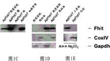
图1C,使用抗Fhit对AdFHIT-感染的A549亚细胞级分的免疫印迹分析表明胞质溶胶、膜、细胞骨架和线粒体中的Fhit蛋白分布。Figure 1C, Immunoblot analysis of AdFHIT-infected A549 subcellular fractions using anti-Fhit demonstrates Fhit protein distribution in the cytosol, membrane, cytoskeleton and mitochondria.
图1D,对用碳酸钠(图1E)和浓度增加的毛地黄皂苷(图1F)处理后AdFHIT-His6感染的A549细胞的线粒体蛋白的免疫印迹分析表明,Fhit主要分布于线粒体基质中;用Fhit和CoxIV抗血清探测滤膜;图1F中的泳道代表用0%、0.10%、0.15%和0.20%毛地黄皂苷处理后的上清液。Figure 1D, Western blot analysis of mitochondrial proteins from AdFHIT-His6- infected A549 cells after treatment with sodium carbonate (Figure 1E) and increasing concentrations of digitonin (Figure 1F) showed that Fhit was mainly distributed in the mitochondrial matrix; Filters were probed with Fhit and CoxIV antisera; lanes in Figure IF represent supernatants after treatment with 0%, 0.10%, 0.15% and 0.20% digitonin.
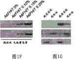
图1G,使用抗Fhit对MKN74/E4和MKN74/A116细胞(稳定表达外源Fhit),和图1H,HCT116(内源Fhit阳性克隆癌细胞系)的亚细胞级分的免疫印迹分析证实了Fhit的线粒体定位;GAPDH和CoxIV抗血清充当对照。Figure 1G, Western blot analysis of subcellular fractions of MKN74/E4 and MKN74/A116 cells (stably expressing exogenous Fhit), and Figure 1H, using anti-Fhit, confirmed Fhit Mitochondrial localization of ; GAPDH and CoxIV antisera served as controls.
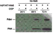
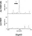
图2A-F-外源和内源Fhit与内源Hsp60、Hsp10和Fdxr蛋白形成复合体。将用重组Fhit-His6蛋白分离的蛋白复合体在聚丙烯酰胺凝胶上分离,并用针对Hsp60(图2A)、Hsp10(图2B)和Fdxr(图2C)的抗血清探测;在后一图中,其于线粒体分离后制备,显示Fhit在线粒体中以时间依赖性方式募集Fdxr。以有或无DSP下用AdFHIT-His6感染A549细胞后分离的蛋白对滤膜进行上样。Figure 2A-F - Exogenous and endogenous Fhit form complexes with endogenous Hsp60, Hsp10 and Fdxr proteins. Protein complexes isolated with recombinant Fhit-His6 protein were separated on polyacrylamide gels and probed with antisera against Hsp60 (Fig. 2A), Hsp10 (Fig. 2B) and Fdxr (Fig. 2C); in the latter panel , which were prepared after mitochondrial isolation, showed that Fhit recruits Fdxr in mitochondria in a time-dependent manner. Filters were loaded with protein isolated after infection of A549 cells with AdFHIT-His6 with or without DSP.
图2D,用AdFHIT感染A549细胞后以抗Hsp60进行的免疫共沉淀;以Hsp60、Fhit和Hsp10抗血清探测滤膜。Figure 2D, Co-immunoprecipitation with anti-Hsp60 after infection of A549 cells with AdFHIT; filters probed with Hsp60, Fhit and Hsp10 antisera.
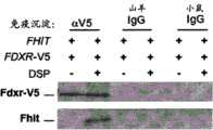
图2E,用V5标签的FDXR基因和FHIT质粒共转染A549细胞;用抗V5进行免疫沉淀并用Fdxr和Fhit抗血清进行检测。Figure 2E, A549 cells were co-transfected with V5-tagged FDXR gene and FHIT plasmid; immunoprecipitation was performed with anti-V5 and detected with Fdxr and Fhit antisera.
图2F,来自DSP处理的Fhit阳性HCT116细胞的内源相互作用物蛋白(Fdxr和Hsp10)的免疫沉淀和免疫印迹检测。以针对每种靶蛋白的抗血清探测滤膜。内源Fhit与Hs p10和Fdxr共沉淀。Figure 2F, Immunoprecipitation and immunoblot detection of endogenous interactor proteins (Fdxr and Hsp10) from DSP-treated Fhit-positive HCT116 cells. Filters were probed with antisera to each target protein. Endogenous Fhit co-precipitated with Hsp10 and Fdxr.
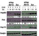
图3A-D-Hsp60/Hsp10的敲低降低了线粒体中的Fhit水平。Figure 3A-D - Knockdown of Hsp60/Hsp10 reduces Fhit levels in mitochondria.
图3A,使用H6抗体对AdFHIT-His6感染的A549细胞的亚细胞级分(胞质溶胶和线粒体)进行的镍-H6下拉(pull down)实验;将溶胞产物与镍珠孵育以分离DSP交联的Fhit-His6蛋白复合体并上样至4-20%聚丙烯酰胺凝胶。感染后24h,Hsp60-Fhit复合体存在于两个区室中;感染后48h,复合体在两个区室中同样是可检测的,并且Fhit复合体蛋白的增加显示与AdFHIT-His6感染后48h Fhit蛋白的增加相关,线粒体中有轻微增加(对输入样品进行光密度法分析)。Figure 3A, Nickel-H6 pull down assay on subcellular fractions (cytosol and mitochondria) of AdFHIT-His6- infected A549 cells using H6 antibody; lysates were incubated with nickel beads to isolate DSP The Fhit-His6 protein complex was cross-linked and loaded onto a 4-20% polyacrylamide gel. At 24h post-infection, the Hsp60-Fhit complex was present in both compartments; at 48h post-infection, the complex was equally detectable in both compartments, and an increase in Fhit complex protein was shown to be consistent with AdFHIT-His6 post-infection The 48h increase in Fhit protein was associated with a slight increase in mitochondria (densitometry analysis of input samples).
图3B,对Hsp60/Hsp10沉默72h后Fhit阳性D1细胞中的Hsp60、Hsp10、Fhit和GAPDH的免疫印迹分析,其显示CHX追踪(30μg/ml)1-12h后的Fhit、Hsp60和Hsp10水平。Figure 3B, Western blot analysis of Hsp60, Hsp10, Fhit and GAPDH in Fhit-positive D1 cells after Hsp60/Hsp10 silencing for 72 h, showing Fhit, Hsp60 and Hsp10 levels after CHX chasing (30 μg/ml) for 1-12 h.
图3C,用Hsp60和Hsp10 siRNA转染后72h和m.o.i.1的AdFHIT感染后24h,使用Hsp60、Hsp10、Fhit、GAPDH和CoxIV抗血清,对A549细胞的胞质溶胶/线粒体蛋白级分的免疫印迹分析。Hsp60/10沉默未显示影响Fhit胞质溶胶水平,但与线粒体级分中Fhit的下降有关。零乱(Scrambled)(Scr)siRNA用作对照。Figure 3C, Western blot analysis of cytosol/mitochondrial protein fractions of A549 cells using Hsp60, Hsp10, Fhit, GAPDH and CoxIV antisera at 72h post-transfection with Hsp60 and Hsp10 siRNA and 24h post-infection with AdFHIT at m.o.i.1 . Hsp60/10 silencing did not appear to affect Fhit cytosolic levels, but was associated with a decrease in Fhit in the mitochondrial fraction. Scrambled (Scr) siRNA was used as a control.
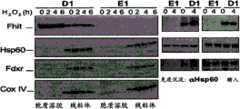
图3D,“内源”Fhit复合体蛋白的亚细胞分级和免疫沉淀。将有和无过氧化物处理的PonA-诱导的D1和E1细胞分级为胞质溶胶和线粒体,并评估诱导后48h亚细胞级分中Fhit和相互作用物(左侧)的存在;每个泳道上样25μg蛋白。内源Hsp60共沉淀Fhit和Fdxr。Figure 3D, Subcellular fractionation and immunoprecipitation of "endogenous" Fhit complex proteins. PonA-induced D1 and E1 cells with and without peroxide treatment were fractionated into cytosol and mitochondria and assessed for the presence of Fhit and interactors (left) in subcellular fractions 48 h after induction; each lane 25 μg protein was loaded. Endogenous Hsp60 co-precipitates Fhit and Fdxr.
图4A-F-Fhit表达诱导用过氧化物处理细胞后胞内ROS产生。Figure 4A-F - Fhit expression induces intracellular ROS production after treatment of cells with peroxide.
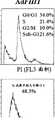
图4A,在有和无5小时H2O2处理下,用FHIT质粒转染后48h的A549细胞中ROS评估的荧光激活细胞分选仪(FACS)分析。空载体转染的细胞充当对照。根据氢化乙啡啶(hydroethidine)被O2氧化产生的乙啡啶的荧光确定胞内超氧化物。M2指ROS阳性细胞的分数。Fig. 4A, Fluorescence-activated cell sorter (FACS) analysis for ROS assessment in A549 cells 48 h after transfection with FHIT plasmid with and without 5 h H2 O2 treatment. Cells transfected with empty vector served as controls. Intracellular superoxide was determined from the fluorescence of ethidinium produced by the oxidation of hydroethidine byO2 . M2 refers to the fraction of ROS-positive cells.
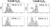
图4B,通过D1和E1细胞中氢化乙啡啶氧化产生的荧光进行ROS评估的FACS分析;PonA处理后48h,将细胞用0.5和1.0mM H2O2处理5h,并测量氧化应激;%阳性指指示ROS的荧光细胞的分数。重复这些实验三次,得到了相似的结果。Fig. 4B, FACS analysis of ROS assessment by fluorescence generated by oxidation of ethidium in D1 and E1 cells; 48 h after PonA treatment, cells were treated with 0.5 and 1.0 mMH2O2 for 5 h, and oxidative stresswas measured; % Positive refers to the fraction of fluorescent cells indicative of ROS. These experiments were repeated three times with similar results.
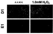
图4C,应激条件下H1299Fhit表达细胞(D1)中增加的绿色荧光DCF信号。Fhit诱导后48h和H2O2处理E1和D1细胞5小时后,将细胞与2′,7′-二氯二氢荧光素二乙酸酯孵育,2′,7′-二氯二氢荧光素二乙酸酯是在ROS存在下可被氧化为高度绿色荧光染料DCF的ROS指示物(放大率x40)。Figure 4C, Increased green fluorescent DCF signal in H1299Fhit expressing cells (D1) under stress conditions. 48 h after Fhit induction and H2 O2 treatment of E1 and D1 cells for 5 h, cells were incubated with 2′,7′-dichlorodihydrofluorescein diacetate, 2′,7′-dichlorodihydrofluorescein Diacetate is a ROS indicator that can be oxidized to the highly green fluorescent dye DCF in the presence of ROS (magnification x40).
图4D,对E1和D1细胞进行MTS细胞存活测定。将细胞用PonA处理48h,然后用浓度增加的H2O2(0.125、0.25和0.5mM)处理4h。在H2O2处理后24h进行分析。柱报告四个实验的平均值±S.E。每个点测量四个重复,并计算标准偏差;认为p<0.05是显著的。Figure 4D, MTS cell survival assay on E1 and D1 cells. Cells were treated with PonA for 48h followed by increasing concentrationsofH2O2 (0.125, 0.25 and 0.5mM) for 4h. Analysis was performed 24 h after H2 O2 treatment. Columns report mean ± SE of four experiments. Four replicates were measured for each point and standard deviation calculated; p<0.05 was considered significant.
图4E,氧化应激处理后48h,D1和E1细胞周期动力学的FACS分析。将细胞用PonA处理48h,然后用浓度增加的H2O2(0.25和0.5mM)处理4h。在H2O2处理后48h进行分析。所有实验以三个重复进行两次。Fig. 4E, FACS analysis of D1 and E1 cell cycle kinetics 48 h after oxidative stress treatment. Cells were treated with PonAfor 48h followed by increasing concentrations ofH2O2 (0.25 and 0.5mM) for 4h. Analysis was performed 48 h after H2 O2 treatment. All experiments were performed twice in triplicate.
图4F,5mM PonA刺激和所示浓度下的5小时H2O2处理后H1299/D1和H1299/E1细胞的集落形成测定。Figure 4F, Colony formation assay of H1299/D1 and H1299/E1 cells after 5 mM PonA stimulation and 5 h ofH2O2 treatment at the indicated concentrations.
图5A-H.-Fhit病毒转导触发的凋亡可通过其与Fdxr的相互作用介导。Figure 5A-H. - Apoptosis triggered by Fhit viral transduction can be mediated through its interaction with Fdxr.
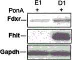
图5A,使用针对Fdxr、Fhit和GAPDH的抗血清的免疫印迹分析。蛋白是在用PonA处理后48h从E1(对照)和D1细胞提取的。Figure 5A, Western blot analysis using antisera against Fdxr, Fhit and GAPDH. Proteins were extracted from E1 (control) and D1 cells 48 h after treatment with PonA.
图5B,用25μM蛋白酶体抑制剂MG132进行4小时处理后,D1和E1细胞中Fdxr表达的免疫印迹分析。GAPDH检测显示相等的蛋白上样。Figure 5B, Immunoblot analysis of Fdxr expression in D1 and E1 cells after 4 h treatment with 25 μM proteasome inhibitor MG132. GAPDH assay showed equal protein loading.
图5C,D1细胞和E1细胞中Fdxr、Fhit和GAPDH的免疫印迹分析显示CHX追踪(30μg/ml)4-12h后的Fdxr水平,所述D1细胞表达Fhit。基于GAPDH水平的光密度法显示Fhit存在下增强的Fdxr稳定性。Figure 5C, Western blot analysis of Fdxr, Fhit and GAPDH in D1 cells expressing Fhit showing Fdxr levels after CHX chase (30 μg/ml) for 4-12 h and in E1 cells. Densitometry based on GAPDH levels revealed enhanced Fdxr stability in the presence of Fhit.
图5D,用AdFHIT m.o.i.50和100感染后FDXR+/+/+和FDXR+/-/-细胞周期动力学的FACS分析。实验在感染后48h进行,并重复三次,得到相似结果。Ad GFP-感染细胞的谱图与非感染的细胞的谱图(未显示)相似。Figure 5D, FACS analysis of FDXR+/+/+ and FDXR+/-/- cell cycle kinetics after infection with
图5E,显示用AdFHIT m.o.i.50和100感染FDXR+/+/+和FDXR+/-/-后Fdxr、Fhit和GAPDH的表达的免疫印迹分析。蛋白质在感染后48h提取。Figure 5E, Western blot analysis showing the expression of Fdxr, Fhit and GAPDH after infection of FDXR+/+/+ and FDXR+/-/- with AdFHIT moi50 and 100. Protein was extracted 48h after infection.
图5F,AdFHIT m.o.i.50后48h的FDXR表达的实时RT-PCR分析。将PCR产物对GADPH和肌动蛋白表达进行标准化,并且每个点重复四次;对照和Fhit阳性样品之间的差异不显著。Figure 5F, Real-time RT-PCR analysis of FDXR expression 48h after AdFHIT m.o.i.50. PCR products were normalized for GADPH and actin expression, and each point was replicated four times; differences between control and Fhit-positive samples were not significant.
图5G,半胱天冬酶3和Parp1激活。使用Fhit、半胱天冬酶3、Parp1抗血清,对m.o.i.50的AdFHIT和Ad GFP感染后48、72和96h的HCT116 FDXR+/+/+细胞的总细胞溶胞产物的免疫印迹分析。GAPDH和CoxIV充当内部蛋白标志物。Figure 5G,
图5H,使用Fhit和细胞色素c抗血清,对m.o.i.50的AdFHIT和Ad GFP感染后48、72和96h的HCT116 FDXR+/+/+细胞的胞质溶胶/线粒体级分的免疫印迹分析。GAPDH和β-肌动蛋白充当内部蛋白标志物。Figure 5H, Immunoblot analysis of cytosol/mitochondrial fractions of HCT116 FDXR+/+/+ cells 48, 72 and 96h after infection with AdFHIT and AdGFP at moi50 using Fhit and cytochrome c antisera. GAPDH and β-actin serve as internal protein markers.
图6A-E-Fhit增强癌细胞对紫杉醇和顺铂的敏感性。Figure 6A-E-Fhit enhances the sensitivity of cancer cells to paclitaxel and cisplatin.
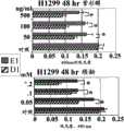
对E1和D1细胞进行MTS测定。将细胞用PonA处理48h,然后用紫杉醇(50-500ng/ml)(图6A)或顺铂(0.05-0.2mM)(图6B)处理24或48h。条报告四个实验的平均值±S.E。每个点测量四个重复并计算标准偏差;图6A和图6B中方括号旁边的星号指示D1和E1细胞对药物应答的统计学显著的差异,p<0.05。MTS assays were performed on E1 and D1 cells. Cells were treated with PonA for 48h, followed by paclitaxel (50-500ng/ml) (Figure 6A) or cisplatin (0.05-0.2mM) (Figure 6B) for 24 or 48h. Bars report mean±S.E. of four experiments. Four replicates were measured for each point and standard deviation calculated; asterisks next to square brackets in Figures 6A and 6B indicate statistically significant differences in response of D1 and E1 cells to drugs, p<0.05.
图6C和图6D,图显示E1和D1细胞的流式细胞术分析的代表性结果。将细胞用PonA处理48h,然后用紫杉醇(50-500ng/ml)(图6C)或顺铂(0.05-0.2mM)(图6D)处理。每个数据点在24、48和72h测量三个重复(显示了48h的数据)。Figure 6C and Figure 6D, graphs showing representative results of flow cytometry analysis of E1 and D1 cells. Cells were treated with PonA for 48h, followed by paclitaxel (50-500 ng/ml) (Figure 6C) or cisplatin (0.05-0.2mM) (Figure 6D). Each data point was measured in triplicate at 24, 48 and 72h (data for 48h are shown).
图6E,半胱天冬酶3和Parp1切割:使用Fhit、半胱天冬酶3和Parp1抗血清,对用紫杉醇(50和100ng/ml)或顺铂(0.05和0.1mM)处理48h后PonA诱导的D1细胞的总细胞溶胞产物的免疫印迹分析。GAPDH充当上样对照。Figure 6E,
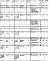
图7.表1通过质谱法分离的候选Fhit蛋白配偶体。在A549AdFHIT-H6感染的细胞样品中选择性捕获的蛋白质。列出了鉴定的肽的氨基酸序列、Mascot评分和蛋白质序列覆盖率。Figure 7. Table 1 Candidate Fhit protein partners isolated by mass spectrometry. Proteins selectively captured in A549AdFHIT-H6 infected cell samples. Amino acid sequences, Mascot scores, and protein sequence coverage of identified peptides are listed.
图8A-8C.Ad-His6生物活性与AdFHIT相当。Figures 8A-8C. Ad-His6 biological activity is comparable to AdFHIT.
图8A,用Ad-His6、MOI 20感染的A549细胞的蛋白质印迹分析。通过抗pentaHis和抗Fhit血清检测Fhit-His蛋白。Ad FHIT和Ad-His6二者均携带通过FHIT下游的内部核糖体进入序列受到CMV5启动子调控的GFP cDNA。使用γ-微管蛋白对样品上样进行标准化。Figure 8A, Western blot analysis of A549 cells infected with Ad-His6,
图8B,用Ad-His 6、MOI 15感染后96hr的A549细胞的流式细胞术分析。上图指示感染细胞的subG1 DNA含量(实验重复三次;subG1级分的平均值:Ad FHIT为22%+/-4.3,Ad-His6为29%+/-5;差异不是统计学显著的;下图显示具有指示凋亡的成熟半胱天冬酶-3的细胞的百分比。用Ad-His6感染的A549细胞中细胞死亡的程度与在用Ad FHIT感染后获得的结果相当。Fig. 8B, flow cytometric analysis of A549 cells infected with Ad-His 6,
图8C,Fhit-His6的体内交联。His6下拉和交联逆转条件后的细胞溶胞产物通过4-20%梯度SDS-PAGE分离的凝胶的银染色。内部阴性对照包括Ad FHIT感染的细胞(交联的,CL)和Ad-His6感染的细胞(未交联的,NT)的His6下拉。Figure 8C, In vivo crosslinking of Fhit-His6. Silver staining of gels separated by 4-20% gradient SDS-PAGE of cell lysates after His6 pulldown and crosslink reversal conditions. Internal negative controls included His6 pulldown of Ad FHIT-infected cells (cross-linked, CL) and Ad-His6-infected cells (non-cross-linked, NT).
图9A-9F.通过纳米孔(nanobore)LC-MS/MS鉴定的候选Fhit蛋白配偶体的初步验证。显示了AdFHIT-His6和对照样品的选择离子色谱图(Selected ion chromatograms,SIC)。六个SIC对报告了下列六个m/z值的离子电流:1)672.8(保留时间为30min的峰鉴定为属于Hsp60的胰蛋白酶肽TVIIEQSWGSPK[SEQ ID NO:5]),2)685.4(保留时间为32min的峰鉴定为属于Aldh2的胰蛋白酶肽LGPALATGNVVVMK[SEQ ID NO:22]),3)617.3(保留时间为39min的峰鉴定为属于Mdh的胰蛋白酶肽IFGVTTLDIVR[SEQ ID NO:10]),4)658.4(保留时间为26min的峰鉴定为属于Hsp10的胰蛋白酶肽VLQATVVAVGSGSK[SEQ ID NO:19]),5)551.7(保留时间为28min的峰鉴定为属于Etfb的胰蛋白酶肽EIDGGLETLR[SEQ ID NO:15]),6)598.3(保留时间为23min的峰鉴定为属于Fdxr的胰蛋白酶肽FGVAPDHPEVK[SEQ ID NO:23])。用红色箭头指示的感兴趣的肽专一地存在于Ad-His6样品中。Figures 9A-9F. Preliminary validation of candidate Fhit protein partners identified by nanobore LC-MS/MS. Selected ion chromatograms (SIC) of AdFHIT-His6 and control samples are shown. The six SIC pairs reported ionic currents for the following six m/z values: 1) 672.8 (the peak with a retention time of 30 min was identified as the tryptic peptide TVIIEQSWGSPK [SEQ ID NO: 5] belonging to Hsp60), 2) 685.4 (retention The peak at 32min was identified as the tryptic peptide LGPALATGNVVVMK[SEQ ID NO:22]) belonging to Aldh2, 3)617.3 (the peak at 39min was identified as the tryptic peptide IFGVTTLDIVR[SEQ ID NO:10] belonging to Mdh) , 4) 658.4 (the peak at retention time of 26 min is identified as the tryptic peptide VLQATVVAVGSGSK [SEQ ID NO: 19] belonging to Hsp10), 5) 551.7 (the peak at retention time of 28 min is identified as the tryptic peptide EIDGGLETLR [SEQ ID NO: 19] belonging to Etfb ID NO: 15]), 6) 598.3 (the peak with a retention time of 23 min was identified as the tryptic peptide FGVAPDHPEVK [SEQ ID NO: 23] belonging to Fdxr). The peptide of interest indicated by the red arrow is exclusively present in the Ad-His6 sample.
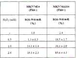
图10.表2,Fhit在MKN74胃癌细胞中诱导ROS产生。ROS评估使用携带p53突变等位基因并表达外源Fhit的人胃癌细胞系MKN74A116进行;Fhit阴性MKN74E4细胞用作对照。为了诱导ROS产生,我们用0.5、1.0和2.0mM H2O2处理MKN74细胞5hr。结果表明与对照相比,表达外源Fhit的细胞中有显著更高的ROS产生率;2mM H2O2处理后,在Fhit-表达细胞中观察到毒性。数字报告四个实验的平均值±S.E.。Figure 10. Table 2, Fhit induces ROS production in MKN74 gastric cancer cells. ROS assessment was performed using the human gastric cancer cell line MKN74A116 carrying a p53 mutant allele and expressing exogenous Fhit; Fhit-negative MKN74E4 cells were used as controls. To induce ROS production, wetreated MKN74 cells with 0.5, 1.0 and 2.0 mMH2O2 for 5 hr. The results indicated a significantly higher rate of ROS production in cells expressing exogenous Fhit compared to controls; toxicity was observed in Fhit-expressing cells after 2 mM H2 O2 treatment. Numbers report mean ± SE of four experiments.
优选实施方案详述DETAILED DESCRIPTION OF THE PREFERRED EMBODIMENT
应理解,上面的一般描述和下面的详细描述仅是示例性的和解释性的,并不意图限制本文的教导的范围。在本申请中,除非另外指明,否则使用单数时包括复数。It is to be understood that both the foregoing general description and the following detailed description are exemplary and explanatory only and are not intended to limit the scope of the teachings herein. In this application, the use of the singular includes the plural unless otherwise specified.
在权利要求和/或说明书中,当与术语“包含(comprising)”联合使用时,词“一种(a)”或“一种(an)”的使用可意指“一种(one)”,但其也与“一种或多种”、“至少一种”和“一种或多于一种”一致。In the claims and/or specification, use of the word "a" or "an" may mean "one" when used in conjunction with the term "comprising" , but it is also consistent with "one or more", "at least one" and "one or more than one".
另外,“包含(comprise)”、“含有(contain)”和“包括(include)”或这些根词的修饰形式,例如但不限于“包含(comprises)”、“含有(contained)”和“包括(including)”,并不意图是限制性的。术语“和/或”意指前后的术语可以是一起的或单独的。出于例证目的,但不是作为限制,“X和/或Y”可意指“X”或“Y”或“X和Y”。In addition, "comprise", "contain" and "include" or modified forms of these root words, such as but not limited to "comprises", "contained" and "including (including)" and are not intended to be limiting. The term "and/or" means that the preceding and following terms can be taken together or alone. For purposes of illustration, but not limitation, "X and/or Y" may mean "X" or "Y" or "X and Y".
如本文所用的术语“其组合”指在该术语之前所列的条目的所有排列和组合。例如,“A、B、C或其组合”意图包括以下的至少一种:A、B、C、AB、AC、BC或ABC,以及,如果在具体环境中顺序是重要的,还包括BA、CA、CB、ACB、CBA、BCA、BAC或CAB。As used herein, the term "combinations thereof" refers to all permutations and combinations of items listed before the term. For example, "A, B, C, or combinations thereof" is intended to include at least one of the following: A, B, C, AB, AC, BC, or ABC, and, if order is important in a particular context, BA, CA, CB, ACB, CBA, BCA, BAC, or CAB.
如本文可交换使用的,“基因产物”、“DNA”和“基因”在本文可交换使用。As used interchangeably herein, "gene product", "DNA" and "gene" are used interchangeably herein.
本文可使用以下缩写词:GAPDH,甘油醛-3-磷酸脱氢酶;DSP,二硫代二(琥珀酰亚胺丙酸酯);LC-MS/MS,液相色谱串联质谱;Fdxr,铁氧还蛋白还原酶;PonA,松甾酮A;m.o.i.,感染复数;ROS,活性氧簇(reactive oxygen species);FU,5-氟尿嘧啶;DCFH-DA,二氯荧光素二乙酸酯;DCF,2′,7′-二氯荧光素;CHX,环己酰亚胺;siRNA,小干扰RNA;RT,反转录酶;MTS,3-(4,5-二甲基噻唑-2-基)-5-(3-羧基甲氧基苯基)-2-(4-磺苯基)-2H-四唑。The following abbreviations may be used herein: GAPDH, glyceraldehyde-3-phosphate dehydrogenase; DSP, dithiobis(succinimidyl propionate); LC-MS/MS, liquid chromatography tandem mass spectrometry; Fdxr, iron Redoxin reductase; PonA, pine stesterone A; m.o.i., multiplicity of infection; ROS, reactive oxygen species (reactive oxygen species); FU, 5-fluorouracil; DCFH-DA, dichlorofluorescein diacetate; DCF, 2′,7′-dichlorofluorescein; CHX, cycloheximide; siRNA, small interfering RNA; RT, reverse transcriptase; MTS, 3-(4,5-dimethylthiazol-2-yl) -5-(3-carboxymethoxyphenyl)-2-(4-sulfophenyl)-2H-tetrazole.
本文使用的各部分的标题仅是为了组织目的,而不应以任何方式视为限制所描述的主题。本申请中引用的所有文献和相似材料,包括专利、专利申请、文章、专著、论文和互联网网页,出于任何目的而通过引用明确地整体并入。如果所并入的文献和相似材料中的一种或多种以与本申请中的术语定义矛盾的方式定义或使用了该术语,则以本申请为准。The section headings used herein are for organizational purposes only and should not be considered in any way to limit the subject matter described. All literature and similar materials, including patents, patent applications, articles, monographs, treatises, and Internet web pages, cited in this application are expressly incorporated by reference in their entirety for any purpose. Should one or more of the incorporated literature and similar materials define or use a term in a manner that contradicts the definition of that term in this application, this application controls.
Fhit蛋白在大多数癌症中是丢失的,其恢复抑制肿瘤发生,并且病毒介导的FHIT基因治疗在临窗前模型中诱导凋亡并抑制肿瘤。使用蛋白质交联和蛋白质组学方法表征参与触发Fhit-介导的凋亡的Fhit蛋白复合体。该复合体包括介导Fhit稳定性并可影响向线粒体的输入的Hsp60和Hsp10,在线粒体中,该复合体与铁氧还蛋白还原酶相互作用,铁氧还蛋白还原酶负责经由铁氧还蛋白将电子从NADPH转移给细胞色素P450。病毒介导的Fhit恢复增加胞内活性氧簇的产生,然后是氧化应激条件下肺癌细胞凋亡的增加;相反,Fhit-阴性细胞逃避了凋亡,其携带可促成增加的突变率的严重氧化型DNA损伤。Fhit相互作用蛋白的表征鉴定了Fhit-介导的凋亡途径的直接效应物,由于Fhit的丢失,该途径在大多数癌症中是丢失的。The Fhit protein is lost in most cancers, its restoration suppresses tumorigenesis, and virus-mediated FHIT gene therapy induces apoptosis and suppresses tumors in preclinical models. The Fhit protein complexes involved in triggering Fhit-mediated apoptosis were characterized using protein cross-linking and proteomic approaches. This complex includes Hsp60 and Hsp10, which mediate Fhit stability and can affect import into mitochondria, where the complex interacts with ferredoxin reductase, which is responsible for the transfer of ferredoxin Transfers electrons from NADPH to cytochrome P450. Virus-mediated restoration of Fhit increases the production of intracellular reactive oxygen species, followed by increased apoptosis in lung cancer cells under oxidative stress; conversely, Fhit-negative cells escape apoptosis, carrying severe Oxidative DNA damage. Characterization of Fhit-interacting proteins identified direct effectors of the Fhit-mediated apoptotic pathway, which is lost in most cancers due to loss of Fhit.
对Fhit-相互作用蛋白的更早检索指出了几种候选蛋白,其中没有一个可通过免疫共沉淀实验被我们确认为相互作用物,这些候选蛋白包括Ubc9、α-微管蛋白和Mdm2(35-37)。为了重新着手于Fhit蛋白相互作用物的问题,使用了如下:用于交联后Fhit复合体纯化的腺病毒转导的Fhit-His6,并鉴定了Fhit-连接蛋白、Hsp60、Hsp10和Fdxr;这些蛋白的亚细胞定位暗示线粒体可能是Fhit活性的焦点。Hsp“应激蛋白”作为分子伴侣蛋白执行诸如蛋白易位、折叠和装配的功能(38)。AdFHIT感染后Fhit与Hsp60/Hsp10相互作用的发现暗示,在激活凋亡途径之前,Hsp复合体对Fhit稳定性以及可能地对于它的正确折叠以输入到线粒体,可能是重要的,我们通过敲低AdFHIT-感染的肺癌细胞中的Hsp60、Hsp10或二者表达研究了这一暗示;CHX追踪后评估了表达可诱导的Fhit的肺癌细胞系H1299D1细胞中的Fhit稳定性。敲低Hsp60和Hsp10二者后,分离的线粒体中的Fhit蛋白降低,强化了Fhit-Hsp60/10相互作用参与Fhit稳定性和/或正确折叠以输入到线粒体的提议。An earlier search for Fhit-interacting proteins pointed to several candidates, none of which could be identified by us as interactors by co-immunoprecipitation experiments, including Ubc9, α-tubulin, and Mdm2 (35- 37). To revisit the question of Fhit protein interactors, the following was used: adenovirus-transduced Fhit-His6 for purification of the Fhit complex after cross-linking, and the Fhit-linker proteins, Hsp60, Hsp10 and Fdxr were identified; these The subcellular localization of the protein suggests that mitochondria may be the focus of Fhit activity. Hsp "stress proteins" act as molecular chaperones to perform functions such as protein translocation, folding and assembly (38). The finding that Fhit interacts with Hsp60/Hsp10 after AdFHIT infection suggests that the Hsp complex may be important for Fhit stability and possibly for its proper folding for import into mitochondria prior to activation of the apoptotic pathway, we determined by knocking down Hsp60, Hsp10 or both expression in AdFHIT-infected lung cancer cells This implication was investigated; Fhit stability in H1299D1 cells, a lung cancer cell line expressing inducible Fhit, was assessed after CHX trace. Following knockdown of both Hsp60 and Hsp10, Fhit protein was reduced in isolated mitochondria, strengthening the proposal that the Fhit-Hsp60/10 interaction is involved in Fhit stability and/or proper folding for import into mitochondria.
HCT116结肠癌细胞中FDXR基因的靶向破坏显示其是存活必需的;基因拷贝数的降低造成对5-氟尿嘧啶-诱导的凋亡的敏感性的降低(29),而FDXR是p53家族的靶基因(30)。Fdxr的过表达使结肠癌细胞对H2O2、5-氟尿嘧啶和阿霉素-诱导的细胞死亡敏感,表明Fdxr通过在线粒体中产生氧化应激促成p53-介导的凋亡。因此,激活的p53部分是通过ROS诱导响应于细胞应激的凋亡,并且p53同时增加FDXR基因的转录,这通过增加ROS-诱导的凋亡依次增强了p53功能(29,30)。Targeted disruption of the FDXR gene in HCT116 colon cancer cells was shown to be essential for survival; reduction in gene copy number resulted in reduced susceptibility to 5-fluorouracil-induced apoptosis (29), while FDXR is a target gene of the p53 family (30). Overexpression of Fdxr sensitizes colon cancer cells to H2 O2 , 5-fluorouracil, and doxorubicin-induced cell death, suggesting that Fdxr contributes to p53-mediated apoptosis by generating oxidative stress in mitochondria. Thus, activated p53 induces apoptosis in response to cellular stress in part through ROS, and p53 concomitantly increases transcription of the FDXR gene, which in turn enhances p53 function by increasing ROS-induced apoptosis (29, 30).
现在本文显示了Fhit在线粒体级分中的存在;当Fhit被过表达或Fhit-表达细胞受到应激时,Fhit可保护Fdxr不受蛋白体降解,导致Fdxr蛋白水平的增加,这与ROS的产生有关并跟随有凋亡。Fhit不影响FDXR转录水平但可影响蛋白的稳定性。在缺乏Fhit和p53二者的H1299细胞中,Fdxr过表达增加对ROS-诱导的细胞死亡的敏感性,表达可诱导的Fhit或p53的H1299细胞对ROS-诱导的细胞死亡敏感;缺乏Fhit、p53或二者的癌细胞将缺乏增加Fdxr表达的方式,并将对氧化损伤更不敏感,并将存活。This paper now shows the presence of Fhit in the mitochondrial fraction; when Fhit is overexpressed or Fhit-expressing cells are stressed, Fhit protects Fdxr from proteosomal degradation, leading to increased Fdxr protein levels, which is consistent with ROS production Associated with and followed by apoptosis. Fhit does not affect FDXR transcript level but can affect protein stability. In H1299 cells lacking both Fhit and p53, Fdxr overexpression increases sensitivity to ROS-induced cell death, H1299 cells expressing inducible Fhit or p53 are sensitive to ROS-induced cell death; lack of Fhit, p53 Cancer cells of either or both will lack the means to increase Fdxr expression and will be less sensitive to oxidative damage and will survive.
Fhit在凋亡中通过与Fdxr相互作用的线粒体功能的发现,现在延伸了在大多数癌症中顺序丢失的并参与对DNA损伤的应答的重要肿瘤抑制物Fhit和p53的功能平行物,并阐明它们的差异,p53充当转录水平的Fdxr调控物,而Fhit充当转录后Fdxr调控物。对Fhit抑制途径的直接下游效应物的描绘将导致对可影响激活Fhit途径的预防和治疗策略的Fhit功能的机制研究。The discovery of a mitochondrial function of Fhit in apoptosis through interaction with Fdxr now extends and elucidates the functional parallels of the important tumor suppressors Fhit and p53 that are sequentially lost in most cancers and involved in the response to DNA damage p53 acts as a transcriptional Fdxr regulator, whereas Fhit acts as a post-transcriptional Fdxr regulator. Delineation of the immediate downstream effectors of the Fhit inhibitory pathway will lead to mechanistic studies of Fhit function that can influence preventive and therapeutic strategies that activate the Fhit pathway.
ROS产生对Fhit-介导的凋亡至关重要的发现强调了Fhit丢失作为阴性预后因子在不同临床环境中的重要性;例如,评估肿瘤发生前或肿瘤病症中的Fhit状态可预测对抗氧化剂治疗的应答。The finding that ROS production is critical for Fhit-mediated apoptosis underscores the importance of Fhit loss as a negative prognostic factor in different clinical settings; for example, assessment of Fhit status in preneoplastic or neoplastic conditions predicts antioxidant therapy answer.
为了鉴定与Fhit相互作用以实现下游凋亡途径的蛋白,本文中本发明人在肺癌细胞中于病毒介导的Fhit过表达后交联了细胞内的蛋白,并表征了与Fhit相关的蛋白和它们所影响的途径。In order to identify proteins that interact with Fhit to achieve downstream apoptotic pathways, the inventors herein cross-linked intracellular proteins after virus-mediated overexpression of Fhit in lung cancer cells, and characterized the proteins associated with Fhit and pathways they affect.
结果result
Fhit蛋白复合体的分离-为了鉴定Fhit-相互作用蛋白,我们产生了携带在其3′端用编码His6表位标签的序列修饰的FHITcDNA的腺病毒(AdFHIT-His6)。A549细胞中表达的该标签的Fhit蛋白的生物活性与野生型Fhit活性相当(图8)。Isolation of Fhit protein complexes - To identify Fhit-interacting proteins, we generated an adenovirus carrying FHIT cDNA modified at its 3' end with a sequence encoding a His6 epitope tag (AdFHIT-His6 ). The biological activity of the tagged Fhit protein expressed in A549 cells was comparable to that of wild-type Fhit ( FIG. 8 ).
对Fhit-诱导的凋亡易感的A549肺癌衍生细胞(10)用AdFHIT或AdFHIT-His6进行感染,并用DSP处理,DSP是横穿膜并于体内固定复合体中的蛋白的交联剂。裂解细胞,并用对His6表位标签亲和的镍珠分离蛋白。用二硫苏糖醇处理纯化蛋白以切割DSP并使复合体解离,并通过胰蛋白酶消化;通过LC-MS/MS鉴定蛋白成分(图7-表1和图9)。A549 lung cancer-derived cells susceptible to Fhit-induced apoptosis (10) were infected with AdFHIT or AdFHIT-His6 and treated with DSP, a cross-linker that traverses membranes and immobilizes proteins in complexes in vivo. Cells were lysed and proteins were isolated using nickel beads with affinity for the His6 epitope tag. Purified protein was treated with dithiothreitol to cleave DSP and dissociate the complex, and digested by trypsin; protein components were identified by LC-MS/MS (Figure 7-Table 1 and Figure 9).

鉴定了六种蛋白,所有蛋白均具有线粒体定位:Hsp60和Hsp10、铁氧还蛋白还原酶(Fdxr)、苹果酸脱氢酶、电子传递黄素蛋白和线粒体醛脱氢酶2;Hsp60和Hsp10还分布于胞质溶胶(23)。Six proteins were identified, all with a mitochondrial localization: Hsp60 and Hsp10, ferredoxin reductase (Fdxr), malate dehydrogenase, electron transport flavoprotein, and
Fhit亚细胞定位-因为候选Fhit相互作用物是线粒体蛋白,所以本发明人在此确定了缺乏线粒体定位信号的Fhit是否定位于这些细胞器。用诱导子PonA处理携带可诱导的FHIT cDNA的Fhit阴性H1299肺癌细胞(D1细胞)48h,并使用抗Fhit血清和线粒体标志物MitoTracker Red 580评估Fhit亚细胞定位的间接免疫荧光检测;Fhit荧光信号(绿色染色,图1A)是细胞质定位的并与MitoTracker Red染料部分共定位(黄色染色,图1A,右下),表明外源Fhit定位于线粒体和胞质溶胶。抗Fhit特异性得到Fhit阴性H1299克隆E1细胞中缺乏绿色荧光的证实(未显示)。为了证实线粒体定位,通过抗pentaHis染色于48h后用免疫电子显微术检查以m.o.i.20用AdFHIT-His6或AdFHIT感染的A549细胞;FhitHis6-转导的细胞展示线粒体中有显著数量的金粒(图1B,右图),而AdFHIT-转导的细胞显示稀疏的反应性(图1B,左图)。Fhit subcellular localization - Since candidate Fhit interactors are mitochondrial proteins, the inventors here determined whether Fhit, which lacks a mitochondrial localization signal, localizes to these organelles. Fhit-negative H1299 lung cancer cells (D1 cells) carrying inducible FHIT cDNA were treated with the elicitor PonA for 48 hours, and the indirect immunofluorescence detection of Fhit subcellular localization was evaluated using anti-Fhit serum and mitochondrial marker MitoTracker Red 580; Fhit fluorescence signal ( Green staining, Figure 1A) was cytoplasmically localized and partially co-localized with MitoTracker Red dye (yellow staining, Figure 1A, lower right), indicating that exogenous Fhit localized to mitochondria and the cytosol. Anti-Fhit specificity was confirmed by the lack of green fluorescence in Fhit-negative H1299 clone E1 cells (not shown). To confirm mitochondrial localization, A549 cells infected with AdFHIT-His6 or AdFHIT at m.o.i.20 were examined by immunoelectron microscopy after 48 h by anti-pentaHis staining; FhitHis6-transduced cells exhibited a significant number of gold particles in mitochondria (Fig. 1B, right panel), whereas AdFHIT-transduced cells showed sparse reactivity (FIG. 1B, left panel).
为了评估Fhit的亚线粒体定位,从如上所述的用AdFHITm.o.i.1感染的A549细胞纯化线粒体。碳酸钠方法是非破坏性方法,其允许在诱导展开膜层产生后有效释放胞内膜的可溶蛋白和外周膜蛋白到上清液中;另外,它允许回收膜的整合蛋白(沉淀)(24)。To assess the submitochondrial localization of Fhit, mitochondria were purified from A549 cells infected with AdFHIT m.o.i.1 as described above. The sodium carbonate method is a non-destructive method that allows the efficient release of soluble and peripheral membrane proteins of the intracellular membrane into the supernatant after induction of the unfolded membrane layer; additionally, it allows the recovery of integral membrane proteins (precipitation) (24 ).
图1E显示Fhit是可溶级分中唯一可检测的。为了进一步限定Fhit的亚线粒体定位,用0.10%和0.15%毛地黄皂苷处理线粒体以选择性破坏线粒体外膜,释放膜间腔和基质中含有的蛋白;如图1F所示,外膜和内膜的逐渐破坏释放增加量的Fhit蛋白,暗示Fhit主要分布于内膜的腔侧或线粒体基质中。线粒体定位在稳定表达外源Fhit的胃癌衍生的MKN74A116细胞(9)和表达内源Fhit的HCT116结肠癌细胞中得到证实(图1G和图1H)。Figure 1E shows that Fhit was only detectable in the soluble fraction. To further define the submitochondrial localization of Fhit, mitochondria were treated with 0.10% and 0.15% digitonin to selectively disrupt the outer mitochondrial membrane, releasing proteins contained in the intermembrane cavity and matrix; as shown in Figure 1F, the outer and inner membranes The gradual disruption of the release of increasing amounts of Fhit protein, suggesting that Fhit is mainly distributed in the luminal side of the inner membrane or in the mitochondrial matrix. Mitochondrial localization was confirmed in gastric cancer-derived MKN74A116 cells stably expressing exogenous Fhit (9) and in HCT116 colon carcinoma cells expressing endogenous Fhit (Fig. 1G and Fig. 1H).
Fhit与Hsp60、Hsp10和Fdxr相互作用-在候选相互作用物蛋白中,发明人集中于为可能的伴侣蛋白的Hsp60和Hsp10,以及被p53反式激活并参与对治疗药物的应答的线粒体呼吸链蛋白Fdxr(25)。为了证实相互作用,在有或无DSP下,以m.o.i.20用AdFHIT或AdFHIT-His6感染A549细胞。通过His6标签纯化Fhit复合体,并用针对Hsp60、Hsp10和Fdxr的抗血清检测共纯化的蛋白;Hsp60和Fdxr仅在暴露于DSP的细胞的溶胞产物中检测到(图2A和图2C),而Hsp10在无交联下也是可检测的(图2B)。Fhit interacts with Hsp60, Hsp10 and Fdxr - Among candidate interactor proteins, the inventors focused on Hsp60 and Hsp10 as possible chaperones, and mitochondrial respiratory chain proteins that are transactivated by p53 and involved in response to therapeutic drugs Fdxr(25). To confirm the interaction, A549 cells were infected with AdFHIT or AdFHIT-His6 at moi20 with or without DSP. The Fhit complex was purified by His6 tag and the co-purified proteins were detected with antisera against Hsp60, Hsp10 and Fdxr; Hsp60 and Fdxr were only detected in the lysates of cells exposed to DSP (Figure 2A and Figure 2C), Whereas Hsp10 was also detectable without cross-linking (Fig. 2B).
感染后的时间过程实验显示Fhit对Fdxr的募集(图2C);另外,内源Hsp60在DSP不存在下免疫共沉淀Fhit和Hsp10(图2D)。Time-course experiments after infection revealed recruitment of Fdxr by Fhit (Fig. 2C); additionally, endogenous Hsp60 co-immunoprecipitated Fhit and Hsp10 in the absence of DSP (Fig. 2D).
为了验证相互作用的特异性,我们产生了具有3′V5表位标签的FDXR cDNA表达质粒。用FDXR-V5和FHIT质粒共转染A549细胞,并用单克隆抗V5沉淀蛋白;共沉淀的Fhit仅在DSP交联后可检测(图2E)。To verify the specificity of the interaction, we generated a FDXR cDNA expression plasmid with a 3′ V5 epitope tag. A549 cells were co-transfected with FDXR-V5 and FHIT plasmids, and the protein was precipitated with monoclonal anti-V5; co-precipitated Fhit was only detectable after DSP crosslinking (Figure 2E).
为了确定这些蛋白是否还与内源Fhit相互作用,发明人从DSP-处理的Fhit阳性HCT116细胞中免疫沉淀了每种内源候选相互作用物蛋白并寻找内源Fhit的共沉淀(图2F)。To determine whether these proteins also interact with endogenous Fhit, the inventors immunoprecipitated each endogenous candidate interactor protein from DSP-treated Fhit-positive HCT116 cells and looked for co-precipitation of endogenous Fhit (Fig. 2F).
内源Fhit与Hsp10和Fdxr共沉淀,证实了内源Fhit在线粒体中的存在以及在不存在应激下其与内源伴侣蛋白和呼吸链蛋白的相互作用。Endogenous Fhit was co-precipitated with Hsp10 and Fdxr, confirming the presence of endogenous Fhit in mitochondria and its interaction with endogenous chaperones and respiratory chain proteins in the absence of stress.
Hsp60/10相互作用影响Fhit稳定性和/或线粒体输入-Hsp60和Hsp10是在复合体中发现的分子伴侣蛋白(26)并可对于蛋白的折叠和输入到线粒体是重要的。本发明人现在在此认为Hsp60/10复合体负责Fhit的正确折叠和线粒体寻址。Hsp60/10 Interaction Affects Fhit Stability and/or Mitochondrial Import - Hsp60 and Hsp10 are molecular chaperones found in complexes (26) and may be important for protein folding and import into mitochondria. The inventors here now believe that the Hsp60/10 complex is responsible for the correct folding and mitochondrial addressing of Fhit.
为了研究这些相互作用的定位,用AdFHIT-His6m.o.i.5感染A549细胞,并从交联后的胞质溶胶和线粒体级分中收集蛋白溶胞产物。通过Fhit-H6-镍下拉分离复合体,在聚丙烯酰胺凝胶上分离,并用Hsp60和Fhit抗血清探测滤膜。在感染后24和48h,在胞质溶胶和线粒体中观察到与Hsp60的相互作用(图3A),与这些时间的Fhit表达增加相称(输入),如图3A所示。To investigate the localization of these interactions, A549 cells were infected with AdFHIT-His6 moi5, and protein lysates were collected from the cross-linked cytosolic and mitochondrial fractions. Complexes were isolated by Fhit-H6-Ni pulldown, resolved on polyacrylamide gels, and filters probed with Hsp60 and Fhit antisera. Interaction with Hsp60 was observed in the cytosol and mitochondria at 24 and 48 h after infection (Fig. 3A), commensurate with increased Fhit expression at these times (input), as shown in Fig. 3A.
为了确定Fhit-Hsp60/10相互作用对于Fhit蛋白的稳定性是否重要,用Hsp60和Hsp10 siRNA转染具有可诱导的Fhit表达的H1299(D1细胞),并在转染后72h,在1、6和12h进行CHX追踪,评估Fhit蛋白表达并将其与用零乱序列转染的细胞进行比较。In order to determine whether the Fhit-Hsp60/10 interaction is important for the stability of Fhit protein, H1299 (D1 cells) with inducible Fhit expression were transfected with Hsp60 and Hsp10 siRNA, and 72h after transfection, at 1, 6 and CHX tracing was performed at 12h to assess Fhit protein expression and compare it to cells transfected with scrambled sequences.
如图3B所示,在Hsp60和Hsp10沉默后CHX的6和12h,存在Fhit表达的强烈降低(在1和12h,分别为从1至0.4)。接下来,将Hsp60和Hsp10 siRNA分别或组合地转染入A549细胞;24h后,用AdFHIT m.o.i.1感染细胞,并在24h后分级胞质溶胶和线粒体。沉默两种Hsp后,与对照相比,胞质溶胶中的Fhit水平未受影响但线粒体中的降低(图3B),显示Hsp60/10复合体可介导病毒转导的Fhit稳定和线粒体定位。如果Hsp60和Hsp10参与Fhit病毒转导后的Fhit稳定性,则以下也是真实的:具有较少Fhit的细胞区室将受到Fhit稳定性降低的影响。本发明人在此还检查了表达可诱导的Fhit的H1299D1细胞中的Fhit复合体,使用Fhit阴性E1细胞作为对照;D1细胞中Fhit诱导后48h(图3C,左图),有和无H2O2下,在D1和E1细胞的胞质溶胶和线粒体中的Fhit复合体蛋白的分布是相似的。As shown in Figure 3B, at 6 and 12 h of CHX after Hsp60 and Hsp10 silencing, there was a strong decrease in Fhit expression (from 1 to 0.4 at 1 and 12 h, respectively). Next, Hsp60 and Hsp10 siRNA were transfected into A549 cells separately or in combination; 24 h later, cells were infected with AdFHIT moi1, and cytosol and mitochondria were fractionated after 24 h. After silencing both Hsps, Fhit levels in the cytosol were unaffected but decreased in mitochondria compared to controls (Fig. 3B), suggesting that the Hsp60/10 complex may mediate virally transduced Fhit stabilization and mitochondrial localization. If Hsp60 and Hsp10 are involved in Fhit stability following Fhit viral transduction, the following is also true: cellular compartments with less Fhit will be affected by reduced Fhit stability. The inventors here also examined the Fhit complex in H1299D1 cells expressing inducible Fhit, using Fhit-negative E1 cells as a control; 48h after Fhit induction in D1 cells (Fig. 3C, left panel), with and withoutH2 The distribution of Fhit complex proteins in the cytosol and mitochondria of D1 and E1 cells was similar underO2 .
有或无H2O2下,PonA诱导后48h,从这些细胞的总细胞溶胞产物中免疫沉淀Hsp60,并共沉淀Fhit和Fdxr(图3C,右图)。D1细胞中Fhit表达的诱导并没有引起体外生物学变化;因此Fhit复合体并不是作为凋亡的结果形成的。有和无应激条件下,在PonA-诱导的Fhit表达后在D1细胞中进行时间过程实验,以确定是否存在Fhit蛋白相互作用物的生物学变化。共同发明人未检测Fhit表达后的定位变化。Hsp60 was immunoprecipitated and Fhit and Fdxr were co-precipitated from total cell lysates of these cells 48 h after PonA induction with or without H2 O2 ( FIG. 3C , right panel). Induction of Fhit expression in D1 cells did not cause biological changes in vitro; thus the Fhit complex was not formed as a result of apoptosis. Time-course experiments were performed in D1 cells following PonA-induced Fhit expression with and without stress to determine if there were biological changes in Fhit protein interactors. The co-inventors did not detect localization changes upon expression of Fhit.
Fhit诱导活性氧簇(ROS)的产生-Fdxr是54-kDa黄素蛋白,它定位于线粒体内膜的基质侧,并负责经由单电子穿梭铁氧还蛋白-细胞色素P450将电子从NADPH传递至底物(27)。在底物限制条件下,电子从此穿梭系统中泄漏并产生ROS(28)。Fdxr通过ROS产生介导结直肠癌细胞中的p53-依赖性、5-氟尿嘧啶-诱导的凋亡(29,30),ROS是有效的胞内氧化剂和凋亡的调节物(31)。Fhit induces the generation of reactive oxygen species (ROS)-Fdxr is a 54-kDa flavoprotein that localizes to the matrix side of the inner mitochondrial membrane and is responsible for electron transfer from NADPH to Substrate (27). Under substrate-limited conditions, electrons leak from this shuttle system and generate ROS (28). Fdxr mediates p53-dependent, 5-fluorouracil-induced apoptosis in colorectal cancer cells through production of ROS, a potent intracellular oxidant and regulator of apoptosis (31).
本发明人随后在此研究确定了ROS产生是否可参与Fhit-介导的凋亡。Fdxr的过表达增加了肿瘤细胞对H2O2处理通过ROS产生引起的凋亡的敏感性(29,30)。发明人检查了在用FHIT表达质粒瞬时转染后,有和无H2O2处理下A549细胞中的ROS产生。通过测量乙啡啶荧光评估胞内超氧化物,乙啡啶荧光是氢化乙啡啶被超氧化物氧化的结果。测量用浓度增加的H2O2刺激后5h的胞内超氧化物。FHIT-转染的细胞中的ROS产生是约3倍高(在0.5mM H2O2下,为16.7%对比5.4%。而在1.0mM H2O2下,为18.8%对比7.7%)。2mM H2O2对Fhit-表达细胞是毒性的,但对不表达的细胞则是无毒的(图4A)。The inventors then worked here to determine whether ROS production could be involved in Fhit-mediated apoptosis.Overexpression of Fdxr increases the sensitivity of tumor cells to apoptosis induced byH2O2 treatment through ROS generation (29, 30). The inventors examined ROS production in A549 cells with and without H2 O2 treatment after transient transfection with FHIT expression plasmid. Intracellular superoxide was assessed by measuring ethidium fluorescence, which is the result of oxidation of hydroethadine by superoxide. Intracellular superoxide was measured 5 h afterstimulation with increasing concentrations ofH2O2 . ROS production in FHIT-transfected cells was approximately 3-fold higher (16.7% vs. 5.4% at 0.5 mMH2O2 and 18.8% vs. 7.7% at 1.0 mMH2O2 ). 2 mM H2 O2 was toxic to Fhit-expressing cells, but not to non-expressing cells ( FIG. 4A ).
使用分别携带PonA-可诱导的FHIT和空载体表达质粒的p53和Fhit阴性的肺癌衍生H1299D1和E1克隆进行了相似的实验;用5μMPonA处理细胞,并在48h时用0.5和1.0mM H2O2处理;在Fhit-阳性D1细胞中的%ROS-阳性细胞比在E1对照细胞中的高(分别为在0.5mM H2O2下的20%对比3.5%和在1.0mM H2O2下的78%对比25%)(图4B)。Similar experiments were performed using p53- and Fhit-negative lung cancer-derived H1299D1 and E1 clones carrying PonA-inducible FHIT andempty vector expression plasmids, respectively; cells were treated with 5 μM PonA and treated with 0.5 and 1.0 mMH2O2 at 48 h treatment; % ROS-positive cells in Fhit-positive D1 cells were higher than in E1 control cells (20% vs. 3.5% at 0.5 mM H2 O2 and 1.0 mM H2 O2 78% vs. 25%) (Fig. 4B).
这些结果与使用人胃癌衍生细胞MKN74A116的实验相似(图10),MKN74A116表达突变的p53(32)并稳定表达外源Fhit(9)。These results are similar to experiments using human gastric cancer-derived cells MKN74A116 (Figure 10), which express mutated p53 (32) and stably express exogenous Fhit (9).
为了进一步研究氧化应激过程中Fhit重建后的ROS产生,使用DCFH-DA测量Fhit-过表达细胞的氧化还原态。过氧化物酶、细胞色素c和Fe2+可在H2O2存在下将DCFH-DA氧化为荧光2′,7′-二氯荧光素(DCF);因此,DCF指示H2O2水平和过氧化物活性。在应激条件下,与E1细胞相比,在D1细胞中检测到增加的DCF荧光(图4C)。To further investigate ROS generation after Fhit reconstitution during oxidative stress, the redox state of Fhit-overexpressing cells was measured using DCFH-DA. Peroxidase, cytochrome c, and Fe2+ can oxidize DCFH-DA to
还通过H2O2处理后24h的MTS细胞毒性测定评估了H2O2处理后Fhit-表达细胞中细胞活力的下降。H2O2处理在E1和D1两种细胞中均导致细胞活力下降或生长停止,但是这一表型在D1细胞中更加显著(图4D)。Thedecrease in cellviability in Fhit-expressing cells afterH2O2 treatment was also assessed by MTS cytotoxicity assay 24 h afterH2O2 treatment. H2 O2 treatment resulted in decreased cell viability or growth arrest in both E1 and D1 cells, but this phenotype was more pronounced in D1 cells ( FIG. 4D ).
为了确定有或无Fhit下的H2O2处理是否可影响细胞活力或细胞周期动力学,我们进行了流式细胞术(图4E);当Fhit存在时,在应激条件下,存在0.25和0.5mM H2O2处理后48h G2/M停止的一致增加,分别为45.5%和49.5%,相比之下,在相同条件下,E1细胞中为27.5%和29%。To determine whether H2 O2 treatment with or without Fhit could affect cell viability or cell cycle kinetics, we performed flow cytometry (Fig. 4E); in the presence of Fhit, under stress conditions, there were 0.25 and Consistent increases inG2 /M arrest at 48 h after 0.5 mMH2O2 treatment were 45.5% and 49.5%, respectively, compared to 27.5% and 29% in E1 cells under the same conditions.
为了评估G2/M停止是否可影响细胞的长期活力,进行了集落测定(图4F)。在暴露于0.25mM或更高浓度的H2O2之后,未检测到Fhit-表达细胞的集落。To assess whetherG2 /M arrest could affect the long-term viability of cells, a colony assay was performed (Fig. 4F). Colonies of Fhit-expressing cells were notdetected after exposure toH2O2 concentrations of 0.25 mM or higher.
Fhit诱导的ROS产生是Fdxr依赖性的-为了评估Fdxr在Fhit-介导的ROS产生中的作用,发明人检查了Fhit诱导后D1细胞中的Fdxr水平,并观察到与E1细胞相比其表达增加了2.4倍(图5A),这一增加不是因为转录的增加,如实时RT-PCR所确定的(图5F)。Fhit-induced ROS production is Fdxr-dependent - To assess the role of Fdxr in Fhit-mediated ROS production, the inventors examined Fdxr levels in D1 cells after Fhit induction and observed its expression compared to E1 cells The 2.4-fold increase (Fig. 5A) was not due to increased transcription, as determined by real-time RT-PCR (Fig. 5F).
接下来,发明人测量了在有或无Fhit表达下,在蛋白酶体降解抑制剂MG132存在下的Fdxr水平;MG132处理后4h,与E1细胞相比,D1细胞中观察到Fdxr蛋白的显著增加(图5B),显示Fhit保护Fdxr免受蛋白酶体降解。Next, the inventors measured Fdxr levels in the presence of proteasome degradation inhibitor MG132 with or without Fhit expression; 4 h after MG132 treatment, a significant increase in Fdxr protein was observed in D1 cells compared to E1 cells ( Figure 5B), showing that Fhit protects Fdxr from proteasomal degradation.
通过4-12小时CHX追踪评估了在Fhit蛋白存在或不存在下的Fdxr降解率(图5C);与D1细胞相比,Fhit阴性E1细胞中的Fdxr降解率更高(从1降至0.3),而D1细胞中没有明显的下降。因此,本发明人在此现在认为Fhit通过保护Fdxr蛋白免受蛋白酶体降解来防止它失去稳定性。The rate of Fdxr degradation in the presence or absence of Fhit protein was assessed by 4–12 hr CHX tracing (Fig. 5C); the rate of Fdxr degradation was higher in Fhit-negative E1 cells compared to D1 cells (from 1 to 0.3) , while there was no significant decrease in D1 cells. Therefore, the inventors herein now believe that Fhit prevents the destabilization of the Fdxr protein by protecting it from proteasomal degradation.
使用表达内源野生型p53和Fhit并携带三个FDXR等位基因(FDXR+/+/+)的HCT116结肠癌细胞和两个等位基因被敲除的HCT116FDXR+/-/-细胞(28),来确定AdFHIT诱导的凋亡是否受Fdxr表达水平的影响;FDXR无效状态与活力不相容(29)。HCT116 colon cancer cells expressing endogenous wild-type p53 and Fhit and carrying three FDXR alleles (FDXR+/+/+ ) and HCT116FDXR+/-/- cells with two alleles knocked out were used (28) , to determine whether AdFHIT-induced apoptosis was affected by Fdxr expression levels; the FDXR-null state was incompatible with viability (29).
用AdFHIT m.o.i.50或100感染这些细胞并评估感染后48和72h的凋亡。野生型HCT116细胞(FDXR+/+/+)以剂量依赖方式易感外源Fhit介导的凋亡,因为在m.o.i.50和100下的sub-G1细胞分数分别为12.1%和18.8%;FDXR+/-/-细胞在48和72h对Fhit诱导的细胞死亡是不应的(数据未显示),在m.o.i.50和100下的sub-G1群体为4.7%和4.3%(图5D)。These cells were infected with
Fhit过表达导致FDXR+/+/+和FDXR+/-/-两种细胞中增加的Fdxr蛋白水平(图5E),并且FDXR+/-/-细胞在感染后72h经受Fhit-介导的凋亡。Fhit overexpression resulted in increased Fdxr protein levels in both FDXR+/+/+ and FDXR+/-/- cells (Fig. 5E), and FDXR+/-/- cells were subjected to Fhit-mediated apoptosis 72 h after infection Death.
Fhit介导的Fdxr表达的增加不是在转录水平,如通过实时RT-PCR所确定的(图5F),并因此与p53的转录激活无关。The Fhit-mediated increase in Fdxr expression was not at the transcriptional level, as determined by real-time RT-PCR (Fig. 5F), and thus was not associated with transcriptional activation of p53.
为了更好地确定AdFHIT感染后在FDXR+/+/+细胞中检测的sub-G1峰是否与凋亡诱导相关,在半胱天冬酶3和Parp1切割的48、72和96h进行了时间过程实验,并与Ad GFP感染的细胞进行了比较(图5G)。To better determine whether the sub-G1 peak detected in FDXR+/+/+ cells after AdFHIT infection is associated with apoptosis induction, time was taken at 48, 72 and 96 h of
在病毒介导的Fhit过表达后48、72和96h,观察到了半胱天冬酶3切割和相关的Parp1切割。在用AdFHIT m.o.i.100感染HCT116细胞之后,评估了细胞色素c从线粒体释放到胞质溶胶的时间过程(图5H);与GFP感染的细胞相比,在HCT116 FDXR+/+/+细胞中观察到进行性细胞色素c释放,指示Fhit过表达的HCT116 FDXR+/+/+细胞中凋亡级联的开始。
Fhit增强化疗剂的ROS相关的效应-胞内ROS的产生是用紫杉醇处理诱导的肺癌细胞凋亡中的早期事件(33)。发明人对有或无诱导的Fhit表达的H1299D1和E1细胞测试了紫杉醇。在诱导Fhit表达之后,D1细胞比E1细胞对紫杉醇更加敏感(图6A),如通过MTS细胞活力测试所测量的。顺铂诱导Fdxr表达,而顺铂诱导的凋亡途径与ROS产生有关(34)。Fhit potentiates ROS-related effects of chemotherapeutic agents - generation of intracellular ROS is an early event in apoptosis of lung cancer cells induced by paclitaxel treatment (33). The inventors tested paclitaxel on H1299D1 and E1 cells with and without induced Fhit expression. After induction of Fhit expression, Dl cells were more sensitive to paclitaxel than El cells (Fig. 6A), as measured by the MTS cell viability assay. Cisplatin induces Fdxr expression, and the cisplatin-induced apoptotic pathway is associated with ROS production (34).
Fhit表达的D1细胞比E1细胞对顺铂更加敏感,如24和48h的MTS测定所测量的(图6B)。Fhit-expressing D1 cells were more sensitive to cisplatin than E1 cells, as measured by the MTS assay at 24 and 48 h (Fig. 6B).
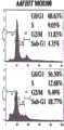
为了检查药物处理后的细胞活力,我们进行了流式细胞术分析(图6,图6C和图6D);用增加的紫杉醇浓度(50-500ng/ml)处理的PonA-诱导的D1和E1细胞显示增加的48h sub-G1群体:对于D1细胞分别为9.6%、36%和40%,相比之下E1细胞的为4%、16.7%和30%(图6C)。To examine cell viability after drug treatment, we performed flow cytometric analysis (Figure 6, Figure 6C and Figure 6D); PonA-induced D1 and E1 cells treated with increasing paclitaxel concentrations (50-500 ng/ml) An increased 48h sub-G1 population was shown: 9.6%, 36% and 40% for D1 cells, respectively, compared to 4%, 16.7% and 30% for E1 cells (Fig. 6C).
相似地,增加的顺铂浓度(0.05-0.2mM)导致增加的48h sub-G1群体:D1细胞中的分别为5%、16.2%和30%,相比之下E1细胞的为2.3%、7%和14.6%(图6C)。Similarly, increasing cisplatin concentrations (0.05-0.2 mM) resulted in increased 48h sub-G1 populations: 5%, 16.2% and 30%, respectively, in D1 cells compared to 2.3%, 16.2% and 30% in E1 cells. 7% and 14.6% (Fig. 6C).
在24和72h(数据未显示),也检测到与E1细胞相比D1中增加的sub-G1群体。为了确定D1细胞的sub-G1级分是否代表凋亡细胞,制备了48h的药物处理细胞的溶胞产物,并进行了半胱天冬酶3和Parp1切割的免疫印迹分析(图6D)。At 24 and 72h (data not shown), an increased sub-G1 population was also detected in D1 compared to E1 cells. To determine whether the sub-G1 fraction of D1 cells represented apoptotic cells, lysates of drug-treated cells at 48 h were prepared and subjected to immunoblot analysis for
与未处理的细胞(对照)相比,观察到紫杉醇(50和100ng/ml)和顺铂(0.05和0.1mM)处理后激活的半胱天冬酶3和相关的Parp1切割。本发明人在此现在认为Fhit表达通过与Fdxr一起参与ROS产生增加了对氧化损伤的敏感性。Activation of
实施例IExample I
材料和方法Materials and methods
细胞、载体和抗血清-A549、H1299、MKN74-E4和A116和HCT116细胞在加10%胎牛血清和青霉素/链霉素(Sigma)的RPMI 1640培养基上维持。用于制备重组腺病毒的HEK293细胞(Microbix)在加10%胎牛血清和青霉素/链霉素的Dulbecco改良的Eagle培养基中培养。AdFHIT-His6病毒的制备如本文的实施例II中所述。[His6-SEQ IDNO:32][penta-His-SEQ ID NO:33]。Cells, Vector and Antisera - A549, H1299, MKN74-E4 and A116 and HCT116 cells were maintained in RPMI 1640 medium plus 10% fetal calf serum and penicillin/streptomycin (Sigma). HEK293 cells (Microbix) used to prepare recombinant adenoviruses were cultured in Dulbecco's modified Eagle's medium plus 10% fetal bovine serum and penicillin/streptomycin. AdFHIT-His6 virus was prepared as described in Example II herein. [His6-SEQ ID NO: 32][penta-His-SEQ ID NO: 33].
从人脑部cDNA(Clontech)中克隆了全长FDXR,将其亚克隆到pcDNA3.1/V5-HisTOPO TA载体(Invitrogen)并测序;详情如在下文所附方法所述。使用LipofectamineTM(Invitrogen)根据生产商的指导转染细胞。Full-length FDXR was cloned from human brain cDNA (Clontech), subcloned into the pcDNA3.1/V5-HisTOPO TA vector (Invitrogen) and sequenced; details are described in the accompanying methods below. Cells were transfected using Lipofectamine™ (Invitrogen) according to the manufacturer's instructions.
蛋白质印迹分析-如(13)所述使用以下进行免疫印迹分析:单克隆抗pentaHis(Qiagen);兔多克隆抗Fhit(Zymed LaboratoriesInc.);针对GFP、Hsp60、Hsp10和细胞色素c的兔多克隆抗血清(SantaCruz Biotechnology);兔多克隆抗Fdxr(Abcam);单克隆抗CoxIV(Molecular Probes);抗V5(Sigma);抗Parp1(Santa CruzBiotechnology);和抗半胱天冬酶3(Cell Signaling)。蛋白水平相对于β-肌动蛋白或/和GAPDH3水平进行标准化,用适当的抗血清(Santa Cruz Biotechnology)进行检测。Western blot analysis - Western blot analysis was performed as described in (13) using: monoclonal anti-pentaHis (Qiagen); rabbit polyclonal anti-Fhit (Zymed Laboratories Inc.); rabbit polyclonal against GFP, Hsp60, Hsp10 and cytochrome c Antiserum (Santa Cruz Biotechnology); rabbit polyclonal anti-Fdxr (Abcam); monoclonal anti-CoxIV (Molecular Probes); anti-V5 (Sigma); anti-Parp1 (Santa Cruz Biotechnology); . Protein levels were normalized to β-actin or/and GAPDH3 levels and detected with appropriate antisera (Santa Cruz Biotechnology).
质谱研究-将蛋白沉淀溶解,并通过胰蛋白酶消化,如本文所述。注射肽混合物用于LC-MS/MS分析。通过数据库搜索鉴定蛋白后,进行LC-MS/MS数据检查以评估属于用AdFHIT-His 6感染的细胞的样品中的候选配偶体蛋白的质量峰的唯一存在。Mass Spectrometry Studies - Protein pellets were solubilized and digested by trypsinization as described here. The peptide mixture was injected for LC-MS/MS analysis. After identifying the protein by database search, LC-MS/MS data inspection was performed to assess the unique presence of mass peaks belonging to the candidate partner protein in samples of cells infected with AdFHIT-
蛋白质相互作用分析-在15mM Tris-Cl、pH 7.5、120mM NaCl、25mM KCl、2mM EGTA、0.1mM二硫苏糖醇、0.5%Triton X-100、10mg/ml亮肽素、0.5mM苯甲磺酰氯中提取蛋白。在有或无二硫代二(琥珀酰亚胺丙酸酯)(DSP)下,通过如下进行免疫共沉淀实验:将1mg总蛋白与轭合琼脂糖凝胶(Sepharose)的Hsp60、Hsp10、Fdxr、penta-His和V5抗血清在4℃下孵育2h;清洗后,将珠在1x SDS样品缓冲液中煮沸并在4-20%聚丙烯酰胺凝胶(Bio-Rad)上分离蛋白,转移至聚(偏二氟乙烯)滤膜(poly(vinylidene difluoride)filter)(Millipore),并用特异性抗血清探测。Protein Interaction Assay - In 15mM Tris-Cl, pH 7.5, 120mM NaCl, 25mM KCl, 2mM EGTA, 0.1mM Dithiothreitol, 0.5% Triton X-100, 10mg/ml Leupeptin, 0.5mM Phenylsulfonate Extract protein from acid chloride. Co-immunoprecipitation experiments were performed in the presence or absence of dithiobis(succinimidyl propionate) (DSP) by mixing 1 mg of total protein with Sepharose-conjugated Hsp60, Hsp10, Fdxr , penta-His and V5 antiserum were incubated at 4°C for 2h; after washing, the beads were boiled in 1x SDS sample buffer and the proteins were separated on a 4-20% polyacrylamide gel (Bio-Rad), transferred to Poly(vinylidene difluoride) filter (Millipore) and probed with specific antiserum.
Fhit蛋白的亚细胞定位-通过使用抗Fhit血清的间接免疫荧光检测在松甾酮A(PonA)-诱导的、Fhit-表达H1299D1细胞中对Fhit进行亚定位,并通过使用抗pentaHis在免疫电子显微图中检测A549AdFHIT-His6-感染的细胞中的FhitHis6对Fhit进行亚定位。在分级研究中,使用线粒体/胞质溶胶分级试剂盒分离线粒体,并使用FractionPREPTM细胞分级系统从胞质溶胶、膜、核和细胞骨架中提取蛋白(Biovision Research Products)。对于根据Dahéron等人的方法(22)的亚线粒体定位,将线粒体重悬于在冰上的0.1M碳酸钠、pH11.5保持30min,且定期涡旋,并如本文所述进行分级。Subcellular localization of Fhit protein - Sublocalization of Fhit in Ponasterone A (PonA)-induced, Fhit-expressing H1299D1 cells detected by indirect immunofluorescence using anti-Fhit serum and detected by immunoelectron microscopy using anti-pentaHis Sublocalization of Fhit by detection of FhitHis6 in A549AdFHIT-His6 -infected cells in micromaps. In fractionation studies, mitochondria were isolated using the Mitochondrial/Cytosol Fractionation Kit, and proteins were extracted from the cytosol, membrane, nucleus, and cytoskeleton using the FractionPREP™ Cell Fractionation System (Biovision Research Products). For submitochondrial localization according to the method of Dahéron et al. (22), mitochondria were resuspended in 0.1 M sodium carbonate, pH 11.5 on ice for 30 min with periodic vortexing and fractionated as described herein.
流式细胞术-以m.o.i.50和100用AdFHIT或Ad GFP感染HCT116FDXR+/+/+和FDXR+/-/-细胞,并在感染后48h进行评估。用0.25和0.5mMH2O2或用化疗药物处理PonA-诱导的H1299D1和E1细胞并孵育不同的时间,如文本和图中所示。对于两种实验,收集细胞,用磷酸缓冲盐水清洗并重悬于冷70%乙醇。对于分析,将细胞离心沉淀,在磷酸缓冲盐水中清洗,并在室温重悬于0.1mg/ml碘化丙啶/Triton X-100染色溶液(0.1%Triton X-100、0.2mg/ml无DNA酶的RNA酶A)保持30,并通过流式细胞术分析。Flow Cytometry - HCT116FDXR+/+/+ and FDXR+/-/- cells were infected with AdFHIT or Ad GFP at moi of 50 and 100 and evaluated 48h after infection. PonA-induced H1299D1 and E1 cells were treated with 0.25 and0.5 mMH2O2 or with chemotherapeutic drugs and incubated for different times, as indicated in the text and figures. For both experiments, cells were harvested, washed with phosphate buffered saline and resuspended in cold 70% ethanol. For analysis, cells were pelleted, washed in phosphate-buffered saline, and resuspended in 0.1 mg/ml propidium iodide/Triton X-100 staining solution (0.1% Triton X-100, 0.2 mg/ml DNA-free Enzyme RNase A) was maintained at 30 and analyzed by flow cytometry.
胞内活性氧簇(ROS)的评估-通过氢化乙啡啶(二氢乙啡啶-HE;Molecular Probes)氧化造成的乙啡啶荧光测量胞内超氧化物。在37℃下,用0.5、1.0、2.0和4.0mM H2O2处理稳定表达Fhit的MNK74细胞、瞬时表达Fhit的A549细胞和可诱导地表达Fhit的H1299细胞;4h后,向细胞中添加氢化乙啡啶(10μM)并在37℃下孵育15min。通过流式细胞术测量荧光。二氯荧光素二乙酸酯(DCFH-DA)(Molecular Probes)用于表达诱导的Fhit的D1细胞,用H2O2(0.1至1.0mM)施加应激,用10μM DCFH-DA处理,在37℃下孵育1h。通过流式细胞术在FAC-Scan流式细胞仪上和通过荧光显微术测量DCF荧光。Assessment of Intracellular Reactive Oxygen Species (ROS) - Intracellular superoxide was measured by ethidium fluorescence due to oxidation of hydroethidium (Dihydroethidium-HE; Molecular Probes). MNK74 cellsstably expressing Fhit, A549 cells transiently expressing Fhit, and H1299 cells inducibly expressing Fhit were treated with 0.5, 1.0, 2.0, and 4.0 mMH2O2 at 37°C; 4 h later, hydrogenation was added to the cells ethiphenidine (10 μM) and incubated at 37° C. for 15 min. Fluorescence was measured by flow cytometry. Dichlorofluorescein diacetate (DCFH-DA) (Molecular Probes) was used in D1 cells expressing induced Fhit, stressed with H2 O2 (0.1 to 1.0 mM), treated with 10 μM DCFH-DA, at Incubate at 37°C for 1h. DCF fluorescence was measured by flow cytometry on a FAC-Scan flow cytometer and by fluorescence microscopy.
Hsp60和Hsp10沉默-通过Lipofectamine 2000试剂(Invitrogen)和6μg Hsp60和/或Hsp10 siRNA(Dharmacon目录号分别为NM_002156[GenBank]和NM_002157[GenBank])转染8x10⁵/孔(6孔板)的A549肺癌细胞;48h后,以m.o.i.1用AdFHIT感染细胞,并在24h后收集用于胞质溶胶/线粒体蛋白分级。通过SDS-PAGE和免疫印迹分析蛋白;用Hsp60、Hsp10和Fhit抗血清探测滤膜。蛋白上样用GAPDH和CoxIV进行标准化。1x106H1299D1和E1肺癌细胞如上所述地进行转染,并在转染后24h,对细胞进行PonA-诱导;诱导后48h,进行1、4、6和12h环己酰亚胺(CHX)(10μg/ml)追踪,并如本文所述地分析蛋白溶胞产物。Hsp60 and Hsp10 silencing -8x10⁵ /well (6-well plate) of A549 lung cancers were transfected by Lipofectamine 2000 reagent (Invitrogen) and 6 μg of Hsp60 and/or Hsp10 siRNA (Dharmacon catalog numbers NM_002156 [GenBank] and NM_002157 [GenBank], respectively) Cells; after 48h, cells were infected with AdFHIT at moi1 and harvested after 24h for cytosolic/mitochondrial protein fractionation. Proteins were analyzed by SDS-PAGE and immunoblotting; filters were probed with Hsp60, Hsp10 and Fhit antisera. Protein loading was normalized with GAPDH and CoxIV. 1×10 6 H1299D1 and E1 lung cancer cells were transfected as described above, and 24 h after transfection, the cells were subjected to PonA-induction; 48 h after induction, 1, 4, 6 and 12 h of cycloheximide (CHX) ( 10 μg/ml), and protein lysates were analyzed as described herein.
实对RT-PCR-使用TRIzol试剂(Invitrogen)分离的总RNA在DNA酶处理(Ambion)后直接通过使用SuperScript First-Strand(Invitrogen)的反转录加工为cDNA。使用Power SYBR Green PCRMaster Mix(Applied Biosystems)通过qPCR扩增靶序列。FDXR引物为:正向,3′-TCGACCCAAGCGTGCCCTTTG-5′[SEQ ID No.24];反向,3′-GTGGCCCAGGAGGCGCAGCATC-5′[SEQ ID No.25]。使用肌动蛋白和GADPH基因将样品标准化。Real RT-PCR - Total RNA isolated using TRIzol reagent (Invitrogen) was processed to cDNA by reverse transcription using SuperScript First-Strand (Invitrogen) directly after DNase treatment (Ambion). Target sequences were amplified by qPCR using Power SYBR Green PCRMaster Mix (Applied Biosystems). FDXR primers are: forward, 3′-TCGACCCAAGCGTGCCCTTTG-5′ [SEQ ID No.24]; reverse, 3′-GTGGCCCAGGAGGCGCAGCATC-5′ [SEQ ID No.25]. Samples were normalized using the actin and GADPH genes.
化疗药物处理-将紫杉醇(Sigma)溶解于DMSO作为10mmol/L贮液并储存于-80℃。将顺铂(Sigma)溶解于水并在使用前新鲜配制。将H1299D1和E1细胞接种于(1x104个细胞/孔)96孔培养板中,PonA-诱导,并在24h后用紫杉醇(50、100和500ng/ml)或顺铂(0.05、0.1和0.2mM)处理。将PonA-诱导的H1299D1和E1细胞孵育24、48和72h,并使用MTS试剂盒(Cell Titer
实施例IIIExample III
重组腺病毒的产生-携带野生型FHIT cDNA的重组腺病毒(AdFHIT)按照之前的描述制备(Ishii等人,2001 Cancer Res 61:1578-1584)。His标签的FHIT cDNA通过使用下列寡核苷酸的PCR产生:5′-ACgTggATCCCTgTgAggACATgTCgTTCAgATTTggC-3′(正向)[SEQ IDNO:26]和5′-TTgTggATCCTTATCAgTgATggTgATggTgATgCgATCCTCTCTgAAAgTAgCCCgCAg-3′[SHQ ID NO:27]。这些引物设计为具有BamHI限制位点以亚克隆至转移载体pAdenoVator-CMV5-IRES-GFP。Ad-His 6是使用AdenoVatorTM试剂盒(Qbiogene,Carlsbad,CA)按照生产商的方法产生的。用作对照的Ad GFP购自Qbiogene(Carlsbad,CA)。Production of Recombinant Adenovirus - Recombinant adenovirus carrying wild-type FHIT cDNA (AdFHIT) was prepared as previously described (Ishii et al., 2001 Cancer Res 61: 1578-1584). His-tagged FHIT cDNA was generated by PCR using the following oligonucleotides: 5'-ACgTggATCCCTgTgAggACATgTCgTTCAgATTTggC-3' (forward) [SEQ ID NO: 26] and 5'-TTgTggATCCTTATCAgTgATggTgATggTgATgCgATCCTCTCTgAAAgTAgCCCgCAg-3' [SHQ ID NO: 27]. These primers were designed with a BamHI restriction site for subcloning into the transfer vector pAdenoVator-CMV5-IRES-GFP. Ad-His 6 was generated using the AdenoVator™ kit (Qbiogene, Carlsbad, CA) following the manufacturer's protocol. Ad GFP used as a control was purchased from Qbiogene (Carlsbad, CA).
携带FDXR cDNA的重组表达载体的产生-使用以下引物从人脑部cDNA(Clontech,Palo Alto,CA)扩增野生型铁氧还蛋白还原酶全长:5’-CTgTTCCCAgCCATggCTTCgCgCTg-3’(正向)[SEQ ID NO:28]和5’-TCAgTggCCCAggAggCgCAgCATC-3’[SEQ ID NO:29]。将扩增产物亚克隆至pcDNA 3.1/V5-His TOPO TA载体(Invitrogen,Carlsbad,CA)。测序排除了扩增产物中的突变。Generation of recombinant expression vector carrying FDXR cDNA - full-length wild-type ferredoxin reductase was amplified from human brain cDNA (Clontech, Palo Alto, CA) using the following primers: 5'-CTgTTCCCAgCCATggCTTCgCgCTg-3' (forward) [SEQ ID NO:28] and 5'-TCAgTggCCCAggAggCgCAgCATC-3'[SEQ ID NO:29]. The amplified product was subcloned into the pcDNA 3.1/V5-His TOPO TA vector (Invitrogen, Carlsbad, CA). Sequencing excluded mutations in the amplified products.
对于V5标签的铁氧还蛋白还原酶cDNA的制备,使用排除反向引物中的FDXR生理终止密码子的相同引物序列进行PCR扩增。也就是说,野生型和V5标签的铁氧还蛋白还原酶(FDXR)cDNA二者均是通过使用人铁氧还蛋白还原酶基因(GenBank登录号NM_024417)的野生型编码序列作为模板制备的。该铁氧还蛋白还原酶编码序列是从人脑部cDNA(Clontech)扩增的。For the preparation of V5-tagged ferredoxin reductase cDNA, PCR amplification was performed using the same primer sequence excluding the FDXR physiological stop codon in the reverse primer. That is, both wild-type and V5-tagged ferredoxin reductase (FDXR) cDNAs were prepared by using the wild-type coding sequence of the human ferredoxin reductase gene (GenBank Accession No. NM_024417) as a template. The ferredoxin reductase coding sequence was amplified from human brain cDNA (Clontech).
用于产生V5标签的FDXR cDNA的引物为:正向:5’-CTgTTCCCAgCCATggCTTCgCgCTg-3’[SHQ ID NO:30];和反向:5’-gTggCCCAggAggCgCAgCATC-3’[SEQ ID NO:31]。应注意,除了用于产生V5标签的cDNA的反向引物之外,寡核苷酸序列是相同的,在所述反向引物中,FDXR的生理终止密码子被省略以符合读框地将FDXR编码序列与V5标签融合。The primers used to generatethe V5 tagged FDXR cDNA were: forward: 5'-CTgTTCCCAgCCATggCTTCgCgCTg-3' [SHQ ID NO: 30]; and reverse: 5'-gTggCCCAggAggCgCAgCATC-3' [SEQ ID NO: 31]. Note that the oligonucleotide sequences are identical except for the reverse primer used to generate the V5-tagged cDNA in which the physiological stop codon for FDXR was omitted to place FDXR in-frame. The coding sequence is fused to the V5 tag.
将扩增产物亚克隆至pcDNA3.1/V5-HisTOPO TA载体(Invitrogen,Carlsbad,CA)。测序排除扩增产物中的突变。The amplified product was subcloned into the pcDNA3.1/V5-HisTOPO TA vector (Invitrogen, Carlsbad, CA). Sequencing excluded mutations in the amplified products.
在某些实施方案中,能够分离Fhit-His6的腺病毒包括携带FHIT-His6 cDNA的腺病毒。FHIT-His6 cDNA可通过使用人FHIT基因(GenBank登录号NM_002012)的野生型编码序列作为模板来制备。为了在最终Fhit产物的C-末端引入多聚组氨酸标签,野生型FHIT编码序列的PCR扩增使用设计为去除生理终止密码子并向内源FHIT序列添加后接人工终止密码子的编码六个组氨酸的18bp片段的反向引物进行。另外,正向和反向两种引物均携带BamHI限制位点以易于亚克隆。用于此扩增的寡核苷酸序列是如下序列:正向:5′-ACgTggATCCCTgTgAggACATgTCgTTCAgATTTggC-3′[SEQ ID NO:26];和反向:5’-TTgTggATCCTTATCAgTgATggTgATggTgATgCgATCCTCTCTgAAAgTAgACCCgCAg-3’[SEQID NO:27]。对PCR扩增产物进行测序以在克隆到Ad5重组基因组(AdenoVatorTM,一种由Qbiogene购买的载体)之前排除随机突变。In certain embodiments, an adenovirus capable of isolating Fhit-His6 includes an adenovirus carrying a FHIT-His6 cDNA. FHIT-His6 cDNA can be prepared by using the wild-type coding sequence of the human FHIT gene (GenBank Accession No. NM_002012) as a template. To introduce a polyhistidine tag at the C-terminus of the final Fhit product, PCR amplification of the wild-type FHIT coding sequence used a coding sequence designed to remove a physiological stop codon and add to the endogenous FHIT sequence followed by an artificial stop codon. The reverse primer of the 18bp fragment of histidine was carried out. Additionally, both forward and reverse primers carry BamHI restriction sites for easy subcloning. The oligonucleotide sequences used for this amplification were the following sequences: forward: 5'-ACgTggATCCCTgTgAggACATgTCgTTCAgATTTggC-3' [SEQ ID NO: 26]; and reverse: 5'-TTgTggATCCTTATCAgTgATggTgATggTgATgCgATCCTCTCTgAAAgTAgACCCgCAg-3' [SEQ ID NO: 27] . PCR amplification products were sequenced to rule out random mutations before cloning into the Ad5 recombinant genome (AdenoVatorTM, a vector purchased from Qbiogene).
在某些实施方案中,分离外源过表达Fhit-His6的方法包括使用携带FHIT-His6 cDNA的腺病毒,其中通过His标签分离Fhit-His6。Fhit-His6代表通过Ad FHIT-His6载体被驱动在哺乳动物细胞中表达的重组蛋白。His6表位允许通过利用Ni-NTA系统回收重组Fhit-His6蛋白以及与所述重组蛋白本身相互作用的蛋白复合体。该系统是从Qiagen可商业获得的。简要而言,用Ad FHIT-His6感染人A549癌细胞;感染后48小时,使用购自Pierce的交联剂二硫代二(琥珀酰亚胺丙酸酯)[DSP]进行胞内蛋白复合体的光交联以稳定活细胞内的蛋白复合体。在蛋白提取缓冲液中破裂细胞,并使用Ni-NTA磁珠技术分离Fhit-His6蛋白复合体,该技术是通过利用His6标签对此类珠的强亲和力。然后通过质谱研究分离的Fhit-His6蛋白复合体以鉴定该复合体中存在的所有蛋白。In certain embodiments, the method of isolating exogenously overexpressed Fhit-His6 comprises using an adenovirus carrying the FHIT-His6 cDNA, wherein Fhit-His6 is isolated by a His tag. Fhit-His6 represents a recombinant protein expressed in mammalian cells driven by the Ad FHIT-His6 vector. The His6 epitope allows recovery of recombinant Fhit-His6 protein as well as protein complexes interacting with the recombinant protein itself by utilizing the Ni-NTA system. This system is commercially available from Qiagen. Briefly, human A549 cancer cells were infected with Ad FHIT-His6; 48 hours after infection, intracellular protein complexes were analyzed using the cross-linker dithiobis(succinimidyl propionate) [DSP] purchased from Pierce photocrosslinking to stabilize protein complexes in living cells. Cells were disrupted in protein extraction buffer and Fhit-His6 protein complexes were isolated using Ni-NTA magnetic bead technology by exploiting the strong affinity of the His6 tag for such beads. The isolated Fhit-His6 protein complex was then studied by mass spectrometry to identify all proteins present in the complex.
在某些实施方案中,携带脆性组氨酸三联体(Fhit)FHIT cDNA的重组腺病毒可被修饰为在FHIT cDNA的3′带有编码六组氨酸表位标签的序列(AdFHIT-His6)。In certain embodiments, the recombinant adenovirus carrying the fragile histidine triplet (Fhit) FHIT cDNA can be modified to have a sequence encoding a hexahistidine epitope tag (AdFHIT-His6) at the 3′ of the FHIT cDNA .
在某些实施方案中,在至少一个细胞中介导凋亡过程的方法包括使所述细胞暴露于足以在所述细胞中介导凋亡过程的量的脆性组氨酸三联体(Fhit)基因产物。In certain embodiments, the method of mediating an apoptotic process in at least one cell comprises exposing said cell to a fragile histidine triad (Fhit) gene in an amount sufficient to mediate an apoptotic process in said cell product.
在某些实施方案中,在细胞中诱导凋亡过程的方法包括使所述细胞暴露于足以在所述细胞中引起活性氧簇(ROS)产生的量的脆性组氨酸三联体(Fhit)基因产物。In certain embodiments, the method of inducing an apoptotic process in a cell comprises exposing the cell to the fragile histidine triad (Fhit) gene in an amount sufficient to cause production of reactive oxygen species (ROS) in the cell product.
在某些实施方案中,在至少一个细胞中介导凋亡过程的方法包括:使所述细胞暴露于足量的脆性组氨酸三联体(Fhit)基因产物以允许Fhit进入细胞的线粒体并与细胞中的Fdxr蛋白相互作用,从而引起与ROS产生相关的Fdxr蛋白水平增加,并引起细胞中凋亡过程的变化。In certain embodiments, the method of mediating the apoptotic process in at least one cell comprises: exposing the cell to a sufficient amount of the fragile histidine triad (Fhit) gene product to allow Fhit to enter the mitochondria of the cell and associate with Fdxr proteins in cells interact, thereby causing an increase in the level of Fdxr proteins associated with ROS production and causing changes in the apoptotic process in cells.
应注意,在先前的研究中,已广泛证明Fhit阴性癌细胞中的Fhit蛋白过表达能够触发程序性细胞死亡(或凋亡)。在本发明中,发明人提供了关于Fhit蛋白在凋亡过程中的作用的基本原理。事实上,用Ad FHIT(以感染复数50或MOI50,即每个细胞50个病毒粒子)对癌细胞进行的FHIT基因治疗负责Fhit过表达;然后,新合成的重组蛋白被其相互作用物Hsp60/Hsp10从胞质溶胶带到线粒体,在线粒体中,Fhit与属于呼吸链的蛋白FDXR(铁氧还蛋白还原酶)相互作用。此相互作用导致线粒体产生ROS(活性氧簇)。ROS代表启动凋亡过程的内在(或线粒体)途径的早期步骤;事实上,它们诱导线粒体膜的损伤,该损伤依次释放细胞色素c到胞质溶胶。该步骤对于凋亡的执行是至关重要的,因为细胞色素c与其他胞质溶胶分子(即Apaf-1和半胱天冬酶3酶原(pro-caspase 3))促成凋亡体的产生,凋亡体是能够驱动细胞以不可逆方式凋亡的多蛋白复合体。It should be noted that in previous studies, it has been widely demonstrated that overexpression of Fhit protein in Fhit-negative cancer cells can trigger programmed cell death (or apoptosis). In the present invention, the inventors provide a rationale for the role of Fhit protein in the process of apoptosis. In fact, FHIT gene therapy of cancer cells with Ad FHIT (at multiplicity of
研究凋亡常用的方法在于通过流式细胞术分析(BectonDickinson)检测成熟半胱天冬酶-3(初期凋亡的指示物)[有关参考文献,参见Trapasso等人,2003,PNAS,100,1592-1597]。A common method for studying apoptosis consists in the detection of mature caspase-3 (indicator of incipient apoptosis) by flow cytometry analysis (Becton Dickinson) [For references, see Trapasso et al., 2003, PNAS, 100, 1592 -1597].
在某些实施方案中,制备V5标签的铁氧还蛋白还原酶cDNA的方法包括用以下引物序列进行PCR扩增:5′-CTgTTCCCAgCCATggCTTCgCgCTg-3′(正向)[SEQ ID NO:28]和5′-TCAgTggCCCAggAggCgCAgCATC-3′[SEQ ID NO:29],并将扩增产物亚克隆至pcDNA3.1/V5-HisTOPO TA载体。In certain embodiments, the method of preparing a V5-tagged ferredoxin reductase cDNA comprises performing PCR amplification with the following primer sequences: 5′-CTgTTCCCAgCCATggCTTCgCgCTg-3′ (forward) [SEQ ID NO: 28] and 5 '-TCAgTggCCCAggAggCgCAgCATC-3'[SEQ ID NO: 29], and the amplified product was subcloned into the pcDNA3.1/V5-HisTOPO TA vector.
野生型和V5标签的铁氧还蛋白还原酶(FDXR)cDNA二者均是通过使用人铁氧还蛋白还原酶基因(GenBank登录号NM_024417)的野生型编码序列作为模板制备的。该铁氧还蛋白还原酶编码序列是从人脑部cDNA(Clontech)扩增的。也就是说,用于扩增野生型FDXR cDNA的引物为:正向:5’-CTgTTCCCAgCCATggCTTCgCgCTg-3’[SEQ ID NO:28];反向:5’-TCAgTggCCCAggAggCgCAgCATC-3’[SEQ ID NO:29]。Both wild-type and V5-tagged ferredoxin reductase (FDXR) cDNAs were prepared by using the wild-type coding sequence of the human ferredoxin reductase gene (GenBank Accession No. NM_024417) as a template. The ferredoxin reductase coding sequence was amplified from human brain cDNA (Clontech). That is, the primers used to amplifywild-type FDXR cDNA are: forward: 5'-CTCCCAgCCATggCTTCgCgCTg-3' [SEQ ID NO: 28]; reverse: 5'-TCAgTggCCCAggAggCgCAgCATC-3' [SEQ ID NO: 29 ].
用于产生V5标签的FDXR cDNA的引物为:正向:5’-CTgTTCCCAgCCATggCTTCgCgCTg-3’[SEQ ID NO:30],和反向:5’-gTggCCCAggAggCgCAgCATC-3’[SEQ ID NO:311。应注意,除了用于产生V5标签的cDNA的反向引物之外,寡核苷酸序列是相同的,在所述反向引物中,FDXR的生理终止密码子被省略以符合读框地将FDXR编码序列与V5标签融合。The primers used to generatethe V5 tagged FDXR cDNA were: forward: 5'-CTgTTCCCAgCCATggCTTCgCgCTg-3' [SEQ ID NO: 30], and reverse: 5'-gTggCCCAggAggCgCAgCATC-3' [SEQ ID NO: 311 . Note that the oligonucleotide sequences are identical except for the reverse primer used to generate the V5-tagged cDNA in which the physiological stop codon for FDXR was omitted to place FDXR in-frame. The coding sequence is fused to the V5 tag.
最后,将两种产物亚克隆至pcDNA3.1/V5-HisTOPO TA表达载体(购自Invitrogen),然后测序以评估两种产物不合随机突变。Finally, the two products were subcloned into the pcDNA3.1/V5-HisTOPO TA expression vector (purchased from Invitrogen), and then sequenced to assess whether the two products did not contain random mutations.
在某些实施方案中,用于产生重组腺病毒的方法包括制备携带野生型FHIT cDNA的重组腺病毒(AdFHIT);和使用以下寡核苷酸进行的PCR产生His标签的FHIT cDNA:5′-ACgTggATCCCTgTgAggACATgTCgTTCAgATTTggC-3′(正向)[SEQ ID NO:26],和5′-TTgTggATCCTTATCAgTgATggTgATggTgATgCgATCCTCTCTgAAAgTAgACCCgCAg-3′[SEQ ID NO:27]。FHIT-His6 cDNA的制备如本文所述。重组腺病毒载体Ad FHIT-His6根据生产商的建议制备(Qbiogene)。简要而言,用BamHI消化扩增的FHIT-His6 PCR片段,并亚克隆至BamHI线性化的转移载体pAdenoVator-CMV5-IRES-GFP。将pAdenoVator-CMV5-IRES-GFP/FHIT-His6与E1/E3缺失的Ad5主链病毒DNA共转染到293细胞。通过使用特异性penta-His抗体(Qiagen)的蛋白质印迹筛选病毒噬斑中Fhit-His6蛋白的存在。噬斑纯化一个阳性克隆并在293细胞中扩增。冷冻/解冻循环之后,在两个连续的氯化铯梯度上纯化上清液中的腺病毒。通过TCID50法滴定重组腺病毒并分成等份。病毒贮液在-80℃储存。最后,携带野生型FHIT cDNA的重组腺病毒(Ad FHIT)是Trapasso等人(2003,PNAS,100,1592-1597)以前制备的。In certain embodiments, the method for producing a recombinant adenovirus comprises preparing a recombinant adenovirus carrying a wild-type FHIT cDNA (AdFHIT); and generating a His-tagged FHIT cDNA by PCR using the following oligonucleotides: 5'- ACgTggATCCCTgTgAggACATgTCgTTCAgATTTggC-3' (forward) [SEQ ID NO: 26], and 5'-TTgTggATCCTTATCAgTgATggTgATggTgATgCgATCCTCTCTgAAAgTAgACCCgCAg-3' [SEQ ID NO: 27]. FHIT-His6 cDNA was prepared as described herein. The recombinant adenoviral vector Ad FHIT-His6 was prepared according to the manufacturer's recommendations (Qbiogene). Briefly, the amplified FHIT-His6 PCR fragment was digested with BamHI and subcloned into the BamHI linearized transfer vector pAdenoVator-CMV5-IRES-GFP. pAdenoVator-CMV5-IRES-GFP/FHIT-His6 was co-transfected with E1/E3 deleted Ad5 backbone viral DNA into 293 cells. Viral plaques were screened for the presence of Fhit-His6 protein by Western blotting using a specific penta-His antibody (Qiagen). One positive clone was plaque purified and expanded in 293 cells. Following freeze/thaw cycles, the adenovirus in the supernatant was purified on two successive cesium chloride gradients. Recombinant adenovirus was titrated by TCID50 method and aliquoted. Virus stocks were stored at -80°C. Finally, a recombinant adenovirus carrying wild-type FHIT cDNA (Ad FHIT) was previously prepared by Trapasso et al. (2003, PNAS, 100, 1592-1597).
线粒体定位研究-使用共聚焦显微术通过免疫荧光评估Fhit蛋白分布;具有可诱导的FHIT cDNA的H1299 D1细胞和具有空载体的E1细胞用PonA处理48hr,并用Mitotracker Red 580(M-22425,Molecular Probes,Eugene,OR)以500nM的工作浓度在生长条件下对活细胞染色40min。固定细胞,并通过在冰冷的丙酮中孵育5min然后在PBS中清洗使细胞可渗透。使细胞与5%BSA孵育1hr以阻断非特异的相互作用然后与工作浓度为1.6μg/ml的Fhit抗血清(Zymed,S.San Francisco,CA)孵育过夜,用PBS清洗,并用AlexaFluor 488驴抗兔IgG(Molecular Probes)孵育。将载玻片封闭在封闭介质中以获得DAPI(Vector,Burlingane,CA)的荧光并显现。对于Fhit的免疫电子显微术定位,用AdHis6或AdFHIT MOI 5感染的A549细胞在于PBS pH7.2的4%多聚甲醛中在4℃下固定30min,用PBS清洗3次,并使用在于PBS中的0.05%氢化硼钠中孵育30min还原剩余的游离醛基。PBS清洗之后,用于PBS中的50mM甘氨酸封闭样品30min,在PBS中清洗两次,并在25%和50%乙醇中各脱水15min,然后换3次70%乙醇,每次15min。然后用2∶1的70%乙醇+硬质LR White树脂(Electron Microscopy Sciences,Hatfield,PA)渗透样品1hr,1∶2的70%乙醇+LR White渗透1hr,100%LR White渗透1hr和100%LRW在4℃下过夜。第二天细胞再接受多于2次100%LR White更换,并在58℃下于明胶胶囊中聚合20-24hr。使用Reichert UCTUltramicrotome和钻石刀切出900nm薄切片并置于镍网上。使镍网以切片侧向下浮于PBS液滴5min,在室温(RT)下浮于在PBS中的5%山羊血清1hr,然后在潮湿室中在4℃下浮于含有0.1%BSA和0.05%Tween-20(BSA/Tw)的PBS稀释的20mg/ml的Penta-His小鼠单克隆抗体(Qiagen,Valencia,CA)或单独的BSA/Tw过夜。第二天,用PBS洗涤网6次,每次5分钟,然后与1∶10稀释于BSA/Tw的山羊抗小鼠10nm胶体金轭合物(Ted Pella,Redding,CA)在RT下孵育2hr。用PBS洗涤网6次,每次5分钟,用DI H2O润洗,然后用2.5%乙酸双氧铀水溶液后染色3min。在配备有US1000 Gatan 2K数码相机的Tecnai 12电子显微镜上收集图像。Mitochondrial localization study - assessment of Fhit protein distribution by immunofluorescence using confocal microscopy; H1299 D1 cells with inducible FHIT cDNA and E1 cells with empty vector were treated with PonA for 48hr and treated with Mitotracker Red 580 (M-22425, Molecular Probes, Eugene, OR) stained live cells for 40 min under growth conditions at a working concentration of 500 nM. Cells were fixed and made permeable by incubation in ice-cold acetone for 5 min followed by washing in PBS. Cells were incubated with 5% BSA for 1 hr to block non-specific interactions and then incubated overnight with Fhit antiserum (Zymed, S. San Francisco, CA) at a working concentration of 1.6 μg/ml, washed with PBS, and washed with AlexaFluor 488 Anti-rabbit IgG (Molecular Probes) incubation. Slides were blocked in blocking medium for DAPI (Vector, Burlingane, CA) fluorescence and visualized. For immunoelectron microscopy localization of Fhit, A549 cells infected with AdHis6 or
样品消化和LC-MS/MS分析-将用Ni-NTA珠分离的蛋白用冷丙酮沉淀,并重悬于用0.1M Tris-HCl缓冲至pH 8的6M尿素。通过加入DTT(终浓度10mM,在37℃下孵育1hr)和碘乙酰胺(终浓度25mM,在37℃下孵育1hr)分别进行蛋白还原和烷基化。用DTT(另外的5mM)中和过量的碘乙酰胺后,通过用1mM CaCl2稀释将尿素浓度降低至1.5M。使用50ng TPCK处理的胰蛋白酶(Sigma)进行过夜消化。总消化溶液的体积为100μl。Sample Digestion and LC-MS/MS Analysis - Proteins isolated with Ni-NTA beads were precipitated with cold acetone and resuspended in 6M urea buffered to
使用来自Dionex(Sunnyvale,CA)的Ultimate nano LC系统进行色谱。在将RP阱转换为与75μmX150mm Pepmap C18 nano LC柱联机之前,将消化混合物(30μl)直接注射于Pepmap C18RP筒(0.3mmID X 5mm长)并用H2O/三氟乙酸(TFA)/乙腈97.9∶0.1∶2(v/v/v)洗涤10分钟。使用从5%B至50%B的45-min线性梯度以300nl/min实现肽的梯度洗脱。流动相A为H2O/乙腈/甲酸(FA)/TFA97.9∶2∶0.08∶0.02(v/v/v/v);流动相B为H2O/乙腈/FA/TFA4.9∶95∶0.08∶0.02(v/v/v/v)。Chromatography was performed using an Ultimate nano LC system from Dionex (Sunnyvale, CA). Before converting the RP trap to on-line with the 75μmX150mm Pepmap C18 nano LC column, the digestion mixture (30μl) was directly injected into the Pepmap C18 RP cartridge (0.3mmID X 5mm long) and washed with H2O/trifluoroacetic acid (TFA)/acetonitrile 97.9 : 0.1:2 (v/v/v) wash for 10 minutes. Gradient elution of peptides was achieved using a 45-min linear gradient from 5% B to 50% B at 300 nl/min. Mobile phase A is H2O/acetonitrile/formic acid (FA)/TFA97.9:2:0.08:0.02 (v/v/v/v); mobile phase B is H2O/acetonitrile/FA/TFA4.9:95:0.08: 0.02 (v/v/v/v).
MS检测在以正离子模式操作的Applied Biosystems(Framingham,MA)QSTAR XL hybrid LC-MS/MS上进行,纳米电喷雾电位为1800V、气帘气为15单位,CAD气为3单位。信息依赖性采集(IDA)通过在持续2秒钟的从400至1200m/z的完全TOF-MS扫描之后选择两个丰度最高的峰进行MS/MS分析来实行。两个MS/MS分析都在增强模式(2秒/扫描)下进行。MS detection was performed on an Applied Biosystems (Framingham, MA) QSTAR XL hybrid LC-MS/MS operating in positive ion mode with a nanoelectrospray potential of 1800 V, curtain gas of 15 units, and CAD gas of 3 units. Information-dependent acquisition (IDA) was performed by selecting the two most abundant peaks for MS/MS analysis after a full TOF-MS scan from 400 to 1200 m/z lasting 2 seconds. Both MS/MS analyzes were performed in enhanced mode (2 sec/scan).
LC-MS/MS数据分析-通过询问Mascot搜索引擎(www.matrixscience.com)(2006年6月登录)上的Swiss Prot数据库搜索MS/MS谱。使用以下搜索参数。MS耐受性:50ppm;MS/MS耐受性:1Da;甲硫氨酸氧化(可变修饰);半胱氨酸脲甲基化(固定修饰);酶:胰蛋白酶;最大未切割数:1。LC-MS/MS data analysis - MS/MS spectra were searched by querying the Swiss Prot database on the Mascot search engine (www.matrixscience.com) (accessed June 2006). Use the following search parameters. MS Tolerance: 50ppm; MS/MS Tolerance: 1Da; Methionine Oxidation (Variable Modification); Cysteine Carboxylation (Fixed Modification); Enzyme: Trypsin; Maximum Uncleaved Number: 1.
比较了分别从用Ad FHIT-His 6和对照感染的A549获得的蛋白列表,并保留专一地存在于A549-Ad FHIT-His6列表中的蛋白用于进一步的验证。作为第一验证方法,使用选择离子色谱(SIC)展示模式检查LC-MS/MS原始数据。通过SIC比较,可评估鉴定为属于检查的六种候选蛋白的感兴趣的肽在Ad FHIT-His6样品中的专一存在。此验证步骤已经为六种候选蛋白的特异性捕获提供了相当强的证据。另外,通过生化和功能测定进一步验证了这些发现。The protein lists obtained from A549 infected with Ad FHIT-His 6 and controls, respectively, were compared and proteins present exclusively in the A549-Ad FHIT-His 6 list were retained for further validation. As a first validation method, LC-MS/MS raw data were examined using selected ion chromatography (SIC) display mode. By SIC comparison, the specific presence of peptides of interest identified as belonging to the six candidate proteins examined can be assessed in Ad FHIT-His6 samples. This validation step has provided fairly strong evidence for the specific capture of the six candidate proteins. Additionally, these findings were further validated by biochemical and functional assays.
根据专利法规的规定,在本发明的优选实施方案中解释和例证了本发明的原理和操作模式。然而,必须理解,本发明可以以不同于所具体解释和例证的方式实施而不悖离其精神或范围。In accordance with the provisions of the patent statutes, the principles and modes of operation of the invention are explained and illustrated in the preferred embodiments of the invention. However, it must be understood that this invention may be practiced otherwise than as specifically explained and illustrated without departing from its spirit or scope.
参考文献references
上文讨论的参考文献和以下参考文献,在它们对本文所述内容提供示例性方法或其它补充细节的程度上,通过引用具体地并入本文。The references discussed above and the references below are specifically incorporated by reference to the extent they provide exemplary procedures or other supplementary details to that described herein.
1.Ohta,M.,Inoue,H.,Cotticelli,M.G.,Kastury,K.,Baffa,R.,Palazzo,J.,Siprashvili,Z.,Mori,M.,McCue,P.,Druck,T.,Croce,C.M.,和Huebner,K.(1996)Cell 84,587-597.1. Ohta, M., Inoue, H., Cotticelli, M.G., Kastury, K., Baffa, R., Palazzo, J., Siprashvili, Z., Mori, M., McCue, P., Druck, T. , Croce, C.M., and Huebner, K. (1996)
2.Matsuyama,A.,Shiraishi,T.,Trapasso,F.,Kuroki,T.,Alder,H.,Mori,M.,Huebner,K.,和Croce,C.M.(2003)Proc.Natl.Acad.Sci.U.S.A.100,14988-14993.2. Matsuyama, A., Shiraishi, T., Trapasso, F., Kuroki, T., Alder, H., Mori, M., Huebner, K., and Croce, C.M. (2003) Proc. Natl. Acad. Sci. U.S.A. 100, 14988-14993.
3.Huebner,K.,和Croce,C.M.(2001)Nat.Rev.Cancer1,214-221.3. Huebner, K., and Croce, C.M. (2001)
4.Huebner,K.,和Croce,C.M.(2003)Br.J.Cancer 88,1501-1506.4. Huebner, K., and Croce, C.M. (2003) Br. J. Cancer 88, 1501-1506.
5.Fong,L.Y.,Fidanza,V.,Zanesi,N.,Lock,L.,Siracusa,L.,Mancini,R.,Siprashvili,Z.,Ottey,M.,Martin,S.E.,Druck,T.,McCue,P.A.,Croce,C.M.,和Huebner,K.(2000)Proc.Natl.Acad.Sci.U.S.A.97,4742-4747.5. Fong, L.Y., Fidanza, V., Zanesi, N., Lock, L., Siracusa, L., Mancini, R., Siprashvili, Z., Ottey, M., Martin, S.E., Druck, T., McCue, P.A., Croce, C.M., and Huebner, K. (2000) Proc. Natl. Acad. Sci. U.S.A. 97, 4742-4747.
6.Zanesi,N.,Fidanza,V.,Fong,L.Y.,Mancini,R.,Druck,T.,Valtieri,M.,Rudiger,T.,McCue,P.A.,Croce,C.M.,和Huebner,K.(2001)Proc.Natl.Acad.Sci.U.S.A.98,10250-10255.6. Zanesi, N., Fidanza, V., Fong, L.Y., Mancini, R., Druck, T., Valtieri, M., Rudiger, T., McCue, P.A., Croce, C.M., and Huebner, K. ( 2001) Proc. Natl. Acad. Sci. U.S.A. 98, 10250-10255.
7.Dumon,K.R.,Ishii,H.,Fong,L.Y.,Zanesi,N.,Fidanza,V.,Mancini,R.,Vecchione,A.,Baffa,R.,Trapasso,F.,During,M.J.,Huebner,K.,和Croce,C.M.(2001)Proc.Natl.Acad.Sci.U.S.A.98,3346-3351.7. Dumon, K.R., Ishii, H., Fong, L.Y., Zanesi, N., Fidanza, V., Mancini, R., Vecchione, A., Baffa, R., Trapasso, F., During, M.J., Huebner , K., and Croce, C.M. (2001) Proc. Natl. Acad. Sci. U.S.A. 98, 3346-3351.
8.Ishii,H.,Zanesi,N.,Vecchione,A.,Trapasso,F.,Yendamuri,S.,Sarti,M.,Baffa,R.,During,M.J.,Huebner,K.,Fong,L.Y.,和Croce,C.M.(2003)FASEB J.17,1768-1770.8. Ishii, H., Zanesi, N., Vecchione, A., Trapasso, F., Yendamuri, S., Sarti, M., Baffa, R., During, M.J., Huebner, K., Fong, L.Y., and Croce, C.M. (2003) FASEB J.17, 1768-1770.
9.Siprashvili,Z.,Sozzi,G.,Barnes,L.D.,McCue,P.,Robinson,A.K.,Eryomin,V.,Sard,L.,Tagliabue,E.,Greco,A.,Fusetti,L.,Schwartz,G.,Pierotti,M.A.,Croce,C.M.,和Huebner,K.(1997)Proc.Natl.Acad.Sci.U.S.A.94,13771-13776.9. Siprashvili, Z., Sozzi, G., Barnes, L.D., McCue, P., Robinson, A.K., Eryomin, V., Sard, L., Tagliabue, E., Greco, A., Fusetti, L., Schwartz, G., Pierotti, M.A., Croce, C.M., and Huebner, K. (1997) Proc. Natl. Acad. Sci. U.S.A. 94, 13771-13776.
10.Ji,L.,Fang,B.,Yen,N.,Fong,K.,Minna,J.D.,和Roth,J.A.(1999)Cancer Res.59,3333-3339.10. Ji, L., Fang, B., Yen, N., Fong, K., Minna, J.D., and Roth, J.A. (1999) Cancer Res. 59, 3333-3339.
11.Ishii,H.,Dumon,K.R.,Vecchione,A.,Trapasso,F.,Mimori,K.,Alder,H.,Mori,M.,Sozzi,G.,Baffa,R.,Huebner,K.,和Croce,C.M.(2001)Cancer Res.61,1578-1584.11. Ishii, H., Dumon, K.R., Vecchione, A., Trapasso, F., Mimori, K., Alder, H., Mori, M., Sozzi, G., Baffa, R., Huebner, K. , and Croce, C.M. (2001) Cancer Res. 61, 1578-1584.
12.Roz,L.,Gramegna,M.,Ishii,H.,Croce,C.M.,和Sozzi,G.(2002)Proc.Natl.Acad.Sci.U.S.A.99,3615-3620.12. Roz, L., Gramegna, M., Ishii, H., Croce, C.M., and Sozzi, G. (2002) Proc. Natl. Acad. Sci. U.S.A. 99, 3615-3620.
13.Trapasso,F.,Krakowiak,A.,Cesari,R.,Arkles,J.,Yendamuri,S.,Ishii,H.,Vecchione,A.,Kuroki,T.,Bieganowski,P.,Pace,H.C.,Huebner,K.,和Croce,C.M.(2003)Proc.Natl.Acad.Sci.U.S.A.100,1592-1597.13. Trapasso, F., Krakowiak, A., Cesari, R., Arkles, J., Yendamuri, S., Ishii, H., Vecchione, A., Kuroki, T., Bieganowski, P., Pace, H.C. , Huebner, K., and Croce, C.M. (2003) Proc. Natl. Acad. Sci. U.S.A. 100, 1592-1597.
14.Gorgoulis,V.G.,Vassiliou,L.V.,Karakaidos,P.,Zacharatos,P.,Kotsinas,A.,Liloglou,T.,Venere,M.,Ditullio,R.A.,Jr.,Kastrinakis,N.G.,Levy,B.,Kletsas,D.,Yoneta,A.,Herlyn,M.,Kittas,C.,和Halazonetis,T.D.(2005)Nature 434,907-913.14. Gorgoulis, V.G., Vassiliou, L.V., Karakaidos, P., Zacharatos, P., Kotsinas, A., Liloglou, T., Venere, M., Ditullio, R.A., Jr., Kastrinakis, N.G., Levy, B. , Kletsas, D., Yoneta, A., Herlyn, M., Kittas, C., and Halazonetis, T.D. (2005) Nature 434, 907-913.
15.Bartkova,J.,Horejsi,Z.,Koed,K.,Kramer,A.,Tort,F.,Zieger,K.,Guldberg,P.,Sehested,M.,Nesland,J.M.,Lukas,C.,和Bartek,J.(2005)Nature 434,864-870.15. Bartkova, J., Horejsi, Z., Koed, K., Kramer, A., Tort, F., Zieger, K., Guldberg, P., Sehested, M., Nesland, J.M., Lukas, C. , and Bartek, J. (2005) Nature 434, 864-870.
16.Wistuba,I.I.,Lam,S.,Behrens,C.,Virmani,A.K.,Fong,K.M.,LeRiche,J.,Samet,J.M.,Srivastava,S.,Minna,J.D.,和Gazdar,A.F.(1997)J.Natl.Cancer Inst.89,1366-1373.16. Wistuba, I.I., Lam, S., Behrens, C., Virmani, A.K., Fong, K.M., LeRiche, J., Samet, J.M., Srivastava, S., Minna, J.D., and Gazdar, A.F. (1997) J. . Natl. Cancer Inst. 89, 1366-1373.
17.Mao,L.,Lee,J.S.,Kurie,J.M.,Fan,Y.H.,Lippman,S.M.,Lee,J.J.,Ro,J.Y.,Broxson,A.,Yu,R.,Morice,R.C.,Kemp,B.L.,Khuri,F.R.,Walsh,G.L.,Hittelman,W.N.,和Hong,W.H.(1997)J.Natl.Cancer Inst.89,857-862.17. Mao, L., Lee, J.S., Kurie, J.M., Fan, Y.H., Lippman, S.M., Lee, J.J., Ro, J.Y., Broxson, A., Yu, R., Morice, R.C., Kemp, B.L., Khuri , F.R., Walsh, G.L., Hittelman, W.N., and Hong, W.H. (1997) J. Natl. Cancer Inst. 89, 857-862.
18.Sozzi,G.,Pastorino,U.,Moiraghi,L.,Tagliabue,E.,Pezzella,F.,Ghirelli,C.,Tornielli,S.,Sard,L.,Huebner,K.,Pierotti,M.A.,Croce,C.M.,和Pilotti,S.(1998)CancerRes.58,5032-5037.18. Sozzi, G., Pastorino, U., Moiraghi, L., Tagliabue, E., Pezzella, F., Ghirelli, C., Tornielli, S., Sard, L., Huebner, K., Pierotti, M.A. , Croce, C.M., and Pilotti, S. (1998) Cancer Res. 58, 5032-5037.
19.Thavathiru,E.,Ludes-Meyers,J.H.,MacLeod,M.C.,和Aldaz,C.M.(2005)Mol.Carcinog.44,174-182.19. Thavathiru, E., Ludes-Meyers, J.H., MacLeod, M.C., and Aldaz, C.M. (2005) Mol. Carcinog. 44, 174-182.
20.Ottey,M.,Han,S.Y.,Druck,T.,Barnoski,B.L.,McCorkell,K.A.,Croce,C.M.,Raventos-Suarez,C.,Fairchild,C.R.,Wang,Y.,Huebner,K.,和Croce,C.M.(2004)Br.J.Cancer91,1669-1677.20. Ottey, M., Han, S.Y., Druck, T., Barnoski, B.L., McCorkell, K.A., Croce, C.M., Raventos-Suarez, C., Fairchild, C.R., Wang, Y., Huebner, K., and Croce, C.M. (2004) Br. J. Cancer 91, 1669-1677.
21.Ishii,H.,Mimori,K.,Inoue,H.,Inagata,T.,Ishikawa,K.,Semba,S.,Druck,T.,Trapasso,F.,Tani,K.,Vecchione,A.,Croce,C.M.,Mori,M.,和Huebner,K.(2006)Cancer Res.66,11287-11292.21. Ishii, H., Mimori, K., Inoue, H., Inagata, T., Ishikawa, K., Semba, S., Druck, T., Trapasso, F., Tani, K., Vecchione, A. ., Croce, C.M., Mori, M., and Huebner, K. (2006) Cancer Res. 66, 11287-11292.
22.Dahéron,L.,Zenz,T.,Siracusa,L.D.,Brenner,C.,和Calabretta,B.(2001)Nucleic Acids Res.29,1308-1316.22. Dahéron, L., Zenz, T., Siracusa, L.D., Brenner, C., and Calabretta, B. (2001) Nucleic Acids Res. 29, 1308-1316.
23.Gupta,S.,和Knowlton,A.A.(2005).J.Cell.Mol.Med.9,51-58.23. Gupta, S., and Knowlton, A.A. (2005). J. Cell. Mol. Med. 9, 51-58.
24.Fujiki,Y.,Hubbard,A.L.,Fowler,S.,和Lazarow,P.B.(1982)J.Cell Biol.93,97-102.24. Fujiki, Y., Hubbard, A.L., Fowler, S., and Lazarow, P.B. (1982) J. Cell Biol. 93, 97-102.
25.Ichikawa,W.,Ooyama,A.,Toda,E.,Sugimoto,Y.,Oka,T.,Takahashi,T.,Shimizu,M.,Sasaki,Y.,和Hirayama,R.(2006)Cancer Chemother.Pharmacol.58,794-801.25. Ichikawa, W., Ooyama, A., Toda, E., Sugimoto, Y., Oka, T., Takahashi, T., Shimizu, M., Sasaki, Y., and Hirayama, R. (2006) Cancer Chemother. Pharmacol. 58, 794-801.
26.Hartl,F.U.,和Hayer-Hartl,M.(2002)Science 295,1852-1858.26. Hartl, F.U., and Hayer-Hartl, M. (2002) Science 295, 1852-1858.
27.Kimura,T.,和Suzuki,K.(1967)J.Biol.Chem.242,485-491.27. Kimura, T., and Suzuki, K. (1967) J. Biol. Chem. 242, 485-491.
28.Hanukoglu,I.,Rapoport,R.,Weiner,L.,和Sklan,D.(1993)Arch.Biochem.Biophys.305,489-498.28. Hanukoglu, I., Rapoport, R., Weiner, L., and Sklan, D. (1993) Arch. Biochem. Biophys. 305, 489-498.
29.Hwang,P.M.,Bunz,F.,Yu,J.,Rago,C.,Chan,T.A.,Murphy,M.P.,Kelso,G.F.,Smith,R.A.,Kinzler,K.W.,和Vogelstein,B.(2001)Nat.Med.7,1111-1117.29. Hwang, P.M., Bunz, F., Yu, J., Rago, C., Chan, T.A., Murphy, M.P., Kelso, G.F., Smith, R.A., Kinzler, K.W., and Vogelstein, B. (2001) Nat .Med.7, 1111-1117.
30.Liu,G.,和Chen,X.(2002)Oncogene 21,7195-7204.30. Liu, G., and Chen, X. (2002)
31.Danial,N.N.,和Korsmeyer,S.J.(2004)Cell 116,205-219.31. Danial, N.N., and Korsmeyer, S.J. (2004) Cell 116, 205-219.
32.Ohashi,M.,Kanai,F.,Ueno,H.,Tanaka,T.,Tateishi,K.,Kawakami,T.,Koike,Y.,Ikenoue,T.,Shiratori,Y.,Hamada,H.,和Omata,M.(1999)Gut 44,336-371.32. Ohashi, M., Kanai, F., Ueno, H., Tanaka, T., Tateishi, K., Kawakami, T., Koike, Y., Ikenoue, T., Shiratori, Y., Hamada, H. ., and Omata, M. (1999) Gut 44, 336-371.
33.Alexandre,J.,Batteux,F.,Nicco,C.,Chereau,C.,Laurent,A.,Guillevin,L.,Weill,B.,和Goldwasser,F.(2006)Int.J.Cancer 119,41-48.33. Alexandre, J., Batteux, F., Nicco, C., Chereau, C., Laurent, A., Guillevin, L., Weill, B., and Goldwasser, F. (2006) Int. J. Cancer 119, 41-48.
34.Kerley-Hamilton,J.S.,Pike,A.M.,Li,N.,DiRenzo,J.,和Spinella,M.J.(2005)Oncogene 24,6090-6100.34. Kerley-Hamilton, J.S., Pike, A.M., Li, N., DiRenzo, J., and Spinella, M.J. (2005)
35.Shi,Y.,Zou,M.,Farid,N.R.,和Paterson,M.C.(2000)Biochem.J.352,443-448.35. Shi, Y., Zou, M., Farid, N.R., and Paterson, M.C. (2000) Biochem. J. 352, 443-448.
36.Chaudhur i,A.R.,Khan,I.A.,Prasad,V.,Robinson,A.K.,Luduena,R.F.,和Barnes,L.D.(1999)J.Biol.Chem.274,24378-24382.36. Chaudhuri, A.R., Khan, I.A., Prasad, V., Robinson, A.K., Luduena, R.F., and Barnes, L.D. (1999) J. Biol. Chem. 274, 24378-24382.
37.Nishizaki,M.,Sasaki,J.,Fang,B.,Atkinson,E.N.,Minna,J.D.,Roth,J.A.,和Ji,L.(2004)Cancer Res.64,5745-5752.37. Nishizaki, M., Sasaki, J., Fang, B., Atkinson, E.N., Minna, J.D., Roth, J.A., and Ji, L. (2004) Cancer Res. 64, 5745-5752.
38.Hendrick,J.P.,和Hartl,F.U.(1993)Annu.Rev.Biochem.62,349-3.38. Hendrick, J.P., and Hartl, F.U. (1993) Annu. Rev. Biochem. 62, 349-3.
Claims (2)
Translated fromChineseApplications Claiming Priority (3)
| Application Number | Priority Date | Filing Date | Title |
|---|---|---|---|
| US48007P | 2007-10-26 | 2007-10-26 | |
| US61/000,480 | 2007-10-26 | ||
| PCT/US2008/081294WO2009055773A2 (en) | 2007-10-26 | 2008-10-27 | Methods for identifying fragile histidine triad (fhit) interaction and uses thereof |
Related Child Applications (1)
| Application Number | Title | Priority Date | Filing Date |
|---|---|---|---|
| CN201410137410.0ADivisionCN103898069A (en) | 2007-10-26 | 2008-10-27 | Methods for identifying fragile histidine triad (fhit) interaction and uses thereof |
Publications (2)
| Publication Number | Publication Date |
|---|---|
| CN102137927A CN102137927A (en) | 2011-07-27 |
| CN102137927Btrue CN102137927B (en) | 2014-03-12 |
Family
ID=40580439
Family Applications (2)
| Application Number | Title | Priority Date | Filing Date |
|---|---|---|---|
| CN200880119206.9AExpired - Fee RelatedCN102137927B (en) | 2007-10-26 | 2008-10-27 | Methods of identifying fragile histidine triad (Fhit) interactions and uses thereof |
| CN201410137410.0APendingCN103898069A (en) | 2007-10-26 | 2008-10-27 | Methods for identifying fragile histidine triad (fhit) interaction and uses thereof |
Family Applications After (1)
| Application Number | Title | Priority Date | Filing Date |
|---|---|---|---|
| CN201410137410.0APendingCN103898069A (en) | 2007-10-26 | 2008-10-27 | Methods for identifying fragile histidine triad (fhit) interaction and uses thereof |
Country Status (7)
| Country | Link |
|---|---|
| US (3) | US8911998B2 (en) |
| EP (1) | EP2201112A4 (en) |
| JP (1) | JP2011504093A (en) |
| CN (2) | CN102137927B (en) |
| AU (1) | AU2008316577B2 (en) |
| CA (1) | CA2703707A1 (en) |
| WO (1) | WO2009055773A2 (en) |
Families Citing this family (24)
| Publication number | Priority date | Publication date | Assignee | Title |
|---|---|---|---|---|
| CN103820562B (en) | 2005-08-01 | 2015-05-13 | 俄亥俄州立大学研究基金会 | MicroRNA-based methods and compositions for the diagnosis, prognosis and treatment of breast cancer |
| WO2007033023A2 (en) | 2005-09-12 | 2007-03-22 | The Ohio State University Research Foundation | Compositions and methods for the diagnosis and therapy of bcl2-associated cancers |
| WO2007081680A2 (en) | 2006-01-05 | 2007-07-19 | The Ohio State University Research Foundation | Microrna expression abnormalities in pancreatic endocrine and acinar tumors |
| EP2487258B1 (en) | 2006-01-05 | 2014-10-01 | The Ohio State University Research Foundation | MicroRNA-based methods for the diagnosis of colon, pancreas and stomach cancer |
| CA2633674A1 (en) | 2006-01-05 | 2007-07-19 | The Ohio State University Research Foundation | Microrna-based methods and compositions for the diagnosis, prognosis and treatment of lung cancer |
| EP1996731A2 (en) | 2006-03-20 | 2008-12-03 | The Ohio State University Research Foundation | Microrna fingerprints during human megakaryocytopoiesis |
| EP2436783B1 (en) | 2006-07-13 | 2013-09-11 | The Ohio State University Research Foundation | MIR-103-2 for diagnosing poor survival prognosis colon adenocarcinoma. |
| WO2008097277A2 (en) | 2006-09-19 | 2008-08-14 | The Ohio State University Research Foundation | Tcl1 expression in chronic lymphocytic leukemia (cll) regulated by mir-29 and mir-181 |
| CA2667617A1 (en) | 2006-11-01 | 2008-05-08 | The Ohio State University Research Foundation | Microrna expression signature for predicting survival and metastases in hepatocellular carcinoma |
| CA2674895A1 (en) | 2007-01-31 | 2008-08-07 | The Ohio State University Research Foundation | Microrna-based methods and compositions for the diagnosis, prognosis and treatment of acute myeloid leukemia (aml) |
| CN101711287B (en) | 2007-06-08 | 2016-04-27 | 由卫生与公众服务部代表的美利坚合众国政府 | Methods for Determining Hepatocellular Carcinoma Subtypes and Detecting Liver Cancer Stem Cells |
| EP2167521A4 (en) | 2007-06-15 | 2011-11-23 | Univ Ohio State Res Found | ALL-1 ONCOGEN FUSION PROTEINS TO TARGE TREATMENT OF MICRO-RNA REGULATED BY DROSHA |
| ES2496172T3 (en) | 2007-07-31 | 2014-09-18 | The Ohio State University Research Foundation | Methods to reverse methylation by targeted selection of DNMT3A and DNMT3B |
| ES2627059T3 (en) | 2007-08-03 | 2017-07-26 | The Ohio State University Research Foundation | Ultraconserved regions encoding RNAnc |
| WO2009026487A1 (en) | 2007-08-22 | 2009-02-26 | The Ohio State University Research Foundation | Methods and compositions for inducing deregulation of epha7 and erk phosphorylation in human acute leukemias |
| CN102137927B (en) | 2007-10-26 | 2014-03-12 | 俄亥俄州立大学研究基金会 | Methods of identifying fragile histidine triad (Fhit) interactions and uses thereof |
| AU2009257410B2 (en) | 2008-06-11 | 2014-03-06 | Fudan University | Use of miR-26 family as a predictive marker of hepatocellular carcinoma and responsiveness to therapy |
| CA2781547A1 (en) | 2009-11-23 | 2011-05-26 | The Ohio State University | Materials and methods useful for affecting tumor cell growth, migration and invasion |
| ES2606146T3 (en) | 2010-11-12 | 2017-03-22 | The Ohio State University Research Foundation | Methods related to microRNA-21 and repair of disappearance in colorectal cancer |
| AU2011329066B2 (en) | 2010-11-15 | 2017-03-09 | The Ohio State University Research Foundation | Controlled release mucoadhesive systems |
| US8664192B2 (en) | 2011-03-07 | 2014-03-04 | The Ohio State University | Mutator activity induced by microRNA-155 (miR-155) links inflammation and cancer |
| JP2014530612A (en) | 2011-10-14 | 2014-11-20 | ジ・オハイオ・ステート・ユニバーシティ | Methods and materials for ovarian cancer |
| WO2013090556A1 (en) | 2011-12-13 | 2013-06-20 | The Ohio State University | Methods and compositions related to mir-21 and mir-29a, exosome inhibition, and cancer metastasis |
| WO2013110053A1 (en) | 2012-01-20 | 2013-07-25 | The Ohio State University | Breast cancer biomarker signatures for invasiveness and prognosis |
Citations (1)
| Publication number | Priority date | Publication date | Assignee | Title |
|---|---|---|---|---|
| CN1511040A (en)* | 2001-05-25 | 2004-07-07 | ����˹���ѷ��ѧ | Alternative spliced forms of proteins underlying multiple therapeutic modalities |
Family Cites Families (168)
| Publication number | Priority date | Publication date | Assignee | Title |
|---|---|---|---|---|
| US4196265A (en) | 1977-06-15 | 1980-04-01 | The Wistar Institute | Method of producing antibodies |
| US4172124A (en) | 1978-04-28 | 1979-10-23 | The Wistar Institute | Method of producing tumor antibodies |
| US4608337A (en) | 1980-11-07 | 1986-08-26 | The Wistar Institute | Human hybridomas and the production of human monoclonal antibodies by human hybridomas |
| US4701409A (en) | 1984-11-15 | 1987-10-20 | The Wistar Institute | Detection of B-cell neoplasms |
| US4693975A (en) | 1984-11-20 | 1987-09-15 | The Wistar Institute | Human hybridroma fusion partner for production of human monoclonal antibodies |
| US5015568A (en) | 1986-07-09 | 1991-05-14 | The Wistar Institute | Diagnostic methods for detecting lymphomas in humans |
| US5202429A (en) | 1986-07-09 | 1993-04-13 | The Wistar Institute | DNA molecules having human BCL-2 gene sequences |
| US5198338A (en) | 1989-05-31 | 1993-03-30 | Temple University | Molecular probing for human t-cell leukemia and lymphoma |
| JPH05500302A (en) | 1989-07-03 | 1993-01-28 | テンプル・ユニバーシティ | TCL-5 gene rearrangement associated with T-cell leukemia and melanoma |
| US5149628A (en) | 1989-11-15 | 1992-09-22 | Temple University | Methods for detecting bcl-3 gene in human leukemias |
| US6040140A (en) | 1991-12-11 | 2000-03-21 | Thomas Jefferson University | Methods for screening and treating leukemias resulting from all-1 region chromosome abnormalities |
| US5633135A (en) | 1991-12-11 | 1997-05-27 | Thomas Jefferson University | Chimeric nucleic acids and proteins resulting from ALL-1 region chromosome abnormalities |
| WO1993012136A1 (en) | 1991-12-11 | 1993-06-24 | Thomas Jefferson University | Detection and treatment of acute leukemias resulting from chromosome abnormalities in the all-1 region |
| US5674682A (en) | 1992-10-29 | 1997-10-07 | Thomas Jefferson University | Nucleic acid primers for detecting micrometastasis of prostate cancer |
| EP0667920B1 (en) | 1992-10-29 | 2003-01-15 | Thomas Jefferson University | Methods of detecting micrometastasis of prostate cancer |
| GB9307754D0 (en) | 1993-04-15 | 1993-06-02 | Perry Robert E | Diagnostic probes and therapeutic products |
| WO1994026930A1 (en) | 1993-05-14 | 1994-11-24 | Thomas Jefferson University | Methods for screening and treating leukemias resulting from all-1 region chromosome abnormalities |
| US7175995B1 (en) | 1994-10-27 | 2007-02-13 | Thomas Jefferson University | TCL-1 protein and related methods |
| US5985598A (en) | 1994-10-27 | 1999-11-16 | Thomas Jefferson University | TCL-1 gene and protein and related methods and compositions |
| US5695944A (en) | 1995-05-05 | 1997-12-09 | Thomas Jefferson University | Modulation of bcl-2 phosphorylation |
| US5567586A (en) | 1995-05-18 | 1996-10-22 | Thomas Jefferson University | Methods of indentifying solid tumors with chromosome abnormalities in the ALL-1 region |
| US6242212B1 (en) | 1996-02-09 | 2001-06-05 | Thomas Jefferson University | Fragile histidine triad (FHIT) nucleic acids and methods of producing FHIT proteins |
| US5928884A (en) | 1996-02-09 | 1999-07-27 | Croce; Carlo M. | FHIT proteins and nucleic acids and methods based thereon |
| WO1998009510A1 (en) | 1996-09-04 | 1998-03-12 | Howard Florey Institute Of Experimental Physiology And Medicine | Methods of diagnosing and treating cancer |
| WO1998035707A1 (en) | 1997-02-18 | 1998-08-20 | Thomas Jefferson University | Compositions that bind to pancreatic cancer cells and methods of using the same |
| EP0972083A1 (en) | 1997-04-04 | 2000-01-19 | THE TEXAS A&M UNIVERSITY SYSTEM | Noninvasive detection of colonic biomarkers using fecal messenger rna |
| CA2335315A1 (en) | 1998-07-20 | 2000-01-27 | Thomas Jefferson University | Nitrilase homologs |
| WO2000005419A1 (en) | 1998-07-24 | 2000-02-03 | Yeda Research And Development Company Ltd. | Prevention of metastasis with 5-aza-2'-deoxycytidine |
| WO2000046364A1 (en)* | 1999-02-05 | 2000-08-10 | The Uab Research Foundation | Fiber receptor-independent system for the propagation of adenoviral vectors |
| EP1163252A4 (en) | 1999-02-25 | 2004-04-07 | Univ Jefferson | COMPOSITIONS, KITS AND METHODS RELATING TO THE HUMAN FEZ1 GENE, A NEW TUMOR SUPPRESSOR |
| US7141417B1 (en) | 1999-02-25 | 2006-11-28 | Thomas Jefferson University | Compositions, kits, and methods relating to the human FEZ1 gene, a novel tumor suppressor gene |
| WO2000055169A1 (en) | 1999-03-15 | 2000-09-21 | Thomas Jefferson University | TCL-1b GENE AND PROTEIN AND RELATED METHODS AND COMPOSITIONS |
| US6579857B1 (en) | 1999-06-11 | 2003-06-17 | Evanston Northwestern Healthcare Research Institute | Combination cancer therapy comprising adenosine and deaminase enzyme inhibitors |
| US7163801B2 (en) | 1999-09-01 | 2007-01-16 | The Burnham Institute | Methods for determining the prognosis for cancer patients using tucan |
| US20040132017A1 (en) | 1999-12-16 | 2004-07-08 | Robert Richards | Oxidoreductase gene associated with the fra16d fragile site |
| US6891031B2 (en) | 2000-02-18 | 2005-05-10 | The Regents Of The University Of California | Coordinate cytokine regulatory sequences |
| WO2001068666A1 (en) | 2000-03-14 | 2001-09-20 | Thomas Jefferson University | Tcl1 enhances akt kinase activity and mediates its nuclear translocation |
| JP2004516002A (en) | 2000-04-11 | 2004-06-03 | トーマス・ジェファーソン・ユニバーシティー | Muir-Toll-like syndrome in Fhit-deficient mice |
| US20020086331A1 (en) | 2000-05-16 | 2002-07-04 | Carlo Croce | Crystal structure of worm NitFhit reveals that a Nit tetramer binds two Fhit dimers |
| US7060811B2 (en) | 2000-10-13 | 2006-06-13 | Board Of Regents, The University Of Texas System | WWOX: a tumor suppressor gene mutated in multiple cancers |
| WO2002064172A2 (en) | 2001-02-12 | 2002-08-22 | Thomas Jefferson University | Fhit gene therapy prevents tumor development in fhit-deficient mice |
| WO2002064171A1 (en) | 2001-02-12 | 2002-08-22 | Thomas Jefferson University | Adenoviral transduction of fragile histidine triad (fhit) into cancer cells |
| US20040033502A1 (en) | 2001-03-28 | 2004-02-19 | Amanda Williams | Gene expression profiles in esophageal tissue |
| US20050176025A1 (en) | 2001-05-18 | 2005-08-11 | Sirna Therapeutics, Inc. | RNA interference mediated inhibition of B-cell CLL/Lymphoma-2 (BCL-2) gene expression using short interfering nucleic acid (siNA) |
| WO2003029459A2 (en) | 2001-09-28 | 2003-04-10 | MAX-PLANCK-Gesellschaft zur Förderung der Wissenschaften e.V. | Micro-rna molecules |
| US7371736B2 (en) | 2001-11-07 | 2008-05-13 | The Board Of Trustees Of The University Of Arkansas | Gene expression profiling based identification of DKK1 as a potential therapeutic targets for controlling bone loss |
| GB0128898D0 (en) | 2001-12-03 | 2002-01-23 | Biotech Res Ventures Pte Ltd | Materials and methods relating to the stabilization and activation of a tumour suppressor protein |
| CA2478065C (en) | 2002-03-08 | 2013-01-08 | Eisai Co. Ltd. | Macrocyclic compounds useful as pharmaceuticals |
| CA2992643C (en) | 2002-03-13 | 2019-06-18 | Genomic Health, Inc. | Gene expression profiling in biopsied tumor tissues |
| AU2003226279A1 (en) | 2002-04-08 | 2003-10-27 | Ciphergen Biosystems, Inc. | Serum biomarkers in hepatocellular carcinoma |
| AU2003241325A1 (en) | 2002-04-29 | 2003-11-17 | Thomas Jefferson University | Human chronic lymphocytic leukemia modeled in mouse by targeted tcl1 expression |
| JP2005528115A (en) | 2002-05-31 | 2005-09-22 | ザ ボード オブ トラスティーズ オブ ザ リーランド スタンフォード ジュニア ユニバーシティ | Methods for identifying and isolating stem cells and cancer stem cells |
| US20050260639A1 (en) | 2002-09-30 | 2005-11-24 | Oncotherapy Science, Inc. | Method for diagnosing pancreatic cancer |
| US20050266443A1 (en) | 2002-10-11 | 2005-12-01 | Thomas Jefferson University | Novel tumor suppressor gene and compositions and methods for making and using the same |
| EP1581621A4 (en) | 2002-10-11 | 2006-07-05 | Univ Jefferson | NOVEL TUMOR SUPPRESSOR GENE, COMPOSITIONS AND METHODS OF MAKING AND USING SAME |
| AU2003291433B2 (en) | 2002-11-13 | 2008-05-22 | Thomas Jefferson University | Compositions and methods for cancer diagnosis and therapy |
| US7250496B2 (en) | 2002-11-14 | 2007-07-31 | Rosetta Genomics Ltd. | Bioinformatically detectable group of novel regulatory genes and uses thereof |
| WO2004071464A2 (en) | 2003-02-12 | 2004-08-26 | Johns Hopkins University School Of Medicine | Diagnostic application of differentially-expressed genes in lympho-hematopoietic stem cells |
| WO2004079013A1 (en) | 2003-03-03 | 2004-09-16 | Arizona Board Of Regents On Behalf Of The University Of Arizona | Ecto-5’-nucleotidase (cd73) used in the diagnosis and the treatment of pancreatic cancer |
| US7183384B2 (en) | 2003-03-06 | 2007-02-27 | A & G Pharmaceutical, Inc. | Monoclonal antibody 7H11 reactive with human cancer |
| AU2003286741A1 (en) | 2003-05-02 | 2004-11-26 | Thomas Jefferson University | Methods and compositions for diagnosis and therapy of parkin-associated disorders |
| WO2006031210A1 (en) | 2003-05-29 | 2006-03-23 | Board Of Regents, The University Of Texas Systems | Jabi as a prognostic marker and a therapeutic target for human cancer |
| ATE420160T1 (en) | 2003-06-18 | 2009-01-15 | Genelux Corp | MODIFIED RECOMBINANT VACCINIA VIRUSES, USES THEREOF |
| EP2530157B1 (en) | 2003-07-31 | 2016-09-28 | Regulus Therapeutics Inc. | Oligomeric compounds and compositions for use in modulation of miRNAs |
| US8106180B2 (en) | 2003-08-07 | 2012-01-31 | Whitehead Institute For Biomedical Research | Methods and products for expression of micro RNAs |
| US20050037362A1 (en) | 2003-08-11 | 2005-02-17 | Eppendorf Array Technologies, S.A. | Detection and quantification of siRNA on microarrays |
| US8412541B2 (en) | 2003-08-14 | 2013-04-02 | Edda Technology, Inc. | Method and system for intelligent qualitative and quantitative analysis for medical diagnosis |
| US20050084883A1 (en) | 2003-08-25 | 2005-04-21 | The Johns Hopkins University School Of Medicine | Method of diagnosis and treatment of pancreatic endocrine neoplasms based on differential gene expression analysis |
| JP2007505634A (en) | 2003-09-22 | 2007-03-15 | ロゼッタ インファーマティクス エルエルシー | Synthetic lethal screening using RNA interference |
| WO2005028675A2 (en) | 2003-09-24 | 2005-03-31 | Oncotherapy Science, Inc. | Methods for detecting, diagnosing and treating hepatocellular carcinomas (hcc) |
| US20050186589A1 (en) | 2003-11-07 | 2005-08-25 | University Of Massachusetts | Interspersed repetitive element RNAs as substrates, inhibitors and delivery vehicles for RNAi |
| US20050164252A1 (en) | 2003-12-04 | 2005-07-28 | Yeung Wah Hin A. | Methods using non-genic sequences for the detection, modification and treatment of any disease or improvement of functions of a cell |
| US8034548B2 (en) | 2003-12-19 | 2011-10-11 | The Regents Of The University Of California | Methods and materials for assessing prostate cancer therapies |
| EP1713938A2 (en) | 2004-02-09 | 2006-10-25 | Thomas Jefferson University | DIAGNOSIS AND TREATMENT OF CANCERS WITH MicroRNA LOCATED IN OR NEAR CANCER-ASSOCIATED CHROMOSOMAL FEATURES |
| WO2005078096A2 (en) | 2004-02-09 | 2005-08-25 | University Of Massachusetts | Dual functional oligonucleotides for use in repressing mutant gene expression |
| AU2005214904B2 (en) | 2004-02-13 | 2011-07-21 | Rockefeller University | Anti-microRNA oligonucleotide molecules |
| JP4980878B2 (en) | 2004-02-23 | 2012-07-18 | エラスムス ユニバーシティ メディカル センター ロッテルダム | Classification, diagnosis, and prognosis of acute myeloid leukemia by gene expression profiling |
| US7365058B2 (en) | 2004-04-13 | 2008-04-29 | The Rockefeller University | MicroRNA and methods for inhibiting same |
| CN101056991A (en) | 2004-04-20 | 2007-10-17 | 基因耐克生物医学制品有限公司 | Method for detecting ncRNA |
| WO2005111211A2 (en) | 2004-05-14 | 2005-11-24 | Rosetta Genomics Ltd. | Micronas and uses thereof |
| EP2290068A3 (en) | 2004-05-28 | 2012-01-04 | Asuragen, Inc. | Methods and compositions involving microRNA |
| US7635563B2 (en) | 2004-06-30 | 2009-12-22 | Massachusetts Institute Of Technology | High throughput methods relating to microRNA expression analysis |
| US20060037088A1 (en) | 2004-08-13 | 2006-02-16 | Shulin Li | Gene expression levels as predictors of chemoradiation response of cancer |
| EP2338994B1 (en) | 2004-09-02 | 2014-03-19 | Yale University | Regulation of oncogenes by microRNAs |
| US7592441B2 (en) | 2004-10-04 | 2009-09-22 | Rosetta Genomics Ltd | Liver cancer-related nucleic acids |
| US7642348B2 (en) | 2004-10-04 | 2010-01-05 | Rosetta Genomics Ltd | Prostate cancer-related nucleic acids |
| FR2877350B1 (en) | 2004-11-03 | 2010-08-27 | Centre Nat Rech Scient | IDENTIFICATION AND USE OF miRNAs INVOLVED IN THE DIFFERENTIATION OF CELLS FROM MYELOID LEUKEMIA |
| EP2284265B1 (en) | 2004-11-12 | 2015-01-07 | Asuragen, Inc. | Methods and compositions involving miRNA and miRNA inhibitor molecules |
| AU2005316384B2 (en) | 2004-12-14 | 2012-02-09 | Alnylam Pharmaceuticals, Inc. | RNAi modulation of MLL-AF4 and uses thereof |
| US20060185027A1 (en) | 2004-12-23 | 2006-08-17 | David Bartel | Systems and methods for identifying miRNA targets and for altering miRNA and target expression |
| US20070099196A1 (en) | 2004-12-29 | 2007-05-03 | Sakari Kauppinen | Novel oligonucleotide compositions and probe sequences useful for detection and analysis of micrornas and their target mRNAs |
| US8071306B2 (en) | 2005-01-25 | 2011-12-06 | Merck Sharp & Dohme Corp. | Methods for quantitating small RNA molecules |
| CA2595716A1 (en) | 2005-01-25 | 2006-08-03 | Rosetta Inpharmatics Llc | Methods for quantitating small rna molecules |
| US20070065840A1 (en) | 2005-03-23 | 2007-03-22 | Irena Naguibneva | Novel oligonucleotide compositions and probe sequences useful for detection and analysis of microRNAS and their target mRNAS |
| GB2425311A (en) | 2005-04-15 | 2006-10-25 | Ist Superiore Sanita | Micro RNA against kit protein |
| JP5173793B2 (en) | 2005-04-29 | 2013-04-03 | ザ ロックフェラー ユニバーシティ | MicroRNA and method for inhibiting the same |
| EP1904111A4 (en) | 2005-06-03 | 2009-08-19 | Univ Johns Hopkins | COMPOSITIONS AND METHODS FOR DECREASING MICROARN EXPRESSION FOR THE TREATMENT OF NEOPLASIA |
| US20070065844A1 (en) | 2005-06-08 | 2007-03-22 | Massachusetts Institute Of Technology | Solution-based methods for RNA expression profiling |
| US20060292616A1 (en) | 2005-06-23 | 2006-12-28 | U.S. Genomics, Inc. | Single molecule miRNA-based disease diagnostic methods |
| CN103820562B (en) | 2005-08-01 | 2015-05-13 | 俄亥俄州立大学研究基金会 | MicroRNA-based methods and compositions for the diagnosis, prognosis and treatment of breast cancer |
| US20070123482A1 (en) | 2005-08-10 | 2007-05-31 | Markus Stoffel | Chemically modified oligonucleotides for use in modulating micro RNA and uses thereof |
| US20070213292A1 (en) | 2005-08-10 | 2007-09-13 | The Rockefeller University | Chemically modified oligonucleotides for use in modulating micro RNA and uses thereof |
| WO2007033023A2 (en) | 2005-09-12 | 2007-03-22 | The Ohio State University Research Foundation | Compositions and methods for the diagnosis and therapy of bcl2-associated cancers |
| JP2009511482A (en) | 2005-10-05 | 2009-03-19 | ジ・オハイオ・ステイト・ユニバーシティ・リサーチ・ファウンデイション | WWOX gene, vectors containing the same, and use in the treatment of cancer |
| US20070092882A1 (en) | 2005-10-21 | 2007-04-26 | Hui Wang | Analysis of microRNA |
| US7390792B2 (en) | 2005-12-15 | 2008-06-24 | Board Of Regents, The University Of Texas System | MicroRNA1 therapies |
| EP2487258B1 (en) | 2006-01-05 | 2014-10-01 | The Ohio State University Research Foundation | MicroRNA-based methods for the diagnosis of colon, pancreas and stomach cancer |
| WO2007081680A2 (en) | 2006-01-05 | 2007-07-19 | The Ohio State University Research Foundation | Microrna expression abnormalities in pancreatic endocrine and acinar tumors |
| CA2633674A1 (en) | 2006-01-05 | 2007-07-19 | The Ohio State University Research Foundation | Microrna-based methods and compositions for the diagnosis, prognosis and treatment of lung cancer |
| WO2007084486A2 (en) | 2006-01-13 | 2007-07-26 | Battelle Memorial Institute | Animal model for assessing copd-related diseases |
| EP1996731A2 (en) | 2006-03-20 | 2008-12-03 | The Ohio State University Research Foundation | Microrna fingerprints during human megakaryocytopoiesis |
| US20090324618A1 (en) | 2006-03-24 | 2009-12-31 | Armstrong Scott A | Novel signature self renewal gene expression programs |
| CA2649045C (en) | 2006-04-03 | 2019-06-11 | Santaris Pharma A/S | Pharmaceutical composition comprising anti-mirna antisense oligonucleotides |
| US7667090B2 (en) | 2006-04-24 | 2010-02-23 | The Ohio State University Research Foundation | Transgenic mouse model of B cell malignancy |
| EP2436783B1 (en) | 2006-07-13 | 2013-09-11 | The Ohio State University Research Foundation | MIR-103-2 for diagnosing poor survival prognosis colon adenocarcinoma. |
| JP5399904B2 (en) | 2006-08-30 | 2014-01-29 | ザ リージェンツ オブ ザ ユニバーシティ オブ ミシガン | MDM2 small molecule inhibitors and uses thereof |
| US20080193943A1 (en) | 2006-09-05 | 2008-08-14 | Abbott Laboratories | Companion diagnostic assays for cancer therapy |
| JP2010504350A (en) | 2006-09-19 | 2010-02-12 | アシュラジェン インコーポレイテッド | Genes and pathways regulated by miR-200 as targets for therapeutic intervention |
| EP2145001A2 (en) | 2006-09-19 | 2010-01-20 | Asuragen, Inc. | Mir-15, mir-26, mir -31,mir -145, mir-147, mir-188, mir-215, mir-216 mir-331, mmu-mir-292-3p regulated genes and pathways as targets for therapeutic intervention |
| WO2008097277A2 (en) | 2006-09-19 | 2008-08-14 | The Ohio State University Research Foundation | Tcl1 expression in chronic lymphocytic leukemia (cll) regulated by mir-29 and mir-181 |
| EP2487240B1 (en) | 2006-09-19 | 2016-11-16 | Interpace Diagnostics, LLC | Micrornas differentially expressed in pancreatic diseases and uses thereof |
| CA2667617A1 (en) | 2006-11-01 | 2008-05-08 | The Ohio State University Research Foundation | Microrna expression signature for predicting survival and metastases in hepatocellular carcinoma |
| US8293684B2 (en) | 2006-11-29 | 2012-10-23 | Exiqon | Locked nucleic acid reagents for labelling nucleic acids |
| WO2008070082A2 (en) | 2006-12-04 | 2008-06-12 | The Johns Hopkins University | Stem-progenitor cell specific micro-ribonucleic acids and uses thereof |
| EP2104735A2 (en) | 2006-12-08 | 2009-09-30 | Asuragen, INC. | Mir-21 regulated genes and pathways as targets for therapeutic intervention |
| DK2104737T3 (en) | 2006-12-08 | 2013-05-27 | Asuragen Inc | Functions and Purposes of Easy-7 Micro-RNAs |
| WO2008073915A2 (en) | 2006-12-08 | 2008-06-19 | Asuragen, Inc. | Micrornas differentially expressed in leukemia and uses thereof |
| CA2671194A1 (en) | 2006-12-08 | 2008-06-19 | Asuragen, Inc. | Mir-20 regulated genes and pathways as targets for therapeutic intervention |
| CA2671270A1 (en) | 2006-12-29 | 2008-07-17 | Asuragen, Inc. | Mir-16 regulated genes and pathways as targets for therapeutic intervention |
| CA2674895A1 (en) | 2007-01-31 | 2008-08-07 | The Ohio State University Research Foundation | Microrna-based methods and compositions for the diagnosis, prognosis and treatment of acute myeloid leukemia (aml) |
| CA2699418A1 (en) | 2007-02-27 | 2008-09-04 | Moshe Oren | Composition and methods for modulating cell proliferation and cell death |
| US8097427B2 (en) | 2007-03-16 | 2012-01-17 | Covalx Ag | Direct mass spectrometric analysis of drug candidates targeting protein complexes |
| TWI367949B (en) | 2007-04-10 | 2012-07-11 | Univ Nat Taiwan | Predicting post-treatment survival in cancer patients with microrn |
| CA2685840C (en) | 2007-04-30 | 2016-12-13 | The Ohio State University Research Foundation | Methods for differentiating pancreatic cancer from normal pancreatic function and/or chronic pancreatitis |
| US20090005336A1 (en) | 2007-05-08 | 2009-01-01 | Zhiguo Wang | Use of the microRNA miR-1 for the treatment, prevention, and diagnosis of cardiac conditions |
| US20090131354A1 (en) | 2007-05-22 | 2009-05-21 | Bader Andreas G | miR-126 REGULATED GENES AND PATHWAYS AS TARGETS FOR THERAPEUTIC INTERVENTION |
| US20090232893A1 (en) | 2007-05-22 | 2009-09-17 | Bader Andreas G | miR-143 REGULATED GENES AND PATHWAYS AS TARGETS FOR THERAPEUTIC INTERVENTION |
| WO2008154098A2 (en) | 2007-06-07 | 2008-12-18 | Wisconsin Alumni Research Foundation | Reagents and methods for mirna expression analysis and identification of cancer biomarkers |
| US20090227533A1 (en) | 2007-06-08 | 2009-09-10 | Bader Andreas G | miR-34 Regulated Genes and Pathways as Targets for Therapeutic Intervention |
| CN101711287B (en) | 2007-06-08 | 2016-04-27 | 由卫生与公众服务部代表的美利坚合众国政府 | Methods for Determining Hepatocellular Carcinoma Subtypes and Detecting Liver Cancer Stem Cells |
| EP2167521A4 (en) | 2007-06-15 | 2011-11-23 | Univ Ohio State Res Found | ALL-1 ONCOGEN FUSION PROTEINS TO TARGE TREATMENT OF MICRO-RNA REGULATED BY DROSHA |
| ES2496172T3 (en) | 2007-07-31 | 2014-09-18 | The Ohio State University Research Foundation | Methods to reverse methylation by targeted selection of DNMT3A and DNMT3B |
| ES2627059T3 (en) | 2007-08-03 | 2017-07-26 | The Ohio State University Research Foundation | Ultraconserved regions encoding RNAnc |
| WO2009026487A1 (en) | 2007-08-22 | 2009-02-26 | The Ohio State University Research Foundation | Methods and compositions for inducing deregulation of epha7 and erk phosphorylation in human acute leukemias |
| US20090061424A1 (en) | 2007-08-30 | 2009-03-05 | Sigma-Aldrich Company | Universal ligation array for analyzing gene expression or genomic variations |
| WO2009033140A1 (en) | 2007-09-06 | 2009-03-12 | The Ohio State University Research Foundation | Microrna signatures in human ovarian cancer |
| WO2009049129A1 (en) | 2007-10-11 | 2009-04-16 | The Ohio State University Research Foundation | Methods and compositions for the diagnosis and treatment of esphageal adenocarcinomas |
| CN102137927B (en) | 2007-10-26 | 2014-03-12 | 俄亥俄州立大学研究基金会 | Methods of identifying fragile histidine triad (Fhit) interactions and uses thereof |
| AU2008321253B2 (en) | 2007-11-12 | 2014-01-16 | Masaryk Memorial Cancer Institute | Therapeutic applications of p53 isoforms in regenerative medicine, aging and cancer |
| US20090123933A1 (en) | 2007-11-12 | 2009-05-14 | Wake Forest University Health Sciences | Microrna biomarkers in lupus |
| US20100323357A1 (en) | 2007-11-30 | 2010-12-23 | The Ohio State University Research Foundation | MicroRNA Expression Profiling and Targeting in Peripheral Blood in Lung Cancer |
| US8071562B2 (en) | 2007-12-01 | 2011-12-06 | Mirna Therapeutics, Inc. | MiR-124 regulated genes and pathways as targets for therapeutic intervention |
| US20090192114A1 (en) | 2007-12-21 | 2009-07-30 | Dmitriy Ovcharenko | miR-10 Regulated Genes and Pathways as Targets for Therapeutic Intervention |
| CN102084000B (en) | 2008-02-01 | 2016-03-16 | 总医院有限公司 | Use of microbubbles in the diagnosis, prognosis and treatment of medical diseases and conditions |
| US20090263803A1 (en) | 2008-02-08 | 2009-10-22 | Sylvie Beaudenon | Mirnas differentially expressed in lymph nodes from cancer patients |
| EP3112477A1 (en) | 2008-02-28 | 2017-01-04 | The Ohio State University Research Foundation | Microrna-based methods and compositions for the diagnosis, prognosis and treatment of prostate related disorders |
| CA2717030A1 (en) | 2008-02-28 | 2009-09-03 | The Ohio State University Research Foundation | Microrna signatures associated with cytogenetics and prognosis in acute myeloid leukemia (aml) and uses thereof |
| EP2254668A4 (en) | 2008-02-28 | 2012-08-15 | Univ Ohio State Res Found | MICRORNA SIGNATURES ASSOCIATED WITH HUMAN CHRONIC LYMPHOCYTIC LEUKEMIA (CLL) AND THEIR USE |
| US20110034538A1 (en) | 2008-02-28 | 2011-02-10 | The Ohio State University Research Foundation | MicroRNA-Based Methods and Compositions for the Diagnosis, Prognosis and Treatment of Gastric Cancer |
| US20090233297A1 (en) | 2008-03-06 | 2009-09-17 | Elizabeth Mambo | Microrna markers for recurrence of colorectal cancer |
| US20090253780A1 (en) | 2008-03-26 | 2009-10-08 | Fumitaka Takeshita | COMPOSITIONS AND METHODS RELATED TO miR-16 AND THERAPY OF PROSTATE CANCER |
| EP2990487A1 (en) | 2008-05-08 | 2016-03-02 | Asuragen, INC. | Compositions and methods related to mirna modulation of neovascularization or angiogenesis |
| CN102112110A (en) | 2008-06-06 | 2011-06-29 | 米尔纳医疗股份有限公司 | Novel compositions for in vivo delivery of RNAi agents |
| AU2009257410B2 (en) | 2008-06-11 | 2014-03-06 | Fudan University | Use of miR-26 family as a predictive marker of hepatocellular carcinoma and responsiveness to therapy |
| CN102149401A (en) | 2008-08-12 | 2011-08-10 | 俄亥俄州立大学研究基金会 | Micro-RNA-based compositions and methods for the diagnosis, prognosis and treatment of multiple myeloma |
| CA2744326A1 (en) | 2008-11-21 | 2010-05-27 | The Ohio State University Research Foundation | Tcl1 as a transcriptional regulator |
| WO2010065156A1 (en) | 2008-12-05 | 2010-06-10 | The Ohio State University Research Foundation | Microrna-based methods and compositions for the diagnosis and treatment of ovarian cancer |
| CN102549166A (en) | 2009-02-26 | 2012-07-04 | 俄亥俄州立大学研究基金会 | Micrornas in never-smokers and related materials and methods |
- 2008
- 2008-10-27CNCN200880119206.9Apatent/CN102137927B/ennot_activeExpired - Fee Related
- 2008-10-27WOPCT/US2008/081294patent/WO2009055773A2/enactiveApplication Filing
- 2008-10-27USUS12/739,541patent/US8911998B2/enactiveActive
- 2008-10-27JPJP2010531311Apatent/JP2011504093A/enactivePending
- 2008-10-27AUAU2008316577Apatent/AU2008316577B2/ennot_activeCeased
- 2008-10-27CACA2703707Apatent/CA2703707A1/ennot_activeAbandoned
- 2008-10-27CNCN201410137410.0Apatent/CN103898069A/enactivePending
- 2008-10-27EPEP08841700Apatent/EP2201112A4/ennot_activeWithdrawn
- 2014
- 2014-09-11USUS14/483,228patent/US20150010647A1/ennot_activeAbandoned
- 2016
- 2016-09-13USUS15/264,325patent/US20160375107A1/ennot_activeAbandoned
Patent Citations (1)
| Publication number | Priority date | Publication date | Assignee | Title |
|---|---|---|---|---|
| CN1511040A (en)* | 2001-05-25 | 2004-07-07 | ����˹���ѷ��ѧ | Alternative spliced forms of proteins underlying multiple therapeutic modalities |
Non-Patent Citations (2)
| Title |
|---|
| JI LIN ET AL.Induction of apoptosis and inhibition of tumorigenicity and tumor growth by adenovirus vector-mediated fragile histidine triad (FHIT) gene overexpression..《Cancer Research》.1999,第59卷* |
| 邓在春.FHIT基因和肺癌.《现代实用医学》.2001,第13卷(第09期),471-473.* |
Also Published As
| Publication number | Publication date |
|---|---|
| WO2009055773A2 (en) | 2009-04-30 |
| JP2011504093A (en) | 2011-02-03 |
| CN103898069A (en) | 2014-07-02 |
| AU2008316577A1 (en) | 2009-04-30 |
| WO2009055773A3 (en) | 2009-06-18 |
| US20150010647A1 (en) | 2015-01-08 |
| CA2703707A1 (en) | 2009-04-30 |
| US8911998B2 (en) | 2014-12-16 |
| EP2201112A4 (en) | 2011-02-02 |
| EP2201112A2 (en) | 2010-06-30 |
| US20120010092A1 (en) | 2012-01-12 |
| US20160375107A1 (en) | 2016-12-29 |
| AU2008316577B2 (en) | 2014-04-10 |
| CN102137927A (en) | 2011-07-27 |
Similar Documents
| Publication | Publication Date | Title |
|---|---|---|
| CN102137927B (en) | Methods of identifying fragile histidine triad (Fhit) interactions and uses thereof | |
| Trapasso et al. | Fhit interaction with ferredoxin reductase triggers generation of reactive oxygen species and apoptosis of cancer cells | |
| Pan et al. | A novel protein encoded by exosomal CircATG4B induces oxaliplatin resistance in colorectal cancer by promoting autophagy | |
| MX2010008168A (en) | P53 biomarkers. | |
| Ouderkirk-Pecone et al. | Myosin 1e promotes breast cancer malignancy by enhancing tumor cell proliferation and stimulating tumor cell de-differentiation | |
| Yang et al. | LATS1 K751 acetylation blocks activation of Hippo signalling and switches LATS1 from a tumor suppressor to an oncoprotein | |
| CN115381949A (en) | Application of targeted inhibition of pigment epithelium-derived factor in promoting liver regeneration and improving liver injury | |
| CN114699394B (en) | Pharmaceutical composition for radiotherapy sensitization of liver cancer and application thereof | |
| Wu et al. | The histone lactylation of AIM2 influences the suppression of ferroptosis by ACSL4 through STAT5B and promotes the progression of lung cancer | |
| CN116143907A (en) | Non-phosphorylated and non-ubiquitinated CREPT proteins and uses thereof | |
| Xande et al. | Bicistronic transfer of CDKN2A and p53 culminates in collaborative killing of human lung cancer cells in vitro and in vivo | |
| AU2014203456B2 (en) | Methods for identifying fragile histidine triad (FHIT) interaction and uses thereof | |
| US20230375528A1 (en) | Screening method for the identification of novel therapeutic compounds | |
| CA2523517C (en) | Bcl2l12 polypeptide activators and inhibitors | |
| CN117202925A (en) | Ssu72 protein or polynucleotide for treating and diagnosing liver cancer | |
| Göttig et al. | The human adenovirus PI3K-Akt activator E4orf1 is targeted by the tumor suppressor p53 | |
| US8187586B2 (en) | Methods of inducing apoptosis in hyperproliferative cells | |
| US20240342310A1 (en) | Ccdc7 circular rna encoded protein inhibits the progression of prostate cancer by up-regulating flrt3 | |
| CN114908158B (en) | Application of CDK1 in the diagnosis and treatment of advanced gastrointestinal stromal tumors | |
| WO2014034609A1 (en) | P53 family chimeric molecule and use thereof | |
| CN120586067A (en) | Application of G3BP1 in preparation of medicine for protecting barrier function integrity of vascular endothelial cells | |
| CN111560432A (en) | Application of ICAM-1 as a marker and therapeutic target for lung squamous cell carcinoma | |
| Chin | Inhibition of TNF signal transduction by the adenovirus RID complex | |
| WO2005115434A1 (en) | Therapeutic compositions and methods for treating hpv induced skin cancer | |
| Daniels | The potential role of LEDGF/p75 in prostate cancer |
Legal Events
| Date | Code | Title | Description |
|---|---|---|---|
| C06 | Publication | ||
| PB01 | Publication | ||
| C10 | Entry into substantive examination | ||
| SE01 | Entry into force of request for substantive examination | ||
| GR01 | Patent grant | ||
| GR01 | Patent grant | ||
| CF01 | Termination of patent right due to non-payment of annual fee | ||
| CF01 | Termination of patent right due to non-payment of annual fee | Granted publication date:20140312 Termination date:20171027 |