CN102088896A - Biopsy device - Google Patents
Biopsy deviceDownload PDFInfo
- Publication number
- CN102088896A CN102088896ACN2009801094639ACN200980109463ACN102088896ACN 102088896 ACN102088896 ACN 102088896ACN 2009801094639 ACN2009801094639 ACN 2009801094639ACN 200980109463 ACN200980109463 ACN 200980109463ACN 102088896 ACN102088896 ACN 102088896A
- Authority
- CN
- China
- Prior art keywords
- optical fiber
- axis
- biopsy apparatus
- wall
- steps
- Prior art date
- Legal status (The legal status is an assumption and is not a legal conclusion. Google has not performed a legal analysis and makes no representation as to the accuracy of the status listed.)
- Granted
Links
Images


Classifications
- A—HUMAN NECESSITIES
- A61—MEDICAL OR VETERINARY SCIENCE; HYGIENE
- A61B—DIAGNOSIS; SURGERY; IDENTIFICATION
- A61B10/00—Instruments for taking body samples for diagnostic purposes; Other methods or instruments for diagnosis, e.g. for vaccination diagnosis, sex determination or ovulation-period determination; Throat striking implements
- A61B10/02—Instruments for taking cell samples or for biopsy
- A61B10/0233—Pointed or sharp biopsy instruments
- A—HUMAN NECESSITIES
- A61—MEDICAL OR VETERINARY SCIENCE; HYGIENE
- A61B—DIAGNOSIS; SURGERY; IDENTIFICATION
- A61B17/00—Surgical instruments, devices or methods
- A61B2017/00017—Electrical control of surgical instruments
- A61B2017/00022—Sensing or detecting at the treatment site
- A61B2017/00057—Light
- A—HUMAN NECESSITIES
- A61—MEDICAL OR VETERINARY SCIENCE; HYGIENE
- A61B—DIAGNOSIS; SURGERY; IDENTIFICATION
- A61B90/00—Instruments, implements or accessories specially adapted for surgery or diagnosis and not covered by any of the groups A61B1/00 - A61B50/00, e.g. for luxation treatment or for protecting wound edges
- A61B90/30—Devices for illuminating a surgical field, the devices having an interrelation with other surgical devices or with a surgical procedure
- A61B2090/306—Devices for illuminating a surgical field, the devices having an interrelation with other surgical devices or with a surgical procedure using optical fibres
- A—HUMAN NECESSITIES
- A61—MEDICAL OR VETERINARY SCIENCE; HYGIENE
- A61B—DIAGNOSIS; SURGERY; IDENTIFICATION
- A61B5/00—Measuring for diagnostic purposes; Identification of persons
- A61B5/0059—Measuring for diagnostic purposes; Identification of persons using light, e.g. diagnosis by transillumination, diascopy, fluorescence
- A61B5/0082—Measuring for diagnostic purposes; Identification of persons using light, e.g. diagnosis by transillumination, diascopy, fluorescence adapted for particular medical purposes
- A61B5/0084—Measuring for diagnostic purposes; Identification of persons using light, e.g. diagnosis by transillumination, diascopy, fluorescence adapted for particular medical purposes for introduction into the body, e.g. by catheters
Landscapes
- Health & Medical Sciences (AREA)
- Life Sciences & Earth Sciences (AREA)
- Surgery (AREA)
- Animal Behavior & Ethology (AREA)
- Biomedical Technology (AREA)
- Heart & Thoracic Surgery (AREA)
- Medical Informatics (AREA)
- Molecular Biology (AREA)
- Pathology (AREA)
- Engineering & Computer Science (AREA)
- General Health & Medical Sciences (AREA)
- Public Health (AREA)
- Veterinary Medicine (AREA)
- Endoscopes (AREA)
- Investigating Or Analysing Materials By Optical Means (AREA)
- Surgical Instruments (AREA)
Abstract
Description
Translated fromChinese技术领域technical field
本发明大体涉及一种活组织检查装置。特别地,本发明涉及包括带有用于光学检查的光纤的轴的活组织检查装置。The present invention generally relates to a biopsy device. In particular, the invention relates to biopsy devices comprising a shaft with optical fibers for optical examination.
背景技术Background technique
如附图1所示,光纤可被集成在活组织检查装置的轴壁内。通过这些光纤,使得对围绕装置的组织进行光学检查成为可能。来自发射光纤中的光以垂直于装置的旋转轴线的方向穿过装置外表面中的小孔被耦合输出。该光在组织中将发生散射。也集成在装置中且位于发射光纤附近的相同光纤或其它光纤用于收集散射光。发射光纤和收集光纤均平行于装置的旋转轴线布置。由于发射或收集的光的主方向均沿着垂直于装置旋转轴线的方向行进,因此光纤末端的平面相对于其旋转轴线具有45度的倾角,如附图1所示。As shown in Figure 1, the optical fiber may be integrated within the shaft wall of the biopsy device. Optical inspection of the tissue surrounding the device is made possible by means of these fibers. Light from the launch fiber is coupled out through small holes in the outer surface of the device in a direction perpendicular to the axis of rotation of the device. This light will be scattered in the tissue. The same optical fiber or another optical fiber, also integrated in the device and located near the launch fiber, is used to collect the scattered light. Both the launch fiber and the collection fiber are arranged parallel to the axis of rotation of the device. Since the main direction of emitted or collected light travels along the direction perpendicular to the rotation axis of the device, the plane of the fiber end has an inclination angle of 45 degrees relative to its rotation axis, as shown in Figure 1 .
然而,在现有技术中存在以下的问题或不足。在活组织检查探针的外表面上必须具有透明窗口,通过该透明窗口使光耦合输出到组织内,或通过该透明窗口从组织收集光。这些窗口还必须用作密封以避免组织的液体和颗粒渗透到装置中。此外,这些密封本身以及光纤与密封之间的接合不能遮挡光由光纤射入组织的通道,反之亦然。制造和组装这种类型的密封是困难的,这是由于小的尺寸以及各种各样的光纤需要被引入。However, there are the following problems or deficiencies in the prior art. On the outer surface of the biopsy probe there must be a transparent window through which light is coupled out into the tissue or through which light is collected from the tissue. These windows must also act as a seal to prevent tissue fluids and particles from penetrating the device. Furthermore, the seals themselves and the bonding between the fibers and the seals must not block the passage of light from the fibers into the tissue and vice versa. Manufacturing and assembling this type of seal is difficult due to the small size and the variety of optical fibers that need to be introduced.
发明内容Contents of the invention
本发明的一个目的是提供一种活组织检查装置,该活组织检查装置的制造被简化,因此其制造成本被降低。It is an object of the present invention to provide a biopsy device, the manufacture of which is simplified and thus its manufacturing costs reduced.
这个目的通过各独立权利要求的方案来实现。进一步示例性的实施例在从属权利要求中描述。This object is achieved by the measures of the independent claims. Further exemplary embodiments are described in the dependent claims.
通常,根据本发明的活组织检查装置包括伸长轴,伸长轴带有围绕内部空间的壁并且带有光纤,该光纤具有末端并允许发射和/或接受光,其中壁包括透明的第一材料,并且其中光纤的末端嵌入到第一材料中。Typically, a biopsy device according to the invention comprises an elongated shaft with a wall surrounding the interior space and with an optical fiber having an end and allowing light to be emitted and/or received, wherein the wall comprises a transparent first material, and wherein the ends of the optical fibers are embedded in the first material.
根据本发明的第一实施例,壁完全由第一材料形成,即活组织检查装置的整个壁是透明的并且光纤或多个光纤被嵌入到壁中,以使得光纤不会遮挡任何被引入到轴内的物体。此外,当执行活组织检查过程时,光纤上没有边缘或锋利的部分与组织相接触。According to a first embodiment of the invention, the wall is formed entirely of the first material, i.e. the entire wall of the biopsy device is transparent and the optical fiber or optical fibers are embedded in the wall such that the optical fiber does not obscure any objects in the axis. Additionally, no edges or sharp portions of the fiber come into contact with tissue when the biopsy procedure is performed.
根据本发明的第二实施例,活组织检查装置的轴的壁进一步包括由第二材料所形成的不透明的外部管,其中在外部管中设有孔。According to a second embodiment of the invention, the wall of the shaft of the biopsy device further comprises an opaque outer tube formed of the second material, wherein the hole is provided in the outer tube.
根据实施例的一方面,光纤的末端可包括相对于伸长轴的轴线倾斜的端表面。倾斜表面的优点在于,通过光纤所发射的光将在所期望的方向(例如垂直于轴的轴线方向)中被引导。通过这种布置,在轴侧面处的组织也可被检查。According to an aspect of the embodiment, the end of the optical fiber may comprise an end surface inclined relative to the axis of the elongate shaft. An advantage of an inclined surface is that the light emitted through the fiber will be directed in a desired direction, eg perpendicular to the axis of the shaft. With this arrangement, tissue at the side of the shaft can also be inspected.
另外,在光纤的末端的倾斜的端表面处可设置有反射层,以向着期望的方向改进对所发射/接受的光的反射。In addition, a reflection layer may be provided at the inclined end surface of the end of the optical fiber to improve reflection of emitted/received light toward a desired direction.
可替代地,在光纤末端的倾斜端表面处的第一材料中,可设置气泡,或可在光纤末端的前部设置反射颗粒,其中反射颗粒可设置在独立形成的微滴或层中。Alternatively, in the first material at the inclined end surface of the fiber end, gas bubbles may be provided, or reflective particles may be provided in the front of the fiber end, wherein the reflective particles may be provided in a separately formed droplet or layer.
相应地,根据本发明的制造活组织检查装置的方法基于以下步骤:Accordingly, the method of manufacturing a biopsy device according to the invention is based on the following steps:
1.将光纤施加到内部管的表面处。每个光纤包括在光纤末端处的倾斜平面,该平面被反射层所覆盖。1. Apply the optical fiber at the surface of the inner tube. Each optical fiber includes an inclined plane at the end of the fiber, the plane being covered by a reflective layer.
2.围绕这些光纤悬浮(suspend)透明液体,通过这种方式,光纤被完全覆盖。2. A transparent liquid is suspended around these fibers, in this way the fibers are completely covered.
3.当光纤的末端的倾斜平面没有被反射层所覆盖时,设法使这个平面不与透明液体相接触,例如通过设置气泡。3. When the inclined plane of the end of the optical fiber is not covered by the reflective layer, try to keep this plane out of contact with the transparent liquid, for example by providing air bubbles.
4.使透明液体凝固成固体。4. To solidify the transparent liquid into a solid.
在步骤3中能够用模具形成探针的外表面。其也可以浸入透明液体中。In step 3 the outer surface of the probe can be formed with a mold. It can also be immersed in clear liquids.
在步骤4中能够使用UV可凝固液体(例如Vitralit),其中通过对液体照射UV获得从液体到固体的转变。In step 4 a UV curable liquid (eg Vitralit) can be used, where the transition from liquid to solid is obtained by irradiating the liquid with UV.
也能够使用包括两种组分的液体(例如Araldit),其中通过在一定时间段内的化学反应来获得从液体到固体的转变。It is also possible to use a liquid comprising two components (for example Araldit), where the transition from liquid to solid is obtained by a chemical reaction over a certain period of time.
附图说明Description of drawings
下面,将参考附图通过优选实施例来描述本发明,其中:Below, the present invention will be described by preferred embodiments with reference to the accompanying drawings, wherein:
图1示出了现有技术中的活组织检查装置的轴的截面图,在轴的外表面中具有孔;Figure 1 shows a cross-sectional view of a shaft of a prior art biopsy device, having a hole in the outer surface of the shaft;
图2示出了本发明的活组织检查装置的第一实施例的截面图;Figure 2 shows a cross-sectional view of a first embodiment of the biopsy device of the present invention;
图3示出了嵌入到透明介质中的光纤末端的各种不同变化中的三种详细示图;Figure 3 shows three detailed views of various variations of the fiber end embedded in a transparent medium;
图4示出了根据本发明的第二实施例的光纤末端的各种不同变化中的三种详细示图。Fig. 4 shows three detailed views of various variants of an optical fiber tip according to a second embodiment of the invention.
具体实施方式Detailed ways
如图2所示,根据本发明的活组织检查装置的轴包括这样的构造,其中发射光纤和接受光纤均嵌入到形成轴的透明材料中,这种材料本身形成了活组织检查装置的外表面。As shown in Figure 2, the shaft of a biopsy device according to the present invention comprises a configuration in which both the launch and receiver fibers are embedded in a transparent material forming the shaft, which itself forms the outer surface of the biopsy device .
相应地,活组织检查装置的轴包括内部空间10、由透明材料18形成的壁和至少一个用于发射和/或接受光的光纤20。在附图中,通过光纤发射的光如箭头30所示。Accordingly, the shaft of the biopsy device comprises an
内部空间10还可以是常规的探针,例如用于活组织检查过程的中空的金属探针,在该中空的金属探针周围具有透明材料18。The
为了使光从光纤20穿过透明材料18射入组织的通道不被遮挡并且反之亦然,光纤20的末端必须被嵌入到透明材料中。In order for the passage of light from the
为了在光纤末端的倾斜平面的表面处获得光的最大反射,以下方法的之一应当被实施。In order to obtain maximum reflection of light at the surface of the inclined plane of the fiber end, one of the following methods should be implemented.
根据如附图3所示的变化A,在光纤20的末端的倾斜平面(b)的外表面处施加反射层。According to variant A shown in FIG. 3 , a reflective layer is applied at the outer surface of the inclined plane (b) of the end of the
根据如附图3所示的变化B,在透明材料18(也被标示为a)中设置气泡(c),以使得倾斜平面的外表面只与空气相接触而不是与透明的可凝固材料相接触。According to variant B shown in FIG. 3, air bubbles (c) are provided in the transparent material 18 (also denoted a) so that the outer surface of the inclined plane is only in contact with the air and not with the transparent settable material touch.
根据如图3所示的第一实施例的变化C,首先设置可凝固的微滴或层以覆盖光纤的倾斜端,该微滴或层中包含反射颗粒(被标示为d)。以这种方式,光纤的倾斜端被反射颗粒所覆盖,导致光的倾斜的耦合输出。随后,完全透明的可凝固的层(a)被用于形成完全平整而不带锋利边缘的外部表面。According to variant C of the first embodiment as shown in FIG. 3 , first a solidifiable droplet or layer containing reflective particles (indicated d) is provided to cover the inclined end of the optical fiber. In this way, the slanted end of the fiber is covered with reflective particles, resulting in an slanted outcoupling of light. Subsequently, a completely transparent settable layer (a) is used to form a completely flat outer surface without sharp edges.
由于轴的壁是完全透明的,原则上可相对于轴的轴线以任何方向从光纤的末端发射光。因此,光应当从相应光纤的末端向预定方向发射,即具有预定的角度,以确保装置的使用者会知道定位在活组织检查装置的周围或内部的哪个组织被检查了。Since the walls of the shaft are completely transparent, light can in principle be emitted from the end of the fiber in any direction relative to the axis of the shaft. Therefore, light should be emitted from the end of the respective optical fiber in a predetermined direction, ie at a predetermined angle, to ensure that a user of the device will know which tissue located around or inside the biopsy device was examined.
根据一个示例性的实施例,设置有多个光纤,其中的每一个光纤具有相对于轴的轴线的预定倾斜,其中每个光纤之间的倾斜不同。还可将光纤的末端定位到轴的端部处,以使光将沿轴的轴线方向(即向着前方)发射。According to an exemplary embodiment, a plurality of optical fibers is provided, each of which has a predetermined inclination with respect to the axis of the shaft, wherein the inclination is different between each optical fiber. The end of the optical fiber can also be positioned at the end of the shaft so that light will be emitted in the axial direction of the shaft (ie towards the front).
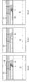
根据本发明的第二实施例,活组织检查探针包括由非透明材料制成的外部管14,在其外表面上制造有小孔16,如附图4所示。According to a second embodiment of the invention, the biopsy probe comprises an
发射光纤和接受光纤相对于内部空间10被固定,以使得光纤20的末端正好位于孔16下方。为避免组织的液体或颗粒渗透到装置中,在外部管14中的孔16被填充有材料18(也被标示为a),材料18为透明材料以用于发射和收集光。The launch and receiver fibers are secured relative to the
为了使光从光纤20穿过孔16的通道不被遮挡,光纤的末端还必须嵌入到透明材料中,并且可采用与本发明的第一实施例相同的方法。In order for the passage of light from the
根据如附图4所示的变化D,在倾斜平面(b)的外表面处施加反射层。According to variant D as shown in FIG. 4 , a reflective layer is applied at the outer surface of the inclined plane (b).
根据如附图4所示的变化E,在介质(a)中设置气泡(c),以使倾斜平面的外表面只与空气相接触,而不是和透明的可凝固材料(a)相接触。According to variant E shown in FIG. 4 , the air bubbles (c) are arranged in the medium (a) so that the outer surface of the inclined plane is only in contact with the air and not with the transparent settable material (a).
根据第二实施例中的变化F,首先设置可凝固的微滴或层以覆盖光纤20的倾斜端,其中可凝固的微滴或层包含反射颗粒(被标示为d)。以这种方式,光纤的倾斜端被反射颗粒所覆盖,导致光的倾斜的耦合输出。完全透明的可凝固层(a)则被用于形成完全平整而不带锋利边缘的轴的外部表面。According to variant F in the second embodiment, a solidifiable droplet or layer is first provided to cover the slanted end of the
应当指出的是,根据本发明的活组织检查装置可以是活组织检查探针本身,其中内部空间适于容纳期望被分析的组织。另一方面,活组织检查装置可以是将被导入身体中以引导活组织检查探针的导管,通过该导管活组织检查探针可被导入身体中以实现真实的活组织检查。It should be noted that a biopsy device according to the invention may be a biopsy probe itself, wherein the internal volume is adapted to accommodate the tissue desired to be analyzed. On the other hand, a biopsy device may be a catheter to be introduced into the body to guide a biopsy probe through which the biopsy probe can be introduced into the body to perform an actual biopsy.
尽管在附图和之前的说明书中详细地阐明和描述了本发明,但是这些阐明和描述仅仅只是示例性或说明性的,而非限制性的;本发明并不限于公开的实施例。While the invention has been illustrated and described in detail in the drawings and foregoing description, such illustration and description are illustrative or illustrative only and not restrictive; the invention is not limited to the disclosed embodiments.
本领域技术人员在实践所主张的发明时,能够通过学习附图、公开内容以及附属权利要求来理解和实现公开的实施例的其它变化。在权利要求书中,用语“包括”并不排除其它的要素或步骤,并且不定冠词“一”或“一个”并不排除多个。分别叙述在彼此不同的从属权利要求中的特定特征并不意味着这些特征不能被组合以获得有益效果。在权利要求书中的任何附图标记并不用于限制其保护范围。Other variations to the disclosed embodiments can be understood and effected by those skilled in the art in practicing the claimed invention, from a study of the drawings, the disclosure, and the appended claims. In the claims, the word "comprising" does not exclude other elements or steps, and the indefinite article "a" or "an" does not exclude a plurality. The mere fact that certain features are recited separately in mutually different dependent claims does not imply that these features cannot be combined to advantage. Any reference signs in the claims are not intended to limit the scope thereof.
Claims (14)
Applications Claiming Priority (3)
| Application Number | Priority Date | Filing Date | Title |
|---|---|---|---|
| EP08152902.6 | 2008-03-18 | ||
| EP08152902 | 2008-03-18 | ||
| PCT/IB2009/051026WO2009115952A1 (en) | 2008-03-18 | 2009-03-12 | Biopsy device |
Publications (2)
| Publication Number | Publication Date |
|---|---|
| CN102088896Atrue CN102088896A (en) | 2011-06-08 |
| CN102088896B CN102088896B (en) | 2013-11-20 |
Family
ID=40727108
Family Applications (1)
| Application Number | Title | Priority Date | Filing Date |
|---|---|---|---|
| CN2009801094639AActiveCN102088896B (en) | 2008-03-18 | 2009-03-12 | Biopsy device |
Country Status (5)
| Country | Link |
|---|---|
| US (1) | US20110009772A1 (en) |
| EP (1) | EP2265161B1 (en) |
| JP (1) | JP5677284B2 (en) |
| CN (1) | CN102088896B (en) |
| WO (1) | WO2009115952A1 (en) |
Cited By (6)
| Publication number | Priority date | Publication date | Assignee | Title |
|---|---|---|---|---|
| CN105025808A (en)* | 2013-02-27 | 2015-11-04 | 皇家飞利浦有限公司 | Optical guided vacuum assisted biopsy device |
| CN105074519A (en)* | 2013-01-18 | 2015-11-18 | 庆北大学校产学协力团 | Bundled fiber optic probe |
| CN105578970A (en)* | 2013-07-26 | 2016-05-11 | 学术发展皇家机构/麦吉尔大学 | Biopsy device and method of obtaining tomographic images of tissue volumes using biopsy device |
| CN106793948A (en)* | 2014-08-28 | 2017-05-31 | 皇家飞利浦有限公司 | Side-looking lung biopsy device |
| CN107923847A (en)* | 2015-08-17 | 2018-04-17 | Gpx医疗公司 | Systems and methods for laser-based internal analysis of gases in the human body |
| CN110840397A (en)* | 2019-11-01 | 2020-02-28 | 福建师范大学 | Endoscopic Raman spectrum detection device for intracavity tissue |
Families Citing this family (4)
| Publication number | Priority date | Publication date | Assignee | Title |
|---|---|---|---|---|
| WO2009109879A2 (en)* | 2008-03-03 | 2009-09-11 | Koninklijke Philips Electronics N.V. | Biopsy guidance by electromagnetic tracking and photonic needle |
| US10850046B2 (en) | 2016-03-28 | 2020-12-01 | Becton, Dickinson And Company | Cannula locator device |
| US10835718B2 (en)* | 2016-03-28 | 2020-11-17 | Becton, Dickinson And Company | Cannula with light-emitting optical fiber |
| US11478150B2 (en) | 2016-03-28 | 2022-10-25 | Becton, Dickinson And Company | Optical fiber sensor |
Family Cites Families (20)
| Publication number | Priority date | Publication date | Assignee | Title |
|---|---|---|---|---|
| US4994060A (en)* | 1984-09-17 | 1991-02-19 | Xintec Corporation | Laser heated cautery cap with transparent substrate |
| US4961738A (en)* | 1987-01-28 | 1990-10-09 | Mackin Robert A | Angioplasty catheter with illumination and visualization within angioplasty balloon |
| US5019040A (en)* | 1989-08-31 | 1991-05-28 | Koshin Sangyo Kabushiki Kaisha | Catheter |
| US5032123A (en)* | 1989-12-28 | 1991-07-16 | Cordis Corporation | Laser catheter with radially divergent treatment beam |
| US5222949A (en)* | 1991-07-23 | 1993-06-29 | Intermed, Inc. | Flexible, noncollapsible catheter tube with hard and soft regions |
| US5217456A (en)* | 1992-02-24 | 1993-06-08 | Pdt Cardiovascular, Inc. | Device and method for intra-vascular optical radial imaging |
| WO1996002863A1 (en)* | 1994-07-13 | 1996-02-01 | Fujikura Ltd. | Stereoscopic viewer |
| US5562657A (en)* | 1994-09-19 | 1996-10-08 | Griffin; Stephen E. | Side fire laser catheter method and apparatus |
| US6058323A (en)* | 1996-11-05 | 2000-05-02 | Lemelson; Jerome | System and method for treating select tissue in a living being |
| EP1042029B1 (en)* | 1997-12-22 | 2015-09-16 | Celgard, LLC | Device for removal of gas bubbles and dissolved gasses in liquid |
| US6384915B1 (en)* | 1998-03-30 | 2002-05-07 | The Regents Of The University Of California | Catheter guided by optical coherence domain reflectometry |
| WO1999051143A1 (en)* | 1998-04-07 | 1999-10-14 | Windy Hill Technology, Inc. | Methods and devices for the localization of lesions in solid tissue |
| US6632183B2 (en)* | 2001-02-12 | 2003-10-14 | Thermal Technologies, Inc. | Perfusion sensitive biopsy extractor |
| US6891984B2 (en)* | 2002-07-25 | 2005-05-10 | Lightlab Imaging, Llc | Scanning miniature optical probes with optical distortion correction and rotational control |
| JP2004233551A (en)* | 2003-01-29 | 2004-08-19 | Sony Corp | Optical communication module and connector |
| EP1610865A4 (en)* | 2003-03-14 | 2007-11-28 | Light Sciences Oncology Inc | Light generating device to intravascular use |
| US20050105877A1 (en)* | 2003-11-13 | 2005-05-19 | Duke University | Optical fiber illuminators having integral distal light diffusers especially useful for ophthalmic surgical procedures, and methods of making the same |
| US20050203419A1 (en)* | 2004-02-24 | 2005-09-15 | Nirmala Ramanujam | Side-firing probe for performing optical spectroscopy during core needle biopsy |
| US7604830B2 (en)* | 2004-06-24 | 2009-10-20 | Cook Incorporated | Method and apparatus for coating interior surfaces of medical devices |
| CA2666283C (en)* | 2006-10-11 | 2016-08-02 | Light Sciences Oncology, Inc. | Light delivery system |
- 2009
- 2009-03-12CNCN2009801094639Apatent/CN102088896B/enactiveActive
- 2009-03-12WOPCT/IB2009/051026patent/WO2009115952A1/enactiveApplication Filing
- 2009-03-12JPJP2011500323Apatent/JP5677284B2/ennot_activeExpired - Fee Related
- 2009-03-12USUS12/922,714patent/US20110009772A1/ennot_activeAbandoned
- 2009-03-12EPEP09723538.6Apatent/EP2265161B1/enactiveActive
Cited By (10)
| Publication number | Priority date | Publication date | Assignee | Title |
|---|---|---|---|---|
| CN105074519A (en)* | 2013-01-18 | 2015-11-18 | 庆北大学校产学协力团 | Bundled fiber optic probe |
| US9829634B2 (en) | 2013-01-18 | 2017-11-28 | Kyungpook National University Industry-Academic Cooperation Foundation | Bundled optical fiber probe |
| CN105025808A (en)* | 2013-02-27 | 2015-11-04 | 皇家飞利浦有限公司 | Optical guided vacuum assisted biopsy device |
| CN105578970A (en)* | 2013-07-26 | 2016-05-11 | 学术发展皇家机构/麦吉尔大学 | Biopsy device and method of obtaining tomographic images of tissue volumes using biopsy device |
| CN106793948A (en)* | 2014-08-28 | 2017-05-31 | 皇家飞利浦有限公司 | Side-looking lung biopsy device |
| CN107923847A (en)* | 2015-08-17 | 2018-04-17 | Gpx医疗公司 | Systems and methods for laser-based internal analysis of gases in the human body |
| CN107923847B (en)* | 2015-08-17 | 2021-10-01 | Gpx医疗公司 | System and method for laser-based internal analysis of gases in the human body |
| US11744467B2 (en) | 2015-08-17 | 2023-09-05 | Neola Medical AB | System and method for laser based internal analysis of gases in a body of a human |
| US12178548B2 (en) | 2015-08-17 | 2024-12-31 | Neola Medical AB | System and method for laser based internal analysis of gases in a body of a human |
| CN110840397A (en)* | 2019-11-01 | 2020-02-28 | 福建师范大学 | Endoscopic Raman spectrum detection device for intracavity tissue |
Also Published As
| Publication number | Publication date |
|---|---|
| JP2011515140A (en) | 2011-05-19 |
| WO2009115952A1 (en) | 2009-09-24 |
| CN102088896B (en) | 2013-11-20 |
| EP2265161A1 (en) | 2010-12-29 |
| US20110009772A1 (en) | 2011-01-13 |
| JP5677284B2 (en) | 2015-02-25 |
| EP2265161B1 (en) | 2016-08-24 |
Similar Documents
| Publication | Publication Date | Title |
|---|---|---|
| CN102088896B (en) | Biopsy device | |
| CN102933955B (en) | A kit and method for inspecting the surface of a light-conducting article | |
| US11666224B2 (en) | Intraoperative optoacoustic guide apparatus and method | |
| CN108472021B (en) | Device for staining 3D biopsy tissue | |
| JP5624541B2 (en) | Catheter for measuring blood flow in living tissue | |
| ES2963294T3 (en) | Procedures for the fabrication of a microfluidic rotor device | |
| US20110251494A1 (en) | Needle with optical fibers | |
| CN106489088B (en) | Optical cable, method for manufacturing same, and light source module provided with same | |
| GB2343248A (en) | Optical analysis of micro volume samples | |
| JP2012509159A (en) | Needle with built-in fiber | |
| ES2370732T3 (en) | BUCKET. | |
| EP2420821A3 (en) | Standard media suspension body and verification method for an optical particulate measurement instrument | |
| US20150374268A1 (en) | Apparatus having surface-enhanced spectroscopy elements on an exterior surface | |
| CN102589700A (en) | Optical fiber detecting probe head for portable raman spectrometer | |
| JP2011513723A (en) | Fiber optic sensor | |
| CN1969183A (en) | Combined ultrasonic imaging and spectroscopic molecular analysis | |
| ES2247820T3 (en) | CIRCULATION SHEAR ANALYZER FOR ACTIVE BIOLOGICAL MOLECULES IN LIQUID LAYERS ON SURFACES, PROCEDURE FOR ANALYSIS OF A LIQUID AND PROCEDURE TO DETERMINE A THICKNESS OF A ULTRAFINE LIQUID LAYER. | |
| CN109009429A (en) | device for laser ablation | |
| CN1777394A (en) | Catheter head | |
| Perra et al. | Ultrasonic actuation of a fine-needle improves biopsy yield | |
| US10018784B2 (en) | Fiber-optic device and process for manufacturing the device | |
| WO2014162242A1 (en) | Photonic needle | |
| JP2007064742A (en) | Chemical chip and connection device | |
| JP2008164434A (en) | Biological sample discrimination device | |
| JP6861168B2 (en) | Catheter for measuring blood flow in living tissue |
Legal Events
| Date | Code | Title | Description |
|---|---|---|---|
| C06 | Publication | ||
| PB01 | Publication | ||
| C10 | Entry into substantive examination | ||
| SE01 | Entry into force of request for substantive examination | ||
| C14 | Grant of patent or utility model | ||
| GR01 | Patent grant |