CN102076866A - Cell lines, systems and methods for optically controlling second messengers - Google Patents
Cell lines, systems and methods for optically controlling second messengersDownload PDFInfo
- Publication number
- CN102076866A CN102076866ACN2009801259133ACN200980125913ACN102076866ACN 102076866 ACN102076866 ACN 102076866ACN 2009801259133 ACN2009801259133 ACN 2009801259133ACN 200980125913 ACN200980125913 ACN 200980125913ACN 102076866 ACN102076866 ACN 102076866A
- Authority
- CN
- China
- Prior art keywords
- membranin
- photoreactivity
- expression
- chimeric
- cell
- Prior art date
- Legal status (The legal status is an assumption and is not a legal conclusion. Google has not performed a legal analysis and makes no representation as to the accuracy of the status listed.)
- Pending
Links
Images


Classifications
- C—CHEMISTRY; METALLURGY
- C07—ORGANIC CHEMISTRY
- C07K—PEPTIDES
- C07K14/00—Peptides having more than 20 amino acids; Gastrins; Somatostatins; Melanotropins; Derivatives thereof
- C07K14/435—Peptides having more than 20 amino acids; Gastrins; Somatostatins; Melanotropins; Derivatives thereof from animals; from humans
- C07K14/705—Receptors; Cell surface antigens; Cell surface determinants
- C07K14/72—Receptors; Cell surface antigens; Cell surface determinants for hormones
- C07K14/723—G protein coupled receptor, e.g. TSHR-thyrotropin-receptor, LH/hCG receptor, FSH receptor
- A—HUMAN NECESSITIES
- A61—MEDICAL OR VETERINARY SCIENCE; HYGIENE
- A61B—DIAGNOSIS; SURGERY; IDENTIFICATION
- A61B5/00—Measuring for diagnostic purposes; Identification of persons
- A61B5/0059—Measuring for diagnostic purposes; Identification of persons using light, e.g. diagnosis by transillumination, diascopy, fluorescence
- A—HUMAN NECESSITIES
- A61—MEDICAL OR VETERINARY SCIENCE; HYGIENE
- A61B—DIAGNOSIS; SURGERY; IDENTIFICATION
- A61B5/00—Measuring for diagnostic purposes; Identification of persons
- A61B5/48—Other medical applications
- A61B5/4848—Monitoring or testing the effects of treatment, e.g. of medication
- A—HUMAN NECESSITIES
- A61—MEDICAL OR VETERINARY SCIENCE; HYGIENE
- A61K—PREPARATIONS FOR MEDICAL, DENTAL OR TOILETRY PURPOSES
- A61K41/00—Medicinal preparations obtained by treating materials with wave energy or particle radiation ; Therapies using these preparations
- A—HUMAN NECESSITIES
- A61—MEDICAL OR VETERINARY SCIENCE; HYGIENE
- A61K—PREPARATIONS FOR MEDICAL, DENTAL OR TOILETRY PURPOSES
- A61K48/00—Medicinal preparations containing genetic material which is inserted into cells of the living body to treat genetic diseases; Gene therapy
- A61K48/005—Medicinal preparations containing genetic material which is inserted into cells of the living body to treat genetic diseases; Gene therapy characterised by an aspect of the 'active' part of the composition delivered, i.e. the nucleic acid delivered
- A61K48/0058—Nucleic acids adapted for tissue specific expression, e.g. having tissue specific promoters as part of a contruct
- A—HUMAN NECESSITIES
- A61—MEDICAL OR VETERINARY SCIENCE; HYGIENE
- A61K—PREPARATIONS FOR MEDICAL, DENTAL OR TOILETRY PURPOSES
- A61K49/00—Preparations for testing in vivo
- A—HUMAN NECESSITIES
- A61—MEDICAL OR VETERINARY SCIENCE; HYGIENE
- A61N—ELECTROTHERAPY; MAGNETOTHERAPY; RADIATION THERAPY; ULTRASOUND THERAPY
- A61N5/00—Radiation therapy
- A61N5/06—Radiation therapy using light
- A61N5/0613—Apparatus adapted for a specific treatment
- A61N5/062—Photodynamic therapy, i.e. excitation of an agent
- A—HUMAN NECESSITIES
- A61—MEDICAL OR VETERINARY SCIENCE; HYGIENE
- A61P—SPECIFIC THERAPEUTIC ACTIVITY OF CHEMICAL COMPOUNDS OR MEDICINAL PREPARATIONS
- A61P43/00—Drugs for specific purposes, not provided for in groups A61P1/00-A61P41/00
- C—CHEMISTRY; METALLURGY
- C07—ORGANIC CHEMISTRY
- C07K—PEPTIDES
- C07K14/00—Peptides having more than 20 amino acids; Gastrins; Somatostatins; Melanotropins; Derivatives thereof
- C07K14/435—Peptides having more than 20 amino acids; Gastrins; Somatostatins; Melanotropins; Derivatives thereof from animals; from humans
- C07K14/705—Receptors; Cell surface antigens; Cell surface determinants
- C07K14/70571—Receptors; Cell surface antigens; Cell surface determinants for neuromediators, e.g. serotonin receptor, dopamine receptor
- C—CHEMISTRY; METALLURGY
- C12—BIOCHEMISTRY; BEER; SPIRITS; WINE; VINEGAR; MICROBIOLOGY; ENZYMOLOGY; MUTATION OR GENETIC ENGINEERING
- C12N—MICROORGANISMS OR ENZYMES; COMPOSITIONS THEREOF; PROPAGATING, PRESERVING, OR MAINTAINING MICROORGANISMS; MUTATION OR GENETIC ENGINEERING; CULTURE MEDIA
- C12N13/00—Treatment of microorganisms or enzymes with electrical or wave energy, e.g. magnetism, sonic waves
- C—CHEMISTRY; METALLURGY
- C12—BIOCHEMISTRY; BEER; SPIRITS; WINE; VINEGAR; MICROBIOLOGY; ENZYMOLOGY; MUTATION OR GENETIC ENGINEERING
- C12N—MICROORGANISMS OR ENZYMES; COMPOSITIONS THEREOF; PROPAGATING, PRESERVING, OR MAINTAINING MICROORGANISMS; MUTATION OR GENETIC ENGINEERING; CULTURE MEDIA
- C12N5/00—Undifferentiated human, animal or plant cells, e.g. cell lines; Tissues; Cultivation or maintenance thereof; Culture media therefor
- C12N5/06—Animal cells or tissues; Human cells or tissues
- C12N5/0602—Vertebrate cells
- C12N5/0618—Cells of the nervous system
- C12N5/0619—Neurons
- G—PHYSICS
- G01—MEASURING; TESTING
- G01N—INVESTIGATING OR ANALYSING MATERIALS BY DETERMINING THEIR CHEMICAL OR PHYSICAL PROPERTIES
- G01N33/00—Investigating or analysing materials by specific methods not covered by groups G01N1/00 - G01N31/00
- G01N33/48—Biological material, e.g. blood, urine; Haemocytometers
- G01N33/50—Chemical analysis of biological material, e.g. blood, urine; Testing involving biospecific ligand binding methods; Immunological testing
- G01N33/5005—Chemical analysis of biological material, e.g. blood, urine; Testing involving biospecific ligand binding methods; Immunological testing involving human or animal cells
- G01N33/5008—Chemical analysis of biological material, e.g. blood, urine; Testing involving biospecific ligand binding methods; Immunological testing involving human or animal cells for testing or evaluating the effect of chemical or biological compounds, e.g. drugs, cosmetics
- G01N33/502—Chemical analysis of biological material, e.g. blood, urine; Testing involving biospecific ligand binding methods; Immunological testing involving human or animal cells for testing or evaluating the effect of chemical or biological compounds, e.g. drugs, cosmetics for testing non-proliferative effects
- G01N33/5035—Chemical analysis of biological material, e.g. blood, urine; Testing involving biospecific ligand binding methods; Immunological testing involving human or animal cells for testing or evaluating the effect of chemical or biological compounds, e.g. drugs, cosmetics for testing non-proliferative effects on sub-cellular localization
- C—CHEMISTRY; METALLURGY
- C07—ORGANIC CHEMISTRY
- C07K—PEPTIDES
- C07K2319/00—Fusion polypeptide
- C—CHEMISTRY; METALLURGY
- C12—BIOCHEMISTRY; BEER; SPIRITS; WINE; VINEGAR; MICROBIOLOGY; ENZYMOLOGY; MUTATION OR GENETIC ENGINEERING
- C12N—MICROORGANISMS OR ENZYMES; COMPOSITIONS THEREOF; PROPAGATING, PRESERVING, OR MAINTAINING MICROORGANISMS; MUTATION OR GENETIC ENGINEERING; CULTURE MEDIA
- C12N2510/00—Genetically modified cells
- G—PHYSICS
- G01—MEASURING; TESTING
- G01N—INVESTIGATING OR ANALYSING MATERIALS BY DETERMINING THEIR CHEMICAL OR PHYSICAL PROPERTIES
- G01N2333/00—Assays involving biological materials from specific organisms or of a specific nature
- G01N2333/435—Assays involving biological materials from specific organisms or of a specific nature from animals; from humans
- G01N2333/705—Assays involving receptors, cell surface antigens or cell surface determinants
- G01N2333/72—Assays involving receptors, cell surface antigens or cell surface determinants for hormones
- G01N2333/726—G protein coupled receptor, e.g. TSHR-thyrotropin-receptor, LH/hCG receptor, FSH
Landscapes
- Health & Medical Sciences (AREA)
- Life Sciences & Earth Sciences (AREA)
- Chemical & Material Sciences (AREA)
- Engineering & Computer Science (AREA)
- Biomedical Technology (AREA)
- General Health & Medical Sciences (AREA)
- Organic Chemistry (AREA)
- Immunology (AREA)
- Molecular Biology (AREA)
- Genetics & Genomics (AREA)
- Biochemistry (AREA)
- Medicinal Chemistry (AREA)
- Zoology (AREA)
- Cell Biology (AREA)
- Biophysics (AREA)
- Bioinformatics & Cheminformatics (AREA)
- Toxicology (AREA)
- Animal Behavior & Ethology (AREA)
- Veterinary Medicine (AREA)
- Public Health (AREA)
- Biotechnology (AREA)
- Gastroenterology & Hepatology (AREA)
- Proteomics, Peptides & Aminoacids (AREA)
- Pathology (AREA)
- Wood Science & Technology (AREA)
- Microbiology (AREA)
- Neurology (AREA)
- Urology & Nephrology (AREA)
- Hematology (AREA)
- Physics & Mathematics (AREA)
- Epidemiology (AREA)
- Pharmacology & Pharmacy (AREA)
- General Engineering & Computer Science (AREA)
- General Physics & Mathematics (AREA)
- Analytical Chemistry (AREA)
- Tropical Medicine & Parasitology (AREA)
- Nuclear Medicine, Radiotherapy & Molecular Imaging (AREA)
- Food Science & Technology (AREA)
- Heart & Thoracic Surgery (AREA)
- Medical Informatics (AREA)
Abstract
Description
Translated fromChinese相关专利文件Related Patent Documents
依据35U.S.C.§119(e),本专利文件要求2008/5/29提交的题为“光学控制第二信使的细胞系、系统和方法”的美国临时专利申请序列号61/057,108的优先权,所述临时申请通过引用完整纳入本文。Pursuant to 35 U.S.C. §119(e), this patent document claims priority to U.S. Provisional Patent Application Serial No. 61/057,108, filed 5/29/2008, entitled "Cell Lines, Systems, and Methods for Optically Controlling Second Messenger" , said provisional application is hereby incorporated by reference in its entirety.
通过引用纳入电子提交材料Incorporation by reference into electronic submissions
通过引用完整纳入随本文一起提交的计算机可读形式核苷酸/氨基酸序列表,具体是:一份在2009/4/29生成的名为“STFD195PCT_ST25.txt”的12,342字节的ASCII(文本)文件。Incorporated by reference in its entirety is the nucleotide/amino acid sequence listing submitted herewith in computer readable form, specifically: A 12,342 byte ASCII (text) titled "STFD195PCT_ST25.txt" generated on 2009/4/29 document.
发明领域field of invention
本发明一般涉及响应光学刺激产生第二信使的系统和方法,更具体涉及各与响应光产生第二信使有关的细胞系、核苷酸序列、嵌合蛋白以及它们的应用。The present invention generally relates to systems and methods for producing second messengers in response to optical stimuli, and more particularly to cell lines, nucleotide sequences, chimeric proteins, and applications thereof, each related to the production of second messengers in response to light.
背景background
鸟嘌呤核苷酸-结合蛋白(G蛋白)被认为能在无活性的鸟苷二磷酸(GDP)状态和有活性的鸟苷三磷酸(GTP)结合状态之间发生改变。这两种状态与细胞内第二信使的释放相关联。释放的第二信使能够调节下游细胞过程。Guanine nucleotide-binding proteins (G proteins) are thought to change between an inactive guanosine diphosphate (GDP) state and an active guanosine triphosphate (GTP) bound state. These two states are associated with the release of intracellular second messengers. The released second messengers are able to regulate downstream cellular processes.
第二信使包括迅速产生/释放的信号传导分子。这些分子通过激活细胞内的效应蛋白产生细胞反应。细胞信号传导系统的例子包括磷酸肌醇体系、环腺苷酸(cAMP)体系和花生四烯酸体系。Second messengers include rapidly produced/released signaling molecules. These molecules generate a cellular response by activating effector proteins within the cell. Examples of cell signaling systems include the phosphoinositide system, the cyclic AMP (cAMP) system, and the arachidonic acid system.
称为G蛋白-偶联受体(GPCR)、G蛋白-连接受体(GPLR)、七跨膜结构域受体(7TM受体)或七螺旋受体的蛋白质可触发G蛋白不同状态之间的变化。该蛋白质家族包括各种跨膜受体。这些受体通过激活细胞内的信号转导途径而响应外部刺激(例如,光、神经递质、气味或激素)。具体地说,配体结合并激活转导途径从而造成G蛋白改变状态。GPCR-相关活性与许多疾病有关,因此GPCR是许多药物和治疗的靶点。Proteins known as G protein-coupled receptors (GPCRs), G protein-linked receptors (GPLRs), seven transmembrane domain receptors (7TM receptors), or heptaplast receptors trigger the transition between different states of the G protein. The change. This family of proteins includes various transmembrane receptors. These receptors respond to external stimuli (eg, light, neurotransmitters, odors, or hormones) by activating signal transduction pathways within the cell. Specifically, the ligand binds and activates the transduction pathway causing the G protein to change state. GPCR-associated activity has been implicated in many diseases and thus GPCRs are the targets of many drugs and treatments.
据信,市场上超过30%的药物寻靶G-蛋白偶联受体(GPCR),因此其中的大多数涉及第二信使cAMP的产生或抑制。有许多病理过程直接涉及cAMP,包括神经生理学疾病、内分泌学疾病、心脏疾病、代谢疾病和免疫疾病。在研究哺乳动物复杂行为时,技术局限性导致不能在时空上精确控制胞内信号传导过程。调节第二信使水平,如cAMP水平的现有化学方法起效较慢,导致不能在第一时间研究与某些组织如如神经或心脏组织有关的机体的活性。这些化学方法通常缺乏探测这些快速时间量程(例如,在筛选新疗法时)的速度。It is believed that more than 30% of the drugs on the market target G-protein coupled receptors (GPCRs), and therefore the majority of these are involved in the production or inhibition of the second messenger cAMP. There are many pathological processes that directly involve cAMP, including neurophysiological, endocrinological, cardiac, metabolic, and immune diseases. When studying complex behaviors in mammals, technical limitations prevent the precise control of intracellular signaling processes in time and space. Existing chemical methods of modulating the levels of second messengers, such as cAMP, work slowly, preventing the first study of the body's activity in relation to certain tissues such as nerve or heart tissue. These chemical methods often lack the speed to probe these fast timescales (for example, when screening for new therapeutics).
概述overview
本发明涉及克服上述挑战,还涉及第二信使的产生以及有关成像装置和它们的应用。本发明有许多实例和应用,其中一些概述在下文中。The present invention relates to overcoming the above-mentioned challenges, and also to the generation of second messengers and related imaging devices and their applications. The invention has many examples and applications, some of which are outlined below.
与本发明的一个实施方式一致,一种方法用于在细胞内产生第二信使。用一个或多个异源受体亚单位{例如,肾上腺素能受体(α1,β2)}修饰表达光反应性嵌合膜蛋白(例如,视紫质)的核苷酸序列。光反应性膜蛋白在细胞内表达以便响应光产生第二信使。Consistent with one embodiment of the invention, a method is for producing a second messenger within a cell. The nucleotide sequence expressing a light-responsive chimeric membrane protein (eg, rhodopsin) is modified with one or more heterologous receptor subunits {eg, adrenergic receptor (α1, β2)}. Light-responsive membrane proteins are expressed intracellularly to produce second messengers in response to light.
与本发明的一个实施方式一致,一种方法用于评价涉及胞内信使相关的假定治疗方案(例如,药物或电刺激或经由这些第二信使发挥作用的任何其他物质)的效果。用一个或多个异源受体亚单位{例如,肾上腺素能受体(α1,β2)}修饰表达光反应性嵌合膜蛋白(例如,视紫质)的核苷酸序列。光反应性膜蛋白在细胞内表达以便响应光产生第二信使。将所述蛋白暴露于光。评价治疗效果。Consistent with one embodiment of the present invention, a method is used to evaluate the effect of a putative treatment regimen involving intracellular messengers (eg, drugs or electrical stimulation or any other substance acting via these second messengers). The nucleotide sequence expressing a light-responsive chimeric membrane protein (eg, rhodopsin) is modified with one or more heterologous receptor subunits {eg, adrenergic receptor (α1, β2)}. Light-responsive membrane proteins are expressed intracellularly to produce second messengers in response to light. The protein is exposed to light. Evaluate the effect of treatment.
本发明的实施方式涉及表达具有一个或多个异源受体亚单位{例如,肾上腺素能受体(α1,β2)}的光反应性嵌合膜蛋白(视紫质)的细胞。Embodiments of the invention relate to cells expressing a light-responsive chimeric membrane protein (rhodopsin) with one or more heterologous receptor subunits {eg, adrenergic receptors (α1, β2)}.
本发明的实施方式涉及表达具有一个或多个异源受体亚单位{例如,肾上腺素能受体(α1,β2)}的光反应性嵌合膜蛋白(视紫质)的核苷酸序列。Embodiments of the invention relate to the expression of nucleotide sequences for a light-responsive chimeric membrane protein (rhodopsin) with one or more heterologous receptor subunits {e.g., adrenergic receptors (α1, β2)} .
上述本发明的概述不是要描述本发明的每个示例性实施方式或每个应用。下面的附图和详细描述更具体阐述了这些实施方式。The above summary of the present invention is not intended to describe each exemplary embodiment or every application of the present invention. The Figures and Detailed Description that follow more particularly set forth these embodiments.
附图简述Brief description of the drawings
结合附图,通过本发明各个实施方式的详细描述可以更全面地理解本发明,图中:In conjunction with the accompanying drawings, the present invention can be more fully understood through the detailed description of various embodiments of the present invention, in which:
图1A是与本发明的示例性实施方式一致的optoGs和optoGq的示意图;Figure 1A is a schematic diagram of optoGs and optoGq consistent with an exemplary embodiment of the invention;
图1B是与本发明的示例性实施方式一致的未被转染或者用optoGs或optoGq转染的细胞的cAMP、cGMP和IP1的酶联免疫吸附试验(ELISA)示意图;FIG. 1B is a schematic diagram of an enzyme-linked immunosorbent assay (ELISA) for cAMP, cGMP, andIP1 of cells that were not transfected or transfected with optoGs or optoGq, consistent with an exemplary embodiment of the present invention;
图1C是与本发明的示例性实施方式一致的被optoGs和optoGq的mCherry融合蛋白转染的细胞的Ca-成像示意图;Figure 1C is a schematic diagram of Ca-imaging of cells transfected with mCherry fusion proteins of optoGs and optoGq consistent with an exemplary embodiment of the present invention;
图2是与本发明的示例性实施方式一致的被optoGs和optoGq的mCherry融合蛋白转染的细胞的Ca-成像示意图;Figure 2 is a schematic diagram of Ca-imaging of cells transfected with mCherry fusion proteins of optoGs and optoGq consistent with an exemplary embodiment of the present invention;
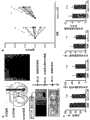
图3A是与本发明的示例性实施方式一致的表达各种构建物的HEK细胞的cAMP、IP1和IP3水平的示意图;Figure 3A is a schematic representation of cAMP, IP1 and IP3 levels in HEK cells expressing various constructs, consistent with an exemplary embodiment of the invention;
图3B是与本发明的示例性实施方式一致的慢病毒表达载体,opto-α1AR-表达细胞的GAD免疫染色,以及光刺激10分钟后在optoXR-表达细胞(mCherry+)中观察到的pCREB活化;Figure 3B is a lentiviral expression vector consistent with an exemplary embodiment of the present invention, GAD immunostaining of opto-α1AR -expressing cells, and pCREB observed in optoXR-expressing cells (mCherry+) after 10 minutes of light stimulation activation;
图4A是与本发明的示例性实施方式一致的转导的伏核(accumbens)的光电极(optrode)寻靶,波峰波形以及所示构建物的基线代谢率;4A is optrode targeting of transduced accumbens, peak waveforms, and baseline metabolic rates of the constructs shown, consistent with an exemplary embodiment of the invention;
图4B是与本发明的示例性实施方式一致的用光刺激记录的体内光电极示意图;Figure 4B is a schematic diagram of an in vivo photoelectrode recorded with photostimulation consistent with an exemplary embodiment of the invention;
图4C是与本发明的示例性实施方式一致的波峰频率与光和基线的变化情况的示意图;Figure 4C is a schematic diagram of peak frequency versus light and baseline, consistent with an exemplary embodiment of the present invention;
图4D是与本发明的示例性实施方式一致的代谢率变化(firing rate change)动力学的示意图;Figure 4D is a schematic diagram of the kinetics of a firing rate change consistent with an exemplary embodiment of the invention;
图5A是与本发明的示例性实施方式一致的转导区的立体定向寻靶(stereotactic targeting),植入了光学纤维的自由移动小鼠,位置偏好装置和试验的示意图以及自由移动小鼠的轨迹;Figure 5A is a schematic diagram of stereotactic targeting of transduction regions, freely moving mice implanted with optical fibers, place preference apparatus and assays, and diagrams of freely moving mice consistent with an exemplary embodiment of the invention. track;
图5B是与本发明的示例性实施方式一致的对照和opto-α1AR的偏好的示意图;和Figure 5B is a schematic representation of the preference for control and opto-α1 AR consistent with an exemplary embodiment of the invention; and
图5C是与本发明的示例性实施方式一致的各种旷场试验的总距离结果的示意图。Figure 5C is a graphical representation of total distance results for various open field tests consistent with an exemplary embodiment of the invention.
尽管本发明可包括各种修饰和改变形式,但其具体形式已在附图中通过举例形式列出,并将在下文中详细描述。然而,应理解,这并不是要将本发明限于所述具体实施方式。相反,本发明包括落入本发明精神和范围内的所有修饰、等价形式和改变。While the present invention may include various modifications and alterations, specific forms thereof have been shown by way of example in the accompanying drawings and will be described in detail hereinafter. It should be understood, however, that the intention is not to limit the invention to the particular embodiments described. On the contrary, the invention includes all modifications, equivalents and changes falling within the spirit and scope of the invention.
发明详述Detailed description of the invention
本发明被认为可用于各种光学系统和方法的实际应用,并且发现本发明特别适用于对细胞内第二信使的水平进行光学控制的系统和方法。由于本发明不必局限于这些应用,因此通过对本文中各种实施例的讨论可以了解本发明的各个方面。The present invention is contemplated to have practical application in a variety of optical systems and methods, and it finds particular application in systems and methods for optically controlling levels of second messengers within cells. Since the invention is not necessarily limited to these applications, various aspects of the invention can be appreciated through the discussion of the various embodiments herein.
本发明的实施方式涉及通过在细胞内释放第二信使而响应光学刺激的嵌合膜蛋白。在具体例子中,所述嵌合蛋白是异源受体亚单位与通过光异构化与光起反应而经历构象(变化)并因此被光激活的蛋白的组合。视紫质或亚视黄基(retinylidene)蛋白是可被修饰以包含异源受体亚单位的光反应性蛋白的例子。Embodiments of the invention relate to chimeric membrane proteins that respond to optical stimuli by releasing second messengers within the cell. In a specific example, the chimeric protein is a combination of a heterologous receptor subunit and a protein that undergoes a conformation (change) in response to light by photoisomerization and is thus activated by light. Rhodopsin or retinylidene proteins are examples of light-responsive proteins that can be modified to include heterologous receptor subunits.
根据本发明的实施方式,对据信含有七跨膜α-螺旋结构域的蛋白质进行修饰以包含与第二信使相关的异源受体亚单位。当在细胞膜内表达时,所述蛋白质通过共形(conformal)改变与光发生反应。共形改变引发第二信使的释放/产生。According to an embodiment of the invention, a protein believed to contain a seven-transmembrane alpha-helical domain is modified to include a heterologous receptor subunit associated with a second messenger. When expressed within the cell membrane, the protein responds to light through conformal changes. The conformal change triggers the release/production of the second messenger.
本发明的实施方式涉及通过在细胞内释放第二信使而响应光学刺激的嵌合膜蛋白的编码核苷酸序列。Embodiments of the invention relate to nucleotide sequences encoding chimeric membrane proteins that respond to optical stimuli by releasing second messengers within cells.
本发明的实施方式涉及表达异源嵌合膜蛋白的细胞。所述嵌合膜蛋白通过引发第二信使在细胞内的释放而响应光学刺激。在某些实施方式中,嵌合膜蛋白的表达在体内发生。在其他实施方式中,嵌合膜蛋白的表达在体外发生。Embodiments of the invention relate to cells expressing heterologous chimeric membrane proteins. The chimeric membrane protein responds to optical stimuli by triggering the release of second messengers within the cell. In certain embodiments, expression of the chimeric membrane protein occurs in vivo. In other embodiments, expression of the chimeric membrane protein occurs in vitro.
本发明的实施方式可用于通过修饰鸟嘌呤核苷酸-结合蛋白偶联的受体蛋白(GPCR)以包含合适的受体亚单位而产生任何合适的第二信使。Embodiments of the invention can be used to generate any suitable second messenger by modifying a guanine nucleotide-binding protein coupled receptor protein (GPCR) to include a suitable receptor subunit.
本发明的实施方式可使用响应各种波长和强度的光的蛋白质。Embodiments of the invention may use proteins that respond to light of various wavelengths and intensities.
本发明的实施方式涉及使用本文所述的嵌合GPCR蛋白来确定感兴趣的第二信使活性的任何下游效应。Embodiments of the invention relate to the use of the chimeric GPCR proteins described herein to determine any downstream effects of second messenger activity of interest.
本发明的实施方式涉及在各种细胞类型中表达嵌合GPCR蛋白,所述细胞类型包括但不限于哺乳动物细胞、干细胞、植物细胞以及酵母和大肠杆菌等单细胞生物。Embodiments of the invention relate to the expression of chimeric GPCR proteins in various cell types including, but not limited to, mammalian cells, stem cells, plant cells, and unicellular organisms such as yeast and E. coli.
本发明的一个具体实施方式涉及优化表达连接有荧光蛋白以利于目测观察的嵌合蛋白,以及形态的优化应用以研究光诱导的第二信使活性的下游效应。A specific embodiment of the invention relates to the optimized expression of chimeric proteins linked to fluorescent proteins for visual inspection, and the optimized use of morphology to study the downstream effects of light-induced second messenger activity.
发明的实施方式涉及本文所述的遗传靶向嵌合GPCR蛋白,涉及在其中表达蛋白质的具体细胞群。存在的细胞类型特异性启动子在靶细胞类型中选择性表达(例如,突触蛋白-1靶向神经元;肌钙蛋白变体靶向心脏组织)。将这些启动子置于表达载体中嵌合GPCR蛋白的上游可使蛋白质在感兴趣的细胞类型内靶向表达。其中包括可诱导、可逆、或其他可控制的启动子系统,如Tet-响应、ER-响应和Cre/Lox系统。Embodiments of the invention relate to the genetically targeted chimeric GPCR proteins described herein, to the specific population of cells in which the protein is expressed. Cell type-specific promoters present are selectively expressed in the target cell type (eg, synapsin-1 targets neurons; troponin variants target cardiac tissue). Placing these promoters upstream of the chimeric GPCR protein in the expression vector allows targeted expression of the protein in the cell type of interest. These include inducible, reversible, or other controllable promoter systems such as Tet-responsive, ER-responsive, and Cre/Lox systems.
根据本发明的示例性实施方式,开发了遗传可编码蛋白质,从而,当这些蛋白质在感兴趣细胞类型中表达时就会响应光而产生环腺苷酸(cAMP)。例如,这可用于目测观察细胞生理学的下游效应,包括但不限于药物筛选。其他实施方式采用导致响应光而释放第二信使的嵌合和异源GPCR。示例性的第二信使包括cAMP、环鸟苷酸(cGMP)、肌醇三磷酸/肌醇1,4,5-三磷酸/三磷酸肌醇(IP3)和花生四烯酸。According to an exemplary embodiment of the present invention, genetically encodeable proteins are developed such that, when expressed in a cell type of interest, these proteins produce cyclic adenosine monophosphate (cAMP) in response to light. For example, this can be used to visualize downstream effects of cell physiology, including but not limited to drug screening. Other embodiments employ chimeric and heterologous GPCRs that result in the release of second messengers in response to light. Exemplary second messengers include cAMP, cyclic guanosine monophosphate (cGMP), inositol triphosphate/inositol 1,4,5-triphosphate/inositol triphosphate (IP3 ), and arachidonic acid.
与本发明的实施方式一致,一种方法被用于评价与胞内形式相关的假定治疗方案(例如,药物或电刺激或通过这些第二信使发挥作用的任何其他物质)的功效。用一个或多个异源受体亚单位{例如,肾上腺素能受体(α1,β2)}修饰核苷酸序列以表达光反应性嵌合膜蛋白(例如,视紫质)。所述光反应性膜蛋白在细胞内表达以便响应光产生第二信使。所述蛋白质暴露于光。评价治疗效果。Consistent with embodiments of the present invention, a method is used to evaluate the efficacy of putative therapeutic regimens associated with intracellular forms (eg, drugs or electrical stimulation or any other substance that acts through these second messengers). The nucleotide sequence is modified with one or more heterologous receptor subunits {eg, adrenergic receptor (α1, β2)} to express a light-responsive chimeric membrane protein (eg, rhodopsin). The light-responsive membrane protein is expressed intracellularly to produce a second messenger in response to light. The protein is exposed to light. Evaluate the effect of treatment.
可按照所需刺激曲线施加光。在一种实施方式中,表达的膜蛋白在数十毫秒内响应光。因此,这种刺激曲线可包括一系列快速交替的光脉冲,并可利用例如Ca2+敏感染料监测所得效果。Light can be applied according to the desired stimulation profile. In one embodiment, the expressed membrane protein responds to light within tens of milliseconds. Thus, such a stimulation profile can comprise a series of rapidly alternating light pulses and the resulting effect can be monitored using, for example, a Ca2+ sensitive dye.
在一个例子中,首先可刺激细胞而不作处理。一旦给予处理后再次刺激细胞。可比较各次测试的结果以评价治疗效果。In one example, the cells may first be stimulated without treatment. Cells were restimulated once treatment had been given. The results of the individual tests can be compared to assess the effect of the treatment.
所述处理可包括多种不同方法,其中包括但不限于药物、对细胞的修饰(遗传修饰或其他修饰)、细胞的物理参数(例如,温度变化或电刺激)或施加于生物的治疗方案。The treatment can include a variety of different methods including, but not limited to, drugs, modifications to the cell (genetic or otherwise), physical parameters of the cell (eg, temperature changes or electrical stimulation), or therapeutic regimens applied to the organism.
在一种实施方式中,所述处理是光刺激表达的膜蛋白。在该例子中,例如,可通过监测与被治疗疾病相关的症状来监测效果。In one embodiment, the treatment is photostimulation of expressed membrane proteins. In this instance, efficacy may be monitored, for example, by monitoring symptoms associated with the disease being treated.
在另一实施方式中,所述治疗方案被用作疾病或病症建模的一部分。例如,可使用疾病模型(细胞或动物)并评估背景/基线状态,然后表达蛋白质并评价治疗方案。In another embodiment, the treatment regimen is used as part of disease or condition modeling. For example, one can use a disease model (cell or animal) and assess the background/baseline state, then express the protein and evaluate the treatment regimen.
实验结果显示,可通过用cAMP-诱导物和cAMP-靶阳离子通道转染细胞并用Ca2+-敏感染料目测观察所得结果来目测观察光引发的cAMP对靶离子通道的调节。第二信使活性的这种遗传可编码的、光活化调节物的组合可用于筛选新的疗法,且其本身就可作为一种新的治疗形式,暗示了cAMP涉及ADHD和心脏通道病变等众多疾病状态。可工程构建该蛋白质用于各种其他第二信使(例如,IP3),通过工程构建视黄醛结合位点或选择具有不同吸收/作用光谱的视紫质或锥形视蛋白的嵌合体用于光活化的其它颜色,以及第二信使的其他下游效应,如钙信号传导和/或激酶活性。Experimental results show that light-induced cAMP modulation of target ion channels can be visualized by transfecting cells with cAMP-inducer and cAMP-target cation channels and visualizing the result with a Ca2+ -sensitive dye. This combination of genetically codable, light-activated modulators of second messenger activity can be used to screen for new therapies and itself as a new treatment modality, implicating cAMP in diseases as diverse as ADHD and cardiac channelopathy state. This protein can be engineered for various other second messengers (e.g., IP3 ) by engineering a retinal binding site or selecting chimeras of rhodopsin or cone opsins with different absorption/action spectra Additional colors due to photoactivation, and other downstream effects of second messengers such as calcium signaling and/or kinase activity.
图1A、1B和1C显示了从optoGs和optoGq获得的实验数据,它们是已经开发出来的第二信使信号传导的光活化诱导物的两个例子(‘optoXR’)。这些光活化诱导物是视紫质/GPCR嵌合体。OptoGq为Gq信号传导提供光反应性控制,而OptoGs为Gs信号传导提供光反应性控制。Figures 1A, 1B and 1C show experimental data obtained from optoGs and optoGq, two examples of photoactivatable inducers of second messenger signaling that have been developed ('optoXR'). These photoactivatable inducers are rhodopsin/GPCR chimeras. OptoGq provides photoresponsive control for Gq signaling, while OptoGs provides photoreactive control for Gs signaling.
在optoGs和optoGq中都显示出在黑暗中基线cAMP和IP3水平的差异可忽略,且与其他第二信使途径如cGMP途径没有交叉。光刺激optoGq时看到的cAMP水平的升高是IP3产生的预期下游效应。Both optoGs and optoGq showed negligible differences in baseline cAMP andIP levels in the dark and no crossover with other second messenger pathways such as the cGMP pathway. The increase in cAMP levels seen upon photostimulation of optoGq is an expected downstream effect ofIP3 production.
图1A是与本发明的示例性实施方式一致的optoGs和optoGq的示意图。对于每种蛋白,视紫质的胞内环被那些通常偶联于Gs(β2)或Gq(α1)的肾上腺素能蛋白代替。遗传编码序列为在人和鼠细胞中表达最优化。所得序列的例子包括optoGs:Seq.Id.No.1和Seq.Id.No.2;optoGq:Seq.Id No.3和Seq.Id.No 4。Figure 1A is a schematic diagram of optoGs and optoGq consistent with an exemplary embodiment of the invention. For each protein, the intracellular loops of rhodopsin were replaced by those adrenergic proteins normally coupled to Gs (β2) or Gq (α1). The genetic coding sequence is optimized for expression in human and murine cells. Examples of resulting sequences include optoGs: Seq.Id.No.1 and Seq.Id.No.2; optoGq: Seq.Id.No.3 and Seq.Id.No.4.
如本领域技术人员已知的,提供的蛋白质的氨基酸序列是支持实施方式的非限制性例子,其范围涵盖提供一致、可互换或等价结果的遗传序列的变化形式(例如,点突变)。The amino acid sequence of the protein provided is a non-limiting example of a supporting embodiment, the scope of which encompasses variations (e.g., point mutations) of the genetic sequence that provide identical, interchangeable, or equivalent results, as known to those skilled in the art .
图1B是与本发明的示例性实施方式一致的未被转染或者用optoGs或optoGq转染的细胞的cAMP(上图)、cGMP(中图)和IP1(下图;IP3的降解产物)的酶联免疫吸附试验(ELISA)示意图。如图所示,图1B的结果在从在每个点上用504nm光(波长20nm)刺激1分钟或保持在黑暗中的细胞获得的。Figure 1B is cAMP (upper panel), cGMP (middle panel) and IP1 (lower panel; degradation products of IP3 ) of cells that were not transfected or transfected with optoGs or optoGq, consistent with an exemplary embodiment of the invention ) schematic diagram of enzyme-linked immunosorbent assay (ELISA). As indicated, the results in Figure IB were obtained from cells stimulated with 504 nm light (
用环境控制倒式培养显微镜(莱卡(Leica)DMI6000B)进行刺激。在cAMP试验中,一些细胞用10uM福司柯林处理30分钟作为试验的饱和、阳性对照。OptoGs响应光而显著提高cAMP水平。在OptoGs作用下未发现cAMP基线水平显著升高,也未发现cGMP或IP3水平有差异。OptoGq响应光而显著提高IP3水平,但未显著改变cGMP水平。cAMP水平和IP3产量的提高被认为是胞内Ca2+释放的共有序列。Stimulation was performed with an environmentally controlled inverted culture microscope (Leica DMI6000B). In the cAMP assay, some cells were treated with 10 uM forskolin for 30 minutes as a saturating, positive control for the assay. OptoGs significantly increased cAMP levels in response to light. No significant increase in baseline levels of cAMP, nor differences in cGMP or IP3 levels were found with OptoGs. OptoGq significantly increased IP3 levels in response to light, but did not significantly alter cGMP levels. Increased cAMP levels and IP3 production are considered consensus sequences for intracellular Ca2+ release.
图1C是与本发明的示例性实施方式一致的被optoGs和optoGq的mCherry融合蛋白转染的细胞的Ca-成像示意图。为检测cAMP,转染超过optoGs的环核苷酸门控Ca2+通道CNGA2的cAMP-选择性突变体。IP3激活胞内Ca2+储存的释放,从而提供可靠的Gq活化信号。对照群也仅用mCherry转染,存在过量突变体CNGA2。细胞用fura-2(培育20-25分钟)加载,并且每2秒钟在340nm和380nm下暴露2毫秒。在optoGs和optoGq每种情况下,单独照射都足以产生Ca信号,而在对照群中未检测到显著信号。Figure 1C is a schematic diagram of Ca-imaging of cells transfected with mCherry fusion proteins of optoGs and optoGq, consistent with an exemplary embodiment of the present invention. To detect cAMP, a cAMP-selective mutant of the cyclic nucleotide-gated Ca2+ channel CNGA2 over optoGs was transfected.IP3 activates the release of intracellular Ca2+ stores, thereby providing a reliable Gq activation signal. The control population was also transfected only with mCherry, in the presence of excess mutant CNGA2. Cells were loaded with fura-2 (20-25 min incubation) and exposed to 340 nm and 380 nm every 2 sec for 2 ms. In each case of optoGs and optoGq, irradiation alone was sufficient to generate a Ca signal, whereas no significant signal was detected in the control population.
图1显示了从具体实验装置获得的数据,但本发明不限于此。例如,可采用除转染外的各种递送技术,其中包括但不限于病毒转导、弹道基因递送(基因枪)以及自发核酸摄入。Figure 1 shows data obtained from a specific experimental setup, but the invention is not limited thereto. For example, various delivery techniques other than transfection can be employed, including but not limited to viral transduction, ballistic gene delivery (gene gun), and spontaneous nucleic acid uptake.
可修饰基础-视紫质以用于任何合适的异源受体亚单位,如Gi-偶联受体如α2-肾上腺素能受体或多巴胺D2受体或血清素5HT2A受体;或其他Gs-或Gq-偶联受体如多巴胺D1A受体或亲代谢性谷氨酸盐受体。Basal-rhodopsin can be modified for any suitable heterologous receptor subunit, such as Gi-coupled receptors such as α2-adrenergic receptors or dopamine D2 receptors or serotonin 5HT2A receptors; or other Gs - or Gq-coupled receptors such as dopamine D1A receptors or metabotropic glutamate receptors.
根据本发明的一个实施方式,所述基础-视紫质是衍生自欧洲牛(Bostaurus)的蛋白质。According to one embodiment of the invention, said basal-rhodopsin is a protein derived from European bovine (Bostaurus).
根据一个实施方式,也可使用除上述基础-视紫质之外的基础-蛋白质,包括各种7-跨膜蛋白,如锥形视蛋白(红色、绿色或蓝色)、其他物种的视紫质、以及配体-门控受体如多巴胺或血清素受体。According to one embodiment, base-proteins other than the above-mentioned base-rhodopsins may also be used, including various 7-transmembrane proteins such as cone opsins (red, green or blue), rhodopsins of other species substances, and ligand-gated receptors such as dopamine or serotonin receptors.
各种实现方式涉及在哺乳动物体内应用。这些实现方式包括,但不限于,测试以及证实神经回路和疾病模式。Various implementations involve in vivo use in mammals. These implementations include, but are not limited to, testing and validating neural circuits and disease models.
图3A和3B显示了在体内应用第二信使信号传导光活化诱导物的两个例子-optoGs(opto-β2AR)和optoGq(opto-α1AR)的实验数据。本发明的一些方面涉及遗传编码光学工具(‘optoXR’)万能家族的应用和开发,所述家族能平衡G-蛋白-偶联受体(GPCR)的一般构效关系,以便以高时空精确度募集和控制受体引发的生物化学信号传导途径。Figures 3A and 3B show the experimental data of two examples of in vivo application of photoactivatable inducers of second messenger signaling - optoGs (opto-β2 AR) and optoGq (opto-α1 AR). Aspects of the invention relate to the application and development of a universal family of genetically encoded optical tools ('optoXR') that balance the general structure-activity relationship of G-protein-coupled receptors (GPCRs) to Recruits and controls receptor-triggered biochemical signaling pathways.
图3A和3B所示结果涉及两种响应光而选择性募集不同的靶信号传导途径的特定optoXR。在体内,这两种optoXR对伏核的波峰形成具有相反作用,其本身对伏核的精确定时optoXR光刺激足以驱动自由移动小鼠的条件位置偏好。optoXR法能够以可寻靶和时间上精确的方式测试与行为哺乳动物中生化信号传导因果影响有关的假说。The results shown in Figures 3A and 3B relate to two specific optoXRs that selectively recruit different target signaling pathways in response to light. In vivo, these two optoXRs have opposing effects on nucleus accumbens crest formation, and precisely timed optoXR photostimulation of the nucleus accumbens by itself is sufficient to drive conditioned place preference in freely moving mice. The optoXR method enables testing of hypotheses related to the causal influence of biochemical signaling in behaving mammals in a targetable and temporally precise manner.
对胞内信号传导的光学控制被用于哺乳动物,采用GPCR的共有构效关系在体内开发和表达具有将信号偶联于效应物的新转导逻辑(transduction logic)的多种不同视蛋白/GPCR2嵌合体。与各种应用一致,工程构建一种或多种嵌合视蛋白-受体蛋白在哺乳动物体内发挥功能,可被特定细胞寻靶,以及响应精确定时的光脉冲。这种方法能够在精确确定且行为相关的时刻使用高速光学刺激(和蛋白质响应)来测试和表征胞内生化事件。一些非限制性的例子包括,脉动与补充调节(tonic modulation),不同调节系统之间的同步,以及指定细胞类型在一段时间量程内的其他基础生理与病理过程。Optical control of intracellular signaling has been used in mammals to exploit the shared structure-activity relationship of GPCRs to develop and express in vivo multiple different opsins/opsins with novel transduction logics that couple signals to effectors. GPCR2 chimera. Consistent with various applications, one or more chimeric opsin-receptor proteins are engineered to function in mammals, be targeted by specific cells, and respond to precisely timed light pulses. This approach enables the use of high-speed optical stimuli (and protein responses) at precisely defined and behaviorally relevant moments to test and characterize intracellular biochemical events. Some non-limiting examples include, pulsatile and tonic modulation, synchronization between different regulatory systems, and other fundamental physiological and pathological processes in a given cell type over time scales.
哺乳动物应用已被成功实现。在一个示例性应用中,首先通过比对Gq-偶联人αIa肾上腺素能受体(α1AR)和Gs-偶联仓鼠β2-肾上腺素能受体(β2AR)与Gt-偶联牛视紫质的保守残基,用特定肾上腺素能受体的那些环取代视紫质的胞内环(图1A)。基于结构模型工程改造交换每个受体的胞内区(包括羧基末端结构域)以便从Gt转移G-蛋白偶联,并为在哺乳动物体内表达最优化各受体。用各种配体活化后,天然受体可具有多种总体状态以募集配体-偏好信号传导现象的经典和非经典途径。optoXR在读出灯(sensing light)下可能以生物依赖性方式选择单一活性总体状态。Mammalian applications have been successfully implemented. In one exemplary application, Gq-coupled humanα1a adrenergic receptor (α1AR ) and Gs-coupled hamster β2-adrenergic receptor (β2AR ) were first compared with Gt-coupled The conserved residues of bovine rhodopsin were linked, replacing the intracellular loops of rhodopsin with those of specific adrenergic receptors (Fig. 1A). The intracellular region of each receptor, including the carboxy-terminal domain, was engineered based on structural models to transfer G-protein coupling from Gt and to optimize each receptor for in vivo expression in mammals. Upon activation with various ligands, natural receptors can have multiple overall states to recruit canonical and non-canonical pathways of ligand-preferred signaling phenomena. optoXR under sensing light may select single active population states in a biologically dependent manner.
编码嵌合体(opto-α1AR和optoβ2AR)的基因融合于荧光蛋白。用仅被opto-α1AR转染的HEK细胞(预计通过Gq募集[Ca2+]i)或同时被opto-β2AR(预计通过Gs募集环状AMP)和cAMP-门控Ca2+通道CNGA2-C460W/E583M转染的HEK细胞中的成像[Ca2+]i(胞内钙浓度)确认功能性optoXR表达。参比[Ca2+]i成像证实60秒绿光刺激(504+/-6nm,7mW mm-2)足以驱动显著的optoXR下游[Ca2+]i信号,但对照条件下除外(图2),显示功能性表达。为测试每种optoXR控制的信号传导的特异性,转导的HEK细胞用3mW mm-2504+/-6nm光照射60秒,然后裂解并通过免疫测定分析cGMP、cAMP和IP1(IP3降解产物)的水平。opto-β2AR的预计经典模式对应于其分子设计,即光学刺激在opto-β2AR-表达细胞中导致显著产生cAMP(图3A,上图),类似于野生型β2AR药理学刺激且不募集IP3(图3A,中图)、[Ca2+]i所实现的(图2)或实质上的暗活性。相反,产生的光刺激显著上调opto-α1AR-表达细胞中的IP3信号传导(图3A,中图),类似于野生型α1AR药理学刺激诱导的水平。结合[Ca2+]i评估(图2),这些数据揭示了Gq募集的预期模式,一种在opto-β2AR-表达细胞中未发现的模式(图3A,上图)。光学刺激表达任一构建物的细胞不能调节cGMP水平(图3A,下图),进一步说明了嵌合蛋白的信号传导特异性。类似试验显示,optoXR的作用光谱非常接近天然视紫质,能够整合生物适用的光通量范围内的信号,并且能够激活非-经典途径到与野生型受体类似的程度,如p42/p44-MAPK信号传导。The genes encoding the chimeras (opto-α1 AR and opto β2 AR) were fused to fluorescent proteins. HEK cells transfected with opto-α1 AR alone (predicted to recruit [Ca2+ ]i through Gq) or both opto-β2 AR (predicted to recruit cyclic AMP through Gs) and cAMP-gated Ca2+ Imaging [Ca2+ ]i (intracellular calcium concentration) in channel CNGA2-C460W/E583M transfected HEK cells confirmed functional optoXR expression. Reference [Ca2+ ]i imaging confirmed that 60 s of green light stimulation (504 +/- 6nm, 7mW mm-2 ) was sufficient to drive a significant optoXR downstream [Ca2+ ]i signal, except under control conditions (Figure 2) , showing functional expression. To test the specificity of each optoXR-controlled signaling, transduced HEK cells were irradiated with 3 mW mm-2 504 +/- 6 nm light for 60 s, then lysed and analyzed by immunoassay for cGMP, cAMP, and IP1 (IP3 degradation products )s level. The predicted canonical pattern of opto-β2AR corresponds to its molecular design, i.e. optical stimulation leads to a marked production of cAMP in opto-β2AR -expressing cells (Fig. 3A, upper panel), similar to pharmacological stimulation of wild-typeβ2AR And did not recruit IP3 ( FIG. 3A , middle panel), [Ca2+ ]i achieved ( FIG. 2 ), or substantially dark activity. In contrast, the resulting photostimulation significantly upregulated IP3 signaling in opto-α1 AR-expressing cells ( FIG. 3A , middle panel), similar to the levels induced by wild-type α1 AR pharmacological stimulation. Combined with [Ca2+ ]i assessment (Fig. 2), these data revealed the expected pattern of Gq recruitment, a pattern not found in opto-β2AR -expressing cells (Fig. 3A, upper panel). Optical stimulation of cells expressing either construct failed to modulate cGMP levels (Figure 3A, lower panel), further illustrating the signaling specificity of the chimeric proteins. Similar experiments have shown that optoXR has a spectrum of action very close to native rhodopsin, is able to integrate signals within a biologically applicable light flux range, and is able to activate non-canonical pathways to a similar extent as wild-type receptors, such as p42/p44-MAPK signaling conduction.
已经测试了完整神经组织内OptoXR的性能,包括是否需要补充视黄醛辅因子。在该测试中,携带受突触蛋白-I启动子控制的optoXR融合基因的慢病毒载体(为了将生化调节靶向局部神经元而非其他潜在的Gs/Gq-反应性细胞组织元件,如神经胶质和内皮细胞;图3B,上左)被立体定位注入成年小鼠的伏核。这种方法的目标是用伏核内的躯体树突区室对神经元进行生化调节(~95%GABA能中型棘神经元,无进一步的亚型特异性;图3B,左图),并且,由于这些慢病毒属不能通过轴突转导细胞,从而排除通道纤维或传入突触前末稍。转导2周后,在人造脑脊液内制备伏核的急性冠状切片(acute coronal slice),光学刺激10分钟,立即固定并用Ser 133-磷酸化CREB(pCREB,cAMP和Ca2+-偶联信号传导级联的生化整合物)染色。在没有补充外源类视色素时,在表达optoXR的细胞群内观察到pCREB显著升高(图3B,右图),但在未光照组织内无此现象。The performance of OptoXR has been tested in intact neural tissue, including with and without the need for retinaldehyde cofactor supplementation. In this test, a lentiviral vector carrying the optoXR fusion gene under the control of the synapsin-I promoter (in order to target biochemical regulation to local neurons rather than other potentially Gs/Gq-responsive cellular tissue elements such as neuronal Glial and endothelial cells; Figure 3B, upper left) were stereotaxically injected into the nucleus accumbens of adult mice. This approach targets biochemical modulation of neurons with the somatic dendritic compartment within the nucleus accumbens (~95% GABAergic medium spiny neurons, no further subtype specificity; Figure 3B, left panel), and, Since these lentiviruses are unable to transduce cells through axons, channel fibers or afferents to presynaptic terminals are excluded. Two weeks after transduction, acute coronal slices of the nucleus accumbens were prepared in artificial cerebrospinal fluid, optically stimulated for 10 min, immediately fixed and treated with Ser 133-phosphorylated CREB (pCREB, cAMP and Ca2+ -coupled signaling cascade of biochemical integrants) staining. In the absence of exogenous retinoid supplementation, a significant increase in pCREB was observed in the optoXR-expressing cell population (Fig. 3B, right panel), but not in non-illuminated tissues.
用靶向转导的伏核的光电极通过记录多单位体内神经元开通(multi-unit in vivo neuronal firing)来确定optoXR激活对伏核局部电活性的功能后果(图4A)。在黑暗中用任一构建物均未观察到基线代谢率有显著差异(图4A,右下)。光学刺激导致表达opto-β2AR的伏核中网络开通降低(图4B左图显示了效应动力学;总结数据分别显示在图4C和4D中),与之前靶向Gs的药理学研究一致。光学刺激增加了表达opto-α1AR的伏核中的开通(图4B右图;图4C,4D)。波峰频率直方图显示,optoXR对代谢率的影响的动力学与生物化学一致,而非信号的电引发(图4D)。这些电生理学数据以及早期生化验证结果都支持optoXR能在体内功能性表达,允许对胞内级联进行差异性光可激活控制,以及调节网络生理学。The functional consequences of optoXR activation on local electrical activity in the nucleus accumbens were determined by recording multi-unit in vivo neuronal firing with photoelectrodes targeting the transduced nucleus accumbens (Fig. 4A). No significant difference in baseline metabolic rate was observed with either construct in the dark (Fig. 4A, bottom right). Optical stimulation resulted in decreased network opening in the nucleus accumbens expressing opto-β2AR (Fig. 4B left panel shows effect kinetics; summary data are shown in Figs. 4C and 4D, respectively), consistent with previous pharmacological studies targeting Gs. Optical stimulation increased opening in the nucleus accumbens expressing opto-α1 AR (Fig. 4B right panel; Fig. 4C, 4D). Peak frequency histograms revealed that the kinetics of optoXR's effect on metabolic rate were consistent with biochemical rather than electrical priming of the signal (Fig. 4D). These electrophysiological data, together with earlier biochemical validation results, support that optoXR can be expressed functionally in vivo, allowing differential photoactivatable control of intracellular cascades and modulating network physiology.
在一种应用中,光遗传学(optogenetic)被用来评价精确定时的optoXR刺激调节自由移动小鼠行为的能力。将便携式固态光传送与转基因表达optoXR组合以便以用于操作性行为的时间精确方式对伏核神经元内的胞内信号传导进行光学控制(图5A)。共焦分析显示表达局限于局部伏核;尤其在传入纤维中、在伏核的远端突出区域、在神经胶质内或在周围区域内未观察到标记。作为三天操作性条件化位置偏好试验的一部分,光学刺激靶向转导的伏核(图5A)。在该测试的每一天让动物自由探索位置偏好装置(图5A,下图)。在第1天,动物自由探索该装置而不作光学刺激。在第2天,一旦动物自由进入指定的条件室,安装在转导区的激光二极管-偶联光学纤维传递10Hz的光脉冲以接近强奖励期间单胺能输入类似的强度。路径追踪显示,柔性光学纤维法能够完全地不受障碍地探索整个室(图5A,下图)。在第3天再让动物自由探索没有光刺激的装置,通过两个独立的不知情记录员量化动物花费在条件室内的时间。显然,表达opto-α1AR的动物在光学刺激后对装置的条件化侧有强烈偏好(图5B)。这种时间上精确的生化调节效果在两次独立opto-α1AR动物测试中是可重现的(每次测试斯氏t检验为:n=5-6,P<0.05,对条件室中的时间;n=11,P<0.01,对所有群体),而其他视蛋白基因,opto-β2AR和ChR2,显示出较少驱使偏好效应。opto-α1AR对伏核的刺激效应特异于奖励相关行为,并且不能延伸至直接调节焦虑-相关行为或运动行为,因为在旷场测试中输送到相同动物组的相同光学刺激显示对朝墙壁的移动距离或偏好没有显著影响(图5C)。In one application, optogenetics was used to evaluate the ability of precisely timed optoXR stimulation to modulate behavior in freely moving mice. Portable solid-state light delivery was combined with transgenic expression optoXR for optical control of intracellular signaling in nucleus accumbens neurons in a time-accurate manner for operant behavior (Fig. 5A). Confocal analysis revealed that expression was localized to the nucleus accumbens; in particular no labeling was observed in afferent fibers, in the distal projecting regions of the nucleus accumbens, within glia, or in surrounding areas. Optical stimuli were targeted to the transduced nucleus accumbens as part of a three-day operant conditioning place preference test (Fig. 5A). Animals were allowed free exploration of the place preference device on each day of the test (Fig. 5A, lower panel). On day 1, animals explored the device freely without optical stimulation. On
现在将描述与上述实验一致的一个具体的非限制性实施方式。体内记录和分析采用光电极,其由与记录电极(1MV钨,A-M系统(A-M Systems))偶联的直径200mm的多模光学纤维(托尔实验室(Thor labs))构成,降低200-400mm的电极/纤维端点到端点的距离从而进入小鼠转导的伏核(电极端点低于前卤4.8-5.2mm),小鼠被放置在立体定向框架内(DK仪器公司(David Kopf Instruments))并用异氟醚麻醉。473nm二极管激光器(CL公司(CrystaLaser))的光经纤维传送。使电信号通过带通滤波器并放大(0.3-1kHz,1800微电极交流放大器,A-M系统),并用pClamp 10.0(分子装置公司(Molecular Devices))分析。通过阈值检测波峰,并由检测人员独立验证。One specific non-limiting embodiment consistent with the experiments described above will now be described. In vivo recording and analysis employed a photoelectrode consisting of a 200 mm diameter multimode optical fiber (Thor labs) coupled to a recording electrode (1MV tungsten, A-M Systems), lowered by 200-400 mm The electrode/fiber end-to-end distance to enter the transduced nucleus accumbens of the mouse (electrode end point is 4.8-5.2 mm below the bregma) and the mouse was placed in a stereotaxic frame (David Kopf Instruments) ) and anesthetized with isoflurane. Light from a 473 nm diode laser (CL (CrystaLaser)) was delivered through the fiber. Electrical signals were passed through a bandpass filter and amplified (0.3-1 kHz, 1800 Microelectrode AC Amplifier, A-M Systems) and analyzed with pClamp 10.0 (Molecular Devices). Peaks were detected by threshold and independently verified by inspectors.
采用光刺激进行行为分析,通过偶联于473nm蓝色二极管激光器(CL公司)的光学纤维(直径200mm,托尔实验室)施加光刺激,并用套管寻靶伏核(距离端点0-100mm)登记。通过函数发生器(Agilent 33220A)给optoXR输送脉冲宽度为50ms的光。在标准装置(SD仪器公司(SD Instruments))内进行位置偏好(测试),移开两室之间的墙壁以允许自由探索。采用MATLAB(Mathworks)运行定制的计数轨迹(tallying script),由两名独立的不知情的观察者根据录像分析数据,以了解动物花费在每个室上的时间量。对于旷场测试,将动物放在40340cm的方形旷场内;用与位置偏好实验相同的参数输送光刺激。采用自动软件(Viewpoint)分析录像,以了解在中间15315cm方形区域以及外部环状区域(场地的其余部分)内的总时间和总距离。Behavioral analysis was performed using photostimulation, which was applied through an optical fiber (diameter 200mm, Thor Laboratories) coupled to a 473nm blue diode laser (CL company), and a cannula was used to target the nucleus accumbens (0-100mm from the endpoint) register. Light with a pulse width of 50 ms was delivered to optoXR through a function generator (Agilent 33220A). Place preference was performed in a standard setup (SD Instruments), with the walls between the two chambers removed to allow free exploration. A custom tallying script was run using MATLAB (Mathworks) and data was analyzed from videotape by two independent blinded observers for the amount of time animals spent in each chamber. For the open field test, animals were placed in a 40340 cm square open field; photostimuli were delivered with the same parameters as for place preference experiments. Video footage was analyzed using automated software (Viewpoint) for total time and total distance within the central 15315 cm square area as well as the outer ring area (the rest of the field).
用双尾斯氏t检验(用Microsoft Excel计算)或单向ANOVA加Tukey后验测试(GraphPad Prism)进行上述统计分析。所有归纳性柱状图用平均值+/-s.e.m.表示,显著性如下:*P<0.05,**P<0.01,***P<0.001。The above statistical analyzes were performed with two-tailed Student's t-test (calculated with Microsoft Excel) or one-way ANOVA with Tukey's posterior test (GraphPad Prism). All inductive histograms are presented as means +/- sem, with the following significance:* P<0.05,** P<0.01,*** P<0.001.
支持本发明各种实施方式的令人惊讶的结果和功效的进一步细节见《时间上精确体内控制胞内信号传导》(Temporally precise in vivo control of in vivo signalling),Raag D.Airan等,Nature 458,1025-1029(2009/4/23),将其通过引用完整纳入本文。Further details supporting the surprising results and efficacy of various embodiments of the present invention are found in Temporally precise in vivo control of in vivo signaling, Raag D. Airan et al., Nature 458 , 1025-1029 (2009/4/23), which is incorporated herein by reference in its entirety.
下面的内容详细描述了与本发明实施方式一致的具体非限制性方法。该方法的诸多变化也包含在本发明范围之内。The following text details specific non-limiting methods consistent with embodiments of the present invention. Variations of this method are also within the scope of the invention.
载体构建vector construction
合成opto-α1AR和opto-β2AR的哺乳动物密码子最佳序列(图1A中的氨基酸序列),将其克隆入pcDNA3.1,并用NotI位点与mCherry或YFP(删除其起始密码子)的N-末端融合。optoXR和mCherry/YFP之间的接头是5’GCGGCCGCC 3’。通过将每种optoXR mCherry的转基因克隆入pLenti突触蛋白I hChR2 mCherry WPRE载体的AgeI和EcoRI位点来构建含有突触蛋白IoptoXR mCherry的慢病毒载体。Mammalian codon-optimized sequences of opto-α1 AR and opto-β2 AR (amino acid sequence in Fig. codon) N-terminal fusion. The linker between optoXR and mCherry/YFP is 5'GCGGCCGCC 3'. Lentiviral vectors containing the synapsin IoptoXR mCherry were constructed by cloning each optoXR mCherry transgene into the AgeI and EcoRI sites of the pLenti synapsin I hChR2 mCherry WPRE vector.
慢病毒制造lentiviral production
制造了高效价慢病毒。简言之,HEK 293FT细胞在4-层细胞工厂(Nunc)内用含10%FBS的DMEM平铺培养至90%汇合。细胞用690μg上述慢病毒载体和两种辅助质粒(690μg pΔCMVR8.74和460μg pMD2.G)共转染。转染15小时后更换培养基。转染24小时后将培养基更换成200-220mL含5mM丁酸钠的无血清UltraCULTURE(坎布莱克斯公司(Cambrex))。在转染后40小时将现在含有病毒的培养上清液以1000rpm离心5分钟以除去细胞碎片,然后用0.45μm低蛋白结合抽滤瓶过滤。澄清的上清液然后用SW 28转子(贝克曼公司(Beckman))以55,000g超滤2小时以沉淀病毒。离心之后弃去上清液并将所得病毒沉淀溶于总共100μL冷的(4℃)PBS。将重悬后的病毒以7000rpm离心5分钟以除去剩余细胞和病毒碎片。将等分样品于-80℃冷冻直至使用。High titer lentiviruses were produced. Briefly, HEK 293FT cells were plated in DMEM containing 10% FBS to 90% confluency in a 4-layer cell factory (Nunc). Cells were co-transfected with 690 μg of the above lentiviral vector and two helper plasmids (690 μg pΔCMVR8.74 and 460 μg pMD2.G). The medium was changed 15 hours after transfection. 24 hours after transfection the medium was changed to 200-220 mL of serum-free UltraCULTURE (Cambrex) containing 5 mM sodium butyrate. Forty hours post-transfection, the culture supernatant, now containing the virus, was centrifuged at 1000 rpm for 5 minutes to remove cell debris and then filtered through a 0.45 μm low protein binding suction filter flask. The clarified supernatant was then ultrafiltered at 55,000 g for 2 hours using a SW 28 rotor (Beckman) to pellet the virus. After centrifugation the supernatant was discarded and the resulting virus pellet was dissolved in a total of 100 μL of cold (4° C.) PBS. The resuspended virus was centrifuged at 7000 rpm for 5 minutes to remove remaining cells and virus debris. Aliquots were frozen at -80°C until use.
动物外科学和行为学Animal Surgery and Behavior
按照斯坦福大学的实验脊椎动物手册的规定关养和处理10-12周龄的雌性C57BL/6小鼠。如下将病毒溶液输送到右伏核。用异氟醚麻醉动物并剪除头顶的毛。在异氟醚麻醉下将动物的头放入立体定向框架(DK仪器公司)。在头皮正中线上切一切口并在前卤前1.10mm、侧1.45mm处在颅骨上钻一个直径约1mm的孔。然后将预先加入病毒的斜角33号针(NanoFil,WP仪器公司(World Precision Instruments))插入伏核下部(针尖距离前卤腹侧4.70-4.80mm)并用自动注射泵(NanoFil,WP仪器公司)以100nL/分的速度注射1.0μL病毒。注射后让组织放松以及让液体扩散3-5分钟,然后取出针头。对进行急性切片或体内记录实验的动物,用牙粘固粉(朗氏牙科(Lang Dental))填充开颅部分并用VetBond(3M公司)封闭切口。对进行行为分析的动物,插管(C316G,在基座下切开4.5mm;PlasticsOne)和基座一起放入头盖骨。插管用Metabond(帕卡公司(Parkell))和牙粘固粉(朗氏牙科)固定。将VetBond或牙粘固粉干燥,然后将动物从框架中取出,使其恢复至少1周,再进行操作。对行为实验的对照动物进行与试验动物相同的操作(手术、植入插管、光刺激),但仅注射载体(PBS)代替病毒。在位置偏好实验中,研究采用了对任一侧室(>70%或<10%)或对中间室(>40%)均未显示出基线偏好的动物;超过90%的动物满足关于非偏好、平衡位置偏好设计的标准。10-12 week old female C57BL/6 mice were housed and handled according to Stanford University's Manual of Laboratory Vertebrate Animals. The virus solution was delivered to the right nucleus accumbens as follows. Animals were anesthetized with isoflurane and the top hair was clipped. The animal's head was placed in a stereotaxic frame (DK Instruments) under isoflurane anesthesia. An incision was made on the midline of the scalp and a hole about 1 mm in diameter was drilled in the skull at 1.10 mm anterior to the bregma and 1.45 mm lateral. Then a beveled 33-gauge needle (NanoFil, WP Instruments (World Precision Instruments)) pre-loaded with virus was inserted into the lower part of the nucleus accumbens (the needle tip is 4.70-4.80 mm from the ventral side of the bregma) and an automatic syringe pump (NanoFil, WP Instruments) was used. Inject 1.0 μL of virus at a rate of 100 nL/min. Allow tissue to relax and fluid to diffuse for 3-5 minutes after injection, then remove needle. For animals undergoing acute sectioning or in vivo recording experiments, the craniotomy was filled with dental cement (Lang Dental) and the incision was closed with VetBond (3M). For animals undergoing behavioral analysis, a cannula (C316G, cut 4.5mm below the base; PlasticsOne) was placed into the cranium along with the base. Cannulas were secured with Metabond (Parkell) and dental cement (Lang Dental). Allow the VetBond or dental cement to dry before removing the animal from the frame and allowing it to recover for at least 1 week before proceeding. Behavioral control animals were subjected to the same manipulations (surgery, cannulation, photostimulation) as the test animals, but only the vehicle (PBS) was injected instead of the virus. In the place preference experiment, the study employed animals that showed no baseline preference for either lateral compartment (>70% or <10%) or for the middle compartment (>40%); Criteria for balanced place preference designs.
急性切片制备Acute slice preparation
动物用异氟醚麻醉并用外科剪刀(精细科学工具公司(Fine Science Tools))断头。切下275μm厚的包含伏核的冠状切片并储存于含有64mM NaCl、2.5mM KCl、1.25mM NaH2PO4、25mM NaHCO3、10mM葡萄糖、120mM蔗糖、0.5mM CaCl2和7mM MgCl2的切割溶液(用95%O2/5%CO2平衡)。切片之后将切片在切片溶液内32-35℃培育30分钟,然后室温培育直至实验。离体optoXR刺激时将切片放在直立式显微镜(BX51W,奥林巴斯(Olympus))的载物台上并用含有124mM NaCl、3mM KCl、1.25mM NaH2PO4、26mM NaHCO3、10mM葡萄糖、2.4mM CaCl2和1.3mM MgCl2的人工脑脊液(用95%O2/5%CO2平衡)灌注。使300W Lambda DG-4(苏特公司(Sutter))产生的光通过473nm±20nm带通滤波器(萨姆洛克公司(Semrock))并施加于切片,采用4X目镜(0.28NA),照射10分钟,然后立即固定进行随后的分析。Animals were anesthetized with isoflurane and decapitated with surgical scissors (Fine Science Tools). 275 μm thick coronal sections containing the nucleus accumbens were cut and stored in cutting solution containing 64 mM NaCl, 2.5 mM KCl, 1.25 mM NaH2 PO4 , 25 mM NaHCO3 , 10 mM glucose, 120 mM sucrose, 0.5 mM CaCl2 and 7 mM MgCl2 (Balanced with 95%O2 /5%CO2 ). After slicing, the slices were incubated in slicing solution at 32-35°C for 30 minutes, then at room temperature until the experiment. During ex vivo optoXR stimulation, the sections were placed on the stage of an upright microscope (BX51W, Olympus) and treated with a mixture containing 124mM NaCl, 3mM KCl, 1.25mM NaH2 PO4 , 26mM NaHCO3 , 10mM glucose, Artificial cerebrospinal fluid (balanced with 95%O2 /5%CO2 ) was perfused with 2.4 mMCaCl2 and 1.3 mMMgCl2 . Light generated by a 300W Lambda DG-4 (Sutter) was passed through a 473nm±20nm bandpass filter (Semrock) and applied to the section using a 4X eyepiece (0.28NA) for 10 minutes , and then immediately fixed for subsequent analysis.
信号传导确认试验Signal transduction confirmation test
HEK293FT细胞(英杰公司(Invitrogen))在24孔板内用脂转染胺2000(英杰公司)转染,转染4-6小时后更换成无血清培养基。为进行Ca2+成像,将细胞铺放在基质胶涂覆的盖玻片上,并加入配制在Tyrode(含1μM ATR)中的F-127普流尼克/DMSO(探针公司(Probes))中的5μg/ml fura-2AM,在37℃和5%CO2气氛中培育20-25分钟。加载后将盖玻片在奥林巴斯BX51W上340nm/380nm成像,采用Metafluor(艾克森仪器公司(Axon Instruments))控制300W Lambda DG-4(苏特公司)。为进行免疫测定,转染18-24小时后加入1μM ATR和50mM LiCl(以防止IP1降解)并将平板转移至环境控制显微镜(莱卡DMI6000;37℃,5%CO2)。每次光学刺激每个孔中的5个区域(苏特300W Lambda DG-4;萨姆洛克504/12nm带通滤波器;10X 0.30NA目镜);3孔/条件。培育(cAMP/cGMP:20分钟;IP1:1小时),之后裂解细胞并通过HTRF(CB公司(CisBio))分析,用Biotek Synergy4读数。HEK293FT cells (Invitrogen) were transfected with lipofectamine 2000 (Invitrogen) in 24-well plates, and the medium was replaced with serum-free medium 4-6 hours after transfection. For Caimaging , cells were plated on Matrigel-coated coverslips and added to F-127 Pluronic/DMSO (Probes) in Tyrode (with 1 μM ATR) Incubate with 5 μg/ml fura-2AM at 37 °C and 5% COatmosphere for 20-25 min. After loading the coverslips were imaged at 340nm/380nm on an Olympus BX51W using a Metafluor (Axon Instruments) controlled 300W Lambda DG-4 (Suter). For immunoassays, 1 μM ATR and 50 mM LiCl were added 18-24 hours after transfection (to prevent IP1 degradation) and the plates were transferred to an environmentally controlled microscope (Leica DMI6000; 37°C, 5%CO2 ). 5 fields in each well were optically stimulated per time (Suter 300W Lambda DG-4; Samrock 504/12nm bandpass filter; 10X 0.30NA eyepieces); 3 wells/condition. After incubation (cAMP/cGMP: 20 minutes; IP1 : 1 hour), cells were lysed and analyzed by HTRF (CisBio), read with a Biotek Synergy4.
免疫组织化学和共焦分析Immunohistochemistry and confocal analysis
体内刺激结束之后小鼠用冰冷却的4%低聚甲醛(PFA)的PBS(pH 7.4)溶液经心脏灌注90分钟。取出大脑并用4%PFA固定过夜,然后用30%蔗糖的PBS溶液平衡。在冷冻切片机上切下40μm厚的冠状切片,储存于4℃的冷冻保护剂直至进行免疫组织化学处理。自由流动切片用PBS洗涤,然后在0.3%Tx100和3%标准驴血清(NDS)中培育30分钟。为进行急性切片实验,刺激后立即将275μm厚切片在冰冷却的4%PFA中固定1小时并用0.5%Tx100和3%NDS培育。为进行MAPK实验,HEK293细胞刺激后立即将盖玻片固定15分钟,用0.6%H2O2培育,然后用3%NDS配制的0.1%Tx100渗透化处理。在0.01%Tx100和3%NDS中与第一抗体培育过夜:小鼠抗-GAD67 1∶500(马萨诸塞州比尔里卡的密里博公司(Millipore,Billerica,MA));大鼠抗-cfos 1∶500(加利福尼亚州圣迭戈的卡尔生化公司(Calbiochem,San Diego,CA));大鼠抗-磷酸-CREB Ser133 1∶500(密里博公司)。洗涤切片并与偶联于FITC或Cy5(宾夕法尼亚州西格罗夫的杰克逊实验室(Jackson Laboratories,West Grove,PA))的第二抗体(1∶1000)室温培育3小时。与DAPI(1∶50,000)培育20分钟后洗涤切片并用PVD-DABCO固定在显微镜载玻片上。其余与第一抗体(兔抗-磷酸Erk1/2;抗-磷酸-MAPK p38 1∶500(威斯康星州麦迪逊的普利马公司(Promega,Madison,WI));小鼠单克隆抗-多巴胺D1受体1∶50(开米肯公司(Chemicon));兔多克隆抗-多巴胺D2受体1∶50(密里博公司);山羊多克隆抗-胆碱乙酰基转移酶1∶200(密里博公司))培育过夜,然后与生物素化第二抗体(1∶500,杰克逊实验室)培育,用链霉抗生物素蛋白-生物素-辣根过氧化物酶(ABC试剂盒(加利福尼亚州伯林格姆的载体实验室(Vector Labs,Burlingame,CA)))处理,并按照制造商的说明进行TSA检测(康涅狄格州舍尔顿的帕金埃尔默公司(Perkin Elmer,Shelton,CT))。After in vivo stimulation, mice were perfused transcardially for 90 minutes with ice-cold 4% paraformaldehyde (PFA) in PBS (pH 7.4). Brains were removed and fixed overnight with 4% PFA, then equilibrated with 30% sucrose in PBS. Coronal sections of 40 μm thickness were cut on a cryostat and stored in cryoprotectant at 4°C until processed for immunohistochemistry. Free-flow sections were washed with PBS and then incubated for 30 minutes in 0.3% Tx100 and 3% normal donkey serum (NDS). For acute slice experiments, 275 μm thick sections were fixed in ice-cold 4% PFA for 1 hour immediately after stimulation and incubated with 0.5% Tx100 and 3% NDS. For MAPK experiments, coverslips were fixed for 15 min immediately after HEK293 cell stimulation, incubated with 0.6% H2 O2 , and permeabilized with 0.1% Tx100 in 3% NDS. Overnight incubation with primary antibodies in 0.01% Tx100 and 3% NDS: mouse anti-GAD67 1:500 (Millipore, Billerica, MA); rat anti-cfos 1 :500 (Calbiochem, San Diego, CA); rat anti-phospho-CREB Ser133 1:500 (Millipore). Sections were washed and incubated with secondary antibody (1:1000) conjugated to FITC or Cy5 (Jackson Laboratories, West Grove, PA) for 3 hours at room temperature. Sections were washed after 20 min incubation with DAPI (1:50,000) and mounted on microscope slides with PVD-DABCO. The remainder was reacted with primary antibodies (rabbit anti-phospho-Erk1/2; anti-phospho-MAPK p38 1:500 (Promega, Madison, WI); mouse monoclonal anti-dopamine D1 Receptor 1:50 (Chemicon); rabbit polyclonal anti-dopamine D2 receptor 1:50 (Miribo); goat polyclonal anti-choline acetyltransferase 1:200 (Chemicon) Ribo Company)) overnight, and then incubated with biotinylated secondary antibody (1:500, Jackson Laboratories), with streptavidin-biotin-horseradish peroxidase (ABC kit (California Vector Labs, Burlingame, CA (Vector Labs, Burlingame, CA))) and TSA testing (Perkin Elmer, Shelton, CT) according to the manufacturer's instructions )).
用20X/0.70NA或40X/1.25NA油镜在莱卡TCS SP5扫描激光显微镜上获得共焦荧光图像。在插管通道下500μm区域内获得每种条件的4张连续叠加图像。采用Volocity(Improvision)软件,用DAPI染色来描绘伏核以确定cfos或pCREB免疫反应性的平均像素强度。阳性或pCREB-活性细胞通过强度阈确定,获取图像并在不知晓实验条件的情况下进行分析。Confocal fluorescence images were acquired on a Leica TCS SP5 scanning laser microscope with a 20X/0.70NA or 40X/1.25NA oil objective. Four consecutive superimposed images of each condition were acquired within the 500 μm region below the cannula channel. Using Volocity (Improvision) software, the nucleus accumbens was delineated with DAPI staining to determine the average pixel intensity of cfos or pCREB immunoreactivity. Positive or pCREB-active cells were determined by intensity thresholding, images were acquired and analyzed blinded to experimental conditions.
表S1Table S1
数据的pCREB强度原始数值(au)列在图3B中。每个亚组的平均值和SEM用黑体表示;各亚组相比对照的双尾和t检验p-值用斜体表示。The pCREB intensity raw values (au) of the data are listed in Figure 3B. Mean and SEM for each subgroup are in bold; two-tailed and t-test p-values for each subgroup compared to control are in italics.

表S2Table S2
数据的基线代谢率(Hz)的原始数值列在图4A中。每个亚组的平均值和SEM用黑体表示;各亚组相比对照的t检验p-值用斜体表示。Raw values for baseline metabolic rate (Hz) of the data are presented in Figure 4A. Mean and SEM for each subgroup are in bold; t-test p-values for each subgroup compared to control are in italics.

表S3Table S3
数据的代谢率变化(Hz)的原始数值列在图4C中,计算基线代谢率本身的(‘基线’)以及基线和光刺激阶段(‘光照’)之间的。The raw values of the metabolic rate change (Hz) of the data are presented in Figure 4C, calculated for the baseline metabolic rate itself (‘baseline’) and between baseline and the photostimulation phase (‘illumination’).

据此,本发明的实施方式涉及光遗传控制胞内信号传导,以及用于动物行为体内操作时的时间精确性,同时显示非常低的暗活性,而且能募集天然受体下游的多种信号传导分子,藉此在一种技术中集合了其他方法的许多有利方面。类似的实施方式直接探测其他调节物(包括众多神经递质和内分泌激素)引发的七跨膜依赖性信号传导途径的因果显著性。其他实施方式采用optoXR法,这些方式超越可兴奋细胞以利用光学纤维深度寻靶与光遗传靶向光敏感性的万能组合。一种此类实施方式涉及探测不同可兴奋组织内时间上精确生化信号传导的因果显著性。Accordingly, embodiments of the present invention relate to optogenetic control of intracellular signaling and temporal precision for in vivo manipulation of animal behavior while exhibiting very low dark activity and the ability to recruit diverse signaling downstream of natural receptors molecules, whereby many advantageous aspects of other methods are combined in one technology. Similar embodiments directly probe the causal significance of hepta-transmembrane-dependent signaling pathways triggered by other regulators, including numerous neurotransmitters and endocrine hormones. Other embodiments employ optoXR approaches that go beyond excitable cells to take advantage of the versatile combination of fiber optic depth targeting and optogenetic targeting of photosensitivity. One such embodiment involves probing the causal significance of temporally precise biochemical signaling in different excitable tissues.
本发明的实施方式利用了配体偏好信号传导现象,其中各种配体可稳定总体受体构象状态从而使受体的胞内作用偏向于偶联备用转导级联。用optoXR来诱导这些备用级联至药理学操作类似水平(例如,opto-β2AR可诱导与天然配体在野生型β2AR中作用类似的MAPK活性变化);然而,可能不是总能发现个体optoXR能控制导致配体偏好信号传导的所有构象状态。由于哺乳动物组织中存在内源生色团,且在黑暗中的活性尤其低,因此基于视黄醛的工具尤其有用。光遗传可采取连接于快速单组分视黄醛结合模块的不同效应物的形式,利用光学的时间精确性。Embodiments of the present invention take advantage of the phenomenon of ligand-biased signaling, wherein various ligands can stabilize the overall receptor conformational state such that the receptor's intracellular action is biased toward coupling to alternate transduction cascades. OptoXR was used to induce these alternate cascades to levels similar to pharmacological manipulation (eg, opto-β2AR could induce changes in MAPK activity similar to the natural ligand in wild-typeβ2AR ); however, it may not always be possible to find Individual optoXRs can control all conformational states that lead to ligand-preferred signaling. Retinal-based tools are especially useful due to the presence of endogenous chromophores in mammalian tissues and their particularly low activity in the dark. Optoinheritance can take the form of different effectors linked to fast one-component retinal binding modules, exploiting the temporal precision of optics.
本发明的实施方式采用optoXR法补充微生物视蛋白策略,提供了另一种对行为哺乳动物进行快速、可寻靶细胞控制的方法。Embodiments of the present invention complement the microbial opsin strategy with the optoXR approach, providing another approach to rapid, targetable cellular control of behaving mammals.
与本发明的其他实施方式一致,可使用基于已知视蛋白基因和不同作用光谱的optoXR的波长变化形式。这种optoXR对于提供生化和电学控制的可分离通道尤其有用。Consistent with other embodiments of the invention, wavelength variants of optoXR based on known opsin genes and different action spectra may be used. This optoXR is especially useful for separable channels that provide biochemical and electrical control.
本文所述具体蛋白质序列的变体与本发明的实施方式一致。一些变体与这些蛋白质序列的同源性大于约75%,而其他变体的同源性大于约80%、85%或90%。在一些实施方式中,同源性可高达约93%至约95%或约98%。本发明的组合物包括本文提供的蛋白质和核酸序列,包括与所提供序列的同源性大于约50%的变体,并最高包括100%同源的变体。Variants of specific protein sequences described herein are consistent with embodiments of the invention. Some variants have greater than about 75% homology to these protein sequences, while other variants have greater than about 80%, 85% or 90% homology. In some embodiments, the homology can be as high as about 93% to about 95% or about 98%. Compositions of the invention include the protein and nucleic acid sequences provided herein, including variants having greater than about 50% homology to the provided sequences, and including variants having up to 100% homology.
本文所述的各种实施方式可与快速回路读出技术组合,以便在正常操作和疾病状态中进行神经回路的高级询问和反向构建。Various embodiments described herein can be combined with fast circuit readout techniques for advanced interrogation and reverse construction of neural circuits in normal operation and disease states.
上述各种实施方式仅作为例子提供,不构成对本发明的限制。基于上述描述和列举,本领域技术人员将了解,可对本发明进行各种修饰和变化而不必严格遵照文中列举和描述的示例性实施方式和应用。例如,这种变化可包括改变制造的第二信使。这种修饰和变化不背离由以下权利要求列出的本发明的真实精神和范围。The various embodiments described above are provided as examples only, and do not limit the present invention. Based on the foregoing description and illustrations, those skilled in the art will appreciate that various modifications and changes may be made to the present invention without strictly following the exemplary embodiments and applications illustrated and described herein. For example, such changes may include altering the manufactured second messenger. Such modifications and changes are made without departing from the true spirit and scope of the invention as set forth in the following claims.


Claims (12)
Applications Claiming Priority (3)
| Application Number | Priority Date | Filing Date | Title |
|---|---|---|---|
| US5710808P | 2008-05-29 | 2008-05-29 | |
| US61/057,108 | 2008-05-29 | ||
| PCT/US2009/045611WO2009148946A2 (en) | 2008-05-29 | 2009-05-29 | Cell line, system and method for optical control of secondary messengers |
Publications (1)
| Publication Number | Publication Date |
|---|---|
| CN102076866Atrue CN102076866A (en) | 2011-05-25 |
Family
ID=41398792
Family Applications (1)
| Application Number | Title | Priority Date | Filing Date |
|---|---|---|---|
| CN2009801259133APendingCN102076866A (en) | 2008-05-29 | 2009-05-29 | Cell lines, systems and methods for optically controlling second messengers |
Country Status (13)
| Country | Link |
|---|---|
| US (4) | US8729040B2 (en) |
| EP (3) | EP2294208B1 (en) |
| JP (3) | JP5890176B2 (en) |
| KR (1) | KR20110018924A (en) |
| CN (1) | CN102076866A (en) |
| AU (1) | AU2009256457B2 (en) |
| BR (1) | BRPI0913285A2 (en) |
| CA (1) | CA2726128C (en) |
| ES (3) | ES2426095T3 (en) |
| IL (1) | IL209575A0 (en) |
| MX (1) | MX2010012986A (en) |
| MY (1) | MY152003A (en) |
| WO (1) | WO2009148946A2 (en) |
Cited By (27)
| Publication number | Priority date | Publication date | Assignee | Title |
|---|---|---|---|---|
| CN105431046A (en)* | 2013-04-29 | 2016-03-23 | 小利兰·斯坦福大学托管委员会 | Devices, systems and methods for optogenetic modulation of action potentials in target cells |
| US9829492B2 (en) | 2005-07-22 | 2017-11-28 | The Board Of Trustees Of The Leland Stanford Junior University | Implantable prosthetic device comprising a cell expressing a channelrhodopsin |
| US9840541B2 (en) | 2011-12-16 | 2017-12-12 | The Board Of Trustees Of The Leland Stanford Junior University | Opsin polypeptides and methods of use thereof |
| US9850290B2 (en) | 2010-11-05 | 2017-12-26 | The Board Of Trustees Of The Leland Stanford Junior University | Light-activated chimeric opsins and methods of using the same |
| US9855442B2 (en) | 2007-03-01 | 2018-01-02 | The Board Of Trustees Of The Leland Stanford Junior University | Method for optically controlling a neuron with a mammalian codon optimized nucleotide sequence that encodes a variant opsin polypeptide derived from natromonas pharaonis (NpHR) |
| US9878176B2 (en) | 2008-04-23 | 2018-01-30 | The Board Of Trustees Of The Leland Stanford Junior University | System utilizing Volvox carteri light-activated ion channel protein (VChR1) for optical stimulation of target cells |
| US9968652B2 (en) | 2010-11-05 | 2018-05-15 | The Board Of Trustees Of The Leland Stanford Junior University | Optically-controlled CNS dysfunction |
| US9992981B2 (en) | 2010-11-05 | 2018-06-12 | The Board Of Trustees Of The Leland Stanford Junior University | Optogenetic control of reward-related behaviors |
| US10018695B2 (en) | 2010-11-22 | 2018-07-10 | The Board Of Trustees Of The Leland Stanford Junior University | Optogenetic magnetic resonance imaging |
| US10035027B2 (en) | 2007-10-31 | 2018-07-31 | The Board Of Trustees Of The Leland Stanford Junior University | Device and method for ultrasonic neuromodulation via stereotactic frame based technique |
| US10046174B2 (en) | 2005-07-22 | 2018-08-14 | The Board Of Trustees Of The Leland Stanford Junior University | System for electrically stimulating target neuronal cells of a living animal in vivo |
| US10052497B2 (en) | 2005-07-22 | 2018-08-21 | The Board Of Trustees Of The Leland Stanford Junior University | System for optical stimulation of target cells |
| US10064912B2 (en) | 2008-11-14 | 2018-09-04 | The Board Of Trustees Of The Leland Stanford Junior University | Optically-based stimulation of target cells and modifications thereto |
| US10086012B2 (en) | 2010-11-05 | 2018-10-02 | The Board Of Trustees Of The Leland Stanford Junior University | Control and characterization of memory function |
| US10105551B2 (en) | 2007-01-10 | 2018-10-23 | The Board Of Trustees Of The Leland Stanford Junior University | System for optical stimulation of target cells |
| US10252076B2 (en) | 2010-11-05 | 2019-04-09 | The Board Of Trustees Of The Leland Stanford Junior University | Upconversion of light for use in optogenetic methods |
| US10307609B2 (en) | 2013-08-14 | 2019-06-04 | The Board Of Trustees Of The Leland Stanford Junior University | Compositions and methods for controlling pain |
| US10426970B2 (en) | 2007-10-31 | 2019-10-01 | The Board Of Trustees Of The Leland Stanford Junior University | Implantable optical stimulators |
| US10451608B2 (en) | 2005-07-22 | 2019-10-22 | The Board Of Trustees Of The Leland Stanford Junior University | Cell line, system and method for optical-based screening of ion-channel modulators |
| US10568307B2 (en) | 2010-11-05 | 2020-02-25 | The Board Of Trustees Of The Leland Stanford Junior University | Stabilized step function opsin proteins and methods of using the same |
| US10568516B2 (en) | 2015-06-22 | 2020-02-25 | The Board Of Trustees Of The Leland Stanford Junior University | Methods and devices for imaging and/or optogenetic control of light-responsive neurons |
| US10569099B2 (en) | 2005-07-22 | 2020-02-25 | The Board Of Trustees Of The Leland Stanford Junior University | System for optical stimulation of target cells |
| US10583309B2 (en) | 2008-07-08 | 2020-03-10 | The Board Of Trustees Of The Leland Stanford Junior University | Materials and approaches for optical stimulation of the peripheral nervous system |
| US10711242B2 (en) | 2008-06-17 | 2020-07-14 | The Board Of Trustees Of The Leland Stanford Junior University | Apparatus and methods for controlling cellular development |
| US10974064B2 (en) | 2013-03-15 | 2021-04-13 | The Board Of Trustees Of The Leland Stanford Junior University | Optogenetic control of behavioral state |
| US11103723B2 (en) | 2012-02-21 | 2021-08-31 | The Board Of Trustees Of The Leland Stanford Junior University | Methods for treating neurogenic disorders of the pelvic floor |
| US11294165B2 (en) | 2017-03-30 | 2022-04-05 | The Board Of Trustees Of The Leland Stanford Junior University | Modular, electro-optical device for increasing the imaging field of view using time-sequential capture |
Families Citing this family (21)
| Publication number | Priority date | Publication date | Assignee | Title |
|---|---|---|---|---|
| US8401609B2 (en) | 2007-02-14 | 2013-03-19 | The Board Of Trustees Of The Leland Stanford Junior University | System, method and applications involving identification of biological circuits such as neurological characteristics |
| CN102076866A (en) | 2008-05-29 | 2011-05-25 | 利兰·斯坦福青年大学托管委员会 | Cell lines, systems and methods for optically controlling second messengers |
| WO2009155371A1 (en) | 2008-06-17 | 2009-12-23 | The Board Of Trustees Of The Leland Stanford Junior University | Methods, systems and devices for optical stimulation of target cells using an optical transmission element |
| CN102686147B (en) | 2009-11-05 | 2016-01-20 | 格雷特巴奇有限公司 | waveguide neural interface device |
| ES2676274T3 (en) | 2010-03-17 | 2018-07-18 | The Board Of Trustees Of The Leland Stanford Junior University | Light sensitive molecules that allow the passage of ions |
| JP5965392B2 (en)* | 2010-05-28 | 2016-08-03 | オックスフォード バイオメディカ (ユーケー) リミテッド | Delivery of lentiviral vectors to the brain |
| US20140128800A1 (en)* | 2011-06-28 | 2014-05-08 | University Of Rochester | Photoactivatable receptors and their uses |
| WO2013056037A1 (en) | 2011-10-13 | 2013-04-18 | The Cleveland Clinic Foundation | Estimation of neural response for optical stimulation |
| PT3401402T (en)* | 2012-06-08 | 2020-11-11 | Alkermes Pharma Ireland Ltd | Ligands modified by circular permutation as agonists and antagonists |
| US9636380B2 (en) | 2013-03-15 | 2017-05-02 | The Board Of Trustees Of The Leland Stanford Junior University | Optogenetic control of inputs to the ventral tegmental area |
| EP3581580A1 (en) | 2014-03-28 | 2019-12-18 | The Board of Trustees of the Leland Stanford Junior University | Engineered light-activated anion channel proteins and methods of use thereof |
| JP6821688B2 (en)* | 2015-10-09 | 2021-01-27 | ミルテニー・バイオテク・テクノロジー・インコーポレイテッドMiltenyi Biotec Technology, Inc. | Chimeric antigen receptor and usage |
| JP6875815B2 (en)* | 2016-09-29 | 2021-05-26 | 住友化学株式会社 | Olfactory receptor co-receptor |
| US11723579B2 (en) | 2017-09-19 | 2023-08-15 | Neuroenhancement Lab, LLC | Method and apparatus for neuroenhancement |
| US11717686B2 (en) | 2017-12-04 | 2023-08-08 | Neuroenhancement Lab, LLC | Method and apparatus for neuroenhancement to facilitate learning and performance |
| US12280219B2 (en) | 2017-12-31 | 2025-04-22 | NeuroLight, Inc. | Method and apparatus for neuroenhancement to enhance emotional response |
| US11273283B2 (en) | 2017-12-31 | 2022-03-15 | Neuroenhancement Lab, LLC | Method and apparatus for neuroenhancement to enhance emotional response |
| US11364361B2 (en) | 2018-04-20 | 2022-06-21 | Neuroenhancement Lab, LLC | System and method for inducing sleep by transplanting mental states |
| EP3849410A4 (en) | 2018-09-14 | 2022-11-02 | Neuroenhancement Lab, LLC | SLEEP ENHANCEMENT SYSTEM AND METHOD |
| US11795208B2 (en)* | 2019-10-01 | 2023-10-24 | The Regents Of The University Of California | Modulators of Cas9 polypeptide activity and methods of use thereof |
| CA3210385A1 (en) | 2021-03-24 | 2022-09-29 | Jijun Dong | Upar antibodies and fusion proteins with the same |
Family Cites Families (247)
| Publication number | Priority date | Publication date | Assignee | Title |
|---|---|---|---|---|
| US2968302A (en)* | 1956-07-20 | 1961-01-17 | Univ Illinois | Multibeam focusing irradiator |
| US3131690A (en)* | 1962-10-22 | 1964-05-05 | American Optical Corp | Fiber optics devices |
| US3499437A (en)* | 1967-03-10 | 1970-03-10 | Ultrasonic Systems | Method and apparatus for treatment of organic structures and systems thereof with ultrasonic energy |
| US3567847A (en)* | 1969-01-06 | 1971-03-02 | Edgar E Price | Electro-optical display system |
| US4343301A (en)* | 1979-10-04 | 1982-08-10 | Robert Indech | Subcutaneous neural stimulation or local tissue destruction |
| US4559951A (en) | 1982-11-29 | 1985-12-24 | Cardiac Pacemakers, Inc. | Catheter assembly |
| US4616231A (en) | 1984-03-26 | 1986-10-07 | Hughes Aircraft Company | Narrow-band beam steering system |
| FR2580277B1 (en) | 1985-04-15 | 1988-06-10 | Oreal | NOVEL NAPHTHALENIC DERIVATIVES WITH RETINOIC ACTION, THEIR PREPARATION METHOD AND MEDICINAL AND COSMETIC COMPOSITIONS CONTAINING THEM |
| US4865042A (en) | 1985-08-16 | 1989-09-12 | Hitachi, Ltd. | Ultrasonic irradiation system |
| DE68916080T2 (en)* | 1988-03-28 | 1994-09-22 | Canon Kk | Ion permeable membrane and ion transport method using this membrane. |
| US5082670A (en)* | 1988-12-15 | 1992-01-21 | The Regents Of The University Of California | Method of grafting genetically modified cells to treat defects, disease or damage or the central nervous system |
| JP2882818B2 (en)* | 1989-09-08 | 1999-04-12 | 株式会社エス・エル・ティ・ジャパン | Laser irradiation equipment |
| CA2028261C (en) | 1989-10-28 | 1995-01-17 | Won Suck Yang | Non-invasive method and apparatus for measuring blood glucose concentration |
| US5032123A (en)* | 1989-12-28 | 1991-07-16 | Cordis Corporation | Laser catheter with radially divergent treatment beam |
| DE69127829T2 (en) | 1990-11-26 | 1998-03-19 | Chiron Corp., Emeryville, Calif. | EXPRESSION OF PACE IN HOST CELLS AND METHOD FOR USE THEREOF |
| US5550316A (en)* | 1991-01-02 | 1996-08-27 | Fox Chase Cancer Center | Transgenic animal model system for human cutaneous melanoma |
| US6497872B1 (en) | 1991-07-08 | 2002-12-24 | Neurospheres Holdings Ltd. | Neural transplantation using proliferated multipotent neural stem cells and their progeny |
| US5249575A (en) | 1991-10-21 | 1993-10-05 | Adm Tronics Unlimited, Inc. | Corona discharge beam thermotherapy system |
| SE9103752D0 (en) | 1991-12-18 | 1991-12-18 | Astra Ab | NEW COMPOUNDS |
| US5670113A (en) | 1991-12-20 | 1997-09-23 | Sibia Neurosciences, Inc. | Automated analysis equipment and assay method for detecting cell surface protein and/or cytoplasmic receptor function using same |
| US5739273A (en)* | 1992-02-12 | 1998-04-14 | Yale University | Transmembrane polypeptide and methods of use |
| US5460954A (en) | 1992-04-01 | 1995-10-24 | Cheil Foods & Chemicals, Inc. | Production of human proinsulin using a novel vector system |
| US5330515A (en) | 1992-06-17 | 1994-07-19 | Cyberonics, Inc. | Treatment of pain by vagal afferent stimulation |
| US5382516A (en) | 1992-09-15 | 1995-01-17 | Schleicher & Schuell, Inc. | Method and devices for delivery of substrate for the detection of enzyme-linked, membrane-based binding assays |
| US5527695A (en)* | 1993-01-29 | 1996-06-18 | Purdue Research Foundation | Controlled modification of eukaryotic genomes |
| WO1994021789A1 (en)* | 1993-03-25 | 1994-09-29 | The Regents Of The University Of California | Expression of heterologous polypeptides in halobacteria |
| US5411540A (en) | 1993-06-03 | 1995-05-02 | Massachusetts Institute Of Technology | Method and apparatus for preferential neuron stimulation |
| GB2278783A (en)* | 1993-06-11 | 1994-12-14 | Daniel Shellon Gluck | Method of magnetically stimulating neural cells |
| US5445608A (en) | 1993-08-16 | 1995-08-29 | James C. Chen | Method and apparatus for providing light-activated therapy |
| JPH07171162A (en) | 1993-09-07 | 1995-07-11 | Olympus Optical Co Ltd | Laser probe |
| US5470307A (en) | 1994-03-16 | 1995-11-28 | Lindall; Arnold W. | Catheter system for controllably releasing a therapeutic agent at a remote tissue site |
| JPH10501686A (en) | 1994-04-13 | 1998-02-17 | ザ ロックフェラー ユニヴァーシティ | AAV-mediated delivery of DNA to cells of the nervous system |
| US6436908B1 (en)* | 1995-05-30 | 2002-08-20 | Duke University | Use of exogenous β-adrenergic receptor and β-adrenergic receptor kinase gene constructs to enhance myocardial function |
| US5495541A (en)* | 1994-04-19 | 1996-02-27 | Murray; Steven C. | Optical delivery device with high numerical aperture curved waveguide |
| US5807285A (en) | 1994-08-18 | 1998-09-15 | Ethicon-Endo Surgery, Inc. | Medical applications of ultrasonic energy |
| US5520188A (en)* | 1994-11-02 | 1996-05-28 | Focus Surgery Inc. | Annular array transducer |
| US6334846B1 (en)* | 1995-03-31 | 2002-01-01 | Kabushiki Kaisha Toshiba | Ultrasound therapeutic apparatus |
| US5795581A (en)* | 1995-03-31 | 1998-08-18 | Sandia Corporation | Controlled release of molecular components of dendrimer/bioactive complexes |
| WO1996032076A1 (en) | 1995-04-11 | 1996-10-17 | Baxter Internatonal Inc. | Tissue implant systems |
| US6342379B1 (en) | 1995-06-07 | 2002-01-29 | The Regents Of The University Of California | Detection of transmembrane potentials by optical methods |
| US6480743B1 (en) | 2000-04-05 | 2002-11-12 | Neuropace, Inc. | System and method for adaptive brain stimulation |
| US5755750A (en) | 1995-11-13 | 1998-05-26 | University Of Florida | Method and apparatus for selectively inhibiting activity in nerve fibers |
| US5722426A (en)* | 1996-02-26 | 1998-03-03 | Kolff; Jack | Coronary light probe and method of use |
| US5703985A (en) | 1996-04-29 | 1997-12-30 | Eclipse Surgical Technologies, Inc. | Optical fiber device and method for laser surgery procedures |
| US5939320A (en)* | 1996-05-20 | 1999-08-17 | New York University | G-coupled receptors associated with macrophage-trophic HIV, and diagnostic and therapeutic uses thereof |
| US5898058A (en) | 1996-05-20 | 1999-04-27 | Wellman, Inc. | Method of post-polymerization stabilization of high activity catalysts in continuous polyethylene terephthalate production |
| US20040076613A1 (en) | 2000-11-03 | 2004-04-22 | Nicholas Mazarakis | Vector system |
| US7732129B1 (en)* | 1998-12-01 | 2010-06-08 | Crucell Holland B.V. | Method for the production and purification of adenoviral vectors |
| US5741316A (en) | 1996-12-02 | 1998-04-21 | Light Sciences Limited Partnership | Electromagnetic coil configurations for power transmission through tissue |
| US5756351A (en)* | 1997-01-13 | 1998-05-26 | The Regents Of The University Of California | Biomolecular optical sensors |
| US5782896A (en)* | 1997-01-29 | 1998-07-21 | Light Sciences Limited Partnership | Use of a shape memory alloy to modify the disposition of a device within an implantable medical probe |
| US5904659A (en)* | 1997-02-14 | 1999-05-18 | Exogen, Inc. | Ultrasonic treatment for wounds |
| JP2000514094A (en)* | 1997-04-17 | 2000-10-24 | レオネ,パオラ | Delivery system for gene therapy to the brain |
| US5816256A (en) | 1997-04-17 | 1998-10-06 | Bioanalytical Systems, Inc. | Movement--responsive system for conducting tests on freely-moving animals |
| US7276488B2 (en)* | 1997-06-04 | 2007-10-02 | Oxford Biomedica (Uk) Limited | Vector system |
| US5984861A (en)* | 1997-09-29 | 1999-11-16 | Boston Scientific Corporation | Endofluorescence imaging module for an endoscope |
| US6016449A (en) | 1997-10-27 | 2000-01-18 | Neuropace, Inc. | System for treatment of neurological disorders |
| US6597954B1 (en)* | 1997-10-27 | 2003-07-22 | Neuropace, Inc. | System and method for controlling epileptic seizures with spatially separated detection and stimulation electrodes |
| US6647296B2 (en) | 1997-10-27 | 2003-11-11 | Neuropace, Inc. | Implantable apparatus for treating neurological disorders |
| US6790652B1 (en) | 1998-01-08 | 2004-09-14 | Bioimage A/S | Method and apparatus for high density format screening for bioactive molecules |
| WO1999051143A1 (en)* | 1998-04-07 | 1999-10-14 | Windy Hill Technology, Inc. | Methods and devices for the localization of lesions in solid tissue |
| US6319241B1 (en)* | 1998-04-30 | 2001-11-20 | Medtronic, Inc. | Techniques for positioning therapy delivery elements within a spinal cord or a brain |
| AU5898599A (en)* | 1998-08-19 | 2000-03-14 | Board Of Trustees Of The Leland Stanford Junior University | Methods and compositions for genomic modification |
| US6377842B1 (en)* | 1998-09-22 | 2002-04-23 | Aurora Optics, Inc. | Method for quantitative measurement of fluorescent and phosphorescent drugs within tissue utilizing a fiber optic probe |
| US6253109B1 (en)* | 1998-11-05 | 2001-06-26 | Medtronic Inc. | System for optimized brain stimulation |
| WO2000027293A1 (en) | 1998-11-06 | 2000-05-18 | University Of Rochester | A method to improve circulation to ischemic tissue |
| US6303362B1 (en) | 1998-11-19 | 2001-10-16 | The Board Of Trustees Of The Leland Stanford Junior University | Adenoviral vector and methods for making and using the same |
| US6790657B1 (en) | 1999-01-07 | 2004-09-14 | The United States Of America As Represented By The Department Of Health And Human Services | Lentivirus vector system |
| US7507545B2 (en)* | 1999-03-31 | 2009-03-24 | Cardiome Pharma Corp. | Ion channel modulating activity method |
| US6161045A (en) | 1999-06-01 | 2000-12-12 | Neuropace, Inc. | Method for determining stimulation parameters for the treatment of epileptic seizures |
| US7655423B2 (en) | 1999-06-14 | 2010-02-02 | Henry Ford Health System | Nitric oxide donors for inducing neurogenesis |
| US6662039B2 (en) | 1999-06-18 | 2003-12-09 | The Trustees Of Columbia University In The City Of New York | Optical probing of neuronal connections with fluorescent indicators |
| US20040034882A1 (en) | 1999-07-15 | 2004-02-19 | Vale Wylie W. | Corticotropin releasing factor receptor 2 deficient mice and uses thereof |
| EP1207788A4 (en) | 1999-07-19 | 2009-12-09 | St Jude Medical Atrial Fibrill | Apparatus and method for ablating tissue |
| CN1249672C (en) | 1999-08-06 | 2006-04-05 | 阿尔卑斯电气株式会社 | Tray for conveying magnetic head for magnetic disk |
| GB9923558D0 (en) | 1999-10-05 | 1999-12-08 | Oxford Biomedica Ltd | Producer cell |
| GB9928248D0 (en) | 1999-12-01 | 2000-01-26 | Gill Steven S | An implantable guide tube for neurosurgery |
| DE60109571D1 (en) | 2000-01-14 | 2005-04-28 | Migenix Corp | SCREENING ASSAYS USING INTRAMITOCHONDRIAL CALCIUM |
| US6595934B1 (en) | 2000-01-19 | 2003-07-22 | Medtronic Xomed, Inc. | Methods of skin rejuvenation using high intensity focused ultrasound to form an ablated tissue area containing a plurality of lesions |
| US7706882B2 (en)* | 2000-01-19 | 2010-04-27 | Medtronic, Inc. | Methods of using high intensity focused ultrasound to form an ablated tissue area |
| US6808925B2 (en)* | 2000-02-18 | 2004-10-26 | The Board Of Trustees Of The Leland Stanford Junior University | Altered recombinases for genome modification |
| US6473639B1 (en) | 2000-03-02 | 2002-10-29 | Neuropace, Inc. | Neurological event detection procedure using processed display channel based algorithms and devices incorporating these procedures |
| AU2001295193A1 (en) | 2000-05-01 | 2001-11-12 | Novartis Ag | Vectors for ocular transduction and use thereof for genetic therapy |
| US6599281B1 (en) | 2000-05-03 | 2003-07-29 | Aspect Medical Systems, Inc. | System and method for adaptive drug delivery |
| US6551346B2 (en)* | 2000-05-17 | 2003-04-22 | Kent Crossley | Method and apparatus to prevent infections |
| US7201898B2 (en) | 2000-06-01 | 2007-04-10 | The University Of North Carolina At Chapel Hill | Methods and compounds for controlled release of recombinant parvovirus vectors |
| US7067626B2 (en) | 2000-07-05 | 2006-06-27 | Pharmacia & Upjohn Company | Human ion channel proteins |
| US7312043B2 (en) | 2000-07-10 | 2007-12-25 | Vertex Pharmaceuticals (San Diego) Llc | Ion channel assay methods |
| US6437639B1 (en)* | 2000-07-18 | 2002-08-20 | Lucent Technologies Inc. | Programmable RC filter |
| US6921413B2 (en)* | 2000-08-16 | 2005-07-26 | Vanderbilt University | Methods and devices for optical stimulation of neural tissues |
| US6567690B2 (en)* | 2000-10-16 | 2003-05-20 | Cole Giller | Method and apparatus for probe localization in brain matter |
| US6536440B1 (en)* | 2000-10-17 | 2003-03-25 | Sony Corporation | Method and system for generating sensory data onto the human neural cortex |
| US6584357B1 (en)* | 2000-10-17 | 2003-06-24 | Sony Corporation | Method and system for forming an acoustic signal from neural timing difference data |
| US20020106327A1 (en) | 2000-11-15 | 2002-08-08 | Brian Storrie | B/b-like fragment targeting for the purposes of photodynamic therapy and medical imaging |
| US6506154B1 (en)* | 2000-11-28 | 2003-01-14 | Insightec-Txsonics, Ltd. | Systems and methods for controlling a phased array focused ultrasound system |
| SE525540C2 (en) | 2000-11-30 | 2005-03-08 | Datainnovation I Lund Ab | System and procedure for automatic sampling from a sample object |
| US20070196838A1 (en) | 2000-12-08 | 2007-08-23 | Invitrogen Corporation | Methods and compositions for synthesis of nucleic acid molecules using multiple recognition sites |
| US6489115B2 (en) | 2000-12-21 | 2002-12-03 | The Board Of Regents Of The University Of Nebraska | Genetic assays for trinucleotide repeat mutations in eukaryotic cells |
| US6615080B1 (en) | 2001-03-29 | 2003-09-02 | John Duncan Unsworth | Neuromuscular electrical stimulation of the foot muscles for prevention of deep vein thrombosis and pulmonary embolism |
| US7047078B2 (en) | 2001-03-30 | 2006-05-16 | Case Western Reserve University | Methods for stimulating components in, on, or near the pudendal nerve or its branches to achieve selective physiologic responses |
| WO2002080758A2 (en) | 2001-04-04 | 2002-10-17 | Irm Llc | Methods for treating drug addiction |
| US7107996B2 (en) | 2001-04-10 | 2006-09-19 | Ganz Robert A | Apparatus and method for treating atherosclerotic vascular disease through light sterilization |
| US6810285B2 (en) | 2001-06-28 | 2004-10-26 | Neuropace, Inc. | Seizure sensing and detection using an implantable device |
| WO2003016486A2 (en)* | 2001-08-16 | 2003-02-27 | Sloan Kettering Institute For Cancer Research | Bio-synthetic photostimulators and methods of use |
| US6974448B2 (en) | 2001-08-30 | 2005-12-13 | Medtronic, Inc. | Method for convection enhanced delivery catheter to treat brain and other tumors |
| US7904176B2 (en) | 2006-09-07 | 2011-03-08 | Bio Control Medical (B.C.M.) Ltd. | Techniques for reducing pain associated with nerve stimulation |
| WO2003020103A2 (en) | 2001-09-04 | 2003-03-13 | Amit Technology Science & Medicine Ltd. | Method of and device for therapeutic illumination of internal organs and tissues |
| US20060057192A1 (en)* | 2001-09-28 | 2006-03-16 | Kane Patrick D | Localized non-invasive biological modulation system |
| US7175596B2 (en)* | 2001-10-29 | 2007-02-13 | Insightec-Txsonics Ltd | System and method for sensing and locating disturbances in an energy path of a focused ultrasound system |
| US8308784B2 (en)* | 2006-08-24 | 2012-11-13 | Jackson Streeter | Low level light therapy for enhancement of neurologic function of a patient affected by Parkinson's disease |
| US7303578B2 (en) | 2001-11-01 | 2007-12-04 | Photothera, Inc. | Device and method for providing phototherapy to the brain |
| US7790845B2 (en) | 2001-11-08 | 2010-09-07 | Children's Medical Center Corporation | Bacterial ion channel |
| US20030104512A1 (en)* | 2001-11-30 | 2003-06-05 | Freeman Alex R. | Biosensors for single cell and multi cell analysis |
| US6873868B2 (en)* | 2001-12-31 | 2005-03-29 | Infraredx, Inc. | Multi-fiber catheter probe arrangement for tissue analysis or treatment |
| US6721603B2 (en) | 2002-01-25 | 2004-04-13 | Cyberonics, Inc. | Nerve stimulation as a treatment for pain |
| AU2003208901A1 (en) | 2002-02-01 | 2003-09-02 | The Cleveland Clinic Foundation | Microinfusion device |
| AU2003219843B2 (en)* | 2002-02-20 | 2009-04-23 | Medicis Technologies Corporation | Ultrasonic treatment and imaging of adipose tissue |
| US20030186249A1 (en) | 2002-04-01 | 2003-10-02 | Zairen Sun | Human TARPP genes and polypeptides |
| US20070135875A1 (en)* | 2002-04-08 | 2007-06-14 | Ardian, Inc. | Methods and apparatus for thermally-induced renal neuromodulation |
| DE10216005A1 (en) | 2002-04-11 | 2003-10-30 | Max Planck Gesellschaft | Use of biological photoreceptors as direct light-controlled ion channels |
| US7283861B2 (en) | 2002-04-30 | 2007-10-16 | Alexander Bystritsky | Methods for modifying electrical currents in neuronal circuits |
| US9592409B2 (en) | 2002-04-30 | 2017-03-14 | The Regents Of The University Of California | Methods for modifying electrical currents in neuronal circuits |
| EP1506417A1 (en) | 2002-05-13 | 2005-02-16 | Koninklijke Philips Electronics N.V. | Reduction of susceptibility artifacts in subencoded single-shot magnetic resonance imaging |
| WO2003102156A2 (en) | 2002-05-31 | 2003-12-11 | Sloan-Kettering Institute For Cancer Research | Heterologous stimulus-gated ion channels and methods of using same |
| EP1513584A2 (en) | 2002-06-04 | 2005-03-16 | Cyberkinetics, Inc. | Optically-connected implants and related systems and methods of use |
| US7292890B2 (en) | 2002-06-20 | 2007-11-06 | Advanced Bionics Corporation | Vagus nerve stimulation via unidirectional propagation of action potentials |
| US20060106543A1 (en) | 2002-08-09 | 2006-05-18 | Gustavo Deco | Method for analyzing effectiveness of pharmaceutical preparation |
| ATE424448T1 (en) | 2002-10-10 | 2009-03-15 | Merck & Co Inc | TEST METHODS FOR CONDITION-DEPENDENT CALCIUM CHANNEL AGONISTS/ANTAGONISTS |
| US7355033B2 (en)* | 2002-11-18 | 2008-04-08 | Health Research, Inc. | Screening for West Nile Virus antiviral therapy |
| US20040122475A1 (en)* | 2002-12-18 | 2004-06-24 | Myrick Andrew J. | Electrochemical neuron systems |
| US7377900B2 (en) | 2003-06-02 | 2008-05-27 | Insightec - Image Guided Treatment Ltd. | Endo-cavity focused ultrasound transducer |
| US7091500B2 (en) | 2003-06-20 | 2006-08-15 | Lucent Technologies Inc. | Multi-photon endoscopic imaging system |
| US20050153885A1 (en)* | 2003-10-08 | 2005-07-14 | Yun Anthony J. | Treatment of conditions through modulation of the autonomic nervous system |
| WO2005039696A1 (en) | 2003-10-21 | 2005-05-06 | The Regents Of The University Of Michigan | Intracranial neural interface system |
| US6952097B2 (en) | 2003-10-22 | 2005-10-04 | Siemens Aktiengesellschaft | Method for slice position planning of tomographic measurements, using statistical images |
| US20080119421A1 (en)* | 2003-10-31 | 2008-05-22 | Jack Tuszynski | Process for treating a biological organism |
| US20060034943A1 (en)* | 2003-10-31 | 2006-02-16 | Technology Innovations Llc | Process for treating a biological organism |
| WO2005053811A2 (en) | 2003-11-21 | 2005-06-16 | Johns Hopkins University | Biomolecule partition motifs and uses thereof |
| US20050124897A1 (en)* | 2003-12-03 | 2005-06-09 | Scimed Life Systems, Inc. | Apparatus and methods for delivering acoustic energy to body tissue |
| US7783349B2 (en) | 2006-04-10 | 2010-08-24 | Cardiac Pacemakers, Inc. | System and method for closed-loop neural stimulation |
| US7662114B2 (en) | 2004-03-02 | 2010-02-16 | Focus Surgery, Inc. | Ultrasound phased arrays |
| US20050215764A1 (en) | 2004-03-24 | 2005-09-29 | Tuszynski Jack A | Biological polymer with differently charged portions |
| ITMI20040598A1 (en)* | 2004-03-26 | 2004-06-26 | Carlotta Giorgi | METHOD FOR DETECTION OF INTRACELLULAR PARAMETERS WITH LUMINESCENT PROTEIN PROBES FOR THE SCREENING OF MOLECULES ABLE TO ALTER THE SAID PARAMETERS |
| US8512219B2 (en)* | 2004-04-19 | 2013-08-20 | The Invention Science Fund I, Llc | Bioelectromagnetic interface system |
| EP1750800A1 (en) | 2004-04-30 | 2007-02-14 | Advanced Neuromodulation Systems, Inc. | Method of treating mood disorders and/or anxiety disorders by brain stimulation |
| US7670838B2 (en) | 2004-05-24 | 2010-03-02 | The Board Of Trustees Of The Leland Stanford Junior University | Coupling of excitation and neurogenesis in neural stem/progenitor cells |
| US20050279354A1 (en) | 2004-06-21 | 2005-12-22 | Harvey Deutsch | Structures and Methods for the Joint Delivery of Fluids and Light |
| US20060057614A1 (en) | 2004-08-04 | 2006-03-16 | Nathaniel Heintz | Tethering neuropeptides and toxins for modulation of ion channels and receptors |
| US7699780B2 (en)* | 2004-08-11 | 2010-04-20 | Insightec—Image-Guided Treatment Ltd. | Focused ultrasound system with adaptive anatomical aperture shaping |
| US8409099B2 (en)* | 2004-08-26 | 2013-04-02 | Insightec Ltd. | Focused ultrasound system for surrounding a body tissue mass and treatment method |
| WO2006024038A2 (en)* | 2004-08-27 | 2006-03-02 | Codman & Shurtleff | Light-based implant for treating alzheimer’s disease |
| US8239029B2 (en) | 2004-10-21 | 2012-08-07 | Advanced Neuromodulation Systems, Inc. | Stimulation of the amygdalohippocampal complex to treat neurological conditions |
| US7544171B2 (en) | 2004-10-22 | 2009-06-09 | General Patent Llc | Methods for promoting nerve regeneration and neuronal growth and elongation |
| US7833257B2 (en)* | 2004-11-12 | 2010-11-16 | Northwestern University | Apparatus and methods for optical stimulation of the auditory nerve |
| EP1814630A4 (en)* | 2004-11-15 | 2008-05-07 | Christopher Decharms | Applications of the stimulation of neural tissue using light |
| US8109981B2 (en) | 2005-01-25 | 2012-02-07 | Valam Corporation | Optical therapies and devices |
| US7686839B2 (en)* | 2005-01-26 | 2010-03-30 | Lumitex, Inc. | Phototherapy treatment devices for applying area lighting to a wound |
| US9034650B2 (en)* | 2005-02-02 | 2015-05-19 | Intrexon Corporation | Site-specific serine recombinases and methods of their use |
| US7553284B2 (en) | 2005-02-02 | 2009-06-30 | Vaitekunas Jeffrey J | Focused ultrasound for pain reduction |
| US7548780B2 (en) | 2005-02-22 | 2009-06-16 | Cardiac Pacemakers, Inc. | Cell therapy and neural stimulation for cardiac repair |
| US7288108B2 (en) | 2005-03-14 | 2007-10-30 | Codman & Shurtleff, Inc. | Red light implant for treating Parkinson's disease |
| EP1871476B1 (en) | 2005-03-31 | 2018-01-03 | Esther Mayer | Probe device for photobiomodulation of tissue lining a body cavity |
| JP2006295350A (en) | 2005-04-07 | 2006-10-26 | Sony Corp | Imaging apparatus and method of processing imaging result |
| US9445211B2 (en) | 2005-04-11 | 2016-09-13 | St. Jude Medical, Atrial Fibrillation Division, Inc. | Methods for manufacturing high intensity ultrasound transducers |
| GB0508254D0 (en) | 2005-04-23 | 2005-06-01 | Smith & Nephew | Ultrasound device |
| US7640057B2 (en) | 2005-04-25 | 2009-12-29 | Cardiac Pacemakers, Inc. | Methods of providing neural markers for sensed autonomic nervous system activity |
| WO2006113998A1 (en) | 2005-04-26 | 2006-11-02 | University Of Victoria Innovation And Development Corporation | Production of light from sol-gel derived thin films made with lanthanide doped nanoparticles, and preparation thereof |
| PT1879623E (en)* | 2005-05-02 | 2012-12-20 | Genzyme Corp | Gene therapy for spinal cord disorders |
| CN1879906A (en) | 2005-06-15 | 2006-12-20 | 郑云峰 | Magnetic stimulating device for nervous centralis system and its usage method |
| US20090093403A1 (en) | 2007-03-01 | 2009-04-09 | Feng Zhang | Systems, methods and compositions for optical stimulation of target cells |
| US10052497B2 (en)* | 2005-07-22 | 2018-08-21 | The Board Of Trustees Of The Leland Stanford Junior University | System for optical stimulation of target cells |
| US8926959B2 (en)* | 2005-07-22 | 2015-01-06 | The Board Of Trustees Of The Leland Stanford Junior University | System for optical stimulation of target cells |
| US9238150B2 (en) | 2005-07-22 | 2016-01-19 | The Board Of Trustees Of The Leland Stanford Junior University | Optical tissue interface method and apparatus for stimulating cells |
| EP2465925A1 (en) | 2005-07-22 | 2012-06-20 | The Board Of Trustees Of The Leland | Light-activated cation channel and uses thereof |
| US9274099B2 (en) | 2005-07-22 | 2016-03-01 | The Board Of Trustees Of The Leland Stanford Junior University | Screening test drugs to identify their effects on cell membrane voltage-gated ion channel |
| US20070034882A1 (en)* | 2005-08-15 | 2007-02-15 | Takayoshi Fujii | Semiconductor light-emitting device |
| US8852184B2 (en)* | 2005-09-15 | 2014-10-07 | Cannuflow, Inc. | Arthroscopic surgical temperature control system |
| US20080077200A1 (en)* | 2006-09-21 | 2008-03-27 | Aculight Corporation | Apparatus and method for stimulation of nerves and automated control of surgical instruments |
| US8058509B2 (en) | 2005-12-21 | 2011-11-15 | Pioneer Hi-Bred International, Inc. | Methods and compositions for in planta production of inverted repeats |
| US7610100B2 (en) | 2005-12-30 | 2009-10-27 | Boston Scientific Neuromodulation Corporation | Methods and systems for treating osteoarthritis |
| US20070191906A1 (en) | 2006-02-13 | 2007-08-16 | Anand Iyer | Method and apparatus for selective nerve stimulation |
| US20070219600A1 (en) | 2006-03-17 | 2007-09-20 | Michael Gertner | Devices and methods for targeted nasal phototherapy |
| US20070282404A1 (en) | 2006-04-10 | 2007-12-06 | University Of Rochester | Side-firing linear optic array for interstitial optical therapy and monitoring using compact helical geometry |
| US20070253995A1 (en) | 2006-04-28 | 2007-11-01 | Medtronic, Inc. | Drug Delivery Methods and Devices for Treating Stress Urinary Incontinence |
| US8057464B2 (en) | 2006-05-03 | 2011-11-15 | Light Sciences Oncology, Inc. | Light transmission system for photoreactive therapy |
| EP2019588A4 (en) | 2006-05-04 | 2010-11-24 | Univ Wayne State | RECOVERY OF VISUAL REACTIONS BY IN VIVO ADMINISTRATION OF RHODOPSIN NUCLEIC ACIDS |
| US20080176076A1 (en) | 2006-05-11 | 2008-07-24 | University Of Victoria Innovation And Development Corporation | Functionalized lanthanide rich nanoparticles and use thereof |
| US20080262411A1 (en) | 2006-06-02 | 2008-10-23 | Dobak John D | Dynamic nerve stimulation in combination with other eating disorder treatment modalities |
| EP2965782B1 (en)* | 2006-06-19 | 2017-08-09 | Highland Instruments, Inc. | Apparatus for stimulation of biological tissue |
| US7795632B2 (en) | 2006-06-26 | 2010-09-14 | Osram Sylvania Inc. | Light emitting diode with direct view optic |
| WO2008014382A2 (en)* | 2006-07-26 | 2008-01-31 | Case Western Reserve University | System and method for controlling g-protein coupled receptor pathways |
| US20080027505A1 (en)* | 2006-07-26 | 2008-01-31 | G&L Consulting, Llc | System and method for treatment of headaches |
| US7848797B2 (en)* | 2006-08-17 | 2010-12-07 | Neurometrix, Inc. | Motor unit number estimation (MUNE) for the assessment of neuromuscular function |
| US7521590B2 (en) | 2006-09-01 | 2009-04-21 | Korea Institute Of Science And Technology | Phospholipase C β1 (PLCβ1) knockout mice as a model system for testing schizophrenia drugs |
| WO2008051939A2 (en)* | 2006-10-24 | 2008-05-02 | Medapps, Inc. | Systems and methods for medical data transmission |
| US10420948B2 (en) | 2006-10-30 | 2019-09-24 | Medtronic, Inc. | Implantable medical device with variable data retransmission characteristics based upon data type |
| EE200600039A (en) | 2006-12-12 | 2008-10-15 | Tartu Ülikool | Animal Transgenic Model for Modeling Pathological Anxiety, Method for Identifying Compounds Suitable for the Treatment of Pathological Anxiety Diseases or Conditions, and Method for Wfs1 Protein Targeting Against Pathological Anxiety |
| US8398692B2 (en)* | 2007-01-10 | 2013-03-19 | The Board Of Trustees Of The Leland Stanford Junior University | System for optical stimulation of target cells |
| US8401609B2 (en) | 2007-02-14 | 2013-03-19 | The Board Of Trustees Of The Leland Stanford Junior University | System, method and applications involving identification of biological circuits such as neurological characteristics |
| US8282559B2 (en) | 2007-03-09 | 2012-10-09 | Philip Chidi Njemanze | Method for inducing and monitoring long-term potentiation and long-term depression using transcranial doppler ultrasound device in head-down bed rest |
| US8139339B2 (en) | 2007-03-16 | 2012-03-20 | Old Dominion University Research Foundation | Modulation of neuromuscular functions with ultrashort electrical pulses |
| US20080287821A1 (en) | 2007-03-30 | 2008-11-20 | Searete Llc, A Limited Liability Corporation Of The State Of Delaware | Computational user-health testing |
| EP2142082A4 (en) | 2007-05-01 | 2015-10-28 | Neurofocus Inc | Neuro-informatics repository system |
| US20110165681A1 (en) | 2009-02-26 | 2011-07-07 | Massachusetts Institute Of Technology | Light-Activated Proton Pumps and Applications Thereof |
| US8097422B2 (en) | 2007-06-20 | 2012-01-17 | Salk Institute For Biological Studies | Kir channel modulators |
| US9138596B2 (en) | 2007-08-22 | 2015-09-22 | Cardiac Pacemakers, Inc. | Optical depolarization of cardiac tissue |
| US10035027B2 (en)* | 2007-10-31 | 2018-07-31 | The Board Of Trustees Of The Leland Stanford Junior University | Device and method for ultrasonic neuromodulation via stereotactic frame based technique |
| US10434327B2 (en) | 2007-10-31 | 2019-10-08 | The Board Of Trustees Of The Leland Stanford Junior University | Implantable optical stimulators |
| US20090157145A1 (en) | 2007-11-26 | 2009-06-18 | Lawrence Cauller | Transfer Coil Architecture |
| US8562658B2 (en) | 2007-12-06 | 2013-10-22 | Technion Research & Development Foundation Limited | Method and system for optical stimulation of neurons |
| US8883719B2 (en) | 2008-01-16 | 2014-11-11 | University Of Connecticut | Bacteriorhodopsin protein variants and methods of use for long term data storage |
| US20090254134A1 (en) | 2008-02-04 | 2009-10-08 | Medtrode Inc. | Hybrid ultrasound/electrode device for neural stimulation and recording |
| WO2009119782A1 (en) | 2008-03-24 | 2009-10-01 | 国立大学法人東北大学 | Modified photoreceptor channel type rhodopsin protein |
| EP2268311A4 (en) | 2008-04-04 | 2014-08-27 | Immunolight Llc | Non-invasive systems and methods for in-situ photobiomodulation |
| EP3165534B1 (en) | 2008-04-23 | 2018-09-26 | The Board of Trustees of The Leland Stanford Junior University | Systems, methods and compositions for optical stimulation of target cells |
| AU2009274482A1 (en)* | 2008-05-20 | 2010-01-28 | Eos Neuroscience, Inc. | Vectors for delivery of light-sensitive proteins and methods of use |
| CN102076866A (en) | 2008-05-29 | 2011-05-25 | 利兰·斯坦福青年大学托管委员会 | Cell lines, systems and methods for optically controlling second messengers |
| WO2009155371A1 (en) | 2008-06-17 | 2009-12-23 | The Board Of Trustees Of The Leland Stanford Junior University | Methods, systems and devices for optical stimulation of target cells using an optical transmission element |
| SG191604A1 (en)* | 2008-06-17 | 2013-07-31 | Univ Leland Stanford Junior | Apparatus and methods for controlling cellular development |
| US9101759B2 (en) | 2008-07-08 | 2015-08-11 | The Board Of Trustees Of The Leland Stanford Junior University | Materials and approaches for optical stimulation of the peripheral nervous system |
| NZ602416A (en) | 2008-11-14 | 2014-08-29 | Univ Leland Stanford Junior | Optically-based stimulation of target cells and modifications thereto |
| US8380318B2 (en) | 2009-03-24 | 2013-02-19 | Spinal Modulation, Inc. | Pain management with stimulation subthreshold to paresthesia |
| KR101081360B1 (en) | 2009-03-25 | 2011-11-08 | 한국과학기술연구원 | Photostimulation array apparatus |
| TWI511756B (en) | 2009-04-21 | 2015-12-11 | 伊穆諾萊特公司 | Pharmaceutical composition or kit for modifying a target structure which mediates or is associated with a biological activity |
| WO2011005978A2 (en) | 2009-07-08 | 2011-01-13 | Duke University | Methods of manipulating cell signaling |
| US20110112463A1 (en) | 2009-11-12 | 2011-05-12 | Jerry Silver | Compositions and methods for treating a neuronal injury or neuronal disorders |
| US8936630B2 (en) | 2009-11-25 | 2015-01-20 | Medtronic, Inc. | Optical stimulation therapy |
| ES2676274T3 (en) | 2010-03-17 | 2018-07-18 | The Board Of Trustees Of The Leland Stanford Junior University | Light sensitive molecules that allow the passage of ions |
| US20130225664A1 (en) | 2010-04-05 | 2013-08-29 | Alan Horsager | Methods and compositions for decreasing chronic pain |
| ES2545992T3 (en) | 2010-09-08 | 2015-09-17 | MAX-PLANCK-Gesellschaft zur Förderung der Wissenschaften e.V. | Mutant Canalrodopsin 2 |
| EP2635295B1 (en) | 2010-11-05 | 2017-12-20 | The Board of Trustees of the Leland Stanford Junior University | Control and characterization of memory function |
| EP2635109A4 (en) | 2010-11-05 | 2014-03-19 | Univ Leland Stanford Junior | OPTICALLY CONTROLLED CNS DYSFUNCTION |
| EP2635341B1 (en) | 2010-11-05 | 2018-08-08 | The Board of Trustees of the Leland Stanford Junior University | Upconversion of light for use in optogenetic methods |
| CA2816987C (en) | 2010-11-05 | 2018-09-18 | The Board Of Trustees Of The Leland Stanford Junior University | Control and characterization of psychotic states |
| JP6276591B2 (en) | 2010-11-05 | 2018-02-07 | ザ ボード オブ トラスティーズ オブ ザ レランド スタンフォード ジュニア ユニバーシティー | Photo-activated chimeric opsin and method of use thereof |
| WO2012061744A2 (en) | 2010-11-05 | 2012-05-10 | The Board Of Trustees Of The Leland Stanford Junior University | Stabilized step function opsin proteins and methods of using the same |
| EP2635346B1 (en) | 2010-11-05 | 2017-03-29 | The Board of Trustees of the Leland Stanford Junior University | Optogenetic control of reward-related behaviors |
| US8696722B2 (en) | 2010-11-22 | 2014-04-15 | The Board Of Trustees Of The Leland Stanford Junior University | Optogenetic magnetic resonance imaging |
| WO2012106407A2 (en) | 2011-02-01 | 2012-08-09 | The University Of Vermont And State Agricultural College | Diagnostic and therapeutic methods and products related to anxiety disorders |
| US20120253261A1 (en) | 2011-03-29 | 2012-10-04 | Medtronic, Inc. | Systems and methods for optogenetic modulation of cells within a patient |
| US9782091B2 (en) | 2011-07-25 | 2017-10-10 | Neuronexus Technologies, Inc. | Opto-electrical device for artifact reduction |
| KR102023754B1 (en) | 2011-07-27 | 2019-09-20 | 더 보오드 오브 트러스티스 오브 더 유니버시티 오브 일리노이즈 | Nanopore sensors for biomolecular characterization |
| JP6406581B2 (en) | 2011-12-16 | 2018-10-17 | ザ ボード オブ トラスティーズ オブ ザ レランド スタンフォード ジュニア ユニバーシティー | Opsin polypeptides and uses thereof |
| US20140024701A1 (en) | 2012-02-21 | 2014-01-23 | Circuit Therapeutics, Inc. | Compositions and Methods for Treating Neurogenic Disorders of the Pelvic Floor |
| CN104270942B (en) | 2012-03-20 | 2018-02-02 | 斯坦福大学托管董事会 | Non-human animal's depression model and its application method |
| US9636380B2 (en) | 2013-03-15 | 2017-05-02 | The Board Of Trustees Of The Leland Stanford Junior University | Optogenetic control of inputs to the ventral tegmental area |
| US20150112411A1 (en) | 2013-10-18 | 2015-04-23 | Varaya Photoceuticals, Llc | High powered light emitting diode photobiology compositions, methods and systems |
- 2009
- 2009-05-29CNCN2009801259133Apatent/CN102076866A/enactivePending
- 2009-05-29BRBRPI0913285Apatent/BRPI0913285A2/ennot_activeApplication Discontinuation
- 2009-05-29AUAU2009256457Apatent/AU2009256457B2/ennot_activeCeased
- 2009-05-29CACA2726128Apatent/CA2726128C/ennot_activeExpired - Fee Related
- 2009-05-29ESES09759107Tpatent/ES2426095T3/enactiveActive
- 2009-05-29USUS12/993,605patent/US8729040B2/enactiveActive
- 2009-05-29ESES13160448.0Tpatent/ES2532235T3/enactiveActive
- 2009-05-29ESES14191235.2Tpatent/ES2606458T3/enactiveActive
- 2009-05-29EPEP09759107.7Apatent/EP2294208B1/ennot_activeNot-in-force
- 2009-05-29MYMYPI20105597patent/MY152003A/enunknown
- 2009-05-29KRKR1020107029438Apatent/KR20110018924A/ennot_activeWithdrawn
- 2009-05-29EPEP13160448.0Apatent/EP2610350B1/ennot_activeNot-in-force
- 2009-05-29JPJP2011511844Apatent/JP5890176B2/ennot_activeExpired - Fee Related
- 2009-05-29EPEP14191235.2Apatent/EP2857519B1/ennot_activeNot-in-force
- 2009-05-29WOPCT/US2009/045611patent/WO2009148946A2/enactiveApplication Filing
- 2009-05-29MXMX2010012986Apatent/MX2010012986A/ennot_activeApplication Discontinuation
- 2010
- 2010-11-25ILIL209575Apatent/IL209575A0/enunknown
- 2013
- 2013-03-26USUS13/850,426patent/US8962589B2/enactiveActive
- 2015
- 2015-01-05USUS14/589,756patent/US9453215B2/ennot_activeExpired - Fee Related
- 2016
- 2016-02-18JPJP2016028870Apatent/JP6513588B2/ennot_activeExpired - Fee Related
- 2016-09-07USUS15/258,876patent/US20170081388A1/ennot_activeAbandoned
- 2018
- 2018-12-06JPJP2018228644Apatent/JP6637581B2/ennot_activeExpired - Fee Related
Cited By (45)
| Publication number | Priority date | Publication date | Assignee | Title |
|---|---|---|---|---|
| US10046174B2 (en) | 2005-07-22 | 2018-08-14 | The Board Of Trustees Of The Leland Stanford Junior University | System for electrically stimulating target neuronal cells of a living animal in vivo |
| US9829492B2 (en) | 2005-07-22 | 2017-11-28 | The Board Of Trustees Of The Leland Stanford Junior University | Implantable prosthetic device comprising a cell expressing a channelrhodopsin |
| US10451608B2 (en) | 2005-07-22 | 2019-10-22 | The Board Of Trustees Of The Leland Stanford Junior University | Cell line, system and method for optical-based screening of ion-channel modulators |
| US10422803B2 (en) | 2005-07-22 | 2019-09-24 | The Board Of Trustees Of The Leland Stanford Junior University | Light-activated cation channel and uses thereof |
| US10627410B2 (en) | 2005-07-22 | 2020-04-21 | The Board Of Trustees Of The Leland Stanford Junior University | Light-activated cation channel and uses thereof |
| US10094840B2 (en) | 2005-07-22 | 2018-10-09 | The Board Of Trustees Of The Leland Stanford Junior University | Light-activated cation channel and uses thereof |
| US10569099B2 (en) | 2005-07-22 | 2020-02-25 | The Board Of Trustees Of The Leland Stanford Junior University | System for optical stimulation of target cells |
| US10052497B2 (en) | 2005-07-22 | 2018-08-21 | The Board Of Trustees Of The Leland Stanford Junior University | System for optical stimulation of target cells |
| US10036758B2 (en) | 2005-07-22 | 2018-07-31 | The Board Of Trustees Of The Leland Stanford Junior University | Delivery of a light-activated cation channel into the brain of a subject |
| US11007374B2 (en) | 2007-01-10 | 2021-05-18 | The Board Of Trustees Of The Leland Stanford Junior University | System for optical stimulation of target cells |
| US10369378B2 (en) | 2007-01-10 | 2019-08-06 | The Board Of Trustees Of The Leland Stanford Junior University | System for optical stimulation of target cells |
| US10105551B2 (en) | 2007-01-10 | 2018-10-23 | The Board Of Trustees Of The Leland Stanford Junior University | System for optical stimulation of target cells |
| US9855442B2 (en) | 2007-03-01 | 2018-01-02 | The Board Of Trustees Of The Leland Stanford Junior University | Method for optically controlling a neuron with a mammalian codon optimized nucleotide sequence that encodes a variant opsin polypeptide derived from natromonas pharaonis (NpHR) |
| US10589123B2 (en) | 2007-03-01 | 2020-03-17 | The Board Of Trustees Of The Leland Stanford Junior University | Systems, methods and compositions for optical stimulation of target cells |
| US10035027B2 (en) | 2007-10-31 | 2018-07-31 | The Board Of Trustees Of The Leland Stanford Junior University | Device and method for ultrasonic neuromodulation via stereotactic frame based technique |
| US10426970B2 (en) | 2007-10-31 | 2019-10-01 | The Board Of Trustees Of The Leland Stanford Junior University | Implantable optical stimulators |
| US10434327B2 (en) | 2007-10-31 | 2019-10-08 | The Board Of Trustees Of The Leland Stanford Junior University | Implantable optical stimulators |
| US9878176B2 (en) | 2008-04-23 | 2018-01-30 | The Board Of Trustees Of The Leland Stanford Junior University | System utilizing Volvox carteri light-activated ion channel protein (VChR1) for optical stimulation of target cells |
| US10350430B2 (en) | 2008-04-23 | 2019-07-16 | The Board Of Trustees Of The Leland Stanford Junior University | System comprising a nucleotide sequence encoding a volvox carteri light-activated ion channel protein (VCHR1) |
| US10711242B2 (en) | 2008-06-17 | 2020-07-14 | The Board Of Trustees Of The Leland Stanford Junior University | Apparatus and methods for controlling cellular development |
| US10583309B2 (en) | 2008-07-08 | 2020-03-10 | The Board Of Trustees Of The Leland Stanford Junior University | Materials and approaches for optical stimulation of the peripheral nervous system |
| US10071132B2 (en) | 2008-11-14 | 2018-09-11 | The Board Of Trustees Of The Leland Stanford Junior University | Optically-based stimulation of target cells and modifications thereto |
| US10064912B2 (en) | 2008-11-14 | 2018-09-04 | The Board Of Trustees Of The Leland Stanford Junior University | Optically-based stimulation of target cells and modifications thereto |
| US10252076B2 (en) | 2010-11-05 | 2019-04-09 | The Board Of Trustees Of The Leland Stanford Junior University | Upconversion of light for use in optogenetic methods |
| US10086012B2 (en) | 2010-11-05 | 2018-10-02 | The Board Of Trustees Of The Leland Stanford Junior University | Control and characterization of memory function |
| US9850290B2 (en) | 2010-11-05 | 2017-12-26 | The Board Of Trustees Of The Leland Stanford Junior University | Light-activated chimeric opsins and methods of using the same |
| US9968652B2 (en) | 2010-11-05 | 2018-05-15 | The Board Of Trustees Of The Leland Stanford Junior University | Optically-controlled CNS dysfunction |
| US9992981B2 (en) | 2010-11-05 | 2018-06-12 | The Board Of Trustees Of The Leland Stanford Junior University | Optogenetic control of reward-related behaviors |
| US10196431B2 (en) | 2010-11-05 | 2019-02-05 | The Board Of Trustees Of The Leland Stanford Junior University | Light-activated chimeric opsins and methods of using the same |
| US10568307B2 (en) | 2010-11-05 | 2020-02-25 | The Board Of Trustees Of The Leland Stanford Junior University | Stabilized step function opsin proteins and methods of using the same |
| US10018695B2 (en) | 2010-11-22 | 2018-07-10 | The Board Of Trustees Of The Leland Stanford Junior University | Optogenetic magnetic resonance imaging |
| US10371776B2 (en) | 2010-11-22 | 2019-08-06 | The Board Of Trustees Of The Leland Stanford Junior University | Optogenetic magnetic resonance imaging |
| US10914803B2 (en) | 2010-11-22 | 2021-02-09 | The Board Of Trustees Of The Leland Stanford Junior University | Optogenetic magnetic resonance imaging |
| US10087223B2 (en) | 2011-12-16 | 2018-10-02 | The Board Of Trustees Of The Leland Stanford Junior University | Opsin polypeptides and methods of use thereof |
| US9840541B2 (en) | 2011-12-16 | 2017-12-12 | The Board Of Trustees Of The Leland Stanford Junior University | Opsin polypeptides and methods of use thereof |
| US10538560B2 (en) | 2011-12-16 | 2020-01-21 | The Board Of Trustees Of The Leland Stanford Junior University | Opsin polypeptides and methods of use thereof |
| US9969783B2 (en) | 2011-12-16 | 2018-05-15 | The Board Of Trustees Of The Leland Stanford Junior University | Opsin polypeptides and methods of use thereof |
| US11103723B2 (en) | 2012-02-21 | 2021-08-31 | The Board Of Trustees Of The Leland Stanford Junior University | Methods for treating neurogenic disorders of the pelvic floor |
| US10974064B2 (en) | 2013-03-15 | 2021-04-13 | The Board Of Trustees Of The Leland Stanford Junior University | Optogenetic control of behavioral state |
| CN105431046B (en)* | 2013-04-29 | 2020-04-17 | 小利兰·斯坦福大学托管委员会 | Devices, systems, and methods for optogenetic modulation of action potentials in target cells |
| CN105431046A (en)* | 2013-04-29 | 2016-03-23 | 小利兰·斯坦福大学托管委员会 | Devices, systems and methods for optogenetic modulation of action potentials in target cells |
| US10220092B2 (en) | 2013-04-29 | 2019-03-05 | The Board Of Trustees Of The Leland Stanford Junior University | Devices, systems and methods for optogenetic modulation of action potentials in target cells |
| US10307609B2 (en) | 2013-08-14 | 2019-06-04 | The Board Of Trustees Of The Leland Stanford Junior University | Compositions and methods for controlling pain |
| US10568516B2 (en) | 2015-06-22 | 2020-02-25 | The Board Of Trustees Of The Leland Stanford Junior University | Methods and devices for imaging and/or optogenetic control of light-responsive neurons |
| US11294165B2 (en) | 2017-03-30 | 2022-04-05 | The Board Of Trustees Of The Leland Stanford Junior University | Modular, electro-optical device for increasing the imaging field of view using time-sequential capture |
Also Published As
| Publication number | Publication date |
|---|---|
| JP2016146833A (en) | 2016-08-18 |
| WO2009148946A3 (en) | 2010-02-25 |
| WO2009148946A2 (en) | 2009-12-10 |
| US20150218547A1 (en) | 2015-08-06 |
| US9453215B2 (en) | 2016-09-27 |
| ES2426095T3 (en) | 2013-10-21 |
| CA2726128A1 (en) | 2009-12-10 |
| ES2606458T3 (en) | 2017-03-24 |
| EP2857519A1 (en) | 2015-04-08 |
| EP2294208B1 (en) | 2013-05-08 |
| US20170081388A1 (en) | 2017-03-23 |
| EP2857519B1 (en) | 2016-09-14 |
| US8962589B2 (en) | 2015-02-24 |
| US20130331441A1 (en) | 2013-12-12 |
| IL209575A0 (en) | 2011-01-31 |
| JP6513588B2 (en) | 2019-05-15 |
| ES2532235T3 (en) | 2015-03-25 |
| EP2610350A1 (en) | 2013-07-03 |
| US8729040B2 (en) | 2014-05-20 |
| EP2294208A4 (en) | 2011-05-18 |
| EP2294208A2 (en) | 2011-03-16 |
| JP6637581B2 (en) | 2020-01-29 |
| AU2009256457A1 (en) | 2009-12-10 |
| CA2726128C (en) | 2016-10-18 |
| US20110112179A1 (en) | 2011-05-12 |
| AU2009256457B2 (en) | 2014-06-26 |
| BRPI0913285A2 (en) | 2018-10-16 |
| JP2019054817A (en) | 2019-04-11 |
| MY152003A (en) | 2014-08-15 |
| EP2610350B1 (en) | 2014-12-17 |
| KR20110018924A (en) | 2011-02-24 |
| JP2011523851A (en) | 2011-08-25 |
| MX2010012986A (en) | 2011-05-25 |
| JP5890176B2 (en) | 2016-03-22 |
Similar Documents
| Publication | Publication Date | Title |
|---|---|---|
| JP6637581B2 (en) | Cell lines, systems, and methods for light controlling a second messenger | |
| JP6590786B2 (en) | Photo-activated chimeric opsin and method of use thereof | |
| JP5933556B2 (en) | Mutant channel rhodopsin 2 | |
| BR122022007044B1 (en) | PHARMACEUTICAL COMPOSITIONS COMPRISING A CHIMERIC GPCR PROTEIN, CHIMERIC GPCR PROTEIN, NUCLEIC ACID MOLECULE, AND VECTOR | |
| US20160039902A1 (en) | Channelrhodopsin variants and uses thereof | |
| Kimchi et al. | Reward contingency gates selective cholinergic suppression of amygdala neurons | |
| Kolb et al. | iGABASnFR2: Improved genetically encoded protein sensors of GABA | |
| AU2014233591A1 (en) | Cell line, system and method for optical control of secondary messengers | |
| US12162921B2 (en) | Inverted transporter polypeptides and methods of using | |
| WO2017207761A1 (en) | Mutant light-inducible ion channel of chrimson | |
| Chuong | Development of next-generation optical neural silencers | |
| Airan | Optical Techniques for Integrated Control and Recording of Neural Activity |
Legal Events
| Date | Code | Title | Description |
|---|---|---|---|
| C06 | Publication | ||
| PB01 | Publication | ||
| C02 | Deemed withdrawal of patent application after publication (patent law 2001) | ||
| WD01 | Invention patent application deemed withdrawn after publication | Open date:20110525 |