CN102028561A - Basis cranii repairing body - Google Patents
Basis cranii repairing bodyDownload PDFInfo
- Publication number
- CN102028561A CN102028561ACN 201010575236CN201010575236ACN102028561ACN 102028561 ACN102028561 ACN 102028561ACN 201010575236CN201010575236CN 201010575236CN 201010575236 ACN201010575236 ACN 201010575236ACN 102028561 ACN102028561 ACN 102028561A
- Authority
- CN
- China
- Prior art keywords
- expansion body
- conduit
- annular space
- prosthesis
- fungus
- Prior art date
- Legal status (The legal status is an assumption and is not a legal conclusion. Google has not performed a legal analysis and makes no representation as to the accuracy of the status listed.)
- Granted
Links
- 210000001154skull baseAnatomy0.000titleclaimsabstractdescription30
- XLYOFNOQVPJJNP-UHFFFAOYSA-NwaterSubstancesOXLYOFNOQVPJJNP-UHFFFAOYSA-N0.000claimsabstractdescription21
- 239000011347resinSubstances0.000claimsabstractdescription13
- 229920005989resinPolymers0.000claimsabstractdescription13
- 229920000642polymerPolymers0.000claimsabstractdescription12
- VYPSYNLAJGMNEJ-UHFFFAOYSA-NSilicium dioxideChemical compoundO=[Si]=OVYPSYNLAJGMNEJ-UHFFFAOYSA-N0.000claimsabstractdescription7
- 239000000741silica gelSubstances0.000claimsabstractdescription7
- 229910002027silica gelInorganic materials0.000claimsabstractdescription7
- 241000233866FungiSpecies0.000claimsdescription5
- 230000007547defectEffects0.000abstractdescription15
- 235000001674Agaricus brunnescensNutrition0.000abstract1
- 206010016717FistulaDiseases0.000description7
- 210000001951dura materAnatomy0.000description7
- 230000003890fistulaEffects0.000description7
- 239000002250absorbentSubstances0.000description5
- 238000000034methodMethods0.000description5
- 206010008164Cerebrospinal fluid leakageDiseases0.000description3
- 230000000694effectsEffects0.000description3
- 239000000463materialSubstances0.000description3
- 238000001356surgical procedureMethods0.000description3
- 210000001519tissueAnatomy0.000description3
- 108010010803GelatinProteins0.000description2
- 229910001069Ti alloyInorganic materials0.000description2
- 230000000903blocking effectEffects0.000description2
- 210000000988bone and boneAnatomy0.000description2
- 230000002950deficientEffects0.000description2
- 239000008273gelatinSubstances0.000description2
- 229920000159gelatinPolymers0.000description2
- 235000019322gelatineNutrition0.000description2
- 235000011852gelatine dessertsNutrition0.000description2
- 230000003902lesionEffects0.000description2
- 229920002529medical grade siliconePolymers0.000description2
- 210000004877mucosaAnatomy0.000description2
- 238000002360preparation methodMethods0.000description2
- 238000007789sealingMethods0.000description2
- 210000003718sphenoid sinusAnatomy0.000description2
- 208000023442CephaloceleDiseases0.000description1
- 229920001661ChitosanPolymers0.000description1
- 208000002403EncephaloceleDiseases0.000description1
- 206010062767HypophysitisDiseases0.000description1
- 201000009906MeningitisDiseases0.000description1
- 206010028980NeoplasmDiseases0.000description1
- 208000007913Pituitary NeoplasmsDiseases0.000description1
- 208000035965Postoperative ComplicationsDiseases0.000description1
- 208000027418Wounds and injuryDiseases0.000description1
- 239000000853adhesiveSubstances0.000description1
- 230000001070adhesive effectEffects0.000description1
- 230000009286beneficial effectEffects0.000description1
- 210000004027cellAnatomy0.000description1
- 230000006378damageEffects0.000description1
- 230000007812deficiencyEffects0.000description1
- 230000018109developmental processEffects0.000description1
- 238000010586diagramMethods0.000description1
- 238000005516engineering processMethods0.000description1
- 239000003292glueSubstances0.000description1
- 239000000017hydrogelSubstances0.000description1
- 238000002347injectionMethods0.000description1
- 239000007924injectionSubstances0.000description1
- 208000014674injuryDiseases0.000description1
- 238000007917intracranial administrationMethods0.000description1
- 230000001817pituitary effectEffects0.000description1
- 210000003635pituitary glandAnatomy0.000description1
- 208000010916pituitary tumorDiseases0.000description1
- 239000002244precipitateSubstances0.000description1
- 239000000047productSubstances0.000description1
- 230000008929regenerationEffects0.000description1
- 238000011069regeneration methodMethods0.000description1
- 238000002271resectionMethods0.000description1
- 210000004872soft tissueAnatomy0.000description1
Images


Landscapes
- Prostheses (AREA)
Abstract
Translated fromChinese
Description
Translated fromChinese技术领域technical field
本发明涉及外科用品,具体涉及修补颅底缺损修补材料。The invention relates to surgical supplies, in particular to a repairing material for repairing skull base defects.
背景技术Background technique
神经内镜是利用直径0.2-0.6厘米的内镜对颅内病变进行治疗的一种技术。随着神经内镜技术的发展,通过经蝶窦扩大入路手术对颅底鞍区及鞍旁区肿瘤进行切除已成为一种常规的治疗方法。但是切除病变后常发生较大的颅底骨质、硬脑膜和术野局部软组织的缺损,增加了术后发生脑脊液漏、脑膜炎、气颅和脑膨出等并发症的可能性。脑脊液漏是经蝶窦扩大入路手术后最常见的并发症,为了防止出现脑脊液漏,需要对术后颅底的缺损部分进行重建。目前,临床上用于经蝶窦扩大入路手术后颅底缺损重建的方法有多种,例如,通常颅底缺损小于0.5cm时,可采用黏膜和明胶海绵进行填塞的方法修补;颅底缺损为0.5~1.2cm时,可采用黏膜、明胶海绵和人工硬膜进行填塞的方法修补;而在颅底缺损大于2.0cm的情况下,可利用人工硬膜和生物蛋白胶对硬膜缺损进行修补,此外还需要通过采用自体组织或者钛合金网来对颅底骨质缺损(即瘘口)进行修补。但是,自体组织应用在临床上会有脂肪液化的情况出现,同时自体取材还增加了患者的痛苦,延长了手术时间;而采用钛合金网时,由于其形状较固定,当瘘口较大时,必须扩大手术入路才能将其置入,既增加了手术的工作量,也加大了患者的痛苦。Neuroendoscopy is a technology that uses endoscopes with a diameter of 0.2-0.6 cm to treat intracranial lesions. With the development of neuroendoscopic techniques, it has become a routine treatment to resect tumors in the sellar region and parasellar region of the skull base through extended transsphenoidal approach. However, large skull base bone, dura mater, and local soft tissue defects often occur after resection of the lesion, which increases the possibility of postoperative complications such as cerebrospinal fluid leakage, meningitis, pneumocranium, and encephalocele. Cerebrospinal fluid leakage is the most common complication after the enlarged transsphenoidal approach. In order to prevent cerebrospinal fluid leakage, it is necessary to reconstruct the defect of the skull base after surgery. At present, there are many clinical methods for the reconstruction of skull base defects after transsphenoidal enlarged approach surgery. For example, when the skull base defect is less than 0.5cm, it can be repaired by filling with mucosa and gelatin sponge; skull base defect When the defect size is 0.5-1.2 cm, it can be repaired by filling with mucosa, gelatin sponge and artificial dura mater; when the skull base defect is larger than 2.0 cm, artificial dura mater and bioprotein glue can be used to repair the dura mater defect , In addition, it is necessary to use autologous tissue or titanium alloy mesh to repair the bony defect of the skull base (ie fistula). However, the application of autologous tissue in clinical practice will cause fat liquefaction, and at the same time, the autologous tissue will increase the pain of the patient and prolong the operation time; when the titanium alloy mesh is used, due to its relatively fixed shape, when the fistula is large Therefore, it is necessary to expand the surgical approach to put it in, which not only increases the workload of the operation, but also increases the pain of the patient.
发明内容Contents of the invention
鉴于现有技术的不足,本发明所要解决的技术问题是提供一种神经内镜下修补颅底缺损的外科用品,它不仅可修复缺损面积大的颅底损伤,而且所需要的手术入路也相对较小。In view of the deficiencies in the prior art, the technical problem to be solved by the present invention is to provide a surgical article for repairing a skull base defect under a neuroendoscope, which can not only repair a skull base injury with a large defect area, but also require less surgical access. Relatively small.
本发明解决上述问题的技术方案如下:The technical scheme that the present invention solves the above problems is as follows:
一种颅底修补体,其特征在于,该修补体由采用医用硅胶制成的呈蕈伞状的支承体和采用医用硅胶制成的呈蕈柄状的膨胀体上下连接成蕈菌状,其中,所述的膨胀体为空心的薄壁结构,其底面的中部向上延伸一导管,该导管的上端与支承体连接,下端的外壁与膨胀体的底面连成一体,使得膨胀体内的空间为环形;所述的膨胀体内的环形空间内填充有相当于所述环形空间体积的1~5%的医用高分子吸水树脂,所述的导管的壁上设有与膨胀体内的环形空间连通的进水孔。A skull base prosthesis, characterized in that the prosthesis is made of a fungus-shaped support made of medical silica gel and a fungus-shaped expansion body made of medical silica gel connected up and down to form a fungus, wherein , the expansion body is a hollow thin-walled structure, a conduit extends upward from the middle of its bottom surface, the upper end of the conduit is connected to the support body, and the outer wall of the lower end is integrated with the bottom surface of the expansion body, so that the space in the expansion body is annular ; The annular space in the expansion body is filled with medical polymer water-absorbing resin equivalent to 1% to 5% of the volume of the annular space, and the wall of the catheter is provided with a water inlet communicating with the annular space in the expansion body hole.
本发明所述的一种颅底修补体,其中所述的膨胀体的底面直径大于其与支承体结合面的直径,以进一步提高防脱落效果。In the cranial base prosthesis according to the present invention, the diameter of the bottom surface of the expansion body is larger than the diameter of the joint surface with the supporting body, so as to further improve the anti-falling effect.
本发明所述的一种颅底修补体,其中所述的医用高分子吸水树脂可以是以天然高分子材料壳聚糖及其衍生物为原料,对其进行化学改性而制得的高吸水性树脂,具体制备方法可参照专利号为200410080300.1的发明专利中所公开的制备方法自行制备,也可以是市场上购买的类似产品。A skull base prosthesis according to the present invention, wherein the medical high-molecular water-absorbent resin can be a super-absorbent material obtained by chemically modifying the natural high-molecular material chitosan and its derivatives. The specific preparation method can refer to the preparation method disclosed in the invention patent No. 200410080300.1 to prepare by yourself, or it can be a similar product purchased on the market.
本发明所述的颅底修补体,其中所述的膨胀体内填充的的医用高分子吸水树脂不仅能够迅速吸收其自身重量数百倍乃至上千倍的水形成水凝胶而膨胀,而且受挤压水不会析出。因此,手术中能够通过所述的导管上的进水口向膨胀体内注水使其体积迅速膨胀,实现对颅底瘘口的封堵。此外,由医用硅胶不仅具有较好的强度,而且又具有良好的生物相容性,所以可长期与缺损的硬脑膜接触,并能够为颅底脑膜细胞再生提供有力的支撑。In the cranial base prosthesis of the present invention, the medical polymer water-absorbing resin filled in the expansion body can not only quickly absorb hundreds or even thousands of times its own weight of water to form a hydrogel to expand, but also Pressurized water will not precipitate. Therefore, during the operation, water can be injected into the expansion body through the water inlet on the catheter so that the volume can expand rapidly, and the closure of the skull base fistula can be realized. In addition, medical silica gel not only has good strength, but also has good biocompatibility, so it can be in contact with the defective dura mater for a long time, and can provide strong support for the regeneration of meningeal cells at the skull base.
本发明所述的颅底修补体具有以下的有益效果:The skull base prosthesis of the present invention has the following beneficial effects:
1.由于支承体和膨胀体均由医用硅胶制成,且膨胀体为空心的薄壁结构,因此在未注水前整个修补体可压缩,即具有较小的初始体积,从而无需扩大手术入路便可将其置入颅底,特别在修补较大颅底缺损时更显其优越性。1. Since both the support body and the expansion body are made of medical silicone, and the expansion body is a hollow thin-walled structure, the entire prosthesis can be compressed before water injection, that is, it has a small initial volume, so there is no need to expand the surgical approach It can be placed into the skull base, especially when repairing large skull base defects.
2.医用高分子吸水树脂吸水膨胀后产生的内部压力使得膨胀体的外表面与瘘口紧密地贴合,密封效果十分显著。2. The internal pressure generated after the medical polymer water-absorbing resin absorbs water and expands makes the outer surface of the expansion body closely fit the fistula opening, and the sealing effect is very remarkable.
3.医用高分子吸水树脂吸水膨胀后产生的内部压力可使所述的导管变形而收缩,自动迅速地将进水口封堵,可避免水被所述的医用高分子吸水树脂在完全吸收前溢出,既免除了封堵进水口的程序,又提高了防止了吸水膨胀效果。3. The internal pressure generated after the medical polymer water-absorbent resin absorbs water and expands can cause the catheter to deform and shrink, automatically and quickly seal the water inlet, and prevent water from overflowing before it is completely absorbed by the medical polymer water-absorbent resin , which not only eliminates the procedure of blocking the water inlet, but also improves the effect of preventing water expansion.
附图说明Description of drawings
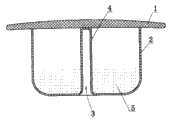
图1和图2为本发明所述的颅底修补体的一个具体实施例的结构示意图,其中,图1为主视图,图2为图1的A-A剖视图。Fig. 1 and Fig. 2 are schematic structural views of a specific embodiment of the skull base prosthesis according to the present invention, wherein Fig. 1 is a front view, and Fig. 2 is a sectional view along A-A of Fig. 1 .
图3为图1和图2所示实施例的使用状态图。Fig. 3 is a usage state diagram of the embodiment shown in Fig. 1 and Fig. 2 .
图4为本发明所述的颅底修补体的另一个具体实施例的结构示意图。Fig. 4 is a schematic structural view of another specific embodiment of the skull base prosthesis according to the present invention.
具体实施方式Detailed ways
实施例1Example 1
参见图1和图2,整个修补体呈蕈菌状,上部的支承体1呈蕈伞状,下部的膨胀体2呈蕈柄状,二者均使用医用硅胶制作,并采用热合的方法上下连接在一起。如图1和图2所示,膨胀体2为空心的薄壁结构,其与支承体1结合面的直径小于底面的直径,呈圆台形。膨胀体2底面的中心向上延伸一导管3,该导管3的管壁上设有进水孔4,它的上端与支承体1热合在一起,下端的外壁与膨胀体2的底面连成一体,使得膨胀体2内的空间为环形。膨胀体2内的环形空间内填充有医用高分子吸水树脂5,其填充量为所述环形空间体积的1%。参见图3,导管3通过进水孔4与上述的环形空间连通,通过导管3经进水孔4向环形空间注水后,医用高分子吸水树脂5迅速膨胀,迫使膨胀体2膨胀成扁鼓形。Referring to Figure 1 and Figure 2, the entire prosthetic body is in the shape of a fungus, the upper supporting
实施例2Example 2
参见图4,本实施例与实施例1的不同之处在于,所述的膨胀体2的外形为圆柱形;支承体1与膨胀体2之间采用粘合剂上下粘接在一起;膨胀体2的环形空间内高分子吸水树脂5的填充量为所述环形空间体积的5%。Referring to Fig. 4, the difference between this embodiment and
下面结合附图对本发明所述的颅底修补体应用于修补经碟扩大手术入路切除脑垂体瘤后的颅底缺损的使用方法描述如下:Below in conjunction with the accompanying drawings, the skull base prosthesis of the present invention is applied to the method of repairing the skull base defect after the pituitary tumor is resected through the enlarged disc operation approach and is described as follows:
参见图1~图3,在神经内镜的引导下,将修补体经碟扩大手术入路送达蝶窦内,并通过碟骨6上的瘘口将支承体1置入垂体窝8内,而膨胀体2伸出瘘口留在蝶窦内。支承体1的上表面与脑垂体7的缺损硬脑膜接触并对硬脑膜起到支撑作用,在脑垂体7的压力作用下,支承体1的下表面贴合在垂体窝8底部的表面,起到封堵作用。此时,通过膨胀体1中的导管3经进水口4向膨胀体2内注水,位于膨胀体2内的医用高分子吸水树脂5吸水后迅速膨胀,将碟骨6上的瘘口封堵;同时,膨胀体2内的导管3在医用高分子吸水树脂5膨胀压力的作用下自动贴合封闭(见图3)。Referring to Figures 1 to 3, under the guidance of the neuroendoscope, the prosthetic body is delivered to the sphenoid sinus through the enlarged disc surgery approach, and the supporting
Claims (2)
Translated fromChinesePriority Applications (1)
| Application Number | Priority Date | Filing Date | Title |
|---|---|---|---|
| CN201010575236XACN102028561B (en) | 2010-12-03 | 2010-12-03 | Basis cranii repairing body |
Applications Claiming Priority (1)
| Application Number | Priority Date | Filing Date | Title |
|---|---|---|---|
| CN201010575236XACN102028561B (en) | 2010-12-03 | 2010-12-03 | Basis cranii repairing body |
Publications (2)
| Publication Number | Publication Date |
|---|---|
| CN102028561Atrue CN102028561A (en) | 2011-04-27 |
| CN102028561B CN102028561B (en) | 2012-08-15 |
Family
ID=43882385
Family Applications (1)
| Application Number | Title | Priority Date | Filing Date |
|---|---|---|---|
| CN201010575236XAExpired - Fee RelatedCN102028561B (en) | 2010-12-03 | 2010-12-03 | Basis cranii repairing body |
Country Status (1)
| Country | Link |
|---|---|
| CN (1) | CN102028561B (en) |
Cited By (1)
| Publication number | Priority date | Publication date | Assignee | Title |
|---|---|---|---|---|
| CN106491246A (en)* | 2016-11-30 | 2017-03-15 | 重庆博仕康科技有限公司 | For the basis cranii repair apparatus through butterfly Operation of Pituitary Adenomas |
Citations (3)
| Publication number | Priority date | Publication date | Assignee | Title |
|---|---|---|---|---|
| WO2000015152A1 (en)* | 1998-09-14 | 2000-03-23 | Bionx Implants Oy | A bioabsorbable, layered composite material for guided bone tissue regeneration |
| CN2636840Y (en)* | 2003-08-14 | 2004-09-01 | 四川国纳科技有限公司 | Bionic arc bone lamella |
| CN101627932A (en)* | 2008-07-14 | 2010-01-20 | 上海交通大学医学院附属第九人民医院 | Piston-shaped bone repairing support and application thereof |
- 2010
- 2010-12-03CNCN201010575236XApatent/CN102028561B/ennot_activeExpired - Fee Related
Patent Citations (3)
| Publication number | Priority date | Publication date | Assignee | Title |
|---|---|---|---|---|
| WO2000015152A1 (en)* | 1998-09-14 | 2000-03-23 | Bionx Implants Oy | A bioabsorbable, layered composite material for guided bone tissue regeneration |
| CN2636840Y (en)* | 2003-08-14 | 2004-09-01 | 四川国纳科技有限公司 | Bionic arc bone lamella |
| CN101627932A (en)* | 2008-07-14 | 2010-01-20 | 上海交通大学医学院附属第九人民医院 | Piston-shaped bone repairing support and application thereof |
Cited By (1)
| Publication number | Priority date | Publication date | Assignee | Title |
|---|---|---|---|---|
| CN106491246A (en)* | 2016-11-30 | 2017-03-15 | 重庆博仕康科技有限公司 | For the basis cranii repair apparatus through butterfly Operation of Pituitary Adenomas |
Also Published As
| Publication number | Publication date |
|---|---|
| CN102028561B (en) | 2012-08-15 |
Similar Documents
| Publication | Publication Date | Title |
|---|---|---|
| JP5620384B2 (en) | Process for forming microwall encapsulated balloons | |
| CN101627933B (en) | Covered stent | |
| US20160089151A1 (en) | Left atrial appendage occlusion devices and methods | |
| WO2007016097A2 (en) | Implantable prosthetic vascular valve | |
| WO2018059178A1 (en) | Partial left ventriculectomy device | |
| JP2022545254A (en) | Medical implants and delivery devices for medical implants | |
| CA2896760A1 (en) | Device and method for the application of a curable fluid composition to a portion of a bodily organ | |
| US20130090734A1 (en) | Sac for use in spinal surgery | |
| CN204698615U (en) | Novel skin dilator | |
| CN102028561B (en) | Basis cranii repairing body | |
| CN103976781B (en) | A kind of compound basis cranii repairs holder | |
| CN107397561A (en) | A kind of oval hole plugging device | |
| CN204890258U (en) | Rebuild acetabular cup false body | |
| CN206120427U (en) | Integral type expansible sacculus bag bagging apparatus | |
| CN104921763B (en) | A kind of device for being used to repair skull base defects | |
| IL305913A (en) | A sealing and anchoring mechanism for irregular tissues in a body and a method of sealing and anchoring thereof | |
| CN208769848U (en) | Skull base defects block repair apparatus | |
| CN204193279U (en) | The self-service miniature soft tissue expander of nose taking prosthese | |
| CN206333936U (en) | A kind of perivalvular leakage plugging device | |
| CN103784220B (en) | Multipurpose adsorbable artificial vertebral plate | |
| CN114041899A (en) | A breast prosthesis with variable volume and construction method | |
| CN110368052A (en) | Cystidium | |
| CN111904657A (en) | Support device for preventing I-shaped internal leakage | |
| CN205073072U (en) | Basis cranii patching device | |
| CN208598538U (en) | A kind of cardiovascular and cerebrovascular operation expander special |
Legal Events
| Date | Code | Title | Description |
|---|---|---|---|
| C06 | Publication | ||
| PB01 | Publication | ||
| C10 | Entry into substantive examination | ||
| SE01 | Entry into force of request for substantive examination | ||
| C14 | Grant of patent or utility model | ||
| GR01 | Patent grant | ||
| CF01 | Termination of patent right due to non-payment of annual fee | Granted publication date:20120815 Termination date:20141203 | |
| EXPY | Termination of patent right or utility model |