CN101933794A - A fluorescent multi-parameter endoscopic measurement method and system - Google Patents
A fluorescent multi-parameter endoscopic measurement method and systemDownload PDFInfo
- Publication number
- CN101933794A CN101933794ACN 201010279725CN201010279725ACN101933794ACN 101933794 ACN101933794 ACN 101933794ACN 201010279725CN201010279725CN 201010279725CN 201010279725 ACN201010279725 ACN 201010279725ACN 101933794 ACN101933794 ACN 101933794A
- Authority
- CN
- China
- Prior art keywords
- fluorescence
- sub
- beams
- sample
- excitation light
- Prior art date
- Legal status (The legal status is an assumption and is not a legal conclusion. Google has not performed a legal analysis and makes no representation as to the accuracy of the status listed.)
- Granted
Links
Images

Landscapes
- Investigating, Analyzing Materials By Fluorescence Or Luminescence (AREA)
Abstract
Translated fromChinese
Description
Translated fromChinese技术领域technical field
本发明属于光电检测领域,尤其涉及一种荧光多参量内窥测量方法及系统。The invention belongs to the field of photoelectric detection, in particular to a fluorescent multi-parameter endoscopic measurement method and system.
背景技术Background technique
荧光显微技术已经成为生命科学,尤其是细胞生物学研究的重要工具。多光子激发荧光显微技术具有对生命体的杀伤作用小,穿透深度大,具有层析能力等优点,已经成为生命科学研究的重要手段。除荧光强度的分布和变化反应样品的结构信息以外,荧光光谱和荧光寿命都包含有丰富的生物体系功能信息。由于荧光发射波长与荧光团的能级结构有关,因此荧光光谱测量可以区分样品的分子种类或鉴别不同的荧光团。荧光寿命对荧光团所处微环境非常敏感,能够对离子浓度(Ca2+,Na+)、pH值和pO2等生理生化参数进行定量测量。将荧光强度、荧光光谱和荧光寿命测量相结合能够为生物医学检测和分析提供不同但互补的结构和功能信息,多种特征参量能够分别从不同的层次、不同的角度,互补的研究同一生命现象。Fluorescence microscopy has become an important tool in life sciences, especially cell biology research. Multi-photon excitation fluorescence microscopy has the advantages of small killing effect on living organisms, large penetration depth, and chromatographic ability, and has become an important means of life science research. In addition to the distribution of fluorescence intensity and the structural information of the change response sample, the fluorescence spectrum and fluorescence lifetime both contain rich functional information of biological systems. Since the fluorescence emission wavelength is related to the energy level structure of the fluorophore, the measurement of the fluorescence spectrum can distinguish the molecular species of the sample or identify different fluorophores. The fluorescence lifetime is very sensitive to the microenvironment of the fluorophore, and can quantitatively measure physiological and biochemical parameters such as ion concentration (Ca2+ , Na+ ), pH value and pO2 . The combination of fluorescence intensity, fluorescence spectrum and fluorescence lifetime measurement can provide different but complementary structural and functional information for biomedical detection and analysis, and multiple characteristic parameters can complement each other to study the same life phenomenon from different levels and different angles .
近年来,随着新型光纤和微制造技术的迅猛发展,光纤双光子荧光显微镜和内窥镜的研究使双光子荧光显微成像技术在活体的内部器官和活体动物中的研究成为可能。目前双光子荧光内窥显微技术已经引起了国际上的高度重视,针对这一课题做出了大量的研究成果,在内窥系统设计、扫描机制、光学传导和高数值孔径的微物镜及其应用等方面取得了很多研究成果。受到活体内窥应用条件限制,测量时间不宜过长。然而目前荧光多参量内窥测量的速度慢,效率低,耗时长,对生物体造成极大的影响。In recent years, with the rapid development of new optical fiber and micro-manufacturing technology, the research of fiber-optic two-photon fluorescence microscope and endoscope has made it possible to study two-photon fluorescence microscopy imaging technology in living internal organs and living animals. At present, two-photon fluorescence endoscopic microscopy technology has attracted great attention in the world, and a large number of research results have been made on this subject. A lot of research results have been obtained in the fields of application and so on. Limited by the application conditions of in vivo endoscopy, the measurement time should not be too long. However, the current measurement of fluorescence multi-parameter endoscopy is slow, inefficient, and time-consuming, which has a great impact on organisms.
发明内容Contents of the invention
本发明实施例的目的在于提供一种荧光多参量内窥测量方法,旨在解决现有荧光多参量内窥测量速度慢、效率低的问题。The purpose of the embodiments of the present invention is to provide a fluorescent multi-parameter endoscopic measurement method, aiming to solve the problems of slow speed and low efficiency of the existing fluorescent multi-parameter endoscopic measurement.
本发明实施例是这样实现的,一种荧光多参量内窥测量方法,包括以下步骤:The embodiment of the present invention is achieved in this way, a fluorescent multi-parameter endoscopic measurement method, comprising the following steps:
产生激发光;generate excitation light;
将所述激发光分为多个子光束,所述多个子光束对应于样品的多个子区域,所述样品内均匀分布有荧光物质;Dividing the excitation light into multiple sub-beams, the multiple sub-beams correspond to multiple sub-regions of the sample, and fluorescent substances are evenly distributed in the sample;
调整所述多个子光束,使所述多个子光束传导至生物体内并聚焦于所述样品;adjusting the multiple sub-beams so that the multiple sub-beams are transmitted into the living body and focused on the sample;
利用所述多个子光束对所述样品进行扫描,使各子区域内的荧光物质发出荧光;Scanning the sample by using the plurality of sub-beams, so that the fluorescent substances in each sub-area emit fluorescence;
实时采集所述荧光;collecting the fluorescence in real time;
分辨所述荧光的多参量信息。Multiparametric information of the fluorescence is resolved.
本发明实施例的另一目的在于提供一种荧光多参量内窥测量系统,所述系统包括:Another object of the embodiments of the present invention is to provide a fluorescent multi-parameter endoscopic measurement system, the system comprising:
激发光源,用于产生激发光;an excitation light source for generating excitation light;
分光器,用于将所述激发光分为多个子光束,所述多个子光束对应于样品的多个子区域,所述样品内均匀分布有荧光物质;a beam splitter, configured to divide the excitation light into multiple sub-beams, the multiple sub-beams correspond to multiple sub-regions of the sample, and fluorescent substances are evenly distributed in the sample;
柔性介质,用于调整所述多个子光束,使所述多个子光束传导至生物体内并聚焦于所述样品;a flexible medium, used to adjust the multiple sub-beams, so that the multiple sub-beams are transmitted into the living body and focused on the sample;
扫描镜,用于利用所述多个子光束对所述样品进行扫描,使各子区域内的荧光物质发出荧光;a scanning mirror, used to scan the sample by using the plurality of sub-beams, so that the fluorescent substances in each sub-area emit fluorescence;
双色镜,用于导出所述荧光;a dichroic mirror for deriving said fluorescence;
色散元件,用于分辨所述荧光的光谱信息;a dispersion element, used to distinguish the spectral information of the fluorescence;
扫描相机,用于分辨所述荧光的寿命信息;a scanning camera, used to distinguish the lifetime information of the fluorescence;
探测器,用于记录所述荧光的多参量信息。a detector for recording multi-parameter information of the fluorescence.
本发明实施例将激发光分为与样品的多个子区域一一对应的多个子光束,该多个子光束通过柔性介质并行传导至生物体内,各子光束聚焦于具有荧光物质的样品,形成多点激发荧光,将所激发的荧光通过柔性介质导出,由多个子光束对样品进行二维扫描,从而获取整个样品不同位置光谱分辨的荧光寿命信息,速度快、时间短,对生物体损伤小,有利于生物医学研究,特别是对癌症早期诊断,具有重要意义。In the embodiment of the present invention, the excitation light is divided into multiple sub-beams corresponding to multiple sub-regions of the sample, and the multiple sub-beams are transmitted to the living body in parallel through a flexible medium, and each sub-beam is focused on the sample with fluorescent substances to form a multi-point Excite fluorescence, export the excited fluorescence through a flexible medium, and scan the sample two-dimensionally by multiple sub-beams, so as to obtain the fluorescence lifetime information of spectral resolution at different positions of the entire sample, fast speed, short time, little damage to organisms, effective It is beneficial to biomedical research, especially for early diagnosis of cancer, which is of great significance.
附图说明Description of drawings
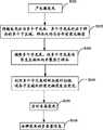
图1是本发明实施例提供的荧光多参量内窥测量方法的实现流程图;Fig. 1 is a flow chart of the realization of the fluorescent multi-parameter endoscopic measurement method provided by the embodiment of the present invention;
图2是本发明实施例提供的荧光多参量内窥测量系统的结构及其光路图;Fig. 2 is the structure and optical path diagram of the fluorescent multi-parameter endoscopic measurement system provided by the embodiment of the present invention;
图3是本发明实施例提供的激光阵列点图;Fig. 3 is a laser array point diagram provided by an embodiment of the present invention;
图4是探测器所记录的一幅光谱分辨的荧光衰减信息图像;Fig. 4 is a spectrally resolved fluorescence decay information image recorded by the detector;
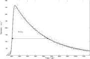
图5是样品上一点的荧光衰减曲线。Figure 5 is the fluorescence decay curve at a point on the sample.
具体实施方式Detailed ways
为了使本发明的目的、技术方案及优点更加清楚明白,以下结合附图及实施例,对本发明进行进一步详细说明。应当理解,此处所描述的具体实施例仅仅用以解释本发明,并不用于限定本发明。In order to make the object, technical solution and advantages of the present invention clearer, the present invention will be further described in detail below in conjunction with the accompanying drawings and embodiments. It should be understood that the specific embodiments described here are only used to explain the present invention, not to limit the present invention.
本发明实施例将激发光分为与样品多个子区域一一对应的多个子光束,使该多个子光束传导至生物体内,各子光束聚焦于具有荧光物质的样品,形成多点激发荧光,将荧光导出,由多个子光束对样品进行二维扫描,从而获取整个样品不同位置光谱分辨的荧光寿命信息,速度快、时间短,对生物体损伤小,有利于生物医学研究。In the embodiment of the present invention, the excitation light is divided into multiple sub-beams corresponding to multiple sub-regions of the sample, and the multiple sub-beams are transmitted into the living body, and each sub-beam is focused on the sample with fluorescent substances to form multi-point excitation fluorescence. Fluorescence derivation, two-dimensional scanning of the sample by multiple sub-beams, so as to obtain the fluorescence lifetime information of the spectral resolution of different positions of the entire sample, the speed is fast, the time is short, and the damage to the organism is small, which is beneficial to biomedical research.
本发明实施例提供的荧光多参量内窥测量方法包括以下步骤:The fluorescent multi-parameter endoscopic measurement method provided by the embodiment of the present invention includes the following steps:
产生激发光;generate excitation light;
将所述激发光分为多个子光束,所述多个子光束对应于样品的多个子区域,所述样品内均匀分布有荧光物质;Dividing the excitation light into multiple sub-beams, the multiple sub-beams correspond to multiple sub-regions of the sample, and fluorescent substances are evenly distributed in the sample;
调整所述多个子光束,使所述多个子光束传导至生物体内并聚焦于所述样品;adjusting the multiple sub-beams so that the multiple sub-beams are transmitted into the living body and focused on the sample;
利用所述多个子光束对所述样品进行扫描,使各子区域内的荧光物质发出荧光;Scanning the sample by using the plurality of sub-beams, so that the fluorescent substances in each sub-area emit fluorescence;
实时采集所述荧光;collecting the fluorescence in real time;
分辨所述荧光的多参量信息。Multiparametric information of the fluorescence is resolved.
本发明实施例提供的荧光多参量内窥测量系统包括:The fluorescent multi-parameter endoscopic measurement system provided by the embodiment of the present invention includes:
激发光源,用于产生激发光;an excitation light source for generating excitation light;
分光器,用于将所述激发光分为多个子光束,所述多个子光束对应于样品的多个子区域,所述样品内均匀分布有荧光物质;a beam splitter, configured to divide the excitation light into multiple sub-beams, the multiple sub-beams correspond to multiple sub-regions of the sample, and fluorescent substances are evenly distributed in the sample;
柔性介质,用于调整所述多个子光束,使所述多个子光束传导至生物体内并聚焦于所述样品;a flexible medium, used to adjust the multiple sub-beams, so that the multiple sub-beams are transmitted into the living body and focused on the sample;
扫描镜,用于利用所述多个子光束对所述样品进行扫描,使各子区域内的荧光物质发出荧光;a scanning mirror, used to scan the sample by using the plurality of sub-beams, so that the fluorescent substances in each sub-area emit fluorescence;
双色镜,用于导出所述荧光;a dichroic mirror for deriving said fluorescence;
色散元件,用于分辨所述荧光的光谱信息;a dispersion element, used to distinguish the spectral information of the fluorescence;
扫描相机,用于分辨所述荧光的寿命信息;a scanning camera, used to distinguish the lifetime information of the fluorescence;
探测器,用于记录所述荧光的多参量信息。a detector for recording multi-parameter information of the fluorescence.
以下结合具体实施例对本发明的具体实现进行详细描述。The specific implementation of the present invention will be described in detail below in conjunction with specific embodiments.
图1示出了本发明实施例提供的荧光多参量内窥测量方法的实现流程,详述如下:Figure 1 shows the implementation process of the fluorescent multi-parameter endoscopic measurement method provided by the embodiment of the present invention, which is described in detail as follows:
在步骤S101中,产生激发光;In step S101, generating excitation light;
本发明实施例优选工作频率为76MHz,周期为120fs,中心波长为800nm的脉冲激光作为激发光,此激发光可实现荧光物质的双光子激发。In the embodiment of the present invention, a pulsed laser with a working frequency of 76 MHz, a cycle of 120 fs, and a center wavelength of 800 nm is preferably used as excitation light, which can realize two-photon excitation of fluorescent substances.
作为本发明的一个实施例,使上述脉冲激光产生负色散,按所需频率提取具有负色散特性的脉冲激光,对获取的脉冲激光进行扩束准直并调整其强度分布,使脉冲激光的强度分布均匀。例如,提取76MHz的脉冲激光获得2MHz的脉冲激光。As an embodiment of the present invention, the above-mentioned pulsed laser is made to produce negative dispersion, the pulsed laser with negative dispersion characteristics is extracted according to the required frequency, the acquired pulsed laser is expanded and collimated and its intensity distribution is adjusted to make the intensity of the pulsed laser evenly distributed. For example, extracting a 76MHz pulsed laser yields a 2MHz pulsed laser.
在步骤S102中,将激发光分为多个子光束,多个子光束对应于样品的多个子区域;In step S102, the excitation light is divided into multiple sub-beams, and the multiple sub-beams correspond to multiple sub-regions of the sample;
本发明实施例中,将激发光均分为多个平行的子光束,多个子光束一一对应于具有荧光物质的样品的多个子区域。In the embodiment of the present invention, the excitation light is equally divided into multiple parallel sub-beams, and the multiple sub-beams correspond to the multiple sub-regions of the sample with the fluorescent substance one by one.
为将多个子光束调整至生物体内,对各个子光束进行准直,使各个子光束成为平行光。In order to adjust the multiple sub-beams into the living body, each sub-beam is collimated so that each sub-beam becomes a parallel light.
在步骤S103中,调整多个子光束,使多个子光束通过柔性介质传导至生物体内并聚焦于样品;In step S103, adjusting the multiple sub-beams so that the multiple sub-beams are transmitted into the living body through the flexible medium and focused on the sample;
本发明实施例中,将多个子光束调整至柔性介质的输入端,使多个子光束并行传导至生物体内,并各自聚焦于样品。In the embodiment of the present invention, multiple sub-beams are adjusted to the input end of the flexible medium, so that the multiple sub-beams are transmitted into the living body in parallel, and are respectively focused on the sample.
在步骤S104中,利用多个子光束对样品进行扫描,使各子区域内的荧光物质发出荧光;In step S104, the sample is scanned with multiple sub-beams, so that the fluorescent substances in each sub-area emit fluorescence;
本发明实施例中的扫描分为逐点扫描和步进扫描,具体过程如下:Scanning in the embodiment of the present invention is divided into point-by-point scanning and step-by-step scanning, and the specific process is as follows:
1、逐点扫描1. Scan point by point
多个子光束经聚焦形成激发光阵列点分别投射至样品的各个子区域,沿样品纵向对各子区域进行逐点扫描,各子区域内的荧光物质在激发光阵列点的作用下发出荧光。A plurality of sub-beams are focused to form an excitation light array point and projected to each sub-area of the sample, and each sub-area is scanned point by point along the longitudinal direction of the sample, and the fluorescent substances in each sub-area emit fluorescence under the action of the excitation light array point.
2、步进扫描2. Step scan
对各个子区域纵向的逐点扫描结束后,沿样品横向对各个子区域进行步进扫描即调整激发光阵列点在样品横向的位置。After the point-by-point scanning of each sub-region in the vertical direction is completed, step-by-step scanning of each sub-region along the lateral direction of the sample is performed to adjust the position of the excitation light array point in the lateral direction of the sample.
循环执行上述逐点扫描和步进扫描,直至完成对样品各个子区域的扫描。应当理解,具体实施时还可以调换逐点扫描与步进扫描的方向。The above-mentioned point-by-point scanning and step-by-step scanning are performed cyclically until the scanning of each sub-area of the sample is completed. It should be understood that the directions of point-by-point scanning and step-by-step scanning can also be exchanged during specific implementation.
在步骤S105中,实时采集扫描时发出的荧光;In step S105, the fluorescence emitted during scanning is collected in real time;
本发明实施例中,对各子区域进行逐点扫描的同时,采集各子区域内荧光物质发出的荧光。In the embodiment of the present invention, while performing point-by-point scanning on each sub-area, the fluorescence emitted by the fluorescent substance in each sub-area is collected.
在步骤S106中,分辨荧光的多参量信息;In step S106, distinguish the multi-parameter information of fluorescence;
本发明实施例中,将荧光的多参量信息转换为可分辨的光谱和空间信息,从而获取样品各点荧光的多参量信息。In the embodiment of the present invention, the multi-parameter information of fluorescence is converted into resolvable spectral and spatial information, so as to obtain the multi-parameter information of fluorescence at each point of the sample.
具体地,对荧光进行色散,获取荧光的光谱;将荧光的寿命信息转换为可分辨的空间信息,获取荧光的寿命信息。Specifically, the fluorescence is dispersed to obtain the spectrum of the fluorescence; the lifetime information of the fluorescence is converted into distinguishable spatial information to obtain the lifetime information of the fluorescence.
本领域的普通技术人员应当理解,实现上述实施例方法中的全部或部分步骤可以通过程序来指令相关的硬件完成,该程序可以存储于一计算机可读取存储介质中,如ROM/RAM、磁盘、光盘等。Those of ordinary skill in the art should understand that all or part of the steps in the methods of the above-mentioned embodiments can be completed by instructing related hardware through a program, and the program can be stored in a computer-readable storage medium, such as ROM/RAM, disk , CD, etc.
上述实施例将激发光分为与样品多个子区域一一对应的多个子光束,该多个子光束通过柔性介质并行传导至生物体内,并各自聚焦于具有荧光物质的样品,形成多点激发荧光,将所激发的荧光通过柔性介质导出,由多个子光束对样品进行二维扫描,从而获取整个样品不同位置光谱分辨的荧光寿命信息,速度快、时间短,对生物体损伤小,有利于生物医学研究。In the above embodiment, the excitation light is divided into multiple sub-beams corresponding to multiple sub-regions of the sample, and the multiple sub-beams are transmitted into the living body in parallel through a flexible medium, and are respectively focused on the sample with fluorescent substances to form multi-point excitation fluorescence. The excited fluorescence is exported through a flexible medium, and the sample is scanned two-dimensionally by multiple sub-beams, so as to obtain the fluorescence lifetime information of the spectral resolution at different positions of the entire sample, with fast speed, short time, and little damage to organisms, which is beneficial to biomedicine Research.
图2示出了本发明实施例提供的荧光多参量内窥测量系统的结构,为了便于说明,仅示出了与本发明实施例相关的部分。Fig. 2 shows the structure of the fluorescence multi-parameter endoscopic measurement system provided by the embodiment of the present invention. For the convenience of description, only the parts related to the embodiment of the present invention are shown.
本发明实施例提供的荧光多参量内窥测量系统具有一激发光路和一探测光路。激发光路包括激发光源、色散补偿器、脉冲提取器、扩束准直装置、整形器、分光器、准直透镜、耦合透镜、光子晶体光纤阵列、自聚焦透镜、扫描镜以及微物镜。探测光路包括微物镜、扫描镜、自聚焦透镜、光子晶体光纤阵列、耦合透镜、双色镜、滤光片、色散元件、成像透镜、扫描相机以及探测器。其中耦合透镜、光子晶体光纤阵列、自聚焦透镜、扫描镜以及微物镜为激发光路和探测光路所共有。The fluorescent multi-parameter endoscopic measurement system provided by the embodiment of the present invention has an excitation light path and a detection light path. The excitation optical path includes an excitation light source, a dispersion compensator, a pulse extractor, a beam expander collimator, a shaper, a beam splitter, a collimator lens, a coupling lens, a photonic crystal fiber array, a self-focus lens, a scanning mirror and a micro-objective lens. The detection optical path includes a micro-objective lens, a scanning mirror, a self-focusing lens, a photonic crystal fiber array, a coupling lens, a dichroic mirror, a filter, a dispersion element, an imaging lens, a scanning camera and a detector. The coupling lens, photonic crystal fiber array, self-focusing lens, scanning mirror and micro-objective lens are shared by the excitation light path and the detection light path.
以下对激发光路的结构进行详细说明。The structure of the excitation light path will be described in detail below.
如图2所示,本发明实施例优选钛宝石飞秒激光器101作为激发光源,其可产生中心波长为800nm、频率为76MHz、周期为120fs的脉冲激光,该脉冲激光可实现荧光物质的双光子激发。色散补偿器102用于产生负色散,钛宝石飞秒激光器101发出的脉冲激光经色散补偿器102产生负色散。脉冲提取器103用于选择性提取脉冲激光,钛宝石飞秒激光器101发出的高频率(如76MHz)的脉冲激光经脉冲提取后形成低频率(如2MHz)的脉冲激光。脉冲激光经由扩束准直透镜104和扩束准直透镜105构成的扩束准直装置,变成所需尺寸的准直光。As shown in Figure 2, the embodiment of the present invention preferably uses a Ti:Sapphire femtosecond laser 101 as the excitation light source, which can generate a pulsed laser with a center wavelength of 800 nm, a frequency of 76 MHz, and a period of 120 fs. The pulsed laser can realize two-photon generation of fluorescent substances excitation. The
本发明实施例中,整形器为光束整形器106,经准直的脉冲激光经光束整形器106整形,形成强度平顶分布的光束,强度分布均匀。分光器可为微透镜阵列、衍射光学元件或分束器,本实施例优选微透镜阵列107,平顶分布的脉冲激光经微透镜阵列107被分成多个子光束,多个子光束对应样品的多个子区域,本实施例中微透镜阵列107为3×3微透镜阵列即微透镜阵列107具有九个微透镜。In the embodiment of the present invention, the shaper is the beam shaper 106, and the collimated pulsed laser is shaped by the beam shaper 106 to form a beam with a flat-top distribution of intensity, and the intensity distribution is uniform. The beam splitter can be a microlens array, a diffractive optical element, or a beam splitter. In this embodiment, the microlens array 107 is preferred. The flat-top distributed pulse laser is divided into multiple sub-beams through the microlens array 107, and the multiple sub-beams correspond to multiple sub-beams of the sample. In this embodiment, the microlens array 107 is a 3×3 microlens array, that is, the microlens array 107 has nine microlenses.
本发明实施例中,准直透镜108的后焦面与微透镜阵列107的前焦面重合,子光束在微透镜阵列107的前焦面即在准直透镜108的后焦面聚焦,各子光束经准直透镜108均变为平行光的子光束。In the embodiment of the present invention, the rear focal plane of the collimator lens 108 coincides with the front focal plane of the microlens array 107, and the sub-beams focus on the front focal plane of the microlens array 107, that is, the rear focal plane of the collimator lens 108, and each sub-beam The light beams are transformed into sub-beams of parallel light through the collimating lens 108 .
在本发明实施例中,多个子光束经双色镜109调整投射至耦合透镜110,由耦合透镜110将多个子光束耦合进入光子晶体光纤阵列111。光子晶体光纤阵列111由多根光子晶体光纤等间距排列形成,光子晶体光纤的个数及其排列方式与微透镜阵列107的相同。多个子光束经光子晶体光纤阵列111传导至生物体内,从光子晶体光纤阵列111出射的多个子光束经自聚焦透镜112投射至扫描镜113。自聚焦透镜112为折射率沿径向渐变的棒透镜,各子光束经自聚焦透镜112均变为平行光。各子光束经扫描镜113投射至具有荧光物质的样品114,样品114与扫描镜113之间设有起会聚作用的微物镜115。所述扫描镜113优选为MEMS(Micro-Electro-Mechanical Systems,微机电系统)扫描镜,该MEMS扫描镜可进行二维扫描。各子光束经本激发光路传导后形成激发光阵列点并聚焦于样品,激发样品内的荧光物质发出荧光。In the embodiment of the present invention, the multiple sub-beams are adjusted and projected to the coupling lens 110 through the dichroic mirror 109 , and the coupling lens 110 couples the multiple sub-beams into the photonic crystal fiber array 111 . The photonic crystal fiber array 111 is formed by a plurality of photonic crystal fibers arranged at equal intervals, and the number and arrangement of the photonic crystal fibers are the same as those of the microlens array 107 . The multiple sub-beams are transmitted into the living body through the photonic crystal fiber array 111 , and the multiple sub-beams emitted from the photonic crystal fiber array 111 are projected to the scanning mirror 113 through the self-focusing lens 112 . The self-focusing lens 112 is a rod lens whose refractive index gradually changes along the radial direction, and each sub-beam becomes parallel light through the self-focusing lens 112 . Each sub-beam is projected to a sample 114 with a fluorescent substance through a scanning mirror 113 , and a micro-objective lens 115 for converging is provided between the sample 114 and the scanning mirror 113 . The scanning mirror 113 is preferably a MEMS (Micro-Electro-Mechanical Systems, Micro-Electro-Mechanical Systems) scanning mirror, and the MEMS scanning mirror can perform two-dimensional scanning. Each sub-beam is transmitted through the excitation light path to form an excitation light array point and focus on the sample to excite the fluorescent substance in the sample to emit fluorescence.
以下对探测光路的结构进行详细说明。The structure of the detection light path will be described in detail below.
本发明实施例探测光路中双色镜109设于耦合透镜110与准直透镜108之间,双色镜109对中心波长为800nm的脉冲激光高反,对波长为400~700nm的荧光高透,双色镜109与子光束之间的夹角为45°或135°。荧光经双色镜109透射后的传导光路上依次设有色散元件、成像透镜117、扫描相机118以及探测器。The dichromatic mirror 109 is arranged between the coupling lens 110 and the collimating lens 108 in the detection optical path of the embodiment of the present invention. The dichromatic mirror 109 is highly reflective to pulsed laser light with a center wavelength of 800 nm, and highly transparent to fluorescent light with a wavelength of 400-700 nm. The angle between 109 and the sub-beam is 45° or 135°. A dispersive element, an imaging lens 117 , a scanning camera 118 and a detector are sequentially arranged on the transmission light path of the fluorescence transmitted through the dichroic mirror 109 .
上述色散元件优选为色散棱镜116,亦可为光栅。色散棱镜116将荧光按不同波长区分开,以便探测器识别荧光光谱信息,从而获取样品各点的荧光光谱信息。The dispersion element is preferably a dispersion prism 116, and may also be a grating. The dispersion prism 116 distinguishes the fluorescence according to different wavelengths, so that the detector can identify the fluorescence spectrum information, so as to obtain the fluorescence spectrum information of each point of the sample.
在本发明实施例中,成像透镜117将荧光按不同波长会聚于扫描相机118的光电阴极的不同位置。扫描相机118具有电压线性变化的偏转电场,能够将在空间上不可分辨的的寿命信息转换为在空间上可分辨的信息,以便探测器识别荧光衰减信息,从而获取样品各点的荧光寿命信息。In the embodiment of the present invention, the imaging lens 117 converges the fluorescent light at different positions of the photocathode of the scanning camera 118 according to different wavelengths. The scanning camera 118 has a deflection electric field whose voltage varies linearly, and can convert the spatially indistinguishable lifetime information into spatially resolvable information, so that the detector can identify the fluorescence decay information, thereby obtaining the fluorescence lifetime information of each point of the sample.
本发明实施例中,探测器为面阵探测器119,面阵探测器119优选为CCD相机或CMOS相机,面阵探测器119用于记录不同波长的荧光衰减信息即光谱分辨的荧光衰减信息,同时记录荧光的空间位置。面阵探测器119与计算机120连接,计算机120用于读取、存储和处理探测器探测到的光谱分辨的荧光衰减信息,由计算机120控制面阵探测器119的曝光。工作时,脉冲提取器103每提取一个脉冲激光,扫描镜113扫描一次,面阵探测器119曝光一次。计算机120同时控制扫描镜113进行逐点扫描和步进扫描,扫描镜每进行一次逐点扫描或步进扫描,计算机120记录各次扫描所对应样品上的位置。In the embodiment of the present invention, the detector is an area array detector 119, and the area array detector 119 is preferably a CCD camera or a CMOS camera. The area array detector 119 is used to record fluorescence attenuation information of different wavelengths, that is, spectrally resolved fluorescence attenuation information. Simultaneously record the spatial position of the fluorescence. The area array detector 119 is connected with a computer 120, and the computer 120 is used to read, store and process the spectrally resolved fluorescence attenuation information detected by the detector, and the exposure of the area array detector 119 is controlled by the computer 120. During operation, every time the pulse extractor 103 extracts a pulse laser, the scanning mirror 113 scans once, and the area array detector 119 exposes once. The computer 120 simultaneously controls the scanning mirror 113 to perform point-by-point scanning and step-by-step scanning. Every time the scanning mirror performs point-by-point scanning or step-by-step scanning, the computer 120 records the position on the sample corresponding to each scan.
此外,色散元件116与双色镜109之间设滤光片,滤光片为干涉滤光片121,干涉滤光片121将反射回来的激光以及其它杂散光去除,避免干扰,各参量测量准确。In addition, a filter is provided between the dispersion element 116 and the dichroic mirror 109, and the filter is an interference filter 121, which removes the reflected laser light and other stray light to avoid interference, and the measurement of each parameter is accurate.
本发明实施例中,各个子光束经扫描镜113沿样品纵向进行逐点扫描,各子光束沿样品纵向每移动一个点位,面阵探测器119曝光一次即记录一幅图像,如图3所示,该图像包含样品114的九个位置点的荧光光谱和荧光寿命信息。沿样品纵向的扫描结束后,扫描镜113步进扫描即沿样品横向移至下一位置,进行下一纵向位置的逐点扫描,如此循环,即可实现对整个样品的扫描,获取整个样品不同位置处光谱分辨的荧光衰减信息。In the embodiment of the present invention, each sub-beam is scanned point-by-point along the longitudinal direction of the sample through the scanning mirror 113, and each time the sub-beams move one point along the longitudinal direction of the sample, the area array detector 119 is exposed once to record an image, as shown in Figure 3 As shown, the image contains the fluorescence spectrum and fluorescence lifetime information of nine positions of the sample 114. After the scanning along the longitudinal direction of the sample is completed, the scanning mirror 113 moves to the next position along the lateral direction of the sample for step-by-step scanning, and performs point-by-point scanning of the next longitudinal position. In this way, the scanning of the entire sample can be realized, and different values of the entire sample can be obtained. Spectrally resolved fluorescence decay information at the position.
显然,通过计算机控制并调整扫描镜的方位,即可进行逐点扫描或步进扫描。具体实施时还可互换逐点扫描与步进扫描的方向。Obviously, point-by-point scanning or step-by-step scanning can be performed by controlling the computer and adjusting the orientation of the scanning mirror. During specific implementation, the directions of point-by-point scanning and step-by-step scanning can also be interchanged.
本发明实施例中,钛宝石飞秒激光器101发出的激光脉冲由脉冲提取器103按所需频率提取用于触发扫描相机118,使扫描相机118与脉冲提取器103同步开启。钛宝石飞秒激光器101发出的激光脉冲到达样品114时激发出荧光,扫描相机118的光电阴极接收到所激发出的荧光时发射出光电子,所发出的光电子在扫描相机118的偏转电场的作用下发生偏转,光电子于扫描相机118的荧光屏上按空间位置产生二次荧光,所产生的二次荧光对应于样品上不同位置点发出的不同波长的荧光不同时间的强度分布,即荧光强度的衰减信息,如图5所示。如图5所示,计算出荧光衰减至其最大强度值的1/e的时间即为荧光寿命。In the embodiment of the present invention, the laser pulse emitted by the Ti:Sapphire femtosecond laser 101 is extracted by the pulse extractor 103 according to the required frequency to trigger the scanning camera 118, so that the scanning camera 118 and the pulse extractor 103 are turned on synchronously. When the laser pulse emitted by the Ti:Sapphire femtosecond laser 101 reaches the sample 114, fluorescence is excited, and the photocathode of the scanning camera 118 emits photoelectrons when receiving the excited fluorescence, and the emitted photoelectrons are under the action of the deflection electric field of the scanning camera 118 When the deflection occurs, the photoelectrons generate secondary fluorescence according to the spatial position on the fluorescent screen of the scanning camera 118, and the generated secondary fluorescence corresponds to the intensity distribution of fluorescence of different wavelengths emitted from different positions on the sample at different times, that is, the attenuation information of the fluorescence intensity , as shown in Figure 5. As shown in Figure 5, the calculated time for the fluorescence to decay to 1/e of its maximum intensity value is the fluorescence lifetime.
本发明实施例中,扫描镜移动一个位置,面阵探测器记录一幅图像,一幅图像同时记录样品上九个点的荧光多参量信息。通过扫描镜的二维扫描即可实现对整个样品的扫描,获取整个样品不同位置不同光谱的荧光寿命信息。In the embodiment of the present invention, the scanning mirror moves one position, the area array detector records one image, and one image simultaneously records fluorescence multi-parameter information of nine points on the sample. The entire sample can be scanned through the two-dimensional scanning of the scanning mirror, and the fluorescence lifetime information of different spectra at different positions of the entire sample can be obtained.
图4所示为面阵探测器119所记录的荧光图像,该图像包含荧光的多参量信息,其横轴为荧光光谱信息,纵轴为荧光寿命信息。Fig. 4 shows the fluorescence image recorded by the area array detector 119, the image contains multi-parameter information of fluorescence, the horizontal axis is the fluorescence spectrum information, and the vertical axis is the fluorescence lifetime information.
本发明实施例同样对于512×512图像,阵列点为3x3,仅需记录29127幅图像;阵列点为4×4,仅需记录16384幅图像;依此类推,阵列点越多,需记录的图像越少。In the embodiment of the present invention, for a 512×512 image, the array points are 3×3, and only 29,127 images need to be recorded; the array points are 4×4, and only 16,384 images need to be recorded; and so on, the more array points, the more images to be recorded less.
本发明实施例将激发光分为与样品的多个子区域一一对应的多个子光束,该多个子光束通过柔性介质并行传导至生物体内,各子光束聚焦于具有荧光物质的样品,形成多点激发荧光,将所激发的荧光通过柔性介质导出,由多个子光束对样品进行二维扫描,从而获取整个样品不同位置光谱分辨的荧光寿命信息,速度快、时间短,对生物体损伤小,有利于生物医学研究,特别是对癌症早期诊断,具有重要意义。In the embodiment of the present invention, the excitation light is divided into multiple sub-beams corresponding to multiple sub-regions of the sample, and the multiple sub-beams are transmitted to the living body in parallel through a flexible medium, and each sub-beam is focused on the sample with fluorescent substances to form a multi-point Excite fluorescence, export the excited fluorescence through a flexible medium, and scan the sample two-dimensionally by multiple sub-beams, so as to obtain the fluorescence lifetime information of spectral resolution at different positions of the entire sample, fast speed, short time, little damage to organisms, effective It is beneficial to biomedical research, especially for early diagnosis of cancer, which is of great significance.
以上所述仅为本发明的较佳实施例而已,并不用以限制本发明,凡在本发明的精神和原则之内所作的任何修改、等同替换和改进等,均应包含在本发明的保护范围之内。The above descriptions are only preferred embodiments of the present invention, and are not intended to limit the present invention. Any modifications, equivalent replacements and improvements made within the spirit and principles of the present invention should be included in the protection of the present invention. within range.
Claims (10)
Translated fromChinesePriority Applications (1)
| Application Number | Priority Date | Filing Date | Title |
|---|---|---|---|
| CN 201010279725CN101933794B (en) | 2010-09-13 | 2010-09-13 | Fluorescent multi-parameter endoscopic measuring method and system |
Applications Claiming Priority (1)
| Application Number | Priority Date | Filing Date | Title |
|---|---|---|---|
| CN 201010279725CN101933794B (en) | 2010-09-13 | 2010-09-13 | Fluorescent multi-parameter endoscopic measuring method and system |
Publications (2)
| Publication Number | Publication Date |
|---|---|
| CN101933794Atrue CN101933794A (en) | 2011-01-05 |
| CN101933794B CN101933794B (en) | 2013-02-13 |
Family
ID=43387538
Family Applications (1)
| Application Number | Title | Priority Date | Filing Date |
|---|---|---|---|
| CN 201010279725Expired - Fee RelatedCN101933794B (en) | 2010-09-13 | 2010-09-13 | Fluorescent multi-parameter endoscopic measuring method and system |
Country Status (1)
| Country | Link |
|---|---|
| CN (1) | CN101933794B (en) |
Cited By (8)
| Publication number | Priority date | Publication date | Assignee | Title |
|---|---|---|---|---|
| CN102162907A (en)* | 2011-04-15 | 2011-08-24 | 中国科学院化学研究所 | Multi-wavelength micro illumination device |
| WO2012075860A1 (en)* | 2010-12-09 | 2012-06-14 | 深圳大学 | Fluorescence endoscopic imaging method and system |
| CN102621765A (en)* | 2012-03-28 | 2012-08-01 | 中国科学院物理研究所 | Femtosecond laser fiber spectroscopic device based on dispersion pre-compensation |
| CN104116497A (en)* | 2014-07-22 | 2014-10-29 | 中国科学院自动化研究所 | Endoscopic optical molecular imaging guidance system and multi-spectral imaging method |
| CN105592252A (en)* | 2016-02-24 | 2016-05-18 | 河南工程学院 | Enteroscope system having lesion classification analysis function and three-dimensional image display function |
| CN109770853A (en)* | 2019-01-30 | 2019-05-21 | 上海师范大学 | A zebrafish wound detection method |
| CN110082898A (en)* | 2019-04-24 | 2019-08-02 | 中国科学院自动化研究所 | Fluorescence microscopy lens head and fluorescence microscope |
| CN110333193A (en)* | 2019-07-23 | 2019-10-15 | 无锡迅杰光远科技有限公司 | MEMS type Static Closed Loop spectrum imaging system |
Citations (3)
| Publication number | Priority date | Publication date | Assignee | Title |
|---|---|---|---|---|
| CN1737536A (en)* | 2004-08-18 | 2006-02-22 | 深圳大学 | Five-dimensional fluorescence microscopy imaging technology |
| CN101375786A (en)* | 2007-09-12 | 2009-03-04 | 深圳大学 | Fluorescence endoscopic imaging method and device |
| CN101793829A (en)* | 2010-02-04 | 2010-08-04 | 深圳大学 | Fluorescence microscopy imaging method and system |
- 2010
- 2010-09-13CNCN 201010279725patent/CN101933794B/ennot_activeExpired - Fee Related
Patent Citations (3)
| Publication number | Priority date | Publication date | Assignee | Title |
|---|---|---|---|---|
| CN1737536A (en)* | 2004-08-18 | 2006-02-22 | 深圳大学 | Five-dimensional fluorescence microscopy imaging technology |
| CN101375786A (en)* | 2007-09-12 | 2009-03-04 | 深圳大学 | Fluorescence endoscopic imaging method and device |
| CN101793829A (en)* | 2010-02-04 | 2010-08-04 | 深圳大学 | Fluorescence microscopy imaging method and system |
Non-Patent Citations (2)
| Title |
|---|
| 《中国激光》 20100531 李恒等 基于微透镜阵列和振镜扫描的光谱分辨多焦点多光子显微技术 第1241页左栏最后一段,附图1 5-10 第37卷, 第5期 2* |
| 《物理学报》 20061231 林子扬等 双光子阵列点激发同时多维荧光信息的处理 第6701页右栏第2段-第6702页右栏第1段,第6703页左栏第2段,图1-2 5-10 第55卷, 第12期 2* |
Cited By (10)
| Publication number | Priority date | Publication date | Assignee | Title |
|---|---|---|---|---|
| WO2012075860A1 (en)* | 2010-12-09 | 2012-06-14 | 深圳大学 | Fluorescence endoscopic imaging method and system |
| CN102162907A (en)* | 2011-04-15 | 2011-08-24 | 中国科学院化学研究所 | Multi-wavelength micro illumination device |
| CN102621765A (en)* | 2012-03-28 | 2012-08-01 | 中国科学院物理研究所 | Femtosecond laser fiber spectroscopic device based on dispersion pre-compensation |
| CN104116497A (en)* | 2014-07-22 | 2014-10-29 | 中国科学院自动化研究所 | Endoscopic optical molecular imaging guidance system and multi-spectral imaging method |
| CN105592252A (en)* | 2016-02-24 | 2016-05-18 | 河南工程学院 | Enteroscope system having lesion classification analysis function and three-dimensional image display function |
| CN109770853A (en)* | 2019-01-30 | 2019-05-21 | 上海师范大学 | A zebrafish wound detection method |
| CN109770853B (en)* | 2019-01-30 | 2022-01-14 | 上海师范大学 | Zebra fish wound detection method |
| CN110082898A (en)* | 2019-04-24 | 2019-08-02 | 中国科学院自动化研究所 | Fluorescence microscopy lens head and fluorescence microscope |
| CN110333193A (en)* | 2019-07-23 | 2019-10-15 | 无锡迅杰光远科技有限公司 | MEMS type Static Closed Loop spectrum imaging system |
| CN110333193B (en)* | 2019-07-23 | 2020-07-07 | 无锡迅杰光远科技有限公司 | MEMS type static closed loop spectral imaging system |
Also Published As
| Publication number | Publication date |
|---|---|
| CN101933794B (en) | 2013-02-13 |
Similar Documents
| Publication | Publication Date | Title |
|---|---|---|
| CN101933794B (en) | Fluorescent multi-parameter endoscopic measuring method and system | |
| US10394008B2 (en) | Hyperspectral multiphoton microscope for biomedical applications | |
| US10831014B2 (en) | Systems and methods for three dimensional imaging | |
| JP6596001B2 (en) | Multifocal multiphoton imaging system and method | |
| CN104597590B (en) | A kind of super-resolution fluorescence light spectrum image-forming microscope | |
| CN108742532A (en) | The wide visual field chromatography ultraphotic spectrum micro imaging method and device focused based on space-time | |
| CN108414442A (en) | Confocal microscope system suitable for near-infrared 2nd area fluorescent vital imaging | |
| CN102525411A (en) | Fluorescent endoscopic imaging method and system | |
| US20110031414A1 (en) | Device for microscopy having selective illumination of a plane | |
| JP5806450B2 (en) | Cell observation method | |
| CN101002081A (en) | Multimarking fiber fluorescence microscopic imagery system and method | |
| CN202069569U (en) | Fluorescent spectrum endoscope system | |
| CN101485558A (en) | Single-optical fiber multiphoton fluorescence scanning endoscope | |
| CN1912587A (en) | Time resolution fluorescence spectral measuring and image forming method and its device | |
| CN102998290A (en) | Fluorescent lifetime microimaging system | |
| CN101793829A (en) | Fluorescence microscopy imaging method and system | |
| CN103852458B (en) | A kind of microscopic method based on wide field stimulated emission difference and device | |
| WO2016020684A1 (en) | Multiplexed optical tomography | |
| AU2018352821A1 (en) | Image reconstruction method, device and microscopic imaging device | |
| CN206627441U (en) | A kind of fluorescent confocal microscopy endoscopic imaging system | |
| CN202069570U (en) | Fluorescent endoscopic imgaing system | |
| CN112835189B (en) | Self-confocal near-infrared two-region fluorescence lifetime microscope | |
| CN117705773A (en) | Modularized multi-mode microscopic optical analysis system | |
| CN102551661B (en) | Fluorescence spectrum endoscopic imaging method and system | |
| CN101832931A (en) | Method and system for measuring fluorescence service life |
Legal Events
| Date | Code | Title | Description |
|---|---|---|---|
| C06 | Publication | ||
| PB01 | Publication | ||
| C10 | Entry into substantive examination | ||
| SE01 | Entry into force of request for substantive examination | ||
| C14 | Grant of patent or utility model | ||
| GR01 | Patent grant | ||
| CF01 | Termination of patent right due to non-payment of annual fee | Granted publication date:20130213 Termination date:20150913 | |
| EXPY | Termination of patent right or utility model |