CN101848757B - Method and apparatus using electric field for improved biological assays - Google Patents
Method and apparatus using electric field for improved biological assaysDownload PDFInfo
- Publication number
- CN101848757B CN101848757BCN2008800245961ACN200880024596ACN101848757BCN 101848757 BCN101848757 BCN 101848757BCN 2008800245961 ACN2008800245961 ACN 2008800245961ACN 200880024596 ACN200880024596 ACN 200880024596ACN 101848757 BCN101848757 BCN 101848757B
- Authority
- CN
- China
- Prior art keywords
- reaction zone
- electric field
- beads
- electrodes
- electrode
- Prior art date
- Legal status (The legal status is an assumption and is not a legal conclusion. Google has not performed a legal analysis and makes no representation as to the accuracy of the status listed.)
- Expired - Fee Related
Links
- 238000000034methodMethods0.000titleclaimsabstractdescription91
- 230000005684electric fieldEffects0.000titleclaimsabstractdescription82
- 238000004166bioassayMethods0.000titleclaimsabstractdescription4
- 239000012530fluidSubstances0.000claimsabstractdescription60
- 230000001939inductive effectEffects0.000claimsabstract2
- 239000011324beadSubstances0.000claimsdescription103
- 238000006243chemical reactionMethods0.000claimsdescription97
- 102000039446nucleic acidsHuman genes0.000claimsdescription19
- 108020004707nucleic acidsProteins0.000claimsdescription19
- 150000007523nucleic acidsChemical class0.000claimsdescription19
- 239000002245particleSubstances0.000claimsdescription17
- 229920002120photoresistant polymerPolymers0.000claimsdescription17
- 239000000835fiberSubstances0.000claimsdescription15
- 239000007788liquidSubstances0.000claimsdescription14
- 230000005291magnetic effectEffects0.000claimsdescription11
- 239000004793PolystyreneSubstances0.000claimsdescription9
- 229920000052poly(p-xylylene)Polymers0.000claimsdescription9
- 229920002223polystyrenePolymers0.000claimsdescription9
- 239000007787solidSubstances0.000claimsdescription8
- VYPSYNLAJGMNEJ-UHFFFAOYSA-NSilicium dioxideChemical compoundO=[Si]=OVYPSYNLAJGMNEJ-UHFFFAOYSA-N0.000claimsdescription6
- 239000003989dielectric materialSubstances0.000claimsdescription6
- 238000006073displacement reactionMethods0.000claimsdescription6
- 238000000576coating methodMethods0.000claimsdescription5
- AMGQUBHHOARCQH-UHFFFAOYSA-Nindium;oxotinChemical compound[In].[Sn]=OAMGQUBHHOARCQH-UHFFFAOYSA-N0.000claimsdescription5
- 229910052814silicon oxideInorganic materials0.000claimsdescription5
- 229910052581Si3N4Inorganic materials0.000claimsdescription4
- 239000011248coating agentSubstances0.000claimsdescription4
- 239000004205dimethyl polysiloxaneSubstances0.000claimsdescription4
- 235000013870dimethyl polysiloxaneNutrition0.000claimsdescription4
- CXQXSVUQTKDNFP-UHFFFAOYSA-NoctamethyltrisiloxaneChemical compoundC[Si](C)(C)O[Si](C)(C)O[Si](C)(C)CCXQXSVUQTKDNFP-UHFFFAOYSA-N0.000claimsdescription4
- 238000004987plasma desorption mass spectroscopyMethods0.000claimsdescription4
- 229920000435poly(dimethylsiloxane)Polymers0.000claimsdescription4
- HQVNEWCFYHHQES-UHFFFAOYSA-Nsilicon nitrideChemical compoundN12[Si]34N5[Si]62N3[Si]51N64HQVNEWCFYHHQES-UHFFFAOYSA-N0.000claimsdescription4
- 229920000642polymerPolymers0.000claimsdescription3
- 238000003556assayMethods0.000abstractdescription17
- 239000011148porous materialSubstances0.000abstractdescription15
- 239000000126substanceSubstances0.000abstractdescription13
- 230000015572biosynthetic processEffects0.000abstractdescription10
- 239000000376reactantSubstances0.000abstractdescription9
- 238000003786synthesis reactionMethods0.000abstractdescription8
- 238000001712DNA sequencingMethods0.000abstractdescription7
- 239000010410layerSubstances0.000description59
- 108020004414DNAProteins0.000description54
- 125000003729nucleotide groupChemical group0.000description49
- 239000002773nucleotideSubstances0.000description46
- 239000000463materialSubstances0.000description26
- 239000011521glassSubstances0.000description22
- 239000000243solutionSubstances0.000description21
- 102000004190EnzymesHuman genes0.000description19
- 108090000790EnzymesProteins0.000description19
- 238000012175pyrosequencingMethods0.000description19
- 238000012163sequencing techniqueMethods0.000description19
- 238000001514detection methodMethods0.000description16
- 150000002500ionsChemical class0.000description16
- 241000894007speciesSpecies0.000description16
- 239000003153chemical reaction reagentSubstances0.000description13
- XPPKVPWEQAFLFU-UHFFFAOYSA-Ndiphosphoric acidChemical compoundOP(O)(=O)OP(O)(O)=OXPPKVPWEQAFLFU-UHFFFAOYSA-N0.000description12
- 235000011180diphosphatesNutrition0.000description11
- 238000004094preconcentrationMethods0.000description11
- 239000013615primerSubstances0.000description11
- 229940048084pyrophosphateDrugs0.000description11
- 230000000694effectsEffects0.000description10
- 239000000758substrateSubstances0.000description10
- 230000008569processEffects0.000description9
- 108091034117OligonucleotideProteins0.000description8
- 239000007850fluorescent dyeSubstances0.000description8
- 238000003752polymerase chain reactionMethods0.000description8
- 102000004169proteins and genesHuman genes0.000description8
- 108090000623proteins and genesProteins0.000description8
- 238000005406washingMethods0.000description8
- 229920001486SU-8 photoresistPolymers0.000description7
- 238000004458analytical methodMethods0.000description7
- 238000004519manufacturing processMethods0.000description7
- 108091032973(ribonucleotides)n+mProteins0.000description6
- 102000053602DNAHuman genes0.000description6
- 238000010348incorporationMethods0.000description6
- 230000003287optical effectEffects0.000description6
- 108091033319polynucleotideProteins0.000description6
- 102000040430polynucleotideHuman genes0.000description6
- 239000002157polynucleotideSubstances0.000description6
- 229910052710siliconInorganic materials0.000description6
- 239000010703siliconSubstances0.000description6
- 108060001084LuciferaseProteins0.000description5
- 239000005089LuciferaseSubstances0.000description5
- 102000004523Sulfate AdenylyltransferaseHuman genes0.000description5
- 108010022348Sulfate adenylyltransferaseProteins0.000description5
- 239000003990capacitorSubstances0.000description5
- 239000012528membraneSubstances0.000description5
- 230000007935neutral effectEffects0.000description5
- 239000013307optical fiberSubstances0.000description5
- 125000006850spacer groupChemical group0.000description5
- JLCPHMBAVCMARE-UHFFFAOYSA-N[3-[[3-[[3-[[3-[[3-[[3-[[3-[[3-[[3-[[3-[[3-[[5-(2-amino-6-oxo-1H-purin-9-yl)-3-[[3-[[3-[[3-[[3-[[3-[[5-(2-amino-6-oxo-1H-purin-9-yl)-3-[[5-(2-amino-6-oxo-1H-purin-9-yl)-3-hydroxyoxolan-2-yl]methoxy-hydroxyphosphoryl]oxyoxolan-2-yl]methoxy-hydroxyphosphoryl]oxy-5-(5-methyl-2,4-dioxopyrimidin-1-yl)oxolan-2-yl]methoxy-hydroxyphosphoryl]oxy-5-(6-aminopurin-9-yl)oxolan-2-yl]methoxy-hydroxyphosphoryl]oxy-5-(6-aminopurin-9-yl)oxolan-2-yl]methoxy-hydroxyphosphoryl]oxy-5-(6-aminopurin-9-yl)oxolan-2-yl]methoxy-hydroxyphosphoryl]oxy-5-(6-aminopurin-9-yl)oxolan-2-yl]methoxy-hydroxyphosphoryl]oxyoxolan-2-yl]methoxy-hydroxyphosphoryl]oxy-5-(5-methyl-2,4-dioxopyrimidin-1-yl)oxolan-2-yl]methoxy-hydroxyphosphoryl]oxy-5-(4-amino-2-oxopyrimidin-1-yl)oxolan-2-yl]methoxy-hydroxyphosphoryl]oxy-5-(5-methyl-2,4-dioxopyrimidin-1-yl)oxolan-2-yl]methoxy-hydroxyphosphoryl]oxy-5-(5-methyl-2,4-dioxopyrimidin-1-yl)oxolan-2-yl]methoxy-hydroxyphosphoryl]oxy-5-(6-aminopurin-9-yl)oxolan-2-yl]methoxy-hydroxyphosphoryl]oxy-5-(6-aminopurin-9-yl)oxolan-2-yl]methoxy-hydroxyphosphoryl]oxy-5-(4-amino-2-oxopyrimidin-1-yl)oxolan-2-yl]methoxy-hydroxyphosphoryl]oxy-5-(4-amino-2-oxopyrimidin-1-yl)oxolan-2-yl]methoxy-hydroxyphosphoryl]oxy-5-(4-amino-2-oxopyrimidin-1-yl)oxolan-2-yl]methoxy-hydroxyphosphoryl]oxy-5-(6-aminopurin-9-yl)oxolan-2-yl]methoxy-hydroxyphosphoryl]oxy-5-(4-amino-2-oxopyrimidin-1-yl)oxolan-2-yl]methyl [5-(6-aminopurin-9-yl)-2-(hydroxymethyl)oxolan-3-yl] hydrogen phosphatePolymersCc1cn(C2CC(OP(O)(=O)OCC3OC(CC3OP(O)(=O)OCC3OC(CC3O)n3cnc4c3nc(N)[nH]c4=O)n3cnc4c3nc(N)[nH]c4=O)C(COP(O)(=O)OC3CC(OC3COP(O)(=O)OC3CC(OC3COP(O)(=O)OC3CC(OC3COP(O)(=O)OC3CC(OC3COP(O)(=O)OC3CC(OC3COP(O)(=O)OC3CC(OC3COP(O)(=O)OC3CC(OC3COP(O)(=O)OC3CC(OC3COP(O)(=O)OC3CC(OC3COP(O)(=O)OC3CC(OC3COP(O)(=O)OC3CC(OC3COP(O)(=O)OC3CC(OC3COP(O)(=O)OC3CC(OC3COP(O)(=O)OC3CC(OC3COP(O)(=O)OC3CC(OC3COP(O)(=O)OC3CC(OC3COP(O)(=O)OC3CC(OC3CO)n3cnc4c(N)ncnc34)n3ccc(N)nc3=O)n3cnc4c(N)ncnc34)n3ccc(N)nc3=O)n3ccc(N)nc3=O)n3ccc(N)nc3=O)n3cnc4c(N)ncnc34)n3cnc4c(N)ncnc34)n3cc(C)c(=O)[nH]c3=O)n3cc(C)c(=O)[nH]c3=O)n3ccc(N)nc3=O)n3cc(C)c(=O)[nH]c3=O)n3cnc4c3nc(N)[nH]c4=O)n3cnc4c(N)ncnc34)n3cnc4c(N)ncnc34)n3cnc4c(N)ncnc34)n3cnc4c(N)ncnc34)O2)c(=O)[nH]c1=OJLCPHMBAVCMARE-UHFFFAOYSA-N0.000description4
- 230000008901benefitEffects0.000description4
- 238000009739bindingMethods0.000description4
- 239000000872bufferSubstances0.000description4
- 238000000151depositionMethods0.000description4
- 239000000975dyeSubstances0.000description4
- 238000005868electrolysis reactionMethods0.000description4
- 238000005370electroosmosisMethods0.000description4
- 239000000839emulsionSubstances0.000description4
- 238000002474experimental methodMethods0.000description4
- 238000009396hybridizationMethods0.000description4
- QKNYBSVHEMOAJP-UHFFFAOYSA-N2-amino-2-(hydroxymethyl)propane-1,3-diol;hydron;chlorideChemical compoundCl.OCC(N)(CO)COQKNYBSVHEMOAJP-UHFFFAOYSA-N0.000description3
- RYGMFSIKBFXOCR-UHFFFAOYSA-NCopperChemical compound[Cu]RYGMFSIKBFXOCR-UHFFFAOYSA-N0.000description3
- 108010014303DNA-directed DNA polymeraseProteins0.000description3
- 102000016928DNA-directed DNA polymeraseHuman genes0.000description3
- PEDCQBHIVMGVHV-UHFFFAOYSA-NGlycerineChemical compoundOCC(O)COPEDCQBHIVMGVHV-UHFFFAOYSA-N0.000description3
- XUIMIQQOPSSXEZ-UHFFFAOYSA-NSiliconChemical compound[Si]XUIMIQQOPSSXEZ-UHFFFAOYSA-N0.000description3
- 230000003321amplificationEffects0.000description3
- 238000013459approachMethods0.000description3
- 239000008364bulk solutionSubstances0.000description3
- 230000015556catabolic processEffects0.000description3
- 239000012141concentrateSubstances0.000description3
- 229910052802copperInorganic materials0.000description3
- 239000010949copperSubstances0.000description3
- SUYVUBYJARFZHO-RRKCRQDMSA-NdATPChemical compoundC1=NC=2C(N)=NC=NC=2N1[C@H]1C[C@H](O)[C@@H](COP(O)(=O)OP(O)(=O)OP(O)(O)=O)O1SUYVUBYJARFZHO-RRKCRQDMSA-N0.000description3
- SUYVUBYJARFZHO-UHFFFAOYSA-NdATPNatural productsC1=NC=2C(N)=NC=NC=2N1C1CC(O)C(COP(O)(=O)OP(O)(=O)OP(O)(O)=O)O1SUYVUBYJARFZHO-UHFFFAOYSA-N0.000description3
- 238000010586diagramMethods0.000description3
- 238000009826distributionMethods0.000description3
- 238000001962electrophoresisMethods0.000description3
- 238000000684flow cytometryMethods0.000description3
- 239000000499gelSubstances0.000description3
- 238000001502gel electrophoresisMethods0.000description3
- 238000010438heat treatmentMethods0.000description3
- 238000003384imaging methodMethods0.000description3
- 239000006249magnetic particleSubstances0.000description3
- 229910052751metalInorganic materials0.000description3
- 239000002184metalSubstances0.000description3
- 239000000203mixtureSubstances0.000description3
- 238000003199nucleic acid amplification methodMethods0.000description3
- 239000000523sampleSubstances0.000description3
- 239000007790solid phaseSubstances0.000description3
- 238000012360testing methodMethods0.000description3
- XLYOFNOQVPJJNP-UHFFFAOYSA-NwaterSubstancesOXLYOFNOQVPJJNP-UHFFFAOYSA-N0.000description3
- LMDZBCPBFSXMTL-UHFFFAOYSA-N1-ethyl-3-(3-dimethylaminopropyl)carbodiimideChemical compoundCCN=C=NCCCN(C)CLMDZBCPBFSXMTL-UHFFFAOYSA-N0.000description2
- 229920000936AgarosePolymers0.000description2
- 108010017826DNA Polymerase IProteins0.000description2
- 102000004594DNA Polymerase IHuman genes0.000description2
- 108010008286DNA nucleotidylexotransferaseProteins0.000description2
- ZHNUHDYFZUAESO-UHFFFAOYSA-NFormamideChemical compoundNC=OZHNUHDYFZUAESO-UHFFFAOYSA-N0.000description2
- NQTADLQHYWFPDB-UHFFFAOYSA-NN-HydroxysuccinimideChemical compoundON1C(=O)CCC1=ONQTADLQHYWFPDB-UHFFFAOYSA-N0.000description2
- 229920000557Nafion®Polymers0.000description2
- 108091028043Nucleic acid sequenceProteins0.000description2
- CDBYLPFSWZWCQE-UHFFFAOYSA-LSodium CarbonateChemical compound[Na+].[Na+].[O-]C([O-])=OCDBYLPFSWZWCQE-UHFFFAOYSA-L0.000description2
- 108010090804StreptavidinProteins0.000description2
- 238000005119centrifugationMethods0.000description2
- 238000004891communicationMethods0.000description2
- 230000000295complement effectEffects0.000description2
- 238000005260corrosionMethods0.000description2
- 230000007797corrosionEffects0.000description2
- 230000008878couplingEffects0.000description2
- 238000010168coupling processMethods0.000description2
- 238000005859coupling reactionMethods0.000description2
- 230000007423decreaseEffects0.000description2
- 230000008021depositionEffects0.000description2
- 238000011161developmentMethods0.000description2
- 230000005681electric displacement fieldEffects0.000description2
- 239000003792electrolyteSubstances0.000description2
- 238000005516engineering processMethods0.000description2
- 238000006911enzymatic reactionMethods0.000description2
- 238000005530etchingMethods0.000description2
- 230000004907fluxEffects0.000description2
- 125000002887hydroxy groupChemical group[H]O*0.000description2
- 238000003018immunoassayMethods0.000description2
- 230000006872improvementEffects0.000description2
- 239000003112inhibitorSubstances0.000description2
- 238000002372labellingMethods0.000description2
- 239000003446ligandSubstances0.000description2
- 239000007791liquid phaseSubstances0.000description2
- 238000004020luminiscence typeMethods0.000description2
- 238000001819mass spectrumMethods0.000description2
- 229910044991metal oxideInorganic materials0.000description2
- 150000004706metal oxidesChemical class0.000description2
- 239000004005microsphereSubstances0.000description2
- 238000000059patterningMethods0.000description2
- NBIIXXVUZAFLBC-UHFFFAOYSA-KphosphateChemical compound[O-]P([O-])([O-])=ONBIIXXVUZAFLBC-UHFFFAOYSA-K0.000description2
- 238000000206photolithographyMethods0.000description2
- 238000006116polymerization reactionMethods0.000description2
- 238000002360preparation methodMethods0.000description2
- 238000012545processingMethods0.000description2
- 230000002829reductive effectEffects0.000description2
- 230000004044responseEffects0.000description2
- 230000035945sensitivityEffects0.000description2
- 238000000926separation methodMethods0.000description2
- 238000004088simulationMethods0.000description2
- 150000003384small moleculesChemical class0.000description2
- WGTYBPLFGIVFAS-UHFFFAOYSA-Mtetramethylammonium hydroxideChemical compound[OH-].C[N+](C)(C)CWGTYBPLFGIVFAS-UHFFFAOYSA-M0.000description2
- 230000001225therapeutic effectEffects0.000description2
- ANKIUPGAZUYELN-UHFFFAOYSA-N2-hexoxyethyl acetateChemical compoundCCCCCCOCCOC(C)=OANKIUPGAZUYELN-UHFFFAOYSA-N0.000description1
- HRPVXLWXLXDGHG-UHFFFAOYSA-NAcrylamideChemical compoundNC(=O)C=CHRPVXLWXLXDGHG-UHFFFAOYSA-N0.000description1
- 239000012103Alexa Fluor 488Substances0.000description1
- 239000012099Alexa Fluor familySubstances0.000description1
- 102000007347ApyraseHuman genes0.000description1
- 108010007730ApyraseProteins0.000description1
- 229920002799BoPETPolymers0.000description1
- JQZNEWNSFCOFDG-UHFFFAOYSA-NCC(O)=O.CC(O)=O.CC(O)=O.CC(O)=O.CC(O)=O.CC(O)=OChemical compoundCC(O)=O.CC(O)=O.CC(O)=O.CC(O)=O.CC(O)=O.CC(O)=OJQZNEWNSFCOFDG-UHFFFAOYSA-N0.000description1
- KXDHJXZQYSOELW-UHFFFAOYSA-NCarbamic acidChemical groupNC(O)=OKXDHJXZQYSOELW-UHFFFAOYSA-N0.000description1
- 108020004635Complementary DNAProteins0.000description1
- IGXWBGJHJZYPQS-SSDOTTSWSA-ND-LuciferinChemical compoundOC(=O)[C@H]1CSC(C=2SC3=CC=C(O)C=C3N=2)=N1IGXWBGJHJZYPQS-SSDOTTSWSA-N0.000description1
- 239000003155DNA primerSubstances0.000description1
- 230000006820DNA synthesisEffects0.000description1
- 102100029764DNA-directed DNA/RNA polymerase muHuman genes0.000description1
- 102000004163DNA-directed RNA polymerasesHuman genes0.000description1
- 108090000626DNA-directed RNA polymerasesProteins0.000description1
- CYCGRDQQIOGCKX-UHFFFAOYSA-NDehydro-luciferinNatural productsOC(=O)C1=CSC(C=2SC3=CC(O)=CC=C3N=2)=N1CYCGRDQQIOGCKX-UHFFFAOYSA-N0.000description1
- 238000004435EPR spectroscopyMethods0.000description1
- 239000004593EpoxySubstances0.000description1
- 241000588724Escherichia coliSpecies0.000description1
- 108060002716ExonucleaseProteins0.000description1
- 108090000331Firefly luciferasesProteins0.000description1
- BJGNCJDXODQBOB-UHFFFAOYSA-NFivefly LuciferinNatural productsOC(=O)C1CSC(C=2SC3=CC(O)=CC=C3N=2)=N1BJGNCJDXODQBOB-UHFFFAOYSA-N0.000description1
- 239000007995HEPES bufferSubstances0.000description1
- UFHFLCQGNIYNRP-UHFFFAOYSA-NHydrogenChemical compound[H][H]UFHFLCQGNIYNRP-UHFFFAOYSA-N0.000description1
- 108010093096Immobilized EnzymesProteins0.000description1
- DDWFXDSYGUXRAY-UHFFFAOYSA-NLuciferinNatural productsCCc1c(C)c(CC2NC(=O)C(=C2C=C)C)[nH]c1Cc3[nH]c4C(=C5/NC(CC(=O)O)C(C)C5CC(=O)O)CC(=O)c4c3CDDWFXDSYGUXRAY-UHFFFAOYSA-N0.000description1
- 239000005041Mylar™Substances0.000description1
- -1N-hydroxysuccinimide (NHS) esterChemical class0.000description1
- 238000005481NMR spectroscopyMethods0.000description1
- CTQNGGLPUBDAKN-UHFFFAOYSA-NO-XyleneChemical compoundCC1=CC=CC=C1CCTQNGGLPUBDAKN-UHFFFAOYSA-N0.000description1
- 238000012408PCR amplificationMethods0.000description1
- 229910019142PO4Inorganic materials0.000description1
- 108010010677Phosphodiesterase IProteins0.000description1
- 102000045595Phosphoprotein PhosphatasesHuman genes0.000description1
- 108700019535Phosphoprotein PhosphatasesProteins0.000description1
- 241000254064Photinus pyralisSpecies0.000description1
- 239000004642PolyimideSubstances0.000description1
- 102000001708Protein IsoformsHuman genes0.000description1
- 108010029485Protein IsoformsProteins0.000description1
- 241000205160PyrococcusSpecies0.000description1
- 238000001069Raman spectroscopyMethods0.000description1
- 240000004808Saccharomyces cerevisiaeSpecies0.000description1
- 244000061456Solanum tuberosumSpecies0.000description1
- 235000002595Solanum tuberosumNutrition0.000description1
- 241000205180Thermococcus litoralisSpecies0.000description1
- 241000204652ThermotogaSpecies0.000description1
- 241000589499Thermus thermophilusSpecies0.000description1
- 239000002253acidSubstances0.000description1
- 150000007513acidsChemical class0.000description1
- NIXOWILDQLNWCW-UHFFFAOYSA-Nacrylic acid groupChemical groupC(C=C)(=O)ONIXOWILDQLNWCW-UHFFFAOYSA-N0.000description1
- 230000009471actionEffects0.000description1
- 239000000853adhesiveSubstances0.000description1
- 230000001070adhesive effectEffects0.000description1
- 150000001338aliphatic hydrocarbonsChemical class0.000description1
- 125000003277amino groupChemical group0.000description1
- 239000000427antigenSubstances0.000description1
- 102000036639antigensHuman genes0.000description1
- 108091007433antigensProteins0.000description1
- 238000003491arrayMethods0.000description1
- 238000002820assay formatMethods0.000description1
- QVGXLLKOCUKJST-UHFFFAOYSA-Natomic oxygenChemical compound[O]QVGXLLKOCUKJST-UHFFFAOYSA-N0.000description1
- 239000012472biological sampleSubstances0.000description1
- 239000005388borosilicate glassSubstances0.000description1
- 229910052793cadmiumInorganic materials0.000description1
- BEQNOZDXPONEMR-UHFFFAOYSA-Ncadmium;oxotinChemical compound[Cd].[Sn]=OBEQNOZDXPONEMR-UHFFFAOYSA-N0.000description1
- 238000004364calculation methodMethods0.000description1
- 125000003178carboxy groupChemical group[H]OC(*)=O0.000description1
- 150000001732carboxylic acid derivativesChemical class0.000description1
- 150000001735carboxylic acidsChemical class0.000description1
- 238000001444catalytic combustion detectionMethods0.000description1
- 230000003197catalytic effectEffects0.000description1
- 230000008859changeEffects0.000description1
- BGTFCAQCKWKTRL-YDEUACAXSA-Nchembl1095986Chemical compoundC1[C@@H](N)[C@@H](O)[C@H](C)O[C@H]1O[C@@H]([C@H]1C(N[C@H](C2=CC(O)=CC(O[C@@H]3[C@H]([C@@H](O)[C@H](O)[C@@H](CO)O3)O)=C2C=2C(O)=CC=C(C=2)[C@@H](NC(=O)[C@@H]2NC(=O)[C@@H]3C=4C=C(C(=C(O)C=4)C)OC=4C(O)=CC=C(C=4)[C@@H](N)C(=O)N[C@@H](C(=O)N3)[C@H](O)C=3C=CC(O4)=CC=3)C(=O)N1)C(O)=O)=O)C(C=C1)=CC=C1OC1=C(O[C@@H]3[C@H]([C@H](O)[C@@H](O)[C@H](CO[C@@H]5[C@H]([C@@H](O)[C@H](O)[C@@H](C)O5)O)O3)O[C@@H]3[C@H]([C@@H](O)[C@H](O)[C@@H](CO)O3)O[C@@H]3[C@H]([C@H](O)[C@@H](CO)O3)O)C4=CC2=C1BGTFCAQCKWKTRL-YDEUACAXSA-N0.000description1
- 239000003638chemical reducing agentSubstances0.000description1
- 238000004140cleaningMethods0.000description1
- 238000011509clonal analysisMethods0.000description1
- 150000001875compoundsChemical class0.000description1
- 238000004590computer programMethods0.000description1
- 230000021615conjugationEffects0.000description1
- 238000007796conventional methodMethods0.000description1
- 125000004122cyclic groupChemical group0.000description1
- 238000006731degradation reactionMethods0.000description1
- 238000004925denaturationMethods0.000description1
- 230000036425denaturationEffects0.000description1
- 238000013461designMethods0.000description1
- 239000003599detergentSubstances0.000description1
- 238000007865dilutingMethods0.000description1
- 239000013013elastic materialSubstances0.000description1
- 239000007772electrode materialSubstances0.000description1
- 239000008151electrolyte solutionSubstances0.000description1
- 238000005538encapsulationMethods0.000description1
- DEFVIWRASFVYLL-UHFFFAOYSA-Nethylene glycol bis(2-aminoethyl)tetraacetic acidChemical compoundOC(=O)CN(CC(O)=O)CCOCCOCCN(CC(O)=O)CC(O)=ODEFVIWRASFVYLL-UHFFFAOYSA-N0.000description1
- 230000005284excitationEffects0.000description1
- 230000001747exhibiting effectEffects0.000description1
- 102000013165exonucleaseHuman genes0.000description1
- 230000005294ferromagnetic effectEffects0.000description1
- 238000001917fluorescence detectionMethods0.000description1
- 238000002875fluorescence polarizationMethods0.000description1
- 238000007672fourth generation sequencingMethods0.000description1
- 125000000524functional groupChemical group0.000description1
- 238000007429general methodMethods0.000description1
- 230000007614genetic variationEffects0.000description1
- 238000012268genome sequencingMethods0.000description1
- 150000004676glycansChemical class0.000description1
- 230000005484gravityEffects0.000description1
- BHEPBYXIRTUNPN-UHFFFAOYSA-Nhydridophosphorus(.) (triplet)Chemical compound[PH]BHEPBYXIRTUNPN-UHFFFAOYSA-N0.000description1
- 239000001257hydrogenSubstances0.000description1
- 229910052739hydrogenInorganic materials0.000description1
- 230000002401inhibitory effectEffects0.000description1
- 239000011810insulating materialSubstances0.000description1
- 230000003993interactionEffects0.000description1
- 229920000831ionic polymerPolymers0.000description1
- 238000002032lab-on-a-chipMethods0.000description1
- 239000004816latexSubstances0.000description1
- 229920000126latexPolymers0.000description1
- 230000000670limiting effectEffects0.000description1
- 239000004973liquid crystal related substanceSubstances0.000description1
- 238000003754machiningMethods0.000description1
- 230000005389magnetismEffects0.000description1
- 239000003550markerSubstances0.000description1
- 238000004949mass spectrometryMethods0.000description1
- 238000005259measurementMethods0.000description1
- 230000007246mechanismEffects0.000description1
- 239000004530micro-emulsionSubstances0.000description1
- 238000002493microarrayMethods0.000description1
- 239000011325microbeadSubstances0.000description1
- 238000004377microelectronicMethods0.000description1
- 238000013508migrationMethods0.000description1
- 230000005012migrationEffects0.000description1
- 238000002156mixingMethods0.000description1
- 239000003068molecular probeSubstances0.000description1
- 238000012544monitoring processMethods0.000description1
- 238000007837multiplex assayMethods0.000description1
- 238000003499nucleic acid arrayMethods0.000description1
- 238000007899nucleic acid hybridizationMethods0.000description1
- 230000005257nucleotidylationEffects0.000description1
- 230000000414obstructive effectEffects0.000description1
- 239000003921oilSubstances0.000description1
- 230000005693optoelectronicsEffects0.000description1
- 239000001301oxygenSubstances0.000description1
- 229910052760oxygenInorganic materials0.000description1
- 239000012071phaseSubstances0.000description1
- 239000010452phosphateSubstances0.000description1
- 229940085991phosphate ionDrugs0.000description1
- 229920002401polyacrylamidePolymers0.000description1
- 229920001721polyimidePolymers0.000description1
- 229920001282polysaccharidePolymers0.000description1
- 239000005017polysaccharideSubstances0.000description1
- 229920001296polysiloxanePolymers0.000description1
- 239000003361porogenSubstances0.000description1
- HJRIWDYVYNNCFY-UHFFFAOYSA-Mpotassium;dimethylarsinateChemical compound[K+].C[As](C)([O-])=OHJRIWDYVYNNCFY-UHFFFAOYSA-M0.000description1
- 102000004196processed proteins & peptidesHuman genes0.000description1
- 108090000765processed proteins & peptidesProteins0.000description1
- 238000005086pumpingMethods0.000description1
- 238000000746purificationMethods0.000description1
- 229940005657pyrophosphoric acidDrugs0.000description1
- 239000010453quartzSubstances0.000description1
- 230000005855radiationEffects0.000description1
- 239000011541reaction mixtureSubstances0.000description1
- 230000009467reductionEffects0.000description1
- 230000001846repelling effectEffects0.000description1
- 238000011160researchMethods0.000description1
- 230000002441reversible effectEffects0.000description1
- 239000004065semiconductorSubstances0.000description1
- 230000001953sensory effectEffects0.000description1
- 239000002356single layerSubstances0.000description1
- 229910000029sodium carbonateInorganic materials0.000description1
- 239000002904solventSubstances0.000description1
- 238000004528spin coatingMethods0.000description1
- 239000007921spraySubstances0.000description1
- 230000006641stabilisationEffects0.000description1
- 238000011105stabilizationMethods0.000description1
- 238000012289standard assayMethods0.000description1
- 229940071182stannateDrugs0.000description1
- 239000011550stock solutionSubstances0.000description1
- 235000000346sugarNutrition0.000description1
- 150000008163sugarsChemical class0.000description1
- 238000004416surface enhanced Raman spectroscopyMethods0.000description1
- 238000002198surface plasmon resonance spectroscopyMethods0.000description1
- XOLBLPGZBRYERU-UHFFFAOYSA-Ntin dioxideChemical compoundO=[Sn]=OXOLBLPGZBRYERU-UHFFFAOYSA-N0.000description1
- 229910001887tin oxideInorganic materials0.000description1
- 230000001131transforming effectEffects0.000description1
- 230000005945translocationEffects0.000description1
- 239000001226triphosphateSubstances0.000description1
- 235000011178triphosphateNutrition0.000description1
- 125000002264triphosphate groupChemical class[H]OP(=O)(O[H])OP(=O)(O[H])OP(=O)(O[H])O*0.000description1
- 230000000007visual effectEffects0.000description1
- 238000003260vortexingMethods0.000description1
- 239000011240wet gelSubstances0.000description1
- 239000008096xyleneSubstances0.000description1
- NWONKYPBYAMBJT-UHFFFAOYSA-Lzinc sulfateChemical compound[Zn+2].[O-]S([O-])(=O)=ONWONKYPBYAMBJT-UHFFFAOYSA-L0.000description1
- 229910000368zinc sulfateInorganic materials0.000description1
- 239000011686zinc sulphateSubstances0.000description1
- 235000009529zinc sulphateNutrition0.000description1
Images


Classifications
- B—PERFORMING OPERATIONS; TRANSPORTING
- B01—PHYSICAL OR CHEMICAL PROCESSES OR APPARATUS IN GENERAL
- B01L—CHEMICAL OR PHYSICAL LABORATORY APPARATUS FOR GENERAL USE
- B01L3/00—Containers or dishes for laboratory use, e.g. laboratory glassware; Droppers
- B01L3/50—Containers for the purpose of retaining a material to be analysed, e.g. test tubes
- B01L3/502—Containers for the purpose of retaining a material to be analysed, e.g. test tubes with fluid transport, e.g. in multi-compartment structures
- B01L3/5027—Containers for the purpose of retaining a material to be analysed, e.g. test tubes with fluid transport, e.g. in multi-compartment structures by integrated microfluidic structures, i.e. dimensions of channels and chambers are such that surface tension forces are important, e.g. lab-on-a-chip
- B01L3/502761—Containers for the purpose of retaining a material to be analysed, e.g. test tubes with fluid transport, e.g. in multi-compartment structures by integrated microfluidic structures, i.e. dimensions of channels and chambers are such that surface tension forces are important, e.g. lab-on-a-chip specially adapted for handling suspended solids or molecules independently from the bulk fluid flow, e.g. for trapping or sorting beads, for physically stretching molecules
- B—PERFORMING OPERATIONS; TRANSPORTING
- B01—PHYSICAL OR CHEMICAL PROCESSES OR APPARATUS IN GENERAL
- B01J—CHEMICAL OR PHYSICAL PROCESSES, e.g. CATALYSIS OR COLLOID CHEMISTRY; THEIR RELEVANT APPARATUS
- B01J2219/00—Chemical, physical or physico-chemical processes in general; Their relevant apparatus
- B01J2219/00274—Sequential or parallel reactions; Apparatus and devices for combinatorial chemistry or for making arrays; Chemical library technology
- B01J2219/00277—Apparatus
- B01J2219/00279—Features relating to reactor vessels
- B01J2219/00306—Reactor vessels in a multiple arrangement
- B01J2219/00313—Reactor vessels in a multiple arrangement the reactor vessels being formed by arrays of wells in blocks
- B01J2219/00315—Microtiter plates
- B01J2219/00317—Microwell devices, i.e. having large numbers of wells
- B—PERFORMING OPERATIONS; TRANSPORTING
- B01—PHYSICAL OR CHEMICAL PROCESSES OR APPARATUS IN GENERAL
- B01J—CHEMICAL OR PHYSICAL PROCESSES, e.g. CATALYSIS OR COLLOID CHEMISTRY; THEIR RELEVANT APPARATUS
- B01J2219/00—Chemical, physical or physico-chemical processes in general; Their relevant apparatus
- B01J2219/00274—Sequential or parallel reactions; Apparatus and devices for combinatorial chemistry or for making arrays; Chemical library technology
- B01J2219/00277—Apparatus
- B01J2219/00457—Dispensing or evacuation of the solid phase support
- B01J2219/00459—Beads
- B01J2219/00466—Beads in a slurry
- B—PERFORMING OPERATIONS; TRANSPORTING
- B01—PHYSICAL OR CHEMICAL PROCESSES OR APPARATUS IN GENERAL
- B01J—CHEMICAL OR PHYSICAL PROCESSES, e.g. CATALYSIS OR COLLOID CHEMISTRY; THEIR RELEVANT APPARATUS
- B01J2219/00—Chemical, physical or physico-chemical processes in general; Their relevant apparatus
- B01J2219/00274—Sequential or parallel reactions; Apparatus and devices for combinatorial chemistry or for making arrays; Chemical library technology
- B01J2219/00583—Features relative to the processes being carried out
- B01J2219/00596—Solid-phase processes
- B—PERFORMING OPERATIONS; TRANSPORTING
- B01—PHYSICAL OR CHEMICAL PROCESSES OR APPARATUS IN GENERAL
- B01J—CHEMICAL OR PHYSICAL PROCESSES, e.g. CATALYSIS OR COLLOID CHEMISTRY; THEIR RELEVANT APPARATUS
- B01J2219/00—Chemical, physical or physico-chemical processes in general; Their relevant apparatus
- B01J2219/00274—Sequential or parallel reactions; Apparatus and devices for combinatorial chemistry or for making arrays; Chemical library technology
- B01J2219/00583—Features relative to the processes being carried out
- B01J2219/00603—Making arrays on substantially continuous surfaces
- B01J2219/00646—Making arrays on substantially continuous surfaces the compounds being bound to beads immobilised on the solid supports
- B01J2219/00648—Making arrays on substantially continuous surfaces the compounds being bound to beads immobilised on the solid supports by the use of solid beads
- B—PERFORMING OPERATIONS; TRANSPORTING
- B01—PHYSICAL OR CHEMICAL PROCESSES OR APPARATUS IN GENERAL
- B01J—CHEMICAL OR PHYSICAL PROCESSES, e.g. CATALYSIS OR COLLOID CHEMISTRY; THEIR RELEVANT APPARATUS
- B01J2219/00—Chemical, physical or physico-chemical processes in general; Their relevant apparatus
- B01J2219/00274—Sequential or parallel reactions; Apparatus and devices for combinatorial chemistry or for making arrays; Chemical library technology
- B01J2219/00583—Features relative to the processes being carried out
- B01J2219/00603—Making arrays on substantially continuous surfaces
- B01J2219/00653—Making arrays on substantially continuous surfaces the compounds being bound to electrodes embedded in or on the solid supports
- B—PERFORMING OPERATIONS; TRANSPORTING
- B01—PHYSICAL OR CHEMICAL PROCESSES OR APPARATUS IN GENERAL
- B01J—CHEMICAL OR PHYSICAL PROCESSES, e.g. CATALYSIS OR COLLOID CHEMISTRY; THEIR RELEVANT APPARATUS
- B01J2219/00—Chemical, physical or physico-chemical processes in general; Their relevant apparatus
- B01J2219/00274—Sequential or parallel reactions; Apparatus and devices for combinatorial chemistry or for making arrays; Chemical library technology
- B01J2219/0068—Means for controlling the apparatus of the process
- B01J2219/00702—Processes involving means for analysing and characterising the products
- B—PERFORMING OPERATIONS; TRANSPORTING
- B01—PHYSICAL OR CHEMICAL PROCESSES OR APPARATUS IN GENERAL
- B01L—CHEMICAL OR PHYSICAL LABORATORY APPARATUS FOR GENERAL USE
- B01L2200/00—Solutions for specific problems relating to chemical or physical laboratory apparatus
- B01L2200/06—Fluid handling related problems
- B01L2200/0647—Handling flowable solids, e.g. microscopic beads, cells, particles
- B01L2200/0668—Trapping microscopic beads
- B—PERFORMING OPERATIONS; TRANSPORTING
- B01—PHYSICAL OR CHEMICAL PROCESSES OR APPARATUS IN GENERAL
- B01L—CHEMICAL OR PHYSICAL LABORATORY APPARATUS FOR GENERAL USE
- B01L2300/00—Additional constructional details
- B01L2300/08—Geometry, shape and general structure
- B01L2300/0809—Geometry, shape and general structure rectangular shaped
- B01L2300/0819—Microarrays; Biochips
- B—PERFORMING OPERATIONS; TRANSPORTING
- B01—PHYSICAL OR CHEMICAL PROCESSES OR APPARATUS IN GENERAL
- B01L—CHEMICAL OR PHYSICAL LABORATORY APPARATUS FOR GENERAL USE
- B01L2400/00—Moving or stopping fluids
- B01L2400/04—Moving fluids with specific forces or mechanical means
- B01L2400/0403—Moving fluids with specific forces or mechanical means specific forces
- B01L2400/0415—Moving fluids with specific forces or mechanical means specific forces electrical forces, e.g. electrokinetic
Landscapes
- Chemical & Material Sciences (AREA)
- Health & Medical Sciences (AREA)
- General Health & Medical Sciences (AREA)
- Dispersion Chemistry (AREA)
- Fluid Mechanics (AREA)
- Analytical Chemistry (AREA)
- Physics & Mathematics (AREA)
- Hematology (AREA)
- Clinical Laboratory Science (AREA)
- Chemical Kinetics & Catalysis (AREA)
- Apparatus Associated With Microorganisms And Enzymes (AREA)
- Investigating Or Analysing Materials By The Use Of Chemical Reactions (AREA)
- Optical Measuring Cells (AREA)
- Investigating Or Analyzing Materials By The Use Of Electric Means (AREA)
Abstract
Translated fromChinese
Description
Translated fromChinese发明人inventor
Mostafa Ronaghi,Tarun Khuarana,Juan G.SantiagoMostafa Ronaghi, Tarun Khuarana, Juan G. Santiago
相关申请的交叉引用Cross References to Related Applications
本申请要求2007年7月13日提交的美国临时专利申请第60/959,398号的优先权,将所述临时申请通过引用整体并入本文。This application claims priority to US Provisional Patent Application Serial No. 60/959,398, filed July 13, 2007, which is hereby incorporated by reference in its entirety.
关于政府支持的声明Statement on Government Support
无。none.
涉及序列表、计算机程序或致密盘References to sequence listings, computer programs or compact discs
无。none.
发明背景Background of the invention
发明领域field of invention
本发明涉及的领域是生物测定法和用于进行这些测定法的设备,如微流装置(microfluidic device),向所述装置施加电场以控制带电分子的移动。所述测定法涉及带电的分子物质(charged molecular species),诸如核苷酸(由于磷酸根离子)、或因其离子性质而含有电荷的其它分子,如某些蛋白质或小分子。The field to which the present invention relates is biological assays and devices for performing these assays, such as microfluidic devices, to which electric fields are applied to control the movement of charged molecules. The assays involve charged molecular species, such as nucleotides (due to the phosphate ion), or other molecules that contain a charge due to their ionic nature, such as certain proteins or small molecules.
相关领域Related areas
硅的微加工(silicon microfabrication)方面取得的进展已被应用于生产多种芯片上实验室平台所用的微通道和微阵列。优势包括低试剂消耗、小型化和快反应速率。然而,挑战是有效地将生物样品分离并存放到单独的孔(well)中用于高通量分析。近来,已经实现了随机阵列,其中使用固相支持体来单独地捕获独特的生物学分子并将这些固相支持体存放到具有相同尺寸范围的几何形状的反应孔(reaction wells with a geometry of the same size range)中。这些平台所面临的另一项挑战是当在孔内分离的相同的珠子上进行重复测定时。常见这些挑战的一个好例子是DNA测序。Advances in silicon microfabrication have been applied to produce microchannels and microarrays for a variety of lab-on-a-chip platforms. Advantages include low reagent consumption, miniaturization, and fast reaction rates. However, the challenge is to efficiently separate and deposit biological samples into individual wells for high-throughput analysis. Recently, random arrays have been realized in which solid supports are used to individually capture unique biological molecules and deposit these into reaction wells with a geometry of the same size range. same size range). Another challenge faced by these platforms is when repeated assays are performed on the same beads separated within the wells. A good example of where these challenges are common is DNA sequencing.
在某些DNA测序方法中,将DNA固定在固相支持体上,并将核苷酸和酶递送至DNA用于连续掺入核苷酸。这通常称作使用通过合成进行的测序的DNA测序。通过清洗将核苷酸去除以允许核苷酸的重复添加。在通过合成进行的测序中的主要挑战之一是将核苷酸递送到DNA附近从而能够快速并入以及有效去除核苷酸以提高阅读长度。In certain DNA sequencing methods, DNA is immobilized on a solid support, and nucleotides and enzymes are delivered to the DNA for continuous incorporation of nucleotides. This is commonly referred to as DNA sequencing using sequencing by synthesis. Nucleotides are removed by washing to allow repeated addition of nucleotides. One of the major challenges in sequencing by synthesis is the delivery of nucleotides near the DNA to enable rapid incorporation and efficient removal of nucleotides to increase read length.
具体专利和出版物Specific Patents and Publications
Dressman等,“Transforming single DNA molecules into fluorescentmagnetic particles for detection and enumeration of genetic variations,”Proc NatAcad Sci July 22,2003,vol.100,no.15,8817-8822页,公开了一种技术,其中将DNA分子集合中的每个DNA分子转化成单一的磁性颗粒,数千个拷贝的与原始DNA序列相同的DNA与所述磁性颗粒结合。然后通过经由流式细胞仪对荧光标记的颗粒进行计数能够简单地评定原始DNA分子群体内的变异。基于这种方法的四种主要要素(珠子、乳剂、扩增和磁性)将其称作BEAM。在进行PCR循环之后,用洗涤剂破坏微乳剂,再通过离心并将试管置于Dynal的MPC-S磁体上而将珠子从油相分离。Dressman et al., "Transforming single DNA molecules into fluorescent magnetic particles for detection and enumeration of genetic variations," Proc NatAcad Sci July 22, 2003, vol.100, no.15, pages 8817-8822, disclosed a technique in which DNA Each DNA molecule in the collection of molecules is converted into a single magnetic particle to which thousands of copies of the DNA with the same sequence as the original DNA are bound. Variation within the original population of DNA molecules can then be assessed simply by counting fluorescently labeled particles via flow cytometry. This method is called BEAM based on its four main elements (beads, emulsion, amplification and magnetism). After PCR cycles, the microemulsions were disrupted with detergent and the beads were separated from the oil phase by centrifugation and placing the tubes on Dynal's MPC-S magnet.
Margulies等,“Genome sequencing in microfabricated high-density picolitrereactors,”Nature 437,376-380(2005)公开了一种用于通过合成进行测序的方法和装置,其使用光导纤维切片(fiber optic slide)的开孔。该方法使用设计成利用小规模孔的优势的改良型热测序方案(modified pyrosequencingprotocol)。光导纤维切片通过对光导纤维块进行切片来制造,光导纤维块通过重复拉伸和融合光纤获得。切片含有大约160万个孔,将所述切片装载有珠子并装配到设计在孔开口之上产生300-mm高的通道的流式腔中,测序试剂流经所述通道。未经蚀刻的切片基底与另一光导纤维成像束在光学上接触,所述光导纤维成像束与电荷耦合器件(CCD)传感器结合,允许捕获从每个单独的孔底部发射出的光子。在标准2-ml试管中制备含有150万颗珠子的800ml乳剂。将每种乳剂等分至8个PCR管用于扩增。PCR之后,破坏乳剂以释放珠子,包括带有经扩增、固定的DNA模板的珠子和空珠子。Margulies et al., "Genome sequencing in microfabricated high-density picolitreactors," Nature 437, 376-380 (2005) discloses a method and device for sequencing by synthesis using the development of fiber optic slides. hole. The method uses a modified pyrosequencing protocol designed to take advantage of the small-scale wells. Optical fiber slices are manufactured by slicing optical fiber blocks obtained by repeatedly drawing and fusing optical fibers. Slices containing approximately 1.6 million wells were loaded with beads and fitted into flow chambers designed to create 300-mm high channels above the well openings through which sequencing reagents flowed. The unetched sliced substrate is in optical contact with another fiber optic imaging bundle, which is combined with a charge-coupled device (CCD) sensor, allowing capture of photons emitted from the bottom of each individual well. An 800 ml emulsion containing 1.5 million beads was prepared in a standard 2-ml test tube. Each emulsion was aliquoted into 8 PCR tubes for amplification. Following PCR, the emulsion is disrupted to release the beads, both with amplified, immobilized DNA template and empty beads.
通过离心使富集的携带模板的珠子沉积在开孔中。将用链霉抗生物素蛋白包覆的SeraMag珠与退火至DNA捕获珠上所固定的模板的生物素化富集引物结合。要点在于不要涡旋振荡珠子,因为涡旋振荡可能破坏SeraMag和DNA捕获珠之间的连接。The enriched template-bearing beads are deposited in the open pores by centrifugation. SeraMag beads coated with streptavidin were bound to biotinylated enrichment primers annealed to the template immobilized on the DNA capture beads. It is important not to vortex the beads as vortexing may break the connection between the SeraMag and the DNA Capture Beads.
Erickson等,“Electrokinetically Based Approach for Single-NucleotidePolymorphism Discrimination Using a Microfluidic Device,”Anal.Chem.,77(13),4000-4007,(2005)公开了一种用于单核苷酸多态性(SNP)鉴别的动电方法,其使用PDMS/基于玻璃的微流控芯片。该技术利用了施加外部电势所提供的在探针阵列中存在的耦合热能(焦耳加热(Joule heating))、剪切力(电渗)和电能(电泳)的精确控制。其中描述了一种四端口装置(four-port device),对不同端口施加有不同的电压。Erickson et al., "Electrokinetically Based Approach for Single-Nucleotide Polymorphism Discrimination Using a Microfluidic Device," Anal.Chem., 77(13), 4000-4007, (2005) discloses a method for single nucleotide polymorphism (SNP ) identification using a PDMS/glass-based microfluidic chip. This technique utilizes the precise control of coupled thermal energy (Joule heating), shear force (electroosmosis) and electrical energy (electrophoresis) present in the probe array provided by the application of an external electrical potential. It describes a four-port device with different voltages applied to the different ports.
Chen等,“Nanopore sequencing of polynucleotides assisted by a rotatingelectric field,”Applied Physics Letters volume 82,number 8,2003年2月24日1308-1310公开了一种通过旋转电场辅助的、控制多核苷酸经过毫微孔(nanopore)的易位过程的方法。尽管所述工作基于模拟,但表明所述方法能够通过在薄膜上加入两对平行电极而轻易地在毫微孔测序实验中实现。Chen et al., "Nanopore sequencing of polynucleotides assisted by a rotating electric field," Applied Physics Letters volume 82, number 8, February 24, 2003 1308-1310 disclosed a method of controlling polynucleotides assisted by a rotating electric field through nanopore Method of the translocation process of the nanopore. Although the work is based on simulations, it was shown that the method can be easily implemented in nanowell sequencing experiments by incorporating two pairs of parallel electrodes on the membrane.
Erickson,D.,Liu,X.,Krull,D.,Li,D.“An electrokinetically controlledDNA hybridization microfluidic chip enabling rapid target analysis,”AnalyticalChemistry,2004,76,7269-7277公开了一种装置,其中对“H”型流动通道的不同末端施以不同的电压。论文还进一步描述了芯片加工技术。Erickson, D., Liu, X., Krull, D., Li, D. "An electrokinetically controlledDNA hybridization microfluidic chip enabling rapid target analysis," Analytical Chemistry, 2004, 76, 7269-7277 discloses a device in which " Different voltages are applied to different ends of the H"-shaped flow channel. The paper further describes the chip processing technology.
Edman等,“Electric field directed nucleic acid hybridization onmicrochips,”Nucleic Acids Research,Vol 25,Issue 244907-4914公开了一种基于微芯片的核酸阵列,其中通过向所选测试位点下单独的微电极选择性施加直流正偏压(DC positive bias)来进行电寻址和/或杂交。这引起带负电的核酸分子在微电子阵列上所选位置上的快速转运和浓缩。然后可以将核酸(DNA、RNA、多核苷酸、寡核苷酸等)通过直接附接到微电极上覆盖的渗透层或通过与之前寻址并附接的核酸杂交来固定。这篇论文描述了在实践此处教导的方法时适用的缓冲条件等。Sosnowski,R.G.,Tu,E.,Butler,W.F.,O′Connell,J.P.and Heller,M.J.Proc.Natl.Acad.Sci.USA,1997,94,1119-1123(在这篇论文中引用)证明受控电场可以用于调节单链和双链寡核苷酸的转运、浓缩、杂交和变性。具有大范围变化的结合强度的寡核苷酸杂合体之间的辨别可以通过简单调节电场强度来获得。Edman et al., "Electric field directed nucleic acid hybridization onmicrochips," Nucleic Acids Research, Vol 25, Issue 244907-4914 discloses a microchip-based nucleic acid array in which selective A DC positive bias is applied for electrical addressing and/or hybridization. This results in rapid transport and concentration of negatively charged nucleic acid molecules at selected locations on the microelectronic array. Nucleic acids (DNA, RNA, polynucleotides, oligonucleotides, etc.) can then be immobilized by direct attachment to the overlying permeable layer on the microelectrodes or by hybridization to previously addressed and attached nucleic acids. This paper describes buffer conditions, etc., that apply when practicing the methods taught here. The controlled Electric fields can be used to regulate the transport, concentration, hybridization, and denaturation of single- and double-stranded oligonucleotides. Discrimination between oligonucleotide hybrids with widely varying binding strengths can be achieved by simple adjustment of the electric field strength.
Horejsh等,“A molecular beacon,bead-based assay for the detection ofnucleic acids by flow cytometry,”Nucleic Acids Res.,2005,33(2):e13公开了另外一种使用珠子的测定形式。在这种情况下,使用微球缀合型分子信标(molecular beacon)的流体阵列系统使用流式细胞仪对溶液中未标记核酸的进行特异性、多路检测。对于这种测定系统,使用生物素-链霉抗生物素蛋白连接将分子信标与微球缀合。Horejsh et al., "A molecular beacon, bead-based assay for the detection of nuclear acids by flow cytometry," Nucleic Acids Res., 2005, 33(2):e13 discloses another assay format using beads. In this case, a fluid array system using microsphere-conjugated molecular beacons uses flow cytometry for specific, multiplexed detection of unlabeled nucleic acids in solution. For this assay system, molecular beacons are conjugated to microspheres using a biotin-streptavidin linkage.
Nikiforov在2001年9月11日授权的题目为“Assay methods and system,”的US 6,287,774公开了一种测定系统,其包含排列在主体结构中的第一通道。所述第一通道与包含具有荧光标记的第一试剂的第一试剂混合物来源、与第一试剂反应以产生与第一试剂具有实质上不同的电荷的荧光标记产物的第二试剂来源、和聚离子的来源以流体方式连接(fluidly connected)。该系统还包括用于将第一试剂、第二试剂和聚离子引入第一通道的材料转运系统,和与第一通道以传感通信的方式布置的(deposited in sensory communication)检测器。将检测器配置成检测在检测区域中的试剂的荧光极化水平。US 6,287,774, entitled "Assay methods and system," issued September 11, 2001 to Nikiforov, discloses an assay system comprising a first channel arranged in a body structure. The first channel is connected with a source of a first reagent mixture comprising a first reagent having a fluorescent label, a source of a second reagent that reacts with the first reagent to produce a fluorescently labeled product having a substantially different charge from the first reagent, and a polymer The source of ions is fluidly connected. The system also includes a material transport system for introducing the first reagent, the second reagent, and the polyion into the first channel, and a detector deposited in sensory communication with the first channel. The detector is configured to detect the level of fluorescence polarization of the reagent in the detection zone.
如上述专利中所引用的,受控动电转运系统在Ramsey的美国专利No.5,858,195中有详细描述。这类动电材料转运和定向系统包括依赖于在施加于所述结构的电场内带电物质(charged species)的电泳迁移率的那些系统。这些系统更具体地被称作电泳材料转运系统。其它动电材料定向和转运系统依赖通道或腔结构内流体和材料的电渗流,其产生自跨越这些结构施加的电场。简言之,在将流体置于通道中时,所述通道具有承载带电官能团(例如在蚀刻玻璃通道或玻璃微毛细管中的羟基)的表面,那些基团能够电离。在羟基官能团的情况下,这种电离,例如,在中性pH下,导致质子从所述表面释放并进入流体,在接近流体/表面界面处产生某种质子浓度,或在通道中的大量流体(bulk fluid)周围产生正电层(positively charged sheath)。沿通道长度施加电压梯度将导致质子层沿着电压下降的方向移动,即,朝负极移动。As cited in the aforementioned patents, controlled electrokinetic transport systems are described in detail in Ramsey, US Patent No. 5,858,195. Such electrokinetic material transport and orientation systems include those that rely on the electrophoretic mobility of charged species within an electric field applied to the structure. These systems are more specifically referred to as electrophoretic material transport systems. Other electrokinetic material orientation and transport systems rely on electroosmotic flow of fluids and materials within channel or luminal structures, resulting from the application of electric fields across these structures. Briefly, when a fluid is placed in a channel, the channel has surfaces bearing charged functional groups (such as hydroxyl groups in etched glass channels or glass microcapillaries) that are capable of ionizing. In the case of hydroxyl functional groups, this ionization, for example, at neutral pH, results in the release of protons from the surface and into the fluid, creating a certain concentration of protons close to the fluid/surface interface, or bulk fluid in channels A positively charged sheath is generated around (bulk fluid). Applying a voltage gradient along the length of the channel will cause the proton shell to move in the direction of voltage drop, ie, towards the negative pole.
2004年5月11日授权的Fritsch等人的题目为“Microfluidics and smallvolume mixing based on redox magnetohydrodynamics methods,”的US6,733,244公开了一种装置,其中将利用磁流体动力学的微流通道用于泵送(pump)非常小体积的溶液。所述通道沿通道壁具有电极,且溶液内携带电流的物质带着电流通过溶液。通过使用携带电流的物质生成的电场与施加于通道的磁场垂直。将这两个场垂直于期望的流动方向施加。电场和磁场的组合致使溶液垂直于两个场流过通道。US 6,733,244 entitled "Microfluidics and small volume mixing based on redox magnetohydrodynamics methods," issued May 11, 2004 to Fritsch et al. discloses a device in which microfluidic channels using magnetohydrodynamics are used in pumps A very small volume of solution is pumped. The channel has electrodes along the channel walls, and the current-carrying species in the solution carries the current through the solution. The electric field generated by using the current-carrying substance is perpendicular to the magnetic field applied to the channel. These two fields are applied perpendicular to the desired flow direction. The combination of the electric and magnetic fields causes the solution to flow through the channel perpendicular to both fields.
应该注意,本装置提供一个电场,其能够使带电粒子(分子)移动经过溶液。所述场不移动溶液本身。另外,所述场不需要是电磁场,也不依赖铁磁原理引起移动。即,此处的场不是简单地用磁铁吸引珠子。这不会引起此处所述的离子移动。It should be noted that the present device provides an electric field which enables the movement of charged particles (molecules) through the solution. The field does not move the solution itself. Additionally, the field need not be electromagnetic and does not rely on ferromagnetic principles to cause movement. That is, the field here is not simply attracting the beads with a magnet. This does not cause ion movement as described here.
发明概述Summary of the invention
以下概述不意在包括本发明的全部特性和方面,也不暗指本发明必需包括此概述中讨论的全部特性和方面。The following summary is not intended to include all features and aspects of the invention, nor does it imply that the invention necessarily includes all features and aspects discussed in this summary.
本发明涉及电场(“e场”)用于有效地将带电物质,如珠子、分子(ATP、酶)、DNA等,沉积到固定的反应物之上或附近的用途。已经发现电场能够将底物和酶浓缩到DNA分子附近并有效地去除核苷酸。在一个设计用于进行热测序的微流装置的实施方案中实施了这种技术。所述装置被设计为可提高从光发生反应中获得的信号的整体质量并改进阅读长度。具体而言,我们证明,可以将核苷酸集中于单链DNA的珠子的附近或将它们从含单链DNA的珠子移开,来提高核苷酸的掺入或清洗。该技术可一般地应用于任何为了高通量分析而需要集中于靶位点或从靶位点移去的带电物质。这种技术使用一种带有直流偏压的交流电场以吸引/排斥核苷酸(带电分子)。改变直流偏压的极性导致核苷酸浓缩到含DNA珠的孔中或从含DNA珠的孔中去除核苷酸。所述偏压通常在约1V以上,但可以高至由介电击穿强度限定的最大电压,其可以是~15-20V或更高。The present invention relates to the use of an electric field ("e-field") for the efficient deposition of charged species, such as beads, molecules (ATP, enzymes), DNA, etc., onto or near immobilized reactants. Electric fields have been found to be able to concentrate substrates and enzymes into close proximity to the DNA molecule and efficiently remove nucleotides. This technique is implemented in one embodiment of a microfluidic device designed to perform pyrosequencing. The device is designed to increase the overall quality of the signal obtained from the photogenerated reaction and to improve the read length. Specifically, we demonstrate that nucleotide incorporation or washout can be enhanced by concentrating nucleotides near or moving them away from beads containing ssDNA. This technique can be generally applied to any charged species that needs to be concentrated at or removed from a target site for high throughput analysis. This technique uses an AC electric field with a DC bias to attract/repel nucleotides (charged molecules). Changing the polarity of the DC bias results in the concentration of nucleotides into or removal of nucleotides from the DNA bead-containing wells. The bias voltage is typically above about 1V, but can be as high as the maximum voltage defined by the dielectric breakdown strength, which can be ~15-20V or higher.
在某些实施方案中,本发明包含一种装置,其具有至少一个流体通道和被界定为与所述流体通道相通的反应区。所述反应区可以是孔、腔、管或其它物理区域。反应区包含到流体通道的朝向或开口,还包含偏离(offset from)流体通道的底部,所述装置被构建成使流体沿着横穿(transverse to)反应区开口的方向流动,其包含:(a)与底部相邻的第一电极;(b)与开口相邻的第二电极;和(c)在第一和第二电极之间的可控电压源,其可以被控制以向给定的电极提供交变的正电荷和负电荷、和直流偏压,由此通过电极之间的电场引导流体通道中流体中的带电物质进入或退出反应区。In certain embodiments, the present invention encompasses a device having at least one fluid channel and a reaction zone defined in communication with the fluid channel. The reaction zone may be a well, cavity, tube or other physical area. The reaction zone includes an orientation or opening to the fluid channel, and also includes an offset from the bottom of the fluid channel, and the device is configured to allow fluid to flow in a direction transverse to the reaction zone opening, comprising: ( a) a first electrode adjacent to the bottom; (b) a second electrode adjacent to the opening; and (c) a controllable voltage source between the first and second electrodes that can be controlled to a given The electrodes provide alternating positive and negative charges, and a DC bias, whereby the charged species in the fluid in the fluid channel are guided into or out of the reaction zone by the electric field between the electrodes.
由于所述装置可以在测序或其它注重反应检测的反应中使用,所以所述装置可以进一步包含与反应区耦合的反应传感器用于检测反应区中的反应。这可以是光电倍增管、CCD或其它装置。可以使用光纤进行改进的检测。在反应传感器包含光导纤维面板的情况下,可以自与面板单独耦合的每个反应孔获得改进的灵敏度和特异性。反应传感器包含CMOS光敏元件用于检测低水平的光,并进一步用于定量这种水平。Since the device can be used in sequencing or other reactions focusing on reaction detection, the device can further comprise a reaction sensor coupled to the reaction zone for detecting the reaction in the reaction zone. This could be a photomultiplier tube, CCD or other device. Improved detection can be performed using optical fibers. Where the reaction sensor comprises a fiber optic panel, improved sensitivity and specificity can be obtained from each reaction well being individually coupled to the panel. Reactive sensors contain CMOS photosensitive elements for detecting low levels of light and further for quantifying such levels.
所述装置可以进一步描述为包含含有珠子的工作流体的微流装置,其中反应区是大小仅可容纳一颗珠子(sized to contain only one bead)的孔。在微流装置中,反应区可以在选自光致抗蚀剂和PDMS的惰性固体聚合物中界定。如果珠子是带负电的,则此处的移动(the present movements)被易化。这些珠子可以是例如聚苯乙烯。珠子也可以是磁性的。The device can be further described as a microfluidic device comprising a bead-containing working fluid, wherein the reaction zone is a well sized to contain only one bead. In a microfluidic device, the reaction zone may be defined in an inert solid polymer selected from photoresist and PDMS. If the beads are negatively charged, the present movements are facilitated here. These beads can be, for example, polystyrene. Beads can also be magnetic.
在某些实施方案中,与底部相邻的电极是ITO(氧化锡铟)薄层,厚度小于约150nm。这种电极将是透光的,以便通过反应传感器对反应进行监测。In certain embodiments, the electrode adjacent to the bottom is a thin layer of ITO (indium tin oxide) less than about 150 nm thick. The electrodes will be light-transmissive to allow monitoring of the reaction by the reaction sensor.
电极优选包含介电涂层(dielectric coating)。已经发现这样可防止腐蚀并增加电场。介电涂层可以是例如一种或多种
所述装置可以配置成适合于与另外的电子装置连接并包含合适的流体通道和电极的一次性装置(disposable device),例如,用于引导带电粒子在液体中移动的装置,其中将所述粒子引导入反应区,其包含:(a)处于反应区中液体的一侧的包覆有介电材料的第一电极;(b)处于反应区中所述液体的相对侧的包覆有介电材料的第二电极;(c)横穿(transverse to)反应区的流体流动通道;和(d)用于信号发生器的连接,所述信号发生器用于向第一电极和第二电极施加交流电压和直流电压,由此电极被构建并排列从而在它们之间产生电场。The device may be configured as a disposable device suitable for connection to additional electronics and containing suitable fluidic channels and electrodes, for example, a device for guiding the movement of charged particles in a liquid, wherein the particles are guided into a reaction zone comprising: (a) a first electrode coated with a dielectric material on one side of the liquid in the reaction zone; (b) a first electrode coated with a dielectric material on the opposite side of the liquid in the reaction zone (c) a fluid flow channel transverse to the reaction zone; and (d) a connection for a signal generator for applying an alternating voltage to the first electrode and the second electrode and a DC voltage whereby the electrodes are constructed and arranged to generate an electric field between them.
本发明还包括用于在微流装置中如上所述移动带电分子物质的方法,其中所述分子物质从与反应区相通的流体通道移动进入反应区,所述方法包括步骤:(a)使带电分子物质在流体通道中沿流动方向流动;(b)提供具有跨越反应区的阳极端和阴极端的电场(an electric field having a positive end and anegative end across the reaction area);和(c)通过向反应区中的电场施加与分子物质上的电荷相反的电荷(applying a charge to the electric field in the reactionarea opposite to the charge on the molecular species)来引导带电分子物质进入反应区。在这种实施方案的一个方面中,使超过一种分子物质移入反应区,由此引起分子物质之间的反应。在另一方面,一种或多种分子物质已在反应区中,导致带电分子物质和反应区中所述一种或多种分子物质之间的反应。所述电场含有频率为至少100kHz的交流部分(component),还优选包含直流偏压,其至少可以是1伏特,但通常不是高压。所述方法可以进一步包括反相(reverse)直流偏压的极性的步骤,以引导带电分子物质退出反应区。The present invention also includes a method for moving charged molecular species as described above in a microfluidic device, wherein the molecular species moves into the reaction zone from a fluid channel communicating with the reaction zone, the method comprising the steps of: (a) making the charged Molecular species flow in the fluid channel along the flow direction; (b) provide an electric field having a positive end and negative end across the reaction area (an electric field having a positive end and negative end across the reaction area); and (c) pass to The electric field in the reaction area applies a charge to the electric field in the reaction area opposite to the charge on the molecular species to guide the charged molecular species into the reaction area. In one aspect of this embodiment, more than one molecular species is moved into the reaction zone, thereby causing a reaction between the molecular species. In another aspect, one or more molecular species are already in the reaction zone, resulting in a reaction between the charged molecular species and the one or more molecular species in the reaction zone. The electric field has an AC component with a frequency of at least 100 kHz, and preferably also a DC bias, which can be at least 1 Volt, but is usually not high voltage. The method may further comprise the step of reversing the polarity of the DC bias voltage to direct the charged molecular species out of the reaction zone.
附图简述Brief description of the drawings
图1是根据本发明的装置的示意图,其显示了一个带有微加工的孔和ITO电极的光导纤维面板。通过施加跨越这些电极的电势差能够引导带负电的核苷酸朝向DNA珠;Figure 1 is a schematic diagram of a device according to the present invention showing a fiber optic panel with micromachined holes and ITO electrodes. Negatively charged nucleotides can be directed towards the DNA beads by applying a potential difference across these electrodes;
图2A和2B是用于在DNA珠(或其它靶位点)附近集中或自其移开核苷酸(或其它带电分子)的设置的示意图,其中图2B中显示的是一种备选的电极排列;Figures 2A and 2B are schematic illustrations of setups for concentrating or removing nucleotides (or other charged molecules) near a DNA bead (or other target site), with an alternative shown in Figure 2B. electrode arrangement;
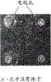
图3A和3B是显示在50μm孔内电场辅助诱捕(electric assisted trapping)1μm珠子的照片。在图片中显示的4个电极中,关闭电压(3A),然后在2个电极处施加电压(3B),而在这些位点观察到粒子的堆叠(stacking)。Figures 3A and 3B are photographs showing electric assisted trapping of 1 μm beads in 50 μm pores. Of the 4 electrodes shown in the picture, the voltage was turned off (3A), then a voltage was applied at 2 electrodes (3B), and stacking of particles was observed at these sites.
图4A和4B是用于显示电场中的荧光染料浓度的实验装置的示意图(4A为透视图,4B为侧视图);4A and 4B are schematic diagrams (4A is a perspective view and 4B is a side view) of an experimental setup for displaying the concentration of a fluorescent dye in an electric field;
图5A和5B是显示未被集中的荧光染料(5A)和通过电场被集中的荧光染料(5B)的照片;和Figures 5A and 5B are photographs showing a fluorescent dye that is not concentrated (5A) and a fluorescent dye that is concentrated by an electric field (5B); and
图6是显示因电场导致的增加的化学发光的图片,其使反应区中产生光的酶(light generating enzyme)附近的焦磷酸增加。Figure 6 is a picture showing increased chemiluminescence due to an electric field, which increases pyrophosphate near a light generating enzyme in the reaction zone.
优选实施方案详述DETAILED DESCRIPTION OF THE PREFERRED EMBODIMENT
定义definition
除非另有定义,本文使用的所有技术和科学术语与本发明所属技术领域内那些普通技术人员所通常理解的具有相同含义。尽管与本文所述那些相似或等效的任何方法和材料能够用于实践或测试本发明,但将对优选的方法和材料进行描述。概括而言,如本文使用的、在与生物化学和生物物理学相关的情况下应用的命名法是本领域公知且常用的那些。某些未具体限定的实验技术大体上依据本领域公知的常规方法并如本说明书通篇所引用和讨论的概括的和更具体的参考文献中所述来进行。出于清楚的目的,下文对以下术语进行定义。Unless otherwise defined, all technical and scientific terms used herein have the same meaning as commonly understood by those of ordinary skill in the art to which this invention belongs. Although any methods and materials similar or equivalent to those described herein can be used in the practice or testing of the present invention, the preferred methods and materials are now described. In general terms, as used herein, the nomenclature applied in the context of biochemistry and biophysics are those well known and commonly used in the art. Certain experimental techniques not specifically limited are generally performed according to conventional methods well known in the art and as described in general and more specific references that are cited and discussed throughout the present specification. For purposes of clarity, the following terms are defined below.
术语“微流装置”按常规含义使用,将其理解为所述装置优选用于并有利于小反应体积和液体流速。概括而言,反应孔应不大于100nL,并且可以小达1pL。在下文描述的一个优选实施方案中,它们的直径是35μm。所述装置将包括液体流动通道使缓冲液和反应物流入反应孔。反应孔可具有可容纳单颗带电珠子的大小。反应孔通常是任何如下所述的界定的空间,反应物被聚拢在其中,并且其位于流体通道的直接流动之外(located out of the directflow of the fluid channel),除非装置被配置成借助下述方式引导反应物进入反应区或者退出反应区:对电极充电而提供场,该场吸引带电物质进入孔中或将带电物质排斥到孔外。The term "microfluidic device" is used in the conventional sense, with the understanding that the device is preferably used for and facilitates small reaction volumes and liquid flow rates. In general, reaction wells should be no larger than 100nL and can be as small as 1pL. In a preferred embodiment described below, their diameter is 35 μm. The device will include fluid flow channels to allow buffer and reactants to flow into the reaction wells. The reaction wells can be of a size that can accommodate a single charged bead. A reaction well is generally any defined space, as described below, in which reactants are brought together and which is located out of the direct flow of the fluid channel, unless the device is configured by means of Ways to guide reactants into or out of the reaction zone: charging the electrodes provides a field that attracts charged species into the pores or repels charged species out of the pores.
术语“横穿”(transverse)按照常规含义用于表示交叉的(crosswise),优选为但不必需为正交(perpendicular)。The term "transverse" is used conventionally to mean crosswise, preferably but not necessarily perpendicular.
术语“电场”用于意指因空间体积中或围绕空间体积的介质中电荷(如电子、离子或质子)的存在而产生的作用。基于叠加,每个电荷分布在一个点处对整个场起贡献。置于空间体积中或在周围介质中的电荷具有施加于其上的力。电场通过电压的差产生:电压越高,产生的场将越强。相反,磁场在电流流动时产生:电流越大,磁场越强。电场即使在没有电流流动时也存在。电场以伏特每米度量(V/m)。为了在方便的时限内,在本方法和装置中引起带电离子的移动,电场强度应为大约5V/cm或更高,高至焦耳加热的可实行限值和介电击穿限值,最大上限值为大约1000V/cm。The term "electric field" is used to mean the effect resulting from the presence of charges, such as electrons, ions or protons, in a volume of space or in the medium surrounding the volume of space. Based on superposition, each charge distribution contributes to the entire field at one point. Charges placed in a volume of space or in the surrounding medium have forces exerted on them. An electric field is generated by a difference in voltage: the higher the voltage, the stronger the field generated will be. Instead, a magnetic field is created when an electric current flows: the greater the current, the stronger the magnetic field. Electric fields exist even when no current is flowing. Electric fields are measured in volts per meter (V/m). To induce movement of charged ions in the present method and apparatus within a convenient time frame, the electric field strength should be about 5 V/cm or higher, up to the practicable limit for Joule heating and the dielectric breakdown limit, up to a maximum of The limit is about 1000 V/cm.
作为高强度电场的实例,注意到作为偶极的水可以通过电场进行部分排列,并且这可以通过用静电源使水流移动来容易地显示。非常高的场强(5x10⁹V m-1)使冰中的水重新定向,从而抑制结冰。As an example of a high strength electric field, note that water as dipoles can be partially aligned by an electric field, and this can easily be shown by moving a stream of water with an electrostatic source. Very high field strengths (5x10⁹ V m-1 ) redirect the water in the ice, thereby inhibiting icing.
通用方法和设备General method and equipment
下文描述的是用于由电场引导的带电分子的集中(concentration)和清洗(washing)的设备和方法。Described below are apparatus and methods for concentration and washing of charged molecules guided by an electric field.
先前的电泳集中技术依赖于法拉第电流将带电物质集中在电极部位。这通常导致在电极处发生的电解反应和电解产物(如氧和氢)的生成。本方法使用通过电极的电容耦合的位移场而非通过电极的法拉第电流。Previous electrophoretic concentration techniques relied on Faradaic currents to concentrate charged species at electrode sites. This typically results in an electrolysis reaction at the electrodes and the production of electrolysis products such as oxygen and hydrogen. The method uses a capacitively coupled displacement field through the electrodes rather than a Faradaic current through the electrodes.
本文使用的电场基于公认的电容原理。在将不同电荷的两个平板置于彼此附近时,如在平行的平板电容器中,平板之间的两个E场加成而平板之外的E场抵消。当平板彼此靠近形成电容器时,在整个电容器内部平板之间的E场是恒定的,只要E场不是靠近平板边缘。由于电场是电势梯度的负数,且E场在电容器内部是恒定的,所以电场的强度E与平板之间的电压V和它们的间隔d的关系非常简单。The electric field used here is based on the well-established principle of capacitance. When two plates of different charges are placed close to each other, as in a parallel plate capacitor, the two E-fields between the plates add and the E-fields outside the plates cancel. When the plates are close together to form a capacitor, the E-field between the plates is constant throughout the capacitor interior, as long as the E-field is not near the edges of the plates. Since the electric field is the negative of the potential gradient, and the E field is constant inside the capacitor, the relationship between the strength E of the electric field and the voltage V between the plates and their spacing d is very simple.
式1Formula 1
通过将薄绝缘材料(电介质)置于平板之间,可以使间隔d减小,由此增加电容器的电容并防止平板接触。By placing a thin insulating material (dielectric) between the plates, the separation d can be reduced, thereby increasing the capacitance of the capacitor and preventing the plates from touching.
位移电流是与变化的电场相关的量。其发生在介电材料中,也发生在自由空间中。Displacement current is a quantity related to a changing electric field. It occurs in dielectric materials as well as in free space.
位移电流在数学上定义为电位移场D(已知物理术语,也称为电场/通量密度)的变化率:Displacement current is defined mathematically as the rate of change of the electric displacement field D (a known physical term, also known as electric field/flux density):
式2Formula 2
其中D=εE,其中介电常数ε=ε0εr,并且其中where D = εE, where the permittivity ε = ε0 εr , and where
·εr是电介质的相对介电常数,而εr is the relative permittivity of the dielectric, and
·ε0是自由空间的介电常数(8.854E-12Fm-1)。• ε0 is the permittivity of free space (8.854E-12Fm-1 ).
在本装置中,响应于所施加的跨越电极的直流电压,在电极处产生双电层,其屏蔽施加在电极处的电压。因此,由于双电层的屏蔽,跨越电极的直流电压在通道主体中不产生电场,并且需要法拉第电流以实现集中。然而,如果以比离子形成双层所用时间更快的时程切换跨越电极的电压,那么屏蔽作用变得可以忽略,电场在跨越整个通道的宽度上都存在。对于一种典型情况:10mM离子强度电解质、10nm的厚电双层,且电极之间间隙为~100μm,需要的交流频率是~100kHz。本方法使用交变场,其以~500kHz或更高的频率变化,并带有净直流偏压以实现跨越电极没有任何法拉第电流的净电场。In the present device, in response to an applied DC voltage across the electrodes, an electric double layer is created at the electrodes which shields the voltage applied at the electrodes. Therefore, a DC voltage across the electrodes does not generate an electric field in the channel body due to the shielding of the electric double layer, and a Faradaic current is required for concentration. However, if the voltage across the electrodes is switched on a faster timescale than the time it takes for the ions to form a double layer, then the shielding effect becomes negligible and the electric field exists across the entire width of the channel. For a typical case: 10 mM ionic strength electrolyte, 10 nm thick electrical double layer, and a gap between electrodes of ~100 μm, the required AC frequency is ~100 kHz. The method uses an alternating field, varying at a frequency of -500 kHz or higher, with a net DC bias to achieve a net electric field without any Faradaic current across the electrodes.
如果单独施加横跨流体流动通道的直流电压,则通道的一个表面附近的电压将被距离通道壁约10nm内的双电层所屏蔽。因此在通道的全部剩余宽度上(距离该壁超过~10nm),电场大部分将为零。如果只施加交流电压——它的切换比离子响应时间(~0.1ms)更快——则该电场的作用将同等地施加在整个通道中。然而,平均电场仍将为零。在既有直流又有交流电压(both DAand AC voltage)的情况下,在整个通道上存在时均直流场(time averaged DCfield),从而产生一个力E,其在通道一侧较高,而随着与该侧的距离增加而下降。If a direct voltage is applied across the fluid flow channel alone, the voltage near one surface of the channel will be shielded by the electric double layer within about 10 nm from the channel wall. Thus over the full remaining width of the channel (beyond -10 nm from the wall) the electric field will be mostly zero. If only an AC voltage is applied—which switches faster than the ion response time (~0.1 ms)—the effect of this electric field will be applied equally throughout the channel. However, the average electric field will still be zero. In the presence of both DC and AC voltages (both DA and AC voltage), there is a time-averaged DC field (time averaged DC field) across the channel, resulting in a force E that is higher on one side of the channel and follows Decline with increasing distance from that side.
作为进一步的阐释,可以说(不希望受到任何理论的限制)本方法和装置采用了一种特定类型的动电作用(electrokinesis)。动电作用是指由电场作用于电解质溶液中带电物体周围的可移动离子所引起的一类现象。当具有给定的表面电荷的物体浸入含离子的溶液中时,形成一种弥散的离子云而遮蔽物体的表面电荷。这种由与浸入的物体伴随的由(固定)电荷构成的遮蔽云以及溶液中的一层(移动的)反荷离子构成的排列,称作“双层”。在这个小而厚度有限的区域中,流体不是电中性的。因此,作用于该区域的电场将激发弥散层的离子的运动,而这些离子将继而带走(entrain)周围的流体。产生的流场反映了流体中离子电流的空间分布。电渗是动电现象最简单的例子。其发生在平行于呈现固定表面电荷的样品容器或电极表面施加电场时,如在氧化硅电极的情况下(在中性pH范围内)。当电极双层中的反荷离子被电场加速时,它们拖动溶剂分子并引发(set up)主体流体流动。这种作用在窄毛细管中非常显著,可以有利地用于设计流体泵送系统。As a further illustration, it can be said (without wishing to be bound by any theory) that the present methods and devices employ a specific type of electrokinesis. Electrokinetic interaction refers to a class of phenomena caused by the action of an electric field on mobile ions around charged objects in an electrolyte solution. When an object with a given surface charge is immersed in a solution containing ions, a diffuse cloud of ions is formed that obscures the surface charge of the object. This arrangement, consisting of an obscuring cloud of (fixed) charge accompanying the immersed object and a layer of (mobile) counterions in solution, is called a "double layer". In this small, thickness-limited region, the fluid is not electrically neutral. Thus, an electric field applied to this region will excite the movement of the ions of the diffuse layer, which in turn will entrain the surrounding fluid. The resulting flow field reflects the spatial distribution of ionic currents in the fluid. Electroosmosis is the simplest example of an electrokinetic phenomenon. It occurs when an electric field is applied parallel to the surface of the sample container or electrode exhibiting a fixed surface charge, as in the case of silicon oxide electrodes (in the neutral pH range). When the counter ions in the electrode bilayer are accelerated by the electric field, they drag the solvent molecules and set up the bulk fluid flow. This effect is very pronounced in narrow capillaries and can be advantageously used to design fluid pumping systems.
电泳是一种相近的现象,其是指的浸入电解液的带电粒子在场诱导下的迁移。与电渗一样,电场加速粒子双层中的运动离子(mobile ions)。如果与之前的情况相反,粒子本身是运动的,那么其将会通过反方向移动来抵消这种场诱导的离子运动(和产生的离子电流)。电泳通常在凝胶或具有牢固网孔(solid mesh)的介质中进行,所述凝胶或介质将基于某种因素(例如,大小)来阻碍离子粒子。如下文所述,本文预期粒子将处于液态流体中而没有阻碍性的凝胶或固相存在。Electrophoresis is a related phenomenon that refers to the field-induced migration of charged particles immersed in an electrolyte. Like electroosmosis, an electric field accelerates mobile ions in a particle bilayer. If, contrary to the previous case, the particle itself is moving, it will counteract this field-induced ion motion (and the resulting ion current) by moving in the opposite direction. Electrophoresis is typically performed in a gel or medium with a solid mesh that will hinder ionic particles based on certain factors (eg, size). As described below, it is contemplated herein that the particles will be in a liquid fluid without an obstructive gel or solid phase present.
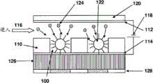
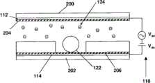
现在参考图1,其中示出了具有孔100的微流装置,孔100限至在SU-8光致抗蚀剂层110内,该装置还包括电极112,其间隔地位于层110上方(spaced above layer 110)并在各孔间延伸,从而在电极112和光致抗蚀剂层110之间界定出流体流动通道(如116所示),电极112与各孔相通。流体通道的深度优选为100μm的量级,这是由于本装置特别适合于10μL的体积。层110为成孔(well-forming)层(即,一种图样化以界定反应区的至少一部分以及流体流动通道的层)。该层由光致抗蚀剂界定以方便在亚微米尺度进行制造。优选的是在孔中实现高的长宽比(例如,d/w>5∶1)。换句话说,反应区或孔(因蚀刻)偏移(offset from)通道一定深度,并且是具有一定(相对窄的)宽度或直径的空腔。珠子可以流经流体流动通道并流入各孔中。第二电极114位于成孔层110下方,其界定了孔的底部。在孔已经(以蚀刻或模制方式)形成于层中的情况下,电极暴露于孔中的流体和物质,其中流体与物质是在所示箭头116处进入装置的。电极112和114优选由ITO(氧化铟锡)形成约100nm厚。如图1进一步所示,这些电极形成了基本上平行的片层(sheets),其间夹有所述流体通道和孔。Referring now to FIG. 1 , there is shown a microfluidic
值得注意的是,由氧化硅或Parylene或氮化硅构成的~100nm厚的介电层被涂施于电极,例如涂施于ITO层之上,如图2(204和206)所示。进一步如图1和图2A和B所示,电源118(图1)连接各电极并充电,使得,就如下文中详述的那样,顶部电极112为负极,底部电极114(在孔的底部)为正极,以便驱使粒子(原子、分子、珠子,等)进入各孔中。在这里使用术语“顶部”和“底部”是为了方便起见,装置可以根据重力或使用时的取向以各种方位配置。Notably, a ~100nm thick dielectric layer composed of silicon oxide or Parylene or silicon nitride is applied to the electrodes, for example on top of the ITO layer, as shown in Figure 2 (204 and 206). As further shown in FIGS. 1 and 2A and B, the power supply 118 (FIG. 1) connects and charges the electrodes such that, as detailed below, the
再次参考图1,顶部电极112被涂施于基底120上,基底120由例如硼硅酸盐玻璃或石英制成,顶部电极112借助任何蚀刻或切削加工结构(如光致抗蚀剂层中的台阶(step)),间隔地位于成孔层110上方。Referring again to FIG. 1 , the
在一个示例性方法中,含有连接在表面并向外延伸的DNA分子的珠子122,如图所示,装在孔100中(每孔一个珠子)。寡核苷酸是以本领域已知的方式连接于珠子的(参见相关专利和出版物)。这些珠子已经藉由流体流动116被传送至孔区域,并在电场(经孔上方和下方的电极112和114实现)或磁体的帮助下进入各孔中。施加电场来驱使带负电荷的分子(如124处所示)移向珠子并进入各孔中。这些分子可以是核苷酸、酶或其他带电的种类。这些分子在合适的缓冲液中传送,并引起与珠子上的DNA链的可探测到的反应。优选用低浓度(~10mM)的Tris-乙酸盐(Tris-Acetate)或Tris HCl作为缓冲液。In one exemplary method,
在一个实施方式中,带电的分子是核苷酸,它们被掺入多核苷酸,并在反应区生成无机含磷物质(inorganic phosphorous),后者被用来生成可探测到的光信号(如焦磷酸测序(pyrosequencing))。相应地,光导纤维面板126贴附到薄的透明电极114上,该电极,带有任何介电涂层,形成了反应区的底部。电极对于要收集的光而言是透明的。光导纤维面板126可以来自市购来源,如来自Schott North America,Inc。光导纤维面板由一束平行排列并垂直于孔100底部表面的融合纤维构成。以这种方式,光高效地从每个单独的孔100传播至光传感器,如耦合至光导纤维面板、在每个孔下面带有传感区的CMOS传感器128。众所周知,CMOS(表示互补金属氧化物半导体)成像器包括光敏二极管的阵列,每个像素内一个二极管。但是与CCD不同的是,CMOS成像器中的每个像素具有自己单独的集成在内部的放大器。由于每个像素具有其自己的放大器,所以这种像素称为“有源像素(active pixel)”。CMOS探测器128中的阴影区对准各单个孔,并从该孔接收最多的光,也只从该孔接收光。每个孔都与一个单独的CMOS探测器元件耦合。In one embodiment, the charged molecules are nucleotides, which are incorporated into polynucleotides and generate inorganic phosphorous in the reaction zone, which is used to generate a detectable optical signal (e.g. pyrosequencing). Accordingly, the
构建装置,使各孔100与CMOS传感器像素几乎完美对准,是为了利用CMOS像素的全部容量(capacity)和实现最佳可能的覆盖(coverage),这对改进系统的通量来说是必要的。然而,这不能用使面板中的纤维与像素对准来实现,这是因为在可获得的纤维面板中的图样中存在不规则处(irregularities)。避免直接对齐的最便利的方法是使用光导纤维,其尺寸远远小于各孔和CMOS像素。使用这种面板防止面板与像素和各孔的对齐问题的发生。但是像素和孔确实必须是对准的。这可以通过固定图像传感器的位置和在X和Y方向上使用两个微米调节器得到含孔的微流平台的完美对齐而容易实现。这种方法可以通过手动或通过更复杂的步进电机机制来实现。每次运行前,在存在ATP或PPi测定的校准量时,用来探测完美对准的校准度量可以设为穿过整个图像传感器区的总共的光载流子量(amount ofcollective photocharge)。如果没有良好对齐的话,光信号就会在各个CMOS像素之间的区域上有损失,但是随着对齐越来越好,损失的光子通量也会减少。最大光强度指示完美的对齐。微流板和CMOS传感器板的自动调节可以通过应用压电调节来实现。在这种技术中,微流板保持器将配备有单个或多个压电致动器。一旦将板插入保持器中,来自CMOS输出的反馈就能激活压电致动器以每次将板移动至单个位置上。发明人的计算表明2N的力应足够移动板使其对准。能产生该力的压电调节器可以市购得到。这还已经表明为μm级内的对齐可以使用这种技术来实现。就光效率而言,孔的最好位置会是离面板尽可能近的位置。因此,经由沉积和将一层SU-8图样化在面板顶部,本文的孔正好构造在面板的顶部。基于发明人的模拟,通过这种直接耦合可以将光效率从1.6%显著提高到大于90%。Constructing the device so that the
图2A为进一步显示本发明装置和技术的示意图。在这个实施方式中,将氧化铟锡(ITO)电极材料(~150nm厚)涂施(112和114,如图1所示)在标准玻璃片200、202上,以便在通道中施加横穿流动方向的电场。透明的ITO电极进一步涂覆有介电薄层204、206(~20nm),如Parylene或氧化硅或氮化硅(如图2中的图样所示),以防止由于电解造成的电极腐蚀的发生和促进(facilitate)电位移场。这样,流体通道具有一个涂覆有介电层的表面(其与流体接触)和带有电极部分的相对表面,其中所述电极部分也涂覆有介电层。Parylene一个统称,适用于未取代的和取代的种类。Parylene N和SCSParylene HT具有特别高的介电强度和不依赖于频率的介电常数,可能是优选的。关于介电材料的进一步描述参见US 4163828,Mahoney,1979年8月7日授权,标题为“Parylene stabilization”。Figure 2A is a schematic diagram further illustrating the apparatus and techniques of the present invention. In this embodiment, indium tin oxide (ITO) electrode material (~150nm thick) is applied (112 and 114, as shown in Figure 1) on standard glass slides 200, 202 to impose a transverse flow direction of the electric field. The transparent ITO electrode is further coated with a
在图2B中示出了电极排列的一个变化方案。在这个实施方式中,如图2A中所示的位于202处的玻璃层适于CMOS制造工艺,在该工艺中CMOS传感器正好置于孔的下面和透明层117的下方。孔电极优选涂覆有介电层(未示出),但是由一组布线115、115a中的一个或两个形成,或者由成孔层下方的、沿着孔一侧的、位于底部部分的电极条119形成。在俯视图中,布线和电极会是栅格的形式,在每个孔的底部部分的周围靠近传感器处形成正方形。这使得在CMOS传感器像素尺寸的量级上制造各孔成为可能。例如,如果每个孔(像素)为一个20μM的正方形,可以用布线直径约为1μm的单根或双根布线来形成栅格,并产生脉冲以提供AC和DC电荷(charge)。布线邻接孔的底部排布。电极也置于孔的侧壁内,部分地延伸到孔的底部,如图115和115a处所示。同样地,如果认为“像素”是孔底部的一部分,其中孔与传感器光学接触,则像素的四条边被电极所界定,在此处电极为一组金属条,每个金属条大部分在孔的侧壁内,但是稍许延伸至孔的底部。这些金属布线115和金属条119连接至电源,并以如上所述的方式工作。A variant of the electrode arrangement is shown in FIG. 2B. In this embodiment, the glass layer at 202 as shown in FIG. 2A is suitable for a CMOS fabrication process in which the CMOS sensor is placed just below the hole and below the
含有流动通道及用于DNA珠子的各孔的流动结构形成在SU-8(~200μm厚)中。PDMS/硅橡胶垫片(gasket)也可用来产生流动通道。ITO-介电层涂覆的玻璃片形成流动通道的顶部和底部。如图1中所述,珠子122位于含有带电粒子124的流体介质中的孔中,电源连接电极从而引起了粒子朝向或背离孔和孔中的珠子122移动,珠子具有贴附其上的反应物,如与核苷酸124反应的寡聚核苷酸。The flow structure containing the flow channels and wells for the DNA beads was formed in SU-8 (~200 μm thick). PDMS/silicone gaskets can also be used to create flow channels. ITO-dielectric coated glass sheets form the top and bottom of the flow channel. As shown in FIG. 1 , the
常规的光刻技术可以用于SU-8图样化。这种制造SU-8的方法已经经过测试并在Stanford纳米器件加工装置上得到验证。SU-8加工能用来制造长宽比(aspect ratio)大的孔,这对减少相邻珠子之间的化学串扰而言是很关键的。即,反应区应当完全容纳珠子。Conventional photolithographic techniques can be used for SU-8 patterning. This method of making SU-8 has been tested and validated at the Stanford Nano Device Fabrication Facility. SU-8 processing can be used to create pores with a large aspect ratio, which is critical for reducing chemical crosstalk between adjacent beads. That is, the reaction zone should completely contain the beads.
在给出上述描述本发明装置优选实施方式的条件下,很明显各种备选构造都是可能的。尽管没有示出,但是可以想到在一个实施方式中,每个孔都可耦合到单独受控的电极对上,在同一时间,不同的孔可处于带电物质受到吸引或排斥的不同状态。Given the above description of preferred embodiments of the device of the invention, it will be apparent that various alternative configurations are possible. Although not shown, it is envisioned that in one embodiment each aperture could be coupled to an individually controlled pair of electrodes, and at the same time different apertures could be in different states where charged species are attracted or repelled.
电极可以由各种透明的导电层制成,例如金属氧化物如氧化铟锡(ITO)、锑掺杂的氧化锡和锡酸镉(氧化镉锡),其中每种都常用作光电装置(如液晶显示器)中的透明电极。出于光学探测的目的,电极是透明的。在以热或电方式测量反应的装置中,电极无需为透明的。例如,装置可用于电探测结合反应。参见US 5,284,748,Mroczkowski等人,1994年2月8日,标题为“Method for electrical detection of a binding reaction”。伏安免疫测定法可以通过用电活性物质标记一种免疫反应物来进行。Pace,美国专利号4,233,144,1980年11月11日授权,解释了一种这样的技术。另一种方法涉及将抗原-抗体层夹在两个导电层之间并测量所得叠层的电容。Giaever,美国专利号4,054,646,1977年10月18日授权,记载了这样一种方法。电容测量系统的另一种类型包括一对电极,这对电极涂覆有基底并浸入含有特别与基底结合的物质的介质中,正如Arwin,美国专利号4,072,576中所描述的。Electrodes can be made of various transparent conductive layers, such as metal oxides such as indium tin oxide (ITO), antimony-doped tin oxide, and cadmium stannate (cadmium tin oxide), each of which is commonly used in optoelectronic devices (such as transparent electrodes in liquid crystal displays). For the purpose of optical detection, the electrodes are transparent. In devices where reactions are measured thermally or electrically, the electrodes need not be transparent. For example, the device can be used to electrically detect binding reactions. See US 5,284,748, Mroczkowski et al., February 8, 1994, entitled "Method for electrical detection of a binding reaction". Voltammetric immunoassays can be performed by labeling an immunoreactant with an electroactive species. Pace, US Patent No. 4,233,144, issued November 11, 1980, explains one such technique. Another method involves sandwiching the antigen-antibody layer between two conductive layers and measuring the capacitance of the resulting stack. Giaever, US Patent No. 4,054,646, issued October 18, 1977, describes such a method. Another type of capacitive measurement system includes a pair of electrodes coated with a substrate and immersed in a medium containing a substance specifically bound to the substrate, as described in Arwin, US Patent No. 4,072,576.
成孔层可以由任何惰性材料形成。光刻技术可以用来将层图样化入一系列流体通道和反应区中,例如US 6,960,437,Enzelberger等人,2005年11月1日授权,标题为“Nucleic acid amplification utilizing microfluidicdevices”中所记载的。如此文中所述,微流装置至少部分由弹性材料构成,并且通过单层和多层软刻蚀(MLSL))技术和/或牺牲层包封方法构成(参见,例如,Unger等人,(2000),Science 288:113-116和PCT公开WO01/01025)。利用这些方法,微流装置可以如此设计:其中流经装置的流动通道的溶液至少部分地利用一个或多个控制通道控制,该控制通道通过弹性膜或片段与流体通道分隔开。更具体地来说,一些制造方法涉及通过用光致抗蚀剂的光刻技术初期制造硅晶片上的用于顶部层(具有控制通道的弹性层)和底部层(具有流体通道的弹性层)的母模(Shipley SJR 5740)。通道高度可以受到旋涂速率的精确控制。光致抗蚀剂通道通过将光致抗蚀剂暴光于UV光并接着显影而形成。热回流工艺和保护处理如先前所述的那样进行(M.A.Unger,H.-P.Chou,T.Throsen,A.Scherer和S.R.Quake,Science 288,113(2000))。The porogen can be formed from any inert material. Photolithography can be used to pattern layers into a series of fluidic channels and reaction regions, such as described in US 6,960,437, Enzelberger et al., issued November 1, 2005, entitled "Nucleic acid amplification utilizing microfluidic devices". As described herein, microfluidic devices are constructed at least in part from elastic materials and are constructed by monolayer and multilayer soft etch (MLSL) techniques and/or sacrificial layer encapsulation methods (see, e.g., Unger et al., (2000 ), Science 288:113-116 and PCT Publication WO01/01025). Using these methods, microfluidic devices can be designed in which solution flow through the flow channels of the device is at least partially controlled by one or more control channels separated from the fluidic channels by elastic membranes or segments. More specifically, some fabrication methods involve the initial fabrication of layers for the top layer (elastic layer with control channels) and bottom layer (elastic layer with fluidic channels) on a silicon wafer by photolithography with photoresist. The master model (Shipley SJR 5740). The channel height can be precisely controlled by the spin-coating rate. Photoresist channels are formed by exposing photoresist to UV light followed by development. Thermal reflow process and protection treatment were performed as previously described (M.A. Unger, H.-P. Chou, T. Throsen, A. Scherer and S.R. Quake, Science 288, 113 (2000)).
光致抗蚀剂物质可以旋涂或喷涂至基底如硅晶片上,或者作为膜或丝网应用至晶片上。市购的干燥膜状光致抗蚀剂物质包括:丙烯酸基物质,例如从Mitsui(日本)以商品名Ordyl PR132购得的物质;环氧基物质,例如从E.I.DuPont de Nemours and Company Corporation(Wilmington,Del.)以商品名RISTON购得的物质,或者从MicroChem Corporation(Newton,Mass.)以商品名SU-8购得的物质,(或者例如Lexmark International,Inc.(Lexington,Ky.)内部使用的专利物质,内部称为GSP920);以及聚酰亚胺基光致抗蚀剂物质,例如从HD Microsystems(Parlin,NJ.)以商品名HD4000购得的物质。The photoresist material can be spin-coated or spray-coated onto a substrate such as a silicon wafer, or applied as a film or screen onto a wafer. Commercially available dry film photoresist materials include: acrylic based materials such as those available from Mitsui (Japan) under the tradename Ordyl PR132; epoxy based materials such as those available from E.I. DuPont de Nemours and Company Corporation (Wilmington , Del.) commercially available under the tradename RISTON, or from MicroChem Corporation (Newton, Mass.) under the tradename SU-8, (or, for example, Lexmark International, Inc. (Lexington, Ky.) for internal use proprietary material known internally as GSP920); and polyimide-based photoresist materials such as those commercially available under the trade designation HD4000 from HD Microsystems (Parlin, NJ.).
在将光致抗蚀剂物质应用至晶片基底的流体侧上之后,通过掩膜使光致抗蚀剂物质曝光于光化辐射如紫外(UV)光以使光致抗蚀剂物质图样化,以便在显影光致抗蚀剂物质时提供流体通道在光致抗蚀剂物质中的位置。然后,通过用显影化学试剂将未固化的物质从晶片的流体通道/孔区域溶解,显影得到图样化的光致抗蚀剂物质。显影化学试剂可以选自四甲基氢氧化铵、二甲苯、脂肪烃、碳酸钠和乙酸2-丁基乙氧乙酯(BCA)。进一步细节参见US7,043,838,Smoot等人,2006年5月16日授权,标题为“Process formanufacturing a micro-fluid ejection device”。After applying the photoresist material onto the fluid side of the wafer substrate, exposing the photoresist material to actinic radiation, such as ultraviolet (UV) light, through a mask to pattern the photoresist material, In order to provide the position of the fluid channel in the photoresist material when developing the photoresist material. The patterned photoresist material is then developed by dissolving the uncured material from the fluid channel/well regions of the wafer with developing chemicals. The developing chemistry may be selected from tetramethylammonium hydroxide, xylene, aliphatic hydrocarbons, sodium carbonate and 2-butylethoxyethyl acetate (BCA). See US 7,043,838, Smoot et al., issued May 16, 2006, entitled "Process formanufacturing a micro-fluid ejection device" for further details.
一种用于核苷酸预浓集(preconcentration)/清洗的通用测定规程如下所述:A general assay protocol for nucleotide preconcentration/washing is as follows:
1.首先以DNA珠子填充流动通道。1. First fill the flow channel with DNA beads.
2.用ITO介电玻璃片从顶部密封流动通道。2. Seal the flow channel from the top with a piece of ITO dielectric glass.
3.接着用含核苷酸的溶液填充流动通道。3. The flow channel is then filled with the nucleotide-containing solution.
4.将ITO电极连接到高频AC源,并且跨越通道施加高频方波脉冲(V峰~5-7V,>100kHz)。即,电源118向电极112、114提供AC场,电极114仅在孔底部是有效的。AC场在2至20伏、优选5至7伏范围内,并且频率在约10kHz至约10MHz的范围内,优选为大于100kHz。4. Connect the ITO electrodes to a high frequency AC source and apply a high frequency square wave pulse (Vpeak ~5-7V, >100kHz) across the channel. That is, the
高频AC场消除了在电极-液体界面处形成的双电层的作用。如果没有AC场,双电层会屏蔽施加在电极间的DC电压,这样在通道内部就不会有DC电场。The high-frequency AC field eliminates the effect of the electric double layer formed at the electrode-liquid interface. In the absence of an AC field, the double layer shields the DC voltage applied between the electrodes, so there is no DC field inside the channel.
5.将小DC电压(~1.5V)叠加在存在的AC电压上,该DC电压也来自电源118。根据DC场的极性,核苷酸或者浓集在DNA珠子附近或者被各孔中的DNA珠子排开。5. A small DC voltage (-1.5V) is superimposed on the existing AC voltage, also from the
本装置可以用于广泛多样的测定法。优选的测定法包括涉及核苷酸转运的那些。这些测定法包括:The device can be used in a wide variety of assays. Preferred assays include those involving nucleotide transport. These assays include:
引物延伸/降解测定法Primer extension/degradation assay
在这种测定法中,通过将dATP结合到ssDNA中来检测蛋白质制备物中的末端转移酶活性。典型的流程包括:将130nM TdTS或130nM TdTL在200nM二甲胂酸钾、25mM Tris-HCl,pH 6.6、0.25mg/ml BSA、4mMMgCl2、4μM ZnSO4、5%甘油、1mM dATP和20nM 5′-32P-标记的(dA)10引物中在35℃温育。使用同一测定法在缺乏dATP的条件下查找蛋白质制备物中的3′5′核酸外切酶活性。在第0、5、15、30和60分钟取等分试样,补充以甲酰胺染料混合物,并在16%丙烯酰胺变性凝胶上进行电泳。在将湿胶在Kodak胶片(Biomax MR)下于-70℃曝光之后使产物显像。参见See TheJournal of Immunology,2004,172:6764-6767.“Evidence That the Long MurineTerminal Deoxynucleotidyltransferase Isoform Plays No Role in the Control ofV(D)J Junctional Diversity.”。In this assay, terminal transferase activity is detected in protein preparations by incorporation of dATP into ssDNA. A typical protocol includes: 130nM TdTS or 130nM TdTL in 200nM potassium cacodylate, 25mM Tris-HCl, pH 6.6, 0.25mg/ml BSA, 4mMMgCl2, 4μM ZnSO4, 5% glycerol, 1mM dATP and 20nM 5′-32P -Incubate at 35°C in the labeled (dA)10 primer. The same assay was used to look for 3'5' exonuclease activity in protein preparations in the absence of dATP. Aliquots were taken at 0, 5, 15, 30 and 60 minutes, supplemented with formamide dye mixture, and run on a 16% acrylamide denaturing gel. The product was visualized after exposing the wet gel to Kodak film (Biomax MR) at -70°C. See TheJournal of Immunology, 2004, 172:6764-6767. "Evidence That the Long MurineTerminal Deoxynucleotidyltransferase Isoform Plays No Role in the Control ofV(D)J Junctional Diversity.".
DNA的克隆分析,或复用分析Clonal analysis of DNA, or multiplex analysis
这种测定法使用具有多个反应区的装置,其中每个反应区被设计成可容纳一颗且仅可容纳一颗珠子的孔。使用本领域已知的方法使DNA分子附着在电中性的珠子上,每颗珠子一种DNA。可以用于珠子的中性材料的实例包括玻璃、聚丙烯酰胺、聚苯乙烯、
在此实施方案的一个方面中,用条形码为珠子编码。条形码是用于唯一性地检测一个分子的一种特殊标记。条形码可以是本领域已知的任何类型的条形码,包括但不限于光学标记、荧光标记、电响应标记和一组质量不同的标记。使用取决于条形码类型的方法来解码条形码,所述方法包括但不限于质量、电、视觉、荧光和核酸检测。这样能够鉴定每个反应区中的DNA。In one aspect of this embodiment, the beads are coded with a barcode. A barcode is a special marker used to uniquely detect a molecule. The barcode can be any type of barcode known in the art, including but not limited to optical labels, fluorescent labels, electro-responsive labels, and a set of labels of varying mass. Barcodes are decoded using methods depending on the type of barcode including, but not limited to, mass, electrical, visual, fluorescent, and nucleic acid detection. This enables the identification of DNA in each reaction zone.
如此,这种方法允许将一组珠子引入流动通道中,这组珠子中仅有一些含有要分析的分子(例如,DNA),其中含有分子的珠子含有不同的分子种类(例如,不同的DNA序列、不同的蛋白质等)。将珠子随机放入孔中,并通过条形码编码来鉴定。分子仅需要响应存在的E场。如图5中所示,甚至某些染料也响应,以及蛋白质和核酸(DNA、RNA)也是。As such, this method allows for the introduction into the flow channel of a set of beads, only some of which contain the molecule to be analyzed (e.g., DNA), where the beads containing the molecules contain different molecular species (e.g., different DNA sequences). , different proteins, etc.). Beads are randomly placed into wells and identified by barcoding. The molecules need only respond to the E-field present. As shown in Figure 5, even certain dyes responded, as did proteins and nucleic acids (DNA, RNA).
复用分析在多个孔中进行,其可以是大约数百或数千个不同的孔。可以利用不同的流体通道寻址每个孔(或孔的子组)。一旦将靶分子引导进入单独的孔中,就将反应物特异性地寻址到那些分子进行化学分析。如上所述读取结果,并且分析可以进一步包括去卷积(deconvolute)条形码以识别靶分子。本文灵活地(loosely)使用术语“条形码”来指代与靶分子直接地或通过固相支持体如珠子相关联的独特分子(如寡核苷酸或磁性颗粒)。更多细节可以在例如2001年7月17日授权的、题为“Fixed address analysis of sequence tags.”的Lizardi等人的US 6,261,782中找到。其它能够依据本方法使用的标签包括分子或金属条形码、质量标签,和可通过核磁共振、电子顺磁共振、表面增强型拉曼散射、表面细胞质基因组反响(surface plasmon resonance)、荧光、磷光、化学发光、拉曼共振、微波或其组合检测的标签。质量标签是在质谱中具有区别性质量特征(distinctive mass signature)、或为标记的组分提供在质谱中的区别性质量特征的化合物或成分(moiety)。治疗标签在使用质谱进行检测时有用。优选的治疗标签是肽、核酸和糖。也可以使用标签的组合。例如,具有例如265种独特标签组合的颜色编码微珠可用于区别多种组分。例如,可以独特地标记和检测256种不同的连接物-检测物(ligator-detector),使得所公开的方法能够复用和自动操作。Multiplex assays are performed in multiple wells, which can be on the order of hundreds or thousands of different wells. Each well (or subset of wells) can be addressed with a different fluidic channel. Once the target molecules are directed into the individual wells, the reactants are specifically addressed to those molecules for chemical analysis. Results are read as described above, and analysis may further include deconvoluting the barcodes to identify target molecules. The term "barcode" is used loosely herein to refer to a unique molecule (such as an oligonucleotide or a magnetic particle) associated with a target molecule either directly or via a solid support such as a bead. Further details can be found, for example, in US 6,261,782, issued July 17, 2001, entitled "Fixed address analysis of sequence tags." to Lizardi et al. Other tags that can be used in accordance with this method include molecular or metallic barcodes, mass tags, and other methods that can be detected by nuclear magnetic resonance, electron paramagnetic resonance, surface-enhanced Raman scattering, surface plasmon resonance, fluorescence, phosphorescence, chemical Labels for detection by luminescence, Raman resonance, microwave or a combination thereof. A mass tag is a compound or moiety that has a distinctive mass signature in a mass spectrum, or provides a labeled component with a distinctive mass signature in a mass spectrum. Therapeutic labels are useful for detection using mass spectrometry. Preferred therapeutic labels are peptides, nucleic acids and sugars. Combinations of tags can also be used. For example, color-coded microbeads with, for example, 265 unique tag combinations can be used to distinguish between various components. For example, 256 different ligator-detectors can be uniquely labeled and detected, enabling multiplexing and automation of the disclosed methods.
聚合酶链式反应polymerase chain reaction
这种标准测定法检测DNA分子中界定序列的存在,所述序列与一对寡核苷酸引物互补。通过加入加热元件,PCR反应可以在例如图1中所示例的装置中进行。PCR在例如基本专利中有述,所述专利如美国专利No.4,683,202;4,683,195;4,800,159和4,965,188。1996年4月30日授权的、题为“Methods and reagents for the polymerase chain reaction amplification of longDNA sequences,”的Cheng的US 5,512,462描述了通过聚合酶链式反应(PCR)扩增长于10kb的DNA序列的方法和试剂。所述方法使用由来自嗜热栖热菌(Thermus thermophilus)的主要热稳定DNA聚合酶与较少量的来自海岸热球菌(Thermococcus litoralis)、火球菌属菌种GB-D(Pyrococcus species GB-D)或海栖热袍菌(Thermotoga maritime)的、拥有3′到5′的核酸外切酶活性的次要热稳定DNA聚合酶组成的组合物。This standard assay detects the presence in a DNA molecule of a defined sequence that is complementary to a pair of oligonucleotide primers. By adding a heating element, a PCR reaction can be performed in a device such as that illustrated in FIG. 1 . PCR is described, for example, in basic patents such as U.S. Pat. Cheng, "US 5,512,462 describes methods and reagents for amplifying DNA sequences longer than 10 kb by polymerase chain reaction (PCR). The method uses a combination of a major thermostable DNA polymerase from Thermus thermophilus and a lesser amount from Thermococcus litoralis, Pyrococcus species GB-D ) or a composition consisting of a minor thermostable DNA polymerase of 3' to 5' exonuclease activity from Thermotoga marine.
本方法也可以应用于多种通过合成进行DNA测序的方法。The method can also be applied to various methods of DNA sequencing by synthesis.
热测序thermal sequencing
本文描述的焦磷酸方法是一种通过合成进行的测序。参见Ronaghi等,″ASequencing Method Based on Real-Time Pyrophosphate,″Science,281:363 365(1998)和Hyman,″A New Method of Sequencing DNA,″Anal.Biochem.,174:423 436(1988)。The pyrophosphate method described here is a type of sequencing by synthesis. See Ronaghi et al., "ASequencing Method Based on Real-Time Pyrophosphate," Science, 281: 363 365 (1998) and Hyman, "A New Method of Sequencing DNA," Anal. Biochem., 174: 423 436 (1988).
如Ronaghi,“Pyrosequencing Sheds Light on DNA Sequencing,”GenomeResearch Vol.11,Issue 1,3-11,January 2001中所述,热测序是一种以检测DNA合成过程中释放的焦磷酸(PPi)为基础的DNA测序技术。在一连串酶促反应中,产生与结合的核苷酸数成比例的可见光。这一连串反应始于核酸聚合反应,其中因通过聚合酶进行的核苷酸结合而导致无机PPi的释放。释放的PPi继而由ATP硫酸化酶转化成ATP,ATP为萤光素酶氧化萤光素并生成光提供能量。由于加入的核苷酸是已知的,所以能够测定模板的序列。所述核酸分子可以是RNA或DNA。然而,由于对于有限的核苷酸延伸DNA聚合酶与RNA聚合酶相比显示出更高的催化活性,所以集中针对引物DNA模板(primed DNA template)用于热测序的用途作出努力。标准热测序使用大肠杆菌DNA Pol I的Klenow片段,其是相对缓慢的聚合酶。热测序中使用的ATP硫酸化酶是来自酵母酿酒酵母(Saccharomyces cerevisiae)的重组形式,而萤光素酶来自美洲萤火虫Photinus pyralis。从聚合到光检测的全部反应在室温下发生于3-4秒之内。热测序反应中的1pmol DNA产生6×10¹¹ATP分子,其继而在560纳米的波长生成超过6×10⁹个光子。这种量的光可简单地通过光电二极管、光电倍增管或电荷耦合器件(CCD)相机检测。有两种不同的热测序策略:固相热测序(Ronaghi等1996)和液相热测序。固相热测序利用先前描述的三酶系统中的固定的DNA。在此系统中,在每次核苷酸添加之后进行清洗步骤以去除过量的底物。在液相热测序中,引入腺苷三磷酸双磷酸酶,一种来自马铃薯的核苷酸降解酶,从而成为四酶系统。这种酶的加入消除了对固相支持体和中间清洗的需要,由此使得热测序反应能够在一个试管中进行。As described in Ronaghi, "Pyrosequencing Sheds Light on DNA Sequencing," GenomeResearch Vol.11, Issue 1, 3-11, January 2001, pyrosequencing is a method based on the detection of pyrophosphate (PPi) released during DNA synthesis. DNA sequencing technology. In a series of enzymatic reactions, visible light is generated proportional to the number of nucleotides incorporated. This cascade of reactions begins with nucleic acid polymerization, in which inorganic PPi is released due to nucleotide incorporation by the polymerase. The released PPi is then converted by ATP sulfurylase to ATP, which provides energy for luciferase to oxidize luciferin and generate light. Since the added nucleotides are known, the sequence of the template can be determined. The nucleic acid molecule may be RNA or DNA. However, since DNA polymerases exhibit higher catalytic activity compared to RNA polymerases for limited nucleotide extensions, efforts have focused on the use of primed DNA templates for pyrosequencing. Standard pyrosequencing uses the Klenow fragment of E. coli DNA Pol I, which is a relatively slow polymerase. The ATP sulfurylase used in pyrosequencing was a recombinant form from the yeast Saccharomyces cerevisiae, while the luciferase was from the American firefly Photinus pyralis. The entire reaction from polymerization to photodetection occurs within 3-4 seconds at room temperature. 1 pmol of DNA in a pyrosequencing reaction produces 6×10¹¹ ATP molecules, which in turn generate more than 6×10⁹ photons at a wavelength of 560 nanometers. This amount of light can be detected simply by a photodiode, photomultiplier tube, or charge-coupled device (CCD) camera. There are two different pyrosequencing strategies: solid-phase pyrosequencing (Ronaghi et al. 1996) and liquid-phase pyrosequencing. Solid-phase pyrosequencing utilizes immobilized DNA in a previously described three-enzyme system. In this system, a wash step is performed after each nucleotide addition to remove excess substrate. In liquid-phase pyrosequencing, apyrase, a nucleotide-degrading enzyme from potato, was introduced, thus becoming a four-enzyme system. The addition of this enzyme eliminates the need for solid supports and intermediate washes, thereby enabling pyrosequencing reactions to be performed in one tube.
尽管在使用天然核苷酸方面是有利的,但是焦磷酸方法需要将聚合酶同步化在DNA链上,已知这会限制序列阅读长度。同样,预期所述检测方法无法接近单一分子敏感性,原因是在所述流程中由萤光素酶产生的光的有限的量子效率。此外,整体测序速度受到必要的清洗步骤、后续的目的为鉴定焦磷酸存在的化学步骤的限制,也受到用全部四种碱基相继测试要测序的每个碱基对所需的固有时间的限制。在精确测定序列中同型核苷酸序列段(stretch)中的困难也是公认的。Although advantageous in terms of using natural nucleotides, the pyrophosphate method requires synchronization of the polymerase on the DNA strand, which is known to limit sequence read length. Also, the detection method is not expected to approach single molecule sensitivity due to the limited quantum efficiency of light generated by luciferase in the process. Furthermore, the overall sequencing speed is limited by the necessary washing steps, subsequent chemical steps aimed at identifying the presence of pyrophosphate, and by the inherent time required to sequentially test each base pair to be sequenced with all four bases . Difficulties in accurately determining homotypic nucleotide stretches in sequences are also well recognized.
使用上文所列的DNA、核苷酸、PPi和酶的动电现象的本方法为热测序提供了显著的改进。反应物更好地流入孔中,解决上文列出的关于同步、阅读长度和速度的潜在问题领域。The present method using the electrokinetic phenomena of DNA, nucleotides, PPi and enzymes listed above provides a significant improvement over pyrosequencing. Reactants flow better into the wells, addressing the potential problem areas listed above with respect to synchronization, read length, and speed.
引物延伸primer extension
如2003年9月2日授权的、题为“Sequencing by incorporation”的Parce等的US 6,613,513中所述,通过合成或结合进行的测序通常都涉及将核苷酸或核苷酸类似物加入包含核酸模板和引物的反应混合物,例如,DNA或RNA。将核苷酸掺入引物,产生延伸的引物。随着每个附加的互补核苷酸并入引物而使序列得到测定,并重复这些步骤直到整个模板序列或其部分得到测定。As described in US 6,613,513 to Parce et al., entitled "Sequencing by incorporation," issued September 2, 2003, sequencing by synthesis or conjugation generally involves the addition of nucleotides or nucleotide analogs to nucleic acid containing A reaction mixture of template and primer, eg, DNA or RNA. Nucleotides are incorporated into the primers, resulting in extended primers. The sequence is determined as each additional complementary nucleotide is incorporated into the primer, and these steps are repeated until the entire template sequence, or a portion thereof, is determined.
在本方法的一个实施方案中,核苷酸或核苷酸类似物,或其部分,包含3′-封闭基团和可检测的标签模块,所述模块通常包含磷酸或氨基甲酸基团。3′-封闭基团提供可逆的链终止。当添加至生长中的核酸链时,这些核苷酸类似物导致不能延伸的引物。通常将3′-封闭基团去除,例如通过还原剂和/或磷酸酶,以产生可延伸引物,向该可延伸引物添加更多的核苷酸,由此允许对核酸模板的持续测序。3′-封闭基团的去除任选在检测添加的核苷酸之前或之后进行。In one embodiment of the method, the nucleotide or nucleotide analog, or portion thereof, comprises a 3'-blocking group and a detectable labeling moiety, typically comprising a phosphate or carbamate group. The 3'-blocking group provides reversible chain termination. When added to a growing nucleic acid strand, these nucleotide analogs result in primers that cannot be extended. Typically the 3'-blocking group is removed, eg, by reducing agents and/or phosphatases, to generate an extendable primer to which more nucleotides are added, thereby allowing continued sequencing of the nucleic acid template. Removal of the 3'-blocking group is optionally performed before or after detection of the added nucleotide.
在本方法的另一实施方案中,核苷酸或核苷酸类似物包含荧光标签。使用荧光核苷酸通过合成进行的测序通常包括在检测添加的核苷酸之后光漂白荧光标签。光漂白包括施加光脉冲,光脉冲破坏核苷酸(例如,已经添加至引物的荧光核苷酸)的荧光或将其降低至可以接受的水平,例如,背景水平或降低至足以防止信号经过几个测序循环而积累的低水平。In another embodiment of the method, the nucleotide or nucleotide analog comprises a fluorescent tag. Sequencing by synthesis using fluorescent nucleotides typically involves photobleaching the fluorescent tags after detection of the added nucleotides. Photobleaching involves applying a pulse of light that destroys the fluorescence of a nucleotide (e.g., a fluorescent nucleotide that has been added to a primer) or reduces it to an acceptable level, e.g., a background level or sufficiently to prevent the signal from passing through several Low levels accumulated over sequencing cycles.
已经开发了与末端核苷酸相关的使用染料或荧光标签的相关方法,其中序列测定也通过凝胶电泳和自动荧光检测器来进行。例如,最近已将Sanger延伸法修改用于自动微型测序系统中,其仅需要亚微升体积的试剂和染料标记的三磷酸双脱氧核糖核苷酸。在Soper等的美国专利No.5,846,727中,荧光检测在芯片上进行,所述芯片具有一个将激发光传送至毛细管通道的单模光纤,和另一个收集荧光光子的单模光纤。估计序列读取在400至500个碱基范围内,这相对于用传统Sanger或Maxam-Gilbert方法获得的序列信息量没有显著改进。另外,Soper方法在开始分离反应之前需要PCR扩增模板DNA,并对寡核苷酸测序“梯子”进行纯化和凝胶电泳。这些系统都需要大量目标DNA。即使是Cheeseman的美国专利5,302,509中描述的方法,其不使用凝胶电泳进行序列测定,也需要至少一百万个DNA分子。Related methods using dyes or fluorescent tags associated with terminal nucleotides have been developed, where sequence determination is also performed by gel electrophoresis and automated fluorescence detectors. For example, the Sanger extension method has recently been modified for use in automated miniature sequencing systems that require only sub-microliter volumes of reagents and dye-labeled dideoxyribonucleotide triphosphates. In US Patent No. 5,846,727 to Soper et al., fluorescence detection is performed on a chip with a single-mode fiber that delivers excitation light to capillary channels, and another single-mode fiber that collects fluorescent photons. Sequence reads were estimated to be in the range of 400 to 500 bases, which is not a significant improvement over the amount of sequence information obtained with traditional Sanger or Maxam-Gilbert methods. In addition, the Soper method requires PCR amplification of template DNA, purification and gel electrophoresis of the oligonucleotide sequencing "ladder" before starting the separation reaction. These systems all require large amounts of target DNA. Even the method described in US Patent 5,302,509 to Cheeseman, which does not use gel electrophoresis for sequence determination, requires at least one million DNA molecules.
另外,可以使目前的电场设备适合用于涉及蛋白质和其它蛋白质之间或蛋白质和小分子之间的接触的测定法。例如,可以将固定的酶与底物(含或不含抑制剂)接触,由此使底物和任何抑制剂作为带电粒子存在于溶液中。施加的E场使反应物朝着酶移动以缩短过程时间。类似地,可以按照公开的方法设计使用固定在微量滴定板上的捕获抗体的免疫测定形式,从而改进带电试剂(抗原、标记的抗体)朝向或离开捕获抗体的流速。In addition, current electric field devices can be adapted for use in assays involving contacts between proteins and other proteins or between proteins and small molecules. For example, an immobilized enzyme can be contacted with a substrate (with or without inhibitor), whereby the substrate and any inhibitor are present in solution as charged particles. The applied E-field moves the reactants towards the enzyme to shorten the process time. Similarly, immunoassay formats using capture antibodies immobilized on microtiter plates can be designed according to the disclosed methods to improve the flow rate of charged reagents (antigen, labeled antibody) towards or away from the capture antibodies.
实施例Example
实施例1:将珠子集中到电极孔中Example 1: Concentrating beads into electrode holes
在本实施例中,示例了在50μm孔内由电场辅助诱捕1μm荧光聚苯乙烯珠。如图3中所示,制备了四个电极孔。孔使用一侧带有粘合剂的150μm厚Mylar片层(sheet)制成。将电极加工在印刷电路板上,且每个电极能够通过施加跨越该电极和主体溶液的电压来单独地激活。80μm厚的电流传导性Nafion膜形成了孔的底部,并且使电极与含有珠子的溶液绝缘。In this example, electric field assisted trapping of 1 μm fluorescent polystyrene beads within a 50 μm pore is exemplified. As shown in Fig. 3, four electrode holes were prepared. Wells were made using a 150 μm thick Mylar sheet with adhesive on one side. The electrodes are fabricated on a printed circuit board, and each electrode can be activated individually by applying a voltage across the electrode and the bulk solution. An 80 μm thick, electrically conductive Nafion membrane forms the bottom of the wells and insulates the electrodes from the bead-containing solution.
将一个多输出式计算机控制的电源(multi-output computer controlledpower supply)(Labsmith,HVS 3000D)用于单独激活电极。使用立式Nikon落射荧光显微镜进行成像。通过将母液在10mM Tris-HEPES缓冲液中稀释10,000x制备珠的溶液。然后将珠的溶液注入电极之上3cm长、5mm宽的流动腔中。跨越电极和主体溶液施加~75V/cm的额定直流电场,珠子继而在不到10秒之内被诱捕到孔中。电压仅施加在四个电极中的两个上,并在这些激活的电极部位处的Nafion膜上观察到颗粒堆叠。在此实施方案中,存在将珠子引导至孔内的膜的直流电流。在下文描述的实施方案中,在电极上存在介电层,其阻止这些直流电流。因此,那些实验需要来自交流和直流电压的组合的位移电流。A multi-output computer controlled power supply (Labsmith, HVS 3000D) was used to activate electrodes individually. Imaging was performed using an upright Nikon epifluorescence microscope. A solution of beads was prepared by diluting the stock solution 10,000x in 10 mM Tris-HEPES buffer. The bead solution was then injected into a flow chamber 3 cm long and 5 mm wide above the electrodes. A nominal DC electric field of -75 V/cm was applied across the electrodes and bulk solution, and the beads were then trapped into the pores in less than 10 seconds. Voltage was applied to only two of the four electrodes, and particle stacking was observed on the Nafion membrane at these activated electrode sites. In this embodiment, there is a direct current that directs the beads to the membrane within the pores. In the embodiments described below, there is a dielectric layer on the electrodes which prevents these direct currents. Therefore, those experiments required displacement currents from a combination of AC and DC voltages.
实施例2:电场引导的荧光物质的预浓集Example 2: Pre-concentration of fluorescent substances guided by electric field
现参考图4,其示例了用于说明电场引导的带电分子移动的原型装置。该装置包含一层250μM厚的硅垫片材料410,在该层中具有1mm直径的孔。将其置于20nm聚对二甲苯介电层408上,聚对二甲苯介电层408已经放置在1 in.x 3 in玻璃片404上的150nm ITO层406上,形成“底部”。为了形成“顶部”,将另一玻璃片414类似地用ITO层415包覆以形成电极,且聚对二甲苯层417位于顶部电极的“底部”一侧与流体接触,流体可以容纳在孔412中,孔412在两个片包夹到一起时形成。Reference is now made to Figure 4, which illustrates a prototype setup for demonstrating electric field-guided movement of charged molecules. The device comprised a layer of 250 μιη thick
将Coolsnap fx-16 CCD照相机,Olympus IX70落射荧光显微镜成像装置416布置在流体孔之下。使用导电铜带将电极与信号发生器连接。A Coolsnap fx-16 CCD camera, Olympus IX70 epifluorescence
将含有500nM Alexa-Fluor 488荧光染料(Molecular Probes)的10mMTris-HCl溶液按照以下流程置于孔中:将导电铜带与玻璃片上曝露的ITO层连接。将硅垫片置于底部玻璃片上并且对玻璃片按压以形成密封。用荧光染料溶液填满垫片中的1mm孔。将第二片玻璃片置于垫片顶部以密封含荧光染料的孔的顶部。控制信号发生器402操作,以跨越玻璃片上的顶部和底部ITO电极(即,孔上和孔下)提供5V交流峰峰值、500kHz频率。跨越底部和顶部玻璃片施加偏压VDC=1.5V以实现将荧光Alexa-Fluor分子预浓集在孔底部附近。A 10 mM Tris-HCl solution containing 500 nM Alexa-Fluor 488 fluorescent dye (Molecular Probes) was placed in the wells according to the following procedure: a conductive copper tape was connected to the exposed ITO layer on the glass slide. A silicon spacer was placed on the bottom glass slide and pressed against the glass slide to form a seal. Fill the 1 mm holes in the spacers with the fluorescent dye solution. Place a second glass slide on top of the spacer to seal the top of the well containing the fluorescent dye. The
结果示于图5中。在图5B的照片中可见,当在玻璃片之间施加+1.5V直流电压和交流电压时,荧光染料集中在孔底部附近。在仅施加交流电压或仅施加直流电压时,未观察到预浓集(5A)。如上所述,本装置在此使用位移电流(或交流场,参见上文的式2)来防止在液体-电介质界面形成双电层,双电层将使一个表面附近的场集中(concentrate the field near one surface)。双电层通常会屏蔽施加的整个直流电压,在主体液体中将不存在电场。因此,主体溶液中的离子不会经受任何电场。通过施加高频交流场,双电层的影响消失。因此,当施加带有直流偏压的高频交流电时,有可能在主体液体中具有净电场。交流场使因双电层产生的电压屏蔽作用瓦解,而直流偏压在溶液中产生净电场。这个系统的一个优势是系统中不需要法拉第电流(引起电解)来实现预浓集。The results are shown in FIG. 5 . As can be seen in the photograph of Figure 5B, when +1.5V DC and AC voltages were applied between the glass slides, the fluorescent dye was concentrated near the bottom of the wells. No preconcentration (5A) was observed when AC voltage alone or DC voltage alone was applied. As mentioned above, the device here uses a displacement current (or AC field, see Equation 2 above) to prevent the formation of an electric double layer at the liquid-dielectric interface, which would concentrate the field near one surface near one surface). The electric double layer normally shields the entire applied dc voltage and there will be no electric field in the host liquid. Therefore, the ions in the bulk solution are not subjected to any electric field. By applying a high-frequency alternating field, the effect of the electric double layer disappears. Therefore, it is possible to have a net electric field in the bulk liquid when a high frequency alternating current with a direct current bias is applied. The AC field breaks down the voltage shielding due to the electric double layer, while the DC bias produces a net electric field in the solution. An advantage of this system is that no Faradaic current (causing electrolysis) is required in the system to achieve pre-concentration.
实施例3:电场引导的焦磷酸的预浓集Example 3: Preconcentration of Pyrophosphoric Acid Induced by Electric Field
基本上如图4中所示构建了一个装置,有以下不同:代替照相机和显微镜,将磁体和Hamamatsu光电倍增管布置在孔412之下,如图4中416处所示。A setup was basically constructed as shown in FIG. 4 with the following difference: Instead of a camera and microscope, a magnet and Hamamatsu photomultiplier tube were placed under the
流程如下:将导电铜带与玻璃片上曝露的ITO层连接。将硅垫片置于底部玻璃片上并且对玻璃片按压以形成密封。用含酶的磁珠装填垫片中的1mm孔。将磁体置于玻璃片下以使磁珠保持静止。用焦磷酸盐溶液注满垫片中1mm的孔。将第二片玻璃片置于垫片顶部来密封含化学品的孔的顶部。化学品如下:焦磷酸盐溶液、装载有ATP硫酸化酶的磁珠和萤光素酶(获得自454 Life Scineces)。The process is as follows: connect the conductive copper tape to the exposed ITO layer on the glass sheet. A silicon spacer was placed on the bottom glass slide and pressed against the glass slide to form a seal. The 1 mm wells in the spacers were filled with enzyme-containing magnetic beads. Place a magnet under the glass slide to keep the beads still. Fill the 1mm hole in the spacer with pyrophosphate solution. A second glass slide was placed on top of the spacer to seal the top of the well containing the chemical. The chemicals were as follows: pyrophosphate solution, magnetic beads loaded with ATP sulfurylase and luciferase (obtained from 454 Life Scineces).
在顶部和底部ITO片之间施加峰峰值5V、500kHz频率的交流电压。在底部和顶部片之间施加偏压VDC=1.5V以实现将焦磷酸分子预浓集在孔的底部附近。An AC voltage of 5V peak-to-peak at a frequency of 500kHz was applied between the top and bottom ITO sheets. A bias voltageVDC = 1.5 V was applied between the bottom and top sheets to achieve pre-concentration of pyrophosphate molecules near the bottom of the wells.
图6显示了跨越通道施加或未施加直流偏压时的光电倍增管的输出。当施加直流偏压时,来自化学发光反应的光信号增加,原因是酶珠附近的焦磷酸盐浓度增加。当去除偏压时,背景光信号降回到原始水平。当在片之间施加+1.5VDC电压时,来自酶促反应的光信号因焦磷酸分子预浓集到孔底部酶珠附近而增加。当仅施加交流电压或仅施加直流电压时,观察不到预浓集。在本实施例中,使用磁珠固定酶,并且在下方有将珠子保持在适当位置的磁体。当施加纯交流场时,不存在预浓集,因为主体液体中的净电场为零。当仅施加直流场时,双电层屏蔽了施加的电压,因而主体液体中的离子仍然不经受净电场。Figure 6 shows the output of the photomultiplier tube with and without DC bias applied across the channels. When a DC bias is applied, the light signal from the chemiluminescent reaction increases due to the increased concentration of pyrophosphate near the enzyme beads. When the bias voltage is removed, the background light signal drops back to the original level. When a +1.5VDC voltage is applied between the sheets, the light signal from the enzymatic reaction increases due to the preconcentration of pyrophosphate molecules near the enzyme beads at the bottom of the well. No preconcentration was observed when only AC voltage or only DC voltage was applied. In this example, magnetic beads are used to immobilize the enzyme and there are magnets underneath to hold the beads in place. When a pure AC field is applied, there is no pre-concentration because the net electric field in the bulk liquid is zero. When only a dc field is applied, the electric double layer shields the applied voltage, so the ions in the bulk liquid still do not experience the net electric field.
实施例4:电场引导的热测序Example 4: Electric Field-Guided Thermal Sequencing
为了在微流芯片上进行热测序,优选在反应孔中分离各个珠子。For pyrosequencing on a microfluidic chip, the individual beads are preferably separated in reaction wells.
为了定位光信号并产生高强度发光,将检测酶(萤光素酶和ATP硫酸化酶)固定在用羧酸官能化的0.5μm聚苯乙烯珠上。首先使用N-(3-二甲基氨基丙基)-N′-乙基碳二亚胺(DEC)和N-羟基琥珀酰亚胺(NHS)将珠子上的羧酸转化成胺活性的NHS酯。用于将配体与珠子耦合的多种方案在“Particulate solidsupports functionalized with EGTA ligands,”US 6,551,515中给出。然后这些NHS酯参与用酶表面上的氨基基团形成酰胺键。通过采用两步式明确界定的固定化策略来最小化酶中关键残基的损失。这些相对透明的聚苯乙烯珠远小于454使用的2.8μm不透明的磁珠(Margulies等,2005,cited under″ParticularPatents and Publications″)。因此,结合容量显著增加,并且将产生更高的每体积固定材料的酶单位量。另外,由珠子上的羧基基团提供的负电荷和酶上的净负电荷(对于萤火虫萤光素酶和ATP硫酸化酶PI分别为6.2-6.4和5.3-5.7)在7.5的热测序pH下,将为珠子提供有效的负电荷。To localize the light signal and generate high-intensity luminescence, detection enzymes (luciferase and ATP sulfurylase) were immobilized on 0.5 μm polystyrene beads functionalized with carboxylic acids. The carboxylic acid on the beads is first converted to amine-reactive NHS using N-(3-dimethylaminopropyl)-N′-ethylcarbodiimide (DEC) and N-hydroxysuccinimide (NHS) ester. Various protocols for coupling ligands to beads are given in "Particulate solid supports functionalized with EGTA ligands," US 6,551,515. These NHS esters then participate in the formation of amide bonds with amino groups on the enzyme surface. Minimize loss of key residues in the enzyme by employing a two-step, well-defined immobilization strategy. These relatively transparent polystyrene beads are much smaller than the 2.8 μm opaque magnetic beads used by 454 (Margulies et al., 2005, cited under "Particular Patents and Publications"). Consequently, the binding capacity is significantly increased and a higher amount of enzyme units per volume of immobilized material will result. Additionally, the negative charge provided by the carboxyl groups on the beads and the net negative charge on the enzyme (6.2-6.4 and 5.3-5.7 for firefly luciferase and ATP sulfurylase PI, respectively) at a thermosequencing pH of 7.5 , will provide the beads with an effective negative charge.
然后通过电场(e场)的使用将这些带高电荷的珠子沉积在孔中。为了在芯片上实现e场,将通过沉积如图1-2中所示的ITO薄层(~0.1μm)而加工的透明电极用在光导纤维面板和流体腔的顶盖上。These highly charged beads are then deposited in the pores by the use of an electric field (e-field). To realize the e-field on the chip, transparent electrodes fabricated by depositing thin layers (~0.1 μm) of ITO as shown in Figures 1-2 were used on the fiber optic faceplate and the top cover of the fluid chamber.
跨越包覆的电极施加~1V电势差以避免任何电解反应并实现~100V/cm的电场。这些电极也将有助于沉积带有DNA的30μm聚苯乙烯珠和0.5μm聚苯乙烯酶珠,它们带有净负电荷。由于珠子溶液的电荷分布和降低的电导率,面板孔覆盖率将被改进到~80%。我们已经证明了使用微加工电极对孔内1μm聚苯乙烯珠进行的选择性诱捕(如实施例1中所述)。A potential difference of ~1 V was applied across the coated electrodes to avoid any electrolytic reaction and to achieve an electric field of ~100 V/cm. These electrodes will also facilitate the deposition of 30 µm polystyrene beads with DNA and 0.5 µm polystyrene enzyme beads, which have a net negative charge. Due to the charge distribution and reduced conductivity of the bead solution, the panel hole coverage will be improved to ~80%. We have demonstrated selective trapping of 1 μm polystyrene beads within the wells using microfabricated electrodes (as described in Example 1).
如上所述,为了实现将核苷酸活性预浓集至反应孔附近,我们施加了横穿流动方向的e场。这种策略提供了几个优势。在每次热测序运行过程中释放的PPi将通过使负电荷进入孔中的电场被限制在反应孔,并且将两个相邻孔之间的化学品相互干扰最小化。这对于将来为了高密度平台而降低孔和珠的大小是关键的。另外,清洗步骤能够通过简单地逆转电场方向并因此将核苷酸排斥到孔外而变得更加有效。这种方法对于提高清洗效率以实现长阅读的价值是无法衡量的。As mentioned above, to achieve pre-concentration of nucleotide activity near the reaction well, we applied an e-field transverse to the flow direction. This strategy offers several advantages. The PPi released during each pyrosequencing run will be confined to the reaction wells by an electric field that brings negative charges into the wells and minimizes the mutual interference of chemicals between two adjacent wells. This is critical for future reductions in well and bead size for high density platforms. Alternatively, the wash step can be made more efficient by simply reversing the direction of the electric field and thus repelling the nucleotides out of the pores. The value of this approach for improving cleaning efficiency to enable long reads is immeasurable.
在热测序实验开始时,进行PPi清洗以测量跨越整个芯片产生的光信号。从每个孔释放的光信号在所有的孔中应该相等。然后进行清洗,其后循环添加核苷酸。第一核苷酸序列提供关于“钥匙”的信息,“钥匙”将帮助我们启动系统。在碱基判定之后,将这个钥匙序列从实际序列去除以提供初生序列(nascent sequence)用于装配。At the beginning of a pyrosequencing experiment, a PPi wash is performed to measure the light signal generated across the entire chip. The light signal released from each well should be equal in all wells. Washing is then performed, followed by cyclic addition of nucleotides. The first nucleotide sequence provides information about the "key" that will help us start the system. After base calling, this key sequence is removed from the actual sequence to provide a nascent sequence for assembly.
结论in conclusion
上文的具体描述意在示例并阐明本发明,而不应被看作对本发明范围的限制,本发明的范围由随附权利要求字面上的范围和等同的范围来界定。在本说明书中提及的任何专利或出版物表明了所述专利所属领域技术人员的水平,并且旨在传达可能没有明确列出但是本领域工作人员应该理解的细节。这些专利或出版物通过引用并入本文,其程度就像为了描述并使所涉及的方法或材料能够进行的目的所需,通过具体和个别地引用将每一篇参考文献并入本文一样。The specific description above is intended to illustrate and clarify the present invention, but should not be considered as limiting the scope of the present invention, which is defined by the literal scope and equivalent scope of the appended claims. Any reference to patents or publications in this specification is an indication of the level of skill in the art to which the patent pertains and is intended to convey details that may not be expressly listed but would be understood by those skilled in the art. These patents or publications are herein incorporated by reference to the same extent as if each individual reference was specifically and individually indicated to be incorporated by reference for the purpose of describing and enabling the process or materials involved.
Claims (25)
Translated fromChineseApplications Claiming Priority (4)
| Application Number | Priority Date | Filing Date | Title |
|---|---|---|---|
| US95939807P | 2007-07-13 | 2007-07-13 | |
| US60/959,398 | 2007-07-13 | ||
| US60/959398 | 2007-07-13 | ||
| PCT/US2008/069638WO2009012112A1 (en) | 2007-07-13 | 2008-07-10 | Method and apparatus using electric field for improved biological assays |
Publications (2)
| Publication Number | Publication Date |
|---|---|
| CN101848757A CN101848757A (en) | 2010-09-29 |
| CN101848757Btrue CN101848757B (en) | 2013-12-04 |
Family
ID=40260000
Family Applications (1)
| Application Number | Title | Priority Date | Filing Date |
|---|---|---|---|
| CN2008800245961AExpired - Fee RelatedCN101848757B (en) | 2007-07-13 | 2008-07-10 | Method and apparatus using electric field for improved biological assays |
Country Status (8)
| Country | Link |
|---|---|
| US (2) | US8277628B2 (en) |
| EP (1) | EP2173467B1 (en) |
| JP (1) | JP2010533840A (en) |
| CN (1) | CN101848757B (en) |
| AU (1) | AU2008276308A1 (en) |
| BR (1) | BRPI0813718A2 (en) |
| CA (1) | CA2693059A1 (en) |
| WO (1) | WO2009012112A1 (en) |
Families Citing this family (149)
| Publication number | Priority date | Publication date | Assignee | Title |
|---|---|---|---|---|
| US8562804B2 (en) | 2006-07-20 | 2013-10-22 | The Board Of Trustees Of The Leland Stanford Junior University | Fluorescent finger prints for indirect detection in isotachophoresis |
| CA2672315A1 (en) | 2006-12-14 | 2008-06-26 | Ion Torrent Systems Incorporated | Methods and apparatus for measuring analytes using large scale fet arrays |
| US8262900B2 (en) | 2006-12-14 | 2012-09-11 | Life Technologies Corporation | Methods and apparatus for measuring analytes using large scale FET arrays |
| US11339430B2 (en) | 2007-07-10 | 2022-05-24 | Life Technologies Corporation | Methods and apparatus for measuring analytes using large scale FET arrays |
| US8349167B2 (en) | 2006-12-14 | 2013-01-08 | Life Technologies Corporation | Methods and apparatus for detecting molecular interactions using FET arrays |
| CA2700397A1 (en)* | 2007-09-26 | 2009-04-02 | Massachusetts Institute Of Technology | Electrokinetic concentration device and methods of use thereof |
| US8865401B2 (en) | 2007-12-14 | 2014-10-21 | The Johns Hopkins University | Purification and concentration of proteins and DNA from a complex sample using isotachophoresis and a device to perform the purification |
| US8614059B2 (en)* | 2007-12-14 | 2013-12-24 | The Johns Hopkins University | Purification and concentration of proteins and DNA from a complex sample using isotachophoresis and a device to perform the purification |
| US8039817B2 (en) | 2008-05-05 | 2011-10-18 | Illumina, Inc. | Compensator for multiple surface imaging |
| GB0809237D0 (en)* | 2008-05-21 | 2008-06-25 | Vivacta Ltd | A sensor |
| CN102203282B (en) | 2008-06-25 | 2014-04-30 | 生命技术公司 | Methods and apparatus for measuring analytes using large scale FET arrays |
| US8383345B2 (en) | 2008-09-12 | 2013-02-26 | University Of Washington | Sequence tag directed subassembly of short sequencing reads into long sequencing reads |
| US20100301398A1 (en) | 2009-05-29 | 2010-12-02 | Ion Torrent Systems Incorporated | Methods and apparatus for measuring analytes |
| US20100137143A1 (en) | 2008-10-22 | 2010-06-03 | Ion Torrent Systems Incorporated | Methods and apparatus for measuring analytes |
| US8846314B2 (en)* | 2009-03-03 | 2014-09-30 | The Board Of Trustees Of The Leland Stanford Junior University | Isotachophoretic focusing of nucleic acids |
| US8673627B2 (en) | 2009-05-29 | 2014-03-18 | Life Technologies Corporation | Apparatus and methods for performing electrochemical reactions |
| US8776573B2 (en) | 2009-05-29 | 2014-07-15 | Life Technologies Corporation | Methods and apparatus for measuring analytes |
| US20120261274A1 (en) | 2009-05-29 | 2012-10-18 | Life Technologies Corporation | Methods and apparatus for measuring analytes |
| US8721858B2 (en)* | 2010-03-12 | 2014-05-13 | The Board Of Trustees Of The Leland Stanford Junior University | Non-focusing tracers for indirect detection in electrophoretic displacement techniques |
| JP2012013551A (en)* | 2010-06-30 | 2012-01-19 | Tosoh Corp | Particle fixing structure, particle analyzing device, and analyzing method |
| JP2013540259A (en) | 2010-06-30 | 2013-10-31 | ライフ テクノロジーズ コーポレーション | Array column integrator |
| US20130099143A1 (en)* | 2010-06-30 | 2013-04-25 | Tosoh Corporation | Structure for particle immobilization and apparatus for particle analysis |
| JP2012013550A (en)* | 2010-06-30 | 2012-01-19 | Tosoh Corp | Particle fixing structure, particle analyzing device, and analyzing method |
| JP5720129B2 (en)* | 2010-06-30 | 2015-05-20 | 東ソー株式会社 | Particle fixing structure, particle analysis apparatus, and analysis method |
| WO2012003363A1 (en) | 2010-06-30 | 2012-01-05 | Life Technologies Corporation | Ion-sensing charge-accumulation circuits and methods |
| TWI539172B (en) | 2010-06-30 | 2016-06-21 | 生命技術公司 | Methods and apparatus for testing isfet arrays |
| US11307166B2 (en) | 2010-07-01 | 2022-04-19 | Life Technologies Corporation | Column ADC |
| EP2589065B1 (en) | 2010-07-03 | 2015-08-19 | Life Technologies Corporation | Chemically sensitive sensor with lightly doped drains |
| JP2012029676A (en)* | 2010-07-07 | 2012-02-16 | Sony Corp | Cartridge for concentrating and collecting nucleic acid, method for concentrating and collecting nucleic acid and method for fabricating the cartridge |
| US8986529B2 (en) | 2010-09-13 | 2015-03-24 | The Board Of Trustees Of The Leland Stanford Junior University | Isotachophoresis having interacting anionic and cationic shock waves |
| WO2012036679A1 (en) | 2010-09-15 | 2012-03-22 | Life Technologies Corporation | Methods and apparatus for measuring analytes |
| US8796036B2 (en) | 2010-09-24 | 2014-08-05 | Life Technologies Corporation | Method and system for delta double sampling |
| JP5839790B2 (en)* | 2010-09-28 | 2016-01-06 | 国立大学法人 東京大学 | DNA microarray manufacturing method, DNA microarray, protein array manufacturing method, protein array, and functional protein identification method |
| WO2012047889A2 (en) | 2010-10-04 | 2012-04-12 | Genapsys Inc. | Systems and methods for automated reusable parallel biological reactions |
| US9184099B2 (en) | 2010-10-04 | 2015-11-10 | The Board Of Trustees Of The Leland Stanford Junior University | Biosensor devices, systems and methods therefor |
| US9399217B2 (en) | 2010-10-04 | 2016-07-26 | Genapsys, Inc. | Chamber free nanoreactor system |
| US9074251B2 (en) | 2011-02-10 | 2015-07-07 | Illumina, Inc. | Linking sequence reads using paired code tags |
| US8524061B2 (en) | 2010-11-29 | 2013-09-03 | The Board Of Trustees Of The Leland Stanford Junior University | On-chip hybridization coupled with ITP based purification for fast sequence specific identification |
| US9097661B2 (en)* | 2011-01-27 | 2015-08-04 | Caterpillar, Inc. | Electrochemical sulfur sensor and the method of making the same |
| CN103443338B (en) | 2011-02-02 | 2017-09-22 | 华盛顿大学商业化中心 | Massively parallel continguity mapping |
| JP5958066B2 (en)* | 2011-05-13 | 2016-07-27 | 株式会社Jvcケンウッド | Sample analysis disc |
| US9926596B2 (en) | 2011-05-27 | 2018-03-27 | Genapsys, Inc. | Systems and methods for genetic and biological analysis |
| US8585973B2 (en) | 2011-05-27 | 2013-11-19 | The Board Of Trustees Of The Leland Stanford Junior University | Nano-sensor array |
| US9308542B2 (en)* | 2011-07-29 | 2016-04-12 | Tokyo Electron Limited | Treatment solution supply apparatus, treatment solution supply method, and computer storage medium |
| US8691067B2 (en)* | 2011-09-16 | 2014-04-08 | International Business Machines Corporation | Charged entities as locomotive to control motion of polymers through a nanochannel |
| LT2768607T (en)* | 2011-09-26 | 2021-12-27 | Thermo Fisher Scientific Geneart Gmbh | MULTI-CELLULAR PLATE FOR HIGH-EFFICIENCY, LOW-VOLUME NUCLEIC ACID SYNTHESIS |
| WO2013055553A1 (en) | 2011-10-03 | 2013-04-18 | Life Technologies Corporation | Electric field directed loading of microwell array |
| US10093975B2 (en) | 2011-12-01 | 2018-10-09 | Genapsys, Inc. | Systems and methods for high efficiency electronic sequencing and detection |
| US9970984B2 (en) | 2011-12-01 | 2018-05-15 | Life Technologies Corporation | Method and apparatus for identifying defects in a chemical sensor array |
| US8747748B2 (en) | 2012-01-19 | 2014-06-10 | Life Technologies Corporation | Chemical sensor with conductive cup-shaped sensor surface |
| US8821798B2 (en) | 2012-01-19 | 2014-09-02 | Life Technologies Corporation | Titanium nitride as sensing layer for microwell structure |
| CN104254619B (en) | 2012-02-16 | 2018-08-24 | 吉尼亚科技公司 | Method for producing bilayers for nanopore sensors |
| EP2645091B1 (en)* | 2012-03-30 | 2018-10-17 | ams international AG | Integrated circuit comprising a gas sensor |
| US10029915B2 (en) | 2012-04-04 | 2018-07-24 | International Business Machines Corporation | Functionally switchable self-assembled coating compound for controlling translocation of molecule through nanopores |
| WO2013176767A1 (en) | 2012-05-25 | 2013-11-28 | The University Of North Carolina At Chapel Hill | Microfluidic devices, solid supports for reagents and related methods |
| US8786331B2 (en) | 2012-05-29 | 2014-07-22 | Life Technologies Corporation | System for reducing noise in a chemical sensor array |
| US9012022B2 (en) | 2012-06-08 | 2015-04-21 | Illumina, Inc. | Polymer coatings |
| US8895249B2 (en) | 2012-06-15 | 2014-11-25 | Illumina, Inc. | Kinetic exclusion amplification of nucleic acid libraries |
| JP6128764B2 (en)* | 2012-06-27 | 2017-05-17 | 京セラ株式会社 | Biosensor, detection method, detection system, and detection apparatus |
| US20140216935A1 (en)* | 2012-08-08 | 2014-08-07 | Lehigh University | Dielectrophoretic Tweezers as a Platform for Molecular Force Spectroscopy in a Highly Parallel Format |
| US9034261B2 (en)* | 2012-09-12 | 2015-05-19 | Sony Corporation | System and method for depositing particles on a disc |
| US9593371B2 (en) | 2013-12-27 | 2017-03-14 | Intel Corporation | Integrated photonic electronic sensor arrays for nucleic acid sequencing |
| CN103055968A (en)* | 2012-12-31 | 2013-04-24 | 苏州汶颢芯片科技有限公司 | Oil-water-separation micro-fluidic chip and preparation method thereof |
| CN103055976A (en)* | 2012-12-31 | 2013-04-24 | 苏州汶颢芯片科技有限公司 | Microfluidic chip with oil-water demulsification and separation under electric field/interface synergistic effect and preparation method thereof |
| CN103055977A (en)* | 2012-12-31 | 2013-04-24 | 苏州汶颢芯片科技有限公司 | Electrically responded microfluid self-driven microfluidic chip and preparation method thereof |
| US9080968B2 (en) | 2013-01-04 | 2015-07-14 | Life Technologies Corporation | Methods and systems for point of use removal of sacrificial material |
| US9841398B2 (en) | 2013-01-08 | 2017-12-12 | Life Technologies Corporation | Methods for manufacturing well structures for low-noise chemical sensors |
| US9683230B2 (en) | 2013-01-09 | 2017-06-20 | Illumina Cambridge Limited | Sample preparation on a solid support |
| US8962366B2 (en) | 2013-01-28 | 2015-02-24 | Life Technologies Corporation | Self-aligned well structures for low-noise chemical sensors |
| US9512422B2 (en) | 2013-02-26 | 2016-12-06 | Illumina, Inc. | Gel patterned surfaces |
| US8841217B1 (en) | 2013-03-13 | 2014-09-23 | Life Technologies Corporation | Chemical sensor with protruded sensor surface |
| US8963216B2 (en) | 2013-03-13 | 2015-02-24 | Life Technologies Corporation | Chemical sensor with sidewall spacer sensor surface |
| EP2970951B1 (en) | 2013-03-13 | 2019-02-20 | Illumina, Inc. | Methods for nucleic acid sequencing |
| US10563240B2 (en) | 2013-03-14 | 2020-02-18 | Life Technologies Corporation | High efficiency, small volume nucleic acid synthesis |
| US9322820B2 (en)* | 2013-03-14 | 2016-04-26 | Wisconsin Alumni Research Foundation | System and apparatus for nanopore sequencing |
| US20140274747A1 (en) | 2013-03-15 | 2014-09-18 | Illumina, Inc. | Super resolution imaging |
| JP6671274B2 (en) | 2013-03-15 | 2020-03-25 | ライフ テクノロジーズ コーポレーション | Chemical device with thin conductive element |
| EP2972279B1 (en) | 2013-03-15 | 2021-10-06 | Life Technologies Corporation | Chemical sensors with consistent sensor surface areas |
| US9835585B2 (en) | 2013-03-15 | 2017-12-05 | Life Technologies Corporation | Chemical sensor with protruded sensor surface |
| US9116117B2 (en) | 2013-03-15 | 2015-08-25 | Life Technologies Corporation | Chemical sensor with sidewall sensor surface |
| US9193998B2 (en) | 2013-03-15 | 2015-11-24 | Illumina, Inc. | Super resolution imaging |
| US9809852B2 (en)* | 2013-03-15 | 2017-11-07 | Genapsys, Inc. | Systems and methods for biological analysis |
| EP2972280B1 (en) | 2013-03-15 | 2021-09-29 | Life Technologies Corporation | Chemical sensor with consistent sensor surface areas |
| US20140336063A1 (en) | 2013-05-09 | 2014-11-13 | Life Technologies Corporation | Windowed Sequencing |
| US10458942B2 (en) | 2013-06-10 | 2019-10-29 | Life Technologies Corporation | Chemical sensor array having multiple sensors per well |
| US9188578B2 (en) | 2013-06-19 | 2015-11-17 | Globalfoundries Inc. | Nanogap device with capped nanowire structures |
| GB201312035D0 (en)* | 2013-07-04 | 2013-08-21 | Cytomos Ltd | Biological sensing apparatus |
| US9352315B2 (en) | 2013-09-27 | 2016-05-31 | Taiwan Semiconductor Manufacturing Company, Ltd. | Method to produce chemical pattern in micro-fluidic structure |
| EP3080300B1 (en) | 2013-12-11 | 2020-09-02 | Genapsys Inc. | Systems and methods for biological analysis and computation |
| US9354195B2 (en)* | 2013-12-12 | 2016-05-31 | Intel Corporation | Highly selective coated-electrode nanogap transducers for the detection of redox molecules |
| DK3083994T3 (en) | 2013-12-20 | 2021-09-13 | Illumina Inc | Preservation of genomic connectivity information in fragmented genomic DNA samples |
| WO2015138648A1 (en) | 2014-03-11 | 2015-09-17 | Illumina, Inc. | Disposable, integrated microfluidic cartridge and methods of making and using same |
| EP3556864B1 (en) | 2014-04-18 | 2020-12-09 | Genapsys, Inc. | Methods and systems for nucleic acid amplification |
| JP6700246B2 (en)* | 2014-07-09 | 2020-05-27 | シーメンス・ヘルスケア・ダイアグノスティックス・インコーポレーテッドSiemens Healthcare Diagnostics Inc. | Small sample detector |
| JP6510029B2 (en) | 2014-07-15 | 2019-05-08 | イラミーナ インコーポレーテッド | Biochemically operated electronic devices |
| EP3982121B1 (en) | 2014-07-31 | 2024-04-24 | Illumina, Inc. | Nucleic acid sequencing with hybrid nanopore sensors |
| US11873480B2 (en) | 2014-10-17 | 2024-01-16 | Illumina Cambridge Limited | Contiguity preserving transposition |
| CA2967135A1 (en)* | 2014-11-19 | 2016-05-26 | Imec Vzw | Microbubble generator device, systems and method to fabricate |
| CN115927293A (en) | 2014-12-09 | 2023-04-07 | 生命技术公司 | Efficient small volume nucleic acid synthesis |
| KR102593647B1 (en) | 2014-12-18 | 2023-10-26 | 라이프 테크놀로지스 코포레이션 | High data rate integrated circuit with transmitter configuration |
| US10077472B2 (en) | 2014-12-18 | 2018-09-18 | Life Technologies Corporation | High data rate integrated circuit with power management |
| CN107407656B (en) | 2014-12-18 | 2020-04-07 | 生命科技公司 | Method and apparatus for measuring analytes using large scale FET arrays |
| EP3042722B1 (en)* | 2015-01-07 | 2019-11-27 | Personal Genomics Inc. | Oriented loading systems and method for orienting a particle loaded in a well |
| WO2016168490A1 (en) | 2015-04-15 | 2016-10-20 | University Of Washington | Isotachophoresis enhanced isothermal nucleic acid amplification |
| KR20180019500A (en) | 2015-04-22 | 2018-02-26 | 선전 제노리비전 테크놀로지 컴퍼니, 리미티드 | Biological detector |
| CN107683337B (en)* | 2015-06-23 | 2021-10-22 | 深圳华大生命科学研究院 | Microporous electrode and method for analyzing chemical substances |
| CN108025304B (en) | 2015-07-22 | 2022-01-21 | 北卡罗来纳-查佩尔山大学 | Fluidic devices containing bead well geometries with spatially separated bead retention and signal detection segments and related methods |
| US10920312B2 (en)* | 2015-08-31 | 2021-02-16 | Roche Sequencing Solutions, Inc. | Electrochemical cell with increased current density |
| WO2017045107A1 (en)* | 2015-09-14 | 2017-03-23 | Shenzhen Genorivision Technology Co. Ltd. | Biosensor |
| CN108140532B (en) | 2015-09-14 | 2019-09-27 | 深圳源光科技有限公司 | Photocell and method of manufacture |
| ES2618892B1 (en)* | 2015-12-22 | 2018-04-10 | Universidad De Burgos | Device for the realization of spectrum electrochemical measurements of UV / VIS absorption and Raman dispersion simultaneously. |
| CN109415722A (en) | 2016-01-29 | 2019-03-01 | 普瑞珍生物系统公司 | Isotachophoresis for nucleic acid purification |
| WO2018017884A1 (en) | 2016-07-20 | 2018-01-25 | Genapsys, Inc. | Systems and methods for nucleic acid sequencing |
| US10711300B2 (en)* | 2016-07-22 | 2020-07-14 | Pacific Biosciences Of California, Inc. | Methods and compositions for delivery of molecules and complexes to reaction sites |
| GB201704754D0 (en) | 2017-01-05 | 2017-05-10 | Illumina Inc | Kinetic exclusion amplification of nucleic acid libraries |
| WO2018148469A1 (en)* | 2017-02-08 | 2018-08-16 | Essenlix Corp. | Bio/chemical material extraction and assay |
| WO2018168819A1 (en)* | 2017-03-14 | 2018-09-20 | 凸版印刷株式会社 | Analysis device, analysis kit, and analysis system |
| KR101852719B1 (en)* | 2017-04-06 | 2018-04-27 | 인제대학교 산학협력단 | Apparatus for Measuring Velocity of Micro Fluid Having Separable Structure by Ultra Thin Film |
| NL2019043B1 (en)* | 2017-04-25 | 2018-11-05 | Illumina Inc | Sensors having integrated protection circuitry |
| US10710079B2 (en) | 2017-06-29 | 2020-07-14 | International Business Machines Corporation | Electro-kinectic device for species exchange |
| CN111492222A (en)* | 2017-08-01 | 2020-08-04 | Essenlix公司 | Sample collection, retention and assay |
| CA3071816A1 (en) | 2017-08-02 | 2019-02-07 | Purigen Biosystems, Inc. | Systems, devices, and methods for isotachophoresis |
| US11067526B2 (en) | 2017-08-17 | 2021-07-20 | Abbott Point Of Care Inc. | Devices, systems, and methods for performing optical and electrochemical assays |
| US20190056384A1 (en)* | 2017-08-17 | 2019-02-21 | Abbott Point Of Care Inc. | Single-use test device for imaging assay beads |
| WO2019060628A1 (en) | 2017-09-21 | 2019-03-28 | Genapsys, Inc. | Systems and methods for nucleic acid sequencing |
| CN114950588A (en)* | 2018-01-08 | 2022-08-30 | 宽腾矽公司 | System and method for electrokinetic loading of sub-micron reaction chambers |
| JP7386167B2 (en) | 2018-01-24 | 2023-11-24 | ペルキネルマー ヘルス サイエンシーズ, インコーポレイテッド | Purification of nucleic acids in microfluidic chips by separation |
| US10542906B2 (en) | 2018-04-25 | 2020-01-28 | Spectrohm, Inc. | Tomographic systems and methods for determining characteristics of inhomogenous specimens using guided electromagnetic fields |
| US12228535B2 (en) | 2018-04-25 | 2025-02-18 | Spectrohm, Inc. | Methods for determining regional impedance characteristics of inhomogenous specimens using guided electromagnetic fields |
| CN112154190A (en) | 2018-05-18 | 2020-12-29 | 北卡罗来纳-查佩尔山大学 | Composition, device and method for improving surface properties of a substrate |
| NL2021377B1 (en) | 2018-07-03 | 2020-01-08 | Illumina Inc | Interposer with first and second adhesive layers |
| US11143585B2 (en)* | 2018-12-28 | 2021-10-12 | National Applied Research Laboratories | Photoelectrical device for concentration detection, method for concentration detection thereof and method for testing an antibiotic susceptibility on bacteria |
| WO2020153955A1 (en)* | 2019-01-23 | 2020-07-30 | Hewlett-Packard Development Company, L.P. | Particle monitoring |
| WO2020171823A1 (en)* | 2019-02-22 | 2020-08-27 | Hewlett-Packard Development Company, L.P. | Nucleic acid detection |
| CN110066723A (en)* | 2019-05-05 | 2019-07-30 | 京东方科技集团股份有限公司 | Gene sequencing chip, equipment, manufacturing method |
| US11593649B2 (en) | 2019-05-16 | 2023-02-28 | Illumina, Inc. | Base calling using convolutions |
| AU2020276115A1 (en) | 2019-05-16 | 2021-01-07 | Illumina, Inc. | Systems and devices for characterization and performance analysis of pixel-based sequencing |
| US11423306B2 (en) | 2019-05-16 | 2022-08-23 | Illumina, Inc. | Systems and devices for characterization and performance analysis of pixel-based sequencing |
| CN112689682A (en)* | 2019-05-31 | 2021-04-20 | 伊鲁米纳公司 | Obtaining information from a biological sample in a flow cell |
| US11712177B2 (en) | 2019-08-12 | 2023-08-01 | Essenlix Corporation | Assay with textured surface |
| CN110591903A (en)* | 2019-09-19 | 2019-12-20 | 京东方科技集团股份有限公司 | Gene sequencing substrate, manufacturing method thereof, and gene sequencing chip |
| KR102776052B1 (en) | 2020-01-03 | 2025-03-05 | 삼성디스플레이 주식회사 | Glass article and method of manufacturing for the same |
| CN111321066A (en)* | 2020-03-18 | 2020-06-23 | 重庆信络威科技有限公司 | Nucleic acid image fluorescence detection device integrated with virus pretreatment structure |
| CN111440854B (en)* | 2020-03-18 | 2023-12-12 | 贝塔智能科技(北京)有限公司 | Nucleic acid intelligent detection method based on fluorescence image sequencing |
| CN114517145A (en)* | 2020-11-20 | 2022-05-20 | 京东方科技集团股份有限公司 | Biomolecule Capture Mechanism and Microfluidic Chip |
| US20220203367A1 (en)* | 2020-12-29 | 2022-06-30 | International Business Machines Corporation | Nanoparticle trapping and transport techniques |
| TWI796673B (en)* | 2021-04-23 | 2023-03-21 | 國立中正大學 | Microfluidic device |
| WO2023220958A1 (en)* | 2022-05-18 | 2023-11-23 | Singleron (Nanjing) Biotechnologies, Ltd. | Microfluidic device, system, kit, method of analyzing nucleic acids, method of manipulating nucleic acids, method of detecting biomolecule, and method of analyzing biomolecule |
| EP4592403A1 (en)* | 2022-11-22 | 2025-07-30 | MGI Tech Co., Ltd. | Gene sequencing chip, package structure, system, and clean, production and sequencing methods |
Citations (10)
| Publication number | Priority date | Publication date | Assignee | Title |
|---|---|---|---|---|
| US20020058332A1 (en)* | 2000-09-15 | 2002-05-16 | California Institute Of Technology | Microfabricated crossflow devices and methods |
| US20020125134A1 (en)* | 2001-01-24 | 2002-09-12 | Santiago Juan G. | Electrokinetic instability micromixer |
| US6482306B1 (en)* | 1998-09-22 | 2002-11-19 | University Of Washington | Meso- and microfluidic continuous flow and stopped flow electroösmotic mixer |
| US20040007463A1 (en)* | 1999-08-12 | 2004-01-15 | Ramsey J. Michael | Microfluidic devices for the controlled manipulation of small volumes |
| US6734436B2 (en)* | 2001-08-07 | 2004-05-11 | Sri International | Optical microfluidic devices and methods |
| US20040256230A1 (en)* | 1999-06-03 | 2004-12-23 | University Of Washington | Microfluidic devices for transverse electrophoresis and isoelectric focusing |
| US20050098299A1 (en)* | 2001-09-28 | 2005-05-12 | The Board Of Trustees Of The Leland Stanford Junior University | Electroosmotic microchannel cooling system |
| US20050211559A1 (en)* | 1999-04-21 | 2005-09-29 | Kayyem Jon F | Use of microfluidic systems in the electrochemical detection of target analytes |
| US20060105373A1 (en)* | 2004-11-12 | 2006-05-18 | The Board Of Trustees Of The Leland Stanford Junior University | Charge perturbation detection system for DNA and other molecules |
| US20070114180A1 (en)* | 2005-11-18 | 2007-05-24 | Intel Corporation | Device, method, and system for separation and detection of biomolecules and cells |
Family Cites Families (49)
| Publication number | Priority date | Publication date | Assignee | Title |
|---|---|---|---|---|
| US5527670A (en) | 1990-09-12 | 1996-06-18 | Scientific Generics Limited | Electrochemical denaturation of double-stranded nucleic acid |
| US5824477A (en) | 1990-09-12 | 1998-10-20 | Scientific Generics Limited | Electrochemical denaturation of double-stranded nucleic acid |
| US6051380A (en) | 1993-11-01 | 2000-04-18 | Nanogen, Inc. | Methods and procedures for molecular biological analysis and diagnostics |
| US6017696A (en) | 1993-11-01 | 2000-01-25 | Nanogen, Inc. | Methods for electronic stringency control for molecular biological analysis and diagnostics |
| US5846708A (en)* | 1991-11-19 | 1998-12-08 | Massachusetts Institiute Of Technology | Optical and electrical methods and apparatus for molecule detection |
| IL103674A0 (en)* | 1991-11-19 | 1993-04-04 | Houston Advanced Res Center | Method and apparatus for molecule detection |
| US7172864B1 (en) | 1993-11-01 | 2007-02-06 | Nanogen | Methods for electronically-controlled enzymatic reactions |
| US5965452A (en) | 1996-07-09 | 1999-10-12 | Nanogen, Inc. | Multiplexed active biologic array |
| US6225059B1 (en) | 1993-11-01 | 2001-05-01 | Nanogen, Inc. | Advanced active electronic devices including collection electrodes for molecular biological analysis and diagnostics |
| JPH09510107A (en) | 1994-03-15 | 1997-10-14 | サイエンティフィック ジェネリクス リミテッド | Electrochemical denaturation of double-stranded nucleic acids |
| US6071394A (en) | 1996-09-06 | 2000-06-06 | Nanogen, Inc. | Channel-less separation of bioparticles on a bioelectronic chip by dielectrophoresis |
| US6001229A (en) | 1994-08-01 | 1999-12-14 | Lockheed Martin Energy Systems, Inc. | Apparatus and method for performing microfluidic manipulations for chemical analysis |
| US6207369B1 (en)* | 1995-03-10 | 2001-03-27 | Meso Scale Technologies, Llc | Multi-array, multi-specific electrochemiluminescence testing |
| CA2704228C (en)* | 1995-03-10 | 2013-10-22 | Meso Scale Technologies, Llc | Multi-array, multi-specific electrochemiluminescence testing |
| US5968745A (en) | 1995-06-27 | 1999-10-19 | The University Of North Carolina At Chapel Hill | Polymer-electrodes for detecting nucleic acid hybridization and method of use thereof |
| US5661028A (en)* | 1995-09-29 | 1997-08-26 | Lockheed Martin Energy Systems, Inc. | Large scale DNA microsequencing device |
| NZ331865A (en) | 1996-03-18 | 1999-04-29 | Univ Wales Bangor Change Of Na | Apparatus with electrode arrays for carrying out chemical, physical or physico-chemical reactions |
| US5728532A (en) | 1996-05-31 | 1998-03-17 | Ackley; Donald E. | Electrode configuration for matrix addressing of a molecular detection device |
| US7381525B1 (en)* | 1997-03-07 | 2008-06-03 | Clinical Micro Sensors, Inc. | AC/DC voltage apparatus for detection of nucleic acids |
| US7014992B1 (en) | 1996-11-05 | 2006-03-21 | Clinical Micro Sensors, Inc. | Conductive oligomers attached to electrodes and nucleoside analogs |
| EP0864860A1 (en) | 1997-03-10 | 1998-09-16 | Japan Science and Technology Corporation | Sample plate and multicapillary electrophoresis apparatus |
| DE69837703T2 (en)* | 1997-08-04 | 2008-01-10 | Advanced Life Science Institute, Inc., Iruma | METHODS FOR THE DETECTION AND ANALYSIS OF VIRUS |
| JP3176570B2 (en)* | 1997-08-04 | 2001-06-18 | 株式会社先端生命科学研究所 | HCV detection or measurement method |
| JP3171827B2 (en)* | 1997-08-04 | 2001-06-04 | 株式会社先端生命科学研究所 | Virus detection or measurement method |
| US20050244954A1 (en)* | 1998-06-23 | 2005-11-03 | Blackburn Gary F | Binding acceleration techniques for the detection of analytes |
| US6136171A (en) | 1998-09-18 | 2000-10-24 | The University Of Utah Research Foundation | Micromachined electrical field-flow fractionation system |
| US6913679B1 (en)* | 1999-02-11 | 2005-07-05 | The Regents Of The University Of California | Apparatus and methods for high resolution separation of sample components on microfabricated channel devices |
| US6238909B1 (en) | 1999-05-04 | 2001-05-29 | Motorola, Inc. | Method and apparatus for obtaining electric field-enhanced bioconjugation |
| US6287774B1 (en) | 1999-05-21 | 2001-09-11 | Caliper Technologies Corp. | Assay methods and system |
| US6451191B1 (en) | 1999-11-18 | 2002-09-17 | 3M Innovative Properties Company | Film based addressable programmable electronic matrix articles and methods of manufacturing and using the same |
| US6303082B1 (en) | 1999-12-15 | 2001-10-16 | Nanogen, Inc. | Permeation layer attachment chemistry and method |
| ES2420279T3 (en)* | 2000-03-02 | 2013-08-23 | Microchips, Inc. | Microfabricated devices and methods for storage and selective exposure of chemicals |
| US6537433B1 (en)* | 2000-03-10 | 2003-03-25 | Applera Corporation | Methods and apparatus for the location and concentration of polar analytes using an alternating electric field |
| WO2002045835A2 (en)* | 2000-12-08 | 2002-06-13 | The Regents Of The University Of California | Microelectronic arrays for cell-based functional genomics / high throughput phenotyping by electrokinetic assembly |
| US6733244B1 (en) | 2000-12-20 | 2004-05-11 | University Of Arkansas, N.A. | Microfluidics and small volume mixing based on redox magnetohydrodynamics methods |
| CA2437033C (en)* | 2001-02-07 | 2013-04-02 | Massachusetts Institute Of Technology | Optoelectronic detection system |
| DK2461156T3 (en) | 2001-06-29 | 2020-08-03 | Meso Scale Technologies Llc | Device for luminescence test measurements |
| US6960298B2 (en) | 2001-12-10 | 2005-11-01 | Nanogen, Inc. | Mesoporous permeation layers for use on active electronic matrix devices |
| US6887362B2 (en)* | 2002-02-06 | 2005-05-03 | Nanogen, Inc. | Dielectrophoretic separation and immunoassay methods on active electronic matrix devices |
| TW200307563A (en) | 2002-02-14 | 2003-12-16 | Sixty Inc C | Use of BUCKYSOME or carbon nanotube for drug delivery |
| WO2003091458A1 (en)* | 2002-04-26 | 2003-11-06 | The Penn State Research Foundation | Integrated nanomechanical sensor array chips |
| US7217542B2 (en) | 2002-10-31 | 2007-05-15 | Hewlett-Packard Development Company, L.P. | Microfluidic system for analyzing nucleic acids |
| JP4328168B2 (en)* | 2003-10-02 | 2009-09-09 | ソニー株式会社 | Detection unit for interaction between substances using capillary phenomenon, method using the detection unit, and substrate for bioassay |
| US7390622B2 (en) | 2003-10-16 | 2008-06-24 | Hai Kang Life Corporation Limited | Apparatus and methods for detecting nucleic acid in biological samples |
| JP4461920B2 (en)* | 2004-06-23 | 2010-05-12 | 株式会社デンソー | Parking assistance device |
| EP1626278A3 (en)* | 2004-08-03 | 2006-06-21 | OnChip Cellomics Consortium | Cellomics system |
| JP4630015B2 (en)* | 2004-08-03 | 2011-02-09 | 一般社団法人オンチップ・セロミクス・コンソーシアム | Cell separation and recovery method, cell separation chip and cell separation apparatus |
| JP4099540B2 (en)* | 2004-08-03 | 2008-06-11 | 有限責任中間法人 オンチップ・セロミクス・コンソーシアム | Biological sample analysis chip and analysis method |
| US7782237B2 (en)* | 2008-06-13 | 2010-08-24 | The Board Of Trustees Of The Leland Stanford Junior University | Semiconductor sensor circuit arrangement |
- 2008
- 2008-07-10CNCN2008800245961Apatent/CN101848757B/ennot_activeExpired - Fee Related
- 2008-07-10CACA 2693059patent/CA2693059A1/ennot_activeAbandoned
- 2008-07-10USUS12/170,941patent/US8277628B2/ennot_activeExpired - Fee Related
- 2008-07-10WOPCT/US2008/069638patent/WO2009012112A1/enactiveApplication Filing
- 2008-07-10JPJP2010516240Apatent/JP2010533840A/enactivePending
- 2008-07-10BRBRPI0813718-8A2Apatent/BRPI0813718A2/ennot_activeIP Right Cessation
- 2008-07-10AUAU2008276308Apatent/AU2008276308A1/ennot_activeAbandoned
- 2008-07-10EPEP08781608.8Apatent/EP2173467B1/ennot_activeNot-in-force
- 2012
- 2012-09-14USUS13/618,228patent/US8702948B2/ennot_activeExpired - Fee Related
Patent Citations (10)
| Publication number | Priority date | Publication date | Assignee | Title |
|---|---|---|---|---|
| US6482306B1 (en)* | 1998-09-22 | 2002-11-19 | University Of Washington | Meso- and microfluidic continuous flow and stopped flow electroösmotic mixer |
| US20050211559A1 (en)* | 1999-04-21 | 2005-09-29 | Kayyem Jon F | Use of microfluidic systems in the electrochemical detection of target analytes |
| US20040256230A1 (en)* | 1999-06-03 | 2004-12-23 | University Of Washington | Microfluidic devices for transverse electrophoresis and isoelectric focusing |
| US20040007463A1 (en)* | 1999-08-12 | 2004-01-15 | Ramsey J. Michael | Microfluidic devices for the controlled manipulation of small volumes |
| US20020058332A1 (en)* | 2000-09-15 | 2002-05-16 | California Institute Of Technology | Microfabricated crossflow devices and methods |
| US20020125134A1 (en)* | 2001-01-24 | 2002-09-12 | Santiago Juan G. | Electrokinetic instability micromixer |
| US6734436B2 (en)* | 2001-08-07 | 2004-05-11 | Sri International | Optical microfluidic devices and methods |
| US20050098299A1 (en)* | 2001-09-28 | 2005-05-12 | The Board Of Trustees Of The Leland Stanford Junior University | Electroosmotic microchannel cooling system |
| US20060105373A1 (en)* | 2004-11-12 | 2006-05-18 | The Board Of Trustees Of The Leland Stanford Junior University | Charge perturbation detection system for DNA and other molecules |
| US20070114180A1 (en)* | 2005-11-18 | 2007-05-24 | Intel Corporation | Device, method, and system for separation and detection of biomolecules and cells |
Also Published As
| Publication number | Publication date |
|---|---|
| US8702948B2 (en) | 2014-04-22 |
| CA2693059A1 (en) | 2009-01-22 |
| JP2010533840A (en) | 2010-10-28 |
| AU2008276308A1 (en) | 2009-01-22 |
| EP2173467A4 (en) | 2013-09-18 |
| BRPI0813718A2 (en) | 2014-12-30 |
| US8277628B2 (en) | 2012-10-02 |
| CN101848757A (en) | 2010-09-29 |
| US20090032401A1 (en) | 2009-02-05 |
| WO2009012112A1 (en) | 2009-01-22 |
| EP2173467B1 (en) | 2016-05-04 |
| US20130008789A1 (en) | 2013-01-10 |
| EP2173467A1 (en) | 2010-04-14 |
Similar Documents
| Publication | Publication Date | Title |
|---|---|---|
| CN101848757B (en) | Method and apparatus using electric field for improved biological assays | |
| US12011719B2 (en) | Digital microfluidic system for single-cell isolation and characterization of analytes | |
| AU702083B2 (en) | Micro-electrophoresis chip for moving and separating nucleicacids and other charged molecules | |
| EP3038834B1 (en) | Manipulation of droplets on hydrophilic or variegated-hydrophilic surfaces | |
| JP4538148B2 (en) | Microfabricated capillary array electrophoresis apparatus and method | |
| EP1520623A2 (en) | Detecting interaction between substance | |
| US20010006785A1 (en) | Method and apparatus for analyzing nucleic acids | |
| EP1702684A2 (en) | Microfluidic devices and methods of using microfluidic devices | |
| KR20110008174A (en) | Microfluidic chip device and its use | |
| US10890559B2 (en) | Method and device for accelerated surface-based reactions | |
| EP1677110A1 (en) | Method for producing bioassay substrate by superposing two substrates one on another and bioassay substrate | |
| US20020179439A1 (en) | Microelectronic system and method of use and fabrication | |
| KR20150007718A (en) | Device for identifying the target using fine structure and the method thereby | |
| JP2005249739A (en) | Electroendosmotic flow cell, insulator sheet, and electroendosmotic flow cell chip | |
| Vijayaraghavan | Novel 3-D all-polymer high collection efficiency pathogen detection biochip | |
| WO2002044143A9 (en) | Microcapillary arrays for high-throughput screening and separation of differently charged molecules using an electric field | |
| Watson | Towards A Microfluidic Toolbox for Proteomics: Novel Sample Pre-Processing and Separation Techniques | |
| WO2008020364A2 (en) | Biochemical sensor device | |
| HK1227814A1 (en) | Manipulation of droplets on hydrophilic or variegated-hydrophilic surfaces |
Legal Events
| Date | Code | Title | Description |
|---|---|---|---|
| C06 | Publication | ||
| PB01 | Publication | ||
| C10 | Entry into substantive examination | ||
| SE01 | Entry into force of request for substantive examination | ||
| C14 | Grant of patent or utility model | ||
| GR01 | Patent grant | ||
| CF01 | Termination of patent right due to non-payment of annual fee | Granted publication date:20131204 Termination date:20160710 | |
| CF01 | Termination of patent right due to non-payment of annual fee |