CN101821411A - Nucleic acid sequence identification - Google Patents
Nucleic acid sequence identificationDownload PDFInfo
- Publication number
- CN101821411A CN101821411ACN200880111408ACN200880111408ACN101821411ACN 101821411 ACN101821411 ACN 101821411ACN 200880111408 ACN200880111408 ACN 200880111408ACN 200880111408 ACN200880111408 ACN 200880111408ACN 101821411 ACN101821411 ACN 101821411A
- Authority
- CN
- China
- Prior art keywords
- nucleic acid
- probe
- target nucleic
- duplex
- target
- Prior art date
- Legal status (The legal status is an assumption and is not a legal conclusion. Google has not performed a legal analysis and makes no representation as to the accuracy of the status listed.)
- Pending
Links
- 150000007523nucleic acidsChemical group0.000titleclaimsabstractdescription310
- 108091028043Nucleic acid sequenceProteins0.000titledescription16
- 239000000523sampleSubstances0.000claimsabstractdescription445
- 102000039446nucleic acidsHuman genes0.000claimsabstractdescription299
- 108020004707nucleic acidsProteins0.000claimsabstractdescription299
- 238000000034methodMethods0.000claimsabstractdescription144
- 108060002716ExonucleaseProteins0.000claimsabstractdescription128
- 102000013165exonucleaseHuman genes0.000claimsabstractdescription128
- 238000001514detection methodMethods0.000claimsabstractdescription58
- 230000029087digestionEffects0.000claimsabstractdescription49
- 238000001069Raman spectroscopyMethods0.000claimsabstractdescription46
- 238000009396hybridizationMethods0.000claimsabstractdescription41
- 108020004414DNAProteins0.000claimsdescription124
- 230000000295complement effectEffects0.000claimsdescription44
- 239000002773nucleotideSubstances0.000claimsdescription27
- 125000003729nucleotide groupChemical group0.000claimsdescription27
- 230000008859changeEffects0.000claimsdescription21
- 239000002105nanoparticleSubstances0.000claimsdescription21
- MCJGNVYPOGVAJF-UHFFFAOYSA-Nquinolin-8-olChemical compoundC1=CN=C2C(O)=CC=CC2=C1MCJGNVYPOGVAJF-UHFFFAOYSA-N0.000claimsdescription16
- 239000000126substanceSubstances0.000claimsdescription10
- 239000000758substrateSubstances0.000claimsdescription10
- 238000004416surface enhanced Raman spectroscopyMethods0.000claimsdescription10
- 238000012360testing methodMethods0.000claimsdescription9
- 125000002467phosphate groupChemical group[H]OP(=O)(O[H])O[*]0.000claimsdescription8
- 108020004635Complementary DNAProteins0.000claimsdescription5
- 239000011230binding agentSubstances0.000claimsdescription4
- 238000002515oligonucleotide synthesisMethods0.000claimsdescription4
- 239000002253acidSubstances0.000claimsdescription2
- 238000007899nucleic acid hybridizationMethods0.000claims1
- 230000000694effectsEffects0.000abstractdescription27
- 239000000975dyeSubstances0.000description58
- YBJHBAHKTGYVGT-ZKWXMUAHSA-N(+)-BiotinChemical compoundN1C(=O)N[C@@H]2[C@H](CCCCC(=O)O)SC[C@@H]21YBJHBAHKTGYVGT-ZKWXMUAHSA-N0.000description44
- 108091034117OligonucleotideProteins0.000description41
- 102000004190EnzymesHuman genes0.000description38
- 108090000790EnzymesProteins0.000description38
- 238000003556assayMethods0.000description38
- 102000053602DNAHuman genes0.000description36
- 239000011324beadSubstances0.000description33
- 238000006243chemical reactionMethods0.000description33
- 238000003752polymerase chain reactionMethods0.000description29
- 108020005187Oligonucleotide ProbesProteins0.000description27
- 239000002751oligonucleotide probeSubstances0.000description27
- 229960002685biotinDrugs0.000description25
- 239000011616biotinSubstances0.000description25
- 229910019142PO4Inorganic materials0.000description23
- 239000010452phosphateSubstances0.000description23
- 108010090804StreptavidinProteins0.000description22
- 235000020958biotinNutrition0.000description22
- 238000002474experimental methodMethods0.000description19
- 239000000243solutionSubstances0.000description19
- 238000002844meltingMethods0.000description17
- ABZLKHKQJHEPAX-UHFFFAOYSA-NtetramethylrhodamineChemical compoundC=12C=CC(N(C)C)=CC2=[O+]C2=CC(N(C)C)=CC=C2C=1C1=CC=CC=C1C([O-])=OABZLKHKQJHEPAX-UHFFFAOYSA-N0.000description17
- JLCPHMBAVCMARE-UHFFFAOYSA-N[3-[[3-[[3-[[3-[[3-[[3-[[3-[[3-[[3-[[3-[[3-[[5-(2-amino-6-oxo-1H-purin-9-yl)-3-[[3-[[3-[[3-[[3-[[3-[[5-(2-amino-6-oxo-1H-purin-9-yl)-3-[[5-(2-amino-6-oxo-1H-purin-9-yl)-3-hydroxyoxolan-2-yl]methoxy-hydroxyphosphoryl]oxyoxolan-2-yl]methoxy-hydroxyphosphoryl]oxy-5-(5-methyl-2,4-dioxopyrimidin-1-yl)oxolan-2-yl]methoxy-hydroxyphosphoryl]oxy-5-(6-aminopurin-9-yl)oxolan-2-yl]methoxy-hydroxyphosphoryl]oxy-5-(6-aminopurin-9-yl)oxolan-2-yl]methoxy-hydroxyphosphoryl]oxy-5-(6-aminopurin-9-yl)oxolan-2-yl]methoxy-hydroxyphosphoryl]oxy-5-(6-aminopurin-9-yl)oxolan-2-yl]methoxy-hydroxyphosphoryl]oxyoxolan-2-yl]methoxy-hydroxyphosphoryl]oxy-5-(5-methyl-2,4-dioxopyrimidin-1-yl)oxolan-2-yl]methoxy-hydroxyphosphoryl]oxy-5-(4-amino-2-oxopyrimidin-1-yl)oxolan-2-yl]methoxy-hydroxyphosphoryl]oxy-5-(5-methyl-2,4-dioxopyrimidin-1-yl)oxolan-2-yl]methoxy-hydroxyphosphoryl]oxy-5-(5-methyl-2,4-dioxopyrimidin-1-yl)oxolan-2-yl]methoxy-hydroxyphosphoryl]oxy-5-(6-aminopurin-9-yl)oxolan-2-yl]methoxy-hydroxyphosphoryl]oxy-5-(6-aminopurin-9-yl)oxolan-2-yl]methoxy-hydroxyphosphoryl]oxy-5-(4-amino-2-oxopyrimidin-1-yl)oxolan-2-yl]methoxy-hydroxyphosphoryl]oxy-5-(4-amino-2-oxopyrimidin-1-yl)oxolan-2-yl]methoxy-hydroxyphosphoryl]oxy-5-(4-amino-2-oxopyrimidin-1-yl)oxolan-2-yl]methoxy-hydroxyphosphoryl]oxy-5-(6-aminopurin-9-yl)oxolan-2-yl]methoxy-hydroxyphosphoryl]oxy-5-(4-amino-2-oxopyrimidin-1-yl)oxolan-2-yl]methyl [5-(6-aminopurin-9-yl)-2-(hydroxymethyl)oxolan-3-yl] hydrogen phosphatePolymersCc1cn(C2CC(OP(O)(=O)OCC3OC(CC3OP(O)(=O)OCC3OC(CC3O)n3cnc4c3nc(N)[nH]c4=O)n3cnc4c3nc(N)[nH]c4=O)C(COP(O)(=O)OC3CC(OC3COP(O)(=O)OC3CC(OC3COP(O)(=O)OC3CC(OC3COP(O)(=O)OC3CC(OC3COP(O)(=O)OC3CC(OC3COP(O)(=O)OC3CC(OC3COP(O)(=O)OC3CC(OC3COP(O)(=O)OC3CC(OC3COP(O)(=O)OC3CC(OC3COP(O)(=O)OC3CC(OC3COP(O)(=O)OC3CC(OC3COP(O)(=O)OC3CC(OC3COP(O)(=O)OC3CC(OC3COP(O)(=O)OC3CC(OC3COP(O)(=O)OC3CC(OC3COP(O)(=O)OC3CC(OC3COP(O)(=O)OC3CC(OC3CO)n3cnc4c(N)ncnc34)n3ccc(N)nc3=O)n3cnc4c(N)ncnc34)n3ccc(N)nc3=O)n3ccc(N)nc3=O)n3ccc(N)nc3=O)n3cnc4c(N)ncnc34)n3cnc4c(N)ncnc34)n3cc(C)c(=O)[nH]c3=O)n3cc(C)c(=O)[nH]c3=O)n3ccc(N)nc3=O)n3cc(C)c(=O)[nH]c3=O)n3cnc4c3nc(N)[nH]c4=O)n3cnc4c(N)ncnc34)n3cnc4c(N)ncnc34)n3cnc4c(N)ncnc34)n3cnc4c(N)ncnc34)O2)c(=O)[nH]c1=OJLCPHMBAVCMARE-UHFFFAOYSA-N0.000description16
- 230000004048modificationEffects0.000description16
- 238000012986modificationMethods0.000description16
- XLYOFNOQVPJJNP-UHFFFAOYSA-NwaterSubstancesOXLYOFNOQVPJJNP-UHFFFAOYSA-N0.000description16
- 230000005291magnetic effectEffects0.000description15
- 239000003550markerSubstances0.000description15
- 230000008018meltingEffects0.000description15
- 239000011535reaction bufferSubstances0.000description15
- 241000606153Chlamydia trachomatisSpecies0.000description14
- FAPWRFPIFSIZLT-UHFFFAOYSA-MSodium chlorideChemical compound[Na+].[Cl-]FAPWRFPIFSIZLT-UHFFFAOYSA-M0.000description14
- 239000000084colloidal systemSubstances0.000description14
- BQCADISMDOOEFD-UHFFFAOYSA-NSilverChemical compound[Ag]BQCADISMDOOEFD-UHFFFAOYSA-N0.000description13
- 230000003321amplificationEffects0.000description13
- 238000003199nucleic acid amplification methodMethods0.000description13
- NBIIXXVUZAFLBC-UHFFFAOYSA-KphosphateChemical compound[O-]P([O-])([O-])=ONBIIXXVUZAFLBC-UHFFFAOYSA-K0.000description13
- 229910052709silverInorganic materials0.000description13
- 239000004332silverSubstances0.000description13
- 238000001228spectrumMethods0.000description13
- 238000003753real-time PCRMethods0.000description12
- 108020004682Single-Stranded DNAProteins0.000description11
- 230000002776aggregationEffects0.000description11
- 238000004220aggregationMethods0.000description11
- 230000015572biosynthetic processEffects0.000description11
- 239000002184metalSubstances0.000description11
- 229910052751metalInorganic materials0.000description11
- 239000000047productSubstances0.000description11
- 239000011541reaction mixtureSubstances0.000description11
- DHMQDGOQFOQNFH-UHFFFAOYSA-NGlycineChemical compoundNCC(O)=ODHMQDGOQFOQNFH-UHFFFAOYSA-N0.000description10
- FOIXSVOLVBLSDH-UHFFFAOYSA-NSilver ionChemical compound[Ag+]FOIXSVOLVBLSDH-UHFFFAOYSA-N0.000description10
- 238000002372labellingMethods0.000description10
- 239000000203mixtureSubstances0.000description10
- 238000003786synthesis reactionMethods0.000description10
- 2390000057258-HydroxyquinolineSubstances0.000description9
- LFQSCWFLJHTTHZ-UHFFFAOYSA-NEthanolChemical compoundCCOLFQSCWFLJHTTHZ-UHFFFAOYSA-N0.000description9
- 230000009471actionEffects0.000description9
- 238000004458analytical methodMethods0.000description9
- 239000007850fluorescent dyeSubstances0.000description9
- PCHJSUWPFVWCPO-UHFFFAOYSA-NgoldChemical compound[Au]PCHJSUWPFVWCPO-UHFFFAOYSA-N0.000description9
- 229960003540oxyquinolineDrugs0.000description9
- 239000002245particleSubstances0.000description9
- PEDCQBHIVMGVHV-UHFFFAOYSA-NGlycerineChemical compoundOCC(O)COPEDCQBHIVMGVHV-UHFFFAOYSA-N0.000description8
- 238000000137annealingMethods0.000description8
- 229940038705chlamydia trachomatisDrugs0.000description8
- 239000000985reactive dyeSubstances0.000description8
- 230000035945sensitivityEffects0.000description8
- 239000006228supernatantSubstances0.000description8
- 230000009182swimmingEffects0.000description8
- 108091032973(ribonucleotides)n+mProteins0.000description7
- KRKNYBCHXYNGOX-UHFFFAOYSA-KCitrateChemical compound[O-]C(=O)CC(O)(CC([O-])=O)C([O-])=OKRKNYBCHXYNGOX-UHFFFAOYSA-K0.000description7
- 239000012491analyteSubstances0.000description7
- 230000008901benefitEffects0.000description7
- 230000015556catabolic processEffects0.000description7
- 238000006731degradation reactionMethods0.000description7
- 230000005284excitationEffects0.000description7
- 238000002866fluorescence resonance energy transferMethods0.000description7
- 229910052737goldInorganic materials0.000description7
- 239000010931goldSubstances0.000description7
- 238000002360preparation methodMethods0.000description7
- 108090000623proteins and genesProteins0.000description7
- 239000011780sodium chlorideSubstances0.000description7
- KCXVZYZYPLLWCC-UHFFFAOYSA-NEDTAChemical compoundOC(=O)CN(CC(O)=O)CCN(CC(O)=O)CC(O)=OKCXVZYZYPLLWCC-UHFFFAOYSA-N0.000description6
- OKKJLVBELUTLKV-UHFFFAOYSA-NMethanolChemical compoundOCOKKJLVBELUTLKV-UHFFFAOYSA-N0.000description6
- 239000000872bufferSubstances0.000description6
- 239000003795chemical substances by applicationSubstances0.000description6
- 238000004891communicationMethods0.000description6
- 239000000499gelSubstances0.000description6
- 230000002779inactivationEffects0.000description6
- 238000005259measurementMethods0.000description6
- 150000008300phosphoramiditesChemical class0.000description6
- 230000002829reductive effectEffects0.000description6
- PYWVYCXTNDRMGF-UHFFFAOYSA-Nrhodamine BChemical compound[Cl-].C=12C=CC(=[N+](CC)CC)C=C2OC2=CC(N(CC)CC)=CC=C2C=1C1=CC=CC=C1C(O)=OPYWVYCXTNDRMGF-UHFFFAOYSA-N0.000description6
- QKNYBSVHEMOAJP-UHFFFAOYSA-N2-amino-2-(hydroxymethyl)propane-1,3-diol;hydron;chlorideChemical compoundCl.OCC(N)(CO)COQKNYBSVHEMOAJP-UHFFFAOYSA-N0.000description5
- 239000004471GlycineSubstances0.000description5
- 239000000987azo dyeSubstances0.000description5
- 239000003153chemical reaction reagentSubstances0.000description5
- JZLLRGCJEXHGNF-UHFFFAOYSA-Mpotassium;2-aminoacetic acid;hydroxideChemical compound[OH-].[K+].NCC(O)=OJZLLRGCJEXHGNF-UHFFFAOYSA-M0.000description5
- 238000012545processingMethods0.000description5
- 238000010791quenchingMethods0.000description5
- 239000007787solidSubstances0.000description5
- YBJHBAHKTGYVGT-ZXFLCMHBSA-N5-[(3ar,4r,6as)-2-oxo-1,3,3a,4,6,6a-hexahydrothieno[3,4-d]imidazol-4-yl]pentanoic acidChemical compoundN1C(=O)N[C@H]2[C@@H](CCCCC(=O)O)SC[C@H]21YBJHBAHKTGYVGT-ZXFLCMHBSA-N0.000description4
- -1DNA (from any sourceChemical class0.000description4
- 102000016928DNA-directed DNA polymeraseHuman genes0.000description4
- TWRXJAOTZQYOKJ-UHFFFAOYSA-LMagnesium chlorideChemical compound[Mg+2].[Cl-].[Cl-]TWRXJAOTZQYOKJ-UHFFFAOYSA-L0.000description4
- 238000012408PCR amplificationMethods0.000description4
- VYPSYNLAJGMNEJ-UHFFFAOYSA-NSilicium dioxideChemical compoundO=[Si]=OVYPSYNLAJGMNEJ-UHFFFAOYSA-N0.000description4
- 229920004890Triton X-100Polymers0.000description4
- 230000009286beneficial effectEffects0.000description4
- 238000001816coolingMethods0.000description4
- 238000010586diagramMethods0.000description4
- GNBHRKFJIUUOQI-UHFFFAOYSA-NfluoresceinChemical compoundO1C(=O)C2=CC=CC=C2C21C1=CC=C(O)C=C1OC1=CC(O)=CC=C21GNBHRKFJIUUOQI-UHFFFAOYSA-N0.000description4
- 239000012634fragmentSubstances0.000description4
- 230000003993interactionEffects0.000description4
- 239000000463materialSubstances0.000description4
- 239000013642negative controlSubstances0.000description4
- UEZVMMHDMIWARA-UHFFFAOYSA-MphosphonateChemical group[O-]P(=O)=OUEZVMMHDMIWARA-UHFFFAOYSA-M0.000description4
- 239000002244precipitateSubstances0.000description4
- 230000004044responseEffects0.000description4
- 150000003839saltsChemical class0.000description4
- 238000000926separation methodMethods0.000description4
- 241000894007speciesSpecies0.000description4
- 238000005406washingMethods0.000description4
- UDGUGZTYGWUUSG-UHFFFAOYSA-N4-[4-[[2,5-dimethoxy-4-[(4-nitrophenyl)diazenyl]phenyl]diazenyl]-n-methylanilino]butanoic acidChemical compoundCOC=1C=C(N=NC=2C=CC(=CC=2)N(C)CCCC(O)=O)C(OC)=CC=1N=NC1=CC=C([N+]([O-])=O)C=C1UDGUGZTYGWUUSG-UHFFFAOYSA-N0.000description3
- WFDIJRYMOXRFFG-UHFFFAOYSA-NAcetic anhydrideChemical compoundCC(=O)OC(C)=OWFDIJRYMOXRFFG-UHFFFAOYSA-N0.000description3
- 108010014303DNA-directed DNA polymeraseProteins0.000description3
- IAZDPXIOMUYVGZ-UHFFFAOYSA-NDimethylsulphoxideChemical compoundCS(C)=OIAZDPXIOMUYVGZ-UHFFFAOYSA-N0.000description3
- HEMHJVSKTPXQMS-UHFFFAOYSA-MSodium hydroxideChemical compound[OH-].[Na+]HEMHJVSKTPXQMS-UHFFFAOYSA-M0.000description3
- 230000007423decreaseEffects0.000description3
- 238000010790dilutionMethods0.000description3
- 239000012895dilutionSubstances0.000description3
- 230000002255enzymatic effectEffects0.000description3
- 238000011156evaluationMethods0.000description3
- KTWOOEGAPBSYNW-UHFFFAOYSA-NferroceneChemical compound[Fe+2].C=1C=C[CH-]C=1.C=1C=C[CH-]C=1KTWOOEGAPBSYNW-UHFFFAOYSA-N0.000description3
- 235000011187glycerolNutrition0.000description3
- 239000002480mineral oilSubstances0.000description3
- 235000010446mineral oilNutrition0.000description3
- 101150115693ompA geneProteins0.000description3
- 230000002085persistent effectEffects0.000description3
- 239000012071phaseSubstances0.000description3
- 230000008569processEffects0.000description3
- 230000000171quenching effectEffects0.000description3
- 238000011160researchMethods0.000description3
- 230000008685targetingEffects0.000description3
- 102000040650(ribonucleotides)n+mHuman genes0.000description2
- HLCPWBZNUKCSBN-UHFFFAOYSA-N2-aminobenzonitrileChemical compoundNC1=CC=CC=C1C#NHLCPWBZNUKCSBN-UHFFFAOYSA-N0.000description2
- VHYFNPMBLIVWCW-UHFFFAOYSA-N4-DimethylaminopyridineChemical compoundCN(C)C1=CC=NC=C1VHYFNPMBLIVWCW-UHFFFAOYSA-N0.000description2
- WCKQPPQRFNHPRJ-UHFFFAOYSA-N4-[[4-(dimethylamino)phenyl]diazenyl]benzoic acidChemical compoundC1=CC(N(C)C)=CC=C1N=NC1=CC=C(C(O)=O)C=C1WCKQPPQRFNHPRJ-UHFFFAOYSA-N0.000description2
- 229920000936AgarosePolymers0.000description2
- 108010053770DeoxyribonucleasesProteins0.000description2
- 102000016911DeoxyribonucleasesHuman genes0.000description2
- 101710185850ExodeoxyribonucleaseProteins0.000description2
- ZHNUHDYFZUAESO-UHFFFAOYSA-NFormamideChemical compoundNC=OZHNUHDYFZUAESO-UHFFFAOYSA-N0.000description2
- 108020004711Nucleic Acid ProbesProteins0.000description2
- SMWDFEZZVXVKRB-UHFFFAOYSA-NQuinolineChemical compoundN1=CC=CC2=CC=CC=C21SMWDFEZZVXVKRB-UHFFFAOYSA-N0.000description2
- 108020004518RNA ProbesProteins0.000description2
- 239000003391RNA probeSubstances0.000description2
- 240000004808Saccharomyces cerevisiaeSpecies0.000description2
- 239000013504Triton X-100Substances0.000description2
- 230000035508accumulationEffects0.000description2
- 238000009825accumulationMethods0.000description2
- 239000011543agarose gelSubstances0.000description2
- 230000004931aggregating effectEffects0.000description2
- PYMYPHUHKUWMLA-LMVFSUKVSA-Naldehydo-D-riboseChemical compoundOC[C@@H](O)[C@@H](O)[C@@H](O)C=OPYMYPHUHKUWMLA-LMVFSUKVSA-N0.000description2
- 239000000427antigenSubstances0.000description2
- 108091007433antigensProteins0.000description2
- 102000036639antigensHuman genes0.000description2
- 238000013459approachMethods0.000description2
- 239000012148binding bufferSubstances0.000description2
- 239000012472biological sampleSubstances0.000description2
- 229920001222biopolymerPolymers0.000description2
- 238000003776cleavage reactionMethods0.000description2
- OPTASPLRGRRNAP-UHFFFAOYSA-NcytosineChemical compoundNC=1C=CNC(=O)N=1OPTASPLRGRRNAP-UHFFFAOYSA-N0.000description2
- 230000000593degrading effectEffects0.000description2
- 238000004925denaturationMethods0.000description2
- 230000036425denaturationEffects0.000description2
- 201000010099diseaseDiseases0.000description2
- 208000037265diseases, disorders, signs and symptomsDiseases0.000description2
- 238000005516engineering processMethods0.000description2
- 230000006862enzymatic digestionEffects0.000description2
- 238000003818flash chromatographyMethods0.000description2
- 108010055863gene b exonucleaseProteins0.000description2
- UYTPUPDQBNUYGX-UHFFFAOYSA-NguanineChemical compoundO=C1NC(N)=NC2=C1N=CN2UYTPUPDQBNUYGX-UHFFFAOYSA-N0.000description2
- 238000010438heat treatmentMethods0.000description2
- 244000052637human pathogenSpecies0.000description2
- 230000001965increasing effectEffects0.000description2
- 238000011901isothermal amplificationMethods0.000description2
- 229910001629magnesium chlorideInorganic materials0.000description2
- 238000010369molecular cloningMethods0.000description2
- 239000002853nucleic acid probeSubstances0.000description2
- 239000008188pelletSubstances0.000description2
- 229920003023plasticPolymers0.000description2
- 239000004033plasticSubstances0.000description2
- 229920000729poly(L-lysine) polymerPolymers0.000description2
- 102000040430polynucleotideHuman genes0.000description2
- 108091033319polynucleotideProteins0.000description2
- 239000002157polynucleotideSubstances0.000description2
- 230000002441reversible effectEffects0.000description2
- 238000005096rolling processMethods0.000description2
- 230000007017scissionEffects0.000description2
- 230000011664signalingEffects0.000description2
- PFNFFQXMRSDOHW-UHFFFAOYSA-NspermineChemical compoundNCCCNCCCCNCCCNPFNFFQXMRSDOHW-UHFFFAOYSA-N0.000description2
- 239000007858starting materialSubstances0.000description2
- 239000008223sterile waterSubstances0.000description2
- 125000003396thiol groupChemical group[H]S*0.000description2
- 238000012546transferMethods0.000description2
- 230000007704transitionEffects0.000description2
- 210000002700urineAnatomy0.000description2
- 239000011534wash bufferSubstances0.000description2
- BCOSEZGCLGPUSL-UHFFFAOYSA-N2,3,3-trichloroprop-2-enoyl chlorideChemical compoundClC(Cl)=C(Cl)C(Cl)=OBCOSEZGCLGPUSL-UHFFFAOYSA-N0.000description1
- OSBLTNPMIGYQGY-UHFFFAOYSA-N2-amino-2-(hydroxymethyl)propane-1,3-diol;2-[2-[bis(carboxymethyl)amino]ethyl-(carboxymethyl)amino]acetic acid;boric acidChemical compoundOB(O)O.OCC(N)(CO)CO.OC(=O)CN(CC(O)=O)CCN(CC(O)=O)CC(O)=OOSBLTNPMIGYQGY-UHFFFAOYSA-N0.000description1
- ASJSAQIRZKANQN-CRCLSJGQSA-N2-deoxy-D-riboseChemical compoundOC[C@@H](O)[C@@H](O)CC=OASJSAQIRZKANQN-CRCLSJGQSA-N0.000description1
- 1080100374973'-nucleotidaseProteins0.000description1
- 101100295756Acinetobacter baumannii (strain ATCC 19606 / DSM 30007 / JCM 6841 / CCUG 19606 / CIP 70.34 / NBRC 109757 / NCIMB 12457 / NCTC 12156 / 81) omp38 geneProteins0.000description1
- 102000002260Alkaline PhosphataseHuman genes0.000description1
- 108020004774Alkaline PhosphataseProteins0.000description1
- 108091093088AmpliconProteins0.000description1
- 108091023037AptamerProteins0.000description1
- 208000007190Chlamydia InfectionsDiseases0.000description1
- 108091026890Coding regionProteins0.000description1
- 208000035473Communicable diseaseDiseases0.000description1
- 108010017826DNA Polymerase IProteins0.000description1
- 230000008836DNA modificationEffects0.000description1
- 239000003298DNA probeSubstances0.000description1
- 241000588724Escherichia coliSpecies0.000description1
- 241000701959Escherichia virus LambdaSpecies0.000description1
- 108091060211Expressed sequence tagProteins0.000description1
- 241000282412HomoSpecies0.000description1
- 238000009015Human TaqMan MicroRNA Assay kitMethods0.000description1
- 208000026350Inborn Genetic diseaseDiseases0.000description1
- 102000004882LipaseHuman genes0.000description1
- 108090001060LipaseProteins0.000description1
- 239000004367LipaseSubstances0.000description1
- ZYFVNVRFVHJEIU-UHFFFAOYSA-NPicoGreenChemical compoundCN(C)CCCN(CCCN(C)C)C1=CC(=CC2=[N+](C3=CC=CC=C3S2)C)C2=CC=CC=C2N1C1=CC=CC=C1ZYFVNVRFVHJEIU-UHFFFAOYSA-N0.000description1
- 238000001237Raman spectrumMethods0.000description1
- XUIMIQQOPSSXEZ-UHFFFAOYSA-NSiliconChemical compound[Si]XUIMIQQOPSSXEZ-UHFFFAOYSA-N0.000description1
- VMHLLURERBWHNL-UHFFFAOYSA-MSodium acetateChemical compound[Na+].CC([O-])=OVMHLLURERBWHNL-UHFFFAOYSA-M0.000description1
- 108091027544Subgenomic mRNAProteins0.000description1
- 239000008051TBE bufferSubstances0.000description1
- 108010006785Taq PolymeraseProteins0.000description1
- 238000002835absorbanceMethods0.000description1
- 238000010521absorption reactionMethods0.000description1
- 150000007513acidsChemical class0.000description1
- 238000000246agarose gel electrophoresisMethods0.000description1
- 150000001408amidesChemical group0.000description1
- 150000001413amino acidsChemical class0.000description1
- PYMYPHUHKUWMLA-UHFFFAOYSA-NarabinoseNatural productsOCC(O)C(O)C(O)C=OPYMYPHUHKUWMLA-UHFFFAOYSA-N0.000description1
- 101150042295arfA geneProteins0.000description1
- XKRFYHLGVUSROY-UHFFFAOYSA-NargonSubstances[Ar]XKRFYHLGVUSROY-UHFFFAOYSA-N0.000description1
- 229910052786argonInorganic materials0.000description1
- 238000010420art techniqueMethods0.000description1
- 125000003118aryl groupChemical group0.000description1
- 238000002820assay formatMethods0.000description1
- SRBFZHDQGSBBOR-UHFFFAOYSA-Nbeta-D-Pyranose-LyxoseNatural productsOC1COC(O)C(O)C1OSRBFZHDQGSBBOR-UHFFFAOYSA-N0.000description1
- 239000007853buffer solutionSubstances0.000description1
- 229910052799carbonInorganic materials0.000description1
- 230000003197catalytic effectEffects0.000description1
- 238000006555catalytic reactionMethods0.000description1
- 230000003196chaotropic effectEffects0.000description1
- 230000002759chromosomal effectEffects0.000description1
- KRKNYBCHXYNGOX-UHFFFAOYSA-Ncitric acidChemical groupOC(=O)CC(O)(C(O)=O)CC(O)=OKRKNYBCHXYNGOX-UHFFFAOYSA-N0.000description1
- 238000010367cloningMethods0.000description1
- 239000011248coating agentSubstances0.000description1
- 238000000576coating methodMethods0.000description1
- 230000001427coherent effectEffects0.000description1
- 239000002299complementary DNASubstances0.000description1
- 150000001875compoundsChemical class0.000description1
- 238000004624confocal microscopyMethods0.000description1
- 230000001268conjugating effectEffects0.000description1
- 238000010276constructionMethods0.000description1
- 239000013068control sampleSubstances0.000description1
- 238000007796conventional methodMethods0.000description1
- 230000008878couplingEffects0.000description1
- 238000010168coupling processMethods0.000description1
- 238000005859coupling reactionMethods0.000description1
- 239000013078crystalSubstances0.000description1
- 229940104302cytosineDrugs0.000description1
- 238000001446dark-field microscopyMethods0.000description1
- 230000003247decreasing effectEffects0.000description1
- 238000002405diagnostic procedureMethods0.000description1
- 239000012954diazoniumSubstances0.000description1
- 150000001989diazonium saltsChemical class0.000description1
- 238000006193diazotization reactionMethods0.000description1
- 239000003085diluting agentSubstances0.000description1
- 238000006073displacement reactionMethods0.000description1
- VHJLVAABSRFDPM-QWWZWVQMSA-NdithiothreitolChemical compoundSC[C@@H](O)[C@H](O)CSVHJLVAABSRFDPM-QWWZWVQMSA-N0.000description1
- 230000009977dual effectEffects0.000description1
- 230000005518electrochemistryEffects0.000description1
- 239000003480eluentSubstances0.000description1
- 230000002708enhancing effectEffects0.000description1
- 238000006911enzymatic reactionMethods0.000description1
- 238000001952enzyme assayMethods0.000description1
- ZMMJGEGLRURXTF-UHFFFAOYSA-Nethidium bromideChemical compound[Br-].C12=CC(N)=CC=C2C2=CC=C(N)C=C2[N+](CC)=C1C1=CC=CC=C1ZMMJGEGLRURXTF-UHFFFAOYSA-N0.000description1
- 239000004744fabricSubstances0.000description1
- 208000002153familial abdominal 3 aortic aneurysmDiseases0.000description1
- 238000001914filtrationMethods0.000description1
- 239000012530fluidSubstances0.000description1
- 238000001917fluorescence detectionMethods0.000description1
- 238000001506fluorescence spectroscopyMethods0.000description1
- 230000007274generation of a signal involved in cell-cell signalingEffects0.000description1
- 208000016361genetic diseaseDiseases0.000description1
- 230000002068genetic effectEffects0.000description1
- 239000001046green dyeSubstances0.000description1
- 229910052736halogenInorganic materials0.000description1
- 150000002367halogensChemical class0.000description1
- 238000004128high performance liquid chromatographyMethods0.000description1
- 239000001257hydrogenSubstances0.000description1
- 229910052739hydrogenInorganic materials0.000description1
- 230000002458infectious effectEffects0.000description1
- 239000000138intercalating agentSubstances0.000description1
- 230000002427irreversible effectEffects0.000description1
- 230000000670limiting effectEffects0.000description1
- 235000019421lipaseNutrition0.000description1
- 239000007788liquidSubstances0.000description1
- 239000012160loading bufferSubstances0.000description1
- 238000007885magnetic separationMethods0.000description1
- 238000004949mass spectrometryMethods0.000description1
- 108020004999messenger RNAProteins0.000description1
- 239000002082metal nanoparticleSubstances0.000description1
- 150000002739metalsChemical class0.000description1
- 238000002493microarrayMethods0.000description1
- 238000002156mixingMethods0.000description1
- 238000001823molecular biology techniqueMethods0.000description1
- 239000002052molecular layerSubstances0.000description1
- 238000012544monitoring processMethods0.000description1
- 125000000896monocarboxylic acid groupChemical group0.000description1
- 238000007837multiplex assayMethods0.000description1
- 230000007935neutral effectEffects0.000description1
- 239000003921oilSubstances0.000description1
- 101150087557omcB geneProteins0.000description1
- 230000003647oxidationEffects0.000description1
- 238000007254oxidation reactionMethods0.000description1
- 230000005298paramagnetic effectEffects0.000description1
- ISWSIDIOOBJBQZ-UHFFFAOYSA-Nphenol groupChemical groupC1(=CC=CC=C1)OISWSIDIOOBJBQZ-UHFFFAOYSA-N0.000description1
- 239000008363phosphate bufferSubstances0.000description1
- 238000005731phosphitylation reactionMethods0.000description1
- 229920000642polymerPolymers0.000description1
- 239000011148porous materialSubstances0.000description1
- 239000013641positive controlSubstances0.000description1
- 230000002028prematureEffects0.000description1
- 238000007639printingMethods0.000description1
- 230000007425progressive declineEffects0.000description1
- 102000004169proteins and genesHuman genes0.000description1
- 238000003908quality control methodMethods0.000description1
- 238000012207quantitative assayMethods0.000description1
- 239000010453quartzSubstances0.000description1
- 239000000376reactantSubstances0.000description1
- 230000006798recombinationEffects0.000description1
- 238000003303reheatingMethods0.000description1
- 230000010076replicationEffects0.000description1
- 239000001022rhodamine dyeSubstances0.000description1
- 238000003118sandwich ELISAMethods0.000description1
- 229910052710siliconInorganic materials0.000description1
- 239000010703siliconSubstances0.000description1
- 239000000377silicon dioxideSubstances0.000description1
- 239000001632sodium acetateSubstances0.000description1
- 235000017281sodium acetateNutrition0.000description1
- 238000010532solid phase synthesis reactionMethods0.000description1
- 238000001179sorption measurementMethods0.000description1
- 229940063675spermineDrugs0.000description1
- 108010068698spleen exonucleaseProteins0.000description1
- 238000003756stirringMethods0.000description1
- 239000011550stock solutionSubstances0.000description1
- 239000012536storage bufferSubstances0.000description1
- 238000002198surface plasmon resonance spectroscopyMethods0.000description1
- 239000004094surface-active agentSubstances0.000description1
- 239000000725suspensionSubstances0.000description1
- RWQNBRDOKXIBIV-UHFFFAOYSA-NthymineChemical groupCC1=CNC(=O)NC1=ORWQNBRDOKXIBIV-UHFFFAOYSA-N0.000description1
- 230000007306turnoverEffects0.000description1
- 241001515965unidentified phageSpecies0.000description1
- 230000000007visual effectEffects0.000description1
- 238000003260vortexingMethods0.000description1
Images


Classifications
- C—CHEMISTRY; METALLURGY
- C12—BIOCHEMISTRY; BEER; SPIRITS; WINE; VINEGAR; MICROBIOLOGY; ENZYMOLOGY; MUTATION OR GENETIC ENGINEERING
- C12Q—MEASURING OR TESTING PROCESSES INVOLVING ENZYMES, NUCLEIC ACIDS OR MICROORGANISMS; COMPOSITIONS OR TEST PAPERS THEREFOR; PROCESSES OF PREPARING SUCH COMPOSITIONS; CONDITION-RESPONSIVE CONTROL IN MICROBIOLOGICAL OR ENZYMOLOGICAL PROCESSES
- C12Q1/00—Measuring or testing processes involving enzymes, nucleic acids or microorganisms; Compositions therefor; Processes of preparing such compositions
- C12Q1/68—Measuring or testing processes involving enzymes, nucleic acids or microorganisms; Compositions therefor; Processes of preparing such compositions involving nucleic acids
- C12Q1/6813—Hybridisation assays
- C12Q1/6816—Hybridisation assays characterised by the detection means
- C12Q1/6823—Release of bound markers
Landscapes
- Chemical & Material Sciences (AREA)
- Organic Chemistry (AREA)
- Life Sciences & Earth Sciences (AREA)
- Health & Medical Sciences (AREA)
- Wood Science & Technology (AREA)
- Proteomics, Peptides & Aminoacids (AREA)
- Zoology (AREA)
- Engineering & Computer Science (AREA)
- Immunology (AREA)
- Biochemistry (AREA)
- Microbiology (AREA)
- Molecular Biology (AREA)
- Analytical Chemistry (AREA)
- Biotechnology (AREA)
- Physics & Mathematics (AREA)
- Biophysics (AREA)
- Bioinformatics & Cheminformatics (AREA)
- General Engineering & Computer Science (AREA)
- General Health & Medical Sciences (AREA)
- Genetics & Genomics (AREA)
- Measuring Or Testing Involving Enzymes Or Micro-Organisms (AREA)
- Investigating, Analyzing Materials By Fluorescence Or Luminescence (AREA)
- Enzymes And Modification Thereof (AREA)
- Apparatus Associated With Microorganisms And Enzymes (AREA)
Abstract
Description
Translated fromChinese技术领域technical field
本发明涉及核酸序列的鉴定。更具体地,本发明涉及使用核酸外切酶通过降解靶核酸序列和探针核酸序列之间形成的核酸双链体来促进靶序列的鉴定,使得当探针与靶标或核酸双链体结合/缔合时在降解时生成可辨别的信号,该信号可通过拉曼光谱法检测到。The present invention relates to the identification of nucleic acid sequences. More specifically, the present invention relates to the use of an exonuclease to facilitate the identification of a target sequence by degrading the nucleic acid duplex formed between the target nucleic acid sequence and the probe nucleic acid sequence, so that when the probe binds to the target or the nucleic acid duplex/ The association generates a discernible signal upon degradation which can be detected by Raman spectroscopy.
介绍introduce
现代分子诊断学产业价值200亿英镑,并且以每年10%的速度增长。尽管依赖于多种不同的技术,此不断增长的市场中主流的是基于扩增的方法(主要是基于聚合酶链式反应(PCR)的测定法)。其原因是生物体和人的基因组的确特别复杂,并且对于特定的疾病或诊断试验需要对目的区进行简化。除了降低基因组的复杂性以外,基于扩增的方法还增加了为检测所获取的材料量。当考虑诸如荧光或化学发光的常规技术的可利用性和灵敏度时这是很重要的。The modern molecular diagnostics industry is worth £20 billion and is growing at 10% per annum. Although relying on a number of different technologies, amplification-based methods (mainly polymerase chain reaction (PCR)-based assays) dominate this growing market. The reason for this is that the genomes of organisms and humans are indeed particularly complex, and regions of interest need to be simplified for specific diseases or diagnostic tests. In addition to reducing the complexity of the genome, amplification-based methods increase the amount of material obtained for detection. This is important when considering the availability and sensitivity of conventional techniques such as fluorescence or chemiluminescence.
PCR是一种用于复制和扩增DNA链的特定区(或基因)的分子生物学技术。小量的DNA可被指数地扩增,而不使用活生物体诸如酵母或大肠杆菌(E.coli),从而得到足够的要被适当地检测的DNA。该技术用于多种应用诸如检测感染性或遗传性疾病和疾病状态、基因的克隆、法医学中的亲子鉴定和遗传指纹分析。PCR is a molecular biology technique used to copy and amplify specific regions (or genes) of DNA strands. Small amounts of DNA can be exponentially amplified without the use of living organisms such as yeast or E. coli, resulting in sufficient DNA to be properly detected. This technique is used in a variety of applications such as detection of infectious or genetic diseases and disease states, cloning of genes, paternity testing in forensics and genetic fingerprinting.
PCR方法循环进行。每个循环由三个步骤组成:变性、退火和延伸。The PCR method was performed in cycles. Each cycle consists of three steps: denaturation, annealing and extension.
在变性步骤中,要被扩增的含有靶区或目的基因的双链DNA模板被加热以破坏两条链之间的氢键。这导致它们分离并能与引物(与要扩增的DNA区的起始和末端互补的DNA或RNA短片段)接触。During the denaturation step, the double-stranded DNA template to be amplified containing the target region or gene of interest is heated to break the hydrogen bonds between the two strands. This causes them to separate and become accessible to primers (short pieces of DNA or RNA complementary to the beginning and end of the DNA region to be amplified).
在退火步骤中,温度降低,使得引物与靶DNA上的互补序列杂交,所述互补序列侧邻要扩增区。由于经常使用大量过量的引物,靶链与引物彼此相对地结合。During the annealing step, the temperature is lowered so that the primers hybridize to complementary sequences on the target DNA that flank the region to be amplified. Since a large excess of primers is often used, the target strand and primers bind relative to each other.
在延伸步骤中,DNA聚合酶(合成目的DNA区的新拷贝的酶)以各个引物开始拷贝DNA链,并将新链以5’至3’的方向延伸。这样产生了组成用于下一循环的模板的两个拷贝的双链DNA。由此,在各个新循环中复制两倍的DNA量。第二循环产生4个拷贝的双链靶DNA序列。在第三个循环后,有8个拷贝的双链靶DNA序列,其中两个恰好组成靶区。其他拷贝也包括侧翼DNA区。一般进行约20~35个循环。In the elongation step, DNA polymerase (an enzyme that synthesizes a new copy of the DNA region of interest) starts copying the DNA strand with each primer and extends the new strand in the 5' to 3' direction. This produces two copies of double-stranded DNA that constitute the template for the next cycle. Thus, twice the amount of DNA is replicated in each new cycle. The second cycle produces 4 copies of the double-stranded target DNA sequence. After the third cycle, there are 8 copies of the double-stranded target DNA sequence, two of which exactly make up the target region. Other copies also include flanking DNA regions. Generally, about 20 to 35 cycles are performed.
定量PCR(QPCR)也称为实时PCR,是一种PCR的改良方法。该技术通常用来检测样品内的DNA的特定序列。如果存在该特定序列,则QPCR可实时地迅速测定PCR产物的量。由此,其可用于间接地测定存在的起始物质的量。大多数QPCR法使用荧光报告分子,该报告分子随着每个扩增循环的PCR产物的积累而增加。Quantitative PCR (QPCR), also known as real-time PCR, is a modified method of PCR. This technique is commonly used to detect specific sequences of DNA within a sample. If the specific sequence is present, QPCR can rapidly measure the amount of PCR product in real time. As such, it can be used to indirectly determine the amount of starting material present. Most QPCR methods use fluorescent reporters that increase with the accumulation of PCR product with each amplification cycle.
一种此类技术是SYBR Green法。SYBR Green染料可与刚合成的双链DNA结合,并且因此可测量到荧光强度的增加。这样随后能够测定初始DNA浓度。One such technique is the SYBR Green method. SYBR Green dye binds to freshly synthesized double-stranded DNA, and thus a measurable increase in fluorescence intensity. This then enables the initial DNA concentration to be determined.
序列特异性探针(例如,TaqMan探针或分子信标(Molecular Beacon))也普遍用于QPCR。TaqMan探针被设计成与特定DNA序列,通常为所需PCR产物的一段杂交。探针含有发荧光的报告染料。探针也含有猝灭剂,其吸收由报告染料发射的荧光。猝灭剂紧密接近报告染料会阻止后者的荧光。在PCR循环的延伸期间,DNA聚合酶的核酸外切酶活性使得其“重写”探针将探针断裂成多个独立的片段。因而,猝灭剂分子与报告染料分开,荧光增加。Sequence-specific probes (eg, TaqMan probes or Molecular Beacons) are also commonly used in QPCR. TaqMan probes are designed to hybridize to a specific DNA sequence, usually a stretch of the desired PCR product. The probes contain fluorescent reporter dyes. The probe also contains a quencher, which absorbs the fluorescent light emitted by the reporter dye. The close proximity of the quencher to the reporter dye prevents the fluorescence of the latter. During the extension of the PCR cycle, the exonuclease activity of the DNA polymerase causes it to "rewrite" the probe breaking the probe into multiple separate fragments. Thus, the quencher molecule separates from the reporter dye and the fluorescence increases.
分子信标的作用方式类似于TaqMan探针。分子信标是发夹形探针,其一般也含有荧光团/猝灭剂对,并且被设计成检测DNA的特定序列。猝灭剂的紧密接近也阻止了荧光团的荧光。然而,当该探针与互补的核苷酸序列杂交时,该探针矫正,在荧光团和猝灭剂之间引入了足够的距离以发生荧光。尽管如此,荧光检测与灵敏度有关,为了成功地检测,可能需要扫描超过背景的相对大的荧光信号。Molecular Beacons act in a manner similar to TaqMan probes. Molecular beacons are hairpin probes, which generally also contain fluorophore/quencher pairs, and are designed to detect specific sequences of DNA. The close proximity of the quencher also prevents the fluorescence of the fluorophore. However, when the probe hybridizes to a complementary nucleotide sequence, the probe corrects, introducing sufficient distance between the fluorophore and quencher for fluorescence to occur. Nonetheless, fluorescence detection is sensitive and for successful detection it may be necessary to scan a relatively large fluorescent signal over background.
放射性标记的探针也已经用来检测DNA。其应用的优点在于允许非常敏感的技术,但缺点是难以安全地处理和处置放射性标记探针。另外,这些技术不能进行持续的测定(有时称为均相测定),因为在可能进行任何定量测定之前需要分离未反应的底物。因此,这些技术常常不能用作较新的技术诸如涉及分子信标和TaqMan探针的技术。Radiolabeled probes have also been used to detect DNA. The advantage of its application is that it allows a very sensitive technique, but the disadvantage is that it is difficult to safely handle and dispose of radiolabeled probes. Additionally, these techniques do not allow for continuous assays (sometimes referred to as homogeneous assays) because of the need to separate unreacted substrate before any quantitative assay can be performed. Consequently, these techniques often cannot be used with newer techniques such as those involving molecular beacons and TaqMan probes.
S.C.Hillier等人报道(Electrochemistry Communications(电化学通信),6,1227-1232(2004))了一种利用针对互补探针靶寡核苷酸序列的T7核酸外切酶活性的电化学基因检测测定法,其中说明的方法用于使用5’-二茂铁标记的探针检测特定DNA序列。该探针与靶区杂交,然后对双链DNA显示5’至3’核酸外切酶活性的T7核酸外切酶在探针的5’端切割末端核苷酸,释放二茂铁。释放的二茂铁迁移至电极表面导致电极处的二茂铁氧化电流增加。然而,该测定不是均相的,因为任何未掺入或未杂交的探针要在检测之前被分离,否则它们会产生背景信号。S.C. Hillier et al. reported (Electrochemistry Communications, 6, 1227-1232 (2004)) an electrochemical gene detection assay utilizing T7 exonuclease activity against complementary probe target oligonucleotide sequences method, where the described method is used to detect specific DNA sequences using 5'-ferrocene-labeled probes. The probe hybridizes to the target region, and T7 exonuclease, which exhibits 5' to 3' exonuclease activity on double-stranded DNA, cleaves the terminal nucleotide at the 5' end of the probe, releasing the ferrocene. The released ferrocene migrates to the electrode surface resulting in an increase in the ferrocene oxidation current at the electrode. However, the assay is not homogeneous since any unincorporated or unhybridized probes are separated prior to detection, otherwise they would generate background signal.
美国专利第5,853,990号(Winger等人)描述了使用RNA探针测定核苷酸序列。RNA探针与靶DNA序列杂交,该RNA探针已经被修饰使得其含有类似于TaqMan测定中使用的标记。然后使用RNaseH酶来选择性地降解嵌合体的RNA链以便从猝灭剂释放该标记。US Patent No. 5,853,990 (Winger et al.) describes the use of RNA probes to determine nucleotide sequence. An RNA probe, which has been modified such that it contains a label similar to that used in the TaqMan assay, hybridizes to the target DNA sequence. The RNaseH enzyme is then used to selectively degrade the RNA strand of the chimera to release the tag from the quencher.
发明概述Summary of the invention
本发明基于对某些核酸外切酶消化双链(即,双链体)核酸的能力的认识。该加工能力能够提供一种系统,其中信号传导分子可在消化核酸双链体时被释放并且该信号传导分子/标记可通过拉曼光谱法并且尤其是SE(R)RS而被检测到。The present invention is based on the recognition of the ability of certain exonucleases to digest double-stranded (ie, duplex) nucleic acids. This processing capability can provide a system where signaling molecules can be released upon digestion of nucleic acid duplexes and the signaling molecules/labels can be detected by Raman spectroscopy and especially SE(R)RS.
因此,在第一个方面,本发明提供了一种用于检测靶核酸的方法,包括以下步骤:Therefore, in a first aspect, the present invention provides a method for detecting a target nucleic acid comprising the steps of:
(i)将单链探针核酸与目的样品在以下条件下接触:如果存在所述靶核酸,通过所述探针核酸与靶核酸的特异性杂交,则有效产生探针/靶核酸双链体;(i) contacting a single-stranded probe nucleic acid with a sample of interest under conditions effective to generate a probe/target nucleic acid duplex by specific hybridization of the probe nucleic acid to the target nucleic acid, if the target nucleic acid is present ;
(ii)将任意的探针/靶核酸双链体与核酸外切酶接触以实现所述双链体的消化并从所述双链体释放标记分子;以及(ii) contacting any probe/target nucleic acid duplex with an exonuclease to effect digestion of said duplex and release a labeling molecule from said duplex; and
(iii)通过拉曼光谱法检测所述标记。(iii) detecting the label by Raman spectroscopy.
优选地,核酸外切酶不具有寡核苷酸合成或聚合酶能力;换句话说,与核酸外切酶接触仅导致寡核苷酸降解,而非寡核苷酸的构建。Preferably, the exonuclease does not have oligonucleotide synthesis or polymerase ability; in other words, contact with the exonuclease only results in degradation of the oligonucleotide, not construction of the oligonucleotide.
靶核酸获自目标样品,目标样品可以是流体、液体、空气、来自固体表面的拭子等。The target nucleic acid is obtained from a sample of interest, which can be a fluid, a liquid, air, a swab from a solid surface, or the like.
在进一步的方面,本发明提供了一种由多个部分组成的试剂盒,包括:In a further aspect, the invention provides a kit of parts comprising:
(i)单链探针核酸;(i) single-stranded probe nucleic acid;
(ii)能够消化双链核酸的核酸外切酶;以及(ii) an exonuclease capable of digesting double-stranded nucleic acids; and
(iii)拉曼可检测标记。(iii) Raman detectable labels.
在本发明的一个实施方案中,探针核酸用标记进行标记。在另一个实施方案中,该标记能够与双链体核酸结合。该标记是这样的,以致其可通过拉曼光谱法检测,其中使用诸如表面增强拉曼散射(surface-enhancedRaman scattering)(SERS)或表面增强共振拉曼散射(surface-enhancedresonance Raman scattering)(SERRS)技术的方法。适当的标记公开在后面提到的现有技术中,并且可称为“SE(R)RS标记”,该术语指可由SERS和/或SERRS检测到的标记。In one embodiment of the invention, the probe nucleic acid is labeled with a label. In another embodiment, the label is capable of binding to duplex nucleic acids. The label is such that it is detectable by Raman spectroscopy using methods such as surface-enhanced Raman scattering (SERS) or surface-enhanced resonance Raman scattering (SERRS) technical approach. Suitable markers are disclosed in the hereinafter mentioned prior art and may be referred to as "SE(R)RS markers", which term refers to markers detectable by SERS and/or SERRS.
本发明相对于现有技术诸如PCR的一个特殊的优点在于,没有必要必须进行靶核酸的扩增,因为可检测信号,尤其是采用SE(R)RS,使用微量靶核苷酸就能提供足够的灵敏度。A particular advantage of the present invention over prior art techniques such as PCR is that no amplification of the target nucleic acid is necessarily necessary, since detectable signals, especially with SE(R)RS, provide sufficient sensitivity.
例如,在WO97/05280、WO99/60157和WO2005/019812以及许多研究文章,尤其是Duncan Graham(共同)编著的那些文章(见例如,SERRSDyes(SERRS染料).第二部分Synthesis and evaluation of dyes for multiplelabelling for SERRS(用于SERRS多重标记的染料的合成和评价)(McHugh,C.J.,Docherty,F.T.,Graham,D.,Smith,W.E.Analyst,2004,129,1,69-72);以及Biosensing Using Silver Nanoparticles and Surface Enhanced ResonanceRaman Scattering(使用银纳米颗粒的生物传感和表面增强共振拉曼散射)(Graham,D,Faulds,K,Smith,W.E.,Chemical Communications(化学通信),2006,42,4363-4371))中公开了SE(R)RS作为检测方式的应用。For example, in WO97/05280, WO99/60157 and WO2005/019812 and many research articles, especially those edited (co-)by Duncan Graham (see for example, SERRSDyes (SERRS dyes). Part II Synthesis and evaluation of dyes for multiple labeling for SERRS (synthesis and evaluation of dyes for SERRS multiple labeling) (McHugh, C.J., Docherty, F.T., Graham, D., Smith, W.E. Analyst, 2004, 129, 1, 69-72); and Biosensing Using Silver Nanoparticles and Surface Enhanced Resonance Raman Scattering (Biosensing and Surface Enhanced Resonance Raman Scattering Using Silver Nanoparticles) (Graham, D, Faulds, K, Smith, W.E., Chemical Communications (Chemical Communications), 2006, 42, 4363-4371) ) discloses the application of SE(R)RS as a detection method.
在WO97/05280、WO99/60157和WO2005/019812中,说明了SE(R)RS在用于检测或鉴定特定核酸序列的方法中的应用。在这些出版物中,检测基于/测定靶序列的结合对SE(R)RS的直接作用。相反,要理解的是本发明允许通过核酸双链体的消化而引起的可检测标记的释放来检测靶序列,该释放指示探针已经结合于靶序列。In WO97/05280, WO99/60157 and WO2005/019812 the use of SE(R)RS in methods for detecting or identifying specific nucleic acid sequences is described. In these publications, detection is based/determined on the direct effect of target sequence binding on the SE(R)RS. Rather, it is understood that the present invention allows detection of a target sequence by release of a detectable label resulting from digestion of the nucleic acid duplex, which release indicates that the probe has bound to the target sequence.
据我们所知,其中使用核酸外切酶来从杂交的探针/靶双链体中释放拉曼/SE(R)RS可检测标记的拉曼或SE(R)RS检测与核酸序列鉴定的组合至今还未曾报道过,并且该组合代表了本发明进一步的方面。在此方面,本发明提供了一种用于检测靶核酸的方法,包括以下步骤:To the best of our knowledge, Raman or SE(R)RS detection and nucleic acid sequence identification in which an exonuclease is used to release a Raman/SE(R)RS detectable label from a hybridized probe/target duplex The combination has not been reported hitherto and represents a further aspect of the invention. In this regard, the invention provides a method for detecting target nucleic acid, comprising the steps of:
(i)将单链探针核酸与目的样品在有效地允许所述探针核酸与靶核酸的特异性杂交的条件下接触,如果在所述目的样品中存在所述靶核酸则生成探针/靶核酸双链体;(i) contacting a single-stranded probe nucleic acid with a sample of interest under conditions effective to allow specific hybridization of said probe nucleic acid to a target nucleic acid, generating a probe/ target nucleic acid duplex;
(ii)将任意的探针/靶核酸双链体与核酸外切酶接触以实现所述双链体的消化并从所述双链体释放标记分子;(ii) contacting any probe/target nucleic acid duplex with an exonuclease to effect digestion of said duplex and release a labeling molecule from said duplex;
该标记分子通过SE(R)RS检测。The labeled molecule is detected by SE(R)RS.
在本发明的一个更进一步的方面,提供了一种同时检测目的样品中的多个不同的靶核酸的方法,包括使用多种根据本发明的用于检测靶核酸的方法,其中不同的标记用于检测各种所述靶核酸。In a further aspect of the present invention, a method for simultaneously detecting a plurality of different target nucleic acids in a target sample is provided, comprising using a plurality of methods for detecting target nucleic acids according to the present invention, wherein different labels are used for the detection of various target nucleic acids.
在本发明的一个进一步的方面,本发明提供一种由多个部分组成的试剂盒,包括:In a further aspect of the present invention, the present invention provides a kit of parts comprising:
(i)探针核酸;(i) probe nucleic acid;
(ii)能够消化双链核酸的核酸外切酶;以及(ii) an exonuclease capable of digesting double-stranded nucleic acids; and
(iii)SE(R)RS可检测标记。(iii) SE(R)RS detectable label.
在某些实施方案中,试剂盒中的探针核酸是具有5’-磷酸酯基团的单链探针核酸;并且核酸外切酶是λ核酸外切酶。In certain embodiments, the probe nucleic acid in the kit is a single-stranded probe nucleic acid having a 5'-phosphate group; and the exonuclease is a lambda exonuclease.
检测有时小量的靶核酸而不需复制靶标的能力是本发明的一个特别的优势,并且代表了本发明的一个进一步的方面。The ability to detect sometimes small amounts of target nucleic acid without requiring replication of the target is a particular advantage of the invention and represents a further aspect of the invention.
在此方面,本发明提供了一种用于检测靶核酸的方法,所述方法包括以下步骤:In this regard, the invention provides a method for detecting a target nucleic acid, the method comprising the steps of:
(i)将单链探针核酸与目的样品在通过所述探针核酸与靶核酸的特异性杂交,如果存在所述靶核酸则有效产生探针/靶核酸双链体的条件下接触;以及(i) contacting a single-stranded probe nucleic acid with a sample of interest under conditions effective to generate a probe/target nucleic acid duplex, if present, by specific hybridization of said probe nucleic acid to a target nucleic acid; and
(ii)将任意的探针/靶核酸双链体与核酸外切酶接触以实现所述双链体的消化并从所述双链体释放标记分子,(ii) contacting any probe/target nucleic acid duplex with an exonuclease to effect digestion of said duplex and release a labeling molecule from said duplex,
其中在步骤(i)中过量的所述探针核酸与所述目的样品接触使得步骤(ii)中双链体的所述消化使靶核酸再循环一次或多次,从而使更多的探针分子与靶核酸特异性地杂交,形成额外的探针/靶核酸双链体,额外的探针/靶核酸双链体被消化以释放更多的标记分子。wherein in step (i) an excess of said probe nucleic acid is contacted with said sample of interest such that said digestion of the duplex in step (ii) recycles the target nucleic acid one or more times, thereby allowing more probe The molecule specifically hybridizes to the target nucleic acid, forming additional probe/target nucleic acid duplexes, which are digested to release more labeled molecules.
在本发明的一个更进一步的方面,提供了靶核酸作为模板的应用,单链探针核酸与该模板特异性杂交并且得到的探针/靶核酸双链体被核酸外切酶降解从而从该双链体释放标记分子,其中所述靶核酸用作所述模板多次以便从多个双链体中释放多个标记分子,并且其中各个所述多个双链体包括相同的靶核酸。In a further aspect of the present invention, there is provided the use of a target nucleic acid as a template to which a single-stranded probe nucleic acid specifically hybridizes and the resulting probe/target nucleic acid duplex is degraded by an exonuclease so as to be removed from the template. The duplex releases the marker molecule, wherein the target nucleic acid is used as the template multiple times to release a plurality of marker molecules from a plurality of duplexes, and wherein each of the plurality of duplexes includes the same target nucleic acid.
附图简述Brief description of the drawings
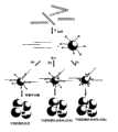
图1(a)示意性地显示本发明的一个采用SERRS检测的实施方案。开始采集含基因组未扩增DNA的样品,并加热使DNA成为单链。然后加入探针,探针特异性地与DNA的靶序列结合。此探针以特定的方式设计,取决于所使用的检测策略:其可具有5’磷酸酯基团,例如,用于使λ核酸外切酶发生作用。在SERRS检测的情况下,探针可以,例如,含有3’羟基喹啉染料,该染料在附着于探针的同时不产生SERRS信号。然而,在酶的作用和探针的消化后,染料被释放至溶液中,使得在添加适当的基底诸如银纳米颗粒时获得SERRS信号。Figure 1(a) schematically shows an embodiment of the present invention using SERRS detection. A sample containing genomic unamplified DNA is initially collected and heated to make the DNA single-stranded. Probes are then added, which specifically bind to the target sequence in the DNA. This probe is designed in a specific way, depending on the detection strategy used: it may have a 5' phosphate group, for example, for the action of lambda exonuclease. In the case of SERRS detection, the probe may, for example, contain a 3' hydroxyquinoline dye that, while attached to the probe, does not generate a SERRS signal. However, after the action of the enzyme and digestion of the probe, the dye is released into solution, allowing a SERRS signal to be obtained upon addition of a suitable substrate such as silver nanoparticles.
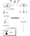
图2显示了本发明进一步的实施方案,其中检测探针是附着在探针序列表面上的金属纳米颗粒。由于静电排斥作用,使得这些纳米颗粒不能聚集在一起。探针序列可以不同的方式被修饰,尤其是被修饰以促进SERRS检测以及可视化检测。系统(a)使用在3’端处附着于表面并且在5’端处已经用磷酸酯基团修饰的探针。在与靶序列杂交并且随后由核酸外切酶消化后,DNA被降解导致颗粒的聚集,此聚集可作为表面等离子体上的位移以及任选地独特的颜色变化而被观察到。在系统(b)中,SERRS活性染料将间隔地排列在纳米颗粒的表面上,然而,直至与靶标杂交、被酶消化并且因此发生聚集时才观察到信号。在发生聚集后,将获得SERRS信号。在系统(c)中,使用的探针与图1中使用的探针相同;在杂交和消化后,SERRS染料将被释放,并且当纳米颗粒杂交时其能够与表面附着并产生SERRS信号。Figure 2 shows a further embodiment of the invention wherein the detection probes are metal nanoparticles attached to the surface of the probe sequences. Due to electrostatic repulsion, these nanoparticles cannot aggregate together. The probe sequences can be modified in different ways, especially to facilitate SERRS detection as well as visual detection. System (a) uses a probe that is attached to the surface at the 3' end and has been modified with a phosphate group at the 5' end. After hybridization to the target sequence and subsequent digestion by exonucleases, the DNA is degraded resulting in aggregation of the particles, which can be observed as a displacement on the surface plasmon and optionally a distinctive color change. In system (b), the SERRS reactive dyes will be spaced apart on the surface of the nanoparticles, however, no signal will be observed until hybridized to the target, digested by the enzyme and thus aggregated. After aggregation occurs, a SERRS signal will be obtained. In system (c), the probes used are the same as those used in Figure 1; after hybridization and digestion, the SERRS dye will be released and it can attach to the surface and generate a SERRS signal when the nanoparticles hybridize.
图3是根据本发明的测定的示意流程图,该测定当寡核苷酸探针与靶核酸结合时采用λ核酸外切酶来消化寡核苷酸探针并去除未被消化的探针。Figure 3 is a schematic flow diagram of an assay according to the present invention which employs lambda exonuclease to digest an oligonucleotide probe and remove undigested probe when it binds to a target nucleic acid.
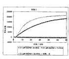
图4显示当其中一条链带有5’磷酸酯部分的双链DNA暴露于λ核酸外切酶时荧光随时间的变化而增加,并且与缺少λ核酸外切酶时进行的相同试验进行比较。Figure 4 shows the increase in fluorescence over time when double stranded DNA with a 5' phosphate moiety on one strand is exposed to lambda exonuclease and compared to the same experiment performed in the absence of lambda exonuclease.
图5显示当其中一条链带有5’磷酸酯部分的双链DNA暴露于两种不同用量的λ核酸外切酶时荧光随时间的变化而增加,并且与缺少λ核酸外切酶时进行的相同试验进行比较。Figure 5 shows the increase in fluorescence over time when double-stranded DNA with a 5' phosphate moiety on one strand was exposed to two different amounts of λ exonuclease, and compared to that performed in the absence of λ exonuclease. Compare the same experiment.
图6显示在λ核酸外切酶对暴露于λ核酸外切酶的同一双链DNA(其结果显示在图1和图2中)和其中基底具有5’缺口;平5’端和3’尾;5’缺口和3’尾的修饰的DNA加工时荧光的增加;以及在缺少λ核酸外切酶时与未修饰的DNA比较。Figure 6 shows the same double-stranded DNA exposed to a lambda exonuclease pair (the results of which are shown in Figures 1 and 2) and wherein the substrate has a 5' gap; a flat 5' end and a 3' tail ; the increase in fluorescence upon processing of modified DNA of the 5' nick and 3' tail; and comparison to unmodified DNA in the absence of lambda exonuclease.
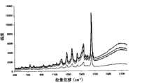
图7显示在存在/缺少互补序列时在酶消化和随后的清除步骤后测定反应物的SERRS光谱。Figure 7 shows the SERRS spectra of reactants measured after enzymatic digestion and subsequent cleanup steps in the presence/absence of complementary sequences.
图8显示在存在/缺少互补靶序列时被λ核酸外切酶消化后在TAMRA标记探针(PTBPROBE)的1650em-1处的平均主峰高的SERRS强度。也显示了在相同条件下操作但缺少酶的对照反应用于比较。引用的值为对每种类型反应的三个单独的重复试验进行的三次测定的平均值。Figure 8 shows the SERRS intensity of the average main peak height at 1650 em-1 of the TAMRA-labeled probe (PTBPROBE) after digestion by lambda exonuclease in the presence/absence of complementary target sequences. A control reaction operated under the same conditions but lacking the enzyme is also shown for comparison. Values quoted are means of triplicate determinations performed on three separate replicates of each type of response.
图9显示在消化、灭活和生物素去除步骤后的测定反应(虚线)以及在缺少酶时进行的相同反应(直线)的SERRS光谱。Figure 9 shows the SERRS spectra of the assay reaction (dashed line) after digestion, inactivation and biotin removal steps and the same reaction performed in the absence of enzyme (straight line).
图10显示基于FRET的测定的示意图。Figure 10 shows a schematic diagram of a FRET-based assay.
图11显示在低靶标浓度催化研究中使用的测定形式。染料标记的探针显示为八角星,小沟结合剂为十字(X),靶标为粗线。在λ核酸外切酶的存在下消化不同比例的FAM-标记探针与互补的靶序列(1∶1;10∶1,100∶1)。FAM标记探针和H33258的浓度恒定地保持在1μM;减小互补序列的浓度。反应混合物加热至37℃30分钟,然后在75℃下进行酶灭活步骤15分钟。在荧光的水平低于检测器水平时,加入过量的靶标以试图形成双链体并且因此如果有相当大的量的探针未被消化则产生熔解曲线。Figure 11 shows the assay format used in the low target concentration catalysis studies. Dye-labeled probes are shown as eight-pointed stars, minor groove binders as crosses (X), and targets as bold lines. Different ratios of FAM-labeled probe to complementary target sequence (1:1; 10:1, 100:1) were digested in the presence of lambda exonuclease. Concentrations of FAM labeled probe and H33258 were kept constant at 1 [mu]M; the concentration of complementary sequences was decreased. The reaction mixture was heated to 37°C for 30 minutes, followed by an enzyme inactivation step at 75°C for 15 minutes. At levels of fluorescence below the detector level, excess target is added in an attempt to form duplexes and thus produce a melting curve if a significant amount of probe is not digested.
图12显示在存在(下部的曲线)/缺少(上部的曲线)λ核酸外切酶时FRET双链体在30分钟内的荧光强度变化。荧光强度减小表明消化破坏了FRET系统。标记探针:补体的比例为(1∶1)。Figure 12 shows the change in fluorescence intensity of FRET duplexes over 30 minutes in the presence (lower curve)/absence (upper curve) of lambda exonuclease. A decrease in fluorescence intensity indicates that digestion has disrupted the FRET system. The ratio of labeled probe:complement was (1:1).
图13显示含有相对于靶序列初始过量(100∶1)的探针的样品的荧光退火曲线。两种样品中掺入额外的靶标。下部的迹线显示其中加入酶的样品,显示荧光强度没有大的变化。上部的迹线缺少酶,因此显示急剧的荧光跃迁~58C,与此序列的双链体的Tm相关。Figure 13 shows the fluorescence annealing curves for samples containing an initial excess (100:1) of probe relative to the target sequence. Both samples were spiked with additional targets. The lower trace shows the sample to which the enzyme was added, showing no large change in fluorescence intensity. The upper trace lacks the enzyme and thus shows a sharp fluorescence transition ~58C, correlating with the Tm of the duplex of this sequence.
图14显示在H33258和λexo的存在下对照单链FAM探针(下部的实线);具有H33258的双链DNA(杂交的FAM探针)(上部实线);以及具有H33258和λexo的双链DNA(虚线)的荧光,显示在30分钟内消化作用的进程。Figure 14 shows control single-stranded FAM probe (lower solid line) in the presence of H33258 and λexo; double-stranded DNA (hybridized FAM probe) with H33258 (upper solid line); and double-stranded DNA with H33258 and λexo Fluorescence of DNA (dashed line), showing the progress of digestion over 30 minutes.
图15显示未消化的(实线)和消化的(虚线)DNA的退火曲线。未消化的样品显示Tm≈57℃。Figure 15 shows the annealing curves of undigested (solid line) and digested (dashed line) DNA. The undigested sample showed a Tm ≈ 57°C.
图16显示使用FAM探针的不同补体用λexo消化30分钟后的强度变化。对于每对,左侧条形代表无酶样品的荧光变化,右侧为有酶样品(除5’突出端仅一个测定值以外,显示三次测定的平均值)。Figure 16 shows the change in intensity of different complements using the FAM probe after digestion with λexo for 30 minutes. For each pair, bars on the left represent the change in fluorescence for samples without enzyme, and for samples with enzyme on the right (average of three determinations is shown except for only one determination for the 5' overhang).
图17显示本发明的一个实施方案的示意图,其中使用包括SE(R)RS标记和生物素的单链探针核酸,使用链霉抗生物素蛋白涂布的磁体去除未反应或过量的探针,使得在被核酸外切酶降解之前仅检测到存在于探针/靶双链体中的SE(R)RS标记。Figure 17 shows a schematic diagram of an embodiment of the invention in which a single-stranded probe nucleic acid comprising a SE(R)RS label and biotin is used, and unreacted or excess probe is removed using a streptavidin-coated magnet , such that only the SE(R)RS label present in the probe/target duplex is detected prior to degradation by the exonuclease.
图18显示琼脂糖TBE凝胶,该凝胶显示了根据本发明的实施例对靶核酸(沙眼衣原体(C.trachomatis)DNA)(2~9泳道)与阴性对照(12~19泳道)相比较的检测。Figure 18 shows an agarose TBE gel, which shows the comparison of the target nucleic acid (Chlamydia trachomatis (C.trachomatis) DNA) (lanes 2 to 9) with the negative control (
图19描绘了沙眼衣原体DNA与阴性对照,以及不同的探针浓度的SERRS分析的结果。Figure 19 depicts the results of SERRS analysis of Chlamydia trachomatis DNA with negative control, and different probe concentrations.
图20描绘了含有沙眼衣原体DNA的样品与阴性对照相比较的SERRS光谱。Figure 20 depicts the SERRS spectra of samples containing Chlamydia trachomatis DNA compared to negative controls.
图21示意性地绘制了一个测定试验方案,其中靶核酸相继地与结合于链霉抗生物素蛋白涂布磁珠的生物素化捕获探针接触,然后与末端为SERRS活性染料标记的5’-磷酸酯的探针接触,所得的双链体被λ核酸外切酶消化,使得能够使用SERRS检测染料。Figure 21 schematically depicts an assay protocol in which the target nucleic acid is sequentially contacted with a biotinylated capture probe bound to streptavidin-coated magnetic beads, followed by a 5' terminal labeled with a SERRS reactive dye. -Phosphate-based probe contact and the resulting duplexes digested with lambda exonuclease, enabling the dye to be detected using SERRS.
图22显示图21中绘制的测定(上部的光谱)与其中使用无染料标记的探针的阴性对照(下部的光谱)的SERRS响应。Figure 22 shows the SERRS response of the assay plotted in Figure 21 (upper spectrum) versus a negative control in which no dye-labeled probe was used (lower spectrum).
图23显示8-羟基喹啉衍生染料的SERRS光谱。Figure 23 shows the SERRS spectra of 8-hydroxyquinoline derived dyes.
图24显示本发明的一种方法,该方法涉及从在两个探针核酸和靶核酸杂交时形成的探针/靶核酸双链体释放标记。Figure 24 shows a method of the invention involving the release of a label from a probe/target nucleic acid duplex formed when two probe nucleic acids and a target nucleic acid hybridize.
图25显示图24中绘制的实施方案的变型,其中靶DNA被固定在固体表面诸如珠子或平板上的捕获探针捕获。Figure 25 shows a variation of the embodiment drawn in Figure 24, wherein the target DNA is captured by capture probes immobilized on a solid surface such as a bead or plate.
发明详述Detailed description of the invention
术语“靶标”、“样品”或“目的样品”在此是指含任意核酸的样品,例如从(多个)个体分离的含核酸的样品。“靶核酸”可以是任意的核酸,包括DNA(来自任意来源,例如,基因组、cDNA、合成的等)、RNA(例如,mRNA、tRNA、rRNA、合成的等)或它们的衍生物(诸如本领域中已知的稀有/罕见的天然来源核苷酸碱基和/或合成的核苷酸碱基的内含物)。样品可代表指定来源中存在的所有或仅一些核酸。可在检测之前制备样品以便使其中的靶核酸更有效地为检测方法所利用。例如,靶核酸可全部或部分地被纯化和/或可产生并分离其片段。作为直接使用样品中的核酸的替代方法,或除直接使用以外,可制备并使用其拷贝(例如,通过PCR)。术语“靶核酸”涵盖所有这些可能性。The term "target", "sample" or "sample of interest" refers herein to any nucleic acid-containing sample, eg, a nucleic acid-containing sample isolated from an individual(s). A "target nucleic acid" can be any nucleic acid, including DNA (from any source, e.g., genomic, cDNA, synthetic, etc.), RNA (e.g., mRNA, tRNA, rRNA, synthetic, etc.), or derivatives thereof (such as the present Inclusions of rare/rare nucleotide bases of natural origin and/or synthetic nucleotide bases known in the art). A sample may represent all or only some of the nucleic acids present in a given source. Samples may be prepared prior to detection in order to make the target nucleic acid therein more efficiently available to the detection method. For example, a target nucleic acid can be purified in whole or in part and/or fragments thereof can be produced and isolated. As an alternative to, or in addition to, direct use of nucleic acid in a sample, copies thereof can be made and used (eg, by PCR). The term "target nucleic acid" covers all these possibilities.
术语“核酸”、“多核苷酸”和“寡核苷酸”在此用来指基于含有2-脱氧-D-核糖或D-核糖的核苷酸,以及本领域中已知的PNA或LNA分子的聚合物或寡聚体。因此,这些术语包括dsDNA和ssDNA,以及dsRNA和ssRNA。这些术语还可能包涵前述物质的嵌合混合物,该混合物的一个实例是包括DNA和PNA或LNA的单链寡核苷酸构建体。在术语“核酸”、“多核苷酸”和“寡核苷酸”之间没有实际的区别;这些术语可互换使用。The terms "nucleic acid", "polynucleotide" and "oligonucleotide" are used herein to refer to nucleic acids based on nucleotides containing 2-deoxy-D-ribose or D-ribose, as well as PNA or LNA known in the art. A polymer or oligomer of molecules. Accordingly, these terms include dsDNA and ssDNA, as well as dsRNA and ssRNA. These terms may also encompass chimeric mixtures of the foregoing, an example of which is a single-stranded oligonucleotide construct comprising DNA and PNA or LNA. There is no real distinction between the terms "nucleic acid," "polynucleotide," and "oligonucleotide"; these terms are used interchangeably.
靶核酸不一定是从任意现有或天然序列中分离的物种,但可以是存在于需要研究其的样品中或之内的任意长度的任意序列。因此其可以是基因组,或亚基因组核酸,染色体,染色体外载体,或基因,或基序,或非编码序列,或序列标签位点,或表达序列标签中存在的任意序列。该序列可来自任意来源,例如,根据已发表的资料制备的或数据库上的资料制备的。The target nucleic acid need not be a species isolated from any existing or native sequence, but may be any sequence of any length present in or within the sample for which it is desired to study. It may thus be a genomic, or subgenomic nucleic acid, a chromosomal, an extrachromosomal vector, or a gene, or a motif, or a non-coding sequence, or a sequence tag site, or any sequence present in an expressed sequence tag. The sequence may be from any source, for example prepared from published data or from data in a database.
术语“特异性杂交”意指与靶标的核酸序列基本上互补的探针核酸序列。其指这样的一些寡核苷酸,当对位排列使得一条序列的5′端与另一条序列的3′端配对时,序列之间的同一性(即,Watson-Crick碱基配对)为至少95%、典型地为至少97%,更典型地为至少98%和最典型地为至少99%。在核酸中可掺入(经酶或合成地)天然核酸中通常不存在的修饰的碱基类似物。如本领域所公知的,两条核酸链的互补性可能并不完美:一些稳定的双链体可含有错配的碱基对或不匹配的碱基,并且核酸技术领域的技术人员可通过考虑除寡核苷酸长度和寡核苷酸中胞嘧啶和鸟嘌呤碱基的浓度和同一性之外的许多变量来推测确定其稳定性。就此而言,要理解的是在任何给定情况下使用的溶液的严格性可根据涉及的实施例的要求而变化,并且专业技术人员能够选择适当的严格性。The term "specifically hybridizes" means a nucleic acid sequence of a probe that is substantially complementary to a nucleic acid sequence of a target. It refers to oligonucleotides that, when aligned so that the 5' end of one sequence pairs with the 3' end of another sequence, have an identity (i.e., Watson-Crick base pairing) between the sequences of at least 95%, typically at least 97%, more typically at least 98% and most typically at least 99%. Modified base analogs not normally found in naturally occurring nucleic acids can be incorporated (enzymatically or synthetically) into nucleic acids. As is well known in the art, the complementarity of two nucleic acid strands may not be perfect: some stable duplexes may contain mismatched base pairs or unmatched bases, and those skilled in the art of nucleic acid technology may consider A number of variables besides oligonucleotide length and the concentration and identity of cytosine and guanine bases in the oligonucleotide are used to speculatively determine its stability. In this regard, it is to be understood that the stringency of the solutions used in any given case may vary according to the requirements of the examples involved, and that the skilled artisan will be able to select an appropriate stringency.
专业技术人员知晓例如,适当地控制温度和/或盐浓度,能够确保仅与特定靶标具有所需程度的特异性的探针序列能与靶标杂交,并且非特异性探针不与该靶标杂交。专业技术人员理解此涉及特异性杂交。The skilled artisan is aware, for example, that proper control of temperature and/or salt concentration can ensure that only probe sequences with the desired degree of specificity for a particular target hybridize to the target, and that non-specific probes do not hybridize to that target. The skilled artisan understands that this involves specific hybridization.
如本领域公知的,可改变盐浓度和/或温度的参数以达到探针和靶核酸之间所需的同一性。本领域技术人员很容易获得关于这些条件的指导,并且尤其可见Sambrook等人,Molecular Cloning:ALaboratory Manual(分子克隆:实验室指南)(第三版),Cold Spring Harbor Laboratory Press,ColdSpring Harbor(冷泉港实验室印刷,冷泉港),NY,7.9~7.12页(2001)。Parameters of salt concentration and/or temperature may be varied to achieve the desired identity between the probe and target nucleic acid, as is known in the art. Guidance on these conditions is readily available to those skilled in the art, and see especially Sambrook et al., Molecular Cloning: A Laboratory Manual (Molecular Cloning: A Laboratory Manual) (Third Edition), Cold Spring Harbor Laboratory Press, Cold Spring Harbor (Cold Spring Harbor Laboratory Printing, Cold Spring Harbor), NY, pp. 7.9-7.12 (2001).
通过熔解或解链温度,或“Tm”来测定核酸双链体的稳定性。在特定反应条件下特定核酸双链体的Tm是碱基对的一半存在于双链体结构中并且一半存在于单链DNA中时的温度。The stability of nucleic acid duplexes is determined by the melting or melting temperature, or "Tm". The Tm of a particular nucleic acid duplex under particular reaction conditions is the temperature at which half of the base pairs are present in the duplex structure and half are present in single-stranded DNA.
测定Tm的主要决定因素是序列长度和G-C含量。用于测定Tm的理论和试验步骤公开在Molecular Cloning-A Laboratory Manual(分子克隆-实验室指南),第二版,J Sambrook等人,Cold Spring Harbor(冷泉港),第II章46和55节。实际上,对于短于18个核苷酸的寡核苷酸,通过将杂交体中A+T残基的数目乘以2℃,G+C残基的数目乘以4℃,并将两者加在一起来估计杂交体的Tm。对于长度为约14~70个核苷酸的寡核苷酸,由Bolton和McCarthy设计的用于测定长DNA分子的TM的下列等式也适用:The main determinants for determining Tm are sequence length and G-C content. Theoretical and experimental procedures for determining Tm are disclosed in Molecular Cloning-A Laboratory Manual, Second Edition, J Sambrook et al., Cold Spring Harbor, Chapter II, Sections 46 and 55 . In fact, for oligonucleotides shorter than 18 nucleotides, by multiplying the number of A+T residues in the hybrid by 2°C, the number of G+C residues by 4°C, and multiplying the two Added together to estimate the Tm of the hybrid. For oligonucleotides of about 14 to 70 nucleotides in length, the following equation devised by Bolton and McCarthy for determining the TM of long DNA molecules also applies:
Tm=81.5-16.6(log[Na+])+0.41(%G+C)-(600/N)。Tm = 81.5-16.6(log[Na+])+0.41(%G+C)-(600/N).
(其中N为链长,[Na+]是杂交溶液的离子强度)。(where N is the chain length and [Na+] is the ionic strength of the hybridization solution).
用于计算Tm的其他公式对本领域技术人员是公知的。Other formulas for calculating Tm are known to those skilled in the art.
如上所述,专业技术人员知晓确切的Tm将取决于许多因素,诸如反应温度、盐浓度、变性剂诸如甲酰胺的存在,以及与寡核苷酸要杂交的序列的互补程度。As noted above, the skilled artisan knows that the exact Tm will depend on many factors, such as reaction temperature, salt concentration, the presence of denaturing agents such as formamide, and the degree of complementarity to the sequence to which the oligonucleotide is hybridizing.
对于寡核苷酸杂交,最佳杂交温度通常在Tm之下2~10℃的条件下进行。理想地,杂交温度被精确地控制在优选±2℃,更优选±0.5℃或更好一些,尤其是当捕获寡核苷酸的可杂交长度很小并且需要区分可能在可杂交序列的一个或另一个末端处仅有单个核苷酸不同的两条序列时。For oligonucleotide hybridization, the optimal hybridization temperature is usually 2-10°C below the Tm. Ideally, the hybridization temperature is precisely controlled at preferably ±2°C, more preferably ±0.5°C or better, especially when the hybridizable length of the capture oligonucleotide is small and it is necessary to distinguish between one or more of the hybridizable sequences. When two sequences differ by only a single nucleotide at the other end.
选择的杂交条件被设计为尽可能接近探针和靶核酸形成的双链体的计算Tm。使用的杂交溶液中的盐的浓度特别重要。在1M NaCl时,G∶C碱基对比A∶T碱基对更稳定。类似地,具有较高G-C含量的双链寡核苷酸的Tm比具有相同长度但具有较高A-T含量的双链寡核苷酸的Tm高。如果在需要被区分的靶核酸中差别很小,即,单核苷酸差异,则建立最佳杂交条件是很重要的,尤其是当寡核苷酸的可杂交长度很小时(<约30mers)。因为目的样品中除靶核苷酸以外还有不同组成的核苷酸,因此Tm的范围很宽。由此,这些条件可通过使用离液序列高的杂交溶液来操作以便消除Tm对核苷酸组成的依赖性。这可以例如,通过向杂交溶液中掺入叔酰胺或季酰胺来实现。Hybridization conditions are selected to be as close as possible to the calculated Tm of the duplex formed by the probe and target nucleic acid. The concentration of salt in the hybridization solution used is of particular importance. At 1M NaCl, the G:C base pair is more stable than the A:T base pair. Similarly, a double-stranded oligonucleotide with a higher G-C content has a higher Tm than a double-stranded oligonucleotide of the same length but with a higher A-T content. If the differences in the target nucleic acids to be differentiated are small, i.e., single nucleotide differences, it is important to establish optimal hybridization conditions, especially when the hybridizable length of the oligonucleotides is small (< about 30mers) . Since there are nucleotides of different compositions in addition to the target nucleotides in the target sample, the range of Tm is wide. Thus, these conditions can be manipulated by using a chaotropic hybridization solution so as to eliminate the dependence of Tm on nucleotide composition. This can be achieved, for example, by incorporating tertiary or quaternary amides into the hybridization solution.
如在此所使用的,探针核酸或“探针”包括被设计为与靶核酸中基本上互补的序列形成双链体结构的核酸序列。要理解的是探针核酸可由此,例如,与单链或双链靶核酸形成双链体。采用后者,所得的三联体结构将包括靶核酸的一条链和单链探针之间杂交形成的双链体。本领域技术人员要理解,有可能是含更高级双链体的寡核苷酸体系,例如,四联体结构(quartet structure)。As used herein, a probe nucleic acid or "probe" includes a nucleic acid sequence designed to form a duplex structure with a substantially complementary sequence in a target nucleic acid. It is understood that a probe nucleic acid may thereby, for example, form a duplex with a single- or double-stranded target nucleic acid. With the latter, the resulting triplex structure will include a duplex formed by hybridization between one strand of the target nucleic acid and the single-stranded probe. Those skilled in the art will appreciate that oligonucleotide systems containing higher order duplexes are possible, eg, quartet structures.
在某些实施方案中,标记与“探针”核酸的一个或多个核苷酸碱基直接附着、结合或以其他方式缔合。缔合要被理解为包涵其中形成原始探针核酸的一部分的单个核苷酸或其一部分一旦不再与原始探针核酸附着就可被检测到的实施方案。In certain embodiments, a label is directly attached, bound or otherwise associated with one or more nucleotide bases of a "probe" nucleic acid. Association is to be understood as encompassing embodiments in which a single nucleotide forming part of the original probe nucleic acid, or a part thereof, is detectable once it is no longer attached to the original probe nucleic acid.
用于通过拉曼/SE(R)RS检测的适当的标记描述在,例如,W097/05280、W099/60157和W02005/019812中。其他适当的标记包括在SERRSDyes(SERRS染料),第二部分,Synthesis and evaluation of dyes for multiplelabelling for SERRS(用于SERRS多重标记的染料的合成和评价)(McHugh,C.J.,Docherty,F.T.,Graham,D.,Smith,W.E.Analyst,2004,129,1,69-72);以及Biosensing Using Silver Nanoparticles and Surface Enhanced ResonanceRaman Scattering(使用银纳米颗粒的生物传感和表面增强共振拉曼散射)(Graham,D,Faulds,K,Smith,W.E.,Chemical Communications(化学通信),2006,42,4363-4371)中公开的那些染料。也有可能包括具有特殊的共振频率的标记,当大量标记紧密接近时该频率可改变。Suitable labels for detection by Raman/SE(R)RS are described, for example, in WO97/05280, WO99/60157 and WO2005/019812. Other suitable labels are included in SERRSDyes (SERRS Dyes), Part II, Synthesis and evaluation of dyes for multiple labeling for SERRS (McHugh, C.J., Docherty, F.T., Graham, D ., Smith, W.E.Analyst, 2004, 129, 1, 69-72); and Biosensing Using Silver Nanoparticles and Surface Enhanced Resonance Raman Scattering (Biosensing Using Silver Nanoparticles and Surface Enhanced Resonance Raman Scattering) (Graham, D, Those dyes disclosed in Faulds, K, Smith, W.E., Chemical Communications (Chemical Communications), 2006, 42, 4363-4371). It is also possible to include markers with a particular resonant frequency that can change when a large number of markers are in close proximity.
荧光标记普遍用于多种检测系统中,并且多种荧光标记是本领域技术人员所熟知的。本发明还可采用FRET和/或猝灭荧光信号的技术,将在后面更详细地说明。Fluorescent labels are commonly used in a variety of detection systems, and a variety of fluorescent labels are well known to those skilled in the art. The present invention may also employ the techniques of FRET and/or quenching of fluorescent signals, which will be described in more detail later.
因此,术语“标记”指可被用于提供可检测的,优选可以计量的,优选实时的信号的任意原子或分子。可检测标记可以附着在核酸探针上或是核酸探针固有的部分或可以其他的方式与探针核酸和靶核酸之间形成的双链体结合。这些标记可与双链体的小沟或大沟结合,或另外地嵌入至双链体核酸中。然而,要理解,此类型的标记应该不能够与靶核酸连接,除非探针和靶核酸之间形成了双链体。许多此类标记是已知的,并且包括


如在本申请中所使用的,“实时”指检测信号的动态生成,包括获取多个读数以便在一段时间内表征信号。例如,实时测定可包括测定可检测产物增加的速率。可选地,实时测定可包括测定在靶序列被扩增至可检测水平前所需的时间。As used in this application, "real-time" refers to the dynamic generation of a detection signal, including taking multiple readings in order to characterize the signal over a period of time. For example, real-time assays can include determining the rate at which a detectable product increases. Alternatively, real-time assays can include determining the time required before a target sequence is amplified to a detectable level.
在一些实施方案中,可能必须要从反应中去除未杂交的探针以便能够仅检测已经掺入探针/靶核酸双链体中或与探针/靶核酸双链体缔合的标记。达到此目的的一种方式是将捕获部分(在此也称为可捕获部分)掺入至探针核酸之上/之中(例子包括磁性部分和/或生物素)。在磁性部分的情况下或使用链霉抗生物素蛋白涂层材料在生物素的情况下,如果探针核酸不与靶标结合,则探针将不被核酸外切酶消化,并且未被消化的探针可被磁体诸如磁珠去除。然而,如果探针形成探针/靶双链体,在双链体消化时捕获部分将从探针释放但标记仍然可被检测到。In some embodiments, it may be necessary to remove unhybridized probe from the reaction in order to be able to detect only labels that have been incorporated into or associated with the probe/target nucleic acid duplex. One way to achieve this is to incorporate capture moieties (also referred to herein as captureable moieties) onto/into the probe nucleic acid (examples include magnetic moieties and/or biotin). If the probe nucleic acid does not bind to the target in the case of a magnetic moiety or in the case of biotin using a streptavidin coating material, the probe will not be digested by the exonuclease and the undigested Probes can be removed by magnets such as magnetic beads. However, if the probe forms a probe/target duplex, upon duplex digestion the capture moiety will be released from the probe but the label will still be detectable.
因此,例如,通过例如,在3’-端,用可由拉曼光谱法(例如,SE(R)RS)检测到的标记和生物素双重标记探针,可通过使用链霉抗生物素蛋白涂布的磁珠去除未消化的探针(例如,因为靶核酸不存在)或过量探针,这意味着仅当靶核酸存在时检测到信号。Thus, for example, by dual-labeling a probe, e.g., at the 3'-end, with a label detectable by Raman spectroscopy (e.g., SE(R)RS) and biotin, a probe can be coated with streptavidin. The magnetic beads of the cloth remove undigested probe (for example, because the target nucleic acid is not present) or excess probe, which means that the signal is only detected when the target nucleic acid is present.
此法示意性地显示在图17中(作为实例,使用SE(R)RS检测和生物素/链霉抗生物素蛋白捕获)。This approach is shown schematically in Figure 17 (using SE(R)RS detection and biotin/streptavidin capture as an example).
此方法学可被理解为类似Taq-Man探针的使用。采用Taq-Man探针,通过使用猝灭剂抑制信号,在PCR扩增期间一旦该猝灭剂从报告染料中分离就不再抑制报告染料的荧光,并且荧光是可检测的。然而,在本发明中,可省略猝灭剂的使用,因为另外作为过量或未杂交探针的结果而产生的信号可通过使用可捕获部分诸如附着在探针上的生物素以及拉曼可检测染料的使用来去除。有益的是,在检测方式的灵敏度允许时(例如,当使用SE(R)RS时)可使用尽量小的探针。This methodology can be understood as similar to the use of Taq-Man probes. With Taq-Man probes, by using a quencher to suppress the signal, once the quencher is dissociated from the reporter dye during PCR amplification, the fluorescence of the reporter dye is no longer suppressed and the fluorescence is detectable. In the present invention, however, the use of a quencher can be omitted, since the signal otherwise generated as a result of excess or unhybridized probe can be detected by using captureable moieties such as biotin attached to the probe and Raman. Dye is used to remove. Advantageously, as small a probe as possible can be used as the sensitivity of the detection format allows (eg, when using SE(R)RS).
还在本发明的进一步的实施方案中,本发明的方法可任选地利用一个以上的探针核酸组合作用来探测靶核酸。因此,例如,可采用使用两个探针,一个用来捕获目的样品中的靶核酸(如果存在),并且另一个为存在靶标的信号提供(一般地)可检测标记。因此第一探针核酸可以,例如,在3’-端被生物素化,从而提供可捕获部分。该部分可与目的样品接触。如果存在靶核酸,其与第一探针杂交,并且使用链霉抗生物素蛋白涂布的磁珠捕获所形成的双链体。然后目的样品中未结合的探针和其他组分被洗掉。可选地,第一探针核酸可初步与链霉抗生物素蛋白涂布的磁体(或磁珠)接触,在与目的样品接触之前未结合的捕获探针被洗掉,之后目的样品的未杂交组分可被洗掉。此可选实施方案示意性地绘制在图21中。In still further embodiments of the invention, the methods of the invention may optionally utilize more than one probe nucleic acid in combination to detect the target nucleic acid. Thus, for example, the use of two probes may be employed, one to capture the target nucleic acid (if present) in the sample of interest and the other to provide a (generally) detectable label for the signal that the target is present. Thus the first probe nucleic acid may, for example, be biotinylated at the 3'-end, thereby providing a captureable moiety. This portion can be in contact with the sample of interest. If a target nucleic acid is present, it is hybridized to the first probe, and the resulting duplex is captured using streptavidin-coated magnetic beads. Unbound probe and other components in the sample of interest are then washed away. Alternatively, the first probe nucleic acid can be initially contacted with a streptavidin-coated magnet (or magnetic bead), and unbound capture probes are washed off prior to contact with the sample of interest, after which unbound capture probes from the sample of interest are washed away. Hybridization components can be washed away. This alternative embodiment is schematically drawn in FIG. 21 .
在捕获靶核酸后,然后拉曼可检测标记可被导入至第一探针核酸与靶核酸杂交生成的双链体中。这可通过使用如在此所述可嵌入至双链体内的标记来完成。可选地,并且一般地,标记与第二探针核酸一起被导入,该第二探针核酸与已经结合于第一探针核酸的靶核酸杂交。然后可通过核酸外切酶降解所得到的包括与靶核酸的不同区互补的第一和任选的第二探针核酸的链的双链体以释放拉曼可检测(例如,SE(R)RS活性)标记。Following capture of the target nucleic acid, a Raman detectable label can then be introduced into the duplex formed by the hybridization of the first probe nucleic acid to the target nucleic acid. This can be accomplished by using tags that can be intercalated within the duplex as described herein. Optionally, and typically, a label is introduced together with a second probe nucleic acid which hybridizes to a target nucleic acid already bound to the first probe nucleic acid. The resulting duplex comprising strands of the first and optional second probe nucleic acids complementary to different regions of the target nucleic acid can then be degraded by an exonuclease to release a Raman detectable (e.g., SE(R) RS activity) marker.
在本发明的所有实施方案中,在有可能从靶核酸分离未结合标记,例如,通过使用在此所述的捕获技术时,在探针包括拉曼可检测标记时对于探针中的该标记,不包括猝灭部分是有可能的并经常是有益的。这是因为有可能去除未结合的含标记的探针(否则考虑到标记的检测指示靶核酸,会产生背景假阳性结果)。然而,在不使用在此所述的捕获技术时,或甚至在使用所述捕获技术时,对于探针核酸中的标记包括猝灭剂会是有益的。以此方式,任何残余的但是未杂交的含标记的探针不会有助于任何被检测到的信号。In all embodiments of the invention, when it is possible to separate the unbound label from the target nucleic acid, for example, by using the capture techniques described herein, when the probe comprises a Raman detectable label, for the label in the probe , it is possible and often beneficial to not include the quenching moiety. This is because of the potential to remove unbound labeled-containing probes (which would otherwise give background false-positive results given that detection of the labeled is indicative of the target nucleic acid). However, when not using the capture techniques described herein, or even when using such capture techniques, it may be beneficial to include a quencher for the label in the probe nucleic acid. In this way, any residual but unhybridized labeled probe does not contribute to any detected signal.
图21示意性地概括地绘制了之前以通用术语描述的实施方案的实例,其中采用了独立的第一和第二核酸探针,并描述了特定的但非必须的一系列洗涤阶段。将观察到第二探针核酸包括标记和猝灭剂(猝灭剂为“标记”探针中的黑圈),尽管从前面的讨论中会立即理解,此猝灭剂的存在非必要特征。“P”描述的是末端5’-磷酸酯部分,其作用是使所得的双链核酸作为λ核酸外切酶的底物。因此图21显示与链霉抗生物素蛋白涂布的磁珠结合的短捕获探针(3’生物素化)。未结合的探针被洗掉。然后靶序列与被固定的捕获探针(第一探针核酸)杂交。未结合的靶核酸被洗掉。然后染料标记的寡核苷酸序列(具有5’磷酸酯基团)与被捕获的靶序列杂交。λ核酸外切酶的作用发生在杂交的探针序列上。该酶将杂交的探针DNA降解为单核苷酸,使染料游离。Figure 21 schematically outlines an example of the embodiment previously described in general terms, in which separate first and second nucleic acid probes are employed, and depicts a specific but not necessarily a series of washing stages. It will be observed that the second probe nucleic acid includes a label and a quencher (the quencher is the black circle in the "label" probe), although it will be immediately understood from the preceding discussion that the presence of this quencher is not an essential feature. "P" describes the terminal 5'-phosphate moiety, which serves to make the resulting double-stranded nucleic acid a substrate for the lambda exonuclease. Figure 21 thus shows short capture probes (3' biotinylated) bound to streptavidin-coated magnetic beads. Unbound probe is washed away. The target sequence is then hybridized to the immobilized capture probe (first probe nucleic acid). Unbound target nucleic acid is washed away. A dye-labeled oligonucleotide sequence (with a 5' phosphate group) then hybridizes to the captured target sequence. The action of lambda exonuclease occurs on hybridized probe sequences. The enzyme degrades hybridized probe DNA to single nucleotides, freeing the dye.
作为本发明的可利用一个以上的核酸组合探测靶核酸的实施方案的其他实例,可使用两个探针核酸,其中第一探针包括被设计成探测靶核酸的序列。除了与靶核酸的至少一部分互补的第一探针的一部分以外,第一探针还包括与第二探针核酸互补的序列。此第二探针核酸可包括SE(R)RS或其他拉曼活性标记(当从第二探针核酸裂解时至少是SE(R)RS或拉曼可检测的)和任选地包括猝灭剂。可选地,该标记可另外地提供为,例如,如在此所述的可嵌入至靶核酸和探针核酸(在此实施方案中由第一和第二探针核酸组成)之间杂交形成的双链体中的标记。第一或第二探针,一般为第二探针,也可包括5’-磷酸酯基团,其中使用的核酸外切酶为λ核酸外切酶。图24显示了本发明的此实施方案的示意图,其中检测方式被指示为SERRS;靶核酸是DNA;第二探针核酸用SERRS活性染料标记(显示为星号)并且具有5’-磷酸酯;并且使用的核酸外切酶为λ核酸外切酶。As a further example of an embodiment of the invention where more than one nucleic acid may be used in combination to detect a target nucleic acid, two probe nucleic acids may be used, where the first probe includes a sequence designed to detect the target nucleic acid. In addition to a portion of the first probe that is complementary to at least a portion of the target nucleic acid, the first probe includes a sequence that is complementary to the second probe nucleic acid. This second probe nucleic acid may comprise a SE(R)RS or other Raman active label (at least SE(R)RS or Raman detectable when cleaved from the second probe nucleic acid) and optionally a quencher agent. Alternatively, the label may additionally be provided, for example, as described herein intercalatable between a target nucleic acid and a probe nucleic acid (consisting in this embodiment of a first and a second probe nucleic acid) to form a hybrid markers in the duplex. The first or second probe, typically the second probe, may also include a 5'-phosphate group, wherein the exonuclease used is lambda exonuclease. Figure 24 shows a schematic diagram of this embodiment of the invention, wherein the detection means is indicated as SERRS; the target nucleic acid is DNA; the second probe nucleic acid is labeled with a SERRS reactive dye (shown as an asterisk) and has a 5'-phosphate; And the exonuclease used is lambda exonuclease.
本发明的此实施方案的一个优点是该实施方案基于第一探针的一部分序列被设计成与靶核酸杂交,从而使第一探针用作靶特异性探针。该序列与第二探针杂交的序列的部分可以是通用的,意味着该序列与靶核酸是非特异的而与第二探针是特异的。因此第二探针不需要具有与靶核酸互补的任意序列,并且因此可用于检测多个靶核酸。这是有益的,如所指出的,因为第二探针核酸一般在结构上较为复杂,一般包括标记和任选地包括猝灭剂,以及任选地包括5’-磷酸酯基团。第二探针不一定是基于靶核酸定制的;部分地基于靶核酸中存在的序列的第一探针可通过简单的寡核苷酸合成来制备。One advantage of this embodiment of the invention is that it is based on the fact that a portion of the sequence of the first probe is designed to hybridize to the target nucleic acid, thereby enabling the first probe to serve as a target-specific probe. The portion of the sequence to which the sequence hybridizes to the second probe may be universal, meaning that the sequence is non-specific for the target nucleic acid but specific for the second probe. The second probe therefore need not have any sequence complementary to the target nucleic acid, and thus can be used to detect multiple target nucleic acids. This is beneficial, as noted, because the second probe nucleic acid is typically structurally more complex, typically including a label and optionally a quencher, and optionally a 5'-phosphate group. The second probe need not be custom-made based on the target nucleic acid; the first probe based in part on the sequence present in the target nucleic acid can be prepared by simple oligonucleotide synthesis.
该方法可允许使用通用探针来用于测定,即,第二探针的序列(和第一探针中的其互补序列)可用于每一个靶核酸,并且可被优化从而为酶的作用、发生杂交以及为最佳标记构型提供最可能好的条件。This method can allow the use of universal probes for the assay, i.e. the sequence of the second probe (and its complement in the first probe) can be used for each target nucleic acid and can be optimized for enzyme action, Hybridization occurs and provides the best possible conditions for optimal label configuration.
然后第一探针核酸的靶区可被改变以与靶标杂交。特别有益的是,该序列的此部分序列也可含有增加杂交效率的碱基,诸如LNA碱基。The target region of the first probe nucleic acid can then be altered to hybridize to the target. Particularly advantageously, this part of the sequence may also contain bases which increase hybridization efficiency, such as LNA bases.
本发明的此实施方案的进一步优点是,该实施方案允许包括PNA或LNA的靶核酸的直接检测:由于其不是靶标和被裂解的第一探针之间杂交形成的(任选地嵌合的)双链核酸,因此不需要利用基于靶DNA的构造选择的核酸外切酶,或使用仅结合例如,DNA或RNA的探针核酸。通过采用LNA和PNA所提供的杂交效率的提高可由(i)由第一探针的靶向区靶向的靶核酸的部分或(ii)靶向区本身包括PNA或LNA中的任一种或两种因素来提供。A further advantage of this embodiment of the invention is that it allows direct detection of target nucleic acids comprising PNAs or LNAs: since they are not formed by hybridization between the target and the cleaved first probe (optionally chimeric ) double-stranded nucleic acid, so there is no need to utilize an exonuclease selected based on the conformation of the target DNA, or to use a probe nucleic acid that only binds, for example, DNA or RNA. The increase in hybridization efficiency provided by the use of LNAs and PNAs may result from (i) the portion of the target nucleic acid targeted by the targeting region of the first probe or (ii) the targeting region itself comprising either PNA or LNA or Two factors are provided.
因此这是比仅对RNA或DNA更通用的检测方法学。靶标可以是使用形成三联体的寡核苷酸(TFO)的dsDNA、使用适体的蛋白质或与适当的靶标结合的抗体。在这些实施方案中,可以与在此所述的相同方式进行检测(例如,对于具有包括第一和第二探针序列的探针序列的单链DNA)。因此,例如,采用双链DNA作为靶核酸,第一探针核酸可以是具有形成三联体的独特序列的TFO,即,其靶向双链DNA,并且其缀合于与第二探针核酸互补的核酸序列。第二探针核酸的杂交和所得双链体的随后降解的作用是检测所需dsDNA的存在而不需要熔解。This is therefore a more general detection methodology than just RNA or DNA. Targets can be dsDNA using triplet-forming oligonucleotides (TFOs), proteins using aptamers, or antibodies that bind to appropriate targets. In these embodiments, detection can be performed in the same manner as described herein (eg, for single-stranded DNA with a probe sequence comprising a first and a second probe sequence). Thus, for example, using double-stranded DNA as the target nucleic acid, the first probe nucleic acid can be a TFO with a unique sequence forming a triplet, i.e., it targets the double-stranded DNA, and it is conjugated to a DNA complementary to the second probe nucleic acid. nucleic acid sequence. The effect of hybridization of the second probe nucleic acid and subsequent degradation of the resulting duplex is to detect the presence of the desired dsDNA without melting.
类似地,核酸可与抗体缀合,然后抗体,例如,可被固定在固体载体上。结合事件(例如,与抗原)的检测可通过本发明的方法来完成,在该方法中用作靶核酸的核酸可附着于另一抗体(例如,在类似于夹心ELISA测定的测定中)。然后可根据本发明的方法通过检测靶核酸来确定固体支持的抗原的存在。以此方式,在任何指定研究中,本发明的方法可用来通过将作为靶核酸的适当核酸与生物分子探针缀合来检测任何结合事件。然后可通过将生物分子探针-靶核酸缀合物暴露于探针核酸并实施本发明的方法来检测结合作用。Similarly, nucleic acids can be conjugated to antibodies, which can then, for example, be immobilized on a solid support. Detection of binding events (eg, with an antigen) can be accomplished by methods of the invention in which a nucleic acid used as a target nucleic acid can be attached to another antibody (eg, in an assay similar to a sandwich ELISA assay). The presence of the solid-supported antigen can then be determined by detecting the target nucleic acid according to the methods of the invention. In this way, in any given study, the methods of the invention can be used to detect any binding event by conjugating an appropriate nucleic acid as target nucleic acid to a biomolecular probe. Binding can then be detected by exposing the biomolecular probe-target nucleic acid conjugate to the probe nucleic acid and performing the methods of the invention.
本发明的此实施方案的进一步优点是只需通过改变第二探针中的标记和第一探针的靶向序列轻松地实现多重检测。实际上被核酸外切酶降解的序列可仍然不变,关于核酸外切酶仅需要建立一组优化条件,其可以保持不变,甚至在要检测多个靶核酸的多重测定中,对核酸外切酶仅需建立一组最佳条件。A further advantage of this embodiment of the invention is that multiplex detection is easily achieved by simply changing the label in the second probe and the targeting sequence of the first probe. The sequence actually degraded by the exonuclease can remain unchanged, only one set of optimized conditions needs to be established for the exonuclease, which can remain unchanged, even in multiplex assays where multiple target nucleic acids are to be detected, the Dicer only needs to establish an optimal set of conditions.
前面所述的通用的核酸外切酶可裂解序列的应用也可与在此所述的捕获方法学结合使用。因此,例如,被设计成与第二探针核酸杂交的第一探针核酸的部分可在3’-端用生物素衍生从而能够使用在此所述并参照图21的基于链霉抗生物素蛋白-生物素的捕获和洗涤方法学。以此方式,第一探针核酸可用作在此所述的可捕获探针。这些方法学的应用(如果需要)可提高本发明的方法的特异性(并且因此提高可靠性)。The use of the general exonuclease cleavable sequences described above can also be used in conjunction with the capture methodology described herein. Thus, for example, a portion of a first probe nucleic acid designed to hybridize to a second probe nucleic acid can be derivatized at the 3'-end with biotin to enable the use of streptavidin-based probes as described herein and with reference to FIG. Protein-biotin capture and wash methodology. In this way, the first probe nucleic acid can be used as a captureable probe as described herein. The application of these methodologies, if desired, can increase the specificity (and thus reliability) of the methods of the invention.
提高特异性的可选方式描述在图25中。该图描述了使用两种捕获探针,它们可附着在固体表面例如,珠子或平板上,或示例性的备选地用生物素衍生以允许被链霉抗生物素蛋白涂布的磁体捕获。描述了两种捕获探针;可使用一种或多种。因此在采用捕获方法学时,在使用由通用的核酸外切酶可裂解序列制成的捕获探针时,该捕获探针不需要是与标记探针杂交的探针相同的探针(如图24中所示)。An alternative way to improve specificity is depicted in FIG. 25 . This figure depicts the use of two capture probes, which can be attached to a solid surface such as a bead or plate, or alternatively derivatized with biotin illustratively to allow capture by a streptavidin-coated magnet. Two capture probes are described; one or more can be used. Thus when using capture methodology, when using a capture probe made from a universal exonuclease cleavable sequence, the capture probe need not be the same probe that hybridizes to the label probe (as shown in Figure 24 shown in ).
在本发明的一个实施方案中,与靶核酸特异地杂交的含5’-磷酸酯的探针核酸被核酸外切酶诸如λ核酸外切酶消化,从而降解含5’-磷酸酯的链。在两条链可被降解和消化的同时,在本发明的一些实施方案中足以仅消化探针并释放拉曼/SE(R)RS可检测标记。In one embodiment of the invention, the 5'-phosphate-containing probe nucleic acid that specifically hybridizes to the target nucleic acid is digested with an exonuclease, such as lambda exonuclease, thereby degrading the 5'-phosphate-containing strand. While both strands can be degraded and digested, it is sufficient in some embodiments of the invention to only digest the probe and release the Raman/SE(R)RS detectable label.
λ核酸外切酶是由λ噬菌体编码的24kD酶。该酶涉及噬菌体的遗传重组但也可作为DNA加工酶从商业渠道获得。酶的晶体结构揭示其是一种具有环形四级结构的同源三聚体。Lambda exonuclease is a 24kD enzyme encoded by lambda phage. This enzyme is involved in the genetic recombination of bacteriophage but is also commercially available as a DNA processing enzyme. The crystal structure of the enzyme revealed that it is a homotrimer with a circular quaternary structure.
λ核酸外切酶消化以磷酸化5’末端起始的双链(dsDNA)的一条链。λ核酸外切酶的中央通道的尺寸是这样的以致其可以在通道的一端仅可容纳dsDNA。该酶是高度持续的,使3’DNA链穿过该酶中央的锥形通道(tapered channel)的中部。5’链被消化,释放游离的5’单核苷酸。具有磷酸化5’末端的单链DNA也显示被消化,但与dsDNA相比是较差的底物。(见P.G Mitsis和J.G Kwagh,Nucleic Acids Research(核酸研究),27(15),3057-3063(1999)以及其中引用的参考文献)。Lambda exonuclease digests one strand of the double-stranded (dsDNA) starting at the phosphorylated 5' end. The size of the central channel of the lambda exonuclease is such that it can accommodate only dsDNA at one end of the channel. The enzyme is highly persistent, passing the 3' DNA strand through the middle of a tapered channel in the center of the enzyme. The 5' strand is digested, releasing a free 5' mononucleotide. Single-stranded DNA with phosphorylated 5' ends was also shown to be digested, but was a poor substrate compared to dsDNA. (See P.G Mitsis and J.G Kwagh, Nucleic Acids Research, 27(15), 3057-3063 (1999) and references cited therein).
在本发明的一些实施方案中,核酸外切酶是λ核酸外切酶。在其他实施方案中,核酸外切酶不是λ核酸外切酶。适合用于本发明中的其他核酸外切酶是不管5’还是3’末端核酸被修饰,诸如被磷酸化,能够持续性地以5’或3’方向消化核酸双链体的一条或两条链的核酸外切酶。然而,应该理解的是核酸外切酶应该具有很小地或不具有消化单链核酸的能力。其他适当的核酸外切酶包括聚合酶,诸如Taq聚合酶、DNA聚合酶I和DNA聚合酶T4。In some embodiments of the invention, the exonuclease is a lambda exonuclease. In other embodiments, the exonuclease is not a lambda exonuclease. Other exonucleases suitable for use in the present invention are those capable of digesting one or both nucleic acid duplexes in a 5' or 3' direction, regardless of whether the 5' or 3' terminal nucleic acid is modified, such as phosphorylated. chain exonuclease. However, it should be understood that the exonuclease should have little or no ability to digest single-stranded nucleic acids. Other suitable exonucleases include polymerases such as Taq polymerase, DNA polymerase I and DNA polymerase T4.
探针可具有任意合适的长度。其实际长度将取决于需要如何精确地测定指定的靶序列。然而,一般来说,探针的长度将为约10~50个核苷酸,例如,长度为20~30个核苷酸。修饰探针以便能够在核酸外切酶,诸如λ核酸外切酶消化探针时在要检测的标记的环境中发生变化。优选当探针的部分,或是当探针作为单链核酸存在时和/或当其与靶序列杂交时,标记基本上是不可检测的。以此方式,过量的、未杂交的探针,不太优选被所述的核酸外切酶加工,其将是不可检测的,并且因此当附着于探针双链体核酸并且在双链体核酸消化过程中已经被释放时,仅存在可检测的标记。以此方式,检测到的标记的任何变化指示目的样品中靶序列的存在。Probes can be of any suitable length. Its actual length will depend on how precisely a given target sequence needs to be determined. Generally, however, probes will be about 10-50 nucleotides in length, eg, 20-30 nucleotides in length. The probe is modified to enable a change in the environment of the label to be detected upon digestion of the probe by an exonuclease, such as lambda exonuclease. Preferably the label is substantially undetectable when part of the probe, or when the probe is present as a single stranded nucleic acid and/or when it hybridizes to the target sequence. In this way, excess, unhybridized probe, less preferably processed by the exonuclease, will be undetectable and thus when attached to the probe duplex nucleic acid and in the duplex nucleic acid Detectable label is only present when it has been released during digestion. In this way, any change in the detected label indicates the presence of the target sequence in the sample of interest.
与现有技术中的方法相反,在本发明的方法过程中探针链不需要一定被延伸。使用拉曼光谱法,例如,尤其是SE(R)RS,使用可检测的标记的检测的灵敏度使得可检测极低水平的标记。在本发明的某些实施方案中,另外的益处是由于某些核酸外切酶,诸如λ核酸外切酶的非常高的转换率,使得一旦起始探针被消化,第二探针分子可与靶核酸结合并被消化以从靶核酸的相同片段生成更多的信号。实际上在这些实施方案中信号被放大,而相反例如在PCR中靶标被扩增。过量的探针序列可被使用,例如,为样品中存在,或预期或估计存在的模板的10倍或更多倍或100倍或更多倍,以确保杂交动力学有利于快速的生成信号。该方法也通过使用不同标记的探针允许多重反应。In contrast to the methods in the prior art, the probe strands do not necessarily need to be extended during the method of the present invention. Using Raman spectroscopy, eg SE(R)RS in particular, the sensitivity of detection using a detectable label allows detection of very low levels of label. In certain embodiments of the invention, an additional benefit is due to the very high turnover rate of certain exonucleases, such as lambda exonuclease, such that once the initial probe is digested, the second probe molecule can Binds to the target nucleic acid and is digested to generate more signal from the same fragment of the target nucleic acid. Indeed in these embodiments the signal is amplified, whereas the target is instead amplified eg in PCR. An excess of probe sequence may be used, for example, 10 times or more or 100 times or more the template present in the sample, or expected or estimated to be present, to ensure that hybridization kinetics favor rapid signal generation. This method also allows for multiplexing reactions by using differently labeled probes.
然而如果需要可进行靶标的扩增。适当的扩增方法是专业技术人员所公知的,并且包括等温扩增和滚环扩增。However, amplification of the target can be performed if desired. Suitable amplification methods are well known to those skilled in the art and include isothermal amplification and rolling circle amplification.
在本发明的某些实施方案中,荧光本身作为检测方式可增添由拉曼光谱法进行的检测,其中可由拉曼光谱法检测到的标记是荧光。在这些实施方案中,探针一般包括荧光团和猝灭剂,如前面所述的分子信标或TaqMan探针。当完整时,猝灭剂将猝灭来自荧光团的任何荧光。然而,在核酸外切酶降解探针DNA时,在与靶核酸杂交后,荧光团将从猝灭剂释放,使得能够获得荧光。有疑问时,可通过使用共聚焦显微镜来改善背景荧光。这样能够使超灵敏检测达到单分子水平。还有可能使用FRET配置而非猝灭剂-供体并且还可使用荧光寿命测定。可使用任何合适的荧光团。在一个实施方案中可使用荧光素-dT(在516nm处发射)。荧光素-dT是修饰的碱基,其中荧光素(FAM)通过6个碳原子间隔臂附着于胸腺嘧啶环的5位。可使用任何合适的猝灭剂,例如Dabcyl(在380-530nm处猝灭)。In certain embodiments of the invention, fluorescence itself as a means of detection may augment detection by Raman spectroscopy, wherein the label detectable by Raman spectroscopy is fluorescence. In these embodiments, the probe typically includes a fluorophore and a quencher, such as the previously described molecular beacons or TaqMan probes. When intact, the quencher will quench any fluorescence from the fluorophore. However, when the exonuclease degrades the probe DNA, upon hybridization to the target nucleic acid, the fluorophore will be released from the quencher, enabling fluorescence to be obtained. When in doubt, background fluorescence can be improved by using confocal microscopy. This enables ultrasensitive detection down to the single molecule level. It is also possible to use a FRET configuration instead of a quencher-donor and also use fluorescence lifetime measurements. Any suitable fluorophore can be used. In one embodiment fluorescein-dT (emits at 516 nm) may be used. Fluorescein-dT is a modified base in which fluorescein (FAM) is attached to the 5-position of the thymine ring through a 6-carbon spacer arm. Any suitable quencher may be used, such as Dabcyl (quenches at 380-530 nm).
在另一个实施方案中,检测方式基于等离子体光子学,其依赖于纳米颗粒等离子体共振作为杂交的指示剂,并且因此依赖于核酸外切酶的作用后溶液中纳米颗粒的聚集状态的变化。根据本实施方案,单个纳米颗粒用探针功能化,使得末端磷酸酯(例如,在λ核酸外切酶用作核酸外切酶时的5’-磷酸酯)或核苷酸碱基远离纳米颗粒的表面。当功能化时,探针将具有特定的共振频率。当这些探针与靶区杂交时,末端磷酸酯/碱基用作核酸外切酶的识别位点从而能够消化纳米颗粒表面上的探针。涂布以探针序列的纳米颗粒在溶液中是分离的和不聚集的,而在与靶序列杂交以及随后探针序列的降解后纳米颗粒不再分开,并将聚集在一起产生可检测的变化。In another embodiment, the detection means is based on plasmon photonics, which relies on nanoparticle plasmon resonance as an indicator of hybridization, and thus on changes in the aggregation state of nanoparticles in solution following the action of an exonuclease. According to this embodiment, individual nanoparticles are functionalized with probes such that the terminal phosphate (e.g., the 5'-phosphate when lambda exonuclease is used as the exonuclease) or nucleotide base is located away from the nanoparticle. s surface. When functionalized, the probe will have a specific resonant frequency. When these probes hybridize to the target region, the terminal phosphate/base serves as a recognition site for the exonuclease to be able to digest the probes on the nanoparticle surface. Nanoparticles coated with probe sequences are segregated and non-aggregate in solution, whereas after hybridization to the target sequence and subsequent degradation of the probe sequences the nanoparticles no longer separate and will aggregate together to produce detectable changes .
在一个实施方案中,可提供探针核酸附着的表面。该表面是适合用于拉曼(SE(R)RS检测中的表面,并且可以是粗糙的金属表面,诸如银表面。该表面可以是,例如,纳米颗粒的表面、微量滴定板、微阵列表面等。拉曼(例如,SE(R)RS)可检测标记可在消化核酸双链体时落在该表面上。In one embodiment, a surface to which probe nucleic acids are attached may be provided. The surface is a surface suitable for use in Raman (SE(R)RS) detection, and may be a rough metal surface, such as a silver surface. The surface may be, for example, the surface of a nanoparticle, a microtiter plate, a microarray surface etc. A Raman (eg, SE(R)RS) detectable label can land on the surface upon digestion of the nucleic acid duplex.
如果该表面是纳米颗粒的表面,探针还会防止颗粒聚集,但在探针消化时,颗粒可聚集,从而增强了任何可检测的拉曼(例如,SE(R)RS)信号。If the surface is that of nanoparticles, the probe also prevents particle aggregation, but upon digestion of the probe, the particles can aggregate, thereby enhancing any detectable Raman (eg, SE(R)RS) signal.
如果足够少量的探针附着在纳米颗粒表面上(例如,10-20%表面覆盖度),则可假设当探针被降解时纳米颗粒的聚集状态将会改变,并引起纳米颗粒的等离子体振子频率的变化。这种变化可使用,例如,暗视野显微镜来测定(见例如,基于金纳米颗粒探针的比色散射均相检测未扩增基因组DNA序列(J.J.Storhoff等人,Nature Biotech.(自然生物技术)2004,22(7),883-887))。If a sufficiently small amount of probe is attached to the nanoparticle surface (e.g., 10-20% surface coverage), it can be hypothesized that when the probe is degraded, the aggregation state of the nanoparticle will change and induce plasmons in the nanoparticle frequency changes. This change can be determined using, for example, dark-field microscopy (see, e.g., Gold Nanoparticle Probe-Based Colorimetric Scattering Homogeneous Detection of Unamplified Genomic DNA Sequences (J.J. Storhoff et al., Nature Biotech.) 2004, 22(7), 883-887)).
根据此实施方案,SE(R)RS活性染料(SE(R)RS标记)可直接附着于纳米颗粒,但当探针仍然完整时,由于没有发生聚集作用,标记是不可见的。探针/靶双链体的消化使颗粒发生聚集,并且还开启了SERRS效应。According to this embodiment, SE(R)RS reactive dyes (SE(R)RS labels) can be directly attached to the nanoparticles, but when the probes are still intact, the labels are not visible since aggregation does not occur. Digestion of the probe/target duplex aggregates the particles and also turns on the SERRS effect.
金或银胶体可用作纳米颗粒。这些胶体可被认为是溶胶,即,分散在水中的固体颗粒。这些颗粒的大小在纳米尺度上,因此术语纳米颗粒的使用一般指2-100nm,尤其是10-80nm,诸如15-40nm。胶体介质非常适合于DNA杂交反应,因为寡核苷酸可被固定在金或银纳米颗粒的表面上。Gold or silver colloids can be used as nanoparticles. These colloids can be considered sols, ie, solid particles dispersed in water. The size of these particles is on the nanometer scale, so the use of the term nanoparticle generally refers to 2-100 nm, especially 10-80 nm, such as 15-40 nm. Colloidal media are well suited for DNA hybridization reactions because oligonucleotides can be immobilized on the surface of gold or silver nanoparticles.
在优选的实施方案中,如前面所讨论的,检测方式基于SE(R)RS。关于SERS和SERRS的详情见,例如,WO97/05280、WO99/60157和WO2005/019812以及其中引用的参考文献。In a preferred embodiment, the detection means is based on SE(R)RS, as previously discussed. For details on SERS and SERRS see, eg, WO97/05280, WO99/60157 and WO2005/019812 and references cited therein.
如本领域中所公知的,拉曼光谱的产生是因为入射至分析物上的光由于分析物中的核运动和电子的激发而散射。在被记录光谱的分析物与适当的表面诸如粗糙的金属表面紧密缔合时,导致检测灵敏度的大大增加,分析物与“活性”表面越接近,则该效应越显著(最佳位置为表面周围的第一分子层内,即,表面的约2nm内)。这称为SERS。As is known in the art, Raman spectra arise because light incident on an analyte is scattered due to nuclear motion and excitation of electrons in the analyte. When the analyte whose spectrum is being recorded is in close association with a suitable surface such as a rough metal surface, this results in a large increase in detection sensitivity, the effect being more pronounced the closer the analyte is to the "active" surface (the optimal location is around the surface within the first molecular layer of , that is, within about 2 nm of the surface). This is called SERS.
通过在分析物(在此情况下通常为附着于目的靶标的染料)的共振频率处操作可获得灵敏度的进一步增加。使用调节至染料的最大吸光度的相干光源,使灵敏度增加103-105倍(激光激发也可设置为表面等离子体共振的最大值,可以与染料最大值一致或不一致)。表面增强效应和共振效应可合并在一起以得到SERRS,并且一定范围的激发频率仍将得到联合增强效应。A further increase in sensitivity can be obtained by operating at the resonance frequency of the analyte (in this case typically the dye attached to the target of interest). Using a coherent light source tuned to the absorbance maximum of the dye increases the sensitivity by a factor of 103 -105 (laser excitation can also be set to the maximum of the surface plasmon resonance, which may or may not coincide with the dye maximum). Surface enhancement and resonance effects can be combined to obtain SERRS, and a range of excitation frequencies will still result in joint enhancement.
因此,当满足两个条件时SERRS技术提供分析物的振动指纹谱。它们是(i)吸附在合适的金属表面上以及(ii)可见生色团的存在。金属的使用还意味着荧光被有效地猝灭。这意味着大量的有色分子,包括标准的荧光团,产生优良的SE(R)RS信号。Therefore, the SERRS technique provides a vibrational fingerprint of an analyte when two conditions are met. These are (i) adsorption on a suitable metal surface and (ii) the presence of a visible chromophore. The use of metals also means that the fluorescence is efficiently quenched. This means that a large number of colored molecules, including standard fluorophores, produce excellent SE(R)RS signals.
通常优选SERRS,并且在此提到的所有SE(R)RS都可被理解为SERRS除非上下文特指相反的含义。然而,要理解的是,本发明还可采用SERS,并且通常采用拉曼光谱法来实施。例如,当使用红外区内的激发频率而尽量减小背景荧光时,SERS具有有益的效果。SERRS is generally preferred, and all references to SE(R)RS herein are to be understood as SERRS unless the context dictates otherwise. However, it is to be understood that the invention may also be practiced using SERS, and typically Raman spectroscopy. For example, SERS has beneficial effects when excitation frequencies in the infrared region are used to minimize background fluorescence.
在例如,Moore等人(2004Nature Biotech.(自然生物技术),22,p1133-1138)中描述了如何生成对于SE(R)RS不可见,然后在酶作用后变得可见的标记的实例。在此实例中,使用脂肪酶来水解酯键释放染料,该染料变成SE(R)RS活性染料。在其促进下,当应用于本发明时,羟基喹啉偶氮染料可通过染料的酚基附着在探针核酸的3’磷酸酯上。这样将染料遮蔽使其不能附着于金属表面上,并且使该染料对SERRS“不可见”。以此方式,一旦探针与其特异性序列杂交,则核酸外切酶将消化寡核苷酸并释放羟基喹啉染料,然后由于染料能够附着在SE(R)RS活性表面上,因此该染料可被SERRS检测到。An example of how to generate a label that is invisible to SE(R)RS and then becomes visible after enzymatic action is described eg in Moore et al. (2004 Nature Biotech., 22, pl 133-1138). In this example, lipase is used to hydrolyze the ester bond to release the dye, which becomes a SE(R)RS reactive dye. With its facilitation, when applied in the present invention, the hydroxyquinoline azo dye can be attached to the 3' phosphate of the probe nucleic acid through the phenolic group of the dye. This masks the dye from attaching to the metal surface and makes the dye "invisible" to SERRS. In this way, once the probe hybridizes to its specific sequence, the exonuclease will digest the oligonucleotide and release the hydroxyquinoline dye, which is then available due to its ability to attach to the SE(R)RS active surface. Detected by SERRS.
由SE(R)RS的极端灵敏度,在许多情况下可以不需要扩增模板就能使该过程进行运转。然而,如果需要可进行靶标的扩增。适当的扩增方法对于专业技术人员是公知的,并且可包括等温扩增和滚环扩增。Due to the extreme sensitivity of SE(R)RS, it is possible in many cases for the process to work without needing to amplify the template. However, amplification of the target can be performed if desired. Suitable amplification methods are well known to the skilled artisan and may include isothermal amplification and rolling circle amplification.
根据进一步的实施方案,可使用其上附着有5’-修饰的SE(R)RS活性染料的探针。染料可以以这种方式附着,从而使探针不粘在金属表面上并产生SERRS。在形成探针/靶双链体并加入核酸外切酶诸如Taq后,发生降解并释放标记。此探针以与Taqman探针类似的方式被降解;然而,不是在酶作用后去除荧光猝灭并增加荧光,在本发明的此实施方案中,染料粘在金属表面上的能力增加,并且因此增加了SERRS信号。如前所述,此实施方案可与可捕获部分诸如生物素的使用结合实施以去除未结合或过量的探针。采用本发明的这些、或其他实施方案中的任一个方案,这些探针可与例如通过PCR扩增目的样品中存在的核酸联合应用。According to a further embodiment, probes with 5'-modified SE(R)RS reactive dyes attached thereto may be used. The dye can be attached in such a way that the probe does not stick to the metal surface and produces SERRS. After formation of the probe/target duplex and addition of an exonuclease such as Taq, degradation occurs and the label is released. This probe is degraded in a similar manner to the Taqman probe; however, instead of removing fluorescence quenching and increasing fluorescence following enzymatic action, in this embodiment of the invention, the ability of the dye to stick to the metal surface is increased, and thus Added SERRS signal. As previously mentioned, this embodiment can be implemented in conjunction with the use of captureable moieties such as biotin to remove unbound or excess probe. Using any of these, or other embodiments of the invention, these probes can be used in conjunction with nucleic acid present in a sample of interest, eg, by PCR amplification.
方便的是,在扩增技术与拉曼可检测探针结合使用时,使用的探针可被设计成与这样的靶核酸部分杂交,其不同于例如与PCR中使用的引物杂交的部分。以此方式,如果探针被杂交,核酸外切酶,例如,Taq对引物的作用继续作用于探针,释放拉曼可检测标记。Conveniently, where amplification techniques are used in conjunction with Raman detectable probes, the probes used can be designed to hybridize to portions of the target nucleic acid different from, for example, the primers used in PCR. In this way, if the probe is hybridized, the action of the exonuclease, eg, Taq, on the primer continues on the probe, releasing the Raman detectable label.
实施本发明的另一方法涉及使用SE(R)RS活性插入剂,或优选地使用小沟结合剂(MGB),如果它们附着于探针序列将增加杂交的特异性。当探针序列与靶标杂交时,插入剂或MGB被设计成这样以致不获得SE(R)RS;然而,在核酸外切酶消化后,其被释放至溶液中,并且能够吸收在金属表面上并产生SE(R)RS。Another method of practicing the invention involves the use of SE(R)RS active intercalators, or preferably minor groove binders (MGBs), which if attached to the probe sequence will increase the specificity of hybridization. The intercalator or MGB is designed such that no SE(R)RS is obtained when the probe sequence hybridizes to the target; however, it is released into solution after exonuclease digestion and is able to absorb on the metal surface And generate SE(R)RS.
本发明可进一步参照下列非限制性的条项来进一步理解:The present invention can be further understood with reference to the following non-limiting clauses:
1.一种用于检测靶核酸的方法,包括以下步骤:1. A method for detecting target nucleic acid, comprising the following steps:
(i)将单链探针核酸与目的样品在以下条件下接触:如果存在所述靶核酸,通过所述探针核酸与靶核酸的特异性杂交,则有效产生探针/靶核酸双链体;(i) contacting a single-stranded probe nucleic acid with a sample of interest under conditions effective to generate a probe/target nucleic acid duplex by specific hybridization of the probe nucleic acid to the target nucleic acid, if the target nucleic acid is present ;
(ii)将任意的探针/靶核酸双链体与核酸外切酶接触以实现所述双链体的消化并从所述双链体释放标记分子;以及(ii) contacting any probe/target nucleic acid duplex with an exonuclease to effect digestion of said duplex and release a labeling molecule from said duplex; and
(iii)通过拉曼光谱法检测所述标记。(iii) detecting the label by Raman spectroscopy.
2.如项1所述的方法,其中所述核酸外切酶不具有寡核苷酸合成能力。2. The method according to
3.如项1或2所述的方法,进一步包括如果所述标记中存在任意的可检测到的变化,检测所述变化从而检测所述靶核酸。3. The method according to
4.如项3所述的方法,其中在所述目的样品中任意靶核酸的数量参照所述变化的幅度来测定。4. The method according to item 3, wherein the amount of any target nucleic acid in the sample of interest is determined with reference to the magnitude of the change.
5.如项1-4中任意一项所述的方法,其中所述标记与所述探针核酸附着、结合或以其他方式缔合。5. The method of any one of items 1-4, wherein the label is attached, bound or otherwise associated with the probe nucleic acid.
6.如项5所述的方法,其中所述探针核酸与所述标记结合。6. The method according to item 5, wherein the probe nucleic acid binds to the label.
7.如前面任意一项所述的方法,其中所述标记仅当通过所述探针/靶核酸双链体的消化从所述双链体核酸中释放时能够通过拉曼光谱法而被检测到。7. The method of any one of the preceding, wherein the label can only be detected by Raman spectroscopy when released from the duplex nucleic acid by digestion of the probe/target nucleic acid duplex arrive.
8.如项1-7中任意一项所述的方法,其中所述标记是SE(R)RS活性染料。8. The method according to any one of items 1-7, wherein the label is a SE(R)RS reactive dye.
9.如项1-8中任意一项所述的方法,其中所述探针序列附着于SE(R)RS活性基底上。9. The method according to any one of items 1-8, wherein the probe sequence is attached to a SE(R)RS active substrate.
10.如项9所述的方法,其中所述基底是纳米颗粒的表面。10. The method according to item 9, wherein the substrate is the surface of nanoparticles.
11.如项1-10中任意一项所述的方法,其中所述探针核酸是DNA。11. The method according to any one of items 1-10, wherein the probe nucleic acid is DNA.
12.如前面任意一项所述的方法,其中所述靶核酸是DNA。12. The method of any preceding claim, wherein the target nucleic acid is DNA.
13.如前面任意一项所述的方法,其中所述靶核酸是双链核酸。13. The method of any preceding claim, wherein the target nucleic acid is a double-stranded nucleic acid.
14.如前面任意一项所述的方法,其中所述靶核酸是单链核酸。14. The method of any preceding claim, wherein the target nucleic acid is a single-stranded nucleic acid.
15.如前面任意一项所述的方法,其中所述探针核酸包括约20~约30个核苷酸。15. The method of any preceding claim, wherein the probe nucleic acid comprises about 20 to about 30 nucleotides.
16.如前面任意一项所述的方法,其中所述核酸外切酶是λ核酸外切酶。16. The method of any preceding claim, wherein the exonuclease is a lambda exonuclease.
17.如项1-15中任意一项所述的方法,其中所述核酸外切酶不是λ核酸外切酶。17. The method according to any one of items 1-15, wherein the exonuclease is not a lambda exonuclease.
18.一种由多个部分组成的试剂盒,包括:18. A kit of parts comprising:
(i)单链探针核酸;(i) single-stranded probe nucleic acid;
(ii)核酸外切酶;以及(ii) exonucleases; and
(iii)用于项1-17中任意一项所限定的方法中的通过拉曼光谱法可检测到的标记。(iii) A label detectable by Raman spectroscopy for use in the method defined in any one of items 1-17.
19.一种用于检测靶核酸的方法,包括以下步骤:19. A method for detecting target nucleic acid comprising the steps of:
(i)将单链探针核酸与目的样品在以下条件下接触:如果存在所述靶核酸,通过所述探针核酸与具有5’-磷酸酯基团的靶核酸的特异性杂交,则有效产生探针/靶核酸双链体;以及(i) contacting a single-stranded probe nucleic acid with a sample of interest under conditions in which, if the target nucleic acid is present, by specific hybridization of the probe nucleic acid to a target nucleic acid having a 5'-phosphate group, effective generating probe/target nucleic acid duplexes; and
(ii)将任意的探针/靶核酸双链体与λ核酸外切酶接触以实现所述双链体的消化并从所述双链体释放标记分子。(ii) contacting any probe/target nucleic acid duplexes with lambda exonuclease to effect digestion of said duplexes and release of label molecules from said duplexes.
20.一种用于检测靶核酸的方法,包括以下步骤:20. A method for detecting target nucleic acid comprising the steps of:
(i)将单链探针核酸与目的样品在以下条件下接触:如果存在所述靶核酸,通过所述探针核酸与靶核酸的特异性杂交,则有效产生探针/靶核酸双链体;以及(i) contacting a single-stranded probe nucleic acid with a sample of interest under conditions effective to generate a probe/target nucleic acid duplex by specific hybridization of the probe nucleic acid to the target nucleic acid, if the target nucleic acid is present ;as well as
(ii)将任意的探针/靶核酸双链体与核酸外切酶接触以实现所述双链体的消化并从所述双链体释放标记分子,(ii) contacting any probe/target nucleic acid duplex with an exonuclease to effect digestion of said duplex and release a labeling molecule from said duplex,
其中在步骤(i)中过量的所述探针核酸与所述目的样品接触使得步骤(ii)中双链体的消化使靶核酸再循环一次或多次,从而使更多的探针分子与靶特异性地杂交,形成额外的探针/靶核酸双链体,所述额外的探针/靶核酸双链体被消化以释放更多的标记分子。wherein in step (i) an excess of said probe nucleic acid is contacted with said sample of interest such that digestion of the duplex in step (ii) recirculates the target nucleic acid one or more times, thereby allowing more probe molecules to interact with The target hybridizes specifically, forming additional probe/target nucleic acid duplexes that are digested to release more labeled molecules.
21.如项19或20所述的方法,进一步包括如果存在靶核酸,检测所述标记中任何可检测的变化从而检测所述靶核酸。21. The method of
22.如项21所述的方法,其中在所述目的样品中任意靶核酸的数量参照所述变化的幅度来测定。22. The method according to item 21, wherein the quantity of any target nucleic acid in said sample of interest is determined with reference to the magnitude of said change.
23.如项19-22中任意一项所述的方法,其中所述标记仅能够当通过所述探针/靶核酸双链体的消化从所述双链体核酸中释放时而被检测到。23. The method according to any one of items 19-22, wherein said label is only detectable when released from said duplex nucleic acid by digestion of said probe/target nucleic acid duplex.
24.如项19-23中任意一项所述的方法,其中所述检测基于等离子体光子学、荧光或SE(R)RS。24. The method according to any one of items 19-23, wherein said detection is based on plasmonics, fluorescence or SE(R)RS.
25.如项19-24中任意一项所述的方法,其中所述标记与所述探针核酸附着、结合或以其他方式缔合。25. The method of any one of items 19-24, wherein the label is attached, bound or otherwise associated with the probe nucleic acid.
26.如项25所述的方法,其中所述探针核酸与所述标记结合。26. The method according to
27.如项19-26中任意一项所述的方法,其中所述标记是可通过拉曼光谱法检测的。27. The method of any one of items 19-26, wherein the label is detectable by Raman spectroscopy.
28.如项27所述的方法,其中所述标记是SE(R)RS标记。28. The method according to item 27, wherein said marker is a SE(R)RS marker.
29.如项28所述的方法,其中所述标记是SERRS标记。29. The method according to item 28, wherein said marker is a SERRS marker.
30.如项29所述的方法,其中所述标记是3’羟基喹啉染料。30. The method of item 29, wherein the label is a 3' hydroxyquinoline dye.
31.如项19-26中任意一项所述的方法,其中所述标记是荧光团。31. The method of any one of items 19-26, wherein the label is a fluorophore.
32.如项31所述的方法,其中所述探针包括荧光团和在所述消化之前猝灭所述荧光团的猝灭剂。32. The method of item 31, wherein said probe comprises a fluorophore and a quencher that quenches said fluorophore prior to said digestion.
33.如项19-24中任一项所述的方法,其中在步骤(i)中所述标记与所述探针核酸和所述目的样品接触,如果形成探针/靶核酸双链体则所述标记可嵌入至任意一个所述探针/靶核酸双链体中。33. The method according to any one of items 19-24, wherein in step (i), the label is contacted with the probe nucleic acid and the target sample, if a probe/target nucleic acid duplex is formed then The label can be incorporated into any of the probe/target nucleic acid duplexes.
34.如项33所述的方法,其中所述标记可与所述双链体的小沟或大沟结合。34. The method according to item 33, wherein the label can bind to the minor groove or the major groove of the duplex.
35.如项34所述的方法,其中所述标记是大沟结合物。35. The method of item 34, wherein the label is a major groove binder.
36.如项35所述的方法,其中所述标记是
37.如项19-36中任意一项所述的方法,其中所述探针核酸是DNA。37. The method of any one of items 19-36, wherein the probe nucleic acid is DNA.
38.如项19-37中任意一项所述的方法,其中所述靶核酸是DNA。38. The method of any one of items 19-37, wherein the target nucleic acid is DNA.
39.如项19-38中任意一项所述的方法,其中所述靶核酸是双链核酸。39. The method of any one of items 19-38, wherein the target nucleic acid is a double-stranded nucleic acid.
40.如项19-39中任意一项所述的方法,其中所述靶核酸是单链核酸。40. The method of any one of items 19-39, wherein the target nucleic acid is a single-stranded nucleic acid.
41.如项19-40中任意一项所述的方法,其中所述探针核酸包括约20~约30个核苷酸。41. The method of any one of items 19-40, wherein the probe nucleic acid comprises about 20 to about 30 nucleotides.
42.如项19-41中任意一项所述的方法,其中所述探针序列附着或吸附于基底。42. The method according to any one of items 19-41, wherein the probe sequence is attached or adsorbed to a substrate.
43.一种由多个部分组成的试剂盒,包括:43. A kit of parts comprising:
(i)具有5’-磷酸酯基团的单链探针核酸;(i) a single-stranded probe nucleic acid with a 5'-phosphate group;
(ii)λ核酸外切酶;以及(ii) lambda exonuclease; and
(iii)用于项19-42中任意一项所限定的方法中的标记。(iii) A label for use in the method defined in any one of items 19-42.
44.如项43所述的试剂盒,其中所述标记是SE(R)RS活性标记。44. The kit according to item 43, wherein the label is a SE(R)RS activity label.
45.靶核酸作为模板的应用,单链探针核酸与该模板特异性杂交并且得到的探针/靶核酸双链体被核酸外切酶降解从而从所述双链体释放标记分子,其中所述靶核酸用作所述模板多次以便从多个双链体中释放多个标记分子,并且其中各个所述多个双链体包括相同的靶核酸。45. The application of target nucleic acid as template, single-stranded probe nucleic acid hybridizes specifically with this template and the probe/target nucleic acid duplex that obtains is degraded by exonuclease so as to release label molecule from described duplex, wherein The target nucleic acid is used as the template multiple times to release a plurality of marker molecules from a plurality of duplexes, and wherein each of the plurality of duplexes includes the same target nucleic acid.
下列的实施例说明本发明但不以任何方式限制其范围。The following examples illustrate the invention but do not limit its scope in any way.
使用的寡核苷酸Oligonucleotides used
寡核苷酸购自atdbio(英国)和MWG(德国)并经HPLC纯化。Oligonucleotides were purchased from atdbio (UK) and MWG (Germany) and purified by HPLC.
荧光探针和互补链的UV熔解UV melting of fluorescent probes and complementary strands
使用Cary 300Bio UV-Vis分光光度计获得荧光素标记的寡核苷酸(RWFAM)和互补序列(RWCOMP1)的UV熔解曲线。熔解曲线用来发现两条链的熔解温度。UV melting curves of fluorescein-labeled oligonucleotide (RWFAM) and complementary sequence (RWCOMP1) were obtained using a Cary 300Bio UV-Vis spectrophotometer. Melting curves were used to find the melting temperatures of the two chains.
将54.6μl RWFAM和31.8μl RWCOMP1加入至石英玻璃杯中的1913.6μl的0.3M PBS中。这样在2ml的总体积中得到的各寡聚体的总浓度为0.5μM。54.6 μl RWFAM and 31.8 μl RWCOMP1 were added to 1913.6 μl of 0.3M PBS in a quartz glass. This gives a total concentration of 0.5 μM of each oligomer in a total volume of 2 ml.
以4个梯度进行UV熔解,每个阶段后保持1分钟:UV melting was performed in 4 gradients with a 1 min hold after each stage:
●梯度1:25℃→90℃●Gradient 1: 25°C → 90°C
●梯度2:90℃→25℃●Gradient 2: 90°C → 25°C
●梯度3:25℃→90℃●Gradient 3: 25°C → 90°C
●梯度4:90℃→25℃●Gradient 4: 90°C → 25°C
各梯度的第一条导数曲线用来找到RWFAM和RWCOMP 1的平均熔解温度。The first derivative curve of each gradient was used to find the average melting temperature of RWFAM and
荧光探针试验介绍Introduction to Fluorescent Probe Assays
概述overview
这些试验包括使用用荧光团和猝灭剂修饰的寡核苷酸探针。探针被设计成与一系列互补的靶寡核苷酸杂交,并且在此情况下,对λ核酸外切酶的作用敏感。通过荧光的增加检测到探针的任何降解作用。These assays involve the use of oligonucleotide probes modified with fluorophores and quenchers. Probes are designed to hybridize to a series of complementary target oligonucleotides and, in this case, to be sensitive to the action of lambda exonuclease. Any degradation of the probe was detected by an increase in fluorescence.
寡核苷酸稀释液Oligonucleotide Diluent
下表列举了用于这些试验中的寡核苷酸母液的稀释液。The table below lists the dilutions of the oligonucleotide stocks used in these experiments.

酶规格Enzyme specification
●购自Epicentre(Cambio)的λ核酸外切酶●λ exonuclease purchased from Epicentre (Cambio)
●酶储存缓冲液:含有50mM Tris-HCl(pH 7.5),100mM NaCl,1.0mM DTT,0.1mM EDTA和0.1%
●在收到酶后,将其分装为25份x 10μl并储存在冰箱中。每份10μ1含100单位的酶。● Upon receipt of the enzyme, aliquot it into 25 x 10μl and store in the refrigerator. Each 10 μl portion contains 100 units of enzyme.
λ核酸外切酶反应缓冲液Lambda Exonuclease Reaction Buffer
●10X反应缓冲液:670mM甘氨酸-KOH(pH 9.4),25mM MgCl2和0.1%X-10010X Reaction Buffer: 670mM Glycine-KOH (pH 9.4), 25mM MgCl2 and 0.1% X-100
●反应缓冲液分成每份150μl。• Reaction buffer was divided into 150 μl aliquots.
仪器instrument
●所有反应在Stratagene MX4000QPCR上进行。●All reactions were carried out on Stratagene MX4000QPCR.
样品制备Sample Preparation
●所有样品在洁净环境中在无菌手套箱中制备。• All samples were prepared in a sterile glove box in a clean environment.
●样品在无菌的200μl管条(tube strip)中制备。• Samples were prepared in sterile 200 μl tube strips.
●样品制备为总体积150μl。• Samples were prepared in a total volume of 150 μl.
●由于使用的一些反应成分的体积微小,在每个阶段之间样品均简单地离心。这确保所有试剂混合并且样品的主体不附着在管的侧面。- Due to the small volumes of some of the reaction components used, samples were briefly centrifuged between each stage. This ensures that all reagents are mixed and that the bulk of the sample does not cling to the side of the tube.
实验步骤Experimental procedure
使用Stratagene MX4000QPCR工作站进行采用荧光探针的实验,并且涉及两个阶段:Experiments with fluorescent probes were performed using the Stratagene MX4000QPCR Workstation and involved two stages:
1.无酶的反应混合物加热至90℃,并在此温度下保持10分钟以确保DNA链的分离。通过将样品冷却至20℃并在此温度保持5分钟,从而使探针和靶链能够杂交。在用于第二阶段的制备中温度回到37℃。1. Heat the enzyme-free reaction mixture to 90°C and keep at this temperature for 10 minutes to ensure the separation of DNA strands. Probe and target strands were allowed to hybridize by cooling the sample to 20°C and holding at this temperature for 5 minutes. The temperature was returned to 37°C in the preparation for the second stage.
2.将酶(或作为对照的水)加入至反应混合物中。样品加热至37℃并且测定荧光。使用三个稍有不同的程序:2. Add the enzyme (or water as a control) to the reaction mixture. Samples were heated to 37°C and fluorescence was measured. Use three slightly different procedures:
在每次循环结束时进行重复三次的荧光测定。Fluorescence measurements were performed in triplicate at the end of each cycle.
荧光实验Fluorescence experiment
下面给出的数据通过从各个数据点减去初始荧光值而进行标准化。重复测定,仅显示平均值。The data presented below were normalized by subtracting the initial fluorescence value from each data point. Repeat determinations, only mean values are shown.
实验1:初步试验Experiment 1: Preliminary Experiment
进行本实验以确认该技术的可行性。在此进行详细描述作为也用于实验2和3中的步骤的实例。This experiment was performed to confirm the feasibility of the technique. This is described in detail here as an example of the procedure also used in Experiments 2 and 3.
下列的样品在8联管条的各个试管中重复制备:The following samples were prepared in duplicate in individual tubes of 8-tube strips:

制备两个测试样品从而一旦杂交得到0.1μM浓度的dsDNA。将RWFAM(8.2μl,1.8μM)和RWCOMP1(4.8μl,3.1μM)加入至在121μl无菌水中的10X反应缓冲液(15μl)中。Two test samples were prepared so that upon hybridization a concentration of dsDNA of 0.1 [mu]M was obtained. RWFAM (8.2 μl, 1.8 μM) and RWCOMP1 (4.8 μl, 3.1 μM) were added to 10X reaction buffer (15 μl) in 121 μl sterile water.
使用相同体积和浓度的寡核苷酸和反应缓冲液,但使用122μl无菌水制备一式二份的对照样品。Control samples were prepared in duplicate using the same volume and concentration of oligonucleotide and reaction buffer, but using 122 μl sterile water.
将样品置于QPCR仪中用于该步骤的杂交阶段。杂交后,取出样品并将1μl(10单位)的λ核酸外切酶加入至两个测试样品中。将样品返回至QPCR仪中,并使用程序A测定荧光。Place the samples in the QPCR machine for the hybridization phase of this step. After hybridization, samples were removed and 1 μl (10 units) of lambda exonuclease was added to both test samples. Return samples to the QPCR machine and measure fluorescence using Procedure A.
结果显示在图4中,该图显示仅在加入λ核酸外切酶的样品中出现荧光增加。这表明酶能够降解探针,并从猝灭剂中释放荧光团。在50-60分钟后获得恒定水平的荧光。对照样品仍然在基线处。The results are shown in Figure 4, which shows an increase in fluorescence only in the samples to which λ exonuclease was added. This indicates that the enzyme is able to degrade the probe and release the fluorophore from the quencher. A constant level of fluorescence was obtained after 50-60 minutes. Control samples are still at baseline.
实验2Experiment 2
本试验研究了双倍浓度的λ核酸外切酶的作用。This assay investigates the effect of double concentrations of lambda exonuclease.
下列的测试样品和对照一式二份地制备:The following test samples and controls were prepared in duplicate:

如实验1中所述制备和杂交样品。杂交后,从QPCR仪中取出样品。Samples were prepared and hybridized as described in
将1μlλ核酸外切酶加入至样品A&B中。1 μl of lambda exonuclease was added to samples A & B.
将2μlλ核酸外切酶加入至样品C中。2 μl of lambda exonuclease was added to sample C.
样品D用作对照。Sample D was used as a control.
样品返回至QPCR仪并使用程序C测定荧光。Samples were returned to the QPCR machine and fluorescence was measured using Procedure C.
图5中的图再次显示在对照样品中荧光没有发生变化。该数据显示当使用的酶的单位数加倍时,荧光发生更快速的增加,并且在较短的时间内达到最高荧光水平。该数据显示了降解相同浓度的荧光探针所需的时间与存在的酶的浓度大概成比例。对于0.1μM浓度的荧光探针,使用两倍单位数的λ核酸外切酶似乎使达到最大荧光水平所需的时间减半。The graphs in Figure 5 again show that no change in fluorescence occurred in the control samples. The data show that when the number of units of enzyme used is doubled, a more rapid increase in fluorescence occurs and peak fluorescence levels are reached in a shorter time. The data show that the time required to degrade the same concentration of fluorescent probe is roughly proportional to the concentration of enzyme present. Using double the units of lambda exonuclease appeared to halve the time required to reach the maximum level of fluorescence for the fluorescent probe at a concentration of 0.1 μM.
实验3Experiment 3
本实验研究了缺口5’端和3’尾对λ核酸外切酶降解探针链的能力的作用,并且使用三条不同的靶链:This experiment investigates the effect of the nick 5' and 3' tails on the ability of lambda exonuclease to degrade the probe strand and uses three different target strands:
●RWCOMP2-提供20个碱基的5’缺口● RWCOMP2 - Provides a 5' gap of 20 bases
●RWCOMP3-提供平5’端,20个碱基的3’尾。• RWCOMP3 - Provides a blunt 5' end, 20 base 3' tail.
●RWCOMP4-提供20个碱基的5’缺口和20个碱基的3’尾。• RWCOMP4 - Provides a 20 base 5' gap and a 20 base 3' tail.
制备一式两份的下列样品:Prepare the following samples in duplicate:


如前面一样制备样品并杂交。杂交后,从QPCR仪去除样品,并将1μl(10单位)λ核酸外切酶加入至样品A、B、C和D中。Samples were prepared and hybridized as before. After hybridization, samples were removed from the QPCR machine and 1 μl (10 units) of lambda exonuclease was added to samples A, B, C and D.
将样品返回至QPCR仪,并使用程序C测定荧光。Return samples to the QPCR machine and measure fluorescence using Procedure C.
本实验的目的是测定当荧光探针与较长链长的靶DNA杂交时λ核酸外切酶是否能够降解荧光探针。这将使探针的5’端与靶链相比是缺口的,并还产生长尾片段。这是要反映真实的DNA样品,其中互补的靶序列将是较长的核酸链的一部分。The purpose of this experiment is to determine whether lambda exonuclease is able to degrade fluorescent probes when they hybridize to longer strands of target DNA. This will gap the 5' end of the probe compared to the target strand and also generate a long tail fragment. This is to reflect real DNA samples, where the complementary target sequence will be part of a longer nucleic acid strand.
实验数据(显示在图6中)显示探针和靶链之间形成的双链体实际上不影响酶的总体活性,因为在每种情况下获得的总荧光增加量是类似的。The experimental data (shown in Figure 6) show that the duplex formed between the probe and target strands practically does not affect the overall activity of the enzyme, as the total increase in fluorescence obtained in each case is similar.
化学品和试剂:Chemicals and Reagents:
化学品购自Sigma(西格玛)或Aldrich(奥尔德里奇),并且均为分析级。Chemicals were purchased from Sigma or Aldrich and were of analytical grade.
λ核酸外切酶(2500单位,10单位/μl,参见LE032K)和λ核酸外切酶反应缓冲液(670mM甘氨酸-KOH,25mM MgCl2,0.1%Triton X-100,x10母液,参见LE-缓冲液)获自Cambio Ltd.,U.K.。λ核酸外切酶以含50mMTris-HCl(pH 7.5),100mM NaCl,1.0mM二硫苏糖醇,0.1mM EDTA和0.1%
链霉抗生物素蛋白涂布的磁珠购自新英格兰生物实验室(New EnglandBiolabs)。缓冲液用前通过0.2μm孔径注射器式过滤器(Whatman)过滤。Streptavidin-coated magnetic beads were purchased from New England Biolabs. The buffer was filtered through a 0.2 [mu]m pore size syringe filter (Whatman) before use.
寡核苷酸序列:Oligonucleotide sequence:
寡核苷酸购自ATDbio和MGW。Oligonucleotides were purchased from ATDbio and MGW.
A2020/PTBPROBE(18mer)A2020/PTBPROBE (18mer)
5’-TTT TCC CAG TCA CGA CGT5’-TTT TCC CAG TCA CGA CGT
修饰:5’磷酸酯Modification: 5' Phosphate
中间(10)dT TAMRAIntermediate (10)dT TAMRA
3’生物素3'biotin
A2020/PFBPROBE(18mer)A2020/PFBPROBE (18mer)
5’-TTT TCC CAG TCA CGA CGT5’-TTT TCC CAG TCA CGA CGT
修饰:5’磷酸酯Modification: 5' Phosphate
中间(10)dTFAMmiddle(10)dTFAM
3’生物素3'biotin
A0977/互补序列(18mer)-用于上面两条序列A0977/complementary sequence (18mer) - for the above two sequences
5’-ACG TCG TGA CTG GGA AAA5’-ACG TCG TGA CTG GGA AAA
CT序列CT sequence
A2057/CTPROBE(22mer)A2057/CTPROBE (22mer)
5’-GCTAAACTT GCT TGC CAC TCAT5’-GCTAAACTT GCT TGC CAC TCAT
修饰:5’磷酸酯Modification: 5' Phosphate
中间(12)dTFAMIntermediate (12)dTFAM
A2058/CTCOMP(22mer)A2058/CTCOMP (22mer)
5’-ATG AGT GGC AAG CAA GTT TAG C5’-ATG AGT GGC AAG CAA GTT TAG C
第一链CT(100mer扩增子)First strand CT (100mer amplicon)
GGA ATT TCC ACT TGA TAT TAC CGC AGG AAC AGA AGC TGC GACGGA ATT TCC ACT TGA TAT TAC CGC AGG AAC AGA AGC TGC GAC
AGG GAC TAA GGA TGC CTC TAT TGA CTA CCA TGA GTG GCA AGCAGG GAC TAA GGA TGC CTC TAT TGA CTA CCA TGA GTG GCA AGC
AAG TTT AGC CCT TTC TAAG TTT AGC CCT TTC T
FAM探针的结合位点以粗体显示。Binding sites of FAM probes are shown in bold.
5’7个碱基突出端5'7 base overhangs
3’71个碱基3'71 bases
仪器instrument
使用结合在514.5nm和30mW功率下工作氩离子激光器的RenishawIn-Via拉曼显微镜系统收集SERRS光谱。硅标准品用来通过测定520cm-1峰的强度来校准仪器。SERRS spectra were collected using a Renishaw In-Via Raman microscope system combined with an argon ion laser operating at 514.5 nm and 30 mW power. Silicon standards were used to calibrate the instrument by measuring the intensity of the 520 cm-1 peak.
在Stratagene MX400Q-PCR上进行荧光测定,该仪器使用石英卤素灯作为光源。带通滤光片通常设定在EX.350nm,EMM.516。所有其他通道通过使用遮光滤光片遮蔽。Cary Eclipse分光光度计也用于一般的荧光测定。Fluorescence measurements were performed on a Stratagene MX400Q-PCR using a quartz halogen lamp as the light source. The bandpass filter is usually set at EX.350nm, EMM.516. All other channels are shaded by using shading filters. The Cary Eclipse spectrophotometer is also used for general fluorescence measurements.
测定使用λ核酸外切酶。The assay uses lambda exonuclease.
下面的测定基于市售的标记有5’-磷酸酯、内部TAMRA dT修饰和3’末端生物素的18-mer(根据图3的示意图)作为可降解探针的应用。The following assay is based on the use of a commercially available 18-mer labeled with a 5'-phosphate, internal TAMRA dT modification and 3' terminal biotin (schematic according to Figure 3) as a degradable probe.
PTBPROBE=5’磷酸酯-TIT TCC CAG T(X)CA CGA CGT-生物素3’PTBPROBE=5' Phosphate-TIT TCC CAG T(X)CA CGA CGT-Biotin 3'
其中X=邻近T的TAMRA修饰where X = TAMRA modification adjacent to T
简言之,反应混合物(9-10μl,1微微摩尔PTBPROBE在存在/不存在1当量的互补ssDNA时)加热至90℃并冷却至20℃以促进互补序列的杂交。然后加入λ核酸外切酶,并将反应物在37℃下温育30分钟以开始消化。该酶是高度持续性的5′至3′外切脱氧核糖核酸酶,其释放5′-单核苷酸和未水解的互补单链DNA(ssDNA)链作为产物。TAMRA-标记的单核苷酸应该在上述PTBPROBE的消化时被释放。Briefly, reaction mixtures (9-10 μl, 1 picomole of PTBPROBE in the presence/absence of 1 equivalent of complementary ssDNA) were heated to 90°C and cooled to 20°C to facilitate hybridization of complementary sequences. Lambda exonuclease was then added and the reaction was incubated at 37°C for 30 minutes to initiate digestion. The enzyme is a highly persistent 5' to 3' exodeoxyribonuclease that releases a 5'-mononucleotide and an unhydrolyzed complementary single-stranded DNA (ssDNA) strand as products. TAMRA-labeled mononucleotides should be released upon digestion with PTBPROBE as described above.
在消化后,反应混合物加热至75℃15分钟以完全地使酶灭活并在将样品冷却回室温之前基本上猝灭反应。然后将在洗涤和结合缓冲液(0.5MNaCl,20mM Tris-HCl,1mM EDTA,pH 7)中的链霉抗生物素蛋白涂布的磁珠用来经与标记的寡核苷酸上的3’生物素修饰的强相互作用而去除任何未消化的PTBPROBE。引入该步骤是为了去除会引起背景信号的未消化的染料标记的探针。使用磁体将磁珠吸引至eppendorf的侧面,然后将上清液转移至干净的微量滴定孔板。加入聚集剂(aggregating agent)(20μl),然后加入柠檬酸盐还原的银胶体(100μl)。在InVia拉曼显微镜上使用514nm激发激光线在加入银胶体后立即记录SERRS光谱。Following digestion, the reaction mixture was heated to 75°C for 15 minutes to completely inactivate the enzyme and essentially quench the reaction before cooling the sample back to room temperature. Streptavidin-coated magnetic beads in wash and binding buffer (0.5M NaCl, 20mM Tris-HCl, 1mM EDTA, pH 7) were then used to bind to the 3' on the labeled oligonucleotide. Strong interaction with biotin modification removes any undigested PTBPROBE. This step was introduced to remove undigested dye-labeled probes that would cause background signal. Use a magnet to attract the beads to the side of the eppendorf, then transfer the supernatant to a clean microtiter well plate. Aggregating agent (20 μl) was added followed by citrate-reduced silver colloid (100 μl). SERRS spectra were recorded immediately after the addition of silver colloids using the 514 nm excitation laser line on an InVia Raman microscope.
λ核酸外切酶是高度持续性的5′至3′外切脱氧核糖核酸酶,其释放5′-单核苷酸和未水解的互补单链DNA(ssDNA)链作为产物。染料-标记的单核苷酸应该在消化时被释放。Lambda exonuclease is a highly persistent 5' to 3' exodeoxyribonuclease that releases a 5'-mononucleotide and an unhydrolyzed complementary single-stranded DNA (ssDNA) strand as products. Dye-labeled mononucleotides should be released upon digestion.
SERRS检测策略:SERRS detection strategy:
反应混合物含有酶、全长寡核苷酸、截短的消化产物和大量化合物的复杂组合。Reaction mixtures contain complex combinations of enzymes, full-length oligonucleotides, truncated digests, and numerous compounds.
在SERRS分析前反应混合物(20μl,包括生物素洗涤步骤)中不同种类的近似浓度列举在下面:The approximate concentrations of the different species in the reaction mixture (20 μl, including a biotin wash step) prior to SERRS analysis are listed below:
0.1μM寡核苷酸标记探针(初始地,加消化产物)0.1 μM oligonucleotide-labeled probe (initially, plus digestion product)
0.1μM互补寡核苷酸靶标0.1 μM complementary oligonucleotide target
33.5mM甘氨酸KOH,33.5 mM Glycine KOH,
1.25mM MgCl21.25mMMgCl2
0.01%TX-100(表面活性剂)0.01% TX-100 (surfactant)
0.255M NaCl,0.255M NaCl,
12.5mM Tris-HCl12.5mM Tris-HCl
0.505mM EDTA0.505mM EDTA
2.5%甘油2.5% glycerin
0.05mM DTT0.05mM DTT
10单位的λ核酸外切酶10 units of lambda exonuclease
甘氨酸,一种简单的氨基酸,以高浓度存在,并且其已知与柠檬酸盐还原的银胶体的表面相互作用(Surface-enhanced Raman scattering ofbiological molecules on metal colloid II:effects of aggregation of gold colloidand comparison of effects of pH of Glycine solutions between gold and silvercolloids(生物分子在金属胶体II上的表面增强拉曼散射:金胶体聚集的作用以及金和银胶体之间甘氨酸溶液的pH的作用的比较):X.Dou,Y.M.Jung,Z.Cao和Y.Ozaki,Appl.Spectrosc.,1999,53,1140)。其与银表面的相互作用程度受pH的影响,并且该作用程度与不同的离子化种类的pKa值相关:Glycine, a simple amino acid, is present in high concentrations and is known to interact with the surface of citrate-reduced silver colloids (Surface-enhanced Raman scattering of biological molecules on metal colloid II: effects of aggregation of gold colloid and comparison of Effects of pH of Glycine solutions between gold and silvercolloids (surface-enhanced Raman scattering of biomolecules on metal colloids II: comparison of the effect of gold colloid aggregation and the pH of glycine solutions between gold and silver colloids): X.Dou , Y.M. Jung, Z. Cao and Y. Ozaki, Appl. Spectrosc., 1999, 53, 1140). The extent of its interaction with the silver surface is affected by pH and is related to the pKa values of the different ionized species:
稳定银和金胶体中的种类的pKa值:The pKa values of species in stable silver and gold colloids:
柠檬酸盐:3.1,4.7,6.4Citrate: 3.1, 4.7, 6.4
EDTA:2.0,2.7,6.1,10.2EDTA: 2.0, 2.7, 6.1, 10.2
甘氨酸:pK1=2.34(COOH),pK2=9.6(NH3+)等电点为5.97Glycine: pK1 =2.34 (COOH), pK2 =9.6 (NH3+ ) The isoelectric point is 5.97
在pH 9.4(缓冲的)下进行消化反应,但加入柠檬酸盐还原的银胶体(pH6-7)可能在一定程度上降低总pH。无论如何,这表明银胶体上的柠檬酸盐基团在这些条件下将完全地去质子化。此外,在分析时大量的甘氨酸应该为两性离子(NH3+,COO-)状态。The digestion reaction was performed at pH 9.4 (buffered), but the addition of citrate-reduced silver colloids (pH 6-7) may lower the overall pH somewhat. Regardless, this suggests that the citrate groups on the silver colloids will be completely deprotonated under these conditions. In addition, a large amount of glycine should be in the zwitterion (NH3+ , COO− ) state at the time of analysis.
当核酸外切酶反应缓冲液加入至银纳米颗粒中时(以与实际测定中使用的相同比例,但不同的体积),观察到-veζ电位(ve Zeta-potential)的减少(表1)。这表明颗粒表面电荷的变化。然而,在此阶段颗粒显示没有明显的聚集变化。When the exonuclease reaction buffer was added to the silver nanoparticles (in the same ratio as used in the actual assay, but in different volumes), a decrease in the -ve Zeta-potential was observed (Table 1). This indicates a change in the surface charge of the particle. However, the particles showed no apparent change in aggregation at this stage.

表1.银胶体在水和核酸外切酶反应缓冲液中的pH和ζ电位值(在本研究中不使用金但其被包括用于比较)。Table 1. pH and zeta potential values of silver colloids in water and exonuclease reaction buffer (gold was not used in this study but included for comparison).
这用来强调pH的重要性以及其对反应混合物中的一些不同种类的作用。pH有可能决定不同化学品与金属表面的亲和力,并且该亲和力又将影响随后的任何SERRS研究。This is used to emphasize the importance of pH and its effect on some of the different species in the reaction mixture. The pH has the potential to determine the affinity of different chemicals to the metal surface, and this affinity will in turn affect any subsequent SERRS studies.
当使用TAMRA标记的寡核苷酸探针时,用来聚集SERRS颗粒的MgSO4证明达到了我们的目的(图7和8)。有可能获得释放的TAMRA标记的SERRS信号,尽管也观察到来自对照样品的一些背景信号。然而,在使用一系列重复三次的反应的研究中,有可能在对照样品和含有靶序列的阳性检测反应物之间建立很大的鉴别度。MgSO4 used to aggregate SERRS particles proved to serve our purpose when TAMRA-labeled oligonucleotide probes were used (Figures 7 and 8). It was possible to obtain released TAMRA-labeled SERRS signal, although some background signal from control samples was also observed. However, in studies using a series of triplicate reactions, it is possible to establish a large degree of discrimination between control samples and positive detection reactions containing the target sequence.
简要实验方案:Brief experimental plan:
将PTBPROBE(5’-磷酸酯,内部dT(TAMRA),3’生物素)与其互补序列在λ核酸外切酶反应缓冲液(67mM甘氨酸-KOH,2.5MgCl2,0.01%TX-100)中混合,加热至90℃,然后在约15分钟的时间内冷却至20℃(0.1μM,1∶1比例,10μl反应规模,1微微摩尔dsDNA)。加入λ核酸外切酶(1μl,10单位),并且在37℃下消化30分钟,紧接着进行灭活步骤(75℃15分钟)。将反应混合物转移至96孔微量滴定板,并与链霉抗生物素蛋白涂布的磁珠(10μl,4mg/ml)在室温下在旋转式振荡器上温育30分钟。然后将微量滴定板置于磁座上,将磁珠朝向孔的侧面分离,然后将上清液(20μl)转移至干净的孔中。加入聚集剂(20μl,1M MgSO4),然后加入100μl柠檬酸盐还原的银胶体。之后立即获取SERRS光谱(Renishaw in Via Raman,514.5nm,x10物镜,100%功率,10秒,3accu.)。(见图9)PTBPROBE (5'-phosphate, internal dT(TAMRA), 3'biotin) was mixed with its complementary sequence in lambda exonuclease reaction buffer (67 mM glycine-KOH, 2.5MgCl2 , 0.01% TX-100) , heated to 90°C, then cooled to 20°C over a period of about 15 minutes (0.1 μM, 1:1 ratio, 10 μl reaction scale, 1 pmol dsDNA). Lambda exonuclease (1 μl, 10 units) was added and digested at 37°C for 30 minutes, followed by an inactivation step (15 minutes at 75°C). The reaction mixture was transferred to a 96-well microtiter plate and incubated with streptavidin-coated magnetic beads (10 μl, 4 mg/ml) on a rotary shaker for 30 minutes at room temperature. The microtiter plate was then placed on a magnetic stand, the beads were detached towards the side of the well, and the supernatant (20 [mu]l) was transferred to a clean well. Aggregating agent (20 μl, 1 M MgSO4 ) was added, followed by 100 μl of citrate-reduced silver colloid. SERRS spectra were acquired immediately thereafter (Renishaw in Via Raman, 514.5 nm, x10 objective, 100% power, 10 sec, 3accu.). (See Figure 9)
基于Fret的测定Fret-based assay
用于此类型测定的示意图显示在图10中。A schematic for this type of assay is shown in Figure 10.
使用FAM标记探针(5’磷酸酯-TTT TCC CAG(AT-FAM)CA CGACGT-生物素)与互补靶序列的不同比例(1∶1;10∶1,100∶1),以10μl的规模进行反应。FAM标记探针和H33258染料(M.Teng,N.Usman,C.A.Frederick和H.J.Wang,Nucl.Acids Res.(核酸研究),1988,16No.6;D.E.Wemer,Biopolymers(生物聚合物),1999,52,197-211;以及F.M.Ho&E.A.H Hall,Biosensors and Bioelectronics(生物传感器和生物电子学),2004,20,5,1001-1010)的浓度为1μM。加入λ核酸外切酶反应缓冲液(1μl,10单位),并且将反应混合物加热至37℃30分钟,紧接着在75℃下进行酶灭活步骤15分钟。将含有互补靶序列(100μl,0.1μM)的溶液加入至各反应物中,并且测定熔解/退火曲线(温度范围15-75℃,监测每1℃在@Ex350nm,Emm520nm处的荧光变化,x3退火曲线和x3熔解曲线)。对照样品(不加入酶)显示Tm~58℃,该值与UV-熔解数据一致。消化的样品中没有一个显示可辨别的熔解曲线,表明在每个消化的样品中FAM标记探针都被消化了。Use different ratios (1:1; 10:1, 100:1) of the FAM-labeled probe (5' phosphate-TTT TCC CAG (AT-FAM) CA CGACGT-biotin) to the complementary target sequence in a 10 μl scale react. FAM label probe and H33258 dye (M.Teng, N.Usman, CAFrederick and HJWang, Nucl.Acids Res. (nucleic acid research), 1988, 16No.6; DEWemer, Biopolymers (biopolymer), 1999,52,197 -211; and FMHo&E.AH Hall, Biosensors and Bioelectronics (Biosensors and Bioelectronics, 2004, 20, 5, 1001-1010) at a concentration of 1 μM. Lambda exonuclease reaction buffer (1 μl, 10 units) was added and the reaction mixture was heated to 37°C for 30 minutes, followed by an enzyme inactivation step at 75°C for 15 minutes. A solution containing complementary target sequence (100 μl, 0.1 μM) was added to each reaction, and the melting/annealing curve was determined (temperature range 15-75°C, monitoring the fluorescence change at @Ex350nm, Emm520nm per 1°C, x3 annealing curve and x3 melting curve). The control sample (no enzyme added) showed aTm ~ 58°C, a value consistent with the UV-melting data. None of the digested samples showed a discernible melting curve, indicating that the FAM-labeled probe was digested in every digested sample.
这些结果显示在图12和图13中。These results are shown in Figures 12 and 13.
进一步实施例further embodiment
1.材料1. Materials
DNA序列购自ATDbio。使用的DNA序列列在表2中。DNA sequences were purchased from ATDbio. The DNA sequences used are listed in Table 2.
表2:用于核酸外切酶测定法的DNA探针Table 2: DNA Probes Used in Exonuclease Assays
Hoechst染料H33258购自Sigma-Aldrich(西格玛-奥尔德里奇)。Hoechst dye H33258 was purchased from Sigma-Aldrich.
Quant-iTTM
重组λ核酸外切酶和x10核酸外切酶反应缓冲液购自Cambio。当稀释至x1时,该缓冲液的组成为67mM甘氨酸-KOH,2.5mM MgCl2,0.1%Triton X-100;在25℃下pH 9.4。Recombinant lambda exonuclease and x10 exonuclease reaction buffer were purchased from Cambio. When diluted to x1, the composition of the buffer was 67 mM Glycine-KOH, 2.5 mMMgCl2 , 0.1% Triton X-100; pH 9.4 at 25°C.
各种(miscellaneous)化学品购自Sigma-Aldrich(西格玛-奥尔德里奇)。Miscellaneous chemicals were purchased from Sigma-Aldrich.
实验使用的磷酸盐缓冲液为具有0.3M NaCl的6mM磷酸酯。The phosphate buffer used in the experiments was 6 mM phosphate with 0.3 M NaCl.
2.仪器2. Instrument
在CARY 300生物紫外-可见光分光光度计(Bio UV-visiblespectrophotometer)上进行吸收研究和UV-熔解。使用Varian CaryEclipse荧光分光光度计进行发射研究。在两个实验中均使用1cm径长和3ml体积的光学透明塑料杯。使用Stratagene Mx4000PCR仪进行酶学测定。Absorption studies and UV-melting were performed on a CARY 300 Bio UV-visible spectrophotometer. Emission studies were performed using a Varian CaryEclipse Fluorescence Spectrophotometer. Optically clear plastic cups of 1 cm diameter and 3 ml volume were used in both experiments. Enzyme assays were performed using a Stratagene Mx4000 PCR instrument.
6.核酸外切酶测定6. Exonuclease Assay
在x1λ核酸外切酶缓冲液中制备样品,最终体积为150μl。在每个试验中序列、补体和/或Hoechst染料的浓度为1μM。在有酶的样品中,向反应混合物中加入10单位的λexo(1μl10单位/μl浓度)。激发波长设定在350nm(10nm带通),并且对于FRET系统在519nm处获取发射测定值,对于Hoechst染料荧光(10nm带通)在440nm处获取发射测定值。在制备期间将样品置于冷板(cool plate)中以避免过早消化。Prepare samples in x1λ exonuclease buffer to a final volume of 150 μl. The concentration of sequence, complement and/or Hoechst dyes in each assay was 1 [mu]M. In samples with enzyme, 10 units of λexo (1 μl 10 units/μl concentration) were added to the reaction mixture. Excitation wavelength was set at 350 nm (10 nm bandpass) and emission measurements were taken at 519 nm for the FRET system and 440 nm for Hoechst dye fluorescence (10 nm bandpass). Samples were placed on a cool plate during preparation to avoid premature digestion.
将样品在37℃下保持30分钟(消化阶段),每分钟测定荧光。然后酶在75℃下变性(灭活阶段)15分钟。之后进行三个熔解阶段:75~20℃的退火曲线,然后再次加热至75℃,并且最后冷却阶段冷却至25℃。加热或冷却速率为1℃/min,每分钟测定荧光。Samples were kept at 37°C for 30 minutes (digestion phase) and fluorescence was measured every minute. The enzyme was then denatured (inactivation phase) for 15 minutes at 75°C. Three melting stages followed: an annealing profile at 75-20°C, then reheating to 75°C, and a final cooling stage to 25°C. The heating or cooling rate was 1 °C/min, and the fluorescence was measured every minute.
对于PicoGreen-TAMRA系统使用相同的测定,使用0.1μM浓度的TAMRA探针和互补序列以及1/10000稀释度的PicoGreen的样品。样品为λexo反应缓冲液中的150μl体积。激发波长为492nm(10nm带通),在515nm处测定发射(10nm带通)。The same assay was used for the PicoGreen-TAMRA system, using samples of TAMRA probe and complementary sequence at a concentration of 0.1 μM and a 1/10000 dilution of PicoGreen. Samples were in 150 μl volumes in lambda exo reaction buffer. The excitation wavelength was 492 nm (10 nm bandpass) and the emission was measured at 515 nm (10 nm bandpass).
V.结果和讨论V. Results and Discussion
4.核酸外切酶测定4. Exonuclease Assay
4.1.FAM探针的消化4.1.Digestion of FAM probes
如实验章节中所述用λ核酸外切酶进行消化。使用两个对照:Digestion was performed with lambda exonuclease as described in the experimental section. Use two controls:
●对照单链DNA:存在酶的含有1μM FAM探针和核酸外切酶反应缓冲液中的1μM H33258的样品。我们假设此对照的荧光等于100%消化的DNA。● Control ssDNA: sample containing 1 μM FAM probe and 1 μM H33258 in exonuclease reaction buffer in the presence of enzyme. We assume that the fluorescence of this control is equal to 100% digested DNA.
●对照双链DNA:含有1μM FAM探针、补体和Hoechst的样品,但不含核酸外切酶。假设此对照等于0%消化。●Control dsDNA: Sample containing 1 μM FAM probe, complement and Hoechst, but no exonuclease. Assume this control equals 0% digestion.
图14显示在消化30分钟内消化的对照和样品的荧光(显示来自三个重复试验品之一的数据)。对于对照样品,荧光强度没有可观察到的变化。然而,杂交的FAM探针的发射显示优异的衰变,从100%双链DNA的水平开始,并且达到单链的值。我们可看到荧光强度随时间变化而进行性地下降,显示该FRET系统实时监测酶消化的能力。Figure 14 shows the fluorescence of digested controls and samples within 30 minutes of digestion (data from one of three replicates shown). For the control samples, there was no observable change in fluorescence intensity. However, the emission of the hybridized FAM probes showed excellent decay, starting at the level of 100% double-stranded DNA, and reaching values for single-stranded. We can see a progressive decrease in fluorescence intensity over time, demonstrating the ability of this FRET system to monitor enzymatic digestion in real time.
之后对样品测定的退火曲线(图15)提供了对消化更多的证据。无酶样品当冷却时再杂交,显示Tm为57℃左右,与前面对FAM探针的研究所观察到的相同。用λ核酸外切酶消化的样品显示没有能力再杂交,意味着消化作用实际上已经完全地降解了FAM探针。The annealing curves (Figure 15) determined on the samples later provided more evidence for digestion. The enzyme-free samples, rehybridized when cooled, showed a Tm of around 57°C, the same as observed in previous studies with the FAM probe. Samples digested with lambda exonuclease showed no ability to rehybridize, implying that digestion had virtually completely degraded the FAM probe.
4.2.使用不同的补体消化FAM探针4.2.Digestion of FAM probes using different complements
研究当DNA与比完全补体长的序列杂交时λ核酸外切酶消化DNA的能力。为此,对下列的样品进行相同的酶反应:To investigate the ability of lambda exonuclease to digest DNA when it hybridizes to sequences longer than complete complement. To do this, perform the same enzymatic reaction on the following samples:
●对照单链DNA(FAM探针)●Control single-stranded DNA (FAM probe)
●完全补体:FAM探针,完全补体和Hoechst染料,在λexo反应缓冲液中各自为1μMComplete complement: FAM probe, complete complement and Hoechst dye, 1 μM each in λexo reaction buffer
●3’突出端:相同,但使用3’端中多12个碱基对的互补序列3’ overhangs: same, but use 12 base pairs more complementary sequence in the 3’ end
●5’突出端:相同,但5’端中具有突出端5’ overhang: same, but with overhang in the 5’ end
●两侧均有突出端:在3’和5’端中均有12个碱基对的突出端Overhangs on both sides: 12 base pair overhangs in both 3' and 5' ends
对于上述各个样品,使用一组反应物作为对照双链DNA(无酶)(每个类型的样品三次重复),并且如前所述另一组使用λ核酸外切酶进行测定。For each of the above samples, one set of reactions was used as control double-stranded DNA (no enzyme) (three replicates for each type of sample), and another set was assayed using lambda exonuclease as previously described.
在每个情形中,荧光的出现相对时间的作图非常类似于完全补体(图未显示),均显示强度的显著降低(见图16)。之后测定的熔解曲线显示所有样品均已经被消化。In each case, the plot of the onset of fluorescence versus time was very similar to that of complete complement (figure not shown), showing a marked decrease in intensity (see Figure 16). Melting curves determined afterwards showed that all samples had been digested.
在得到这些结果后,我们可得出结论λ核酸外切酶能够消化DNA,即使互补序列比探针长也是这样。After obtaining these results, we can conclude that the lambda exonuclease is able to digest DNA even if the complementary sequence is longer than the probe.
使用双标记探针通过SERRS来检测生物样品中的人类病原体。Detection of human pathogens in biological samples by SERRS using dual-labeled probes.
通过SERRS对来自生物样品的人类病原体进行单管检测以检测沙眼衣原体(Chlamydia trachomatis)。使用类似Taq-Man的技术,通过PCR扩增ompA基因的一个区(GenBank序列AY535172),同时消化双重标记探针。下面的数据显示基于SERRS信号的强度有可能区分沙眼衣原体阳性和沙眼衣原体阴性样品。在此实施例中,使用SE(R)RS活性罗丹明染料R6G;然而可使用任意的SE(R)RS(或其他拉曼)活性标记。在此测定中使用的探针/引物组基于用于鉴定衣原体感染的临床诊断性PCR。通过QCMD程序(用于分子诊断学的质量控制)以加入了各种浓度的沙眼衣原体基因组DNA的冻干尿液的形式提供样品。沙眼衣原体DNA来自在McCoy细胞上生长的3天培养物,这些培养物被收获并被热灭活。Single-tube detection of human pathogens from biological samples by SERRS for the detection of Chlamydia trachomatis. A region of the ompA gene (GenBank sequence AY535172) was amplified by PCR using a Taq-Man-like technique while simultaneously digesting the double-labeled probe. The data below show that it is possible to distinguish between C. trachomatis positive and C. trachomatis negative samples based on the strength of the SERRS signal. In this example, the SE(R)RS reactive rhodamine dye R6G was used; however any SE(R)RS (or other Raman) reactive label could be used. The probe/primer set used in this assay is based on clinical diagnostic PCR for the identification of Chlamydia infection. Samples were provided by the QCMD program (Quality Control for Molecular Diagnostics) in the form of freeze-dried urine spiked with various concentrations of Chlamydia trachomatis genomic DNA. Chlamydia trachomatis DNA was obtained from 3-day cultures grown on McCoy cells that were harvested and heat inactivated.
材料和方法Materials and methods
样品的重构Sample reconstruction
接受含有冻干尿液和沙眼衣原体基因组DNA的无菌橡胶密封瓶作为样品。向样品中注入1.2mL无菌milliQ水,并在37℃下摇动1小时以重构样品。Sterile rubber-sealed vials containing freeze-dried urine and C. trachomatis genomic DNA were accepted as samples. Inject 1.2 mL of sterile milliQ water into the sample and shake at 37 °C for 1 h to reconstitute the sample.
制备模板Prepare template
从瓶中取出全部样品至无菌的1.5mL试管中,并加入酵母tRNA(Sigma(西格玛))至100μg.mL-1的终浓度。向样品中加入2体积的冰冷乙醇和1/10体积的3M乙酸钠。将样品充分地混合并在-20℃下温育15分钟。样品在bench离心机中以最大速度离心15分钟,并弃去上清液。用冰冷70%v/v乙醇将沉淀物洗涤两次,用冰冷乙醇洗涤一次,并风干。将沉淀物重新悬浮在60μL无菌milliQ水中,在37℃下摇动1小时。将其在随后的PCR反应中用作模板。All samples were removed from the bottle into sterile 1.5 mL tubes and yeast tRNA (Sigma) was added to a final concentration of 100 μg.mL−1 . Add 2 volumes of ice-cold ethanol and 1/10 volume of 3M sodium acetate to the sample. Samples were mixed well and incubated at -20°C for 15 minutes. Samples were centrifuged at maximum speed for 15 min in a bench centrifuge and the supernatant was discarded. The pellet was washed twice with ice-cold 70% v/v ethanol, once with ice-cold ethanol, and air-dried. Resuspend the pellet in 60 µL of sterile milliQ water and shake for 1 h at 37 °C. This was used as a template in subsequent PCR reactions.
扩增amplify
基于用于检测沙眼衣原体ompA基因(Personal communication,ProfPaul Wallace)的临床诊断性PCR,使用探针/引物组(MWG Biotech)。引物被设计成从碱基501~碱基600扩增基因的100bp区(基于GenBank序列AY535172),探针退火至碱基596~535。使用的引物和探针的序列从5’至3’详述如下:Probe/primer sets (MWG Biotech) were used based on clinical diagnostic PCR for the detection of the C. trachomatis ompA gene (Personal communication, Prof Paul Wallace). Primers were designed to amplify a 100 bp region of the gene from base 501 to base 600 (based on GenBank sequence AY535172), and the probe annealed to bases 596 to 535. The sequences of the primers and probes used are detailed from 5' to 3' as follows:
引物1:cacttratattaccgcaggaacagPrimer 1: cacttratattaccgcaggaacag
引物2:gctaaacttgctgccactcatPrimer 2: gctaaacttgctgccactcat
探针:生物素-agaggcatccttagtccctgtcgcagc-R6GProbe: biotin-agaggcatccttagtccctgtcgcagc-R6G
使用热启动NovaTaq聚合酶(Novagen)、25pmol各引物,100pmol~0.1pmol的探针、2μL模板和3mM MgSO4进行PCR。反应物的总体积为50μL,并且在矿物油中进行这些反应。PCR was performed using hot-start NovaTaq polymerase (Novagen), 25 pmol of each primer, 100-0.1 pmol of probe, 2 μL of template and 3 mM MgSO4 . The total volume of reactions was 50 μL, and the reactions were performed in mineral oil.
使用下列参数进行PCR 30个以上的扩增循环:Perform PCR for more than 30 amplification cycles using the following parameters:
1循环94℃10分钟1 cycle 94°C for 10 minutes
30循环94℃20s,58℃20s以及72℃20s30 cycles of 94°C for 20s, 58°C for 20s and 72°C for 20s
1循环72℃2分钟。1 cycle 72°C for 2 minutes.
使用链霉抗生物素蛋白珠的样品加工Sample Processing Using Streptavidin Beads
从新英格兰生物实验室(New England Biolabs)获得4mg.mL-1的链霉抗生物素蛋白顺磁珠。对于生物素化寡核苷酸,所述珠具有的结合容量为500pmol.mg-1珠。因为任何PCR反应中的生物素化探针的最大量为100pmol,每个反应需要0.2mg(50μL)的珠。Streptavidin paramagnetic beads at 4 mg.mL−1 were obtained from New England Biolabs. The beads have a binding capacity of 500 pmol.mg−1 beads for biotinylated oligonucleotides. Since the maximum amount of biotinylated probe in any PCR reaction is 100 pmol, 0.2 mg (50 μL) of beads are required per reaction.
制备链霉抗生物素蛋白珠供使用Preparation of streptavidin beads for use
通过轻轻地涡旋将所述珠重悬浮。取出所需的等分试样(50μL乘以要测定的PCR反应的次数)并置于无菌1.5mL试管中。将所述珠置于磁分离支架≥2分钟直至所有珠被螯合。将试管固定在支架上,去除并弃去上清液。从支架上取下试管并将所述珠重悬浮在2体积2x B/W缓冲液,[2x结合和洗涤缓冲液:10mM Tris-HCl pH 7.5,1mM EDTApH 7.5,2M NaCl]中。将试管返回至分离支架上并温育≥2分钟。洗涤过程重复两次。将所述珠重悬浮在2x B/W中,使得其最终体积与初始体积相同。The beads were resuspended by gentle vortexing. The required aliquots (50 μL times the number of PCR reactions to be assayed) were removed and placed into sterile 1.5 mL tubes. Place the beads on a magnetic separation stand for > 2 minutes until all beads are chelated. Fix the tubes in the rack, remove and discard the supernatant. Remove the tube from the stand and resuspend the beads in 2 volumes of 2x B/W buffer, [2x binding and washing buffer: 10 mM Tris-HCl pH 7.5, 1 mM EDTA pH 7.5, 2M NaCl]. Return the tube to the separation stand and incubate > 2 minutes. The washing process was repeated twice. Resuspend the beads in 2x B/W such that their final volume is the same as the initial volume.
样品加工sample processing
将50μL制备的珠加入至50μLPCR反应物中并轻轻混合,然后在室温下温育≥15分钟并不时混合。将试管置于分离支架中使得磁体在矿物油层上方,并在室温下温育≥2分钟或直至所述珠已经移动至矿物油之上。将试管固定在支架上,同时将上清液移至新鲜试管中。然后该上清液用于所有随后的SERRS分析。Add 50 μL prepared beads to 50 μL PCR reaction and mix gently, then incubate > 15 minutes at room temperature with occasional mixing. Place the tube in the separation rack with the magnet above the mineral oil layer and incubate at room temperature for > 2 minutes or until the beads have moved above the mineral oil. Fix the tube in the stand while transferring the supernatant to a fresh tube. This supernatant was then used for all subsequent SERRS analyses.
SERRS检测SERRS detection
制备银纳米颗粒Preparation of silver nanoparticles
使用改良的Lee和Meisel法制备柠檬酸盐还原的银纳米颗粒的胶体悬浮液。Colloidal suspensions of citrate-reduced silver nanoparticles were prepared using a modified Lee and Meisel method.
仪器instrument
使用下列的拉曼仪器:带有514.5-nm氩离子激光器的Renishaw 100型探针系统,采用20x物镜将激光束聚焦至含样品的1-cm塑料杯中。The following Raman instrument was used: a Renishaw Model 100 probe system with a 514.5-nm argon-ion laser, using a 20x objective to focus the laser beam into a 1-cm plastic cup containing the sample.
样品制备Sample Preparation
使用下列用量的试剂制备用于SERRS分析的所有样品:10μL分析物,10μL精胺,250μL水和250μL柠檬酸盐还原的银纳米颗粒。All samples for SERRS analysis were prepared using the following amounts of reagents: 10 μL of analyte, 10 μL of spermine, 250 μL of water, and 250 μL of citrate-reduced silver nanoparticles.
琼脂糖凝胶电泳Agarose gel electrophoresis
在水平、中性、2.5%(w/v)琼脂糖凝胶上使DNA显像。常规制备凝胶,并将其在1x TBE缓冲液(Sigma(西格玛))中跑电泳。100bp标志物(Novagen)和Hyper V(Bioline)标志物用作大小标准品。使用较大的16.5x23cm(200mL)凝胶以确保良好的分离。DNA样品与1/10体积的10xBluejuice加样缓冲液(Sigma(西格玛))混合,然后加样至凝胶上。凝胶在溶解在1x TBE中的0.5μg.mL-1溴化乙锭溶液中染色10-20分钟。DNA was visualized on a horizontal, neutral, 2.5% (w/v) agarose gel. Gels were routinely prepared and run in Ix TBE buffer (Sigma). 100 bp marker (Novagen) and Hyper V (Bioline) marker were used as size standards. A larger 16.5x23 cm (200 mL) gel was used to ensure good separation. DNA samples were mixed with 1/10 volume of 1OxBluejuice loading buffer (Sigma) and loaded onto the gel. Gels were stained in a solution of 0.5 μg.mLethidium bromide dissolved in 1x TBE for 10–20 min.
结果和讨论Results and discussion
该技术是Taq-man的改良技术,使用引物组来扩增来自沙眼衣原体的ompA的特定区,同时消化双重标记探针。This technique is a modification of Taq-man, using a primer set to amplify a specific region of ompA from Chlamydia trachomatis, while simultaneously digesting a double-labeled probe.
在Taq-man中,有两条引物和使两者之间退火的一个双重标记探针。该探针在一端带有猝灭剂,并且在另一端带有荧光染料。尽管探针是完整的,猝灭剂与荧光标记足够接近以防止发射。在PCR扩增期间,随着聚合酶使引物延伸,聚合酶的5’至3’核酸酶活性消化探针,将猝灭剂与荧光标记有效地分离并能够进行检测。In Taq-man, there are two primers and a double labeled probe that anneals between the two. The probe has a quencher on one end and a fluorescent dye on the other. Although the probe is intact, the quencher is in close enough proximity to the fluorescent label to prevent emission. During PCR amplification, as the polymerase extends the primers, the 5' to 3' nuclease activity of the polymerase digests the probe, effectively separating the quencher from the fluorescent label and enabling detection.
在此实施例中,探针用生物素和罗丹明标记。在扩增期间,一定比例的探针被破坏,释放生物素和罗丹明。两者仍然附着在一些核苷酸上,但彼此不再有物理上的联系。PCR后,存在附着有生物素和罗丹明的未反应(完整的)探针、游离的生物素和游离的罗丹明的混合物。In this example, the probes were labeled with biotin and rhodamine. During amplification, a proportion of the probe is destroyed, releasing biotin and rhodamine. The two are still attached to some nucleotides, but are no longer physically related to each other. After PCR, there is a mixture of unreacted (intact) probe with biotin and rhodamine attached, free biotin and free rhodamine.
链霉抗生物素蛋白珠,以链霉抗生物素蛋白和生物素之间的几乎不可逆的相互作用,很容易捕获完整的探针和游离的生物素,在上清液中仅留有游离的罗丹明。该过程螯合了所有的未反应探针,并将其从随后的SERRS分析中去除,使得罗丹明信号指示沙眼衣原体的阳性检测。Streptavidin beads, due to the nearly irreversible interaction between streptavidin and biotin, easily capture intact probe and free biotin, leaving only free biotin in the supernatant Rhodamine. This process chelates any unreacted probe and removes it from subsequent SERRS analysis, allowing the rhodamine signal to indicate positive detection of C. trachomatis.
由于SERRS极度灵敏的特性,如果需要,使用的探针量可以减少到最低程度以减少任何背景并减少成本。我们研究了100pmol~0.1pmol的探针浓度范围以观察那个浓度最有效。试验重复进行两次,进行两组阳性和两组阴性PCR(阴性PCR缺少沙眼衣原体DNA)。10μL等分试样的各PCR产物在2.5%(w/v)TBE琼脂糖凝胶上电泳以检查产物的存在,图18。所有阳性反应物扩增良好,阴性反应物没有一个显示有产物。Due to the extremely sensitive nature of SERRS, the amount of probe used can be minimized if desired to reduce any background and reduce costs. We investigated a probe concentration range of 100 pmol to 0.1 pmol to see which concentration was most effective. The assay was performed in duplicate, with two sets of positive and two sets of negative PCR (negative PCR lacking Chlamydia trachomatis DNA). A 10 [mu]L aliquot of each PCR product was electrophoresed on a 2.5% (w/v) TBE agarose gel to check for the presence of the product, FIG. 18 . All positive reactions amplified well and none of the negative reactions showed product.
图18显示PCR扩增的试验:2.5%(w/v)琼脂糖TBE凝胶对于100pmol探针、10pmol、1pmol和0.1pmol的探针稀释范围显示有两个阳性重复试验品(2-9泳道)和2个阴性重复试验品(12-19泳道)。泳道1)100bp标志物,泳道2)100a,泳道3)100b,泳道4)10a,泳道5)10b,泳道6)1a,泳道7)1b,泳道8)0.1a,泳道9)0.1b,泳道10)100bp标志物,泳道11)Hyper V标志物,泳道12)100a,泳道13)100b,泳道14)10a,泳道15)10b,泳道16)1a,泳道17)1b,泳道18)0.1a,泳道19)0.1b,泳道20)100bp标志物。Figure 18 shows the test of PCR amplification: 2.5% (w/v) agarose TBE gel shows two positive duplicate test articles (2-9 lane ) and 2 negative replicates (lanes 12-19). Lane 1) 100bp marker, lane 2) 100a, lane 3) 100b, lane 4) 10a, lane 5) 10b, lane 6) 1a, lane 7) 1b, lane 8) 0.1a, lane 9) 0.1b, lane 10) 100bp marker, swimming lane 11) Hyper V marker, swimming lane 12) 100a, swimming lane 13) 100b, swimming lane 14) 10a, swimming lane 15) 10b, swimming lane 16) 1a, swimming lane 17) 1b, swimming lane 18) 0.1a, Lane 19) 0.1b, lane 20) 100 bp marker.
制备PCR反应物回到50μL,并使用链霉抗生物素蛋白珠加工以分离完整的和消化的探针。然后上清液用于SERRS分析。Prepare PCR reactions back to 50 µL and process using streptavidin beads to separate intact and digested probes. The supernatant was then used for SERRS analysis.
SERRS分析显示阳性反应得到强SERRS信号,并且尽管也有来自PCR阴性样品的信号,但该信号比阳性对照弱许多倍。见图19&20。图19显示对于探针稀释实验的光谱,图中显示通过SERRS分析的两个阳性系列(红和绿)以及两个阴性系列(蓝和黑)的样品。探针浓度从左至右增加。图20显示使用每个反应100pmol探针,阳性和阴性样品之间光谱的差异。SERRS analysis showed a strong SERRS signal for positive reactions and although there was also a signal from PCR negative samples, this signal was many times weaker than the positive control. See Figures 19 & 20. Figure 19 shows the spectra for the probe dilution experiment showing two positive series (red and green) and two negative series (blue and black) of samples analyzed by SERRS. Probe concentration increases from left to right. Figure 20 shows the difference in spectra between positive and negative samples using 100 pmol probe per reaction.
ExoSERRS珠测定ExoSERRS bead assay
所谓的exoSERRS珠测定描述在图21中。在示意图的开始,即最上面,描述了单链靶DNA和在3’端被生物素化的短捕获探针(绘为小灰白色矩形内部的“B”,其已经与显示为大灰色圆圈的链霉抗生物素蛋白涂布的磁珠结合)。在3’-生物素化捕获探针与链霉抗生物素蛋白涂布的磁珠结合后,未结合的探针被洗掉。在显示的第一步骤中,靶DNA与固定的捕获探针杂交。未结合的靶DNA可被洗掉。核苷酸的5’-磷酸酯标记包括SERRS活性染料(显示朝向5’端的灰白色圆圈(在此例中为TAMRA))并且还任选地包括3’-标记猝灭剂(在此例中BHQ2允许与捕获的靶DNA杂交)。最后得到的包括靶DNA,捕获探针和染料标记探针的双链DNA暴露于λ核酸外切酶的作用,该酶将杂交的染料标记的探针DNA降解为单核苷酸,使染料游离从而能够通过SERRS检测。The so-called exoSERRS bead assay is depicted in FIG. 21 . At the beginning of the schematic, topmost, is depicted the single-stranded target DNA and the short capture probe biotinylated at the 3' end (drawn as a "B" inside a small off-white rectangle, which has been aligned with the "B" shown as a large gray circle). Streptavidin-coated magnetic bead binding). After the 3'-biotinylated capture probes bind to the streptavidin-coated magnetic beads, unbound probes are washed away. In the first step shown, target DNA is hybridized to immobilized capture probes. Unbound target DNA can be washed away. 5'-Phosphate labeling of nucleotides includes a SERRS reactive dye (shown as an off-white circle towards the 5' end (in this case TAMRA)) and optionally also a 3'-label quencher (in this case BHQ2 Allow hybridization to captured target DNA). The resulting double-stranded DNA comprising target DNA, capture probe, and dye-labeled probe is exposed to the action of lambda exonuclease, which degrades the hybridized dye-labeled probe DNA to single nucleotides, freeing the dye Thus, it can pass the SERRS detection.
图21中描述的测定法,利用生物素-链霉抗生物素蛋白的强相互作用,使用短捕获探针、全长靶标和染料标记探针。裂解探针测定法采用具有下列组成的上述的三种合成寡核苷酸:The assay described in Figure 21, exploiting the strong biotin-streptavidin interaction, uses short capture probes, full-length targets, and dye-labeled probes. The cleavage probe assay employs the three synthetic oligonucleotides described above with the following composition:
捕获探针capture probe
3’TCT CCG TAG GAA 3’dT被生物素化3’TCT CCG TAG GAA 3’dT is biotinylated
染料标记探针dye-labeled probe
3’TCA GGG ACA GCG XCG 3’dT用BHQ2修饰3’TCA GGG ACA GCG XCG 3’dT modified with BHQ2
X是用TAMRA修饰的dTX is dT modified with TAMRA
靶标补体target complement
5’AGA GGC ATC CTTAGT CCC TGT CGC AGC5’AGA GGC ATC CTTAGT CCC TGT CGC AGC
试验步骤experiment procedure
1.将捕获探针在1x结合和洗涤(B/W)缓冲液中与链霉抗生物素蛋白涂布的磁珠经3’生物素修饰作用结合。将未结合的探针用B/W缓冲液洗掉,同时结合的探针通过磁力分离。1. Bind capture probes to streptavidin-coated magnetic beads via 3' biotin modification in 1x Binding and Washing (B/W) buffer. Unbound probes were washed away with B/W buffer, while bound probes were magnetically separated.
2.使用加热程序(90℃10分钟,20℃5分钟)将靶标互补序列与结合的捕获探针杂交。未结合的靶标再次使用1x B/W洗涤3+次而被洗掉。2. Hybridize the target complementary sequence to the bound capture probe using a heat program (10 minutes at 90°C, 5 minutes at 20°C). Unbound target was washed away again with 3+ washes with 1x B/W.
3.然后使用前面的加热条件将染料标记探针与已形成的捕获探针/靶双链体杂交。再次如上将未结合的探针洗掉。3. The dye-labeled probe is then hybridized to the formed capture probe/target duplex using the previous heating conditions. Unbound probe was washed away again as above.
4.然后将裂解的探针复合体暴露于λ核酸外切酶和核酸外切酶反应缓冲液。在37℃下消化30分钟,然后在75℃下进行酶灭活步骤15分钟。4. The cleaved probe complex is then exposed to lambda exonuclease and exonuclease reaction buffer. Digestion was performed at 37 °C for 30 min, followed by an enzyme inactivation step at 75 °C for 15 min.
5.将复合体转移至96孔微量滴定板中,并在用于SERRS的制剂中加入银胶体、聚集剂和水。5. Transfer the complex to a 96-well microtiter plate, and add silver colloid, aggregation agent and water to the preparation for SERRS.
6.利用Renishaw inVia拉曼光谱检测仪(Renishaw inVia Raman)在514.5nm,100%功率,x20物镜,10s和3次累积的设置下立即获取SERRS。6. Use the Renishaw inVia Raman spectrometer (Renishaw inVia Raman) to acquire SERRS immediately under the settings of 514.5nm, 100% power, x20 objective lens, 10s and 3 accumulations.
7.还可能进行荧光测定。7. Fluorescence measurements are also possible.
结果result
图22图示说明了全程测定与去除染料标记探针的全程测定相比较的SERRS响应。TAMRA响应的形状是非常明显的。令人高兴的是对照中没有峰出现。Figure 22 graphically illustrates the SERRS response of a full run assay compared to a full run assay with dye-labeled probe removed. The shape of the TAMRA response is very pronounced. Happily no peaks appeared in the controls.
SERRS活性探针的合成Synthesis of SERRS Activity Probe
合成SERRS活性探针,该探针由含有无活性的SERRS标记(即,当其为探针的形式时不产生SERRS信号)的探针序列组成。一旦该序列与靶序列杂交,探针在λ或其他核酸外切酶的作用之前仍然是无活性的,在核酸外切酶的作用下其释放SERRS标记,从而使其成为有SERRS活性的探针。A SERRS active probe is synthesized consisting of a probe sequence that contains an inactive SERRS label (ie, does not produce a SERRS signal when it is in probe form). Once this sequence has hybridized to the target sequence, the probe remains inactive until the action of lambda or other exonuclease, which releases the SERRS label, making it a SERRS active probe .
SERRS活性探针可如下制备:生成带有5’磷酸酯、探针序列的寡核苷酸,然后将3’磷酸酯连接至染料上,该探针在酶裂解后成为有SERRS活性的探针。3’磷酸酯连接允许直接附着于羟基喹啉偶氮染料,已经显示当使用SERRS检测时在从“关”至“开”状态的转换中该染料是碱性磷酸酶的良好底物(F.M.Campbell等人,Analyst,2008,DOI 10.1039/B8087A)。以此方式,使用寡核苷酸的反向亚磷酰胺合成将羟基喹啉偶氮染料加到3’端,即从5’至3’方向的合成(正常寡核苷酸合成为从3’至5’)。这样使得在标准固相合成期间羟基喹啉偶氮染料作为亚磷酰胺被加入。通过改变偶氮染料的其他芳香成分,可对不同的序列使用不同的染料,每个都具有不同的SERRS信号。因此这样能够进行多重检测。SERRS-active probes can be prepared by generating oligonucleotides with a 5' phosphate, the probe sequence, and then attaching the 3' phosphate to the dye, which becomes a SERRS-active probe after enzymatic cleavage . The 3' phosphate linkage allows direct attachment to the hydroxyquinoline azo dye, which has been shown to be a good substrate for alkaline phosphatase in the transition from the "off" to "on" state when detected using SERRS (F.M.Campbell et al., Analyst, 2008, DOI 10.1039/B8087A). In this way, reverse phosphoramidite synthesis of oligonucleotides is used to add the hydroxyquinoline azo dye to the 3' end, i.e. synthesis from the 5' to 3' direction (normal oligonucleotides are synthesized from the 3' to 5'). This allows the quinoline azo dye to be added as a phosphoramidite during standard solid phase synthesis. By varying the other aromatic components of the azo dye, different dyes can be used for different sequences, each with a different SERRS signal. This therefore enables multiplex detection.
8-羟基喹啉衍生染料的合成Synthesis of 8-hydroxyquinoline-derived dyes

8-羟基喹啉衍生染料(3)通过邻氨基苄腈的重氮化然后与8-羟基喹啉偶联而制备。为了纯化该染料,其首先被乙酰化,使得其可通过快速柱色谱从起始原料中分离,并且在转变为亚磷酰胺之前被脱乙酰化。染料的合成通过1H,13C NMR,质谱来确认。8-Hydroxyquinoline-derived dyes (3) were prepared by diazotization of o-aminobenzonitrile followed by coupling with 8-hydroxyquinoline. To purify the dye, it is first acetylated so that it can be separated from the starting material by flash column chromatography, and deacetylated before being converted to the phosphoramidite. The synthesis of the dye was confirmed by1 H,13 C NMR, mass spectrometry.
实验步骤:Experimental steps:
邻氨基苄腈(1当量)在HC1(50%,10ml)中搅拌,并小心地加热10分钟。将混合物在冰浴中冷却至0℃,向其中逐滴加入NaNO2(1.2当量)/2ml水(蒸馏的)。使反应物在0℃下搅拌30min,生成浅黄色溶液。单独地将8-羟基喹啉(1当量)溶解在其中加入NaOH(10%,100ml)的MeOH(30ml)中。将8-羟基喹啉溶液在15分钟内逐滴地加入至重氮盐溶液中,并搅拌过夜。O-aminobenzonitrile (1 eq) was stirred in HCl (50%, 10 mL) and heated carefully for 10 min. The mixture was cooled to 0° C. in an ice bath, and NaNO2 (1.2 eq.)/2 ml water (distilled) was added dropwise thereto. The reaction was allowed to stir at 0 °C for 30 min, resulting in a pale yellow solution. 8-Hydroxyquinoline (1 equiv) was dissolved separately in MeOH (30 ml) to which NaOH (10%, 100 ml) was added. The 8-hydroxyquinoline solution was added dropwise to the diazonium salt solution within 15 minutes and stirred overnight.
通过加入HCl(50%)将反应物中和,通过过滤收集所得沉淀物,并干燥过夜。将沉淀物溶解在含有催化量的4-二甲基氨基吡啶的乙酸酐(100ml)中。TLC分析显示完全反应。将混合物倒入1600ml冰/水中,并静置3h。倒掉过量水,将更多的冰加入至红色的油/水混合物中,产生沉淀。收集沉淀物并干燥,并且通过快速柱色谱纯化(二氧化硅,洗脱剂:DCM-1%MeOH/DCM),得到脱乙酰化的靶产物3,收率为38%。δH(400MHz;DMSO)3.31(1H,br,s,OH),7.29-7.31(1H,d,J 8.0,ArH),7.67-7.71(1H,td,ArH),7.78-7.81(1H,q,J 4.0,ArH),7.87-7.91(1H,td,ArH),8.06-8.12(3H,m,ArH×3),9.00-9.02(1H,dd,ArH),9.36-9.38(1H,dd,ArH)。The reaction was neutralized by adding HCl (50%) and the resulting precipitate was collected by filtration and dried overnight. The precipitate was dissolved in acetic anhydride (100 ml) containing a catalytic amount of 4-dimethylaminopyridine. TLC analysis showed complete reaction. The mixture was poured into 1600ml ice/water and left to stand for 3h. Excess water was poured off and more ice was added to the red oil/water mixture, resulting in a precipitate. The precipitate was collected and dried, and purified by flash column chromatography (silica, eluent: DCM-1% MeOH/DCM) to give the deacetylated target product 3 in 38% yield. δH (400MHz; DMSO) 3.31 (1H, br, s, OH), 7.29-7.31 (1H, d, J 8.0, ArH), 7.67-7.71 (1H, td, ArH), 7.78-7.81 (1H, q , J 4.0, ArH), 7.87-7.91 (1H, td, ArH), 8.06-8.12 (3H, m, ArH×3), 9.00-9.02 (1H, dd, ArH), 9.36-9.38 (1H, dd, ArH).
8-羟基喹啉衍生染料3的SERRSSERRS of 8-hydroxyquinoline-derived dye 3
通过使用1%聚左旋赖氨酸(poly-L-lysine)作为聚集剂分析溶液(0.1μM)来确认染料3的SERRS活性。514nm激光源,1s采集,100%功率,1400cm-1。SERRS光谱显示在图23中。The SERRS activity of dye 3 was confirmed by analyzing the solution (0.1 μM) using 1% poly-L-lysine (poly-L-lysine) as an aggregation agent. 514nm laser source, 1s acquisition, 100% power, 1400cm-1 . The SERRS spectrum is shown in Figure 23.
8-羟基喹啉衍生染料DNA修饰8-Hydroxyquinoline-derived dyes for DNA modification

使用氯亚磷酸化(chlorophosphitylating)试剂4进行8-羟基喹啉衍生染料3的亚磷酰化(phosphitylation)以产生亚磷酰胺5。PIII亚磷酰胺的合成通过31P NMR确认。使用逆向碱基合成将亚磷酰胺5用来生成3’-染料标记的探针序列,通过使用磷酸酯CPG柱在5’端掺入磷酸酯。Phosphitylation of 8-hydroxyquinoline-derived dye 3 was performed using chlorophosphitylating reagent 4 to generate phosphoramidite 5 . Synthesis of the PIII phosphoramidite was confirmed by31 P NMR. Phosphoramidite 5 was used to generate a 3'-dye-labeled probe sequence using reverse base synthesis, incorporating a phosphate at the 5' end by using a phosphate CPG column.
序列表sequence listing
<110>斯特拉斯克莱德大学<110>University of Strathclyde
邓肯·格雷姆Duncan Graham
凯伦·福尔兹Karen Fultz
尤安·史密斯Euan Smith
阿拉斯泰尔·里基茨Alastair Ricketts
<120>核酸序列的鉴定<120> Identification of Nucleic Acid Sequences
the
<130>P15146PC<130>P15146PC
the
<160>27<160>27
the
<170>PatentIn version 3.3<170>PatentIn version 3.3
the
<210>1<210>1
<211>18<211>18
<212>DNA<212>DNA
<213>人工的<213> Artificial
the
<220><220>
<223>寡核苷酸探针<223> Oligonucleotide probes
the
<400>1<400>1
ttttcccagt cacgacgt 18ttttcccagt cacgacgt 18
the
<210>2<210>2
<211>18<211>18
<212>DNA<212>DNA
<213>人工的<213> Artificial
the
<220><220>
<223>寡核苷酸探针<223> Oligonucleotide probes
the
<400>2<400>2
ttttcccagt cacgacgt 18ttttcccagt cacgacgt 18
the
<210>3<210>3
<211>18<211>18
<212>DNA<212>DNA
<213>人工的<213> Artificial
the
<220><220>
<223>寡核苷酸探针<223> Oligonucleotide probes
the
<400>3<400>3
ttttcccagt cacgacgt 18ttttcccagt cacgacgt 18
the
<210>4<210>4
<211>18<211>18
<212>DNA<212>DNA
<213>人工的<213> Artificial
the
<220><220>
<223>寡核苷酸探针<223> Oligonucleotide probes
the
<220><220>
<221>修饰的碱基<221> Modified bases
<222>(10)..(10)<222>(10)..(10)
<223>荧光素dT<223> Fluorescein dT
the
<220><220>
<221>misc_feature<221>misc_feature
<222>(10)..(10)<222>(10)..(10)
<223>n是a,c,g,或t<223> n is a, c, g, or t
the
<400>4<400>4
ttttcccagn cacgacgt 18ttttcccagn cacgacgt 18
the
<210>5<210>5
<211>18<211>18
<212>DNA<212>DNA
<213>人工的<213> Artificial
the
<220><220>
<223>寡核苷酸探针<223> Oligonucleotide probes
the
<400>5<400>5
acgtcgtgac tgggaaaa 18acgtcgtgac tgggaaaa 18
the
<210>6<210>6
<211>38<211>38
<212>DNA<212>DNA
<213>人工的<213> Artificial
the
<220><220>
<223>寡核苷酸探针<223> Oligonucleotide probes
the
<400>6<400>6
acgtcgtgac tgggaaaacc ctggcgttac ccaactta 38acgtcgtgac tgggaaaacc ctggcgttac ccaactta 38
the
<210>7<210>7
<211>38<211>38
<212>DNA<212>DNA
<213>人工的<213> Artificial
the
<220><220>
<223>寡核苷酸探针<223> Oligonucleotide probes
the
<400>7<400>7
tcactggccg tcgttttaca acgtcgtgac tgggaaaa 38tcactggccg tcgttttaca acgtcgtgac tgggaaaa 38
the
<210>8<210>8
<211>54<211>54
<212>DNA<212>DNA
<213>人工的<213> Artificial
the
<220><220>
<223>寡核苷酸探针<223> Oligonucleotide probes
the
<400>8<400>8
tcactggccg tcgttttaca acgtcgtgac tgggaaaacc ctggcgttac ccaa 54tcactggccg tcgttttaca acgtcgtgac tgggaaaacc ctggcgttac ccaa 54
the
<210>9<210>9
<211>18<211>18
<212>DNA<212>DNA
<213>人工的<213> Artificial
the
<220><220>
<223>寡核苷酸探针<223> Oligonucleotide probes
the
<220><220>
<221>修饰的碱基<221> Modified bases
<222>(10)..(10)<222>(10)..(10)
<223>dT TAMRA<223>dT TAMRA
<220><220>
<221>misc_feature<221>misc_feature
<222>(10)..(10)<222>(10)..(10)
<223>n是a,c,g或t<223> n is a, c, g or t
the
<400>9<400>9
ttttcccagn cacgacgt 18ttttcccagn cacgacgt 18
the
<210>10<210>10
<211>18<211>18
<212>DNA<212>DNA
<213>人工的<213> Artificial
the
<220><220>
<223>寡核苷酸探针<223> Oligonucleotide probes
the
<220><220>
<221>修饰的碱基<221> Modified bases
<222>(10)..(10)<222>(10)..(10)
<223>dT FAM<223>dT FAM
the
<400>10<400>10
ttttcccagt cacgacgt 18ttttcccagt cacgacgt 18
the
<210>11<210>11
<211>18<211>18
<212>DNA<212>DNA
<213>人工的<213> Artificial
the
<220><220>
<223>寡核苷酸探针<223> Oligonucleotide probes
the
<400>11<400>11
acgtcgtgac tgggaaaa 18acgtcgtgac tgggaaaa 18
the
<210>12<210>12
<211>22<211>22
<212>DNA<212>DNA
<213>人工的<213> Artificial
the
<220><220>
<223>寡核苷酸探针<223> Oligonucleotide probes
the
<220><220>
<221>修饰的碱基<221> Modified bases
<222>(12)..(12)<222>(12)..(12)
<223>dT FAM<223>dT FAM
the
<220><220>
<221>misc_feature<221>misc_feature
<222>(12)..(12)<222>(12)..(12)
<223>n是a,c,g或t<223> n is a, c, g or t
the
<400>12<400>12
gctaaacttg cntgccactc at 22gctaaacttg cntgccactc at 22
the
<210>13<210>13
<211>22<211>22
<212>DNA<212>DNA
<213>人工的<213> Artificial
<220><220>
<223>寡核苷酸探针<223> Oligonucleotide probes
the
<400>13<400>13
atgagtggcaagcaagttta gc 22atgagtggcaagcaagttta gc 22
the
<210>14<210>14
<211>100<211>100
<212>DNA<212>DNA
<213>人工的<213> Artificial
the
<220><220>
<223>寡核苷酸探针<223> Oligonucleotide probes
the
<400>14<400>14
ggaatttcca cttgatatta ccgcaggaac agaagctgcg acagggacta aggatgcctc 60ggaatttcca cttgatatta ccgcaggaac agaagctgcg acaggggacta aggatgcctc 60
tattgactac catgagtggc aagcaagttt agccctttct 100tattgactac catgagtggc aagcaagttt agccctttct 100
the
<210>15<210>15
<211>18<211>18
<212>DNA<212>DNA
<213>人工的<213> Artificial
the
<220><220>
<223>寡核苷酸探针<223> Oligonucleotide probes
the
<220><220>
<221>修饰的碱基<221> Modified bases
<222>(10)..(10)<222>(10)..(10)
<223>dT FAM<223>dT FAM
the
<220><220>
<2Z1>misc_feature<2Z1>misc_feature
<222>(10)..(10)<222>(10)..(10)
<223>n是a,c,g或t<223> n is a, c, g or t
the
<400>15<400>15
ttttcccagn cacgacgt 18ttttcccagn cacgacgt 18
the
<210>16<210>16
<211>18<211>18
<212>DNA<212>DNA
<213>人工的<213> Artificial
the
<220><220>
<223>寡核苷酸探针<223> Oligonucleotide probes
the
<220><220>
<221>修饰的碱基<221> Modified bases
<222>(10)..(10)<222>(10)..(10)
<223>dT TAMRA<223>dT TAMRA
the
<220><220>
<221>misc_feature<221>misc_feature
<222>(10)..(10)<222>(10)..(10)
<223>n是a,c,g,或t<223> n is a, c, g, or t
the
<400>16<400>16
ttttcccagn cacgacgt 18ttttcccagn cacgacgt 18
the
<210>17<210>17
<211>18<211>18
<212>DNA<212>DNA
<213>人工的<213> Artificial
the
<220><220>
<223>寡核苷酸探针<223> Oligonucleotide probes
the
<400>17<400>17
ttttcccagt cacgacgt 18ttttcccagt cacgacgt 18
the
<210>18<210>18
<211>18<211>18
<212>DNA<212>DNA
<213>人工的<213> Artificial
the
<220><220>
<223>寡核苷酸探针<223> Oligonucleotide probes
the
<400>18<400>18
acgtcgtcac tgcgaaaa 18acgtcgtcac tgcgaaaa 18
the
<210>19<210>19
<211>38<211>38
<212>DNA<212>DNA
<213>人工的<213> Artificial
the
<220><220>
<223>寡核苷酸探针<223> Oligonucleotide probes
the
<400>19<400>19
acgtcgtcac tgcgaaaacc ctggcgttac ccaactta 38acgtcgtcac tgcgaaaacc ctggcgttac ccaactta 38
the
<210>20<210>20
<211>38<211>38
<212>DNA<212>DNA
<213>人工的<213> Artificial
the
<220><220>
<223>寡核苷酸探针<223> Oligonucleotide probes
the
<400>20<400>20
tcactggccg tcgttttaca acgtcgtcac tgccgaaa 38tcactggccg tcgttttaca acgtcgtcac tgccgaaa 38
the
<210>21<210>21
<211>58<211>58
<212>DNA<212>DNA
<213>人工的<213> Artificial
the
<220><220>
<223>寡核苷酸探针<223> Oligonucleotide probes
the
<400>21<400>21
tcactggccg tcgttttaca acgtcgtcac tgccgaaacc ctggcgttac ccaactta 58tcactggccg tcgttttaca acgtcgtcac tgccgaaacc ctggcgttac ccaactta 58
the
<210>22<210>22
<211>24<211>24
<212>DNA<212>DNA
<213>人工的<213> Artificial
the
<220><220>
<223>引物<223> Primer
the
<400>22<400>22
cacttratat taccgcagga acag 24cacttratat taccgcagga acag 24
the
<210>23<210>23
<211>21<211>21
<212>DNA<212>DNA
<213>人工的<213> Artificial
the
<220><220>
<223>引物<223> Primer
the
<400>23<400>23
gctaaacttg ctgccactca t 21gctaaacttg ctgccactca t 21
the
<210>24<210>24
<211>27<211>27
<212>DNA<212>DNA
<213>人工的<213> Artificial
the
<220><220>
<223>寡核苷酸探针<223> Oligonucleotide probes
the
<400>24<400>24
agaggcatcc ttagtccctg tcgcagcagaggcatcc ttagtccctg tcgcagc
the
<210>25<210>25
<211>12<211>12
<212>DNA<212>DNA
<213>人工的<213> Artificial
the
<220><220>
<223>寡核苷酸探针<223> Oligonucleotide probes
the
<220><220>
<221>修饰的碱基<221> Modified bases
<222>(1)..(1)<222>(1)..(1)
<223>dT生物素化的<223>dT biotinylated
the
<220><220>
<221>misc_feature<221>misc_feature
<222>(1)..(1)<222>(1)..(1)
<223>n是a,c,g或t<223> n is a, c, g or t
the
<400>25<400>25
nctccgtagg aa 12
the
<210>26<210>26
<211>15<211>15
<212>DNA<212>DNA
<213>人工的<213> Artificial
the
<220><220>
<223>寡核苷酸探针<223> Oligonucleotide probes
the
<220><220>
<221>修饰的碱基<221> Modified bases
<222>(1)..(1)<222>(1)..(1)
<223>用BHQ2修饰的dT<223> dT modified with BHQ2
the
<220><220>
<221>misc_feature<221>misc_feature
<222>(1)..(1)<222>(1)..(1)
<223>n是a,c,g或t<223> n is a, c, g or t
<220><220>
<221>修饰的碱基<221> Modified bases
<222>(13)..(13)<222>(13)..(13)
<223>dT TAMRA<223>dT TAMRA
the
<220><220>
<221>misc_feature<221>misc_feature
<222>(13)..(13)<222>(13)..(13)
<223>n是a,c,g,或t<223> n is a, c, g, or t
the
<400>26<400>26
ncagggacag cgncg 15ncagggacag cgncg 15
the
<210>27<210>27
<211>27<211>27
<212>DNA<212>DNA
<213>人工的<213> Artificial
the
<220><220>
<223>寡核苷酸<223> oligonucleotide
the
<400>27<400>27
agaggcatcc ttagtccctg tcgcagc 27agaggcatcc ttagtccctg tcgcagc 27
Claims (36)
Applications Claiming Priority (5)
| Application Number | Priority Date | Filing Date | Title |
|---|---|---|---|
| GB0715737AGB0715737D0 (en) | 2007-08-13 | 2007-08-13 | Identification of nucleic acid sequences |
| GB0715739.9 | 2007-08-13 | ||
| GB0715737.3 | 2007-08-13 | ||
| GB0715739AGB0715739D0 (en) | 2007-08-13 | 2007-08-13 | Identification of nucleic acid sequences |
| PCT/GB2008/002727WO2009022125A1 (en) | 2007-08-13 | 2008-08-13 | Identification of nucleic acid sequences |
Publications (1)
| Publication Number | Publication Date |
|---|---|
| CN101821411Atrue CN101821411A (en) | 2010-09-01 |
Family
ID=39846578
Family Applications (1)
| Application Number | Title | Priority Date | Filing Date |
|---|---|---|---|
| CN200880111408APendingCN101821411A (en) | 2007-08-13 | 2008-08-13 | Nucleic acid sequence identification |
Country Status (6)
| Country | Link |
|---|---|
| US (1) | US20120058471A1 (en) |
| EP (1) | EP2188394B1 (en) |
| JP (3) | JP2010535528A (en) |
| CN (1) | CN101821411A (en) |
| ES (1) | ES2547052T3 (en) |
| WO (1) | WO2009022125A1 (en) |
Cited By (11)
| Publication number | Priority date | Publication date | Assignee | Title |
|---|---|---|---|---|
| CN103328655A (en)* | 2010-11-19 | 2013-09-25 | 斯比戴克斯私人有限公司 | Signal amplification |
| CN103926397A (en)* | 2014-02-28 | 2014-07-16 | 河南省农业科学院 | Rapid detection method of ochratoxin A by fluorescence polarization based on exonuclease I circular enzyme digestion and amplification |
| CN105802963A (en)* | 2016-04-01 | 2016-07-27 | 中国科学院成都生物研究所 | Oligonucleotide probe |
| CN104297221B (en)* | 2014-09-28 | 2017-01-25 | 南京诺唯赞生物科技有限公司 | Active determination method for Lambda exonuclease |
| CN109312393A (en)* | 2016-06-21 | 2019-02-05 | 皇家飞利浦有限公司 | The nucleic acid mutation carried out is activated and detected using magnetic bead to detect |
| CN109475835A (en)* | 2016-07-14 | 2019-03-15 | 贝斯4创新公司 | The method of drop in identification heap and associated sequenator |
| CN110184392A (en)* | 2019-06-13 | 2019-08-30 | 重庆医科大学 | Detect the electrochemica biological sensor and preparation method thereof of EBV related gene |
| CN111433372A (en)* | 2017-10-19 | 2020-07-17 | 生物辐射实验室股份有限公司 | Digital amplification assay with unconventional and/or inverse variation of photoluminescence |
| CN111551534A (en)* | 2020-05-18 | 2020-08-18 | 上海交通大学 | Kit based on surface-enhanced Raman probe and its application and imaging method |
| CN111879738A (en)* | 2020-06-15 | 2020-11-03 | 山东师范大学 | Non-labeled aptamer probe system and detection method and application thereof |
| CN114674806A (en)* | 2022-05-26 | 2022-06-28 | 中国药科大学 | Cell sensor based on surface enhanced Raman scattering and application thereof |
Families Citing this family (19)
| Publication number | Priority date | Publication date | Assignee | Title |
|---|---|---|---|---|
| ATE516369T1 (en) | 2005-10-07 | 2011-07-15 | Johnson & Johnson Res Pty Ltd | MULTIPLE COMPONENT NUCLEIC ACID ENZYMES AND METHOD FOR USE THEREOF |
| US9127311B2 (en) | 2006-10-06 | 2015-09-08 | Speedx Pty Ltd | Molecular switches and methods for their use |
| KR101583834B1 (en) | 2007-04-05 | 2016-01-19 | 존슨 앤드 존슨 리서치 피티와이 리미티드 | Nucleic acid enzymes and complexes and their use |
| GB0908633D0 (en)* | 2009-05-20 | 2009-06-24 | D3 Technologies Ltd | Detection of target analytie by signal amplication |
| CA2904545A1 (en)* | 2012-03-20 | 2013-09-26 | Universite Laval | A nucleic acid detection method comprising target specific indexing probes (tsip) comprising a releasable segment to be detected via fluorescence when bound to a capture probe |
| US20150055132A1 (en) | 2012-04-05 | 2015-02-26 | Renishaw Diagnostics Limited | Method for calibrating spectroscopy apparatus and equipment for use in the method |
| CN104350378B (en) | 2012-04-05 | 2017-10-03 | 瑞尼斯豪公司 | Method and apparatus for the performance of measure spectrum system |
| AU2013202354A1 (en)* | 2012-06-18 | 2014-01-16 | Speedx Pty Ltd | Target detection and signal amplification |
| GB201307013D0 (en) | 2013-04-18 | 2013-05-29 | Renishaw Diagnostics Ltd | Seers based assays for oligonucleotides |
| EP3146082A4 (en)* | 2014-05-20 | 2018-05-16 | Sens Foundation, Inc. | High throughput telomeric circle assay |
| GB201415674D0 (en)* | 2014-09-04 | 2014-10-22 | Moorlodge Biotech Ventures Ltd | Nucleic acid analysis |
| EP3012221A1 (en)* | 2014-10-23 | 2016-04-27 | Universitat Rovira I Virgili | Nucleic acid-induced aggregation of metal nanoparticles and uses thereof in methods for detecting nucleic acids |
| WO2017015568A1 (en)* | 2015-07-22 | 2017-01-26 | Vinogradova Ekaterina | Lable-free detection of sialic acid using surface-enhanced raman scattering microscopy |
| WO2017172005A2 (en)* | 2016-01-27 | 2017-10-05 | Dana-Farber Cancer Institute, Inc. | Single molecule timers and clocks |
| KR101941339B1 (en)* | 2016-11-21 | 2019-01-22 | 고려대학교 산학협력단 | Device for Detecting a Gene Comprising Gold Nanoparticle and Method for Detecting a Gene Using Gold Nanoparticle |
| KR102063864B1 (en)* | 2018-05-16 | 2020-01-08 | 사회복지법인 삼성생명공익재단 | Method for detecting a diagnostic marker for infectious disease based on surface-enhanced Raman scattering |
| GB202011732D0 (en)* | 2020-07-29 | 2020-09-09 | Stemnovate Ltd | Method of detection |
| JP7591260B2 (en)* | 2021-01-07 | 2024-11-28 | 国立大学法人静岡大学 | Nanogel or kit for highly sensitive detection of target substances by surface-enhanced Raman scattering and detection method |
| EP4623289A1 (en)* | 2022-11-23 | 2025-10-01 | F. Hoffmann-La Roche AG | Method for determining at least one analyte of interest |
Family Cites Families (19)
| Publication number | Priority date | Publication date | Assignee | Title |
|---|---|---|---|---|
| JPH0630799A (en)* | 1992-07-10 | 1994-02-08 | Hitachi Ltd | Gene detection method and gene detection device |
| GB9517955D0 (en)* | 1995-07-25 | 1995-11-08 | Univ Strathclyde | Nucleotide sequence detection and analysis |
| US6582921B2 (en)* | 1996-07-29 | 2003-06-24 | Nanosphere, Inc. | Nanoparticles having oligonucleotides attached thereto and uses thereof |
| US6225053B1 (en)* | 1997-12-12 | 2001-05-01 | Digene Corporation | Detection of hepatitis B virus |
| WO1999044045A1 (en)* | 1998-02-27 | 1999-09-02 | Massachusetts Institute Of Technology | Single molecule detection with surface-enhanced raman scattering and applications in dna or rna sequencing |
| US6270974B1 (en)* | 1998-03-13 | 2001-08-07 | Promega Corporation | Exogenous nucleic acid detection |
| US6268146B1 (en)* | 1998-03-13 | 2001-07-31 | Promega Corporation | Analytical methods and materials for nucleic acid detection |
| GB9810865D0 (en)* | 1998-05-20 | 1998-07-22 | Zeneca Ltd | Nucleic acid sequence identification |
| GB0008618D0 (en)* | 2000-04-08 | 2000-05-31 | Univ Strathclyde | Analyte detection & apparatus therefor |
| US6972173B2 (en)* | 2002-03-14 | 2005-12-06 | Intel Corporation | Methods to increase nucleotide signals by raman scattering |
| US6982165B2 (en)* | 2001-09-24 | 2006-01-03 | Intel Corporation | Nucleic acid sequencing by raman monitoring of molecular deconstruction |
| WO2003035864A1 (en)* | 2001-10-26 | 2003-05-01 | Matsushita Electric Industrial Co., Ltd. | Method of detecting target nucleic acid and nucleic acid probe |
| US20040110208A1 (en)* | 2002-03-26 | 2004-06-10 | Selena Chan | Methods and device for DNA sequencing using surface enhanced Raman scattering (SERS) |
| US20060166249A1 (en)* | 2003-05-16 | 2006-07-27 | University Of Rochester | Methods for separating short single-stranded nucleic acid from long single-and double-stranded nucleic acid, and associated biomolecular assays |
| GB0319949D0 (en)* | 2003-08-26 | 2003-09-24 | Univ Strathclyde | Nucleic acid sequence identification |
| US20050089915A1 (en)* | 2003-10-27 | 2005-04-28 | Hybridon, Inc. | Hybridization-based fluorescence assay |
| US20050191665A1 (en)* | 2003-12-29 | 2005-09-01 | Xing Su | Composite organic-inorganic nanoclusters |
| CN103884698B (en)* | 2004-06-07 | 2017-04-12 | 先锋生物科技股份有限公司 | Optical lens system and method for microfluidic devices |
| KR20070090233A (en)* | 2004-12-08 | 2007-09-05 | 타케시 야마모토 | Gene sequencing |
- 2008
- 2008-08-13CNCN200880111408Apatent/CN101821411A/enactivePending
- 2008-08-13EPEP08788296.5Apatent/EP2188394B1/ennot_activeNot-in-force
- 2008-08-13JPJP2010520619Apatent/JP2010535528A/ennot_activeWithdrawn
- 2008-08-13USUS12/673,183patent/US20120058471A1/ennot_activeAbandoned
- 2008-08-13ESES08788296.5Tpatent/ES2547052T3/enactiveActive
- 2008-08-13WOPCT/GB2008/002727patent/WO2009022125A1/enactiveApplication Filing
- 2015
- 2015-02-27JPJP2015038274Apatent/JP2015146808A/ennot_activeCeased
- 2016
- 2016-05-16JPJP2016097555Apatent/JP2016182123A/enactivePending
Cited By (16)
| Publication number | Priority date | Publication date | Assignee | Title |
|---|---|---|---|---|
| CN103328655A (en)* | 2010-11-19 | 2013-09-25 | 斯比戴克斯私人有限公司 | Signal amplification |
| CN103926397A (en)* | 2014-02-28 | 2014-07-16 | 河南省农业科学院 | Rapid detection method of ochratoxin A by fluorescence polarization based on exonuclease I circular enzyme digestion and amplification |
| CN103926397B (en)* | 2014-02-28 | 2016-08-24 | 河南省农业科学院 | The ochratoxin A fluorescence polarization method for quick of amplification it is digested based on exonuclease I circulation |
| CN104297221B (en)* | 2014-09-28 | 2017-01-25 | 南京诺唯赞生物科技有限公司 | Active determination method for Lambda exonuclease |
| CN105802963A (en)* | 2016-04-01 | 2016-07-27 | 中国科学院成都生物研究所 | Oligonucleotide probe |
| CN105802963B (en)* | 2016-04-01 | 2019-02-22 | 中国科学院成都生物研究所 | an oligonucleotide probe |
| CN109312393A (en)* | 2016-06-21 | 2019-02-05 | 皇家飞利浦有限公司 | The nucleic acid mutation carried out is activated and detected using magnetic bead to detect |
| CN109475835A (en)* | 2016-07-14 | 2019-03-15 | 贝斯4创新公司 | The method of drop in identification heap and associated sequenator |
| CN111433372B (en)* | 2017-10-19 | 2024-01-26 | 生物辐射实验室股份有限公司 | Digital amplification assay with unusual and/or inverse variation of photoluminescence |
| CN111433372A (en)* | 2017-10-19 | 2020-07-17 | 生物辐射实验室股份有限公司 | Digital amplification assay with unconventional and/or inverse variation of photoluminescence |
| CN110184392A (en)* | 2019-06-13 | 2019-08-30 | 重庆医科大学 | Detect the electrochemica biological sensor and preparation method thereof of EBV related gene |
| CN110184392B (en)* | 2019-06-13 | 2023-10-13 | 重庆医科大学 | Electrochemical biosensor for detecting EBV related genes and preparation method thereof |
| CN111551534A (en)* | 2020-05-18 | 2020-08-18 | 上海交通大学 | Kit based on surface-enhanced Raman probe and its application and imaging method |
| CN111879738A (en)* | 2020-06-15 | 2020-11-03 | 山东师范大学 | Non-labeled aptamer probe system and detection method and application thereof |
| CN111879738B (en)* | 2020-06-15 | 2023-03-14 | 山东师范大学 | Non-labeled aptamer probe system and detection method and application thereof |
| CN114674806A (en)* | 2022-05-26 | 2022-06-28 | 中国药科大学 | Cell sensor based on surface enhanced Raman scattering and application thereof |
Also Published As
| Publication number | Publication date |
|---|---|
| WO2009022125A8 (en) | 2009-07-16 |
| US20120058471A1 (en) | 2012-03-08 |
| JP2016182123A (en) | 2016-10-20 |
| EP2188394B1 (en) | 2015-06-24 |
| JP2010535528A (en) | 2010-11-25 |
| WO2009022125A1 (en) | 2009-02-19 |
| EP2188394A1 (en) | 2010-05-26 |
| ES2547052T3 (en) | 2015-10-01 |
| JP2015146808A (en) | 2015-08-20 |
Similar Documents
| Publication | Publication Date | Title |
|---|---|---|
| CN101821411A (en) | Nucleic acid sequence identification | |
| EP2920324B1 (en) | Localised rca-based amplification method | |
| RU2542478C2 (en) | Target-discriminating probe, method of constructing thereof and method of detecting nucleic acid target-sequence (versions) | |
| CN103703145B (en) | Unfolding proximity probes and methods of use thereof | |
| EP2920320B1 (en) | Rca reporter probes and their use in detecting nucleic acid molecules | |
| RU2620958C2 (en) | Detection of nucleic acid target sequence in analysis without hybridization, depending on cleavage and elongation of the probing and labeling oligonucleotide (pto) | |
| CN107109469B (en) | Method for selecting target nucleic acid sequence | |
| JP2025026849A (en) | Nucleic Acid Detection Methods | |
| US20160289750A1 (en) | Localised rca-based amplification method using a padlock-probe | |
| JP6092114B2 (en) | Signal amplification | |
| US20210285032A1 (en) | Compositions and methods for the detection of nucleic acids | |
| CN112725416B (en) | Fluorescence sensor for simultaneously detecting hOGG1 and hAAG, and detection method and application thereof | |
| CN105658806A (en) | Methods for isolating microorganisms from complex samples | |
| WO2017189878A1 (en) | Compositions and methods for the detection of nucleic acids | |
| Zeng et al. | Strand Displacement Amplification for Multiplex | |
| CN113637673B (en) | DNAzyme combination, long-chain DNAzyme for cleavage of DNA/RNA chimeras and application | |
| US20240117419A1 (en) | Carbon coated magnetic particles and methods for making and using | |
| CN103328655B (en) | Signal amplifies | |
| WEI | ULTRA-SENSITIVE AND SELECTIVE COLORIMETRIC DNA DETECTION VIA OLIGONUCLEOTIDE-MODIFIED GOLD NANOPARTICLES | |
| JP2007053944A (en) | Method for detecting complementary stranded nucleic acid using fluorescent intercalator or method for detecting hybridization | |
| JP2003144197A (en) | Method of detection for nucleic acid |
Legal Events
| Date | Code | Title | Description |
|---|---|---|---|
| C06 | Publication | ||
| PB01 | Publication | ||
| C10 | Entry into substantive examination | ||
| SE01 | Entry into force of request for substantive examination | ||
| C02 | Deemed withdrawal of patent application after publication (patent law 2001) | ||
| WD01 | Invention patent application deemed withdrawn after publication | Application publication date:20100901 |